Crystallization and Nanostructural Features of Metformin Drug an AFM-Investigations
Upload
independentCategory
view
0download
0
AFM, CLSM and EIS characterization of the immobilizationof antibodies on indium–tin oxide electrode and their captureof Legionella pneumophila
Mina Souiri a, Nesrine Blel a,b, Dejla Sboui a,c, Lotfi Mhamdi a, Thibaut Epalle c,Ridha Mzoughi b, Serge Riffard c,n,1, Ali Othmane a,1
a Laboratory Advanced Materials and Interfaces, Faculty of Medicine, 5019 Monastir, Tunisiab Regional Laboratory of Hygiene, University Hospital Farhat Hached, 4000 Sousse, Tunisiac Groupe Immunité des Muqueuses et Agents Pathogènes (GIMAP), EA 3064, SFR 143, University of Lyon, F42023, France
a r t i c l e i n f o
Article history:Received 17 May 2013Received in revised form24 September 2013Accepted 26 September 2013Available online 15 October 2013
Keywords:ImmunosensorAntibody immobilizationElectrochemical impedance spectroscopyDetectionLegionella pneumophila
a b s t r a c t
The microscopic surface molecular structures and properties of monoclonal anti-Legionella pneumophilaantibodies on an indium–tin oxide (ITO) electrode surface were studied to elaborate an electrochemicalimmunosensor for Legionella pneumophila detection. A monoclonal anti-Legionella pneumophila antibody(MAb) has been immobilized onto an ITO electrode via covalent chemical bonds between antibodiesamino-group and the ring of (3-Glycidoxypropyl) trimethoxysilane (GPTMS).
The functionalization of the immunosensor was characterized by atomic force microscopy (AFM),water contact angle measurement, cyclic voltammetry (CV) and electrochemical impedance spectroscopy(EIS) in the presence of [Fe(CN)6]3�/4� as a redox probe. Specific binding of Legionella pneumophila sgp1 cells onto the antibody-modified ITO electrode was shown by confocal laser scanning microscopy(CLSM) imaging and EIS. AFM images evidenced the dense and relatively homogeneous morphology onthe ITO surface. The formation of the complex epoxysilane-antibodies acting as barriers for the electrontransfer between the electrode surface and the redox species in the solution induced a significantincrease in the charge transfer resistance (Rct) compared to all the electric elements.
A linear relationship between the change in charge transfer resistance (ΔRct¼Rct after immunoreac-tions – Rct control) and the logarithmic concentration value of L. pneumophila was observed in the rangeof 5�101–5�104 CFU mL�1 with a limit of detection 5�101 CFU mL�1.
The present study has demonstrated the successful deposition of an anti-L. pneumophila antibodieson an indium–tin oxide surface, opening its subsequent use as immuno-captor for the specific detectionof L. pneumophila in environmental samples.
& 2013 Elsevier B.V. All rights reserved.
1. Introduction
Legionella are ubiquitous in natural and man-made waterecosystems [1]. Legionella pneumophila is the major causativeagent of legionnaires' disease, a pneumonia-like illness with acase/fatality ratio of 20% [2,3]. Transmission to humans from theenvironment occurs through contaminated aerosols [4]. Detectionof Legionella in environmental samples can be achieved throughstandardized methods: cultivation (AFNOR T90-431 or ISO 11731)
and PCR (AFNOR T90-471). Both methods are hampered either bytheir low sensitivity and the time needed to complete the analysis(cultivation) or by their cost and inefficiency to discriminatebetween live and dead bacteria (PCR). Several attempts have beenmade to purpose other detection tools such as flow cytometry forinstance in a wish to identify viable but not culturable forms ofL. pneumophila [5,6] but this method remains technically demand-ing. The use of specific antibodies (immunosensors) may be seenas alternative tools for Legionella detection. Such methods havebeen proposed to increase the recovery rates from complexplurimicrobial samples [7], or to extend the detection range toother Legionella species [8], a few antibodies being available forsuch purposes.
Antibodies, on these sensors, are usually immobilized throughorganic covalent bonds on various oxide surfaces such as SiO2,Al2O3, SnO2, and TiO2 [9]. Immobilization of biomolecules such as
Contents lists available at ScienceDirect
journal homepage: www.elsevier.com/locate/talanta
Talanta
0039-9140/$ - see front matter & 2013 Elsevier B.V. All rights reserved.http://dx.doi.org/10.1016/j.talanta.2013.09.049
n Correspondence to: Groupe Immunité des Muqueuses et Agents Pathogènes(GIMAP), EA 3064, SFR 143, Faculté de Médecine J. Lisfranc, Université Jean Monnet,15 rue Ambroise Paré, F42023, France. Tel.: þ33 4 77 42 14 67;fax: þ33 4 77 42 14 86.
E-mail address: [email protected] (S. Riffard).1 Contributed equally to the work.
Talanta 118 (2014) 224–230

antibodies has been recently expended to ITO [10]. Although ITOsurfaces are stable, transparent and conductive making themsuitable for the design of several applications [11], the reproduci-bility of the results is strongly correlated to the cleaning andfunctionalization process [12,13].
Depending on transducers, detection of Legionella using electro-chemical impedance spectroscopy [14,15], surface plasmon resonanceor optical waveguide light mode spectroscopy [16–19] are stillhampered by a low sensitivity probably due to the use of low affinityantibodies [14–19] or by the fact that so far only DNA detection wasachieved [20] but not direct sensing of Legionella bacteria cells. Inaddition, discriminating between living and dead bacteria by choosinga redox probe that can freely transverse across the bacterial cellmembrane and probe the cell activity may be useful [20].
Here, we characterized a rapid, miniaturized low-cost electro-chemical system that uses an indium–tin oxide (ITO) electrode incombination with a specific monoclonal antibody previouslytested for its very high efficiency in recovering L. pneumophilaserogroup 1 strains from environmental samples [21].
2. Material and methods
2.1. Chemicals, antibodies and ITO electrodes
An epoxysilane compound, (3-Glycidoxypropyl)trimethoxysi-lane, was purchased from Aldrich (St. Louis MO). Carbon tetra-chloride USP-NF was from Panreac Quimica SA Barcelona (Espana).Potassium ferrocyanide trihydrate (K4Fe(CN)6 �3H2O), potassiumferricyanide (K3Fe(CN)6), disodium hydrogen orthophosphate andpotassium dihydrogen orthophosphate were received from Sigma-Aldrich (France). Absolute ethanol from Carlo Erba (France),Hydrogen peroxide (30%) and ammonium hydroxide (28%) wereobtained from Prolabo (France). All reagents were of analyticalgrade and ultrapure water (resistance 18.2 MΩ cm�1) produced bya Millipore Milli-Q system (MilliQ academic, Millipore).
A mouse monoclonal antibody (MAb), DP-Lp1-O2, raised against aLegionella- specific epitope of the LPS protein and taken from theDresden Legionella MAb panel [22] was used. The DP-Lp1-O2 anti-body does not recognize all the L. pneumophila serogroups [23] butwe have previously shown its ability to detect a large number of L.pneumophila serogroup 1 strains [21] although no detection of non-Legionella species including those frequently co-existing with L.pneumophila in water samples [24] could be observed when usingit for ELISA [22] or immuno-magnetic separation (IMS) purposes [21].
Phosphate buffer saline solution (0.16 mol L�1, pH 7.2) wasprepared with Na2HPO4 and KH2PO4. Tin-doped indium oxide onglass (ITO) which has a surface resistance r200 Ω, was cut intosmall pieces of pre-defined geometric area with dimensions1 cm�1 cm and 1 mm thickness, used as working electrodes.
2.2. Preparation of calibrated suspensions of Legionella
Calibrated suspensions were made using a wild L. pneumophilastrain isolated from an environmental sample (water) in Tunisia.It was stored at �20 1C in Cryobank tubes (Mast Diagnostic,Amiens, France). The strain was grown for 3 days on BCYEαmedium (buffered charcoal yeast extract agar) at 37 1C beforebeing used in the experiments.
All suspensions were prepared by resuspending Legionellacolonies into sterile saline buffer (0.9% NaCl). The optical density(OD) of the bacterial culture was measured at 600 nm and serialdilutions (spiked samples) were used to study the interaction ofthe bacteriumwith the electrode. Real numbers of bacteria in eachdilution were obtained by plating some aliquots on BCYE agar
media (in duplicates). Bacterial numbers were expressed as ofColony Forming Units (CFU) per mL.
2.3. Electrode modification and immobilization of the antibodies
Prior to use, the ITO coated glass were ultrasonically cleanedusing acetone, absolute ethanol, and distilled water for 15 mineach, and dried with nitrogen flow. Then it was immersed into abasic solution 5 M of KOH for 1 h and rinsed thoroughly withcopious amounts of ultra-pure water. The electrodes were alsocleaned chemically by immersion in RCA solution (NH4OH (28%),H2O2 (30%), H2O; 1:4:20 v/v) at 60 1C for 30 min followed byrinsing with ultra-pure water and dried with nitrogen flow. APiranha solution (3:7 v/v of H2O2 and H2SO4) was applied for 1 min.Finally, the electrodes were washed extensively with water, abso-lute ethanol and dried under a stream of nitrogen before thesilanization step.
Silanization was conducted by immersing the cleaned ITOelectrode in 2% epoxysilane (20 mL CCl4/400 mL silane) solutionovernight at room temperature. Electrodes were then rinsed withabsolute ethanol and with pure carbon tetrachloride to removeunbound epoxysilane from the surface and the modified electro-des were then dried 2 h in an oven at 120 1C.
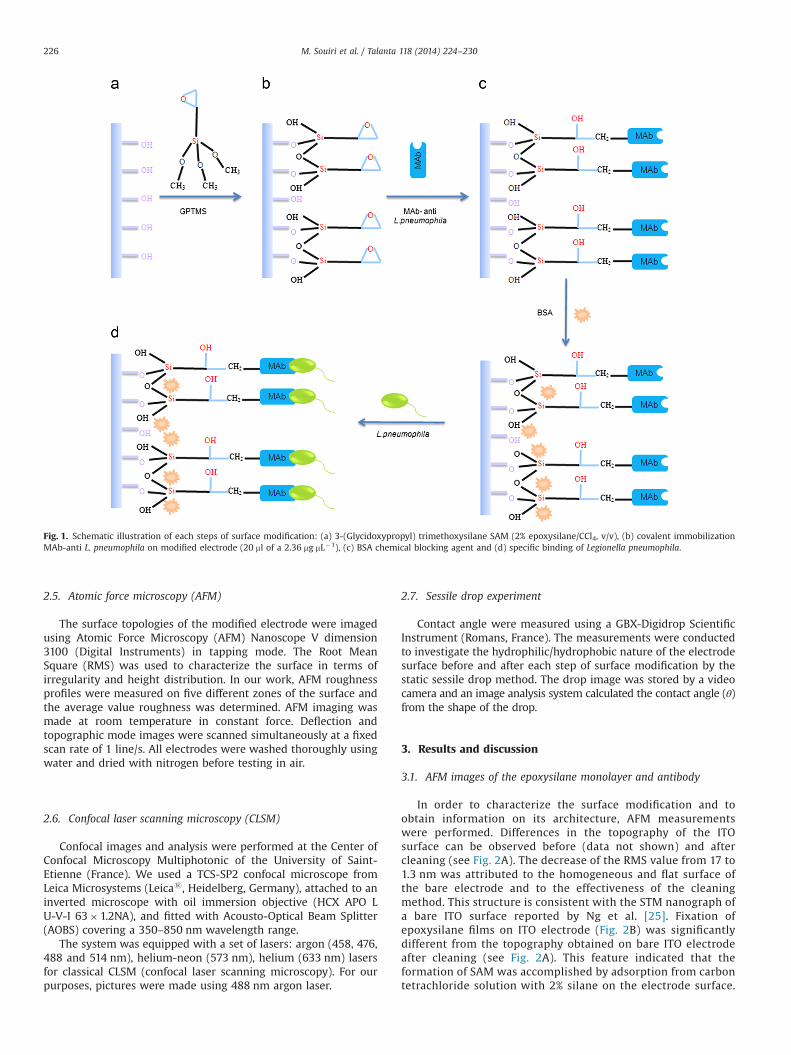
Then, 20 mL of monoclonal antibody (2.36 mg mL�1) were addedon the ITO electrode, incubated 2 h at 4 1C and washed with thePBS (0.16 M, pH 7.2). To block the non-specific binding sites on theelectrode surface, 20 mL of BSA 1% were added onto the electrodesurface, incubated 1 h at 4 1C and washed with PBS. The mono-clonal antibody modified electrode (ITO-Epoxy-MAb) was storedfor further use at 4 1C. The way to assemble the epoxysilanemonolayer and the immobilization of the anti-L. pneumophilaantibodies is shown in Fig. 1.
2.4. Electrochemical impedance spectroscopy (EIS) measurements
Impedance measurements were performed using the Volta Lab 40(PGZ 301) impedance analyzer (Radiometer Analytical SA, Villeur-banne, France) with a conventional electrochemical setup. The three-electrode consists of the ITO as working electrode, a platinum counterelectrode, and a saturated Calomel Hg/Hg2Cl2/KCl (þ0.248 V vs SHE)as the reference electrode. The impedance spectra were recorded inthe frequency range from 0.1 Hz to 100 kHz at the formal potential ofthe [Fe(CN)6]3� /4�redox couple 200 mV. The amplitude of the alter-nating voltage was 10 mV. The measurements were carried out atroom temperature (2273 1C) in a Faraday cage in order to improvethe signal-to-noise ratio and repeated three times in order to ensureresult reproducibility. The impedance data were represented in thecomplex impedance plot (Nyquist plot). The data of electrochemicalimpedance measurements were simulated with an equivalent circuitto understand the role of each electrical component in this circuit. Theelectric equivalent circuit bare electrode could be interpreted by theRandle's equivalent circuit model in which: Rs, ZW, Rct, and Cdlrepresent respectively the ohmic resistance, the Warburg impedanceresulting from diffusion of ions from bulk electrolyte to the electrodeinterface, the charge transfer resistance of the redox probe and thedouble layer capacitance. The two components in the electricequivalent circuit of an electrochemical cell, Rs and Zw, represent bulkproperties of the electrolyte solution and diffusion features of theredox probe in solution. These components are not affected bychemical transformations occurring at the electrode surface. Theother two components Rct and Cdl, depend on the dielectric andinsulating features at the electrode/electrolyte.
M. Souiri et al. / Talanta 118 (2014) 224–230 225
2.5. Atomic force microscopy (AFM)
The surface topologies of the modified electrode were imagedusing Atomic Force Microscopy (AFM) Nanoscope V dimension3100 (Digital Instruments) in tapping mode. The Root MeanSquare (RMS) was used to characterize the surface in terms ofirregularity and height distribution. In our work, AFM roughnessprofiles were measured on five different zones of the surface andthe average value roughness was determined. AFM imaging wasmade at room temperature in constant force. Deflection andtopographic mode images were scanned simultaneously at a fixedscan rate of 1 line/s. All electrodes were washed thoroughly usingwater and dried with nitrogen before testing in air.
2.6. Confocal laser scanning microscopy (CLSM)
Confocal images and analysis were performed at the Center ofConfocal Microscopy Multiphotonic of the University of Saint-Etienne (France). We used a TCS-SP2 confocal microscope fromLeica Microsystems (Leicas, Heidelberg, Germany), attached to aninverted microscope with oil immersion objective (HCX APO LU-V-I 63�1.2NA), and fitted with Acousto-Optical Beam Splitter(AOBS) covering a 350–850 nm wavelength range.
The system was equipped with a set of lasers: argon (458, 476,488 and 514 nm), helium-neon (573 nm), helium (633 nm) lasersfor classical CLSM (confocal laser scanning microscopy). For ourpurposes, pictures were made using 488 nm argon laser.
2.7. Sessile drop experiment
Contact angle were measured using a GBX-Digidrop ScientificInstrument (Romans, France). The measurements were conductedto investigate the hydrophilic/hydrophobic nature of the electrodesurface before and after each step of surface modification by thestatic sessile drop method. The drop image was stored by a videocamera and an image analysis system calculated the contact angle (θ)from the shape of the drop.
3. Results and discussion
3.1. AFM images of the epoxysilane monolayer and antibody
In order to characterize the surface modification and toobtain information on its architecture, AFM measurementswere performed. Differences in the topography of the ITOsurface can be observed before (data not shown) and aftercleaning (see Fig. 2A). The decrease of the RMS value from 17 to1.3 nm was attributed to the homogeneous and flat surface ofthe bare electrode and to the effectiveness of the cleaningmethod. This structure is consistent with the STM nanograph ofa bare ITO surface reported by Ng et al. [25]. Fixation ofepoxysilane films on ITO electrode (Fig. 2B) was significantlydifferent from the topography obtained on bare ITO electrodeafter cleaning (see Fig. 2A). This feature indicated that theformation of SAM was accomplished by adsorption from carbontetrachloride solution with 2% silane on the electrode surface.
Fig. 1. Schematic illustration of each steps of surface modification: (a) 3-(Glycidoxypropyl) trimethoxysilane SAM (2% epoxysilane/CCl4, v/v), (b) covalent immobilizationMAb-anti L. pneumophila on modified electrode (20 ml of a 2.36 mg mL�1), (c) BSA chemical blocking agent and (d) specific binding of Legionella pneumophila.
M. Souiri et al. / Talanta 118 (2014) 224–230226

The monolayer composed was dense, complete, relativelysmooth, roughly homogeneous and with only a few aggregates(RMS¼3.13 nm). This result is consistent with the report onepoxy-terminated self-assembled monolayers on single crystalsilicon and ITO surfaces [10,26]. Other studies have confirmed anon-smoothing GPTMS monolayer surface by the RMS value(RMS¼3.5 nm) [27].
The AFM topography image of the SAM after treatment byantibodies exhibited a quite different aspect, consisting indistributed round globular particles that may correspond tothe MAb bound onto the epoxysilane layer (see Fig. 2C). Theantibody molecules did not assemble as the epoxysilane mole-cules in a monolayer, because some aggregates can still beobserved on the surface and then induce an RMS increase(RMS¼6.7 nm). It could be explained by the short length ofthe alkyl chain of GPTMS.
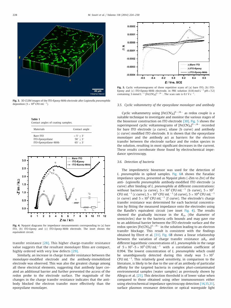
3.2. CLSM images of the specific binding of L. pneumophila sgp 1 cells
In order to verify the L. pneumophila attachment on theelectrode surface, observation of the sensor surface was performedusing a Confocal Laser Scanning Microscope (CLSM). After theelectrode preparation (see paragraph 2.3), suspensions of L.pneumophila (5�106 CFU mL�1) were incubated during 1 h atroom temperature in order to evaluate their capability to bind todetection antibodies. The biosensor was then washed using PBSand dried. A CLSM image of L. pneumophila cells successfullycaptured on the sensor surface is shown in Fig. 3.
3.3. Contact angle
A low contact angle of o51731 (Table 1) initially obtainedfor the bare ITO-glass plate after cleaning was related to surfacesuper-hydrophilic hydroxyl groups. The contact angle increasedfrom 57.61731 for the GPTMS modified ITO electrode to65.51731 after the covalent immobilization of the antibodies.This increase could be explained by the presence of hydro-phobic alkyl chains of GPTMS molecules that reduce thehydroxyl groups of the bare ITO-glass after the silanizationstep and hydrophobic nature of antibodies. These resultswere in accordance with those obtained by Luzinov et al. whoshow 511 under the same conditions but with a 1% epoxysilaneconcentration [26].
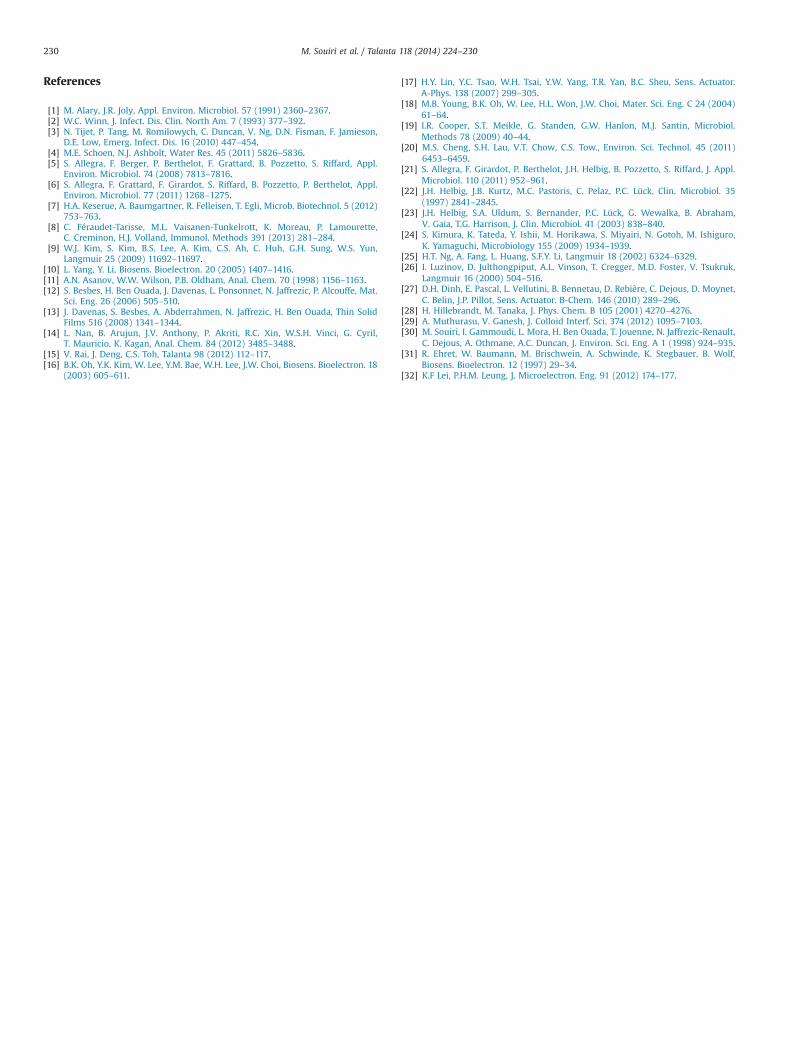
3.4. Electrochemical impedance spectroscopy of the epoxysilanemonolayer and antibody
Fig. 4 shows the impedance response of the [Fe(CN)6]3� /4�
redox probe on the bare ITO electrode (a curve), the SAM modifiedITO electrode (b curve) and the antibody dipped electrode(c curve). This increase in the charge transfer resistance (Rct, thediameter of semicircles) was observed following silanization andthen antibody binding.
It indicates that the presence of the epoxysilane monolayerformed a barrier that prevented direct approach of the redoxspecies to the electrode surface. This observation is consistentwith the result in the literature that the deposition of the SAMof alkylsiloxane on ITO surface led to a large increase in charge-
Fig. 2. AFM images of (A) bare ITO electrode after cleaning. (B) GPTMS-modified electrode and (C) MAb anti-Legionella pneumophila immobilization.
M. Souiri et al. / Talanta 118 (2014) 224–230 227
transfer resistance [28]. This higher charge-transfer resistancevalue suggests that the resultant monolayer films are compact,highly ordered with very low defects [29].
Similarly, an increase in charge transfer resistance between themonolayer-modified electrode and the antibody-immobilizedelectrode was observed. This was also the greatest change amongall these electrical elements, suggesting that antibody layer cre-ated an additional barrier and further prevented the access of theredox probe to the electrode surface. The magnitude of thechanges in the charge transfer resistance indicates that the anti-body blocked the electron transfer more effectively than theepoxysilane monolayer.
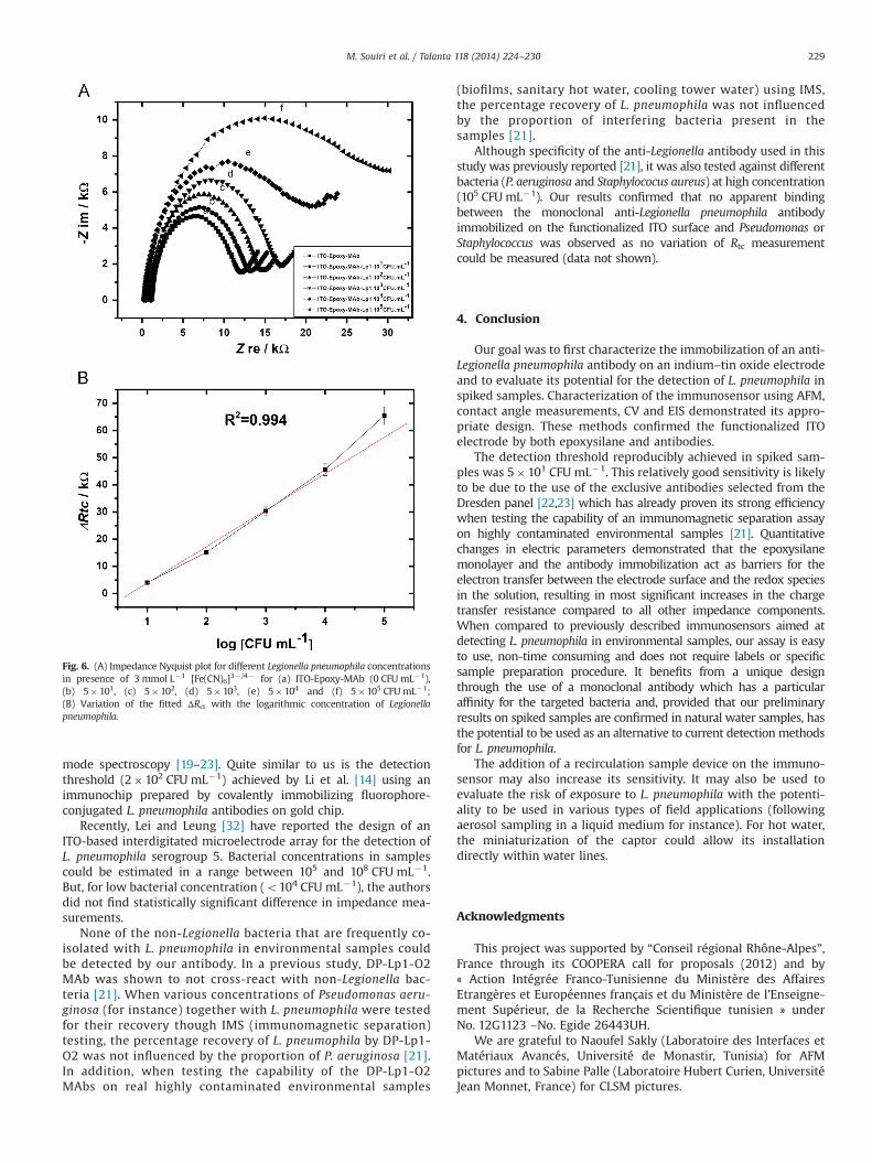
3.5. Cyclic voltammetry of the epoxysilane monolayer and antibody
Cyclic voltammetry using [Fe(CN)6]3� /4� as redox couple is asuitable technique to investigate and monitor the various stages ofthe biosensor construction on ITO electrode [30]. Fig. 5 shows thesuperimposed cyclic voltammograms of [Fe(CN)6]3�/4� recordedfor bare ITO electrode (a curve), silane (b curve) and antibody(c curve) modified ITO electrode. It is shown that the epoxysilanemonolayer and the antibody act as barriers for the electrontransfer between the electrode surface and the redox species inthe solution, resulting in most significant decreases in the current.These results corroborate those found by electrochemical impe-dance spectroscopy.
3.6. Detection of bacteria
The impedimetric biosensor was used for the detection ofL. pneumophila in spiked samples. Fig. 6A shows the Faradaicimpedance spectra, presented as Nyquist plots (-Zim vs Zre) of theanti- Legionella pneumophila antibody-modified ITO electrode (acurve) after binding of L. pneumophila at different concentrations:without bacteria (a curve), 5�101 CFU mL�1 (b curve), 5�102
CFU mL�1 (c curve), 5�103 CFU mL�1 (d curve), 5�104 CFU mL�1
(e curve) and 5�105 CFU mL�1 (f curve). The electrode's chargetransfer resistance was determined for each bacterial concentra-tion by fitting the measured impedance onto the electrodes usingthe Randle's equivalent circuit (see inset Fig. 4). The resultsshowed the gradually increase in the Rct, (the diameter ofsemicircles) due to the bacteria cells bounds and may gave riseto an additional barrier between the ITO electrode surface and theredox species [Fe(CN)6]3� /4� in the solution leading to an electrontransfer blockage. This result is consistent with the findingsobtained by Ehret et al. [31]. Fig. 6B shows a linear relationshipbetween the variation of charge transfer resistance ΔRtc anddifferent logarithmic concentrations of L. pneumophila in the rangeof 5�101–5�105 CFU mL�1 with a correlation coefficient of0.994. The lowest concentration of L. pneumophila which couldbe unambiguously detected during this study was 5�101
CFU mL�1. This relatively good sensitivity, in comparison to theliterature, is likely to be due to the use of an antibody of particularaffinity for the targeted bacteria in complex pluri-contaminatedenvironmental samples (water samples) as previously shown byAllegra et al. [21]. This detection threshold is of lower value whencompared to those obtained using other immunosensors eitherusing electrochemical impedance spectroscopy detection [14,15,31],surface plasmon resonance detection or optical waveguide light
Table 1Contact angles of coating samples.
Materials Contact angle
Bare ITO o51731ITO-Epoxysilane 581731ITO-Epoxysilane-MAb 651731
Fig. 4. Nyquist diagrams for impedance measurements corresponding to (a) bareITO, (b) ITO-Epoxy and (c) ITO-Epoxy-MAb electrode. The inset shows theequivalent circuit.
Fig. 5. Cyclic voltammograms of three repetitive scans of (a) bare ITO, (b) ITO-Epoxy and (c) ITO-Epoxy-MAb electrode, in PBS solution (0.16 mol L�1 pH¼7.2)containing 3 mmol L�1 [Fe(CN)6]3�/4� . The scan rate is 0.1 V s�1.
Fig. 3. 3D CLSM images of the ITO-Epoxy-MAb electrode after Legionella pneumophiladeposition (5�106 CFU mL�1).
M. Souiri et al. / Talanta 118 (2014) 224–230228
mode spectroscopy [19–23]. Quite similar to us is the detectionthreshold (2�102 CFU mL�1) achieved by Li et al. [14] using animmunochip prepared by covalently immobilizing fluorophore-conjugated L. pneumophila antibodies on gold chip.
Recently, Lei and Leung [32] have reported the design of anITO-based interdigitated microelectrode array for the detection ofL. pneumophila serogroup 5. Bacterial concentrations in samplescould be estimated in a range between 105 and 108 CFU mL�1.But, for low bacterial concentration (o104 CFU mL�1), the authorsdid not find statistically significant difference in impedance mea-surements.
None of the non-Legionella bacteria that are frequently co-isolated with L. pneumophila in environmental samples couldbe detected by our antibody. In a previous study, DP-Lp1-O2MAb was shown to not cross-react with non-Legionella bac-teria [21]. When various concentrations of Pseudomonas aeru-ginosa (for instance) together with L. pneumophila were testedfor their recovery though IMS (immunomagnetic separation)testing, the percentage recovery of L. pneumophila by DP-Lp1-O2 was not influenced by the proportion of P. aeruginosa [21].In addition, when testing the capability of the DP-Lp1-O2MAbs on real highly contaminated environmental samples
(biofilms, sanitary hot water, cooling tower water) using IMS,the percentage recovery of L. pneumophila was not influencedby the proportion of interfering bacteria present in thesamples [21].
Although specificity of the anti-Legionella antibody used in thisstudy was previously reported [21], it was also tested against differentbacteria (P. aeruginosa and Staphylococus aureus) at high concentration(105 CFUmL�1). Our results confirmed that no apparent bindingbetween the monoclonal anti-Legionella pneumophila antibodyimmobilized on the functionalized ITO surface and Pseudomonas orStaphylococcus was observed as no variation of Rtc measurementcould be measured (data not shown).
4. Conclusion
Our goal was to first characterize the immobilization of an anti-Legionella pneumophila antibody on an indium–tin oxide electrodeand to evaluate its potential for the detection of L. pneumophila inspiked samples. Characterization of the immunosensor using AFM,contact angle measurements, CV and EIS demonstrated its appro-priate design. These methods confirmed the functionalized ITOelectrode by both epoxysilane and antibodies.
The detection threshold reproducibly achieved in spiked sam-ples was 5�101 CFU mL�1. This relatively good sensitivity is likelyto be due to the use of the exclusive antibodies selected from theDresden panel [22,23] which has already proven its strong efficiencywhen testing the capability of an immunomagnetic separation assayon highly contaminated environmental samples [21]. Quantitativechanges in electric parameters demonstrated that the epoxysilanemonolayer and the antibody immobilization act as barriers for theelectron transfer between the electrode surface and the redox speciesin the solution, resulting in most significant increases in the chargetransfer resistance compared to all other impedance components.When compared to previously described immunosensors aimed atdetecting L. pneumophila in environmental samples, our assay is easyto use, non-time consuming and does not require labels or specificsample preparation procedure. It benefits from a unique designthrough the use of a monoclonal antibody which has a particularaffinity for the targeted bacteria and, provided that our preliminaryresults on spiked samples are confirmed in natural water samples, hasthe potential to be used as an alternative to current detection methodsfor L. pneumophila.
The addition of a recirculation sample device on the immuno-sensor may also increase its sensitivity. It may also be used toevaluate the risk of exposure to L. pneumophila with the potenti-ality to be used in various types of field applications (followingaerosol sampling in a liquid medium for instance). For hot water,the miniaturization of the captor could allow its installationdirectly within water lines.
Acknowledgments
This project was supported by “Conseil régional Rhône-Alpes”,France through its COOPERA call for proposals (2012) and by« Action Intégrée Franco-Tunisienne du Ministère des AffairesEtrangères et Européennes français et du Ministère de l'Enseigne-ment Supérieur, de la Recherche Scientifique tunisien » underNo. 12G1123 –No. Egide 26443UH.
We are grateful to Naoufel Sakly (Laboratoire des Interfaces etMatériaux Avancés, Université de Monastir, Tunisia) for AFMpictures and to Sabine Palle (Laboratoire Hubert Curien, UniversitéJean Monnet, France) for CLSM pictures.
Fig. 6. (A) Impedance Nyquist plot for different Legionella pneumophila concentrationsin presence of 3 mmol L�1 [Fe(CN)6]3�/4� for (a) ITO-Epoxy-MAb (0 CFU mL�1),(b) 5�101, (c) 5�102, (d) 5�103, (e) 5�104 and (f) 5�105 CFUmL�1;(B) Variation of the fitted ΔRct with the logarithmic concentration of Legionellapneumophila.
M. Souiri et al. / Talanta 118 (2014) 224–230 229
References
[1] M. Alary, J.R. Joly, Appl. Environ. Microbiol. 57 (1991) 2360–2367.[2] W.C. Winn, J. Infect. Dis. Clin. North Am. 7 (1993) 377–392.[3] N. Tijet, P. Tang, M. Romilowych, C. Duncan, V. Ng, D.N. Fisman, F. Jamieson,
D.E. Low, Emerg. Infect. Dis. 16 (2010) 447–454.[4] M.E. Schoen, N.J. Ashbolt, Water Res. 45 (2011) 5826–5836.[5] S. Allegra, F. Berger, P. Berthelot, F. Grattard, B. Pozzetto, S. Riffard, Appl.
Environ. Microbiol. 74 (2008) 7813–7816.[6] S. Allegra, F. Grattard, F. Girardot, S. Riffard, B. Pozzetto, P. Berthelot, Appl.
Environ. Microbiol. 77 (2011) 1268–1275.[7] H.A. Keserue, A. Baumgartner, R. Felleisen, T. Egli, Microb. Biotechnol. 5 (2012)
753–763.[8] C. Féraudet-Tarisse, M.L. Vaisanen-Tunkelrott, K. Moreau, P. Lamourette,
C. Creminon, H.J. Volland, Immunol. Methods 391 (2013) 281–284.[9] W.J. Kim, S. Kim, B.S. Lee, A. Kim, C.S. Ah, C. Huh, G.H. Sung, W.S. Yun,
Langmuir 25 (2009) 11692–11697.[10] L. Yang, Y. Li, Biosens. Bioelectron. 20 (2005) 1407–1416.[11] A.N. Asanov, W.W. Wilson, P.B. Oldham, Anal. Chem. 70 (1998) 1156–1163.[12] S. Besbes, H. Ben Ouada, J. Davenas, L. Ponsonnet, N. Jaffrezic, P. Alcouffe, Mat.
Sci. Eng. 26 (2006) 505–510.[13] J. Davenas, S. Besbes, A. Abderrahmen, N. Jaffrezic, H. Ben Ouada, Thin Solid
Films 516 (2008) 1341–1344.[14] L. Nan, B. Arujun, J.V. Anthony, P. Akriti, R.C. Xin, W.S.H. Vinci, G. Cyril,
T. Mauricio, K. Kagan, Anal. Chem. 84 (2012) 3485–3488.[15] V. Rai, J. Deng, C.S. Toh, Talanta 98 (2012) 112–117.[16] B.K. Oh, Y.K. Kim, W. Lee, Y.M. Bae, W.H. Lee, J.W. Choi, Biosens. Bioelectron. 18
(2003) 605–611.
[17] H.Y. Lin, Y.C. Tsao, W.H. Tsai, Y.W. Yang, T.R. Yan, B.C. Sheu, Sens. Actuator.A-Phys. 138 (2007) 299–305.
[18] M.B. Young, B.K. Oh, W. Lee, H.L. Won, J.W. Choi, Mater. Sci. Eng. C 24 (2004)61–64.
[19] I.R. Cooper, S.T. Meikle, G. Standen, G.W. Hanlon, M.J. Santin, Microbiol.Methods 78 (2009) 40–44.
[20] M.S. Cheng, S.H. Lau, V.T. Chow, C.S. Tow., Environ. Sci. Technol. 45 (2011)6453–6459.
[21] S. Allegra, F. Girardot, P. Berthelot, J.H. Helbig, B. Pozzetto, S. Riffard, J. Appl.Microbiol. 110 (2011) 952–961.
[22] J.H. Helbig, J.B. Kurtz, M.C. Pastoris, C. Pelaz, P.C. Lück, Clin. Microbiol. 35(1997) 2841–2845.
[23] J.H. Helbig, S.A. Uldum, S. Bernander, P.C. Lück, G. Wewalka, B. Abraham,V. Gaia, T.G. Harrison, J. Clin. Microbiol. 41 (2003) 838–840.
[24] S. Kimura, K. Tateda, Y. Ishii, M. Horikawa, S. Miyairi, N. Gotoh, M. Ishiguro,K. Yamaguchi, Microbiology 155 (2009) 1934–1939.
[25] H.T. Ng, A. Fang, L. Huang, S.F.Y. Li, Langmuir 18 (2002) 6324–6329.[26] I. Luzinov, D. Julthongpiput, A.L. Vinson, T. Cregger, M.D. Foster, V. Tsukruk,
Langmuir 16 (2000) 504–516.[27] D.H. Dinh, E. Pascal, L. Vellutini, B. Bennetau, D. Rebière, C. Dejous, D. Moynet,
C. Belin, J.P. Pillot, Sens. Actuator. B-Chem. 146 (2010) 289–296.[28] H. Hillebrandt, M. Tanaka, J. Phys. Chem. B 105 (2001) 4270–4276.[29] A. Muthurasu, V. Ganesh, J. Colloid Interf. Sci. 374 (2012) 1095–7103.[30] M. Souiri, I. Gammoudi, L. Mora, H. Ben Ouada, T. Jouenne, N. Jaffrezic-Renault,
C. Dejous, A. Othmane, A.C. Duncan, J. Environ. Sci. Eng. A 1 (1998) 924–935.[31] R. Ehret, W. Baumann, M. Brischwein, A. Schwinde, K. Stegbauer, B. Wolf,
Biosens. Bioelectron. 12 (1997) 29–34.[32] K.F Lei, P.H.M. Leung, J. Microelectron. Eng. 91 (2012) 174–177.
M. Souiri et al. / Talanta 118 (2014) 224–230230
Copyright © 2022 FDOKUMEN