Progress in understanding adjuvant immunotoxicity mechanisms
Upload
independentCategory
view
0download
0
Ari
PKa
b
c
d
e
a
ARRAA
KMN(T
1
ccaitmdcNiatn
0d
Immunology Letters 134 (2010) 26–34
Contents lists available at ScienceDirect
Immunology Letters
journa l homepage: www.e lsev ier .com/ locate /
djuvant effect of Bacillus firmus on the expression of cytokines and toll-likeeceptors in mouse nasopharynx-associated lymphoid tissue (NALT) afterntranasal immunization with inactivated influenza virus type A
eter Zanvita,∗, Ales Tichopádb, Martina Havlíckovác, Olga Novotnáa, Marie Jirkovskád,atarína Kolostováe, Dana Cechováa, Jaroslav Juláka, Ivan Sterzl a, Ludmila Prokesováa
Charles University in Prague, First Faculty of Medicine, Institute of Immunology and Microbiology, Studnickova 7, 128 00 Prague, Czech RepublicTechnical University of Munich, Physiology Weihenstephan, Weihenstephaner Berg 3, 85354 Freising, GermanyNational Institute of Public Health, Srobárova 48, 100 42 Prague 10, Czech RepublicCharles University in Prague, First Faculty of Medicine, Institute of Histology and Embryology, Albertov 4, 128 00 Prague, Czech RepublicCharles University in Prague, Third Faculty of Medicine, Department of Tumor Biology, Ruská 87, 100 00 Prague, Czech Republic
r t i c l e i n f o
rticle history:eceived 21 May 2010eceived in revised form 15 July 2010ccepted 8 August 2010vailable online 13 August 2010
eywords:ultimarker gene expression
a b s t r a c t
Due to the persisting threat of development of new highly pathogenic influenza A subtypes, a mucosalvaccination which would induce a potent and cross-protective reaction is desirable. We succeeded inmucosal immunization of mice with an inactivated influenza A virus by using delipidated Bacillus firmus(DBF) as adjuvant.
The mechanism of adjuvant effect was followed in NALT by comparing the response after intranasalimmunization by inactivated influenza virus type A (H1N1) alone, adjuvant alone (DBF), or by a mixture ofvirus + DBF. Expression of selected gene groups was tested via qPCR at 7 different time-points: cytokines
asopharynx-associated lymphoid tissueNALT)oll-like receptors (TLR)
(IL-2, IFN-�, IL-4, IL-6, and IL-10), type I interferons (IFN-�4, IFN-�11, IFN-�12, and IFN-�), toll-likereceptors (TLR2, TLR3, TLR7, and TLR9), iNOS and CCR7. Intranasally administered DBF and the mixtureof virus + DBF induced an elevated expression of IFN-�, IL-6 and IL-10 cytokines, type I interferons, iNOS,and pDC markers in NALT. Multimarker qPCR data was analyzed by relative quantification and by principalcomponent analysis.
o be aerated
DBF has been shown timmunization. DBF accel
. Introduction
The influenza virus is a globally important respiratory pathogenausing nearly annual epidemics and occasional pandemics. Vac-ination against influenza is currently conducted by parenterallydministered split or subunit vaccines. These vaccines most oftennduce satisfactory homotypical systemic immunity, particularlyhe production of IgG class serum antibodies, but they fail to induce
ucosal immunity or cross-protection. The threat of a sudden pan-emic outbreak necessitates the development of better vaccinesapable of inducing both mucosal and heterosubtypical immunity.atural infection is accompanied by the production of antibod-
es, especially of the IgA class, in the respiratory tract. They causecross-protection (usually only intrasubtypical) more efficiently
han serum antibodies. This finding implies that efficient immu-ization should be performed by the mucosal route, which should
∗ Corresponding author. Tel.: +420 22496 8509; fax: +420 22496 8455.E-mail address: [email protected] (P. Zanvit).
165-2478/$ – see front matter © 2010 Elsevier B.V. All rights reserved.oi:10.1016/j.imlet.2010.08.006
very efficient adjuvant for the stimulation of innate immunity after IN, increased, and prolonged the antiviral response.
© 2010 Elsevier B.V. All rights reserved.
induce the production of mucosal IgA class antibodies [1,2]. Atpresent, anti-influenza vaccination is mostly performed parenter-ally using an inactivated vaccine. Mucosal immunization of humansis licensed only in some countries and is used to a limited extent.Because of the presence of live viruses (cold adapted mutants) inthis vaccine, its application is connected with a certain risk andits restricted indication range excludes persons with the highestrisk – small children and old people [3]. Inactivated viruses areusually not sufficiently effective in mucosal immunization; andit is therefore necessary to use adjuvants capable of adequatelystrengthening the immune response. The use of adjuvants in vac-cination is important for several reasons. Current modern subunitvaccines are better defined and often safer than the older whole-virion vaccines. These vaccines are less reactogenic but, on the otherhand, also less immunogenic and the use of an adjuvant would
therefore be desirable [4,5]. The immunization of mice with aninactivated influenza virus, as reported in our preceding studies,caused an increase in the immune response with the aid of a bacte-rial adjuvant, a delipidated form of the G+ bacterium Bacillus firmus(DBF). As we have previously described [6,7], adjuvant immune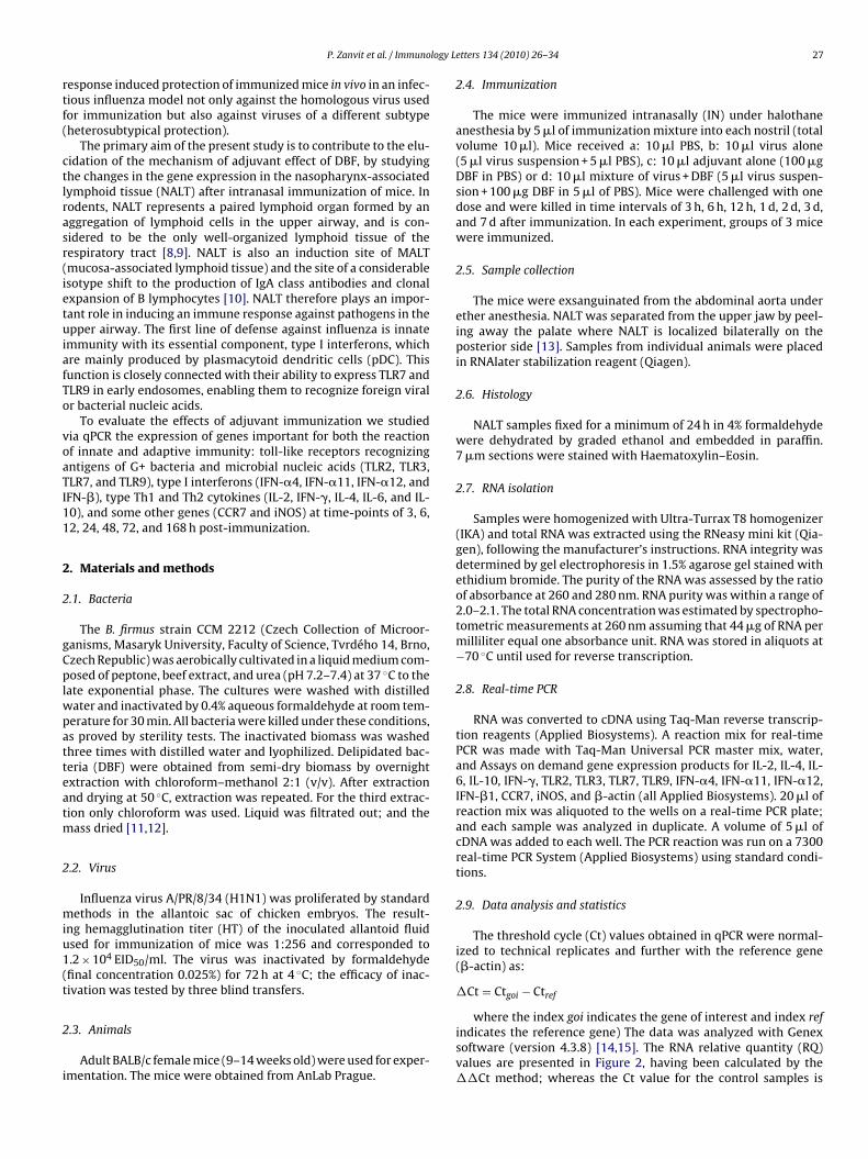
logy L
rtf(
ctlrasr(ietuiafTo
voaTI11
2
2
gCplwpatteatm
2
miu1(t
2
i
P. Zanvit et al. / Immuno
esponse induced protection of immunized mice in vivo in an infec-ious influenza model not only against the homologous virus usedor immunization but also against viruses of a different subtypeheterosubtypical protection).
The primary aim of the present study is to contribute to the elu-idation of the mechanism of adjuvant effect of DBF, by studyinghe changes in the gene expression in the nasopharynx-associatedymphoid tissue (NALT) after intranasal immunization of mice. Inodents, NALT represents a paired lymphoid organ formed by anggregation of lymphoid cells in the upper airway, and is con-idered to be the only well-organized lymphoid tissue of theespiratory tract [8,9]. NALT is also an induction site of MALTmucosa-associated lymphoid tissue) and the site of a considerablesotype shift to the production of IgA class antibodies and clonalxpansion of B lymphocytes [10]. NALT therefore plays an impor-ant role in inducing an immune response against pathogens in thepper airway. The first line of defense against influenza is innate
mmunity with its essential component, type I interferons, whichre mainly produced by plasmacytoid dendritic cells (pDC). Thisunction is closely connected with their ability to express TLR7 andLR9 in early endosomes, enabling them to recognize foreign viralr bacterial nucleic acids.
To evaluate the effects of adjuvant immunization we studiedia qPCR the expression of genes important for both the reactionf innate and adaptive immunity: toll-like receptors recognizingntigens of G+ bacteria and microbial nucleic acids (TLR2, TLR3,LR7, and TLR9), type I interferons (IFN-�4, IFN-�11, IFN-�12, andFN-�), type Th1 and Th2 cytokines (IL-2, IFN-�, IL-4, IL-6, and IL-0), and some other genes (CCR7 and iNOS) at time-points of 3, 6,2, 24, 48, 72, and 168 h post-immunization.
. Materials and methods
.1. Bacteria
The B. firmus strain CCM 2212 (Czech Collection of Microor-anisms, Masaryk University, Faculty of Science, Tvrdého 14, Brno,zech Republic) was aerobically cultivated in a liquid medium com-osed of peptone, beef extract, and urea (pH 7.2–7.4) at 37 ◦C to the
ate exponential phase. The cultures were washed with distilledater and inactivated by 0.4% aqueous formaldehyde at room tem-erature for 30 min. All bacteria were killed under these conditions,s proved by sterility tests. The inactivated biomass was washedhree times with distilled water and lyophilized. Delipidated bac-eria (DBF) were obtained from semi-dry biomass by overnightxtraction with chloroform–methanol 2:1 (v/v). After extractionnd drying at 50 ◦C, extraction was repeated. For the third extrac-ion only chloroform was used. Liquid was filtrated out; and the
ass dried [11,12].
.2. Virus
Influenza virus A/PR/8/34 (H1N1) was proliferated by standardethods in the allantoic sac of chicken embryos. The result-
ng hemagglutination titer (HT) of the inoculated allantoid fluidsed for immunization of mice was 1:256 and corresponded to.2 × 104 EID50/ml. The virus was inactivated by formaldehydefinal concentration 0.025%) for 72 h at 4 ◦C; the efficacy of inac-ivation was tested by three blind transfers.
.3. Animals
Adult BALB/c female mice (9–14 weeks old) were used for exper-mentation. The mice were obtained from AnLab Prague.
etters 134 (2010) 26–34 27
2.4. Immunization
The mice were immunized intranasally (IN) under halothaneanesthesia by 5 �l of immunization mixture into each nostril (totalvolume 10 �l). Mice received a: 10 �l PBS, b: 10 �l virus alone(5 �l virus suspension + 5 �l PBS), c: 10 �l adjuvant alone (100 �gDBF in PBS) or d: 10 �l mixture of virus + DBF (5 �l virus suspen-sion + 100 �g DBF in 5 �l of PBS). Mice were challenged with onedose and were killed in time intervals of 3 h, 6 h, 12 h, 1 d, 2 d, 3 d,and 7 d after immunization. In each experiment, groups of 3 micewere immunized.
2.5. Sample collection
The mice were exsanguinated from the abdominal aorta underether anesthesia. NALT was separated from the upper jaw by peel-ing away the palate where NALT is localized bilaterally on theposterior side [13]. Samples from individual animals were placedin RNAlater stabilization reagent (Qiagen).
2.6. Histology
NALT samples fixed for a minimum of 24 h in 4% formaldehydewere dehydrated by graded ethanol and embedded in paraffin.7 �m sections were stained with Haematoxylin–Eosin.
2.7. RNA isolation
Samples were homogenized with Ultra-Turrax T8 homogenizer(IKA) and total RNA was extracted using the RNeasy mini kit (Qia-gen), following the manufacturer’s instructions. RNA integrity wasdetermined by gel electrophoresis in 1.5% agarose gel stained withethidium bromide. The purity of the RNA was assessed by the ratioof absorbance at 260 and 280 nm. RNA purity was within a range of2.0–2.1. The total RNA concentration was estimated by spectropho-tometric measurements at 260 nm assuming that 44 �g of RNA permilliliter equal one absorbance unit. RNA was stored in aliquots at−70 ◦C until used for reverse transcription.
2.8. Real-time PCR
RNA was converted to cDNA using Taq-Man reverse transcrip-tion reagents (Applied Biosystems). A reaction mix for real-timePCR was made with Taq-Man Universal PCR master mix, water,and Assays on demand gene expression products for IL-2, IL-4, IL-6, IL-10, IFN-�, TLR2, TLR3, TLR7, TLR9, IFN-�4, IFN-�11, IFN-�12,IFN-�1, CCR7, iNOS, and �-actin (all Applied Biosystems). 20 �l ofreaction mix was aliquoted to the wells on a real-time PCR plate;and each sample was analyzed in duplicate. A volume of 5 �l ofcDNA was added to each well. The PCR reaction was run on a 7300real-time PCR System (Applied Biosystems) using standard condi-tions.
2.9. Data analysis and statistics
The threshold cycle (Ct) values obtained in qPCR were normal-ized to technical replicates and further with the reference gene(�-actin) as:
�Ct = Ctgoi − Ctref
where the index goi indicates the gene of interest and index refindicates the reference gene) The data was analyzed with Genexsoftware (version 4.3.8) [14,15]. The RNA relative quantity (RQ)values are presented in Figure 2, having been calculated by the��Ct method; whereas the Ct value for the control samples is

28 P. Zanvit et al. / Immunology Letters 134 (2010) 26–34
Fig. 1. Photomicrograph of the mice nasopharynx-associated lymphoid tissue(((t
rgagpti
efdgtbnapnsvamttVmg
3
wnwcitEaet(
3
ebia
ing
dif
fere
nce
sin
rela
tive
RN
A-a
mou
nt
(aft
erqR
T-PC
R)
betw
een
intr
anas
ally
imm
un
ized
mic
e(w
ith
viru
sal
one,
DB
Fal
one
and
com
bin
atio
nof
viru
s+
DB
F)an
dth
eir
un
imm
un
ized
con
trol
sin
7ti
me
inte
rval
s.
h6
h12
h24
h48
h72
h16
8h
iru
sD
BF
Vir
us
+D
BF
Vir
us
DB
FV
iru
s+
DB
FV
iru
sD
BF
Vir
us
+D
BF
Vir
us
DB
FV
iru
s+
DB
FV
iru
sD
BF
Vir
us
+D
BF
Vir
us
DB
FV
iru
s+
DB
FV
iru
sD
BF
Vir
us
+D
BF
.681
0.00
10.
004
0.86
60.
048
0.11
90.
244
0.66
90.
725
0.24
30.
147
0.04
90.
641
0.92
50.
051
0.71
80.
829
0.18
00.
627
0.21
70.
108
.824
0.21
40.
222
0.79
00.
057
0.79
50.
586
0.13
30.
516
0.37
10.
274
0.05
70.
877
0.11
10.
058
0.11
90.
100
0.32
30.
308
0.04
50.
304
.607
0.99
40.
002
0.14
00.
333
0.06
70.
236
0.88
30.
506
0.00
20.
367
0.18
20.
172
0.67
10.
111
0.17
50.
265
0.20
30.
855
0.29
90.
537
.727
0.05
40.
309
0.15
30.
803
0.64
80.
067
0.75
20.
961
0.14
50.
514
0.49
90.
855
0.29
50.
363
0.87
90.
036
0.81
70.
420
0.08
80.
325
.651
0.21
70.
069
0.17
90.
167
0.04
20.
252
0.49
30.
148
0.19
80.
585
0.54
50.
405
0.99
80.
090
0.46
60.
080
0.06
60.
074
0.27
10.
522
.871
0.00
70.
034
0.11
60.
004
0.37
40.
202
0.24
40.
253
0.01
70.
925
0.41
20.
437
0.52
40.
190
0.13
60.
034
0.36
30.
071
0.48
60.
418
.961
0.00
00.
001
0.74
90.
018
0.01
10.
294
0.66
90.
344
0.40
80.
010
0.01
90.
406
0.77
00.
899
0.99
90.
190
0.12
00.
163
0.84
80.
766
.485
0.09
60.
155
0.98
80.
906
0.33
70.
815
0.05
70.
788
0.36
90.
396
0.15
50.
115
0.42
60.
392
0.03
90.
819
0.04
20.
448
0.78
10.
499
.154
0.84
80.
254
0.97
60.
888
0.58
20.
044
0.90
40.
302
0.96
80.
194
0.04
40.
316
0.66
50.
526
0.51
60.
685
0.54
60.
802
0.33
00.
976
.088
0.99
70.
473
0.88
90.
543
0.72
40.
592
0.68
90.
675
0.32
70.
455
0.24
50.
863
0.85
40.
775
0.16
40.
918
0.58
60.
876
0.07
20.
565
.309
0.90
20.
022
0.54
00.
075
0.40
80.
116
0.67
20.
281
0.33
40.
922
0.07
50.
293
0.84
10.
696
0.58
70.
800
0.20
20.
193
0.19
50.
490
.177
0.79
10.
478
0.26
00.
766
0.40
60.
018
0.28
50.
018
0.58
50.
081
0.03
40.
984
0.93
50.
341
0.87
30.
254
0.81
30.
681
0.70
70.
862
.494
0.00
10.
004
0.49
80.
073
0.04
90.
058
0.37
20.
032
0.00
50.
654
0.91
00.
867
0.38
30.
135
0.47
70.
102
0.20
80.
258
0.58
60.
697
.535
0.00
10.
012
0.66
00.
014
0.12
80.
244
0.53
50.
266
0.39
80.
181
0.10
80.
322
0.07
30.
147
0.46
10.
868
0.10
10.
057
0.33
00.
887
.310
0.00
10.
014
0.24
80.
103
0.10
80.
291
0.49
50.
588
0.99
50.
301
0.04
60.
979
0.73
40.
361
0.51
50.
438
0.01
30.
773
0.37
50.
789
tmen
tgr
oup
N=
3;n
ofo
rmal
test
ofn
orm
alit
yof
the
dat
aco
uld
bep
erfo
rmed
.Th
ed
ata
wer
eth
eref
ore
log-
tran
sfor
med
toas
sure
nor
mal
dis
trib
uti
on.T
oco
mp
are
the
effe
ctof
the
imm
un
izat
ion
wit
hth
eco
ntr
olof
PBS)
atea
chti
me-
poi
nt,
anu
np
aire
dt-
test
was
calc
ula
ted
for
each
gen
e.C
omp
aris
ons
that
gen
erat
edp-
valu
e≤0
.05
wer
eco
nsi
der
edsi
gnifi
can
tan
dp
oten
tial
lyin
volv
edin
the
biol
ogic
alp
roce
ss(b
old
ed).
NALT): the follicle (F) is covered with the columnar follicle-associated epitheliumFAE); at the edge of the follicle high endothelium venules (arrows) occur; arteriesA) and nerves (N) are present in the upper part of the soft palate (SP) covered fromhe oral aspect with stratified squamous cells epithelium (SCE). Magnification = 40×.
epresented by their mean, as well as the Ct value for the treatedroups. To compare the effect of the immunization with the controlt each time-point, an unpaired t-test was calculated for each RQene expression value. As this was an explorative study, we did noterform any adjustment for multiple comparisons. Comparisonshat generated p-value ≤ 0.05 were considered potentially involvedn the biological process (see Table 1).
The method of principal component analysis (PCA) wasmployed to disclose multivariate response to the treatment. PCAor the gene expression data, as well as statistical tests, were pro-uced with SAS v. 9.1.3 for Windows. As N = 3 for each treatmentroup, no formal test of normality of the data could be performed;herefore the data was log-transformed to assure normal distri-ution. PCA involves a mathematical procedure that transforms aumber of variables (here expression values of various genes) intosmaller number of uncorrelated variables called principal com-onents, by which the dimensionality of the data is reduced to aumber that can be represented in a scatter plot (here two dimen-ions). The first principal component accounts for as much of theariance in the data as possible; and each succeeding componentccounts for as much of the remaining variance as possible. Nor-alized expression values of all responding genes were taken as
he initial variables and reduced to only two principal components,hus facilitating resolution of treatment clusters in the scatter plot.arious functional groups of genes were composed; where treat-ent groups tended to cluster distinctly, an involvement of the
roup in the studied process was presumed.
. Results
The change in gene expression in mice after IN immunizationas followed in three separate experiments because of the largeumber of genes and time intervals. In each experiment the miceere separated into controls that received only PBS (ctrl virus,
trl DBF or ctrl virus + DBF) and mice immunized either with thenfluenza virus alone (virus), with adjuvant only (DBF), or withhe combination of the influenza virus plus adjuvant (virus + DBF).xpression was determined at time-points of 3 h, 6 h, 12 h, 1 d, 3 d,nd 7 d after IN immunization in NALT (Fig. 1). Changes in genexpression after immunization were expressed by means of rela-ive quantification (RQ), as well as by principal component analysisPCA).
.1. Relative quantification (RQ)
The method of relative quantification was used to study the genexpression in groups immunized with the virus, DBF or the com-ination virus + DBF, relative to the expression measured in mice
mmunized with PBS (ctrl virus, ctrl DBF, ctrl virus + DBF). Resultsre shown in Fig. 2 and their significance is given in Table 1. Indi- Ta
ble
1p-
Val
ues
refl
ect 3 V
TLR
20
TLR
30
TLR
70
TLR
90
IL-2
0IL
-40
IL-6
0IL
-10
0IF
N-�
40
IFN
-�11
0IF
N-�
120
IFN
-�1
0IF
N-�
0iN
OS
0C
CR
70
As
for
each
trea
(IN
app
lica
tion

P. Zanvit et al. / Immunology Letters 134 (2010) 26–34 29
Fig. 2. mRNA relative quantity (RQ) in nasopharynx-associated lymphoid tissue (NALT) in 7 time intervals after immunization. Results were calculated from normalizedgene expression data. The RQ values indicate the difference between the treated group and the controls. For each compared group the RQ average values ± S.E.M. areshown. Asterisks indicate significant differences between treated groups (with virus alone, DBF alone and mixture of virus + DBF) and their controls (*p < 0.05; **p < 0.01 and***p < 0.001).

3 logy Le
vg
owfcdvIIaeane(o
3
secfetgatIcoetitmvgmidq
cwttitdowatiaitiomdA
0 P. Zanvit et al. / Immuno
idual groups of immunized mice exhibited different dynamics ofene expression.
These results imply that the main changes in gene expressionccur during the first 24 h after immunization. Groups immunizedith DBF alone or with the combination of virus + DBF display very
ast changes, peaking at 3 and 6 h post-immunization. Significanthanges resulting from immunization with the virus alone wereetected later – 12 and 24 h after treatment. Immunization with theirus alone mainly increases the expression of TLR7, IFN-�, and typeinterferons. DBF significantly increases the expression of TLR2,
L-6, iNOS, IFN-�, and CCR7; and lowers the expression of IL-4. Rel-tive to the virus alone, the combination virus + DBF accelerates thexpression of TLR7 and type I interferons; when compared with DBFlone it increases the expression of TLR7, CCR7, and iNOS. Immu-ization with the combination virus + DBF has a faster and longerffects (3–72 h) on the increase in expression of type I interferonsparticularly IFN-�4 and IFN-�1) than treatment with virus aloner DBF alone.
.2. PCA analysis
The PCA method makes it possible to reduce a multidimensionalpace to two-dimensional one, thereby facilitating the graphicalxpression of differences between individual immunizations. Toarry out PCA analysis, individual genes were divided into fourunctional groups. This enabled us to study the differences in genexpression between these functional groups of genes at individualime-points and in individual immunized groups. The functionalroups were as follows: (1) TLR7, TLR9, and CCR7 – genes char-cteristic for pDC; (2) IFN-�4, IFN-�11, IFN-�12, and IFN-�1 –ype I interferons; (3) IL-4, IL-6, and IL-10 – Th2 cytokines; (4)L-2 and IFN-� – Th1 cytokines. Individual groups of mice wereharacterized by ellipses, indicating their range of values. Light col-red ellipses represent for non-immunized controls, dark coloredllipses illustrate the state after immunization (blue: immuniza-ion with virus alone, red: immunization with DBF alone, green:mmunization with combination virus + DBF). The distance of con-rol ellipses (ctrl) from the ellipses corresponding to groups of
ice immunized with virus alone, DBF alone, or the combinationirus + DBF, reflects the magnitude of difference between theseroups. It should be noted that this plot does not provide infor-ation on whether the difference is due to a decrease or increase
n expression relative to the expression in controls. Increased orecreased expression can be inferred from the results of relativeuantification.
At 3 h post-immunization the difference in expression of genesharacteristic for pDC in the group immunized with the virus aloneere very low in contrast to the group immunized with DBF; with
he largest change found in mice immunized with the combina-ion virus + DBF. Expression of genes encoding type I interferonsn this interval was most conspicuous in groups immunized withhe virus alone and those immunized with virus + DBF. The largestifference in expression of groups of genes characteristic for Th1r Th2 response was detected in groups immunized with DBF orith virus + DBF. At this time-point, immunization with the virus
lone had a Th1 polarization (Fig. 3a). At 6 h post-immunization,he groups immunized with the virus alone and with DBF, exhib-ted alterations in expression of genes characteristic for pDC; and
still more conspicuous difference in expression in the groupmmunized with virus + DBF. No marked difference was found inhe expression of genes typical for type I interferons. The group
mmunized with the virus alone exhibited a dominant expressionf genes characteristic for Th1 as compared with Th2, in contrast toice immunized with DBF or with virus + DBF, in which a markedifference in both Th1 and Th2 responses was detected (Fig. 3b).t the 12 h time-point, no pronounced change in the expression
tters 134 (2010) 26–34
of genes typical for pDC was found between individual groups ofmice. The largest difference in expression of genes encoding type Iinterferons was detected in the group immunized with virus + DBF,relative to groups immunized with the virus alone or with DBF.Groups immunized with the virus alone or with the virus + DBFshowed a prevalence of Th1 over Th2 response, while mice immu-nized with DBF exhibited a marked difference in both Th1 and Th2responses (Fig. 3c). At 24 h post-immunization the genes character-istic for pDC displayed the largest difference in groups immunizedwith virus alone or with virus + DBF, relative to the group immu-nized only with DBF. The biggest difference in expression of genesencoding type I interferons was found in the group immunized withthe virus alone or with the virus + DBF. Mice immunized with DBFor virus + DBF displayed a difference mainly in the expression ofgenes characteristic for Th2 response. The group immunized withthe virus alone showed a change in the expression of genes typicalfor both Th1 and Th2 responses (Fig. 3d).
The results of PCA imply that the most pronounced changes inthe group of genes characteristic for pDC were detected in the groupimmunized with the virus + DBF. This group exhibits a similaritybetween the expression of genes of the functional groups for pDCand genes for type I interferons. Groups immunized with the virusalone evince a probable activation of pDC only later at 12–24 hpost-immunization. In contrast, the group immunized with thevirus + adjuvant exhibited a marked change in expression of genestypical for pDC occurring very early on 3 h post-immunization; andthis increased expression persisted until 24 h. Immunization withDBF alone elicited a mild difference in the expression of genes char-acteristic for both pDC and type I interferons. Mice immunized withthe virus alone showed a Th1 polarization of the immune responseat 3–12 h post-immunization, however; 24 h after immunizationthe response was rather Th1/Th2 mixed. In groups immunized withDBF or virus + DBF, the type of immune response in the intervalof 3–12 h post-immunization was mixed (Th1/Th2); at 12 h it wasslightly polarized towards Th1; and at 24 h it was Th2-skewed.
The most efficient in terms of protection of the organism againstvirus infection appears to be immunization with the combinationvirus + DBF, which leads to a fast antiviral immune response basedon pDC activation, the production of interferons, and the activationof a mixed Th1/Th2 immune response.
4. Discussion
The adjuvant used in our previous studies, the delipidatedform of the nonpathogenic environmental G+ bacterium B. firmus,has previously been reported to have marked immunostimula-tory properties in both in vitro and in vivo tests [16–19]. Evenin high doses inactivated bacteria do not damage the viabilityof tissue culture cells; and mice tolerate high doses very wellof not only inactivated but also live bacteria (intraperitoneal ormucosal application, Prokesova – not published data). The excellentadjuvant properties of this bacterium were first described for intra-tracheal immunization of mice in combination with the proteinantigen ovalbumin [20,21], and later for intratracheal immuniza-tion with inactivated type B and also type A influenza viruses.Immunization with inactivated influenza viruses in combinationwith the adjuvant induced a high antibody response, both in serum(high production of class IgG antibodies) and in respiratory tractmucosa (high production of class IgA antibodies) and providedin vivo protection against influenza infection. The adjuvant was
also demonstrated to have an effect on the induction of a markedintersubtypic cross-protection [6,7]. We are aware of differencesbetween mouse and human immune systems and influenza virusis not natural pathogen for mice. Nevertheless, mice are used byabsolute majority of researchers as model animal for influenza
logy L
sptf
Fc–
P. Zanvit et al. / Immuno
tudies. Strain A/PR/8/34 H1N1 was selected as a strain stronglyathogenic for mice and as a strain commonly used in experimen-al work. Moreover, the strain mentioned above is currently usedor construction of vaccinal influenza virus reassortants.
ig. 3. (a–d) Principal component analysis (PCA). Results point out the differences in thontrols, for the 4 different gene functional groups (pDC, type I interferons and Th1/Th2 cyvirus immunization, dark red – DBF, and dark green – combination of virus + DBF) and t
etters 134 (2010) 26–34 31
The present study should contribute to clarifying the mecha-nism of adjuvant effect of DBF. The evaluation of the action of theadjuvant was based on changes in gene expression in NALT afterintranasal immunization. In rodents, NALT is localized on both sides
e gene expression patterns between the treated groups and their correspondingtokines). The dark color has been used for imaging of the treated groups (dark bluehe light color for their corresponding controls.

32 P. Zanvit et al. / Immunology Letters 134 (2010) 26–34
(Cont
oaIam
Fig. 3.
f the nasopharyngeal duct on the upper side of the soft palate,
nd is considered an analogue of the Waldeyer’s ring in humans.t consists of a number of follicles and includes regions rich in Bnd T lymphocytes, antigen presenting cells – dendritic cells andacrophages. The intrafollicular region contains high endotheliuminued )
venules (HEVs), expressing a large amount of adhesive molecules,
in which lymphocytes from blood circulation are captured andpenetrate into the follicles. The epithelium covering NALT (so-called follicle-associated epithelium, FAE) contains a large numberof antigen-transporting cells (M-cells). NALT is thus composed of
logy L
litttoa[oarbg
fmtsiitaw4oiadeeDIIaaDsaIPar
biTattn(aAbasntiOatnotp
P. Zanvit et al. / Immuno
ymphoid cells that function in inducing and regulating the mucosalmmune response [9,22]. Expression of mRNA of cytokines charac-eristic for Th1 and Th2 response in CD4+ cells in NALT was usedo demonstrate a marked predominance of cytokines characteris-ic for Th0; thus indicating that these T lymphocytes are capablef inducing a very fast activation of immune response of both Th1nd Th2 types after recognition of the antigen on the nasal mucosa23]. Intranasal administration of an antigen (e.g. bacterial cell wallr virus-associated antigens) in combination with cholera toxin asmucosal adjuvant led to a marked activation of the Th2 immune
esponse and the stimulation of IgA producing B lymphocytes inoth upper and lower airways, as well as in the intestinal and uro-enital tract [23,24].
Our results show that immunization with DBF alone has a veryast effect, affecting the expression of the genes under study very
arkedly already 3 h after immunization and decreases at laterime-points. This is supported by the fact that bacterial adjuvantsupport mainly innate immunity. Affecting innate immunity then,nfluences adaptive immunity. DBF causes a highly significant earlyncrease in expression of IFN-�, which can considerably supporthe immune response of the Th1 type, important in the defensegainst viral infection. PCA analysis indicates that immunizationith DBF brings about a marked change in type Th2 cytokines (IL-
, IL-10, and IL-6); however, this cannot be ascribed to stimulationf Th2 response because the expression of IL-4 is not significantlyncreased at any time-point – in fact at the first time-points of 3nd 6 h, the expression of IL-4 is significantly lowered. The largeifferences in PCA are apparently due to a markedly increasedxpression of IL-6, which along with the concomitantly increasedxpression of iNOS, is mainly caused by the inflammatory action ofBF. At the same time, DBF tends to also increase the expression of
L-10. An environment with increased concentrations of IL-6 andL-10 is known to support the production of IgA, with the IgA classntibodies being an important component of mucosal protectiongainst influenza virus. In contrast to the group immunized withBF, mice immunized with the virus alone exhibited delayed and
hort-term changes in the followed genes expression. Only 24 hfter immunization was a significant increase in expression of bothL-4 and IFN-� demonstrated. Comparison of the results of RQ andCA indicates that the onset of the Th1 type response occurs 12 hfter immunization; therefore there appears to be a mixed Th1/Th2esponse with a slight Th1 accent.
Adjuvant immunization with virus + DBF brings about a com-ination of the observed responses to the virus and to DBF
ndividually. The response is fast, protracted, and has a mixedh1/Th2 character: the Th1 response being the strongest after 3nd 6 h, while with Th2 after 12 and 24 h. At later time-pointshe changes in expression are small. Innate immunity representshe first defense line in protection against viruses. Innate immu-ity cells express on their surface “pattern recognition receptors”PRR), which can affect the synthesis of type I interferons as wells the synthesis of pro-inflammatory cytokines after activation.
total of 13 types of toll-like receptors (TLR) that have so fareen described in mice as capable of specific recognition of char-cteristic pathogen-associated molecular patterns (PAMP) on theurface of microbes. TLR2 recognizes different bacterial compo-ents such as lipoproteins, lipopeptides, and peptidoglycans and ishe principal receptor for the recognition of G+ bacteria [25]. TLR2s thus important for recognition of DBF obtained from G+ B. firmus.ur results point to a significantly increased expression of TLR2t the time interval of 3–6 h after immunization in mouse groups
hat were immunized either with DBF alone or with the combi-ation virus + DBF. TLR3 and TLR7 participate in the recognitionf influenza virus in certain cell populations. After the recogni-ion of the influenza virus by TLR7 present in early endosomes ofDC, a strong production of type I interferons sets in [26]. TLR9,etters 134 (2010) 26–34 33
which is also expressed in pDC, is able to recognize nonmethy-lated CpG regions in viral and bacterial ssDNA [27,28]; thus thisreceptor could also participate in the recognition of the bacte-rial adjuvant. Mature pDC are also characterized by an increasedexpression of the chemokine receptor CCR7, which plays a keyrole in the migration of pDC to lymph nodes. Our data demon-strate strong activation of genes characteristic for pDC (TLR7, TLR9,and CCR7), mainly in the group immunized with virus + DBF, attime-points soon after immunization. DBF alone does not causeany marked increase in TLR7 expression. The group immunizedwith the virus alone evinced only a non-significant increase in TLR7expression 3 and 12 h after immunization, whereas the increaseafter 24 h was already significant. These data imply that immuniza-tion with the virus alone and with virus + DBF activates pDC, theimmunization with virus + DBF causing a more marked and fasteractivation of genes typical for pDC. The increase in TLR3 expressionwas at the limit of significance, especially in the group immunizedwith virus + DBF in the interval of 24–72 h; whereas groups immu-nized with the virus alone or with DBF exhibited a relatively weakerincrease in TLR3 expression.
Plasmacytoid dendritic cells, as the main producers of type Iinterferons, ensure an early innate protection against viral infec-tion. The mouse genome contains 14 known IFN-� genes and 3IFN-� pseudogenes. The highest anti-proliferation and antiviralactivity relative to IFN-�1 is exhibited especially by IFN-�4, IFN-�11, INF-�12, and IFN-� [29]. For instance the activity of IFN-�4 is5–10× higher than that of IFN-�1 [30,31]. A two-step mechanism ofexpression has been described in interferons [32,33]. Transcriptionof genes encoding IFN-�4 and IFN-� takes place very early afterviral infection and is governed by the transcription factor IRF3.Transcription of further genes of interferons is then controlled bytranscription factor IRF7. Viral infection first activates the expres-sion of IFN-�4 and IFN-�; then followed by an increase in theexpression of other interferon types. Our data indicate that in thegroup immunized with the virus + DBF, the increase in expressionof interferons IFN-�4, IFN-�11, IFN-�12, and IFN-� begins earlyafter immunization and can be detected at all time-points from 3to 72 h, with a significant increase in IFN-�4 and IFN-� expression24 h after immunization. The group immunized with the virusalone exhibited a significantly increased expression of IFN-�4 andIFN-� at 12 h post-immunization, while the group immunizedwith DBF showed only a non-significant increase in the expressionof these genes 6 and 24 h after immunization. Type I interferonsreleased by pDC not only prevent viral infection but also activateNK cells, myeloid dendritic cells (mDC), as well as B and T lympho-cytes; and therefore participate in the regulation of both innate andadaptive elements of immunity. Adjuvant immunization extendsthis period during which the genes for type I interferons areactivated.
Intranasal immunization with inactivated influenza virus incombination with our bacterial adjuvant increases the expressionof a number of genes encoding the components of both innate andadaptive immunity. This results in an acceleration and extension ofexpression of genes for type I interferons, which is caused by theactivation of pDC. Later mDC are also apparently activated, whichare responsible for the expression of TLR3. Adjuvant immuniza-tion also supports the development of type Th1 immune response,which is indispensable for the development of cellular immunity,playing an important role in antiviral defense. The bacterial adju-vant supports the expression of inflammation mediators that canalso positively affect antigen presentation and a specific immune
response.Our study represents a screening of the expression of a largenumber of genes at a number of time-points after IN immunization.The products of these genes participate in the activation of bothinnate and adaptive immunity.

3 logy Le
A
at
R
[
[
[
[
[
[
[
[
[
[
[
[
[
[
[
[
[
[
[
[
[
[
4 P. Zanvit et al. / Immuno
cknowledgements
This work was supported by the Ministry of Education, Youth,nd Sports of the Czech Republic, grant MSM0021620806 and byhe Grant Agency of the Czech Republic, grant 310/07/0675.
eferences
[1] Liew FY, Russell SM, Appleyard G, Brand CM, Beale J. Cross-protection in miceinfected with influenza A virus by the respiratory route is correlated with localIgA antibody rather than serum antibody or cytotoxic T cell reactivity. Eur JImmunol 1984;14(April (4)):350–6.
[2] Waldman RH, Wigley FM, Small Jr PA. Specificity of respiratory secretion anti-body against influenza virus. J Immunol 1970;105(December (6)):1477–83.
[3] Hoffmann E, Mahmood K, Chen Z, et al. Multiple gene segments control thetemperature sensitivity and attenuation phenotypes of ca B/Ann Arbor/1/66. JVirol 2005;79(September (17)):11014–21.
[4] Hunter RL. Overview of vaccine adjuvants: present and future. Vaccine2002;20(May (Suppl. 3)):S7–12.
[5] Ulmer JB, Valley U, Rappuoli R. Vaccine manufacturing: challenges and solu-tions. Nat Biotechnol 2006;24(November (11)):1377–83.
[6] Zanvit P, Havlickova M, Tacner J, Jirkovska M, Petraskova P, Novotna O, et al.Immune response after adjuvant mucosal immunization of mice with inacti-vated influenza virus. Immunol Lett 2005;97(March (2)):251–9.
[7] Zanvit P, Havlickova M, Tacner J, Novotna O, Jirkovska M, Cechova D, et al. Pro-tective and cross-protective mucosal immunization of mice by influenza virustype A with bacterial adjuvant. Immunol Lett 2008;115(January (2)):144–52.
[8] Kraal Georg. Nasal-associated lymphoid tissue. Mucosal immunology. 3rd ed.Elsevier; 2005. p. 415–22.
[9] Kuper CF, Koornstra PJ, Hameleers DM, Biewenga J, Spit BJ, Duijvestijn AM, etal. The role of nasopharyngeal lymphoid tissue. Immunol Today 1992;13(June(6)):219–24.
10] Cesta MF. Normal structure, function, and histology of mucosa-associated lym-phoid tissue. Toxicol Pathol 2006;34(5):599–608.
11] Carroll KK, Cutts JH, Murray EG. The lipids of Listeria monocytogenes. Can JBiochem 1968;46(August (8)):899–904.
12] Folch J, Lees M, Sloane Stanley GH. A simple method for the isolation andpurification of total lipids from animal tissues. J Biol Chem 1957;226(May(1)):497–509.
13] Asanuma H, Thompson AH, Iwasaki T, Sato Y, Inaba Y, Aizawa C, et al. Isolationand characterization of mouse nasal-associated lymphoid tissue. J Immunol-Methods 1997;202(March (2)):123–31.
14] Kubista M, Sindelka R, Tichopad A, Bergkvist A, Lindh D, Forootan A. The primetechnique. Real-time PCR data analysis. GIT Lab J 2007;9/10(33–35).
15] Livak KJ, Schmittgen TD. Analysis of relative gene expression data using real-time quantitative PCR and the 2−��CT method. Methods 2001;25:402–8.
16] Prokesova L, Novakova M, Julak J, Mara M. Effect of Bacillus firmus andother sporulating aerobic microorganisms on in vitro stimulation of humanlymphocytes. A comparative study. Folia Microbiol (Praha) 1994;39(6):501–4.
[
[
tters 134 (2010) 26–34
17] Prokesova L, Mlckova P, Stankova I, Chloubova A, Novotna V, Ladmanova P, etal. Effect of Bacillus firmus on antibody formation after mucosal and parenteralimmunization in mice. Immunol Lett 1998;64(December (2/3)):161–6.
18] Prokesova L, Mlckova P, Stankova I, Ladmanova P, Jezkova J, Chalupna P, etal. Immunostimulatory effect of Bacillus firmus on mouse lymphocytes. FoliaMicrobiol (Praha) 2002;47(2):193–7.
19] Zidek Z, Tuckova L, Mara M, Barot-Ciorbaru R, Prokesova L, Tlaskalova-Hogenova H. Stimulation of macrophages by Bacillus firmus: production of nitricoxide and cytokines. Int J Immunopharmacol 1998;20(July (7)):359–68.
20] Mlckova P, Cechova D, Chalupna P, Novotna O, Prokesova L. Enhanced systemicand mucosal antibody responses to a model protein antigen after intranasaland intratracheal immunisation using Bacillus firmus as an adjuvant. ImmunolLett 2001;77(May (1)):39–45.
21] Mlckova P, Polacek M, Cechova D, Maruskova L, Stankova I, Chalupna P, et al.Intratracheal and intranasal immunization with ovalbumin conjugated withBacillus firmus as a carrier in mice. Folia Microbiol (Praha) 2005;50(3):247–53.
22] Kuper CF, Hameleers DM, Bruijntjes JP, van der Ven I, Biewenga J, Sminia T.Lymphoid and non-lymphoid cells in nasal-associated lymphoid tissue (NALT)in the rat. An immuno- and enzyme-histochemical study. Cell Tissue Res1990;259(February (2)):371–7.
23] Hiroi T, Iwatani K, Iijima H, Kodama S, Yanagita M, Kiyono H. Nasal immunesystem: distinctive Th0 and Th1/Th2 type environments in murine nasal-associated lymphoid tissues and nasal passage, respectively. Eur J Immunol1998;28(October (10)):3346–53.
24] Yanagita M, Hiroi T, Kitagaki N, Hamada S, Ito HO, Shimauchi H, et al. Nasopha-ryngeal associated lymphoreticular tissue (NALT) immunity: fimbriae-specificTh1 and Th2 cell regulated IgA responses for the inhibition of bacterial attach-ment to epithelial cells and subsequent inflammatory cytokine production. JImmunol 1999;162(March (6)):3559–65.
25] Akira S, Takeda K. Toll-like receptor signalling. Nat Rev Immunol 2004;4(July(7)):499–511.
26] Diebold SS, Kaisho T, Hemmi H, Akira S, Reis e Sousa. Innate antiviral responsesby means of TLR7-mediated recognition of single-stranded RNA. Science2004;303(March (5663)):1529–31.
27] Hemmi H, Takeuchi O, Kawai T, Kaisho T, Sato S, Sanjo H, et al. A Toll-likereceptor recognizes bacterial DNA. Nature 2000;408(December (6813)):740–5.
28] Uematsu S, Akira S. Toll-like receptors and Type I interferons. J Biol Chem2007;282(May (21)):15319–23.
29] Van Pesch V, Lanaya H, Renauld JC, Michiels T. Characterization of the murinealpha interferon gene family. J Virol 2004;78(August (15)):8219–28.
30] Van HM, Bosveld IJ, Mooren AA, Trapman J, Zwarthoff EC. Properties of nat-ural and hybrid murine alpha interferons. J Gen Virol 1986;67(October (Pt10)):2215–22.
31] Van HM, Bosveld IJ, Klaassen P, Zwarthoff EC, Trapman J. Structure-functionanalysis of murine interferon-alpha: antiviral properties of novel hybrid inter-ferons. J Interferon Res 1988;8(February (1)):5–14.
32] Marie I, Durbin JE, Levy DE. Differential viral induction of distinct interferon-alpha genes by positive feedback through interferon regulatory factor-7. EMBOJ 1998;17(November (22)):6660–9.
33] Sato M, Suemori H, Hata N, Asagiri M, Ogasawara K, Nakao K, et al. Distinct andessential roles of transcription factors IRF-3 and IRF-7 in response to virusesfor IFN-alpha/beta gene induction. Immunity 2000;13(October (4)):539–48.
Copyright © 2022 FDOKUMEN