Acute Diverticulitis - MTQIP
182
Acute Diverticulitis Andrew B. Peitzman, MD Mark M. Ravitch Professor of Surgery University of Pittsburgh
-
Upload
khangminh22 -
Category
Documents
-
view
3 -
download
0
Transcript of Acute Diverticulitis - MTQIP
Acute Diverticulitis
Andrew B. Peitzman, MD Mark M. Ravitch Professor of
Surgery University of Pittsburgh
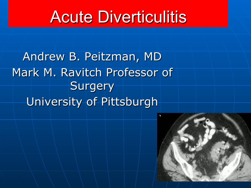
Focus today: when to operate n Recurrent, uncomplicated
diverticulitis; after how many episodes?
n Younger pts ??, immunosuppressed pts
n Complicated diverticulitis n What operation:
• Hartman resection: open or laparoscopic • Resection and anastomosis: open or lap. • Laparoscopic lavage and drainage
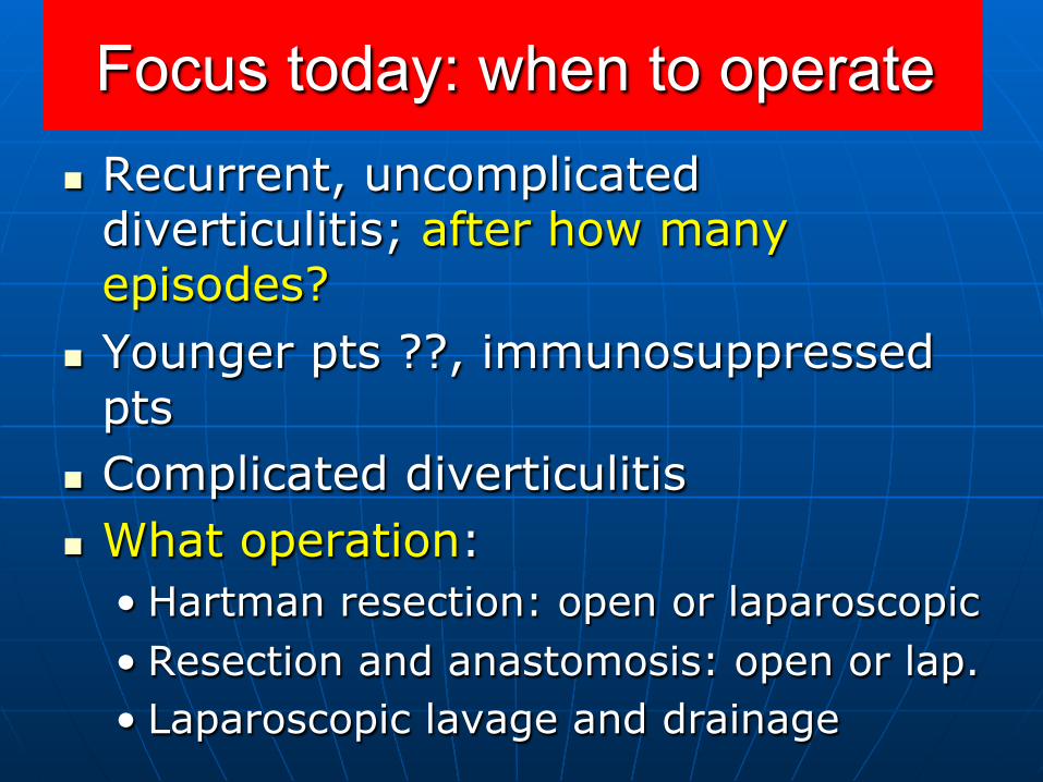
Epidemiology
Diverticulosis of the Colon: • 10% in people younger than 40 years • 50-65% in people older than 80 years • Asymptomatic : 80% • Symptoms : 20%
n Diverticulitis 15% • Uncomplicated (80%) • Complicated (20%)
n Perforation n Fistula n Obstruction
n Hemorrhage 5%
Stollman N, Raskin JB: Diverticular disease of the colon Lancet 2004; 363: 631–39
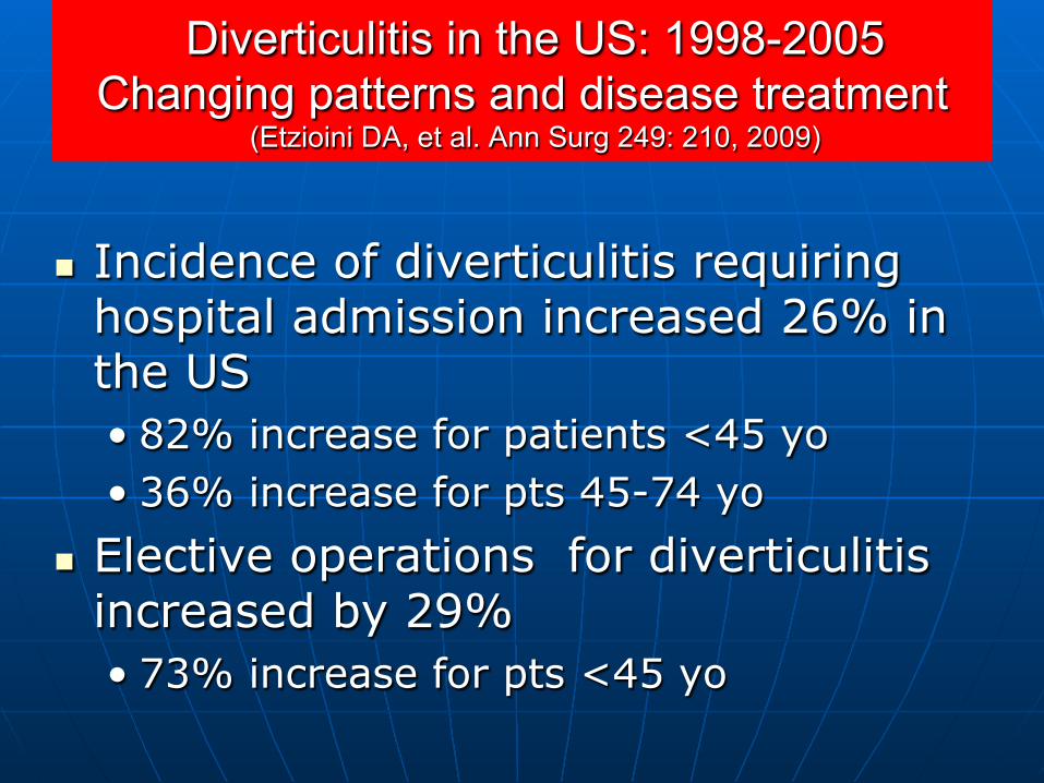
Diverticulitis in the US: 1998-2005 Changing patterns and disease treatment
(Etzioini DA, et al. Ann Surg 249: 210, 2009)
n Incidence of diverticulitis requiring hospital admission increased 26% in the US • 82% increase for patients <45 yo • 36% increase for pts 45-74 yo
n Elective operations for diverticulitis increased by 29% • 73% increase for pts <45 yo
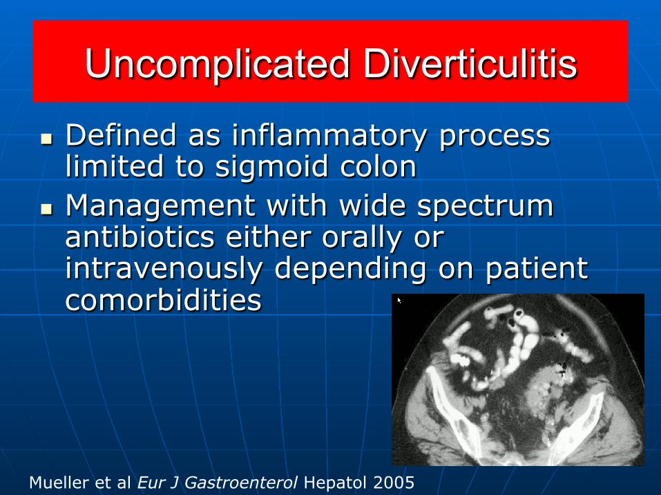
Uncomplicated Diverticulitis
n Defined as inflammatory process limited to sigmoid colon
n Management with wide spectrum antibiotics either orally or intravenously depending on patient comorbidities
Mueller et al Eur J Gastroenterol Hepatol 2005
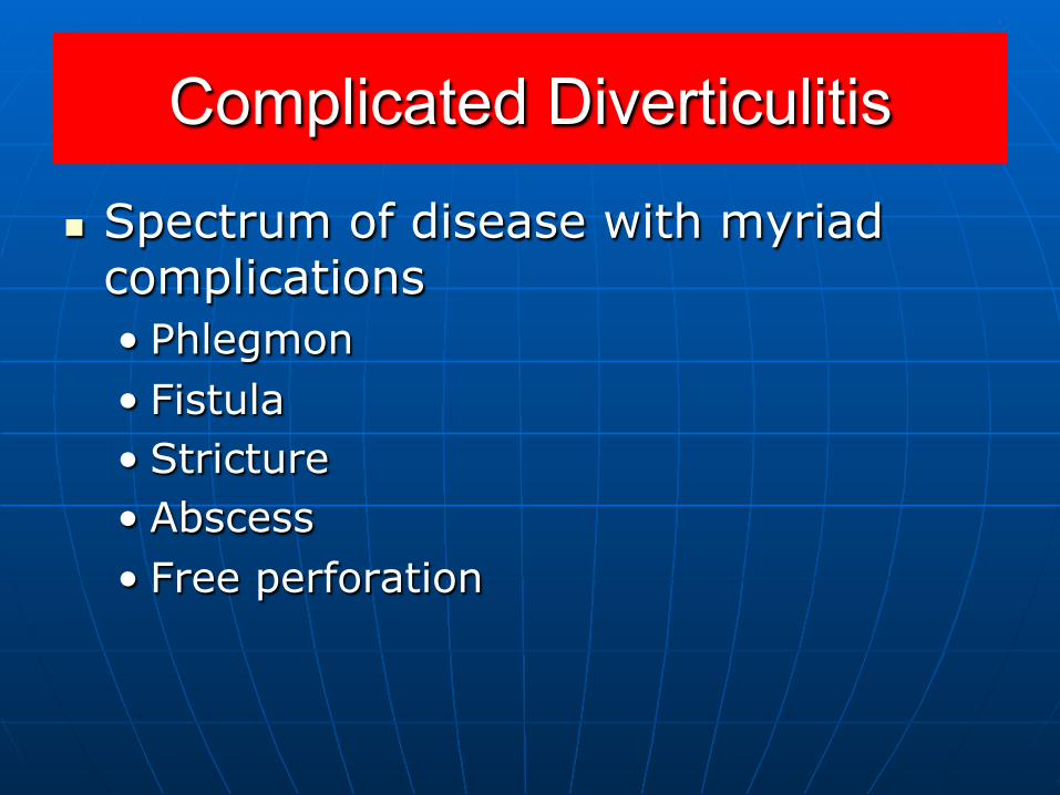
Complicated Diverticulitis
n Spectrum of disease with myriad complications • Phlegmon • Fistula • Stricture • Abscess • Free perforation
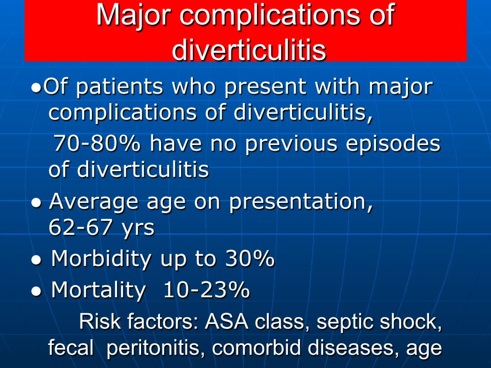
Major complications of diverticulitis
●Of patients who present with major complications of diverticulitis,
70-80% have no previous episodes of diverticulitis
● Average age on presentation, 62-67 yrs ● Morbidity up to 30% ● Mortality 10-23%
Risk factors: ASA class, septic shock, fecal peritonitis, comorbid diseases, age
Hartman resection
n High morbidity and mortality n Average age 62-65 yo n Comorbid disease n Morbidity of the takedown of the
colostomy n Resect only when is necessary to
deal with the perforation n One-third of pts never have the
colostomy reversed
The facts
n The risk of recurrent diverticulitis after an uncomplicated episode is 2% per year
n Average age on presentation is 62-67 yo, thus ~ 18-25% lifetime risk of recurrence in the typical pt (Broderick-Villa, Arch Surg, 2005)
n The risk of free perforation decreases with each bout of diverticulitis (Holmer, Langenbecks Arch Surg, 2011; Guzzo, Dis Colon Rectum, 2004; Anaya, Arch Surg, 2005)
Important principle: major change in approach
n So, when we operate for repeated bouts of uncomplicated diverticulitis we are generally operating to eliminate symptoms --------------- NOT to prevent an episode of diverticulitis with perforation
Important principle
n You need to perform elective resections on 13 patients to prevent that one pt who is destined to perforate
n The morbidity and mortality of the 13 operations is too high to justify such an approach

When to operate after uncomplicated diverticulitis?
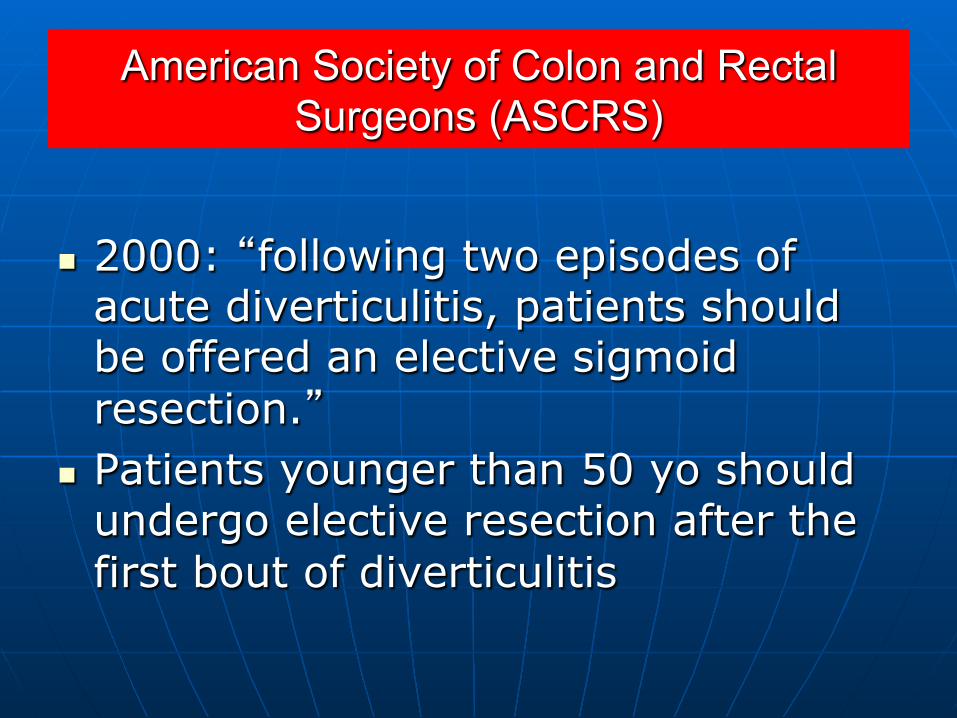
American Society of Colon and Rectal Surgeons (ASCRS)
n 2000: “following two episodes of acute diverticulitis, patients should be offered an elective sigmoid resection.”
n Patients younger than 50 yo should undergo elective resection after the first bout of diverticulitis
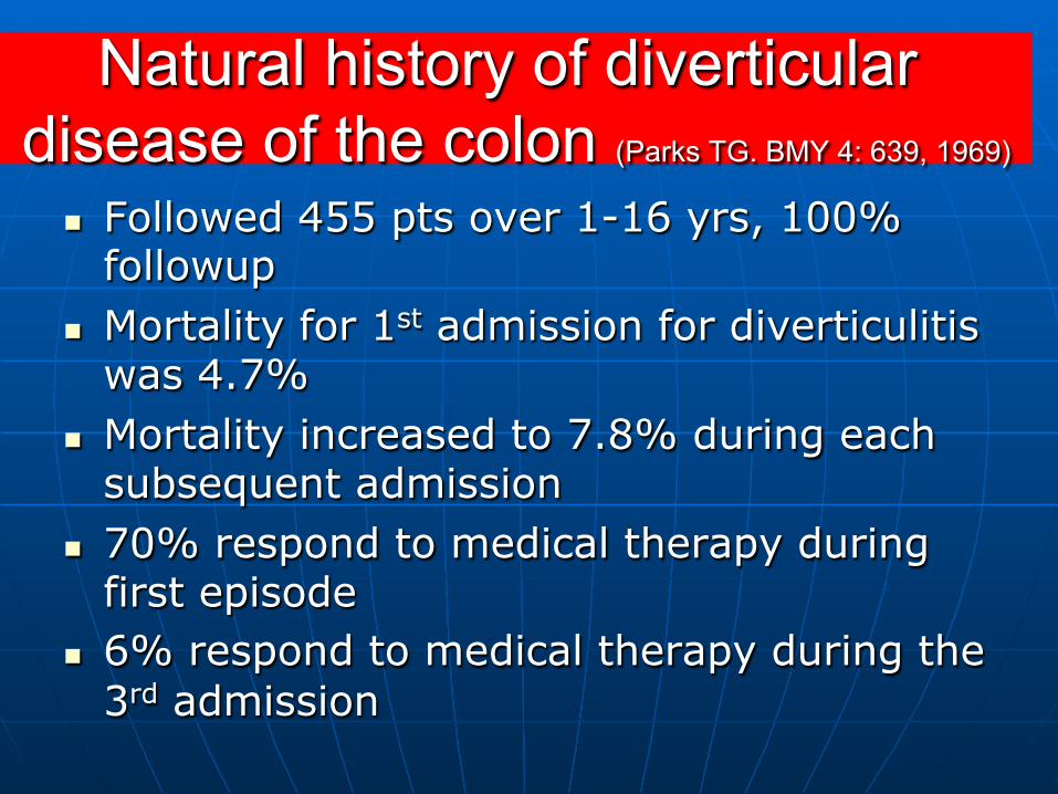
Natural history of diverticular disease of the colon (Parks TG. BMY 4: 639, 1969)
n Followed 455 pts over 1-16 yrs, 100% followup
n Mortality for 1st admission for diverticulitis was 4.7%
n Mortality increased to 7.8% during each subsequent admission
n 70% respond to medical therapy during first episode
n 6% respond to medical therapy during the 3rd admission
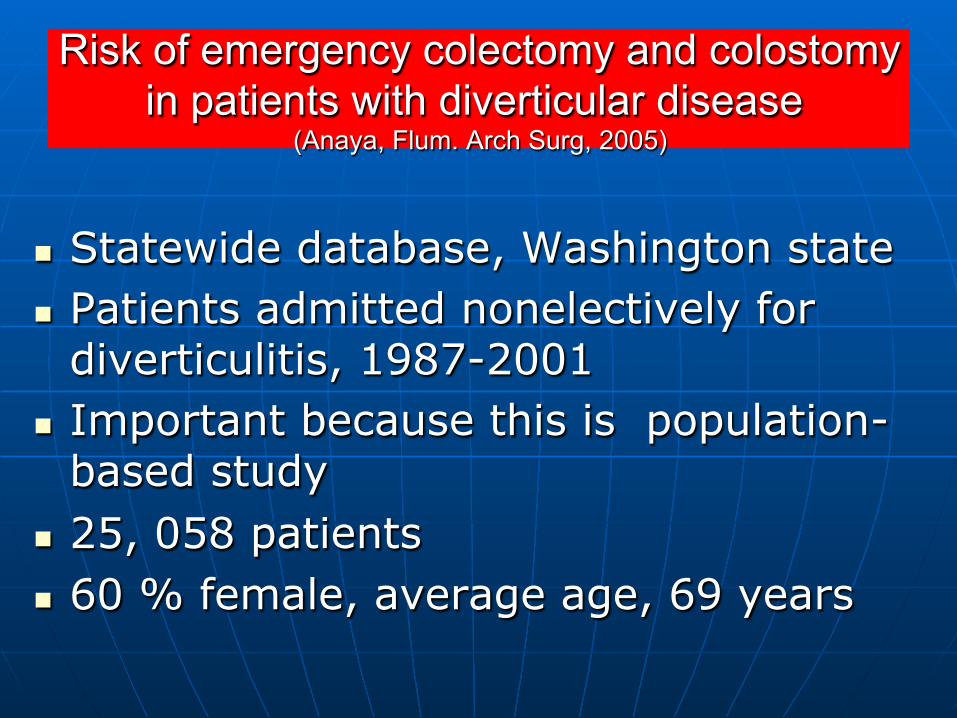
Risk of emergency colectomy and colostomy in patients with diverticular disease
(Anaya, Flum. Arch Surg, 2005)
n Statewide database, Washington state n Patients admitted nonelectively for
diverticulitis, 1987-2001 n Important because this is population-
based study n 25, 058 patients n 60 % female, average age, 69 years
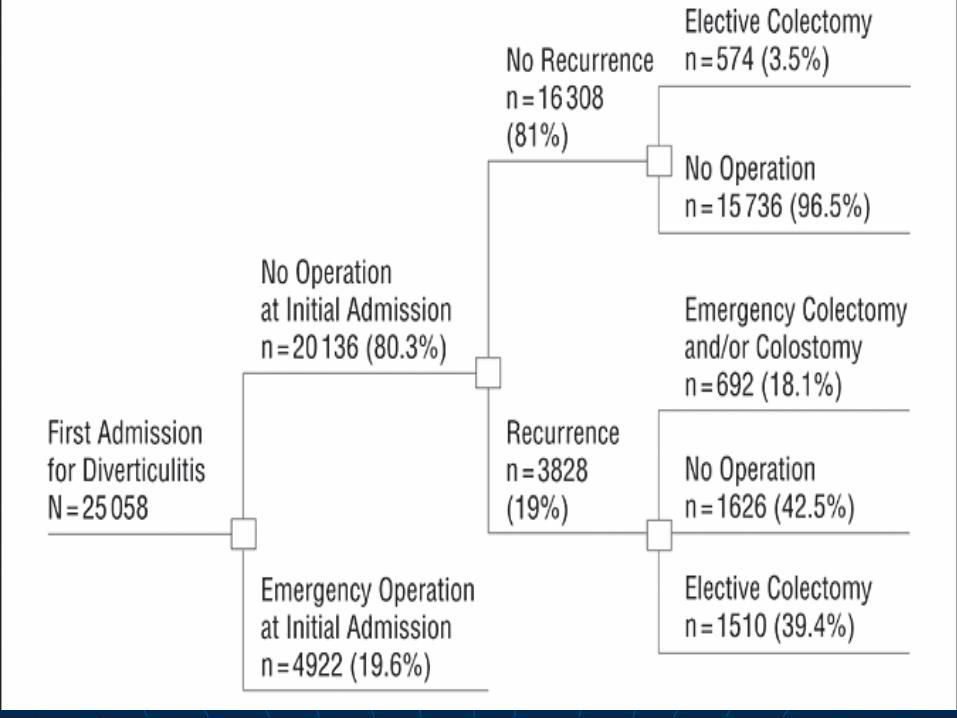
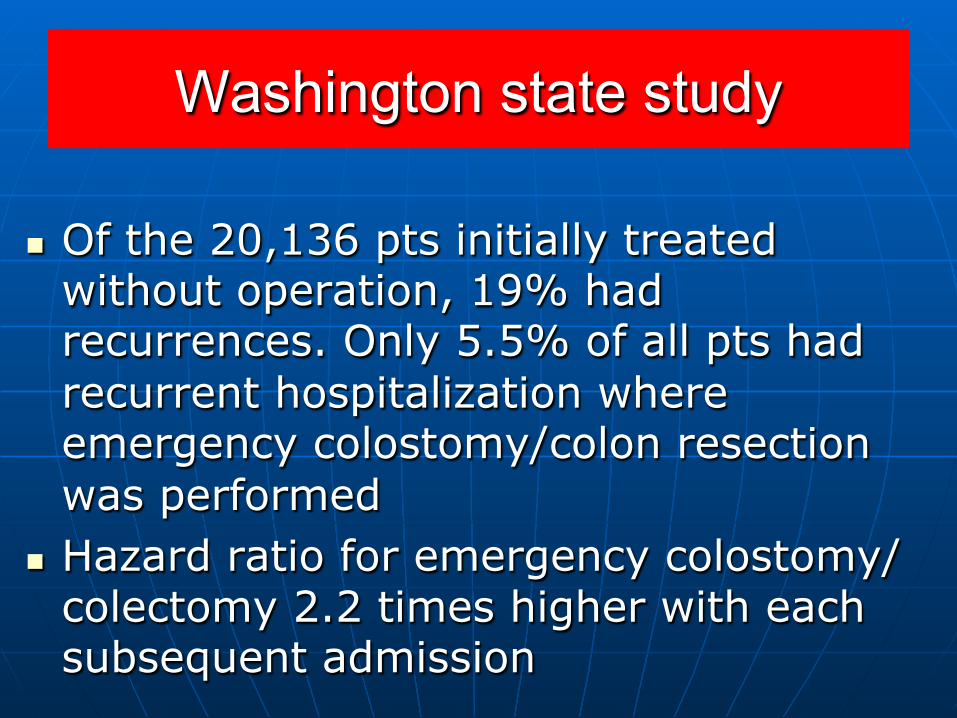
Washington state study
n Of the 20,136 pts initially treated without operation, 19% had recurrences. Only 5.5% of all pts had recurrent hospitalization where emergency colostomy/colon resection was performed
n Hazard ratio for emergency colostomy/colectomy 2.2 times higher with each subsequent admission
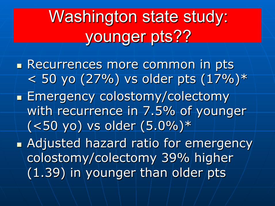
Washington state study: younger pts??
n Recurrences more common in pts < 50 yo (27%) vs older pts (17%)*
n Emergency colostomy/colectomy with recurrence in 7.5% of younger (<50 yo) vs older (5.0%)*
n Adjusted hazard ratio for emergency colostomy/colectomy 39% higher (1.39) in younger than older pts
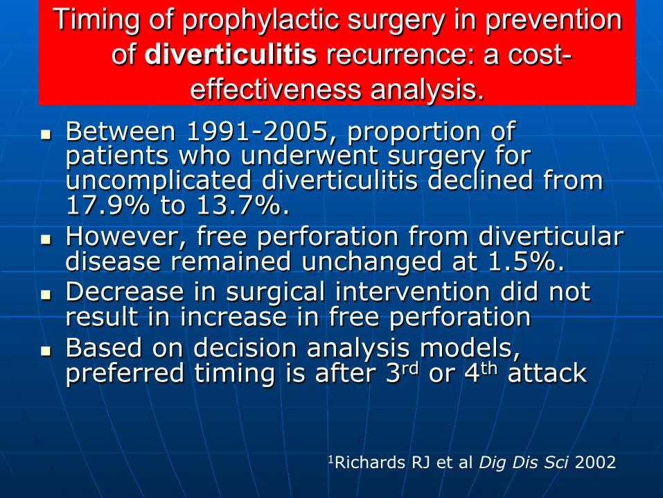
Timing of prophylactic surgery in prevention of diverticulitis recurrence: a cost-
effectiveness analysis. n Between 1991-2005, proportion of
patients who underwent surgery for uncomplicated diverticulitis declined from 17.9% to 13.7%.
n However, free perforation from diverticular disease remained unchanged at 1.5%.
n Decrease in surgical intervention did not result in increase in free perforation
n Based on decision analysis models, preferred timing is after 3rd or 4th attack
1Richards RJ et al Dig Dis Sci 2002
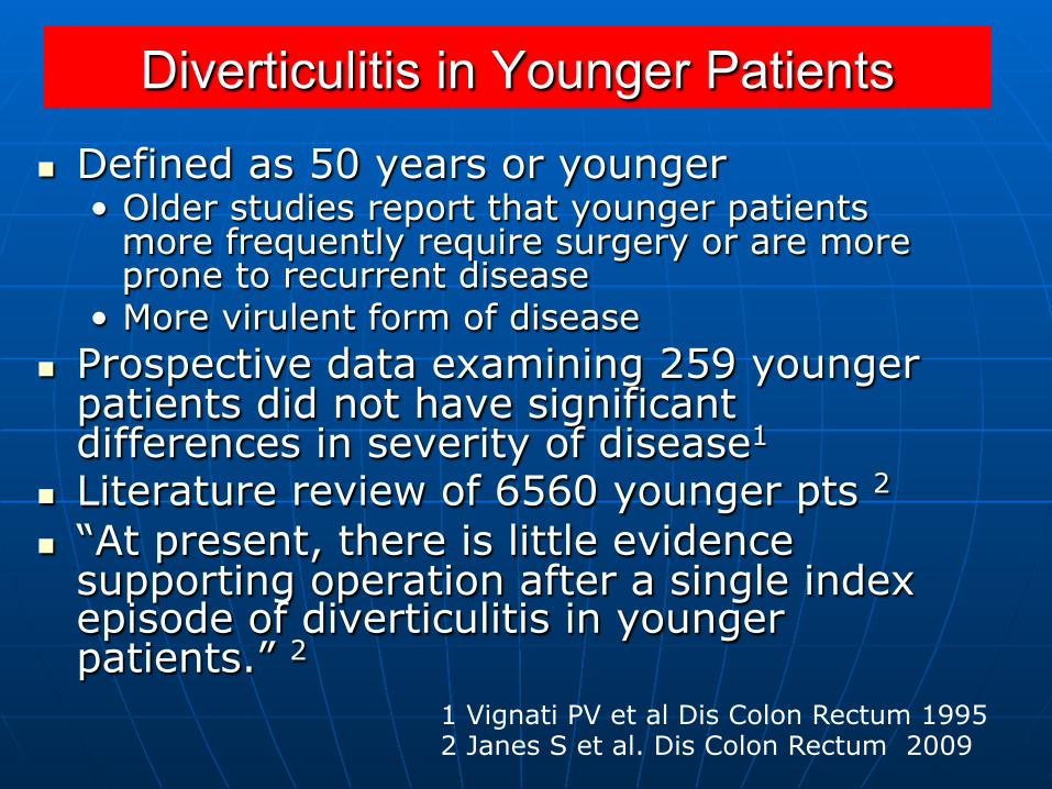
Diverticulitis in Younger Patients
n Defined as 50 years or younger • Older studies report that younger patients
more frequently require surgery or are more prone to recurrent disease
• More virulent form of disease n Prospective data examining 259 younger
patients did not have significant differences in severity of disease1
n Literature review of 6560 younger pts 2
n “At present, there is little evidence supporting operation after a single index episode of diverticulitis in younger patients.” 2
1 Vignati PV et al Dis Colon Rectum 1995 2 Janes S et al. Dis Colon Rectum 2009
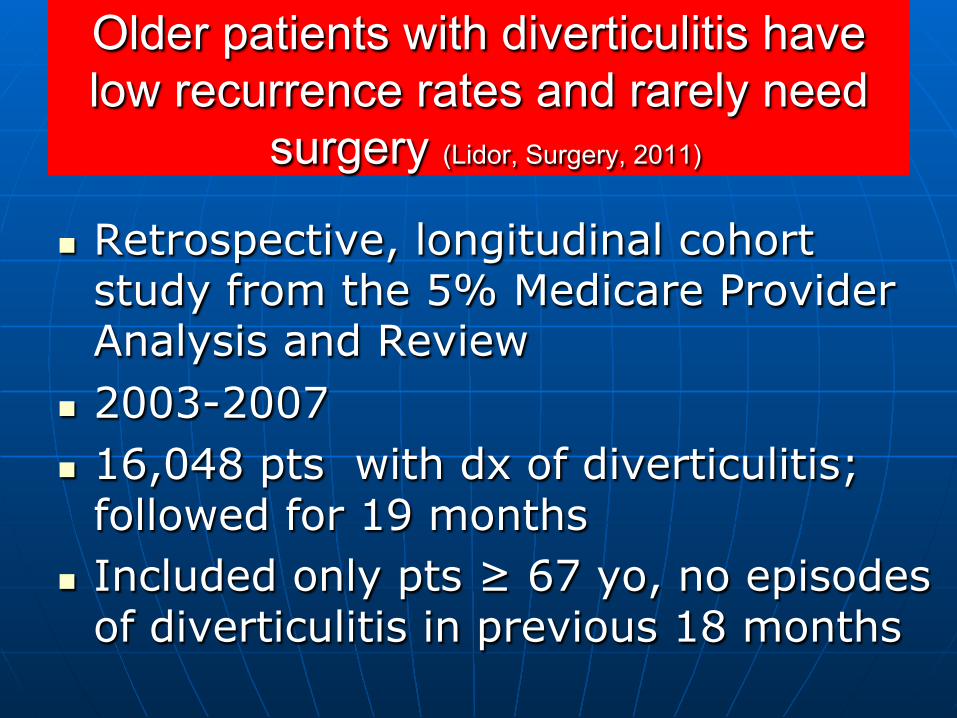
Older patients with diverticulitis have low recurrence rates and rarely need
surgery (Lidor, Surgery, 2011)
n Retrospective, longitudinal cohort study from the 5% Medicare Provider Analysis and Review
n 2003-2007 n 16,048 pts with dx of diverticulitis;
followed for 19 months n Included only pts ≥ 67 yo, no episodes
of diverticulitis in previous 18 months
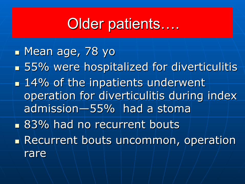
Older patients….
n Mean age, 78 yo n 55% were hospitalized for diverticulitis n 14% of the inpatients underwent
operation for diverticulitis during index admission—55% had a stoma
n 83% had no recurrent bouts n Recurrent bouts uncommon, operation
rare
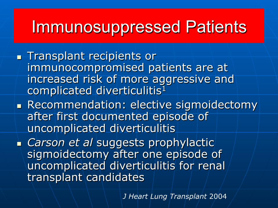
Immunosuppressed Patients n Transplant recipients or
immunocompromised patients are at increased risk of more aggressive and complicated diverticulitis1
n Recommendation: elective sigmoidectomy after first documented episode of uncomplicated diverticulitis
n Carson et al suggests prophylactic sigmoidectomy after one episode of uncomplicated diverticulitis for renal transplant candidates
J Heart Lung Transplant 2004
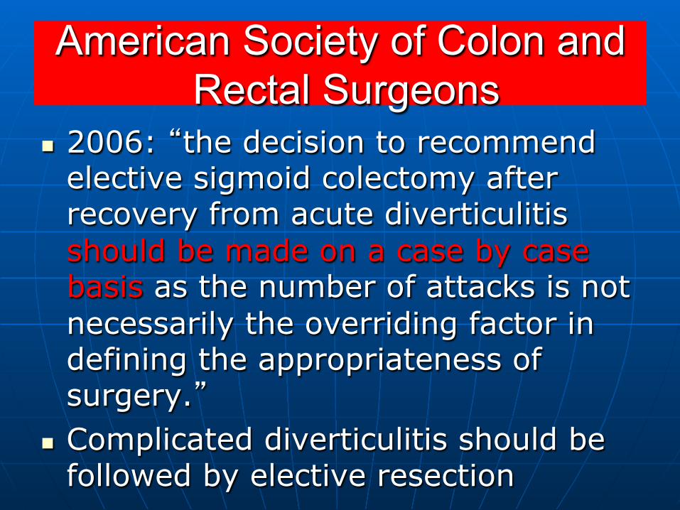
American Society of Colon and Rectal Surgeons
n 2006: “the decision to recommend elective sigmoid colectomy after recovery from acute diverticulitis should be made on a case by case basis as the number of attacks is not necessarily the overriding factor in defining the appropriateness of surgery.”
n Complicated diverticulitis should be followed by elective resection
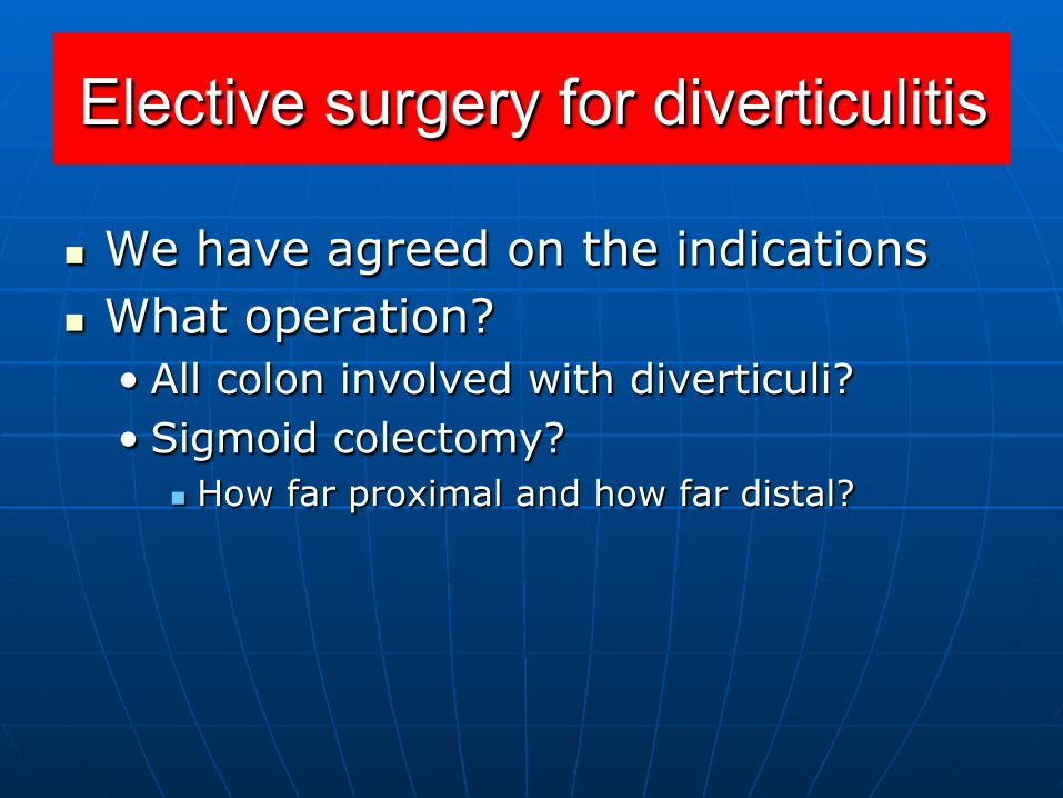
Elective surgery for diverticulitis
n We have agreed on the indications n What operation?
• All colon involved with diverticuli? • Sigmoid colectomy?
n How far proximal and how far distal?
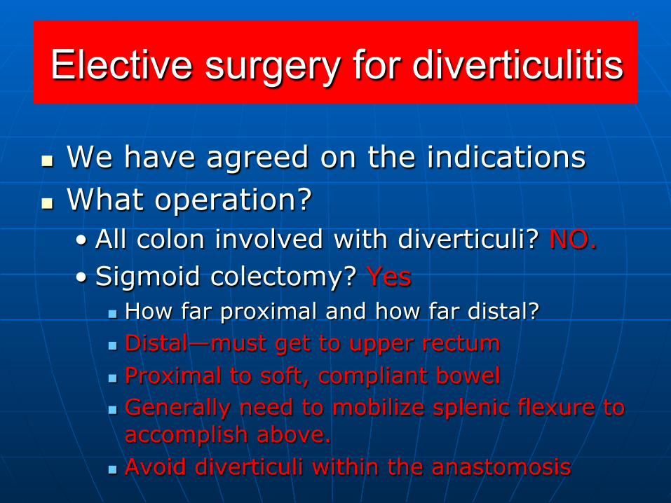
Elective surgery for diverticulitis
n We have agreed on the indications n What operation?
• All colon involved with diverticuli? NO. • Sigmoid colectomy? Yes
n How far proximal and how far distal? n Distal—must get to upper rectum n Proximal to soft, compliant bowel n Generally need to mobilize splenic flexure to
accomplish above. n Avoid diverticuli within the anastomosis
Elective sigmoid colectomy
n Laparoscopic sigmoid colectomy is the operation of choice
n Morbidity n Leak rate n Mortality
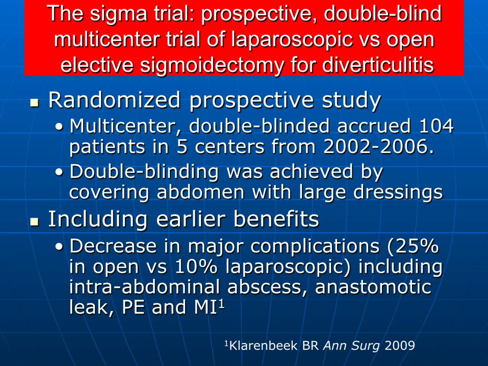
The sigma trial: prospective, double-blind multicenter trial of laparoscopic vs open elective sigmoidectomy for diverticulitis
n Randomized prospective study • Multicenter, double-blinded accrued 104
patients in 5 centers from 2002-2006. • Double-blinding was achieved by
covering abdomen with large dressings n Including earlier benefits
• Decrease in major complications (25% in open vs 10% laparoscopic) including intra-abdominal abscess, anastomotic leak, PE and MI1
1Klarenbeek BR Ann Surg 2009
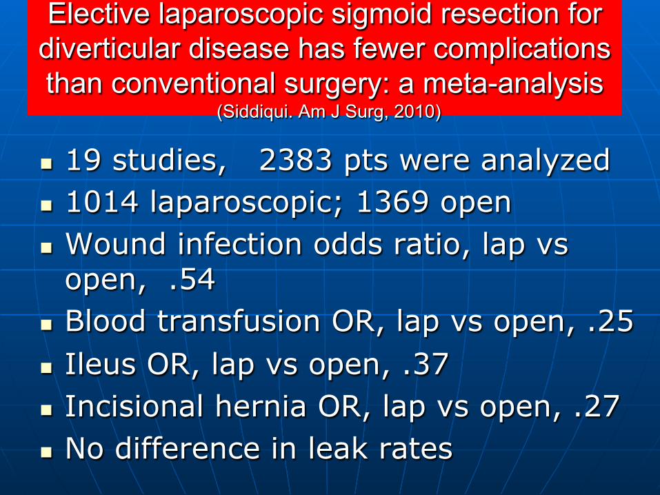
Elective laparoscopic sigmoid resection for diverticular disease has fewer complications than conventional surgery: a meta-analysis
(Siddiqui. Am J Surg, 2010)
n 19 studies, 2383 pts were analyzed n 1014 laparoscopic; 1369 open n Wound infection odds ratio, lap vs
open, .54 n Blood transfusion OR, lap vs open, .25 n Ileus OR, lap vs open, .37 n Incisional hernia OR, lap vs open, .27 n No difference in leak rates
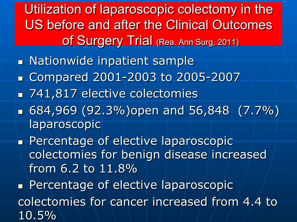
Utilization of laparoscopic colectomy in the US before and after the Clinical Outcomes
of Surgery Trial (Rea. Ann Surg, 2011) n Nationwide inpatient sample n Compared 2001-2003 to 2005-2007 n 741,817 elective colectomies n 684,969 (92.3%)open and 56,848 (7.7%)
laparoscopic n Percentage of elective laparoscopic
colectomies for benign disease increased from 6.2 to 11.8%
n Percentage of elective laparoscopic colectomies for cancer increased from 4.4 to 10.5%

???????
n So, despite the advantages of laparoscopic sigmoid colectomy for diverticular disease, only 11.8% of colectomies are performed laparoscopically in the US
n Lower than other countries
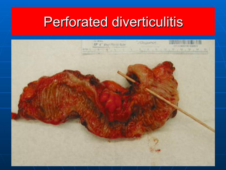
Perforated diverticulitis
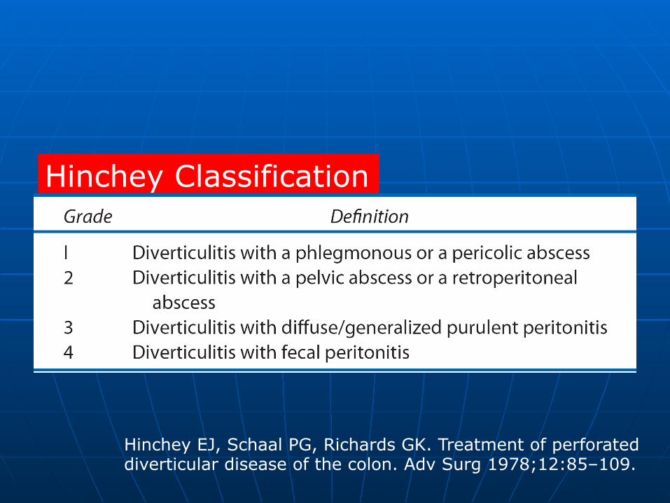
Hinchey EJ, Schaal PG, Richards GK. Treatment of perforated diverticular disease of the colon. Adv Surg 1978;12:85–109.
Hinchey Classification
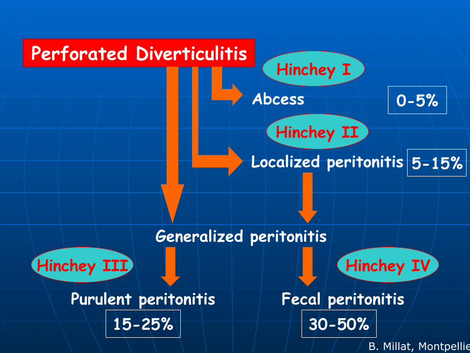
Perforated Diverticulitis
Abcess
Localized peritonitis
Generalized peritonitis
Purulent peritonitis Fecal peritonitis
Hinchey I
Hinchey II
Hinchey III Hinchey IV
15-25%
0-5%
5-15%
30-50% B. Millat, Montpellier
Two areas where we are pushing the envelope…..
n Primary anastomosis vs Hartman procedure for perforated diverticulitis
n Laparoscopic lavage and drainage for Hinchey III diverticulitis
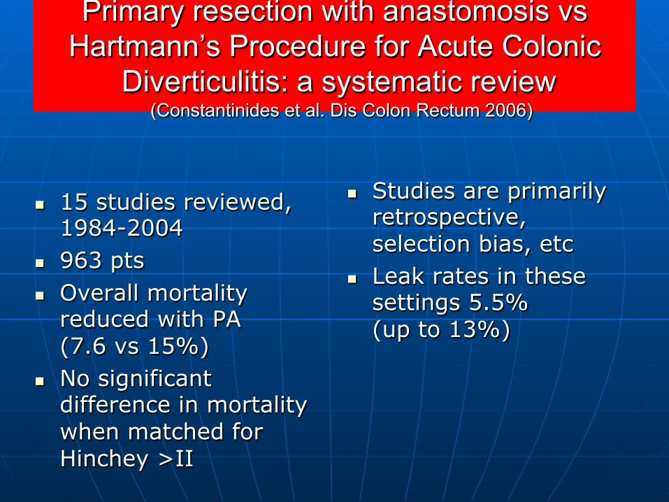
Primary resection with anastomosis vs Hartmann’s Procedure for Acute Colonic
Diverticulitis: a systematic review (Constantinides et al. Dis Colon Rectum 2006)
n 15 studies reviewed, 1984-2004
n 963 pts n Overall mortality
reduced with PA (7.6 vs 15%)
n No significant difference in mortality when matched for Hinchey >II
n Studies are primarily retrospective, selection bias, etc
n Leak rates in these settings 5.5% (up to 13%)
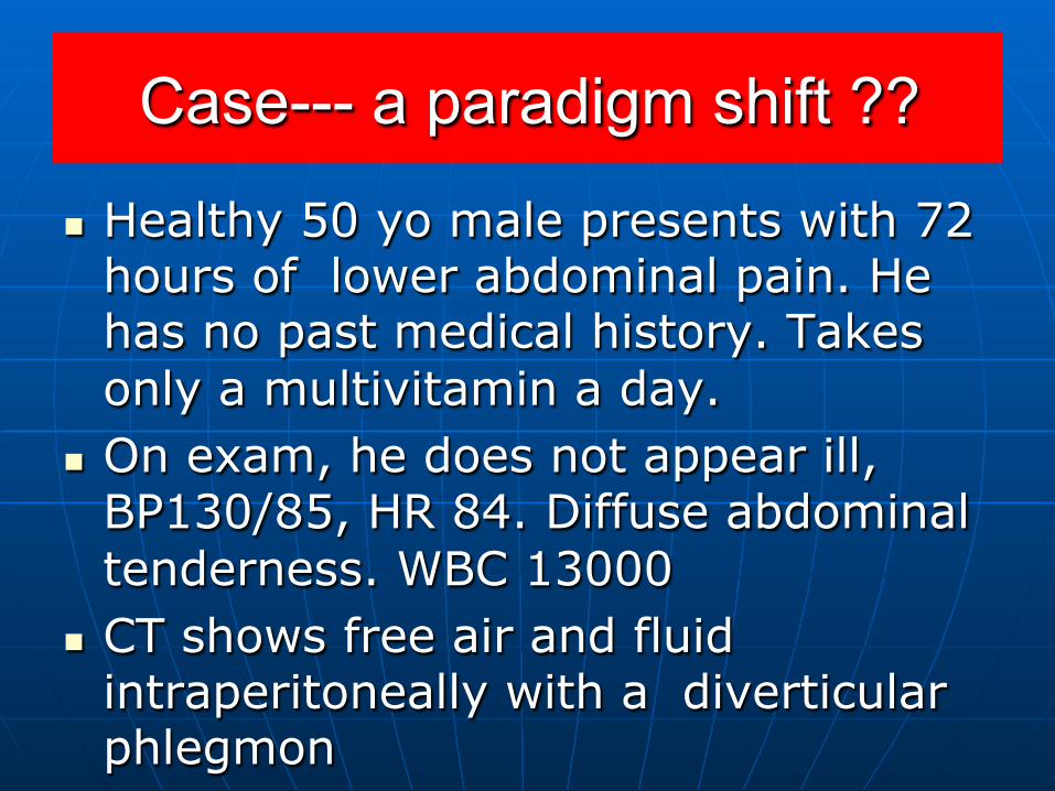
Case--- a paradigm shift ??
n Healthy 50 yo male presents with 72 hours of lower abdominal pain. He has no past medical history. Takes only a multivitamin a day.
n On exam, he does not appear ill, BP130/85, HR 84. Diffuse abdominal tenderness. WBC 13000
n CT shows free air and fluid intraperitoneally with a diverticular phlegmon
Case
n So, you go to the OR and……..
Case
n So, you go to the OR and……..
1---- make a lower midline incision 2---- insert a laparoscope
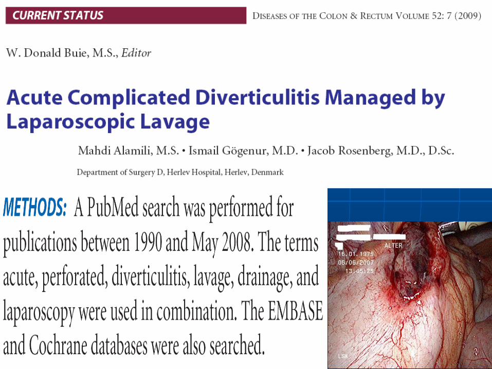
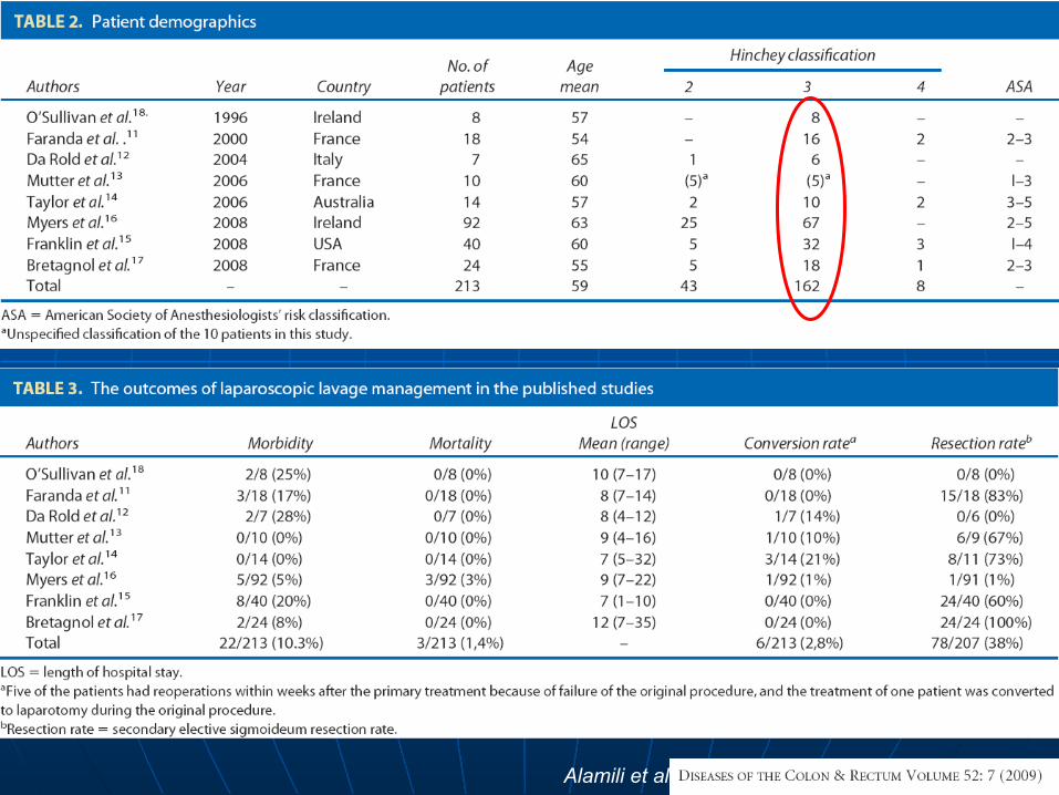
Alamili et al.
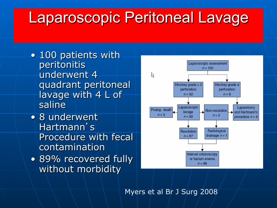
Laparoscopic Peritoneal Lavage
• 100 patients with peritonitis underwent 4 quadrant peritoneal lavage with 4 L of saline
• 8 underwent Hartmann’s Procedure with fecal contamination
• 89% recovered fully without morbidity
Myers et al Br J Surg 2008
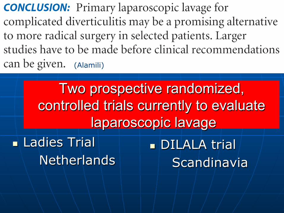
Two prospective randomized, controlled trials currently to evaluate
laparoscopic lavage n Ladies Trial Netherlands
n DILALA trial Scandinavia
(Alamili)
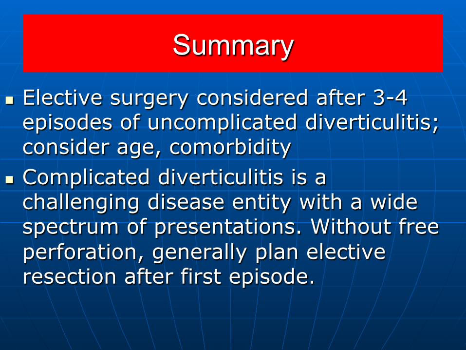
Summary
n Elective surgery considered after 3-4 episodes of uncomplicated diverticulitis; consider age, comorbidity
n Complicated diverticulitis is a challenging disease entity with a wide spectrum of presentations. Without free perforation, generally plan elective resection after first episode.
Summary
n Younger patients should be managed by severity of disease.
n Immunosuppressed patients will require surgical intervention.
n More sigmoid resections should be performed laparoscopically.
n Laparoscopic Lavage may be an alternative for Hinchey III diverticulitis.
Thank you
A rare photo of a general surgeon
University of Pittsburgh School of Medicine
Nonoperative management of complex splenic injuries Andrew B. Peitzman University of Pittsburgh
University of Pittsburgh School of Medicine
Key principles with splenic injury
● Hemodynamically unstable patients require immediate laparotomy. Generally, splenectomy is the best treatment.
● Nonoperative management is an option in the hemodynamically stable patient ONLY.
● Splenorrhaphy is an option in the stable pt with low ISS
● No patient should die as a consequence of nonoperative management of a splenic injury
University of Pittsburgh School of Medicine
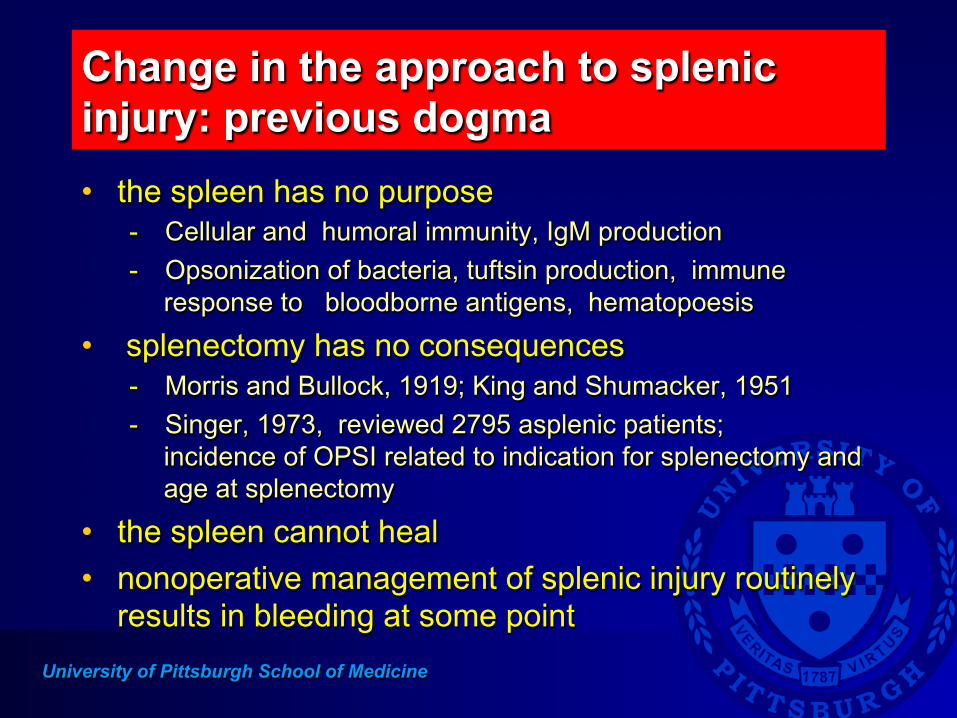
Change in the approach to splenic injury: previous dogma • the spleen has no purpose - Cellular and humoral immunity, IgM production - Opsonization of bacteria, tuftsin production, immune
response to bloodborne antigens, hematopoesis
• splenectomy has no consequences - Morris and Bullock, 1919; King and Shumacker, 1951 - Singer, 1973, reviewed 2795 asplenic patients;
incidence of OPSI related to indication for splenectomy and age at splenectomy
• the spleen cannot heal • nonoperative management of splenic injury routinely
results in bleeding at some point
University of Pittsburgh School of Medicine
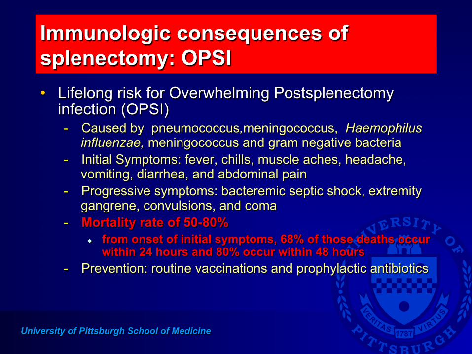
Immunologic consequences of splenectomy: OPSI • Lifelong risk for Overwhelming Postsplenectomy
infection (OPSI) - Caused by pneumococcus,meningococcus, Haemophilus
influenzae, meningococcus and gram negative bacteria - Initial Symptoms: fever, chills, muscle aches, headache,
vomiting, diarrhea, and abdominal pain - Progressive symptoms: bacteremic septic shock, extremity
gangrene, convulsions, and coma - Mortality rate of 50-80%
from onset of initial symptoms, 68% of those deaths occur within 24 hours and 80% occur within 48 hours
- Prevention: routine vaccinations and prophylactic antibiotics
University of Pittsburgh School of Medicine
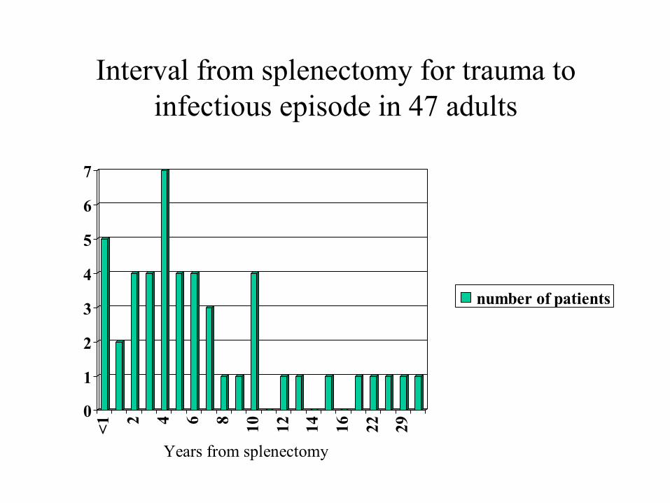
Interval from splenectomy for trauma to infectious episode in 47 adults
0
1
2
3
4
5
6
7
<1 2 4 6 8 10 12 14 16 22 29
number of patients
Years from splenectomy
University of Pittsburgh School of Medicine
How can we preserve the spleen?
• Nonoperative management (observation)
• Splenorrhaphy
University of Pittsburgh School of Medicine
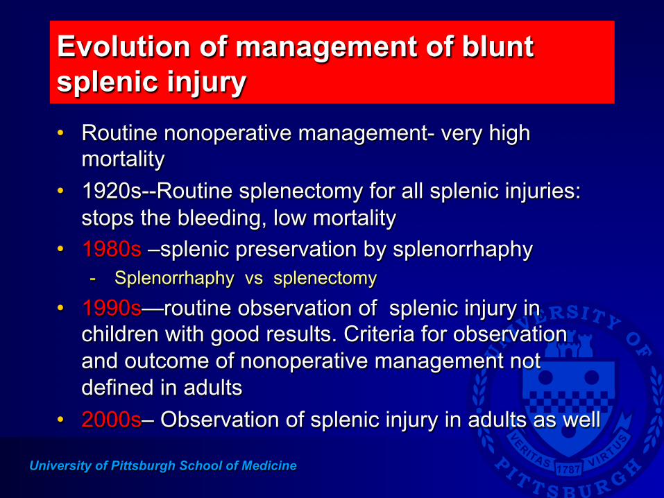
Evolution of management of blunt splenic injury • Routine nonoperative management- very high
mortality • 1920s--Routine splenectomy for all splenic injuries:
stops the bleeding, low mortality • 1980s –splenic preservation by splenorrhaphy - Splenorrhaphy vs splenectomy
• 1990s—routine observation of splenic injury in children with good results. Criteria for observation and outcome of nonoperative management not defined in adults
• 2000s– Observation of splenic injury in adults as well
University of Pittsburgh School of Medicine
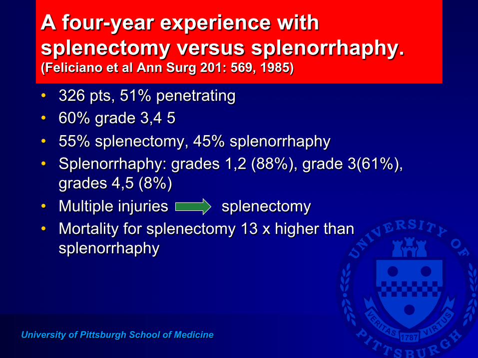
A four-year experience with splenectomy versus splenorrhaphy. (Feliciano et al Ann Surg 201: 569, 1985)
• 326 pts, 51% penetrating • 60% grade 3,4 5 • 55% splenectomy, 45% splenorrhaphy • Splenorrhaphy: grades 1,2 (88%), grade 3(61%),
grades 4,5 (8%) • Multiple injuries splenectomy • Mortality for splenectomy 13 x higher than
splenorrhaphy
University of Pittsburgh School of Medicine

University of Pittsburgh School of Medicine
EAST practice guidelines (published 2003)
• Nonoperative management of blunt injury to the spleen and liver - class II data support nonoperative management of
injuries to the liver or spleen - severity of grade of injury to the liver or the spleen
is not a contraindication to nonoperative management
this is contrary to observations by Buntain 1988; Resciniti 1988; Powell 1997; Cathay 1998; Bee, 2001
University of Pittsburgh School of Medicine
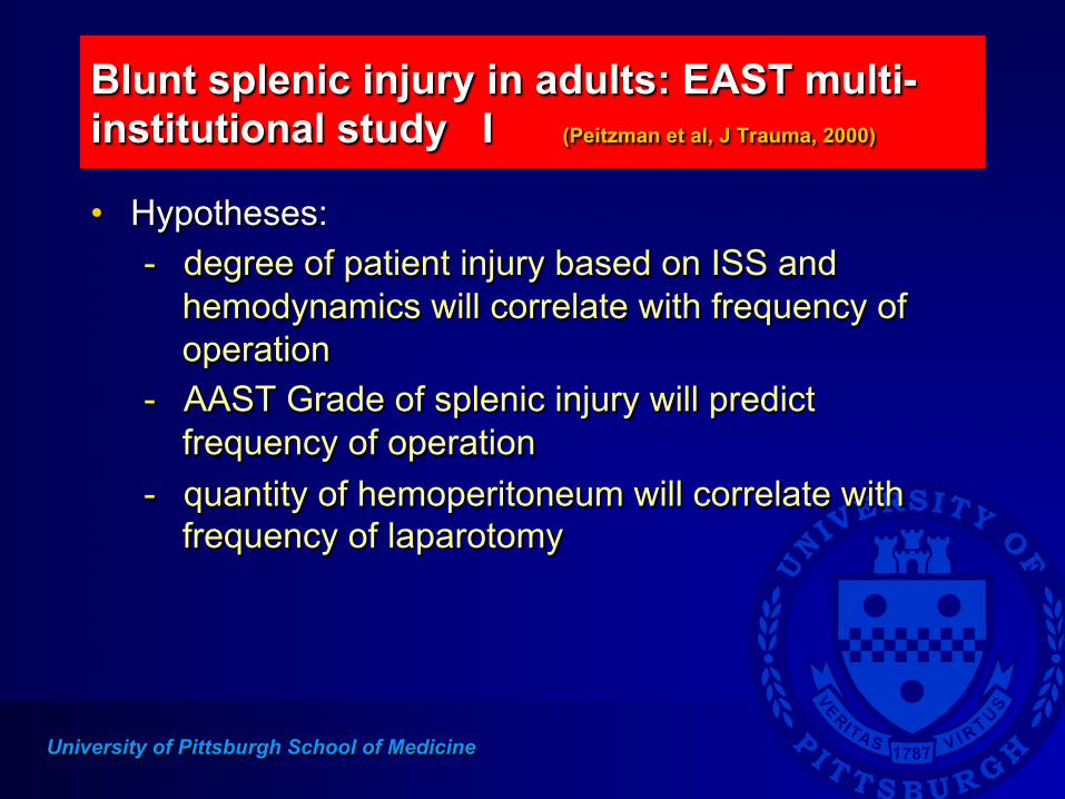
Blunt splenic injury in adults: EAST multi-institutional study I (Peitzman et al, J Trauma, 2000)
• Hypotheses: - degree of patient injury based on ISS and
hemodynamics will correlate with frequency of operation
- AAST Grade of splenic injury will predict frequency of operation
- quantity of hemoperitoneum will correlate with frequency of laparotomy
University of Pittsburgh School of Medicine
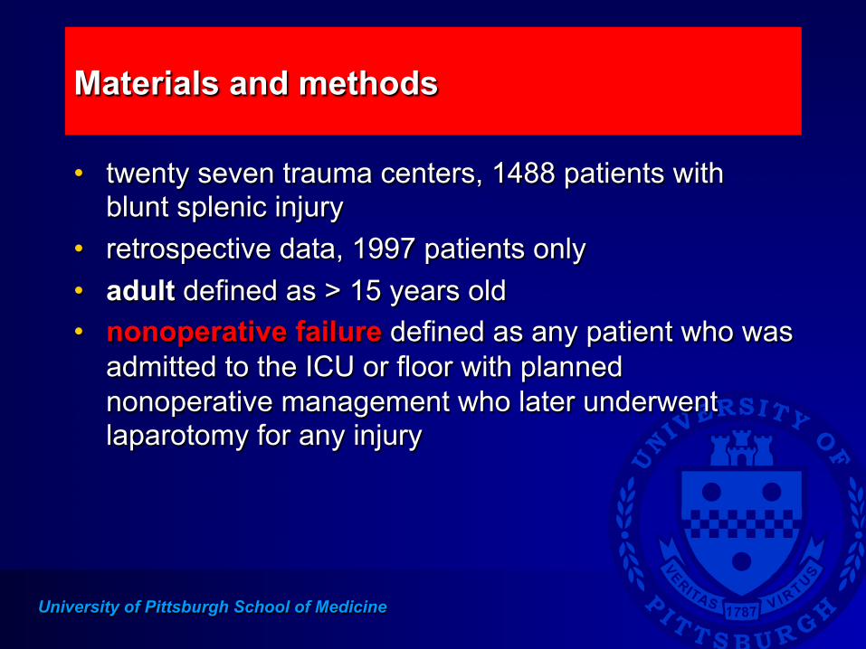
Materials and methods
• twenty seven trauma centers, 1488 patients with blunt splenic injury
• retrospective data, 1997 patients only • adult defined as > 15 years old • nonoperative failure defined as any patient who was
admitted to the ICU or floor with planned nonoperative management who later underwent laparotomy for any injury
University of Pittsburgh School of Medicine
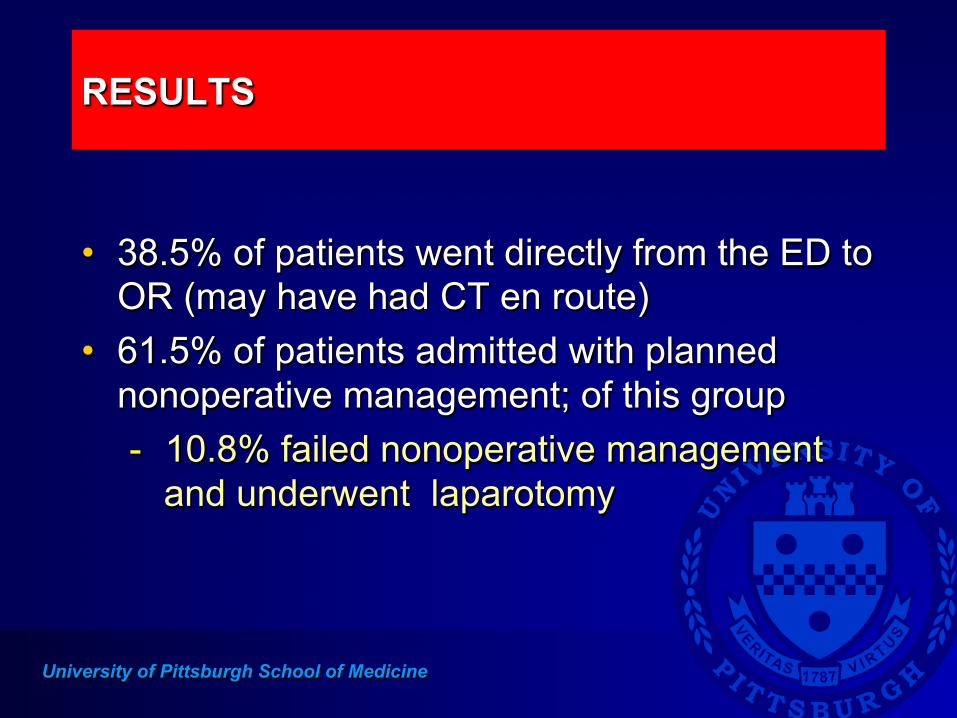
RESULTS
• 38.5% of patients went directly from the ED to OR (may have had CT en route)
• 61.5% of patients admitted with planned nonoperative management; of this group - 10.8% failed nonoperative management
and underwent laparotomy
University of Pittsburgh School of Medicine
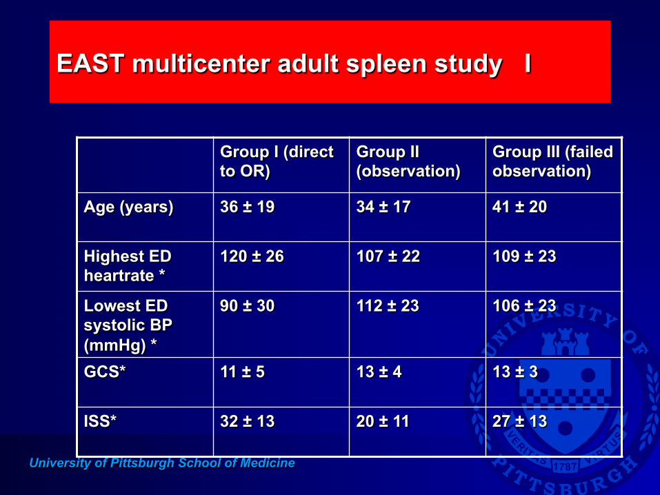
EAST multicenter adult spleen study I
Group I (direct to OR)
Group II (observation)
Group III (failed observation)
Age (years) 36 ± 19 34 ± 17 41 ± 20
Highest ED heartrate *
120 ± 26 107 ± 22 109 ± 23
Lowest ED systolic BP (mmHg) *
90 ± 30 112 ± 23 106 ± 23
GCS* 11 ± 5 13 ± 4 13 ± 3
ISS* 32 ± 13 20 ± 11 27 ± 13
University of Pittsburgh School of Medicine
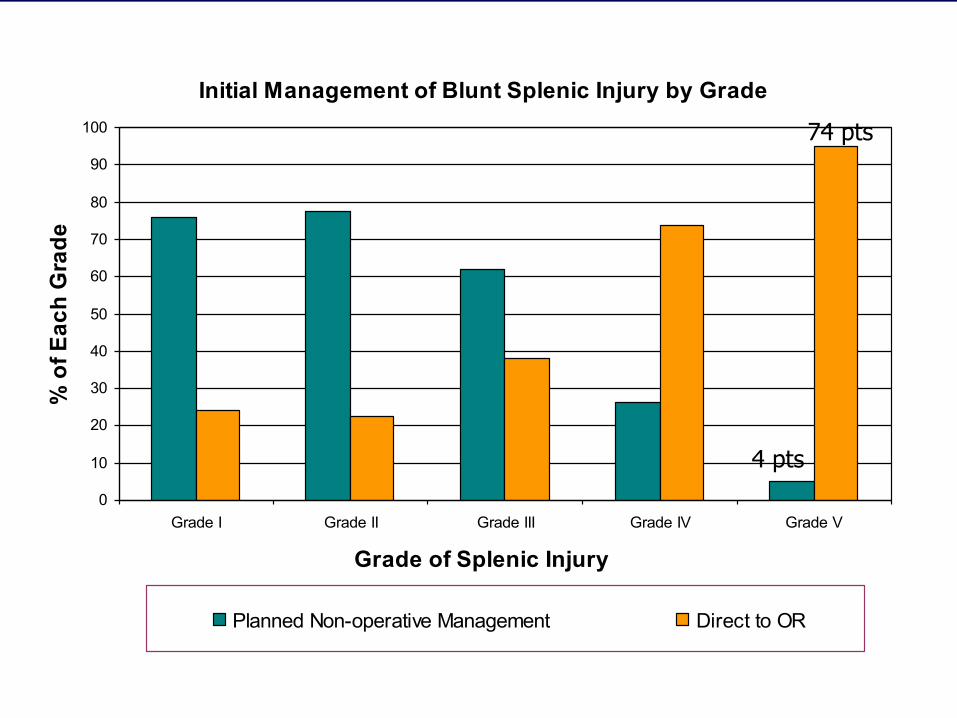
Initial Management of Blunt Splenic Injury by Grade
0
10
20
30
40
50
60
70
80
90
100
Grade I Grade II Grade III Grade IV Grade V
Grade of Splenic Injury
% o
f Eac
h G
rade
Planned Non-operative Management Direct to OR
4 pts
74 pts
University of Pittsburgh School of Medicine
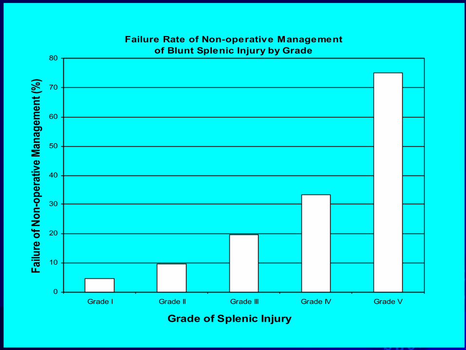
Failure Rate of Non-operative Managementof Blunt Splenic Injury by Grade
0
10
20
30
40
50
60
70
80
Grade I Grade II Grade III Grade IV Grade V
Grade of Splenic Injury
Failu
re o
f Non
-ope
rativ
e M
anag
emen
t (%
)
University of Pittsburgh School of Medicine
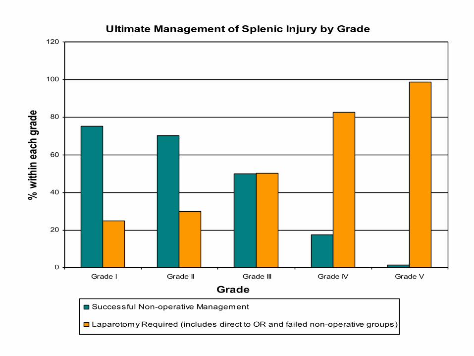
Ultimate Management of Splenic Injury by Grade
0
20
40
60
80
100
120
Grade I Grade II Grade III Grade IV Grade V
Grade
% w
ithin
each
gra
de
Successful Non-operative Management
Laparotomy Required (includes direct to OR and failed non-operative groups)
University of Pittsburgh School of Medicine
0
10
20
30
40
50
60
70
80
90
Grade I Grade II Grade III Grade IV Grade V
Grade of Splenic Injury
% w
ith S
ucce
ssfu
l Non
-ope
rativ
e Man
agem
ent
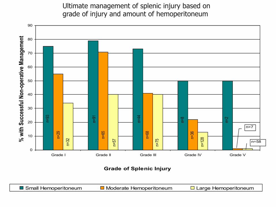
Small Hemoperitoneum Moderate Hemoperitoneum Large Hemoperitoneum
n=7
n=58
n=93
n=29
n=32
n=91
n=65
n=57
n=44
n=68
n=75
n=8
n=36
n=12
8
n=2
Ultimate management of splenic injury based on grade of injury and amount of hemoperitoneum
University of Pittsburgh School of Medicine
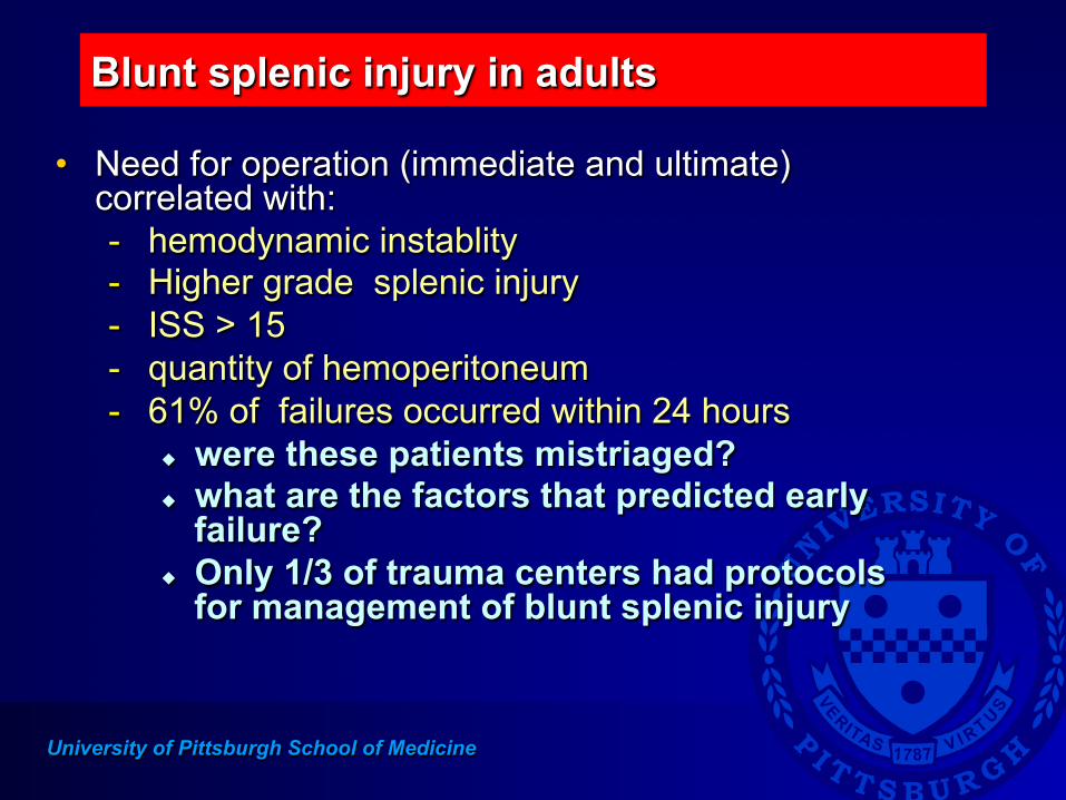
Blunt splenic injury in adults
• Need for operation (immediate and ultimate) correlated with: - hemodynamic instablity - Higher grade splenic injury - ISS > 15 - quantity of hemoperitoneum - 61% of failures occurred within 24 hours
were these patients mistriaged? what are the factors that predicted early
failure? Only 1/3 of trauma centers had protocols
for management of blunt splenic injury
University of Pittsburgh School of Medicine
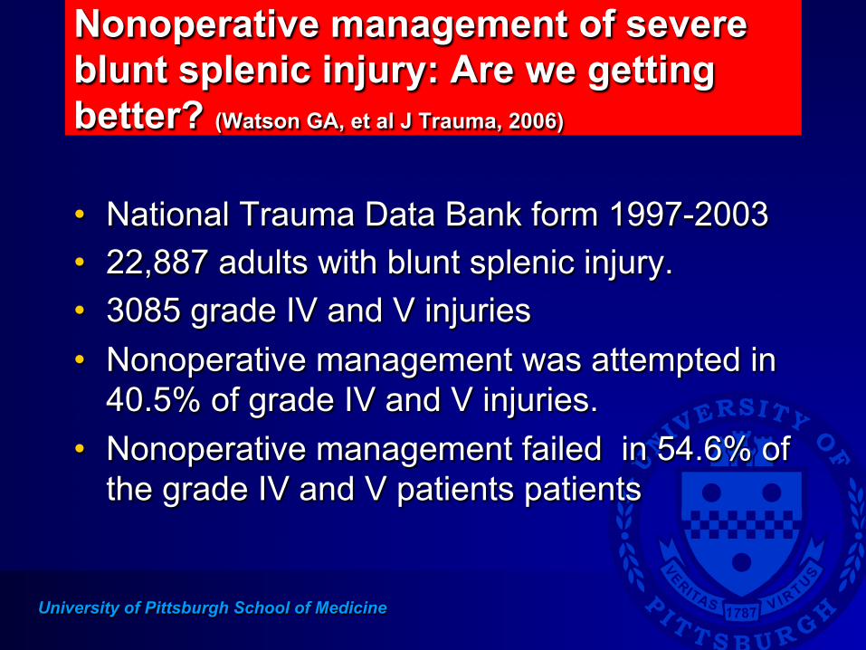
Nonoperative management of severe blunt splenic injury: Are we getting better? (Watson GA, et al J Trauma, 2006)
• National Trauma Data Bank form 1997-2003 • 22,887 adults with blunt splenic injury. • 3085 grade IV and V injuries • Nonoperative management was attempted in
40.5% of grade IV and V injuries. • Nonoperative management failed in 54.6% of
the grade IV and V patients patients
University of Pittsburgh School of Medicine
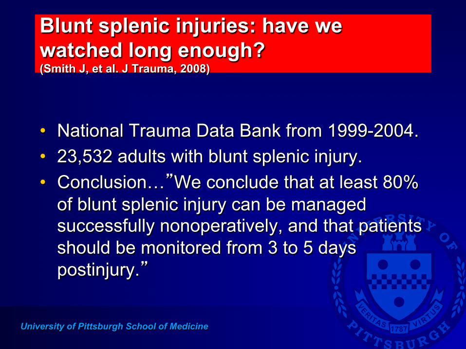
Blunt splenic injuries: have we watched long enough? (Smith J, et al. J Trauma, 2008)
• National Trauma Data Bank from 1999-2004. • 23,532 adults with blunt splenic injury. • Conclusion…”We conclude that at least 80%
of blunt splenic injury can be managed successfully nonoperatively, and that patients should be monitored from 3 to 5 days postinjury.”
University of Pittsburgh School of Medicine
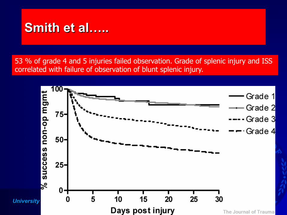
Smith et al…..
53 % of grade 4 and 5 injuries failed observation. Grade of splenic injury and ISS correlated with failure of observation of blunt splenic injury.
University of Pittsburgh School of Medicine
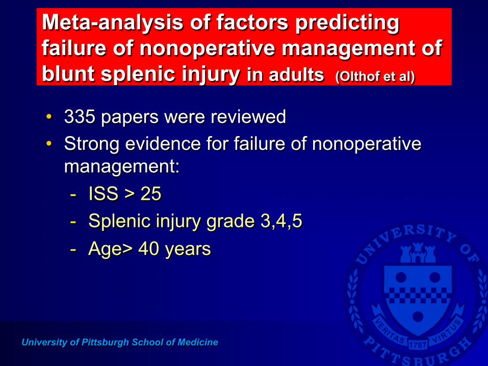
Meta-analysis of factors predicting failure of nonoperative management of blunt splenic injury in adults (Olthof et al)
• 335 papers were reviewed • Strong evidence for failure of nonoperative
management: - ISS > 25 - Splenic injury grade 3,4,5 - Age> 40 years
Failure of nonoperative management of blunt splenic injury in adults: variability in physican
practice and impact on outcome (Peitzman et al, JACS August, 2005)
Multi-institutional study of the Eastern Association for the Surgery of Trauma III
University of Pittsburgh School of Medicine
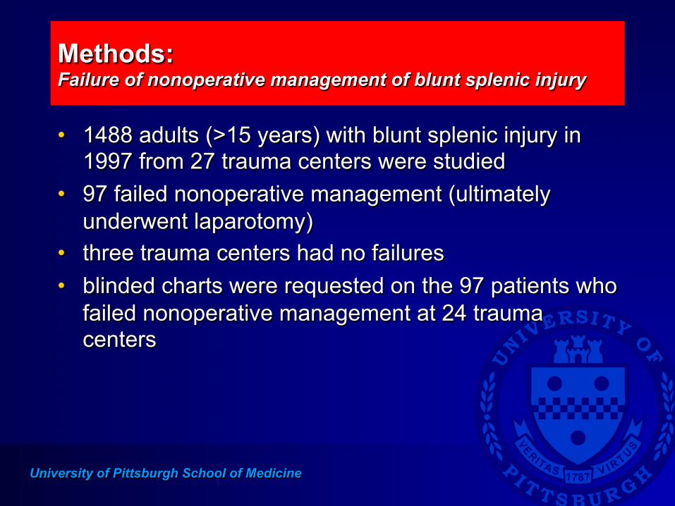
Methods: Failure of nonoperative management of blunt splenic injury
• 1488 adults (>15 years) with blunt splenic injury in 1997 from 27 trauma centers were studied
• 97 failed nonoperative management (ultimately underwent laparotomy)
• three trauma centers had no failures • blinded charts were requested on the 97 patients who
failed nonoperative management at 24 trauma centers
University of Pittsburgh School of Medicine
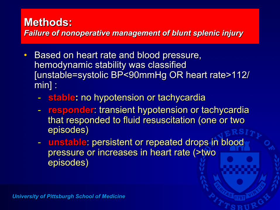
Methods: Failure of nonoperative management of blunt splenic injury
• Based on heart rate and blood pressure, hemodynamic stability was classified [unstable=systolic BP<90mmHg OR heart rate>112/min] : - stable: no hypotension or tachycardia - responder: transient hypotension or tachycardia
that responded to fluid resuscitation (one or two episodes)
- unstable: persistent or repeated drops in blood pressure or increases in heart rate (>two episodes)
University of Pittsburgh School of Medicine
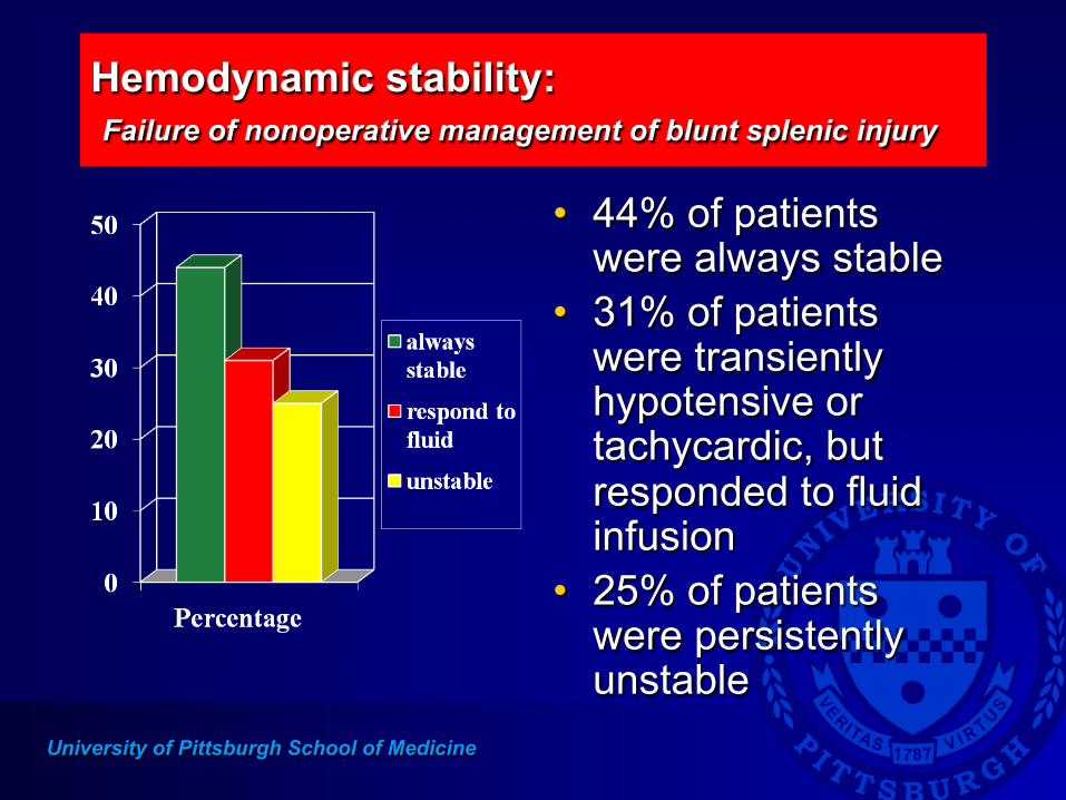
Hemodynamic stability: Failure of nonoperative management of blunt splenic injury
• 44% of patients were always stable
• 31% of patients were transiently hypotensive or tachycardic, but responded to fluid infusion
• 25% of patients were persistently unstable
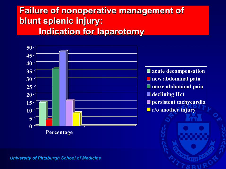
University of Pittsburgh School of Medicine
Failure of nonoperative management of blunt splenic injury: Indication for laparotomy
05
101520253035404550
Percentage
acute decompensationnew abdominal painmore abdominal paindeclining Hctpersistent tachycardiar/o another injury
University of Pittsburgh School of Medicine
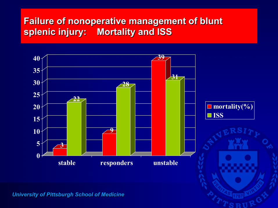
Failure of nonoperative management of blunt splenic injury: Mortality and ISS
3
22
9
28
39
31
0
5
10
15
20
25
30
35
40
stable responders unstable
mortality(%)ISS
University of Pittsburgh School of Medicine
Mortality in adult patients who failed nonoperative management of blunt splenic injury
• ten patients died (12% mortality) • 60% of the deaths were from delayed diagnosis and
treatment of abdominal injuries - Three patients exsanguinated in the hospital, two
of whom never underwent operation - Factors in these deaths:
unstable patients not undergoing laparotomy misreading of CT scans false negative abdominal ultrasound
University of Pittsburgh School of Medicine
Violates a key principle
• No patient with a splenic injury should die from bleeding or missed injury
University of Pittsburgh School of Medicine
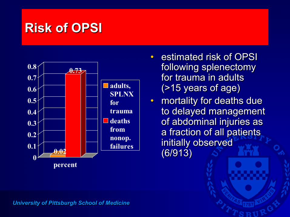
Risk of OPSI
0.02
0.73
00.10.20.30.40.50.60.70.8
percent
adults,SPLNXfortraumadeathsfromnonop. failures
• estimated risk of OPSI following splenectomy for trauma in adults (>15 years of age)
• mortality for deaths due to delayed management of abdominal injuries as a fraction of all patients initially observed (6/913)
University of Pittsburgh School of Medicine
Nonoperative management: where is the pendulum??
• The nonoperative pendulum swung too far • Nonoperative management does not mean
neglect the patient. • Understand injury patterns. • Patients with splenic injury managed
nonoperatively may die acutely as a consequence of the splenic injury or missed injuries.
University of Pittsburgh School of Medicine
Blunt injury to the spleen: angio/embolization????
Where is this literature??
University of Pittsburgh School of Medicine
Angio/embolization.. All studies are historical comparisons
• With the change in practice over this time period, to suggest that the increase in success of nonoperative management is due to angiography and embolization is not yet justified.
University of Pittsburgh School of Medicine
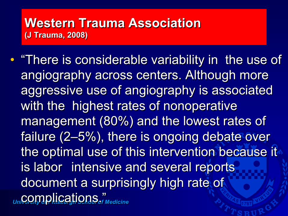
Western Trauma Association (J Trauma, 2008)
• “There is considerable variability in the use of angiography across centers. Although more aggressive use of angiography is associated with the highest rates of nonoperative management (80%) and the lowest rates of failure (2–5%), there is ongoing debate over the optimal use of this intervention because it is labor intensive and several reports document a surprisingly high rate of complications.”
University of Pittsburgh School of Medicine
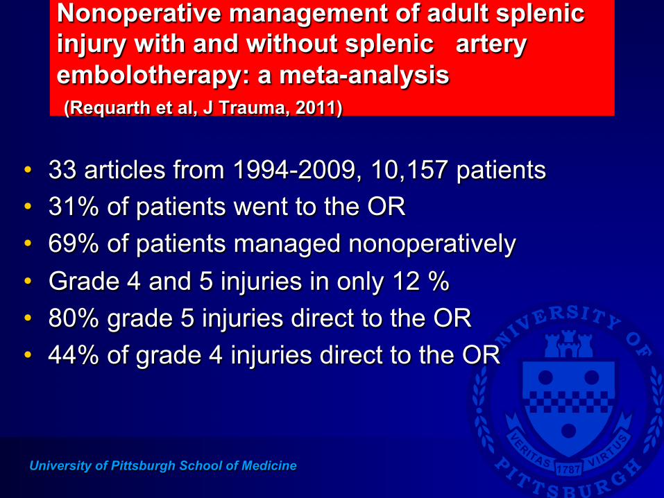
Nonoperative management of adult splenic injury with and without splenic artery embolotherapy: a meta-analysis (Requarth et al, J Trauma, 2011)
• 33 articles from 1994-2009, 10,157 patients • 31% of patients went to the OR • 69% of patients managed nonoperatively • Grade 4 and 5 injuries in only 12 % • 80% grade 5 injuries direct to the OR • 44% of grade 4 injuries direct to the OR
University of Pittsburgh School of Medicine
• Compared failure rate of observation only versus angioembolization - Failure rate of observation only increased with
splenic injury grade - Failure rate of angio/embolization did not increase
significantly with splenic grade
University of Pittsburgh School of Medicine
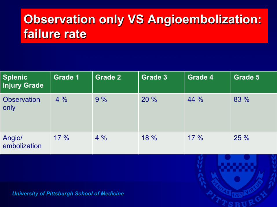
Observation only VS Angioembolization: failure rate
Splenic Injury Grade
Grade 1 Grade 2 Grade 3 Grade 4 Grade 5
Observation only
4 % 9 % 20 % 44 % 83 %
Angio/embolization
17 % 4 % 18 % 17 % 25 %
University of Pittsburgh School of Medicine
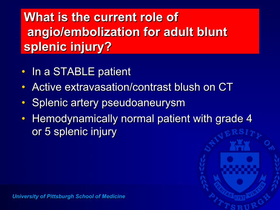
What is the current role of angio/embolization for adult blunt splenic injury?
• In a STABLE patient • Active extravasation/contrast blush on CT • Splenic artery pseudoaneurysm • Hemodynamically normal patient with grade 4
or 5 splenic injury
University of Pittsburgh School of Medicine
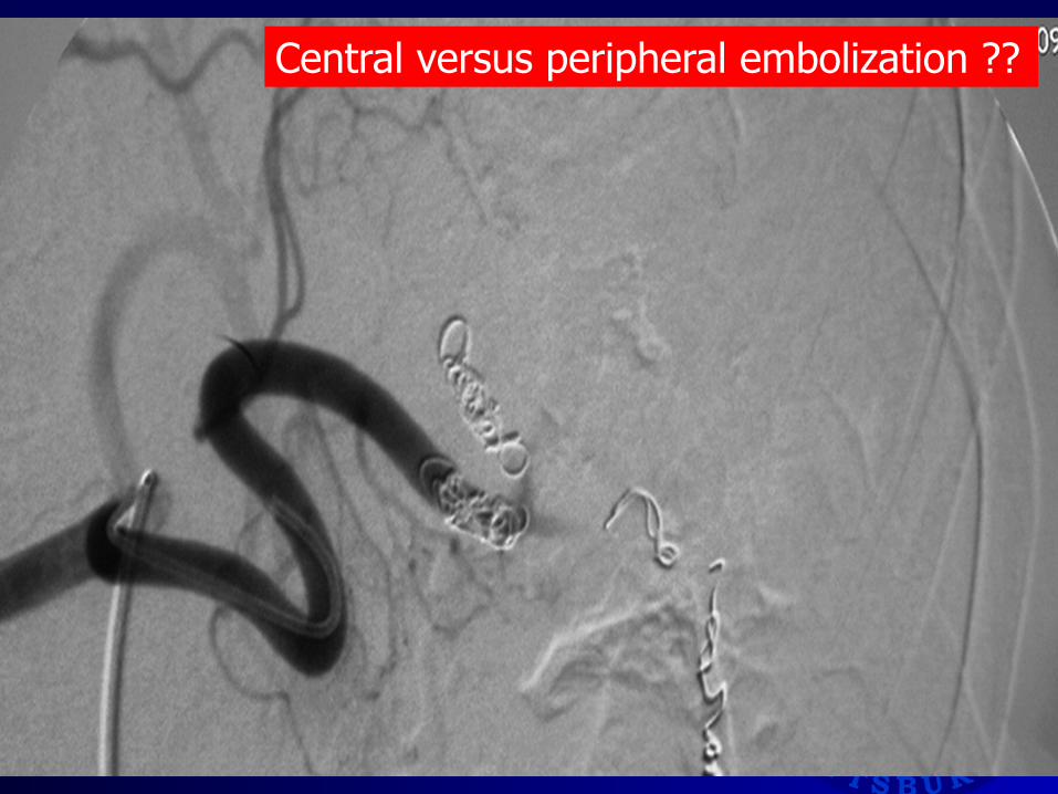
Central versus peripheral embolization ??

University of Pittsburgh School of Medicine
Thank you Thank you
The Michigan Trauma Quality Improvement Program
Ann Arbor, MI October 16, 2012
Agenda
w Andrew Peitzman, MD n Splenic Injury n Acute Diverticulitis
w Group Sessions w Lunch

Agenda
w Group Sessions Wrap-Up Discussion w Judy Mikhail
n QI Projects n Program Updates
w Mark Hemmila n DI/CDM Process Measures n MTQIP Reports n Brain Injury Reports n MSQC - Emergent GS n Final Announcements

Group Sessions
w 40 minutes per session w 2 Sessions total w Group A
n Session 1 - Splenic Injury Management n Session 2 - Diverticulitis Management
w Group B n Session 1 - MTQIP/TQIP Data Changes for 2013 n Session 2 - Understanding MTQIP/TQIP Reports
Information – MTQIP Centers
w Trauma Centers n 23 Total n 10 Level 1 n 13 Level 2 n 23 with data in current report
Information: ACS-TQIP
w Benchmark Reports n June 2012, Shock/TBI n November 2012, Aggregate and Special
w 2011 Data
w ACS-TQIP Meeting n Philadelphia, October 28-30, 2012
Splenic Injury Management Diverticulitis Management
Andrew Peitzman, MD
Group Sessions
w 40 minutes per session w 2 Sessions total w Group A
n Session 1 - Splenic Injury Management n Session 2 - Diverticulitis Management
w Group B n Session 1 - MTQIP/TQIP Data Changes for 2013 n Session 2 - Understanding MTQIP/TQIP Reports
Group Sessions
w Order of rooms based on color on name badge w Follow colored sign outside room w Red - Group A, Room 1(Spleen), 2(Divertic) w Blue - Group A, Room 2(Divertic), 1(Spleen) w Yellow - Group B, Room 1(Data ∆), 2(Reports) w Green - Group B, Room 2(Reports), 1(Data ∆ ) w You can change your group if necessary
Group Sessions
w Two sessions total n Session 1 - 10:55am to 11:35am n Session 2 – 11:40am to 12:20pm
w Return for Lunch n 12:25pm to 1:15pm

Wrap up Discussion

Judy Mikhail MTQIP Program Manager
Update
Administra;ve • Mee;ng a=endance percentage in the 90’s – Surgeons – Program Managers – Registrars
• Mee;ng Evalua;on feedback is invaluable
Website Updates
• Extensive update • Reorganized • Informa;on added • Past mee;ng slides
• Calendars, Due Dates • Registry specific informa;on • Resources • Benchmarking informa;on
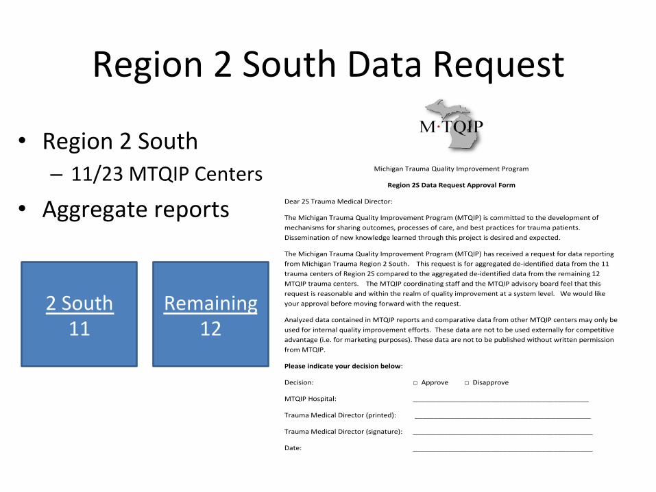
Region 2 South Data Request
• Region 2 South – 11/23 MTQIP Centers
• Aggregate reports
Michigan Trauma Quality Improvement Program
Region 2S Data Request Approval Form
Dear 2S Trauma Medical Director:
The Michigan Trauma Quality Improvement Program (MTQIP) is committed to the development of mechanisms for sharing outcomes, processes of care, and best practices for trauma patients. Dissemination of new knowledge learned through this project is desired and expected.
The Michigan Trauma Quality Improvement Program (MTQIP) has received a request for data reporting from Michigan Trauma Region 2 South. This request is for aggregated de-‐identified data from the 11 trauma centers of Region 2S compared to the aggregated de-‐identified data from the remaining 12 MTQIP trauma centers. The MTQIP coordinating staff and the MTQIP advisory board feel that this request is reasonable and within the realm of quality improvement at a system level. We would like your approval before moving forward with the request.
Analyzed data contained in MTQIP reports and comparative data from other MTQIP centers may only be used for internal quality improvement efforts. These data are not to be used externally for competitive advantage (i.e. for marketing purposes). These data are not to be published without written permission from MTQIP.
Please indicate your decision below:
Decision: □ Approve □ Disapprove
MTQIP Hospital: _____________________________________________
Trauma Medical Director (printed): _____________________________________________
Trauma Medical Director (signature): ______________________________________________
Date: ______________________________________________
2 South 11
Remaining 12
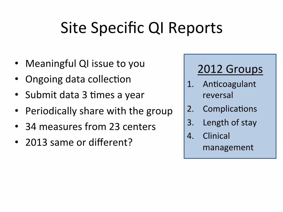
Site Specific QI Reports
• Meaningful QI issue to you • Ongoing data collec;on • Submit data 3 ;mes a year • Periodically share with the group • 34 measures from 23 centers • 2013 same or different?
2012 Groups 1. An;coagulant
reversal 2. Complica;ons 3. Length of stay 4. Clinical
management
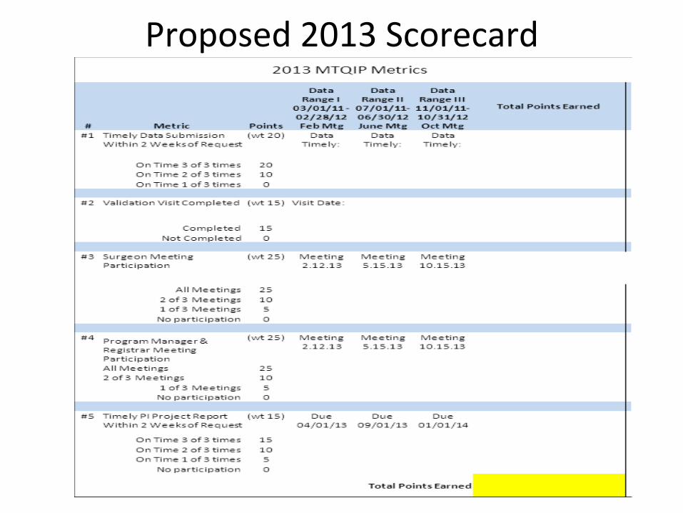
Proposed 2013 Scorecard
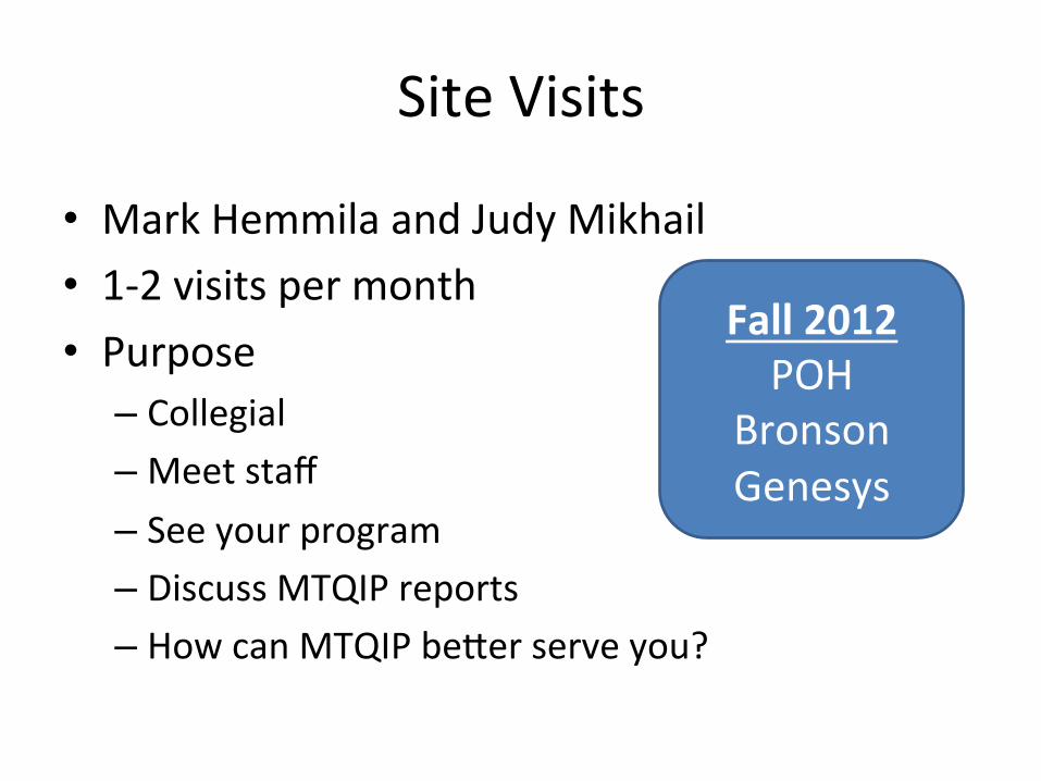
Site Visits
• Mark Hemmila and Judy Mikhail • 1-‐2 visits per month • Purpose – Collegial – Meet staff – See your program – Discuss MTQIP reports – How can MTQIP be=er serve you?
Fall 2012 POH
Bronson Genesys

Research • Publica;ons Commi=ee • Request Form • See Website • Provide you with a disc (de-‐iden;fied data) • QI related research
This data belongs to you
Research
• MCC TBI Study • Helmet repeal before and aaer comparison • Interest? • Capture “protec;ve measures” • Require resubmission past data to MTQIP
MTQIP Reports, etc.
Mark Hemmila, MD
DI/CDM
w 3 year contract w MTQIP custom data elements (module) w Mapping and transmittal of TQIP process
measures w Technical support for MTQIP tab w Preprogramed report templates w Will add future TQIP process measures w Updates for 2013 data done and submitted to
DI/CDM
ArborMetrix
w U of M Board of Regents approval obtained w MTQIP report site w Risk adjustment built-in w Can speed up results for centers w Graphs w Dashboards w Drill down
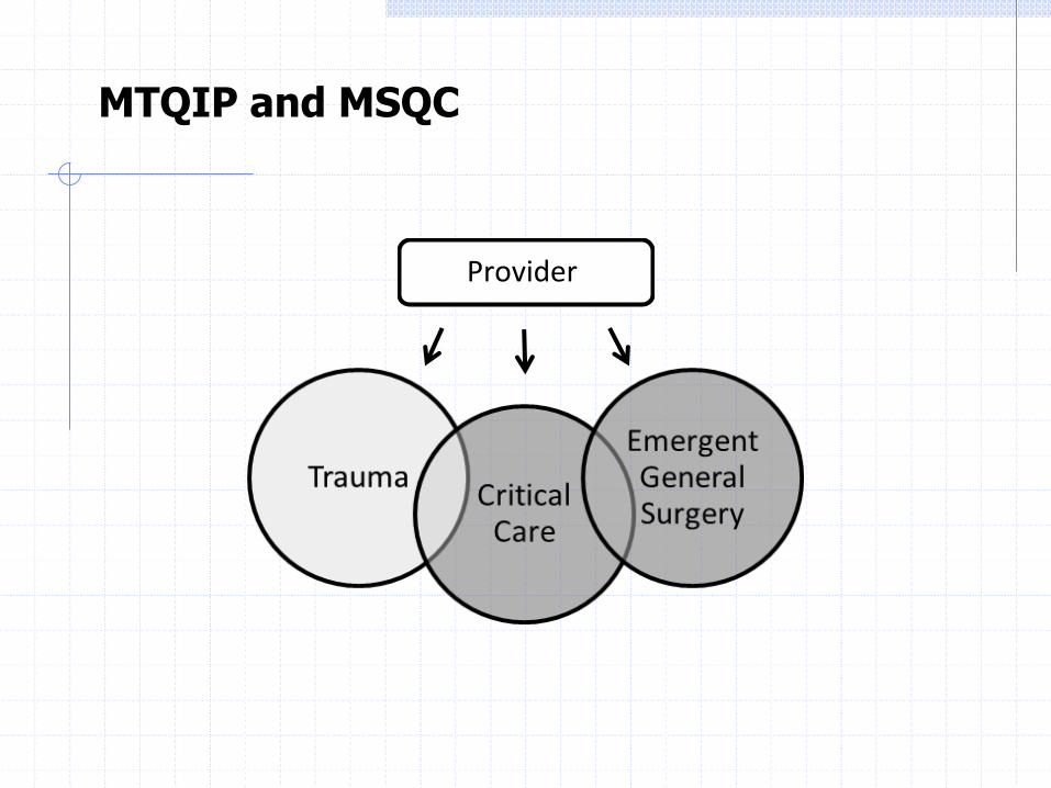
MTQIP and MSQC
Provider
MTQIP and MSQC
w Emergent General Surgery Collaboration n Feedback Reports n Best Practices n Dissemination of Information
w Feedback Reports (5 years data) n Appendectomy (Analysis done)
w December MSQC meeting w February MTQIP meeting
n Colectomy n Elderly n Aggregate
Reports
w 11/1/10 to 10/31/11 w Cohort selection w Summaries w Stratified mortality w Risk adjusted mortality w Risk adjusted complications w Risk adjusted LOS
Cohort Formation
w Cohort 1 n Blunt or penetrating n Age ≥ 18 n ISS ≥ 5 n Hospital LOS ≥ 1 or dead
w Cohort 2 (admit trauma service) w Cohort 3 (blunt multi-system) w Cohort 4 (blunt single-system)
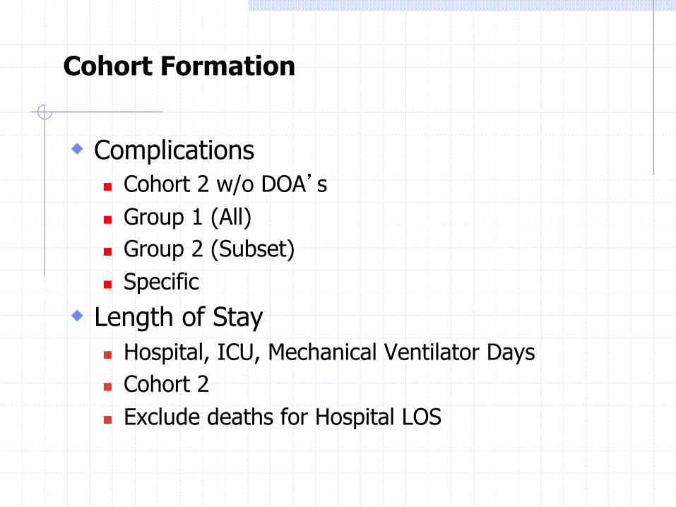
Cohort Formation
w Complications n Cohort 2 w/o DOA’s n Group 1 (All) n Group 2 (Subset) n Specific
w Length of Stay n Hospital, ICU, Mechanical Ventilator Days n Cohort 2 n Exclude deaths for Hospital LOS
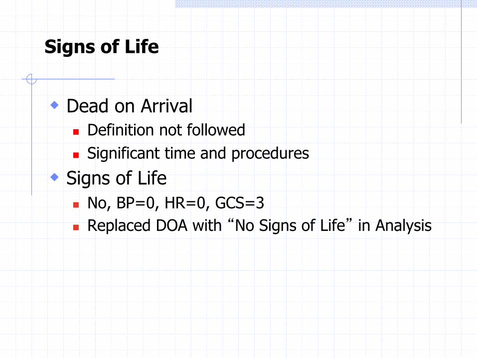
Signs of Life
w Dead on Arrival n Definition not followed n Significant time and procedures
w Signs of Life n No, BP=0, HR=0, GCS=3 n Replaced DOA with “No Signs of Life” in Analysis
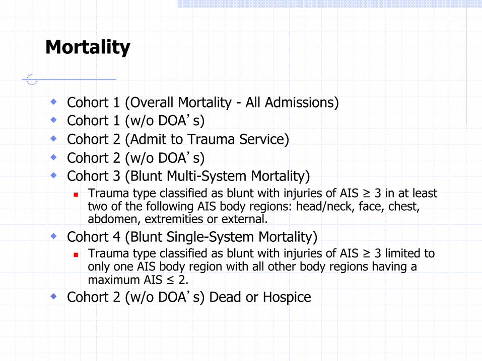
Mortality
w Cohort 1 (Overall Mortality - All Admissions) w Cohort 1 (w/o DOA’s) w Cohort 2 (Admit to Trauma Service) w Cohort 2 (w/o DOA’s) w Cohort 3 (Blunt Multi-System Mortality)
n Trauma type classified as blunt with injuries of AIS ≥ 3 in at least two of the following AIS body regions: head/neck, face, chest, abdomen, extremities or external.
w Cohort 4 (Blunt Single-System Mortality) n Trauma type classified as blunt with injuries of AIS ≥ 3 limited to
only one AIS body region with all other body regions having a maximum AIS ≤ 2.
w Cohort 2 (w/o DOA’s) Dead or Hospice
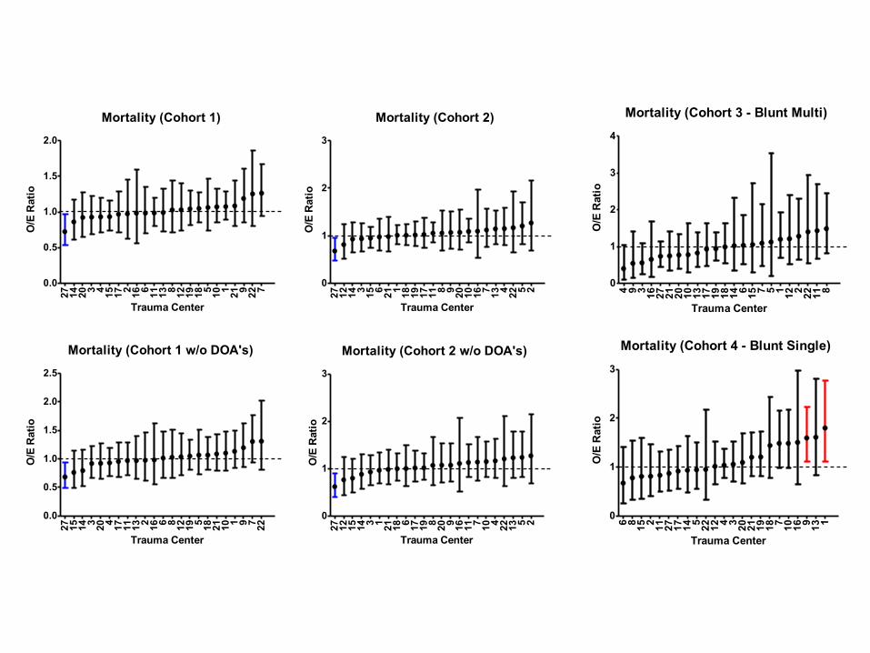
Mortality (Cohort 1)
27 14 20 3 4 15 17 2 16 6 11 13 8 12 19 18 5 10 1 21 9 22 70.0
0.5
1.0
1.5
2.0
Trauma Center
O/E
Rat
io
Mortality (Cohort 2)
27 12 14 3 15 6 21 1 18 19 17 11 8 9 20 10 16 7 13 4 22 5 20
1
2
3
Trauma CenterO
/E R
atio
Mortality (Cohort 3 - Blunt Multi)
4 9 3 16 27 21 20 10 13 17 19 18 14 6 15 7 5 1 12 2 22 11 80
1
2
3
4
Trauma Center
O/E
Rat
io
Mortality (Cohort 1 w/o DOA's)
27 15 14 3 20 4 17 11 13 2 16 6 8 12 19 5 18 21 10 1 9 7 22
0.0
0.5
1.0
1.5
2.0
2.5
Trauma Center
O/E
Rat
io
Mortality (Cohort 2 w/o DOA's)27 12 15 14 3 1 21 18 6 17 19 8 20 9 16 11 7 10 4 22 13 5 20
1
2
3
Trauma Center
O/E
Rat
io
Mortality (Cohort 4 - Blunt Single)
6 8 15 2 11 27 17 14 5 22 12 4 3 20 21 19 18 7 10 16 9 13 10
1
2
3
Trauma Center
O/E
Rat
io
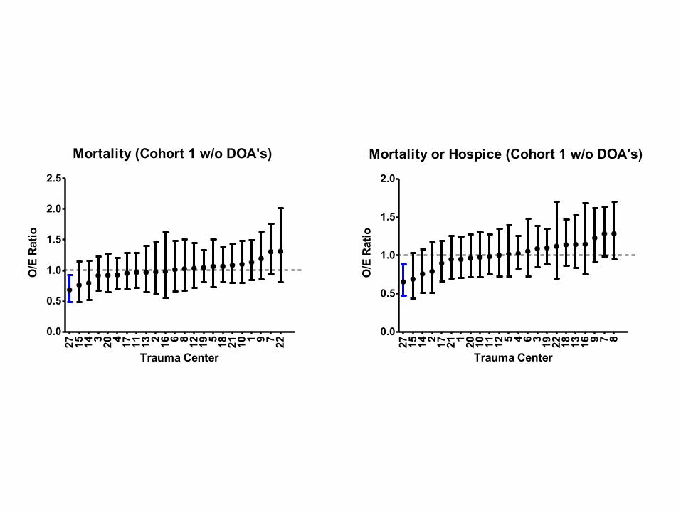
Mortality (Cohort 1 w/o DOA's)
27 15 14 3 20 4 17 11 13 2 16 6 8 12 19 5 18 21 10 1 9 7 22
0.0
0.5
1.0
1.5
2.0
2.5
Trauma Center
O/E
Rat
io
Mortality or Hospice (Cohort 1 w/o DOA's)
27 15 14 2 17 21 1 20 10 11 12 5 4 6 3 19 22 18 13 16 9 7 80.0
0.5
1.0
1.5
2.0
Trauma CenterO
/E R
atio
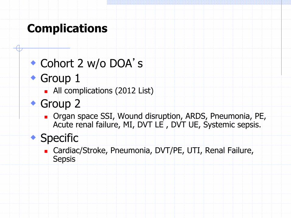
Complications
w Cohort 2 w/o DOA’s w Group 1
n All complications (2012 List) w Group 2
n Organ space SSI, Wound disruption, ARDS, Pneumonia, PE, Acute renal failure, MI, DVT LE , DVT UE, Systemic sepsis.
w Specific n Cardiac/Stroke, Pneumonia, DVT/PE, UTI, Renal Failure,
Sepsis
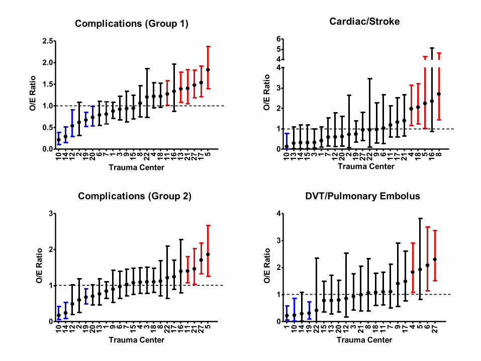
Complications (Group 1)
10 14 12 2 19 20 6 7 1 3 9 15 8 22 4 18 11 16 13 21 27 17 50.0
0.5
1.0
1.5
2.0
2.5
Trauma Center
O/E
Rat
ioCardiac/Stroke
10 13 14 15 3 1 7 12 20 2 19 27 22 9 6 11 17 21 4 18 5 16 80
1
2
3
4456
Trauma Center
O/E
Rat
io
Complications (Group 2)
10 14 12 2 19 20 13 1 9 6 7 15 4 3 18 8 22 17 16 11 21 27 50
1
2
3
Trauma Center
O/E
Rat
io
DVT/Pulmonary Embolus
1 10 14 19 22 15 13 20 12 3 21 8 18 11 7 9 17 4 5 6 27
0
1
2
3
4
Trauma Center
O/E
Rat
io
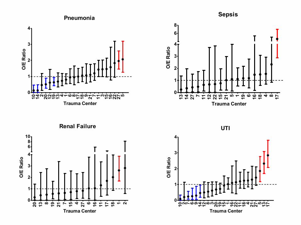
Pneumonia
10 14 2 20 12 19 13 4 1 6 8 17 18 9 21 7 11 3 15 16 22 27 50
1
2
3
4
Trauma Center
O/E
Rat
io
Sepsis
13 14 27 7 11 12 22 15 21 5 1 19 6 16 18 4 8 17
0
1
2
3
4
68
Trauma Center
O/E
Rat
io
Renal Failure
20 13 8 19 21 7 15 4 27 6 16 11 17 18 1 20
1
2
3
468
10
Trauma Center
O/E
Rat
io
UTI
10 2 7 6 9 14 12 8 3 20 19 15 1 16 22 11 18 4 21 27 5 13 17
0
1
2
3
4
Trauma Center
O/E
Rat
io
Length of Stay
w Cohort 2 w Risk Adjusted Rate w Natural log transformed, linear regression w Adjusted for age, ISS, mGCS, comorbids, etc. w Hospital LOS, ICU LOS, MV Days w Exclude deaths for Hospital LOS w 95% CI
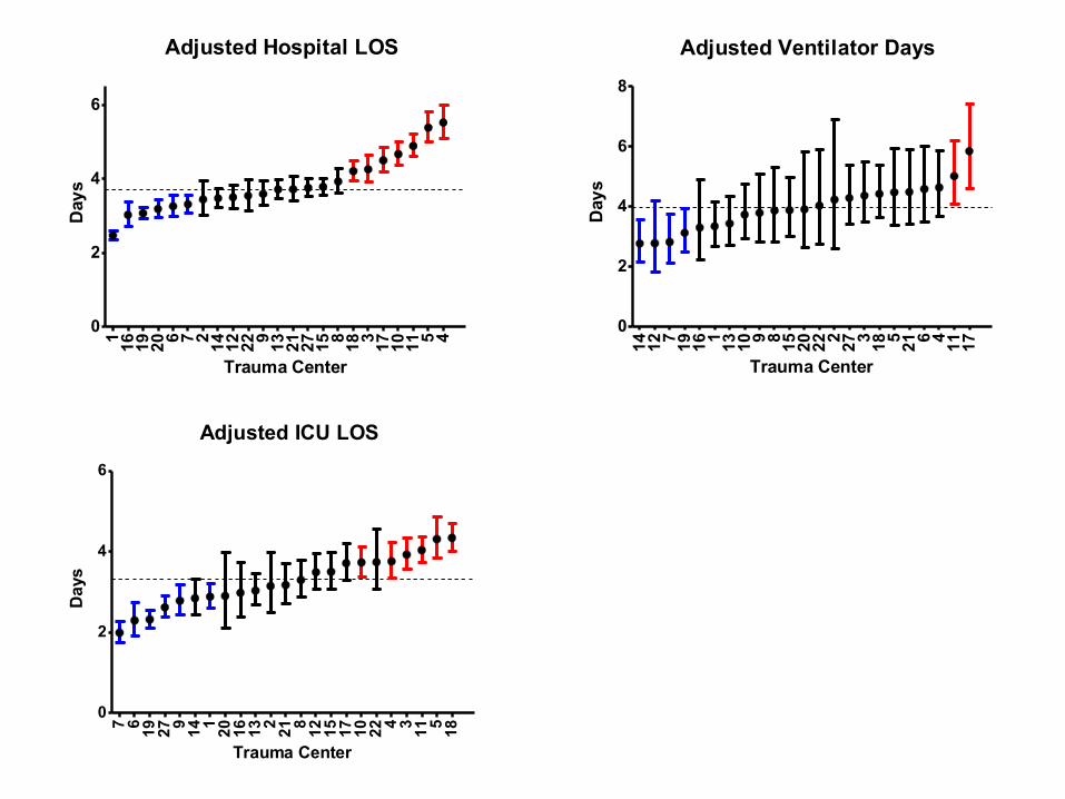
Adjusted Hospital LOS
1 16 19 20 6 7 2 14 12 22 9 13 21 27 15 8 18 3 17 10 11 5 40
2
4
6
Trauma Center
Day
sAdjusted Ventilator Days
14 12 7 19 16 1 13 10 9 8 15 20 22 2 27 3 18 5 21 6 4 11 17
0
2
4
6
8
Trauma Center
Day
s
Adjusted ICU LOS
7 6 19 27 9 14 1 20 16 13 2 21 8 12 15 17 10 22 4 3 11 5 18
0
2
4
6
Trauma Center
Day
s
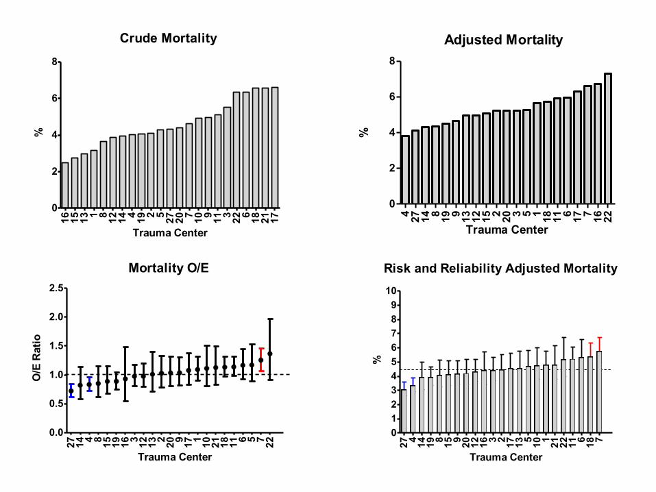
Crude Mortality
16 15 13 1 8 12 14 4 19 2 5 27 20 7 10 9 11 3 22 6 18 21 17
0
2
4
6
8
Trauma Center
%Adjusted Mortality
4 27 14 8 19 9 13 12 15 2 20 3 5 1 18 11 6 17 7 16 22
0
2
4
6
8
Trauma Center
%
Mortality O/E
27 14 4 8 15 19 16 3 12 13 2 20 9 17 1 10 21 18 11 6 5 7 22
0.0
0.5
1.0
1.5
2.0
2.5
Trauma Center
O/E
Rat
io
Risk and Reliability Adjusted Mortality
27 4 14 19 8 15 9 20 12 16 3 2 17 13 5 10 1 21 22 11 6 18 70123456789
10
Trauma Center
%
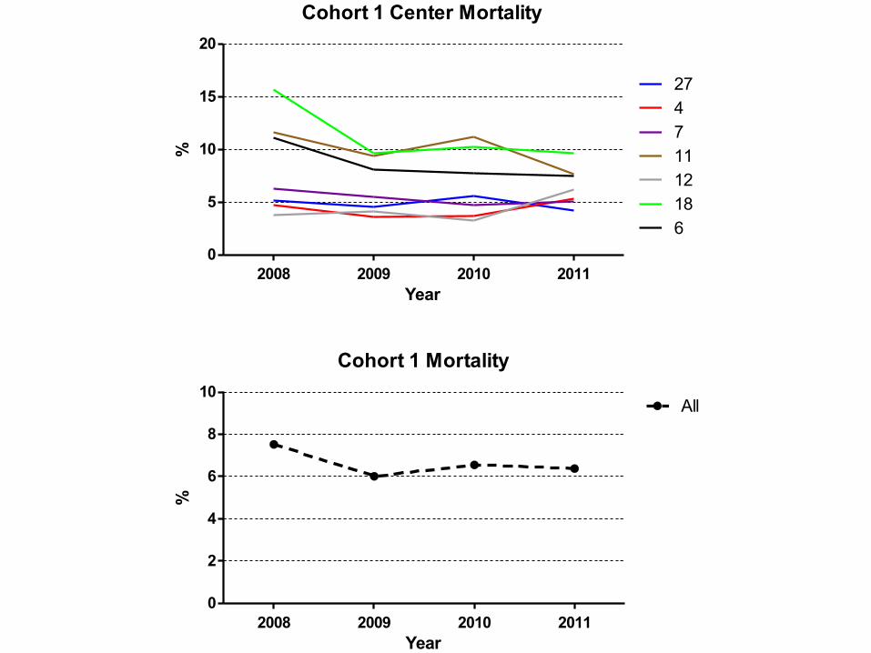
Cohort 1 Center Mortality
2008 2009 2010 20110
5
10
15
20
27471112186
Year
%
Cohort 1 Mortality
2008 2009 2010 20110
2
4
6
8
10All
Year
%
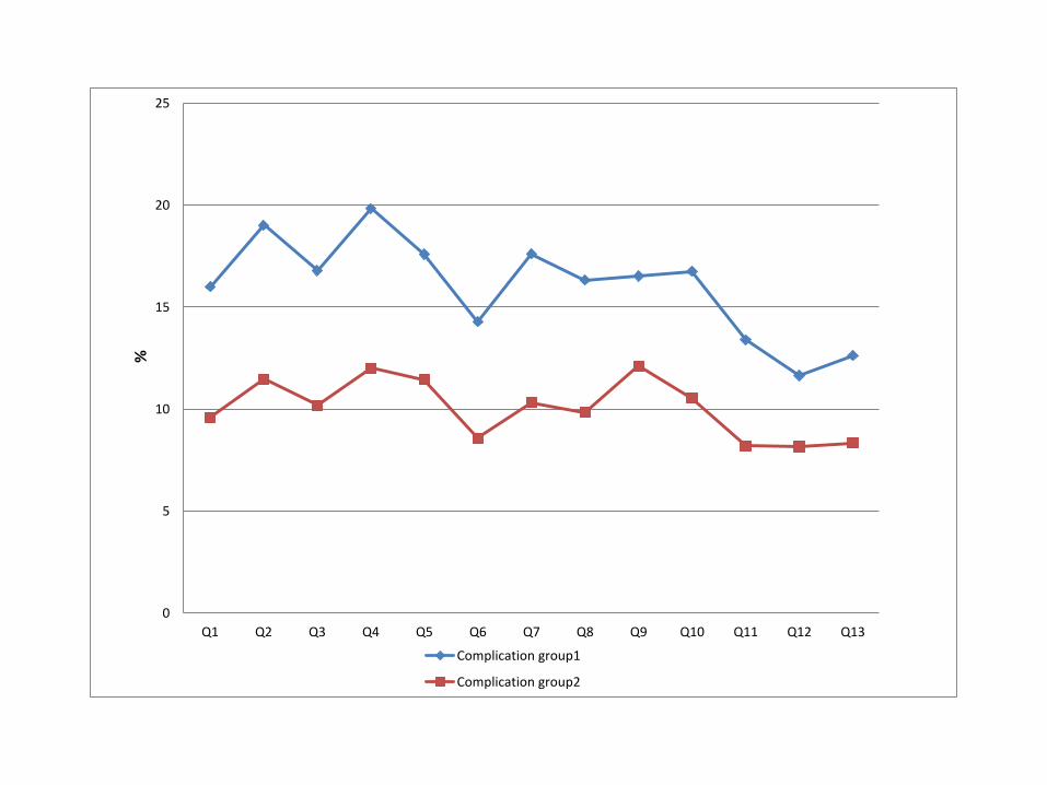
0
5
10
15
20
25
Q1 Q2 Q3 Q4 Q5 Q6 Q7 Q8 Q9 Q10 Q11 Q12 Q13
%
Complication group1
Complication group2
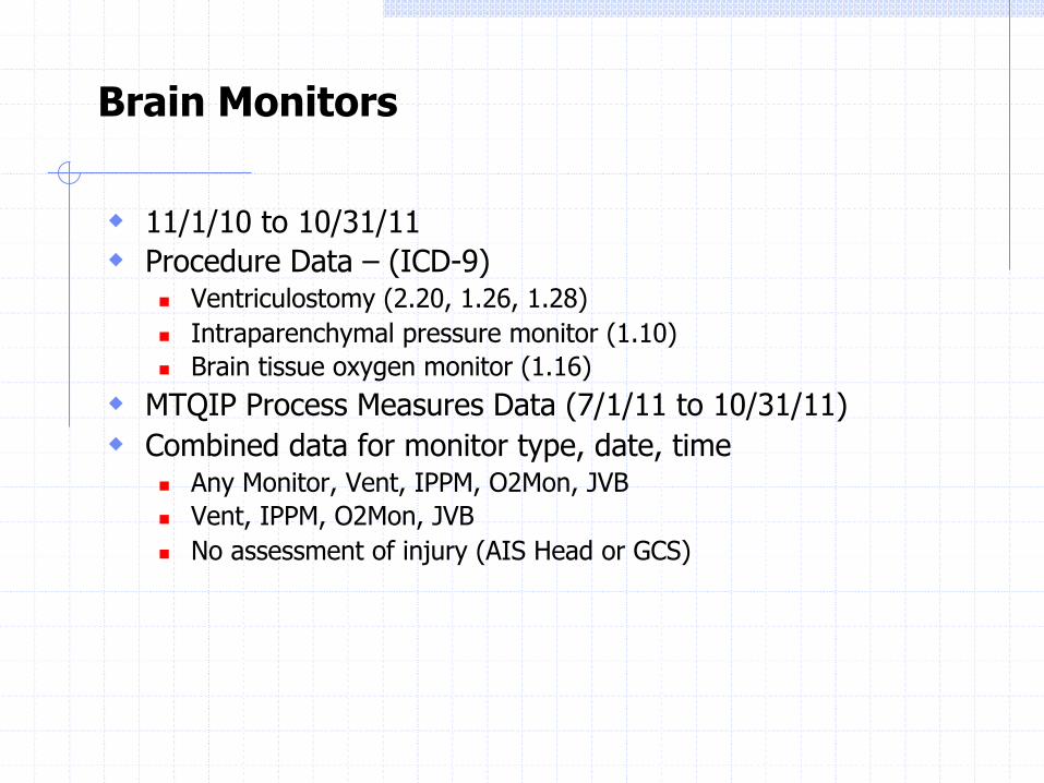
Brain Monitors
w 11/1/10 to 10/31/11 w Procedure Data – (ICD-9)
n Ventriculostomy (2.20, 1.26, 1.28) n Intraparenchymal pressure monitor (1.10) n Brain tissue oxygen monitor (1.16)
w MTQIP Process Measures Data (7/1/11 to 10/31/11) w Combined data for monitor type, date, time
n Any Monitor, Vent, IPPM, O2Mon, JVB n Vent, IPPM, O2Mon, JVB n No assessment of injury (AIS Head or GCS)
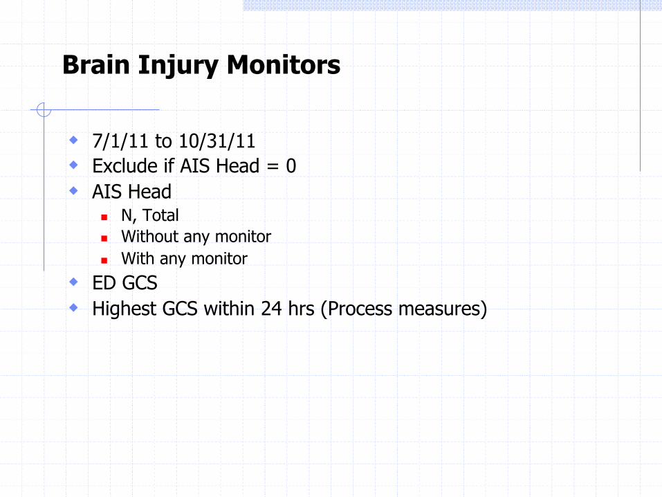
Brain Injury Monitors
w 7/1/11 to 10/31/11 w Exclude if AIS Head = 0 w AIS Head
n N, Total n Without any monitor n With any monitor
w ED GCS w Highest GCS within 24 hrs (Process measures)
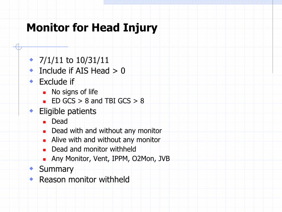
Monitor for Head Injury
w 7/1/11 to 10/31/11 w Include if AIS Head > 0 w Exclude if
n No signs of life n ED GCS > 8 and TBI GCS > 8
w Eligible patients n Dead n Dead with and without any monitor n Alive with and without any monitor n Dead and monitor withheld n Any Monitor, Vent, IPPM, O2Mon, JVB
w Summary w Reason monitor withheld
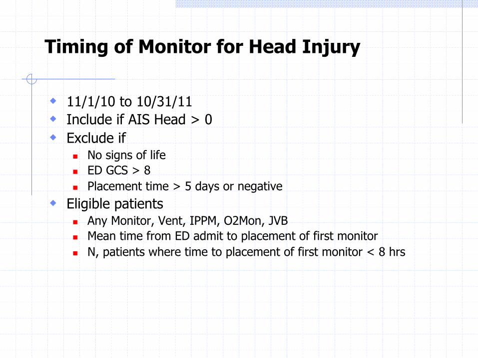
Timing of Monitor for Head Injury
w 11/1/10 to 10/31/11 w Include if AIS Head > 0 w Exclude if
n No signs of life n ED GCS > 8 n Placement time > 5 days or negative
w Eligible patients n Any Monitor, Vent, IPPM, O2Mon, JVB n Mean time from ED admit to placement of first monitor n N, patients where time to placement of first monitor < 8 hrs
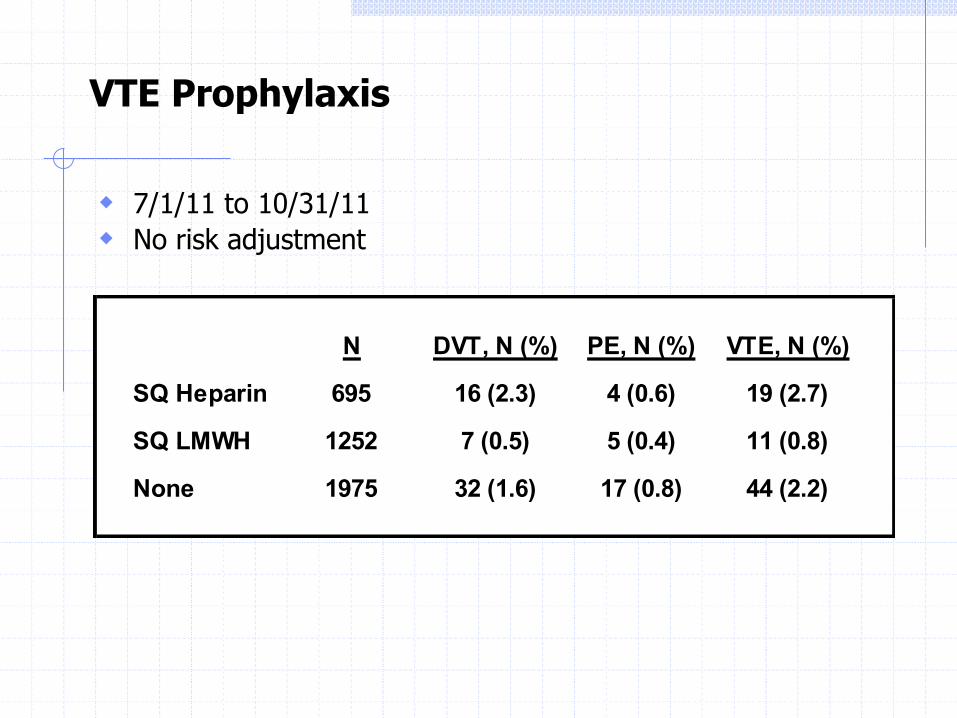
VTE Prophylaxis
w 7/1/11 to 10/31/11 w No risk adjustment
N DVT, N (%) PE, N (%) VTE, N (%)
SQ Heparin 695 16 (2.3) 4 (0.6) 19 (2.7)
SQ LMWH 1252 7 (0.5) 5 (0.4) 11 (0.8)
None 1975 32 (1.6) 17 (0.8) 44 (2.2)
Future Reports
w Head Injury and Monitors w VTE Prophylaxis w Blood Usage w VAP w ?

Questions
Call for Data, Feedback
w Data submitted from 3/1/11 to 2/29/12 n 23 centers
w Next call n Data from 7/1/11 to 6/30/12 n Due February 1, 2013
w Evaluations n Meeting ideas, Reports, Web-site
w CME n Turn in green card, get certificate

Future Meetings
w Tuesday February 12, 2013 n Location: Ann Arbor
w Wednesday May 15, 2013 n Location: Kalamazoo
w Tuesday June 11, 2013 n Location: Ann Arbor n Registrars
w Tuesday October 15, 2013 n Location: Ann Arbor
MTQIP Data Definition Update Review
January 1
2013
October 16, 2012
1
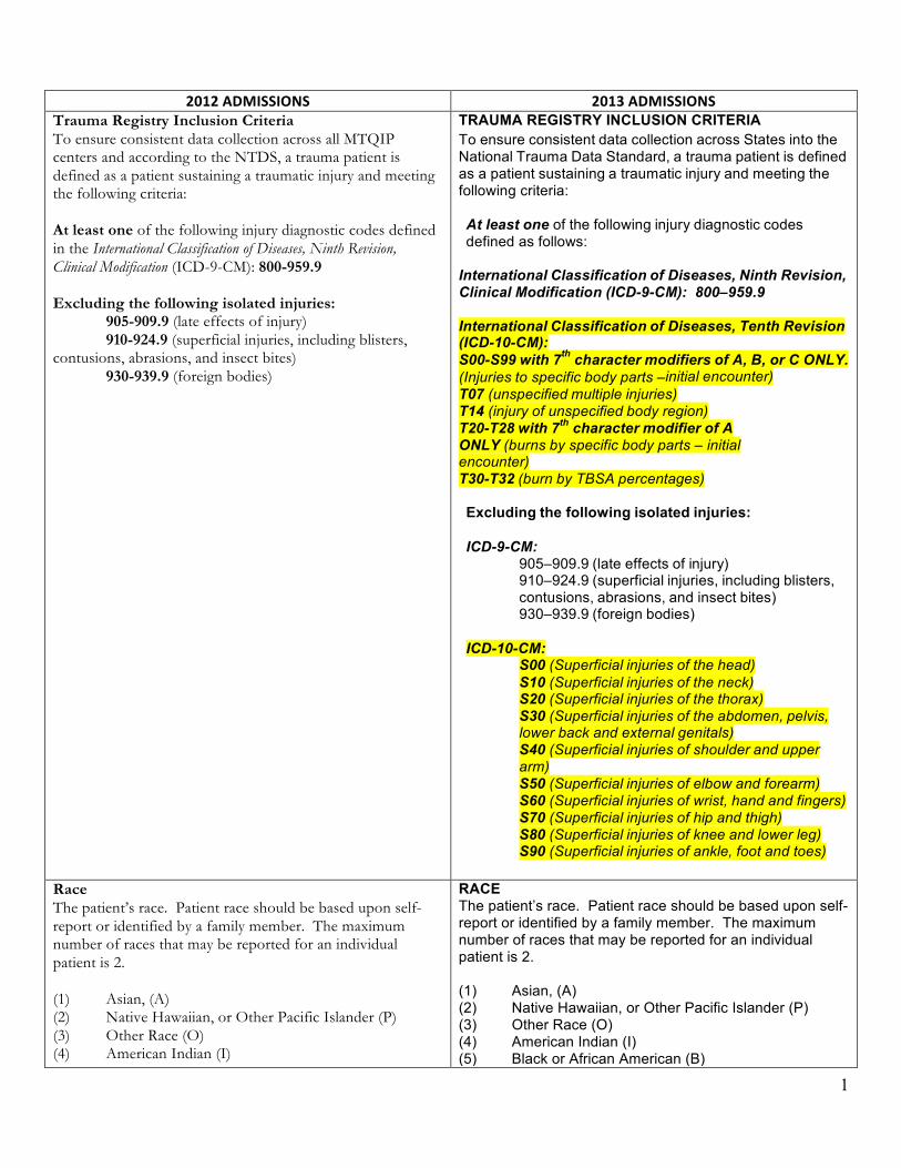
2012 ADMISSIONS 2013 ADMISSIONS Trauma Registry Inclusion Criteria To ensure consistent data collection across all MTQIP centers and according to the NTDS, a trauma patient is defined as a patient sustaining a traumatic injury and meeting the following criteria: At least one of the following injury diagnostic codes defined in the International Classification of Diseases, Ninth Revision, Clinical Modification (ICD-9-CM): 800-959.9 Excluding the following isolated injuries: 905-909.9 (late effects of injury) 910-924.9 (superficial injuries, including blisters, contusions, abrasions, and insect bites) 930-939.9 (foreign bodies)
TRAUMA REGISTRY INCLUSION CRITERIA To ensure consistent data collection across States into the National Trauma Data Standard, a trauma patient is defined as a patient sustaining a traumatic injury and meeting the following criteria: At least one of the following injury diagnostic codes defined as follows: International Classification of Diseases, Ninth Revision, Clinical Modification (ICD-9-CM): 800–959.9 International Classification of Diseases, Tenth Revision (ICD-10-CM): S00-S99 with 7th character modifiers of A, B, or C ONLY. (Injuries to specific body parts –initial encounter) T07 (unspecified multiple injuries) T14 (injury of unspecified body region) T20-T28 with 7th character modifier of A ONLY (burns by specific body parts – initial encounter) T30-T32 (burn by TBSA percentages) Excluding the following isolated injuries: ICD-9-CM:
905–909.9 (late effects of injury) 910–924.9 (superficial injuries, including blisters, contusions, abrasions, and insect bites) 930–939.9 (foreign bodies)
ICD-10-CM:
S00 (Superficial injuries of the head) S10 (Superficial injuries of the neck) S20 (Superficial injuries of the thorax) S30 (Superficial injuries of the abdomen, pelvis, lower back and external genitals) S40 (Superficial injuries of shoulder and upper arm) S50 (Superficial injuries of elbow and forearm) S60 (Superficial injuries of wrist, hand and fingers) S70 (Superficial injuries of hip and thigh) S80 (Superficial injuries of knee and lower leg) S90 (Superficial injuries of ankle, foot and toes)
Race The patient’s race. Patient race should be based upon self-report or identified by a family member. The maximum number of races that may be reported for an individual patient is 2. (1) Asian, (A) (2) Native Hawaiian, or Other Pacific Islander (P) (3) Other Race (O) (4) American Indian (I)
RACE The patient’s race. Patient race should be based upon self-report or identified by a family member. The maximum number of races that may be reported for an individual patient is 2. (1) Asian, (A) (2) Native Hawaiian, or Other Pacific Islander (P) (3) Other Race (O) (4) American Indian (I) (5) Black or African American (B)
2
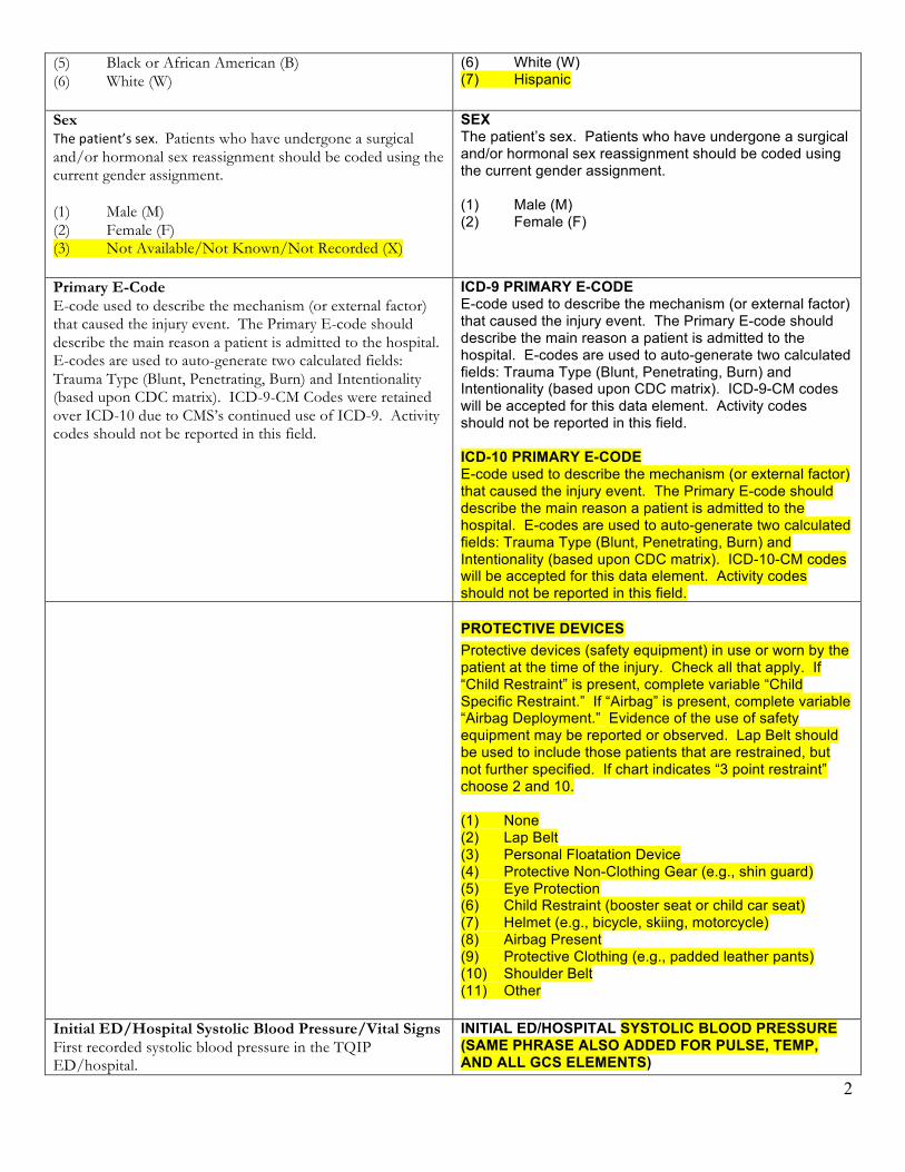
(5) Black or African American (B) (6) White (W)
(6) White (W) (7) Hispanic
Sex The patient’s sex. Patients who have undergone a surgical and/or hormonal sex reassignment should be coded using the current gender assignment. (1) Male (M) (2) Female (F) (3) Not Available/Not Known/Not Recorded (X)
SEX The patient’s sex. Patients who have undergone a surgical and/or hormonal sex reassignment should be coded using the current gender assignment. (1) Male (M) (2) Female (F)
Primary E-Code E-code used to describe the mechanism (or external factor) that caused the injury event. The Primary E-code should describe the main reason a patient is admitted to the hospital. E-codes are used to auto-generate two calculated fields: Trauma Type (Blunt, Penetrating, Burn) and Intentionality (based upon CDC matrix). ICD-9-CM Codes were retained over ICD-10 due to CMS’s continued use of ICD-9. Activity codes should not be reported in this field.
ICD-9 PRIMARY E-CODE E-code used to describe the mechanism (or external factor) that caused the injury event. The Primary E-code should describe the main reason a patient is admitted to the hospital. E-codes are used to auto-generate two calculated fields: Trauma Type (Blunt, Penetrating, Burn) and Intentionality (based upon CDC matrix). ICD-9-CM codes will be accepted for this data element. Activity codes should not be reported in this field. ICD-10 PRIMARY E-CODE E-code used to describe the mechanism (or external factor) that caused the injury event. The Primary E-code should describe the main reason a patient is admitted to the hospital. E-codes are used to auto-generate two calculated fields: Trauma Type (Blunt, Penetrating, Burn) and Intentionality (based upon CDC matrix). ICD-10-CM codes will be accepted for this data element. Activity codes should not be reported in this field.
PROTECTIVE DEVICES Protective devices (safety equipment) in use or worn by the patient at the time of the injury. Check all that apply. If “Child Restraint” is present, complete variable “Child Specific Restraint.” If “Airbag” is present, complete variable “Airbag Deployment.” Evidence of the use of safety equipment may be reported or observed. Lap Belt should be used to include those patients that are restrained, but not further specified. If chart indicates “3 point restraint” choose 2 and 10. (1) None (2) Lap Belt (3) Personal Floatation Device (4) Protective Non-Clothing Gear (e.g., shin guard) (5) Eye Protection (6) Child Restraint (booster seat or child car seat) (7) Helmet (e.g., bicycle, skiing, motorcycle) (8) Airbag Present (9) Protective Clothing (e.g., padded leather pants) (10) Shoulder Belt (11) Other
Initial ED/Hospital Systolic Blood Pressure/Vital Signs First recorded systolic blood pressure in the TQIP ED/hospital.
INITIAL ED/HOSPITAL SYSTOLIC BLOOD PRESSURE (SAME PHRASE ALSO ADDED FOR PULSE, TEMP, AND ALL GCS ELEMENTS)
3
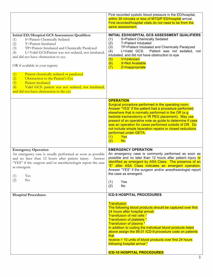
First recorded systolic blood pressure in the ED/hospital, within 30 minutes or less of MTQIP ED/hospital arrival. First recorded/hospital vitals do not need to be from the same assessment.
Initial ED/Hospital GCS Assessment Qualifiers (1) S=Patient Chemically Sedated (2) T=Patient Intubated (3) TP=Patient Intubated and Chemically Paralyzed (4) L=Valid GCS:Patient was not sedated, not intubated, and did not have obstruction to eye
OR if available in your registry
(1) Patient chemically sedated or paralyzed (2) Obstruction to the Patient’s Eye (3) Patient intubated (4) Valid GCS: patient was not sedated, not intubated, and did not have obstruction to the eye
INITIAL ED/HOSPITAL GCS ASSESSMENT QUALIFIERS (1) S=Patient Chemically Sedated (2) T=Patient Intubated (3) TP=Patient Intubated and Chemically Paralyzed (4) L=Valid GCS: Patient was not sedated, not intubated, and did not have obstruction to eye (5) V=Unknown (6) X=Not Available (7) Z=Inappropriate
OPERATION Surgical procedure performed in the operating room. Answer “YES” if the patient had a procedure performed elsewhere that is normally performed in the OR (e.g. bedside tracheostomy or IR PEG placement). May use present of an operative note as guide to determine if case was an operation for cases performed outside of OR. Do not include simple laceration repairs or closed reductions performed under GETA. (1) Yes (2) No
Emergency Operation An emergency case is usually performed as soon as possible and no later than 12 hours after patient injury. Answer “YES” if the surgeon and/or anesthesiologist report the case as emergent. (1) Yes (2) No
EMERGENCY OPERATION An emergency case is commonly performed as soon as possible and no later than 12 hours after patient injury or identified as emergent by ASA Class. The presence of an “E” after ASA Class indicates an emergent operation. Answer “YES” if the surgeon and/or anesthesiologist report the case as emergent. (1) Yes (2) No
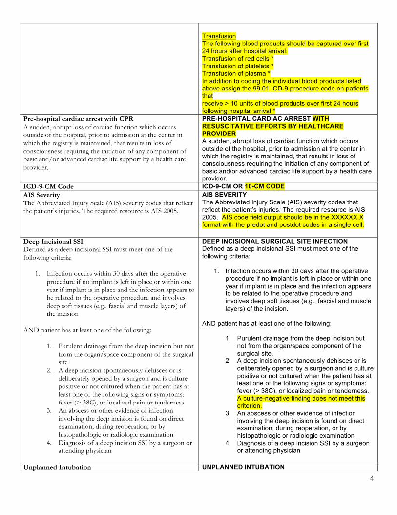
Hospital Procedures ICD-9 HOSPITAL PROCEDURES Transfusion The following blood products should be captured over first 24 hours after hospital arrival: Transfusion of red cells * Transfusion of platelets * Transfusion of plasma * In addition to coding the individual blood products listed above assign the 99.01 ICD-9 procedure code on patients that receive > 10 units of blood products over first 24 hours following hospital arrival * ICD-10 HOSPITAL PROCEDURES
4
Transfusion The following blood products should be captured over first 24 hours after hospital arrival: Transfusion of red cells * Transfusion of platelets * Transfusion of plasma * In addition to coding the individual blood products listed above assign the 99.01 ICD-9 procedure code on patients that receive > 10 units of blood products over first 24 hours following hospital arrival *
Pre-hospital cardiac arrest with CPR A sudden, abrupt loss of cardiac function which occurs outside of the hospital, prior to admission at the center in which the registry is maintained, that results in loss of consciousness requiring the initiation of any component of basic and/or advanced cardiac life support by a health care provider.
PRE-HOSPITAL CARDIAC ARREST WITH RESUSCITATIVE EFFORTS BY HEALTHCARE PROVIDER A sudden, abrupt loss of cardiac function which occurs outside of the hospital, prior to admission at the center in which the registry is maintained, that results in loss of consciousness requiring the initiation of any component of basic and/or advanced cardiac life support by a health care provider.
ICD-9-CM Code ICD-9-CM OR 10-CM CODE AIS Severity The Abbreviated Injury Scale (AIS) severity codes that reflect the patient’s injuries. The required resource is AIS 2005.
AIS SEVERITY The Abbreviated Injury Scale (AIS) severity codes that reflect the patient’s injuries. The required resource is AIS 2005. AIS code field output should be in the XXXXXX.X format with the predot and postdot codes in a single cell.
Deep Incisional SSI Defined as a deep incisional SSI must meet one of the following criteria:
1. Infection occurs within 30 days after the operative procedure if no implant is left in place or within one year if implant is in place and the infection appears to be related to the operative procedure and involves deep soft tissues (e.g., fascial and muscle layers) of the incision
AND patient has at least one of the following:
1. Purulent drainage from the deep incision but not from the organ/space component of the surgical site
2. A deep incision spontaneously dehisces or is deliberately opened by a surgeon and is culture positive or not cultured when the patient has at least one of the following signs or symptoms: fever (> 38C), or localized pain or tenderness
3. An abscess or other evidence of infection involving the deep incision is found on direct examination, during reoperation, or by histopathologic or radiologic examination
4. Diagnosis of a deep incision SSI by a surgeon or attending physician
DEEP INCISIONAL SURGICAL SITE INFECTION Defined as a deep incisional SSI must meet one of the following criteria:
1. Infection occurs within 30 days after the operative procedure if no implant is left in place or within one year if implant is in place and the infection appears to be related to the operative procedure and involves deep soft tissues (e.g., fascial and muscle layers) of the incision.
AND patient has at least one of the following:
1. Purulent drainage from the deep incision but not from the organ/space component of the surgical site.
2. A deep incision spontaneously dehisces or is deliberately opened by a surgeon and is culture positive or not cultured when the patient has at least one of the following signs or symptoms: fever (> 38C), or localized pain or tenderness. A culture-negative finding does not meet this criterion.
3. An abscess or other evidence of infection involving the deep incision is found on direct examination, during reoperation, or by histopathologic or radiologic examination
4. Diagnosis of a deep incision SSI by a surgeon or attending physician
Unplanned Intubation UNPLANNED INTUBATION
5
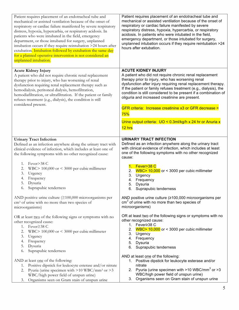
Patient requires placement of an endotracheal tube and mechanical or assisted ventilation because of the onset of respiratory or cardiac failure manifested by severe respiratory distress, hypoxia, hypercarbia, or respiratory acidosis. In patients who were intubated in the field, emergency department, or those intubated for surgery, unplanned intubation occurs if they require reintubation >24 hours after extubation. Intubation followed by extubation the same day for a planned operative intervention is not considered an unplanned intubation.
Patient requires placement of an endotracheal tube and mechanical or assisted ventilation because of the onset of respiratory or cardiac failure manifested by severe respiratory distress, hypoxia, hypercarbia, or respiratory acidosis. In patients who were intubated in the field, emergency department, or those intubated for surgery, unplanned intubation occurs if they require reintubation >24 hours after extubation.
Acute Kidney Injury A patient who did not require chronic renal replacement therapy prior to injury, who has worsening of renal dysfunction requiring renal replacement therapy such as hemodialysis, peritoneal dialysis, hemofiltration, hemodiafiltration, or ultrafiltration. If the patient or family refuses treatment (e.g., dialysis), the condition is still considered present.
ACUTE KIDNEY INJURY A patient who did not require chronic renal replacement therapy prior to injury, who has worsening renal dysfunction after injury requiring renal replacement therapy. If the patient or family refuses treatment (e.g., dialysis), the condition is still considered to be present if a combination of oliguria and increased creatinine are present. GFR criteria: Increase creatinine x3 or GFR decrease > 75%
Urine output criteria: UO < 0.3ml/kg/h x 24 hr or Anuria x 12 hrs
Urinary Tract Infection Defined as an infection anywhere along the urinary tract with clinical evidence of infection, which includes at least one of the following symptoms with no other recognized cause:
1. Fever>38 C 2. WBC> 100,000 or < 3000 per cubic millimeter 3. Urgency 4. Frequency 5. Dysuria 6. Suprapubic tenderness
AND positive urine culture (≥100,000 microorganisms per cm3 of urine with no more than two species of microorganisms) OR at least two of the following signs or symptoms with no other recognized cause:
1. Fever≥38 C 2. WBC> 100,000 or < 3000 per cubic millimeter 3. Urgency 4. Frequency 5. Dysuria 6. Suprapubic tenderness
AND at least one of the following: 1. Positive dipstick for leukocyte esterase and/or nitrate 2. Pyuria (urine specimen with >10 WBC/mm3 or >3
WBC/high power field of unspun urine) 3. Organisms seen on Gram stain of unspun urine
URINARY TRACT INFECTION Defined as an infection anywhere along the urinary tract with clinical evidence of infection, which includes at least one of the following symptoms with no other recognized cause:
1. Fever>38 C 2. WBC> 10,000 or < 3000 per cubic millimeter 3. Urgency 4. Frequency 5. Dysuria 6. Suprapubic tenderness
AND positive urine culture (≥100,000 microorganisms per cm3 of urine with no more than two species of microorganisms) OR at least two of the following signs or symptoms with no other recognized cause:
1. Fever≥38 C 2. WBC> 10,000 or < 3000 per cubic millimeter 3. Urgency 4. Frequency 5. Dysuria 6. Suprapubic tenderness
AND at least one of the following: 1. Positive dipstick for leukocyte esterase and/or
nitrate 2. Pyuria (urine specimen with >10 WBC/mm3 or >3
WBC/high power field of unspun urine) 3. Organisms seen on Gram stain of unspun urine
6
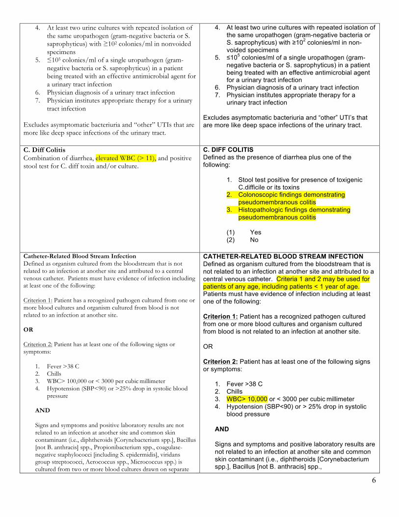
4. At least two urine cultures with repeated isolation of the same uropathogen (gram-negative bacteria or S. saprophyticus) with ≥102 colonies/ml in nonvoided specimens
5. ≤105 colonies/ml of a single uropathogen (gram-negative bacteria or S. saprophyticus) in a patient being treated with an effective antimicrobial agent for a urinary tract infection
6. Physician diagnosis of a urinary tract infection 7. Physician institutes appropriate therapy for a urinary
tract infection Excludes asymptomatic bacteriuria and “other” UTIs that are more like deep space infections of the urinary tract.
4. At least two urine cultures with repeated isolation of the same uropathogen (gram-negative bacteria or S. saprophyticus) with ≥102 colonies/ml in non-voided specimens
5. ≤105 colonies/ml of a single uropathogen (gram-negative bacteria or S. saprophyticus) in a patient being treated with an effective antimicrobial agent for a urinary tract infection
6. Physician diagnosis of a urinary tract infection 7. Physician institutes appropriate therapy for a
urinary tract infection Excludes asymptomatic bacteriuria and “other” UTI’s that are more like deep space infections of the urinary tract.
C. Diff Colitis Combination of diarrhea, elevated WBC (> 11), and positive stool test for C. diff toxin and/or culture.
C. DIFF COLITIS Defined as the presence of diarrhea plus one of the following:
1. Stool test positive for presence of toxigenic C.difficile or its toxins
2. Colonoscopic findings demonstrating pseudomembranous colitis
3. Histopathologic findings demonstrating pseudomembranous colitis
(1) Yes (2) No
Catheter-Related Blood Stream Infection Defined as organism cultured from the bloodstream that is not related to an infection at another site and attributed to a central venous catheter. Patients must have evidence of infection including at least one of the following: Criterion 1: Patient has a recognized pathogen cultured from one or more blood cultures and organism cultured from blood is not related to an infection at another site. OR Criterion 2: Patient has at least one of the following signs or symptoms:
1. Fever >38 C 2. Chills 3. WBC> 100,000 or < 3000 per cubic millimeter 4. Hypotension (SBP<90) or >25% drop in systolic blood
pressure AND Signs and symptoms and positive laboratory results are not related to an infection at another site and common skin contaminant (i.e., diphtheroids [Corynebacterium spp.], Bacillus [not B. anthracis] spp., Propionibacterium spp., coagulase-negative staphylococci [including S. epidermidis], viridans group streptococci, Aerococcus spp., Micrococcus spp.) is cultured from two or more blood cultures drawn on separate
CATHETER-RELATED BLOOD STREAM INFECTION Defined as organism cultured from the bloodstream that is not related to an infection at another site and attributed to a central venous catheter. Criteria 1 and 2 may be used for patients of any age, including patients < 1 year of age. Patients must have evidence of infection including at least one of the following: Criterion 1: Patient has a recognized pathogen cultured from one or more blood cultures and organism cultured from blood is not related to an infection at another site. OR Criterion 2: Patient has at least one of the following signs or symptoms:
1. Fever >38 C 2. Chills 3. WBC> 10,000 or < 3000 per cubic millimeter 4. Hypotension (SBP<90) or > 25% drop in systolic
blood pressure AND Signs and symptoms and positive laboratory results are not related to an infection at another site and common skin contaminant (i.e., diphtheroids [Corynebacterium spp.], Bacillus [not B. anthracis] spp.,
7
occasions.
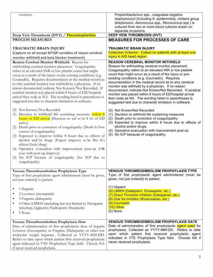
Propionibacterium spp., coagulase-negative staphylococci [including S. epidermidis], viridans group streptococci, Aerococcus spp., Micrococcus spp.) is cultured from two or more blood cultures drawn on separate occasions.
Deep Vein Thrombosis (DVT) / Thrombophlebitis DEEP VEIN THROMBOSIS (DVT) PROCESS MEASURES TRAUMATIC BRAIN INJURY (Capture on all except MTQIP variables of reason cerebral monitor withheld and beta blocker treatment)
MEASURES FOR PROCESSES OF CARE TRAUMATIC BRAIN INJURY Collection Criterion: Collect on patients with at least one injury in AIS head region.
Reason Cerebral Monitor Withheld: Reason for withholding cerebral monitor placement. Coagulopathy refers to an elevated INR or low platelet count that might occur as a result of the injury or pre-existing conditions (e.g. Coumadin). Requires documentation in the medical record as to why cerebral monitor was withheld by a physician. If no reason documented, indicate Not Known/Not Recorded. If cerebral monitor was placed within 8 hours of ED/hospital arrival then code as NA. The wording listed in parentheses is suggested text due to character limitation in software. (0) Not Known/Not Recorded (1) Decision to withhold life sustaining measures within 8
hours of ED arrival (Decision to wd w/in 8 hr of ED arrive)
(2) Death prior to correction of coagulopathy (Death b/fore correct of coagulopathy)
(3) Expected to improve within 8 hours due to effects of alcohol and/or drugs (Expect improve w/in 8hr d/t effects Etoh/drug)
(4) Operative evacuation with improvement post-op (OR evac with post-op improve)
(5) No ICP because of coagulopathy (No ICP due to coagulopathy)
REASON CEREBRAL MONITOR WITHHELD Reason for withholding cerebral monitor placement. Coagulopathy refers to an elevated INR or low platelet count that might occur as a result of the injury or pre-existing conditions (e.g. Coumadin). Requires documentation in the medical record as to why cerebral monitor was withheld by a physician. If no reason documented, indicate Not Known/Not Recorded. If cerebral monitor was placed within 8 hours of ED/hospital arrival then code as NA. The wording listed in parentheses is suggested text due to character limitation in software. (0) Not Known/Not Recorded (1) Decision to withhold life sustaining measures (2) Death prior to correction of coagulopathy (3) Expected to improve within 8 hours due to effects of
alcohol and/or drugs (4) Operative evacuation with improvement post-op (5) No ICP because of coagulopathy
Venous Thromboembolism Prophylaxis Type Type of first prophylactic agent administered (must be given, not just ordered) to patient. • 1 Heparin • 2 Lovenox (enoxaparin) • 3 Fragmin (dalteparin) • 4 Other LMWH (including but not limited to Tinzaparin
(innohep, logiparin); Nadroparin (fraxiparin) • 5 None
VENOUS THROMBOEMBOLISM PROPHYLAXIS TYPE Type of first prophylactic agent administered (must be given, not just ordered) to patient. (1) Heparin (6) LMWH (Dalteparin, Enoxaparin, etc.) (7) Direct Thrombin Inhibitor (Dabigatran, etc.) (8) Oral Xa Inhibitor (Rivaroxaban, etc.) (9) Coumadin (10) Other (5) None
Venous Thromboembolism Prophylaxis Date Date of administration of first prophylactic dose of heparin Lovenox (Enoxaparin) or Fragmin (Dalteparin) or other low molecular weight heparins.. Collected as YYYY-MM-DD. Refers to date upon which patient first received prophylactic agent indicated in VTE Prophylaxis Type field. Choose NA if never received prophylaxis.
VENOUS THROMBOEMBOLISM PROPHYLAXIS DATE Date of administration of first prophylactic agent used for prophylaxis. Collected as YYYY-MM-DD. Refers to date upon which patient first received prophylactic agent indicated in VTE Prophylaxis Type field. Choose NA if never received prophylaxis.
8
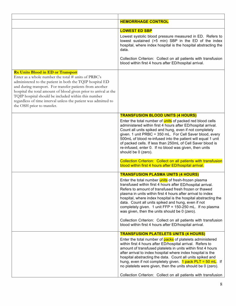
HEMORRHAGE CONTROL
LOWEST ED SBP
Lowest systolic blood pressure measured in ED. Refers to lowest sustained (>5 min) SBP in the ED of the index hospital, where index hospital is the hospital abstracting the data. Collection Criterion: Collect on all patients with transfusion blood within first 4 hours after ED/hospital arrival.
Rx Units Blood in ED or Transport Enter as a whole number the total # units of PRBC’s administered to the patient in both the TQIP hospital ED and during transport. For transfer patients from another hospital the total amount of blood given prior to arrival at the TQIP hospital should be included within this number regardless of time interval unless the patient was admitted to the OSH prior to transfer.
TRANSFUSION BLOOD UNITS (4 HOURS) Enter the total number of units of packed red blood cells administered within first 4 hours after ED/hospital arrival. Count all units spiked and hung, even if not completely given. 1 unit PRBC = 350 mL. For Cell Saver blood, every 500mL of blood re-infused into the patient will equal 1 unit of packed cells. If less than 250mL of Cell Saver blood is re-infused, enter 0. If no blood was given, then units should be 0 (zero). Collection Criterion: Collect on all patients with transfusion blood within first 4 hours after ED/hospital arrival.
TRANSFUSION PLASMA UNITS (4 HOURS) Enter the total number units of fresh-frozen plasma transfused within first 4 hours after ED/hospital arrival. Refers to amount of transfused fresh frozen or thawed plasma in units within first 4 hours after arrival to index hospital, where index hospital is the hospital abstracting the data. Count all units spiked and hung, even if not completely given. 1 unit FFP = 150-250 mL. If no plasma was given, then the units should be 0 (zero). Collection Criterion: Collect on all patients with transfusion blood within first 4 hours after ED/hospital arrival.
TRANSFUSION PLATELETS UNITS (4 HOURS) Enter the total number of packs of platelets administered within first 4 hours after ED/hospital arrival. Refers to amount of transfused platelets in units within first 4 hours after arrival to index hospital where index hospital is the hospital abstracting the data. Count all units spiked and hung, even if not completely given. 1 pack PLT = 50 mL. If no platelets were given, then the units should be 0 (zero). Collection Criterion: Collect on all patients with transfusion
9
blood within first 4 hours after ED/hospital arrival.
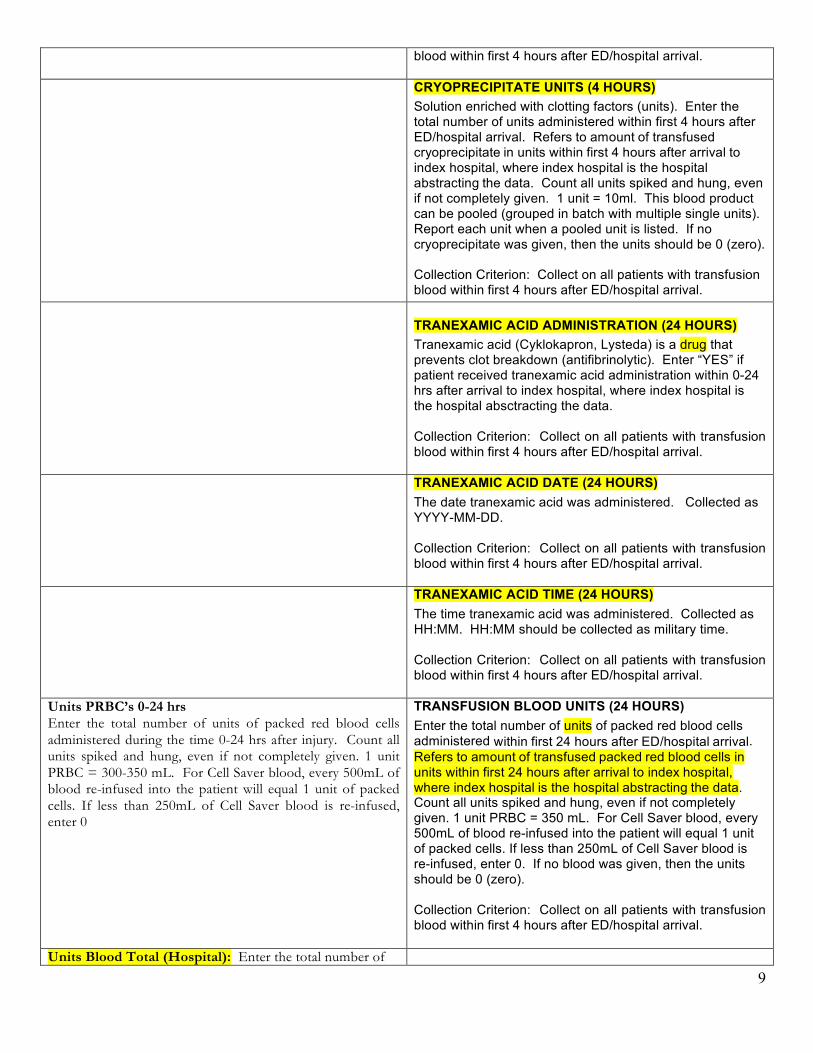
CRYOPRECIPITATE UNITS (4 HOURS) Solution enriched with clotting factors (units). Enter the total number of units administered within first 4 hours after ED/hospital arrival. Refers to amount of transfused cryoprecipitate in units within first 4 hours after arrival to index hospital, where index hospital is the hospital abstracting the data. Count all units spiked and hung, even if not completely given. 1 unit = 10ml. This blood product can be pooled (grouped in batch with multiple single units). Report each unit when a pooled unit is listed. If no cryoprecipitate was given, then the units should be 0 (zero). Collection Criterion: Collect on all patients with transfusion blood within first 4 hours after ED/hospital arrival.
TRANEXAMIC ACID ADMINISTRATION (24 HOURS) Tranexamic acid (Cyklokapron, Lysteda) is a drug that prevents clot breakdown (antifibrinolytic). Enter “YES” if patient received tranexamic acid administration within 0-24 hrs after arrival to index hospital, where index hospital is the hospital absctracting the data. Collection Criterion: Collect on all patients with transfusion blood within first 4 hours after ED/hospital arrival.
TRANEXAMIC ACID DATE (24 HOURS) The date tranexamic acid was administered. Collected as YYYY-MM-DD. Collection Criterion: Collect on all patients with transfusion blood within first 4 hours after ED/hospital arrival.
TRANEXAMIC ACID TIME (24 HOURS) The time tranexamic acid was administered. Collected as HH:MM. HH:MM should be collected as military time. Collection Criterion: Collect on all patients with transfusion blood within first 4 hours after ED/hospital arrival.
Units PRBC’s 0-24 hrs Enter the total number of units of packed red blood cells administered during the time 0-24 hrs after injury. Count all units spiked and hung, even if not completely given. 1 unit PRBC = 300-350 mL. For Cell Saver blood, every 500mL of blood re-infused into the patient will equal 1 unit of packed cells. If less than 250mL of Cell Saver blood is re-infused, enter 0
TRANSFUSION BLOOD UNITS (24 HOURS) Enter the total number of units of packed red blood cells administered within first 24 hours after ED/hospital arrival. Refers to amount of transfused packed red blood cells in units within first 24 hours after arrival to index hospital, where index hospital is the hospital abstracting the data. Count all units spiked and hung, even if not completely given. 1 unit PRBC = 350 mL. For Cell Saver blood, every 500mL of blood re-infused into the patient will equal 1 unit of packed cells. If less than 250mL of Cell Saver blood is re-infused, enter 0. If no blood was given, then the units should be 0 (zero). Collection Criterion: Collect on all patients with transfusion blood within first 4 hours after ED/hospital arrival.
Units Blood Total (Hospital): Enter the total number of
10
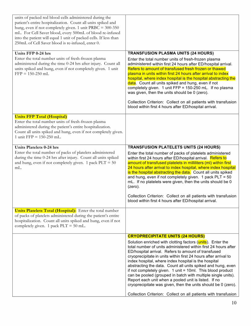
units of packed red blood cells administered during the patient’s entire hospitalization. Count all units spiked and hung, even if not completely given. 1 unit PRBC = 300-350 mL. For Cell Saver blood, every 500mL of blood re-infused into the patient will equal 1 unit of packed cells. If less than 250mL of Cell Saver blood is re-infused, enter 0. Units FFP 0-24 hrs Enter the total number units of fresh-frozen plasma administered during the time 0-24 hrs after injury. Count all units spiked and hung, even if not completely given. 1 unit FFP = 150-250 mL
TRANSFUSION PLASMA UNITS (24 HOURS) Enter the total number units of fresh-frozen plasma administered within first 24 hours after ED/hospital arrival. Refers to amount of transfused fresh frozen or thawed plasma in units within first 24 hours after arrival to index hospital, where index hospital is the hospital abstracting the data. Count all units spiked and hung, even if not completely given. 1 unit FFP = 150-250 mL. If no plasma was given, then the units should be 0 (zero). Collection Criterion: Collect on all patients with transfusion blood within first 4 hours after ED/hospital arrival.
Units FFP Total (Hospital) Enter the total number units of fresh-frozen plasma administered during the patient’s entire hospitalization. Count all units spiked and hung, even if not completely given. 1 unit FFP = 150-250 mL.
Units Platelets 0-24 hrs Enter the total number of packs of platelets administered during the time 0-24 hrs after injury. Count all units spiked and hung, even if not completely given. 1 pack PLT = 50 mL.
TRANSFUSION PLATELETS UNITS (24 HOURS) Enter the total number of packs of platelets administered within first 24 hours after ED/hospital arrival. Refers to amount of transfused platelets in milliliters (ml) within first 24 hours after arrival to index hospital, where index hospital is the hospital abstracting the data. Count all units spiked and hung, even if not completely given. 1 pack PLT = 50 mL. If no platelets were given, then the units should be 0 (zero). Collection Criterion: Collect on all patients with transfusion blood within first 4 hours after ED/hospital arrival.
Units Platelets Total (Hospital): Enter the total number of packs of platelets administered during the patient’s entire hospitalization. Count all units spiked and hung, even if not completely given. 1 pack PLT = 50 mL.
CRYOPRECIPITATE UNITS (24 HOURS) Solution enriched with clotting factors (units). Enter the total number of units administered within first 24 hours after ED/hospital arrival. Refers to amount of transfused cryoprecipitate in units within first 24 hours after arrival to index hospital, where index hospital is the hospital abstracting the data. Count all units spiked and hung, even if not completely given. 1 unit = 10ml. This blood product can be pooled (grouped in batch with multiple single units). Report each unit when a pooled unit is listed. If no cryoprecipitate was given, then the units should be 0 (zero). Collection Criterion: Collect on all patients with transfusion
11
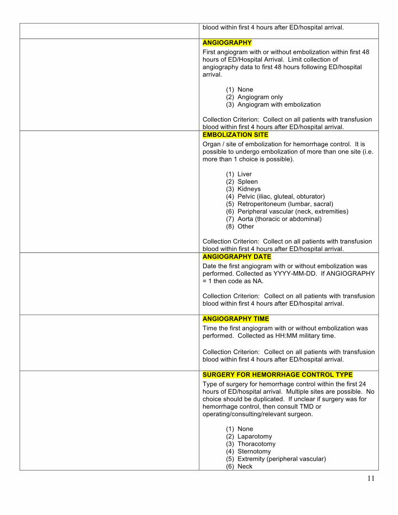
blood within first 4 hours after ED/hospital arrival.
ANGIOGRAPHY First angiogram with or without embolization within first 48 hours of ED/Hospital Arrival. Limit collection of angiography data to first 48 hours following ED/hospital arrival. (1) None (2) Angiogram only (3) Angiogram with embolization Collection Criterion: Collect on all patients with transfusion blood within first 4 hours after ED/hospital arrival.
EMBOLIZATION SITE Organ / site of embolization for hemorrhage control. It is possible to undergo embolization of more than one site (i.e. more than 1 choice is possible). (1) Liver (2) Spleen (3) Kidneys (4) Pelvic (iliac, gluteal, obturator) (5) Retroperitoneum (lumbar, sacral) (6) Peripheral vascular (neck, extremities) (7) Aorta (thoracic or abdominal) (8) Other Collection Criterion: Collect on all patients with transfusion blood within first 4 hours after ED/hospital arrival.
ANGIOGRAPHY DATE Date the first angiogram with or without embolization was performed. Collected as YYYY-MM-DD. If ANGIOGRAPHY = 1 then code as NA. Collection Criterion: Collect on all patients with transfusion blood within first 4 hours after ED/hospital arrival.
ANGIOGRAPHY TIME Time the first angiogram with or without embolization was performed. Collected as HH:MM military time. Collection Criterion: Collect on all patients with transfusion blood within first 4 hours after ED/hospital arrival.
SURGERY FOR HEMORRHAGE CONTROL TYPE Type of surgery for hemorrhage control within the first 24 hours of ED/hospital arrival. Multiple sites are possible. No choice should be duplicated. If unclear if surgery was for hemorrhage control, then consult TMD or operating/consulting/relevant surgeon. (1) None (2) Laparotomy (3) Thoracotomy (4) Sternotomy (5) Extremity (peripheral vascular) (6) Neck

12
(7) Mangled extremity/traumatic amputation Collection Criterion: Collect on all patients with transfusion blood within first 4 hours after ED/hospital arrival.
SURGERY FOR HEMORRHAGE CONTROL DATE Date of first surgery for hemorrhage control within first 24 hours of ED/hospital arrival. Collected as YYYY-MM-DD. If unclear if surgery was for hemorrhage control, then consult TMD or operating/consulting/relevant surgeon. Code as Not Applicable if Surgery for Hemorrhage Control is None. Collection Criterion: Collect on all patients with transfusion blood within first 4 hours after ED/hospital arrival.
SURGERY FOR HEMORRHAGE CONTROL TIME Time of first surgery for hemorrhage control within first 24 hours of ED/hospital arrival. Collected as HH:MM military time. Collection Criterion: Collect on all patients with transfusion blood within first 4 hours after ED/hospital arrival.
WITHDRAWAL OF CARE DATE The date care was withdrawn. Collected as YYYY-MM-DD. Code as Not Applicable if Withdrawal of Care is No. Collection Criterion: Collect on all patients. WITHDRAWAL OF CARE TIME The time care was withdrawn. Collected as HH:MM. HH:MM should be collected as military time. Code as Not Applicable if Withdrawal of Care is No Collection Criterion: Collect on all patients.
Main Cohort Formation • Blunt or penetrating mechanism of injury • Age > 18 years old • ISS > 5 • All deaths OR Length of stay > 1 day who are
discharged alive
Main Cohort Formation • Blunt or penetrating mechanism of injury • Age > 16 years old • ISS > 5 • All deaths • Length of stay > 1 day who are discharged alive
Understanding ᶺ MTQIP Reports
Judy Mikhail, RN, MSN, MBA Program Manager MTQIP MeeAng October 16, 2012
and sharing


Information is the currency of professionals

The Registry

Your office?

Will People Come to See Your Data?

Sharing InformaAon
• Makes you valuable • Increases your visibility • Increases your programs visibility
• Improves your program!
Opening QuesAons
1. What do you not understand about MQIP reports? 2. Which reports are most useful to you? 3. What future reports would you like to see? 4. Are you skepAcal about any of the reports?
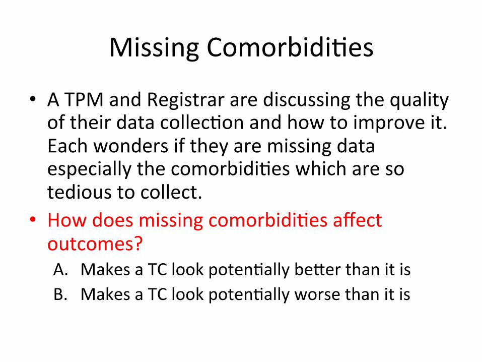
Missing ComorbidiAes
• A TPM and Registrar are discussing the quality of their data collecAon and how to improve it. Each wonders if they are missing data especially the comorbidiAes which are so tedious to collect.
• How does missing comorbidiAes affect outcomes? A. Makes a TC look potenAally beZer than it is B. Makes a TC look potenAally worse than it is
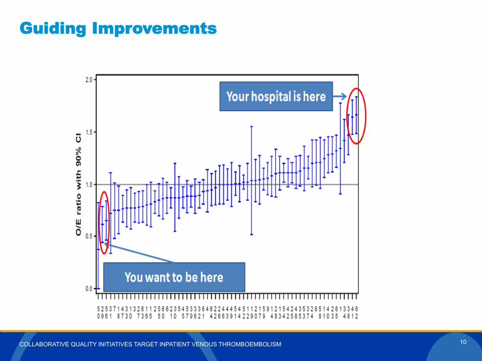
COLLABORATIVE QUALITY INITIATIVES TARGET INPATIENT VENOUS THROMBOEMBOLISM
Guiding Improvements
10
ComorbidiAes QuesAon
A trauma registrar wonders if she can use MTQIP reports to help drive improvement in the collecAon of comorbidiAes. What report could she track over Ame that would provide some feedback?
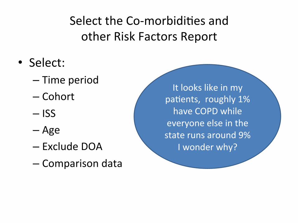
Select the Co-‐morbidiAes and other Risk Factors Report
• Select: – Time period – Cohort – ISS – Age – Exclude DOA – Comparison data
It looks like in my paAents, roughly 1% have COPD while
everyone else in the state runs around 9%
I wonder why?
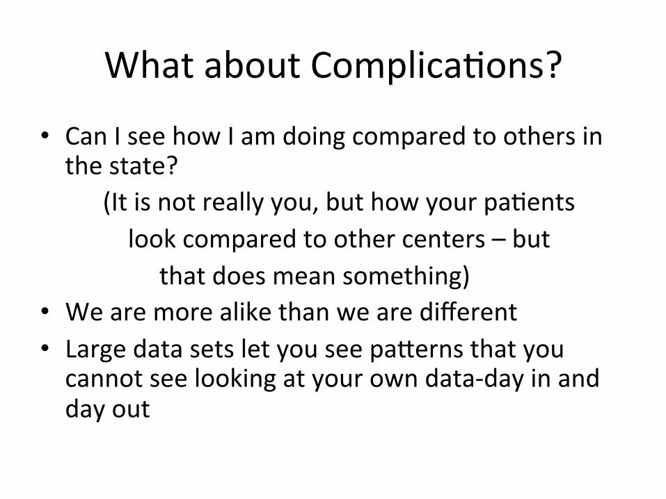
What about ComplicaAons?
• Can I see how I am doing compared to others in the state?
(It is not really you, but how your paAents look compared to other centers – but that does mean something) • We are more alike than we are different • Large data sets let you see paZerns that you cannot see looking at your own data-‐day in and day out
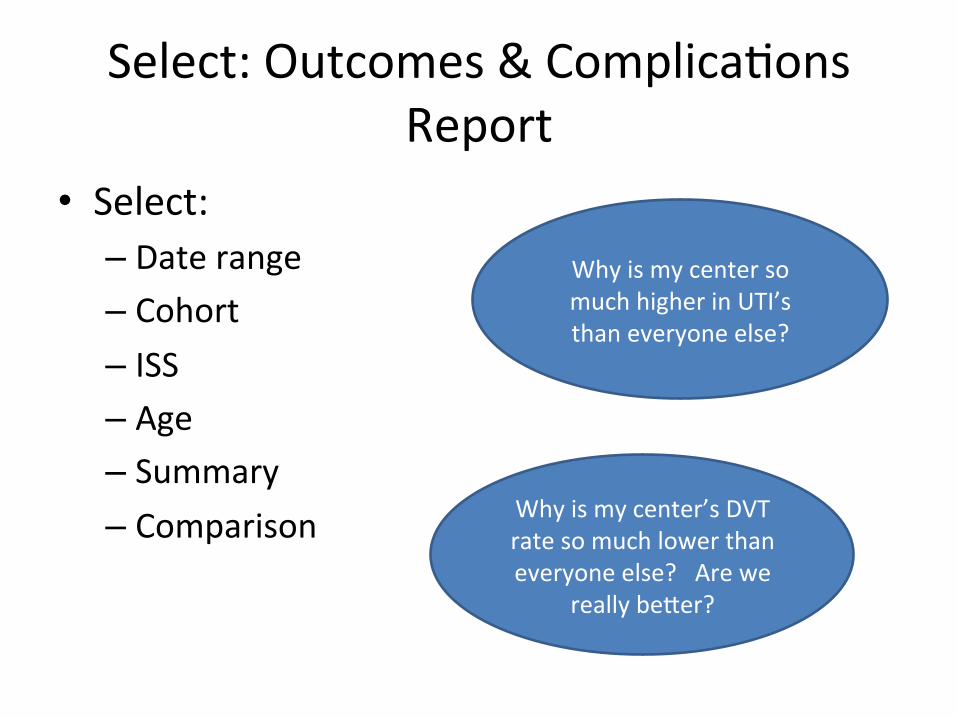
Select: Outcomes & ComplicaAons Report
• Select: – Date range – Cohort – ISS – Age – Summary – Comparison
Why is my center so much higher in UTI’s than everyone else?
Why is my center’s DVT rate so much lower than everyone else? Are we
really beZer?
What is the effect of not collecAng complicaAons on my data?
• Consider your complicaAons in relaAon to your mortality
• Consider your complicaAons in relaAon to resource uAlizaAon: LOS, vent days, etc
In relaAon to co-‐morbidiAes and complicaAons…..
• UnAl the data collecAon improves in the state • It may be best to trend towards average • Constantly track your percentages in both co-‐morbidiAes and complicaAons
• ConAnually ask yourself why are we different from the rest of the state in this one area____.
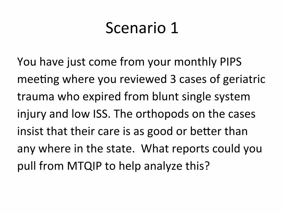
Scenario 1
You have just come from your monthly PIPS meeAng where you reviewed 3 cases of geriatric trauma who expired from blunt single system injury and low ISS. The orthopods on the cases insist that their care is as good or beZer than any where in the state. What reports could you pull from MTQIP to help analyze this?
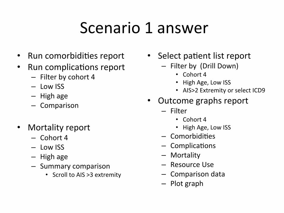
Scenario 1 answer • Run comorbidiAes report • Run complicaAons report
– Filter by cohort 4 – Low ISS – High age – Comparison
• Mortality report
– Cohort 4 – Low ISS – High age – Summary comparison
• Scroll to AIS >3 extremity
• Select paAent list report – Filter by (Drill Down)
• Cohort 4 • High Age, Low ISS • AIS>2 Extremity or select ICD9
• Outcome graphs report – Filter
• Cohort 4 • High Age, Low ISS
– ComorbidiAes – ComplicaAons – Mortality – Resource Use – Comparison data – Plot graph
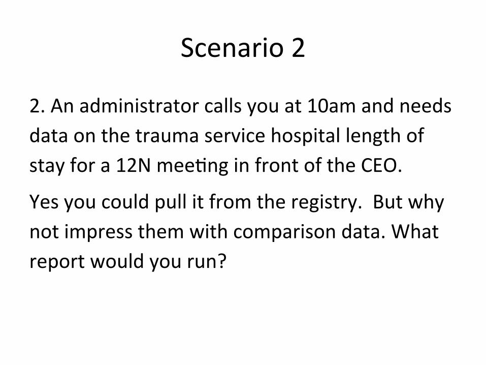
Scenario 2
2. An administrator calls you at 10am and needs data on the trauma service hospital length of stay for a 12N meeAng in front of the CEO.
Yes you could pull it from the registry. But why not impress them with comparison data. What report would you run?
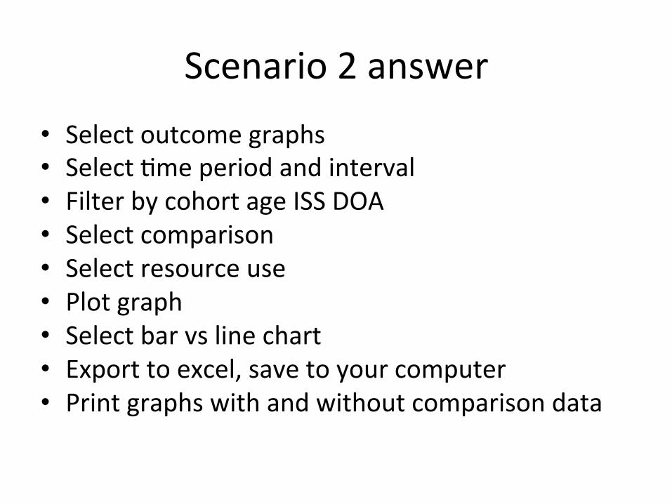
Scenario 2 answer • Select outcome graphs • Select Ame period and interval • Filter by cohort age ISS DOA • Select comparison • Select resource use • Plot graph • Select bar vs line chart • Export to excel, save to your computer • Print graphs with and without comparison data
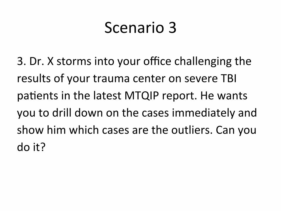
Scenario 3
3. Dr. X storms into your office challenging the results of your trauma center on severe TBI paAents in the latest MTQIP report. He wants you to drill down on the cases immediately and show him which cases are the outliers. Can you do it?
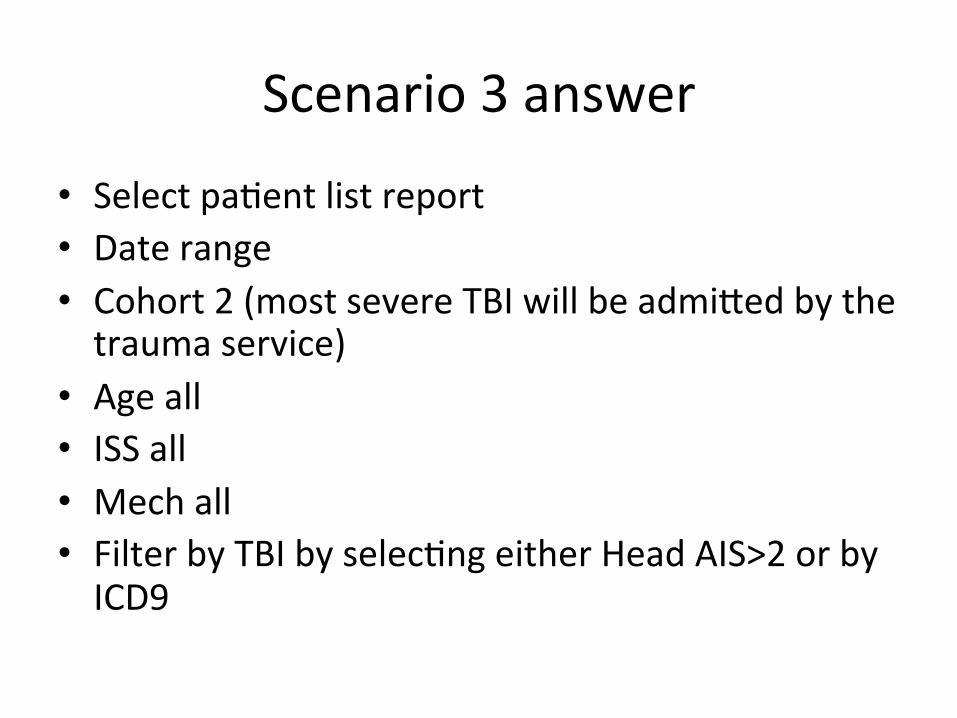
Scenario 3 answer
• Select paAent list report • Date range • Cohort 2 (most severe TBI will be admiZed by the trauma service)
• Age all • ISS all • Mech all • Filter by TBI by selecAng either Head AIS>2 or by ICD9
Scenario 4
4. Hospital X is experiencing delays genng paAents into the ICU. Beds are never available. AdministraAon would like data on this concern.
What report could you run?
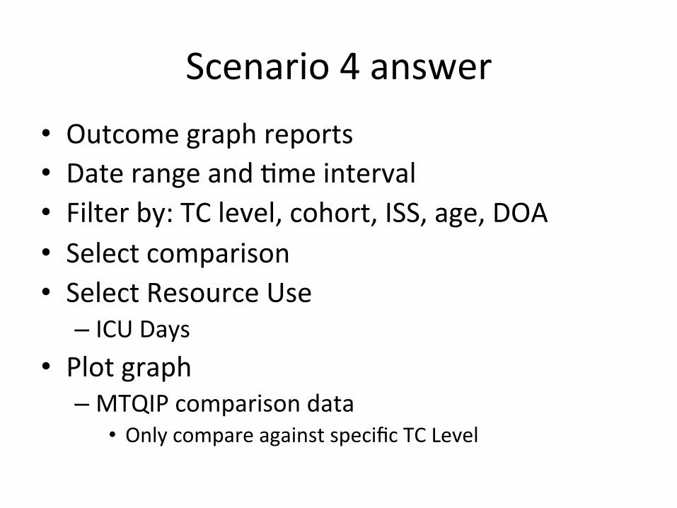
Scenario 4 answer • Outcome graph reports • Date range and Ame interval • Filter by: TC level, cohort, ISS, age, DOA • Select comparison • Select Resource Use
– ICU Days • Plot graph
– MTQIP comparison data • Only compare against specific TC Level
Scenario 5
5. One of the surgeons needs informaAon on pregnant trauma paAents because he is giving a lecture to medical students next week. What reports could you run?
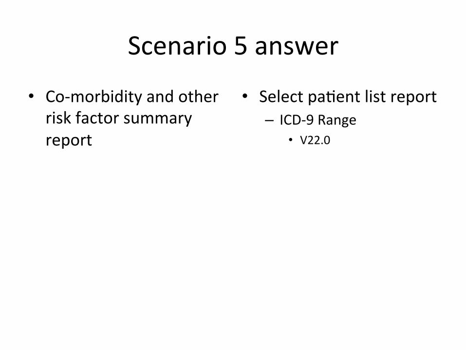
Scenario 5 answer
• Co-‐morbidity and other risk factor summary report
• Select paAent list report – ICD-‐9 Range
• V22.0
Scenario 6
You have been asked to make a presentaAon on your trauma centers mortality results for calendar year 2011.
What reports could you run that would augment your registry reports?
Scenario 6 answer
• Outcome graph reports – Trended report showing 2011 in relaAonship to other years
– Filter by where the deaths occurred – Type of paAent
Scenario 7
Myocardial infarcAon in trauma is a relaAvely infrequent event. One of your surgeons notes that he has seen several himself just within the past year. He asks you to pull a list from the trauma registry. What report could you run that would impress him even more?
Scenario 7 answer
• Co-‐morbidiAes and other risk factors report • Select date range • Cohort 1 (all) or cohort 2 (admit to trauma) • ISS all • Age all • Exclude DOA • Comparison

Share Your Knowledge!
Unlock the vault!