BiP clustering facilitates protein folding in the endoplasmic reticulum
Upload
independentCategory
view
0download
0
Activation of endoplasmic reticulum stresssignaling pathway is associated with neuronaldegeneration in MoMuLV-ts1-inducedspongiform encephalomyelopathy
Hun-Taek Kim1, Kara Waters1, George Stoica1, Wenan Qiang2, Na Liu2, Virginia L Scofield2
and Paul KY Wong2
1Department of Pathobiology, Texas A&M University, College Station, TX, USA and 2Department ofCarcinogenesis, The University of Texas MD Anderson Cancer Center Science Park-Research Division,Smithville, TX, USA
Temperature-sensitive mutant of Moloney murine leukemia virus-TB (MoMuLV-ts1)-mediated neuronal death inmice is likely due to both loss of glial support and release of cytokines and neurotoxins from ts1-infected glialcells. Cytotoxic mediators present in ts1-induced spongiform lesions may generate endoplasmic reticulum (ER)stress, which has been implicated in the pathogenesis of a variety of neurodegenerative diseases. Weinvestigated whether ER stress signaling is involved in ts1-mediated neuronal loss in the brain of infected mice.ts1-infected brainstems were found to show significant increases in phosphorylation of the double-strandedRNA-dependent protein kinase-like ER kinase and eukaryotic initiation factor 2-a. In addition, increasedexpression of growth arrest DNA damage 153 (GADD153), glucose-regulated protein 78, and caspase-12 wereaccompanied by increases in processing of caspase-12 and its downstream target, caspase-3. All of theseevents are markers of ER stress. We observed that GADD153 and cleaved caspase-3 were present indegenerative neurons in the lesions of infected mice, but not in uninfected controls. Phosphorylatedcalmodulin-dependent protein kinase II-a was significantly increased, and was coexpressed with GADD153 in alarge proportion of neurons undergoing early and advanced degenerative changes. Finally, neuronaldegeneration in spongiform lesions was associated with increase in calcium (Ca2þ ) accumulation inmitochondria. Together, these results suggest that ts1 infection-mediated neuronal degeneration in micemay result from activation of ER stress signaling pathways, presumably initiated by perturbation of Ca2þ
homeostasis. Our findings highlight the importance of the ER stress signaling pathway in ts1 infection-inducedneuronal degeneration and death.Laboratory Investigation (2004) 84, 816–827, advance online publication, 19 April 2004; doi:10.1038/labinvest.3700104
Keywords: MoMuLV-ts1; ER stress; GADD153; CaMKII-a; calcium; neuronal death; spongiform encephalo-myelopathy
Moloney murine leukemia virus-TB (MoMuLV) is atype C retrovirus that induces T-cell lymphomas insusceptible strains of mice.1 Its temperature-sensi-tive mutant ts1 has a single point mutation in theenvelope gene, which confers upon the virus theability to cause loss of T cells and motor neurons insusceptible host strains.2–4 ts1 infection of the CNScauses a progressive neurodegenerative disease that
morphologically manifests as spongiform polio-encephalomyelopathy.5 ts1-mediated neuronaldegeneration is likely due to loss of glial supportand release of proinflammatory cytokines andneurotoxins such as tumor necrosis factor-a(TNF-a), interleukin-1 (IL-1), and nitric oxide (NO)from surrounding ts1-infected glial cells.5–7
In brainstems of ts1-infected mice, neuronsdisplay apoptosis, vacuolization and inclusionbodies.5,8 These histopathological hallmarks havebeen associated with animal and human neuro-nal diseases such as human immunodeficiencyvirus (HIV)-induced neurodegenerative disease,9
Parkinson’s disease,10 amyotrophic lateralsclerosis,11 motor neuronopathy,12 and transmissible
Received 17 November 2003; revised and accepted 2 March 2004;published online 19 April 2004
Correspondence: Dr Professor G Stoica, DVM, PhD, Department ofPathobiology, Texas A&M University, College Station, TX 77843,USA.E-mail: [email protected]
Laboratory Investigation (2004) 84, 816–827& 2004 USCAP, Inc All rights reserved 0023-6837/04 $30.00
www.laboratoryinvestigation.org

spongiform encephalopathy.13 However, there havebeen no detailed in vivo investigations regarding themolecular mechanism(s) involved in neuronal lossin ts1-induced encephalomyelopathy.
The ER is a principal site for protein synthesis andfolding, and also serves as a cellular storage sitefor Ca2þ . ER homeostasis is sensitive to a varietyof different stimuli, such as glucose deprivation,perturbation of intracellular and extracellularCa2þ concentration, and exposure to free radicals.Such conditions can trigger ER stress signals thatprovoke three functionally distinct responses.14–16
The first one is upregulation of genes encoding ERchaperone proteins such as glucose-regulated pro-tein 78 (Bip/GRP78). The second is translationalattenuation by phosphorylating the double-strandedRNA-dependent protein kinase-like ER kinase(PERK) and eukaryotic initiation factor 2-a(eIF2-a).Unless those two previous adaptive responsesare sufficient, a cell death program is activatedby transcriptional activation of the gene forGADD153/CHOP (CCAAT/enhancer-binding protein(C/EBP)-homology protein) and by cleavage activa-tion of the ER-associated caspase-12.17–20 ProlongedER stress is a primary component in the neuro-pathology of a wide variety of neurologicaldisorders.16,21
Recent evidence suggests that either TNF-a orNO causes the ER to release Ca2þ , leading toactivation of ER-stress signaling pathways and celldeath in various types of cells.22–24 ER stressactivates the mitochondrial apoptosis pathway.25,26
The release of proapoptotic factors from mitochon-dria is triggered by the proapoptotic Bcl-2 familymembers such as Bax.26,27 ts1 infection of glial cellssecretes neurotoxic products such as TNF-a and NO,and impair proteasome activity in neighboringneurons.6–8 We have shown that the expressionlevel of Bax is increased in neuronal cells inbrainstems of ts1-infected mice.28 In addition, ourpreliminary studies show that mitochondria ofdegenerating neurons have damaged cristae andCa2þ deposits. All of these connected eventsmay cause ER stress in neurons, leading to neuronaldeath.
The aim of this study is to elucidate the causes ofneuronal death in the ts1-infected brainstems, withemphasis on ER stress signaling pathways. Wereport here that ts1 infection causes ER stress,leading to neuronal death, presumably initiated byinterference with Ca2þ homeostasis. These findingshighlight the involvement of the ER stress signalingpathway in retroviral infection-induced neuronalcell degeneration and death in vivo.
Materials and methods
Reagents and Antibodies
Sodium orthovanadate (Na3VO4), sodim b-glycero-phosphate, sodium fluoride (NaF), dithiothreitol
(DTT), and phenylmethylsulfonyl fluoride (PMSF)were purchased from Sigma Chemical Company (StLouis, MO, USA). Leupeptin, pepstatin, and aproti-nin were purchased from Boehringer Mannheim(Indianapolis, IN, USA). Iodoacetamide (an isopep-tidase inhibitor) was obtained from Fluka ChemicalCorp. (Milwaukee, WI, USA).
Goat IgG antibody against gPr80env has beendescribed previously.4,5 Rabbit IgG antibodiesagainst phosphospecific eIF2-a and PERK, caspase-3, cleaved caspase-3, and caspase-12 were pur-chased from Cell Signaling Technology (Beverly,MA, USA). Rabbit IgG antibodies against GADD153/CHOP, phosphorylated calmodulin kinase II-a (CaM-KII-a), and goat IgG antibody against Bip/GRP78were from Santa Cruz Biotechnology (Santa Cruz,CA, USA). Rabbit IgG antibody against glial fibril-lary acidic protein (GFAP) was purchased fromDAKO (Carpinteria, CA, USA). Monoclonal mouseIgG antibody against b-actin was purchased fromSigma.
Virus
ts1 virus was propagated in the thymus-bonemarrow cell line TB. Virus titers were determinedusing a modified direct focus assay in 15F cells,which are a murine sarcoma-positive, leukemia-negative cell line, as described.29
Animals and Virus Infection
FVB/N mice were obtained from Taconic Farms(Germantown, NY, USA). Mice were maintained insterilized microisolators and supplied with auto-claved food and water ad libitum. Newborn FVB/Nmice were inoculated intraperitoneally with 0.1 mlof ts1 viral suspension containing 106–107 infectiousunits/ml, as described previously,8 while controlmice were inoculated with medium only. The micewere observed daily for signs of paralysis, and werekilled at 20 and 25 days post-infection (dpi). Wehave determined previously that histologicallydetectable neuronal loss is not significant at20 dpi, but is evident at 25 dpi.6,8 This experimentalprotocol was approved by the Texas A&MUniversity Institutional Animal Care and UseCommittee.
Tissue Processing
For histopathology and immunohistochemistry, ts1-infected (n¼ 5) and control (n¼ 5) mice wereanesthetized using an intraperitoneal injection ofpentobarbital (150 mg/kg) and perfused with 10%buffered formalin, using a peristaltic pump. After a12 h fixation, the brain segments were separated forfurther processing. For RT-PCR and Western blottinganalyses of mRNAs and proteins in the brainstem,
Induction of ER stress in neurons by MoMuLV-ts1H-T Kim et al
817
Laboratory Investigation (2004) 84, 816–827

control (n¼ 4) and ts1-infected (n¼ 4) mice werekilled, their brains removed, and the brainstem tissuessnap-frozen in liquid nitrogen and stored at �801C.
Semiquantitative RT-PCR Analyses
Total RNA was isolated using the Trizol reagent(Invitrogen, Carlsbad, CA, USA), as described pre-viously.30 Reverse transcription was performedusing the ultra-HF RT-PCR system kit (Strategene,La Jolla, CA, USA). Briefly, 1mg of DNAase-treatedtotal RNA was mixed with oligo (dT) primers anddNTPs. Synthesized cDNA was amplified by astandard PCR protocol using Taq polymerase andmouse specific primers for gPr80env (forward: 50-ATAACA ATC TCA CCT CTG ACC AGG-30; reverse: 50-GAG CAG AGG TAT GGT TGG AGT AGG-30),GADD153/CHOP (forward: 50-TGC CTT TCA CCTTGG AGA CG-30; reverse: 50-CCA TAG AAC TCTGAC TGG AAT CTG G-30), Bip/GRP78 (forward: 50-CCA CTA ATG GAG ATA CTC ACC TGG G-30;reverse: 50-GTA AGG GGA CAC ACA TCA AGC AG-30), caspase-12 (forward: 50-GGA GGT AAA TGCTGG ATT GGC-30; reverse: 50-GCT TTT TGT TTCGGT GCT TCA C-30), and caspase-3 (forward: 50-GAG CAC TGG AAT GTC ATC TCG C-30; reverse: 50-AAG CAT ACA GGA AGT CAG CCT CC-30). Parallelamplifications with primers for b-actin cDNA (for-ward: 50-ATG TAC GTA AGC CAT CCA GGC-30;reverse: 50-AAG GAA GGC TGG AAA AGA GC-30)were performed. The numbers of cycles (between 21and 24) giving the linear range and annealingtemperatures (between 56.0 and 58.31C) were adjusteddepending on the genes amplified. Reaction productswere electrophoresed on 1.5% agarose gels. Bandintensities were analyzed using a densitometer (Mod-el GS-690; Bio-Rad, Hercules, CA, USA) equippedwith the Multi-Analyst software program (Version1.01; Bio-Rad). For quantification, the densities of thesignals were normalized to that for b-actin.
Tissue and Cell Extracts
Brainstem tissue lysates were prepared by homo-genization of frozen tissues in 10 volumes of lysisbuffer containing 50 mM Tris-HCl (pH 7.9), 150 mMNaCl, 1 mM EDTA, 1 mM Na3VO4, 30 mM sodium b-glycerophosphate, 50 mM NaF, 10 mM iodoaceta-mide, 1 mM DTT, 1% Nonidet P-40, 1 mM PMSF,10 mg/ml pepstatin, 10 mg/ml leupeptin, and 5 mg/mlaprotinin. The lysates were cleared by centrifuga-tion at 13,000 g at 41C for 20 min, and the super-natants kept frozen at –801C. The protein content ofthe lysates was determined using the BradfordAssay (Bio-Rad), with bovine serum albumin as thestandard.
Western Blotting
Proteins (20–50 mg) were separated by 6–12%sodium dodecyl sulfate-polyacrylamide gel electro-
phoresis (SDS-PAGE), and then transferred to nitro-cellulose membranes (Schleicher and Schuell,Keene, NH, USA). The membranes were incubatedfor 1 h in blocking buffer (20 mM Tris-HCl bufferedsaline containing 5% nonfat milk powder and 0.1%Tween 20) at room temperature, and then probedwith appropriate antibodies in blocking buffer over-night at 41C. Normal mouse, rabbit, or goat IgGs atthe same dilutions were used as controls. Theblots were incubated with appropriate secondaryIgG-peroxidase conjugates (1:10,000 dilution;Kirkegaard Perry Laboratories, Gaithersburg, MD,USA), and developed using the enhanced chemi-luminescence (ECL) method (Amersham LifeScience, Arlington Heights, IL, USA). After imaging,the blots were stripped and reincubated with amouse monoclonal anti-b-actin antibody toconfirm equal protein loading. Densitometric analy-sis of autoradiographs was performed using adensitometer as described above.
Immunohistochemistry
Paraffin-embedded sections (5 mm) were first depar-affinized and were then subjected to an antigen-retrieval protocol, in which sections were heated in10 mM citrate buffer (pH 6.0). for 10 min, blockedwith 5% normal serum in Tris-buffered saline (TBS;100 mM Tris, 150 mM NaCl, pH 7.4), and thenincubated overnight at 41C with primary antibodiesat a dilution of 1:100. After washing, sections wereincubated with biotin-conjugated secondary IgG,and then treated with reagents from a Vecta-Elitestreptavidin–peroxidase kit with a benzidine sub-strate for color development. For double immuno-staining, sections were pretreated with rabbitanti-GADD153/CHOP IgG as described above. Sec-tions were then blocked with 5% normal goat orrabbit serum in TBS, after which the tissue sectionswere incubated with either rabbit anti-GFAP,cleaved caspase-3, or phosphorylated-CaMKII-aantibody at a dilution of 1:100 overnight at 41C.After incubation with secondary antibodies, sec-tions were treated with an alkaline phosphatase–avidin–biotin substrate and with red chromagen(Vector red; Vector Laboratories) for color develop-ment. Sections were counterstained with dilutedhematoxylin. Sections without primary antibodieswere similarly processed to control for binding ofthe secondary antibodies. On control sections, nospecific immunoreactivity was detected.
Ultrastructural Localization of Ca2þ
The oxalate–pyroantimonate procedure was usedto visualize the subcellular distribution of Ca2þ
in cells from mouse brainstems, as previouslydescribed.31 At 20 dpi, control (n¼ 3) and ts1-infected mice (n¼ 3) were anesthetized with pento-barbital (150 mg/kg, intraperitoneal injection), and
Induction of ER stress in neurons by MoMuLV-ts1H-T Kim et al
818
Laboratory Investigation (2004) 84, 816–827

perfused with a solution containing 2% paraformal-dehyde, 2% glutaraldehyde and 90 mM potasiumoxalate (pH 7.4) and kept overnight at 41C in thesame solution. Brainstems were removed andwashed for 15 min in 7.5% sucrose containing90 mM potassium oxalate and then postfixed in amixture of 1% osmium tetroxide and 2% potassiumpyroantimonate in 0.01 N acetic acid (pH 7.4) for 2 h.Ultrathin sections were stained with 0.5% uranylacetate and 0.4% lead citrate and examined with aZeiss EM 10 electron microscope. The specificity of
the cytochemical reaction was tested by treatingmounted ultrathin sections with 3 mM ethyleneglycol bis(b-aminoethylether)-N,N0-tetraacetic acid(EGTA) at 601C for 1 h. EGTA treatment, in turn, wascontrolled by incubation of sections for 1 h at 601Cin distilled water.
Statistical Analysis
Data obtained from densitometric analyses of RT-PCR and Western blots were analyzed by Student’st-test. P-values of o0.05 were considered as statis-tically significant.
Figure 1 Increased phosphorylation of eIF2-a and PERK in ts1-infected brainstems. (a) Representative Western blots showinglevels of phospho-eIF2-a and -PERK and in control and ts1-infected brainstems at 20 and 25 dpi. (b) Quantitative analyses oflevels of phospho-eIF2-a and -PERK in control and ts1-infectedbrainstems at 20 and 25 dpi. The results shown in the histogramare the mean7s.d. for tissues from four control and four ts1-infected mice. *Significantly different from controls (Po0.01). (c)Representative photographs of semiquantitative RT-PCR andWestern blots showing levels of viral envelope gPr80env mRNAand protein in control and ts1-infected brainstems at 20 and25 dpi.
Figure 2 Induction of GADD153/CHOP and Bip/GRP78 inbrainstems of ts1-infected mice. (a) Representative photographsof semiquantitative RT-PCR analyses for GADD153/CHOP andBip/GRP78 mRNAs in control and ts1-infected brainstems at 20and 25 dpi. (b) Quantitative analysis of GADD153/CHOP and Bip/GRP78 mRNA levels in control and ts1-infected brainstems. (c)Representative Western blots showing levels of GADD153/CHOPand Bip/GRP78 proteins in control and ts1-infected brainstems.b-Actin served as a loading control. The results shown in thehistogram are the mean7s.d. for tissues from four control andfour ts1-infected mice. *Significantly different from controls(Po 0.01).
Induction of ER stress in neurons by MoMuLV-ts1H-T Kim et al
819
Laboratory Investigation (2004) 84, 816–827

Figure 3 Increased expression of GADD153/CHOP protein in neurons of brainstems of ts1-infected mice. (a) GADD153/CHOPimmunoreactivity (brown) in the brainstem from a control mouse. (b) GADD153/CHOP immunoreactivity in neurons of ts1-infectedbrainstem at 25 dpi. (c) Higher magnification view of the glial and neuronal cells in panel (a). Note the absent or very weak staining inneurons (arrows). (d) Higher magnification views of the boxed area in panel (b). Note the strong staining in neurons undergoingdegenerative changes such as vacuolization (arrowheads) and the absence of immunoreactivity in neurons with normal appearance(arrows). (e) Higher magnification views of the boxed area in panel (d). (f) GADD153/CHOP immunoreactivity in neurons of ts1-infectedbrainstem in early stage (at 20 dpi). Note the strong cytoplasmic and nuclear staining in neurons without morphologic changes (arrows).(g) Double immunostaining for GADD153/CHOP (brown) and GFAP (red) in ts1-infected brainstem (at 20 dpi). Note the absence ofGADD153/CHOP immunoreactivity in astrocytes showing GFAP immunoreactivity (arrowheads), alongside strong GADD153/CHOPstaining in neurons (arrows). Bars indicate 200 mm.
Induction of ER stress in neurons by MoMuLV-ts1H-T Kim et al
820
Laboratory Investigation (2004) 84, 816–827

Results
Activation of Upstream Regulators of ER StressSignaling Pathway in Brainstems of ts1-infected Mice
To determine whether ts1 infection causes ER stressin the brainstem, we first examined whether PERKand eIF2-a, two major upstream transducers of ERstress signaling pathways, undergo phosphoryla-tion.14 Phosphorylated eIF2-a in ts1-infected brain-stems was increased 2.0- and 3.3-fold at 20 and25 dpi, respectively, compared to controls (Figure1aand b; Po0.01). Phosphorylated PERK in ts1-infected brainstems was increased 2.3- and 3.8-foldat 20 and 25 dpi, respectively, compared to controls(Figure1a and b; Po0.01). To ascertain whetherphosphorylation of PERK and eIF2-a is correlatedwith viral infection in brainstems, we determinedthe amounts of ts1 viral envelope gPr80env mRNAand protein in brainstems. The expression ofgPr80env mRNA and protein was increased in ts1-infected mice in a time-dependent manner (Figure1c). This result is consistent with those reportedpreviously.4,32
Induction of ER Stress-associated Genes in Brainstemsof ts1-infected Mice
To determine whether phosphorylation of PERK andeIF2-a is accompanied by upregulation of ER stress-associated genes, GADD153/CHOP and GRP78,semiquantitative RT-PCR was used to compareamounts of GADD153/CHOP and Bip/GRP78mRNAs in brainstems of control and ts1-infectedmice (Figure 2a). Figure 2b shows that ts1 infectioninduced 2.4- and 3.5-fold increases in GADD153/CHOP mRNA at 20 and 25 dpi, respectively,compared to controls (Po0.01). Bip/GRP78 mRNAincreased 2.0- and 2.3-fold at 20 and 25 dpi(Po0.01), respectively, compared to controls. Thepattern of increase in GADD153/CHOP and Bip/GRP78 proteins was consistent with the increasesin GADD153/CHOP and Bip/GRP78 mRNAs(Figure 2c).
Increased Expression of GADD153/CHOP in NeuronsShowing Degenerative Changes
Immunostaining for GADD153/CHOP was per-formed to localize GADD153/CHOP in infectedbrainstem. Figure 3 shows that GADD153/CHOPwere virtually absent in neurons and glial cells ofcontrol mice (Figure 3a and c), but abundant inneurons in ts1-infected brainstems (Figure 3b andd). Notably, immunoreactivity was evident inboth intact and visibly damaged neurons showingswelling, cytoskeletal collapse, vacuolization, anddisintegration of nuclear and cytoplasmic mem-brane structures in the early and late stages ofdisease progression (Figure 3e and f). Doubleimmunostaining for GADD153/CHOP and GFAP
showed astrocytes in affected areas to be negativefor GADD153/CHOP (Figure 3g).
Activation of Caspase-12 and -3 Pathways ints1-infected Brainstems
In response to ER stress, caspase-12 specificallycleaves and activates effector caspases such ascaspase-3.20,33,34 Figure 4a and b show that brain-stem tissues from ts1-infected mice had significantlyhigher levels of cleaved forms of caspase-12(approximately 35–39 kDa) and caspase-12 thanthose from uninfected controls at 20 and 25 dpi(Po0.01). Figure 4a and b show that ts1-infectedbrainstem tissues also contained more cleavedcaspase-3 at 20 and 25 dpi (Po0.01), being asso-ciated with decrease in amounts of caspase-3 at
Figure 4 Expression of caspase-12 and -3 in brainstems of controland ts1-infected mice. (a) Representative Western blots showinglevels of caspase-12 and cleaved caspase-12, caspase-3, andcleaved caspase-3 proteins in control and ts1-infected brainstemsat 20 and 25 dpi. b-Actin acted as a loading control. (b)Quantitative analyses of levels of caspase-12, cleaved caspase-12, caspase-3, and cleaved caspase-3 proteins in control and ts1-infected brainstems. The results shown in the histogram are themean7s.d. for tissues from four control and four ts1-infectedmice. *Significantly different from controls (Po0.01). (c) Repre-sentative photographs of semiuantitative RT-PCR analyses forcaspase-12 and -3 mRNAs in control and ts1-infected brainstems.
Induction of ER stress in neurons by MoMuLV-ts1H-T Kim et al
821
Laboratory Investigation (2004) 84, 816–827
25 dpi (Po0.01). As shown in Figure 4c, levels ofcaspase-12 mRNA were consistent with changes inamounts of caspase-12 protein (immunoblottingresults in Figure 4a and b) at the correspondingtime points. In contrast, levels of caspase-3 mRNAin ts1-infected brainstems were not significantlydifferent from ones of uninfected controls.
Coexpression of Cleaved Caspase-3 with GADD153/CHOP in Neurons Undergoing Degenerative Changes
To determine whether increased GADD153/CHOPprotein levels occur together with cleavage ofcaspase-3, double immunostaining for GADD153/CHOP and cleaved caspase-3 was performed(Figure 5). While no immunoreactivity for eitherGADD153/CHOP or cleaved caspase-3 was evidentin brainstem neurons of control mice (Figure 5a),most of the neurons positive for cleaved caspase-3
immunostaining had moderate to strong nuclearGADD153/CHOP immunoreactivity at 20 and 25 dpi(Figure 5b–d).
Phosphorylation of CaMKII-a is Increased inBrainstems of ts1-infected Mice and Correlated withInduction of GADD153/CHOP in Neurons
Perturbation of Ca2þ homeostasis can elicit ERstress.35–37 One of the target kinases activated byintracellular Ca2þ increase is CaMKII.38,39 Therefore,we examined whether phosphorylation of neuron-specific CaMKII-a is increased in brainstems ofts1-infected mice. As shown in Figure 6a and b,phosphorylated CaMKII-a was increased 2.9- and5.1-fold in ts1-infected brainstems at 20 and 25 dpi,respectively, compared to controls (Po0.01).Immunostaining for phosphorylated CaMKII-ashowed that neurons with degenerative changes in
Figure 5 Colocalization of GADD153/CHOP and cleaved caspase-3 in neurons in brainstem section from ts1-infected mice. (a) Doubleimmunostaining for GADD153/CHOP and cleaved caspase-3 in brainstem from a control mouse. (b) Double immunostaining toGADD153/CHOP (brown, nuclei) and cleaved caspase-3 (red, cytoplasm) in neurons undergoing degenerative changes in ts1-infectedbrainstem at 25 dpi. (c) Higher magnification views of the boxed area in panel (b). (d) Double immunostaining for GADD153/CHOP(brown, nuclei) and cleaved caspase-3 (red, cytoplasm) in neurons with normal appearance in ts1-infected brainstem at 20 dpi. Barsindicate 200mm.
Induction of ER stress in neurons by MoMuLV-ts1H-T Kim et al
822
Laboratory Investigation (2004) 84, 816–827

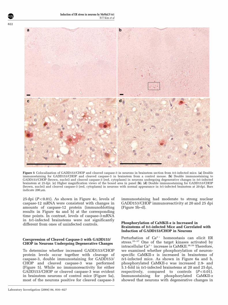
ts1-infected brainstems showed moderate to strongcytoplasmic staining for phosphorylated CaMKII-a(Figure 7b and d), compared to controls (Figure 7aand c). Most (495%) of neurons positive forimmunoreactive GADD153/CHOP were also posi-tive for phosphorylated CaMKII-a (Figure 7e and f).We used transmission electron microscopy to deter-mine whether intracellular Ca2þ increase is in-volved in early development of neuronaldegeneration. As shown in Figure 7h, increasedintracellular Ca2þ was illustrated by cytochemicalstaining for Ca2þ deposits in damaged mitochondriaof neurons at early stage, compared to control(Figure 7g).
Discussion
The present study shows that ts1 infection triggersactivation of ER stress signaling pathways in thebrainstem, as evidenced by increases in phosphor-ylation of PERK and eIF2-a, induction of GADD153/CHOP and GRP78, and activation of the ER-associated caspase-12. The concomitant increasesin GADD153/CHOP, cleaved caspase-3, phosphory-lated CaMKII-a, and intracellular Ca2þ in neuronsimplicate ER stress following intracellular Ca2þ
increase in mechanisms leading to neurodegenera-tion in ts1-infected brainstems. Induction of these
ER stress markers is correlated temporally withincreases in viral burden and noxious cytokines,neuronal loss, and the development of neuroclinicalsigns in infected animals.4,6,8,32
Particularly relevant to the present work are tworecent studies describing roles of ER stress inneurodegeneration. Hetz et al40 demonstrated thatER stress and caspase-12 mediate neurotoxicity ofpathological prion protein. Dimcheff et al41 alsoshowed that another murine neurotropic virus,FrCasE, triggers ER stress in the brainstems leadingto spongiform degeneration. Together with theserecent publications, our results strongly support theidea that spongiform neurodegeneration is likely tobe mediated by ER stress.
GADD153/CHOP has emerged recently as a ubi-quitous regulator of neuronal death.42 Induction ofGADD153/CHOP appears to be an early eventcontributing to neuronal dysfunction and death ints1-infected brainstems. This conclusion is based onthe observation that increased expression andnuclear translocation of GADD153/CHOP is evidentearly in the disease course in neurons withoutmorphological changes and becomes more pro-nounced in neurons showing advanced degenera-tive changes in late stages.
What is the role of GADD153/CHOP in neuronaldegeneration in ts1-infected brainstems? A previousstudy showed that GADD153/CHOP is a negativeregulator of Bcl-2 expression.43 Also, GADD153/CHOP may sensitize cells to noxious stimuli byinducing the expression of pro-apoptotic genes suchas Bax and caspases.44,45 It is noteworthy that theBcl-2 protein levels in brainstems of ts1-infectedmice are decreased at late stages of disease (data notshown). However, the precise pro-cell-death targetsof GADD153/CHOP transcription factor during ts1infection of the brainstem remain to be identified.
In contrast to GADD153/CHOP, the chaperoneGRP78 promotes protein folding and thus attenuatescellular death under stress conditions.46,47 WhileGRP78 was found to be increased in early and latestages of disease in ts1-infected brainstems, therewas no progressive increase in GRP78 mRNA andprotein from 20 to 25 dpi. Similarly, previousstudies with FrCasE- or scrapie-infected mice haveshown that there is no clear correlation between theexpression of GRP78 and the development of ERstress-mediated neurodegeneration.40,41 These com-bined results indicate that GRP78 may be increasedearly after infection, but, under the conditions ofprolonged ER stress in late stages of diseases, theinduction of GRP78 may not be as efficient as thoseof other ER stress markers. Such levels of GRP78induction may not be sufficient to protect neuronsagainst ER stress in ts1-, FrCasE-, or scrapie-infectedmice, since GRP78 overexpression protects cellulardeath under ER stress conditions by stabilizing Ca2þ
homeostasis.47,48
The activation of caspase-12 is a crucial event inER stress-mediated cell death.20 In ts1-infected
Figure 6 Increased expression of phosphorylated CaMKII-a inbrainstems of ts1-infected mice. (a) Representative Western blotsshowing levels of phosphorylated CaMKII-a (p-CaMKII-a) incontrol and ts1-infected brainstems at 20 and 25 dpi. b-actinserved as a loading control. (b) Quantitative analysis of levels ofp-CaMKII-a in control and ts1-infected brainstems. The resultsshown in the histogram are the mean7s.d. for tissues from fourcontrol and four ts1-infected mice. *Significantly different fromcontrols (P o 0.01).
Induction of ER stress in neurons by MoMuLV-ts1H-T Kim et al
823
Laboratory Investigation (2004) 84, 816–827
Figure 7 Increased expression of p-CaMKII-a protein and Ca2þ deposits in neurons of brainstems of ts1-infected mice. (a) p-CaMKII-aimmunoreactivity (brown) in brainstem from a control mouse. (b) p-CaMKII-a immunoreactivity in ts1-infected brainstem at 25 dpi. (c)Higher magnification view of the neuronal cells (arrows) in panel (a). Note the absent or very weak cytoplasmic staining in neurons(arrows). (d) Higher magnification views of the neuronal cells in panel (b). Note the strong staining in neurons undergoing degenerativechanges such as vacuolization (arrowheads) and the weak immunoreactivity in neurons with normal appearance (arrows). (e, f)Coexpression of GADD153/CHOP (brown, nuclei) and p-CaMKIIa (red, cytoplasm) in the neurons (arrowheads) in ts1-infected brainstemat 25 dpi (e) and 20 dpi (f). Bars indicate 200mm. (g, h) Electron micrograph showing dense granular Ca2þ pyroantimonate deposits inmitochondria of neurons (arrows) from ts1-infected brainstem at 20 dpi (h). Note the absence of granular deposits in mitochondria ofneurons (arrows) from control (g). Bars indicate 3 mm.
Induction of ER stress in neurons by MoMuLV-ts1H-T Kim et al
824
Laboratory Investigation (2004) 84, 816–827

brainstem tissue, expression and activation ofcaspase-12 are increased, and this is accompaniedby increases in cleavage activation of its down-stream target, caspase-3. This cleaved caspase-3 isthe caspase most frequently reported to participatein neuronal cell death. Interestingly, increase incaspase-12 protein was correlated with increasedamounts of caspase-12 mRNA. This induction maybe due to expression of interferon-g induced duringts1 infection, as demonstrated by recent publica-tions that either interferon-g alone or in concert withTNF-a, is capable of activating the transcription ofcaspase-12 gene as well as the processing of caspase-12.49,50
Previous studies have shown that the ER Ca2þ
ATPase inhibitor Thapsigarin, or Ca2þ ionoporesinduce transcriptional activation of GADD153/CHOP by elevating intracellular Ca2þ .35,51 We there-fore attempted to dissect the mechanism of induc-tion of ER stress signaling pathway in ts1-infectedbrainstems by investigating Ca2þ signaling path-ways. In this study, we found that phosphorylationof CaMKII-a is increased and that phosphorylatedCaMKII-a is present, together with GADD153/CHOPand cleaved caspase-3, in most neurons in brain-stems from both early and advanced stages of thedisease (Figures 6 and 7). This result suggests thatperturbation of Ca2þ homeostasis is a potentialmechanism responsible for induction of ER stressin neurons. This notion is supported by electronmicroscopic evidence of increased intracellularCa2þ , manifested as Ca2þ precipitates in the mito-chondria in neurons during early stages (Figure 7).
One key issue that should be addressed is whattypes of cells in brainstems are specially affected byER stress. We have obtained immunohistologicaldata showing the induction of ER stress markers inneurons, but not in glial cells (Figures 3, 5, and 7).Involvement of ER stress in neurons is furthersupported by observation of increase in intracellularCa2þ (Figure 7). These results suggest that activationof ER stress signaling pathways in neurons are likelydue to an indirect mechanism(s), since our previouswork has revealed that ts1 virus infects glial cells,but not neurons.5 Therefore, the present studystrongly indicates that ER stress-mediated spongi-form neurodegeneration is likely to be caused byneurotoxic products secreted from glial cells by ts1infection.
Among the elements present within the lesions ofthe ts1-infected brainstem, TNF-a and NO areprobably prime candidates as triggers for ER stressin neurons, since both are upregulated in astrocytesby ts1 infection.6,7 These agents may cause ER Ca2þ
release in surrounding neurons, leading to activa-tion of ER stress signaling pathways, as demon-strated by previous reports.22–24 Also, TNF-a maycause glutamate accumulation in the vicinity ofneurons, which activates N-methyl-D-aspartate(NMDA) receptor-operated channels on the neuro-nal surface, causing Ca2þ influx and activation of
CaMKII,52,53 as shown in Figure 7. This notion issupported by recent reports that excitotoxicityassociated with NMDA receptor inducesGADD153/CHOP production, and activation andphosphorylation of CaMKII-a, leading to neuronaldeath.54–56
In conclusion, we show here that activation of ERstress signaling pathways constitutes a sequentialand inter-related cytotoxic signaling cascade thatcauses neuronal injury in the ts1-infected brainstem.The potential importance of the Ca2þ–ER stress linkin the pathogenesis of spongiform encephalopathyby ts1 infection, particularly neuronal degeneration,is now evident. Furthermore, the data presentedhere support the idea that ER stress-mediatedneurodegeneration is likely to be caused by neuro-toxic products secreted from glial cells by ts1infection.
Acknowledgements
This work was supported in part by grants to PKYWong from the NIH (AI28283 and NS43984). Wethank Vanessa Edwards for her assistance in prepar-ing the manuscript.
References
1 Yuen PH, Szurek PF. The reduced virulence of thethymotropic Moloney murine leukemia virus deriva-tive MoMuLV-TB is mapped to 11 mutations withinthe U3 region of the long terminal repeat. J Virol1989;63:471–480.
2 Wong PKY, Prasad G, Hansen J, et al. ts1, a mutant ofMoloney murine leukemia virus-TB, causes bothimmunodeficiency and neurologic disorders inBALB/c mice. Virology 1989;170:450–459.
3 Szurek PF, Yuen PH, Ball JK, et al. A Val-25-to-Ilesubstitution in the envelope precursor polyprotein,gPr80env, is responsible for the temperature sensitiv-ity, inefficient processing of gPr80env, and neuroviru-lence of ts1, a mutant of Moloney murine leukemiavirus TB. J Virol 1990;64:467–475.
4 Wong PKY, Floyd E, Szurek PF. High susceptibility ofFVB/N mice to the paralytic disease induced by ts1, amutant of Moloney murine leukemia virus TB.Virology 1991;180:365–371.
5 Stoica G, Illanes O, Tasca SI, et al. Temporal centraland peripheral nervous system changes induced by aparalytogenic mutant of Moloney murine leukemiavirus TB. Lab Invest 1993;69:724–735.
6 Choe W, Stoica G, Lynn W, et al. Neurodegenerationinduced by MoMuLV-ts1 and increased expression ofFas and TNF-a in the central nervous system. Brain Res1998;779:1–8.
7 Kim HT, Qiang W, Wong PKY, et al. Enhancedproteolysis of IkBa and IkBb proteins in astrocytes byMoloney murine leukemia virus (MoMuLV)-ts1 infec-tion: A potential mechanism of NF-kB activation.J Neurovirol 2001;7:466–475.
8 Stoica G, Tasca SI, Wong PKY. Motor neuronal loss andneurofilament-ubiquitin alteration in MoMuLV-ts1
Induction of ER stress in neurons by MoMuLV-ts1H-T Kim et al
825
Laboratory Investigation (2004) 84, 816–827

encephalopathy. Acta Neuropathol (Berl) 2000;99:238–244.
9 Tyor WR, Wesselingh SL, Griffin JW, et al. Unifyinghypothesis for the pathogenesis of HIV-associateddementia complex, vacuolar myelopathy, and sensoryneuropathy. J Acquir Immune Defic Syndr HumRetrovirol. 1995;9:379–388.
10 Forno LS. Neuropathology of Parkinson’s disease.J Neuropathol Exp Neurol 1996;55:259–272.
11 Tu PH, Raju P, Robinson KA, et al. Transgenic micecarrying a human mutant superoxide dismutasetransgene develop neuronal cytoskeletal pathologyresembling human amyotrophic lateral sclerosislesions. Proc Natl Acad Sci USA 1996;93:3155–3160.
12 Sagot Y, Dubois-Dauphin M, Tan SA, et al. Bcl-2overexpression prevents motoneuron cell body lossbut not axonal degeneration in a mouse modelof a neurodegenerative disease. J Neurosci 1995;15:7727–7733.
13 Prusiner SB. Prions. Proc Natl Acad Sci USA1998;95:13363–13383.
14 Harding HP, Zhang Y, Ron D. Protein translation andfolding are coupled by an endoplasmic reticulum-resident kinase. Nature 1999;397:271–274.
15 Kaufman RJ. Stress signaling from the lumen of theendoplasmic reticulum: coordination of gene trans-criptional and translational controls. Genes Dev1999;13:1211–1233.
16 Paschen W. Dependence of vital cell function onendoplasmic reticulum calcium levels: implicationsfor the mechanisms underlying neuronal cell injury indifferent pathological states. Cell Calcium 2001;29:1–11.
17 Ron D, Habener JF. CHOP, a novel developmentallyregulated nuclear protein that dimerizes with trans-cription factors C/EBP and LAP and functions as adominant-negative inhibitor of gene transcription.Genes Dev 1992;6:439–453.
18 Barone MV, Crozat A, Tabaee A, et al. CHOP(GADD153) and its oncogenic variant, TLS-CHOP,have opposing effects on the induction of G1/S arrest.Genes Dev 1994;8:453–464.
19 Fawcett TW, Eastman HB, Martindale JL, et al.Physical and functional association between GADD153and CCAAT/enhancer-binding protein b during cellu-lar stress. J Biol Chem 1996;271:14285–14289.
20 Nakagawa T, Zhu H, Morishima N, et al. Caspase-12mediates endoplasmic-reticulum-specific apoptosisand cytotoxicity by amyloid-beta. Nature 2000;403:98–103.
21 Sherman MY, Goldberg AL. Cellular defenses againstunfolded proteins: a cell biologist thinks aboutneurodegenerative diseases. Neuron 2001;29:15–32.
22 Oyadomari S, Takeda K, Takiguchi M, et al. Nitricoxide-induced apoptosis in pancreatic beta cells ismediated by the endoplasmic reticulum stress path-way. Proc Natl Acad Sci USA 2001;98:10845–10850.
23 Gotoh T, Oyadomari S, Mori K, et al. Nitric oxide-induced apoptosis in RAW 264.7 macrophages ismediated by endoplasmic reticulum stress pathwayinvolving ATF6 and CHOP. J Biol Chem 2002;277:12343–12350.
24 Kim BC, Kim HT, Mamura M, et al. Tumor necrosisfactor induces apoptosis in hepatoma cells by increas-ing Ca(2+) release from the endoplasmic reticulumand suppressing Bcl-2 expression. J Biol Chem 2002;277:31381–31389.
25 Hacki J, Egger L, Monney L, et al. Apoptoticcrosstalk between the endoplasmic reticulum andmitochondria controlled by Bcl-2. Oncogene 2000;19:2286–2295.
26 Wei MC, Zong WX, Cheng EH, et al. ProapoptoticBAX and BAK: a requisite gateway to mitochondrialdysfunction and death. Science 2001;292:727–730.
27 Desagher S, Martinou JC. Mitochondria as the centralcontrol point of apoptosis. Trends Cell Biol 2000;10:369–377.
28 Kim HT, Tasca S, Qiang W, et al. Induction of p53accumulation by Moloney murine leukemia virus-ts1infection in astrocytes via activation of extracellularsignal-regulated kinases 1/2. Lab Invest 2002;82:693–702.
29 Wong PKY, Soong MM, Yuen PH. Replication ofmurine leukemia virus in heterologous cells: interac-tion between ecotropic and xenotropic viruses. Viro-logy 1981;109:366–378.
30 Kim HT, Stoica G, Bazer FW, et al. Interferon tau-induced hepatocyte apoptosis in sheep. Hepatology2000;31:1275–1284.
31 Siklos L, Engelhardt JI, Reaume AG, et al. Alteredcalcium homeostasis in spinal motoneurons but not inoculomotor neurons of SOD-1 knockout mice. ActaNeuropathol 2000;99:517–524.
32 Gonzales-Scarano F, Nathanson N, Wong PKY.Retroviruses and the nervous system. In: Levy JA(ed). The Retroviridae. Plenum Press: New York, 1995,pp 127–136.
33 Earnshaw WC, Martins LM, Kaufmann SH. Mammal-ian caspases: structure, activation, substrates, andfunctions during apoptosis. Annu Rev Biochem1999;68:383–424.
34 Rao RV, Castro-Obregon S, Frankowski H, et al.Coupling endoplasmic reticulum stress to the celldeath program. An Apaf-1-independent intrinsicpathway. J Biol Chem 2002;277:21836–21842.
35 Bartlett JD, Luethy JD, Carlson SG, et al. Calciumionophore A23187 induces expression of the growtharrest and DNA damage inducible CCAAT/enhancer-binding protein (C/EBP)-related gene, gadd153. Ca2+
increases transcriptional activity and mRNA stability.J Biol Chem 1992;267:20465–20470.
36 Lipton SA, Nicotera P. Calcium, free radicals andexcitotoxins in neuronal apoptosis. Cell Calcium1997;23:165–171.
37 Orrenius S, Zhivotovsky B, Nicotera P. Regulation ofcell death: the calcium-apoptosis link. Nat Rev MolCell Biol 2003;4:552–565.
38 Braun AP, Schulman H. The multifunctional calcium/calmodulin-dependent protein kinase: from form tofunction. Annu Rev Physiol 1995;57:417–445.
39 Soderling TR. The Ca-calmodulin-dependent pro-tein kinase cascade. Trends Biochem Sci 1999;24:232–236.
40 Hetz C, Russelakis-Carneiro M, Maundrell K, et al.Caspase-12 and endoplasmic reticulum stress mediateneurotoxicity of pathological prion protein. EMBO J2003;22:5435–5445.
41 Dimcheff DE, Askovic S, Baker AH, et al. Endoplasmicreticulum stress is a determinant of retrovirus-inducedspongiform neurodegeneration. J Virol 2003;77:12617–12629.
42 Spear E, Ng DT. The unfolded protein response: nolonger just a special teams player. Traffic 2001;2:515–523.
Induction of ER stress in neurons by MoMuLV-ts1H-T Kim et al
826
Laboratory Investigation (2004) 84, 816–827

43 McCullough KD, Martindale JL, Klotz LO, et al.Gadd153 sensitizes cells to endoplasmic reticulumstress by down-regulating Bcl2 and perturbing thecellular redox state. Mol Cell Biol 2001;21:1249–1259.
44 Boya P, Cohen I, Zamzami N, et al. Endoplasmicreticulum stress-induced cell death requires mitochon-drial membrane permeabilization. Cell Death Differ2002;9:465–467.
45 Zong WX, Li C, Hatzivassiliou G, et al. Bax and Bakcan localize to the endoplasmic reticulum to initiateapoptosis. J Cell Biol 2003;162:59–69.
46 Liu H, Bowes III RC, van de Water B, et al.Endoplasmic reticulum chaperones GRP78 and calre-ticulin prevent oxidative stress, Ca2+ disturbances, andcell death in renal epithelial cells. J Biol Chem1997;272:21751–21759.
47 Morris JA, Dorner AJ, Edwards CA, et al. Immunoglo-bulin binding protein (BiP) function is required toprotect cells from endoplasmic reticulum stress but isnot required for the secretion of selective proteins.J Biol Chem 1997;272:4327–4334.
48 Yu Z, Luo H, Fu W, et al. The endoplasmic reticulumstress-responsive protein GRP78 protects neuronsagainst excitotoxicity and apoptosis: suppression ofoxidative stress and stabilization of calcium home-ostasis. Exp Neurol 1999;155:302–314.
49 Kalai M, Lamkanfi M, Denecker G, et al. Regulation ofthe expression and processing of caspase-12. J Cell Biol2003;162:457–467.
50 Watanabe Y, Suzuki O, Haruyama T, et al. Interferon-gamma induces reactive oxygen species and endoplas-mic reticulum stress at the hepatic apoptosis. J CellBiochem 2003;89:244–253.
51 Thastrup O, Cullen PJ, Drobak BK, et al. Thapsigargin,a tumor promoter, discharges intracellular Ca2+ storesby specific inhibition of the endoplasmic reticulumCa2(+)-ATPase. Proc Natl Acad Sci USA1990;87:2466–2470.
52 Fine S M, Angel RA, Perry SW, et al. Tumornecrosis factor-a inhibits glutamate uptake byprimary human astrocytes: implications for patho-genesis of HIV-1 dementia. J Biol Chem 1996;271:15303–15306.
53 Bezzi P, Domercq M, Brambilla L, et al. CXCR4-activated astrocytes glutamate release via TNFa:amplification by microglia triggers neurotoxicity. NatNeurosci 2001;4:702–710.
54 Laabich A, Li G, Cooper NG. Characterization ofapoptosis—genes associated with NMDA mediatedcell death in the adult rat retina. Brain Res Mol BrainRes 2001;91:34–42.
55 Foos TM, Wu JY. The role of taurine in the centralnervous system and the modulation of intracellularcalcium homeostasis. Neurochem Res 2002;27:21–26.
56 Takano H, Fukushi H, Morishima Y, et al. Calmodulinand calmodulin-dependent kinase II mediate neuronalcell death induced by depolarization. Brain Res2003;962:41–47.
Induction of ER stress in neurons by MoMuLV-ts1H-T Kim et al
827
Laboratory Investigation (2004) 84, 816–827
Copyright © 2022 FDOKUMEN