research project manual and format of writing and presenting ...

Letters to the Editor Related to New Topics
Transient Exacerbation of Ataxia with
Smoking: A Prevalence Survey
Some cerebellar ataxic patients complain of transient, butmarked, exacerbation of ataxia after smoking cigarettes. Theprevalence of this phenomenon is unknown, although it hasbeen reported previously, mostly among patients with multi-ple system atrophy (MSA).1�4 The finding is reproduced byintravenous injection of nicotine tartrate,1 but the mechanismis not established. We distributed a self-reporting question-naire to patients with various types of chronic cerebellarataxia to determine the prevalence of ataxia exacerbation bysmoking, and whether sensitivity to cigarette smoking is spe-cific to certain subtypes of ataxia.
Written surveys were mailed to adults with cerebellarataxia evaluated in the Massachusetts General Hospital(MGH) Ataxia Unit from 8/2005 to 7/2007 (n 5 140), and tothe Emory University National Ataxia Registry (n 5 305).The survey assessed smoking status, and whether cigarettestransiently exacerbated or alleviated symptoms of ataxia bythe patient’s own report. Eleven symptoms were assessed asfollows: (1) balance, (2) coordination, (3) slurred speech, (4)choking/coughing, (5) tremor, (6) slowed movement, (7)lightheadedness (as though about to pass out), (8) dizziness(spinning sensation), (9) urinary urgency, frequency, orincontinence, (10) muscle weakness, and (11) impaired hand-writing. An open-ended question provided respondents an op-portunity to elaborate upon their smoking-related symptoms.
Telephone interviews were conducted for MGH AtaxiaUnit patients who did not return the survey. Medical recordswere reviewed to determine smoking status in MGH patientswho could not be contacted. This study was approved by thePartners Human Research Committee.
MGH Ataxia Unit patients were clinically evaluated bythe authors. Registry patients provided a copy of genetic testsor a physician’s note elucidating the diagnosis. The registryreleased the respondents’ diagnoses after receipt of medicalrecord release forms included with the mailed surveys.
Patients were grouped into five diagnostic categories asfollows: (1) Friedreich’s Ataxia (FA), (2) Autosomal domi-nant genetic and familial ataxias (Genetic), including spino-cerebellar ataxia (SCA) 1, 2, 3, 5, 6, 7, 8, and autosomaldominant ataxias gene unknown, (3) Idiopathic Late OnsetCerebellar Ataxia (ILOCA), including sporadic ataxias ofunknown etiology, (4) Multiple System Atrophy (MSA), and(5) Miscellaneous ataxias of known causes (Misc), includingtoxic/metabolic cerebellar ataxia, gluten ataxia, postinfectiouscerebellar ataxia, paraneoplastic cerebellar ataxia, cerebellarhypoplasia, autoimmune ataxias, superficial siderosis, fragile
X-associated tremor ataxia syndrome, and Gerstmann-Straussler-Schenker disease.
Of 405 survey recipients, 21 were deceased, had changedaddress, or received duplicate surveys. Of the remainder, 304responded to the survey, for a total response rate of 72%.One hundred and thirty-nine (46%) of the respondents hadever smoked cigarettes. Of these, 53 reported quitting smok-ing before ataxia developed. Of the 86 patients, who smokedafter ataxia onset, 36 (42%) reported transient exacerbationof ataxia from cigarettes. Patients in all diagnostic categoriesreported this phenomenon, with no difference in smokingeffect rate between diagnostic groups (P > 0.5).
The effect rate was similar for the 2 patient cohorts: 41%of MGH patients (n 5 9) and 42% of registry patients (n 527) reported transient exacerbation of ataxia from cigarettes.
Most patients who reported an effect noted transient exac-erbation of ataxia symptoms with smoking, but 6 patientsreported transient improvement (Table 1).
This study confirms the previously reported effect ofsmoking on cerebellar ataxia.1�4 Our questionnaire, com-pleted by 304 patients, shows a deleterious symptomaticeffect of smoking in 42% of patients with ataxia, regardlessof etiology.
The mechanism by which smoking worsens cerebellarataxia is unknown. Although there are >4,000 candidatechemicals in tobacco smoke,5 this phenomenon is likely nico-tine-induced given the exacerbation of ataxia following injec-tion of nicotine tartrate.1 There are abundant nicotinic recep-tors in the cerebellum,6 nicotine has been shown to inhibitcerebellar Purkinje cells in animal models,7 and it increasesblood flow to cerebellum.8 Intranasal administration of nico-tine can induce nystagmus, dizziness, nausea, and posturalimbalance in healthy subjects, possibly by an effect on thevestibular system.9
Limitations of this study are that patients were not exam-ined after smoking; we relied entirely on their self-report ofworsening symptoms; and most (n 5 218) of our cohort ei-ther never smoked or stopped smoking before developingataxia.
Given the high prevalence (42%) of the deleterious effectof cigarette smoking in patients with ataxia, we conclude thatataxic patients have yet another reason not to smoke. We didnot inquire about second hand smoke, but appropriate cautionseems justified. The relationship of nicotine to exacerbationof cerebellar ataxia raises the possibility that treatment withnicotinic receptor modulators should be explored.
Acknowledgments: This research was supported by theDoris Duke Foundation, Harvard Medical School, and theBirmingham Foundation. Many thanks to Sue Gronka andthe Emory University National Ataxia Registry for the use ofthe registry and for their assistance with mailings. Manythanks to Jason Macmore for his indispensable assistance col-lating the data.
Published online 19 February 2009 in Wiley InterScience (www.
interscience.wiley.com). DOI: 10.1002/mds.22470
937
Movement DisordersVol. 24, No. 6, 2009, pp. 937–948� 2009 Movement Disorder Society

Raquel C. Gardner, MD*Roy N. Alcalay, MD
Jeremy D. Schmahmann, MDAtaxia Unit, Department of Neurology
Massachusetts General Hospital and Harvard Medical SchoolBoston, Massachusetts
*E-mail: [email protected]
Reference
1. Spillane JD. The effect of nicotine on spinocerebellar ataxia. BrMed J 1955;2:1345�1351.
2. Graham JG, Oppenheimer DR. Orthostatic hypotension and nico-tine sensitivity in a case of multiple system atrophy. J NeurolNeurosurg Psychiatry 1969;32:28�34.
3. Kluyskens Y, Bossaert L, Snoeck J, Martin JJ. Idiopathic ortho-static hypotension and the Shy and Drager syndrome; physiologi-
cal studies in four cases; pathological report of one case. ActaCardiol 1977;32:317�335.
4. Johnsen JA, Miller VT. Tobacco intolerance in multiple system at-rophy. Neurology 1986;36:986�988.
5. Wullner U, Gundisch D, Herzog H, et al. Smoking upregulatesalpha4beta2* nicotinic acetylcholine receptors in the human brain.Neurosci Lett 2008;430:34�37.
6. Gotti C, Zoli M, Clementi F. Brain nicotinic acetylcholine recep-tors: native subtypes and their relevance. Trends Pharmacol Sci2006;27:482�491.
7. de la Garza R, Freedman R, Hoffer J. Nicotine induced inhibitionof cerebellar Purkinje neurons: specific actions of nicotine andselective blockade by mecamylamine. Neuropharmacology 1989;28:495�501.
8. Kumari V, Gray JA, ffytche DH, et al. Cognitive effects of nico-tine in humans: an fMRI study. Neuroimage 2003;19:1002�1013.
9. Zingler VC, Denecke K, Jahn K, et al. The effect of nicotine onperceptual, ocular motor, postural, and vegetativefunctions at restand in motion. J Neurol 2007;254:1689�1697.
TABLE 1. Prevalence of smoking-induced transient exacerbation and alleviation of specific symptoms of ataxia amongsmoking ataxic patients
Prevalence of effect FA Genetic ILOCA MSA Misc All
Total survey responders 49 102 94 31 28 304Smokers (% of total
survey responders)21 (43%) 27 (26%) 25 (27%) 8 (26%) 5 (18%) 86 (28%)
Smokers with sxexacerbation (% of smokers)
11 (52%) 9 (33%) 12 (48%) 3 (38%) 1 (20%) 36 (42%)
Smokers with sxalleviation (% of smokers)
2 (10%) 3 (11%) 0 (0%) 0 (0%) 1 (20%) 6 (7%)
Specific symptoms E A E A E A E A E A E A
Balance 3 1 6 1 7 � 3 � 1 1 20 3Coordination 4 2 4 2 6 � 3 � � 1 17 5Slurring of speech � 1 3 � 6 � 3 � 1 � 13 1Choking 5 � 1 � 5 � 1 � � � 12 �Tremor 2 1 4 1 3 � 2 � � 1 11 3Slowness of movement 3 � 1 1 3 � 1 � � � 8 1Lightheadedness 8 � 5 � 5 � 1 � 1 1 20 1Dizziness 3 � 3 � 6 � 1 � 1 1 14 1Urinary symptoms 1 � 1 1 2 � � � � � 4 1Muscle weakness 4 � 4 2 3 � � � � � 11 2Impairment of handwriting 1 � 5 � 3 � 3 � � 1 12 1
FA, Friedreich’s Ataxia; Genetic, genetic and familial ataxias; ILOCA, idiopathic late onset cerebellar ataxia or other undiagnosed idiopathicataxia; MSA, multiple system atrophy; Misc, miscellaneous ataxia. Total survey responders, all subjects who responded to the survey. Smokers,subjects who were active smokers or used to smoke while demonstrating symptoms of ataxia. Sx, symptom. The specific symptoms listed arethose that were listed on the surveys. E, number of patients who experienced exacerbation of the symptom. A, number of patients who experi-enced alleviation of the symptom.
938 LETTERS TO THE EDITOR
Movement Disorders, Vol. 24, No. 6, 2009
Multiple System Atrophy with Antecollis That
Later Changed to an Extended Posture:
A Case Report
Antecollis in multiple system atrophy (MSA) first wasdescribed by Quinn in 1989.1 It impairs the patient’s qualityof life, because it disturbs eye contact, conversation, swal-lowing, and walking. It causes neck and back pain and pro-duces cosmetic problems that prevent the person from joiningin social activities. Anti-parkinsonian drugs may reversesymptoms, but in MSA usually the effect is not satisfactory.We report a patient with MSA who had predominant motorsymptoms of Parkinsonism (MSA-P) and antecollis, but laterthe neck posture changed to hyperextension.
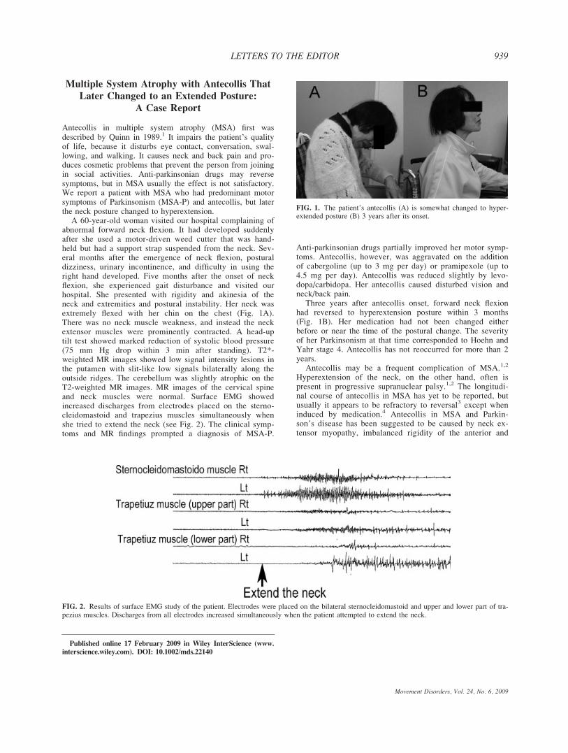
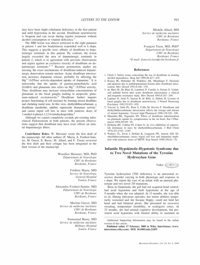
A 60-year-old woman visited our hospital complaining ofabnormal forward neck flexion. It had developed suddenlyafter she used a motor-driven weed cutter that was hand-held but had a support strap suspended from the neck. Sev-eral months after the emergence of neck flexion, posturaldizziness, urinary incontinence, and difficulty in using theright hand developed. Five months after the onset of neckflexion, she experienced gait disturbance and visited ourhospital. She presented with rigidity and akinesia of theneck and extremities and postural instability. Her neck wasextremely flexed with her chin on the chest (Fig. 1A).There was no neck muscle weakness, and instead the neckextensor muscles were prominently contracted. A head-uptilt test showed marked reduction of systolic blood pressure(75 mm Hg drop within 3 min after standing). T2*-weighted MR images showed low signal intensity lesions inthe putamen with slit-like low signals bilaterally along theoutside ridges. The cerebellum was slightly atrophic on theT2-weighted MR images. MR images of the cervical spineand neck muscles were normal. Surface EMG showedincreased discharges from electrodes placed on the sterno-cleidomastoid and trapezius muscles simultaneously whenshe tried to extend the neck (see Fig. 2). The clinical symp-toms and MR findings prompted a diagnosis of MSA-P.
Anti-parkinsonian drugs partially improved her motor symp-toms. Antecollis, however, was aggravated on the additionof cabergoline (up to 3 mg per day) or pramipexole (up to4.5 mg per day). Antecollis was reduced slightly by levo-dopa/carbidopa. Her antecollis caused disturbed vision andneck/back pain.
Three years after antecollis onset, forward neck flexionhad reversed to hyperextension posture within 3 months(Fig. 1B). Her medication had not been changed eitherbefore or near the time of the postural change. The severityof her Parkinsonism at that time corresponded to Hoehn andYahr stage 4. Antecollis has not reoccurred for more than 2years.
Antecollis may be a frequent complication of MSA.1,2
Hyperextension of the neck, on the other hand, often ispresent in progressive supranuclear palsy.1,2 The longitudi-nal course of antecollis in MSA has yet to be reported, butusually it appears to be refractory to reversal3 except wheninduced by medication.4 Antecollis in MSA and Parkin-son’s disease has been suggested to be caused by neck ex-tensor myopathy, imbalanced rigidity of the anterior and
FIG. 1. The patient’s antecollis (A) is somewhat changed to hyper-extended posture (B) 3 years after its onset.
Published online 17 February 2009 in Wiley InterScience (www.
interscience.wiley.com). DOI: 10.1002/mds.22140
FIG. 2. Results of surface EMG study of the patient. Electrodes were placed on the bilateral sternocleidomastoid and upper and lower part of tra-pezius muscles. Discharges from all electrodes increased simultaneously when the patient attempted to extend the neck.
Movement Disorders, Vol. 24, No. 6, 2009
939LETTERS TO THE EDITOR

posterior neck muscles, or dystonia.3,5�7 Needle EMG ofthe neck muscles have not been performed in our patient,however, no neck muscle weakness and surface EMGresults suggesting simultaneous and equal overactivation offlexor and extensor muscles on neck extension of thepatient support the imbalanced tonus hypothesis. That is,imbalance of muscle tonus might have shifted from flexor-to extensor-neck muscle predominance as the disease pro-gressed.
The change in our patient’s neck posture from the flexedto extended neck position indicates that antecollis in basalganglia diseases may be reversed in the natural course of theillness.
Kenichi Kashihara, MD, PhD*Manabu Ohno, MD, PhDDepartment of Neurology
Okayama Kyokuto HospitalOkayama, Japan
*E-mail: [email protected]
References
1. Quinn N. Disproportionate antecollis in multiple system atrophy.Lancet 1989;1:844.
2. Rivest J, Quinn N, Marsden CD. Dystonia in Parkinson’s disease,multiple system atrophy, and progressive supranuclear palsy. Neu-rology 1990;40:1571�1578.
3. Askmark H, Eeg-Olofsson KE, Johansson A, Nilsson P, Olsson Y,Aquilonius SM. Parkinsonism and neck extensor myopathy—a newsyndrome or coincidental findings? Arch Neurol 2001;58: 232�237.
4. Prakash KM, Lang AE. Reversal dopamine agonist induced antero-collis in a multiple system atrophy patient. Move Disord 2007;22:2292�2293.
5. Yoshiyama Y, Takama J, Hattori T. The dropped head sign inparkinsonism. J Neurol Sci 1999;167:22�25.
6. Kashihara K, Ohno M, Tomita S. Dropped head syndrome in Parkin-son’s disease. Mov Disord 2006;21:1213�1216.
7. van den Warrenburg BPC, Cordivari C, Ryan AM, Phadke R, Hol-ton JL, Bhatia KP, Hanna MG, Quinn NP. The phenomenon of dis-proportionate antecollis in Parkinson’s disease and multiple systematrophy. Mov Disord 2007;22:2325�2331.
Pantothenate Kinase-Associated
Neurodegeneration in Two Taiwanese Siblings:
Identification of a Novel PANK2Gene Mutation
Video
Pantothenate kinase-associated neurodegeneration (PKAN) isan autosomal recessive disorder characterized by accumula-tion of iron in the basal ganglia due to mutation of the pan-tothenate kinase 2 gene (PANK2).1 The hallmark of neuro-radiological finding is ‘‘eye of the tiger sign.’’ ClassicPKAN presents in the first decade with dystonia, dysarthria,
intellectual impairment, retinopathy, optic atrophy and pro-gresses rapidly, leading to loss of ambulation by mid-ado-lescence. The atypical form of PKAN presents in lateadolescence (around aged 20), manifesting as a progressivepicture of dementia, Parkinsonism, dystonia, anarthria, andaphonia.2 Herein, we report a compound heterozygotePAKN2 mutation in two Taiwanese siblings with atypicalPKAN.
A 48-year-old male (Case 1) had slight mental retardationsince childhood. He had right hand tremor and progressivegait disturbance for two years. Neurological examinationrevealed severe dysarthria, stuttering, torticollis, and tonguetremor. Mild rigidity, bradykinesia, and postural instabilitywere found. During walking, he had wide base gait alongwith right foot dystonia (see video, Segment 1). His 59-year-old elder brother (Case 2) experienced progressive gaitdisturbance and frequent falls for 8 years. Mild dysarthria,typical parkinsonian tremor in the jaw, tongue, and all fourlimbs, generalized rigidity and bradykinesia, dystonia at feet,wide base gait, and postural instability were noted (see video,Segment 2). Therapeutic trials with levodopa, trihexyphe-nidyl, baclofen, and propranolol have been ineffective. Theirgrandfather had history of tremor and gait disturbance.There were no other family members with similar symp-toms. Brain T2-weighted magnetic resonance imaging(MRI) showed low signal intensity in the pallidum with aslight anteromedial core of high signal intensity, as so-called ‘‘eye of the tiger’’ sign in Case 1, which was less typ-ical in Case 2 (Fig. 1). 99mTc-TRODAT-1 brain SPECT(single-photon emission tomography) studies of both siblingsdemonstrated symmetrical mild decline of dopamine transporterin bilateral striatum, which was normally seen for their age.There was no significant benefit of L-dopa also made the diag-nosis of PD unlikely.
Automated DNA sequence analyses revealed compoundheterozygous mutations in the exon 3 (c.1133A>G)3 and 4(c.1355A>G). Both mutations predict the replacement of anaspartic acid residue with glycine (p.D378G and p.D452G).RT-PCR and cDNA sequencing further revealed that thenovel c.1355A>G primarily creates a potential 50 splice sig-nal, leading to a 58-bp deletion in exon 4 (c.1355_1412del).
FIG. 1. Magnetic resonance imaging of the brain showing hypoin-tensity with a central region of hyperintensity in the medial globuspallidus, the eye of the tiger sign (Case 1, A) or a faint hyperinten-sity region within a slit hypointensity region in the medial globuspallidus (Case 2, B).
Published online 17 February 2009 in Wiley InterScience (www.
interscience.wiley.com). DOI: 10.1002/mds.22458
940 LETTERS TO THE EDITOR
Movement Disorders, Vol. 24, No. 6, 2009

To our knowledge, this would be the first PANK2 mutationreported in Taiwan with atypical PKAN.
Numerous mutations underlying PKAN have beenreported, generally nonsense mutations with typical form andmissense mutations with atypical form.4,5 Reports in ethnicChinese are relatively sparse; only compound missensePANK2 mutations for a Chinese patient with atypical PKANwere reported.6 Both p.D378G and p.D452G are likely patho-genic being highly conserved across species. Moreover themajor product of c.1355A>G is c.1355_1412del, resulting inpredicted protein truncation and no residual activity. PANK2is primarily localized to mitochondria of neurons in humanbrain. Its deficiency is thought to cause mitochondrial accu-mulation of cysteine or cysteine-containing substrate, result-ing in free radical generation and cell death.7 The MRI ofCase 2 did not revealed typical ‘‘eye of the tiger sign,’’which is reminiscent of the findings reported by Baumeisteret al., in which the central hyperintensity continues to dimin-ish and may ultimately disappear.8
In this report, we identified a novel mutation in thePANK2 gene responsible for PKAN and confirmed thatPKAN has a board spectrum of phenotype, even among sib-lings with same mutations.
Legends to the Video
Segment 1. Patient 1 has tongue and right hand tremor.The tremor has a very low-frequency and in one sequence aresting tremor on the right can be detected. Furthermore, heshows a low-frequency intention-tremor, resembling in com-bination with the resting tremor and a Holmes-Tremor. Hehas right big toe dystonia, mild bradykinesia, and impairedrapid alternative movement. During walking, he had widebase gait along with right foot dystonia. Postural instabilitywas detected by the pull test (see Video, Segment 1).
Segment 2. Patient 2 has typical Parkinsonian tremor inthe four limbs, mild bradykinesia, dystonia of left big toe,wide base gait, and postural instability.
Yih-Ru Wu, MDChiung-Mei Chen, MD, PhD
Department of NeurologyChang Gung Memorial Hospital
Chang-Gung University College of MedicineTaipei, Taiwan
Chih-Ying Chao, BSDepartment of Life Science
National Taiwan Normal UniversityTaipei, Taiwan
Ron-Kuo Lyu, MDDepartment of Neurology
Chang Gung Memorial HospitalChang-Gung University College of Medicine
Taipei, Taiwan
Guey-Jen Lee-Chen, PhD*Department of Life Science
National Taiwan Normal UniversityTaipei, Taiwan
*E-mail: [email protected]
References
1. Zhou B, Westaway SK, Levinson B, Johnson MA, Gitschier J,Hayflick SJ. A novel pantothenate kinase gene (PANK2) is defec-tive in Hallervorden-Spatz syndrome. Nat Genet 2001;28:345�349.
2. Hayflick SJ, Westaway SK, Levinson B, Zhou B, Johnson MA,Ching KH, Gitschier J. Genetic, clinical, and radiographic delineationof Hallervorden-Spatz syndrome. N Engl J Med 2003;348: 33�40.
3. Lyoo CH, Prokisch H, Meitinger T, Lee SY, Kim do H, Lee MS.Anticholinergic-responsive gait freezing in a patient with panto-thenate kinase-associated neurodegeneration. Mov Disord 2008;23:283�284.
4. Pellecchia MT, Valente EM, Cif L, et al. The diverse phenotypeand genotype of pantothenate kinase-associated neurodegeneration.Neurology 2005;64:1810�1812.
5. Hartig MB, Hortnagel K, Garavaglia B, et al. Genotypic and pheno-typic spectrum of PANK2 mutations in patients with neurodegenera-tion with brain iron accumulation. Ann Neurol 2006;59: 248�256.
6. Zhang YH, Tang BS, Zhao AL, Xia K, Long ZG, Guo JF, West-away SK, Hayflick SJ. Novel compound heterozygous mutations inthe PANK2 gene in a Chinese patient with atypical pantothenate ki-nase-associated neurodegeneration. Mov Disord 2005;20: 819�821.
7. Kotzbauer PT, Truax AC, Trojanowski JQ, Lee VM. Altered neu-ronal mitochondrial coenzyme A synthesis in neurodegenerationwith brain iron accumulation caused by abnormal processing,stability, and catalytic activity of mutant pantothenate kinase 2.J Neurosci 2005;25:689–698.
8. Baumeister FA, Auer DP, Hortnagel K, Freisinger P, Meitinger T.The eye-of-the-tiger sign is not a reliable disease marker for Hal-lervorden-Spatz syndrome. Neuropediatrics 2005;36:221–222.
Dopamine Transporter Binding is Reduced
Following Disulfiram-Induced Striatal Damage
Disulfiram is an aldehyde dehydrogenase inhibitor, which isused as an adjunctive agent in severe chronic alcoholismbecause of its antabuse effect. It has neurotoxic propertiesresulting in neuropathy, optic neuritis, encephalopathy, andpallidoputaminal necrosis.1 In the latter case, delayed-onsetmovement disorders such as akinesia and dystonia have beenobserved.2�4
We report 2 patients who showed striatopallidal damagewith involvement of the dopaminergic nigrostriatal pathwayin disulfiram-induced encephalopathy.
A 70-year-old woman with chronic alcohol abuse wasadmitted for acute encephalopathy. She had already presented2 weeks before at the emergency room because of a falldue to acute ethanol intoxication. A CT scan was normal.As she was motivated to stop drinking, disulfiram (500mg/day) was started for the first time. The patient left theemergency room without obvious signs of encephalopathyor Parkinsonism.
Clinical evaluation showed a somnolent patient with gaitdifficulties, mild bradykinesia of the upper left limb, and abilateral extensor plantar response. Blood tests revealedmacrocytosis (106 fl) and slightly below-range serum cobala-min (190 pmol/L, N > 197), but no signs of acute alcohol
Published online 17 February 2009 in Wiley InterScience (www.
interscience.wiley.com). DOI: 10.1002/mds.22397
Movement Disorders, Vol. 24, No. 6, 2009
941LETTERS TO THE EDITOR

intoxication. EEG showed diffuse slowing. Brain MRI dis-closed T1-hypointense and T2-hyperintense signal abnormal-ities of the right pallidum (Fig. 1A), which were less pro-nounced on a control MRI 1 year later. HMPAO-SPECTshowed a reduced signal in this area and b-CIT-SPECTrevealed reduced dopamine transporter density in the rightposterior putamen (Fig. 1B,C). The patient progressivelyrecovered, but mild bradykinesia of the upper left limb, gaitdifficulties, and cognitive impairment persisted at the patient’sdischarge. Six months later, mild bradykinesia was stillobserved and levodopa treatment was initiated (300 mg/day)allowing perfect symptom control.
A 54-year-old woman was found comatose at home withmild signs of hypoxemia. She was subsequently admitted tothe intensive care unit for acute encephalopathy and aspira-tion pneumonia requiring artificial ventilation. She sufferedfrom chronic alcoholism and had already been admitted tothe emergency room 2 months earlier with acute ethanolintoxication and dehydration. At that time, a CT scan wasnormal. She decided to stop drinking, and disulfiram was ini-tiated (500 mg/day) for the first time. The patient returned
home after several days without obvious signs of encephalop-athy or Parkinsonism.
Lab testing for acute alcohol intoxication was not per-formed in the emergency room. MRI disclosed a bilateralT1-hypointense and T2-hyperintense signal in the posteriorputamen (Fig. 1D), partly extending to the left pallidum. Theextent of the pallidoputaminal lesion had considerablydecreased on a control MRI 2 months after admission.HMPAO-SPECT showed a reduced signal in the left poste-rior putamen, whereas b-CIT-SPECT revealed a reduced sig-nal in both posterior putamena (Fig. 1E,F), which was morepronounced on the left. She recovered from coma 6 days af-ter admittance and was found to be mildly tetraparetic, pre-dominantly on the right with an extensor plantar response onthe same side. Mild postural tremor of both upper limbs, dys-arthria, and difficulties of swallowing were noted, requiringtransient feeding through a nasogastric tube. The patientalmost completely recovered after 3 months with a persistingextensor plantar response on the right side.
We report 2 new cases of pallidoputaminal damage inrelation to disulfiram intake. Concomitant aggravating factors
FIG. 1. Patient 1: (A) T2-weighted imaging shows an abnormal hypersignal mainly in the right putamen (arrowhead). (B) Low uptake in theright posterior putamen on b-CIT-SPECT (arrow). (C) HMPAO-SPECT shows low uptake in the right posterior putamen (arrow). Patient 2: (D)T2-weighted imaging shows an abnormal hypersignal in both posterior putamena (arrowheads). (E) Low uptake in both putamena on b-CIT-SPECT (arrowheads). (F) HMPAO-SPECT shows low uptake in left posterior putamen (arrowhead). [Color figure can be viewed in the onlineissue, which is available at www.interscience.wiley.com.]
942 LETTERS TO THE EDITOR
Movement Disorders, Vol. 24, No. 6, 2009
may have been slight cobalamin deficiency in the first patientand mild hypoxemia in the second. Disulfiram neurotoxicityis frequent and can occur during regular treatment withoutalcohol consumption or vitamin deficiency.1
The MRI lesion was almost restricted to the right putamenin patient 1 and her bradykinesia responded well to L-dopa.This suggests a specific toxic affinity of disulfiram to dopa-minergic terminals in this patient. By contrast, the lesionlargely exceeded the area of dopaminergic terminals inpatient 2, which is in agreement with previous observationsand argues against an exclusive toxicity of disulfiram on do-paminergic terminals.2�4 Because postmortem studies aremissing, the exact mechanisms of disulfiram-induced dopami-nergic denervation remain unclear. Acute disulfiram intoxica-tion increases dopamine release, probably by affecting theMg21/ATPase activity-dependent uptake of dopamine.5 It isnoteworthy that the uptake of gamma-aminobutylic acid(GABA) and glutamate also relies on Mg21/ATPase activity.Thus, disulfiram may increase extracellular concentrations ofglutamate in the basal ganglia leading to unspecific gluta-mate-induced excitotoxicity.6 Disulfiram further impairsproper functioning of cell enzymes by forming mixed disulfidesand chelating metal ions. In this view, diethyldithiocarbamate, adisulfiram metabolite, inhibits superoxide dismutase activity7
and causes nigral cell loss in mice with nontoxic doses of1-methyl-4-phenyl-1,2,3,6-tetrahydropyridine.8
Although we cannot completely exclude pre-existing infra-clinical Parkinsonism in both patients, the present observa-tions suggest that disulfiram may have toxic effects on stria-tal dopaminergic fibers.
Contributor Roles: W. Meissner wrote the first draft ofthe manuscript. All other authors (F. Macia, A. Foubert-Sam-ier, M. Guyot, E. Bussy, M. Allard, and F. Tison) reviewedthe first draft and their critique has been integrated in thefinal version of the manuscript.
Wassilios Meissner, MD, PhDDepartement de Neurologie
CHU de BordeauxBordeaux, France
Frederic Macia, MDService de Neurologie
General HospitalToulon, France
Alexandra Foubert-Samier, MDDepartement de Neurologie
CHU de BordeauxBordeaux, France
Martine Guyot, MDService de medecine nucleaire
CHU de BordeauxBordeaux, France
Emmanuel Bussy, MDService de medecine nucleaire
Millitary HospitalJoulon, France
Michele Allard, MDService de medecine nucleaire
CHU de BordeauxBordeaux, France
Francois Tison, MD, PhD*Departement de Neurologie
CHU de BordeauxBordeaux, France
*E-mail: [email protected]
References
1. Chick J. Safety issues concerning the use of disulfiram in treatingalcohol dependence. Drug Saf 1999;20:427�435.
2. Krauss JK, Mohadjer M, Wakhloo AK, Mundinger F. Dystoniaand akinesia due to pallidoputaminal lesions after disulfiram intox-ication. Mov Disord 1991;6:166�170.
3. de Mari M, De Blasi R, Lamberti P, Carella A, Ferrari E. Unilat-eral pallidal lesion after acute disulfiram intoxication: a clinicaland magnetic resonance study. Mov Disord 1993;8:247�249.
4. Laplane D, Attal N, Sauron B, de Billy A, Dubois B. Lesions ofbasal ganglia due to disulfiram neurotoxicity. J Neurol NeurosurgPsychiatry 1992;55:925�929.
5. Vaccari A, Saba PL, Ruiu S, Collu M, Devoto P. Disulfiram anddiethyldithiocarbamate intoxication affects the storage and releaseof striatal dopamine. Toxicol Appl Pharmacol 1996;139:102�108.
6. Mamatha RK, Nagendra SN. Effect of disulfiram administrationon glutamate uptake by synaptosomes in the rat brain. Eur J Phar-macol 1994;292:89�94.
7. Heikkila RE, Cabbat FS, Cohen G. In vivo inhibition of superox-ide dismutase in mice by diethyldithiocarbamate. J Biol Chem1976;251:2182�2185.
8. Walters TL, Irwin I, Delfani K, Langston JW, Janson AM. Di-ethyldithiocarbamate causes nigral cell loss and dopamine deple-tion with nontoxic doses of MPTP. Exp Neurol 1999;156:62�70.
Infantile Hypokinetic-Hypotonic Syndrome due
to Two Novel Mutations of the Tyrosine
Hydroxylase Gene
Video
Tyrosine hydroxylase (TH) deficiency is an autosomal re-cessive disorder varying in both phenotype and response toL-dopa. We report the case of an infant with an unusual phe-notype and two novel TH mutations.
Born in Guatemala, the girl had not acquired head control,had axial hypotonia and limb hypertonia at the age of9 months when she was adopted. At 13 months, she was ableto sit. During infectious episodes, her motor abilities tempo-rarily worsened and she became floppy, could not hold herhead and had bilateral ptosis. She presented no excessivesweating, temperature instability, or oculogyris crises. At23 months, she had normal cognitive development, but per-sistent axial hypotonia with limited ability to maintain an
Additional Supporting Information may be found in the online
version of this article.Published online 17 February 2009 in Wiley InterScience (www.
interscience.wiley.com). DOI: 10.1002/mds.22455
Movement Disorders, Vol. 24, No. 6, 2009
943LETTERS TO THE EDITOR
upright position while sitting. She had marked amimia andakinesia with few spontaneous limb movements (Video seg-ment 1). During voluntary movements, symmetrical dystoniaand tremor of the extremities appeared with pes equinovarusand an inability to pick up small objects. Dystonia was presentthroughout the day, whereas axial hypotonia showed diurnalvariation, worsening in the evening and improving in themorning or after a nap when she was able to stand up if helped.
Although limb dystonia only partially improved followinginitiation of low dose L-dopa (0.7 mg/kg/d) treatment, axialtone dramatically improved allowing independent walkingone month later (Video segment 2). Higher dose L-dopa wastried but induced dyskinesia and irritability. Brain MRI, rou-tine blood tests, and metabolic investigations were normal.No mutation was found in the GCH1 gene. During a oneweek treatment interruption period necessary for the analysisof pterins and biogenic amines in the cerebrospinal fluid(CSF) by high-performance liquid chromatography, thepatient lost the ability to walk. Pterins, 5 hydroxyindoleacetic(5HIAA) and 3 ortho-methyl-dopa concentrations were nor-mal; homovanillic acid (HVA) concentration was in thelower range: 254 nmol/L (normal range 211�871) and theratio HVA/5HIAA was low: 0.7 (normal 1.5�3.5) suggestinga TH deficiency. Sequencing of the TH gene revealedtwo heterozygous mutations: p.Phe375Leu/p.Ser467Gly notencountered in 85 controls. During follow-up, L-dopa wasslowly increased up to 1.7 mg/kg/d. Motor abilities were nor-mal at 6 years, except for the persistence of mild dystonicmovements of the hands, and L-dopa induced hyperkinesiaand dyskinesia resulting in impaired handwriting. IQ, meas-ured by Weschsler Preschool and Primary Scale of Intelli-gence, was within normal range (VIQ 105, PIQ 103), but ex-ecutive speed was below average (77).
Since the first description of TH deficiency in 1995,1 29cases have been reported with a wide range of phenotypesfalling into three main categories: Dopa Responsive Dystonia(DRD) (9/29), severe progressive encephalopathy in infancy(6/29), and infantile Parkinsonism (14/29). DRD beginsbetween 1 and 5 years of age1,2 with the phenotype initiallydescribed by Segawa showing dramatic and sustainedimprovement with low dose L-dopa therapy. Severe progres-sive encephalopathy occurs within the first few months oflife with marked truncal hypotonia, peripheral rigidity,tremor, and myoclonic jerks.3 Symptoms of catecholaminedeficiency with lethargic episodes, excessive sweating andsalivation, ptosis, and oculogyric crises are frequent, withmild to moderate mental retardation. Response to L-dopatherapy is null or limited by intolerable side effects such ashyperkinesia and irritability. Infantile Parkinsonism startsbefore the age of one year4 and shows better response to L-dopa. Patients have no or mild mental retardation and lesssevere dysautonomic symptoms.
The present case, associating prominent severe axial hypo-tonia, normal cognitive functions, and an absence of severedysautonomia, therefore represents an unusual phenotype.The diurnal fluctuation of axial hypotonia and increased flop-piness during infectious episodes with ptosis are the hall-marks of our observation.5 The decreased HVA/5HIAA ratiorather than a decreased HVA level in the CSF led to the di-agnosis of TH deficiency. Other possible diseases such as ju-venile Parkinson’s disease or mitochondrial disorders wereconsidered6,7 but rejected based on the age at onset, the clini-
cal features and the normal lactate levels in plasma and CSF.Moreover, this patient has two missense mutations, absent incontrols and located in the catalytic domain of the proteinthereby predicting deficient activity.
The diagnosis of this often treatable rare condition needsto be made from a large spectrum of early onset movementdisorders. Diurnal fluctuations of axial hypotonia and dysau-tonomic symptoms are suggestive of the diagnosis and shouldlead to the study of biogenic amines in the CSF and the com-plete analysis of the TH gene.8
Legends to the Video
Segment 1. At age 23 months, she presented hypokinesiaand axial hypotonia and could not stand up unaided.
Segment 2. After one month of L-dopa therapy, she wasable to walk independently.
Acknowledgments: We thank Prof N. Blau (Zurich) forpterin and neurotransmitter measurements in the CSF. Thiswork was performed at the ‘‘Neurogenetics’’ Reference Centrefor rare diseases and was supported by INSERM (FrenchDystonia Network) and GIS Maladies Rares.
Diane Doummar, MDAP-HP, Service de Neuropediatrie
Hopital Armand TrousseauParis, France
*E-mail: [email protected]
Fabienne Clot, PhDAP-HP, Departement de Genetique et Cytogenetique
Hopital Pitie SalpetriereParis, France
Marie Vidailhet, MD, PhDAP-HP, Departement de Neurologie
Hopital Pitie-SalpetriereINSERM UMR_S679
Universite Pierre et Marie Curie-Paris 6Paris, France
Alexandra Afenjar, MDAP-HP, Service de Neuropediatrie
Hopital Armand TrousseauParis, France
Alexandra Durr, MD, PhDAlexis Brice, MD
AP-HP, Departement de Genetique et CytogenetiqueHopital Pitie Salpetriere
INSERM UMR_S679Universite Pierre et Marie Curie-Paris 6
Paris, France
Cyril Mignot, MD, PhDAgnes Guet, MD
Thierry Billette de Villemeur, MDDiana Rodriguez, MD, PhD
AP-HP, Service de NeuropediatrieHopital Armand Trousseau
Universite Pierre et Marie Curie-Paris 6Paris, France
944 LETTERS TO THE EDITOR
Movement Disorders, Vol. 24, No. 6, 2009

References
1. Ludecke B, Dworniczak B, Bartholome K. A point mutation inthe tyrosine hydroxylase gene associated with Segawa’s syndrome.Hum Genet 1995;95:123�125.
2. Schiller A, Wevers RA, Steenbergen GC, Blau N, Jung HH.Long-term course of L-dopa-responsive dystonia caused bytyrosine hydroxylase deficiency. Neurology 2004;63:1524�1526.
3. S Hoffmann GF, Assmann B, Brautigam C, et al. Tyrosinehydroxylase deficiency causes progressive encephalopathy anddopa-nonresponsive dystonia. Ann Neurol 2003;54:S56�S65.
4. Grattan-Smith PJ, Wevers RA, Steenbergen-Spanjers GC, FungVS, Earl J, Wilcken B. Tyrosine hydroxylase deficiency: clinicalmanifestations of catecholamine insufficiency in infancy. MovDisord 2002;17:354�359.
5. Diepold K, Schutz B, Rostasy K, et al. Levodopa-responsive in-fantile parkinsonism due to a novel mutation in the tyrosinehydroxylase gene and exacerbation by viral infections. Mov Dis-ord 2005;20:764�767.
6. Poceta JS, Parsons L, Engelland S, Kripke DF. Circadian rhythmof CSF monoamines and hypocretin-1 in restless legs syndromeand Parkinson’s disease. Sleep Med 2008. [Epub ahead of print].
7. Garcia-Cazorla A, Duarte S, Serrano M, et al. Mitochondrial dis-eases mimicking neurotransmitter defects. Mitochondrion 2008; 8:273–278.
8. Verbeek MM, Steenbergen-Spanjers GC, Willemsen MA, et al.Mutations in the cyclic adenosine monophosphate responseelement of the tyrosine hydroxylase gene. Ann Neurol 2007;62:422–426.
Life-Threatening Hypothermia in
Parkinson’s Disease
Hyperthermia has been described as the symptom of neuro-leptic malignant syndrome. However, it is rare to find apatient with Parkinson’s disease (PD) developing life-threat-ening hypothermia. A 58-year-old man with PD had beentreated with levodopa/carbidopa, cabergoline, and quetiapinefumarate developed life-threatening hypothermia. The com-mon causes of hypothermia have not been determined. It ispossible to speculate that his thermoregulation breakdownand/or the drugs administered to him such as dopamine D2receptor agonist may affect the thermal homeostasis mecha-nisms. Although it is rare to see a patient with PD with life-threatening hypothermia, the awareness of clinical entity ofhypothermia should be kept in mind.
In patients with PD, hyperthermia is a symptom that needscareful treatment because it is the core symptom of neurolep-tic malignant syndrome. However, it is rare to find a patientwith PD developing recurrent, life-threatening hypothermia.
Case report: A 58-year-old man with PD was admitted toour emergency room for impairment of consciousness on 15December. He had been diagnosed with PD at the age 45,and had since been treated with dopamine replacement ther-apy. At the time of admission, he was being treated with L-dopa/carbidopa 500 mg per day, cabergoline 2 mg per day,and quetiapine fumarate 50 mg per day. He had been treatedwith quetiapine for 3 years to control visual hallucinations;
without inducing any prominent deterioration of his motorsymptoms. His daily activities were limited because of motorfluctuations, which were about Hoehn�Yahr Stage III during‘‘on’’ and stage V during ‘‘off’’. Over the last few years, hiscognitive function had gradually deteriorated (MMSE 5 20).Further increasing the dose of his dopamine replacementtherapy had resulted in deterioration of his psychiatric symp-toms. In the weeks before admission, his caregivers hadoccasionally noticed that his temperature was extremely low(below 358C), but did not take him to hospital because helooked well, as usual. On admission, his temperature was328C and blood pressure 107/68 mm Hg, pulse rate 45/min,and respiratory rate 15/min. He was in a stupor with his eyesopening spontaneously. No voluntary movements of limbs orbody were noted. There was marked muscular rigidity but noovert tremor or shivering. Results of routine laboratory tests,including thyroid function, were normal. Brain CT was nor-mal. EKG demonstrated J waves as described by Osborn,known to be characteristic in hypothermia. He suddenlydeveloped ventricular tachycardia, resulting in cardiopulmo-nary arrest, but he was soon resuscitated. His rectal bodytemperature was found 33.18C. To improve his life-threaten-ing hypothermia, warming with an electric blanket and intra-venous administration of warm saline was immediatelystarted. Subsequently, his rectal temperature slowlyimproved, and he eventually recovered without any sequelae,suggesting that hypothermia was the primary problem. Hiscaregivers stated that he preferred to wear light clothing butwas never exposed for long to low temperatures. Indeed, heusually spent the daytime in a nursing home and the eveningat his home, both had air-conditioning and heating systems.His caregivers were asked to check his body temperature reg-ularly after he was discharged and not let him wear lightclothes. In the 18 months following this event, he neverdeveloped a similar episode.
The exact cause of the life-threatening hypothermia in thepresent patient was unclear, although it was suggested thatPD could be a predisposing factor of hypothermia.1,2 Wethink that hypothermia should not be regarded as an acciden-tal event or as unrelated to PD or its treatment. This isbecause the common causes of hypothermia, such as pro-longed exposure to low temperatures, serious endocrinedisease, and malnutrition have not all been determined. Addi-tionally, from Japan Meteorological Agency records, the av-erage temperatures in the area, that is December, was 8.88C,suggesting that cold circumstances did not cause his hypo-thermia. It is also interesting to note that he never com-plained of feeling cold, despite the low body temperature,and that he did not exhibit shivering, suggesting that theremight be some derangement of his thermal homeostasis sys-tems. It is possible to speculate that his thermoregulationbreakdown might have involved the hypothalamus or hypo-thalamic outflows to the brainstem. Another possibility is thatthe drugs administered to him may have worsened his hypo-thermia. There are reports suggesting that dopamine D2receptor stimulation may affect the thermal homeostasismechanisms.3 Experimentally, in rats, dopamine D2 receptoragonist-induced hypothermia in cold environments may bemainly due to decreased heat production, rather than toincreased heat loss.4 Atypical neuroleptics might also may beinvolved in hypothermia5; however, to our knowledge, que-tiapine has not been reported in this regard.
Published online 17 February 2009 in Wiley InterScience (www.
interscience.wiley.com). DOI: 10.1002/mds.22476
Movement Disorders, Vol. 24, No. 6, 2009
945LETTERS TO THE EDITOR

Although it is rare to see a patient with PD with life-threatening hypothermia, the possibility that hypothermia canoccur during the course of PD should be kept in mind,because it may be potentially fatal if undetected or over-looked.
Kiwa Hama, MD*Hideto Miwa, MD
Tomoyoshi Kondo, MDDepartment of Neurology
Wakayama Medical UniversityWakayama-city, Japan
*E-mail: [email protected]
References
1. Macdonell JE, Wrenn K. Hypothermia in the summer. South MedJ 1991;84:804�805.
2. Tchoua R, Mahamat Y, Loembe PM. Hypothermie grave en mi-lieu tropical. Med Trop 1998;58:158�160.
3. Collins GT, Newman AH, Grundt P, et al. Yawning and hypo-thermia in rats: effects of dopamine D3 and D2 agonists andantagonists. Psychopharmacology 2007;193:159�170.
4. Ootsuka Y, Heidbreder CA, Hagan JJ, Blessing WW. Dopamine D2receptor stimulation inhibits cold-initiated thermogenesis inbrown adipose tissue in conscious rats. Neuroscience 2007;147:127�135.
5. Hagg S, Mjorndal T. Repeated episodes of hypothermia in a subjecttreated with haloperidol, levomepromazine, olanzapine, and thiorida-zine. J Clin Psychopharmacol 2001;21:113�115.
A Multimodal Approach to Physical Therapy
in Parkinson’s Disease: Optimizing Strategies
The recent systematic review of exercise interventions forParkinson’s disease (PD) by Goodwin and colleagues1 rein-forces the evidence supporting a role of exercise in the man-agement of PD.2,3 The authors focused solely on exercise-based interventions in experimental, randomized designs.Studies explicitly evaluating cueing strategies were excludedas such techniques were considered external temporal or spa-tial stimulation to facilitate gait separate from the act of exer-cising. Using such criteria, Goodwin and colleagues concludethat exercise benefits physical functioning, balance, gaitspeed, strength, and health-related quality of life.
We commend the authors’ methodology in addressing theimportant issue of the lack of homogeneity in the interven-tions and study designs used to ascertain effect of physicaltherapy in PD. Although the exclusion of studies explicitlyevaluating cueing strategies homogenizes the interventionand enables extracting information regarding the individualcontribution of exercise, we wonder how to place such find-ings in the context of the recent recommendations of physicaltherapy in PD published in this journal by Keus and col-leagues.4 These evidence-based recommendations were more
inclusive of physiotherapeutic techniques, and included cue-ing strategies, cognitive movement strategies to improvetransfers, specific exercises to improve balance, and trainingof joint mobility as well as muscle power.
In planning future studies of physiotherapeutic interven-tions in PD, both methodologies have their merits and differ-ent goals. Restricting interventions allows one to ascertainthe efficacy of specific techniques while risking smaller effectsize and thus non-significant findings. Combining multiplemodalities at once (i.e., cueing and exercise) may maximizetreatment effect at the cost of not knowing the contributionof an individual technique. It is possible that simultaneousinterventions (i.e., cueing and exercise) are redundant, addi-tive, or synergistic. However, it would not be possible toknow if each potential therapeutic component were testedonly in isolation. One approach may be to first test interven-tions that simultaneously employ multiple modalities. Ifthese seem effective, one may then test each modality inisolation to determine its individual contribution to thera-peutic benefit. However, such an approach carries its ownrisk, as it is conceivable that combined interventions maybe less effective than individual techniques. For example,psychomotor difficulties or complications that occur inthe later stages of PD may make it too difficult for partici-pants to engage in both cueing strategies and exercisesimultaneously.
We appreciate the work of Goodwin, Keus and others asthey have highlighted the limitations and fragilities of pasttrials regarding trial design, patient selection, and duration oftherapeutic effect. Such reviews assist in planning futurestudies in physiotherapeutic interventions for PD.
Tiago Mestre, MD, MSc*Joaquim J. Ferreira, MD
Neurological Clinical Research UnitInstitute of Molecular Medicine
Lisbon, Portugal*E-mail: [email protected]
Samay Jain, MDMovement Disorders Division, Department of Neurology
University of Pittsburgh Medical CenterPittsburgh, Pennsylvania, USA
References
1. Goodwin VA, Richards SH, Taylor RS, Taylor AH, Campbell JL.The effectiveness of exercise interventions for people with Parkin-son’s disease: a systematic review and meta-analysis. Mov Disord2008;23:631�640.
2. de Goede CJ, Keus SH, Kwakkel G, Wagenaar RC. The effects ofphysical therapy in Parkinson’s disease: a research synthesis. ArchPhys Med Rehabil 2001;82:509�515.
3. Deane KH, Jones D, Playford ED, Ben-Shlomo Y, Clarke CE.Physiotherapy for patients with Parkinson’s disease: a comparisonof techniques. Cochrane Database Syst Rev 2001;3:CD002817.
4. Keus SH, Bloem BR, Hendriks EJ, Bredero-Cohen AB, MunnekeM. Evidence-based analysis of physical therapy in Parkinson’s dis-ease with recommendations for practice and research. Mov Disord2007;22:451�460.
Published online 17 February 2009 in Wiley InterScience (www.
interscience.wiley.com). DOI: 10.1002/mds.22478
Movement Disorders, Vol. 24, No. 6, 2009
946 LETTERS TO THE EDITOR
Corticobasal Degeneration Presenting as
Complex Regional Pain Syndrome
Video
Corticobasal degeneration (CBD) is a neurodegenerative dis-order characterized by progressive asymmetric motor signsincluding rigidity, bradykinesia, myoclonus, and dystonia,unresponsive to levodopa, together with asymmetric apraxia,alien limb phenomenon and cortical sensory loss, suggestingcortical and basal ganglia dysfunction.1,2 Despite a high spec-ificity of CBD clinical diagnosis, sensitivity is low (35%).3
In fact, several cases of PSP, Alzheimer’s disease, Creutz-feldt-Jakob disease, frontotemporal dementia, parkinsonismlinked to chromosome 17 (FTDP-17I) and multi-infarct disor-der can present with a CBD-like syndrome (CBS).1,4 Con-versely, there is a substantial overlap of clinical symptomswith other entities.1
Complex Regional Pain Syndrome Type I (CRPS I) is asystemic disease characterized by sensory and autonomic dis-turbances of an extremity, usually after a noxious event.5
Interestingly, in the subacute and chronic phase of CRPSType I, patients may develop motor changes including weak-ness, bradykinesia, dystonia, myoclonus and tremor.6
We report a patient who met clinical criteria of CBD,mimicking CPRS.
A 71-year-old right-handed man noticed progressive writ-ing impairment 4 yr before presentation. During his twenties,he was an amateur boxer who restarted this practice a fewmonths before noticing writing and signature abnormalities.His personal history was relevant for arterial hypertension,brain lacunar state without thalamic involvement, duodenec-tomy, gallblader surgery, and post-transfusional hepatitis C.The family history was irrelevant. Several treatments were allunresponsive (levodopa, dopamine agonists, selegiline, baclo-fen, and benzodiazepines).
After 2 yr, he reported severe paroxysmal pain, stiffness,jerking tremor, and a dystonic posture of the right upperlimb. Fourteen days before admission, he suffered a rightclavicle fracture. Neurological examination showed a patientwith right flexed elbow, intense pain and skin macerationwith nails digging into the palm, a clenched fist, character-ized by wrist flexion, thumb in palm, extension at the secondfinger and flexion at the third, fourth, and fifth metacarpopha-langeal joints. He had paroxystic painful accesses, eitherspontaneous or induced by any attempt to extend the fingers.A levitation phenomenon, myoclonus, tremulousness, anddystonia were evident in the right upper limb at rest andaggravated during action, with a stimulus-sensitive compo-nent. Dysautonomic phenomena such as skin discoloration,temperature changes, abnormal sudomotor function, andedema were also evident.
A brain and cervical MRI showed temporoparietal atrophypredominantly on the left hemisphere, multiple gliotic ische-mic lesions, and a degenerative spinal C5�C6 discopathy.
The X-ray of right arm, routine blood tests, and CSF exami-nation were normal. A diagnosis of CRPS I was suggested.Although, BTXA injections improved the dystonic posture, asevere increase of paroxysmal pain was identified. Over thelast year, balance, postural instability, and motor symptomsprogressed to involve all limbs. Progressive mood disturb-ance, cognitive impairment, ideomotor apraxia, confusionalstate, and fluctuating visual hallucinations responsive to que-tiapine 50 mg/day were identified. Gabapentin 1,200 mg/dayand levetiracetam 500 mg/day induced moderate pain reliefand myoclonus improvement, respectively.
Six years after onset, a CBS was evident; the patient wasdependent for all daily life activities and remained with occa-sional pain.
The disproportionate character of complaints, paroxysticpainful accesses, limited active range of motion and tempera-ture asymmetry would provide in this patient a good indica-tion of CRPS I with high sensitivity (94�100%).5 However,the writing impairment at the beginning of the symptoms andthe subsequent clinical course were also consistent with thediagnosis of CBD according to Riley and Lang’s criteria.3
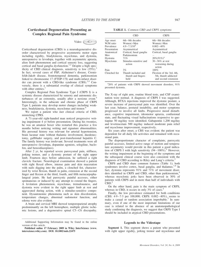
CRPS and CBD share common features (Table 1); bothsyndromes involve cortex, basal ganglia, and thalamus.1,6 Infact, dystonia represents 91% and 71% of movement disor-ders identified in CRPS and CBD, other than parkinsonism,4
whereas myoclonic jerks have been observed in 30% ofpatients with CRPS and in more than half of individuals withCBD.2
On the other hand, pain is the main symptom of CRPS,whereas in CBD, it occurs in only 3% of cases.4
Finally, the low prevalence estimated for both conditions(CBS: 4�9�7�3 per 100,000; CRPS: 0.002�40%), seems tomake a casual or random association improbable.7 In sum-mary, even if one of the most important limitations of ourcase is related to the absence of an anatomopathologicalstudy confirming the diagnosis, we suggest that CRPS Type Ishould be included in atypical CBD presentations.
Legends to the Videotape
Segment 1. This segment shows a patient who presentedwith right upper rigidity, jerking tremor and myoclonus and
Additional Supporting Information may be found in the online
version of this article.Published online 17 February 2009 in Wiley InterScience (www.
interscience.wiley.com). DOI: 10.1002/mds.22475
TABLE 1. Common CBD and CRPS symptoms
CBD CRPS
Age onset 6th�8th decades 6th�8th decadesW/M ratio Women>Men Women>MenPrevalence 4.9�7.3/105 0.002�40%Presentation Asymmetrical AsymmetricalAnatomical Cortical, basal ganglia Cortical, basal gangliaMov Parkinsonism �Dystonia 71% 20%(91%)a
Myoclonus Stimulus-sensitive andaction
30�36% at restworsening duringaction
Pain 3% 100%Clenched fist Thumb included and
thumb and fingersheld
Flextion of the 3rd, 4th,5th, thumb adductedand second extension
a20% of patients with CRPS showed movement disorders, 91%presented dystonia.
947LETTERS TO THE EDITOR
Movement Disorders, Vol. 24, No. 6, 2009

dystonia. He complained of pain and difficulty in using hisright hand; the intention to open his hand or extend his arminduced pain and myoclonus. A clenched right fist is observedwith changes in blood flow and edema. Levitation phenomenawas present. The same patient after botulinum toxin A injec-tions with moderate improvement in the hand posture.
Segment 2. One year after previous examination. Thevideo shows upper limb flexor posture, myoclonus, andapraxia in lower limb foot tapping.
Segment 3. The patient today, 6 year after onset of symp-toms.
Emilia M. Gatto, MD*Area de Enfermedad de Parkinson y Trastornos del
Movimiento Instituto Neurociencias de Buenos Aires, INEBA*E-mail: [email protected] or
Nelida S. Garretto, MDArea de Trastornos del Movimiento Division Neurologıa
Hospital Ramos Mejıa
Jose L. Etcheverry, MDArea de Enfermedad de Parkinson y Trastornos del
Movimiento Instituto Neurociencias de Buenos Aires, INEBA
Gabriel G. Persi, MDDepartamento de Neurologıa. Sanatorio de la
Trinidad Mitre
Virginia L. Parisi, MDDepartamento de Neurologıa. Sanatorio de la
Trinidad Mitre
Oscar Gershanik, MDInstituto de Neurociencias, Fundacion Favoloro University
Hospital, Ciudad Autonoma de Buenos Aires (CABA)
References
1. Wadia PM, Lang AE. The many faces of corticobasal degenera-tion. Parkinsonism Relat Disord 2007;3:S336�S340.
2. Mahapatra RK, Edwards MJ, Schott JM, Bhatia KP. Corticobasaldegeneration. Lancet Neurol 2004;3:736�743.
3. Litvan I, Bhatia KP, Burn DJ, et al. Movement Disorders SocietyScientific Issues Committee. Movement Disorders Society Scien-tific Issues Committee report: SIC Task Force appraisal of clinicaldiagnostic criteria for Parkinsonian disorders I. SCI Task force.Mov Disord 2003;18:467�484.
4. Kompoliti K, Goetz CG, Boeve BF, et al. Clinical presentationand pharmacological therapy in corticobasal degeneration. ArchNeurol 1998;55:957�961.
5. Perez RS, Collins S, Marinus J, Zuurmond WW, de Lange JJ.Diagnostic criteria for CRPS I: Differences between patient pro-files using three different diagnostic sets Eur J Pain 2007;11:895�902.
6. Schott GD. Complex regional pain syndrome. Pract Neurol 2007;7:145�157.
7. de Mos M, de Bruijn AG, Huygen FJ, Dieleman JP, Stricker BH,Sturkenboom MC. The incidence of complex regional pain syn-drome: a population-based study Pain 2007;129:12�20.
Movement Disorders, Vol. 24, No. 6, 2009
948 LETTERS TO THE EDITOR
Copyright © 2022 FDOKUMEN