GCH1, BH4 and Pain
14
1728 Current Pharmaceutical Biotechnology, 2011, 12, 1728-1741 1389-2010/11 $58.00+.00 © 2011 Bentham Science Publishers GCH1, BH4 and Pain Alban Latremoliere* and Michael Costigan* F.M. Kirby Neurobiology Center, Children’s Hospital Boston, Harvard Medical School, 3 Blackfan Circle, CLS 12260, Boston, MA 02115, USA Abstract: Understanding and consequently treating neuropathic pain effectively is a challenge for modern medicine, as unlike inflammation, which can be controlled relatively well, chronic pain due to nerve injury is refractory to most current therapeutics. Here we define a target pathway for a new class of analgesics, tetrahydrobiopterin (BH4) synthesis and me- tabolism. BH4 is an essential co-factor in the synthesis of serotonin, dopamine, epinephrine, norepinephrine and nitric ox- ide and as a result, its availability influences many systems, including neurons. Following peripheral nerve damage, levels of BH4 are dramatically increased in sensory neurons, consequently this has a profound effect on the physiology of these cells, causing increased activity and pain hypersensitivity. These changes are principally due to the upregulation of the rate limiting enzyme for BH4 synthesis GTP Cyclohydrolase 1 (GCH1). A GCH1 pain-protective haplotype which de- creases pain levels in a variety of settings, by reducing the levels of endogenous activation of this enzyme, has been char- acterized in humans. Here we define the control of BH4 homeostasis and discuss the consequences of large perturbations within this system, both negatively via genetic mutations and after pathological increases in the production of this cofactor that result in chronic pain. We explain the nature of the GCH1 reduced-function haplotype and set out the potential for a ‘BH4 blocking’ drug as a novel analgesic. Keywords: Genetic association, dorsal root ganglia, SPR, NO, dorsal horn. INTRODUCTION Lesions to the somatosensory system can lead to patho- logical ongoing sensations that can be both negative (sensory loss) and positive (exaggerated pain) and often persist in- definitely [1]. Painful neuropathic symptoms include: ongo- ing pain (burning sensation), spontaneous pain (bursts of pain) and intense pain in response to previously non-painful (allodynia) or noxious stimuli (hyperalgesia). Currently, commonly prescribed medications for such symptoms in- clude tricyclic antidepressants (e.g. nortriptyline), anticon- vulsants (e.g. gabapentin) and opioids (e.g. morphine). Un- fortunately, these treatments display only moderate efficacy and numerous side effects with some of these, like depend- ency, a particular worry with respect to chronic pain control [2]. Several reasons account for these insufficiencies: first, neuropathic pain is the result of multiple etiologies (me- chanical trauma, metabolic diseases, neurotoxic agents, in- fection or tumor invasion) each with distinct pathophysi- ological mechanisms, complicating its understanding [3]; second, many current pain treatments such as NSAIDs (Non- Steroidal Anti-Inflammatory Drugs) target inflammation as opposed to neuropathic mechanisms which we now realize are different. Third, adequate diagnosis of patients has been hampered by a focus on the precipitating etiologies, relative to the chronic pain itself leading to a lack of diagnostic tools for correctly discriminating pain phenotypes [4-7]. Thus, there is a grave need for novel effective therapeutic ap- proaches to overcome this devastating syndrome. *Address correspondence to these authors at the F.M. Kirby Neurobiology Center, Children’s Hospital Boston, Harvard Medical School, 3 Blackfan Circle, CLS 12260, Boston, MA 02115, USA; Tel: (617) 919-2310; Fax: (617) 919-2772; E-mail: [email protected], [email protected] In the last few decades several new analgesics have been developed, but few have been rationally designed from target identification through drug development to treatment proto- col, consequently most recent successes have been serendipi- tous [8]. Finding new analgesic targets scientifically requires non-biased techniques to define chronic pain mechanisms, one such strategy is whole genome expression profiling us- ing high-density microarrays [9, 10]. We have used such methods to define functional pathways which comprise sev- eral transcripts regulated in concert [11, 12]. Identified both by expression profiling in rodent models of neuropathic pain and by single nucleotide polymorphism (SNP) association studies in humans, GTP cyclohydroxylase 1 (GCH1), the rate-limiting enzyme responsible for the syn- thesis of the pteridine (6R)-L-erythro-5,6,7,8-tetrahydrobio- pterin (BH4), represents a promising new target for the de- velopment of a novel anti-neuropathic treatment [12-14]. Here, we describe why the BH4 synthesis pathway is rele- vant to chronic pain pathophysiology, notably after nerve lesions, and how its modulation could represent a potential new therapeutic approach. TETRAHYDROBIOPTERIN (BH4) BH4 is an essential cofactor for aromatic amino-acid hydroxylases such as the phenylalanine hydroxylase (PAH; [15-17]), tyrosine hydroxylase (TH; [18]) and tryptophan hydroxylases 1 and 2 (TPH; [19, 20]) as well as for all three isoforms of the nitric oxide synthase (NOS; [21-23]), making it critical for numerous biological systems, including pain Fig. (1). Whereas phenylalanine metabolism is prominent in the liver [24] and kidney [25] it is less relevant in the brain [25, 26] and is not known to play a direct role in pain physi-
-
Upload
hms-harvard -
Category
Documents
-
view
1 -
download
0
Transcript of GCH1, BH4 and Pain

1728 Current Pharmaceutical Biotechnology, 2011, 12, 1728-1741
1389-2010/11 $58.00+.00 © 2011 Bentham Science Publishers
GCH1, BH4 and Pain
Alban Latremoliere* and Michael Costigan*
F.M. Kirby Neurobiology Center, Children’s Hospital Boston, Harvard Medical School, 3 Blackfan Circle, CLS 12260,
Boston, MA 02115, USA
Abstract: Understanding and consequently treating neuropathic pain effectively is a challenge for modern medicine, as
unlike inflammation, which can be controlled relatively well, chronic pain due to nerve injury is refractory to most current
therapeutics. Here we define a target pathway for a new class of analgesics, tetrahydrobiopterin (BH4) synthesis and me-
tabolism. BH4 is an essential co-factor in the synthesis of serotonin, dopamine, epinephrine, norepinephrine and nitric ox-
ide and as a result, its availability influences many systems, including neurons. Following peripheral nerve damage, levels
of BH4 are dramatically increased in sensory neurons, consequently this has a profound effect on the physiology of these
cells, causing increased activity and pain hypersensitivity. These changes are principally due to the upregulation of the
rate limiting enzyme for BH4 synthesis GTP Cyclohydrolase 1 (GCH1). A GCH1 pain-protective haplotype which de-
creases pain levels in a variety of settings, by reducing the levels of endogenous activation of this enzyme, has been char-
acterized in humans. Here we define the control of BH4 homeostasis and discuss the consequences of large perturbations
within this system, both negatively via genetic mutations and after pathological increases in the production of this cofactor
that result in chronic pain. We explain the nature of the GCH1 reduced-function haplotype and set out the potential for a
‘BH4 blocking’ drug as a novel analgesic.
Keywords: Genetic association, dorsal root ganglia, SPR, NO, dorsal horn.
INTRODUCTION
Lesions to the somatosensory system can lead to patho-logical ongoing sensations that can be both negative (sensory loss) and positive (exaggerated pain) and often persist in-definitely [1]. Painful neuropathic symptoms include: ongo-ing pain (burning sensation), spontaneous pain (bursts of pain) and intense pain in response to previously non-painful (allodynia) or noxious stimuli (hyperalgesia). Currently, commonly prescribed medications for such symptoms in-clude tricyclic antidepressants (e.g. nortriptyline), anticon-vulsants (e.g. gabapentin) and opioids (e.g. morphine). Un-fortunately, these treatments display only moderate efficacy and numerous side effects with some of these, like depend-ency, a particular worry with respect to chronic pain control [2]. Several reasons account for these insufficiencies: first, neuropathic pain is the result of multiple etiologies (me-chanical trauma, metabolic diseases, neurotoxic agents, in-fection or tumor invasion) each with distinct pathophysi-ological mechanisms, complicating its understanding [3]; second, many current pain treatments such as NSAIDs (Non-Steroidal Anti-Inflammatory Drugs) target inflammation as opposed to neuropathic mechanisms which we now realize are different. Third, adequate diagnosis of patients has been hampered by a focus on the precipitating etiologies, relative to the chronic pain itself leading to a lack of diagnostic tools for correctly discriminating pain phenotypes [4-7]. Thus, there is a grave need for novel effective therapeutic ap-proaches to overcome this devastating syndrome.
*Address correspondence to these authors at the F.M. Kirby Neurobiology Center, Children’s Hospital Boston, Harvard Medical School, 3 Blackfan Circle, CLS 12260, Boston, MA 02115, USA; Tel: (617) 919-2310; Fax: (617) 919-2772; E-mail: [email protected], [email protected]
In the last few decades several new analgesics have been developed, but few have been rationally designed from target identification through drug development to treatment proto-col, consequently most recent successes have been serendipi-tous [8]. Finding new analgesic targets scientifically requires non-biased techniques to define chronic pain mechanisms, one such strategy is whole genome expression profiling us-ing high-density microarrays [9, 10]. We have used such methods to define functional pathways which comprise sev-eral transcripts regulated in concert [11, 12].
Identified both by expression profiling in rodent models of neuropathic pain and by single nucleotide polymorphism (SNP) association studies in humans, GTP cyclohydroxylase 1 (GCH1), the rate-limiting enzyme responsible for the syn-thesis of the pteridine (6R)-L-erythro-5,6,7,8-tetrahydrobio-pterin (BH4), represents a promising new target for the de-velopment of a novel anti-neuropathic treatment [12-14]. Here, we describe why the BH4 synthesis pathway is rele-vant to chronic pain pathophysiology, notably after nerve lesions, and how its modulation could represent a potential new therapeutic approach.
TETRAHYDROBIOPTERIN (BH4)
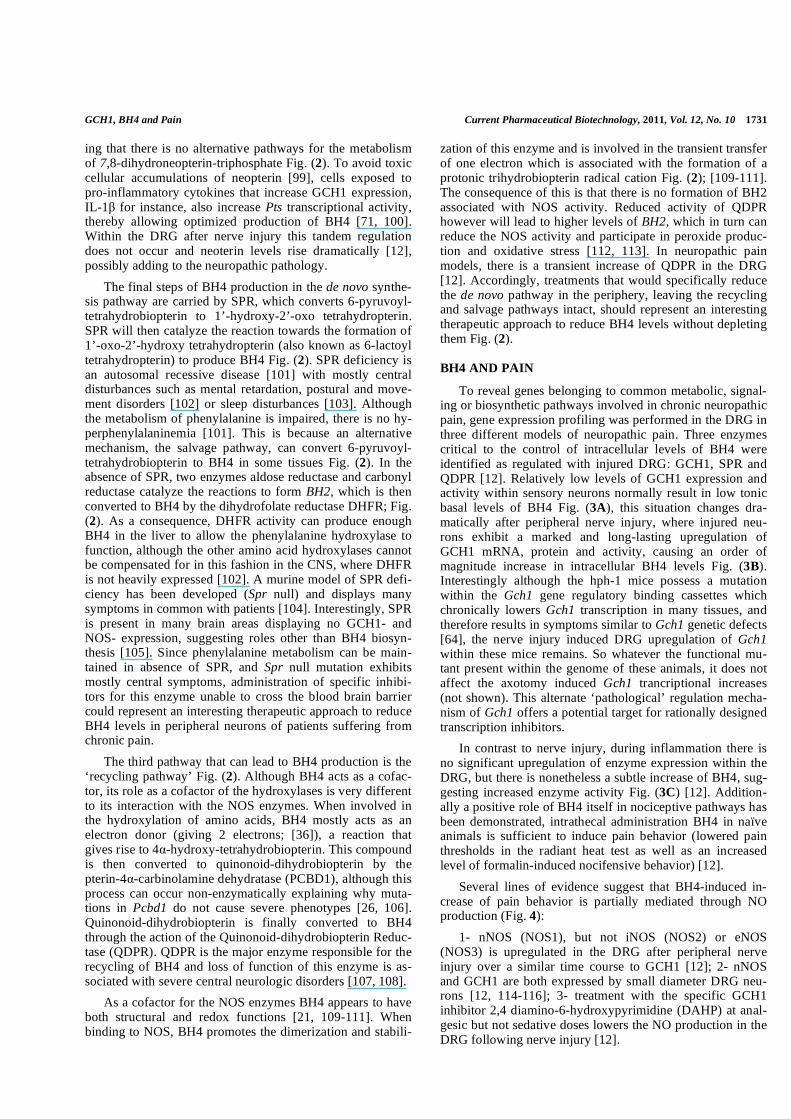
BH4 is an essential cofactor for aromatic amino-acid hydroxylases such as the phenylalanine hydroxylase (PAH; [15-17]), tyrosine hydroxylase (TH; [18]) and tryptophan hydroxylases 1 and 2 (TPH; [19, 20]) as well as for all three isoforms of the nitric oxide synthase (NOS; [21-23]), making it critical for numerous biological systems, including pain Fig. (1). Whereas phenylalanine metabolism is prominent in the liver [24] and kidney [25] it is less relevant in the brain [25, 26] and is not known to play a direct role in pain physi-

GCH1, BH4 and Pain Current Pharmaceutical Biotechnology, 2011, Vol. 12, No. 10 1729
ology. In contrast there is extensive evidence for the roles of neuronal serotonin (5-HT), dopamine (DA), norepinephrine (NE) and nitric oxide (NO) in pain signaling [27-32]. Under normal conditions, the metabolism of BH4 is tightly regu-lated to maintain an adequate homeostatic balance. This fine regulation of intracellular BH4 concentration can be im-paired in several multifactorial chronic diseases such as dia-betes or atherosclerosis [33], or even completely lost in cer-tain monogenic diseases [26]. Inherited BH4 deficiencies are caused by mutations in the enzymes of BH4 metabolism and often lead to low levels of catecholamine and 5-HT and in some cases high phenylalanine concentrations (hyperpheny-lalaninemia or phenylketonuria; PKU)(reviewed in [34]). Whereas PKU is generally caused by mutations in the Pah gene itself [35], low levels of BH4 also lead to altered activ-ity of PAH, thereby preventing effective conversion of phen-ylalanine to tyrosine and therefore resulting in high levels of phenylalanine, a condition called atypical phenylketonuria [34, 36]. Insufficient levels of 5-HT, NE and DA, and too much phenylalanine within the brain during development can lead to cognitive impairments (mental retardation; [26, 37]) and motor disorders (dystonia and tremors; [38, 39]). Inter-estingly, both PKU and atypical PKU can be treated effec-tively with adequate supplements (BH4, L-dopa and 5-hydroxytryptophan) together with a low diet in phenyla-lanine if diagnosed early enough [26].
Fig. (1). Tetrahydrobiopterin (BH4) is an essential cofactor all three
hydroxylases and the three NOS isoforms. The metabolism of
phenylalanine and the synthesis of serotonin, dopamine, epineph-
rine, norepinephrine and NO, are reliant on adequate cellular levels
of BH4.
Both diabetes and atherosclerosis are associated with chronic oxidative stress [40-43] which leads to oxidation of BH4 to dihydrobiopterin (BH2; [33, 44]). BH2 and BH4 are structurally alike and have similar affinities for the pterin binding site of endothelial NOS (eNOS) [45], enabling them to compete for enzyme binding [46]. Whereas eNOS cou-pling with BH4 results in appropriate formation of NO [47, 48], BH2 binding prevents adequate redox reactions by NOS, which instead produces peroxide [49-52] and this in turn worsens the oxidative stress. An interesting conse-quence of these similar binding affinities is that it is the ratio of BH4/BH2, but not the absolute levels of BH4, that ap-pears critical for a proper functioning of the eNOS enzyme [48].
BH4 PRODUCTION AND SALVAGE
Intracellular levels of BH4 are determined by three mechanisms: the de novo synthesis pathway, the recycling pathway and the salvage pathway Fig. (2). In the following paragraphs we will describe these processes, the factors known to regulate their activity and the pathologies associ-ated with their deficiencies.
The de novo BH4 synthesis pathway is characterized by biochemical reactions carried out by three specific enzymes: GCH1 [53], 6-pyruvoyl tetrahydrobiopterin synthase (PTS) [54] and sepiapterin reductase (SPR) [55, 56] Fig. (2). The initial reaction is the conversion of GTP to 7,8-dihydroneopterin-triphosphate by GCH1. GCH1 is a ho-modecamer [57] formed by the association of two pentamers which face each other, each presenting a catalytic pocket at their center [58, 59]. Alterations in the conformation of these pockets, by mutant monomers, can be associated with a dras-tic loss of GCH1 activity resulting in Segawa’s disease (dopa-responsive dystonia; [60]), a dominant form of GCH1 loss of function, which is characterized by postural disorders (dystonia and tremors) due to a selective DA deficiency [61]. The hyperphenylalaninemia (hph-1) mouse mutant line [62] was produced by random mutagenesis leading to an as yet unidentified alteration in the transcriptional control machin-ery of the Gch1 gene [33, 60, 63]. Phenotypically these mice exhibit high neonatal levels of serum phenylalanine [62], extremely low cerebrospinal fluid levels of BH4, low levels of tyrosine hydroxylase protein in the striatum [64] and changes in the DAergic system [65].
GCH1, as the rate-limiting enzyme for BH4 biosynthesis, has its function tightly regulated by several products of BH4 metabolism, enzyme activation is increased by excess con-centrations of phenylalanine [66, 67] and reduced by higher levels of BH4 or low levels of GTP [66]. In order to modu-late function, these two metabolites require the formation of a specific complex between GCH1 and GCH1-feedback regulatory protein (GFRP, also known as p35) [66]. In pres-ence of BH4, one GFRP pentamer binds each GCH1 pen-tamer [67] thus forming the [GFRP-GCH1] co-decamer in-hibition complex [66]. BH4 mediated inhibition can be by-passed in the presence of high concentrations of phenyla-lanine, which changes the inactive [GFRP-GCH1] complex into an active one, thereby preventing toxic accumulations of this amino acid, even at low concentrations of GTP [66]. In addition the [GFRP-GCH1] complex is required for the in-hibitory effects of DAHP [68]. Finally although GFRP is required for BH4-induced negative feedback and the pheny-lalanine-induced positive feedback on GCH1 activity [66] GFRP levels do not appear to be regulated by BH4 [69], fur-ther confirming that GCH1 levels alone determine the intra-cellular concentration of BH4.
GCH1 transcription is stimulated by the cytokines inter-feron gamma (IFN ) and tumor necrosis alpha (TNF ) [70] and at a lesser extent interleukin 1 (IL-1 ; [71]), lipopoly-saccharide [72], nerve growth factor (NGF; [73]) and perox-ide (H2O2; [74, 75]), probably through the activation of the Jak2 tyrosine kinase pathway [76] Fig. (2). GCH1 expression is also under the control of estrogens [77], possibly through a NO-mediated activation of CREB [78, 79]. Interestingly, cAMP is a strong activator of GCH1 expression [80] and
1730 Current Pharmaceutical Biotechnology, 2011, Vol. 12, No. 10 Latremoliere and Costigan
cAMP upregulation is also a central component of the axotomy response in dorsal root ganglion (DRG) neurons [81, 82]. Overall, these results indicate that several inflam-matory mediators are associated with an increase in activity of the BH4 de novo synthesis pathway and support the ob-servations that patients suffering from various chronic in-flammatory diseases (syphilis and lupus for example) exhibit high levels of BH4 in their cerebrospinal fluid [83]. In terms of reducing GCH1 transcriptional activity, melatonin [84], ciliary neurotrophic factor (CNTF) and leukocyte inhibitory factor (LIF) [85] have been identified as factors that reduce expression Fig. (2). In peripheral neurons, melatonin has been shown to reduce voltage dependent calcium (Ca
2+) en-
try [86] which normally increases following peripheral nerve injury. The two trophic factors CNTF and LIF are upregu-lated after axotomy [87, 88] although it is unclear if these processes are linked to GCH1 regulation within the DRG.
GCH1 protein displays several potential phosphorylation sites, either for positive (Ser51, Ser72, Tyr85, Tyr91, Tyr103 or Ser130) or negative (Tyr231) regulation [89]. So far, only PKC [90] and casein kinase 2 [53] have been shown to phos-phorylate GCH1, thereby increasing its activity [91]. Im-munoprecipitation experiments have confirmed an interac-tion between GCH1 and PKC or casein kinase 2, and have
suggested direct interactions with PKA, Ca2+
/calmodulin-dependent protein kinase (CaMK) and several members of the mitogen-activated protein kinase (MAPK) family [92, 93]. Other proteins have been identified to interact with GCH1, including the Activator of heat shock protein 90 (Aha1) which may recruit GCH1 into the NOS/Hsp90 com-plex to allow local synthesis of BH4 for NOS, with this yeast 2-hybrid screen also identifying the Cannabinoid receptor CB1 interacting protein 1 (CB1-IP1) and the 1 subunit of Na/K ATPase membrane transporter [94]. In addition, two transcription factors were isolated suggesting a role for GCH1 in transcription control [94] a task further supported by the presence of GCH1 protein in the nucleus [95]. Other studies have shown GCH1 can interact with another target enzyme tyrosine hydroxylase [96] as well a host of signaling molecules and intracellular proteins [89].
GCH1’s end product (7,8-dihydroneopterin-triphosphate) is converted by PTS to 6-pyruvoyl-tetrahydrobiopterin [54]. Loss of function mutations in the Pts gene are the cause of approximately 60% of atypical phenylketonuria [26] and are associated with severe defects in brain synthesis of 5-HT, DA and NE [26, 34, 97]. Homozygous knock-out of Pts in mice is fatal within 48 hours of birth and is associated with accumulation of neopterin in the brain and liver [98], indicat-
Fig. (2). Intracellular levels of BH4 depend on three production conduits the de novo synthesis pathway, the recycling pathway and the sal-
vage pathway. BH4 biosynthesis is mediated through the de novo pathway which can be modulated by numerous inflammatory signals, nota-
bly through direct regulation of the rate-limiting enzyme GCH1. The recycling pathway allows the recreation of BH4 from the BH2 dis-
pensed by the hydroxylases and is essential to maintain the proper ratio of BH4/BH2 levels. Precise physiological roles for the salvage path-
way are not fully characterized, but it acts to keep BH4 levels stable in a similar way to the recycling pathway. Known mutations for key
enzymes of the metabolism of BH4 are indicated by an asterisk.
GCH1, BH4 and Pain Current Pharmaceutical Biotechnology, 2011, Vol. 12, No. 10 1731
ing that there is no alternative pathways for the metabolism of 7,8-dihydroneopterin-triphosphate Fig. (2). To avoid toxic cellular accumulations of neopterin [99], cells exposed to pro-inflammatory cytokines that increase GCH1 expression, IL-1 for instance, also increase Pts transcriptional activity, thereby allowing optimized production of BH4 [71, 100]. Within the DRG after nerve injury this tandem regulation does not occur and neoterin levels rise dramatically [12], possibly adding to the neuropathic pathology.
The final steps of BH4 production in the de novo synthe-sis pathway are carried by SPR, which converts 6-pyruvoyl-tetrahydrobiopterin to 1’-hydroxy-2’-oxo tetrahydropterin. SPR will then catalyze the reaction towards the formation of 1’-oxo-2’-hydroxy tetrahydropterin (also known as 6-lactoyl tetrahydropterin) to produce BH4 Fig. (2). SPR deficiency is an autosomal recessive disease [101] with mostly central disturbances such as mental retardation, postural and move-ment disorders [102] or sleep disturbances [103]. Although the metabolism of phenylalanine is impaired, there is no hy-perphenylalaninemia [101]. This is because an alternative mechanism, the salvage pathway, can convert 6-pyruvoyl-tetrahydrobiopterin to BH4 in some tissues Fig. (2). In the absence of SPR, two enzymes aldose reductase and carbonyl reductase catalyze the reactions to form BH2, which is then converted to BH4 by the dihydrofolate reductase DHFR; Fig. (2). As a consequence, DHFR activity can produce enough BH4 in the liver to allow the phenylalanine hydroxylase to function, although the other amino acid hydroxylases cannot be compensated for in this fashion in the CNS, where DHFR is not heavily expressed [102]. A murine model of SPR defi-ciency has been developed (Spr null) and displays many symptoms in common with patients [104]. Interestingly, SPR is present in many brain areas displaying no GCH1- and NOS- expression, suggesting roles other than BH4 biosyn-thesis [105]. Since phenylalanine metabolism can be main-tained in absence of SPR, and Spr null mutation exhibits mostly central symptoms, administration of specific inhibi-tors for this enzyme unable to cross the blood brain barrier could represent an interesting therapeutic approach to reduce BH4 levels in peripheral neurons of patients suffering from chronic pain.
The third pathway that can lead to BH4 production is the ‘recycling pathway’ Fig. (2). Although BH4 acts as a cofac-tor, its role as a cofactor of the hydroxylases is very different to its interaction with the NOS enzymes. When involved in the hydroxylation of amino acids, BH4 mostly acts as an electron donor (giving 2 electrons; [36]), a reaction that gives rise to 4 -hydroxy-tetrahydrobiopterin. This compound is then converted to quinonoid-dihydrobiopterin by the pterin-4 -carbinolamine dehydratase (PCBD1), although this process can occur non-enzymatically explaining why muta-tions in Pcbd1 do not cause severe phenotypes [26, 106]. Quinonoid-dihydrobiopterin is finally converted to BH4 through the action of the Quinonoid-dihydrobiopterin Reduc-tase (QDPR). QDPR is the major enzyme responsible for the recycling of BH4 and loss of function of this enzyme is as-sociated with severe central neurologic disorders [107, 108].
As a cofactor for the NOS enzymes BH4 appears to have both structural and redox functions [21, 109-111]. When binding to NOS, BH4 promotes the dimerization and stabili-
zation of this enzyme and is involved in the transient transfer of one electron which is associated with the formation of a protonic trihydrobiopterin radical cation Fig. (2); [109-111]. The consequence of this is that there is no formation of BH2 associated with NOS activity. Reduced activity of QDPR however will lead to higher levels of BH2, which in turn can reduce the NOS activity and participate in peroxide produc-tion and oxidative stress [112, 113]. In neuropathic pain models, there is a transient increase of QDPR in the DRG [12]. Accordingly, treatments that would specifically reduce the de novo pathway in the periphery, leaving the recycling and salvage pathways intact, should represent an interesting therapeutic approach to reduce BH4 levels without depleting them Fig. (2).
BH4 AND PAIN
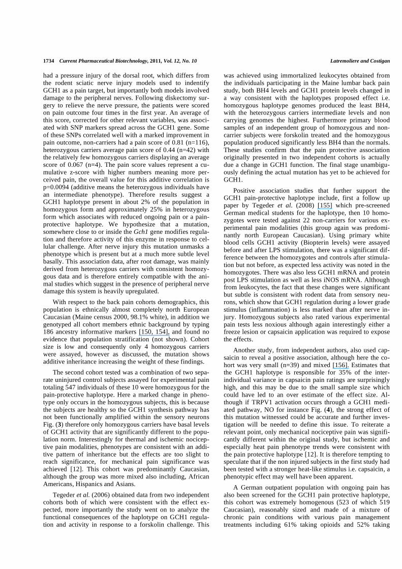
To reveal genes belonging to common metabolic, signal-ing or biosynthetic pathways involved in chronic neuropathic pain, gene expression profiling was performed in the DRG in three different models of neuropathic pain. Three enzymes critical to the control of intracellular levels of BH4 were identified as regulated with injured DRG: GCH1, SPR and QDPR [12]. Relatively low levels of GCH1 expression and activity within sensory neurons normally result in low tonic basal levels of BH4 Fig. (3A), this situation changes dra-matically after peripheral nerve injury, where injured neu-rons exhibit a marked and long-lasting upregulation of GCH1 mRNA, protein and activity, causing an order of magnitude increase in intracellular BH4 levels Fig. (3B). Interestingly although the hph-1 mice possess a mutation within the Gch1 gene regulatory binding cassettes which chronically lowers Gch1 transcription in many tissues, and therefore results in symptoms similar to Gch1 genetic defects [64], the nerve injury induced DRG upregulation of Gch1 within these mice remains. So whatever the functional mu-tant present within the genome of these animals, it does not affect the axotomy induced Gch1 trancriptional increases (not shown). This alternate ‘pathological’ regulation mecha-nism of Gch1 offers a potential target for rationally designed transcription inhibitors.
In contrast to nerve injury, during inflammation there is no significant upregulation of enzyme expression within the DRG, but there is nonetheless a subtle increase of BH4, sug-gesting increased enzyme activity Fig. (3C) [12]. Addition-ally a positive role of BH4 itself in nociceptive pathways has been demonstrated, intrathecal administration BH4 in naïve animals is sufficient to induce pain behavior (lowered pain thresholds in the radiant heat test as well as an increased level of formalin-induced nocifensive behavior) [12].
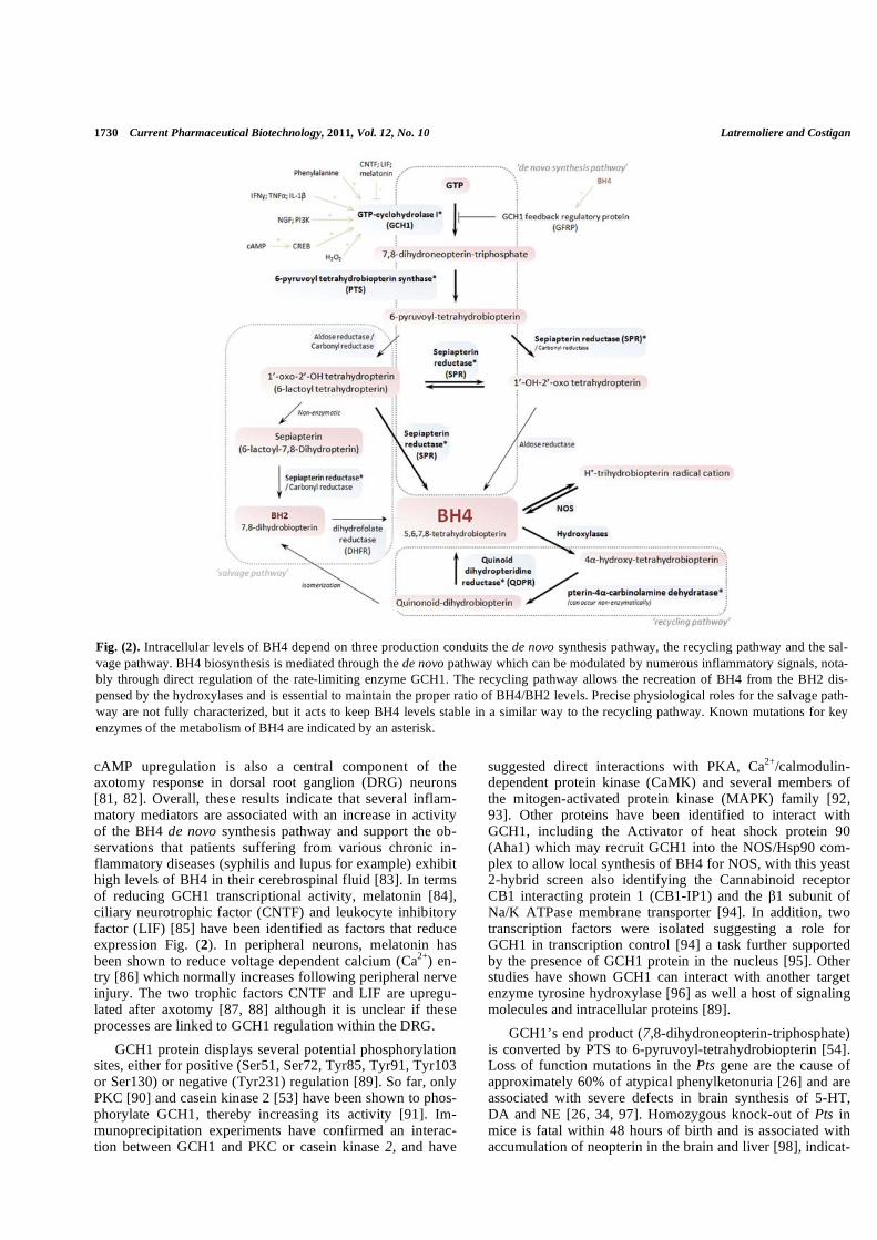
Several lines of evidence suggest that BH4-induced in-crease of pain behavior is partially mediated through NO production (Fig. 4):
1- nNOS (NOS1), but not iNOS (NOS2) or eNOS (NOS3) is upregulated in the DRG after peripheral nerve injury over a similar time course to GCH1 [12]; 2- nNOS and GCH1 are both expressed by small diameter DRG neu-rons [12, 114-116]; 3- treatment with the specific GCH1 inhibitor 2,4 diamino-6-hydroxypyrimidine (DAHP) at anal-gesic but not sedative doses lowers the NO production in the DRG following nerve injury [12].

1732 Current Pharmaceutical Biotechnology, 2011, Vol. 12, No. 10 Latremoliere and Costigan
Small diameter sensory neurons express TRPV1 [117, 118] and TRPA1 [119], two calcium (Ca2+)-permeable ion channels [120] directly activated by NO [121]. Accordingly, it is possible that these two channels carry the NO-dependant Ca2+ influx observed in dissociated DRG neurons cultures incubated with BH4 [12].
Since NOS acts as a homodimer binding two BH4 and two arginine molecules to form one molecule of NO [122], and BH4 binding increases the affinity of NOS for arginine [123], the overexpression of nNOS along with an excess of BH4 creates the ideal conditions for overproduction of NO. Increased NO production would in turn sensitize TRPV1 (expressed by heat-sensitive nociceptors [124]) and TRPA1 (for which formalin is a ligand [125]) channels, possibly explaining the behavioral changes (lower radiant heat threshold and increased pain responses upon intraplantar injection of formalin) following intrathecal administration of BH4 (Fig. 4). In line with these results, administration of the NOS inhibitor L-NAME reduces the pain behaviors caused by peripheral nerve injury [12] as well nocifensive behavior in the formalin test [126].
Finally, a TRPV1/TRPA1 mediated BH4-induced Ca2+ influx activates the phosphatidylinositol-3 kinases (PI3K) pathway [127], which has been shown to play an important role in the hypersensitivity associated with neuropathic pain [128]. PI3K activation is positively associated with GCH1 transcription [129] and may therefore participate in the regu-lation of GCH1 following nerve injury. Finally, since the Ca2+ influx caused by BH4 in DRG neurons is only partially prevented in presence of L-NAME [12], it is possible that BH4 has other cofactor dependant or cofactor independent roles within these cells [130, 131].
Lowering BH4 levels by either inhibiting GCH1 pharma-cologically [12] or genetically [132] or by inhibiting SPR activity [12] alleviates pain behaviors in models of inflam-matory and neuropathic pain Fig. (3D). Importantly, repeated injections or intrathecal infusion of DAHP leads to a robust and long-lasting reduction of BH4 levels in the DRG without altering the behavioral responses in the forced swim test [12], an assay which is highly sensitive to changes in 5-HT and NE mediated neurotransmission in central neurons [133]. In addition AAV-mediated shRNA knockdown of GCH1 specifically in DRG neurons acts as an effective anal-gesic [132]. These results therefore strongly support the hy-pothesis that over-expression of BH4 specifically in DRG neurons is sufficient for the development and the mainte-nance of chronic pain after peripheral nerve injury. Future experiments using transgenic tools to target distinct subpopu-lations of DRG neurons should help us further define these mechanisms. Interestingly, although the analgesic effects of GCH1 or SPR inhibition are due to a reduction of BH4 lev-els within the injured DRGs, this leads to marked changes in the ipsilateral dorsal horn of the spinal cord. These changes include, reduced number of c-fos-positive neurons in the superficial laminae after formalin injection or a reduction of microglial activation in neuropathic pain models Fig. (3D); [12, 132]. It is tempting then to speculate that decreased BH4 levels in the DRG could lead to a reduction of activity in the primary afferent fibers that subsequently prevent the mainte-nance of central sensitization, a phenomenon strongly linked
to activity-dependent inputs [134]. Such a reduction of activ-ity could occur in the periphery, or within pre-synaptic nerve terminals [135] preventing adequate synthesis of NO, an essential factor for “central sensitization” [136]. It is also possible that BH4 is released to act on different cell types in the spinal cord, as it has been proposed in other brain struc-tures [137]. With regard to microglia, it has been clearly es-tablished that the activation and recruitment of these cells is in part adenosine tri-phosphate (ATP) dependent [138], which is co-released with NO [139]. Abnormal primary af-ferent fiber activity following peripheral nerve injury [140] participates in the activation and recruitment of microglia [141]. Once activated, microglia participate in central sensi-tization by increasing the activity of second order nocicep-tive neurons in the dorsal horn [142-144] and altering seg-mental inhibition [145, 146]. Accordingly, a treatment that could lower without depleting the levels of BH4 in the DRG should represent a valuable tool to reduce chronic pain symptoms, and possibly help the central nervous system to return to a non-pathological state Fig. (3D).
ASSOCIATION GENETICS
Single nucleotide polymorphisms are bases that differ in a subgroup people relative to the nucleotide usually present. They are variations in the standard genetic code of that popu-lation. In most cases these changes do little, but in some cases they alter a proteins structure or regulation such that they have a phenotypic effect on the organism, the tangible effects of these mutations can range from barely noticeable to severe. Association genetics tries to link one or more SNPs with a trait, a group of co-inherited SNPs is called a haplotype. It is important to realize that even an extremely well linked SNP is not necessarily the causal mutant and may still just be a flag, which is close to, and therefore co-inherited with the actual mutation.
The genetics literature has been plagued with assign-ments of gene linkage which have failed replication follow-ing the initial study, consequently new associations are often treated with extreme caution by other investigators. While well controlled negative association studies need to be pub-lished and are in many ways as valuable as positive associa-tions, lack of replication cannot be assumed unless the same phenotype is assayed in a population of the same structure [147]. In addition to prove accurate collection of phenotype and genotype data, demonstration of positive associations with genes known to be linked to the condition lends the cohort credence [148]. Due to the lack of clearly linked posi-tive control genes it is currently difficult to assess the quality of negative association data although this situation should improve.
Another issue often discussed is population stratification [149], this describes an errant gene-disease association be-cause a subgroup of individuals, within the sampled popula-tion, share genetic heritage and/or similar environmental determinants. In cases where a trait bearing subgroup are more closely related, they are likely to possess many com-mon SNPs, most of which could be linked to the condition improperly, this is a major source of error and needs to be controlled for. However stratification is usually only relevant when your control and affected populations are different (i.e.

GCH1, BH4 and Pain Current Pharmaceutical Biotechnology, 2011, Vol. 12, No. 10 1733
Fig. (3). The production of BH4 within the DRG plays a critical
role in pain signaling. In basal states (A), the de novo synthesis
pathway exhibits weak activity and BH4 levels are relatively low
but physiologically normal. Here the recycling pathway plays an
important role in maintaining adequate levels of BH4. After periph-
eral nerve injury (B), there is a major upregulation of GCH1
mRNA, protein and consequent enzyme activity and an increase in
SPR and QDPR levels but to a lesser extent, which results in very
high levels of BH4. In chronic inflammatory states (C), the protein
levels of these enzymes are not increased, but there is nonetheless
an overproduction of BH4 in the DRG caused by an increase of
enzyme activity. Therapeutic strategies that block the de novo syn-
thesis pathway but not the recycling pathway (D) in the peripheral
nervous system should allow a reduction of BH4 to non-
pathological levels.
you have a mixed group in the association study or recent population interbreeding has occurred), several ways of con-trolling for this problem have been proposed (including [150, 151]).
Cohort size is another important consideration for well conducted association studies. As many mutations occur at a relatively low rate in the population the number of times the minor allele occurs in homozygous form is often low (in the order of 1 or 2%). This means that there are not a lot of these people present until you have a relatively large initial study group. For instance to obtain 4 homozygous carriers of a minor allele present at 1% you will need around 400 people in your cohort. To cover all of these issues adequately co-horts of at least 500 and preferably in excess of 1000 are suggested, although meaningful data can be achieved with smaller cohorts especially in combination.
DISTINGUISHING GENE ASSOCIATIONS FROM
FALSE POSITIVES
A staged set of controls have been suggested to define a truly associated gene from a false positive associations [152]. First a combination of cohorts should be performed all with consistent genotype-phenotype data to define a potential casual link. Second, confirmation and replication of the as-sociation in independent data sets offers further proof of a true association. Third and most importantly, defining a link between the SNP markers and function of the gene of inter-est allows unambiguous definition of linkage. A further crite-rion is identification and definition of the actual mutation within the gene of interest, to gain these data, re-sequencing of the gene of interest and surrounding control regions is required in multiple case and control genomes, followed by bioinfomatic analysis to define shared mutations that can then be directly tested for function. Up until relatively re-cently re-sequencing candidate genes in detail has not been common practice but with next generation sequencing tech-nologies this position should change.
GCH1 ASSOCIATIONS AND PAIN
Multiple issues need to be considered when discussing the potential link of GCH1 and pain. The first is which phe-notype is to be assayed chronic or acute pain. Primary data obtained from the rat suggests a major role for GCH1 in neu-ropathic pain, following injury induced protein upregulation and the consequent large and sustained increase in enzyme activity within primary sensory neurons Fig. (3B). In addi-tion GCH1 activity but not protein levels are increased in response to inflammation Fig. (3C) and altering the basal level of GCH1 in uninjured sensory neurons allows the pos-sibility of linkage to nociceptive pain Fig. (3A) [12]. Beyond these issues are the modalities of pain that may, or may not be linked, and the involvement of other potentially compli-cating factors for example pain in the presence of cancer or other disease states need to be considered.
In the original study defining the association of a GCH1 pain-protective haplotype two cohorts of individuals were tested [12]. The first the Maine lumber back pain cohort was a prospective group of back pain patients who underwent diskectomy surgery to remove a slipped disk which was im-pinging on their dorsal roots [153]. The patients therefore
1734 Current Pharmaceutical Biotechnology, 2011, Vol. 12, No. 10 Latremoliere and Costigan
had a pressure injury of the dorsal root, which differs from the rodent sciatic nerve injury models used to indentify GCH1 as a pain target, but importantly both models involved damage to the peripheral nerves. Following diskectomy sur-gery to relieve the nerve pressure, the patients were scored on pain outcome four times in the first year. An average of this score, corrected for other relevant variables, was associ-ated with SNP markers spread across the GCH1 gene. Some of these SNPs correlated well with a marked improvement in pain outcome, non-carriers had a pain score of 0.81 (n=116), heterozygous carriers average pain score of 0.44 (n=42) with the relatively few homozygous carriers displaying an average score of 0.067 (n=4). The pain score values represent a cu-mulative z-score with higher numbers meaning more per-ceived pain, the overall value for this additive correlation is p=0.0094 (additive means the heterozygous individuals have an intermediate phenotype). Therefore results suggest a GCH1 haplotype present in about 2% of the population in homozygous form and approximately 25% in heterozygous form which associates with reduced ongoing pain or a pain-protective haplotype. We hypothesize that a mutation, somewhere close to or inside the Gch1 gene modifies regula-tion and therefore activity of this enzyme in response to cel-lular challenge. After nerve injury this mutation unmasks a phenotype which is present but at a much more subtle level basally. This association data, after root damage, was mainly derived from heterozygous carriers with consistent homozy-gous data and is therefore entirely compatible with the ani-mal studies which suggest in the presence of peripheral nerve damage this system is heavily upregulated.
With respect to the back pain cohorts demographics, this population is ethnically almost completely north European Caucasian (Maine census 2000, 98.1% white), in addition we genotyped all cohort members ethnic background by typing 186 ancestry informative markers [150, 154], and found no evidence that population stratification (not shown). Cohort size is low and consequently only 4 homozygous carriers were assayed, however as discussed, the mutation shows additive inheritance increasing the weight of these findings.
The second cohort tested was a combination of two sepa-rate uninjured control subjects assayed for experimental pain totaling 547 individuals of these 10 were homozygous for the pain-protective haplotype. Here a marked change in pheno-type only occurs in the homozygous subjects, this is because the subjects are healthy so the GCH1 synthesis pathway has not been functionally amplified within the sensory neurons Fig. (3) therefore only homozygous carriers have basal levels of GCH1 activity that are significantly different to the popu-lation norm. Interestingly for thermal and ischemic nocicep-tive pain modalities, phenotypes are consistent with an addi-tive pattern of inheritance but the effects are too slight to reach significance, for mechanical pain significance was achieved [12]. This cohort was predominantly Caucasian, although the group was more mixed also including, African Americans, Hispanics and Asians.
Tegeder et al. (2006) obtained data from two independent cohorts both of which were consistent with the effect ex-pected, more importantly the study went on to analyze the functional consequences of the haplotype on GCH1 regula-tion and activity in response to a forskolin challenge. This
was achieved using immortalized leukocytes obtained from the individuals participating in the Maine lumbar back pain study, both BH4 levels and GCH1 protein levels changed in a way consistent with the haplotypes proposed effect i.e. homozygous haplotype genomes produced the least BH4, with the heterozygous carriers intermediate levels and non carrying genomes the highest. Furthermore primary blood samples of an independent group of homozygous and non-carrier subjects were forskolin treated and the homozygous population produced significantly less BH4 than the normals. These studies confirm that the pain protective association originally presented in two independent cohorts is actually due a change in GCH1 function. The final stage unambigu-ously defining the actual mutation has yet to be achieved for GCH1.
Positive association studies that further support the GCH1 pain-protective haplotype include, first a follow up paper by Tegeder et al. (2008) [155] which pre-screened German medical students for the haplotype, then 10 homo-zygotes were tested against 22 non-carriers for various ex-perimental pain modalities (this group again was predomi-nantly north European Caucasian). Using primary white blood cells GCH1 activity (Biopterin levels) were assayed before and after LPS stimulation, there was a significant dif-ference between the homozygotes and controls after stimula-tion but not before, as expected less activity was noted in the homozygotes. There was also less GCH1 mRNA and protein post LPS stimulation as well as less iNOS mRNA. Although from leukocytes, the fact that these changes were significant but subtle is consistent with rodent data from sensory neu-rons, which show that GCH1 regulation during a lower grade stimulus (inflammation) is less marked than after nerve in-jury. Homozygous subjects also rated various experimental pain tests less noxious although again interestingly either a freeze lesion or capsaicin application was required to expose the effects.
Another study, from independent authors, also used cap-saicin to reveal a positive association, although here the co-hort was very small (n=39) and mixed [156]. Estimates that the GCH1 haplotype is responsible for 35% of the inter-individual variance in capsaicin pain ratings are surprisingly high, and this may be due to the small sample size which could have led to an over estimate of the effect size. Al-though if TRPV1 activation occurs through a GCH1 medi-ated pathway, NO for instance Fig. (4), the strong effect of this mutation witnessed could be accurate and further inves-tigation will be needed to define this issue. To reiterate a relevant point, only mechanical nociceptive pain was signifi-cantly different within the original study, but ischemic and especially heat pain phenotype trends were consistent with the pain protective haplotype [12]. It is therefore tempting to speculate that if the non injured subjects in the first study had been tested with a stronger heat-like stimulus i.e. capsaicin, a phenotypic effect may well have been apparent.
A German outpatient population with ongoing pain has also been screened for the GCH1 pain protective haplotype, this cohort was extremely homogenous (523 of which 519 Caucasian), reasonably sized and made of a mixture of chronic pain conditions with various pain management treatments including 61% taking opioids and 52% taking

GCH1, BH4 and Pain Current Pharmaceutical Biotechnology, 2011, Vol. 12, No. 10 1735
NSAIDs [13]. Homozygous carriers had significantly shorter therapy durations, as well as a clear tendency to lower pain scores and reduced opioid doses, although these outcome measures just missed significance. In a further study by Lotsch and colleagues [14] 251 cancer pain patients were assayed for association of the pain protective GCH1 haplo-type, here homozygous carriers required opioid pain man-agement on average 78 months post cancer diagnosis relative to 37 months for heterozygous carriers and 30 months for non-carriers (p=0.002). These patients were Caucasian from Norway.
In addition to associating the reduced-function GCH1 haplotype with pain protection, others have defined a role for this haplotype in the cardiovascular system. The first study determined a single variant GCH1 associated SNP in the 3’UTR region (C+243T) of the transcribed mRNA that asso-ciated with certain cardiovascular phentotypes in a relatively large cohort (n=1049). Association between this haplotype and reduced renal metabolite NO excretion was seen sug-gesting reduced BH4 levels, in addition some work on re-duced GCH1 mRNA stability is also presented, although more work would be required to positively assign this mechanism to the haplotype [157]. Doehring et al. [158] subsequently showed that this mutation is linked to the pain-protective haplotype, further supporting the existence of a genomic variant that alters GCH1 regulation. In addition Channon and colleagues again within the cardiovascular system showed that the reduced-function GCH1 haplotype was associated with increased vascular superoxide produc-tion, and was associated with reduced acetylcholine induced vasorelaxation [159, 160]. These authors also linked the haplotype to reduced GCH1 mRNA production and to differ-ing plasma BH4 levels [159]. So since its discovery in 2006 there are eight cohorts that give a positive association of phenotypes consistent with reduced GCH1 function, of which four definitively link the haplotype to altered GCH1 activity.
One point to consider here is the fact that many of the positive cohorts are relatively homogenous and of similar genetic decent, most are Caucasian originally from northern
Europe, it is possible then that the functional effects of this mutation are only apparent in this population and adding significant proportions of other races drowns out the associa-tion. Here the primary mutation may still be present in other populations but for some other reason the phenotype is not seen, one reason for this could be the existence of a second or third interacting gene or factor [161].
NEGATIVE ASSOCIATION STUDIES
Since the publication of the function changing GCH1 haplotype three studies that have failed to achieve evidence of a GCH1 association with pain. The first was a group of 735 assayed for experimental pain, made up of mixed race. The reasons for lack of association are unclear but multiple experimental and technical issues can result in negative data. The conclusion that the GCH1 haplotype is a false positive due to population stratification is likely unwarranted how-ever, considering the homogenous nature of many of the positive association studies published to date and the fact that a link to gene function has been achieved by multiple independent investigators. Other negative studies include one dealing with chronic pancreatitis pain [162] and another chronic widespread pain [163] again it is hard to make solid conclusions from these data, except that the effect size of the GCH1 mutation is not large enough to be apparent irrelevant of multiple confounders, a situation true of most if not all of the individual contributors to a polygenic syndrome.
OTHER EVIDENCE OF GCH1 LINK TO PAIN
Currently other than the original study only one other publication has defined the contribution of GCH1 to pain in an animal model [132]. This well performed study used re-combinant adeno-associated virus encoding both a small hairpin RNA against GCH1 (shGCH1-AAV) to assay the effects of GCH1 knock down in vivo. Kim et al. (2009) showed that following SNI injury to the sciatic nerve and a later injection of shGCH1-AAV that the normal mechanical hypersensitivity that occurs following nerve injury was re-duced 10 days post injection (19 days post SNI), and was
Fig. (4). Possible interactions between BH4 and NOS with TRPV1 and TRPA1. After peripheral nerve injury (A) there is a concomitant
upregulation of GCH1 and nNOS. This leads to an increased production of BH4 which binds nNOS, thereby increasing the affinity of the
enzyme for L-Arginine (B), resulting in increased production of NO (C). NO sensitizes TRPV1 and TRPA1 channels, explaining how BH4
causes a calcium entry into the neuron (D). Accordingly BH4 application in vivo causes heat hypersensitivity (through TRPV1) and in-
creased pain behavior elicited by formalin (a TRPA1 agonist). Inhibition of GCH1 by DAHP reduces both NO production and pain responses
(A) and inhibition of NOS by L-NAME reduces pain behaviors and BH4-mediated Ca2+
-influx (C and D).
1736 Current Pharmaceutical Biotechnology, 2011, Vol. 12, No. 10 Latremoliere and Costigan
significantly and markedly reduced 14 days post injection (23 days post SNI). Treatment was more effective if the virus was injected prior to the nerve injury. 9 days following SNI surgery there was an impressive improvement in mechanical sensitivity. In addition when microglial activation was as-sayed in the ipsilateral dorsal horn of the spinal cord pre-SNI viral treatment virtually eliminated activation, and post-SNI viral treatment markedly reduced it. To date animal studies of the effectiveness of GCH1 inhibition on neuropathic sen-sitivity have centered on systemic application of DAHP (a selective but low potency GCH1 inhibitor). Clearly systemic application of this drug (at relatively high concentrations to avoid potency issues) may lead to effects within the animal, other than the intended inhibition of GCH1 in primary sen-sory neurons, potentially complicating data interpretation. Therefore selective GCH1 knock down in injured DRG neu-rons is extremely helpful in defining the effects of GCH1 inhibition in these cells. Furthermore these data act as an independent proof of principle that inhibiting GCH1 in pa-tients may well be an effective anti-neuropathic. It remains to be seen if this particular delivery system will be effective in humans, but if clinical trials for other conditions are suc-cessful [164, 165] chronic neuropathic pain would certainly be a first line target for such viral constructs [166].
INHIBITING BH4 DE NOVO SYNTHESIS – A
THERAPEUTIC WINDOW?
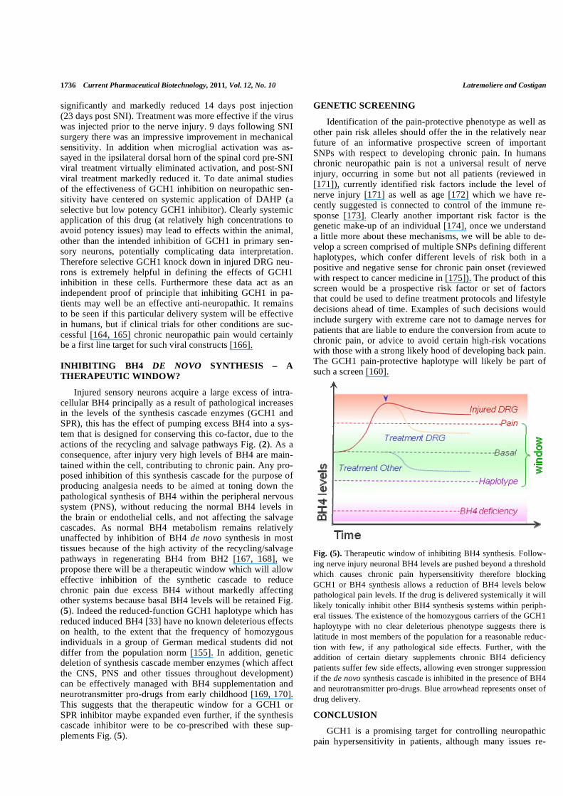
Injured sensory neurons acquire a large excess of intra-cellular BH4 principally as a result of pathological increases in the levels of the synthesis cascade enzymes (GCH1 and SPR), this has the effect of pumping excess BH4 into a sys-tem that is designed for conserving this co-factor, due to the actions of the recycling and salvage pathways Fig. (2). As a consequence, after injury very high levels of BH4 are main-tained within the cell, contributing to chronic pain. Any pro-posed inhibition of this synthesis cascade for the purpose of producing analgesia needs to be aimed at toning down the pathological synthesis of BH4 within the peripheral nervous system (PNS), without reducing the normal BH4 levels in the brain or endothelial cells, and not affecting the salvage cascades. As normal BH4 metabolism remains relatively unaffected by inhibition of BH4 de novo synthesis in most tissues because of the high activity of the recycling/salvage pathways in regenerating BH4 from BH2 [167, 168], we propose there will be a therapeutic window which will allow effective inhibition of the synthetic cascade to reduce chronic pain due excess BH4 without markedly affecting other systems because basal BH4 levels will be retained Fig. (5). Indeed the reduced-function GCH1 haplotype which has reduced induced BH4 [33] have no known deleterious effects on health, to the extent that the frequency of homozygous individuals in a group of German medical students did not differ from the population norm [155]. In addition, genetic deletion of synthesis cascade member enzymes (which affect the CNS, PNS and other tissues throughout development) can be effectively managed with BH4 supplementation and neurotransmitter pro-drugs from early childhood [169, 170]. This suggests that the therapeutic window for a GCH1 or SPR inhibitor maybe expanded even further, if the synthesis cascade inhibitor were to be co-prescribed with these sup-plements Fig. (5).
GENETIC SCREENING
Identification of the pain-protective phenotype as well as other pain risk alleles should offer the in the relatively near future of an informative prospective screen of important SNPs with respect to developing chronic pain. In humans chronic neuropathic pain is not a universal result of nerve injury, occurring in some but not all patients (reviewed in [171]), currently identified risk factors include the level of nerve injury [171] as well as age [172] which we have re-cently suggested is connected to control of the immune re-sponse [173]. Clearly another important risk factor is the genetic make-up of an individual [174], once we understand a little more about these mechanisms, we will be able to de-velop a screen comprised of multiple SNPs defining different haplotypes, which confer different levels of risk both in a positive and negative sense for chronic pain onset (reviewed with respect to cancer medicine in [175]). The product of this screen would be a prospective risk factor or set of factors that could be used to define treatment protocols and lifestyle decisions ahead of time. Examples of such decisions would include surgery with extreme care not to damage nerves for patients that are liable to endure the conversion from acute to chronic pain, or advice to avoid certain high-risk vocations with those with a strong likely hood of developing back pain. The GCH1 pain-protective haplotype will likely be part of such a screen [160].
Fig. (5). Therapeutic window of inhibiting BH4 synthesis. Follow-
ing nerve injury neuronal BH4 levels are pushed beyond a threshold
which causes chronic pain hypersensitivity therefore blocking
GCH1 or BH4 synthesis allows a reduction of BH4 levels below
pathological pain levels. If the drug is delivered systemically it will
likely tonically inhibit other BH4 synthesis systems within periph-
eral tissues. The existence of the homozygous carriers of the GCH1
haploytype with no clear deleterious phenotype suggests there is
latitude in most members of the population for a reasonable reduc-
tion with few, if any pathological side effects. Further, with the
addition of certain dietary supplements chronic BH4 deficiency
patients suffer few side effects, allowing even stronger suppression
if the de novo synthesis cascade is inhibited in the presence of BH4
and neurotransmitter pro-drugs. Blue arrowhead represents onset of
drug delivery.
CONCLUSION
GCH1 is a promising target for controlling neuropathic pain hypersensitivity in patients, although many issues re-

GCH1, BH4 and Pain Current Pharmaceutical Biotechnology, 2011, Vol. 12, No. 10 1737
main for development of a potentially effective treatment. Chief among these problems are the potential side effects of a GCH1 inhibitor in other systems other than injured primary sensory neurons Fig. (5). Inhibiting enzymes in the de novo synthesis pathway other than GCH1 may also help, allowing alternate BH4 synthesis routes in other tissues as well as preventing potential phenylalanine accumulation Fig. (2). We would not propose inhibiting either the recycling path-way or the salvage pathway, as these often act as the major determinant of normal tissue BH4 levels [26], although methotrexate, a DHFR inhibitor, is used in humans as an immunosuppressant and has efficacy against neuropathic sensitivity [141] although these actions are postulated to be via inhibiting immune cell function rather than DHFR inhibi-tion. Plasma neopterin levels have long been considered a useful marker of pathological immune cell action (reviewed in [176, 177]) and therefore decreasing BH4 synthesis may in some circumstances be a useful secondary effect of sys-temic BH4 reduction. Alternately treatment given specifi-cally to injured DRG neurons, as with the gene therapy op-tion using viral shRNA constructs, may circumvent side ef-fect issues. In any event much work remains, but to date pro-gress with the definition the BH4 synthesis cascade as a mechanism of chronic pain has been extraordinary.
ACKNOWLEDGMENTS
This work is funded by the NIH (R01-NS058870) and the Fondation pour la Recherche Medicale (FRM).
REFERENCES
[1] Costigan, M.; Scholz, J.; Woolf, C. J. Neuropathic pain: a maladap-
tive response of the nervous system to damage. Annu. Rev. Neuro-sci., 2009, 32, 1-32.
[2] Finnerup, N. B.; Otto, M.; McQuay, H. J.; Jensen, T. S.; Sindrup, S. H. Algorithm for neuropathic pain treatment: an evidence based
proposal. Pain, 2005, 118(3), 289-305. [3] Woolf, C. J. Dissecting out mechanisms responsible for peripheral
neuropathic pain: implications for diagnosis and therapy. Life Sci., 2004, 74 (21), 2605-2610.
[4] Bouhassira, D.; Attal, N.; Alchaar, H.; Boureau, F.; Brochet, B.; Bruxelle, J.; Cunin, G.; Fermanian, J.; Ginies, P.; Grun-
Overdyking, A.; Jafari-Schluep, H.; Lanteri-Minet, M.; Laurent, B.; Mick, G.; Serrie, A.; Valade, D.; Vicaut, E. Comparison of pain
syndromes associated with nervous or somatic lesions and devel-opment of a new neuropathic pain diagnostic questionnaire (DN4).
Pain, 2005, 114, (1-2), 29-36. [5] Bouhassira, D.; Attal, N.; Fermanian, J.; Alchaar, H.; Gautron, M.;
Masquelier, E.; Rostaing, S.; Lanteri-Minet, M.; Collin, E.; Grisart, J.; Boureau, F. Development and validation of the Neuropathic
Pain Symptom Inventory. Pain, 2004, 108(3), 248-257. [6] Scholz, J.; Mannion, R. J.; Hord, D. E.; Griffin, R. S.; Rawal, B.;
Zheng, H.; Scoffings, D.; Phillips, A.; Guo, J.; Laing, R. J.; Abdi, S.; Decosterd, I.; Woolf, C. J. A novel tool for the assessment of
pain: validation in low back pain. PLoS Med., 2009, 6(4), e1000047.
[7] Woolf, C. J.; Mannion, R. J. Neuropathic pain: aetiology, symp-toms, mechanisms, and management. Lancet, 1999, 353(9168),
1959-1964. [8] Kissin, I. The development of new analgesics over the past 50
years: a lack of real breakthrough drugs. Anesth. Analg., 2010, 110(3), 780-789.
[9] Costigan, M.; Befort, K.; Karchewski, L.; Griffin, R. S.; D'Urso, D.; Allchorne, A.; Sitarski, J.; Mannion, J. W.; Pratt, R. E.; Woolf,
C. J. Replicate high-density rat genome oligonucleotide microar-rays reveal hundreds of regulated genes in the dorsal root ganglion
after peripheral nerve injury. BMC Neurosci., 2002, 3, 16.
[10] Griffin, R. S.; Mills, C. D.; Costigan, M.; Woolf, C. J. Exploiting
microarrays to reveal differential gene expression in the nervous system. Genome Biol., 2003, 4(2), 105.
[11] Griffin, R. S.; Costigan, M.; Brenner, G. J.; Ma, C. H.; Scholz, J.; Moss, A.; Allchorne, A. J.; Stahl, G. L.; Woolf, C. J. Complement
induction in spinal cord microglia results in anaphylatoxin C5a-mediated pain hypersensitivity. J. Neurosci., 2007, 27(32), 8699-
8708. [12] Tegeder, I.; Costigan, M.; Griffin, R. S.; Abele, A.; Belfer, I.;
Schmidt, H.; Ehnert, C.; Nejim, J.; Marian, C.; Scholz, J.; Wu, T.; Allchorne, A.; Diatchenko, L.; Binshtok, A. M.; Goldman, D.; Ad-
olph, J.; Sama, S.; Atlas, S. J.; Carlezon, W. A.; Parsegian, A.; Lotsch, J.; Fillingim, R. B.; Maixner, W.; Geisslinger, G.; Max, M.
B.; Woolf, C. J. GTP cyclohydrolase and tetrahydrobiopterin regu-late pain sensitivity and persistence. Nat. Med., 2006, 12(11), 1269-
1277. [13] Doehring, A.; Freynhagen, R.; Griessinger, N.; Zimmermann, M.;
Sittl, R.; Hentig, N.; Geisslinger, G.; Lotsch, J. Cross-sectional as-sessment of the consequences of a GTP cyclohydrolase 1 haplotype
for specialized tertiary outpatient pain care. Clin. J. Pain, 2009, 25(9), 781-785.
[14] Lotsch, J.; Klepstad, P.; Doehring, A.; Dale, O. A GTP cyclohydro-lase 1 genetic variant delays cancer pain. Pain, 2010, 148(1), 103-
106. [15] Kaufman, S. A new cofactor required for the enzymatic conversion
of phenylalanine to tyrosine. J. Biol. Chem., 1958, 230(2), 931-939. [16] Kaufman, S. Phenylalanine hydroxylation cofactor in phenylke-
tonuria. Science, 1958, 128(3337), 1506-1508. [17] Kaufman, S.; Levenberg, B. Further studies on the phenylalanine-
hydroxylation cofactor. J. Biol. Chem., 1959, 234, 2683-2688. [18] Kettler, R.; Bartholini, G.; Pletscher, A. In vivo enhancement of
tyrosine hydroxylation in rat striatum by tetrahydrobiopterin. Na-ture, 1974, 249(456), 476-478.
[19] Friedman, P. A.; Kappelman, A. H.; Kaufman, S. Partial purifica-tion and characterization of tryptophan hydroxylase from rabbit
hindbrain. J. Biol. Chem., 1972, 247(13), 4165-4173. [20] Sawada, M.; Sugimoto, T.; Matsuura, S.; Nagatsu, T. (6R)-
tetrahydrobiopterin increases the activity of tryptophan hydroxylase in rat raphe slices. J. Neurochem., 1986, 47(5), 1544-1547.
[21] Gorren, A. C.; Bec, N.; Lange, R.; Mayer, B. Redox role for tetra-hydrobiopterin in nitric oxide synthase catalysis: low-temperature
optical absorption spectral detection. Methods Enzymol., 2002, 353, 114-121.
[22] Gorren, A. C.; Mayer, B. Tetrahydrobiopterin in nitric oxide syn-thesis: a novel biological role for pteridines. Curr. Drug Metab.,
2002, 3 (2), 133-157. [23] Tayeh, M. A.; Marletta, M. A. Macrophage oxidation of L-arginine
to nitric oxide, nitrite, and nitrate. Tetrahydrobiopterin is required as a cofactor. J. Biol. Chem., 1989, 264 (33), 19654-19658.
[24] Voss, J. C.; Waisman, H. A. The phenylalanine hydroxylase con-tent of livers of various vertebrates. Comput. Biochem. Physiol.,
1966, 17 (1), 49-58. [25] Lichter-Konecki, U.; Hipke, C. M.; Konecki, D. S. Human pheny-
lalanine hydroxylase gene expression in kidney and other nonhe-patic tissues. Mol. Genet. Metab., 1999, 67 (4), 308-316.
[26] Longo, N. Disorders of biopterin metabolism. J. Inherit. Metab. Dis., 2009, 32 (3), 333-42.
[27] Aley, K. O.; McCarter, G.; Levine, J. D. Nitric oxide signaling in pain and nociceptor sensitization in the rat. J. Neurosci., 1998,
18(17), 7008-7014. [28] Jaaskelainen, S. K.; Rinne, J. O.; Forssell, H.; Tenovuo, O.; Kaasi-
nen, V.; Sonninen, P.; Bergman, J. Role of the dopaminergic sys-tem in chronic pain -- a fluorodopa-PET study. Pain, 2001, 90(3),
257-260. [29] Millan, M. J. The induction of pain: an integrative review. Prog.
Neurobiol., 1999, 57 (1), 1-164. [30] Schmidtko, A.; Tegeder, I.; Geisslinger, G. No NO, no pain? The
role of nitric oxide and cGMP in spinal pain processing. Trends Neurosci., 2009, 32(6), 339-346.
[31] Stahl, S. M.; Grady, M. M.; Moret, C.; Briley, M. SNRIs: their pharmacology, clinical efficacy, and tolerability in comparison
with other classes of antidepressants. CNS Spectrom, 2005, 10 (9), 732-747.
[32] Suzuki, R.; Rygh, L. J.; Dickenson, A. H. Bad news from the brain: descending 5-HT pathways that control spinal pain processing.
Trends Pharmacol. Sci., 2004, 25(12), 613-617.
1738 Current Pharmaceutical Biotechnology, 2011, Vol. 12, No. 10 Latremoliere and Costigan
[33] Channon, K. M. Tetrahydrobiopterin: regulator of endothelial nitric
oxide synthase in vascular disease. Trends Cardiovasc. Med., 2004, 14 (8), 323-327.
[34] Thony, B.; Blau, N. Mutations in the BH4-metabolizing genes GTP cyclohydrolase I, 6-pyruvoyl-tetrahydropterin synthase, sepiapterin
reductase, carbinolamine-4a-dehydratase, and dihydropteridine re-ductase. Hum. Mutat., 2006, 27(9), 870-878.
[35] Erlandsen, H.; Stevens, R. C. The structural basis of phenylketonu-ria. Mol. Genet. Metab., 1999, 68 (2), 103-125.
[36] Thony, B.; Auerbach, G.; Blau, N. Tetrahydrobiopterin biosynthe-sis, regeneration and functions. Biochem. J., 2000, 347, Pt 1, 1-16.
[37] Neville, B. G.; Parascandalo, R.; Farrugia, R.; Felice, A. Sepiap-terin reductase deficiency: a congenital dopa-responsive motor and
cognitive disorder. Brain, 2005, 128(Pt 10), 2291-2296. [38] Furukawa, Y.; Kish, S. J.; Bebin, E. M.; Jacobson, R. D.; Fryburg,
J. S.; Wilson, W. G.; Shimadzu, M.; Hyland, K.; Trugman, J. M. Dystonia with motor delay in compound heterozygotes for GTP-
cyclohydrolase I gene mutations. Ann. Neurol., 1998, 44(1), 10-16. [39] Nagatsu, T.; Ichinose, H. GTP cyclohydrolase I gene, tetrahydro-
biopterin, and tyrosine hydroxylase gene: their relations to dystonia and parkinsonism. Neurochem. Res., 1996, 21(2), 245-250.
[40] Baynes, J. W. Role of oxidative stress in development of complica-tions in diabetes. Diabetes, 1991, 40, (4), 405-412.
[41] Harrison, D.; Griendling, K. K.; Landmesser, U.; Hornig, B.; Drex-ler, H., Role of oxidative stress in atherosclerosis. Am. J. Cardiol.,
2003, 91(3A), 7A-11A. [42] Kondo, T.; Hirose, M.; Kageyama, K. Roles of oxidative stress and
redox regulation in atherosclerosis. J Atheroscler. Thromb., 2009, 16, (5), 532-538.
[43] Victor, V. M.; Rocha, M.; Sola, E.; Banuls, C.; Garcia-Malpartida, K.; Hernandez-Mijares, A. Oxidative stress, endothelial dysfunc-
tion and atherosclerosis. Curr. Pharm. Des., 2009, 15(26), 2988-3002.
[44] Crabtree, M. J.; Tatham, A. L.; Hale, A. B.; Alp, N. J.; Channon, K. M. Critical role for tetrahydrobiopterin recycling by dihydrofo-
late reductase in regulation of endothelial nitric-oxide synthase coupling: relative importance of the de novo biopterin synthesis
versus salvage pathways. J. Biol. Chem., 2009, 284(41), 28128-2836.
[45] Vasquez-Vivar, J.; Martasek, P.; Whitsett, J.; Joseph, J.; Kalyana-raman, B. The ratio between tetrahydrobiopterin and oxidized tet-
rahydrobiopterin analogues controls superoxide release from endo-thelial nitric oxide synthase: an EPR spin trapping study. Biochem.
J., 2002, 362(Pt 3), 733-739. [46] Vasquez-Vivar, J.; Kalyanaraman, B.; Martasek, P.; Hogg, N.;
Masters, B. S.; Karoui, H.; Tordo, P.; Pritchard, K. A. Jr. Superox-ide generation by endothelial nitric oxide synthase: the influence of
cofactors. Proc. Natl. Acad. Sci. USA, 1998, 95(16), 9220-9225. [47] Bendall, J. K.; Alp, N. J.; Warrick, N.; Cai, S.; Adlam, D.; Rockett,
K.; Yokoyama, M.; Kawashima, S.; Channon, K. M. Stoichiomet-ric relationships between endothelial tetrahydrobiopterin, endothe-
lial NO synthase (eNOS) activity, and eNOS coupling in vivo: in-sights from transgenic mice with endothelial-targeted GTP cyclo-
hydrolase 1 and eNOS overexpression. Circ. Res., 2005, 97(9), 864-871.
[48] Crabtree, M. J.; Tatham, A. L.; Al-Wakeel, Y.; Warrick, N.; Hale, A. B.; Cai, S.; Channon, K. M.; Alp, N. J. Quantitative regulation
of intracellular endothelial nitric-oxide synthase (eNOS) coupling by both tetrahydrobiopterin-eNOS stoichiometry and biopterin re-
dox status: insights from cells with tet-regulated GTP cyclohydro-lase I expression. J. Biol. Chem., 2009, 284(2), 1136-1144.
[49] Landmesser, U.; Dikalov, S.; Price, S. R.; McCann, L.; Fukai, T.; Holland, S. M.; Mitch, W. E.; Harrison, D. G. Oxidation of tetra-
hydrobiopterin leads to uncoupling of endothelial cell nitric oxide synthase in hypertension. J. Clin. Invest., 2003, 111(8), 1201-1209.
[50] Pou, S.; Pou, W. S.; Bredt, D. S.; Snyder, S. H.; Rosen, G. M. Generation of superoxide by purified brain nitric oxide synthase. J.
Biol. Chem., 1992, 267 (34), 24173-24176. [51] Vasquez-Vivar, J.; Hogg, N.; Martasek, P.; Karoui, H.; Pritchard,
K. A., Jr.; Kalyanaraman, B. Tetrahydrobiopterin-dependent inhibi-tion of superoxide generation from neuronal nitric oxide synthase.
J. Biol Chem., 1999, 274(38), 26736-26742. [52] Wever, R. M.; van Dam, T.; van Rijn, H. J.; de Groot, F.; Rabelink,
T. J. Tetrahydrobiopterin regulates superoxide and nitric oxide generation by recombinant endothelial nitric oxide synthase. Bio-
chem. Biophys. Res. Commun., 1997, 237(2), 340-344.
[53] Hatakeyama, K.; Inoue, Y.; Harada, T.; Kagamiyama, H. Cloning
and sequencing of cDNA encoding rat GTP cyclohydrolase I. The first enzyme of the tetrahydrobiopterin biosynthetic pathway. J.
Biol. Chem., 1991, 266(2), 765-769. [54] Takikawa, S.; Curtius, H. C.; Redweik, U.; Leimbacher, W.;
Ghisla, S. Biosynthesis of tetrahydrobiopterin. Purification and characterization of 6-pyruvoyl-tetrahydropterin synthase from hu-
man liver. Eur. J. Biochem., 1986, 161(2), 295-302. [55] Levine, R. A.; Kapatos, G.; Kaufman, S.; Milstien, S. Immunologi-
cal evidence for the requirement of sepiapterin reductase for tetra-hydrobiopterin biosynthesis in brain. J. Neurochem., 1990, 54(4),
1218-1224. [56] Milstien, S.; Kaufman, S. Tetrahydro-sepiapterin is an intermediate
in tetrahydrobiopterin biosynthesis. Biochem. Biophys. Res. Com-mun., 1983, 115, (3), 888-893.
[57] Yim, J. J.; Brown, G. M. Characteristics of guanosine triphosphate cyclohydrolase I purified from Escherichia coli. J. Biol. Chem.,
1976, 251(16), 5087-5094. [58] Nar, H.; Huber, R.; Auerbach, G.; Fischer, M.; Hosl, C.; Ritz, H.;
Bracher, A.; Meining, W.; Eberhardt, S.; Bacher, A. Active site to-pology and reaction mechanism of GTP cyclohydrolase I. Proc.
Natl. Acad. Sci. USA, 1995, 92 (26), 12120-12125. [59] Nar, H.; Huber, R.; Meining, W.; Schmid, C.; Weinkauf, S.;
Bacher, A. Atomic structure of GTP cyclohydrolase I. Structure, 1995, 3(5), 459-466.
[60] McDonald, J. D.; Cotton, R. G.; Jennings, I.; Ledley, F. D.; Woo, S. L.; Bode, V. C. Biochemical defect of the hph-1 mouse mutant is
a deficiency in GTP-cyclohydrolase activity. J. Neurochem., 1988, 50(2), 655-657.
[61] Segawa, M.; Nomura, Y.; Nishiyama, N. Autosomal dominant guanosine triphosphate cyclohydrolase I deficiency (Segawa dis-
ease). Ann. Neurol., 2003, 54 (Suppl 6), S32-45. [62] Bode, V. C.; McDonald, J. D.; Guenet, J. L.; Simon, D. hph-1: a
mouse mutant with hereditary hyperphenylalaninemia induced by ethylnitrosourea mutagenesis. Genetics, 1988, 118(2), 299-305.
[63] Montanez, C. S.; McDonald, J. D. Linkage analysis of the hph-1 mutation and the GTP cyclohydrolase I structural gene. Mol. Genet.
Metab., 1999, 68(1), 91-92. [64] Hyland, K.; Gunasekara, R. S.; Munk-Martin, T. L.; Arnold, L. A.;
Engle, T. The hph-1 mouse: a model for dominantly inherited GTP-cyclohydrolase deficiency. Ann. Neurol., 2003, 54 (Suppl 6), S46-
48. [65] Zeng, B. Y.; Heales, S. J.; Canevari, L.; Rose, S.; Jenner, P. Altera-
tions in expression of dopamine receptors and neuropeptides in the striatum of GTP cyclohydrolase-deficient mice. Exp. Neurol., 2004,
190(2), 515-524. [66] Harada, T.; Kagamiyama, H.; Hatakeyama, K. Feedback regulation
mechanisms for the control of GTP cyclohydrolase I activity. Sci-ence, 1993, 260(5113), 1507-1510.
[67] Yoneyama, T.; Brewer, J. M.; Hatakeyama, K. GTP cyclohydrolase I feedback regulatory protein is a pentamer of identical subunits.
Purification, cDNA cloning, and bacterial expression. J. Biol. Chem., 1997, 272, (15), 9690-9696.
[68] Kolinsky, M. A.; Gross, S. S. The mechanism of potent GTP cy-clohydrolase I inhibition by 2,4-diamino-6-hydroxypyrimidine: re-
quirement of the GTP cyclohydrolase I feedback regulatory pro-tein. J. Biol. Chem., 2004, 279(39), 40677-40682.
[69] Tatham, A. L.; Crabtree, M. J.; Warrick, N.; Cai, S.; Alp, N. J.; Channon, K. M. GTP cyclohydrolase I expression, protein, and ac-
tivity determine intracellular tetrahydrobiopterin levels, independ-ent of GTP cyclohydrolase feedback regulatory protein expression.
J. Biol. Chem., 2009, 284(20), 13660-13668. [70] Milstien, S.; Kaufman, S.; Sakai, N. Tetrahydrobiopterin biosyn-
thesis defects examined in cytokine-stimulated fibroblasts. J. In-herit. Metab. Dis., 1993, 16 (6), 975-981.
[71] Franscini, N.; Blau, N.; Walter, R. B.; Schaffner, A.; Schoedon, G. Critical role of interleukin-1beta for transcriptional regulation of
endothelial 6-pyruvoyltetrahydropterin synthase. Arterioscler. Thromb. Vasc. Biol., 2003, 23(11), e50-53.
[72] Kaneko, Y. S.; Ikemoto, K.; Mori, K.; Nakashima, A.; Nagatsu, I.; Ota, A. Expression of GTP cyclohydrolase I in murine locus ce-
ruleus is enhanced by peripheral administration of lipopolysaccha-ride. Brain Res., 2001, 890, (2), 203-210.
[73] Hirayama, K.; Kapatos, G. Regulation of GTP cyclohydrolase I gene expression and tetrahydrobiopterin content by nerve growth

GCH1, BH4 and Pain Current Pharmaceutical Biotechnology, 2011, Vol. 12, No. 10 1739
factor in cultures of superior cervical ganglia. Neurochem. Int.,
1995, 27(2), 157-161. [74] Chavan, B.; Beazley, W.; Wood, J. M.; Rokos, H.; Ichinose, H.;
Schallreuter, K. U. H(2)O(2) increases de novo synthesis of (6R)-L-erythro-5,6,7,8-tetrahydrobiopterin via GTP cyclohydrolase I and
its feedback regulatory protein in vitiligo. J. Inherit. Metab. Dis., 2009, 32(1), 86-94.
[75] Shimizu, S.; Shiota, K.; Yamamoto, S.; Miyasaka, Y.; Ishii, M.; Watabe, T.; Nishida, M.; Mori, Y.; Yamamoto, T.; Kiuchi, Y. Hy-
drogen peroxide stimulates tetrahydrobiopterin synthesis through the induction of GTP-cyclohydrolase I and increases nitric oxide
synthase activity in vascular endothelial cells. Free Radic. Biol. Med., 2003, 34(10), 1343-1352.
[76] Shimizu, S.; Hiroi, T.; Ishii, M.; Hagiwara, T.; Wajima, T.; Miya-zaki, A.; Kiuchi, Y. Hydrogen peroxide stimulates tetrahydrobiop-
terin synthesis through activation of the Jak2 tyrosine kinase path-way in vascular endothelial cells. Intl. J. Biochem. Cell. Biol.,
2008, 40, (4), 755-765. [77] Serova, L. I.; Maharjan, S.; Huang, A.; Sun, D.; Kaley, G.; Sabban,
E. L. Response of tyrosine hydroxylase and GTP cyclohydrolase I gene expression to estrogen in brain catecholaminergic regions var-
ies with mode of administration. Brain Res., 2004, 1015, (1-2), 1-8. [78] Kumar, S.; Sun, X.; Sharma, S.; Aggarwal, S.; Ravi, K.; Fineman,
J. R.; Black, S. M. GTP cyclohydrolase I expression is regulated by nitric oxide: role of cyclic AMP. Am. J. Physiol. Lung Cell Mol.
Physiol., 2009, 297(2), L309-317. [79] Sun, X.; Kumar, S.; Tian, J.; Black, S. M. Estradiol increases
guanosine 5'-triphosphate cyclohydrolase expression via the nitric oxide-mediated activation of cyclic adenosine 5'-monophosphate
response element binding protein. Endocrinology, 2009, 150(8), 3742-3752.
[80] Kapatos, G.; Vunnava, P.; Wu, Y. Protein kinase A-dependent recruitment of RNA polymerase II, C/EBP beta and NF-Y to the rat
GTP cyclohydrolase I proximal promoter occurs without alterations in histone acetylation. J. Neurochem., 2007, 101(4), 1119-1133.
[81] Snider, W. D.; Zhou, F. Q.; Zhong, J.; Markus, A. Signaling the pathway to regeneration. Neuron, 2002, 35, (1), 13-16.
[82] Hannila, S. S.; Filbin, M. T. The role of cyclic AMP signaling in promoting axonal regeneration after spinal cord injury. Exp. Neu-
rol., 2008, 209(2), 321-332. [83] Candito, M.; Nagatsu, T.; Chambon, P.; Chatel, M. High-
performance liquid chromatographic measurement of cerebrospinal fluid tetrahydrobiopterin, neopterin, homovanillic acid and 5-
hydroxindoleacetic acid in neurological diseases. J. Chromatogr. B. Biomed. Appl., 1994, 657 (1), 61-66.
[84] Jang, Y. J.; Hong, H. N.; Lee, J. D.; Hwang, O. Down-regulation of GTP cyclohydrolase I and tetrahydrobiopterin by melatonin. Neu-
roreport, 2000, 11(16), 3627-3630. [85] Stegenga, S. L.; Hirayama, K.; Kapatos, G. Regulation of GTP
cyclohydrolase I gene expression and tetrahydrobiopterin content in cultured sympathetic neurons by leukemia inhibitory factor and
ciliary neurotrophic factor. J. Neurochem., 1996, 66(6), 2541-2545. [86] Ayar, A.; Martin, D. J.; Ozcan, M.; Kelestimur, H. Melatonin in-
hibits high voltage activated calcium currents in cultured rat dorsal root ganglion neurones. Neurosci. Lett., 2001, 313(1-2), 73-77.
[87] Banner, L. R.; Patterson, P. H. Major changes in the expression of the mRNAs for cholinergic differentiation factor/leukemia inhibi-
tory factor and its receptor after injury to adult peripheral nerves and ganglia. Proc. Natl. Acad. Sci. USA, 1994, 91, (15), 7109-7113.
[88] Ip, N. Y.; McClain, J.; Barrezueta, N. X.; Aldrich, T. H.; Pan, L.; Li, Y.; Wiegand, S. J.; Friedman, B.; Davis, S.; Yancopoulos, G. D.
The alpha component of the CNTF receptor is required for signal-ing and defines potential CNTF targets in the adult and during de-
velopment. Neuron, 1993, 10(1), 89-102. [89] Du, J.; Xu, H.; Wei, N.; Wakim, B.; Halligan, B.; Pritchard, K. A.,
Jr.; Shi, Y. Identification of proteins interacting with GTP cyclohy-drolase I. Biochem. Biophys. Res. Commun., 2009, 385(2), 143-
147. [90] Hesslinger, C.; Kremmer, E.; Hultner, L.; Ueffing, M.; Ziegler, I.
Phosphorylation of GTP cyclohydrolase I and modulation of its ac-tivity in rodent mast cells. GTP cyclohydrolase I hyperphosphory-
lation is coupled to high affinity IgE receptor signaling and in-volves protein kinase C. J. Biol. Chem., 1998, 273(34), 21616-
21622. [91] Lapize, C.; Pluss, C.; Werner, E. R.; Huwiler, A.; Pfeilschifter, J.
Protein kinase C phosphorylates and activates GTP cyclohydrolase
I in rat renal mesangial cells. Biochem. Biophys. Res. Commun.,
1998, 251(3), 802-825. [92] Chiarini, A.; Armato, U.; Pacchiana, R.; Dal Pra, I. Proteomic
analysis of GTP cyclohydrolase 1 multiprotein complexes in cul-tured normal adult human astrocytes under both basal and cyto-
kine-activated conditions. Proteomics, 2009, 9(7), 1850-1860. [93] Dal Pra, I.; Chiarini, A.; Nemeth, E. F.; Armato, U.; Whitfield, J. F.
Roles of Ca2+ and the Ca2+-sensing receptor (CASR) in the expres-sion of inducible NOS (nitric oxide synthase)-2 and its BH4 (tetra-
hydrobiopterin)-dependent activation in cytokine-stimulated adult human astrocytes. J. Cell Biochem., 2005, 96(2), 428-438.
[94] Swick, L.; Kapatos, G. A yeast 2-hybrid analysis of human GTP cyclohydrolase I protein interactions. J Neurochem., 2006, 97(5),
1447-1455. [95] Elzaouk, L.; Laufs, S.; Heerklotz, D.; Leimbacher, W.; Blau, N.;
Resibois, A.; Thony, B. Nuclear localization of tetrahydrobiopterin biosynthetic enzymes. Biochim. Biophys. Acta, 2004, 1670(1), 56-
68. [96] Bowling, K. M.; Huang, Z.; Xu, D.; Ferdousy, F.; Funderburk, C.
D.; Karnik, N.; Neckameyer, W.; O'Donnell, J. M. Direct binding of GTP cyclohydrolase and tyrosine hydroxylase: regulatory inter-
actions between key enzymes in dopamine biosynthesis. J. Biol. Chem., 2008, 283(46), 31449-31459.
[97] Blau, N.; Barnes, I.; Dhondt, J. L. International database of tetra-hydrobiopterin deficiencies. J. Inherit. Metab. Dis., 1996, 19(1), 8-
14. [98] Sumi-Ichinose, C.; Urano, F.; Kuroda, R.; Ohye, T.; Kojima, M.;
Tazawa, M.; Shiraishi, H.; Hagino, Y.; Nagatsu, T.; Nomura, T.; Ichinose, H. Catecholamines and serotonin are differently regulated
by tetrahydrobiopterin. A study from 6-pyruvoyltetrahydropterin synthase knockout mice. J. Biol. Chem., 2001, 276 (44), 41150-
41160. [99] Weiss, G.; Fuchs, D.; Hausen, A.; Reibnegger, G.; Werner, E. R.;
Werner-Felmayer, G.; Semenitz, E.; Dierich, M. P.; Wachter, H. Neopterin modulates toxicity mediated by reactive oxygen and
chloride species. FEBS Lett., 1993, 321(1), 89-92. [100] Linscheid, P.; Schaffner, A.; Blau, N.; Schoedon, G. Regulation of
6-pyruvoyltetrahydropterin synthase activity and messenger RNA abundance in human vascular endothelial cells. Circulation, 1998,
98(17), 1703-1706. [101] Bonafe, L.; Thony, B.; Penzien, J. M.; Czarnecki, B.; Blau, N.
Mutations in the sepiapterin reductase gene cause a novel tetrahy-drobiopterin-dependent monoamine-neurotransmitter deficiency
without hyperphenylalaninemia. Am. J. Hum. Genet., 2001, 69(2), 269-277.
[102] Blau, N.; Bonafe, L.; Thony, B. Tetrahydrobiopterin deficiencies without hyperphenylalaninemia: diagnosis and genetics of dopa-
responsive dystonia and sepiapterin reductase deficiency. Mol. Genet. Metab., 2001, 74(1-2), 172-185.
[103] Leu-Semenescu, S.; Karroum, E.; Brion, A.; Konofal, E.; Arnulf, I. Dopamine dysregulation syndrome in a patient with restless legs
syndrome. Sleep Med., 2009, 10(4), 494-496. [104] Yang, S.; Lee, Y. J.; Kim, J. M.; Park, S.; Peris, J.; Laipis, P.; Park,
Y. S.; Chung, J. H.; Oh, S. P. A murine model for human sepiap-terin-reductase deficiency. Am. J. Hum. Genet., 2006, 78(4), 575-
587. [105] Ikemoto, K.; Suzuki, T.; Ichinose, H.; Ohye, T.; Nishimura, A.;
Nishi, K.; Nagatsu, I.; Nagatsu, T. Localization of sepiapterin re-ductase in the human brain. Brain Res., 2002, 954(2), 237-246.
[106] Thony, B.; Neuheiser, F.; Kierat, L.; Blaskovics, M.; Arn, P. H.; Ferreira, P.; Rebrin, I.; Ayling, J.; Blau, N. Hyperphenylalaninemia
with high levels of 7-biopterin is associated with mutations in the PCBD gene encoding the bifunctional protein pterin-4a-
carbinolamine dehydratase and transcriptional coactivator (DCoH). Am. J. Hum. Genet., 1998, 62(6), 1302-1311.
[107] Brewster, T. G.; Moskowitz, M. A.; Kaufman, S.; Breslow, J. L.; Milstien, S.; Abroms, I. F. Dihydropteridine reductase deficiency
associated with severe neurologic disease and mild hyperphenyla-laninemia. Pediatrics, 1979, 63 (1), 94-99.
[108] Kaufman, S.; Holtzman, N. A.; Milstien, S.; Butler, L. J.; Krum-holz, A. Phenylketonuria due to a deficiency of dihydropteridine
reductase. N. Engl. J. Med., 1975, 293(16), 785-790. [109] Presta, A.; Siddhanta, U.; Wu, C.; Sennequier, N.; Huang, L.; Abu-
Soud, H. M.; Erzurum, S.; Stuehr, D. J. Comparative functioning of dihydro- and tetrahydropterins in supporting electron transfer, ca-
1740 Current Pharmaceutical Biotechnology, 2011, Vol. 12, No. 10 Latremoliere and Costigan
talysis, and subunit dimerization in inducible nitric oxide synthase.
Biochemistry, 1998, 37(1), 298-310. [110] Wei, C. C.; Wang, Z. Q.; Arvai, A. S.; Hemann, C.; Hille, R.; Get-
zoff, E. D.; Stuehr, D. J. Structure of tetrahydrobiopterin tunes its electron transfer to the heme-dioxy intermediate in nitric oxide syn-
thase. Biochemistry, 2003, 42(7), 1969-1977. [111] Werner, E. R.; Gorren, A. C.; Heller, R.; Werner-Felmayer, G.;
Mayer, B. Tetrahydrobiopterin and nitric oxide: mechanistic and pharmacological aspects. Exp. Biol. Med., (Maywood) 2003, 228
(11), 1291-1302. [112] Lee, C. K.; Han, J. S.; Won, K. J.; Jung, S. H.; Park, H. J.; Lee, H.
M.; Kim, J.; Park, Y. S.; Kim, H. J.; Park, P. J.; Park, T. K.; Kim, B. Diminished expression of dihydropteridine reductase is a potent
biomarker for hypertensive vessels. Proteomics, 2009, 9(21), 4851-4858.
[113] Moens, A. L.; Kass, D. A. Tetrahydrobiopterin and cardiovascular disease. Arterioscler. Thromb. Vasc. Biol., 2006, 26 (11), 2439-
2444. [114] Fiallos-Estrada, C. E.; Kummer, W.; Mayer, B.; Bravo, R.; Zim-
mermann, M.; Herdegen, T., Long-lasting increase of nitric oxide synthase immunoreactivity, NADPH-diaphorase reaction and c-
JUN co-expression in rat dorsal root ganglion neurons following sciatic nerve transection. Neurosci. Lett., 1993, 150(2), 169-173.
[115] Luo, Z. D.; Chaplan, S. R.; Scott, B. P.; Cizkova, D.; Calcutt, N. A.; Yaksh, T. L. Neuronal nitric oxide synthase mRNA upregula-
tion in rat sensory neurons after spinal nerve ligation: lack of a role in allodynia development. J. Neurosci., 1999, 19(21), 9201-9208.
[116] Zhang, X.; Verge, V.; Wiesenfeld-Hallin, Z.; Ju, G.; Bredt, D.; Synder, S. H.; Hokfelt, T. Nitric oxide synthase-like immunoreac-
tivity in lumbar dorsal root ganglia and spinal cord of rat and mon-key and effect of peripheral axotomy. J. Comput. Neurol., 1993,
335(4), 563-575. [117] Price, T. J.; Flores, C. M. Critical evaluation of the colocalization
between calcitonin gene-related peptide, substance P, transient re-ceptor potential vanilloid subfamily type 1 immunoreactivities, and
isolectin B4 binding in primary afferent neurons of the rat and mouse. J. Pain, 2007, 8(3), 263-272.
[118] Tominaga, M.; Caterina, M. J. Thermosensation and pain. J. Neu-robiol., 2004, 61(1), 3-12.
[119] Story, G. M.; Peier, A. M.; Reeve, A. J.; Eid, S. R.; Mosbacher, J.; Hricik, T. R.; Earley, T. J.; Hergarden, A. C.; Andersson, D. A.;
Hwang, S. W.; McIntyre, P.; Jegla, T.; Bevan, S.; Patapoutian, A. ANKTM1, a TRP-like channel expressed in nociceptive neurons, is
activated by cold temperatures. Cell, 2003, 112 (6), 819-829. [120] Pedersen, S. F.; Owsianik, G.; Nilius, B. TRP channels: an over-
view. Cell Calcium, 2005, 383-4), 233-252. [121] Miyamoto, T.; Dubin, A. E.; Petrus, M. J.; Patapoutian, A. TRPV1
and TRPA1 mediate peripheral nitric oxide-induced nociception in mice. PLoS One, 2009, 4(10), e7596.
[122] Gorren, A. C.; List, B. M.; Schrammel, A.; Pitters, E.; Hemmens, B.; Werner, E. R.; Schmidt, K.; Mayer, B. Tetrahydrobiopterin-free
neuronal nitric oxide synthase: evidence for two identical highly anticooperative pteridine binding sites. Biochemistry, 1996, 35(51),
16735-16745. [123] Brand, M. P.; Heales, S. J.; Land, J. M.; Clark, J. B. Tetrahydro-
biopterin deficiency and brain nitric oxide synthase in the hph1 mouse. J. Inherit. Metab. Dis., 1995, 18(1), 33-39.
[124] Caterina, M. J.; Leffler, A.; Malmberg, A. B.; Martin, W. J.; Trafton, J.; Petersen-Zeitz, K. R.; Koltzenburg, M.; Basbaum, A. I.;
Julius, D. Impaired nociception and pain sensation in mice lacking the capsaicin receptor. Science, 2000, 288(5464), 306-313.
[125] McNamara, C. R.; Mandel-Brehm, J.; Bautista, D. M.; Siemens, J.; Deranian, K. L.; Zhao, M.; Hayward, N. J.; Chong, J. A.; Julius,
D.; Moran, M. M.; Fanger, C. M. TRPA1 mediates formalinin-duced pain. Proc. Natl. Acad. Sci. USA, 2007, 104(33), 13525-
13530. [126] Roche, A. K.; Cook, M.; Wilcox, G. L.; Kajander, K. C. A nitric
oxide synthesis inhibitor (L-NAME) reduces licking behavior and Fos-labeling in the spinal cord of rats during formalin-induced in-
flammation. Pain, 1996, 66, (2-3), 331-341. [127] Zhuang, Z. Y.; Xu, H.; Clapham, D. E.; Ji, R. R. Phosphatidylinosi-
tol 3-kinase activates ERK in primary sensory neurons and medi-ates inflammatory heat hyperalgesia through TRPV1 sensitization.
J. Neurosci., 2004, 24(38), 8300-8309.
[128] Cheng, J. K.; Ji, R. R. Intracellular signaling in primary sensory
neurons and persistent pain. Neurochem. Res., 2008, 33(10), 1970-1978.
[129] Ishii, M.; Shimizu, S.; Nagai, T.; Shiota, K.; Kiuchi, Y.; Yama-moto, T. Stimulation of tetrahydrobiopterin synthesis induced by
insulin: possible involvement of phosphatidylinositol 3-kinase. Int. J. Biochem. Cell Biol., 2001, 33(1), 65-73.
[130] Kapatos, G.; Hirayama, K.; Lentz, S. I.; Zhu, M.; Stegenga, S. Differential metabolism of tetrahydrobiopterin in monoamine neu-
rons: a hypothesis based upon clinical and basic research. Adv. Exp. Med. Biol., 1993, 338, 217-22.
[131] Wolf, W. A.; Ziaja, E.; Arthur, R. A., Jr.; Anastasiadis, P. Z.; Levine, R. A.; Kuhn, D. M. Effect of tetrahydrobiopterin on sero-
tonin synthesis, release, and metabolism in superfused hippocampal slices. J. Neurochem., 1991, 57(4), 1191-1197.
[132] Kim, S. J.; Lee, W. I.; Lee, Y. S.; Kim, D. H.; Chang, J. W.; Kim, S. W.; Lee, H. Effective relief of neuropathic pain by adeno-
associated virus-mediated expression of a small hairpin RNA against GTP cyclohydrolase 1. Mol. Pain 2009, 5, 67.
[133] Detke, M. J.; Rickels, M.; Lucki, I., Active behaviors in the rat forced swimming test differentially produced by serotonergic and
noradrenergic antidepressants. Psychopharmacology, (Berl) 1995, 121, (1), 66-72.
[134] Latremoliere, A.; Woolf, C. J. Central sensitization: a generator of pain hypersensitivity by central neural plasticity. J. Pain, 2009, 10,
(9), 895-926. [135] Sumi-Ichinose, C.; Urano, F.; Shimomura, A.; Sato, T.; Ikemoto,
K.; Shiraishi, H.; Senda, T.; Ichinose, H.; Nomura, T. Genetically rescued tetrahydrobiopterin-depleted mice survive with hyperphen-
ylalaninemia and region-specific monoaminergic abnormalities. J. Neurochem., 2005, 95 (3), 703-714.
[136] Ikeda, H.; Stark, J.; Fischer, H.; Wagner, M.; Drdla, R.; Jager, T.; Sandkuhler, J. Synaptic amplifier of inflammatory pain in the spi-
nal dorsal horn. Science, 2006, 312, (5780), 1659-1662. [137] Hwang, O.; Baker, H.; Gross, S.; Joh, T. H. Localization of GTP
cyclohydrolase in monoaminergic but not nitric oxide-producing cells. Synapse, 1998, 28(2), 140-153.
[138] Nimmerjahn, A.; Kirchhoff, F.; Helmchen, F. Resting microglial cells are highly dynamic surveillants of brain parenchyma in vivo.
Science, 2005, 308(5726), 1314-1318. [139] Duan, Y.; Sahley, C. L.; Muller, K. J. ATP and NO dually control
migration of microglia to nerve lesions. Dev. Neurobiol., 2009, 69(1), 60-72.
[140] Devor, M. Ectopic discharge in Abeta afferents as a source of neu-ropathic pain. Exp. Brain Res., 2009, 196(1), 115-128.
[141] Scholz, J.; Abele, A.; Marian, C.; Haussler, A.; Herbert, T. A.; Woolf, C. J.; Tegeder, I. Low-dose methotrexate reduces peripheral
nerve injury-evoked spinal microglial activation and neuropathic pain behavior in rats. Pain, 2008, 138(1), 130-142.
[142] Hathway, G. J.; Vega-Avelaira, D.; Moss, A.; Ingram, R.; Fitz-gerald, M. Brief, low frequency stimulation of rat peripheral C-
fibres evokes prolonged microglial-induced central sensitization in adults but not in neonates. Pain, 2009, 144(1-2), 110-118.
[143] Kawasaki, Y.; Zhang, L.; Cheng, J. K.; Ji, R. R. Cytokine mecha-nisms of central sensitization: distinct and overlapping role of inter-
leukin-1beta, interleukin-6, and tumor necrosis factor-alpha in regulating synaptic and neuronal activity in the superficial spinal
cord. J. Neurosci., 2008, 28(20), 5189-5194. [144] Suter, M. R.; Berta, T.; Gao, Y. J.; Decosterd, I.; Ji, R. R. Large A-
fiber activity is required for microglial proliferation and p38 MAPK activation in the spinal cord: different effects of resinifera-
toxin and bupivacaine on spinal microglial changes after spared nerve injury. Mol. Pain, 2009, 5, 53.
[145] Coull, J. A.; Beggs, S.; Boudreau, D.; Boivin, D.; Tsuda, M.; Inoue, K.; Gravel, C.; Salter, M. W.; De Koninck, Y. BDNF from
microglia causes the shift in neuronal anion gradient underlying neuropathic pain. Nature, 2005, 438(7070), 1017-1021.
[146] Coull, J. A.; Boudreau, D.; Bachand, K.; Prescott, S. A.; Nault, F.; Sik, A.; De Koninck, P.; De Koninck, Y. Trans-synaptic shift in an-
ion gradient in spinal lamina I neurons as a mechanism of neuro-pathic pain. Nature, 2003, 424(6951), 938-942.
[147] Chanock, S. J.; Manolio, T.; Boehnke, M.; Boerwinkle, E.; Hunter, D. J.; Thomas, G.; Hirschhorn, J. N.; Abecasis, G.; Altshuler, D.;
Bailey-Wilson, J. E.; Brooks, L. D.; Cardon, L. R.; Daly, M.; Don-nelly, P.; Fraumeni, J. F., Jr.; Freimer, N. B.; Gerhard, D. S.; Gun-
ter, C.; Guttmacher, A. E.; Guyer, M. S.; Harris, E. L.; Hoh, J.;
GCH1, BH4 and Pain Current Pharmaceutical Biotechnology, 2011, Vol. 12, No. 10 1741
Hoover, R.; Kong, C. A.; Merikangas, K. R.; Morton, C. C.;
Palmer, L. J.; Phimister, E. G.; Rice, J. P.; Roberts, J.; Rotimi, C.; Tucker, M. A.; Vogan, K. J.; Wacholder, S.; Wijsman, E. M.;
Winn, D. M.; Collins, F. S. Replicating genotype-phenotype asso-ciations. Nature, 2007, 447(7145), 655-660.
[148] Pearson, T. A.; Manolio, T. A. How to interpret a genome-wide association study. JAMA, 2008, 299, (11), 1335-1344.
[149] Cardon, L. R.; Palmer, L. J. Population stratification and spurious allelic association. Lancet, 2003, 361 (9357), 598-604.
[150] Pritchard, J. K.; Stephens, M.; Rosenberg, N. A.; Donnelly, P. Association mapping in structured populations. Am. J. Hum.
Genet., 2000, 67(1), 170-181. [151] Price, A. L.; Patterson, N. J.; Plenge, R. M.; Weinblatt, M. E.;
Shadick, N. A.; Reich, D. Principal components analysis corrects for stratification in genome-wide association studies. Nat. Genet.,
2006, 38 (8), 904-909. [152] Andersson, U.; McKean-Cowdin, R.; Hjalmars, U.; Malmer, B.
Genetic variants in association studies--review of strengths and weaknesses in study design and current knowledge of impact on
cancer risk. Acta Oncol., 2009, 48(7), 948-954. [153] Atlas, S. J.; Deyo, R. A.; Keller, R. B.; Chapin, A. M.; Patrick, D.
L.; Long, J. M.; Singer, D. E., The Maine Lumbar Spine Study, Part II. 1-year outcomes of surgical and nonsurgical management
of sciatica. Spine, (Phila Pa 1976) 1996, 21(15), 1777-86. [154] Enoch, M. A.; Shen, P. H.; Xu, K.; Hodgkinson, C.; Goldman, D.
Using ancestry-informative markers to define populations and de-tect population stratification. J. Psychopharmacol., 2006, 20(4
Suppl), 19-26. [155] Tegeder, I.; Adolph, J.; Schmidt, H.; Woolf, C. J.; Geisslinger, G.;
Lotsch, J. Reduced hyperalgesia in homozygous carriers of a GTP cyclohydrolase 1 haplotype. Eur. J. Pain, 2008, 12(8), 1069-1077.
[156] Campbell, C. M.; Edwards, R. R.; Carmona, C.; Uhart, M.; Wand, G.; Carteret, A.; Kim, Y. K.; Frost, J.; Campbell, J. N. Polymor-
phisms in the GTP cyclohydrolase gene (GCH1) are associated with ratings of capsaicin pain. Pain, 2009, 141(1-2), 114-118.
[157] Zhang, L.; Rao, F.; Zhang, K.; Khandrika, S.; Das, M.; Vaingankar, S. M.; Bao, X.; Rana, B. K.; Smith, D. W.; Wessel, J.; Salem, R.
M.; Rodriguez-Flores, J. L.; Mahata, S. K.; Schork, N. J.; Ziegler, M. G.; O'Connor, D. T. Discovery of common human genetic vari-
ants of GTP cyclohydrolase 1 (GCH1) governing nitric oxide, autonomic activity, and cardiovascular risk. J. Clin. Invest, 2007,
117(9), 2658-2671. [158] Doehring, A.; Antoniades, C.; Channon, K. M.; Tegeder, I.; Lotsch,
J. Clinical genetics of functionally mild non-coding GTP cyclohy-drolase 1 (GCH1) polymorphisms modulating pain and cardiovas-
cular risk. Mutat. Res., 2008, 659(3), 195-201. [159] Antoniades, C.; Shirodaria, C.; Van Assche, T.; Cunnington, C.;
Tegeder, I.; Lotsch, J.; Guzik, T. J.; Leeson, P.; Diesch, J.; Tou-soulis, D.; Stefanadis, C.; Costigan, M.; Woolf, C. J.; Alp, N. J.;
Channon, K. M. GCH1 haplotype determines vascular and plasma biopterin availability in coronary artery disease effects on vascular
superoxide production and endothelial function. J. Am. Coll. Car-diol., 2008, 52(2), 158-165.
[160] Lotsch, J.; Belfer, I.; Kirchhof, A.; Mishra, B. K.; Max, M. B.; Doehring, A.; Costigan, M.; Woolf, C. J.; Geisslinger, G.; Tegeder,
I. Reliable screening for a pain-protective haplotype in the GTP cy-
clohydrolase 1 gene (GCH1) through the use of 3 or fewer single
nucleotide polymorphisms. Clin. Chem., 2007, 53 (6), 1010-1015. [161] Lotsch, J.; Geisslinger, G. Current evidence for a genetic modula-
tion of the response to analgesics. Pain, 2006, 121(1-2), 1-5. [162] Lazarev, M.; Lamb, J.; Barmada, M. M.; Dai, F.; Anderson, M. A.;
Max, M. B.; Whitcomb, D. C. Does the pain-protective GTP cyclo-hydrolase haplotype significantly alter the pattern or severity of
pain in humans with chronic pancreatitis? Mol. Pain, 2008, 4, 58. [163] Holliday, K. L.; Nicholl, B. I.; Macfarlane, G. J.; Thomson, W.;
Davies, K. A.; McBeth, J. Do genetic predictors of pain sensitivity associate with persistent widespread pain? Mol. Pain, 2009, 5, 56.
[164] Beutler, A. S.; Reinhardt, M., AAV for pain: steps towards clinical translation. Gene. Ther., 2009, 16 (4), 461-469.
[165] Mueller, C.; Flotte, T. R. Clinical gene therapy using recombinant adeno-associated virus vectors. Gene. Ther., 2008, 15(11), 858-
863. [166] Pohl, M.; Meunier, A. Experimental gene therapy of chronic pain.
Curr. Opin. Anaesthesiol., 2003, 16(5), 547-551. [167] Hasegawa, H.; Sawabe, K.; Nakanishi, N.; Wakasugi, O. K. Deliv-
ery of exogenous tetrahydrobiopterin (BH4) to cells of target or-gans: role of salvage pathway and uptake of its precursor in effec-
tive elevation of tissue BH4. Mol. Genet. Metab., 2005, 86 (Suppl 1), S2-10.
[168] Ponzone, A.; Spada, M.; Ferraris, S.; Dianzani, I.; de Sanctis, L. Dihydropteridine reductase deficiency in man: from biology to
treatment. Med. Res. Rev., 2004, 24(2), 127-150. [169] Jaggi, L.; Zurfluh, M. R.; Schuler, A.; Ponzone, A.; Porta, F.; Fiori,
L.; Giovannini, M.; Santer, R.; Hoffmann, G. F.; Ibel, H.; Wendel, U.; Ballhausen, D.; Baumgartner, M. R.; Blau, N. Outcome and
long-term follow-up of 36 patients with tetrahydrobiopterin defi-ciency. Mol. Genet. Metab., 2008, 93 (3), 295-305.
[170] Liu, K. M.; Liu, T. T.; Lee, N. C.; Cheng, L. Y.; Hsiao, K. J.; Niu, D. M. Long-term follow-up of Taiwanese Chinese patients treated
early for 6-pyruvoyl-tetrahydropterin synthase deficiency. Arch. Neurol., 2008, 65 (3), 387-392.
[171] Kehlet, H.; Jensen, T. S.; Woolf, C. J. Persistent postsurgical pain: risk factors and prevention. Lancet, 2006, 367(9522), 1618-1625.
[172] Kristensen, A. D.; Pedersen, T. A.; Hjortdal, V. E.; Jensen, T. S.; Nikolajsen, L. Chronic pain in adults after thoracotomy in child-
hood or youth. Br. J. Anaesth., 104 (1), 75-79. [173] Costigan, M.; Moss, A.; Latremoliere, A.; Johnston, C.; Verma-
Gandhu, M.; Herbert, T. A.; Barrett, L.; Brenner, G. J.; Vardeh, D.; Woolf, C. J.; Fitzgerald, M. T-cell infiltration and signaling in the
adult dorsal spinal cord is a major contributor to neuropathic pain-like hypersensitivity. J. Neurosci., 2009, 29(46), 14415-14422.
[174] Norbury, T. A.; MacGregor, A. J.; Urwin, J.; Spector, T. D.; McMahon, S. B. Heritability of responses to painful stimuli in
women: a classical twin study. Brain, 2007, 130(Pt 11), 3041-3049. [175] Hartman, M.; Loy, E. Y.; Ku, C. S.; Chia, K. S. Molecular epide-
miology and its current clinical use in cancer management. Lancet Oncol., 2010, 11 (4), 383-390.
[176] Murr, C.; Widner, B.; Wirleitner, B.; Fuchs, D. Neopterin as a marker for immune system activation. Curr. Drug Metab., 2002,
3(2), 175-187. [177] Sucher, R.; Schroecksnadel, K.; Weiss, G.; Margreiter, R.; Fuchs,
D.; Brandacher, G. Neopterin, a prognostic marker in human ma-lignancies. Cancer Lett., 2010, 287(1), 13-22.
Received: May 03, 2010 Accepted: September 02, 2010