Activation of DNA Methyltransferase 1 by EBV LMP1 Involves c-Jun NH2Terminal Kinase Signaling
10
2006;66:11668-11676. Cancer Res Chia-Lung Tsai, Hsin-Pai Li, Yen-Jung Lu, et al. -Terminal Kinase Signaling 2 Involves c-Jun NH Activation of DNA Methyltransferase 1 by EBV LMP1 Updated version http://cancerres.aacrjournals.org/content/66/24/11668 Access the most recent version of this article at: Material Supplementary http://cancerres.aacrjournals.org/content/suppl/2006/12/15/66.24.11668.DC1.html Access the most recent supplemental material at: Cited Articles http://cancerres.aacrjournals.org/content/66/24/11668.full.html#ref-list-1 This article cites by 50 articles, 19 of which you can access for free at: Citing articles http://cancerres.aacrjournals.org/content/66/24/11668.full.html#related-urls This article has been cited by 17 HighWire-hosted articles. Access the articles at: E-mail alerts related to this article or journal. Sign up to receive free email-alerts Subscriptions Reprints and . [email protected] Department at To order reprints of this article or to subscribe to the journal, contact the AACR Publications Permissions . [email protected] Department at To request permission to re-use all or part of this article, contact the AACR Publications Cancer Research. on September 28, 2013. © 2006 American Association for cancerres.aacrjournals.org Downloaded from Cancer Research. on September 28, 2013. © 2006 American Association for cancerres.aacrjournals.org Downloaded from Cancer Research. on September 28, 2013. © 2006 American Association for cancerres.aacrjournals.org Downloaded from Cancer Research. on September 28, 2013. © 2006 American Association for cancerres.aacrjournals.org Downloaded from Cancer Research. on September 28, 2013. © 2006 American Association for cancerres.aacrjournals.org Downloaded from Cancer Research. on September 28, 2013. © 2006 American Association for cancerres.aacrjournals.org Downloaded from Cancer Research. on September 28, 2013. © 2006 American Association for cancerres.aacrjournals.org Downloaded from Cancer Research. on September 28, 2013. © 2006 American Association for cancerres.aacrjournals.org Downloaded from Cancer Research. on September 28, 2013. © 2006 American Association for cancerres.aacrjournals.org Downloaded from Cancer Research. on September 28, 2013. © 2006 American Association for cancerres.aacrjournals.org Downloaded from
-
Upload
independent -
Category
Documents
-
view
0 -
download
0
Transcript of Activation of DNA Methyltransferase 1 by EBV LMP1 Involves c-Jun NH2Terminal Kinase Signaling
2006;66:11668-11676. Cancer Res Chia-Lung Tsai, Hsin-Pai Li, Yen-Jung Lu, et al.
-Terminal Kinase Signaling2Involves c-Jun NHActivation of DNA Methyltransferase 1 by EBV LMP1
Updated version
http://cancerres.aacrjournals.org/content/66/24/11668
Access the most recent version of this article at:
Material
Supplementary
http://cancerres.aacrjournals.org/content/suppl/2006/12/15/66.24.11668.DC1.html
Access the most recent supplemental material at:
Cited Articles
http://cancerres.aacrjournals.org/content/66/24/11668.full.html#ref-list-1
This article cites by 50 articles, 19 of which you can access for free at:
Citing articles
http://cancerres.aacrjournals.org/content/66/24/11668.full.html#related-urls
This article has been cited by 17 HighWire-hosted articles. Access the articles at:
E-mail alerts related to this article or journal.Sign up to receive free email-alerts
Subscriptions
Reprints and
To order reprints of this article or to subscribe to the journal, contact the AACR Publications
Permissions
To request permission to re-use all or part of this article, contact the AACR Publications
Cancer Research. on September 28, 2013. © 2006 American Association forcancerres.aacrjournals.org Downloaded from
Cancer Research. on September 28, 2013. © 2006 American Association forcancerres.aacrjournals.org Downloaded from
Cancer Research. on September 28, 2013. © 2006 American Association forcancerres.aacrjournals.org Downloaded from
Cancer Research. on September 28, 2013. © 2006 American Association forcancerres.aacrjournals.org Downloaded from
Cancer Research. on September 28, 2013. © 2006 American Association forcancerres.aacrjournals.org Downloaded from
Cancer Research. on September 28, 2013. © 2006 American Association forcancerres.aacrjournals.org Downloaded from
Cancer Research. on September 28, 2013. © 2006 American Association forcancerres.aacrjournals.org Downloaded from
Cancer Research. on September 28, 2013. © 2006 American Association forcancerres.aacrjournals.org Downloaded from
Cancer Research. on September 28, 2013. © 2006 American Association forcancerres.aacrjournals.org Downloaded from
Cancer Research. on September 28, 2013. © 2006 American Association forcancerres.aacrjournals.org Downloaded from

Activation of DNA Methyltransferase 1 by EBV LMP1 Involves
c-Jun NH2-Terminal Kinase Signaling
Chia-Lung Tsai,1,5Hsin-Pai Li,
3Yen-Jung Lu,
5Chuen Hsueh,
4Ying Liang,
2Chi-Long Chen,
6
Sai Wah Tsao,7Ka-Po Tse,
5Jau-Song Yu,
1,2and Yu-Sun Chang
1
1Graduate Institute of Basic Medical Sciences and 2Pathology Core, Chang-Gung Molecular Medicine Research Center, Chang-GungUniversity; Departments of 3Medical Research and 4Pathology, Chang-Gung Memorial Hospital at Lin-Kou, Taoyuan, Taiwan,Republic of China; 5Institute of Microbiology and Immunology, National Yang-Ming University; 6Department of Pathology,Taipei Medial University, Taipei, Taiwan, Republic of China; and 7Department of Anatomy, University of Hong Kong,Pokfulam, Hong Kong
Abstract
EBV latent membrane protein 1 (LMP1) activates cellular DNAmethyltransferases, resulting in hypermethylation and silenc-ing of E-cadherin. However, the underlying mechanismremains to be elucidated. In this study, we show that LMP1directly induces the dnmt1 promoter activity through itsCOOH-terminal activation region-2 YYD domain. Using (i)LMP1 mutants, (ii) dominant negative mutants c-jun NH2-terminal kinase (JNK)-DN, p38-DN, and constitutive activemutant IKB, as well as (iii) dsRNAs targeting c-Jun, JNK, andtumor necrosis factor receptor–associated death domainprotein, and (iv) signal transduction inhibitors, we show thatLMP1-mediated DNA methyltransferase-1 (DNMT1) activationinvolves JNK but not nuclear factor KB and p38/mitogen-activated protein kinase signaling. In addition, LMP1 isunable to activate dnmt1-P1 promoter with activator pro-tein-1 (AP-1) site mutation. Chromatin immunoprecipitationassay results also confirm that LMP1 activates P1 promotervia the JNK-AP-1 pathway. Furthermore, chromatin immuno-precipitation assay data in LMP1-inducible cells disclose thatLMP1 induces formation of a transcriptional repressioncomplex, composed of DNMT1 and histone deacetylase, whichlocates on E-cadherin gene promoter. Treatment with JNKinhibitor, SP600125, prevents the formation of this repressioncomplex. Statistical analyses of the immunohistochemicalstaining of 32 nasopharyngeal carcinoma (NPC) biopsiesshow LMP1 expression (18 of 32, 56.25%), DNMT1 expression(31 of 32, 97%), and phospho-c-Jun (27 of 32, 84.38%),suggesting that overexpression of these proteins is observedin NPC tumor. Overall, these results support a mechanisticlink between JNK-AP-1 signaling and DNA methylationinduced by the EBV oncogene product LMP1. (Cancer Res2006; 66(24): 11668-76)
Introduction
EBV is closely associated with human malignancies, includingnasopharyngeal carcinoma (NPC; ref. 1), Burkitt’s lymphoma, T-celllymphoma, gastric carcinoma (2), and invasive breast cancer (3).
NPC is a human squamous cell cancer prevalent in southeasternChina and Taiwan, which comprises f40% of the head and neckcancers and is notorious for its highly metastatic nature (4). InNPC, EBV infection is predominantly latent and viral geneexpression is restricted. One of the viral genes, latent membraneprotein 1 (LMP1), expressed inf70% of NPC (5), has the ability totransform rodent cells (6) and renders cell growth in soft agar (7).Human epithelial cells expressing LMP1 display significantly higherinvasive capacity, correlating with a decreased expression of a cell-surface adhesion molecule, E-cadherin (8). An earlier study showsthat E-cadherin repression is the result of LMP1-induced hyper-methylation of the E-cadherin gene promoter through activation ofcellular DNA methyltransferases (9). This finding strongly suggeststhat LMP1 down-regulates the expression of critical genes usingcellular DNA methylation machinery.DNA methylation plays an important role in regulating various
cellular and developmental processes. However, aberrant methyl-ation patterns within specific CpG islands are a hallmark of humancancers; hypermethylation of CpG islands at promoter region actsas a strong suppressor in transcription (10, 11). Three activemammalian DNA methyltransferases (DNMT), specifically DNMT1,DNMT3a, and DNMT3b, have been identified. These enzymesmodify chromatin structure by adding a methyl group to cytosineof CpG dinucleotides. Methylation of CpG islands located withinpromoter and proximal exon regions of a gene leads to recruitmentof additional protein factors, such as methyl-CpG binding proteinsand transcriptional repressors. This, in turn, alters the chromatinstructure of the region, making it inaccessible for transcriptionfactors, and results in gene silencing. Transcriptional regulatoryregion of dnmt1 containing four promoters (P1, P2, P3, and P4)were identified (12). Among these, P1 is the major promoterlocated within a CG-rich region whereas P2 to P4 are minor andCG-poor promoters (12).Structural and functional analyses of LMP1 provide indications
about the role of LMP1 in cellular signaling (13). This 63-kDaintegral membrane protein is composed of a short NH2-terminaldomain, six transmembrane domains, and a 200-amino-acidCOOH-terminal domain. Most LMP1-mediated signals are re-stricted to the COOH-terminal activation region (CTAR), which isfurther subdivided into two major domains, CTAR1 and CTAR2.CTAR1 associates with tumor necrosis factor (TNF) receptor–associated factor proteins (TRAF), whereas CTAR2 interacts withTNF receptor–associated death domain protein (TRADD); bothCTAR1 and CTAR2 mediate nuclear factor nB (NF-nB) and p38/mitogen-activated protein kinase (MAPK) pathways (14, 15).CTAR2 of LMP1, specifically the last three amino acids (YYD),is the key region triggering LMP1-mediated activator protein-1
Note: Supplementary data for this article are available at Cancer Research Online(http://cancerres.aacrjournals.org/).C-L. Tsai and H-P. Li contributed equally to this work.Requests for reprints: Yu-Sun Chang, Graduate Institute of Basic Medical
Sciences, Chang-Gung University, 259, Wen-Hwa 1st Road, Kwei-Shan, Taoyuan 333,Taiwan, Republic of China. Phone: 886-3-211-8683; Fax: 886-3-211-8683; E-mail:[email protected].
I2006 American Association for Cancer Research.doi:10.1158/0008-5472.CAN-06-2194
Cancer Res 2006; 66: (24). December 15, 2006 11668 www.aacrjournals.org
Research Article
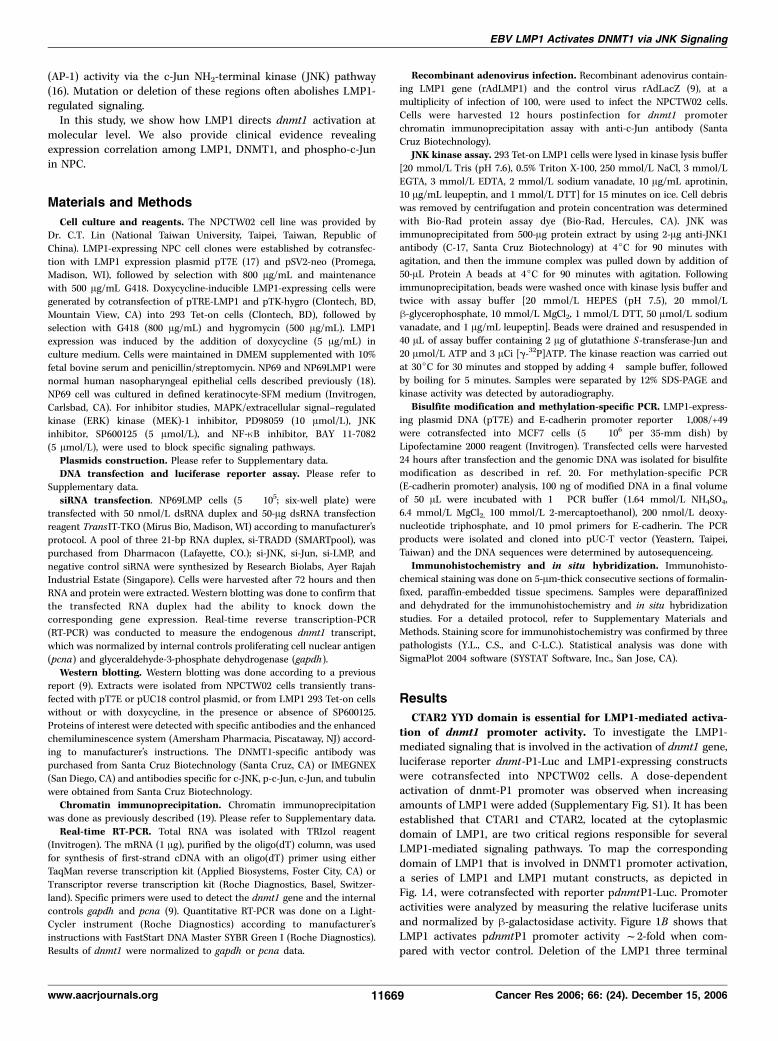
(AP-1) activity via the c-Jun NH2-terminal kinase (JNK) pathway(16). Mutation or deletion of these regions often abolishes LMP1-regulated signaling.In this study, we show how LMP1 directs dnmt1 activation at
molecular level. We also provide clinical evidence revealingexpression correlation among LMP1, DNMT1, and phospho-c-Junin NPC.
Materials and Methods
Cell culture and reagents. The NPCTW02 cell line was provided byDr. C.T. Lin (National Taiwan University, Taipei, Taiwan, Republic of
China). LMP1-expressing NPC cell clones were established by cotransfec-
tion with LMP1 expression plasmid pT7E (17) and pSV2-neo (Promega,Madison, WI), followed by selection with 800 Ag/mL and maintenance
with 500 Ag/mL G418. Doxycycline-inducible LMP1-expressing cells weregenerated by cotransfection of pTRE-LMP1 and pTK-hygro (Clontech, BD,
Mountain View, CA) into 293 Tet-on cells (Clontech, BD), followed byselection with G418 (800 Ag/mL) and hygromycin (500 Ag/mL). LMP1expression was induced by the addition of doxycycline (5 Ag/mL) inculture medium. Cells were maintained in DMEM supplemented with 10%
fetal bovine serum and penicillin/streptomycin. NP69 and NP69LMP1 werenormal human nasopharyngeal epithelial cells described previously (18).
NP69 cell was cultured in defined keratinocyte-SFM medium (Invitrogen,
Carlsbad, CA). For inhibitor studies, MAPK/extracellular signal–regulated
kinase (ERK) kinase (MEK)-1 inhibitor, PD98059 (10 Amol/L), JNKinhibitor, SP600125 (5 Amol/L), and NF-nB inhibitor, BAY 11-7082
(5 Amol/L), were used to block specific signaling pathways.Plasmids construction. Please refer to Supplementary data.DNA transfection and luciferase reporter assay. Please refer to
Supplementary data.
siRNA transfection. NP69LMP cells (5 � 105; six-well plate) were
transfected with 50 nmol/L dsRNA duplex and 50-Ag dsRNA transfectionreagent TransIT-TKO (Mirus Bio, Madison, WI) according to manufacturer’s
protocol. A pool of three 21-bp RNA duplex, si-TRADD (SMARTpool), was
purchased from Dharmacon (Lafayette, CO.); si-JNK, si-Jun, si-LMP, and
negative control siRNA were synthesized by Research Biolabs, Ayer RajahIndustrial Estate (Singapore). Cells were harvested after 72 hours and then
RNA and protein were extracted. Western blotting was done to confirm that
the transfected RNA duplex had the ability to knock down thecorresponding gene expression. Real-time reverse transcription-PCR
(RT-PCR) was conducted to measure the endogenous dnmt1 transcript,
which was normalized by internal controls proliferating cell nuclear antigen
(pcna) and glyceraldehyde-3-phosphate dehydrogenase (gapdh).Western blotting. Western blotting was done according to a previous
report (9). Extracts were isolated from NPCTW02 cells transiently trans-
fected with pT7E or pUC18 control plasmid, or from LMP1 293 Tet-on cells
without or with doxycycline, in the presence or absence of SP600125.Proteins of interest were detected with specific antibodies and the enhanced
chemiluminescence system (Amersham Pharmacia, Piscataway, NJ) accord-
ing to manufacturer’s instructions. The DNMT1-specific antibody waspurchased from Santa Cruz Biotechnology (Santa Cruz, CA) or IMEGNEX
(San Diego, CA) and antibodies specific for c-JNK, p-c-Jun, c-Jun, and tubulin
were obtained from Santa Cruz Biotechnology.
Chromatin immunoprecipitation. Chromatin immunoprecipitationwas done as previously described (19). Please refer to Supplementary data.
Real-time RT-PCR. Total RNA was isolated with TRIzol reagent
(Invitrogen). The mRNA (1 Ag), purified by the oligo(dT) column, was usedfor synthesis of first-strand cDNA with an oligo(dT) primer using eitherTaqMan reverse transcription kit (Applied Biosystems, Foster City, CA) or
Transcriptor reverse transcription kit (Roche Diagnostics, Basel, Switzer-
land). Specific primers were used to detect the dnmt1 gene and the internal
controls gapdh and pcna (9). Quantitative RT-PCR was done on a Light-Cycler instrument (Roche Diagnostics) according to manufacturer’s
instructions with FastStart DNA Master SYBR Green I (Roche Diagnostics).
Results of dnmt1 were normalized to gapdh or pcna data.
Recombinant adenovirus infection. Recombinant adenovirus contain-ing LMP1 gene (rAdLMP1) and the control virus rAdLacZ (9), at a
multiplicity of infection of 100, were used to infect the NPCTW02 cells.
Cells were harvested 12 hours postinfection for dnmt1 promoter
chromatin immunoprecipitation assay with anti-c-Jun antibody (SantaCruz Biotechnology).
JNK kinase assay. 293 Tet-on LMP1 cells were lysed in kinase lysis buffer[20 mmol/L Tris (pH 7.6), 0.5% Triton X-100, 250 mmol/L NaCl, 3 mmol/L
EGTA, 3 mmol/L EDTA, 2 mmol/L sodium vanadate, 10 Ag/mL aprotinin,10 Ag/mL leupeptin, and 1 mmol/L DTT] for 15 minutes on ice. Cell debriswas removed by centrifugation and protein concentration was determined
with Bio-Rad protein assay dye (Bio-Rad, Hercules, CA). JNK was
immunoprecipitated from 500-Ag protein extract by using 2-Ag anti-JNK1antibody (C-17, Santa Cruz Biotechnology) at 4jC for 90 minutes with
agitation, and then the immune complex was pulled down by addition of
50-AL Protein A beads at 4jC for 90 minutes with agitation. Followingimmunoprecipitation, beads were washed once with kinase lysis buffer and
twice with assay buffer [20 mmol/L HEPES (pH 7.5), 20 mmol/L
h-glycerophosphate, 10 mmol/L MgCl2, 1 mmol/L DTT, 50 Amol/L sodiumvanadate, and 1 Ag/mL leupeptin]. Beads were drained and resuspended in40 AL of assay buffer containing 2 Ag of glutathione S-transferase-Jun and
20 Amol/L ATP and 3 ACi [g-32P]ATP. The kinase reaction was carried outat 30jC for 30 minutes and stopped by adding 4� sample buffer, followed
by boiling for 5 minutes. Samples were separated by 12% SDS-PAGE andkinase activity was detected by autoradiography.
Bisulfite modification and methylation-specific PCR. LMP1-express-ing plasmid DNA (pT7E) and E-cadherin promoter reporter �1,008/+49were cotransfected into MCF7 cells (5 � 106 per 35-mm dish) by
Lipofectamine 2000 reagent (Invitrogen). Transfected cells were harvested
24 hours after transfection and the genomic DNA was isolated for bisulfite
modification as described in ref. 20. For methylation-specific PCR(E-cadherin promoter) analysis, 100 ng of modified DNA in a final volume
of 50 AL were incubated with 1� PCR buffer (1.64 mmol/L NH4SO4,
6.4 mmol/L MgCl2, 100 mmol/L 2-mercaptoethanol), 200 nmol/L deoxy-
nucleotide triphosphate, and 10 pmol primers for E-cadherin. The PCRproducts were isolated and cloned into pUC-T vector (Yeastern, Taipei,
Taiwan) and the DNA sequences were determined by autosequenceing.
Immunohistochemistry and in situ hybridization. Immunohisto-chemical staining was done on 5-Am-thick consecutive sections of formalin-fixed, paraffin-embedded tissue specimens. Samples were deparaffinized
and dehydrated for the immunohistochemistry and in situ hybridization
studies. For a detailed protocol, refer to Supplementary Materials andMethods. Staining score for immunohistochemistry was confirmed by three
pathologists (Y.L., C.S., and C-L.C.). Statistical analysis was done with
SigmaPlot 2004 software (SYSTAT Software, Inc., San Jose, CA).
Results
CTAR2 YYD domain is essential for LMP1-mediated activa-tion of dnmt1 promoter activity. To investigate the LMP1-mediated signaling that is involved in the activation of dnmt1 gene,luciferase reporter dnmt-P1-Luc and LMP1-expressing constructswere cotransfected into NPCTW02 cells. A dose-dependentactivation of dnmt-P1 promoter was observed when increasingamounts of LMP1 were added (Supplementary Fig. S1). It has beenestablished that CTAR1 and CTAR2, located at the cytoplasmicdomain of LMP1, are two critical regions responsible for severalLMP1-mediated signaling pathways. To map the correspondingdomain of LMP1 that is involved in DNMT1 promoter activation,a series of LMP1 and LMP1 mutant constructs, as depicted inFig. 1A , were cotransfected with reporter pdnmtP1-Luc. Promoteractivities were analyzed by measuring the relative luciferase unitsand normalized by h-galactosidase activity. Figure 1B shows thatLMP1 activates pdnmtP1 promoter activity f2-fold when com-pared with vector control. Deletion of the LMP1 three terminal
EBV LMP1 Activates DNMT1 via JNK Signaling
www.aacrjournals.org 11669 Cancer Res 2006; 66: (24). December 15, 2006
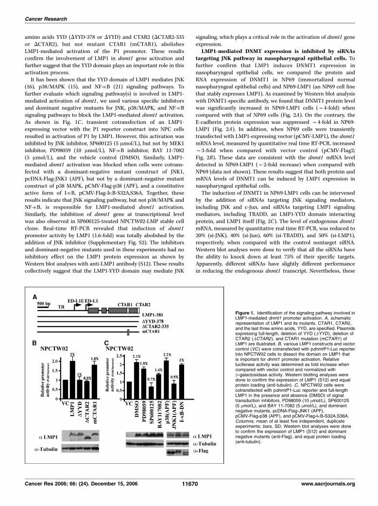
amino acids YYD (DYYD-378 or DYYD) and CTAR2 (DCTAR2-335or DCTAR2), but not mutant CTAR1 (mCTAR1), abolishesLMP1-mediated activation of the P1 promoter. These resultsconfirm the involvement of LMP1 in dnmt1 gene activation andfurther suggest that the YYD domain plays an important role in thisactivation process.It has been shown that the YYD domain of LMP1 mediates JNK
(16), p38/MAPK (15), and NF-nB (21) signaling pathways. Tofurther evaluate which signaling pathway(s) is involved in LMP1-mediated activation of dnmt1 , we used various specific inhibitorsand dominant negative mutants for JNK, p38/MAPK, and NF-nBsignaling pathways to block the LMP1-mediated dnmt1 activation.As shown in Fig. 1C , transient cotransfection of an LMP1-expressing vector with the P1 reporter construct into NPC cellsresulted in activation of P1 by LMP1. However, this activation wasinhibited by JNK inhibitor, SP600125 (5 Amol/L), but not by MEK1inhibitor, PD98059 (10 Amol/L), NF-nB inhibitor, BAY 11-7082(5 Amol/L), and the vehicle control (DMSO). Similarly, LMP1-mediated dnmt1 activation was blocked when cells were cotrans-fected with a dominant-negative mutant construct of JNK1,pcDNA-Flag-JNK1 (APF), but not by a dominant-negative mutantconstruct of p38 MAPK, pCMV-Flag-p38 (APF), and a constitutiveactive form of I-nB, pCMV-Flag-InB-S32A,S36A. Together, theseresults indicate that JNK signaling pathway, but not p38/MAPK andNF-nB, is responsible for LMP1-mediated dnmt1 activation.Similarly, the inhibition of dnmt1 gene at transcriptional levelwas also observed in SP600125-treated NPCTW02-LMP stable cellclone. Real-time RT-PCR revealed that induction of dnmt1promoter activity by LMP1 (1.6-fold) was totally abolished by theaddition of JNK inhibitor (Supplementary Fig. S2). The inhibitorsand dominant-negative mutants used in these experiments had noinhibitory effect on the LMP1 protein expression as shown byWestern blot analyses with anti-LMP1 antibody (S12). These resultscollectively suggest that the LMP1-YYD domain may mediate JNK
signaling, which plays a critical role in the activation of dnmt1 geneexpression.LMP1-mediated DNMT expression is inhibited by siRNAs
targeting JNK pathway in nasopharyngeal epithelial cells. Tofurther confirm that LMP1 induces DNMT1 expression innasopharyngeal epithelial cells, we compared the protein andRNA expression of DNMT1 in NP69 (immortalized normalnasopharyngeal epithelial cells) and NP69-LMP1 (an NP69 cell linethat stably expresses LMP1). As examined by Western blot analysiswith DNMT1-specific antibody, we found that DNMT1 protein levelwas significantly increased in NP69-LMP1 cells (f4-fold) whencompared with that of NP69 cells (Fig. 2A). On the contrary, theE-cadherin protein expression was suppressed f4-fold in NP69-LMP1 (Fig. 2A). In addition, when NP69 cells were transientlytransfected with LMP1-expressing vector (pCMV-LMP1), the dnmt1mRNA level, measured by quantitative real time RT-PCR, increasedf3-fold when compared with vector control (pCMV-Flag2;Fig. 2B). These data are consistent with the dnmt1 mRNA leveldetected in NP69-LMP1 (f2-fold increase) when compared withNP69 (data not shown). These results suggest that both protein andmRNA levels of DNMT1 can be induced by LMP1 expression innasopharyngeal epithelial cells.The induction of DNMT1 in NP69-LMP1 cells can be intervened
by the addition of siRNAs targeting JNK signaling mediators,including JNK and c-Jun, and siRNAs targeting LMP1 signalingmediators, including TRADD, an LMP1-YYD domain interactingprotein, and LMP1 itself (Fig. 2C). The level of endogenous dnmt1mRNA, measured by quantitative real time RT-PCR, was reduced to20% (si-JNK), 40% (si-Jun), 60% (si-TRADD), and 50% (si-LMP1),respectively, when compared with the control nontarget siRNA.Western blot analyses were done to verify that all the siRNAs havethe ability to knock down at least 75% of their specific targets.Apparently, different siRNAs have slightly different performancein reducing the endogenous dnmt1 transcript. Nevertheless, these
Figure 1. Identification of the signaling pathway involved inLMP1-mediated dnmt1 promoter activation. A, schematicrepresentation of LMP1 and its mutants. CTAR1, CTAR2,and the last three amino acids, YYD, are specified. Plasmidsexpressing full-length, deletion of YYD (DYYD ), deletion ofCTAR2 (DCTAR2 ), and CTAR1 mutation (mCTAR1 ) ofLMP1 are illustrated. B, various LMP1 constructs and vectorcontrol (VC ) were cotransfected with pdnmtP1-Luc reporterinto NPCTW02 cells to dissect the domain on LMP1 thatis important for dnmt1 promoter activation. Relativeluciferase activity was determined as fold increase whencompared with vector control and normalized withh-galactosidase activity. Western blotting analyses weredone to confirm the expression of LMP1 (S12) and equalprotein loading (anti-tubulin). C, NPCTW02 cells werecotransfected with pdnmtP1-Luc reporter and full-lengthLMP1 in the presence and absence (DMSO) of signaltransduction inhibitors, PD98059 (10 Amol/L), SP600125(5 Amol/L), and BAY 11-7082 (5 Amol/L), and dominantnegative mutants, pcDNA-Flag-JNK1 (APF),pCMV-Flag-p38 (APF), and pCMV-Flag-InB-S32A,S36A.Columns, mean of at least five independent, duplicateexperiments; bars, SD. Western blot analyses were doneto confirm the expression of LMP1 (S12) and dominantnegative mutants (anti-Flag), and equal protein loading(anti-tubulin).
Cancer Research
Cancer Res 2006; 66: (24). December 15, 2006 11670 www.aacrjournals.org
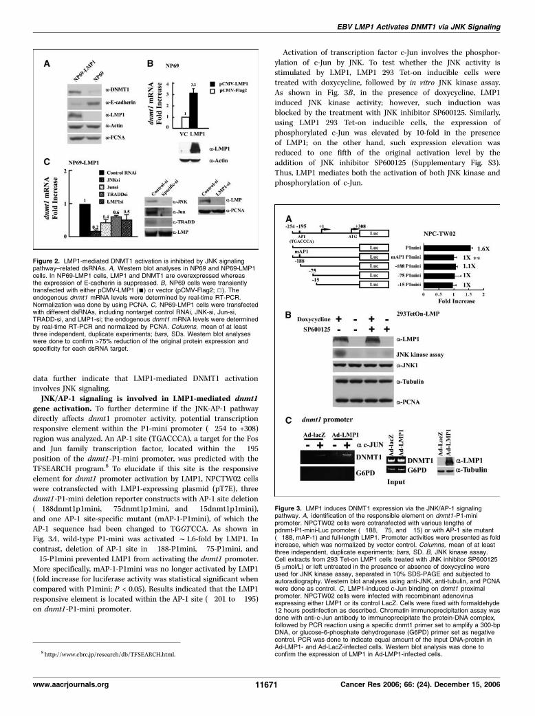
data further indicate that LMP1-mediated DNMT1 activationinvolves JNK signaling.JNK/AP-1 signaling is involved in LMP1-mediated dnmt1
gene activation. To further determine if the JNK-AP-1 pathwaydirectly affects dnmt1 promoter activity, potential transcriptionresponsive element within the P1-mini promoter (�254 to +308)region was analyzed. An AP-1 site (TGACCCA), a target for the Fosand Jun family transcription factor, located within the �195position of the dnmt1-P1-mini promoter, was predicted with theTFSEARCH program.8 To elucidate if this site is the responsiveelement for dnmt1 promoter activation by LMP1, NPCTW02 cellswere cotransfected with LMP1-expressing plasmid (pT7E), threednmt1-P1-mini deletion reporter constructs with AP-1 site deletion(�188dnmt1p1mini, �75dnmt1p1mini, and �15dnmt1p1mini),and one AP-1 site-specific mutant (mAP-1-P1mini), of which theAP-1 sequence had been changed to TGGTCCA. As shown inFig. 3A , wild-type P1-mini was activated f1.6-fold by LMP1. Incontrast, deletion of AP-1 site in �188-P1mini, �75-P1mini, and�15-P1mini prevented LMP1 from activating the dnmt1 promoter.More specifically, mAP-1-P1mini was no longer activated by LMP1( fold increase for luciferase activity was statistical significant whencompared with P1mini; P < 0.05). Results indicated that the LMP1responsive element is located within the AP-1 site (�201 to �195)on dnmt1-P1-mini promoter.
Activation of transcription factor c-Jun involves the phosphor-ylation of c-Jun by JNK. To test whether the JNK activity isstimulated by LMP1, LMP1 293 Tet-on inducible cells weretreated with doxycycline, followed by in vitro JNK kinase assay.As shown in Fig. 3B , in the presence of doxycycline, LMP1induced JNK kinase activity; however, such induction wasblocked by the treatment with JNK inhibitor SP600125. Similarly,using LMP1 293 Tet-on inducible cells, the expression ofphosphorylated c-Jun was elevated by 10-fold in the presenceof LMP1; on the other hand, such expression elevation wasreduced to one fifth of the original activation level by theaddition of JNK inhibitor SP600125 (Supplementary Fig. S3).Thus, LMP1 mediates both the activation of both JNK kinase andphosphorylation of c-Jun.
8 http://www.cbrc.jp/research/db/TFSEARCH.html.
Figure 3. LMP1 induces DNMT1 expression via the JNK/AP-1 signalingpathway. A, identification of the responsible element on dnmt1-P1-minipromoter. NPCTW02 cells were cotransfected with various lengths ofpdnmt-P1-mini-Luc promoter (�188, �75, and �15) or with AP-1 site mutant(�188, mAP-1) and full-length LMP1. Promoter activities were presented as foldincrease, which was normalized by vector control. Columns, mean of at leastthree independent, duplicate experiments; bars, SD. B, JNK kinase assay.Cell extracts from 293 Tet-on LMP1 cells treated with JNK inhibitor SP600125(5 Amol/L) or left untreated in the presence or absence of doxycycline wereused for JNK kinase assay, separated in 10% SDS-PAGE and subjected toautoradiography. Western blot analyses using anti-JNK, anti-tubulin, and PCNAwere done as control. C, LMP1-induced c-Jun binding on dnmt1 proximalpromoter. NPCTW02 cells were infected with recombinant adenovirusexpressing either LMP1 or its control LacZ. Cells were fixed with formaldehyde12 hours postinfection as described. Chromatin immunoprecipitation assay wasdone with anti-c-Jun antibody to immunoprecipitate the protein-DNA complex,followed by PCR reaction using a specific dnmt1 primer set to amplify a 300-bpDNA, or glucose-6-phosphate dehydrogenase (G6PD) primer set as negativecontrol. PCR was done to indicate equal amount of the input DNA-protein inAd-LMP1- and Ad-LacZ-infected cells. Western blot analysis was done toconfirm the expression of LMP1 in Ad-LMP1-infected cells.
Figure 2. LMP1-mediated DNMT1 activation is inhibited by JNK signalingpathway–related dsRNAs. A, Western blot analyses in NP69 and NP69-LMP1cells. In NP69-LMP1 cells, LMP1 and DNMT1 are overexpressed whereasthe expression of E-cadherin is suppressed. B, NP69 cells were transientlytransfected with either pCMV-LMP1 (n) or vector (pCMV-Flag2; 5). Theendogenous dnmt1 mRNA levels were determined by real-time RT-PCR.Normalization was done by using PCNA. C, NP69-LMP1 cells were transfectedwith different dsRNAs, including nontarget control RNAi, JNK-si, Jun-si,TRADD-si, and LMP1-si; the endogenous dnmt1 mRNA levels were determinedby real-time RT-PCR and normalized by PCNA. Columns, mean of at leastthree independent, duplicate experiments; bars, SDs. Western blot analyseswere done to confirm >75% reduction of the original protein expression andspecificity for each dsRNA target.
EBV LMP1 Activates DNMT1 via JNK Signaling
www.aacrjournals.org 11671 Cancer Res 2006; 66: (24). December 15, 2006
To show that the activated c-Jun physically binds to the dnmt1promoter in the presence of LMP1, chromatin immunoprecipitationassay using anti-c-Jun antibody was conducted. From cells infectedwith the LMP1-expressing recombinant adenovirus, a differentialhigher level of dnmt1 promoter DNA was detected in the chromatinimmunoprecipitation assay as compared with that of the controlvirus (Fig. 3C), indicating that LMP1 induces dnmt1 promoteractivation through phosphorylation of c-Jun transcription factor.And this activated phospho-c-Jun, in turn, binds to the dnmt1promoter AP-1 site and transcriptionally activates the promoter.JNK inhibitor blocks LMP1-mediated formation of tran-
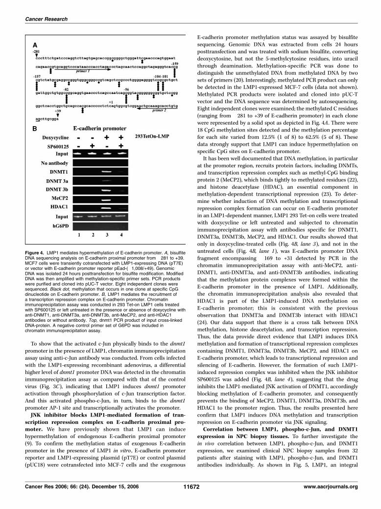
scription repression complex on E-cadherin proximal pro-moter. We have previously shown that LMP1 can inducehypermethylation of endogenous E-cadherin proximal promoter(9). To confirm the methylation status of exogenous E-cadherinpromoter in the presence of LMP1 in vitro , E-cadherin promoterreporter and LMP1-expressing plasmid (pT7E) or control plasmid(pUC18) were cotransfected into MCF-7 cells and the exogenous
E-cadherin promoter methylation status was assayed by bisulfitesequencing. Genomic DNA was extracted from cells 24 hoursposttransfection and was treated with sodium bisulfite, convertingdeoxycytosine, but not the 5-methylcytosine residues, into uracilthrough deamination. Methylation-specific PCR was done todistinguish the unmethylated DNA from methylated DNA by twosets of primers (20). Interestingly, methylated PCR product can onlybe detected in the LMP1-expressed MCF-7 cells (data not shown).Methylated PCR products were isolated and cloned into pUC-Tvector and the DNA sequence was determined by autosequencing.Eight independent clones were examined; the methylated C residues(ranging from �281 to +39 of E-cadherin promoter) in each clonewere represented by a solid spot as depicted in Fig. 4A . There were18 CpG methylation sites detected and the methylation percentagefor each site varied from 12.5% (1 of 8) to 62.5% (5 of 8). Thesedata strongly support that LMP1 can induce hypermethylation onspecific CpG sites on E-cadherin promoter.It has been well documented that DNA methylation, in particular
at the promoter region, recruits protein factors, including DNMTs,and transcription repression complex such as methyl-CpG bindingprotein 2 (MeCP2), which binds tightly to methylated residues (22),and histone deacetylase (HDAC), an essential component inmethylation-dependent transcriptional repression (23). To deter-mine whether induction of DNA methylation and transcriptionalrepression complex formation can occur on E-cadherin promoterin an LMP1-dependent manner, LMP1 293 Tet-on cells were treatedwith doxycycline or left untreated and subjected to chromatinimmunoprecipitation assay with antibodies specific for DNMT1,DNMT3a, DNMT3b, MeCP2, and HDAC1. Our results showed thatonly in doxycycline-treated cells (Fig. 4B, lane 3), and not in theuntreated cells (Fig. 4B, lane 1), was E-cadherin promoter DNAfragment encompassing �169 to +31 detected by PCR in thechromatin immunoprecipitation assay with anti-MeCP2, anti-DNMT1, anti-DNMT3a, and anti-DNMT3b antibodies, indicatingthat the methylation protein complexes were formed within theE-cadherin promoter in the presence of LMP1. Additionally,the chromatin immunoprecipitation analysis also revealed thatHDAC1 is part of the LMP1-induced DNA methylation onE-cadherin promoter; this is consistent with the previousobservation that DNMT3a and DNMT3b interact with HDAC1(24). Our data support that there is a cross talk between DNAmethylation, histone deacetylation, and transcription repression.Thus, the data provide direct evidence that LMP1 induces DNAmethylation and formation of transcriptional repression complexescontaining DNMT1, DNMT3a, DNMT3b, MeCP2, and HDAC1 onE-cadherin promoter, which leads to transcriptional repression andsilencing of E-cadherin. However, the formation of such LMP1-induced repression complex was inhibited when the JNK inhibitorSP600125 was added (Fig. 4B, lane 4), suggesting that the druginhibits the LMP1-mediated JNK activation of DNMT1, accordinglyblocking methylation of E-cadherin promoter, and consequentlyprevents the binding of MeCP2, DNMT1, DNMT3a, DNMT3b, andHDAC1 to the promoter region. Thus, the results presented hereconfirm that LMP1 induces DNA methylation and transcriptionrepression on E-cadherin promoter via JNK signaling.Correlation between LMP1, phospho-c-Jun, and DNMT1
expression in NPC biopsy tissues. To further investigate thein vivo correlation between LMP1, phospho-c-Jun, and DNMT1expression, we examined clinical NPC biopsy samples from 32patients after staining with LMP1, phospho-c-Jun, and DNMT1antibodies individually. As shown in Fig. 5, LMP1, an integral
Figure 4. LMP1 mediates hypermethylation of E-cadherin promoter. A, bisulfiteDNA sequencing analysis on E-cadherin proximal promoter from �281 to +39.MCF7 cells were transiently cotransfected with LMP1-expressing DNA (pT7E)or vector with E-cadherin promoter reporter pEad-(�1,008/+49). GenomicDNA was isolated 24 hours posttransfection for bisulfite modification. ModifiedDNA was then amplified with methylation-specific primer sets. PCR productswere purified and cloned into pUC-T vector. Eight independent clones weresequenced. Black dot, methylation that occurs in one clone at specific CpGdinucleotide on E-cadherin promoter. B, LMP1 mediates the recruitment ofa transcription repression complex on E-cadherin promoter. Chromatinimmunoprecipitation assay was conducted in 293 Tet-on LMP1 cells treatedwith SP600125 or left untreated in the presence or absence of doxycycline withanti-DNMT1, anti-DNMT3a, anti-DNMT3b, anti-MeCP2, and anti-HDAC1antibodies or without antibody. Top, dnmt1 PCR product of input cross-linkedDNA-protein. A negative control primer set of G6PD was included inchromatin immunoprecipitation assay.
Cancer Research
Cancer Res 2006; 66: (24). December 15, 2006 11672 www.aacrjournals.org
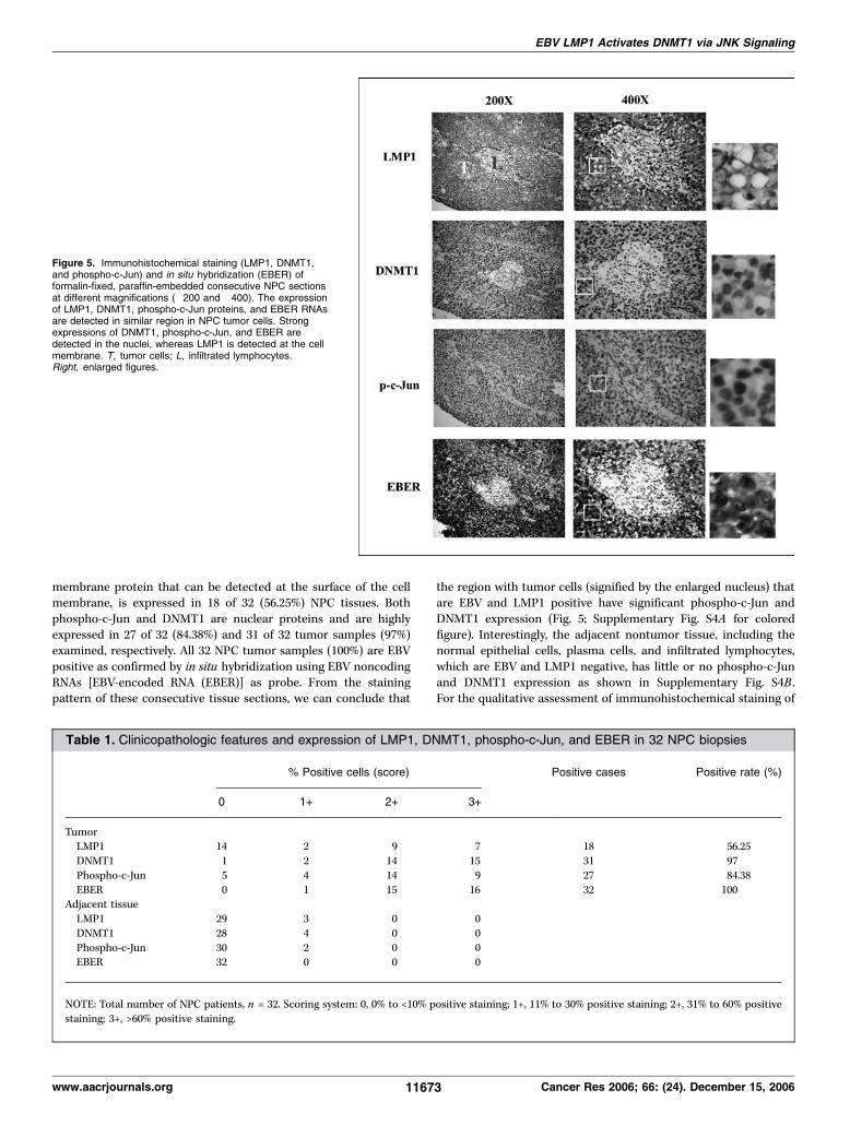
membrane protein that can be detected at the surface of the cellmembrane, is expressed in 18 of 32 (56.25%) NPC tissues. Bothphospho-c-Jun and DNMT1 are nuclear proteins and are highlyexpressed in 27 of 32 (84.38%) and 31 of 32 tumor samples (97%)examined, respectively. All 32 NPC tumor samples (100%) are EBVpositive as confirmed by in situ hybridization using EBV noncodingRNAs [EBV-encoded RNA (EBER)] as probe. From the stainingpattern of these consecutive tissue sections, we can conclude that
the region with tumor cells (signified by the enlarged nucleus) thatare EBV and LMP1 positive have significant phospho-c-Jun andDNMT1 expression (Fig. 5; Supplementary Fig. S4A for coloredfigure). Interestingly, the adjacent nontumor tissue, including thenormal epithelial cells, plasma cells, and infiltrated lymphocytes,which are EBV and LMP1 negative, has little or no phospho-c-Junand DNMT1 expression as shown in Supplementary Fig. S4B .For the qualitative assessment of immunohistochemical staining of
Figure 5. Immunohistochemical staining (LMP1, DNMT1,and phospho-c-Jun) and in situ hybridization (EBER) offormalin-fixed, paraffin-embedded consecutive NPC sectionsat different magnifications (�200 and �400). The expressionof LMP1, DNMT1, phospho-c-Jun proteins, and EBER RNAsare detected in similar region in NPC tumor cells. Strongexpressions of DNMT1, phospho-c-Jun, and EBER aredetected in the nuclei, whereas LMP1 is detected at the cellmembrane. T, tumor cells; L, infiltrated lymphocytes.Right, enlarged figures.
Table 1. Clinicopathologic features and expression of LMP1, DNMT1, phospho-c-Jun, and EBER in 32 NPC biopsies
% Positive cells (score) Positive cases Positive rate (%)
0 1+ 2+ 3+
TumorLMP1 14 2 9 7 18 56.25
DNMT1 1 2 14 15 31 97
Phospho-c-Jun 5 4 14 9 27 84.38
EBER 0 1 15 16 32 100Adjacent tissue
LMP1 29 3 0 0
DNMT1 28 4 0 0
Phospho-c-Jun 30 2 0 0EBER 32 0 0 0
NOTE: Total number of NPC patients, n = 32. Scoring system: 0, 0% to <10% positive staining; 1+, 11% to 30% positive staining; 2+, 31% to 60% positive
staining; 3+, >60% positive staining.
EBV LMP1 Activates DNMT1 via JNK Signaling
www.aacrjournals.org 11673 Cancer Res 2006; 66: (24). December 15, 2006

tumor and nontumor cells, a 0 to 3+ scoring system was used torepresent the percentage of the positive cells, as shown in Table 1.A moderately positive correlation was found between LMP1 andDNMT1 expression (Pearson correlation R = 0.58; SupplementaryFig. S5) and between LMP1 and phospho-c-Jun (R = 0.63). However,significantly positive correlations between (i) DNMT1 and EBERexpression (R = 0.91; Supplementary Fig. S5), (ii) phospho-c-Junand EBER expression (R = 0.75), and (iii) phospho-c-Jun andDNMT1 expression (R = 0.75) are observed. These clinical datasupport our findings that LMP1 expression correlates with highphospho-c-Jun and DNMT1 expression.
Discussion
LMP1, an EBV oncoprotein, is required for EBV-mediatedtransformation. The higher migration ability of LMP1-expressedcells correlates with the repression of E-cadherin (9, 25), anadhesion molecule that is often lost in many cancers includingNPC (26); nevertheless, the molecular mechanism was not clear.Previously, we have shown that LMP1 is responsible for the down-regulation of E-cadherin expression by inducing DNMTs, resultingin hypermethylation of E-cadherin promoter (9). To continue ourlast study, here we provide detailed evidence on how LMP1 confersDNMT induction at molecular level.LMP1 mainly activates the major promoter, P1, of the dnmt1
gene. Deletion of CTAR2, or specifically the last three amino acids,YYD, resulted in failure to activate the P1 promoter. To date, thereare at least three well-defined signaling pathways, NF-nB, p38/MAPK, and JNK, which are elicited by LMP1 COOH-terminaldomain (13, 27). According to the studies on inhibitors anddominant negative mutants and si-targets, we have identified theJNK pathway, but not the NF-nB or p38/MAPK signaling pathway,which contributes to LMP1-mediated P1 dnmt1 gene activation.JNK is one of the three subfamilies of MAPK and is characterizedas a stress-activated protein kinase in response to growth stimuli,cellular transformation, and tumor cell metastasis (28–30). Theactivated kinase subsequently phosphorylates its direct target,c-Jun, a component of AP-1 complex, forming homodimer orheterodimeric complexes with c-Fos. Finally, the phosphorylatedc-Jun binds to and transactivates the promoter containing the AP-1site. LMP1 induction of JNK kinase activity may be regarded asa growth advantage to EBV latent infected cells due to the versatilenature of this important signaling pathway. However, LMP1-induced dnmt1 activation and JNK kinase activity can be specificallyblocked by the inhibitor SP600125 (5 Amol/L). The compoundpreferentially blocks c-Jun phosphorylation and has no effect onother MAPKs, such as ERKs, p38/MAPK, and InB kinases (ref. 31;data not shown). To confirm the involvement of JNK signaling inregulating LMP1-mediated dnmt1 promoter activity, we searchedfor the potential AP-1 site on dnmt1 promoter. As expected, welocated an AP-1 responsive element within the �201 to �195 site ofthe dnmt-P1-mini promoter. The importance of this particular AP-1site was confirmed by the in vitro dnmt1 promoter reporter assay(Fig. 4A), in which the mutated AP-1 site on dnmt1 promoterabolishes the LMP1-mediated dnmt1 activation. Furthermore,chromatin immunoprecipitation assay showed that in the presenceof LMP1, more c-Jun physically binds to the AP-1 site on dnmt1promoter (Fig. 4C). Accordingly, we show for the first time thatDNMT1 is a direct target of LMP1-mediated JNK signaling.Our previous report showed that LMP1 additionally activates
DNMT3a and DNMT3b (9). In this study, DNMT3a and DNMT3b
are detected in the LMP1-induced repression complex; yet, theformation of this complex is inhibited by SP600125 (Fig. 4B). Anf2- to 3-fold decrease in DNMT3a and DNMT3b mRNA levels wasdetected in the presence of SP600125 (data not shown). Sequenceanalyses of DNMT3a and DNMT3b promoters reveal the presenceof AP-1 sites that are potentially regulated by LMP1. Thus, LMP1may also activate these two promoters via JNK activation.We identified earlier an LMP1-mediated DNMT1 target gene,
E-cadherin (9), an adhesion molecule that mediates cell-cellcontact and is important for tissue morphogenesis, cell polarity,and tumor invasiveness (32). On DNMT1 activation, bothtranscriptional and translational levels of E-cadherin are signifi-cantly reduced. DNMT1 catalyzes the methylation of CpGdinucleotides on DNA, which leads to suppression of geneexpression. Loss of membranous E-cadherin expression results inenhanced cell migration activity (9, 33), which significantlycorrelates with tumor invasion, advanced disease stage, and tumormetastasis (34). Thus, E-cadherin is considered as a tumorsuppressor gene (35–39). In this current study, chromatinimmunoprecipitation assay reveals that LMP1 mediates inductionof a repression complex containing DNMT1, DNMT3a, DNMT3b,MeCP2, and HDAC1 on E-cadherin proximal promoter. This findingis consistent with a previous report showing that DNMT3a andDNMT3b interact with HDAC1 (24). At present, the sequentialorder for recruiting these transcription repression factors remainsunclear. Again, the formation of this repression complex isinhibited by SP600125, suggesting that JNK signaling is the key
Figure 6. A model for LMP1-mediated DNMT1 activation via JNK/AP-1signaling. Activated DNMT1 hypermethylates E-cadherin promoter and recruitsa transcriptional repression complex leading to gene silencing. The YYDdomain of LMP1 activates the JNK signaling pathway and activated JNK, inturn, phosphorylates transcription factor c-Jun. Phosphorylated c-Jun of AP-1complex binds and transactivates the dnmt1 promoter. Elevated DNMT1expression leads to hypermethylation of E-cadherin gene and formation of atranscriptional repression complex including DNMTs, methyl-binding proteins(MBD ), and HDAC. This LMP1-mediated DNMT1 activation can be blocked byJNK inhibitor SP600125, dominant negative mutant (DN-JNK ), and dsRNAs(JNK-si, c-Jun-si, TRADD-si, and LMP1-si).
Cancer Research
Cancer Res 2006; 66: (24). December 15, 2006 11674 www.aacrjournals.org
for triggering the LMP1-DNMT1–mediated gene silencing cascade.One may speculate that DNMTs bind to the E-cadherin first;however, it is difficult to predict which factor, HDAC or MeCP2,would be the next one to be recruited.We investigated the correlation between the expression of LMP1,
DNMT1, phospho-c-Jun, and EBER in 32 NPC biopsies. Clinicaldata in this study also support that LMP1 expression or EBVinfection correlates with JNK signaling and DNMT1 overexpression.Indeed, elevated DNMT expression may be functionally importantfor NPC tumor formation because activation of DNA methyltrans-ferases has been reported in other human malignant diseases(40–43). The positive rate for LMP1 expression detected byimmunostaining was moderate (f56%) and variable whencompared with that of DNMT1 (>90%) and phospho-c-Jun(84.38%) in NPC biopsies. Among the LMP1-positive biopsies, thestaining pattern for LMP1 is heterogeneous, which is similar toprevious reports (44–46), suggesting that only a subset of NPC cellsexpress LMP1. Using a more sensitive RT-PCR–based detection,several reports revealed much higher frequencies (>80%) of LMP1expression (47, 48). In addition, >70% of NPC patients’ sera arepositive for anti-LMP1 (49). Taken together, the positive rate forLMP1 in NPC is likely underestimated due to the sensitivity andspecificity of the detection method. Apart from these limitations,we do observe in the consecutive NPC sections that most of theLMP1-positive cells are also DNMT1 and phospho-c-Jun positive,and these three proteins coexpress in a similar tissue region but atdifferent cellular compartments, as shown in Fig. 5. Although thecorrelation between LMP1 and DNMT1 and between LMP1 andphospho-c-Jun in our study is considered moderate (R = 0.58 andR = 0.63, respectively), this may not reflect the real situation whenthe underestimated LMP1 expression is considered. It is interestingto note that the noncoding EBV EBER RNA expression correlateswell to DNMT1 (R = 0.91) and phospho-c-Jun (R = 0.75) expression.Because EBERs do not code for proteins, it is unlikely that EBERexpression has direct effect on DNMT1 and phospho-c-Jun
activation, although this possibility cannot be ruled out. This, onthe other hand, suggests that besides LMP1, EBV infection per se orother EBV latent genes may have an additive effect on DNMT1 andphospho-c-Jun up-regulation.A model for this LMP1-JNK-DNMT1 signaling is then proposed
based on our findings (Fig. 6). Taken together, our data clearlyshow that there is an important link between LMP1-mediatedJNK signaling, epigenetic gene regulation, and gene silencing.Conceivably, E-cadherin is not the only gene of which thetranscription is being down-regulated by LMP1-DNMT1. It wouldbe of interest for us to identify other genes that are regulated bysimilar mechanism. Indeed, an earlier study shows that promotersof two other tumor suppressor genes, p16 and RASSF1A , arehypermethylated in NPC (50). The issue of whether DNAmethylation of these two promoters is mediated by LMP1 is stillunder investigation. Our data suggest that HDAC1 and DNMTsmay be potential dual targets for treating EBV LMP1-positivetumors. Thus, elucidation of the mechanism by which LMP1regulates DNA methyltransferases is critical for the developmentof therapeutic agents.
Acknowledgments
Received 6/19/2006; revised 9/8/2006; accepted 10/11/2006.Grant support: National Science Council, Taiwan, grants NSC92-2320-B-182-043
and NSC92-2320-B-182-048 (Y-S. Chang), Chang-Gung Memorial Hospital grantCMRPG340121 (H-P. Li) and Chang-Gung University grant CMRPD32040 (Y-S. Chang)and Chang Gung Molecular Medicine Research Center grant CMRPD140041(Y-S. Chang).The costs of publication of this article were defrayed in part by the payment of page
charges. This article must therefore be hereby marked advertisement in accordancewith 18 U.S.C. Section 1734 solely to indicate this fact.We thank Prof. Roger Davis (Howard Hughes Medical Institute, University of
Massachusetts Medical School) for providing the dominant negative mutants pcDNA-Flag-JNK1(APF) and pCMV-Flag-p38(APF); Dr. Dean W. Ballard (Vanderbilt University,Nashville, TN) for providing the dominant negative construct InB-S32A,S36A;Prof. James C.K. Shen (Institute of Molecular Biology, Academia Sinica, Taipei,Taiwan) for critical suggestions; and the Chang-Gung Molecular Medicine ResearchCenter, Pathology Core for assisting with the immunohistochemical staining.
EBV LMP1 Activates DNMT1 via JNK Signaling
www.aacrjournals.org 11675 Cancer Res 2006; 66: (24). December 15, 2006
References1. Klein G, Giovanella BC, Lindahl T, et al. Directevidence for the presence of Epstein-Barr virus DNAand nuclear antigen in malignant epithelial cellsfrom patients with poorly differentiated carcinoma ofthe nasopharynx. Proc Natl Acad Sci U S A 1974;71:4737–41.
2. Shibata D, Weiss LM. Epstein-Barr virus-associatedgastric adenocarcinoma. Am J Pathol 1992;140:769–74.
3. Bonnet M, Guinebretiere JM, Kremmer E, et al.Detection of Epstein-Barr virus in invasive breastcancers. J Natl Cancer Inst 1999;91:1376–81.
4. Hsu MM, Tu SM. Nasopharyngeal carcinoma inTaiwan. Clinical manifestations and results of therapy.Cancer 1983;52:362–8.
5. Tsao SW, Tramoutanis G, Dawson CW, Lo AK,Huang DP. The significance of LMP1 expression innasopharyngeal carcinoma. Semin Cancer Biol 2002;12:473–87.
6. Wang D, Liebowitz D, Kieff E. An EBV membraneprotein expressed in immortalized lymphocytestransforms established rodent cells. Cell 1985;43:831–40.
7. Fahraeus R, Rymo L, Rhim JS, Klein G. Morpholog-ical transformation of human keratinocytes expressingthe LMP gene of Epstein-Barr virus. Nature 1990;345:447–9.
8. Kim KR, Yoshizaki T, Miyamori H, et al. Transforma-tion of Madin-Darby canine kidney (MDCK) epithelial
cells by Epstein-Barr virus latent membrane protein 1(LMP1) induces expression of Ets1 and invasive growth.Oncogene 2000;19:1764–71.
9. Tsai CN, Tsai CL, Tse KP, Chang HY, Chang YS. TheEpstein-Barr virus oncogene product, latent mem-brane protein 1, induces the down-regulation of E-cadherin gene expression via activation of DNAmethyltransferases. Proc Natl Acad Sci U S A 2002;99:10084–9.
10. Jones PA, Laird PW. Cancer epigenetics comes of age.Nat Genet 1999;21:163–7.
11. Baylin SB, Herman JG. Promoter hypermethylation-can this change alone ever designate true tumorsuppressor gene function? J Natl Cancer Inst 2001;93:664–5.
12. Bigey P, Ramchandani S, Theberge J, Araujo FD,Szyf M. Transcriptional regulation of the humanDNA methyltransferase (dnmt1) gene. Gene 2000;242:407–18.
13. Li HP, Chang YS. Epstein-Barr virus latent membraneprotein 1: structure and functions. J Biomed Sci 2003;10:490–504.
14. Devergne O, Hatzivassiliou E, Izumi KM, et al.Association of TRAF1, TRAF2, and TRAF3 with anEpstein-Barr virus LMP1 domain important for B-lymphocyte transformation: role in NF-nB activation.Mol Cell Biol 1996;16:7098–108.
15. Eliopoulos AG, Gallagher NJ, Blake SM, Dawson CW,Young LS. Activation of the p38 mitogen-activatedprotein kinase pathway by Epstein-Barr virus-encoded
latent membrane protein 1 coregulates interleukin-6and interleukin-8 production. J Biol Chem 1999;274:16085–96.
16. Kieser A, Kaiser C, Hammerschmidt W. LMP1 signaltransduction differs substantially from TNF receptor 1signaling in the molecular functions of TRADD andTRAF2. EMBO J 1999;18:2511–21.
17. Chen ML, Tsai CN, Liang CL, et al. Cloning andcharacterization of the latent membrane protein (LMP)of a specific Epstein-Barr virus variant derived from thenasopharyngeal carcinoma in the Taiwanese population.Oncogene 1992;7:2131–40.
18. Tsao SW, Wang X, Liu Y, et al. Establishment of twoimmortalized nasopharyngeal epithelial cell lines usingSV40 large T and HPV16E6/E7 viral oncogenes. BiochimBiophys Acta 2002;1590:150–8.
19. Weinmann AS, Bartley SM, Zhang T, Zhang MQ,Farnham PJ. Use of chromatin immunoprecipitation toclone novel E2F target promoters. Mol Cell Biol 2001;21:6820–32.
20. Herman JG, Graff JR, Myohanen S, Nelkin BD, BaylinSB. Methylation-specific PCR: a novel PCR assay formethylation status of CpG islands. Proc Natl Acad SciU S A 1996;93:9821–6.
21. Izumi KM, Kieff ED. The Epstein-Barr virus oncogeneproduct latent membrane protein 1 engages the tumornecrosis factor receptor-associated death domain pro-tein to mediate B lymphocyte growth transformationand activate NF-nB. Proc Natl Acad Sci U S A 1997;94:12592–7.
Cancer Research
Cancer Res 2006; 66: (24). December 15, 2006 11676 www.aacrjournals.org
22. Nan X, Campoy FJ, Bird A. MeCP2 is a transcriptionalrepressor with abundant binding sites in genomicchromatin. Cell 1997;88:471–81.
23. El-Osta A, Wolffe AP. DNA methylation and histonedeacetylation in the control of gene expression: basicbiochemistry to human development and disease. GeneExpr 2000;9:63–75.
24. Datta J, Ghoshal K, Sharma SM, Tajima S, Jacob ST.Biochemical fractionation reveals association of DNAmethyltransferase (Dnmt) 3b with Dnmt1 and that ofDnmt 3a with a histone H3 methyltransferase andHdac1. J Cell Biochem 2003;88:855–64.
25. Reynolds AB, Carnahan RH. Regulation of cadherinstability and turnover by p120ctn: implications indisease and cancer. Semin Cell Dev Biol 2004;15:657–63.
26. Yoshizaki T. Promotion of metastasis in nasopha-ryngeal carcinoma by Epstein-Barr virus latent mem-brane protein-1. Histol Histopathol 2002;17:845–50.
27. Eliopoulos AG, Young LS. LMP1 structure and signaltransduction. Semin Cancer Biol 2001;11:435–44.
28. Rodrigues GA, Park M, Schlessinger J. Activation ofthe JNK pathway is essential for transformation by theMet oncogene. EMBO J 1997;16:2634–45.
29. Johnson GL, Lapadat R. Mitogen-activated proteinkinase pathways mediated by ERK, JNK, and p38 proteinkinases. Science 2002;298:1911–2.
30. Huang C, Jacobson K, Schaller MD. MAP kinases andcell migration. J Cell Sci 2004;117:4619–28.
31. Bennett BL, Sasaki DT, Murray BW, et al. SP600125,an anthrapyrazolone inhibitor of Jun N-terminal kinase.Proc Natl Acad Sci U S A 2001;98:13681–6.
32. Takeichi M. Cadherin cell adhesion receptors as amorphogenetic regulator. Science 1991;251:1451–5.
33. Behrens J, Frixen U, Schipper J, Weidner M,Birchmeier W. Cell adhesion in invasion and metastasis.Semin Cell Biol 1992;3:169–78.
34. Zheng Z, Pan J, Chu B, et al. Down-regulation andabnormal expression of E-cadherin and h-catenin innasopharyngeal carcinoma: close association withadvanced disease stage and lymph node metastasis.Hum Pathol 1999;30:458–66.
35. Feinberg AP, Vogelstein B. Alterations in DNAmethylation in human colon neoplasia. Semin SurgOncol 1987;3:149–51.
36. Momparler RL, Bovenzi V. DNA methylation andcancer. J Cell Physiol 2000;183:145–54.
37. Coleman WB. Mechanisms of human hepatocarcino-genesis. Curr Mol Med 2003;3:573–88.
38. Li LC, Okino ST, Dahiya R. DNA methylationin prostate cancer. Biochim Biophys Acta 2004;1704:87–102.
39. Tamura G. Promoter methylation status of tumorsuppressor and tumor-related genes in neoplastic andnon-neoplastic gastric epithelia. Histol Histopathol2004;19:221–8.
40. Lee PJ, Washer LL, Law DJ, et al. Limited up-regulation of DNA methyltransferase in human coloncancer reflecting increased cell proliferation. Proc NatlAcad Sci U S A 1996;93:10366–70.
41. Melki JR, Warnecke P, Vincent PC, Clark SJ. IncreasedDNA methyltransferase expression in leukaemia. Leu-kemia 1998;12:311–6.
42. Mizuno S, Chijiwa T, Okamura T, et al. Expression ofDNA methyltransferases DNMT1, 3A, and 3B in normalhematopoiesis and in acute and chronic myelogenousleukemia. Blood 2001;97:1172–9.
43. Nephew KP, Huang TH. Epigenetic gene silencing incancer initiation and progression. Cancer Lett 2003;190:125–33.
44. Young LS, Dawson CW, Clark D, et al. Epstein-Barrvirus gene expression in nasopharyngeal carcinoma.J Gen Virol 1988;69:1051–65.
45. Fahraeus R, Fu HL, Ernberg I, et al. Expression ofEpstein-Barr virus-encoded proteins in nasopharyngealcarcinoma. Int J Cancer 1988;42:329–38.
46. Niedobitek G. Epstein-Barr virus infection in thepathogenesis of nasopharyngeal carcinoma. Mol Pathol2000;53:248–54.
47. Brooks L, Yao QY, Rickinson AB, Young LS. EBVlatent gene transcription in nasopharyngeal carcinomacells: coexpression of EBNA1, LMP1, and LMP2 tran-scripts. J Virol 1992;66:2689–97.
48. Lin SY, Tsang NM, Kao SC, et al. Presence of EBVlatent membrane protein 1 gene in the nasopharyngealswabs from patients with nasopharyngeal carcinoma.Head Neck 2001;23:194–200.
49. Xu J, Ahmad A, D’Addario M, et al. Analysis andsignificance of anti-latent membrane protein-1 anti-bodies in the sera of patients with EBV-associateddiseases. J Immunol 2000;164:2815–22.
50. Lo KW, Kwong J, Hui AB, et al. High frequency ofpromoter hypermethylation of RASSF1A in nasopha-ryngeal carcinoma. Cancer Res 2001;61:3877–81.