ABSTRACT Quality Assessment of Limb Tracking within a Therapy ...
151
ABSTRACT Quality Assessment of Limb Tracking within a Therapy-Based Exergaming System Larissa A. Melling, M.S.B.M.E Advisor: Jonathan Rylander, Ph.D. The use of exercise-based video games (exergaming) in physical therapy can improve rehabilitation outcomes. A clinically useful exergaming system requires precise motion tracking and accurate determinations of the correctness of the performed exercise. In this study, both elements were analyzed for a custom exergaming system using Kinect and a custom game, Vitalize. Using a large number of exercises, broad trends about the tracking abilities of a Kinect-based system were discovered: (1) Kinect performs significantly better in the frontal plane than in the sagittal plane; (2) error and variance are both positively correlated with range of motion in the sagittal plane; and (3) every exercise has a unique error profile for each joint involved. In this study, reference tables of errors specific to joint and exercise were created and a custom exergaming software was shown to accurately identify correct motions for use in clinical exergaming applications.
-
Upload
khangminh22 -
Category
Documents
-
view
2 -
download
0
Transcript of ABSTRACT Quality Assessment of Limb Tracking within a Therapy ...
ABSTRACT
Quality Assessment of Limb Tracking within a Therapy-Based Exergaming System
Larissa A. Melling, M.S.B.M.E
Advisor: Jonathan Rylander, Ph.D.
The use of exercise-based video games (exergaming) in physical therapy can
improve rehabilitation outcomes. A clinically useful exergaming system requires precise
motion tracking and accurate determinations of the correctness of the performed exercise.
In this study, both elements were analyzed for a custom exergaming system using Kinect
and a custom game, Vitalize. Using a large number of exercises, broad trends about the
tracking abilities of a Kinect-based system were discovered: (1) Kinect performs
significantly better in the frontal plane than in the sagittal plane; (2) error and variance
are both positively correlated with range of motion in the sagittal plane; and (3) every
exercise has a unique error profile for each joint involved. In this study, reference tables
of errors specific to joint and exercise were created and a custom exergaming software
was shown to accurately identify correct motions for use in clinical exergaming
applications.

Quality Assessment of Limb Tracking within a Therapy-Based Exergaming System
by
Larissa Anne Melling, B.S.
A Thesis
Approved by the Department of Mechanical Engineering
Kenneth Van Treuren, Ph.D., Interim Chairperson
Submitted to the Graduate Faculty of Baylor University in Partial Fulfillment of the
Requirements for the Degree of
Master of Science in Biomedical Engineering
Approved by the Thesis Committee
Jonathan Rylander, Ph.D., Chairperson
Brian Garner, Ph.D.
Jaeho Shim, Ph.D.
Christopher Rabago, Ph.D.
Accepted by the Graduate School
December 2016
J. Larry Lyon, Ph.D., Dean
Page bearing signatures is kept on file in the Graduate School.

Copyright© 2016 by Larissa A. Melling
All rights reserved

The views expressed herein are those of the authors and do not reflect the official
policy or position of Brooke Army Medical Center, U.S. Army Medical Department, U.S.
Army Office of the Surgeon General, Department of the Army, Department of Defense or
the U.S. Government

v
TABLE OF CONTENTS
Chapter One ........................................................................................................................ 1 Introduction ................................................................................................................... 1
Physical Therapy and the Home Exercise Program ................................................ 1 Exergaming and the Home Exercise Program ........................................................ 5 Current State of Research ..................................................................................... 10 Purpose .................................................................................................................. 12
Chapter Two...................................................................................................................... 14 Methods....................................................................................................................... 14
Subjects ................................................................................................................. 14 Exercises ............................................................................................................... 16 Double Motion Capture ........................................................................................ 16 Nexus Processing .................................................................................................. 25 Visual 3D Processing ............................................................................................ 26 Analysis................................................................................................................. 33
Chapter Three.................................................................................................................... 39 Results ......................................................................................................................... 39
Master Database .................................................................................................... 39 Aim 1: Plane of Motion ........................................................................................ 44 Aim 2: Range of Motion ....................................................................................... 51 Aim 3: Joint Involved ........................................................................................... 77 Aim 4: Adding complexity to a motion ................................................................ 86 Aim 5: Vitalize Decision-Making ......................................................................... 88
Chapter Four ..................................................................................................................... 93 Discussion ................................................................................................................... 93
Key Findings ......................................................................................................... 93 Specific Aims ...................................................................................................... 100 Summary of Discussion ...................................................................................... 111
Chapter Five .................................................................................................................... 113 Limitations and Next Steps ....................................................................................... 113
Chapter Six...................................................................................................................... 117 Conclusion ................................................................................................................ 117
Clinical Implications ........................................................................................... 118 Future Work ........................................................................................................ 119
Appendices ...................................................................................................................... 121
References ....................................................................................................................... 137

vi
LIST OF FIGURES
Figure 1. Results of a Clinical Study: Therapy augmented with Exergaming ................... 8
Figure 2. Example of the clothing worn during collections by all subjects ...................... 15
Figure 3. Double Motion Capture set-up for Data Collection .......................................... 17
Figure 4. Vicon Markerset ................................................................................................ 18
Figure 5. Vicon Cameras. Infrared rays reflected from marker back to camera .............. 19
Figure 6. Five Basic Components of the Kinect ............................................................... 20
Figure 7. Joints Tracked by the Kinect ............................................................................. 22
Figure 8. Customization for Navigation within Vitalize Software ................................... 23
Figure 9: Screenshot of Vitalize during Gameplay. .......................................................... 24
Figure 10: Example of a Rigid-Body Gap Fill in Nexus. ................................................. 26
Figure 11: Original Vicon Skeleton and Skeleton with Added Landmarks ..................... 28
Figure 12. Sample Data before (top figures) and after (bottom figures) Time-Sync ....... 30
Figure 13. Adding "Start" and "End" events to split the trial into repetitions .................. 31
Figure 14. Exemplar Data to describe "Start" and "Peak" Errors ..................................... 34
Figure 15. Exemplar Data to describe ROM Error ........................................................... 35
Figure 16. Exemplar Data to describe RMS Error ............................................................ 35
Figure 17. Boxplot comparing Frontal vs. Sagittal Error for all Hip Motions.. ............... 46
Figure 18. Boxplot Comparing Planar Error for Shoulder Motions ................................. 48
Figure 19. Boxplot Comparing Planar Error for controlled ROM shoulder exercises ..... 50
Figure 20. Correlation Plot of ROM vs ROM Error for All Sagittal Knee Motions ........ 52
Figure 21. Correlation plot of ROM vs. Error at Peak for All Sagittal Knee Motions ..... 53
Figure 22. Boxplot Comparing Sagittal Knee Error for Low and High ROM ................. 54
Figure 23. Correlation plot of ROM vs. RMS Error for All Sagittal Knee Motions ........ 55
Figure 24. Correlation plot for ROM vs ROM Error of Knee for March & Lunge .......... 56
Figure 25. Boxplot comparing the ROM Error for Different ROM in Sagittal Knee ...... 56
Figure 26. Correlation plot for ROM vs ROM Error of sagittal plane hip motions ......... 58
Figure 27. Correlation plot for Error at Peak vs ROM for sagittal hip motions ............... 59
vii
Figure 28. Boxplot comparing Error at Peak of Low ROM to High ROM Sagittal Hip. . 60
Figure 29. Correlation plot for ROM vs RMS Error for Sagittal Hip Motions ................ 61
Figure 30. Correlation plot for ROM vs ROM Error for Sagittal Hip Motions ................ 62
Figure 31. Correlation plot for ROM vs. Error at Peak for Select Sagittal Hip ................ 62
Figure 32. Correlation plot for ROM vs RMS Error for Select Sagittal Hip .................... 63
Figure 33. Boxplot comparing ROM Error of Different ROM for Select Sagittal Hip. ... 64
Figure 34. Boxplot comparing Error at the Peak for a Case Study for Sagittal Shoulder. 66
Figure 35. Correlation plot for ROM vs. ROM Error for All Frontal Hip Motions ......... 67
Figure 36. Correlation plot for ROM vs ROM Error for Select Frontal Hip Motions ..... 67
Figure 37. Correlation plot for ROM vs ROM Error for All Frontal Plane Hip ............... 68
Figure 38. Correlation Plot for ROM vs. Error at Peak for Select Frontal Hip ................ 69
Figure 39. Boxplots comparing ROM Error of Low and High ROM Frontal Hip ........... 70
Figure 40. Correlation Plots for ROM vs. RMS Error for Select Frontal Hip .................. 70
Figure 41. Correlation Plot for ROM vs ROM Error of Frontal Shoulder motions ......... 72
Figure 42. Correlation Plot for ROM vs. Error at Peak for all Frontal Shoulder Motions 72
Figure 43. Boxplot comparing ROM Error of Low to High ROM for Frontal Shoulder.. 73
Figure 44. Correlation Plot for ROM vs ROM Error for select Frontal Shoulder ............ 74
Figure 45. Boxplot comparing ROM Error to ROM of select Frontal Shoulder .............. 75
Figure 46. Correlation Plots for ROM vs. RMS Error for all Frontal Shoulder ............... 76
Figure 47. Boxplot comparing ROM Error of All Sagittal Hip, Knee and Shoulder ...... 77
Figure 48. Boxplot comparing ROM Error of All Hip and Shoulder Frontal Motions. ... 78
Figure 49. Boxplot comparing ROM Error for select Sagittal Hip, Shoulder and Knee. . 80
Figure 50. Boxplot comparing ROM Error of ROM select frontal Hip and Shoulder ..... 81
Figure 51. Boxplot comparing ROM Error at the Hip and Shoulder (Jumping Jack) ...... 83
Figure 52. Boxplot comparing ROM Error of Joints during Knee Strike with Row. ....... 84
Figure 53. Boxplot comparing ROM Error during Knee Strike Overhead Arms ............. 85
Figure 54. Boxplot comparing ROM Error of Variations of the Knee Strike .................. 87
Figure 55. Screenshot of Vitalize during gameplay .......................................................... 88
Figure 56. Sample of a Data Collection Sheet after a collection ...................................... 89
Figure 57. Exemplar Data showing ROM effect on "Incorrect" Decisions ...................... 90
Figure 58. Boxplot comparing ROM Error of All Frontal vs. Sagittal Hip Motions. ...... 94

viii
Figure 59. Correlation Plot for ROM vs RMS Error for all Sagittal Knee Motions......... 96
Figure 60. Boxplot comparing Error and Variance for Sagittal Knee Motions. ............... 97
Figure 61. Plot showing Directionality of Error for Frontal Hip Motions ........................ 98
Figure 62. Plot showing Directionality of Error for Sagittal Hip Motions ....................... 98
Figure 63. Exemplar Data showing the underestimate of peak angle ............................. 104
Figure 64. Correlation Plot for ROM vs RMS Error for Frontal Plane Hip Motions ..... 104
Figure 65. Correlation Plot for RMS Error vs. ROM for Sagittal Plane Hip Motions ... 106
Figure 66. Exemplar Data showing two Repetitions that were rejected by Vitalize ...... 111
Figure 67. Exemplar Data showing Artificial Error caused by Time-Sync Methods ..... 114
Figure C.1 Details of Markerset used for including labels for all markers ..................... 131

ix
LIST OF TABLES
Table 1. Virtual Markers (digitizing points) added to the Model ..................................... 27
Table 2. Landmarks added to Visual 3D Model Comparable to Kinect ........................... 29
Table 3. Descriptions of Angles and Segment Lengths Calculated. ................................. 32
Table 4. Sample of the Error Database Created ................................................................ 40
Table 5. Reference Table of Errors for the Hip ................................................................ 41
Table 6. Reference Table of Errors for the Shoulder ........................................................ 42
Table A.1 List of Exercises Collected and Analyzed ..................................................... 122
Table D.1 Definitions for each Segment used to Define the custom V3D Skeleton ...... 132
Table F.1 Error Table for Hip Motions ........................................................................... 134
Table F.2 Error Table for Shoulder Motions .................................................................. 135
Table F.3 Error Table for Knee Motions ........................................................................ 135
Table F.4 Error Table for Elbow Motions ...................................................................... 136
x
ACKNOWLEDGMENTS
I would like to first thank my advising professor Dr. Jonathan Rylander. I greatly
appreciate his support and guidance; I’m also deeply appreciative of his willingness to be
flexible with me as Eric and I were married, had Lillia and moved across the country in
the time I was working on this project. Thank you.
I would also like to thank Dr. Chris Rábago for the vital work he did in the
origination and previous phases of this study. Without the work completed at the CFI this
research would not have been possible. I would also like to thank Alex Wiley, Jenifer
Whitehead and Kelly Rodriguez for the work they did in setting up the initial coding in
Visual 3D and Matlab and for collecting the amputee data for the next phase of the study.
I thank all of the undergraduate students in the Biomotion Lab at Baylor who have
worked on this project. Di Lu and Zach Hostetler, thank your help with data collections.
Danny Spaulding, thank you for your work on the software decisions analysis. Grant
Drewelow, thank you for your work in compiling the error tables. Tyler Jost, thank you
for so willingly giving your coding expertise to this project. Marie Smith, you have been
a great friend; thank you for your help with data collection, coding, data processing and
presenting this project at the ORS conference while I was having Lillia.
Finally, I must express my very profound gratitude to my husband, Eric, for
giving me constant encouragement and support through the high points and low points of
this project over the last two years. Thank you for encouraging me to keep going when I
didn’t want to; thank you also for taking care of Lil so I could focus on finishing up
analysis and writing. This wouldn’t have happened without you. Thank you.
1
CHAPTER ONE
Introduction
Physical Therapy and the Home Exercise Program
According to the American Physical Therapy Association, physical therapy seeks
to optimize movement to improve the health of society. This involves evaluating and
managing an individual’s movement system to both identify and improve activity and
participation restrictions. Physical therapists offer a unique outlook on purposeful, precise
and efficient movement by combining their knowledge and expertise of the human
movement system. They examine and evaluate movement to create a personalized care
plan to help the patient achieve their desired goals. Physical therapy uses movement-
related interventions to maximize the patient’s functional capacity and performance [1],
[2].
Approximately 50% of American adults suffer a musculoskeletal injury that lasts
over three months [3], resulting in 150 million people annually who could benefit from
physical therapy. A condition that requires extensive physical therapy and retraining is an
amputation. Nearly 1800 service members returning from fighting in the Middle East
now live with at least one amputation [4]. These service members have to relearn many
tasks of everyday life; physical and occupational therapy are very important for their
rehabilitation.
However, the number of clinical visits physical therapists are able to provide to a
patient is limited by insurance companies. Home exercise programs (HEPs) are used by
physical therapists to extend the therapy into the patient’s home and spare the clinical
2
visits; they consist of therapeutic exercises to increase the range of motion and muscle
strength and to decrease pain. These programs are taught to the patient in the clinic and
often given to the patient in written form to complete at home between clinic visits.
Studies comparing HEPs to clinic treatment have shown that home exercise programs
increase the functional capacity of patients, but not as much as clinical treatment [5], [6].
One clinical trial has shown that after four weeks of treatment, a 26% improvement was
seen in the group using home exercise programs; whereas a 52% improvement was seen
in the control group receiving clinical physical therapy [6]. Because of the limited
number of clinical visits allowed by insurance, physical therapists must use HEPs; but
there are limitations in the effectiveness of HEPs as compared to in-clinic therapy.
Research is needed to improve the effectiveness of HEP.
This project was developed to support the rehabilitation of wounded warriors as
they move from initial injury to living an active daily life by researching, designing,
developing and testing an exercise-based video game for use as an HEP. The design and
development of the video game, Vitalize, was conducted by Blitz Games Studio
(Lemington Spa, UK) with funding from US Army Medical Research and Material
Command Award Number W81XWH-11-C-0066 in collaboration with Blue Marble
Game Company (Los Angeles, CA) and the Center for the Intrepid (CFI), Brooke Army
Medical Center, JBSA Ft. Sam Houston, TX. The initial validations of motion tracking
were started at the CFI under Christopher A. Rábago, PT Ph.D. and Jason Wilken MPT,
Ph.D. The focus of this thesis research is to determine the tracking accuracy of the
Vitalize exergaming system. While this project was developed for the benefit of wounded
warriors, the findings presented in this research extend beyond that population to the

3
broader use of motion capture-based exergames to improve the effectiveness of HEPs
within physical therapy.
Challenges of the Home Exercise Program
The HEP is widely used because of the limited number of clinic visits allowed by
insurance; however, the positive outcomes from using an HEP were lower than the
outcomes from clinical therapy [6]. This is likely due to the most prominent challenge of
the HEP, compliance. The success of physical therapy, and especially with the HEP
depends greatly on the commitment and motivation of the patient performing the
exercises [7]. Research suggests that inadequate adherence to the HEP may decrease the
effectiveness of the treatment [8], [9]. It is widely acknowledged that patient compliance
with a home exercise program is typically low [10]–[14]. Physical therapists have
estimated that up to 77% of their patients are not compliant with their prescribed program
[12]. Such a high rate of noncompliance is concerning because correct adherence to
prescribed therapy regimes is essential for safe and effective therapy [15].
Types of Noncompliance
There are different types of noncompliance; three categories are considered here.
The first type of noncompliance is the patient not performing the correct dose. This
includes the patient not performing the correct number of repetitions per set of an
exercise; not performing the correct number of sets of an exercise; not performing the
exercises the correct number of times per day; or not performing the exercises the correct
number of days per week.
Another type of noncompliance is the patient not performing the exercises
correctly. There are two primary parts of correct exercise performance: range of motion
4
and body positioning. If the patient performs the exercise with incorrect range of motion
or incorrect body positioning it can be classified as not being compliant with the
prescribed exercise regime.
The last type of noncompliance referenced here is the patient not performing the
exercises at all. This is the type of noncompliance that is most commonly referenced. It is
likely due to the patient not wanting to complete their exercises.
Reasons for Noncompliance
There are several possible reasons for the high reported rates of noncompliance
with home exercise programs; one of which is poor memory of exercises. This could
result from the patient not remembering which exercises were prescribed to them and in
what dose. It could also result from the patient not remembering how to correctly perform
the exercises they were taught in the clinic.
Another possible reason for the high rates, especially with the noncompliance due
to not performing the exercises correctly, is the lack of external feedback during the
home exercise program. In the clinic, the physical therapist tells the patient if the
exercises are being performed correctly or not. During the HEP, the patient does not
receive any outside feedback as to whether they performed the exercise correctly or
incorrectly.
A third possible reason for high noncompliance rates during the HEP is a lack of
patient motivation, the patient simply not wanting to complete the exercises. This lack of
patient motivation is intensified by the lack of accountability. In the clinic, the patient is
directly accountable to the therapist for completing the assigned exercises. However, for
the HEP, the physical therapists must rely on the patient’s report on whether the exercises

5
were completed. This is concerning because either failing to perform the exercises or
performing them incorrectly will not help the patient’s rehabilitation and may lead to
additional injury.
High noncompliance rates are concerning for the effectiveness of treatment and
ultimately the success of the patient’s rehabilitation. The addition of exergaming to the
home exercise program may increase patient compliance with their prescribed exercise
regime.
Exergaming and the Home Exercise Program
Exergaming is a portmanteau of exercise and gaming; in short it can be defined as
video games that require physical exercise [16]. These exergames are controlled by the
user’s motion either through a motion-sensitive controller or through tracking of the
user’s body. In the last several years there has been much interest in using exergames to
augment physical therapy [15], [17]–[22].
Benefits of Exergaming
One of the primary attractions of using exergames in therapy is that the games
have the ability to increase patient motivation by introducing an element of entertainment
into the home exercise program. This leads to more enjoyment of therapy which leads to
improved adherence to the prescribed therapy regime [22]. But there are several
additional benefits to using exergames in therapy.
Software Instructs Execution of Exercises. One advantage is the ability to create
and use specialize software which would allow the physical therapist to manipulate the
game to target specific movements. The physical therapist selects exercises to be
6
included and the software instructs the patient how to correctly perform the exercise. This
will likely improve compliance because the patient is actively being instructed how to
correctly complete the exercise; thus diminishing the noncompliance due to incorrect
performance of exercises.
This introduces the idea that the physical therapist may be able to program the
exergame software to specialize the therapy for their specific patient at the exact point in
their rehabilitation, which greatly increases the effectiveness of using the exergame in the
HEP.
Physical Therapist Assigns Correct Dose. By using the exergaming software to
assign the HEP, the patient has much less responsibility about remembering the correct
dose. The physical therapist can program the desired number of repetitions and sets into
the exergame. This ensures that by simply playing the exergame, the patient will perform
the correct dose of exercises for that day. The patient will need to remember to play the
game a certain number of times per week, but otherwise the game will take care of all the
exercise dosages.
Immediate Feedback about Correctness of Motion. An exergaming system at the
patient’s home will be able to facilitate proper performance of the exercise by providing
continuous feedback during the HEP [15]. This is important because it is crucial that the
patient not only performs the exercises, but does so correctly. Accurate decisions made
by the exergame regarding the correctness of motion will provide the external feedback
that is otherwise lacking during the home exercise program. Positive, immediate
feedback given by the exergame may also help to keep the patient involved and interested
in the therapy [20].
7
Physical Therapist receives Report. Traditionally the only quantitative gauge of
the effectiveness of a home exercise program available to the therapist is the
improvement from one clinic visit to the next. With the use of an exergaming system,
data about the correctness and completion of exercises can be collected during the HEP
and made available to the physical therapist. This will give the therapist the ability to
correct errors in exercise performance and view the patient’s adherence to the HEP [20].
The therapist will easily be able to discern if and how well the patient performed their
home exercise program.
An additional benefit of the therapist receiving this “report card” of the exercises
completed with the HEP is that it increases patient accountability which could further
increase patient compliance.
Clinical Studies
Studies incorporating exergames into physical therapy, both in-clinic [23], [24]
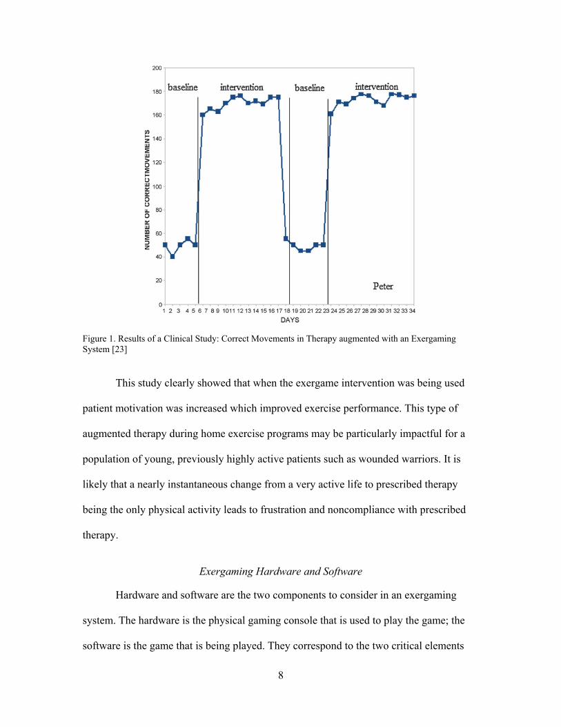
and as part of an HEP [25], [26], show positive results. Figure 1, on the next page, is
from a study that complemented traditional physical therapy with exergames using the
XBOX Kinect [23]. The subject represented in this case study has severe cerebral palsy
and participates in regular rehabilitative therapy.
During the baseline phase, the therapist instructed the participant and ensured
correct movements; then the subject completed 10 repetitions for each of three
movements without correction from the therapist. During the intervention phase, the
subject completed the same repetitions taking cues from the prescribed exergame. It is
easily seen that the number of correct movements greatly increased when physical
therapy was augmented with the Kinect.
8
Figure 1. Results of a Clinical Study: Correct Movements in Therapy augmented with an Exergaming System [23]
This study clearly showed that when the exergame intervention was being used
patient motivation was increased which improved exercise performance. This type of
augmented therapy during home exercise programs may be particularly impactful for a
population of young, previously highly active patients such as wounded warriors. It is
likely that a nearly instantaneous change from a very active life to prescribed therapy
being the only physical activity leads to frustration and noncompliance with prescribed
therapy.
Exergaming Hardware and Software
Hardware and software are the two components to consider in an exergaming
system. The hardware is the physical gaming console that is used to play the game; the
software is the game that is being played. They correspond to the two critical elements

9
for clinical use of an exergaming system. The system must be able to accurately track the
motion, which is primarily a hardware issue. It must also be able to decide if the motion
was performed correctly, a software issue. Both of these conditions must be met before
exergaming systems are used clinically.
Hardware
There are currently three competitive motion-based video game hardware systems
on the market: the Nintendo Wii, the PlayStation MOVE and the XBOX Kinect. Both the
Wii and MOVE require the user to move a controller; the system tracks the motion of the
controller which translates into the motions in the video game. The Kinect has no
controller. Instead, the Kinect makes use of a simple motion capture system which uses
the body of the user as the controller for the video game. In the context of rehabilitation,
the Wii and MOVE are not viable options because they do not require or register any
motions of the lower body. The Kinect is more difficult to “trick” into thinking the
correct motion was completed because it tracks the entire body. Microsoft, which owns
XBOX, has publicly released the drivers for the Kinect and has provided a free developer
kit and full programming support [27], which provides additional incentive to choose the
Kinect because it enables programmers to easily use the Kinect in nontraditional ways.
This study uses the Kinect for XBOX 360 as the hardware for the exergaming system.
Software
Quickly after starting to use exergames in physical therapy, it was discovered that
off-the-shelf games were not suitable; standard games for the Kinect console were too
difficult for patients to perform and lacked the flexibility required in physical therapy
[28]. This highlights the need for custom exergaming software for use in physical
10
therapy. There are many benefits to creating a specialized exergame software, the
primary one being the ability to highly specialize the game for the specific patient’s exact
point in their rehabilitation. The exergames can also be customized for a specific patient
demographic and their interests, which may further increase patient compliance with
prescribed therapy. There have been several studies into developing and testing games for
clinical use [7], [29]–[31]. A custom game should include the ability to choose which
exercises should be performed and to set the sensitivity of the game to avoid patient
frustration. Ideally, the game will also decide if the motion was performed correctly in
order to provide immediate feedback to the patient. This study uses a custom exergaming
software, Vitalize.
Current State of Research
The academic community quickly recognized the potential of the Kinect
technology as a medical tool. Since its debut in 2010, many researchers have explored the
accuracy of the Kinect in the context of clinical rehabilitation [15], [19], [32]–[42]. Most
studies performed have compared the performance of the Kinect to a marker-based
system, which is commonly considered the gold standard in motion capture.
Two validation studies that determined the accuracy of the Kinect by using
manual measurements were referenced [19], [33]. Mobini et al. [19] used a 2-dimensional
wooden model of the upper body and found that the Kinect’s estimation of joint centers
was between 1 and 2 cm; this is an extremely simplified model, but shows the high
potential of the Kinect. Bonnechère et al. [33] physically measured the body segments of
their subjects and found excellent reproducibility by the Kinect, but that the accuracy of
this system depends on the segment being studied.

11
Research has also been performed studying the suitability of the Kinect for use in
gait analysis [34], [40], [41]. These studies do not agree which aspects of gait can be
accurately collected by the Kinect. Clark et al. [34] reports that the temporal
measurements of gait were the least valid. Interestingly, as a result of their research,
Pfister et al. [40] states that the Kinect is not accurate enough for clinical analysis with
the possible exception of the same temporal gait measurements. The study done by Xu et
al. [41] found that the Kinect follows motion trends well, but lacks the ability to
accurately measure magnitudes of motions. There is significant disagreement among
these studies, but they all conclude that the Kinect displays varied levels of accuracy for
different gait parameters.
Several validation studies have considered the accuracy of the Kinect with respect
to specific motions, often related to the rehabilitation of a specific condition [32], [35],
[36], [38], [39]. Bonnechere et al. [32] performed a study with 48 able-bodied subjects
using four simple motions; it was found that the difference between the Kinect and the
marker-based system was within 11 degrees with less error in the upper body than lower
body. Galna’s study [35] used a mixture of able-bodied subjects and patients with
Parkinson’s disease. This study concluded that the Kinect can accurately measure timing
and gross spatial characteristics of motion, but lacks the accuracy to correctly report
smaller motions. Kuster et al. [36] is one of the early validation studies using the
KinectOne; they reported average error for shoulder motions under five degrees, but also
found large discrepancy between the accuracy of tracking the shoulder and the trunk.
It can easily be seen that there is much interest in validating the Kinect for
possible clinical use. However, researchers are not in agreement because the validation
12
results have been widely varied depending on the motions used and the joints analyzed.
This highlights the need for a comprehensive study that uses a large number of
therapeutic motions to discover broad trends in the tracking abilities and limitations of
the Kinect to determine which motions and which joints can be tracked with enough
accuracy for clinical use.
Purpose
The purpose of this study was to determine the tracking accuracy and limitations
of a single depth camera exergaming system. Motion capture data from a large number of
exercises was collected and analyzed with the goal of making a comprehensive reference
database of errors associated with the various body segments and exercises. This will
inform both exergaming software developers and clinicians of which exercises perform
well in a simple depth tracking system and which exercises have large error associated
with the system and so should not be assigned for home exercise programs. Analysis of
the database was initiated to determine trends in the accuracy of the Vitalize system. The
following are the primary aims of this research:
Aim 1: Effect of plane of motion on error. Is there more error associated with
motions that are primarily in the frontal plane versus motions that are primarily in the
sagittal plane? It is hypothesized that the Vitalize system will perform better in the frontal
plane than in the sagittal plane because there is no depth measurement required for frontal
plane motions.
Aim 2: Effect of range of motion on error. Does the range of motion of an
exercise have an effect on the error associated with that exercise? If so, what is the effect
and when is it observed? It is hypothesized that the range of motion will not have an
13
effect on the tracking accuracy of the system; as reported in previous research [32], [36],
[38].
Aim 3: Effect of the involved joint in the motion on error. Does the Vitalize
system’s tracking ability depend on which joint is being tracked? Are hip motions tracked
with more accuracy than shoulder motions? It is hypothesized that there will be minimal
differences between the tracking accuracy of various joints.
Aim 4: Effect of complexity of motion on error. Does the tracking error
associated with a specific joint during an exercise increase when additional components
are added to the exercise? It is hypothesized that adding complexity will not have an
effect on the error of the primary joint involved in the exercise.
Aim 5: Observations on the Decision made by Vitalize. How does Vitalize decide
if a motion was performed correctly? Are those decisions clinically valid? It is
hypothesized that the Vitalize software will occasionally reject a correct motion, but that
it will perform well in making these decisions.
14
CHAPTER TWO
Methods
This study was completed in collaboration with the CFI. Institutional Review
Board approval was given at Baylor University (“Multiple Plane Motion Tracking
Quality Assessment of a Therapy-Based Exergaming System.” Protocol Number:
755134-5. Principle Investigator: Jonathan Rylander, Ph.D.) and Brooke Army Medical
Center (“Vitalize - Game Based Wellbeing Research Initiative”, Protocol Number:
C.2011.057, Principle Investigator: Jason Wilken, MPT, Ph.D.). Two motion capture
systems (Vitalize and Vicon) simultaneously collected the exercise data; the Vitalize
exergaming system was compared to the Vicon marker-based system which was used as
the gold standard. The Vitalize data was collected from the Kinect sensor through the
custom Vitalize software. The Vicon data was collected using a passive marker-based
system and the Nexus software; then the model was created in Visual 3D. The two
datasets were time-synced together to directly compare the repetitions. Any difference
between the datasets was reported as error of the Vitalize system. After creating an error
database, analyses addressing each aim of this study were completed.
Subjects
Data from 15 able-bodied, adult subjects was collected at the Baylor University
BioMotion Lab at the Baylor Research and Innovation Collaborative. A homogeneous
subject group was selected to control for variation in the tracking ability of the Kinect
sensor due to body type. Of the subjects, 7 were female; the average age was 22.6 years
15
with a standard deviation of 3.5 years; the average BMI was 22.1 with a standard
deviation of 2.3. All of the subjects gave their informed consent. All were able to
maintain moderate, intermittent physical activity for an extended period of time and had
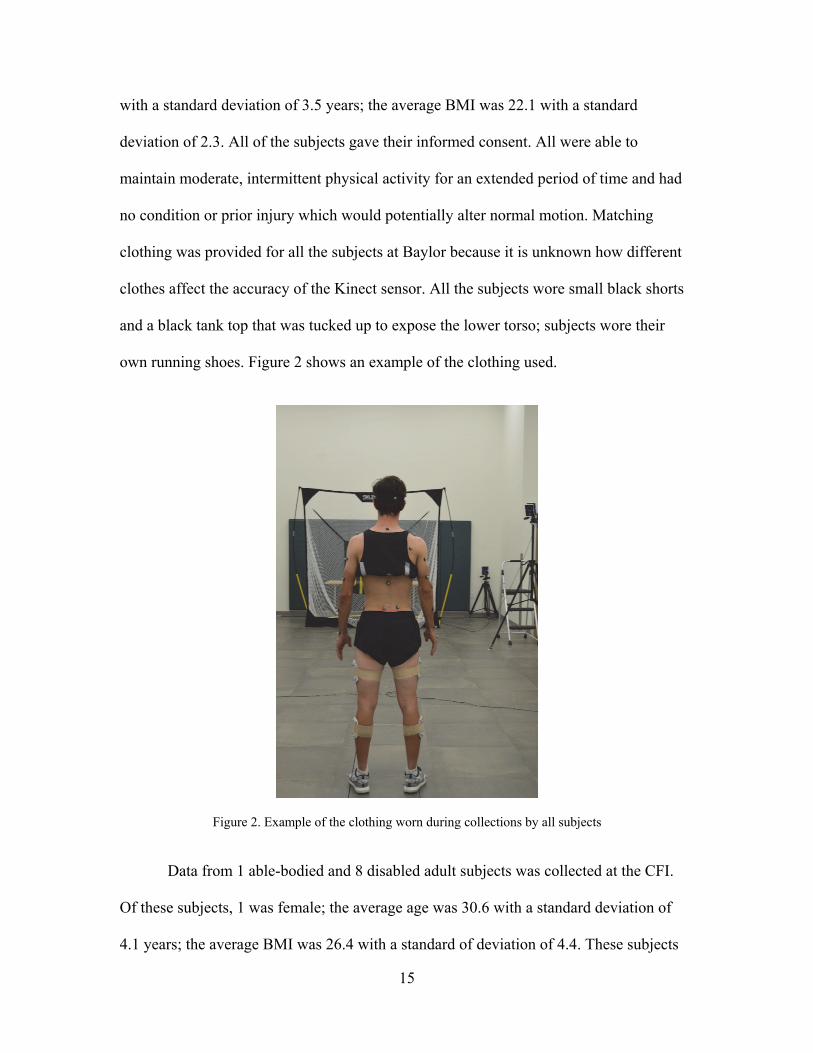
no condition or prior injury which would potentially alter normal motion. Matching
clothing was provided for all the subjects at Baylor because it is unknown how different
clothes affect the accuracy of the Kinect sensor. All the subjects wore small black shorts
and a black tank top that was tucked up to expose the lower torso; subjects wore their
own running shoes. Figure 2 shows an example of the clothing used.
Figure 2. Example of the clothing worn during collections by all subjects
Data from 1 able-bodied and 8 disabled adult subjects was collected at the CFI.
Of these subjects, 1 was female; the average age was 30.6 with a standard deviation of
4.1 years; the average BMI was 26.4 with a standard of deviation of 4.4. These subjects
16
gave their informed consent. The disabled subjects had varied degrees of limb salvage
and amputations. The amputee data has been collected but not yet analyzed. Though this
study was designed for an amputee population, it was decided that additional research
was needed to validate the performance of the Vitalize system in a more controlled way
by using able-bodied subjects before adding the variability of amputee subjects. Research
into the data from the amputee group will include the influence of carbon fiber limbs and
body asymmetry on the tracking ability, but a solid understanding of the abilities of the
system with able-bodied subjects was needed before that analysis could be properly
completed. The young, able-bodied population was chosen due to the similarity to the
young, amputee population. The following work presented here was completed using
only the data from the able-bodied subjects collected by the Baylor group.
Exercises
Each subject performed ten repetitions each of 69 exercises used in physical
therapy. These exercises ranged from simple arm raises to complex motions involving
multiple body segments and multiple planes of motion such as a dodge and cross punch
with a squat. The exercises were selected with accessibility in mind; including a range of
exercises that patients with 1-4 missing limbs could successfully complete. A list of the
exercises is included in Appendix A, the descriptions of every exercise are included in
Appendix B.
Double Motion Capture
All data was simultaneously collected by a gold standard motion marker-based
capture system (Vicon System) and the experimental system (Vitalize System) in order to
17
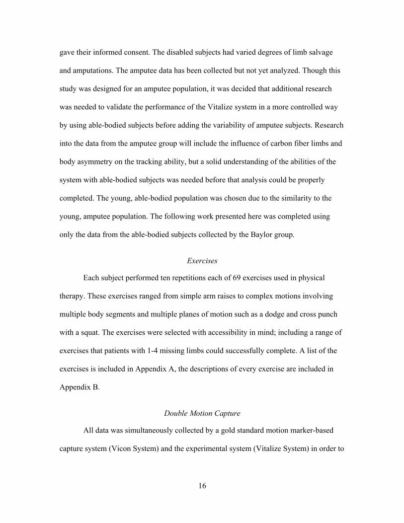
directly compare the results. Figure 3, shows data being collected simultaneously by the
two systems.
Figure 3. Double Motion Capture set-up for Data Collection
Vicon System
The Vicon system consisted of a 14-camera Vicon Vantage infrared system at the
BioMotion Lab at Baylor University. The data was collected at 120 Hz. 57 reflective
markers were placed on critical landmarks and various body segments to track the
kinematics of the whole body as exercises were performed. All markers were placed
directly on the subject’s skin with the exception of four markers on the headband and the
thigh and shank markers which were placed on rigid plates and secured to the respective
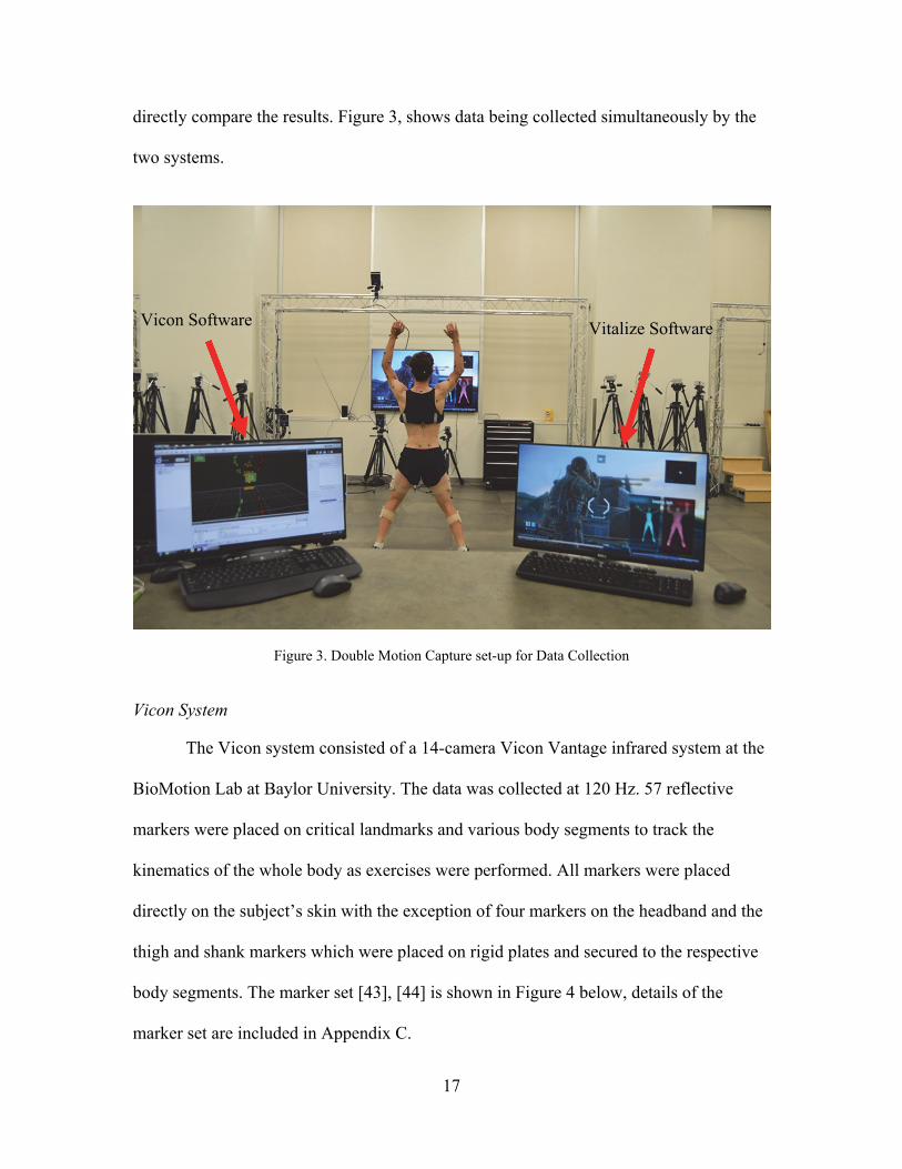
body segments. The marker set [43], [44] is shown in Figure 4 below, details of the
marker set are included in Appendix C.
Vicon Software Vitalize Software
18
Figure 4. Markerset used with the Vicon System
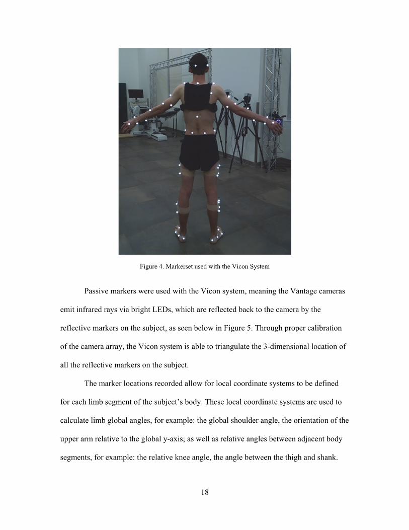
Passive markers were used with the Vicon system, meaning the Vantage cameras
emit infrared rays via bright LEDs, which are reflected back to the camera by the
reflective markers on the subject, as seen below in Figure 5. Through proper calibration
of the camera array, the Vicon system is able to triangulate the 3-dimensional location of
all the reflective markers on the subject.
The marker locations recorded allow for local coordinate systems to be defined
for each limb segment of the subject’s body. These local coordinate systems are used to
calculate limb global angles, for example: the global shoulder angle, the orientation of the
upper arm relative to the global y-axis; as well as relative angles between adjacent body
segments, for example: the relative knee angle, the angle between the thigh and shank.
19
Figure 5. Description of Vicon Cameras. Infrared rays reflected from marker back to camera; adapted from [45]
In addition to the physical reflective markers, twenty bony landmarks on the
subject were found by manual palpation and recorded using a digitizing pointer to create
virtual markers. These virtual markers were referenced back to the physical markers on
the body and used to derive the joint centers of the ankle, knee, hip, shoulder and elbow.
The marker placement and palpation were always performed by the primary researcher to
minimize variation between subjects.
Vicon Nexus 2.2.3 was the software used to track and record the 3D coordinates
for each marker — 120 measurements per second for every marker on the subject.
20
Vitalize System
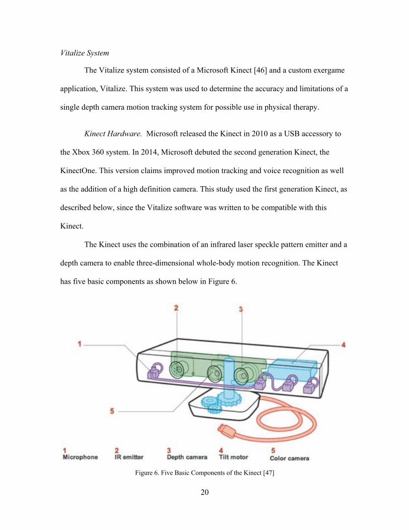
The Vitalize system consisted of a Microsoft Kinect [46] and a custom exergame
application, Vitalize. This system was used to determine the accuracy and limitations of a
single depth camera motion tracking system for possible use in physical therapy.
Kinect Hardware. Microsoft released the Kinect in 2010 as a USB accessory to
the Xbox 360 system. In 2014, Microsoft debuted the second generation Kinect, the
KinectOne. This version claims improved motion tracking and voice recognition as well
as the addition of a high definition camera. This study used the first generation Kinect, as
described below, since the Vitalize software was written to be compatible with this
Kinect.
The Kinect uses the combination of an infrared laser speckle pattern emitter and a
depth camera to enable three-dimensional whole-body motion recognition. The Kinect
has five basic components as shown below in Figure 6.
Figure 6. Five Basic Components of the Kinect [47]
21
1. The microphone array, which consists of four microphones that can separate the voices of the players from the other noise in the room. This allows players to use the voice controls in the system.
2. The IR emitter, which projects infrared light into the room. As the IR light hits surfaces it becomes distorted, this distortion information is read by the depth camera.
3. The depth camera, which uses the distortion in the infrared patterns to build a 3-dimensional mapping of the room and the players.
4. The tilt motor, which adjusts the position of the box based on the height of the player. The tilt motor will angle up for a tall player and down for a shorter player; this optimizes the cameras for any height of player.
5. The color camera, which is similar to a webcam, captures video images that the Kinect uses to make a more accurate picture of the room and the players.
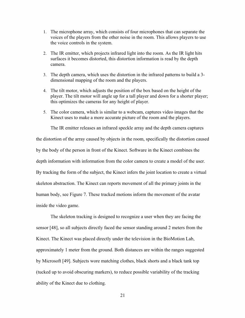
The IR emitter releases an infrared speckle array and the depth camera captures
the distortion of the array caused by objects in the room, specifically the distortion caused
by the body of the person in front of the Kinect. Software in the Kinect combines the
depth information with information from the color camera to create a model of the user.
By tracking the form of the subject, the Kinect infers the joint location to create a virtual
skeleton abstraction. The Kinect can reports movement of all the primary joints in the
human body, see Figure 7. These tracked motions inform the movement of the avatar
inside the video game.
The skeleton tracking is designed to recognize a user when they are facing the
sensor [48], so all subjects directly faced the sensor standing around 2 meters from the
Kinect. The Kinect was placed directly under the television in the BioMotion Lab,
approximately 1 meter from the ground. Both distances are within the ranges suggested
by Microsoft [49]. Subjects wore matching clothes, black shorts and a black tank top
(tucked up to avoid obscuring markers), to reduce possible variability of the tracking
ability of the Kinect due to clothing.
22
Figure 7. Joints Tracked by the Kinect [27]
The Kinect tracks at a variable frame rate near 30Hz. For this study, the Vitalize
collection computer was optimized with a fast video card and solid state hard drive and
reserved only for data collection for this study. Therefore, the collection frame rate was
as near 30Hz as possible.
Vitalize Software. The software used for the experimental system is a custom 3rd-
person shooter exergame called Vitalize, which was developed by Blitz Gaming Studios
as part of this study [50]. The game was developed for the wounded warrior population,
the 3rd person shooter interface was the preferred game among this group. Within the
game, the player performs exercises to charge and fire their weapon at enemy drones.

23
One of the primary goals of this software was accessibility; therefore several
customizable options were built into the game.
The exercises used in gameplay can be specified and customized for the user.
Filters are in place that enable the exercises to be sorted according to the needs of the
patient. For example, if the filters “Hip” and “Shoulder” are both selected, only exercises
that use the hip and shoulder will be used in gameplay. The sensitivity can also be
adjusted to allow for varying degrees of “correctness” to be accepted by the system.
Various limbs can be switched off from the exercise detection system to allow better
tracking of patients with missing limbs; the user can also play in “wheelchair” mode
which makes it easier for a patient in a wheelchair to navigate the game. The game
controls can also be customized, see Figure 8, below.
Figure 8. Customization for Navigation within Vitalize Software
24
By default, the soldier in the game is controlled by the player pointing their hand
in different directions. However, this can be changed (all control options are shown
within the red box in Figure 8) to allow for increased accessibility for players missing
limbs.
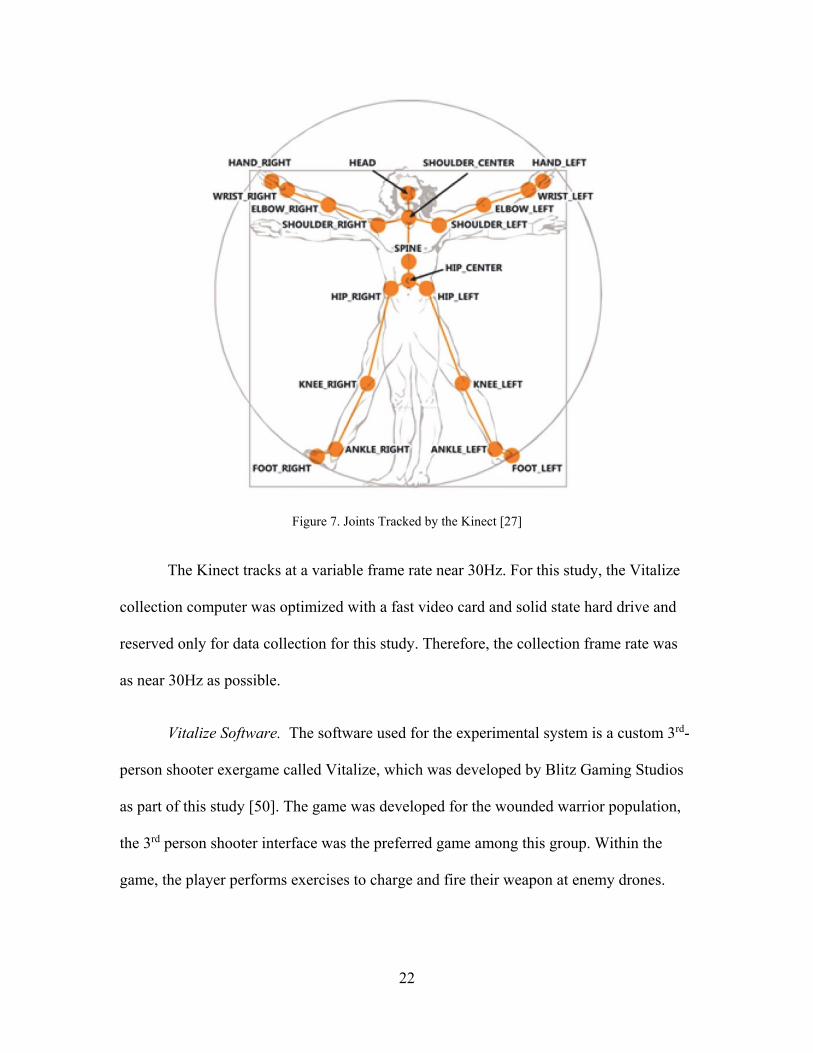
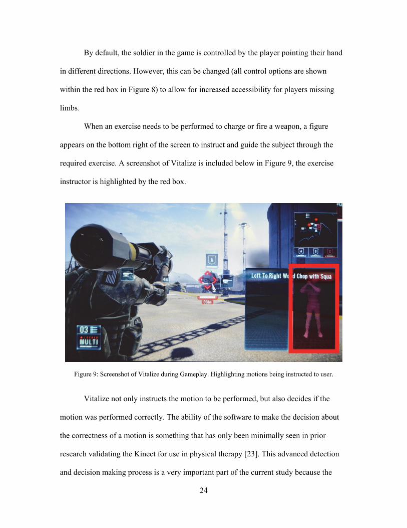
When an exercise needs to be performed to charge or fire a weapon, a figure
appears on the bottom right of the screen to instruct and guide the subject through the
required exercise. A screenshot of Vitalize is included below in Figure 9, the exercise
instructor is highlighted by the red box.
Figure 9: Screenshot of Vitalize during Gameplay. Highlighting motions being instructed to user.
Vitalize not only instructs the motion to be performed, but also decides if the
motion was performed correctly. The ability of the software to make the decision about
the correctness of a motion is something that has only been minimally seen in prior
research validating the Kinect for use in physical therapy [23]. This advanced detection
and decision making process is a very important part of the current study because the

25
patient will rely on the decisions made by the exergame software to gauge whether or not
they are doing the exercises included in their home exercise program correctly.
The data used to track the skeleton by the Kinect is collected through Vitalize.
The 3-dimensional coordinates that infer the location of the 20 joints tracked by the
Kinect are collected by Vitalize for further processing and analysis.
Nexus Processing
There are multiple steps required to process the data from the Vicon system. The
first step is to clean the data after the collection. This is done in Nexus by correcting
marker labels and filling gaps. Each of the 57 markers has a distinct label, see Appendix
C; the researcher must ensure that all the markers are labelled correctly before
proceeding. Gaps in the data must also be addressed. Gaps occur when a marker is
occluded from the cameras. If a marker cannot be seen by at least two cameras, the data
is not collected. Gaps smaller than 35 frames were filled and visually checked in post-
processing. At the 120 Hz. collection rate, this represents just over a ¼ of a second worth
of data. Gaps that occurred on the head, pelvis or leg plates were filled with the Rigid-
Body Gap fill algorithm provided by Vicon Nexus. The Rigid-Body fill uses data from
three reference markers on the same rigid body as the marker containing the gap to fill
the gap; under the assumption that the relative distances between the markers on the rigid
body is constant. Figure 10, below, is an example of a Rigid-Body fill. The gap is on the
marker Shank4, the markers Shank1, Shank2 and Shank3 are used for reference as to the
location of Shank4 as the motion continues. The trajectories of the reference markers are
seen in orange, the blue trajectory is the proposed fill for the missing frames of the
Shank4 marker.

26
Figure 10: Example of a Rigid-Body Gap Fill in Nexus. The blue trajectory is the proposed fill for the missing marker.
Gaps that occurred on any other body segment were filled with the Spline fill or
Pattern fill, with the Pattern fill preferred. The Spline fill (simple interpolation) uses a
numerical fit on the order of 3 to fill gaps that are under 14 frames. The Pattern fill is
similar to the Rigid Body fill, but uses only one reference marker.
Visual 3D Processing
Visual 3D was used for most of the post-processing involved in this project. In
Visual 3D the Vicon data was transformed to match the coordinate system of the Kinect
data and filtered using a low-pass filter with a cutoff frequency of 6Hz. Then a custom
model was built for each subject. In addition to the physical markers, virtual markers
were placed on bony landmarks of the subject’s body, using manual palpation and the
digitizing wand; these are also called digitizing points. These markers are listed in Table
1 below.
27
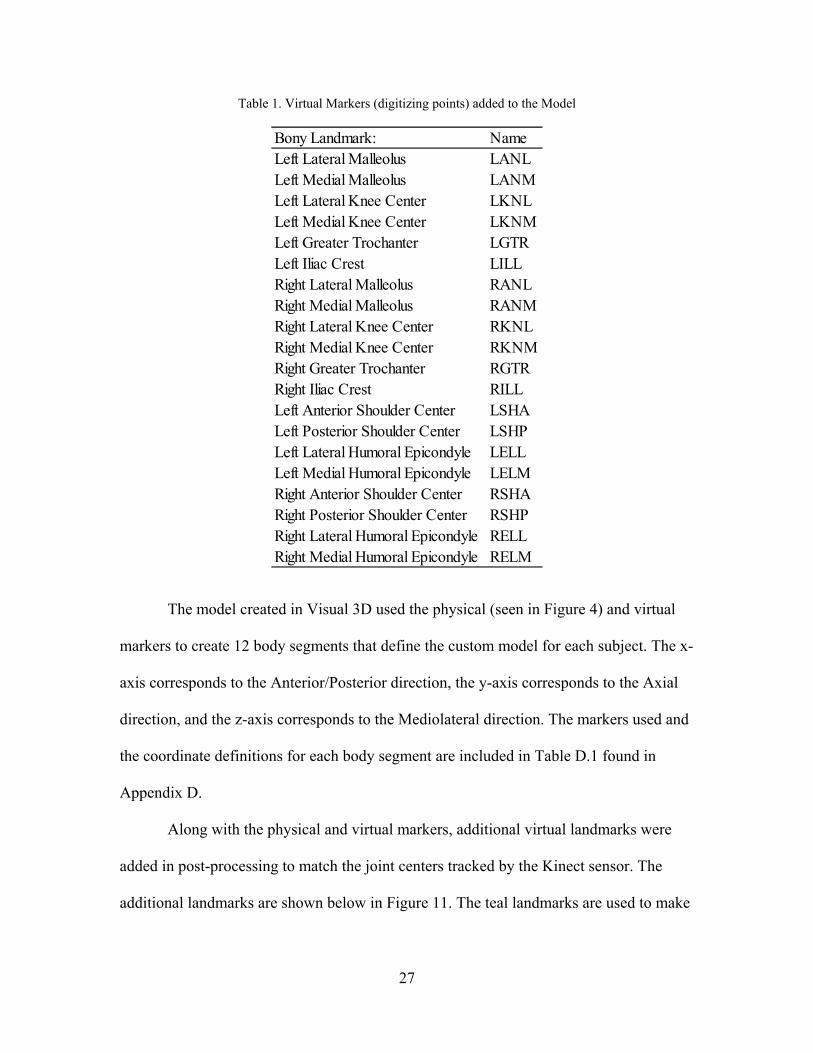
Table 1. Virtual Markers (digitizing points) added to the Model
The model created in Visual 3D used the physical (seen in Figure 4) and virtual
markers to create 12 body segments that define the custom model for each subject. The x-
axis corresponds to the Anterior/Posterior direction, the y-axis corresponds to the Axial
direction, and the z-axis corresponds to the Mediolateral direction. The markers used and
the coordinate definitions for each body segment are included in Table D.1 found in
Appendix D.
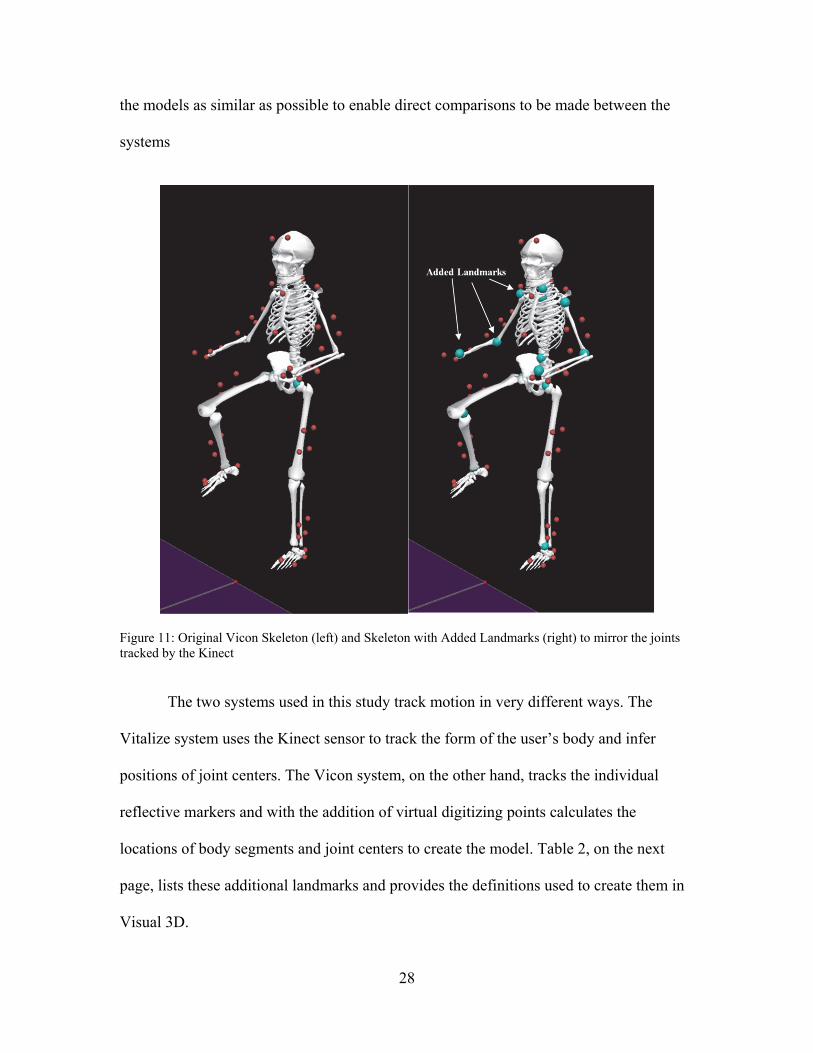
Along with the physical and virtual markers, additional virtual landmarks were
added in post-processing to match the joint centers tracked by the Kinect sensor. The
additional landmarks are shown below in Figure 11. The teal landmarks are used to make
Bony Landmark: NameLeft Lateral Malleolus LANLLeft Medial Malleolus LANMLeft Lateral Knee Center LKNLLeft Medial Knee Center LKNMLeft Greater Trochanter LGTRLeft Iliac Crest LILLRight Lateral Malleolus RANLRight Medial Malleolus RANMRight Lateral Knee Center RKNLRight Medial Knee Center RKNMRight Greater Trochanter RGTRRight Iliac Crest RILLLeft Anterior Shoulder Center LSHALeft Posterior Shoulder Center LSHPLeft Lateral Humoral Epicondyle LELLLeft Medial Humoral Epicondyle LELMRight Anterior Shoulder Center RSHARight Posterior Shoulder Center RSHPRight Lateral Humoral Epicondyle RELLRight Medial Humoral Epicondyle RELM
28
the models as similar as possible to enable direct comparisons to be made between the
systems
Figure 11: Original Vicon Skeleton (left) and Skeleton with Added Landmarks (right) to mirror the joints tracked by the Kinect
The two systems used in this study track motion in very different ways. The
Vitalize system uses the Kinect sensor to track the form of the user’s body and infer
positions of joint centers. The Vicon system, on the other hand, tracks the individual
reflective markers and with the addition of virtual digitizing points calculates the
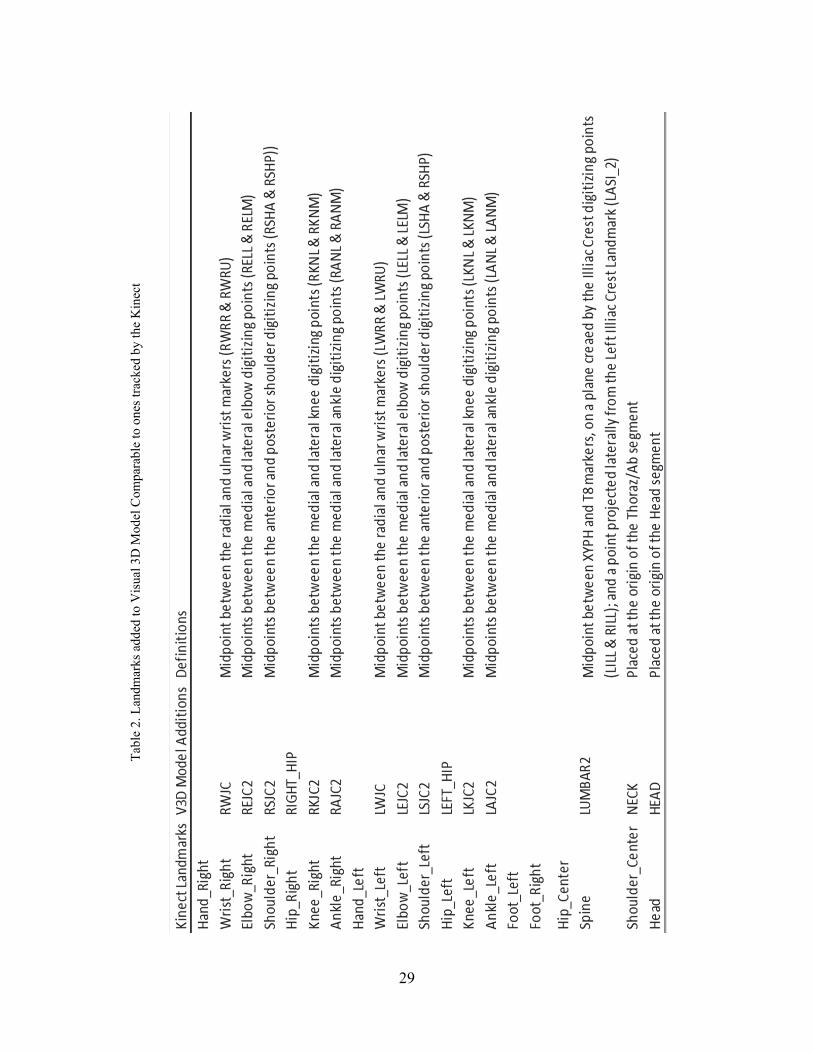
locations of body segments and joint centers to create the model. Table 2, on the next
page, lists these additional landmarks and provides the definitions used to create them in
Visual 3D.
29
Tab
le 2
. Lan
dmar
ks a
dded
to V
isua
l 3D
Mod
el C
ompa
rabl
e to
one
s tr
acke
d by
the
Kin
ect

30
The data was screened and visually time-synced in Matlab; neither sampling rate
from the Vicon or Vitalize data was changed [35], [38] . The manual time sync ensured
that each repetition in the Vicon data was correctly matched to the corresponding
repetition in the Vitalize data to enable direct comparison. An example of the data-sync
can be seen below in Figure 12.
Figure 12. Sample Data before (top figures) and after (bottom figures) Time-Sync
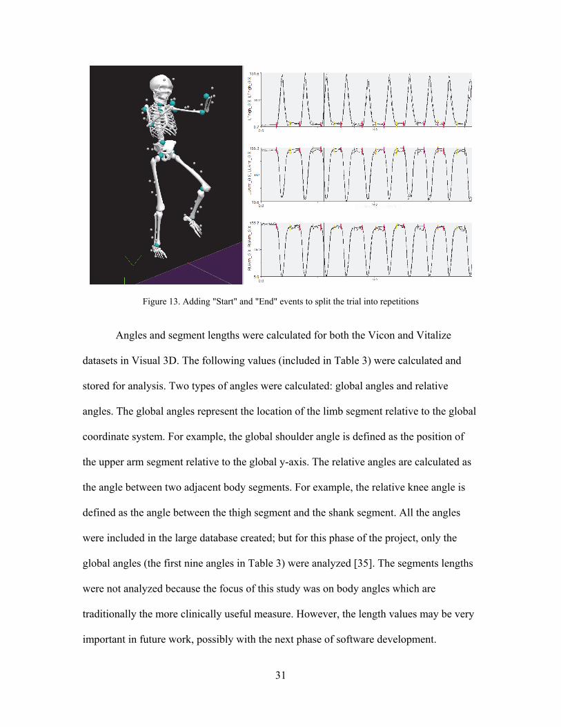
After the datasets were synced together, an automated process, with manual
corrections as needed, was used to split the exercise trials into repetitions by adding a
“Start” and “End” event to each repetition. This enables each repetition to be used
independently. In this study, the repetitions for each subject were averaged together for
each exercise performed. In Figure 13 below, the red marks indicate “Start” events and
the yellow marks indicate “End” events used.
2 4 6 8 10 12 14 16 18
20
40
60
80
100
120
140
Left Upper Arm
2 4 6 8 10 12 14 16 180
50
100
150
Right Upper Arm
MPL
KinectMPL
Kinect
0 2 4 6 8 10 12 14 16 18 200
20
40
60
80
100
120
140
160
180Left Upper Arm
MPL
Kinect
0 2 4 6 8 10 12 14 16 18 200
20
40
60
80
100
120
140
160
180Right Upper Arm
MPL
Kinect
31
Figure 13. Adding "Start" and "End" events to split the trial into repetitions
Angles and segment lengths were calculated for both the Vicon and Vitalize
datasets in Visual 3D. The following values (included in Table 3) were calculated and
stored for analysis. Two types of angles were calculated: global angles and relative
angles. The global angles represent the location of the limb segment relative to the global
coordinate system. For example, the global shoulder angle is defined as the position of
the upper arm segment relative to the global y-axis. The relative angles are calculated as
the angle between two adjacent body segments. For example, the relative knee angle is
defined as the angle between the thigh segment and the shank segment. All the angles
were included in the large database created; but for this phase of the project, only the
global angles (the first nine angles in Table 3) were analyzed [35]. The segments lengths
were not analyzed because the focus of this study was on body angles which are
traditionally the more clinically useful measure. However, the length values may be very
important in future work, possibly with the next phase of software development.

32
Tab
le 3
. Des
crip
tions
of
Ang
les
and
Seg
men
t Len
gths
Cal
cula
ted

33
Analysis
Immense amounts of data were collected for use in this project. To begin sorting
through the data, the analysis was split into two parts. The first was to create a database
of values for each subject, exercise, angle and repetition used; as well as reference tables
of error for hip, shoulder, knee and elbow. The second was to analyze trends in the errors
observed in the reference tables to determine the effect of
Plane of Motion Range of Motion Joint Involved in Motion Complexity of Motion
An additional analysis examined the decisions made by Vitalize about the
correctness of the motions to determine how well the system identified correct and
incorrect motions.
Creation of Database and Reference Tables
Using the angles calculated from the raw datasets, the values of interest were
calculated. These values include the speed, the maximum value and location, start value
and location, and end value and location for each repetition in the Vitalize and Vicon
datasets.
Four measures of error were calculated for analysis in this research, all are
reported in degrees:
Error at Start Error at Peak Range of Motion (ROM) Error Root Mean Square (RMS) Error
For each repetition, the “start” value and “peak” value were calculated, as seen
below in Figure 14.
34
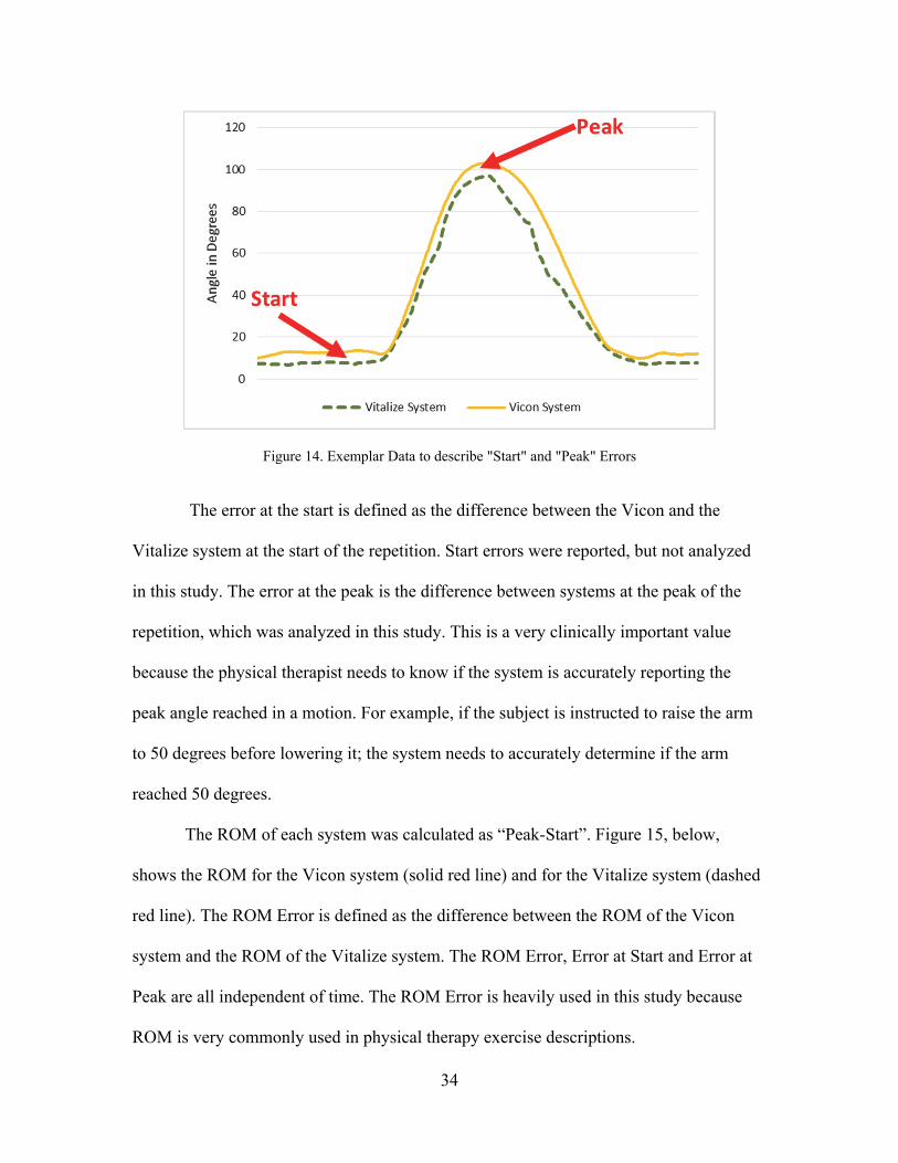
Figure 14. Exemplar Data to describe "Start" and "Peak" Errors
The error at the start is defined as the difference between the Vicon and the
Vitalize system at the start of the repetition. Start errors were reported, but not analyzed
in this study. The error at the peak is the difference between systems at the peak of the
repetition, which was analyzed in this study. This is a very clinically important value
because the physical therapist needs to know if the system is accurately reporting the
peak angle reached in a motion. For example, if the subject is instructed to raise the arm
to 50 degrees before lowering it; the system needs to accurately determine if the arm
reached 50 degrees.
The ROM of each system was calculated as “Peak-Start”. Figure 15, below,
shows the ROM for the Vicon system (solid red line) and for the Vitalize system (dashed
red line). The ROM Error is defined as the difference between the ROM of the Vicon
system and the ROM of the Vitalize system. The ROM Error, Error at Start and Error at
Peak are all independent of time. The ROM Error is heavily used in this study because
ROM is very commonly used in physical therapy exercise descriptions.

35
Figure 15. Exemplar Data to describe ROM Error
Finally, the RMS error was calculated for each repetition; it is defined as the
square root of the average of the square of difference between the systems, see Figure 16.
Unlike the other errors, RMS Error is time-dependent and considers the entire waveform.
Figure 16. Exemplar Data to describe RMS Error

36
All of these values were calculated for each repetition, angle, and exercise for
each subject; which is over 3 million values included in the large error database.
From this database, reference tables of errors were created. Four tables were
made; for the hip, shoulder, knee and elbow joints (see Tables F.1-F.4 in Appendix F).
Each of these tables included all the exercises in this study that used the joint of interest.
Each table included ROM Error, Error at Start, Error at Peak, RMS Error, ROM, Plane of
Motion, and the Category of Motion. The repetitions for each subject were averaged, then
the subjects were averaged together, which created a single reported value for each
exercise included in the table. The data from the elbow was included but not analyzed in
this study because this study is heavily based on plane of motion and the plane of motion
of the elbow is difficult to analyze because it is dependent on the orientation of the
shoulder.
Data Analysis
For each aim, all the repetitions were averaged for each subject, but each subject
was kept separate for the statistical analyses. The repetitions were averaged to help
control for variation seen in a single repetition. Averaging the repetitions in this way
results in a single value being assigned to each subject for each exercise for each type of
error reported.
Aim 1: Effect of the Plane of Motion. Analyses were completed comparing the
frontal to sagittal plane at both the hip and the shoulder to determine if there was a
difference between the error in the sagittal plane of motion and in the frontal plane of
motion.

37
Aim 2: Effect of the Range of Motion. Analyses were completed for the knee, hip
and shoulder in the sagittal plane and for the hip and shoulder in the frontal plane to
determine if and how the error of the Vitalize system was related to the ROM of the
exercise. These analyses were controlled for plane of motion because it is suspected that
the plane has an effect on the error observed.
Aim 3: Effect of Different Joints. Analyses were completed in the sagittal and
frontal planes to determine if there was a relationship between the joint being tracked and
the error of the Vitalize system. The first part of this aim controlled for range of motion
and plane of motion. The second part of the aim was completed examining the error
associated with multiple joints within the same exercise.
Aim 4: Comparison of Simple to Complex Motions. A case study was completed
for the Knee Strike variations (see Appendix B for descriptions):
Full Knee Strike Seated Knee Strike Knee Strike with Overhead Arms Knee Strike with Row
These were analyzed to determine if adding complexity to the motion has an effect on the
tracking error observed at the primary joint.
Aim 5: Observations on the Decisions made by Vitalize. This final analysis
consisted of a case study on the decisions made by the software, Vitalize. For a select
subset of exercises using data from a single subject, the decision made by the software
was compared to both the Vitalize and Vicon data to determine why the software made
the decision and if that decision was correct.
38
Statistics
For all statistical analyses, the significance level was set at α=0.05. For each
analysis that directly compared two things, two-tailed t-tests assuming unequal variance
were used. For analyses that compared more than two things, a one-way ANOVA was
used. Post hoc testing was completed with a series of t-tests and a Bonferroni correction
for all comparisons within the ANOVA to determine where significance existed.
Correlations were completed using a Pearson’s product-moment correlation. For all
correlation analyses, the suggestion by Evans [51] is followed for explaining the absolute
value of r:
.00-.19 “very weak” .20-.39 “weak” .40-.59 “moderate” .60-.79 “strong” .80-1.0 “very strong”
For all boxplots, the lower whisker indicates the first quartile of the data, the
bottom half of the box indicates the second quartile, the top half of the box indicates the
third quartile and the upper whisker indicates the fourth quartile. The black bar in the box
indicates the median of the data and the red bar in the box indicates the mean of the data.

39
CHAPTER THREE
Results
To begin sorting through the data, the analysis was split into two parts. The first
part was to create a database of values for each subject, exercise, angle and repetition
used; as well as reference tables of error for hip, shoulder, knee and elbow. The second
part was to analyze trends in observed error to determine the effect of
Plane of Motion Range of Motion Joint Involved in Motion Complexity of Motion
An additional analysis examined the decisions made by Vitalize for correctness of
motion to determine how well the system identified correct and incorrect motion.
Master Database
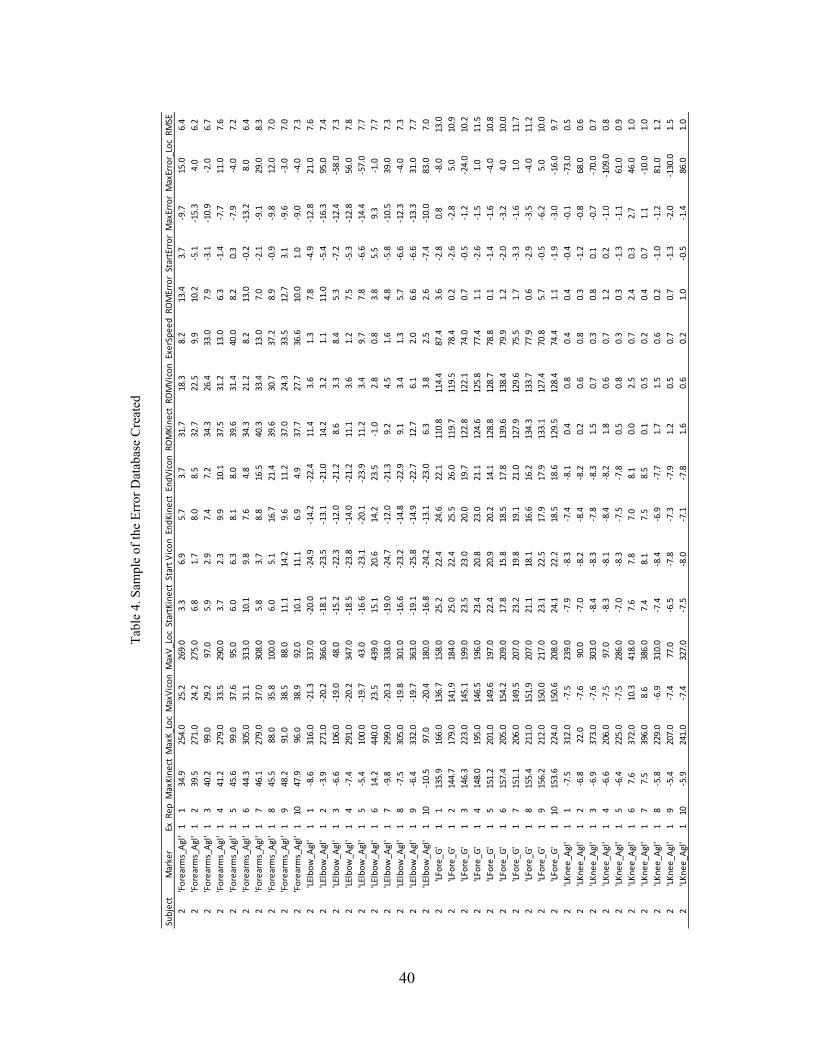
A sample of the database created is included on the next page as Table 4. The far
left column indicates the subject, this sample only shows data from Subject 2; however
the database extends to include data from 15 subjects. The next column indicates the
angle, data from 17 different angles is included for each exercise. The third column is the
exercise, the data from 69 different exercises is included in the database; a list of all the
exercises is included in Appendix A, the descriptions of every exercise is found in
Appendix B. The fourth column indicates the repetition; ten repetitions of each exercise
were performed by each subject. All the column headings indicate values that were either
directly pulled from the raw data or calculated from the data. These values are listed and
explained in Appendix E.
40
Tab
le 4
. Sam
ple
of th
e E
rror
Dat
abas
e C
reat
ed
Subject
Marker
ExRepMaxKinect
MaxK_LocMaxViconMaxV_LocStartKinect
Start ViconEndKinect
EndViconROMKinect
ROMViconExerSpeedROMErrorStartErrorMaxErrorMaxError_LocRMSE
2'Forearms_Agl'1
134.9
254.0
25.2
269.0
3.3
6.9
5.7
3.7
31.7
18.3
8.2
13.4
3.7
‐9.7
15.0
6.4
2'Forearms_Agl'1
239.5
271.0
24.2
275.0
6.8
1.7
8.0
8.5
32.7
22.5
9.9
10.2
‐5.1
‐15.3
4.0
6.2
2'Forearms_Agl'1
340.2
99.0
29.2
97.0
5.9
2.9
7.4
7.2
34.3
26.4
33.0
7.9
‐3.1
‐10.9
‐2.0
6.7
2'Forearms_Agl'1
441.2
279.0
33.5
290.0
3.7
2.3
9.9
10.1
37.5
31.2
13.0
6.3
‐1.4
‐7.7
11.0
7.6
2'Forearms_Agl'1
545.6
99.0
37.6
95.0
6.0
6.3
8.1
8.0
39.6
31.4
40.0
8.2
0.3
‐7.9
‐4.0
7.2
2'Forearms_Agl'1
644.3
305.0
31.1
313.0
10.1
9.8
7.6
4.8
34.3
21.2
8.2
13.0
‐0.2
‐13.2
8.0
6.4
2'Forearms_Agl'1
746.1
279.0
37.0
308.0
5.8
3.7
8.8
16.5
40.3
33.4
13.0
7.0
‐2.1
‐9.1
29.0
8.3
2'Forearms_Agl'1
845.5
88.0
35.8
100.0
6.0
5.1
16.7
21.4
39.6
30.7
37.2
8.9
‐0.9
‐9.8
12.0
7.0
2'Forearms_Agl'1
948.2
91.0
38.5
88.0
11.1
14.2
9.6
11.2
37.0
24.3
33.5
12.7
3.1
‐9.6
‐3.0
7.0
2'Forearms_Agl'1
1047.9
96.0
38.9
92.0
10.1
11.1
6.9
4.9
37.7
27.7
36.6
10.0
1.0
‐9.0
‐4.0
7.3
2'LElbow_A
gl'
11
‐8.6
316.0
‐21.3
337.0
‐20.0
‐24.9
‐14.2
‐22.4
11.4
3.6
1.3
7.8
‐4.9
‐12.8
21.0
7.6
2'LElbow_A
gl'
12
‐3.9
271.0
‐20.2
366.0
‐18.1
‐23.5
‐13.1
‐21.0
14.2
3.2
1.1
11.0
‐5.4
‐16.3
95.0
7.4
2'LElbow_A
gl'
13
‐6.6
106.0
‐19.0
48.0
‐15.2
‐22.3
‐12.0
‐21.2
8.6
3.3
8.4
5.3
‐ 7.2
‐12.4
‐58.0
7.3
2'LElbow_A
gl'
14
‐7.4
291.0
‐20.2
347.0
‐18.5
‐23.8
‐14.0
‐21.2
11.1
3.6
1.2
7.5
‐5.3
‐12.8
56.0
7.8
2'LElbow_A
gl'
15
‐5.4
100.0
‐19.7
43.0
‐16.6
‐23.1
‐20.1
‐23.9
11.2
3.4
9.7
7.8
‐6.6
‐14.4
‐57.0
7.7
2'LElbow_A
gl'
16
14.2
440.0
23.5
439.0
15.1
20.6
14.2
23.5
‐1.0
2.8
0.8
3.8
5.5
9.3
‐1.0
7.7
2'LElbow_A
gl'
17
‐9.8
299.0
‐20.3
338.0
‐19.0
‐24.7
‐12.0
‐21.3
9.2
4.5
1.6
4.8
‐5.8
‐10.5
39.0
7.3
2'LElbow_A
gl'
18
‐7.5
305.0
‐19.8
301.0
‐16.6
‐23.2
‐14.8
‐22.9
9.1
3.4
1.3
5.7
‐6.6
‐12.3
‐4.0
7.3
2'LElbow_A
gl'
19
‐6.4
332.0
‐19.7
363.0
‐19.1
‐25.8
‐14.9
‐22.7
12.7
6.1
2.0
6.6
‐6.6
‐13.3
31.0
7.7
2'LElbow_A
gl'
110
‐ 10.5
97.0
‐20.4
180.0
‐16.8
‐24.2
‐13.1
‐23.0
6.3
3.8
2.5
2.6
‐7.4
‐10.0
83.0
7.0
2'LFore_G
'1
1135.9
166.0
136.7
158.0
25.2
22.4
24.6
22.1
110.8
114.4
87.4
3.6
‐2.8
0.8
‐8.0
13.0
2'LFore_G
'1
2144.7
179.0
141.9
184.0
25.0
22.4
25.5
26.0
119.7
119.5
78.4
0.2
‐2.6
‐2.8
5.0
10.9
2'LFore_G
'1
3146.3
223.0
145.1
199.0
23.5
23.0
20.0
19.7
122.8
122.1
74.0
0.7
‐0.5
‐1.2
‐24.0
10.2
2'LFore_G
'1
4148.0
195.0
146.5
196.0
23.4
20.8
23.0
21.1
124.6
125.8
77.4
1.1
‐2.6
‐1.5
1.0
11.5
2'LFore_G
'1
5151.2
201.0
149.6
197.0
22.4
20.9
20.2
14.1
128.8
128.7
78.8
0.1
‐1.4
‐1.6
‐4.0
10.8
2'LFore_G
'1
6157.4
205.0
154.2
209.0
17.8
15.8
18.5
17.8
139.6
138.4
79.9
1.2
‐2.0
‐3.2
4.0
10.0
2'LFore_G
'1
7151.1
206.0
149.5
207.0
23.2
19.8
19.1
21.0
127.9
129.6
75.5
1.7
‐3.3
‐1.6
1.0
11.7
2'LFore_G
'1
8155.4
211.0
151.9
207.0
21.1
18.1
16.6
16.2
134.3
133.7
77.9
0.6
‐2.9
‐3.5
‐4.0
11.2
2'LFore_G
'1
9156.2
212.0
150.0
217.0
23.1
22.5
17.9
17.9
133.1
127.4
70.8
5.7
‐0.5
‐6.2
5.0
10.0
2'LFore_G
'1
10153.6
224.0
150.6
208.0
24.1
22.2
18.5
18.6
129.5
128.4
74.4
1.1
‐1.9
‐3.0
‐16.0
9.7
2'LKnee_A
gl'
11
‐7.5
312.0
‐7.5
239.0
‐7.9
‐8.3
‐7.4
‐8.1
0.4
0.8
0.4
0.4
‐0.4
‐0.1
‐73.0
0.5
2'LKnee_A
gl'
12
‐6.8
22.0
‐7.6
90.0
‐7.0
‐8.2
‐8.4
‐8.2
0.2
0.6
0.8
0.3
‐1.2
‐0.8
68.0
0.6
2'LKnee_A
gl'
13
‐6.9
373.0
‐7.6
303.0
‐8.4
‐8.3
‐7.8
‐8.3
1.5
0.7
0.3
0.8
0.1
‐0.7
‐70.0
0.7
2'LKnee_A
gl'
14
‐6.6
206.0
‐7.5
97.0
‐8.3
‐8.1
‐8.4
‐8.2
1.8
0.6
0.7
1.2
0.2
‐1.0
‐109.0
0.8
2'LKnee_A
gl'
15
‐6.4
225.0
‐7.5
286.0
‐7.0
‐8.3
‐7.5
‐7.8
0.5
0.8
0.3
0.3
‐1.3
‐1.1
61.0
0.9
2'LKnee_A
gl'
16
7.6
372.0
10.3
418.0
7.6
7.8
7.0
8.1
0.0
2.5
0.7
2.4
0.3
2.7
46.0
1.0
2'LKnee_A
gl'
17
7.5
396.0
8.6
386.0
7.4
8.1
7.5
8.5
0.1
0.5
0.2
0.4
0.7
1.1
‐10.0
1.0
2'LKnee_A
gl'
18
‐5.8
229.0
‐6.9
310.0
‐7.4
‐8.4
‐6.9
‐7.7
1.7
1.5
0.6
0.2
‐1.0
‐1.2
81.0
1.2
2'LKnee_A
gl'
19
‐5.4
207.0
‐7.4
77.0
‐6.5
‐7.8
‐7.3
‐7.9
1.2
0.5
0.7
0.7
‐1.3
‐2.0
‐130.0
1.5
2'LKnee_A
gl'
110
‐5.9
241.0
‐7.4
327.0
‐7.5
‐8.0
‐7.1
‐7.8
1.6
0.6
0.2
1.0
‐0.5
‐1.4
86.0
1.0
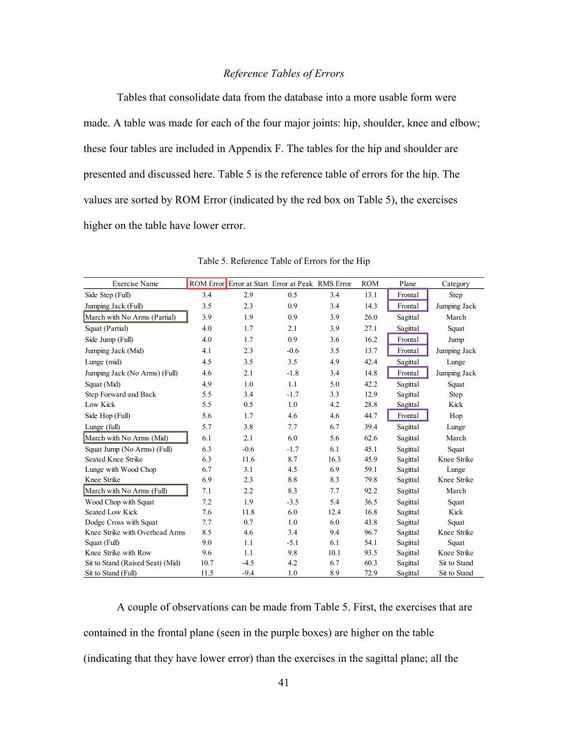
41
Reference Tables of Errors
Tables that consolidate data from the database into a more usable form were
made. A table was made for each of the four major joints: hip, shoulder, knee and elbow;
these four tables are included in Appendix F. The tables for the hip and shoulder are
presented and discussed here. Table 5 is the reference table of errors for the hip. The
values are sorted by ROM Error (indicated by the red box on Table 5), the exercises
higher on the table have lower error.
Table 5. Reference Table of Errors for the Hip
A couple of observations can be made from Table 5. First, the exercises that are
contained in the frontal plane (seen in the purple boxes) are higher on the table
(indicating that they have lower error) than the exercises in the sagittal plane; all the
Exercise Name ROM Error Error at Start Error at Peak RMS Error ROM Plane Category
Side Step (Full) 3.4 2.9 0.5 3.4 13.1 Frontal Step
Jumping Jack (Full) 3.5 2.3 0.9 3.4 14.3 Frontal Jumping Jack
March with No Arms (Partial) 3.9 1.9 0.9 3.9 26.0 Sagittal March
Squat (Partial) 4.0 1.7 2.1 3.9 27.1 Sagittal Squat
Side Jump (Full) 4.0 1.7 0.9 3.6 16.2 Frontal Jump
Jumping Jack (Mid) 4.1 2.3 -0.6 3.5 13.7 Frontal Jumping Jack
Lunge (mid) 4.5 3.5 3.5 4.9 42.4 Sagittal Lunge
Jumping Jack (No Arms) (Full) 4.6 2.1 -1.8 3.4 14.8 Frontal Jumping Jack
Squat (Mid) 4.9 1.0 1.1 5.0 42.2 Sagittal SquatStep Forward and Back 5.5 3.4 -1.7 3.3 12.9 Sagittal StepLow Kick 5.5 0.5 1.0 4.2 28.8 Sagittal Kick
Side Hop (Full) 5.6 1.7 4.6 4.6 44.7 Frontal Hop
Lunge (full) 5.7 3.8 7.7 6.7 39.4 Sagittal Lunge
March with No Arms (Mid) 6.1 2.1 6.0 5.6 62.6 Sagittal March
Squat Jump (No Arms) (Full) 6.3 -0.6 -1.7 6.1 45.1 Sagittal SquatSeated Knee Strike 6.3 11.6 8.7 16.3 45.9 Sagittal Knee StrikeLunge with Wood Chop 6.7 3.1 4.5 6.9 59.1 Sagittal LungeKnee Strike 6.9 2.3 8.8 8.3 79.8 Sagittal Knee Strike
March with No Arms (Full) 7.1 2.2 8.3 7.7 92.2 Sagittal March
Wood Chop with Squat 7.2 1.9 -3.5 5.4 36.5 Sagittal SquatSeated Low Kick 7.6 11.8 6.0 12.4 16.8 Sagittal KickDodge Cross with Squat 7.7 0.7 1.0 6.0 43.8 Sagittal SquatKnee Strike with Overhead Arms 8.5 4.6 3.4 9.4 96.7 Sagittal Knee StrikeSquat (Full) 9.0 1.1 -5.1 6.1 54.1 Sagittal SquatKnee Strike with Row 9.6 1.1 9.8 10.1 93.5 Sagittal Knee StrikeSit to Stand (Raised Seat) (Mid) 10.7 -4.5 4.2 6.7 60.3 Sagittal Sit to StandSit to Stand (Full) 11.5 -9.4 1.0 8.9 72.9 Sagittal Sit to Stand
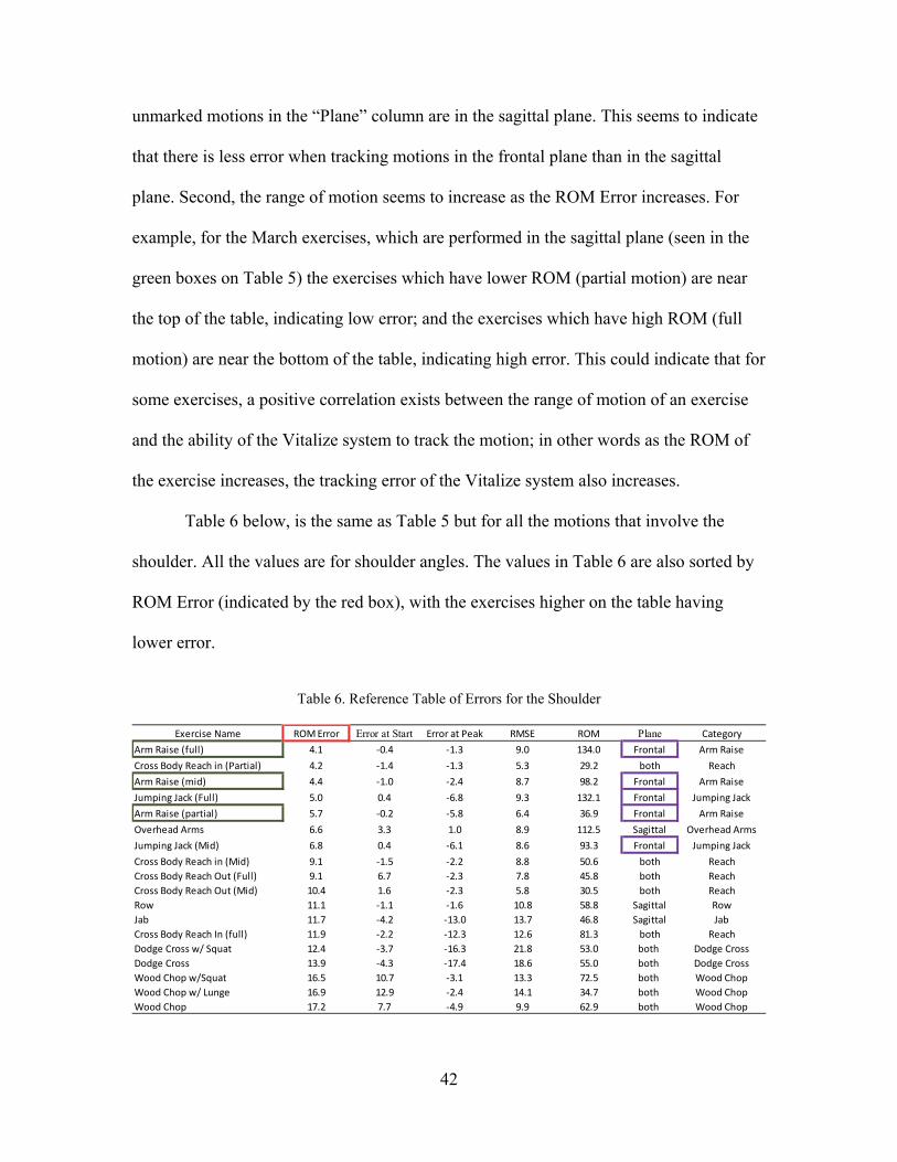
42
unmarked motions in the “Plane” column are in the sagittal plane. This seems to indicate
that there is less error when tracking motions in the frontal plane than in the sagittal
plane. Second, the range of motion seems to increase as the ROM Error increases. For
example, for the March exercises, which are performed in the sagittal plane (seen in the
green boxes on Table 5) the exercises which have lower ROM (partial motion) are near
the top of the table, indicating low error; and the exercises which have high ROM (full
motion) are near the bottom of the table, indicating high error. This could indicate that for
some exercises, a positive correlation exists between the range of motion of an exercise
and the ability of the Vitalize system to track the motion; in other words as the ROM of
the exercise increases, the tracking error of the Vitalize system also increases.
Table 6 below, is the same as Table 5 but for all the motions that involve the
shoulder. All the values are for shoulder angles. The values in Table 6 are also sorted by
ROM Error (indicated by the red box), with the exercises higher on the table having
lower error.
Table 6. Reference Table of Errors for the Shoulder
Exercise Name ROM Error Error at Start Error at Peak RMSE ROM Plane Category
Arm Raise (full) 4.1 ‐0.4 ‐1.3 9.0 134.0 Frontal Arm Raise
Cross Body Reach in (Partial) 4.2 ‐1.4 ‐1.3 5.3 29.2 both Reach
Arm Raise (mid) 4.4 ‐1.0 ‐2.4 8.7 98.2 Frontal Arm Raise
Jumping Jack (Full) 5.0 0.4 ‐6.8 9.3 132.1 Frontal Jumping Jack
Arm Raise (partial) 5.7 ‐0.2 ‐5.8 6.4 36.9 Frontal Arm Raise
Overhead Arms 6.6 3.3 1.0 8.9 112.5 Sagittal Overhead Arms
Jumping Jack (Mid) 6.8 0.4 ‐6.1 8.6 93.3 Frontal Jumping Jack
Cross Body Reach in (Mid) 9.1 ‐1.5 ‐2.2 8.8 50.6 both Reach
Cross Body Reach Out (Full) 9.1 6.7 ‐2.3 7.8 45.8 both Reach
Cross Body Reach Out (Mid) 10.4 1.6 ‐2.3 5.8 30.5 both Reach
Row 11.1 ‐1.1 ‐1.6 10.8 58.8 Sagittal Row
Jab 11.7 ‐4.2 ‐13.0 13.7 46.8 Sagittal Jab
Cross Body Reach In (full) 11.9 ‐2.2 ‐12.3 12.6 81.3 both Reach
Dodge Cross w/ Squat 12.4 ‐3.7 ‐16.3 21.8 53.0 both Dodge Cross
Dodge Cross 13.9 ‐4.3 ‐17.4 18.6 55.0 both Dodge Cross
Wood Chop w/Squat 16.5 10.7 ‐3.1 13.3 72.5 both Wood Chop
Wood Chop w/ Lunge 16.9 12.9 ‐2.4 14.1 34.7 both Wood Chop
Wood Chop 17.2 7.7 ‐4.9 9.9 62.9 both Wood Chop

43
Again it is observed that exercises in the frontal plane (indicated by the purple
boxes in Table 6) are higher on the table than motions in the sagittal plane; exercises that
involve both planes are also included and seem to perform worse than either individual
plane. The range of motion within a category of exercise decreases as ROM Error
increases (descending the table). For example, Arm Raise exercises (seen in the green
boxes in Table 6), which are performed in the frontal plane, have high ROM (full motion)
near the top of the table, indicating low error; with low ROM (partial motion) lower on
the table, indicating higher error. This could indicate that for some exercises, a negative
correlation exists between the range of motion of an exercise and the ability of the
Vitalize system to track the motion; i.e. as ROM increases, the error decreases. Which is
opposite of what was observed for the March exercise at the hip in Table 5. This seems to
imply that a complicated relationship exists between ROM and error.
The analyses performed directly follow from the observations made from these
tables. The first analysis seeks to discover the relationship between the plane of motion
and the tracking ability of the Vitalize system. It is hypothesized that the Vitalize system
will perform better in the frontal plane than in the sagittal plane because there is no depth
measurement required for frontal plane motions. The second analysis attempts to find the
relationship between the range of motion and the error of the Vitalize system. It is
hypothesized that the range of motion will not have an effect on the error of the system;
this has been reported in previous research [32], [36], [38]. However, the initial
observations from the exercise tables created in this study may indicate that a relationship
exists between ROM and error. The third analysis directly compares the performance of
the Vitalize system at the hip to the shoulder and to the knee. It is hypothesized that there

44
will be minimal difference between the tracking accuracy of different joints. The fourth
analysis examines multiple variations of a single exercise to see if there is a difference in
the tracking ability of the Vitalize system as the complexity of the motion increases. It is
hypothesized that adding complexity will not have an effect on the error of the primary
joint involved in the exercise. The final analysis is comprised of a series of observations
about the decisions made by the Vitalize software, whether an exercise was performed
correctly or not.
For all statistical analysis, the ROM error, error at the peak and RMS error were
used to analyze the accuracy of the Vitalize system. The Vicon system is held as the gold
standard, so any deviation of the Vitalize system from the Vicon system is considered
error of the Vitalize system. The repetitions were averaged for each exercise performed
by each subject. For errors of the hip joint, the Lthigh_G and Rthigh_G angles, the global
orientation angles of both thighs, were used. For errors of the shoulder joint, the
LUArm_G and RUArm_G angles, the global orientation angles of both upper arms, were
used. For errors of the knee joint, the LShank_G and RShank_G angles, the global
orientation angles of both shanks, were used.
Aim 1: Plane of Motion
Aim 1 seeks to answer the first question posed from observations of Tables 5 and
6. Does the plane involved in a motion affect the ability of the Vitalize system to
accurately track the motion? To address this question, two analyses were performed:
Analysis 1: Frontal vs. Sagittal Plane motions of the hip Analysis 2: Frontal vs. Sagittal vs. Bi-planar motions of the shoulder
The knee was not used in this analysis because of the anatomical lack of frontal
plane motions involving the knee.

45
Analysis 1: Hip
The exercises at the hip can be characterized as either frontal plane or sagittal
plane motions. The following lists indicate the exercises that were included for the frontal
plane group and the sagittal plane group. This analysis compares all the hip motions
completed in the frontal plane to all the hip motions completed in the sagittal plane.
Frontal Plane Side Step Jumping Jack (full) Side Jump Jumping Jack (Mid) Jumping Jack (no arms) Side Hop
Sagittal Plane
March with No Arms (Partial) March with No Arms (Mid) March with No Arms (Full) Squat (Partial) Squat (Mid) Squat (Full) Lunge (mid) Step Forward and Back Low Kick Lunge (full) Squat Jump (No Arms) (Full) Seated Knee Strike Lunge with Wood Chop Knee Strike Wood Chop with Squat Seated Low Kick Dodge Cross with Squat Knee Strike with Overhead Arms Knee Strike with Row Sit to Stand (Raised Seat) (Mid) Sit to Stand (Full)
The data from all frontal plane motions was compared to the data in all sagittal
plane motions; this comparison is pictured in the below boxplot (Figure 17).

46
Figure 17. Boxplot comparing Frontal vs. Sagittal Error for all Hip Motions. The middle black bar indicated median values and the red bar indicates mean values.
It can quickly be seen that on average, the frontal plane motions (mean 4.2°) have
less error than the sagittal plane motions (mean 6.9°). A two-tailed t-test assuming
unequal variance was performed between the frontal and sagittal planes and it was found
that the sagittal plane motions have significantly more error than the frontal plane
motions at the p=0.05 significance level [t(21)=-4.81, p<0.001]. It can also be observed
from the boxplot in Figure 17, that the sagittal plane motions seem to have higher
variance than the frontal plane motions. However, the sagittal hip motions also have
much higher ranges of motion than the frontal motions, which may contribute to the
higher tracking error. So in the following statistical, exercises with similar ranges of
motion were selected for comparison. The frontal plane exercises included in this
analysis were
Jumping Jack (full) Jumping Jack (no arms) Side Step Side Jump

47
Exercises included in the sagittal plane were
Step forward and back Squat (partial) March (partial) Seated Low kick
The exercises were combined into groups of frontal (mean=3.4°) and sagittal
(mean=4.8°); a two-tailed t-test assuming unequal variance was used to compare the
groups. At the p<0.05 level, there was a significant difference between the frontal plane
and the sagittal plane for hip motion [t(214)=-2.38, p=0.018]. There is significantly less
error for the Vitalize system in tracking hip motions in the frontal plane than motions in
the sagittal plane.
Analysis 2: Shoulder
A similar analysis was completed on the shoulder. At the shoulder, there is a third
category of motion: bi-planar motions which involve motion in both the sagittal and
frontal planes, not purely in either plane. The motions used in the planar analysis for the
shoulder are as follows:
Frontal Plane Bilateral Arm Raise (partial) Unilateral Arm Raise (partial) Unilateral Arm Raise (full)
Sagittal Plane Jab Row Overhead Arms
Bi-Planar Cross-Body Reach in (partial) Cross-Body Reach out (mid) Cross-Body Reach in (mid)

48
These motions were not controlled for range of motion, they span the whole range
available in the data from this study.
Using these groupings, a one-way ANOVA was used for comparison. There was a
significant difference between the groups at the p<0.05 level [F(2,291)=14.07,
p=<0.001]. Figure 18 gives a visual of the groupings used in the ANOVA.
Figure 18. Boxplot Comparing Planar Error for Shoulder Motions (ROM not controlled). The red bar indicates mean values, the black bar indicates median values.
The frontal plane had the least average error, (4.8°); followed by the bi-planar
motions (7.5°) and the sagittal plane (8.1°). Post hoc testing was performed with a
Bonferroni correction to determine which groups differed significantly. Two-sample,
two-tailed t-test assuming unequal variance with a significance level of α/3 (where
α=0.05; for the Bonferroni correction) were used to compare
Frontal Plane group to Both-plane group Both-Plane group to Sagittal plane group Frontal Plane group to Sagittal plane group

49
There was a significant difference for exercises in the frontal plane as compared
to the bi-planar motions [t(96)=-3.67, p<0.001]; and as compared to the sagittal plane
[t(180)=-5.62, p<0.001]. There was not a significant difference between the bi-planar and
sagittal plane groups [t(145)=-0.674, p=0.50]. The bi-planar motions had more variance
than the sagittal plane motions which had more variance than the frontal plane motions,
this can be seen by the extended box and error bars of the bi-planar and sagittal plane
motions in the boxplot (Figure 18) as compared to the frontal plane motions. When
looking at the average error values, the average error of the bi-planar motions falls
between the average errors of the frontal and sagittal groups, which supports the
hypothesized relationship between plane and error that was observed at the hip: that the
frontal plane is tracked with more accuracy than the sagittal plane.
This is the result that was expected because of the relationship between plane of
motion and error that was observed at the hip. However, these results were not controlled
for range of motion, and it is suspected that range of motion has an effect on the error. So
the previous analysis was repeated, but with a smaller number of motions with similar
ranges to control for tracking error due to the different ranges of motion. The exercises
that were used in the following analysis are
Frontal Plane Bilateral Arm Raise (partial) Unilateral Arm Raises (partial)
Bi-Planar Reach in (partial, mid) Reach out (mid)
Sagittal Plane Row Jab
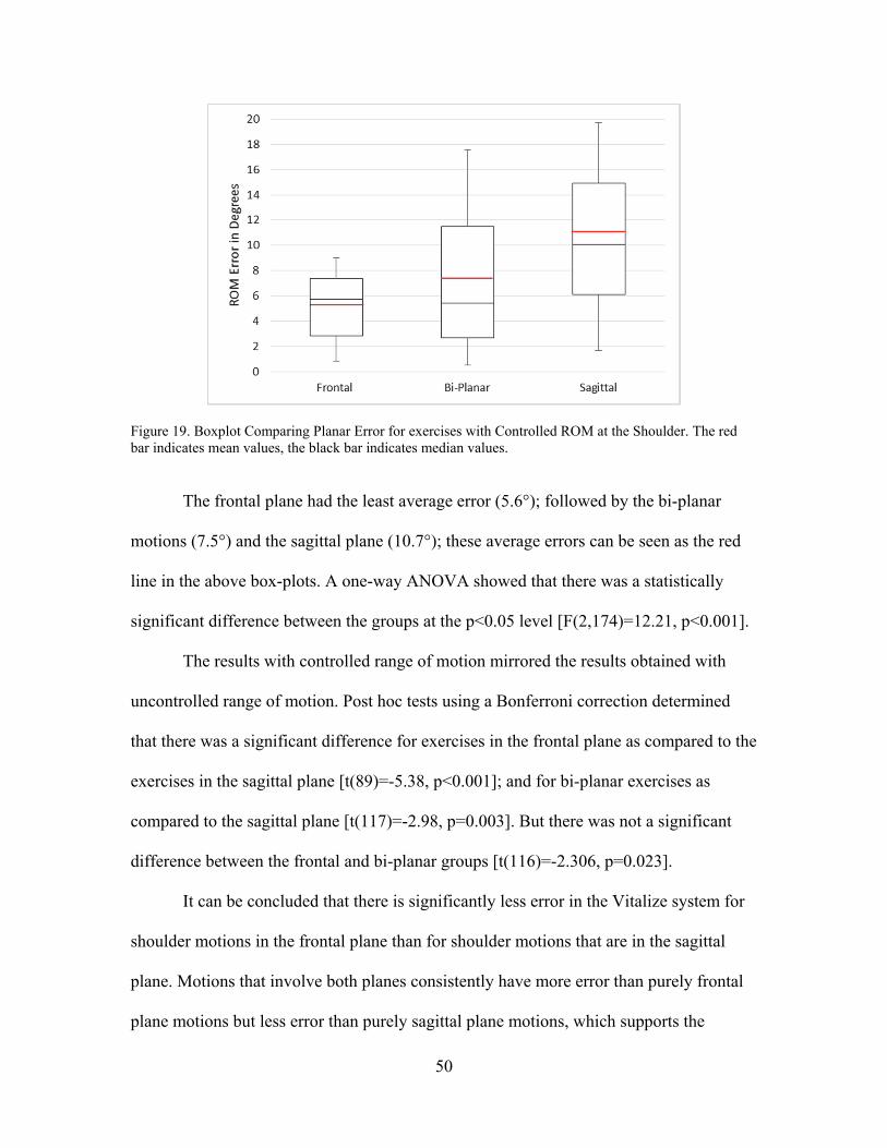
50
Figure 19. Boxplot Comparing Planar Error for exercises with Controlled ROM at the Shoulder. The red bar indicates mean values, the black bar indicates median values.
The frontal plane had the least average error (5.6°); followed by the bi-planar
motions (7.5°) and the sagittal plane (10.7°); these average errors can be seen as the red
line in the above box-plots. A one-way ANOVA showed that there was a statistically
significant difference between the groups at the p<0.05 level [F(2,174)=12.21, p<0.001].
The results with controlled range of motion mirrored the results obtained with
uncontrolled range of motion. Post hoc tests using a Bonferroni correction determined
that there was a significant difference for exercises in the frontal plane as compared to the
exercises in the sagittal plane [t(89)=-5.38, p<0.001]; and for bi-planar exercises as
compared to the sagittal plane [t(117)=-2.98, p=0.003]. But there was not a significant
difference between the frontal and bi-planar groups [t(116)=-2.306, p=0.023].
It can be concluded that there is significantly less error in the Vitalize system for
shoulder motions in the frontal plane than for shoulder motions that are in the sagittal
plane. Motions that involve both planes consistently have more error than purely frontal
plane motions but less error than purely sagittal plane motions, which supports the

51
finding that frontal plane motion is more accurately tracked by the exergaming system
than sagittal plane motion.
The effect of the plane of motion is supported at both the hip and shoulder; the
frontal plane is more accurate than the sagittal plane. At both the hip and shoulder, an
analysis was performed that controlled for the range of motion because it is suspected to
have an impact on the error. The direct effects of the range of motion of an exercise on
the error of the Vitalize system is now addressed in Aim 2.
Aim 2: Range of Motion
To discover the effect range of motion has on the error of the Vitalize system,
several analyses were completed. Because a significant effect has been found based on
the plane of motion, these analyses werebcontrolled for plane. Three joints were used: the
hip, shoulder and knee. The five analyses that were completed are:
Hip in the sagittal plane Knee in the sagittal plane Shoulder in the sagittal plane Hip in the frontal plane Shoulder in the frontal plane
The knee was only analyzed in the sagittal plane because of the anatomical lack of
frontal plane knee motion. Range of motion is a continuous variable, as opposed to the
discrete variables of “frontal” or “sagittal” plane. To take this into account, correlations
were performed with the range of motion data to determine the relationship between
range of motion and errors. However, during data collection some of the exercises were
split into discrete groups based on the range of motion, for example: full ROM march,
mid ROM march, partial ROM march; so some categorical statistical analyses are also
performed in this section.
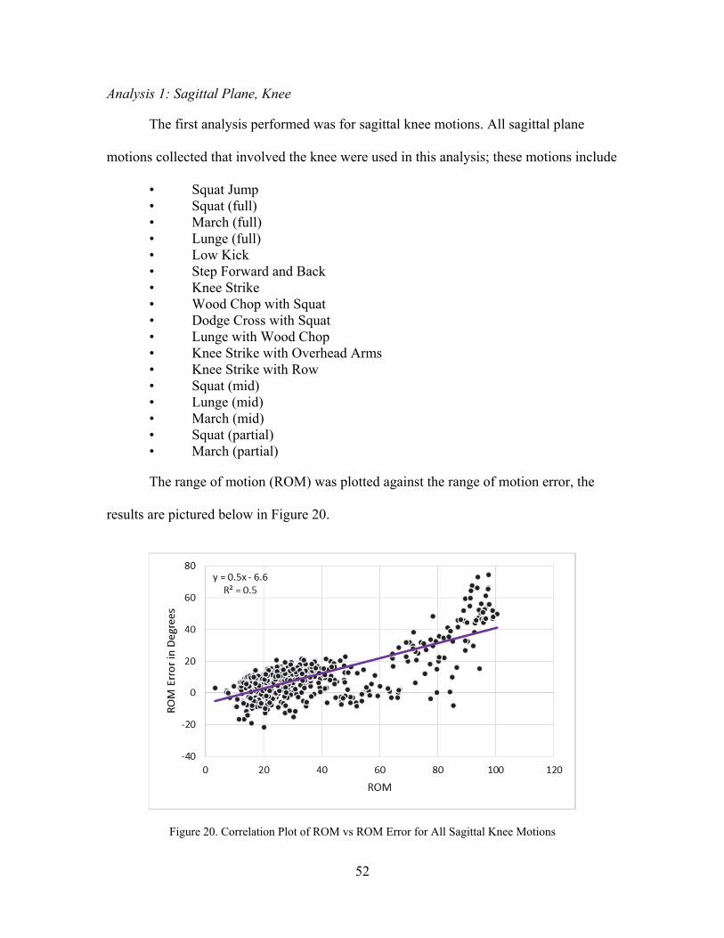
52
Analysis 1: Sagittal Plane, Knee
The first analysis performed was for sagittal knee motions. All sagittal plane
motions collected that involved the knee were used in this analysis; these motions include
• Squat Jump • Squat (full) • March (full) • Lunge (full) • Low Kick • Step Forward and Back • Knee Strike • Wood Chop with Squat • Dodge Cross with Squat • Lunge with Wood Chop • Knee Strike with Overhead Arms • Knee Strike with Row • Squat (mid) • Lunge (mid) • March (mid) • Squat (partial) • March (partial)
The range of motion (ROM) was plotted against the range of motion error, the
results are pictured below in Figure 20.
Figure 20. Correlation Plot of ROM vs ROM Error for All Sagittal Knee Motions
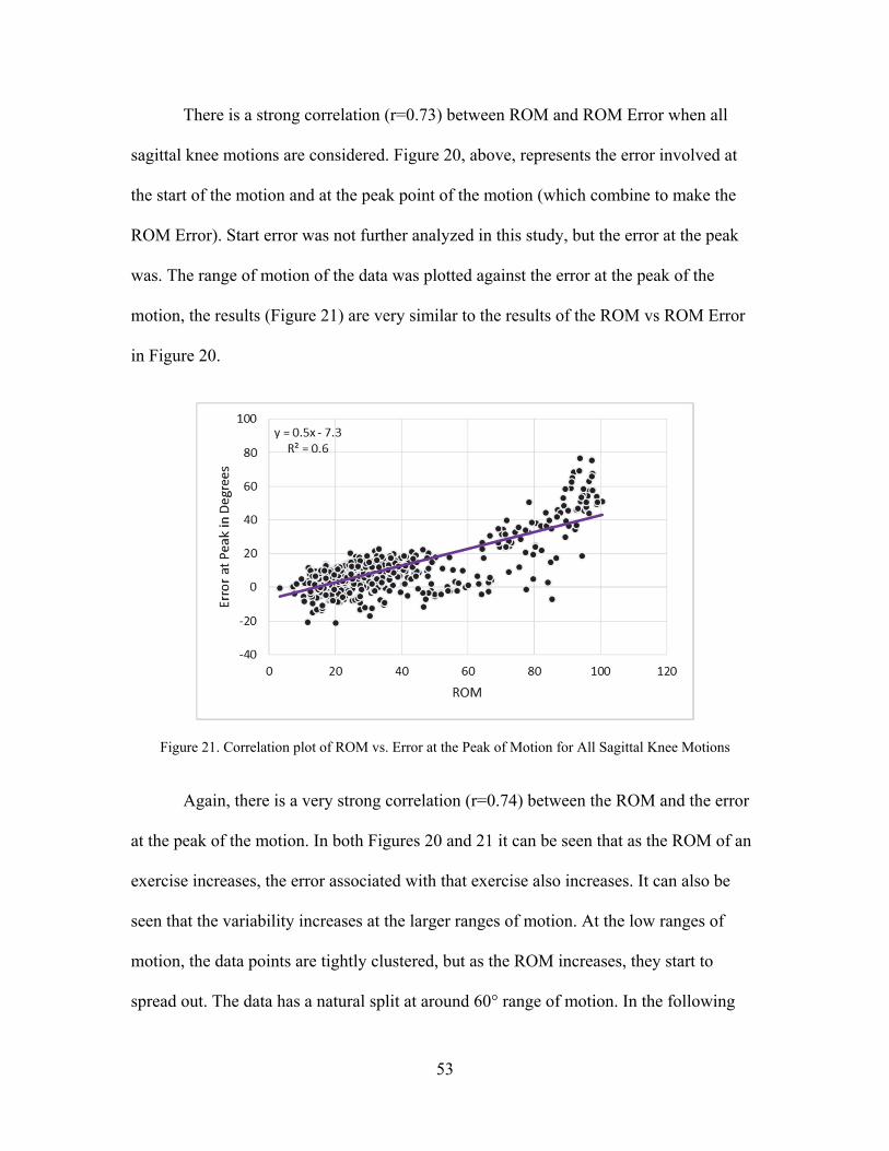
53
There is a strong correlation (r=0.73) between ROM and ROM Error when all
sagittal knee motions are considered. Figure 20, above, represents the error involved at
the start of the motion and at the peak point of the motion (which combine to make the
ROM Error). Start error was not further analyzed in this study, but the error at the peak
was. The range of motion of the data was plotted against the error at the peak of the
motion, the results (Figure 21) are very similar to the results of the ROM vs ROM Error
in Figure 20.
Figure 21. Correlation plot of ROM vs. Error at the Peak of Motion for All Sagittal Knee Motions
Again, there is a very strong correlation (r=0.74) between the ROM and the error
at the peak of the motion. In both Figures 20 and 21 it can be seen that as the ROM of an
exercise increases, the error associated with that exercise also increases. It can also be
seen that the variability increases at the larger ranges of motion. At the low ranges of
motion, the data points are tightly clustered, but as the ROM increases, they start to
spread out. The data has a natural split at around 60° range of motion. In the following
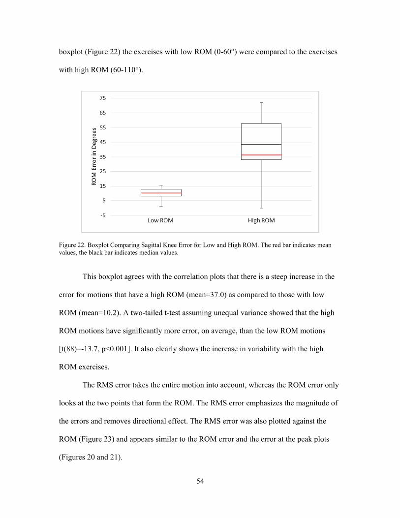
54
boxplot (Figure 22) the exercises with low ROM (0-60°) were compared to the exercises
with high ROM (60-110°).
Figure 22. Boxplot Comparing Sagittal Knee Error for Low and High ROM. The red bar indicates mean values, the black bar indicates median values.
This boxplot agrees with the correlation plots that there is a steep increase in the
error for motions that have a high ROM (mean=37.0) as compared to those with low
ROM (mean=10.2). A two-tailed t-test assuming unequal variance showed that the high
ROM motions have significantly more error, on average, than the low ROM motions
[t(88)=-13.7, p<0.001]. It also clearly shows the increase in variability with the high
ROM exercises.
The RMS error takes the entire motion into account, whereas the ROM error only
looks at the two points that form the ROM. The RMS error emphasizes the magnitude of
the errors and removes directional effect. The RMS error was also plotted against the
ROM (Figure 23) and appears similar to the ROM error and the error at the peak plots
(Figures 20 and 21).
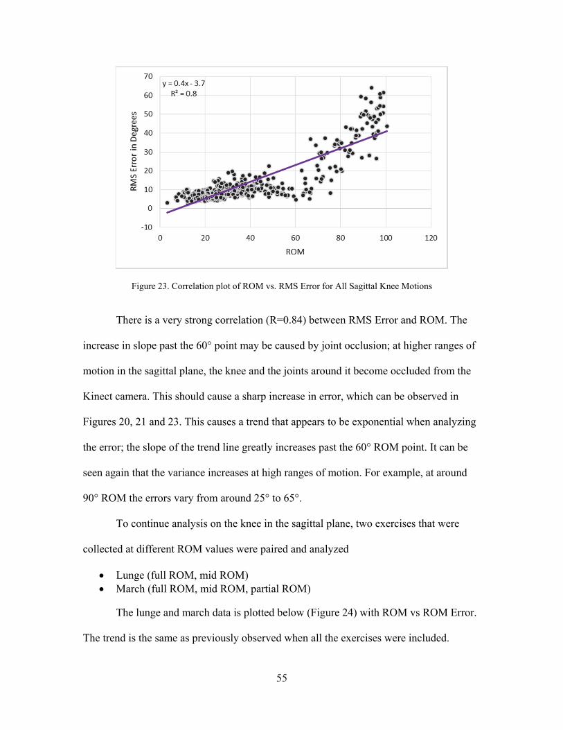
55
Figure 23. Correlation plot of ROM vs. RMS Error for All Sagittal Knee Motions
There is a very strong correlation (R=0.84) between RMS Error and ROM. The
increase in slope past the 60° point may be caused by joint occlusion; at higher ranges of
motion in the sagittal plane, the knee and the joints around it become occluded from the
Kinect camera. This should cause a sharp increase in error, which can be observed in
Figures 20, 21 and 23. This causes a trend that appears to be exponential when analyzing
the error; the slope of the trend line greatly increases past the 60° ROM point. It can be
seen again that the variance increases at high ranges of motion. For example, at around
90° ROM the errors vary from around 25° to 65°.
To continue analysis on the knee in the sagittal plane, two exercises that were
collected at different ROM values were paired and analyzed
Lunge (full ROM, mid ROM) March (full ROM, mid ROM, partial ROM)
The lunge and march data is plotted below (Figure 24) with ROM vs ROM Error.
The trend is the same as previously observed when all the exercises were included.
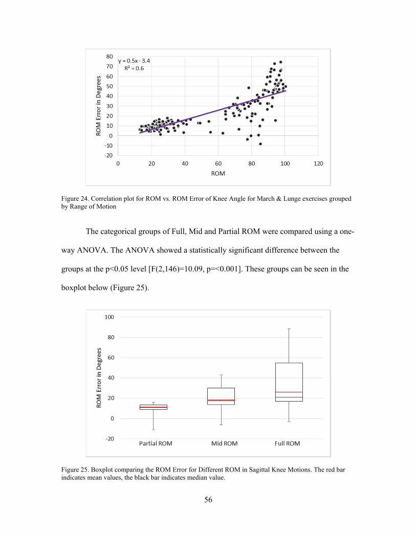
56
Figure 24. Correlation plot for ROM vs. ROM Error of Knee Angle for March & Lunge exercises grouped by Range of Motion
The categorical groups of Full, Mid and Partial ROM were compared using a one-
way ANOVA. The ANOVA showed a statistically significant difference between the
groups at the p<0.05 level [F(2,146)=10.09, p=<0.001]. These groups can be seen in the
boxplot below (Figure 25).
Figure 25. Boxplot comparing the ROM Error for Different ROM in Sagittal Knee Motions. The red bar indicates mean values, the black bar indicates median value.
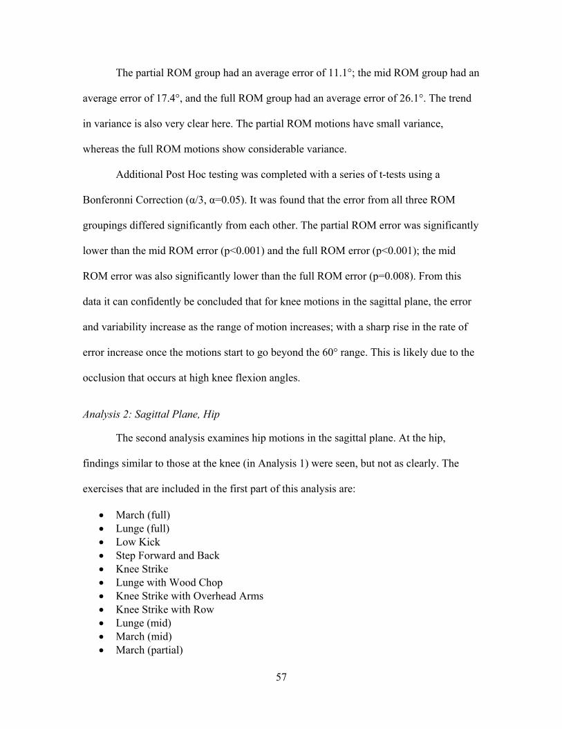
57
The partial ROM group had an average error of 11.1°; the mid ROM group had an
average error of 17.4°, and the full ROM group had an average error of 26.1°. The trend
in variance is also very clear here. The partial ROM motions have small variance,
whereas the full ROM motions show considerable variance.
Additional Post Hoc testing was completed with a series of t-tests using a
Bonferonni Correction (α/3, α=0.05). It was found that the error from all three ROM
groupings differed significantly from each other. The partial ROM error was significantly
lower than the mid ROM error (p<0.001) and the full ROM error (p<0.001); the mid
ROM error was also significantly lower than the full ROM error (p=0.008). From this
data it can confidently be concluded that for knee motions in the sagittal plane, the error
and variability increase as the range of motion increases; with a sharp rise in the rate of
error increase once the motions start to go beyond the 60° range. This is likely due to the
occlusion that occurs at high knee flexion angles.
Analysis 2: Sagittal Plane, Hip
The second analysis examines hip motions in the sagittal plane. At the hip,
findings similar to those at the knee (in Analysis 1) were seen, but not as clearly. The
exercises that are included in the first part of this analysis are:
March (full) Lunge (full) Low Kick Step Forward and Back Knee Strike Lunge with Wood Chop Knee Strike with Overhead Arms Knee Strike with Row Lunge (mid) March (mid) March (partial)
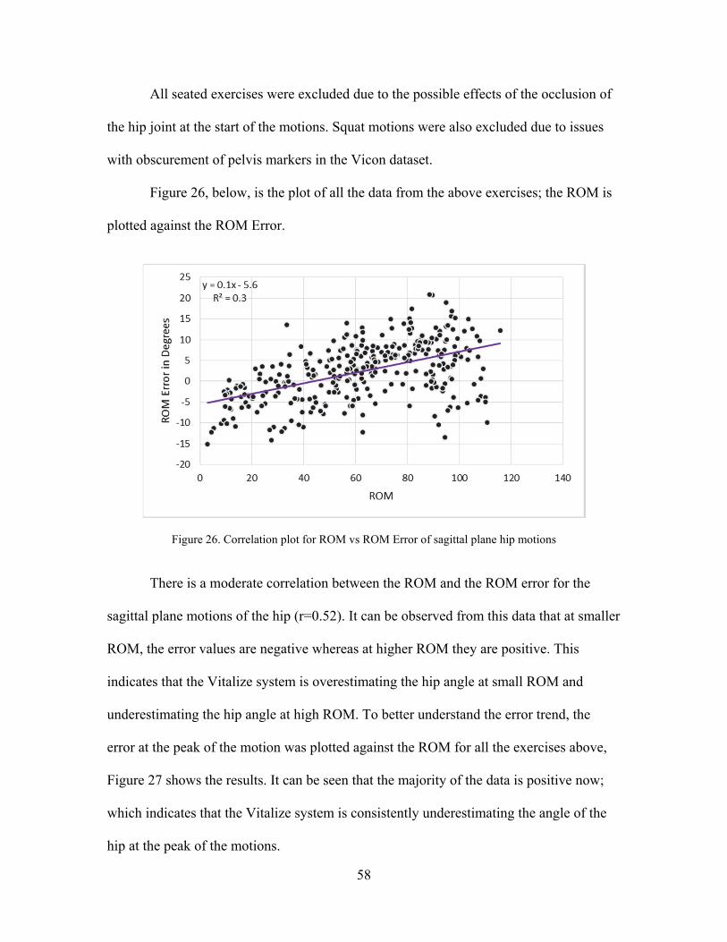
58
All seated exercises were excluded due to the possible effects of the occlusion of
the hip joint at the start of the motions. Squat motions were also excluded due to issues
with obscurement of pelvis markers in the Vicon dataset.
Figure 26, below, is the plot of all the data from the above exercises; the ROM is
plotted against the ROM Error.
Figure 26. Correlation plot for ROM vs ROM Error of sagittal plane hip motions
There is a moderate correlation between the ROM and the ROM error for the
sagittal plane motions of the hip (r=0.52). It can be observed from this data that at smaller
ROM, the error values are negative whereas at higher ROM they are positive. This
indicates that the Vitalize system is overestimating the hip angle at small ROM and
underestimating the hip angle at high ROM. To better understand the error trend, the
error at the peak of the motion was plotted against the ROM for all the exercises above,
Figure 27 shows the results. It can be seen that the majority of the data is positive now;
which indicates that the Vitalize system is consistently underestimating the angle of the
hip at the peak of the motions.

59
Figure 27. Correlation plot for Error at Peak vs ROM for sagittal hip motions
This plot shows a moderate correlation (r=0.57) with the error increasing as the
ROM increases. From Figure 27, it can be seen that not only does the error increase in
magnitude as the ROM increases, but the variance of the error also increases. This is seen
in the rough trumpet-shape of the data. The data around 20° ROM (on the x-axis) has
errors with low variance, with the values varying from around -5° to 2°, but data around
100° ROM (on the x-axis) has high variance with errors that vary from -5° to nearly 20°.
To analyze the magnitudes of the error, the data was grouped by ROM. The 60°
ROM point was used as the dividing point because it is the midpoint between the
maximum and minimum ROM values observed on Figure 27. The Low ROM group
includes all exercises with ROM between 0 and 60°; the High ROM group includes all
exercises with ROM between 60 and 120°. A boxplot (Figure 28) was created to visualize
the error between the ROM groups using the magnitude of the error at the peak of the
motions.

60
Figure 28. Boxplot comparing Error at the Peak of Low ROM to High ROM Sagittal Hip Motions. The red bar indicates mean values, the black bar indicates median values.
It can be seen that the error at the high ranges of motion is greater than the error at
low ranges. However, the difference here is not nearly as stark as the difference observed
at the knee (Figure 22). The low ROM group has an average of 4.5° error at the peak and
the high ROM group has an average of 6.7° error at the peak. A two-tailed t-test
confirmed that the high ROM group had significantly more error than the low ROM
group [t(506)=-6.57, p<0.001]. It can be seen again that the motions with higher ROM
also have higher error variance.
The trend toward higher error and increased variance are supported across the
entire motion by the RMS error. Figure 29 shows the correlation plot of RMS error vs
ROM. This figure shows a strong positive correlation between RMS Error and ROM
(r=0.60). This demonstrates that as the range of motion increases, the error of the Vitalize
system at the peak of the motion also increases. This agrees with the relationship between
ROM and error observed for knee motions in the sagittal plane.
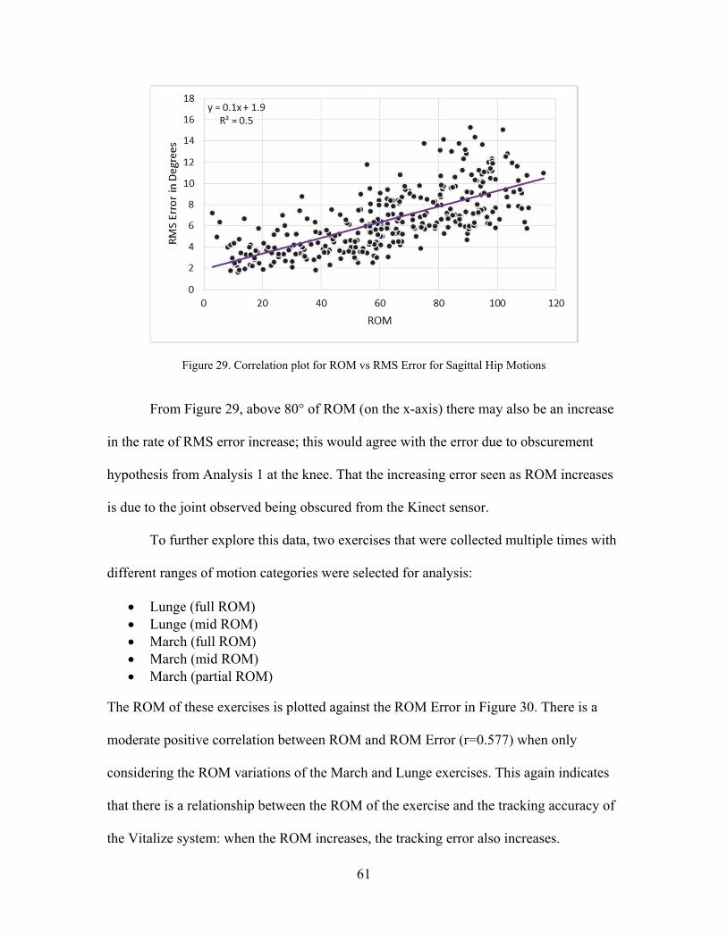
61
Figure 29. Correlation plot for ROM vs RMS Error for Sagittal Hip Motions
From Figure 29, above 80° of ROM (on the x-axis) there may also be an increase
in the rate of RMS error increase; this would agree with the error due to obscurement
hypothesis from Analysis 1 at the knee. That the increasing error seen as ROM increases
is due to the joint observed being obscured from the Kinect sensor.
To further explore this data, two exercises that were collected multiple times with
different ranges of motion categories were selected for analysis:
Lunge (full ROM) Lunge (mid ROM) March (full ROM) March (mid ROM) March (partial ROM)
The ROM of these exercises is plotted against the ROM Error in Figure 30. There is a
moderate positive correlation between ROM and ROM Error (r=0.577) when only
considering the ROM variations of the March and Lunge exercises. This again indicates
that there is a relationship between the ROM of the exercise and the tracking accuracy of
the Vitalize system: when the ROM increases, the tracking error also increases.
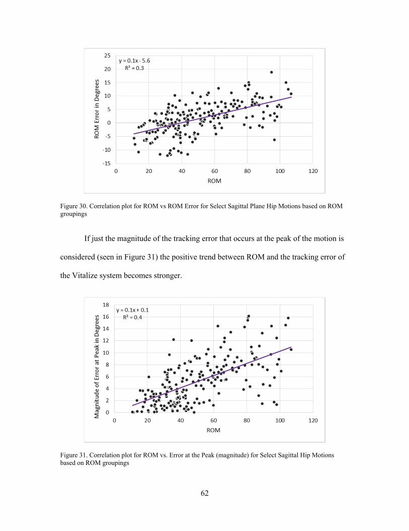
62
Figure 30. Correlation plot for ROM vs ROM Error for Select Sagittal Plane Hip Motions based on ROM groupings
If just the magnitude of the tracking error that occurs at the peak of the motion is
considered (seen in Figure 31) the positive trend between ROM and the tracking error of
the Vitalize system becomes stronger.
Figure 31. Correlation plot for ROM vs. Error at the Peak (magnitude) for Select Sagittal Hip Motions based on ROM groupings

63
There is a strong correlation between the magnitude of error at peak vs. ROM
(r=0.63). This indicates that more error is occurring at the peak of motions that have a
large ROM than at the peak of motions that have smaller ROM. This is supported by the
RMS Error data, pictured below in Figure 32.
Figure 32. Correlation plot for ROM vs RMS Error for Select Sagittal Hip Motions based on ROM groupings
There is a strong correlation between RMS error and the ROM of the exercises
(r=0.67). The RMS error takes into account the error along the whole waveform of the
motion, which provides additional support toward the conclusion that the error is
increasing as the range of motion increases.
These motions were combined into three categories: Full, Mid and Partial ROM.
A one-way ANOVA was performed to determine the effect of range of motion on
sagittal-plane hip motions. In agreement with the correlation data, the statistical analysis
showed that the error increased as the range of motion increased. The ANOVA showed
that the range of motion of an exercise has a significant effect on the error of the Vitalize
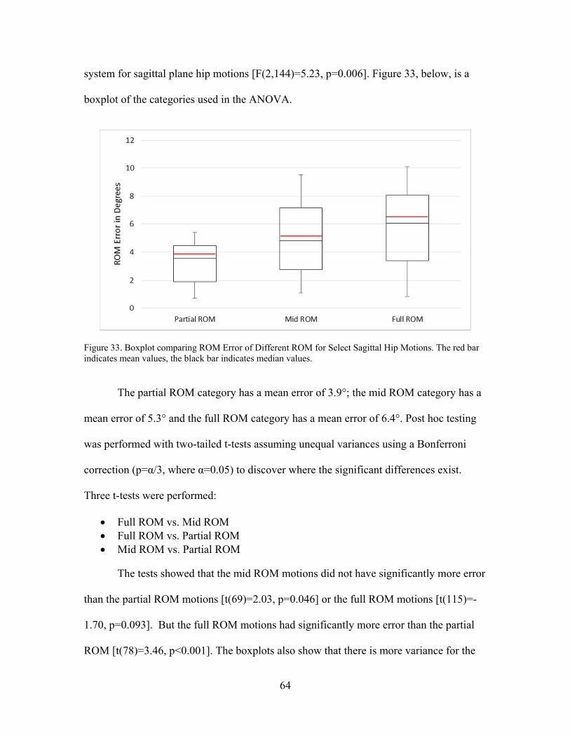
64
system for sagittal plane hip motions [F(2,144)=5.23, p=0.006]. Figure 33, below, is a
boxplot of the categories used in the ANOVA.
Figure 33. Boxplot comparing ROM Error of Different ROM for Select Sagittal Hip Motions. The red bar indicates mean values, the black bar indicates median values.
The partial ROM category has a mean error of 3.9°; the mid ROM category has a
mean error of 5.3° and the full ROM category has a mean error of 6.4°. Post hoc testing
was performed with two-tailed t-tests assuming unequal variances using a Bonferroni
correction (p=α/3, where α=0.05) to discover where the significant differences exist.
Three t-tests were performed:
Full ROM vs. Mid ROM Full ROM vs. Partial ROM Mid ROM vs. Partial ROM
The tests showed that the mid ROM motions did not have significantly more error
than the partial ROM motions [t(69)=2.03, p=0.046] or the full ROM motions [t(115)=-
1.70, p=0.093]. But the full ROM motions had significantly more error than the partial
ROM [t(78)=3.46, p<0.001]. The boxplots also show that there is more variance for the
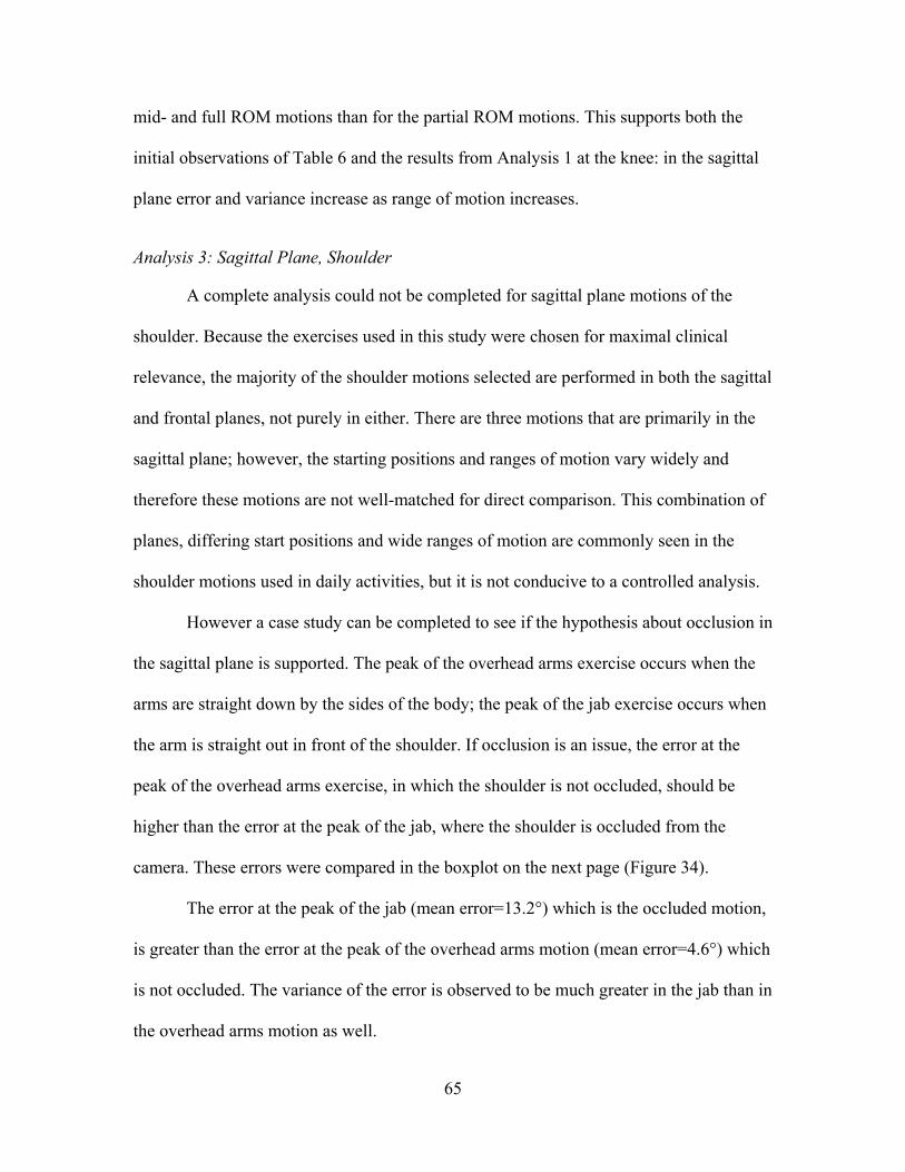
65
mid- and full ROM motions than for the partial ROM motions. This supports both the
initial observations of Table 6 and the results from Analysis 1 at the knee: in the sagittal
plane error and variance increase as range of motion increases.
Analysis 3: Sagittal Plane, Shoulder
A complete analysis could not be completed for sagittal plane motions of the
shoulder. Because the exercises used in this study were chosen for maximal clinical
relevance, the majority of the shoulder motions selected are performed in both the sagittal
and frontal planes, not purely in either. There are three motions that are primarily in the
sagittal plane; however, the starting positions and ranges of motion vary widely and
therefore these motions are not well-matched for direct comparison. This combination of
planes, differing start positions and wide ranges of motion are commonly seen in the
shoulder motions used in daily activities, but it is not conducive to a controlled analysis.
However a case study can be completed to see if the hypothesis about occlusion in
the sagittal plane is supported. The peak of the overhead arms exercise occurs when the
arms are straight down by the sides of the body; the peak of the jab exercise occurs when
the arm is straight out in front of the shoulder. If occlusion is an issue, the error at the
peak of the overhead arms exercise, in which the shoulder is not occluded, should be
higher than the error at the peak of the jab, where the shoulder is occluded from the
camera. These errors were compared in the boxplot on the next page (Figure 34).
The error at the peak of the jab (mean error=13.2°) which is the occluded motion,
is greater than the error at the peak of the overhead arms motion (mean error=4.6°) which
is not occluded. The variance of the error is observed to be much greater in the jab than in
the overhead arms motion as well.
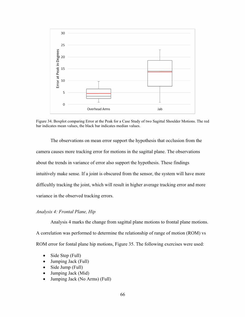
66
Figure 34. Boxplot comparing Error at the Peak for a Case Study of two Sagittal Shoulder Motions. The red bar indicates mean values, the black bar indicates median values.
The observations on mean error support the hypothesis that occlusion from the
camera causes more tracking error for motions in the sagittal plane. The observations
about the trends in variance of error also support the hypothesis. These findings
intuitively make sense. If a joint is obscured from the sensor, the system will have more
difficultly tracking the joint, which will result in higher average tracking error and more
variance in the observed tracking errors.
Analysis 4: Frontal Plane, Hip
Analysis 4 marks the change from sagittal plane motions to frontal plane motions.
A correlation was performed to determine the relationship of range of motion (ROM) vs
ROM error for fontal plane hip motions, Figure 35. The following exercises were used:
Side Step (Full) Jumping Jack (Full) Side Jump (Full) Jumping Jack (Mid) Jumping Jack (No Arms) (Full)
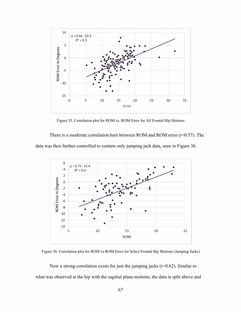
67
Figure 35. Correlation plot for ROM vs. ROM Error for All Frontal Hip Motions
There is a moderate correlation here between ROM and ROM error (r=0.57). The
data was then further controlled to contain only jumping jack data, seen in Figure 36.
Figure 36. Correlation plot for ROM vs ROM Error for Select Frontal Hip Motions (Jumping Jacks)
Now a strong correlation exists for just the jumping jacks (r=0.62). Similar to
what was observed at the hip with the sagittal plane motions, the data is split above and
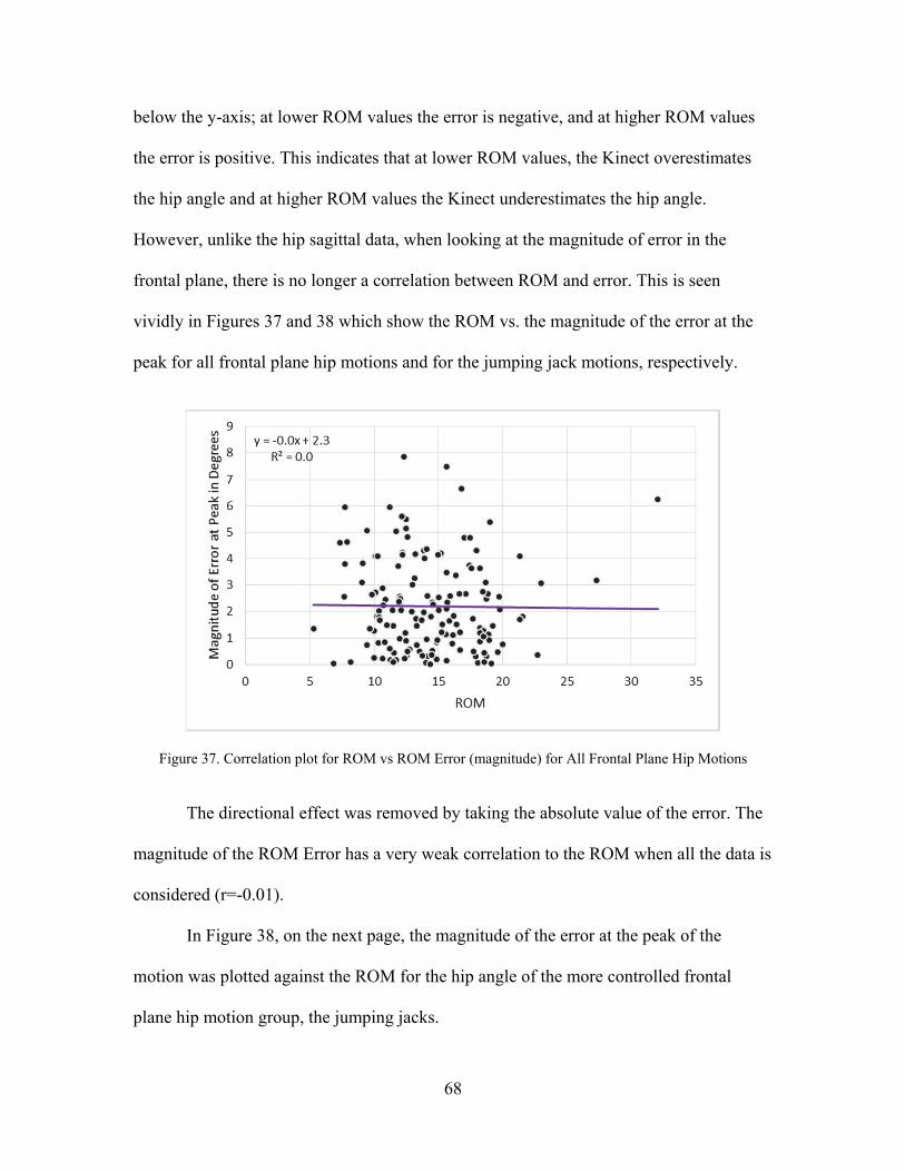
68
below the y-axis; at lower ROM values the error is negative, and at higher ROM values
the error is positive. This indicates that at lower ROM values, the Kinect overestimates
the hip angle and at higher ROM values the Kinect underestimates the hip angle.
However, unlike the hip sagittal data, when looking at the magnitude of error in the
frontal plane, there is no longer a correlation between ROM and error. This is seen
vividly in Figures 37 and 38 which show the ROM vs. the magnitude of the error at the
peak for all frontal plane hip motions and for the jumping jack motions, respectively.
Figure 37. Correlation plot for ROM vs ROM Error (magnitude) for All Frontal Plane Hip Motions
The directional effect was removed by taking the absolute value of the error. The
magnitude of the ROM Error has a very weak correlation to the ROM when all the data is
considered (r=-0.01).
In Figure 38, on the next page, the magnitude of the error at the peak of the
motion was plotted against the ROM for the hip angle of the more controlled frontal
plane hip motion group, the jumping jacks.
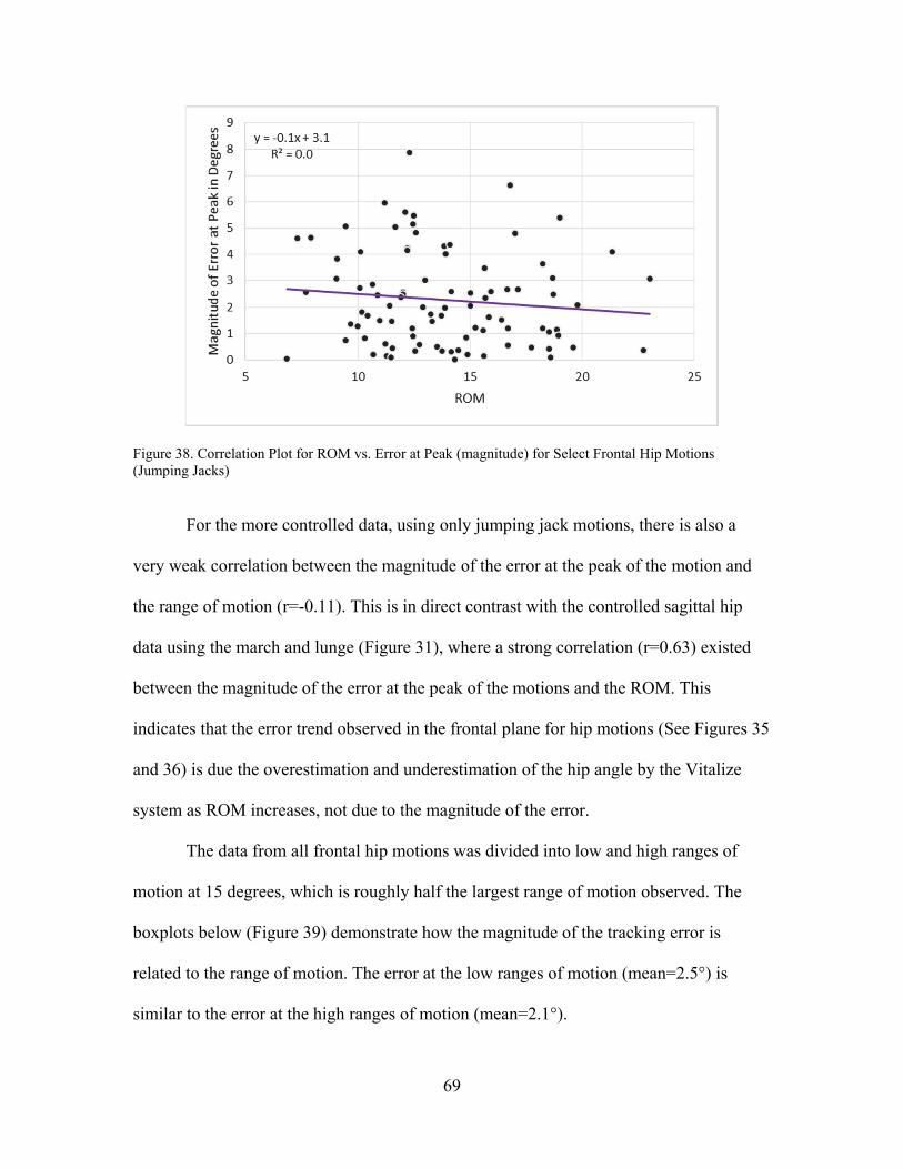
69
Figure 38. Correlation Plot for ROM vs. Error at Peak (magnitude) for Select Frontal Hip Motions (Jumping Jacks)
For the more controlled data, using only jumping jack motions, there is also a
very weak correlation between the magnitude of the error at the peak of the motion and
the range of motion (r=-0.11). This is in direct contrast with the controlled sagittal hip
data using the march and lunge (Figure 31), where a strong correlation (r=0.63) existed
between the magnitude of the error at the peak of the motions and the ROM. This
indicates that the error trend observed in the frontal plane for hip motions (See Figures 35
and 36) is due the overestimation and underestimation of the hip angle by the Vitalize
system as ROM increases, not due to the magnitude of the error.
The data from all frontal hip motions was divided into low and high ranges of
motion at 15 degrees, which is roughly half the largest range of motion observed. The
boxplots below (Figure 39) demonstrate how the magnitude of the tracking error is
related to the range of motion. The error at the low ranges of motion (mean=2.5°) is
similar to the error at the high ranges of motion (mean=2.1°).
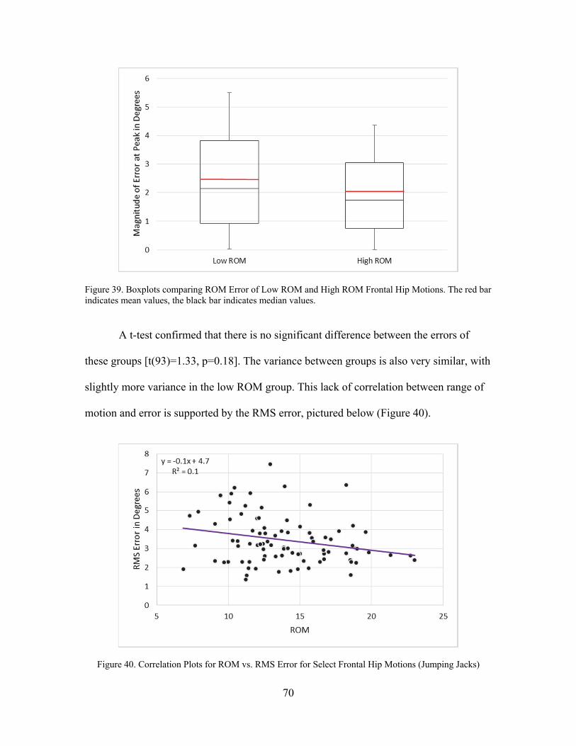
70
Figure 39. Boxplots comparing ROM Error of Low ROM and High ROM Frontal Hip Motions. The red bar indicates mean values, the black bar indicates median values.
A t-test confirmed that there is no significant difference between the errors of
these groups [t(93)=1.33, p=0.18]. The variance between groups is also very similar, with
slightly more variance in the low ROM group. This lack of correlation between range of
motion and error is supported by the RMS error, pictured below (Figure 40).
Figure 40. Correlation Plots for ROM vs. RMS Error for Select Frontal Hip Motions (Jumping Jacks)

71
The RMS error removes the directionality effect. There exists only a weak
correlation between the RMS error and the ROM (r=-0.24) for error at the hip during the
jumping jacks. Again, it can be seen that there is slightly higher variance at lower ranges
of motion; which is opposite what was observed at the hip in the sagittal plane, where the
high range of motion activities had both higher error and variance. The lack of correlation
seen here supports the conclusion that the observed trend for all frontal hip motions in
Figure 35 (at the beginning of this analysis) is due to the directionality of the angle not a
relationship between the magnitude of error and the range of motion. Contrary to the
findings in the sagittal plane, the variance in the frontal hip motions was slightly larger at
smaller ranges of motion.
For hip motions in the frontal plane, a trend toward more error as ROM increases
was found; but it is due to the Vitalize system overestimating the hip angle at small
ranges and underestimating the angle at large ranges; it was found that no relationship
between the magnitude of error and ROM exists for frontal hip motions. This consistent
overestimation at small ROM and underestimation at large ROM was a unexpected
finding of this study.
Analysis 5: Frontal Plane, Shoulder
The final analysis of Aim 2 examines shoulder motions in the frontal plane. The
plot of ROM vs ROM Error for all the shoulder motions that occur in the frontal plane is
included below, Figure 41.
Arm Raise (full) Arm Raise (mid) Jumping Jack (Full) Arm Raise (partial) Jumping Jack (Mid)
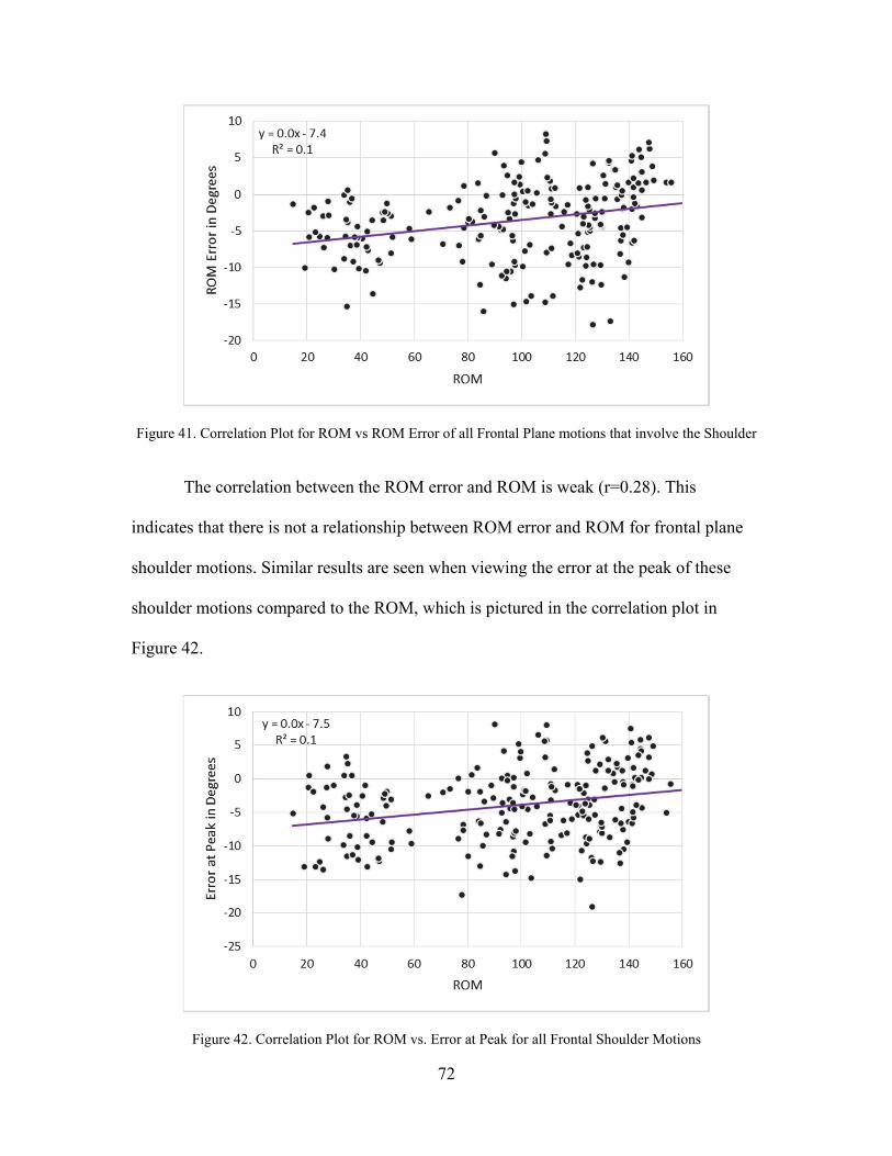
72
Figure 41. Correlation Plot for ROM vs ROM Error of all Frontal Plane motions that involve the Shoulder
The correlation between the ROM error and ROM is weak (r=0.28). This
indicates that there is not a relationship between ROM error and ROM for frontal plane
shoulder motions. Similar results are seen when viewing the error at the peak of these
shoulder motions compared to the ROM, which is pictured in the correlation plot in
Figure 42.
Figure 42. Correlation Plot for ROM vs. Error at Peak for all Frontal Shoulder Motions
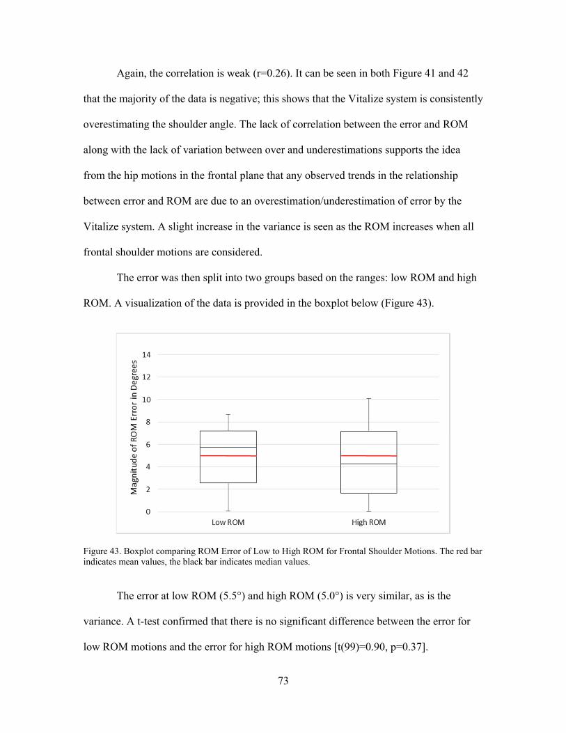
73
Again, the correlation is weak (r=0.26). It can be seen in both Figure 41 and 42
that the majority of the data is negative; this shows that the Vitalize system is consistently
overestimating the shoulder angle. The lack of correlation between the error and ROM
along with the lack of variation between over and underestimations supports the idea
from the hip motions in the frontal plane that any observed trends in the relationship
between error and ROM are due to an overestimation/underestimation of error by the
Vitalize system. A slight increase in the variance is seen as the ROM increases when all
frontal shoulder motions are considered.
The error was then split into two groups based on the ranges: low ROM and high
ROM. A visualization of the data is provided in the boxplot below (Figure 43).
Figure 43. Boxplot comparing ROM Error of Low to High ROM for Frontal Shoulder Motions. The red bar indicates mean values, the black bar indicates median values.
The error at low ROM (5.5°) and high ROM (5.0°) is very similar, as is the
variance. A t-test confirmed that there is no significant difference between the error for
low ROM motions and the error for high ROM motions [t(99)=0.90, p=0.37].
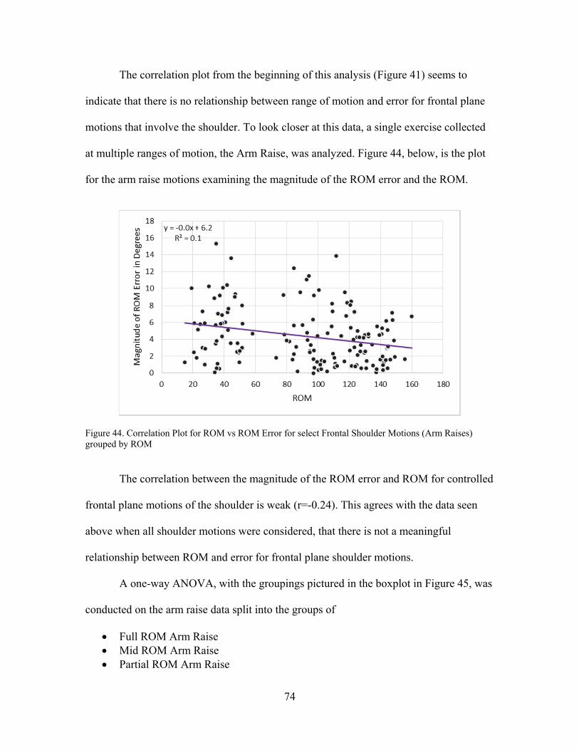
74
The correlation plot from the beginning of this analysis (Figure 41) seems to
indicate that there is no relationship between range of motion and error for frontal plane
motions that involve the shoulder. To look closer at this data, a single exercise collected
at multiple ranges of motion, the Arm Raise, was analyzed. Figure 44, below, is the plot
for the arm raise motions examining the magnitude of the ROM error and the ROM.
Figure 44. Correlation Plot for ROM vs ROM Error for select Frontal Shoulder Motions (Arm Raises) grouped by ROM
The correlation between the magnitude of the ROM error and ROM for controlled
frontal plane motions of the shoulder is weak (r=-0.24). This agrees with the data seen
above when all shoulder motions were considered, that there is not a meaningful
relationship between ROM and error for frontal plane shoulder motions.
A one-way ANOVA, with the groupings pictured in the boxplot in Figure 45, was
conducted on the arm raise data split into the groups of
Full ROM Arm Raise Mid ROM Arm Raise Partial ROM Arm Raise
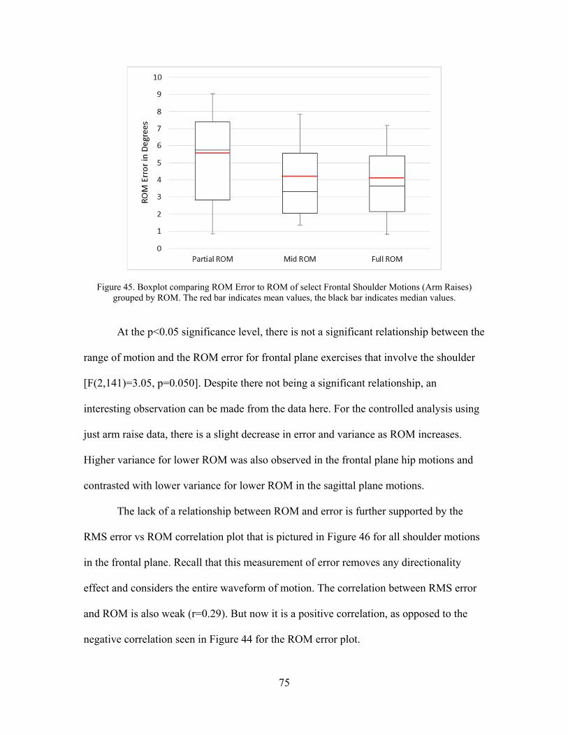
75
Figure 45. Boxplot comparing ROM Error to ROM of select Frontal Shoulder Motions (Arm Raises) grouped by ROM. The red bar indicates mean values, the black bar indicates median values.
At the p<0.05 significance level, there is not a significant relationship between the
range of motion and the ROM error for frontal plane exercises that involve the shoulder
[F(2,141)=3.05, p=0.050]. Despite there not being a significant relationship, an
interesting observation can be made from the data here. For the controlled analysis using
just arm raise data, there is a slight decrease in error and variance as ROM increases.
Higher variance for lower ROM was also observed in the frontal plane hip motions and
contrasted with lower variance for lower ROM in the sagittal plane motions.
The lack of a relationship between ROM and error is further supported by the
RMS error vs ROM correlation plot that is pictured in Figure 46 for all shoulder motions
in the frontal plane. Recall that this measurement of error removes any directionality
effect and considers the entire waveform of motion. The correlation between RMS error
and ROM is also weak (r=0.29). But now it is a positive correlation, as opposed to the
negative correlation seen in Figure 44 for the ROM error plot.
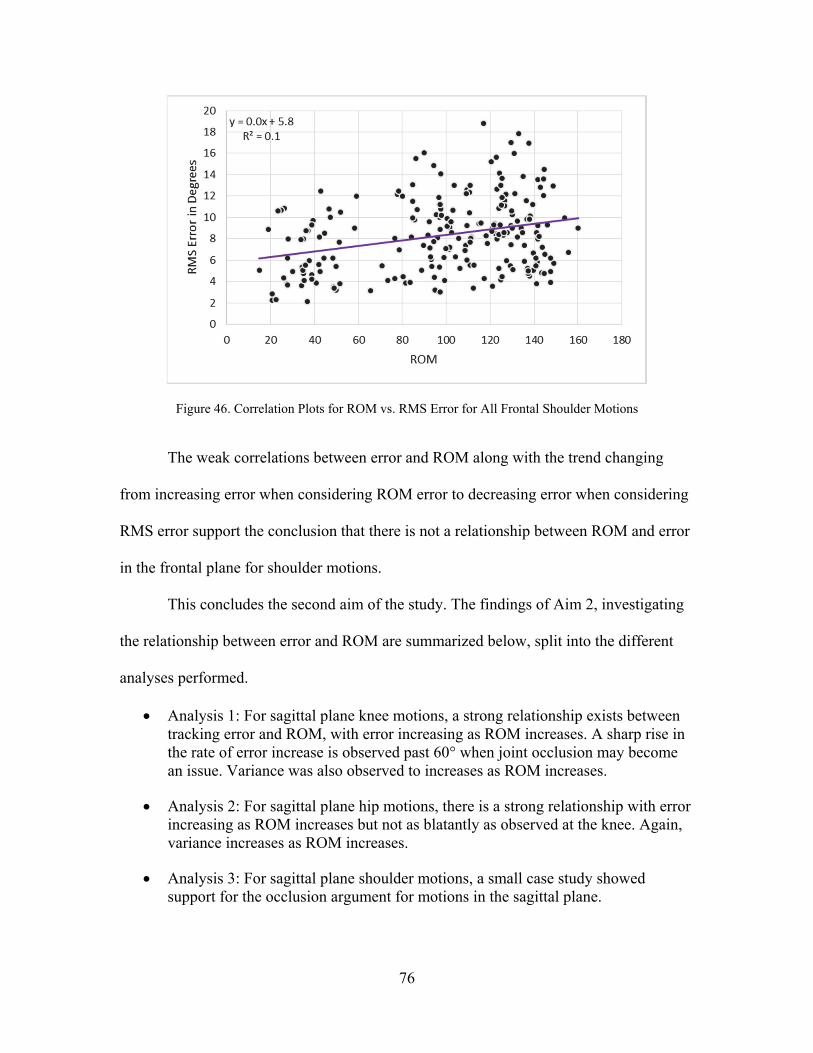
76
Figure 46. Correlation Plots for ROM vs. RMS Error for All Frontal Shoulder Motions
The weak correlations between error and ROM along with the trend changing
from increasing error when considering ROM error to decreasing error when considering
RMS error support the conclusion that there is not a relationship between ROM and error
in the frontal plane for shoulder motions.
This concludes the second aim of the study. The findings of Aim 2, investigating
the relationship between error and ROM are summarized below, split into the different
analyses performed.
Analysis 1: For sagittal plane knee motions, a strong relationship exists between tracking error and ROM, with error increasing as ROM increases. A sharp rise in the rate of error increase is observed past 60° when joint occlusion may become an issue. Variance was also observed to increases as ROM increases.
Analysis 2: For sagittal plane hip motions, there is a strong relationship with error increasing as ROM increases but not as blatantly as observed at the knee. Again, variance increases as ROM increases.
Analysis 3: For sagittal plane shoulder motions, a small case study showed support for the occlusion argument for motions in the sagittal plane.
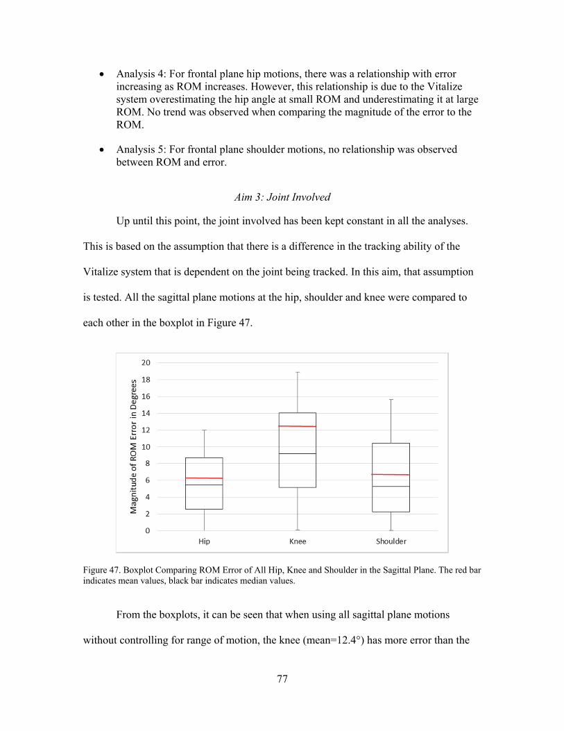
77
Analysis 4: For frontal plane hip motions, there was a relationship with error increasing as ROM increases. However, this relationship is due to the Vitalize system overestimating the hip angle at small ROM and underestimating it at large ROM. No trend was observed when comparing the magnitude of the error to the ROM.
Analysis 5: For frontal plane shoulder motions, no relationship was observed between ROM and error.
Aim 3: Joint Involved
Up until this point, the joint involved has been kept constant in all the analyses.
This is based on the assumption that there is a difference in the tracking ability of the
Vitalize system that is dependent on the joint being tracked. In this aim, that assumption
is tested. All the sagittal plane motions at the hip, shoulder and knee were compared to
each other in the boxplot in Figure 47.
Figure 47. Boxplot Comparing ROM Error of All Hip, Knee and Shoulder in the Sagittal Plane. The red bar indicates mean values, black bar indicates median values.
From the boxplots, it can be seen that when using all sagittal plane motions
without controlling for range of motion, the knee (mean=12.4°) has more error than the
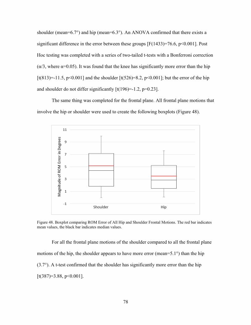
78
shoulder (mean=6.7°) and hip (mean=6.3°). An ANOVA confirmed that there exists a
significant difference in the error between these groups [F(1433)=76.6, p<0.001]. Post
Hoc testing was completed with a series of two-tailed t-tests with a Bonferroni correction
(α/3, where α=0.05). It was found that the knee has significantly more error than the hip
[t(813)=-11.5, p<0.001] and the shoulder [t(526)=8.2, p<0.001]; but the error of the hip
and shoulder do not differ significantly [t(196)=-1.2, p=0.23].
The same thing was completed for the frontal plane. All frontal plane motions that
involve the hip or shoulder were used to create the following boxplots (Figure 48).
Figure 48. Boxplot comparing ROM Error of All Hip and Shoulder Frontal Motions. The red bar indicates mean values, the black bar indicates median values.
For all the frontal plane motions of the shoulder compared to all the frontal plane
motions of the hip, the shoulder appears to have more error (mean=5.1°) than the hip
(3.7°). A t-test confirmed that the shoulder has significantly more error than the hip
[t(387)=3.88, p<0.001].

79
Rough observations can be drawn from the boxplots in Figures 47 and 48, but
controlled analyses should be completed for more accurate results. Two methods were
used to analyze the error based on the joint involved. In the first two analyses, exercises
were grouped to control for the effect that plane and range of motion have on the error of
the Vitalize system, as found in Aim 1 and 2, respectively. In the third analysis, motions
that involve multiple joints within the same exercise were analyzed.
Analysis 1: Sagittal Plane
In this analysis, exercises at the hip were paired with exercises at the knee and
shoulder in the sagittal plane with similar range of motion. This was done to attempt to
control for the error associated with the plane of motion and the range of motion that
were found in aims 1 and 2 of this study.
Hip: Squat (mid) Lunge (mid) March (mid)
Knee: Squat (partial) Lunge (mid) March (partial)
Shoulder: Jab Row
The magnitude of the ROM error for these groupings was compared in a one-way
ANOVA. The boxplots in Figure 49, below, give a visualization of the data. The hip has
an average error of 4.8°, the shoulder has average error of 7.5°, and the knee has average
error of 16.1°. The ANOVA indicated that there exists a significant difference between at
least one pair of the groupings at the α=0.05 level [F(2,263)=64.07, p<0.001].

80
Figure 49. Boxplot comparing ROM Error for ROM Controlled Hip, Shoulder and Knee Motions in the Sagittal Plane. The red bar indicates mean values, the black bar indicates median values.
Post hoc testing was performed with a Bonferroni correction to determine which
groups differed significantly. Two-sample, two-tailed t-test assuming unequal variance
(α/3, where α=0.05; for the Bonferroni correction) were used to compare
Hip Error to Shoulder Error Hip Error to Knee Error Shoulder Error to Knee Error
It was found that the hip had significantly less error than the shoulder [t(137)=-
3.48, p<0.001] and the knee [t(112)=-10.33, p<0.001]; the shoulder also had significantly
less error than the knee [t(151)=-7.03, p<0.001]. So the accuracy is ranked as follows: the
hip has significantly less error than the shoulder which has significantly less error than
the knee.
Analysis 2: Frontal Plane
In the frontal plane, the hip joint was compared to the shoulder joint. The
following exercises were chosen because they have similar ranges of motion:

81
Hip: Jumping Jack (no arms) Side Step
Shoulder: Bilateral Arm Raise (partial) Unilateral Arm Raise (partial)
A two-tailed t-test assuming unequal variance was used to compare the ROM
error for the hip exercises to the ROM error for the shoulder exercises. The comparison is
visualized in the following boxplot (Figure 50).
Figure 50. Boxplot comparing ROM Error of ROM Controlled Hip and Shoulder Motions in the Frontal Plane. The red bar indicates mean values, the black bar indicates median values.
A t-test showed that there was significantly more error associated with the
shoulder motions (mean=5.6°) than the hip motions (mean=4.0°) in the frontal plane
[t(98)=-2.60, p=0.011]. This agrees with the results from the sagittal plane that the hip is
more accurate than the shoulder.
These results indicate that the Vitalize system tracks the hip joint with more
accuracy than the shoulder; and tracks both joints with more accuracy than the knee.
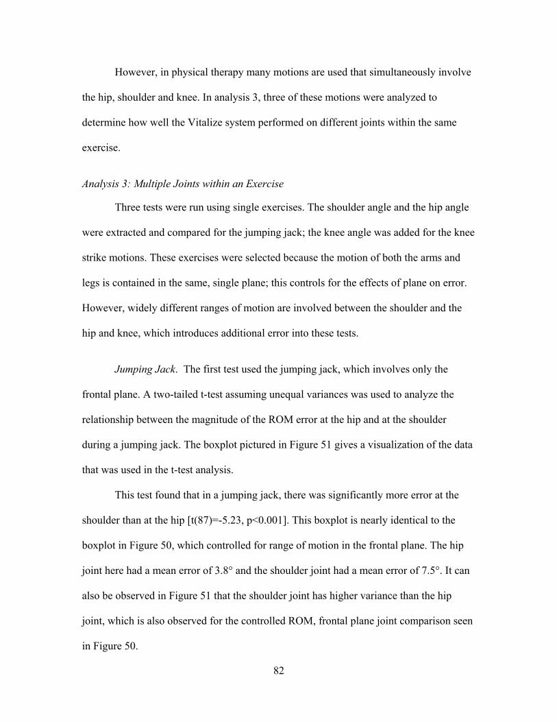
82
However, in physical therapy many motions are used that simultaneously involve
the hip, shoulder and knee. In analysis 3, three of these motions were analyzed to
determine how well the Vitalize system performed on different joints within the same
exercise.
Analysis 3: Multiple Joints within an Exercise
Three tests were run using single exercises. The shoulder angle and the hip angle
were extracted and compared for the jumping jack; the knee angle was added for the knee
strike motions. These exercises were selected because the motion of both the arms and
legs is contained in the same, single plane; this controls for the effects of plane on error.
However, widely different ranges of motion are involved between the shoulder and the
hip and knee, which introduces additional error into these tests.
Jumping Jack. The first test used the jumping jack, which involves only the
frontal plane. A two-tailed t-test assuming unequal variances was used to analyze the
relationship between the magnitude of the ROM error at the hip and at the shoulder
during a jumping jack. The boxplot pictured in Figure 51 gives a visualization of the data
that was used in the t-test analysis.
This test found that in a jumping jack, there was significantly more error at the
shoulder than at the hip [t(87)=-5.23, p<0.001]. This boxplot is nearly identical to the
boxplot in Figure 50, which controlled for range of motion in the frontal plane. The hip
joint here had a mean error of 3.8° and the shoulder joint had a mean error of 7.5°. It can
also be observed in Figure 51 that the shoulder joint has higher variance than the hip
joint, which is also observed for the controlled ROM, frontal plane joint comparison seen
in Figure 50.
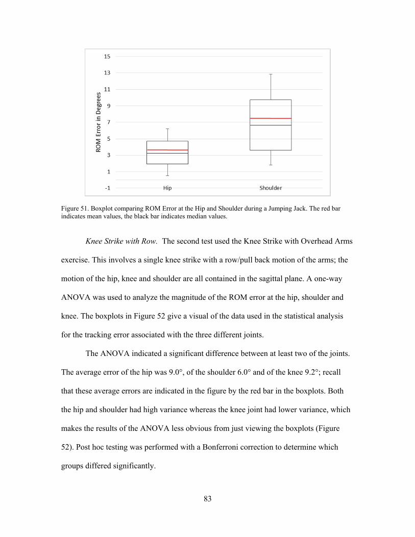
83
Figure 51. Boxplot comparing ROM Error at the Hip and Shoulder during a Jumping Jack. The red bar indicates mean values, the black bar indicates median values.
Knee Strike with Row. The second test used the Knee Strike with Overhead Arms
exercise. This involves a single knee strike with a row/pull back motion of the arms; the
motion of the hip, knee and shoulder are all contained in the sagittal plane. A one-way
ANOVA was used to analyze the magnitude of the ROM error at the hip, shoulder and
knee. The boxplots in Figure 52 give a visual of the data used in the statistical analysis
for the tracking error associated with the three different joints.
The ANOVA indicated a significant difference between at least two of the joints.
The average error of the hip was 9.0°, of the shoulder 6.0° and of the knee 9.2°; recall
that these average errors are indicated in the figure by the red bar in the boxplots. Both
the hip and shoulder had high variance whereas the knee joint had lower variance, which
makes the results of the ANOVA less obvious from just viewing the boxplots (Figure
52). Post hoc testing was performed with a Bonferroni correction to determine which
groups differed significantly.

84
Figure 52. Boxplot comparing ROM Error of Hip, Shoulder and Knee during Knee Strike with Row. The red bar indicates mean values, the black bar indicates median values.
Two-sample, two-tailed t-test assuming unequal variance with a significance level
of α/3, (where α=0.05; for the Bonferroni correction) were used to compare
Hip Error to Shoulder Error Hip Error to Knee Error Shoulder Error to Knee Error
The results showed that the hip had greater error than the shoulder, but not
significantly more [t(50)=2.40, p=0.02]. The hip had slightly less error than the knee, but
no significant difference between the error at the hip and knee existed [t(41)=-0.23,
p=0.82]. The significant difference exists between the error of the shoulder and the knee;
the shoulder has significantly less error than the knee [t(87)=-4.03, p<0.001]. This does
not agree with the results from Analysis 1. The shoulder is the most accurate joint for this
exercise, as opposed to the hip being most accurate for a more controlled analysis.
Knee Strike with Overhead Arms. The third test used the Knee Strike with
Overhead Arms exercise. This involves the same single knee strike with simultaneous
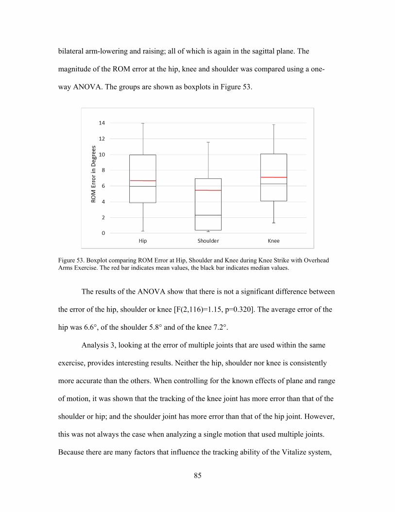
85
bilateral arm-lowering and raising; all of which is again in the sagittal plane. The
magnitude of the ROM error at the hip, knee and shoulder was compared using a one-
way ANOVA. The groups are shown as boxplots in Figure 53.
Figure 53. Boxplot comparing ROM Error at Hip, Shoulder and Knee during Knee Strike with Overhead Arms Exercise. The red bar indicates mean values, the black bar indicates median values.
The results of the ANOVA show that there is not a significant difference between
the error of the hip, shoulder or knee [F(2,116)=1.15, p=0.320]. The average error of the
hip was 6.6°, of the shoulder 5.8° and of the knee 7.2°.
Analysis 3, looking at the error of multiple joints that are used within the same
exercise, provides interesting results. Neither the hip, shoulder nor knee is consistently
more accurate than the others. When controlling for the known effects of plane and range
of motion, it was shown that the tracking of the knee joint has more error than that of the
shoulder or hip; and the shoulder joint has more error than that of the hip joint. However,
this was not always the case when analyzing a single motion that used multiple joints.
Because there are many factors that influence the tracking ability of the Vitalize system,

86
it is possible to pair the most accurate shoulder motion (overhead arms motion) with a
less accurate hip motion (knee strike); and obtain results that refute the broad trend
finding that the hip has less error than the shoulder. This finding shows that every
exercise has its own unique error profile for different joints. It is not true that the hip has
3° less error than the shoulder for every exercise, though that was found to be the
overarching trend. Physical therapy does not control for the plane and range of motion of
an exercise; clinically, there will be a mix of planes and ROM between different joints.
This is why it is important to reference the errors associated with each joint for the
exercise being selected; which can be done using the exercise tables created in this study
(see the tables in Appendix F).
Aim 4: Adding Complexity to a Motion
It has been found that plane of motion, range of motion and the joint involved
have an effect on the ability of the Vitalize system to accurately track motion. Most of the
exercises used so far have been relatively simple; this raises the question of complexity.
What happens to the error at a specific joint when additions are made to the complexity
of the motion? In the set of exercises collected, a few motions have several variations
associated with them. A case study was completed using the “Knee Strike” variations to
determine if the complexity has an effect of the error. The variations used were:
Full Knee Strike Seated Knee Strike Knee Strike with Overhead Arms Knee Strike with Row
A one-way ANOVA was completed on the magnitude of the ROM error of the
hip angle for these four variations.
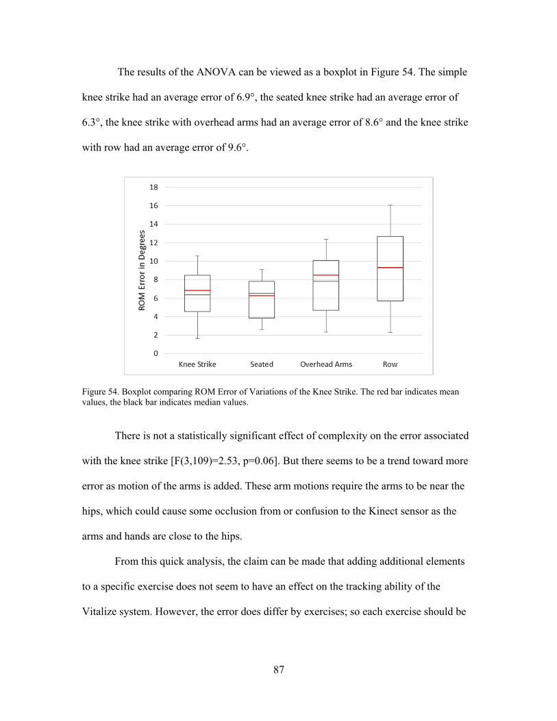
87
The results of the ANOVA can be viewed as a boxplot in Figure 54. The simple
knee strike had an average error of 6.9°, the seated knee strike had an average error of
6.3°, the knee strike with overhead arms had an average error of 8.6° and the knee strike
with row had an average error of 9.6°.
Figure 54. Boxplot comparing ROM Error of Variations of the Knee Strike. The red bar indicates mean values, the black bar indicates median values.
There is not a statistically significant effect of complexity on the error associated
with the knee strike [F(3,109)=2.53, p=0.06]. But there seems to be a trend toward more
error as motion of the arms is added. These arm motions require the arms to be near the
hips, which could cause some occlusion from or confusion to the Kinect sensor as the
arms and hands are close to the hips.
From this quick analysis, the claim can be made that adding additional elements
to a specific exercise does not seem to have an effect on the tracking ability of the
Vitalize system. However, the error does differ by exercises; so each exercise should be
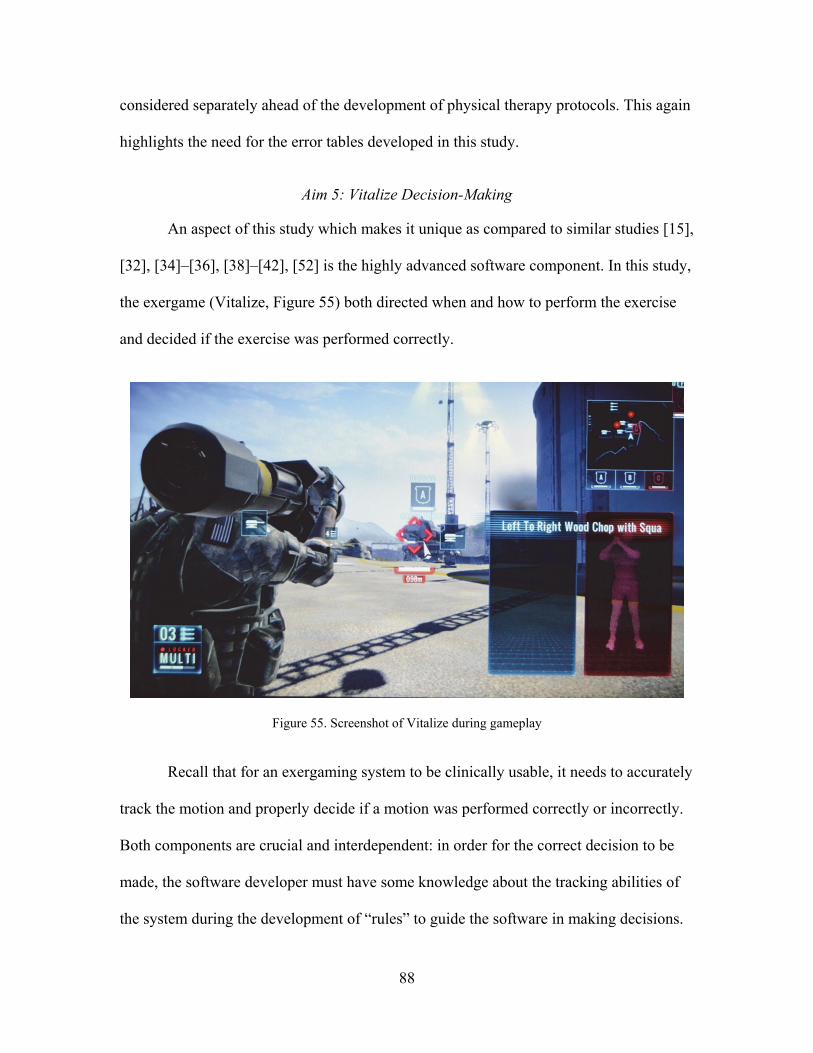
88
considered separately ahead of the development of physical therapy protocols. This again
highlights the need for the error tables developed in this study.
Aim 5: Vitalize Decision-Making
An aspect of this study which makes it unique as compared to similar studies [15],
[32], [34]–[36], [38]–[42], [52] is the highly advanced software component. In this study,
the exergame (Vitalize, Figure 55) both directed when and how to perform the exercise
and decided if the exercise was performed correctly.
Figure 55. Screenshot of Vitalize during gameplay
Recall that for an exergaming system to be clinically usable, it needs to accurately
track the motion and properly decide if a motion was performed correctly or incorrectly.
Both components are crucial and interdependent: in order for the correct decision to be
made, the software developer must have some knowledge about the tracking abilities of
the system during the development of “rules” to guide the software in making decisions.
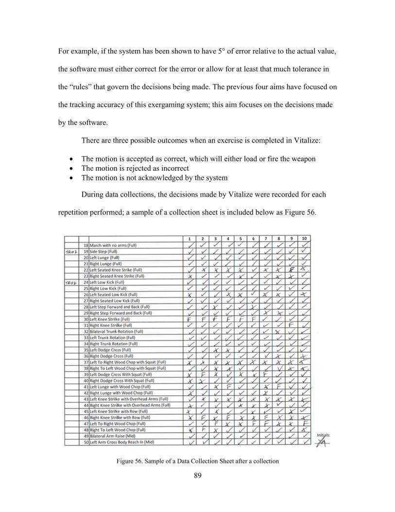
89
For example, if the system has been shown to have 5° of error relative to the actual value,
the software must either correct for the error or allow for at least that much tolerance in
the “rules” that govern the decisions being made. The previous four aims have focused on
the tracking accuracy of this exergaming system; this aim focuses on the decisions made
by the software.
There are three possible outcomes when an exercise is completed in Vitalize:
The motion is accepted as correct, which will either load or fire the weapon The motion is rejected as incorrect The motion is not acknowledged by the system
During data collections, the decisions made by Vitalize were recorded for each
repetition performed; a sample of a collection sheet is included below as Figure 56.
Figure 56. Sample of a Data Collection Sheet after a collection
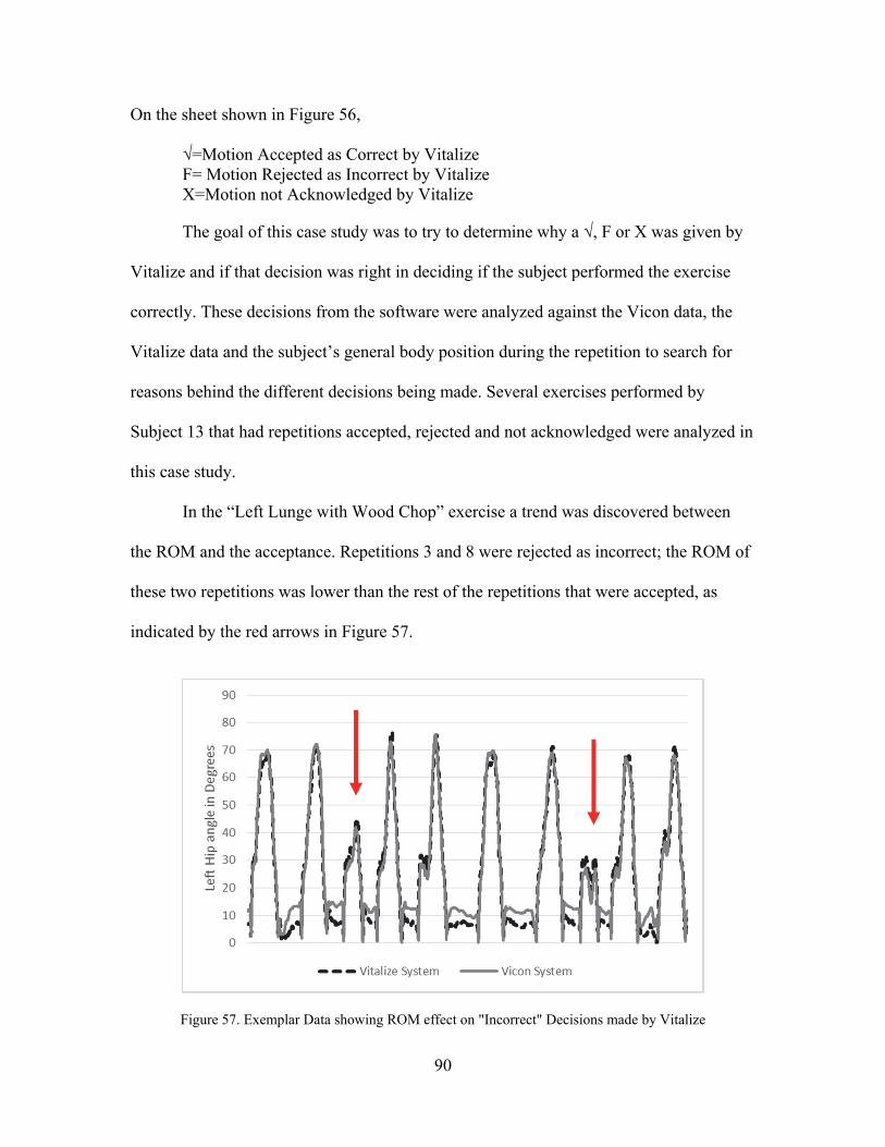
90
On the sheet shown in Figure 56,
√=Motion Accepted as Correct by Vitalize F= Motion Rejected as Incorrect by Vitalize X=Motion not Acknowledged by Vitalize
The goal of this case study was to try to determine why a √, F or X was given by
Vitalize and if that decision was right in deciding if the subject performed the exercise
correctly. These decisions from the software were analyzed against the Vicon data, the
Vitalize data and the subject’s general body position during the repetition to search for
reasons behind the different decisions being made. Several exercises performed by
Subject 13 that had repetitions accepted, rejected and not acknowledged were analyzed in
this case study.
In the “Left Lunge with Wood Chop” exercise a trend was discovered between
the ROM and the acceptance. Repetitions 3 and 8 were rejected as incorrect; the ROM of
these two repetitions was lower than the rest of the repetitions that were accepted, as
indicated by the red arrows in Figure 57.
Figure 57. Exemplar Data showing ROM effect on "Incorrect" Decisions made by Vitalize
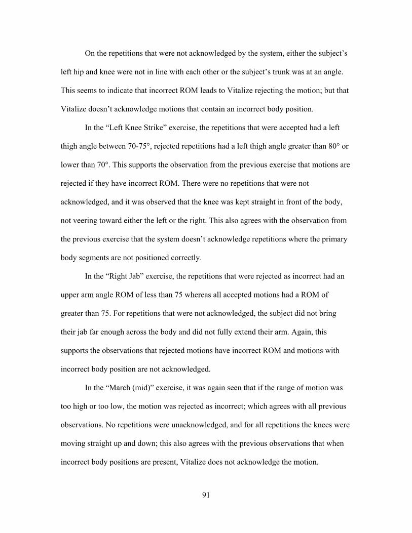
91
On the repetitions that were not acknowledged by the system, either the subject’s
left hip and knee were not in line with each other or the subject’s trunk was at an angle.
This seems to indicate that incorrect ROM leads to Vitalize rejecting the motion; but that
Vitalize doesn’t acknowledge motions that contain an incorrect body position.
In the “Left Knee Strike” exercise, the repetitions that were accepted had a left
thigh angle between 70-75°, rejected repetitions had a left thigh angle greater than 80° or
lower than 70°. This supports the observation from the previous exercise that motions are
rejected if they have incorrect ROM. There were no repetitions that were not
acknowledged, and it was observed that the knee was kept straight in front of the body,
not veering toward either the left or the right. This also agrees with the observation from
the previous exercise that the system doesn’t acknowledge repetitions where the primary
body segments are not positioned correctly.
In the “Right Jab” exercise, the repetitions that were rejected as incorrect had an
upper arm angle ROM of less than 75 whereas all accepted motions had a ROM of
greater than 75. For repetitions that were not acknowledged, the subject did not bring
their jab far enough across the body and did not fully extend their arm. Again, this
supports the observations that rejected motions have incorrect ROM and motions with
incorrect body position are not acknowledged.
In the “March (mid)” exercise, it was again seen that if the range of motion was
too high or too low, the motion was rejected as incorrect; which agrees with all previous
observations. No repetitions were unacknowledged, and for all repetitions the knees were
moving straight up and down; this also agrees with the previous observations that when
incorrect body positions are present, Vitalize does not acknowledge the motion.
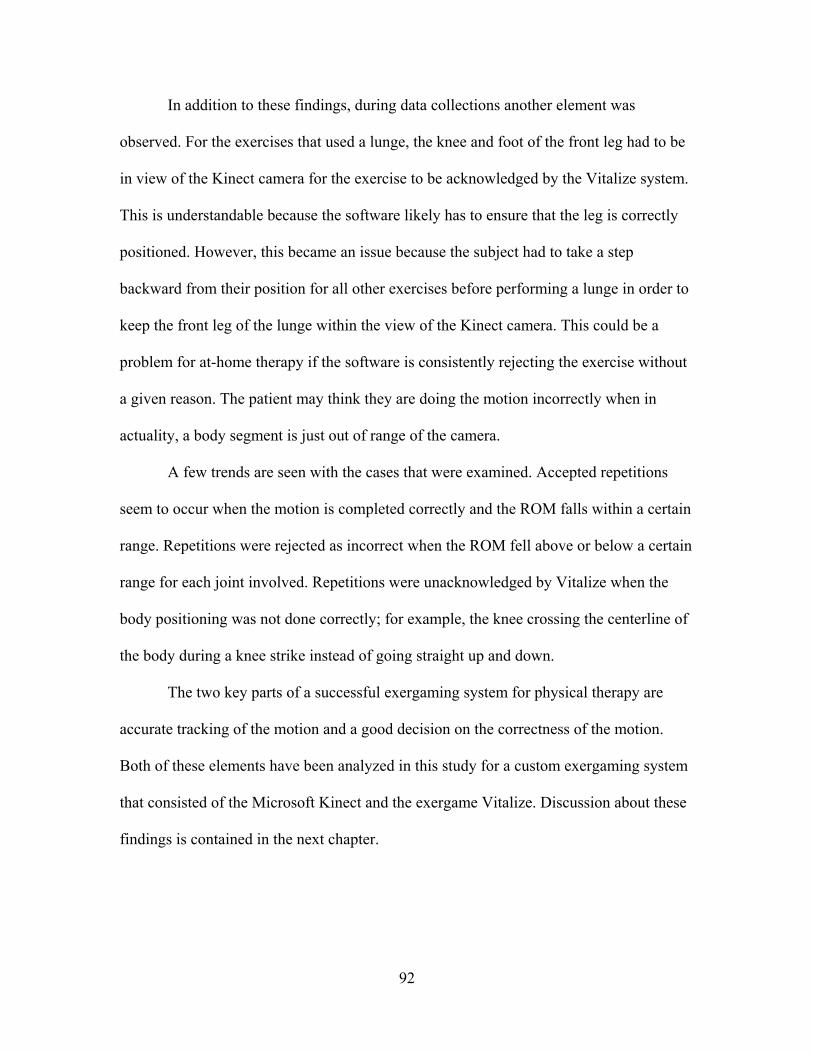
92
In addition to these findings, during data collections another element was
observed. For the exercises that used a lunge, the knee and foot of the front leg had to be
in view of the Kinect camera for the exercise to be acknowledged by the Vitalize system.
This is understandable because the software likely has to ensure that the leg is correctly
positioned. However, this became an issue because the subject had to take a step
backward from their position for all other exercises before performing a lunge in order to
keep the front leg of the lunge within the view of the Kinect camera. This could be a
problem for at-home therapy if the software is consistently rejecting the exercise without
a given reason. The patient may think they are doing the motion incorrectly when in
actuality, a body segment is just out of range of the camera.
A few trends are seen with the cases that were examined. Accepted repetitions
seem to occur when the motion is completed correctly and the ROM falls within a certain
range. Repetitions were rejected as incorrect when the ROM fell above or below a certain
range for each joint involved. Repetitions were unacknowledged by Vitalize when the
body positioning was not done correctly; for example, the knee crossing the centerline of
the body during a knee strike instead of going straight up and down.
The two key parts of a successful exergaming system for physical therapy are
accurate tracking of the motion and a good decision on the correctness of the motion.
Both of these elements have been analyzed in this study for a custom exergaming system
that consisted of the Microsoft Kinect and the exergame Vitalize. Discussion about these
findings is contained in the next chapter.

93
CHAPTER FOUR
Discussion
Due to the large number of exercises used, two important factors were able to be
studied: the broad trends in error that would otherwise be difficult to identify and the
error profile individual to exercise and joint. In this discussion section, first the key
findings of this study will be presented and discussed. Then the results of the specific
aims of this study will be discussed and compared to similar studies.
Key Findings
In the testing of this custom exergaming system, many interesting findings were
discovered. However, a few stand out as the most important for the clinical use of an
exergaming system in a physical therapy home exercise program.
Plane of motion has an effect on the error and variance Range of motion effect on error and variance is dependent on the plane of motion Vitalize system overestimates angles for low ROM motions and underestimates
angles for high ROM motions Each exercise has a unique error profile for each joint involved
Prior studies have hypothesized about the effects of plane, range of motion and
joint on the accuracy of a Kinect-based exergaming system [32], [35], [39]. However, in
this study, evidence based on a large number of exercises has demonstrated which of
these factors lead to error.
Plane of Motion Effect on Error and Variance
This study indicates that the plane of motion has an impact on the error involved
in tracking the exercise. Motions in the frontal plane have been shown to have
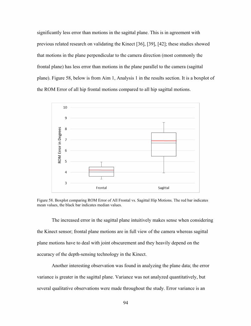
94
significantly less error than motions in the sagittal plane. This is in agreement with
previous related research on validating the Kinect [36], [39], [42]; these studies showed
that motions in the plane perpendicular to the camera direction (most commonly the
frontal plane) has less error than motions in the plane parallel to the camera (sagittal
plane). Figure 58, below is from Aim 1, Analysis 1 in the results section. It is a boxplot of
the ROM Error of all hip frontal motions compared to all hip sagittal motions.
Figure 58. Boxplot comparing ROM Error of All Frontal vs. Sagittal Hip Motions. The red bar indicates mean values, the black bar indicates median values.
The increased error in the sagittal plane intuitively makes sense when considering
the Kinect sensor; frontal plane motions are in full view of the camera whereas sagittal
plane motions have to deal with joint obscurement and they heavily depend on the
accuracy of the depth-sensing technology in the Kinect.
Another interesting observation was found in analyzing the plane data; the error
variance is greater in the sagittal plane. Variance was not analyzed quantitatively, but
several qualitative observations were made throughout the study. Error variance is an

95
important factor to consider in the development and use of exergaming systems. Software
developers must know the range of variance as they are creating the “rules” to determine
the correctness of motion. For example, if the exergaming system has been shown to
consistently overestimate the angle by 5°; that value can be subtracted across the motion
to correct for this tracking inaccuracy. However, variance is much more difficult to
handle. For example, if the system has error varying between ±5° of the actual value, the
software cannot simply make a correction, it must allow for at least the ±5° of tolerance
in the “rules” that govern the decisions being made.
Range of Motion Effect is Dependent on the Plane of Motion
It has also been shown that the range of a motion has an effect on the tracking
accuracy in the sagittal plane. As the range of motion increases there is a significant
increase in the error of the Vitalize system. This result is contrary to the findings of
previous studies that sought to validate the use of the Kinect for clinical settings [32],
[35], [36]. However, more than ten times as many exercises were used in this study than
in any of these previous studies; and with what we now know about the unique error
signature of each exercise and joint, it is very difficult to draw meaningful conclusions
about broad trends from a small number of exercises. This study had an additional benefit
to an in-depth analysis of the effect of range of motion in that several exercises were
collected multiple times at varied ROM; which controls for nearly every compounding
factor. For example, the squat was collected three times for each subject: full ROM squat,
mid ROM squat and partial ROM squat. This gives a great measure of confidence to the
results finding a relationship between ROM and error even though it is contrary to
previous literature.
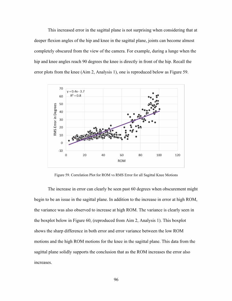
96
This increased error in the sagittal plane is not surprising when considering that at
deeper flexion angles of the hip and knee in the sagittal plane, joints can become almost
completely obscured from the view of the camera. For example, during a lunge when the
hip and knee angles reach 90 degrees the knee is directly in front of the hip. Recall the
error plots from the knee (Aim 2, Analysis 1), one is reproduced below as Figure 59.
Figure 59. Correlation Plot for ROM vs RMS Error for all Sagittal Knee Motions
The increase in error can clearly be seen past 60 degrees when obscurement might
begin to be an issue in the sagittal plane. In addition to the increase in error at high ROM,
the variance was also observed to increase at high ROM. The variance is clearly seen in
the boxplot below in Figure 60, (reproduced from Aim 2, Analysis 1). This boxplot
shows the sharp difference in both error and error variance between the low ROM
motions and the high ROM motions for the knee in the sagittal plane. This data from the
sagittal plane solidly supports the conclusion that as the ROM increases the error also
increases.
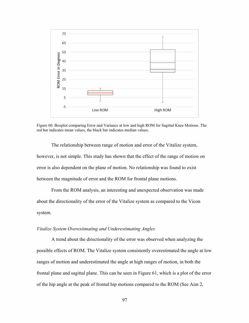
97
Figure 60. Boxplot comparing Error and Variance at low and high ROM for Sagittal Knee Motions. The red bar indicates mean values, the black bar indicates median values.
The relationship between range of motion and error of the Vitalize system,
however, is not simple. This study has shown that the effect of the range of motion on
error is also dependent on the plane of motion. No relationship was found to exist
between the magnitude of error and the ROM for frontal plane motions.
From the ROM analysis, an interesting and unexpected observation was made
about the directionality of the error of the Vitalize system as compared to the Vicon
system.
Vitalize System Overestimating and Underestimating Angles
A trend about the directionality of the error was observed when analyzing the
possible effects of ROM. The Vitalize system consistently overestimated the angle at low
ranges of motion and underestimated the angle at high ranges of motion, in both the
frontal plane and sagittal plane. This can be seen in Figure 61, which is a plot of the error
of the hip angle at the peak of frontal hip motions compared to the ROM (See Aim 2,

98
Analysis 4) and Figure 62, which is a plot of the error of the hip angle at the peak of
sagittal hip motions compared to the ROM (See Aim 2, Analysis 2). Additional red
markings have been added to the correlation plots to highlight the areas of overestimation
and underestimation.
Figure 61. Plot showing Directionality of Error for Frontal Hip Motions
Figure 62. Plot showing Directionality of Error for Sagittal Hip Motions
Underestimate
Overestimate
Overestimate
Underestimate
99
For both the frontal and sagittal planes, at low ranges of motion, the error tends to
be negative because the Vitalize system has overestimated the hip angle; as the ROM
increases, the error tends to be positive which is caused by the Vitalize system
underestimating the hip angle. This is supported by research performed by Yeung [42].
Yeung studied the ability of the Kinect to track body sway, a low range of motion
activity; that study found that the Kinect consistently overestimated the angle of interest
as compared to the Vicon system.
However, no previous research has been found that shows a relationship between
the direction of error (overestimate or underestimate) and the range of motion as
observed in this study. This is an important discovery in analyzing the tracking abilities
of the Kinect as part of an exergaming system as it shows an interesting trend in the error.
Further research is needed to understand why this trend exists.
Each Exercise has a Unique Error Profile for Each Joint Involved
The most significant result of this study was found in analyzing the error at
different joints (Aim 3). When controlling for plane and range of motion, it was found
that the knee had the most error, followed by the shoulder, and the hip had the least error.
This broad trend is based on all the exercises at each of the three joints. However, in
physical therapy, plane and range of motion are not controlled. So three individual
exercises were analyzed at each joint. The conclusion of this exercise- and joint-specific
analysis was that no joint consistently out-performed the others in terms of tracking
accuracy. This indicates that each exercise has a unique error profile for each joint
involved. The claim cannot be made that the knee always has more error than the hip; the
joint-based error rankings depend on which exercise is being evaluated.

100
While broad trends about the abilities and limitations of a Kinect-based
exergaming system can be drawn from this study, the more important result is the
production of unique error profiles for many common exercises at four primary body
joints. This information is included in tables of error at the hip, shoulder, knee and elbow
(see Appendix F). Both the exergaming software developer and the physical therapist will
be able to examine the amount of error in the system’s tracking ability for specific
exercises and joint before using the exercise in a exergaming therapy regime. Because
there are so many interdependent factors that affect the tracking accuracy, it is important
to have reference databases like this to find the amount of error associated with a specific
joint within a specific exercise.
Specific Aims
Beyond these key elements, there are many observations that can be made from
each broad aim completed in this study; this section includes discussion specific to each
aim.
Aim 1: Effect of Plane
Aim 1 examined the effect that the plane of motion had on the tracking accuracy
of the Vitalize system. At the hip, it was shown that the frontal plane had significantly
less error than the sagittal plane (p=0.018). At the shoulder, motions in the frontal plane
were compared to motions in the sagittal plane and motions that used both planes. The
motions in the frontal plane again had significantly less error than the sagittal plane
(p<0.001). It was also shown that the error of motions that used both planes was greater
than the error of purely frontal plane and less than the error of purely sagittal plane
motions; this confirms that the error being observed is due to the plane. This agrees with
101
previous work that found the Kinect performs best in the plane perpendicular to the
camera, which is almost always the frontal plane of the body [36], [42], [53]. For
example, for the hip angle there is more error inherent to the system for a knee strike than
for a jumping jack. Additionally, more variance was observed in the sagittal plane than in
the frontal plane.
This association between the plane of motion and the tracking ability of the
Vitalize system is something that a therapist should keep in mind when assigning
exercises to be completed with an exergaming system. Some motions in the sagittal
plane, especially for the knee, have so much error inherent to the exergaming system that
they should not be included in an exergaming therapy prescription. With such high error
and variance, there are two likely outcomes from the exergaming system: either it will
reject motions that are actually correct because of error due to the hardware of the
system, or it will require such a large tolerance in deciding a “correct” motion that the
decision becomes meaningless. For example, if a motion has error of ±30° associated
with it and the “rules” that govern the decision about correctness of motion take that into
account, the system would be accepting motions that range from 20° to 80°. This may
cause a large number of incorrect motions to be accepted and counted as correct by the
exergaming system, which makes the decision made practically meaningless.
Aim 2: Effect of Range
The second analysis examined the effect range of motion has on error. Five cases
were examined; the knee, hip and shoulder in the sagittal plane and the hip and shoulder
in the frontal plane. This discussion is split into the sagittal plane and frontal plane
findings.

102
Sagittal Plane. For motions in the sagittal plane, there was a statistically
significant relationship between range of motion and the error of the exercise. This
relationship is likely due to joints becoming occluded by other parts of the body during
sagittal plane motions at higher ranges of motion. Three previous studies have referenced
occlusion as a possible source of error [15], [36], [38]. Nixon [38] explains that if a joint
position is occluded from the Kinect sensor, the algorithm makes an inference which
leads to an increase in error, and likely variability which was also observed in this study,
during the occlusion. Kuster [36] noted a significant impact the error associated with the
Kinect (generation 2) during shoulder motions where the elbow and hand were occluding
the view of the shoulder. Zhao [15] also noted that the Kinect system failed to track the
motion during significant self-occlusions.
This finding is clinically important because it informs the exercises and ranges of
motion that can be accurately tracked by the Kinect sensor. A physical therapist should
note that there is much more accuracy in the tracking of a shallow lunge with the hip
going to a 45 degree angle than a deep lunge with the hip going to a 90 degree angle. This
could cause problems in rehabilitation where a patient may be consistently not reaching
the range of motion assigned during their at-home therapy with an exergaming system
because the hardware is reporting inaccurate information. It was also observed that the
Vitalize system was overestimating the angle at low ranges of motion and
underestimating the angle at high ranges of motion. The primary message to those using
the Kinect in a clinical setting is to avoid deep flexion (high range of motion) movements
in the sagittal plane because the system cannot accurately track them, especially at the
knee.

103
Frontal Plane. For the frontal plane, there was no relationship between error and
ROM for shoulder motions. However, there was a relationship observed at the hip. Upon
closer examination, the trend toward more error as the ROM increased is better explained
by the overestimation of the hip angle at low ranges of motion and underestimation of the
hip angle at high ranges of motion. This overestimation at low ranges of motion could be
due to the Kinect struggling to distinguish between the legs and correctly place the hip
joint centers at small hip angles. An overestimated angle at small ranges of motion was
also observed in the frontal plane shoulder data.
A possible explanation to the angle underestimation at high ranges of motion is
that it may be due to the lower sampling rate of the Kinect (30 Hz as compared to 120 Hz
for the Vicon system). At high rates of motion, a lower frame rate becomes a critical
consideration. A slower collection rate could account for an increase of error because the
system interpolates between collected data points; as the distance between the points
increases (as it will as speed of the motion increases) the data must rely more heavily on
the interpolation. This causes error. This is especially true at the peak of the motion,
where there is a change in direction. Data from a single repetition is shown in Figure 63,
as an example of a time when the Vitalize system may be underestimating the angle due
to the speed of the motion.
The lower collection rate of the Kinect-based Vitalize system may cause it to miss
the true peak of the motion and interpolate between two points lower on the curve to
create an underestimated peak; this would be expected to happen more often during fast
motions. The red arrow in Figure 63 calls attention to the difference between the peaks
recorded by the two systems.
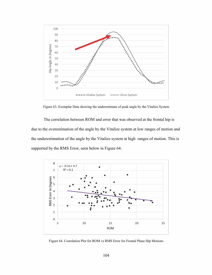
104
Figure 63. Exemplar Data showing the underestimate of peak angle by the Vitalize System
The correlation between ROM and error that was observed at the frontal hip is
due to the overestimation of the angle by the Vitalize system at low ranges of motion and
the underestimation of the angle by the Vitalize system at high ranges of motion. This is
supported by the RMS Error, seen below in Figure 64.
Figure 64. Correlation Plot for ROM vs RMS Error for Frontal Plane Hip Motions

105
The RMS error removes directional effects and considers the entire waveform of
the motion. The correlation between ROM and error here is weak and tending toward less
error as the ROM increases, see Figure 64 on the previous page (Reproduced from Aim
2, Analysis 4 of Results). The magnitude of the error and variance seem to decrease
slightly with increasing ROM in the frontal plane; this is in direct contrast to the sagittal
plane where the error and variance increased when ROM increased.
This decrease in error and variance as ROM increases in the frontal plane is likely
due to the way the Kinect sensor tracks motion. Recall that the Kinect tracks the form of
the body and infers joint positions to create a skeletal model of the user. When there is no
movement or very small movements the Kinect is likely less accurate in placing the
position of the joint centers. For example, if the arm is held next to the body without any
movement, the Kinect has to infer the location of the shoulder joint by determining where
the form of the upper arm stops and the form of the trunk starts. On the other hand, when
the arm is raised above the head, a high ROM motion, the Kinect sensor is better able to
detect the center of rotation, and thus able to make a more informed and therefore, more
accurate inference about the location of the shoulder joint. This helps explain the higher
error that was observed at low ranges of motion in the frontal plane.
This decrease in error with increasing ROM is opposite what was seen for the
RMS error for sagittal hip motions, see Figure 65 (Reproduced from Aim 2, Analysis 2).
A strong positive correlation exists between the RMS error and ROM for motions in the
sagittal plane. The use of RMS error removes the directionality effect that could be
causing a correlation between tracking error and ROM. In the sagittal plane, a
relationship exists between error and ROM regardless of the directionality of the error.

106
Figure 65. Correlation Plot for RMS Error vs. ROM for Sagittal Plane Hip Motions
In summary, these are the primary findings from the Aim 2, which sought to discover
the relationship between ROM and error:
Error and variance increase as ROM increases in the sagittal plane Error and variance are not correlated to ROM in the frontal plane The Vitalize system consistently overestimates the supports the angle at low
ranges of motion and underestimation of the angle at high ranges of motion
Similar to Aim 1, these results serve to inform both exergaming software
developers and physical therapists in choosing which motions to include in an
exergaming therapy regimen.
Aim 3: Joint Involved
When accounting for the error associated with the plane and range of motion, it
was found that the joint involved is also correlated with the amount of error observed. For
similar exercises (controlled for plane and range of motion), the hip consistently
demonstrated a significantly lower amount of error than the shoulder, and the knee had
the highest error of the three joints considered. Bonnechere [32] also found that hip had
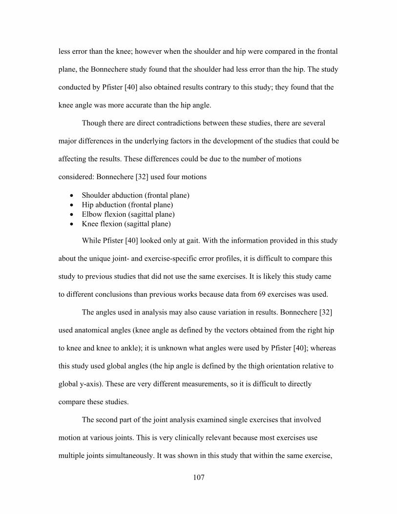
107
less error than the knee; however when the shoulder and hip were compared in the frontal
plane, the Bonnechere study found that the shoulder had less error than the hip. The study
conducted by Pfister [40] also obtained results contrary to this study; they found that the
knee angle was more accurate than the hip angle.
Though there are direct contradictions between these studies, there are several
major differences in the underlying factors in the development of the studies that could be
affecting the results. These differences could be due to the number of motions
considered: Bonnechere [32] used four motions
Shoulder abduction (frontal plane) Hip abduction (frontal plane) Elbow flexion (sagittal plane) Knee flexion (sagittal plane)
While Pfister [40] looked only at gait. With the information provided in this study
about the unique joint- and exercise-specific error profiles, it is difficult to compare this
study to previous studies that did not use the same exercises. It is likely this study came
to different conclusions than previous works because data from 69 exercises was used.
The angles used in analysis may also cause variation in results. Bonnechere [32]
used anatomical angles (knee angle as defined by the vectors obtained from the right hip
to knee and knee to ankle); it is unknown what angles were used by Pfister [40]; whereas
this study used global angles (the hip angle is defined by the thigh orientation relative to
global y-axis). These are very different measurements, so it is difficult to directly
compare these studies.
The second part of the joint analysis examined single exercises that involved
motion at various joints. This is very clinically relevant because most exercises use
multiple joints simultaneously. It was shown in this study that within the same exercise,
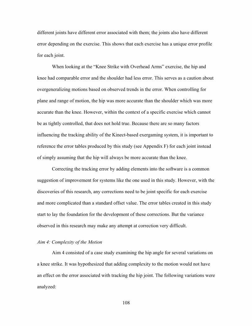
108
different joints have different error associated with them; the joints also have different
error depending on the exercise. This shows that each exercise has a unique error profile
for each joint.
When looking at the “Knee Strike with Overhead Arms” exercise, the hip and
knee had comparable error and the shoulder had less error. This serves as a caution about
overgeneralizing motions based on observed trends in the error. When controlling for
plane and range of motion, the hip was more accurate than the shoulder which was more
accurate than the knee. However, within the context of a specific exercise which cannot
be as tightly controlled, that does not hold true. Because there are so many factors
influencing the tracking ability of the Kinect-based exergaming system, it is important to
reference the error tables produced by this study (see Appendix F) for each joint instead
of simply assuming that the hip will always be more accurate than the knee.
Correcting the tracking error by adding elements into the software is a common
suggestion of improvement for systems like the one used in this study. However, with the
discoveries of this research, any corrections need to be joint specific for each exercise
and more complicated than a standard offset value. The error tables created in this study
start to lay the foundation for the development of these corrections. But the variance
observed in this research may make any attempt at correction very difficult.
Aim 4: Complexity of the Motion
Aim 4 consisted of a case study examining the hip angle for several variations on
a knee strike. It was hypothesized that adding complexity to the motion would not have
an effect on the error associated with tracking the hip joint. The following variations were
analyzed:
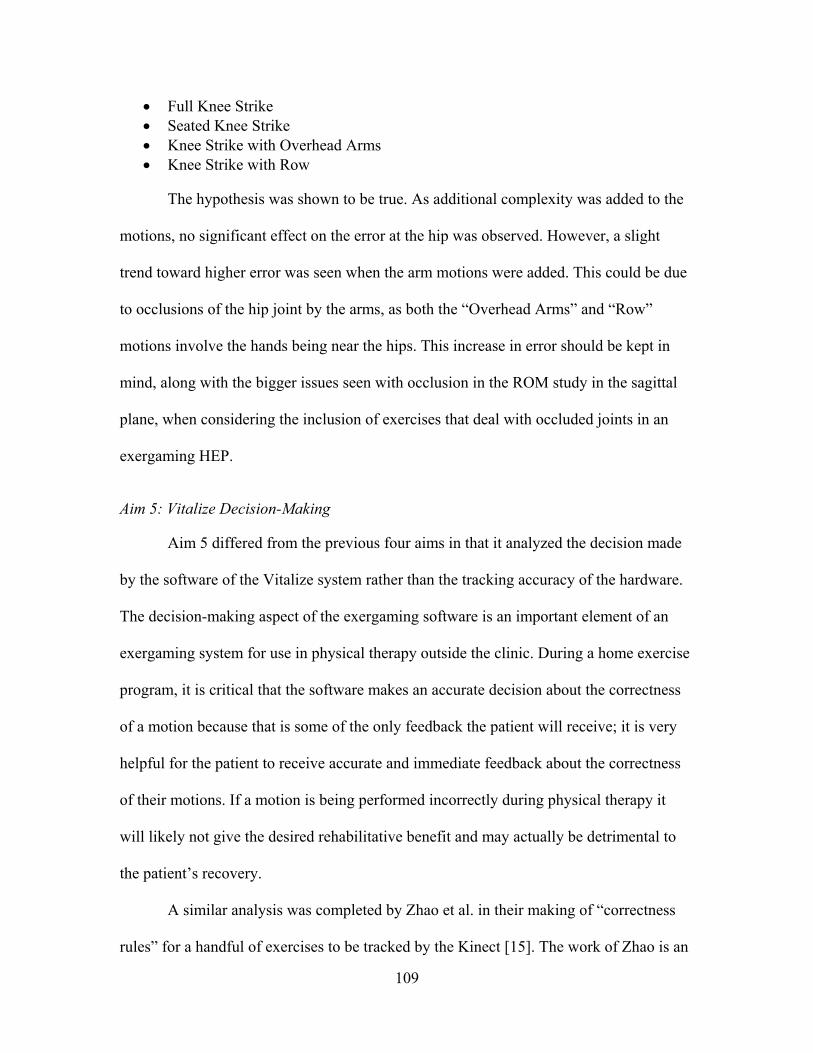
109
Full Knee Strike Seated Knee Strike Knee Strike with Overhead Arms Knee Strike with Row
The hypothesis was shown to be true. As additional complexity was added to the
motions, no significant effect on the error at the hip was observed. However, a slight
trend toward higher error was seen when the arm motions were added. This could be due
to occlusions of the hip joint by the arms, as both the “Overhead Arms” and “Row”
motions involve the hands being near the hips. This increase in error should be kept in
mind, along with the bigger issues seen with occlusion in the ROM study in the sagittal
plane, when considering the inclusion of exercises that deal with occluded joints in an
exergaming HEP.
Aim 5: Vitalize Decision-Making
Aim 5 differed from the previous four aims in that it analyzed the decision made
by the software of the Vitalize system rather than the tracking accuracy of the hardware.
The decision-making aspect of the exergaming software is an important element of an
exergaming system for use in physical therapy outside the clinic. During a home exercise
program, it is critical that the software makes an accurate decision about the correctness
of a motion because that is some of the only feedback the patient will receive; it is very
helpful for the patient to receive accurate and immediate feedback about the correctness
of their motions. If a motion is being performed incorrectly during physical therapy it
will likely not give the desired rehabilitative benefit and may actually be detrimental to
the patient’s recovery.
A similar analysis was completed by Zhao et al. in their making of “correctness
rules” for a handful of exercises to be tracked by the Kinect [15]. The work of Zhao is an
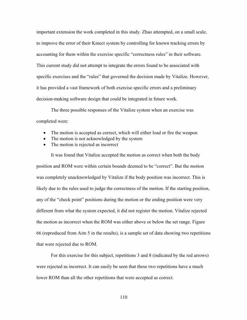
110
important extension the work completed in this study. Zhao attempted, on a small scale,
to improve the error of their Kinect system by controlling for known tracking errors by
accounting for them within the exercise specific “correctness rules” in their software.
This current study did not attempt to integrate the errors found to be associated with
specific exercises and the “rules” that governed the decision made by Vitalize. However,
it has provided a vast framework of both exercise specific errors and a preliminary
decision-making software design that could be integrated in future work.
The three possible responses of the Vitalize system when an exercise was
completed were:
The motion is accepted as correct, which will either load or fire the weapon The motion is not acknowledged by the system The motion is rejected as incorrect
It was found that Vitalize accepted the motion as correct when both the body
position and ROM were within certain bounds deemed to be “correct”. But the motion
was completely unacknowledged by Vitalize if the body position was incorrect. This is
likely due to the rules used to judge the correctness of the motion. If the starting position,
any of the “check point” positions during the motion or the ending position were very
different from what the system expected, it did not register the motion. Vitalize rejected
the motion as incorrect when the ROM was either above or below the set range. Figure
66 (reproduced from Aim 5 in the results), is a sample set of data showing two repetitions
that were rejected due to ROM.
For this exercise for this subject, repetitions 3 and 8 (indicated by the red arrows)
were rejected as incorrect. It can easily be seen that these two repetitions have a much
lower ROM than all the other repetitions that were accepted as correct.

111
Figure 66. Exemplar Data showing two Repetitions that were rejected by Vitalize
Clinically, there are some important observations to make from these results. The
Vitalize software did very well in not accepting motions that were incorrect because the
ROM was outside of the specified bounds. Range of motion needs to be accurate during
physical therapy because a ROM too small will not challenge the body and a ROM too
high may cause damage to the joint being rehabilitated. However, that is only part of the
correctness of the motion; the body positioning also must be correct. The Vitalize
software did not accept motions with improper body positioning, but it also did not reject
them as incorrect. If a patient is performing the exercise with improper body positioning,
they need to be aware of their error in order to change it. Future decision-making
exergaming software should continue to reject motions with incorrect ROM, but also
should reject motions that have incorrect body positioning.
Summary of Discussion
This study has contributed several valuable findings to the field of clinical
exergaming. The exercise-specific error tables give both software designers and physical

112
therapists the ability to decide how much error they are willing to accept in an exercise.
There is not agreement about the amount of error that is clinically acceptable. In one
study [54] on gait analysis, an error of 5° was considered clinically significant. In another
study, values of variation visible to the eye were found to often exceed 10° [43]. The
current study does not attempt to decide how much error is acceptable for a clinical use of
exergaming, it simply provides tables of exercise-specific errors to leave that decision to
the clinician who knows how much error is tolerable for a certain exercise and patient.
In the current literature there is disagreement over whether the Kinect is accurate
enough to be used as a component of an exergaming system for clinical use [32], [35],
[36], [40]. This disagreement is likely due to the small number of exercises represented in
previous work. The maximum number of exercises found in the previous research was
six. There are many factors that contribute to the ability of the Kinect sensor to accurately
track motions; therefore it is critical that an expansive study, such as this one, be
completed where the confounding factors can be controlled. By analyzing a large number
of exercises, this study provides valuable information on the strengths and weaknesses of
the tracking abilities of the Kinect sensor for use in a clinical exergaming system.
The development of joint specific error reference tables is a very valuable
contribution to the field of clinical exergaming. With the discovery that every joint in
every exercise has a different error profile, it becomes much more difficult to predict
errors. The reference tables produced in this study remove some of the necessity of
extrapolating error trends to new exercises. If the desired exercises are among the 69
included in this study, the physical therapist or exergaming software developer can
simply look up the error value for each joint.

113
CHAPTER FIVE
Limitations and Next Steps
There are several acknowledged limitations to this study that provide the basis for
the suggested next steps in this project.
Subject Homogeneity
The first limitation is that a very homogeneous sample of able-bodied subjects
was used (15 subjects, 7 female; age 22.6 (3.5) years; BMI 22.1 (2.3)). This was intended
to attempt to control for extraneous factors while analyzing the abilities of the Vitalize
system. However, because of this homogeneity there are several factors that are not
considered in this study, the most important being the effect of body type on the tracking
ability of the Kinect sensor.
For the next step in this study, the data that has been collected by the CFI on
several wounded warriors with varying levels of amputation and limb salvage should be
analyzed to determine the effects of atypical or missing limbs on the performance of the
Kinect. Additional subjects with vastly different BMIs should also be collected to better
understand the limitations of the Kinect sensor based on user body type.
Manual Time Sync
A manual visual time sync, as described in the “Visual 3D processing” section of
Chapter 2, was used for this study. This method of syncing the datasets had been used in
previous studies [35], [38]. However, during analysis it was discovered that artificial
error was introduced by this method of time syncing. This was especially true for the
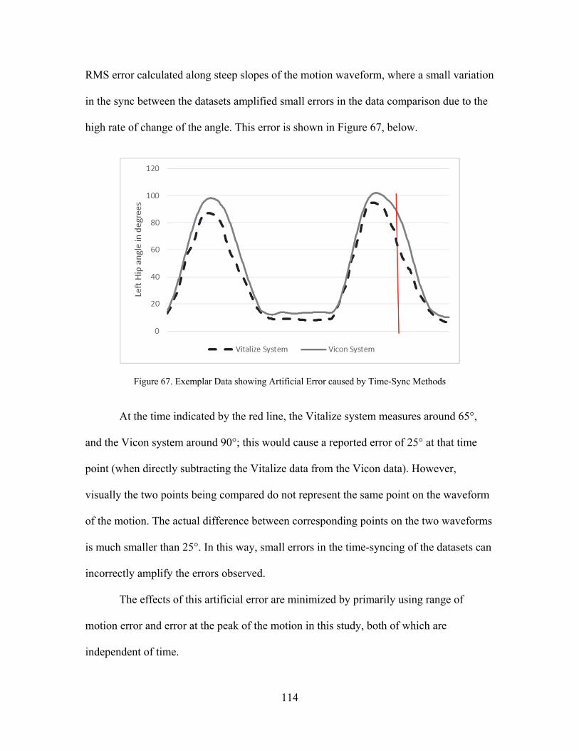
114
RMS error calculated along steep slopes of the motion waveform, where a small variation
in the sync between the datasets amplified small errors in the data comparison due to the
high rate of change of the angle. This error is shown in Figure 67, below.
Figure 67. Exemplar Data showing Artificial Error caused by Time-Sync Methods
At the time indicated by the red line, the Vitalize system measures around 65°,
and the Vicon system around 90°; this would cause a reported error of 25° at that time
point (when directly subtracting the Vitalize data from the Vicon data). However,
visually the two points being compared do not represent the same point on the waveform
of the motion. The actual difference between corresponding points on the two waveforms
is much smaller than 25°. In this way, small errors in the time-syncing of the datasets can
incorrectly amplify the errors observed.
The effects of this artificial error are minimized by primarily using range of
motion error and error at the peak of the motion in this study, both of which are
independent of time.

115
In the next steps of this study, an automated optimization method for time-
syncing should be utilized. A possible method was explained and used by Xu, et al [41];
where the mean-square residual between the datasets was minimized by time-shifting the
Kinect-based data.
Kinect Version
This study was started before the release of the Kinect for XBOX One (generation
2), therefore the software was designed to be compatible with only the Kinect for XBOX
360 (generation 1). However, Microsoft boasts of the new technology having greater
accuracy than the original Kinect. Next steps with this project should involve changes to
the software compatibility in order to utilize the Kinect for XBOX One sensor.
Global Angles
Another decision that was made in the development of this project was the use of
global angles, as seen in Galna’s study [35]. These angles are defined by taking the body
segment relative to the global y-axis. However, during the data analysis for this study,
concerns were raised about using angles defined in this way. The primary concern being
that the directionality of the angle is lost. 20 degrees of hip abduction and 20 degrees of
hip flexion both register as a 20 degree hip angle, but the distinction between abduction
and flexion is lost. This issue was encountered when trying to categorize hip exercises as
being performed in the frontal or sagittal plane. All the frontal plane hip exercises used in
this study have a small amount of sagittal motion (hip and knee flexion) to enable the
frontal plane motion (side step or jump). It has been shown that sagittal plane motion is
subject to higher error that frontal plane motion, but the two distinct, directional
components cannot be separated when only the global hip angles are calculated. It was
116
still found that the frontal plane has significantly lower error than the sagittal plane; but
this issue likely increased the error associated with frontal plane motions.
In the future, the data from this study should be split into three planar
components, using either the global x, y and z coordinates or the subject’s anatomical
frontal, sagittal and transverse planes (as seen in [38]). Which will enable a quantitative
way to distinguish between frontal and sagittal plane motions and allow for a thorough
analysis of the effects of the planes and the range of motion within specific planes.
Additionally, the segment length data, which was collected but not used in this
phase of the project, should be analyzed in the next steps of this project. This data is less
clinically relevant, but very informative about the errors involved in the motion tracking.
For example, the upper arm segment length should remain constant regardless of the
motion being completed. Any variation in reported length can be considered error in the
motion tracking abilities of the system.
Static Error
The last acknowledged limitation of this study involves the static error of the
Vitalize system as compared to the Vicon system. For some subjects, a static error was
observed at the start positions of certain exercises. A small case study was completed
which indicated that hyperextension of the knees while standing may not be registered by
the Kinect sensor which would lead to a static error as compared to the Vicon system.
This could not be further analyzed using only global angles, so it remains a limitation of
the study. However, in the next steps, the components of the angles should be analyzed to
determine the cause of this occasionally observed static error and the effect it has on the
tracking accuracy of the rest of the motion.
117
CHAPTER SIX
Conclusion
For an exergaming system to be successful, two things must happen: the system
hardware must be able to accurately track motion and the system software must be able to
accurately decide if a motion was performed correctly. This study focused on the tracking
ability of a unique exergaming system’s hardware (Kinect for XBOX 360), but also
considered the decision-making abilities of the custom software used (Vitalize). Several
studies have been performed to attempt to validate the Kinect’s accuracy for possible
clinical use. However, the results of these studies have not agreed as to whether the
Kinect is accurate enough for clinical use. This disagreement is likely due to different
motions being analyzed in the different studies. Before this study, six exercises was the
most used in similar research. The choice of which exercises to use can greatly change
the results about tracking accuracy of the Kinect. This study used sixty-nine exercises
common in physical therapy to discover the broad trends governing the tracking abilities
and limitations of an exergaming system using the Kinect and an exergame, Vitalize.
Key Findings
It was clearly shown that the plane of motion has an effect on the error and
variance of error of the Vitalize system. Motions in the frontal plane are tracked with
significantly more accuracy than motions in the sagittal plane; sagittal plane motions
were observed to have higher variance of error as well. It was also shown that the range
of motion has an effect on the error, but that effect is dependent on the plane of motion.

118
For motions in the sagittal plane, the tracking error and variance of error increase as the
range of motion increases. This is likely due to occlusions from the camera; for example,
the deep flexion required in a lunge causes the knee joint to be positioned directly in front
of the hip joint, which causes the hip to be occluded from the Kinect. But for motions in
the frontal plane, there was no relationship between the magnitude of tracking error and
range of motion. In analyzing the effect of different joints on the tracking accuracy of the
system, error was dependent on both the joint and exercise being tracked. This leads to
the conclusion that every exercise has a unique error profile for each joint involved.
An analysis was also completed on the decision-making abilities of Vitalize, the
custom exergaming software used in this study. It was found that Vitalize accurately
accepted motions that were performed correctly (correct ROM and body position), and it
rejected motions that were performed with a range of motion that was outside the
specified bounds. However, if a motion was performed with incorrect body position,
Vitalize neither accepted nor rejected the motion. Additional work is needed to ensure
that motions are rejected if the user’s body position is incorrect. But this decision-making
software is a huge development toward the goal of using an exergaming system for a
physical therapy home exercise program.
Clinical Implications
There are several direct clinical implications from the findings of this study. The
most important is that the error profile of each joint should be examined for exercises
before including them in an exergaming home exercise program. The error depends on
the joint and exercise being tracked, so before using an exercise the error associated with
each joint should be examined in the reference tables produced in this study.
119
Frontal plane exercises, such as jumping jacks, are tracked well and accurate data
regarding the patient’s movement is provided to both the patient and clinician. However,
exercises in the sagittal plane, especially those with large ranges of motion, such as deep
squats, have higher error. If sagittal plane exercises are needed in the home exercise
program, the physical therapist should consider decreasing the range of motion, for
example, by using a half squat instead of a deep squat. This should increase the tracking
accuracy and give less error during game play.
Importance of Reference Error Tables
With the finding that each joint for each exercise has its own unique error profile,
the need for an error database arises. Therefore, four tables were developed in this study
(Tables F.1-F.4, included in Appendix F) listing the errors associated with each exercise
represented in this study that involved the respective joint (Hip, Shoulder, Knee, and
Elbow). These reference tables of errors will serve to inform both the exergaming
software developer and the physical therapist in deciding which exercises should be used
clinically in exergaming therapy.
Future Work
The future of exergaming in physical therapy, specifically with the home exercise
program is very exciting, but some areas remain that need additional work. More work
should be completed to improve the at-home motion tracking ability while maintaining
simplicity for the user. This could entail improvements to the camera, multiple cameras
used, or the addition of inertial measurement units to improve information quality.
However, a balance must be found between the accuracy of the motion tracking data and
the simplicity of using the system. Highly accurate systems, such as the Vicon system

120
used in this study, are far too intricate for a patient to set up and use at their home. The
exergaming system must produce data accurate enough to be useful, but it must also be
simple enough for a patient with minimal technical expertise to successfully use at their
home. Improvements to future systems should improve tracking accuracy while
maintaining the user simplicity that is seen in the Kinect sensor.
Additionally, the decision making abilities of the software should be improved.
Ideally, the software will be able to accurately distinguish a correct motion from any
incorrect variations of that motion. Another improvement to the software would be an
expansion of the feedback given to the patient in order to correct motions performed
incorrectly. For example, if the subject is performing an arm raise but has incorrect elbow
positioning; the system should reject the motion and also be able to instruct the patient
how to fix the elbow position in order to perform the motion correctly.
Clinically, the next step of this project is to put this use of exergaming in an HEP
through clinical trials. This will investigate the potential short and long term benefits of
Vitalize, or a more advanced exergaming system, on the efficacy of home exercise
programs through randomized, prospective studies.
The data collected in this study has provided valuable information about the broad
trends in the tracking abilities and limitations of a Kinect-based exergaming system as
well as the error profiles unique to specific exercises. Using the reference tables of errors
produced in this study and software similar to Vitalize, physical therapists will soon be
able to assign certain exercises to be completed at home by playing an exergame which
will likely improve compliance with the home exercise program leading to improved
patient outcomes.

121
APPENDICES

122
APPENDIX A
List of Exercises Collected and Analyzed
Table A.1 List of Exercises Collected and Analyzed
Exercise
Number Exercise Name
1 Bilateral Arm Raise (Full)
2 Left Arm Cross Body Reach In (Full)
3 Right Arm Cross Body Reach In (Full)
4 Left Arm Cross Body Reach Out (Full)
5 Right Arm Cross Body Reach Out (Full)
6 Left Arm Raise (Full)
7 Right Arm Raise (Full)
8 Left Jab (Full)
9 Right Jab (Full)
10 Jumping Jack (Full)
11 Side Jump (Full)
12 Squat Jump With Arm Swing (Full)‐‐ Unused
13 Jumping Jack (No Arms) (Full)
14 Side Hop (Full)
15 Squat Jump (No Arms) (Full)
16 Sit to Stand (Full)
17 Squat (Full)
18 March with Arm Swing (Full)
19 Side Step (Full)
20 Left Lunge (Full)
21 Right Lunge (Full)
22 Left Seated Knee Strike (Full)
23 Right Seated Knee Strike (Full)
24 Left Low Kick (Full)
25 Right Low Kick (Full)
26 Left Seated Low Kick (Full)
27 Right Seated Low Kick (Full)
28 Left Step Forward and Back (Full)
29 Right Step Forward and Back (Full)
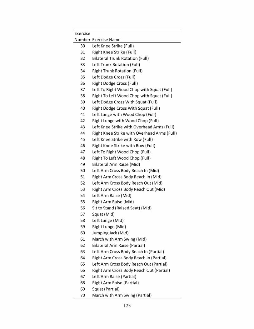
123
Exercise
Number Exercise Name
30 Left Knee Strike (Full)
31 Right Knee Strike (Full)
32 Bilateral Trunk Rotation (Full)
33 Left Trunk Rotation (Full)
34 Right Trunk Rotation (Full)
35 Left Dodge Cross (Full)
36 Right Dodge Cross (Full)
37 Left To Right Wood Chop with Squat (Full)
38 Right To Left Wood Chop with Squat (Full)
39 Left Dodge Cross With Squat (Full)
40 Right Dodge Cross With Squat (Full)
41 Left Lunge with Wood Chop (Full)
42 Right Lunge with Wood Chop (Full)
43 Left Knee Strike with Overhead Arms (Full)
44 Right Knee Strike with Overhead Arms (Full)
45 Left Knee Strike with Row (Full)
46 Right Knee Strike with Row (Full)
47 Left To Right Wood Chop (Full)
48 Right To Left Wood Chop (Full)
49 Bilateral Arm Raise (Mid)
50 Left Arm Cross Body Reach In (Mid)
51 Right Arm Cross Body Reach In (Mid)
52 Left Arm Cross Body Reach Out (Mid)
53 Right Arm Cross Body Reach Out (Mid)
54 Left Arm Raise (Mid)
55 Right Arm Raise (Mid)
56 Sit to Stand (Raised Seat) (Mid)
57 Squat (Mid)
58 Left Lunge (Mid)
59 Right Lunge (Mid)
60 Jumping Jack (Mid)
61 March with Arm Swing (Mid)
62 Bilateral Arm Raise (Partial)
63 Left Arm Cross Body Reach In (Partial)
64 Right Arm Cross Body Reach In (Partial)
65 Left Arm Cross Body Reach Out (Partial)
66 Right Arm Cross Body Reach Out (Partial)
67 Left Arm Raise (Partial)
68 Right Arm Raise (Partial)
69 Squat (Partial)
70 March with Arm Swing (Partial)
124
APPENDIX B
Descriptions of all Exercises Used
1. Bilateral Arm Raise (Full) - Starting with the both arms hanging by the side, both arms are kept straight at the elbow and raised in the frontal plane until they are vertical next to the head
2. Left Arm Cross Body Reach In (Full)- Starting with the left arm vertical next to the head, move the arm across the front of the body ending at the right hip
3. Right Arm Cross Body Reach In (Full)- Starting with the right arm vertical next to the head, move the arm across the front of the body ending at the left hip
4. Left Arm Cross Body Reach Out (Full)- Starting with the left arm at the right hip, move the arm across the front of the body ending next to the head.
5. Right Arm Cross Body Reach Out (Full)- Starting with the right arm at the left hip, move the arm across the front of the body ending next to the head.
6. Left Arm Raise (Full)- Starting with the arm hanging by the left side, the left arm is kept straight at the elbow and raised in the frontal plane until it is vertical next to the head
7. Right Arm Raise (Full)- Starting with the arm hanging by the right side, the right arm is kept straight at the elbow and raised in the frontal plane until it is vertical next to the head
8. Left Jab (Full)- Starting with both arms held in front of the face with elbows bent at 90 degrees, move the left arm straightforward in the sagittal plane by straightening the elbow.
9. Right Jab (Full)- Starting with both arms held in front of the face with elbows bent at 90 degrees, move the right arm straightforward in the sagittal plane by straightening the elbow.
10. Jumping Jack (Full)- Starting with both arms hanging at the side, both arms are raised in the frontal plane until they are above the head. The legs start together and are simultaneously moved outward in the frontal plane. Then both the arms and legs are brought back to the starting position.
11. Side Jump (Full)- Starting with both hands placed on the hips and legs together, jump sideways in the frontal plane, then back to the starting position. Move both legs together in the jump
125
12. Squat Jump with Arm Swing- Unused
13. Jumping Jack (No Arms) (Full)- Starting with both arms hanging at the side and the legs together, the legs are moved outward in the frontal plane then back to the starting position
14. Side Hop (Full)- Starting with both hands placed on the hips and legs together, jump sideways in the frontal plane, one leg at a time; once the legs are together again, jump back to the starting position, one leg at a time.
15. Squat Jump (No Arms) (Full)- Start with both hands placed on the hip and standing with the legs together. Bend the knees at a 90-degree angle keeping the back straight, as the legs are being straightened, jump off the ground before landing back in the starting position.
16. Sit to Stand (Full)- Start with both knees bent at a 90-degree angle, sitting on a small bench. Keep the back straight and stand straight up.
17. Squat (Full)- Start with both hands place on the hips and the legs together and straight. Bend the knees at a 90-degree angle keeping the back straight; then stand back up into the starting position.
18. March with No Arms (Full)- Start with both arms hanging by the side. Lift the right knee to 90-degrees. Lower the right knee then lift the left knee to 90-degrees, lower the left knee back to the starting position.
19. Side Step (Full)- Starting with both hands placed on the hips and legs together, step sideways in the frontal plane, one leg at a time; once the legs are together again, step back to the starting position, one leg at a time.
20. Left Lunge (Full)- Start standing straight with both arms hanging by the side. Step forward with the left leg in the sagittal plane bend the knee and hip to 90 degrees; step back and up to the starting position.
21. Right Lunge (Full)- Start standing straight with both arms hanging by the side. Step forward with the right leg in the sagittal plane bend the knee and hip to 90 degrees; step back and up to the starting position.
22. Left Seated Knee Strike- While seated on a small bench, lift the left thigh upwards without straightening the knee; then lower it back to the seated position
23. Right Seated Knee Strike- While seated on a small bench, lift the right thigh upwards without straightening the knee; then lower it back to the seated position
24. Left Low Kick (Full)- Start standing with legs together with both arms hanging by the side. Extend the left leg forward in the sagittal plane swinging it at the hip; then return to the standing position.

126
25. Right Low Kick (Full)- Start standing with legs together with both arms hanging by the side. Extend the right leg forward in the sagittal plane swinging it at the hip; then return to the standing position
26. Left Seated Low Kick (Full)- While seated on a small bench, start with the legs bent at 90 degrees, kick the left shank out swinging it at the knee, then return it to the 90 degree starting position.
27. Right Seated Low Kick (Full)- While seated on a small bench, start with the legs bent at 90 degrees, kick the left shank out swinging it at the knee, then return it to the 90 degree starting position.
28. Left Step Forward and Back (Full)- Stand with both arms hanging by the side. Step forward in the frontal plane with the left leg, plant the left foot momentarily then step back into the starting position.
29. Right Step Forward and Back (Full)- Stand with both arms hanging by the side. Step forward in the frontal plane with the right leg, plant the right foot momentarily then step back into the starting position.
30. Left Knee Strike (Full)- Starting from a standing position, rotate the left thigh upwards in the sagittal plane without straightening the knee; then lower it back to the starting position.
31. Right Knee Strike (Full)- Starting from a standing position, rotate the right thigh upwards in the sagittal plane without straightening the knee; then lower it back to the starting position.
32. Bilateral Trunk Rotation (Full)- Start with both arms hanging by the side. Rotate the trunk 90 degrees to the left, then 90 degrees to the right then back to the neutral starting position.
33. Left Trunk Rotation (Full)- Start with both arms hanging by the side. Rotate the trunk 90-degrees to the left.
34. Right Trunk Rotation (Full)- Start with both arms hanging by the side. Rotate the trunk 90-degrees to the right.
35. Left Dodge Cross (Full)- Starting with both arms held in front of the face with elbows bent at 90 degrees, move the head and trunk down and to the left in the frontal plane. Then punch the left arm across the centerline of the body while moving the trunk back to vertical.
36. Right Dodge Cross (Full)- Starting with both arms held in front of the face with elbows bent at 90 degrees, move the head and trunk down and to the right in the frontal plane. Then punch the right arm across the centerline of the body while moving the trunk back to vertical.
127
37. Left to Right Wood Chop with Squat (Full)- Start with both hands held together above the left shoulder with the elbows bent and stranding straight with the legs together. Keeping the hands together, bring the arms across the front of the body to the right hip. Simultaneously bend the knees at a 90-degree angle keeping the back straight then stand back up while bringing the arms back into the starting position.
38. Right to Left Wood Chop with Squat (Full)- Start with both hands held together above the right shoulder with the elbows bent and stranding straight with the legs together. Keeping the hands together, bring the arms across the front of the body to the left hip. Simultaneously bend the knees at a 90-degree angle keeping the back straight then stand back up while bringing the arms back into the starting position.
39. Left Dodge Cross with Squat (Full)- Starting with both arms held in front of the face with elbows bent at 90 degrees and standing with legs together, move the head and trunk down and to the left in the frontal plane. Then punch the left arm across the centerline of the body while moving the trunk back to vertical. Simultaneously bend the knees at a 90-degree angle keeping the back straight then stand back up while punching across. Ending position has the right arm in the starting position and the left arm extended after the punch.
40. Right Dodge Cross with Squat (Full)- Starting with both arms held in front of the face with elbows bent at 90 degrees and standing with legs together, move the head and trunk down and to the right in the frontal plane. Then punch the right arm across the centerline of the body while moving the trunk back to vertical. Simultaneously bend the knees at a 90-degree angle keeping the back straight then stand back up while punching across. Ending position has the left arm in the starting position and the right arm extended after the punch.
41. Left Lunge with Wood Chop (Full)- Start with both hands together by the left shoulder with elbows bent. Step forward with the left leg in the sagittal plane bend the knee and hip to 90 degrees. While in the deepest part of the lunge, bring the arms across the body to the right hip, then return them to the starting position by the left shoulder. Lastly, stand back up out of the lunge.
42. Right Lunge with Wood Chop (Full)- Start with both hands together by the right shoulder with elbows bent. Step forward with the right leg in the sagittal plane bend the knee and hip to 90 degrees. While in the deepest part of the lunge, bring the arms across the body to the left hip, then return them to the starting position by the right shoulder. Lastly, stand back up out of the lunge.
43. Left Knee Strike with Overhead Arms (Full)- Starting from a standing position, and the arms held above the head, lift the left thigh upwards in the sagittal plane without straightening the knee; simultaneously lower the arms in the sagittal plane stopping when they are in line with body. Then lower knee back to the starting position while lifting arms back to the starting position.
128
44. Right Knee Strike with Overhead Arms (Full)- Starting from a standing position, and the arms held above the head, lift the right thigh upwards in the sagittal plane without straightening the knee; simultaneously lower the arms in the sagittal plane stopping when they are in line with body. Then lower knee back to the starting position while lifting arms back to the starting position.
45. Left Knee Strike with Row (Full)- Starting from a standing position, and the arms held out parallel to each other directly in front of the body, lift the left thigh upwards in the sagittal plane without straightening the knee; while doing this, bring the hands in towards the hips, bending the elbows; then lower knee back to the starting position while returning arms to starting position.
46. Right Knee Strike with Row (Full)- Starting from a standing position, and the arms held out parallel to each other directly in front of the body, lift the right thigh upwards in the sagittal plane without straightening the knee; while doing this, bring the hands in towards the hips, bending the elbows; then lower knee back to the starting position while returning arms to starting position.
47. Left to Right Wood Chop (Full)- Start with both hands together by the left shoulder with elbows bent, bring the arms across the body to the right hip, then return them to the starting position by the left shoulder.
48. Right to Left Wood Chop (Full)- Start with both hands together by the right shoulder with elbows bent, bring the arms across the body to the left hip, then return them to the starting position by the right shoulder.
49. Bilateral Arm Raise (Mid)- Starting with the both arms hanging by the side, both arms are kept straight at the elbow and raised in the frontal plane until they are horizontal next to the shoulders
50. Left Arm Cross Body Reach In (Mid)- Starting with the left arm next to the head with the elbow slightly bent, move the arm across the front of the body ending close to the right hip; then return to starting position.
51. Right Arm Cross Body Reach In (Mid)- Starting with the right arm next to the head with the elbow slightly bent, move the arm across the front of the body ending close to the left hip; then return to starting position.
52. Left Arm Cross Body Reach Out (Mid)- Starting with the left arm at the right hip, move the arm across the front of the body stopping close to the head with the elbow slightly bent; then return to starting position.
53. Right Arm Cross Body Reach Out (Mid)- Starting with the right arm at the left hip, move the arm across the front of the body stopping close to the head with the elbow slightly bent; then return to starting position.
129
54. Left Arm Raise (Mid)- Starting with the arm hanging by the left side, the left arm is kept straight at the elbow and raised in the frontal plane until it is horizontal next to the shoulder.
55. Right Arm Raise (Mid)- Starting with the arm hanging by the right side, the right arm is kept straight at the elbow and raised in the frontal plane until it is horizontal next to the shoulder
56. Sit to Stand (Raised Seat) (Mid)- Start with both knees bent, sitting on a raised bench. Keep the back straight and stand straight up.
57. Squat (Mid)- Start with both hands place on the hips and the legs together and straight. Bend the knees to roughly a 120-degree angle (thigh to shank angle) keeping the back straight; then stand back up into the starting position.
58. Left Lunge (Mid)- Start standing straight with both arms hanging by the side. Step forward with the left leg in the sagittal plane bend the knee to roughly 120 degrees (thigh to shank angle); step back and up to the starting position.
59. Right Lunge (Mid)- Start standing straight with both arms hanging by the side. Step forward with the right leg in the sagittal plane bend the knee and hip to roughly 120 degrees (thigh to shank angle); step back and up to the starting position.
60. Jumping Jack (Mid)- Starting with both arms hanging at the side, both arms are raised in the frontal plane until they are in line with the shoulder. The legs start together and are simultaneously moved outward in the frontal plane. Then both the arms and legs are brought back to the starting position.
61. March with No Arms (Mid)- Start with both arms hanging by the side. Lift the right knee so the hip is at roughly 60-degrees. Lower the right knee then lift the left knee so the hip is at roughly 60-degrees, lower the left knee back to the starting position.
62. Bilateral Arm Raise (Partial)- Starting with the both arms hanging by the side, both arms are kept straight at the elbow and raised in the frontal plane until the arms make roughly a 45 degree angle with the body; then return to starting position.
63. Left Arm Cross Body Reach In (Partial)- Starting with the left hand next to the head, with the elbow close to the body move the forearm across the front of the body ending close to the right hip; then return to starting position.
64. Right Arm Cross Body Reach In (Partial)- Starting with the right hand next to the head, with the elbow close to the body move the forearm across the front of the body ending close to the left hip; then return to starting position.

130
65. Left Arm Cross Body Reach Out (Partial)- Starting with the left hand near the right hip, pivot the forearm across the front of the body ending with the left hand near the head; then return to starting position.
66. Right Arm Cross Body Reach Out (Partial)- Starting with the right hand near the left hip, pivot the forearm across the front of the body ending with the right hand near the head; then return to starting position.
67. Left Arm Raise (Partial)- Starting with the arm hanging by the left side, the left arm is kept straight at the elbow and raised in the frontal plane until the arm makes roughly a 45 degree angle with the body; then return to starting position.
68. Right Arm Raise (Partial)- Starting with the arm hanging by the right side, the right arm is kept straight at the elbow and raised in the frontal plane until the arm makes roughly a 45 degree angle with the body; then return to starting position.
69. Squat (Partial)- Start with both hands place on the hips and the legs together and straight. Bend the knees to roughly a 120 degree angle (between shank and thigh) keeping the back straight; then stand back up into the starting position
70. March (Partial)-Start with both arms hanging by the side. Lift the right knee so the thigh is roughly 30 degrees from vertical. Lower the right knee then lift the left knee so the thigh is roughly 30 degrees from vertical, lower the left knee back to the starting position.
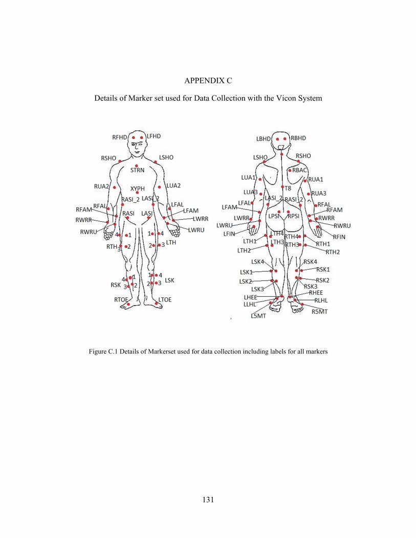
131
APPENDIX C
Details of Marker set used for Data Collection with the Vicon System
Figure C.1 Details of Markerset used for data collection including labels for all markers

132
APPENDIX D
Markers and Coordinate Definitions for Visual 3D Skeleton
Table D.1 Definitions for each Segment used to Define the custom Visual 3D Skeleton for each subject

133
APPENDIX E
Explanations of all Values Calculated for Error Database
MaxVicon: The maximum value (absolute value) of the angle within the given repetition for the Vicon dataset
MaxV_Loc: The location (frame) where the maximum Vicon value occurs
MaxKinect: The maximum value (absolute value) of the angle for the Vitalize dataset near the location of the Maximum Vicon value
MaxK_Loc: The location (frame) where the maximum Vitalize value occurs
StartKinect: The angle value for the start of the repetition for the Vitalize dataset
StartVicon: The angle value for the start of the repetition for the Vicon dataset
EndKinect: The angle value for the end of the repetition for the Vitalize dataset
EndVicon: The angle value for the end of the repetition for the Vicon dataset
ROMKinect: The difference between MaxKinect and StartKinect
ROMVicon: The difference between MaxVicon and StartVicon
ExerSpeed: ROMVicon/length of the repetition in seconds
ROMError: The difference between ROMVicon and ROMKinect
StartError: The difference between StartVicon and StartKinect
MaxError: The difference between MaxVicon and MaxKinect
MaxError_Loc: The location (frame) where the MaxError occurs
RMSE: the root-mean-square error across the entire repetition
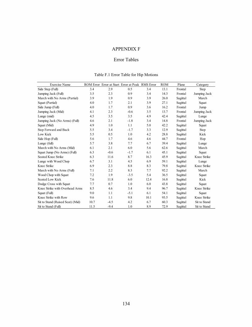
134
APPENDIX F
Error Tables
Table F.1 Error Table for Hip Motions
Exercise Name ROM Error Error at Start Error at Peak RMS Error ROM Plane CategorySide Step (Full) 3.4 2.9 0.5 3.4 13.1 Frontal StepJumping Jack (Full) 3.5 2.3 0.9 3.4 14.3 Frontal Jumping JackMarch with No Arms (Partial) 3.9 1.9 0.9 3.9 26.0 Sagittal MarchSquat (Partial) 4.0 1.7 2.1 3.9 27.1 Sagittal SquatSide Jump (Full) 4.0 1.7 0.9 3.6 16.2 Frontal JumpJumping Jack (Mid) 4.1 2.3 -0.6 3.5 13.7 Frontal Jumping JackLunge (mid) 4.5 3.5 3.5 4.9 42.4 Sagittal LungeJumping Jack (No Arms) (Full) 4.6 2.1 -1.8 3.4 14.8 Frontal Jumping JackSquat (Mid) 4.9 1.0 1.1 5.0 42.2 Sagittal SquatStep Forward and Back 5.5 3.4 -1.7 3.3 12.9 Sagittal StepLow Kick 5.5 0.5 1.0 4.2 28.8 Sagittal KickSide Hop (Full) 5.6 1.7 4.6 4.6 44.7 Frontal HopLunge (full) 5.7 3.8 7.7 6.7 39.4 Sagittal LungeMarch with No Arms (Mid) 6.1 2.1 6.0 5.6 62.6 Sagittal MarchSquat Jump (No Arms) (Full) 6.3 -0.6 -1.7 6.1 45.1 Sagittal SquatSeated Knee Strike 6.3 11.6 8.7 16.3 45.9 Sagittal Knee StrikeLunge with Wood Chop 6.7 3.1 4.5 6.9 59.1 Sagittal LungeKnee Strike 6.9 2.3 8.8 8.3 79.8 Sagittal Knee StrikeMarch with No Arms (Full) 7.1 2.2 8.3 7.7 92.2 Sagittal MarchWood Chop with Squat 7.2 1.9 -3.5 5.4 36.5 Sagittal SquatSeated Low Kick 7.6 11.8 6.0 12.4 16.8 Sagittal KickDodge Cross with Squat 7.7 0.7 1.0 6.0 43.8 Sagittal SquatKnee Strike with Overhead Arms 8.5 4.6 3.4 9.4 96.7 Sagittal Knee StrikeSquat (Full) 9.0 1.1 -5.1 6.1 54.1 Sagittal SquatKnee Strike with Row 9.6 1.1 9.8 10.1 93.5 Sagittal Knee StrikeSit to Stand (Raised Seat) (Mid) 10.7 -4.5 4.2 6.7 60.3 Sagittal Sit to StandSit to Stand (Full) 11.5 -9.4 1.0 8.9 72.9 Sagittal Sit to Stand
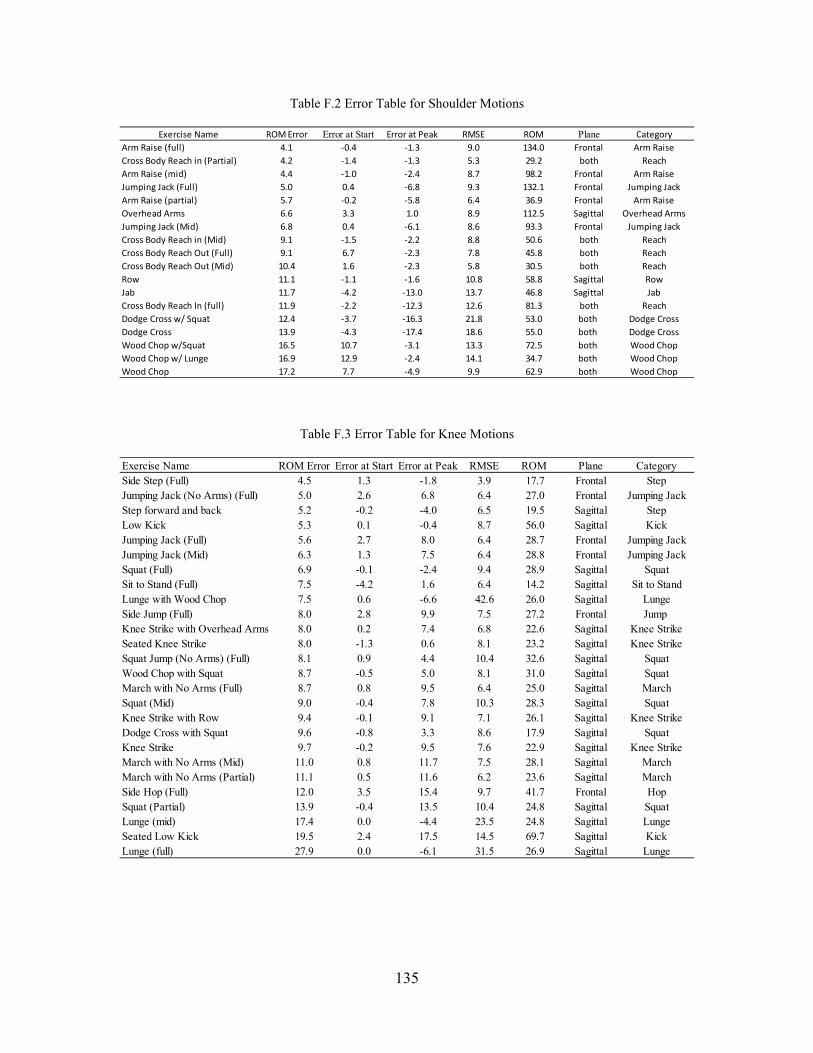
135
Table F.2 Error Table for Shoulder Motions
Table F.3 Error Table for Knee Motions
Exercise Name ROM Error Error at Start Error at Peak RMSE ROM Plane Category
Arm Raise (full) 4.1 ‐0.4 ‐1.3 9.0 134.0 Frontal Arm Raise
Cross Body Reach in (Partial) 4.2 ‐1.4 ‐1.3 5.3 29.2 both Reach
Arm Raise (mid) 4.4 ‐1.0 ‐2.4 8.7 98.2 Frontal Arm Raise
Jumping Jack (Full) 5.0 0.4 ‐6.8 9.3 132.1 Frontal Jumping Jack
Arm Raise (partial) 5.7 ‐0.2 ‐5.8 6.4 36.9 Frontal Arm Raise
Overhead Arms 6.6 3.3 1.0 8.9 112.5 Sagittal Overhead Arms
Jumping Jack (Mid) 6.8 0.4 ‐6.1 8.6 93.3 Frontal Jumping Jack
Cross Body Reach in (Mid) 9.1 ‐1.5 ‐2.2 8.8 50.6 both Reach
Cross Body Reach Out (Full) 9.1 6.7 ‐2.3 7.8 45.8 both Reach
Cross Body Reach Out (Mid) 10.4 1.6 ‐2.3 5.8 30.5 both Reach
Row 11.1 ‐1.1 ‐1.6 10.8 58.8 Sagittal Row
Jab 11.7 ‐4.2 ‐13.0 13.7 46.8 Sagittal Jab
Cross Body Reach In (full) 11.9 ‐2.2 ‐12.3 12.6 81.3 both Reach
Dodge Cross w/ Squat 12.4 ‐3.7 ‐16.3 21.8 53.0 both Dodge Cross
Dodge Cross 13.9 ‐4.3 ‐17.4 18.6 55.0 both Dodge Cross
Wood Chop w/Squat 16.5 10.7 ‐3.1 13.3 72.5 both Wood Chop
Wood Chop w/ Lunge 16.9 12.9 ‐2.4 14.1 34.7 both Wood Chop
Wood Chop 17.2 7.7 ‐4.9 9.9 62.9 both Wood Chop
Exercise Name ROM Error Error at Start Error at Peak RMSE ROM Plane CategorySide Step (Full) 4.5 1.3 -1.8 3.9 17.7 Frontal StepJumping Jack (No Arms) (Full) 5.0 2.6 6.8 6.4 27.0 Frontal Jumping JackStep forward and back 5.2 -0.2 -4.0 6.5 19.5 Sagittal StepLow Kick 5.3 0.1 -0.4 8.7 56.0 Sagittal KickJumping Jack (Full) 5.6 2.7 8.0 6.4 28.7 Frontal Jumping JackJumping Jack (Mid) 6.3 1.3 7.5 6.4 28.8 Frontal Jumping JackSquat (Full) 6.9 -0.1 -2.4 9.4 28.9 Sagittal SquatSit to Stand (Full) 7.5 -4.2 1.6 6.4 14.2 Sagittal Sit to StandLunge with Wood Chop 7.5 0.6 -6.6 42.6 26.0 Sagittal LungeSide Jump (Full) 8.0 2.8 9.9 7.5 27.2 Frontal Jump Knee Strike with Overhead Arms 8.0 0.2 7.4 6.8 22.6 Sagittal Knee StrikeSeated Knee Strike 8.0 -1.3 0.6 8.1 23.2 Sagittal Knee StrikeSquat Jump (No Arms) (Full) 8.1 0.9 4.4 10.4 32.6 Sagittal SquatWood Chop with Squat 8.7 -0.5 5.0 8.1 31.0 Sagittal SquatMarch with No Arms (Full) 8.7 0.8 9.5 6.4 25.0 Sagittal MarchSquat (Mid) 9.0 -0.4 7.8 10.3 28.3 Sagittal SquatKnee Strike with Row 9.4 -0.1 9.1 7.1 26.1 Sagittal Knee StrikeDodge Cross with Squat 9.6 -0.8 3.3 8.6 17.9 Sagittal SquatKnee Strike 9.7 -0.2 9.5 7.6 22.9 Sagittal Knee StrikeMarch with No Arms (Mid) 11.0 0.8 11.7 7.5 28.1 Sagittal MarchMarch with No Arms (Partial) 11.1 0.5 11.6 6.2 23.6 Sagittal MarchSide Hop (Full) 12.0 3.5 15.4 9.7 41.7 Frontal HopSquat (Partial) 13.9 -0.4 13.5 10.4 24.8 Sagittal SquatLunge (mid) 17.4 0.0 -4.4 23.5 24.8 Sagittal LungeSeated Low Kick 19.5 2.4 17.5 14.5 69.7 Sagittal KickLunge (full) 27.9 0.0 -6.1 31.5 26.9 Sagittal Lunge
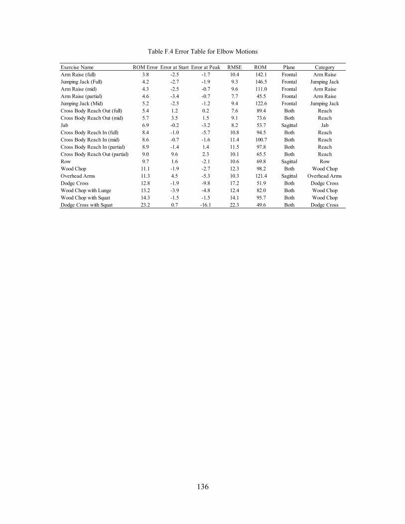
136
Table F.4 Error Table for Elbow Motions
Exercise Name ROM Error Error at Start Error at Peak RMSE ROM Plane CategoryArm Raise (full) 3.8 -2.5 -1.7 10.4 142.1 Frontal Arm RaiseJumping Jack (Full) 4.2 -2.7 -1.9 9.3 146.5 Frontal Jumping JackArm Raise (mid) 4.3 -2.5 -0.7 9.6 111.0 Frontal Arm RaiseArm Raise (partial) 4.6 -3.4 -0.7 7.7 45.5 Frontal Arm RaiseJumping Jack (Mid) 5.2 -2.5 -1.2 9.4 122.6 Frontal Jumping JackCross Body Reach Out (full) 5.4 1.2 0.2 7.6 89.4 Both ReachCross Body Reach Out (mid) 5.7 3.5 1.5 9.1 73.6 Both ReachJab 6.9 -0.2 -3.2 8.2 53.7 Sagittal JabCross Body Reach In (full) 8.4 -1.0 -5.7 10.8 94.5 Both ReachCross Body Reach In (mid) 8.6 -0.7 -1.6 11.4 100.7 Both ReachCross Body Reach In (partial) 8.9 -1.4 1.4 11.5 97.8 Both ReachCross Body Reach Out (partial) 9.0 9.6 2.3 10.1 65.5 Both ReachRow 9.7 1.6 -2.1 10.6 69.8 Sagittal RowWood Chop 11.1 -1.9 -2.7 12.3 98.2 Both Wood ChopOverhead Arms 11.3 4.5 -5.3 10.3 121.4 Sagittal Overhead ArmsDodge Cross 12.8 -1.9 -9.8 17.2 51.9 Both Dodge CrossWood Chop with Lunge 13.2 -3.9 -4.8 12.4 82.0 Both Wood ChopWood Chop with Squat 14.3 -1.5 -1.5 14.1 95.7 Both Wood ChopDodge Cross with Squat 23.2 0.7 -16.1 22.3 49.6 Both Dodge Cross
137
REFERENCES
[1] APTA Staff, “The Movement System Brings It All Together.” May-2016. [Online]. Available: http://policy.apta.org/PTinMotion/2016/5/Feature/MovementSystem/. [Accessed: 20-August-2016]
[2] APTA Staff, “Physical Therapist Practice and the Human Movement System.” Aug-2015.[Online]. Available: https://www.apta.org/MovementSystem/WhitePaper/. [Accessed: 20-August-2016]
[3] R. Klepps, “8 Thought-Provoking Facts About Physical Therapy You Can’t Ignore,” The Strive Labs Blog, Nov-2015. [Online]. Available: http://blog.strivelabs.com/2015/02/19/physical-therapy-facts-cant-ignore/. [Accessed: 24-Sep-2015].
[4] S. Ackerman, “The Cost of War Includes at Least 253,330 Brain Injuries and 1,700 Amputations,” WIRED, 08-Feb-2013. [Online]. Available: https://www.wired.com/2013/02/cost-of-war/. [Accessed: 04-Jul-2016].
[5] S. M. Coppola and S. M. Collins, “Is physical therapy more beneficial than unsupervised home exercise in treatment of post surgical knee disorders? A systematic review,” The Knee, vol. 16, no. 3, pp. 171–175, Jun. 2009.
[6] G. D. Deyle, S. C. Allison, R. L. Matekel, M. G. Ryder, J. M. Stang, D. D. Gohdes, J. P. Hutton, N. E. Henderson, and M. B. Garber, “Physical therapy treatment effectiveness for osteoarthritis of the knee: a randomized comparison of supervised clinical exercise and manual therapy procedures versus a home exercise program,” Physical Therapy, vol. 85, no. 12, pp. 1301–1317, Dec. 2005.
[7] L. Geurts, V. Vanden Abeele, J. Husson, F. Windey, M. Van Overveldt, J.-H. Annema, and S. Desmet, “Digital Games for Physical Therapy: Fulfilling the Need for Calibration and Adaptation,” in Proceedings of the Fifth International Conference on Tangible, Embedded, and Embodied Interaction, New York, NY, USA, 2011, pp. 117–124.
[8] G. S. Kolt and J. F. McEvoy, “Adherence to rehabilitation in patients with low back pain,” Manual Therapy, vol. 8, no. 2, pp. 110–116, May 2003.
[9] P. Escolar-Reina, F. Medina-Mirapeix, J. J. Gascón-Cánovas, J. Montilla-Herrador, F. J. Jimeno-Serrano, S. L. de Oliveira Sousa, M. E. del Baño-Aledo, and R. Lomas-Vega, “How do care-provider and home exercise program characteristics affect patient adherence in chronic neck and back pain: a qualitative study,” Biomed Central Health Services Research., vol. 10, p. 60, 2010.

138
[10] K. Jack, S. M. McLean, J. K. Moffett, and E. Gardiner, “Barriers to treatment adherence in physiotherapy outpatient clinics: A systematic review,” Manual Therapy, vol. 15, no. 3–2, pp. 220–228, Jun. 2010.
[11] R. M. Kaplan and H. J. Simon, “Compliance in medical care: Reconsideration of self-predictions,” Annals of Behavioral Medicine, vol. 12, no. 2, pp. 66–71, 1990.
[12] E. M. Sluijs, G. J. Kok, and J. van der Zee, “Correlates of Exercise Compliance in Physical Therapy,” Physical Therapy, vol. 73, no. 11, pp. 771–782, Nov. 1993.
[13] A. M. Jette, “Improving patient cooperation with arthritis treatment regimens,” Arthritis Rheumatism., vol. 25, no. 4, pp. 447–453, Apr. 1982.
[14] R. Campbell, M. Evans, M. Tucker, B. Quilty, P. Dieppe, and J. L. Donovan, “Why don’t patients do their exercises? Understanding non-compliance with physiotherapy in patients with osteoarthritis of the knee,” Journal of Epidemiology and Community Health, vol. 55, no. 2, pp. 132–138, Feb. 2001.
[15] W. Zhao, D. D. Espy, M. A. Reinthal, and H. Feng, “A feasibility study of using a single Kinect sensor for rehabilitation exercises monitoring: A rule based approach,” 2014 IEEE Symposium on Computational Intelligence in Healthcare and e-health (CICARE), pp. 1–8.
[16] J. Sinclair, P. Hingston, and M. Masek, “Considerations for the Design of Exergames,” in Proceedings of the 5th International Conference on Computer Graphics and Interactive Techniques in Australia and Southeast Asia, New York, NY, USA, 2007, pp. 289–295.
[17] H. De Rosario, J. M. Belda-Lois, F. Fos, E. Medina, R. Poveda-Puente, and M. Kroll, “Correction of Joint Angles From Kinect for Balance Exercising and Assessment,” Journal of Applied Biomechanics, vol. 30, no. 2, pp. 294–299, Apr. 2014.
[18] P. M. Kato, “Video games in health care: Closing the gap,” Review of General Psychology. vol. 14, no. 2, pp. 113–121, 2010.
[19] A. Mobini, S. Behzadipour, and M. S. Foumani, “Accuracy of Kinect’s skeleton tracking for upper body rehabilitation applications,” Disability and Rehabilitation: Assistive Technology, vol. 9, no. 4, pp. 344–352, Jul. 2014.
[20] M. E. Nixon, A. M. Howard, and Y.-P. Chen, “Quantitative evaluation of the Microsoft Kinect for use in an upper extremity virtual rehabilitation environment,” in 2013 International Conference on Virtual Rehabilitation (ICVR), pp. 222–228.
[21] Y. Salem, S. J. Gropack, D. Coffin, and E. M. Godwin, “Effectiveness of a low-cost virtual reality system for children with developmental delay: a preliminary randomised single-blind controlled trial,” Physiotherapy, vol. 98, no. 3, pp. 189–195, Sep. 2012.
139
[22] M. J. D. Taylor, D. McCormick, T. Shawis, R. Impson, and M. Griffin, “Activity-promoting gaming systems in exercise and rehabilitation,” Journal of Rehabilitation Research and Development, vol. 48, no. 10, p. 1171, 2011.
[23] Y.-J. Chang, S.-F. Chen, and J.-D. Huang, “A Kinect-based system for physical rehabilitation: A pilot study for young adults with motor disabilities,” Research in Developmental Disabilities, vol. 32, no. 6, pp. 2566–2570, Nov. 2011.
[24] Y.-J. Chang, W.-Y. Han, and Y.-C. Tsai, “A Kinect-based upper limb rehabilitation system to assist people with cerebral palsy,” Research in Developmental Disabilities, vol. 34, no. 11, pp. 3654–3659, Nov. 2013.
[25] J. Albores, C. Marolda, M. Haggerty, B. Gerstenhaber, and R. Zuwallack, “The use of a home exercise program based on a computer system in patients with chronic obstructive pulmonary disease,” Journal of Cardiopulmonary Rehabilitation and Prevention, vol. 33, no. 1, pp. 47–52, Feb. 2013.
[26] C. Bryanton, J. Bossé, M. Brien, J. McLean, A. McCormick, and H. Sveistrup, “Feasibility, motivation, and selective motor control: virtual reality compared to conventional home exercise in children with cerebral palsy,” Cyberpsychology and Behavior: The Impact of the Internet, Multimedia and Virtual Reality on Behavior and Society, vol. 9, no. 2, pp. 123–128, Apr. 2006.
[27] M. Kolbjornsen, “A Comparison of Motion-Sensing Game Technologies for use in Physical Rehabilitation,” Masters of Science in Informatics, Norwegian University of Science and Technology, Trondheim, Norway, 2012.
[28] B. Lange, C.-Y. Chang, E. Suma, B. Newman, A. S. Rizzo, and M. Bolas, “Development and evaluation of low cost game-based balance rehabilitation tool using the Microsoft Kinect sensor,” in 2011 Annual International Conference of the IEEE Engineering in Medicine and Biology Society, EMBC, 2011, pp. 1831–1834.
[29] D. González-Ortega, F. J. Díaz-Pernas, M. Martínez-Zarzuela, and M. Antón-Rodríguez, “A Kinect-based system for cognitive rehabilitation exercises monitoring,” Computer Methods and Programs in Biomedicine., vol. 113, no. 2, pp. 620–631, Feb. 2014.
[30] B. Lange, C.-Y. Chang, E. Suma, B. Newman, A. S. Rizzo, and M. Bolas, “Development and evaluation of low cost game-based balance rehabilitation tool using the microsoft Kinect sensor,” in 2011 Annual International Conference of the IEEE Engineering in Medicine and Biology Society, EMBC, 2011, pp. 1831–1834.
[31] B. Seamon, M. DeFranco, and M. Thigpen, “Use of the Xbox Kinect virtual gaming system to improve gait, postural control and cognitive awareness in an individual with Progressive Supranuclear Palsy,” Disability and Rehabilitation, vol. 0, no. 0, pp. 1–6, Mar. 2016.

140
[32] B. Bonnechère, B. Jansen, P. Salvia, H. Bouzahouene, L. Omelina, F. Moiseev, V. Sholukha, J. Cornelis, M. Rooze, and S. Van Sint Jan, “Validity and reliability of the Kinect within functional assessment activities: Comparison with standard stereophotogrammetry,” Gait and Posture, vol. 39, no. 1, pp. 593–598, Jan. 2014.
[33] B. Bonnechère, B. Jansen, P. Salvia, H. Bouzahouene, V. Sholukha, J. Cornelis, M. Rooze, and S. Van Sint Jan, “Determination of the precision and accuracy of morphological measurements using the KinectTM sensor: comparison with standard stereophotogrammetry,” Ergonomics, vol. 57, no. 4, pp. 622–631, 2014.
[34] R. A. Clark, K. J. Bower, B. F. Mentiplay, K. Paterson, and Y.-H. Pua, “Concurrent validity of the Microsoft Kinect for assessment of spatiotemporal gait variables,” Journal of Biomechanics, vol. 46, no. 15, pp. 2722–2725, Oct. 2013.
[35] B. Galna, G. Barry, D. Jackson, D. Mhiripiri, P. Olivier, and L. Rochester, “Accuracy of the Microsoft Kinect sensor for measuring movement in people with Parkinson’s disease,” Gait and Posture, vol. 39, no. 4, pp. 1062–1068, Apr. 2014.
[36] R. P. Kuster, B. Heinlein, C. M. Bauer, and E. S. Graf, “Accuracy of KinectOne to quantify kinematics of the upper body,” Gait and Posture, vol. 47, pp. 80–85, Jun. 2016.
[37] H. Mousavi Hondori, M. Khademi, H. Mousavi Hondori, M. Khademi, “A Review on Technical and Clinical Impact of Microsoft Kinect on Physical Therapy and Rehabilitation, A Review on Technical and Clinical Impact of Microsoft Kinect on Physical Therapy and Rehabilitation,” Journal of Medical Engineering, Dec. 2014.
[38] M. E. Nixon, A. M. Howard, and Y.-P. Chen, “Quantitative evaluation of the Microsoft KinectTM for use in an upper extremity virtual rehabilitation environment,” in 2013 International Conference on Virtual Rehabilitation (ICVR), 2013, pp. 222–228.
[39] S. Obdrzalek, G. Kurillo, F. Ofli, R. Bajcsy, E. Seto, H. Jimison, and M. Pavel, “Accuracy and robustness of Kinect pose estimation in the context of coaching of elderly population,” in 2012 Annual International Conference of the IEEE Engineering in Medicine and Biology Society (EMBC), 2012, pp. 1188–1193.
[40] A. Pfister, A. M. West, S. Bronner, and J. A. Noah, “Comparative abilities of Microsoft Kinect and Vicon 3D motion capture for gait analysis,” Journal of Medical Engineering and Technology, vol. 38, no. 5, pp. 274–280, 2014.
[41] X. Xu, R. W. McGorry, L.-S. Chou, J. Lin, and C. Chang, “Accuracy of the Microsoft KinectTM for measuring gait parameters during treadmill walking,” Gait and Posture, vol. 42, no. 2, pp. 145–151, Jul. 2015.
[42] L. F. Yeung, K. C. Cheng, C. H. Fong, W. C. C. Lee, and K.-Y. Tong, “Evaluation of the Microsoft Kinect as a clinical assessment tool of body sway,” Gait and Posture, vol. 40, no. 4, pp. 532–538, Sep. 2014.

141
[43] C. Rabago, J. B. Dingwell, and J. M. Wilken, “Reliability and Minimum Detectable Change of Temporal-Spatial, Kinematic, and Dynamic Stability Measures during Peturbed Gait,” PLoS One, vol. 10(11) 2015, Jun. 2016.
[44] R. Beurskens, J. M. Wilken, and J. B. Dingwell, “Dynamic stability of individuals with transtibial amputation walking in destabilizing environments,” Journal of Biomechanics. vol. 47, no. 7, pp. 1675–1681, May 2014.
[45] iotracker, “iotracker is an affordable optical tracking solution specifically designed to meet the stringent requirements of real-time 6-DOF motion-tracking of immersive visualization systems | Optical Tracking.” [Online]. Available: http://iotracker.com/indexdaed.html?q=optical_tracking. [Accessed: 12-Jun-2016].
[46] “Kinect | Xbox 360,” Xbox.com. [Online]. Available: http://www.xbox.com/en-US/xbox-360/accessories/kinect. [Accessed: 12-Jun-2016].
[47] Tanz, Jason, “Kinect Hackers Are Changing the Future of Robotics,” WIRED, 28-Jun-2011. [Online]. Available: http://www.wired.com/2011/06/mf_kinect/all/. [Accessed: 19-Nov-2014].
[48] MSDN (Microsoft Developer Network), Natural User Interface for Kinect for Windows. 2014.
[49] “Kinect Setup | Xbox Kinect Setup | Xbox 360.” [Online]. Available: http://support.xbox.com/en-US/xbox-360/accessories/kinect-sensor-setup. [Accessed: 12-Jun-2016].
[50] “Blitz Games Studios.” [Online]. Available: http://www.blitzgamesstudios.com/. [Accessed: 14-Jun-2016].
[51] J. Evans, Straightforward Statistics for the Behavioral Sciences. Brooks/Cole Publishing, 1996.
[52] M. van Diest, J. Stegenga, H. J. Wörtche, K. Postema, G. J. Verkerke, and C. J. C. Lamoth, “Suitability of Kinect for measuring whole body movement patterns during exergaming,” Journal of Biomechanics, vol. 47, no. 12, pp. 2925–2932, Sep. 2014.
[53] S. Obdrzalek, G. Kurillo, F. Ofli, R. Bajcsy, E. Seto, H. Jimison, and M. Pavel, “Accuracy and robustness of Kinect pose estimation in the context of coaching of elderly population,” in 2012 Annual International Conference of the IEEE Engineering in Medicine and Biology Society (EMBC), 2012, pp. 1188–1193.
[54] J. L. McGinley, R. Baker, R. Wolfe, and M. E. Morris, “The reliability of three-dimensional kinematic gait measurements: A systematic review,” Gait and Posture, vol. 29, no. 3, pp. 360–369, Apr. 2009.