Abdominal wall reconstruction by a regionally distinct biocomposite of extracellular matrix digest...
14
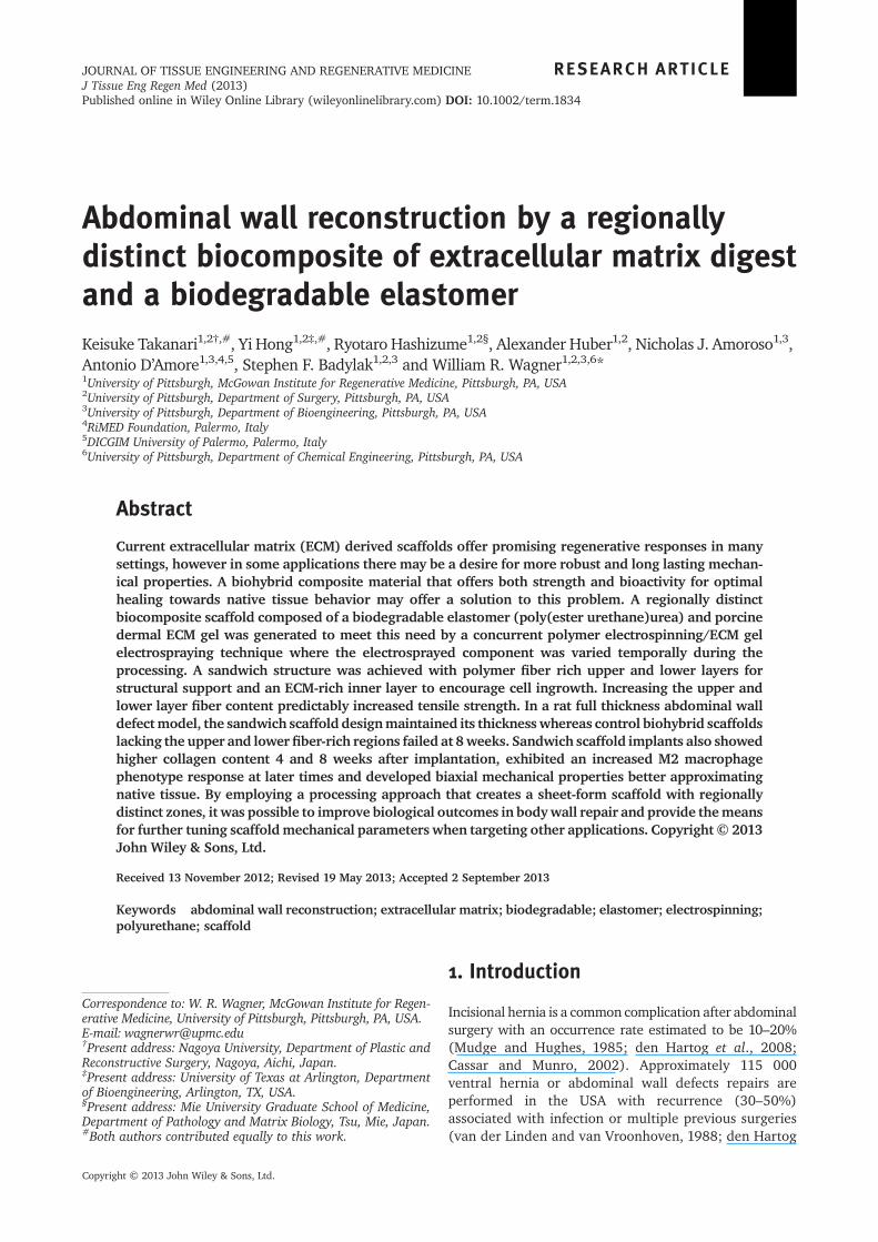
Abdominal wall reconstruction by a regionally distinct biocomposite of extracellular matrix digest and a biodegradable elastomer Keisuke Takanari 1,2†,# , Yi Hong 1,2‡,# , Ryotaro Hashizume 1,2§ , Alexander Huber 1,2 , Nicholas J. Amoroso 1,3 , Antonio D’ Amore 1,3,4,5 , Stephen F. Badylak 1,2,3 and William R. Wagner 1,2,3,6 * 1 University of Pittsburgh, McGowan Institute for Regenerative Medicine, Pittsburgh, PA, USA 2 University of Pittsburgh, Department of Surgery, Pittsburgh, PA, USA 3 University of Pittsburgh, Department of Bioengineering, Pittsburgh, PA, USA 4 RiMED Foundation, Palermo, Italy 5 DICGIM University of Palermo, Palermo, Italy 6 University of Pittsburgh, Department of Chemical Engineering, Pittsburgh, PA, USA Abstract Current extracellular matrix (ECM) derived scaffolds offer promising regenerative responses in many settings, however in some applications there may be a desire for more robust and long lasting mechan- ical properties. A biohybrid composite material that offers both strength and bioactivity for optimal healing towards native tissue behavior may offer a solution to this problem. A regionally distinct biocomposite scaffold composed of a biodegradable elastomer (poly(ester urethane)urea) and porcine dermal ECM gel was generated to meet this need by a concurrent polymer electrospinning/ECM gel electrospraying technique where the electrosprayed component was varied temporally during the processing. A sandwich structure was achieved with polymer fiber rich upper and lower layers for structural support and an ECM-rich inner layer to encourage cell ingrowth. Increasing the upper and lower layer fiber content predictably increased tensile strength. In a rat full thickness abdominal wall defect model, the sandwich scaffold design maintained its thickness whereas control biohybrid scaffolds lacking the upper and lower fiber-rich regions failed at 8 weeks. Sandwich scaffold implants also showed higher collagen content 4 and 8 weeks after implantation, exhibited an increased M2 macrophage phenotype response at later times and developed biaxial mechanical properties better approximating native tissue. By employing a processing approach that creates a sheet-form scaffold with regionally distinct zones, it was possible to improve biological outcomes in body wall repair and provide the means for further tuning scaffold mechanical parameters when targeting other applications. Copyright © 2013 John Wiley & Sons, Ltd. Received 13 November 2012; Revised 19 May 2013; Accepted 2 September 2013 Keywords abdominal wall reconstruction; extracellular matrix; biodegradable; elastomer; electrospinning; polyurethane; scaffold 1. Introduction Incisional hernia is a common complication after abdominal surgery with an occurrence rate estimated to be 10–20% (Mudge and Hughes, 1985; den Hartog et al., 2008; Cassar and Munro, 2002). Approximately 115 000 ventral hernia or abdominal wall defects repairs are performed in the USA with recurrence (30–50%) associated with infection or multiple previous surgeries (van der Linden and van Vroonhoven, 1988; den Hartog Correspondence to: W. R. Wagner, McGowan Institute for Regen- erative Medicine, University of Pittsburgh, Pittsburgh, PA, USA. E-mail: [email protected] † Present address: Nagoya University, Department of Plastic and Reconstructive Surgery, Nagoya, Aichi, Japan. ‡ Present address: University of Texas at Arlington, Department of Bioengineering, Arlington, TX, USA. § Present address: Mie University Graduate School of Medicine, Department of Pathology and Matrix Biology, Tsu, Mie, Japan. # Both authors contributed equally to this work. Copyright © 2013 John Wiley & Sons, Ltd. JOURNAL OF TISSUE ENGINEERING AND REGENERATIVE MEDICINE RESEARCH ARTICLE J Tissue Eng Regen Med (2013) Published online in Wiley Online Library (wileyonlinelibrary.com) DOI: 10.1002/term.1834
-
Upload
independent -
Category
Documents
-
view
2 -
download
0
Transcript of Abdominal wall reconstruction by a regionally distinct biocomposite of extracellular matrix digest...
Abdominal wall reconstruction by a regionallydistinct biocomposite of extracellular matrix digestand a biodegradable elastomerKeisuke Takanari1,2†,#, Yi Hong1,2‡,#, Ryotaro Hashizume1,2§, Alexander Huber1,2, Nicholas J. Amoroso1,3,Antonio D’Amore1,3,4,5, Stephen F. Badylak1,2,3 and William R. Wagner1,2,3,6*1University of Pittsburgh, McGowan Institute for Regenerative Medicine, Pittsburgh, PA, USA2University of Pittsburgh, Department of Surgery, Pittsburgh, PA, USA3University of Pittsburgh, Department of Bioengineering, Pittsburgh, PA, USA4RiMED Foundation, Palermo, Italy5DICGIM University of Palermo, Palermo, Italy6University of Pittsburgh, Department of Chemical Engineering, Pittsburgh, PA, USA
Abstract
Current extracellular matrix (ECM) derived scaffolds offer promising regenerative responses in manysettings, however in some applications there may be a desire for more robust and long lasting mechan-ical properties. A biohybrid composite material that offers both strength and bioactivity for optimalhealing towards native tissue behavior may offer a solution to this problem. A regionally distinctbiocomposite scaffold composed of a biodegradable elastomer (poly(ester urethane)urea) and porcinedermal ECM gel was generated to meet this need by a concurrent polymer electrospinning/ECM gelelectrospraying technique where the electrosprayed component was varied temporally during theprocessing. A sandwich structure was achieved with polymer fiber rich upper and lower layers forstructural support and an ECM-rich inner layer to encourage cell ingrowth. Increasing the upper andlower layer fiber content predictably increased tensile strength. In a rat full thickness abdominal walldefectmodel, the sandwich scaffold designmaintained its thicknesswhereas control biohybrid scaffoldslacking the upper and lowerfiber-rich regions failed at 8weeks. Sandwich scaffold implants also showedhigher collagen content 4 and 8 weeks after implantation, exhibited an increased M2 macrophagephenotype response at later times and developed biaxial mechanical properties better approximatingnative tissue. By employing a processing approach that creates a sheet-form scaffold with regionallydistinct zones, it was possible to improve biological outcomes in bodywall repair and provide themeansfor further tuning scaffoldmechanical parameters when targeting other applications. Copyright © 2013John Wiley & Sons, Ltd.
Received 13 November 2012; Revised 19 May 2013; Accepted 2 September 2013
Keywords abdominal wall reconstruction; extracellular matrix; biodegradable; elastomer; electrospinning;polyurethane; scaffold
1. Introduction
Incisional hernia is a common complication after abdominalsurgery with an occurrence rate estimated to be 10–20%(Mudge and Hughes, 1985; den Hartog et al., 2008;Cassar and Munro, 2002). Approximately 115 000ventral hernia or abdominal wall defects repairs areperformed in the USA with recurrence (30–50%)associated with infection or multiple previous surgeries(van der Linden and van Vroonhoven, 1988; den Hartog
Correspondence to: W. R. Wagner, McGowan Institute for Regen-erative Medicine, University of Pittsburgh, Pittsburgh, PA, USA.E-mail: [email protected]†Present address: Nagoya University, Department of Plastic andReconstructive Surgery, Nagoya, Aichi, Japan.‡Present address: University of Texas at Arlington, Departmentof Bioengineering, Arlington, TX, USA.§Present address: Mie University Graduate School of Medicine,Department of Pathology and Matrix Biology, Tsu, Mie, Japan.#Both authors contributed equally to this work.
Copyright © 2013 John Wiley & Sons, Ltd.
JOURNAL OF TISSUE ENGINEERING AND REGENERATIVE MEDICINE RESEARCH ARTICLEJ Tissue Eng Regen Med (2013)Published online in Wiley Online Library (wileyonlinelibrary.com) DOI: 10.1002/term.1834
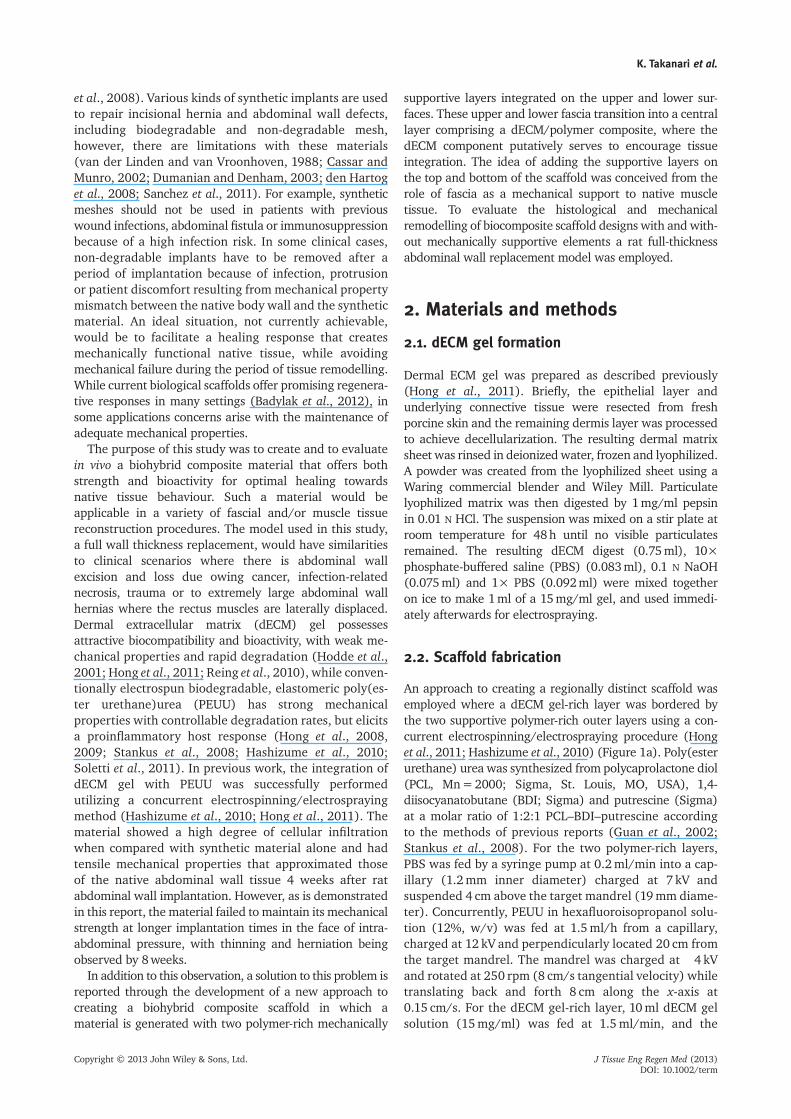
et al., 2008). Various kinds of synthetic implants are usedto repair incisional hernia and abdominal wall defects,including biodegradable and non-degradable mesh,however, there are limitations with these materials(van der Linden and van Vroonhoven, 1988; Cassar andMunro, 2002; Dumanian and Denham, 2003; den Hartoget al., 2008; Sanchez et al., 2011). For example, syntheticmeshes should not be used in patients with previouswound infections, abdominal fistula or immunosuppressionbecause of a high infection risk. In some clinical cases,non-degradable implants have to be removed after aperiod of implantation because of infection, protrusionor patient discomfort resulting from mechanical propertymismatch between the native body wall and the syntheticmaterial. An ideal situation, not currently achievable,would be to facilitate a healing response that createsmechanically functional native tissue, while avoidingmechanical failure during the period of tissue remodelling.While current biological scaffolds offer promising regenera-tive responses in many settings (Badylak et al., 2012), insome applications concerns arise with the maintenance ofadequate mechanical properties.
The purpose of this study was to create and to evaluatein vivo a biohybrid composite material that offers bothstrength and bioactivity for optimal healing towardsnative tissue behaviour. Such a material would beapplicable in a variety of fascial and/or muscle tissuereconstruction procedures. The model used in this study,a full wall thickness replacement, would have similaritiesto clinical scenarios where there is abdominal wallexcision and loss due owing cancer, infection-relatednecrosis, trauma or to extremely large abdominal wallhernias where the rectus muscles are laterally displaced.Dermal extracellular matrix (dECM) gel possessesattractive biocompatibility and bioactivity, with weak me-chanical properties and rapid degradation (Hodde et al.,2001; Hong et al., 2011; Reing et al., 2010), while conven-tionally electrospun biodegradable, elastomeric poly(es-ter urethane)urea (PEUU) has strong mechanicalproperties with controllable degradation rates, but elicitsa proinflammatory host response (Hong et al., 2008,2009; Stankus et al., 2008; Hashizume et al., 2010;Soletti et al., 2011). In previous work, the integration ofdECM gel with PEUU was successfully performedutilizing a concurrent electrospinning/electrosprayingmethod (Hashizume et al., 2010; Hong et al., 2011). Thematerial showed a high degree of cellular infiltrationwhen compared with synthetic material alone and hadtensile mechanical properties that approximated thoseof the native abdominal wall tissue 4 weeks after ratabdominal wall implantation. However, as is demonstratedin this report, the material failed to maintain its mechanicalstrength at longer implantation times in the face of intra-abdominal pressure, with thinning and herniation beingobserved by 8weeks.
In addition to this observation, a solution to this problem isreported through the development of a new approach tocreating a biohybrid composite scaffold in which amaterial is generated with two polymer-rich mechanically
supportive layers integrated on the upper and lower sur-faces. These upper and lower fascia transition into a centrallayer comprising a dECM/polymer composite, where thedECM component putatively serves to encourage tissueintegration. The idea of adding the supportive layers onthe top and bottom of the scaffold was conceived from therole of fascia as a mechanical support to native muscletissue. To evaluate the histological and mechanicalremodelling of biocomposite scaffold designs with and with-out mechanically supportive elements a rat full-thicknessabdominal wall replacement model was employed.
2. Materials and methods
2.1. dECM gel formation
Dermal ECM gel was prepared as described previously(Hong et al., 2011). Briefly, the epithelial layer andunderlying connective tissue were resected from freshporcine skin and the remaining dermis layer was processedto achieve decellularization. The resulting dermal matrixsheet was rinsed in deionized water, frozen and lyophilized.A powder was created from the lyophilized sheet using aWaring commercial blender and Wiley Mill. Particulatelyophilized matrix was then digested by 1mg/ml pepsinin 0.01 N HCl. The suspension was mixed on a stir plate atroom temperature for 48h until no visible particulatesremained. The resulting dECM digest (0.75ml), 10×phosphate-buffered saline (PBS) (0.083ml), 0.1 N NaOH(0.075ml) and 1× PBS (0.092ml) were mixed togetheron ice to make 1ml of a 15mg/ml gel, and used immedi-ately afterwards for electrospraying.
2.2. Scaffold fabrication
An approach to creating a regionally distinct scaffold wasemployed where a dECM gel-rich layer was bordered bythe two supportive polymer-rich outer layers using a con-current electrospinning/electrospraying procedure (Honget al., 2011; Hashizume et al., 2010) (Figure 1a). Poly(esterurethane) urea was synthesized from polycaprolactone diol(PCL, Mn=2000; Sigma, St. Louis, MO, USA), 1,4-diisocyanatobutane (BDI; Sigma) and putrescine (Sigma)at a molar ratio of 1:2:1 PCL–BDI–putrescine accordingto the methods of previous reports (Guan et al., 2002;Stankus et al., 2008). For the two polymer-rich layers,PBS was fed by a syringe pump at 0.2ml/min into a cap-illary (1.2mm inner diameter) charged at 7 kV andsuspended 4 cm above the target mandrel (19mm diame-ter). Concurrently, PEUU in hexafluoroisopropanol solu-tion (12%, w/v) was fed at 1.5ml/h from a capillary,charged at 12 kV and perpendicularly located 20 cm fromthe target mandrel. The mandrel was charged at �4 kVand rotated at 250 rpm (8 cm/s tangential velocity) whiletranslating back and forth 8 cm along the x-axis at0.15 cm/s. For the dECM gel-rich layer, 10ml dECM gelsolution (15mg/ml) was fed at 1.5ml/min, and the
K. Takanari et al.
Copyright © 2013 John Wiley & Sons, Ltd. J Tissue Eng Regen Med (2013)DOI: 10.1002/term
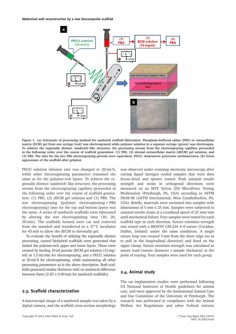
PEUU solution infusion rate was changed to 20ml/h,while other electrospinning parameters remained thesame as for the polymer-rich layers. To achieve the re-gionally distinct ‘sandwich’-like structure, the processingstream from the electrospraying capillary proceeded inthe following order over the course of scaffold genera-tion: (1) PBS, (2) dECM gel solution and (3) PBS. Thewet electrospinning (polymer electrospinning+PBSelectrospraying) time for the top and bottom layers wasthe same. A series of sandwich scaffolds were fabricatedby altering the wet electrospinning time (10, 20,30min). The scaffolds formed were cut and removedfrom the mandrel and transferred to a 37 °C incubatorfor 45min to allow the dECM to thermally gel.
To evaluate the benefit of utilizing the regionally distinctprocessing, control biohybrid scaffolds were generated thatlacked the polymer-rich upper and lower layers. These werecreated by feeding 10ml porcine dECM gel solution (15mg/ml) at 1.5ml/min for electrospraying, and a PEUU solutionat 20ml/h for electrospinning, while maintaining all otherprocessing parameters as in the above description. Both scaf-folds possessed similar thickness with no statistical differencebetween them (2.45±0.09mm for sandwich scaffolds).
2.3. Scaffold characterization
A macroscopic image of a sandwich sample was taken by adigital camera, and the scaffold cross-section morphology
was observed under scanning electronic microscopy aftercutting liquid nitrogen cooled samples that were thenfreeze-dried and sputter coated. Peak uniaxial tensilestrength and strain in orthogonal directions weremeasured on an MTS Tytron 250 MicroForce TestingWorkstation (Pittsburgh, PA, USA) according to ASTMD638-98 (ASTM International, West Conshohocken, PA,USA). Briefly, materials were sectioned into samples withdimensions of 5 mm x 25 mm. Samples were subjected touniaxial tensile strain at a crosshead speed of 25 mm/minuntil mechanical failure. Four samples were tested for eachscaffold type in each direction. Suture retention strengthwas tested with a BIOSYN UM-214 4–0 suture (Coviden,Dublin, Ireland) under the same conditions. A singlesuture loop was created 5mm from the short edge (so asto pull in the longitudinal direction) and fixed on theupper clamp. Suture retention strength was calculated assuture load/(suture diameter× sample thickness) at thepoint of tearing. Four samples were used for each group.
2.4. Animal study
The rat implantation studies were performed followingUS National Institutes of Health guidelines for animalcare, and were approved by the Institutional Animal Careand Use Committee of the University of Pittsburgh. Theresearch was performed in compliance with the AnimalWelfare Act Regulations and other Federal statutes
Figure 1. (a) Schematic of processing method for sandwich scaffold fabrication. Phosphate-buffered saline (PBS) or extracellularmatrix (ECM) gel from one syringe (red) was electrosprayed while polymer solution in a separate syringe (green) was electrospun.To achieve the regionally distinct ‘sandwich’-like structure, the processing stream from the electrospraying capillary proceededin the following order over the course of scaffold generation: (1) PBS, (2) dermal extracellular matrix (dECM) gel solution, and(3) PBS. The time for the two PBS electrospraying periods were equivalent. PEUU, elastomeric poly(ester urethane)urea. (b) Grossappearance of the scaffold after gelation
Abdominal wall reconstruction by a new biocomposite scaffold
Copyright © 2013 John Wiley & Sons, Ltd. J Tissue Eng Regen Med (2013)DOI: 10.1002/term
relating to animals and experiments involving ani-mals and adhered to the principles set forth in theGuide for Care and Use of Laboratory Animals, NationalResearch Council, 1996. Adult female Lewis rats wereobtained from a local vendor (Harlan Sprague DawleyInc., Indianapolis, IN, USA). For the abdominal wallreconstruction procedure, 10- to 12-week-old (200–250g)rats were used.
The rats were anaesthetized with isoflurane (2.5%induction and 1.25–1.5% maintenance with 100% oxygen).The skin of the abdomen was shaved and sterilizedwith povidone-iodine solution. The surgical proceduresperformed were based on a previously reported approach(Hashizume et al., 2010). Approximately 2 cm inferior tothe xiphoid process, a rectangular full-thickness defect(including abdominal wall fascia, muscle and peritoneumand excluding skin and subcutaneous tissue, 1.0 cm wideand 2.5 cm long) was dissected free from the abdominalviscera and removed. All abdominal wall defects wereclosed using identically sized patches generated for usein this study that were either the regionally distinct‘sandwich’ type (with 20min processing time for theupper and lower layers) or comprised dECM/PEUUwithout the upper and lower layers. Both patch typeshad the same thickness and both were oriented so thatthe axial direction of the collecting mandrel was alignedwith the circumferential direction of the animal, andthe circumferential direction of the mandrel wasaligned with the animal’s longitudinal axis upon patchimplantation. The patches were sutured by a continuous7-0 polypropylene suture to the remaining abdominalmuscle and fascia without overlap between the muscleand patch and with direct contact to the subcutaneoustissue and the peritoneal viscera. The skin was thenclosed by double-layer buried suture. The rats wereobserved in the surgical suite until fully recovered fromthe anaesthesia. For postoperative treatment, buprenorphine(0.1mg/kg) and cefuroxime (100mg/kg)were administeredsubcutaneously and intramuscularly two times per day for3days after the procedure.
The implanted samples were surgically retrieved at 4weeks or 8 weeks after implantation (n=7 per group,per time-point). At retrieval, animals were euthanized byisoflurane (5%) inhalation and the abdominal wall wasincised to expose the repaired site. Representativespecimens were photographed in situ for later reviewand comparisons. The patches were explanted bycutting approximately 5mm outside of the suture line.Subsequently, a 1×1 cm square shape was cut fromeach sample and used for measurements of themechanical property of explants. Abdominal wallthickness was measured in these retrieved sampleswith a dial outside micrometer (L.S. Starrett Co.,Athol, MA, USA); the remainder of the retrieved sam-ple from all animals was processed for histological ex-amination, immunostaining and collagen and elastinassays. To observe the change at a longer time-point(12weeks), three rats were implanted with sandwichsamples and three rats were implanted with control
biohybrid samples for control purposes. These sampleswere explanted at 12weeks and assessed only withmacroscopic and histological evaluations.
2.5. Biaxial mechanical property measurements
Biaxial tensile mechanical measurements were performedfor native tissues, for patches before implantation and forretrieved samples at each time-point (4weeks and8weeks) utilizing a previously described method (Billiarand Sacks, 2000; Sacks, 2000). Briefly, samples wereimmersed in Ringer’s solution (82mM NaCl, 60mM KCl,2mM CaCl2, 10mM Trizma-HCl, 10mM Trizma-base,11mM dextrose) supplemented with verapamil (0.5mM)and ethylene glycol tetraacetic acid (EGTA, 0.5mM) for1 h before testing to relax the muscle fibres completely.A 10×10mm sample was removed from the explantedtissue for mechanical testing. Samples were floated in aroom-temperature saline bath and tensile loading wasapplied equally to each axis up to a maximum Lagrangianmembrane tension (T, force/unit length) of 200N/m. Thisvalue was chosen based on previous results that indicatethat this was the maximum tension that native ratabdominal wall tissue can reliably withstand withoutincurring damage. Four fiducial markers were affixed in asquare configuration to the central region of each sampleand were used to compute local strains as well as the defor-mation gradient tensor F. Circumferential and longitudinalaxial stretches were determined from F11 and F22 respec-tively. Two 10-cycle equibiaxial tension protocols wereperformed. The first protocol was used to precondition thesample and data were recorded from the final cycle of thesecond protocol. Post processing was performed using apreconditioned free-float reference.
2.6. Histology, immunohistochemistry andcollagen and elastin assays
For haematoxylin and eosin (H&E) and Masson’s trichrome(MT) staining, the samples were fixed in 10% formalinsolution for 24 h, embedded in paraffin, sectioned into8-μm thick specimens and stained. For immunohistology,samples were fixed in 4% phosphate buffered parafor-maldehyde for 2 h, immersed in 30% sucrose for 48 hthen frozen and cryosectioned into 8-μm thick speci-mens. The primary antibodies used for immunohisto-chemical staining were: mouse anti-collagen I (Abcam,Cambridge, MA, USA) at 1:400 dilution; mouse anti-col-lagen III (Abcam) at 1:500 dilution; mouse anti-ratCD163 (Serotec, Oxford, UK) at 1:100 dilution; andrabbit anti-CCR7 (Abcam) at 1:250 dilution. The slideswere counterstained with Hoechst 33342 (1 μg/ml;Invitrogen, Carlsbad, CA, USA). For each sampleretrieved, 10 different microscopic fields werephotographed for CD163- and CCR7-positive structuresat× 100 magnification and quantified using IMAGE J
software, (National Institutes of Health, Bethesda,MD, USA).
K. Takanari et al.
Copyright © 2013 John Wiley & Sons, Ltd. J Tissue Eng Regen Med (2013)DOI: 10.1002/term
The collagen and elastin content in explanted sampleswere measured using the Sircol collagen assay kit andthe Fastin elastin assay kit (Accurate Chemical andScientific Corp., Portsmouth, UK), respectively, followingthe manufacturer’s instructions.
2.7. Two-photon microscopy
An Olympus FV 1000 multiphoton microscope (CenterValley, PA, USA) was used to detect the second harmonicgeneration signal from explanted constructs and nativemuscle. An excitation wavelength of 830 nm was used toelicit a collagen second harmonic generation signal basedon previous reports (Cahalan et al., 2002; Croix et al.,2007). A depth of approximately 80–100μm from thesurface was scanned. Images were collected and processedinto three-dimensional (3D) stacks using Imaris (Bitplane,Belfast, UK). The collagen network structure was assessedusing a custom-made algorithm based on the intensitygradient texture analysis method (Chauduri et al., 1993;Karlon et al., 1998) which is able to capture the main an-gle of fibre orientation and the level of fibre alignmentwith respect to a detected preferred angle. The orienta-tion index (OI) (Sacks and Chuong, 1992) was employedas a metric for fibre alignment, with OI= 0.5 reflecting aperfectly random (structural isotropy) fibre angledistribution and OI= 1 reflecting parallel fibres (a highlevel of structural anisotropy). A previously developedlocal thresholding method (D’Amore et al., 2010) was
adopted to enhance the collagen network signal againstthe background noise.
2.8. Statistical analyses
Statistical analyses were performed using IBM SPSSStatistics 19 (IBM, Armonk, NY, USA). Results are presentedas mean±standard error of the mean. One-way analysis ofvariance (ANOVA) followed by Tukey–Kramer multiplecomparison testing was applied for comparison of multiplesamples. To compare mechanical property measurements,one-way ANOVA was applied to compare the maximumstretch for each sample. Differences were considered to bestatistically significant at p< 0.05.
3. Results
3.1. Effect of processing on scaffold mechanics
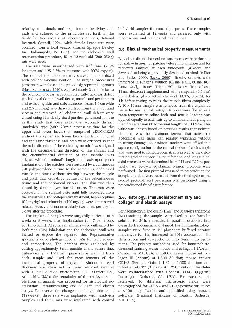
The sandwich samples had the appearance of whiteglistening sheets (Figure 1b), which was similar to thatfor control biohybrid scaffolds, although the formerappeared to be slightly more opaque on the surface.Under scanning electron microscopy, the cross-section ofthe control (Figure 2a) seemed similar with the middlelayer of gel/fibre hybrid in the sandwich scaffold(Figure 2b). The upper and lower electrospun layers ofthe sandwich scaffold (Figure 2c,d) were readily
Figure 2. Electron micrographs of the control (a) and ‘sandwich’ scaffold (b,c,d) cross-sections. The upper and lower polymer-richelectrospun layers are readily identifiable with the middle extracellular matrix (ECM) gel/polymer layer showing markedly fewerand looser fibre-rich structures as the gel component was readily apparent. dECM, dermal extracellular matrix; PEUU, elastomericpoly(ester urethane)urea
Abdominal wall reconstruction by a new biocomposite scaffold
Copyright © 2013 John Wiley & Sons, Ltd. J Tissue Eng Regen Med (2013)DOI: 10.1002/term
identifiable with the middle ECM gel/polymer layer show-ing markedly fewer and looser fibre-rich structures as thegel component was readily apparent.
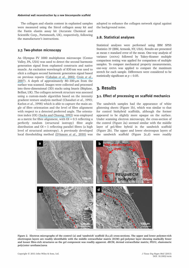
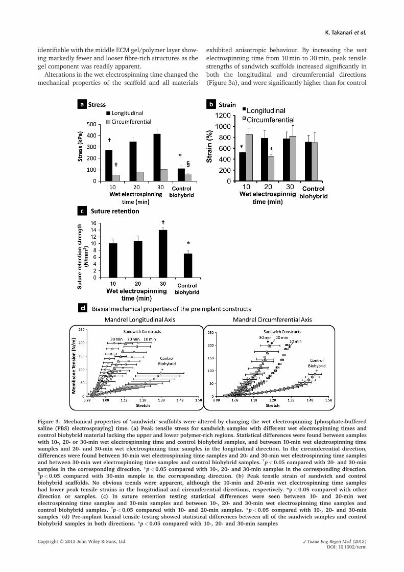
Alterations in the wet electrospinning time changed themechanical properties of the scaffold and all materials
exhibited anisotropic behaviour. By increasing the wetelectrospinning time from 10min to 30min, peak tensilestrengths of sandwich scaffolds increased significantly inboth the longitudinal and circumferential directions(Figure 3a), and were significantly higher than for control
Figure 3. Mechanical properties of ‘sandwich’ scaffolds were altered by changing the wet electrospinning [phosphate-bufferedsaline (PBS) electrospraying] time. (a) Peak tensile stress for sandwich samples with different wet electrospinning times andcontrol biohybrid material lacking the upper and lower polymer-rich regions. Statistical differences were found between sampleswith 10-, 20- or 30-min wet electrospinning time and control biohybrid samples, and between 10-min wet electrospinning timesamples and 20- and 30-min wet electrospinning time samples in the longitudinal direction. In the circumferential direction,differences were found between 10-min wet electrospinning time samples and 20- and 30-min wet electrospinning time samplesand between 30-min wet electrospinning time samples and control biohybrid samples. †p<0.05 compared with 20- and 30-minsamples in the corresponding direction. *p<0.05 compared with 10-, 20- and 30-min samples in the corresponding direction.§p<0.05 compared with 30-min sample in the corresponding direction. (b) Peak tensile strain of sandwich and controlbiohybrid scaffolds. No obvious trends were apparent, although the 10-min and 20-min wet electrospinning time sampleshad lower peak tensile strains in the longitudinal and circumferential directions, respectively. *p<0.05 compared with otherdirection or samples. (c) In suture retention testing statistical differences were seen between 10- and 20-min wetelectrospinning time samples and 30-min samples and between 10-, 20- and 30-min wet electrospinning time samples andcontrol biohybrid samples. †p<0.05 compared with 10- and 20-min samples. *p<0.05 compared with 10-, 20- and 30-minsamples. (d) Pre-implant biaxial tensile testing showed statistical differences between all of the sandwich samples and controlbiohybrid samples in both directions. *p<0.05 compared with 10-, 20- and 30-min samples
K. Takanari et al.
Copyright © 2013 John Wiley & Sons, Ltd. J Tissue Eng Regen Med (2013)DOI: 10.1002/term
biohybrid scaffolds in the corresponding directions. Peakstrains of all samples were greater than 400% in bothdirections (Figure 3b). Suture retention strength was alsoincreased with the regionally distinct scaffold processing(Figure 3c). Increasing the wet electrospinning time hadminimal effect on overall scaffold thickness (on the orderof tens of microns), which did not contribute substantiallyto the overall thickness of the scaffolds (2–2.5mm).Under equibiaxial stretching the anisotropic behaviourobserved in the unidirectional testing was much lesspronounced for all scaffolds (Figure 3d).
3.2. Macroscopic observations afterimplantation period
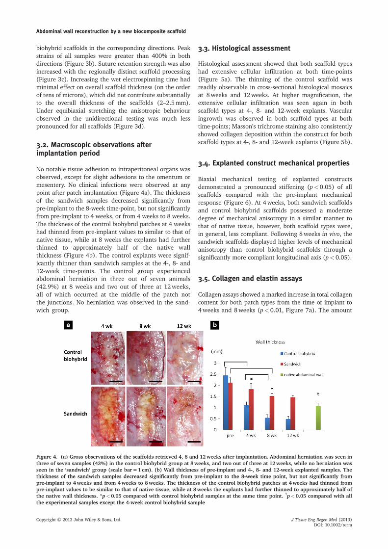
No notable tissue adhesion to intraperitoneal organs wasobserved, except for slight adhesions to the omentum ormesentery. No clinical infections were observed at anypoint after patch implantation (Figure 4a). The thicknessof the sandwich samples decreased significantly frompre-implant to the 8-week time-point, but not significantlyfrom pre-implant to 4 weeks, or from 4 weeks to 8 weeks.The thickness of the control biohybrid patches at 4 weekshad thinned from pre-implant values to similar to that ofnative tissue, while at 8 weeks the explants had furtherthinned to approximately half of the native wallthickness (Figure 4b). The control explants were signif-icantly thinner than sandwich samples at the 4-, 8- and12-week time-points. The control group experiencedabdominal herniation in three out of seven animals(42.9%) at 8 weeks and two out of three at 12weeks,all of which occurred at the middle of the patch notthe junctions. No herniation was observed in the sand-wich group.
3.3. Histological assessment
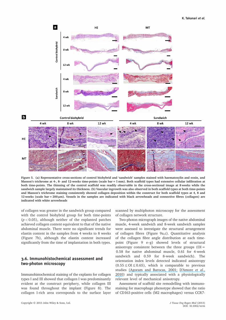
Histological assessment showed that both scaffold typeshad extensive cellular infiltration at both time-points(Figure 5a). The thinning of the control scaffold wasreadily observable in cross-sectional histological mosaicsat 8weeks and 12weeks. At higher magnification, theextensive cellular infiltration was seen again in bothscaffold types at 4-, 8- and 12-week explants. Vascularingrowth was observed in both scaffold types at bothtime-points; Masson’s trichrome staining also consistentlyshowed collagen deposition within the construct for bothscaffold types at 4-, 8- and 12-week explants (Figure 5b).
3.4. Explanted construct mechanical properties
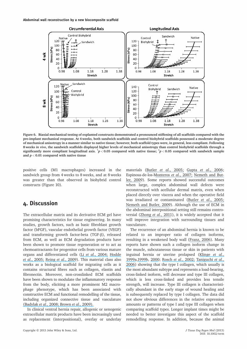
Biaxial mechanical testing of explanted constructsdemonstrated a pronounced stiffening (p< 0.05) of allscaffolds compared with the pre-implant mechanicalresponse (Figure 6). At 4weeks, both sandwich scaffoldsand control biohybrid scaffolds possessed a moderatedegree of mechanical anisotropy in a similar manner tothat of native tissue, however, both scaffold types were,in general, less compliant. Following 8weeks in vivo, thesandwich scaffolds displayed higher levels of mechanicalanisotropy than control biohybrid scaffolds through asignificantly more compliant longitudinal axis (p< 0.05).
3.5. Collagen and elastin assays
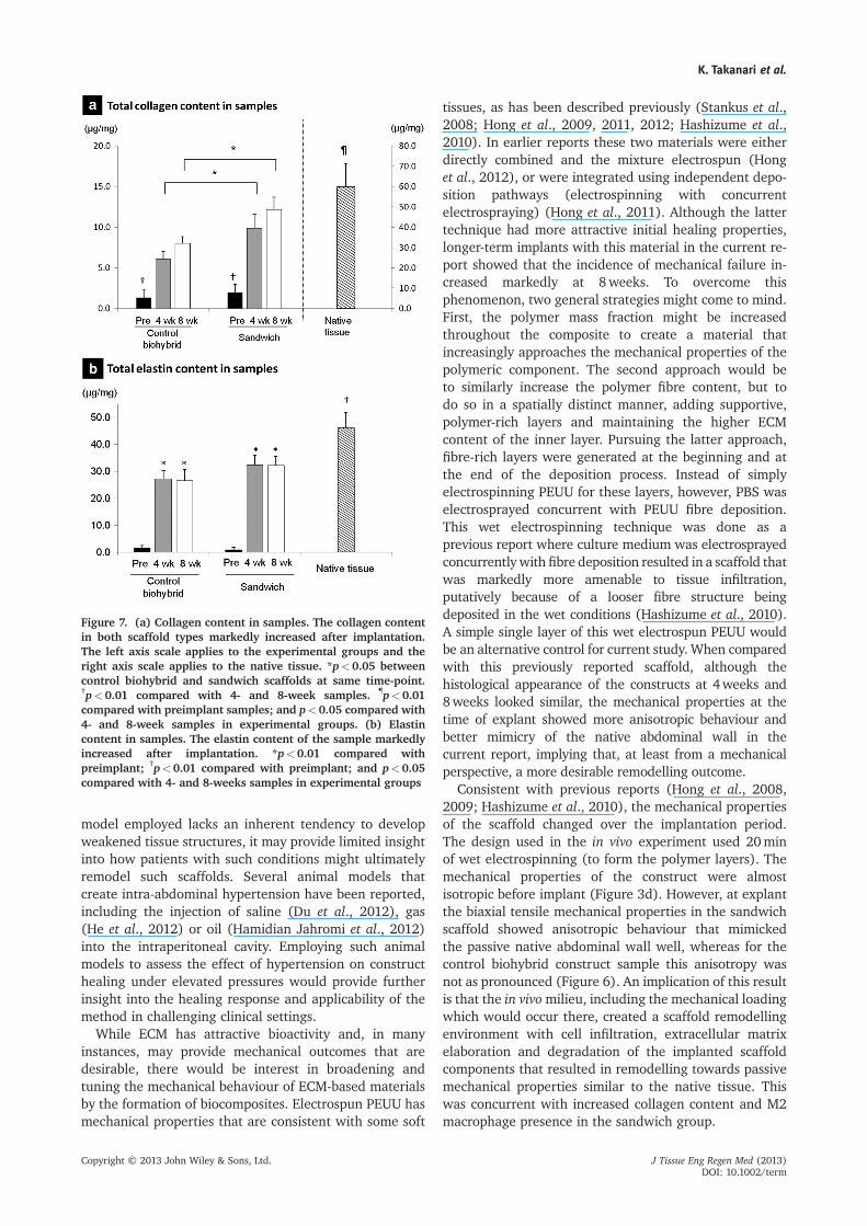
Collagen assays showed a marked increase in total collagencontent for both patch types from the time of implant to4weeks and 8weeks (p< 0.01, Figure 7a). The amount
Figure 4. (a) Gross observations of the scaffolds retrieved 4, 8 and 12weeks after implantation. Abdominal herniation was seen inthree of seven samples (43%) in the control biohybrid group at 8weeks, and two out of three at 12weeks, while no herniation wasseen in the ‘sandwich’ group (scale bar=1 cm). (b) Wall thickness of pre-implant and 4-, 8- and 12-week explanted samples. Thethickness of the sandwich samples decreased significantly from pre-implant to the 8-week time point, but not significantly frompre-implant to 4weeks and from 4weeks to 8weeks. The thickness of the control biohybrid patches at 4weeks had thinned frompre-implant values to be similar to that of native tissue, while at 8weeks the explants had further thinned to approximately half ofthe native wall thickness. *p<0.05 compared with control biohybrid samples at the same time point. †p<0.05 compared with allthe experimental samples except the 4-week control biohybrid sample
Abdominal wall reconstruction by a new biocomposite scaffold
Copyright © 2013 John Wiley & Sons, Ltd. J Tissue Eng Regen Med (2013)DOI: 10.1002/term
of collagen was greater in the sandwich group comparedwith the control biohybrid group for both time-points(p< 0.05), although neither of the explanted patchesachieved collagen content equivalent to that of the nativeabdominal muscle. There were no significant trends forelastin content in the samples from 4 weeks to 8 weeks(Figure 7b), although the elastin content increasedsignificantly from the time of implantation in both types.
3.6. Immunohistochemical assessment andtwo-photon microscopy
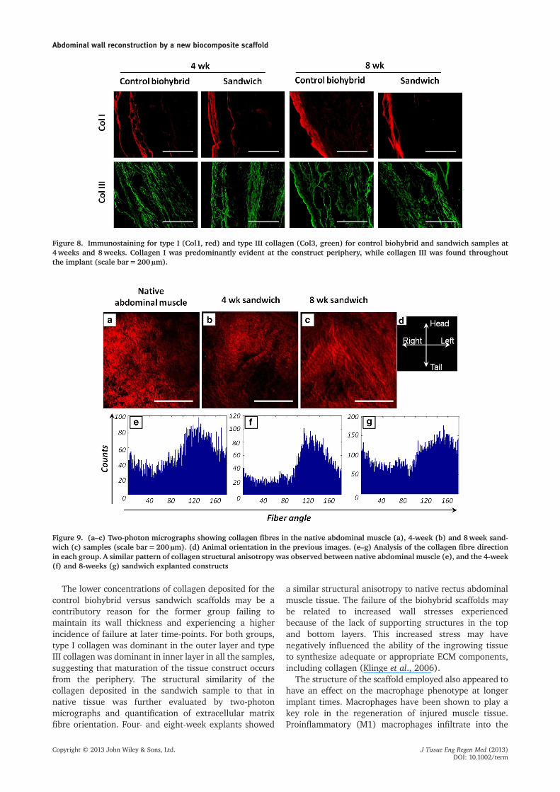
Immunohistochemical staining of the explants for collagentypes I and III showed that collagen I was predominantlyevident at the construct periphery, while collagen IIIwas found throughout the implant (Figure 8). Thecollagen I-rich area corresponds to the surface layer
scanned by multiphoton microscopy for the assessmentof collagen network structure.
Two-photon micrograph images of the native abdominalmuscle, 4-week sandwich and 8-week sandwich sampleswere assessed to investigate the structural arrangementof collagen fibres (Figure 9a,c). Quantitative analysisof the collagen fibre angle distribution at each time-point (Figure 9 e–g) showed levels of structuralanisotropy consistent between the three groups (OI=0.58 for native abdominal muscle, 0.61 for 4-weeksandwich and 0.59 for 8-week sandwich). Theorientation index levels detected indicated anisotropy(0.55≤OI≤ 0.65), which is comparable to previousstudies (Agoram and Barocas, 2001; D’Amore et al.,2010) and typically associated with a physiologicallyrelevant level of mechanical anisotropy.
Assessment of scaffold site remodelling with immuno-staining for macrophage phenotype showed that the ratioof CD163-positive cells (M2 macrophages) versus CCR7-
Figure 5. (a) Representative cross-sections of control biohybrid and ‘sandwich’ samples stained with haematoxylin and eosin, andMasson’s trichrome at 4-, 8- and 12-weeks time-points (scale bar=1mm). Both scaffold types had extensive cellular infiltration atboth time-points. The thinning of the control scaffold was readily observable in the cross-sectional image at 8weeks while thesandwich sample largely maintained its thickness. (b) Vascular ingrowth was also observed in both scaffold types at both time-pointsand Masson’s trichrome staining consistently showed collagen deposition within the construct for both scaffold types at 4, 8 and12weeks (scale bar=200μm). Vessels in the samples are indicated with black arrowheads and connective fibres (collagen) areindicated with white arrowheads
K. Takanari et al.
Copyright © 2013 John Wiley & Sons, Ltd. J Tissue Eng Regen Med (2013)DOI: 10.1002/term
positive cells (M1 macrophages) increased in thesandwich group from 4weeks to 8weeks, and at 8weekswas greater than that observed in biohybrid controlconstructs (Figure 10).
4. Discussion
The extracellular matrix and its derivative ECM gel havepromising characteristics for tissue engineering. In manystudies, growth factors, such as basic fibroblast growthfactor (bFGF), vascular endothelial growth factor (VEGF)and transforming growth factor-beta (TGF-β), releasedfrom ECM, as well as ECM degradation products havebeen shown to promote tissue regeneration or to act aschemoattractants for progenitor cells from various matureorgans and differentiated cells (Li et al., 2004; Hoddeet al., 2005; Reing et al., 2009). This material class alsoworks as a biological scaffold for migrating cells as itcontains structural fibres such as collagen, elastin andfibronectin. Moreover, non-crosslinked ECM scaffoldshave been shown to modulate the inflammatory responsefrom the body, eliciting a more prominent M2 macro-phage phenotype, which has been associated withconstructive ECM and functional remodelling of the tissue,including organized connective tissue and vasculature(Badylak et al., 2008; Brown et al., 2009).
In clinical ventral hernia repair, allogenic or xenogenicextracellular matrix products have been increasingly usedas replacement (interpositional), overlay or underlay
materials (Butler et al., 2005; Gupta et al., 2006;Espinosa-de-los-Monteros et al., 2007; Nemeth and But-ler, 2009). Some reports showed successful outcomeswhen large, complex abdominal wall defects werereconstructed with acellular dermal matrix, even whenplaced directly over viscera and when the operative fieldwas irradiated or contaminated (Butler et al., 2005;Nemeth and Butler, 2009). Although the use of ECM inthe abdominal interpositional setting still remains contro-versial (Zhong et al., 2011), it is widely accepted that itwill improve integration with surrounding tissues andvasculature.
The recurrence of an abdominal hernia is known to berelated to an improper ratio of collagen isoforms,resulting in a weakened body wall (Franz, 2006). Manyreports have shown such a collagen isoform change inthe muscle, subcutaneous tissue or skin in patients withinguinal hernia or uterine prolapsed (Klinge et al.,1999a,1999b, 2000; Rosch et al., 2002; Taniguchi et al.,2006) showing that the type I collagen, which usually isthe most abundant subtype and represents a load-bearing,cross-linked isoform, will decrease and type III collagen,which is less cross-linked and provides less tensilestrength, will increase. Type III collagen is characteristi-cally abundant in the early stage of wound healing andis subsequently replaced by type I collagen. The data didnot show obvious differences in the relative expressionamounts or patterns of type I and type III collagen whencomparing scaffold types. Longer implant times might beneeded to better investigate this aspect of the scaffoldremodelling response. In addition, because the animal
Figure 6. Biaxial mechanical testing of explanted constructs demonstrated a pronounced stiffening of all scaffolds compared with thepre-implant mechanical response. At 4weeks, both sandwich scaffolds and control biohybrid scaffolds possessed a moderate degreeof mechanical anisotropy in a manner similar to native tissue; however, both scaffold types were, in general, less compliant. Following8weeks in vivo, the sandwich scaffolds displayed higher levels of mechanical anisotropy than control biohybrid scaffolds through asignificantly more compliant longitudinal axis. †p<0.05 compared with native tissue; ‡p<0.05 compared with sandwich sampleand p<0.01 compared with native tissue
Abdominal wall reconstruction by a new biocomposite scaffold
Copyright © 2013 John Wiley & Sons, Ltd. J Tissue Eng Regen Med (2013)DOI: 10.1002/term
model employed lacks an inherent tendency to developweakened tissue structures, it may provide limited insightinto how patients with such conditions might ultimatelyremodel such scaffolds. Several animal models thatcreate intra-abdominal hypertension have been reported,including the injection of saline (Du et al., 2012), gas(He et al., 2012) or oil (Hamidian Jahromi et al., 2012)into the intraperitoneal cavity. Employing such animalmodels to assess the effect of hypertension on constructhealing under elevated pressures would provide furtherinsight into the healing response and applicability of themethod in challenging clinical settings.
While ECM has attractive bioactivity and, in manyinstances, may provide mechanical outcomes that aredesirable, there would be interest in broadening andtuning the mechanical behaviour of ECM-based materialsby the formation of biocomposites. Electrospun PEUU hasmechanical properties that are consistent with some soft
tissues, as has been described previously (Stankus et al.,2008; Hong et al., 2009, 2011, 2012; Hashizume et al.,2010). In earlier reports these two materials were eitherdirectly combined and the mixture electrospun (Honget al., 2012), or were integrated using independent depo-sition pathways (electrospinning with concurrentelectrospraying) (Hong et al., 2011). Although the lattertechnique had more attractive initial healing properties,longer-term implants with this material in the current re-port showed that the incidence of mechanical failure in-creased markedly at 8weeks. To overcome thisphenomenon, two general strategies might come to mind.First, the polymer mass fraction might be increasedthroughout the composite to create a material thatincreasingly approaches the mechanical properties of thepolymeric component. The second approach would beto similarly increase the polymer fibre content, but todo so in a spatially distinct manner, adding supportive,polymer-rich layers and maintaining the higher ECMcontent of the inner layer. Pursuing the latter approach,fibre-rich layers were generated at the beginning and atthe end of the deposition process. Instead of simplyelectrospinning PEUU for these layers, however, PBS waselectrosprayed concurrent with PEUU fibre deposition.This wet electrospinning technique was done as aprevious report where culture medium was electrosprayedconcurrently with fibre deposition resulted in a scaffold thatwas markedly more amenable to tissue infiltration,putatively because of a looser fibre structure beingdeposited in the wet conditions (Hashizume et al., 2010).A simple single layer of this wet electrospun PEUU wouldbe an alternative control for current study. When comparedwith this previously reported scaffold, although thehistological appearance of the constructs at 4weeks and8weeks looked similar, the mechanical properties at thetime of explant showed more anisotropic behaviour andbetter mimicry of the native abdominal wall in thecurrent report, implying that, at least from a mechanicalperspective, a more desirable remodelling outcome.
Consistent with previous reports (Hong et al., 2008,2009; Hashizume et al., 2010), the mechanical propertiesof the scaffold changed over the implantation period.The design used in the in vivo experiment used 20minof wet electrospinning (to form the polymer layers). Themechanical properties of the construct were almostisotropic before implant (Figure 3d). However, at explantthe biaxial tensile mechanical properties in the sandwichscaffold showed anisotropic behaviour that mimickedthe passive native abdominal wall well, whereas for thecontrol biohybrid construct sample this anisotropy wasnot as pronounced (Figure 6). An implication of this resultis that the in vivomilieu, including the mechanical loadingwhich would occur there, created a scaffold remodellingenvironment with cell infiltration, extracellular matrixelaboration and degradation of the implanted scaffoldcomponents that resulted in remodelling towards passivemechanical properties similar to the native tissue. Thiswas concurrent with increased collagen content and M2macrophage presence in the sandwich group.
Figure 7. (a) Collagen content in samples. The collagen contentin both scaffold types markedly increased after implantation.The left axis scale applies to the experimental groups and theright axis scale applies to the native tissue. *p<0.05 betweencontrol biohybrid and sandwich scaffolds at same time-point.†p<0.01 compared with 4- and 8-week samples. ¶p<0.01compared with preimplant samples; and p<0.05 compared with4- and 8-week samples in experimental groups. (b) Elastincontent in samples. The elastin content of the sample markedlyincreased after implantation. *p<0.01 compared withpreimplant; †p<0.01 compared with preimplant; and p<0.05compared with 4- and 8-weeks samples in experimental groups
K. Takanari et al.
Copyright © 2013 John Wiley & Sons, Ltd. J Tissue Eng Regen Med (2013)DOI: 10.1002/term
The lower concentrations of collagen deposited for thecontrol biohybrid versus sandwich scaffolds may be acontributory reason for the former group failing tomaintain its wall thickness and experiencing a higherincidence of failure at later time-points. For both groups,type I collagen was dominant in the outer layer and typeIII collagen was dominant in inner layer in all the samples,suggesting that maturation of the tissue construct occursfrom the periphery. The structural similarity of thecollagen deposited in the sandwich sample to that innative tissue was further evaluated by two-photonmicrographs and quantification of extracellular matrixfibre orientation. Four- and eight-week explants showed
a similar structural anisotropy to native rectus abdominalmuscle tissue. The failure of the biohybrid scaffolds maybe related to increased wall stresses experiencedbecause of the lack of supporting structures in the topand bottom layers. This increased stress may havenegatively influenced the ability of the ingrowing tissueto synthesize adequate or appropriate ECM components,including collagen (Klinge et al., 2006).
The structure of the scaffold employed also appeared tohave an effect on the macrophage phenotype at longerimplant times. Macrophages have been shown to play akey role in the regeneration of injured muscle tissue.Proinflammatory (M1) macrophages infiltrate into the
Figure 8. Immunostaining for type I (Col1, red) and type III collagen (Col3, green) for control biohybrid and sandwich samples at4weeks and 8weeks. Collagen I was predominantly evident at the construct periphery, while collagen III was found throughoutthe implant (scale bar=200μm).
Figure 9. (a–c) Two-photon micrographs showing collagen fibres in the native abdominal muscle (a), 4-week (b) and 8week sand-wich (c) samples (scale bar=200μm). (d) Animal orientation in the previous images. (e–g) Analysis of the collagen fibre directionin each group. A similar pattern of collagen structural anisotropy was observed between native abdominal muscle (e), and the 4-week(f) and 8-weeks (g) sandwich explanted constructs
Abdominal wall reconstruction by a new biocomposite scaffold
Copyright © 2013 John Wiley & Sons, Ltd. J Tissue Eng Regen Med (2013)DOI: 10.1002/term
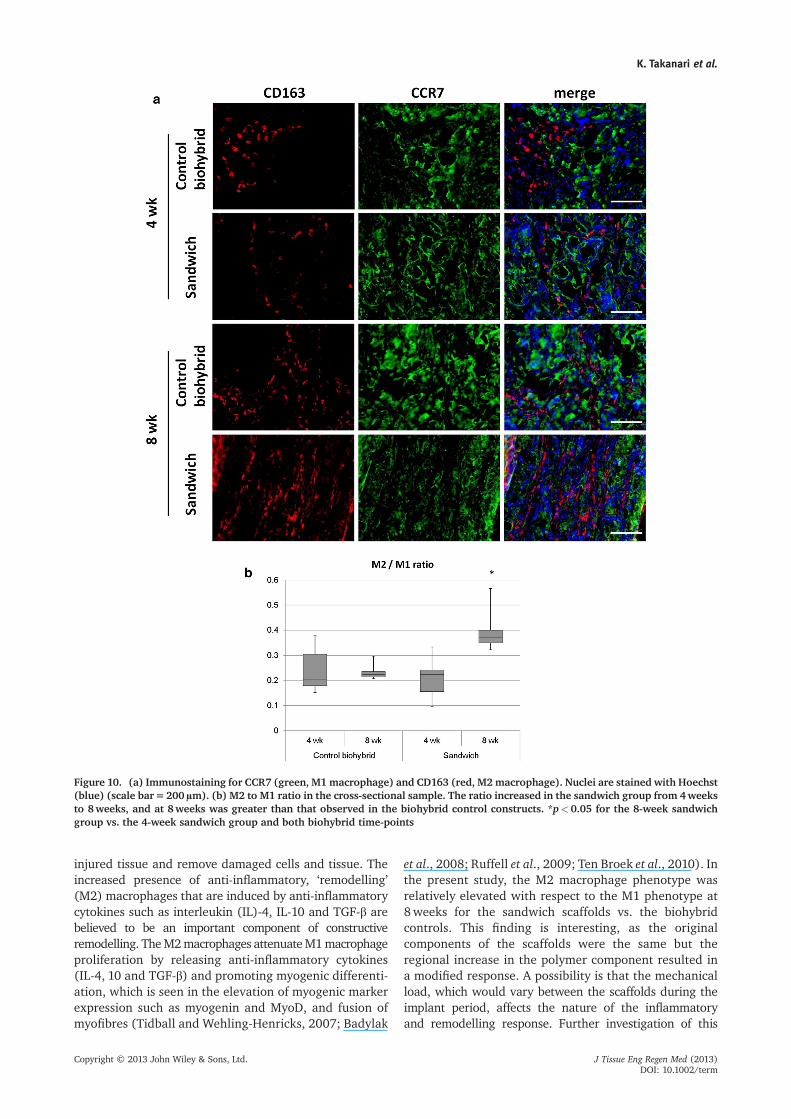
injured tissue and remove damaged cells and tissue. Theincreased presence of anti-inflammatory, ‘remodelling’(M2) macrophages that are induced by anti-inflammatorycytokines such as interleukin (IL)-4, IL-10 and TGF-β arebelieved to be an important component of constructiveremodelling. TheM2macrophages attenuateM1macrophageproliferation by releasing anti-inflammatory cytokines(IL-4, 10 and TGF-β) and promoting myogenic differenti-ation, which is seen in the elevation of myogenic markerexpression such as myogenin and MyoD, and fusion ofmyofibres (Tidball and Wehling-Henricks, 2007; Badylak
et al., 2008; Ruffell et al., 2009; Ten Broek et al., 2010). Inthe present study, the M2 macrophage phenotype wasrelatively elevated with respect to the M1 phenotype at8weeks for the sandwich scaffolds vs. the biohybridcontrols. This finding is interesting, as the originalcomponents of the scaffolds were the same but theregional increase in the polymer component resulted ina modified response. A possibility is that the mechanicalload, which would vary between the scaffolds during theimplant period, affects the nature of the inflammatoryand remodelling response. Further investigation of this
Figure 10. (a) Immunostaining for CCR7 (green, M1 macrophage) and CD163 (red, M2 macrophage). Nuclei are stained with Hoechst(blue) (scale bar=200μm). (b) M2 to M1 ratio in the cross-sectional sample. The ratio increased in the sandwich group from 4weeksto 8weeks, and at 8weeks was greater than that observed in the biohybrid control constructs. *p<0.05 for the 8-week sandwichgroup vs. the 4-week sandwich group and both biohybrid time-points
K. Takanari et al.
Copyright © 2013 John Wiley & Sons, Ltd. J Tissue Eng Regen Med (2013)DOI: 10.1002/term
phenomenon might include more extensive analysis of M2subgroups and the evaluation of functional markers tobetter relate the temporal course of the macrophageinfiltrate with remodelling outcomes (Brown et al., 2012).
In the animalmodel employed in the study, it is consideredthat the tissue ingrowth may occur from all sides except theintraperitoneumwhen there are no adhesions of intraper-itoneal organs and from all sides when there are someadhesions of intraperitoneal organs such as omentum andmesentery. However, in the abdominal wall, the peripheralmuscle tissue is likely of primary importance in theremodelling response, both because of the design of the scaf-fold and because this is the tissue that would be the putativesource of musculogenic healing for small abdominal walltrauma. The sandwich scaffold would have easier accessfor cell migration from the sides vs. the top and bottomlayers, as can be appreciated in examining the cross-sectionimages in Figure 2. The middle layer has a lower density offibres and is open only on the periphery of the scaffold.
Several limitations and opportunities following fromthe current report should be mentioned. No substantialfunctional skeletal muscle formation was observed, exceptfor a limited amount of regional muscle growth from thesurroundingmuscle stump during the period of observation.It would be worthwhile to examine longer periods post-implantation to evaluate skeletal muscle regeneration,and to examine the effect of a muscle onlay or underlayon this remodelling response. Alternative ECM sources,such as skeletal muscle (Wolf et al., 2012) would possiblyenhance the development toward functional muscle tissue.The rodent model is limited to relatively short-term investi-gations by its nature, but, in the clinical setting, the effect onthe remodelling process would need to be determined overyears. Larger animal models with long-term observationperiods would be important to evaluate the long-termphysiological response. The model used in the currentstudy was a full-thickness abdominal wall reconstruction,but the approach to discriminate between the mechanicallysupportive region and bioactive region within one scaffoldmight ultimately be applicable to other target diseases andorgans, such as pelvic floor prolapse, breast reconstructionand urinary incontinence.
It is still unclear as to how long the load-bearingcomponents of the scaffold need to remain in place beforethis load can be adequately taken on by host tissue. Theideal time for material degradation would depend uponthe body forces experienced and the type and amount ofsurrounding tissue generated in the healing response.
Too rapid degradation appears to lead to thinning andfailure, therefore non-degrading or slowly degradingmaterials may be better, although achieving host tissuecapable of full load-bearing may be inhibited by syntheticcomponents that effectively act to shield the tissue fromstress. To examine this effect one might employ alternativechemistries for biodegradable polyurethanes that providesimilar mechanical properties, but exhibit more rapid, orslower degradation (Guan et al., 2005; Hong et al., 2010).Further study in this area is clearly needed.
5. Conclusions
Co-electrospinning–electrospraying was employed to createa ‘sandwich’ scaffold that combined porcine dermal ECM gelwith biodegradable elastic PEUU, where a dermal ECMgel-enriched layer was sandwiched by two PEUU-richlayers. The PEUU-rich layers provided improvedmechanicalstrength while the dECM-rich layer provided a potentialsource for bioactive components. In vivo evaluation comparingthe sandwich scaffold with a control biohybrid scaffoldwithout the upper and lower regions showed a markedincrease in collagen content while preserving good cellularinfiltration in the former, and thinning with multiplemechanical failures in the latter after 8weeks. At explantthe biaxial tensile mechanical behaviour of the sandwichscaffoldmimicked the structural andmechanical anisotropyof the native abdominal wall. By employing a processingapproach that creates a sheet-form scaffold with region-ally distinct zones, it was possible to improve biologicaloutcomes in body wall repair and provide the means forfurther tuning of scaffold mechanical parameters whentargeting other applications.
Conflict of interest
The authors have declared that there is no conflict of interest.
Acknowledgements
This work was supported in part by the Armed Forces Institute forRegenerative Medicine (AFIRM, #W81XWH-08-2-0032) and C.R.Bard, Inc. We also thank Prof. Michael S. Sacks for use of the biaxialmechanical testing device, Deanna Rhoads and Hongbin Jiang forhistological sectioning and Greg Gibson for multiphoton imaging.
References
Agoram B, Barocas VH. 2001; Coupledmacroscopic and microscopic scalemodeling of fibrillar tissues and tissueequivalents. J Biomech Eng 123:362–369.
Badylak SF, Valentin JE, Ravindra AKet al. 2008; Macrophage phenotypeas a determinant of biologic scaffold
remodeling. Tissue Eng Part A 14:1835–1842.
Badylak SF, Weiss DJ, Caplan A et al. 2012;Engineered whole organs and complextissues. Lancet 379: 943–952.
Billiar KL, Sacks MS. 2000; Biaxial mecha-nical properties of the native andglutaraldehyde-treated aortic valve cusp:
Part II – a structural constitutive model. JBiomech Eng 122: 327–335.
Brown BN, Valentin JE, Stewart-Akers AMet al. 2009; Macrophage phenotype andremodeling outcomes in response tobiologic scaffolds with and without acellular component. Biomaterials 30:1482–1491.
Abdominal wall reconstruction by a new biocomposite scaffold
Copyright © 2013 John Wiley & Sons, Ltd. J Tissue Eng Regen Med (2013)DOI: 10.1002/term
Brown BN, Londono R, Tottey S et al. 2012;Macrophage phenotype as a predictor ofconstructive remodeling following the im-plantation of biologically derived surgicalmesh materials. Acta Biomater 8: 978–987.
Butler CE, Langstein HN, Kronowitz SJ.2005; Pelvic, abdominal, and chest wallreconstruction with AlloDerm in patientsat increased risk for mesh-relatedcomplications. Plast Reconstr Surg 116:1263–1275.
Cahalan MD, Parker I, Wei SH et al. 2002;Two-photon tissue imaging: seeing theimmune system in a fresh light. Nat RevImmunol 2: 872–880.
Cassar K, Munro A. 2002; Surgical treat-ment of incisional hernia. Br J Surg 89:534–545.
Chauduri BB, Kundo PK, Nirupam S. 1993;Detection and gradation of orientedtexture. Pattern Recogn Lett 14: 147–153.
Croix CS, Zipfel WR, Watkins SC. 2007;Potential solutions for confocalimaging of living animals. Biotechniques43: 14–19.
D’Amore A, Stella JA, Wagner WR et al. 2010;Characterization of the complete fiber net-work topology of planar fibrous tissues andscaffolds. Biomaterials 31: 5345–5354.
den Hartog D, Dur AH, Tuinebreijer WE et al.2008; Open surgical procedures forincisional hernias. Cochrane Database SystRev 16: CD006438.
Du WH, Xiang W, Liu DC et al. 2012;Usefulness of speckle tracking imagingto assess myocardial contractility inintra-abdominal hypertension: study in amini-pig model. Cell Biochem Biophys 64:123–129
Dumanian GA, Denham W. 2003; Compari-son of repair techniques for majorincisional hernias. Am J Surg 185: 61–65.
Espinosa-de-los-Monteros A, de la Torre JI,Marrero I et al. 2007; Utilization of human ca-daveric acellular dermis for abdominal herniareconstruction. Ann Plast Surg 58: 264–267.
Franz MG. 2006; The biology of hernias andthe abdominal wall. Hernia 10: 462–471.
Guan J, Sacks MS, Beckman EJ et al. 2002; Syn-thesis, characterization, and cytocompatibilityof elastomeric, biodegradable poly(ester-urethane)ureas based on poly(caprolactone) and putrescine. J BiomedMater Res 61: 493–503.
Guan J, Fujimoto KL, Sacks MS et al. 2005;Preparation and characterization ofhighly porous, biodegradable polyure-thane scaffolds for soft tissue applications.Biomaterials 26: 3961–71.
Gupta A, Zahriya K, Mullens PL et al. 2006;Ventral herniorrhaphy: experience withtwo different biosynthetic mesh materials,Surgisis and Alloderm. Hernia 10: 419–425.
Hamidian Jahromi A, Freeland K, YoussefAM. 2012; Intra-abdominal hypertensioncauses disruption of the blood–brainbarrier in mice, which is increased withadded severe head trauma. J Trauma AcuteCare Surg 73: 1175–1179
Hashizume R, Fujimoto KL, Hong Y et al.2010; Morphological and mechanicalcharacteristics of the reconstructed ratabdominal wall following use of a wet
electrospun biodegradable polyurethaneelastomer scaffold. Biomaterials 31:3253–3265.
He Q, Cai L, Zhang S et al. 2012; Oxygeninhalation improves survival time of micewith acute intra-abdominal hypertensionand protects liver cells. Transplant Proc44: 1201–1205
Hodde JP, Record RD, Liang HA et al. 2001;Vascular endothelial growth factor inporcine-derived extracellular matrix,Endothelium 8: 11–24.
Hodde JP, Ernst DM, Hiles MC. 2005; Aninvestigation of the long-term bioactivityof endogenous growth factor in OASISWound Matrix. J Wound Care 14: 23–25.
Hong Y, Fujimoto K, Hashizume R et al.2008; Generating elastic, biodegradablepolyurethane/poly(lactide-co-glycolide)fibrous sheets with controlled antibioticrelease via two-stream electrospinning.Biomacromolecules 9: 1200–1207.
Hong Y, Ye SH, Nieponice A et al. 2009; Asmall diameter, fibrous vascular conduitgenerated from a poly(ester urethane)urea and phospholipid polymer blend.Biomaterials 30: 2457–2467.
Hong Y, Guan J, Fujimoto KL et al. 2010;Tailoring the degradation kinetics of poly(ester carbonate urethane)urea thermo-plastic elastomers for tissue engineeringscaffolds. Biomaterials 31: 4249–4358.
Hong Y, Huber A, Takanari K et al. 2011;Mechanical properties and in vivobehavior of a biodegradable syntheticpolymer microfiber–extracellular matrixhydrogel biohybrid scaffold. Biomaterials32: 3387–3394.
Hong Y, Takanari K, Amoroso NJ et al. 2012;An elastomeric patch electrospun from ablended solution of dermal extracellularmatrix and biodegradable polyurethanefor rat abdominal wall repair. Tissue EngPart C Methods 18: 122–132.
Karlon WJ, Covell JW, McCulloch AD et al.1998; Automated measurement ofmyofiber disarray in transgenic mice withventricular expression of ras. Anat Rec252: 612–625.
Klinge U, Zheng H, Si Z et al. 1999a;Expression of the extracellular matrixproteins collagen I, collagen III and fibro-nectin and matrix metalloproteinase-1and -13 in the skin of patients withinguinal hernia. Eur Surg Res 31: 480–490.
Klinge U, Zheng H, Si ZY et al. 1999b;Synthesis of type I and III collagen,expression of fibronectin and matrixmetalloproteinases-1 and -13 in hernialsac of patients with inguinal hernia. Int JSurg Invest 1: 219–227.
Klinge U, Si ZY, Zheng H et al. 2000;Abnormal collagen I to III distribution inthe skin of patients with incisional hernia.Eur Surg Res 32: 43–48.
KlingeU, BinneböselM, RoschR et al. 2006;Her-nia recurrence as a problemof biology and col-lagen. J Minim Access Surg 2: 151–4.
Li F, Li W, Johnson S, Ingram D, Yoder M,Badylak S. 2004; Low-molecular-weightpeptides derived from extracellular matrixas chemoattractants for primary endothelialcells. Endothelium 11: 199–206.
Mudge M, Hughes LE. 1985; Incisionalhernia: a 10 year prospective study of inci-dence and attitudes. Br J Surg 72: 70–71.
Nemeth NL, Butler CE. 2009; Complex torsoreconstruction with human acellulardermal matrix: long-term clinical follow-up. Plast Reconstr Surg 123: 192–196.
Reing JE, Zhang L, Myers-Irvin J et al. 2009;Degradation products of extracellularmatrix affect cell migration andproliferation. Tissue Eng Part A 15: 605–614.
Reing JE, Brown BN, Daly KA et al. 2010; Theeffects of processing methods uponmechanical and biologic properties ofporcine dermal extracellular matrixscaffolds. Biomaterials 31: 8626–8633.
RoschR, KlingeU, Si Z et al. 2002; A role for thecollagen I/III and MMP-1/-13 genes in pri-mary inguinal hernia? BMC Med Genet 3: 2.
Ruffell D, Mourkioti F, Gambardella A et al.2009; A CREB-C/EBPbeta cascade inducesM2 macrophage-specific gene expressionand promotesmuscle injury repair. Proc NatlAcad Sci U S A 106: 17475–17480.
Sacks MS. 2000; Biaxial mechanicalevaluation of planar biological materials.J Elasticity 61: 199–246.
Sacks MS, Chuong CJ. 1992; Characteriza-tion of collagen fiber architecture in thecanine diaphragmatic central tendon.J Biomech Eng 114: 183–190.
Sanchez VM, Abi-Haidar YE, Itani KM. 2011;Mesh infection in ventral incisional herniarepair: incidence, contributing factors,and treatment. Surg Infect (Larchmt) 12:205–210.
Soletti L, Nieponice A, Hong Y et al. 2011; Invivo performance of a phospholipid-coatedbioerodable elastomeric graft for small-diameter vascular applications. J BiomedMater Res A 96: 436–448.
Stankus JJ, Freytes DO, Badylak SF et al.2008; Hybrid nanofibrous scaffolds fromelectrospinning of a synthetic biodegradableelastomer and urinary bladder matrix. JBiomater Sci Polym Ed 19: 635–652.
Taniguchi S, Ueda K, Inoue T et al. 2006;Impact of collagen subtype proportions inperitoneal tissues on inguinal herniaformation in adults and infants. PediatrSurg Int 22: 600–604.
Ten Broek RW, Grefte S, Von den Hoff JW.2010; Regulatory factors and cellpopulations involved in skeletal muscleregeneration. J Cell Physiol 224: 7–16.
Tidball JG, Wehling-Henricks M. 2007;Macrophages promote muscle membranerepair and muscle fibre growth and regen-eration during modified muscle loading inmice in vivo. J Physiol 578: 327–336.
van der Linden FT, van Vroonhoven TJ.1988; Long-term results after surgicalcorrection of incisional hernia. Neth J Surg40: 127–129.
Wolf MT, Daly KA, Reing JE et al. 2012;Biologic scaffold composed of skeletalmuscle extracellular matrix. Biomaterials33: 2916–2925.
Zhong T, Janis JE, Ahmad J et al. 2011;Outcomes after abdominal wall recon-struction using acellular dermal matrix:a systematic review. J Plast ReconstrAesthet Surg 64: 1562–1571.
K. Takanari et al.
Copyright © 2013 John Wiley & Sons, Ltd. J Tissue Eng Regen Med (2013)DOI: 10.1002/term