
A Versatile Viral System for Expression and Depletion of Proteins in Mammalian Cells
18
A Versatile Viral System for Expression and Depletion of Proteins in Mammalian Cells Eric Campeau 1 *, Victoria E. Ruhl 1 , Francis Rodier 2,3 , Corey L. Smith 1 , Brittany L. Rahmberg 1 , Jill O. Fuss 2 , Judith Campisi 2,3 , Paul Yaswen 2 , Priscilla K. Cooper 2 , Paul D. Kaufman 1 1 Program in Gene Function and Expression, University of Massachusetts Medical School, Worcester, Massachusetts, United States of America, 2 Life Sciences Division, Lawrence Berkeley National Laboratory, Berkeley, California, United States of America, 3 Buck Institute for Age Research, Novato, California, United States of America Abstract The ability to express or deplete proteins in living cells is crucial for the study of biological processes. Viral vectors are often useful to deliver DNA constructs to cells that are difficult to transfect by other methods. Lentiviruses have the additional advantage of being able to integrate into the genomes of non-dividing mammalian cells. However, existing viral expression systems generally require different vector backbones for expression of cDNA, small hairpin RNA (shRNA) or microRNA (miRNA) and provide limited drug selection markers. Furthermore, viral backbones are often recombinogenic in bacteria, complicating the generation and maintenance of desired clones. Here, we describe a collection of 59 vectors that comprise an integrated system for constitutive or inducible expression of cDNAs, shRNAs or miRNAs, and use a wide variety of drug selection markers. These vectors are based on the Gateway technology (Invitrogen) whereby the cDNA, shRNA or miRNA of interest is cloned into an Entry vector and then recombined into a Destination vector that carries the chosen viral backbone and drug selection marker. This recombination reaction generates the desired product with .95% efficiency and greatly reduces the frequency of unwanted recombination in bacteria. We generated Destination vectors for the production of both retroviruses and lentiviruses. Further, we characterized each vector for its viral titer production as well as its efficiency in expressing or depleting proteins of interest. We also generated multiple types of vectors for the production of fusion proteins and confirmed expression of each. We demonstrated the utility of these vectors in a variety of functional studies. First, we show that the FKBP12 Destabilization Domain system can be used to either express or deplete the protein of interest in mitotically-arrested cells. Also, we generate primary fibroblasts that can be induced to senesce in the presence or absence of DNA damage. Finally, we determined that both isoforms of the AT-Rich Interacting Domain 4B (ARID4B) protein could induce G1 arrest when overexpressed. As new technologies emerge, the vectors in this collection can be easily modified and adapted without the need for extensive recloning. Citation: Campeau E, Ruhl VE, Rodier F, Smith CL, Rahmberg BL, et al. (2009) A Versatile Viral System for Expression and Depletion of Proteins in Mammalian Cells. PLoS ONE 4(8): e6529. doi:10.1371/journal.pone.0006529 Editor: Jo ¨ rg Hermann Fritz, University of Toronto, Canada Received January 11, 2009; Accepted July 8, 2009; Published August 6, 2009 Copyright: ß 2009 Campeau et al. This is an open-access article distributed under the terms of the Creative Commons Attribution License, which permits unrestricted use, distribution, and reproduction in any medium, provided the original author and source are credited. Funding: This work was supported by NIH grants AG017242 to JC, R01 CA063503 to PKC, U54 CA112970 to PY, R01 GM55712 and NSF grant MCB-0641390 to PDK, NIH 5 F32 GM076863-03 to CLS and NRSA Fellowship 1F32CA108393 to JF. The funders had no role in study design, data collection and analysis, decision to publish, or preparation of the manuscript. Competing Interests: The authors have declared that no competing interests exist. * E-mail: [email protected] Introduction The abilities to express and deplete proteins in mammalian cells are invaluable tools for understanding diverse biological processes, both normal and pathological. Traditionally, transfection and electroporation of plasmids into cells have been used to manipulate gene expression. However, not all cell types can be efficiently transfected by these methods, and primary or non- dividing cell types are particularly difficult in this regard [1]. The advent of recombinant retroviruses and lentiviruses [1,2,3,4,5] greatly simplified this task, especially since lentiviruses can easily transduce non-dividing cells. For RNA interference (RNAi), plasmid-based systems used to express shRNAs or miRNAs allowed long term depletion of the protein of interest, compared to the transfection of small interfering RNAs (siRNAs) which results in only transient depletion. Several laboratories and companies have therefore developed viral vectors for the delivery of cDNAs and/or shRNAs or miRNAs to a wide variety of cells [6,7,8,9,10,11,12,13,14,15,16]. Even though these vectors have proven extremely useful, there are limited drug selection markers available, and different vector backbones can be required to express cDNAs or shRNAs/miRNAs. We sought to extend the versatility of viral vectors by developing third generation (Tat- independent), self-inactivating (SIN) lentiviral vectors with an expanded range of drug selection markers, promoters, and epitope tags. Furthermore, we wanted to improve their flexibility, allowing the investigator to rapidly change promoters and/or drug selection markers without extensive recloning. We report here a series of 59 vectors to express cDNAs, shRNAs and miRNAs, either constitutively or inducibly, in mammalian cells. These vectors are based on the Gateway System [17] (Invitrogen) whereby a cDNA, shRNA or miRNA is cloned into an Entry vector which is recombined in vitro with the viral Destination vector of choice. This system offers two main advantages: First, the recombination is very efficient and rapid (1–3 h), eliminating most of the stability problems that often arise during traditional cloning in a bacterial host [18,19]. Second, the extensive Destination vector library can be further modified to accommodate future technologies without PLoS ONE | www.plosone.org 1 August 2009 | Volume 4 | Issue 8 | e6529
-
Upload
independent -
Category
Documents
-
view
1 -
download
0
Transcript of A Versatile Viral System for Expression and Depletion of Proteins in Mammalian Cells
A Versatile Viral System for Expression and Depletion ofProteins in Mammalian CellsEric Campeau1*, Victoria E. Ruhl1, Francis Rodier2,3, Corey L. Smith1, Brittany L. Rahmberg1, Jill O. Fuss2,
Judith Campisi2,3, Paul Yaswen2, Priscilla K. Cooper2, Paul D. Kaufman1
1 Program in Gene Function and Expression, University of Massachusetts Medical School, Worcester, Massachusetts, United States of America, 2 Life Sciences Division,
Lawrence Berkeley National Laboratory, Berkeley, California, United States of America, 3 Buck Institute for Age Research, Novato, California, United States of America
Abstract
The ability to express or deplete proteins in living cells is crucial for the study of biological processes. Viral vectors are oftenuseful to deliver DNA constructs to cells that are difficult to transfect by other methods. Lentiviruses have the additionaladvantage of being able to integrate into the genomes of non-dividing mammalian cells. However, existing viral expressionsystems generally require different vector backbones for expression of cDNA, small hairpin RNA (shRNA) or microRNA(miRNA) and provide limited drug selection markers. Furthermore, viral backbones are often recombinogenic in bacteria,complicating the generation and maintenance of desired clones. Here, we describe a collection of 59 vectors that comprisean integrated system for constitutive or inducible expression of cDNAs, shRNAs or miRNAs, and use a wide variety of drugselection markers. These vectors are based on the Gateway technology (Invitrogen) whereby the cDNA, shRNA or miRNA ofinterest is cloned into an Entry vector and then recombined into a Destination vector that carries the chosen viral backboneand drug selection marker. This recombination reaction generates the desired product with .95% efficiency and greatlyreduces the frequency of unwanted recombination in bacteria. We generated Destination vectors for the production of bothretroviruses and lentiviruses. Further, we characterized each vector for its viral titer production as well as its efficiency inexpressing or depleting proteins of interest. We also generated multiple types of vectors for the production of fusionproteins and confirmed expression of each. We demonstrated the utility of these vectors in a variety of functional studies.First, we show that the FKBP12 Destabilization Domain system can be used to either express or deplete the protein ofinterest in mitotically-arrested cells. Also, we generate primary fibroblasts that can be induced to senesce in the presence orabsence of DNA damage. Finally, we determined that both isoforms of the AT-Rich Interacting Domain 4B (ARID4B) proteincould induce G1 arrest when overexpressed. As new technologies emerge, the vectors in this collection can be easilymodified and adapted without the need for extensive recloning.
Citation: Campeau E, Ruhl VE, Rodier F, Smith CL, Rahmberg BL, et al. (2009) A Versatile Viral System for Expression and Depletion of Proteins in MammalianCells. PLoS ONE 4(8): e6529. doi:10.1371/journal.pone.0006529
Editor: Jorg Hermann Fritz, University of Toronto, Canada
Received January 11, 2009; Accepted July 8, 2009; Published August 6, 2009
Copyright: � 2009 Campeau et al. This is an open-access article distributed under the terms of the Creative Commons Attribution License, which permitsunrestricted use, distribution, and reproduction in any medium, provided the original author and source are credited.
Funding: This work was supported by NIH grants AG017242 to JC, R01 CA063503 to PKC, U54 CA112970 to PY, R01 GM55712 and NSF grant MCB-0641390 toPDK, NIH 5 F32 GM076863-03 to CLS and NRSA Fellowship 1F32CA108393 to JF. The funders had no role in study design, data collection and analysis, decision topublish, or preparation of the manuscript.
Competing Interests: The authors have declared that no competing interests exist.
* E-mail: [email protected]
Introduction
The abilities to express and deplete proteins in mammalian cells
are invaluable tools for understanding diverse biological processes,
both normal and pathological. Traditionally, transfection and
electroporation of plasmids into cells have been used to
manipulate gene expression. However, not all cell types can be
efficiently transfected by these methods, and primary or non-
dividing cell types are particularly difficult in this regard [1]. The
advent of recombinant retroviruses and lentiviruses [1,2,3,4,5]
greatly simplified this task, especially since lentiviruses can easily
transduce non-dividing cells. For RNA interference (RNAi),
plasmid-based systems used to express shRNAs or miRNAs
allowed long term depletion of the protein of interest, compared
to the transfection of small interfering RNAs (siRNAs) which
results in only transient depletion. Several laboratories and
companies have therefore developed viral vectors for the delivery
of cDNAs and/or shRNAs or miRNAs to a wide variety of cells
[6,7,8,9,10,11,12,13,14,15,16]. Even though these vectors have
proven extremely useful, there are limited drug selection markers
available, and different vector backbones can be required to
express cDNAs or shRNAs/miRNAs. We sought to extend the
versatility of viral vectors by developing third generation (Tat-
independent), self-inactivating (SIN) lentiviral vectors with an
expanded range of drug selection markers, promoters, and epitope
tags. Furthermore, we wanted to improve their flexibility, allowing
the investigator to rapidly change promoters and/or drug selection
markers without extensive recloning. We report here a series of 59
vectors to express cDNAs, shRNAs and miRNAs, either
constitutively or inducibly, in mammalian cells. These vectors
are based on the Gateway System [17] (Invitrogen) whereby a
cDNA, shRNA or miRNA is cloned into an Entry vector which is
recombined in vitro with the viral Destination vector of choice. This
system offers two main advantages: First, the recombination is very
efficient and rapid (1–3 h), eliminating most of the stability
problems that often arise during traditional cloning in a bacterial
host [18,19]. Second, the extensive Destination vector library can
be further modified to accommodate future technologies without
PLoS ONE | www.plosone.org 1 August 2009 | Volume 4 | Issue 8 | e6529
the need for recloning cDNAs, shRNAs or miRNAs. We used our
vectors to demonstrate that a protein of interest can be
overexpressed or depleted in mitotically-arrested cells, generate
stable cell lines where the senescence secretory associated
phenotype (SASP) can be induced, and show that the chromo
domain of the AT-Rich Interacting Domain 4B (ARID4B) protein
is not required for the G1 arrest upon overexpression.
Results and Discussion
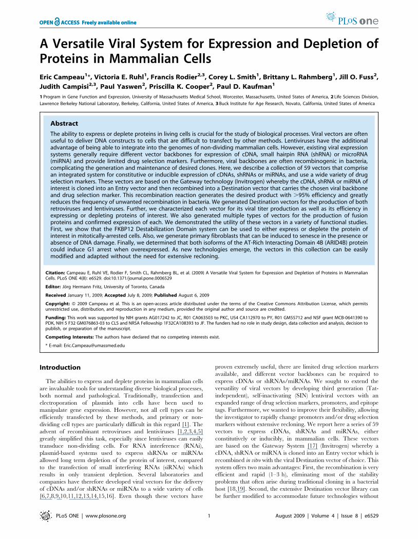
Overview of the vectorscDNAs, shRNAs or miRNAs are initially cloned into Entry
vectors (Figure 1). The insert from the Entry vector clone can then
be recombined efficiently via an attL-attR (LR) reaction [17] into
a variety of Destination vectors to provide the desired viral
backbone and drug selection marker. The recombination reaction
is transformed into Escherichia coli and there is a dual selection for
the desired vector. First, the Entry and Destination vectors carry
different drug resistance genes for selection in E.coli, kanamycin for
the Entry vector, and ampicillin for the Destination vector. The
transformation is plated onto ampicillin plates, selecting against
any unrecombined Entry vector. Second, the Destination vector
contain the ccdB killer gene [20], which is toxic to most E.coli
strains. The ccdB gene is removed from the Destination vector
during the recombination reaction and a susceptible E.coli strain is
used for the transformation, thereby selecting against the
unrecombined Destination vector. In our hands, the efficiency of
recombination into the Destination vector is .95% as determined
by restriction digestion and analysis of the resulting construct.
Because the Entry vectors do not contain any viral-derived
repetitive sequences, they can be easily manipulated without
significant levels of unwanted deletions which often occur at
repetitive sequences propagated in bacteria [18,19,21].
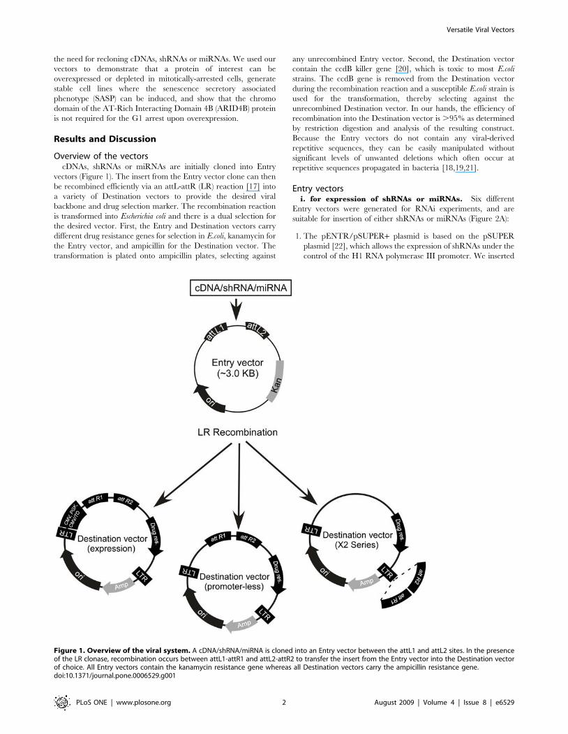
Entry vectorsi. for expression of shRNAs or miRNAs. Six different
Entry vectors were generated for RNAi experiments, and are
suitable for insertion of either shRNAs or miRNAs (Figure 2A):
1. The pENTR/pSUPER+ plasmid is based on the pSUPER
plasmid [22], which allows the expression of shRNAs under the
control of the H1 RNA polymerase III promoter. We inserted
Figure 1. Overview of the viral system. A cDNA/shRNA/miRNA is cloned into an Entry vector between the attL1 and attL2 sites. In the presenceof the LR clonase, recombination occurs between attL1-attR1 and attL2-attR2 to transfer the insert from the Entry vector into the Destination vectorof choice. All Entry vectors contain the kanamycin resistance gene whereas all Destination vectors carry the ampicillin resistance gene.doi:10.1371/journal.pone.0006529.g001
Versatile Viral Vectors
PLoS ONE | www.plosone.org 2 August 2009 | Volume 4 | Issue 8 | e6529
a 750 bp stuffer (denoted by the +, derived from the pTER+plasmid [23]) between the Bgl II and Hind III sites to facilitate
cloning of shRNAs downstream of the promoter.
2. The pENTR/pTER+ plasmid is derived from the pTER+plasmid [23]; this is similar to pSUPER except that a
Tetracycline Operator (TO) was inserted at the end of the
H1 promoter. The TO allows experimental control of the
expression of the shRNA in a ‘‘T-REx’’ cell line (Invitrogen)
that expresses the tetracycline repressor (TetR), because the
transcription-blocking TetR-TO interaction is relieved by
addition of doxycycline or tetracycline [24].
3. The pENTR/pSM2 plasmids are based on the pSM2c
plasmid [14,25] for expression of miRNAs, and include the 59
and 39 flanking sequences from the miR-30 miRNA. The
original U6 promoter was retained to allow constitutive
expression under the control of RNA polymerase III in the
pENTR/pSM2(U6) version of the vector. In the pENTR/
pSM2(CMV) and pENTR/pSM2(CMV/TO) versions, the U6
promoter was replaced by either a constitutive CMV or a
doxycycline-inducible CMV/TO promoter to allow expression
under the control of RNA polymerase II. The CMV promoter
was reported to generate better depletions in some cases [26]
and lower variability than the U6-based promoter when the
virus is integrated in the genome [27].
4. Finally, we generated the vector pENTR/pSM2(CMV-GFP),
in which a GFP cDNA was inserted between the promoter and
the 59miR30 sequence, to further enhance the depletion of the
target protein [8] and facilitate tracking and sorting of cells
expressing the miRNA of interest.
ii. for expression of cDNAs. A large variety of Entry vectors
were generated to clone cDNAs encoding proteins of interest (POI)
(Figure 2B). The original pENTR1A and pENTR4 plasmids
contain the ccdB killer gene. To clone a cDNA into these vectors,
two restriction endonucleases which cut on opposite sides of the
killer gene have to be used in order to replace the ccdB sequence
with the cDNA of interest and thereby select against the parental
plasmid. However, we removed the ccdB gene so that all of the
multiple cloning sites could be exploited, generating constructs
termed ‘‘pENTR1A no ccdB’’ and ‘‘pENTR4 no ccdB’’. We also
generated a variety of Entry vectors that encode epitope tags at
their N-terminus (GFP, V5, FLAG) or C-terminus (GFP, V5-His)
in different reading frames to facilitate detection of the proteins of
interest. For tandem affinity purification (TAP) experiments, we
created Entry vectors encoding tandem N-terminal epitope tags in
three reading frames, based on the pNTAP vector from Stratagene
(NTAP,[28]). Additionally, we created an Entry vector encoding
an N-terminal Glutathione-S-Transferase (GST) followed by a
PreScission protease cleavage site (GE Healthcare Life Sciences),
which can also be used for purification of the protein of interest via
glutathione affinity supports. Other Entry vectors include either an
N-terminal or C-terminal 12 kDa FK506 Binding Protein
(FKBP12) Destabilization Domain (DD) based on the Wandless
Figure 2. Maps of the Entry vectors for either RNAi-mediated protein depletion or protein overexpression. A) shRNAs or miRNAs canbe expressed with either constitutive (H1, U6, CMV) or inducible (H1/TO, CMV/TO) promoters. B) cDNAs encoding the protein of interest (POI) can becloned into Entry vectors with no tags or different tags for either detection or purification. (Abbreviations: CBP, calcium-binding peptide; SBP, S-binding peptide; GST, glutathione-S-transferase; FKBP12DD, FKBP destabilizing domain. LEFT: Promoter-less Entry vectors. RIGHT: Entry vectors for EF-1a promoter-driven protein expression. In this case, Entry vectors containing a promoter should be recombined with a promoter-less Destinationvector (see Figure 4).doi:10.1371/journal.pone.0006529.g002
Versatile Viral Vectors
PLoS ONE | www.plosone.org 3 August 2009 | Volume 4 | Issue 8 | e6529
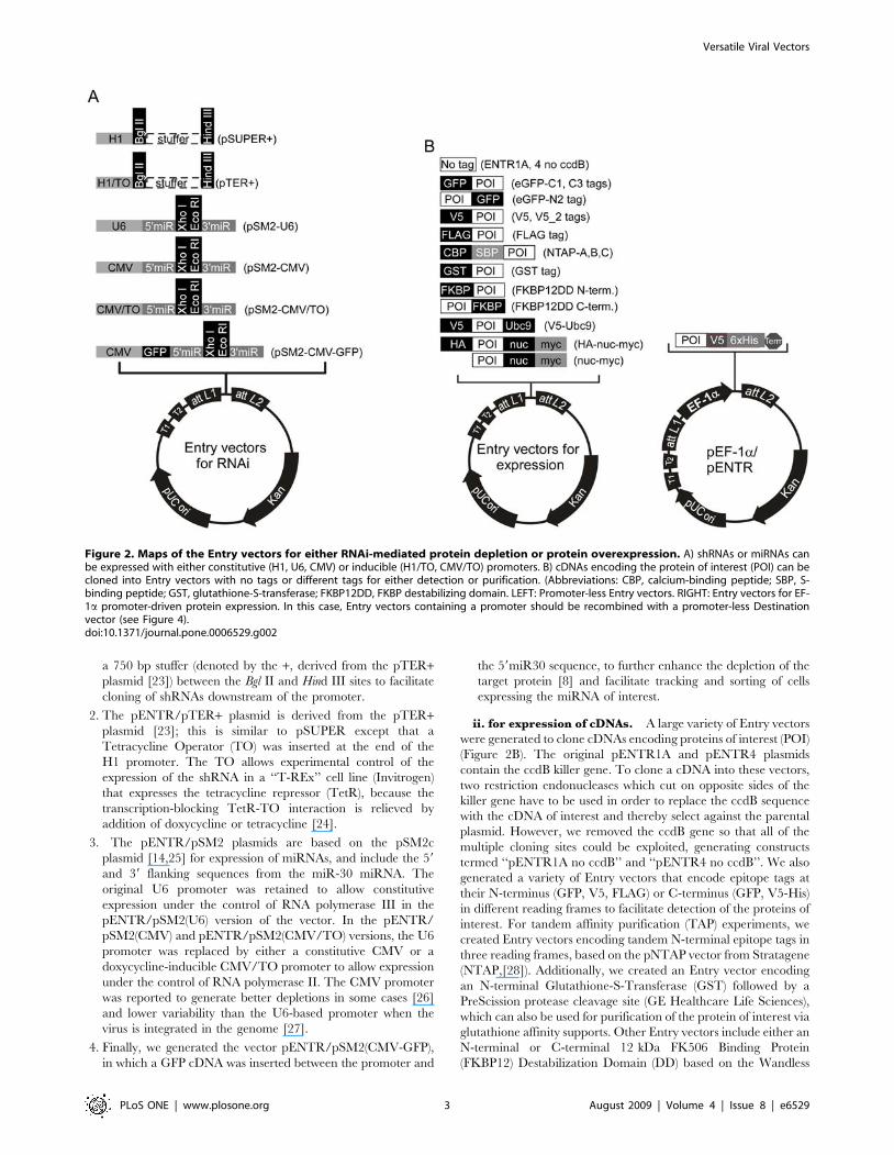
Figure 3. Maps of the Destination vectors. (A, C) The pLenti X1 and X2 series are promoter-less and require that the expression promoter comefrom the Entry vector. (B) The pLenti X2 series is designed for shRNA insertion into the 39 LTR, resulting in insert duplication in the final integratedform of the viral genome. (D) The CMV and PGK series provide the promoters for constitutive expression, and the CMV/TO promoter for regulation ofexpression by doxycycline. (E) The pLenti GFP DEST allows insertion of an expression cassette after the Woodchuck post-transcriptional element (PRE)and expresses GFP to make recipient cells fluorescent. (F) Retroviral Destination vectors for RNAi where the Destination cassette was inserted in the 39LTR, similar to the pLenti X2 series. (G, H). Retroviral Destination vectors for cDNA expression under the control of the CMV promoter. See text, Table 1and Figure 4 for details and compatibilities between each vector.doi:10.1371/journal.pone.0006529.g003
Versatile Viral Vectors
PLoS ONE | www.plosone.org 4 August 2009 | Volume 4 | Issue 8 | e6529
lab design [29,30] (also commercialized by Clontech under the
name ProteoTuner). This domain targets the protein of interest to
the proteosome for rapid degradation upon removal of the
protective ligand Shield-1 (Shld1), with faster kinetics than RNAi-
mediated depletion [30]. For the pENTR-V5-Ubc9 plasmid, the
SUMO E2 ligase Ubc9 was inserted at the C-terminus of the
pENTR-V5 vector so that cDNAs could be inserted between the
V5 epitope and Ubc9, generating a fusion protein that facilitates
detection of the sumoylated target protein, thereby bypassing the
requirement for an E3 SUMO ligase [31]. Finally, we made two
vectors to target protein domains to the nucleus, the pENTR-HA-
nuc-myc and pENTR-nuc-myc, which are derived from the
pCMV/myc/nuc (Invitrogen). These vectors have three tandem
copies of the SV40 Large T antigen nuclear targeting sequence
[32].
For cases where the CMV promoter might not be optimal for
cDNA expression, we also developed Entry vectors that contain
the human EF-1a promoter (Figure 2B). These vectors encode a
C-terminal V5-His epitope tag and were derived from the pEF-
TRACER A, B, C plasmids (Invitrogen). We also expanded the
variety of promoters available by creating Destination vectors
containing the human phosphoglycerate kinase (PGK) promoter
(see below).
Lentiviral Destination vectorsUsing the Gateway technology, each Entry vector can be
recombined into multiple Destination vectors. Every lentiviral
Destination vector contains the Woodchuck post-transcriptional
regulatory element (WPRE) and the central polypurine tract
(cPPT) because these elements increase transduction efficiencies
[33,34,35,36]. The maps of the Destination vectors are shown in
Figure 3. However, not all combinations of Entry and Destination
vectors are meant to be recombined. For example, if an Entry
vector already contains a promoter (such as the vectors used for
RNAi (Figure 2A) or the pEF-ENTR series (Figure 2B)),
recombination with a Destination vector such as the pLenti
CMV, CMV/TO or PGK series would juxtapose the two
promoters and possibly cause interference. Also, recombination
of a promoter-less Entry vector for expression with a promoter-less
Destination vector would result in a construct with no promoter to
drive expression of the cDNA. Some Destination vectors insert the
Entry cassette in their 39LTR (see below), but this imposes an
insert size limit, usually ,800 bp [37], because larger inserts
inactivate the LTR and impair viral integration. Also, some
sequences can be detrimental to the function of the 39LTR [38].
Finally, the pENTR/pSM2 (CMV-GFP) vector encodes GFP and
therefore should not be recombined with the pLenti CMV GFP or
pLenti X1 GFP-Zeo Destination vectors since they also encode the
GFP protein. The compatibilities of each Entry vector for various
Destination vectors are summarized in Table 1 as well as
represented schematically in Figures 4A for lentiviruses, and
Figure 4B for retroviruses.
i. pLenti X1 Destination series. The Gateway Destination
cassette was inserted into a promoter-less lentiviral backbone
(Figure 3A) because some of the Entry vectors provide the
promoter for the expression of shRNA, miRNA or cDNA. One
version of the vector confers resistance to puromycin and two
others confer resistance to zeocin. Additionally, the pLenti X1
GFP-Zeo vector encodes a GFP-zeocin fusion protein to allow
detection of transduced cells by fluorescence.
ii. pLenti X2 Destination series. Lentiviruses are part of
the retrovirus family and have an RNA genome that is reverse-
transcribed into DNA before integration into the host genome.
During this process, there is a duplication of the 39 LTR to
generate the final 59 LTR sequence that is found in the integrated
viral genome. Deletions in the U3 region of the 39 LTR (DU3)
have been used to generate self-inactivating (SIN) vectors for
increased biosafety [39,40], and several groups have found that
these truncated 39 LTRs tolerate insertion of a small cassette, such
as a small promoter (U6, H1, CMV) and an shRNA or miRNA
[41,42,43]. Therefore, inclusion of a shRNA in the 39 LTR of the
original construct results in duplication of this transgene upon
integration (Figure 3B). We therefore inserted the Gateway
Destination cassette within the 39 LTR (Figure 3C). All the
vectors of the pLenti X2 series have the Gateway cassette inserted
in the antisense orientation.
iii. pLenti CMV, CMV/TO and PGK Destination
series. We generated two types of Destination vectors for
expression of cDNAs under the control of the CMV promoter.
The first type has a constitutive CMV promoter (Figure 3D,
pLenti CMV), and the second has an inducible CMV/TO
promoter (pLenti CMV/TO). The pLenti CMV/TO series is
induced upon addition of doxycycline in a T-REx cell line that
expresses the TetR tetracycline repressor protein. To allow
generation of a wide variety of TetR-expressing cell lines, we
also generated an Entry vector with the TetR cDNA (pENTR/
TetR) and recombined it with the pLenti CMV Blast DEST vector
to make pLenti TetR Blast (not shown). We transduced different
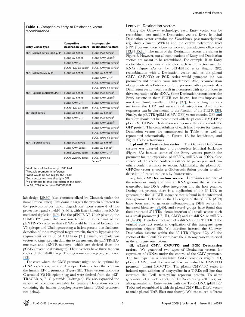
Table 1. Compatibles Entry to Destination vectorrecombinations.
Entry vector typeCompatibleDestination vectors
IncompatibleDestination vectors
pENTR/pSM2 Series (non-GFP) pLenti X1 Series pLenti PGK Seriesb
pLenti X2 Series pLenti CMV Seriesb
pLenti CMV GFP pLenti CMV/TO Seriesb
pQCXI RNAi X2 Seriesa pQCXI CMV/TO Seriesb
pENTR/pSM2(CMV-GFP) pLenti X1 Series pLenti X2 Seriesc
pLenti CMV GFPd
pQCXI CMV/TO Seriesb
pQCXI RNAi X2 Seriesc
pENTR/pTER+ pENTR/pSUPER+ pLenti X1 Series pLenti PGK Seriesb
pLenti X2 Series pLenti CMV Seriesb
pLenti CMV GFP pLenti CMV/TO Seriesb
pQCXI RNAi X2 Series pQCXI CMV/TO Seriesb
pEF-ENTR Series pLenti X1 Series pLenti X2 Seriesc
pLenti CMV GFP pLenti PGK Seriesb
pLenti CMV Seriesb
pLenti CMV/TO Seriesb
pQCXI CMV/TO Seriesb
pQCXI RNAi X2 Seriesc
pENTR-Fusion Series pLenti PGK Series pLenti X1 Seriese
pLenti CMV Series pLenti X2 Seriesc,e
pLenti CMV/TO Series pLenti CMV GFPe
pQCXI CMV/TO Series pQCXI RNAi X2Seriesc,e
aViral titers will be lower by ,100 fold.bProbable promoter interference.cInsert would be too big for the 39LTR.dEntry vector contains already a GFP.eNo promoter to drive expression of the cDNA.doi:10.1371/journal.pone.0006529.t001
Versatile Viral Vectors
PLoS ONE | www.plosone.org 5 August 2009 | Volume 4 | Issue 8 | e6529
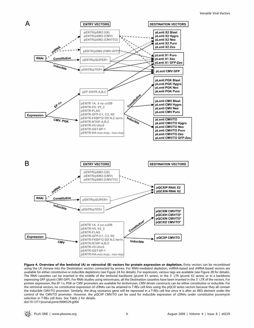
Figure 4. Overview of the lentiviral (A) or retroviral (B) vectors for protein expression or depletion. Entry vectors can be recombinedusing the LR clonase into the Destination vectors connected by arrows. For RNAi-mediated depletion, miRNA-based and shRNA-based vectors areavailable for either constitutive or inducible depletions (see Figure 2A for details). For expression, various tags are available (see Figure 2B for details).The RNAi cassettes can be inserted in the middle of the lentiviral backbone (pLenti X1 series), in the 39 LTR (pLenti X2 series) or in a backboneexpressing GFP (pLenti CMV GFP). For RNAi studies using retroviruses, all the Destination cassettes have been inserted in the 39 LTR of the vectors. Forprotein expression, the EF-1a, PGK or CMV promoters are available for lentiviruses. CMV-driven constructs can be either constitutive or inducible. Forthe retroviral vectors, no constitutive expression of cDNAs can be attained in T-REx cell lines using the pQCXI series vectors because they all containthe inducible CMV/TO promoter. Similarly, the drug resistance gene will be repressed in a T-REx cell line since it is after an IRES element under thecontrol of the CMV/TO promoter. However, the pQCXP CMV/TO can be used for inducible expression of cDNAs under constitutive puromycinselection in T-REx cell lines. See Table 2 for details.doi:10.1371/journal.pone.0006529.g004
Versatile Viral Vectors
PLoS ONE | www.plosone.org 6 August 2009 | Volume 4 | Issue 8 | e6529
cell lines with virus made from this vector, generating cells that
express TetR for inducible transgene expression from TO-
containing promoters. Because the TetR-expressing virus carries
a blasticidin resistance gene, we did not generate an inducible
CMV/TO lentiviral vector carrying the same drug resistance
gene.
Loss of protein expression in vivo due to promoter silencing can
occur with the CMV promoter, but silencing is much less frequent
for transgenes driven by the PGK and EF-1a promoters
[44,45,46]. Therefore, we generated Destination vectors with the
PGK promoter (Figures 3D, 4A). The cellular EF-1apromoter can
also be used for this purpose, but the PGK promoter in the pLenti
PGK series is smaller and therefore minimizes loss of viral titers
when large cDNAs are inserted [47]. Also, the PGK Destination
vectors are more versatile because they harbor more drug
resistance genes, while the pEF-1a Entry vectors can only be
recombined into the pLenti X1 Destination series (Figure 3A),
which currently have only puromycin and zeocin resistance genes
for selection.
iv) pLenti GFP DEST. We sought to create a vector that
would allow detection of transduced cells via GFP expression while
simultaneously delivering a transgene of choice. To do this, we
created a vector in which GFP expression is under the control of
the CMV promoter and either a cDNA, shRNA or miRNA can be
placed under the control of the EF-1a, H1, H1/TO, U6, CMV or
CMV/TO promoters (Figure 4A). GFP expression allows
detection and/or isolation of transduced cells by either
immunofluorescence or cell sorting. For this vector, we also
inserted the Gateway Destination cassette after the WPRE
(Figure 3E).
Retroviral Destination vectorsIn addition to the lentiviral vectors, we also generated retroviral
Destination vectors (Figures 3F, G, H, and Figure 4B) to determine
whether similar levels of expression or depletion could be attained.
With the exception of the pEF-ENTR series and the pENTR/
pSM2(CMV-GFP) vectors, all the other Entry vectors are
compatible with these retroviral vectors, so we can easily
recombine inserts into either retroviral and lentiviral vectors.
Using the same strategy as the pLenti X2 series, we inserted the
Gateway Destination cassette into the 39 LTR of the retroviral
backbone so we could obtain the expression of two shRNA/
miRNA cassettes upon viral integration into the cellular genome
(pQCXI X2 series, Figure 3F, 4B). The insertion of an shRNA
cassette in the 39 LTR of the pQCXIP plasmid was previously
used successfully [48]. We generated two retroviral vectors with
the neomycin and puromycin resistance genes for RNAi. For
expression of cDNAs, we replaced the CMV promoter from the
original pQCXI plasmid with the CMV/TO promoter with the
Gateway Destination cassette (Figure 3G). We also inserted the
zeocin and blasticidin resistance genes after the internal ribosome
entry site (IRES) so we could use these drugs for selection. One
possible drawback of these constructs is the control of the drug
resistance gene by the CMV/TO promoter. In a T-REx cell line,
both the cDNA and the drug resistance gene will be repressed,
allowing selection of transduced cells only when the cDNA is
induced. However, these vectors can be used to transduce non-T-
REx cells for constitutive expression. A construct in which only the
transgene and not the drug resistance gene is inducible was
designed by replacing the IRES with a murine PGK promoter to
control the puromycin resistance gene from an independent and
non-inducible promoter (Figure 3H). Table 2 summarizes which
vectors can be used to generate inducible expression of the cDNA/
miRNA/shRNA in a T-Rex cell line. The vectors that cannot
generate inducible expression will have constitutive expression in
these cells.
Our vectors do not encode the HIV Tat proteinWith each new generation of lentiviral packaging vectors, fewer
HIV proteins are co-expressed with the recombinant viral
genome, resulting in greater biological safety. In second generation
vectors, most of the HIV proteins not required for viral replication
are removed, with the exception of the HIV Tat protein which is
required for transcription of the viral genome from the 59 LTR. It
is provided by the packaging plasmid [49,50,51] and the Tat
protein is produced and secreted into the media by the packaging
cell line. Transfection of a Tat-expressing plasmid results in the
secretion of biologically active Tat [52,53]. In third generation
vectors, the 59 HIV LTR was replaced by either a hybrid 59
CMV/LTR or 59 RSV/LTR to allow transcription of the viral
genome in the absence of the HIV Tat protein without a decrease
in viral titers [40]. Accordingly, third generation packaging
plasmids do not express the HIV Tat protein. Because
several studies have pointed out the toxicity of the HIV Tat
protein and that it interferes with several cellular processes
[54,55,56,57,58,59,60,61,62,63], we used third generation vectors
as the basis for our constructs and all of our lentiviral vectors have
the hybrid 59 RSV/HIV LTR and do not require the HIV Tat for
transcription of the viral genome. We still recommend transduc-
tion with vectors encoding a control shRNA/cDNA or an empty
vector at similar titers to monitor possible toxic effects of the
transduction.
Testing of the vectorsWe compared one of our vectors with other lentiviral vectors
available to the scientific community to ensure that the insertion of
the Gateway Destination and the various drug selection cassettes
did not affect the efficiency of the virus. The pGIPZ (Open
Biosystems), pLKO.1 (The RNAi Consortium) and the pFSIPPW
[64] vectors were chosen because of their popularity and because
they all use puromycin as a selection marker, thereby alleviating
differences due to different drug selections. The empty versions of
each vector were compared to the empty version of the pLenti
CMV/TO Puro plasmid. Each virus was produced at the same
time, using the same transfection protocol adjusting for second or
third generation lentiviruses. As shown in Figure 5, the pLenti
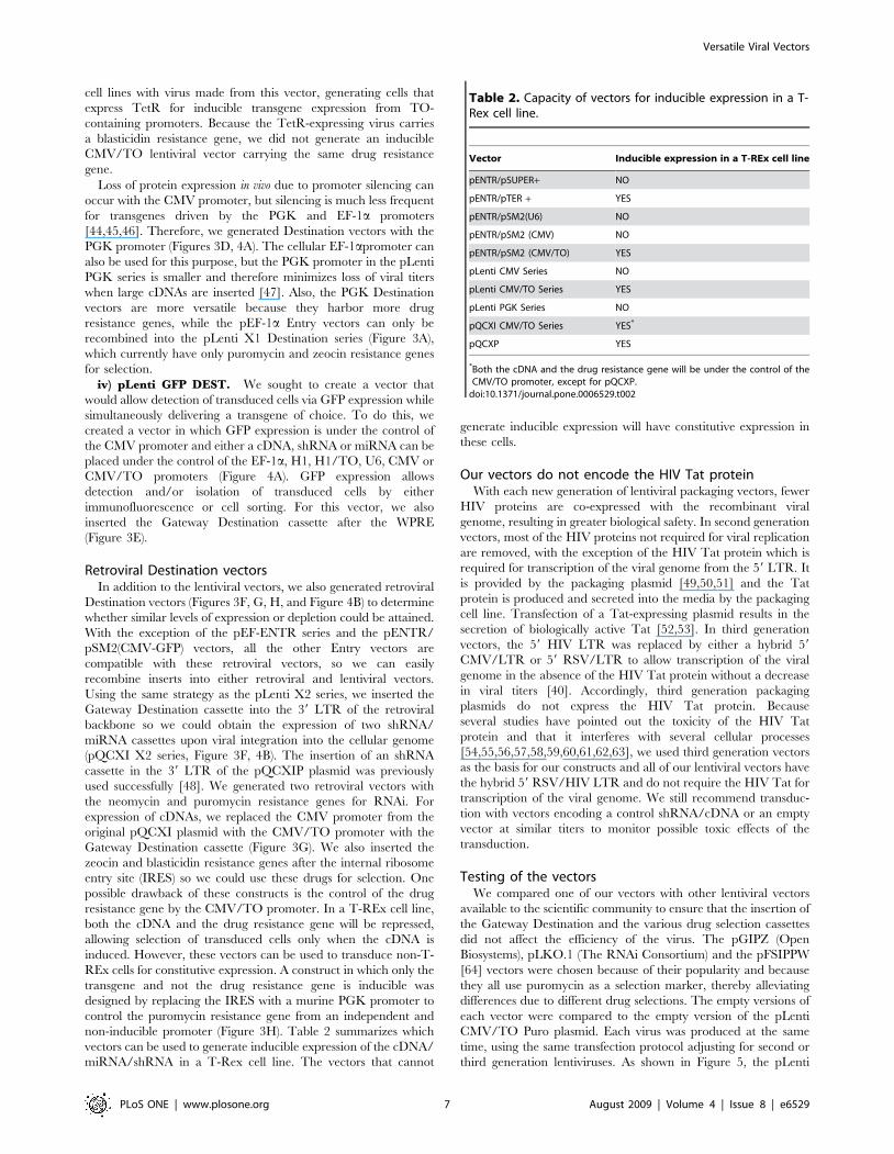
Table 2. Capacity of vectors for inducible expression in a T-Rex cell line.
Vector Inducible expression in a T-REx cell line
pENTR/pSUPER+ NO
pENTR/pTER + YES
pENTR/pSM2(U6) NO
pENTR/pSM2 (CMV) NO
pENTR/pSM2 (CMV/TO) YES
pLenti CMV Series NO
pLenti CMV/TO Series YES
pLenti PGK Series NO
pQCXI CMV/TO Series YES*
pQCXP YES
*Both the cDNA and the drug resistance gene will be under the control of theCMV/TO promoter, except for pQCXP.
doi:10.1371/journal.pone.0006529.t002
Versatile Viral Vectors
PLoS ONE | www.plosone.org 7 August 2009 | Volume 4 | Issue 8 | e6529
CMV/TO Puro, pGIPZ and pLKO vectors produced viruses with
similar titers (2–86105 cfu/ml), indicating that our vectors have
packaging efficiencies similar to several commercially available
vectors. In contrast, the pFSIPPW vector produced higher viral
titers (76106 cfu/ml). Both pFISPPW and pGIPZ are second
generation vectors whereas pLKO and pLenti CMV/TO Puro
are third generation, so this aspect does not explain the differences
in the observed titers.
Each Destination vector was also tested to ensure it was
functional and generated similar viral titers to other vectors
(Table 3 and data not shown). However, we did not test every
possible combination of Entry vector and Destination vectors due
to the great number of such combinations (326 for the lentiviral
vectors, 105 for the retroviral vectors). It is therefore possible that
unforeseen Entry-Destination combinations might negatively
affect the viral titers. We are maintaining a web site with up to
date information of current as well as future vectors at http://
ericcampeau.com.
We titered nearly 250 different recombinant viruses, using
either HT1080 or HeLa cells. For expression vectors, we normally
obtain a titer of 105 cfu/ml, regardless of the promoter or the drug
resistance used (result not shown). Because viral titers generally
decrease with increasing size of the inserted cDNA [47], both the
size of the cDNA and the size of the drug resistance gene need to
be accounted for when choosing a backbone. Some drug resistance
cDNAs, e.g. for blasticidin and zeocin, are ,400 bp whereas the
hygromycin-resistance cDNA is ,1000 bp. When expressing a
large cDNA (.2.5 kb), the choice of the drug resistance backbone
might influence the viral titer. In our hands, we did not notice any
significant decrease in viral titers with cDNAs of less than 2.0 kb
with any Destination vector. We have successfully expressed
cDNAs ,4 kb, with a reduced titer of 104 cfu/ml. If needed, we
would obtain higher viral titers by concentrating the virus by
ultracentrifugation (see methods).
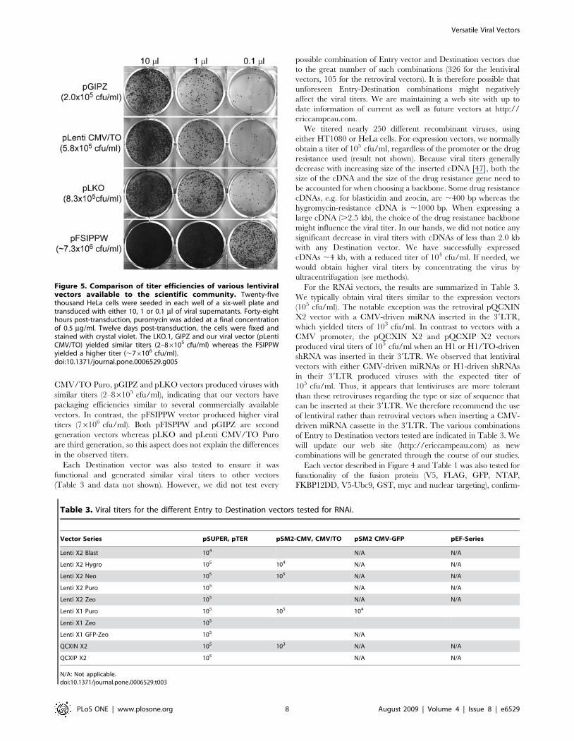
For the RNAi vectors, the results are summarized in Table 3.
We typically obtain viral titers similar to the expression vectors
(105 cfu/ml). The notable exception was the retroviral pQCXIN
X2 vector with a CMV-driven miRNA inserted in the 39LTR,
which yielded titers of 103 cfu/ml. In contrast to vectors with a
CMV promoter, the pQCXIN X2 and pQCXIP X2 vectors
produced viral titers of 105 cfu/ml when an H1 or H1/TO-driven
shRNA was inserted in their 39LTR. We observed that lentiviral
vectors with either CMV-driven miRNAs or H1-driven shRNAs
in their 39LTR produced viruses with the expected titer of
105 cfu/ml. Thus, it appears that lentiviruses are more tolerant
than these retroviruses regarding the type or size of sequence that
can be inserted at their 39LTR. We therefore recommend the use
of lentiviral rather than retroviral vectors when inserting a CMV-
driven miRNA cassette in the 39LTR. The various combinations
of Entry to Destination vectors tested are indicated in Table 3. We
will update our web site (http://ericcampeau.com) as new
combinations will be generated through the course of our studies.
Each vector described in Figure 4 and Table 1 was also tested for
functionality of the fusion protein (V5, FLAG, GFP, NTAP,
FKBP12DD, V5-Ubc9, GST, myc and nuclear targeting), confirm-
Figure 5. Comparison of titer efficiencies of various lentiviralvectors available to the scientific community. Twenty-fivethousand HeLa cells were seeded in each well of a six-well plate andtransduced with either 10, 1 or 0.1 ml of viral supernatants. Forty-eighthours post-transduction, puromycin was added at a final concentrationof 0.5 mg/ml. Twelve days post-transduction, the cells were fixed andstained with crystal violet. The LKO.1, GIPZ and our viral vector (pLentiCMV/TO) yielded similar titers (2–86105 cfu/ml) whereas the FSIPPWyielded a higher titer (,76106 cfu/ml).doi:10.1371/journal.pone.0006529.g005
Table 3. Viral titers for the different Entry to Destination vectors tested for RNAi.
Vector Series pSUPER, pTER pSM2-CMV, CMV/TO pSM2 CMV-GFP pEF-Series
Lenti X2 Blast 104 N/A N/A
Lenti X2 Hygro 105 104 N/A N/A
Lenti X2 Neo 105 105 N/A N/A
Lenti X2 Puro 105 N/A N/A
Lenti X2 Zeo 105 N/A N/A
Lenti X1 Puro 105 105 104
Lenti X1 Zeo 105
Lenti X1 GFP-Zeo 105 N/A
QCXIN X2 105 103 N/A N/A
QCXIP X2 105 N/A N/A
N/A: Not applicable.doi:10.1371/journal.pone.0006529.t003
Versatile Viral Vectors
PLoS ONE | www.plosone.org 8 August 2009 | Volume 4 | Issue 8 | e6529
ing epitope detection by immunoblotting or immunofluorescence
(Figures 6–8 and Supplemental Figures S1 and S2). We also
confirmed the ability of each promoter to drive expression of
downstream cDNA, shRNA, miRNA or drug selection genes. Unless
mentioned, we analyzed the entire cell population that survived the
drug selection, as opposed to a population derived from a single cell
clone. Because viral vectors can integrate into many areas of the
genome, single cell-derived (clonal) populations may provide better
inducibility/repression of the desired cDNA/shRNA/miRNA.
Examples of RNAi-mediated depletionsWe generated an shRNA and two miRNAs against the human
histone chaperone Asf1a and expressed them using both lenti- and
retro-viral vectors in either U2OS or U2OS T-REx cells. As
shown in Figure 6A, Asf1a can be depleted with similar efficiencies
using either a miRNA (lanes 2, 3), or shRNA (lane 5), and using
either a lentiviral or retroviral backbone (Figure S1A). The
efficiency of depletion is affected by several factors, including the
efficiency of the shRNA/miRNA, the mRNA levels, the half-life of
the mRNA and protein of interest, and the cell line used. In the
case of Asf1a, it took between 48 and 72 h to obtain maximal
depletion in U2OS cells (Figure S1A) but some proteins can be
maximally depleted in less than 24 h (result not shown). Stegmeier
et al. [8] reported that more efficient depletions could be
accomplished when a GFP cDNA was inserted between the
CMV promoter and the miR-30 cassette compared to direct
transcription of the miR-30 cassette from the CMV promoter. In
our vector system, we found there was often better depletion when
the GFP cDNA was present, although the effect was generally
quite modest (Figure S1B, C). Whether this difference is due to the
protein being depleted, the viral backbone, or other factors is
currently unknown.
We also used our system to test the effectiveness of an shRNA to
deplete cells of the DNA repair protein MDC1. The shRNA was
cloned into the pENTR/pTER+ vector, recombined with the
pLenti X1 GFP-Zeo and viral particles were used to transduce
U2OS cells. After 48 h, some cells were transferred to a chamber
slide and fixed for immunofluorescence. Fluorescence from the
GFP-Zeocin fusion protein was used to identify the transduced
cells while an antibody was used to detect MDC1. As shown in
Figure 6B, cells transduced by the virus were green and depleted
for MDC1 (red), whereas neighboring untransduced cells were not
green and contained normal levels of MDC1. Additionally,
inducible depletion of MDC1 was also accomplished in diploid
fibroblasts as shown in Figure 6C, where MDC1 was depleted in
the BJ T-REx cell strain. Therefore, both transformed and non-
transformed cell types can be studied with these tools.
Although RNAi-mediated depletion is an extremely useful tool
to study protein or RNA function, residual amounts of proteins
targeted by RNAi were detected in many cases. For example, we
generated an inducible VA13 cell line to test two shRNAs against
the DNA repair protein XPG. As shown in Figure S1D, although
the R6 shRNA was more efficient than the R4 shRNA to deplete
XPG (lanes 2 and 4), residual XPG could be detected. We
confirmed that the residual band is indeed XPG by demonstrating
its absence in the XPG-null cell line 94RD270 (lane 6).
Examples of overexpressed proteinsA major advantage of the various Destination vectors is that a
combination of cDNAs/shRNAs/miRNAs can be expressed in the
same cell, in either a constitutive or inducible manner. We
generated VA13 cell lines that inducibly express the human
histone chaperones Asf1a, Asf1b or HIRA or the combinations
Asf1a/HIRA or Asf1b/HIRA. As shown in Figure 7A, very little
expression of HA-Asf1a, HA-Asf1b and V5-HIRA was detected
without induction, but abundant overexpression of each protein
was detected 48 h after induction. In Figure 7B, inducible
expression of XPG-V5 is compared by Western blotting and
immunofluorescence. In each case, a minority of cells (,1%)
showed constitutive expression or lack of induction.
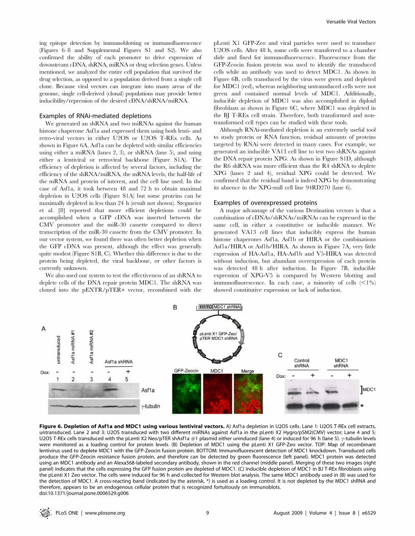
Figure 6. Depletion of Asf1a and MDC1 using various lentiviral vectors. A) Asf1a depletion in U2OS cells. Lane 1: U2OS T-REx cell extracts,untransduced. Lane 2 and 3: U2OS transduced with two different miRNAs against Asf1a in the pLenti X2 Hygro/pSM2(CMV) vector; Lane 4 and 5:U2OS T-REx cells transduced with the pLenti X2 Neo/pTER shAsf1a #1 plasmid either uninduced (lane 4) or induced for 96 h (lane 5). c-tubulin levelswere monitored as a loading control for protein levels. (B) Depletion of MDC1 using the pLenti X1 GFP-Zeo vector. TOP: Map of recombinantlentivirus used to deplete MDC1 with the GFP-Zeocin fusion protein. BOTTOM: Immunofluorescent detection of MDC1 knockdown. Transduced cellsproduce the GFP-Zeocin resistance fusion protein, and therefore can be detected by green fluorescence (left panel). MDC1 protein was detectedusing an MDC1 antibody and an Alexa568-labeled secondary antibody, shown in the red channel (middle panel). Merging of these two images (rightpanel) indicates that the cells expressing the GFP fusion protein are depleted of MDC1. (C) Inducible depletion of MDC1 in BJ T-REx fibroblasts usingthe pLenti X1 Zeo vector. The cells were induced for 96 h and collected for Western blot analysis. The same MDC1 antibody used in (B) was used forthe detection of MDC1. A cross-reacting band (indicated by the asterisk, *) is used as a loading control. It is not depleted by the MDC1 shRNA andtherefore, appears to be an endogenous cellular protein that is recognized fortuitously on immunoblots.doi:10.1371/journal.pone.0006529.g006
Versatile Viral Vectors
PLoS ONE | www.plosone.org 9 August 2009 | Volume 4 | Issue 8 | e6529
We also tested whether overexpressed proteins from our
lentiviral vectors could be assembled into functional multisubunit
complexes. We fused the NTAP tag to the N-terminus of the p150
subunit of the human chromatin assembly factor 1 (CAF-1), a
three subunit (p150, p60, and p48) complex that performs DNA
replication-coupled histone deposition [65]. The pLenti CMV/
TO NTAP-p150 puro plasmid was constructed, tagging p150 with
a streptavidin binding peptide (SBP) and calmodulin binding
peptide (CBP) for purification. We transduced HeLa T-REx cells
and screened for clones that showed minimal leaky expression and
good induction (result not shown). We generated cytosolic (S100)
and nuclear extracts (NE) from uninduced and induced cells and
purified NTAP-p150 from the NE fraction using streptavidin
beads and calmodulin sepharose. As seen in Figure 7C, no
expression of the NTAP-p150 is detected in the uninduced cells,
even in the pellet from the affinity-purified sample, showing tight
control of expression (lanes 1–4, upper band indicated by an
arrow, NTAP blot). Upon induction with doxycycline, the
majority of the NTAP-p150 is found in the nucleus (NE, lane 6)
and .90% can be purified from the extracts (lane 8). By selecting
for single colonies we isolated cell lines that express levels of
NTAP-p150 that are substantially less than the endogenous p150
levels in the nucleus (p150 blot), minimizing the possibility that the
normal function was perturbed. The tagged human CAF-1 was
purified to homogeneity using streptavidin beads and calmodulin
sepharose and all three subunits of the native CAF-1 complex were
obtained, as shown by silver staining (Figure 7C, right). To
monitor if the NTAP-p150 was functional, we used commercially
available antibodies to the CBP domain of the NTAP tag for
immunofluorescence analysis. Consistent with our fractionation
result, the NTAP-p150 is found in the nucleus of the cells
(Figure 7C, lower panel) where it colocalizes with the replication
protein PCNA in S phase cells, as previously shown for
endogenous CAF-1 [66]. In cells outside S phase, both NTAP-
p150 and CAF-1 show a diffuse nuclear staining (result not
shown). These data indicate that fusion to the NTAP tag does not
disrupt the replication-linked function of CAF-1, and illustrates the
usefulness of these reagents for biochemistry and cell biology
studies.
The vectors expressing GFP can be used for live cell studies, as
well as for fluorescence-mediated sorting of transduced cells. These
vectors include pENTR4-GFP-C1, -C3, -N1, pENTR/pSM2
(CMV-GFP) and pLenti CMV GFP (Table 1, Figures 2 and 3).
For example, Asf1a fused to GFP can be detected in live cells
(Figure S2A), and live cells monitored for GFP fluorescence using
lentiviruses derived from the pLenti X1 Puro/pSM2 (CMV-GFP)
are shown in Figure S1B. Co-staining of cells expressing the XPG-
V5 fusion protein with GFP in the pLenti CMV GFP backbone is
shown in Figure S2A. The GFP-Zeocin fusion protein can also be
detected in live cells, although at a lower intensity than unfused
GFP (Figure S2E). Figures S1 and S2 show various Destination
vector backbones we tested for the FLAG (Figure S2B), GST
(Figure S2C), Ubc9 (Figure S1E) and nuclear localization fusions
(Figures S2A, S2C).
Functional StudiesWe also tested whether our system could be used to inducibly
generate senescence phenotypes in human diploid fibroblasts.
Expression of TIN2-15C, a dominant-negative version of the
telomere-associated protein TIN2, results in uncapping of
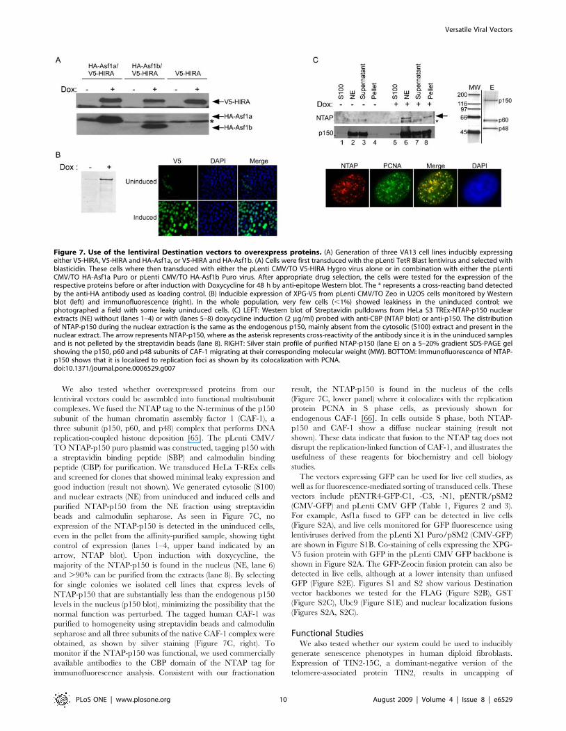
Figure 7. Use of the lentiviral Destination vectors to overexpress proteins. (A) Generation of three VA13 cell lines inducibly expressingeither V5-HIRA, V5-HIRA and HA-Asf1a, or V5-HIRA and HA-Asf1b. (A) Cells were first transduced with the pLenti TetR Blast lentivirus and selected withblasticidin. These cells where then transduced with either the pLenti CMV/TO V5-HIRA Hygro virus alone or in combination with either the pLentiCMV/TO HA-Asf1a Puro or pLenti CMV/TO HA-Asf1b Puro virus. After appropriate drug selection, the cells were tested for the expression of therespective proteins before or after induction with Doxycycline for 48 h by anti-epitope Western blot. The * represents a cross-reacting band detectedby the anti-HA antibody used as loading control. (B) Inducible expression of XPG-V5 from pLenti CMV/TO Zeo in U2OS cells monitored by Westernblot (left) and immunofluorescence (right). In the whole population, very few cells (,1%) showed leakiness in the uninduced control; wephotographed a field with some leaky uninduced cells. (C) LEFT: Western blot of Streptavidin pulldowns from HeLa S3 TREx-NTAP-p150 nuclearextracts (NE) without (lanes 1–4) or with (lanes 5–8) doxycycline induction (2 mg/ml) probed with anti-CBP (NTAP blot) or anti-p150. The distributionof NTAP-p150 during the nuclear extraction is the same as the endogenous p150, mainly absent from the cytosolic (S100) extract and present in thenuclear extract. The arrow represents NTAP-p150, where as the asterisk represents cross-reactivity of the antibody since it is in the uninduced samplesand is not pelleted by the streptavidin beads (lane 8). RIGHT: Silver stain profile of purified NTAP-p150 (lane E) on a 5–20% gradient SDS-PAGE gelshowing the p150, p60 and p48 subunits of CAF-1 migrating at their corresponding molecular weight (MW). BOTTOM: Immunofluorescence of NTAP-p150 shows that it is localized to replication foci as shown by its colocalization with PCNA.doi:10.1371/journal.pone.0006529.g007
Versatile Viral Vectors
PLoS ONE | www.plosone.org 10 August 2009 | Volume 4 | Issue 8 | e6529
telomeres, induction of the DNA damage response, and cellular
senescence [67]. We generated the human fibroblast cell strain
HCA2 T-REx, with an inducible eGFP-IRES-TIN2-15C (GFP-
iTIN2-15C). As shown in Figure 8A, no expression of GFP-iTIN2-
15C was detected in the uninduced cells, as determined by GFP
fluorescence, and no DNA damage was found, as measured by
immunofluorescence detection of foci containing the double-
strand break repair protein 53BP1 [68,69] (upper panels). 53BP1
and MDC1 show a diffuse nuclear staining in the absence of DNA
damage, and they form foci at sites of DNA damage ([70], see also
Figure 6B for MDC1 staining in undamaged cells). Four days after
induction of GFP-TIN2-15C, cells arrest with dysfunctional
telomeres [67] and we detect the presence of abundant (and
presumably telomeric) DNA damage focal 53BP1 immunostain-
ing. As a negative control, we inducibly expressed GFP alone, and
failed to detect any effect on cell proliferation or DNA damage
response indicators (Figure 8A, bottom panels). However, 10–15%
of the uninduced cells showed constitutive expression of GFP,
indicating leakiness of the T-REx system when GFP alone was
expressed, something that we did not observe in GFP-iTIN2-15C
expressing cells. We suspect that leaky cells were eliminated from
the population by the TIN2-15C- induced growth arrest, such that
only cells that had tight regulation of GFP-TIN2-15C continued to
proliferate under uninduced conditions. We further explored
growth-arresting proteins, examining the effects of the cyclin-
dependent kinase inhibitors p16 and p21. In the case of p16,
which induces a strong senescence growth arrest without causing
DNA damage ([71] and Figure S1F), we found that HCA2-T-REx
cells did not express any p16 in the uninduced state. In contrast,
expression of p21, another senescence inducer normally expressed
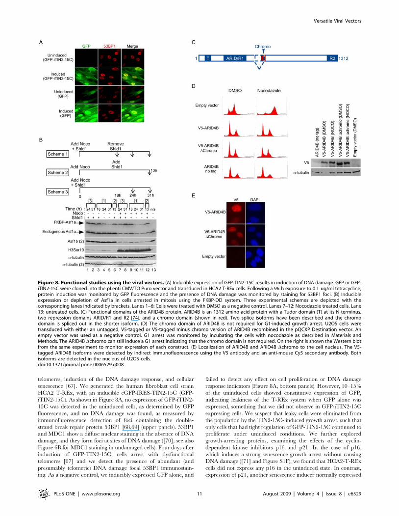
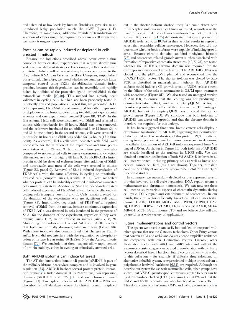
Figure 8. Functional studies using the viral vectors. (A) Inducible expression of GFP-TIN2-15C results in induction of DNA damage. GFP or GFP-iTIN2-15C were cloned into the pLenti CMV/TO Puro vector and transduced in HCA2 T-REx cells. Following a 96 h exposure to 0.1 ug/ml tetracycline,protein induction was monitored by GFP fluorescence and the presence of DNA damage was monitored by staining for 53BP1 foci. (B) Inducibleexpression or depletion of Asf1a in cells arrested in mitosis using the FKBP-DD system. Three experimental schemes are depicted with thecorresponding lanes indicated by brackets. Lanes 1–6: Cells were treated with DMSO as a negative control. Lanes 7–12: Nocodazole treated cells. Lane13: untreated cells. (C) Functional domains of the ARID4B protein. ARID4B is an 1312 amino acid protein with a Tudor domain (T) at its N-terminus,two repression domains ARID/R1 and R2 [74], and a chromo domain (shown in red). Two splice isoforms have been described and the chromodomain is spliced out in the shorter isoform. (D) The chromo domain of ARID4B is not required for G1-induced growth arrest. U2OS cells weretransduced with either an untagged, V5-tagged or V5-tagged minus chromo version of ARID4B recombined in the pQCXP Destination vector. Anempty vector was used as a negative control. G1 arrest was monitored by incubating the cells with nocodazole as described in Materials andMethods. The ARID4B Dchromo can still induce a G1 arrest indicating that the chromo domain is not required. On the right is shown the Western blotfrom the same experiment to monitor expression of each construct. (E) Localization of ARID4B and ARID4B Dchromo to the cell nucleus. The V5-tagged ARID4B isoforms were detected by indirect immunofluorescence using the V5 antibody and an anti-mouse Cy5 secondary antibody. Bothisoforms are detected in the nucleus of U2OS cells.doi:10.1371/journal.pone.0006529.g008
Versatile Viral Vectors
PLoS ONE | www.plosone.org 11 August 2009 | Volume 4 | Issue 8 | e6529
and tolerated at low levels by human fibroblasts, gave rise to an
uninduced leaky population much like eGFP (Figure S1F).
Therefore, in some cases, additional rounds of transduction or
selection of clones might be required to obtain a cell strain with
less leaky transgene expression.
Proteins can be rapidly induced or depleted in cellsarrested in mitosis
Because the inductions described above occur over a time
course of hours or days, experiments that require shorter time
scales require different strategies. For example, cells arrested with
a mitotic inhibitor will often start dying from overexposure to the
drug before RNAi can be effective (Eric Campeau, unpublished
observation). Therefore, we tested whether we could provide faster
temporal control using FKBP destabilization domain fusion
proteins, because this degradation can be reversibly and rapidly
halted by addition of the protective ligand termed Shld1 to the
extracellular media [29,30]. This method had already been
validated in cycling cells, but had not been previously tested in
mitotically arrested populations. To test this, we generated HeLa
cells expressing FKBP-Asf1a and monitored for either expression
or depletion of the fusion protein using two different experimental
schemes and one experimental control (Figure 8B, TOP). In the
first scheme, HeLa cells were incubated with Shld1 and arrested in
mitosis with nocodazole for 18 hours. Shld1 was then removed,
and the cells were incubated for an additional 6 or 13 hours (24 h
and 31 h time points). In the second scheme, cells were arrested in
mitosis for 18 hours and Shld1 was added for 13 hours (13 h time
point). As a control, HeLa cells were incubated with Shld1 and
nocodazole for the duration of the experiment and time points
were taken at 18, 24 and 31 hours. Each time point was also
compared to non-arrested cells to assess expression and depletion
efficiencies. As shown in Figure 8B lane 9, the FKBP-Asf1a fusion
protein could be detected eighteen hours after addition of Shld1
and nocodazole, and most of the cells were arrested in mitosis
(Figure S3, panel 9). Removal of Shld1 induced degradation of
FKBP-Asf1a with the same efficiency in cycling or mitotically-
arrested cells (compare lanes 4, 5 with 10, 11). Next, we tested
whether proteins can be inducibly expressed in mitotically-arrested
cells using this strategy. Addition of Shld1 to nocodazole-treated
cells induced expression of FKBP-Asf1a with the same efficiency as
cycling cells (compare lanes 12 and 6). Cells remain arrested for
the duration of the experiment with no significant cell death
(Figure S3). Importantly, degradation of FKBP-Asf1a required
removal of Shld1 from the media, because continuous expression
of FKBP-Asf1a was detected in cells incubated in the presence of
Shld1 for the duration of the experiment, regardless if they were
cycling (lanes 1, 2, 3) or arrested in mitosis (lanes 7, 8, 9).
Monitoring the endogenous levels of Asf1a and Asf1b revealed
that both are normally down-regulated in mitosis (Figure 8B).
With these tools, we also demonstrated that changes in FKBP-
Asf1a levels did not interfere with the regulation or phosphory-
lation of histone H3 at serine 10 (H3Ser10) by the Aurora mitotic
kinases [72]. We conclude that these reagents allow rapid control
of protein stability, either in cycling or mitotically arrested cells.
Both ARID4B isoforms can induce G1 arrestThe AT-rich interaction domain 4B protein (ARID4B) is part of
the mSin3A histone deacetylase complex and is involved in gene
regulation [73]. ARID4B harbors several protein-protein interac-
tion domains: a tudor domain at its N-terminus, two repression
domains (ARID/R1 and R2) [74] and one chromo domain
(Figure 8C). Two splice isoforms of the ARID4B mRNA are
described in EST databases where the chromo domain is spliced
out in the shorter isoform (dashed lines). We could detect both
mRNA splice isoforms in all cell lines we tested, regardless of the
tissue of origin or if the cell was transformed or not (result not
shown). Binda et al. [74,75] demonstrated that overexpression of
ARID4B (referred to as BCAA in their articles) results in a growth
arrest that resembles cellular senescence. However, they did not
determine whether both isoforms were capable of inducing growth
arrest. Because chromo domains can bind methylated histones
[76] and senescence-related growth arrest is often associated with
formation of repressive chromatin structures [48,77,78], we tested
whether the ARID4B chromo domain was required for the
overexpression-associated growth arrest. The ARID4B cDNA was
cloned into the pENTR-V5 plasmid and recombined into the
pQCXP DEST vector. The shorter isoform was cloned by RT-
PCR as described in materials and methods. Both ARID4B
isoforms could induce a G1 growth arrest in U2OS cells as shown
by the failure of the cells to accumulate in G2/M upon treatment
with nocodazole (Figure 8D). We also tested an untagged version
of ARID4B, to ensure that the V5 tag was not producing a
dominant-negative effect, and an empty pQCXP vector, to
monitor a possible toxic effect of the transduction. The untagged
ARID4B but not the empty pQCXP vector could also induce
growth arrest (Figure 8D). We conclude that both isoforms of
ARID4B can arrest cell growth, and that the chromo domain is
therefore not required for this activity.
It has been suggested that some breast cancer cells display a
cytoplasmic localization of ARID4B, suggesting that perturbation
of the normal nuclear localization of this protein [79,80] is altered
during tumorigenesis. We used immunofluorescence to determine
the cellular localization of ARID4B isoforms expressed from V5-
tagged cDNAs. As shown in Figure 8E, both isoforms of ARID4B
are clearly localized to the nucleus in U2OS cells. We also
obtained a nuclear localization of both V5-ARID4B isoforms in all
cell lines we tested, including primary cells as well as breast and
cervical cancer cell lines (result not shown). Together, our data
confirm the ability of our vector system to be useful for a variety of
functional studies.
In summary, we successfully depleted or overexpressed several
proteins involved in cell-cycle regulation, DNA repair, telomere
maintenance and chromatin homeostasis. We can now use these
cell lines to study various aspects of chromatin dynamics during
cell cycle, DNA repair and establishment of senescence. In our
hands, the viral vectors efficiently transduced all cell lines tested
(human U2OS, HT1080, MCF7, A549, WI38, IMR90, HCA2,
BJ, HOP92, HOP62, OVCAR1, HeLa, K562, MDA468, MDA-
MB-431, MCF10A and mouse 3T3) and we believe they will also
be useful in a wide variety of applications.
Future implementations and control vectorsThe system we describe can easily be modified or integrated with
other systems that use the Gateway technology. Other Entry vectors
that contain attL1 and attL2 and do not encode ampicillin resistance
are compatible with our Destination vectors. Likewise, other
Destination vector with attR1 and attR2 sites and without the
kanamycin resistance gene can be used in combination with the Entry
vectors described here. Therefore, future vectors can easily be added
to this collection – for example, if different drug selections, an
alternative inducible system, or expression of multiple proteins from a
polycistronic lentiviral backbone [6,81] are required. Although we
describe our system for use with mammalian cells, other groups have
shown that VSV-G pseudotyped lentiviruses similar to ours can be
used to transduce chicken (DT40) and insect cells (SF9) and that the
CMV and SV40 promoter are also functional in these cells [6].
Therefore, constructs harboring CMV and SV40 promoters such as
Versatile Viral Vectors
PLoS ONE | www.plosone.org 12 August 2009 | Volume 4 | Issue 8 | e6529
the pLenti CMV Blast or Zeo could conceivably be used to express
proteins in these cells, although we have not tested that option. In
sum, the system we describe is both immediately useful and
accommodates the integration of future developments in the field
without significant modifications to the system.
Concluding remarksOverexpression and depletion of proteins are powerful tools to
study cellular processes. The viral vectors described here can
certainly facilitate such studies. However, as for any other gene
expression system, there are limitations and experiments must be
carefully controlled. First, different cell lines can have different
transduction efficiencies, therefore measuring transduction efficien-
cies in each cell line is critical, and can be done using either a GFP
reporter or an empty vector and selecting for drug resistance.
Second, overexpression or depletion of a protein can have
immediate or delayed side-effects. For example, if overexpression
of a protein is toxic to the cell, methylation of the promoter can
happen and reduce the expression levels of the protein and, after a
brief period of slow growth, the cells can recover with almost normal
growth. Inducible expression of the toxic protein in a T-Rex cell line
might be an alternative, which has worked in some, but not all cases
(Eric Campeau unpublished result). Therefore, careful monitoring
of the cell behavior and freezing of early passage cells could be
critical. Furthermore, different cell lines might react differently to
overexpression or depletion of a protein, depending on tissue of
origin or the status of some stress response pathways such as p53/
Rb/p16 for example. Finally, overexpressing or depleting a control
protein such as GFP or Luciferase could help monitor side-effects
unrelated to the protein of interest, such as overloading of the
transcription/translation/shRNA machinery.
Materials and Methods
Generation and propagation of the vectorsGeneral cloning techniques were used to generate each of the
vectors. Detailed protocols are available upon request. All the
lentiviral Destination vectors are derived from the p156RRL-
sinPPT-CMV-GFP-PRE/Nhe I vector [82,83] and all the
retroviral Destination vectors are derived from the pQCXI series
(Clontech). For the lentiviral vectors, a linker was inserted between
the Kpn I and Eco RI sites to clone the drug selection cassettes
following the Woodchuck post-transcriptional response element
(WPRE). All Destination vectors were propagated in the E.coli
strain DB3.1 (Invitrogen) under appropriate antibiotic selection
(see plasmid maps for details). All Entry vectors were propagated
in the E.coli strain TOP10F’ (Invitrogen) under kanamycin
selection. After the LR recombination reaction, lentiviral vectors
were propagated in the E.coli strain Stbl3 (Invitrogen) and
retroviral vectors in TOP10F’, all under ampicillin selection,
except for the pLenti CMV/TO Zeo DEST (zeocin).
cDNAs, shRNAs and miRNAsThe HA epitope-tagged Asf1a (NCBI GeneID: 25842) and
Asf1b (GeneID: 55723) cDNAs as well as the HIRA (GeneID:
7290) cDNA were a gift from Peter Adams (Beatson Institute,
Glasgow). The HA-Asf1a and HA-Asf1b were subcloned in the
pENTR1A no ccdB plasmid and the HIRA cDNA was cloned into
the pENTR4-V5 plasmid. The XPG (GeneID: 2073) cDNA
cloned in to the pENTR3C vector with a V5 epitope and a GFP-
fusion at its C-terminus (XPG-V5-GFP) was a gift of Ely Kwoh.
The XPG-V5 was subcloned into pENTR3C to remove the GFP
fusion. The Ubc9 (GeneID: 7329) cDNA was a gift from Claude
Gazin (CNRS, UMR217). The ARID4B (GeneID: 51742) cDNA
was obtained from the Kazusa cDNA project (clone HH11923,
Accession #AB210032). The ARID4B cDNA was cloned in the
pENTR4-V5 plasmid. A region of the cDNA encoding the shorter
isoform was amplified by RT-PCR from U2OS cells and
subcloned into the pCR2.1 TA cloning plasmid (Invitrogen) and
sequence verified. The fragment was excised using the Bbv CI/
Hind III restriction enzymes and inserted into the Bbv CI + Hind
III-digested pENTR4-V5-ARID4B plasmid to generate pENTR4-
V5 ARID4B Dchromo.
The shRNA for Asf1a was derived from an siRNA previously
used [84]. We inserted the following annealed oligonucleotides
between the Bgl II/Hind III sites of either pENTR/pTER+ or
pENTR/pSUPER+:
For Asf1a:
59GATCCCGTGAAGAATACGATCAAGTGTGTGCTGTC
CACTTGATCGTATTCTTCACTTTTTGGAAA and 59AGCT
TTTCCAAAAAGTGAAGAATACGATCAAGTGGACAGCAC
ACACTTGATCGTATTCTTCACGG, where the underlined
sequence is specific for human Asf1a.
For MDC1: 59GATCCCCCAACATGCAGAGATTGAAA
TTCAAGAGATTTCAATCTCTGCATGTTGTTTTTGGAA
A and 59AGCTTTTCCAAAAACAACATGCAGAGATTGAA
ATCTCTTGAATTTCAATCTCTGCATGTTGGGG
For XPG: 59GATCCCAGAATACATGCGGTGGATTTTC
AAGAGAAATCCACCGCATGTATTCTTTTTTGGAAA and
AGCTTTTCCAAAAAAGAATACATGCGGTGGATTTCTC
TTGAAAATCCACCGCATGTATTCTGG.
For the Asf1a shRNA, we also designed an miRNA-based loop
in the shRNA because it was reported to result in better depletion
efficiencies [85]. To design the Asf1a miRNA, we used the
algorithm from Open Biosystems (www.openbiosystems.com). The
following oligos were amplified with the Xho I and Eco RI
amplification primers and subcloned into the pENTR/pSM2
vectors according to the manufacturer’s protocol:
59TGCTGTTGACAGTGAGCGAGGTCACAAGATTCCAC
ATTAATAGTGAAGCCACAGATGTATTAATGTGGAATCT
TGTGACCCTGCCTACTGCCTCGGA
59TGCTGTTGACAGTGAGCGAAGGTAGAATACTTTCA
TTATTTAGTGAAGCCACAGATGTAAATAATGAAAGTAT
TCTACCTCTGCCTACTGCCTCGGA
where the underlined sequences are specific for the human
Asf1a.
LR recombination and purification of the plasmid DNAThe LR recombination was performed using the LR clonase mix
(cat. #11791-019, Invitrogen) with 1 ml of miniprep DNA for each
of the Entry and Destination vector, 4 ml of TE pH 8.0, 2 ml of LR
buffer and 2 ml of LR clonase. Reactions were incubated at room
temperature from 2 h to overnight. The proteinase K digestion step
was omitted from the manufacturer’s protocol and 2 ml of the
reaction were used to transform 25 ml of competent E.coli Stbl3 or
TOP10F’ cells generated by the Zymo Research competent cell kit
(cat #T-3002). Colonies were picked and inoculated in 50 ml of LB
broth. 600 ml were used for a miniprep (Zippy, Zymo research) to
confirm the identity of the clone and the remainder of the culture
was used for a Qiagen Midi prep according to the manufacturer’s
instruction for transfecting 293T cells.
Transfection of 293T cells to generate third generationlentiviruses
The day before the transfection, 56106 of 293T cells were
seeded in a 10 cm dish. Transfection was done with 50 ml of
Lipofectamine 2000 (Invitrogen) according to the manufacturer’s
instructions using 15 mg of the transfer vector, 15 mg of pLP1
Versatile Viral Vectors
PLoS ONE | www.plosone.org 13 August 2009 | Volume 4 | Issue 8 | e6529
(Invitrogen), 6 mg of pLP2 (Invitrogen), and 3 mg of pVSV-G
(Invitrogen). The DNA:liposome complex (3 ml) was incubated
with the cells in a final volume of 10 ml of OPTI-MEM
(Invitrogen) overnight.
Transfection of 293T cells to generate second generationlentiviruses
The same procedure as above was used except that 20 mg of
pCMVD8.9 [50] was used instead of the pLP1 and pLP2 vectors.
Transfection of 293gag/pol cells to generate retrovirusesThe day before the transfection, 56106 of 293 gag/pol cells
were inoculated in a 10 cm dish. Lipofectamine 2000 was used for
the transfection following the manufacturer’s recommendations
using 12 mg of the transfer vector, 1.2 mg of pGAG/POL [86] and
1.2 mg of pVSV-G (Invitrogen).
Collection of viral supernatant and titration of virusesAt 48 h post-transfection, viral supernatants were collected and
fresh OPTI-MEM (10 ml) was added to the dish for another
collection at 72 h post-transfection. For each collection, viral
supernatants were filtered through a 0.2 mm syringe filter. The 48
and 72 h collections were pooled, aliquoted and stored at 280uC.
Viral titers were determined by seeding 6-well plates with 2.56104
HT1080 or HeLa cells and transducing them with 10-fold
dilutions (100-1 ml) of viral supernatant as described below. After
12–14 days, cells were fixed in cold methanol and stained with
crystal violet solution (0.5% crystal violet, 25% methanol) and
counted to determine their colony forming units (cfu). When
necessary, viruses were concentrated in a Beckman SW28 rotor at
21,000 rpm for 2 h at 4uC and resuspended in 4 ml of Hank’s
Buffered Saline solution (HBS, Invitrogen). A second ultracentri-
fugation in a Beckman 55Ti rotor at 21,000 rpm for 90 minutes
was performed and the viral pellet was resuspended in 100 ml of
HBS and stored at 280uC in 10 ml aliquots. When measuring viral
titers, we take into consideration several factors. First, the health of
the 293T packaging cell is critical. If the cells are too confluent or
not grown under optimal conditions, it will result in lower viral
titers. Similarly, if the cells have been passaged for a long time, it
will also yield lower titers. We always thaw a fresh vial of 293T
cells when we are producing viruses. Second, the transfection
procedure, liposome reagent, the quality of the media used as well
as the amounts and quality of the vectors co-transfected are also
critical. Third, the viral titers will also be proportional to the
volume of media used to grow the cells in order to collect the viral
supernatant. Finally, the cell line used for the titration, the amount
of cells plated and the concentration of the selecting drug is also
affecting viral titers. Other factors like the protein to be
overexpressed or depleted might affect cell growth and viability
and result in lower viral titers than expected. We recommend
using a control vector expressing GFP to monitor transfection as
well as transduction efficiencies.
Transduction of cell lines and induction with doxycyclineCells were transduced at a MOI between 0.5 and 1. Viruses and
cells were incubated overnight in D-MEM media (Invitrogen)
containing 6 mg/ml of polybrene (Sigma) in a final volume of
950 ml for 6-well plates, 5 ml for 10 cm dishes and 11 ml for
15 cm dishes. The next day, the viruses were removed, the cells
were rinsed twice with PBS and fresh media was added. For
primary cells, a second round of transduction was done. Drug
selection was added at 48 h post-transduction. The following
concentrations were used: blasticidin: 2.5 mg/ml for WI38, HCA2,
BJ cells and 5 mg/ml for the other cells, hygromycin: 100 mg/ml
for WI38, HCA2, BJ cells, 200 mg/ml for U2OS cells and 300 mg/
ml for HT1080 cells, neomycin: 300 mg/ml for WI38, HCA2, BJ
cells and 800 mg/ml for the other cells, puromycin: 0.5 mg/ml for
HeLa cells and 2.0 mg/ml for other cells, zeocin: 400 mg/ml for
HT1080 cells and 200 mg/ml for other cells. Induction of the
cDNA/shRNA/miRNA was typically done by addition of
doxycycline at a final concentration of 1.0 mg/ml for 48 h (cDNA)
or 96 h (shRNA/miRNA) unless indicated otherwise.
Generation of the MDC1 antibodiesThe N-terminal 318 residues of MDC1 were expressed as a
GST fusion in E.coli and separated from GST by cleavage with
PreScission protease by glutathione sepharose (GE Healthcare)
affinity. The domain was further purified by High Q (BioRad) ion
exchange, and Superdex75 (GE Healthcare) size exclusion
chromatography. Rabbits were immunized and antibodies were
purified from sera by affinity chromatography. Both GST and
GST-MDC1 N-terminus affinity columns were prepared by
incubating glutathione sepharose with E. coli expression lysates
overnight, washing the beads extensively with borate buffer,
crosslinking the protein to the beads using dimethypimelimidate
and ethanolamine, and washing with glycine-HCl and phosphate
buffer. Sera was incubated with the GST affinity resin for 2 hours
and then with the GST-MDC1 N-terminus affinity resin
overnight. After extensive washes with PBS, antibody was eluted
with 200 mM glycine-HCl pH 2.5 and immediately neutralized
with 1 M K2HPO4. Antibody pools were dialyzed into PBS and
stored at 280uC.
Other antibodies, protein electrophoresis,immunofluorescence
The following antibodies were used: anti-V5 (Invitrogen,
R96025) at 1:1000 for Western blot and 1:1200 for immunofluo-
rescence; anti-Asf1a (C6E10, cat#2990, Cell Signaling Technology)
at 1:6000 for Western blot, anti-Asf1b (cat.#2769, Cell Signaling
Technology) at 1:500 for Western blot, anti-HA (mouse monoclonal
12CA5) at a final concentration of 1 mg/ml for Western blot, anti-
XPG [87], at 1:500 for Western blot, anti-p16 (Neomarkers, JC8) at
1:500 for immunofluorescence, anti-p21 (BD Biosciences, 556430)
at 1:500 for immunofluorescence and anti-53BP1 (Bethyl Labora-
tories, BL182) at 1:2000 for immunofluorescence anti-GFP
antibody (Abcam, Ab1218) at 1:1000 for Western blot, anti-GST
(GE healthcare, 27-4577-01) at 1:100 for Western blot, anti-CBP
(ICL Labs, RCBP-45A at 1:2000 for Western blot and 1:500 for
immunofluorescence, anti-p150 (ss48, [88]) at 1:1000 for Western
blot, anti-PCNA clone PC10 [89] at 1:500 for immunofluorescence,
a-tubulin hybridoma (DM1A, [90]), 1:2000 for Western blot, anti-
H3Ser10 clone 3H10 (Upstate, 05-806) at 1:2000 for Western blot,
anti-c-myc 9E10 (Santa Cruz biotechnologies, sc-40) at 1:1000 for
Western blot and 1:400 for immunofluorescence. Generation of cell
extracts, protein electrophoresis, Western transfer and antibody
detection were done using standard procedures. For immunofluo-
rescence, cells were fixed in 4% paraformaldehyde in the case where
GFP fluorescence was examined and 100% cold methanol in all
other cases. Immunofluorescence was done as described before
[91], and images were acquired on an Olympus BX60 fluorescence
microscope with the spotfire 3.2.4 software (Diagnostics Instru-
ments) and further processed with Photoshop CS2 (Adobe). For the
MDC1 immunofluorescence, images were acquired on a Zeiss
Axioskop using a Kodak DC120 digital camera and its capture
software and further processed with Photoshop CS2. Images for the
NTAP/PCNA, c-myc, V5 were collected on a Zeiss Axioplan 2
Versatile Viral Vectors
PLoS ONE | www.plosone.org 14 August 2009 | Volume 4 | Issue 8 | e6529
microscope with the Axiovision 4.5 software and processed with
Photoshop CS3.
Live cell imagingBright field and fluorescent live cell pictures were taken on an
Axiovert 200 microscope (Zeiss) using a Qicam fast camera
(Qimaging) and the Openlab 4.0.4 software. The pictures were
exported to Adobe Photoshop CS for brightness/contrast
adjustments.
CAF-1 purification from HeLa S3 T-Rex-NTAP-p150 cellsNuclear extracts were prepared from cells induced with 1 mg/
mL doxycycline as described [65] followed by a modified TAP
method [92]. Briefly, nuclear extracts (400 mM NaCl) were
diluted two fold with Buffer A (25 mM Tris-HCl pH 7.5, 1 mM
EDTA, 0.1% NP-40, 10% glycerol, 1 mM DTT, 0.1 mM PMSF)
to reduce the NaCl concentration to 200 mM final. Extracts were
incubated with streptavidin sepharose (GE Healthcare) for three
hours, rotating at 4uC. Beads were washed three times with large
volumes of Buffer A+200 mM NaCl (A200) and eluted with
A200+2 mM biotin [93]. Elutions were bound to calmodulin
sepharose (GE Healthcare) for 2 hours in A200+2 mM CaCl2.
Beads were washed and eluted with A200+5 mM EGTA. Samples
were then TCA precipitated and loaded on 5–20% gradient gels
and either silver stained or western blotted.
Shld1 experimentHeLa cells transduced with the FKBP-Asf1a virus were
incubated for 18 hours in the presence of nocodazole (200 ng/
ml) or DMSO (0.1%) and Shld1 (1 mM, [30]). Shld1 was removed
and both suspension and adherent cells were washed twice with
PBS followed by addition of fresh media containing nocodazole.
At the 18, 24 and 31 hour time-points, suspension and adherent
cells were collected, pooled, and divided in two aliquots for
Western and FACS analysis. For reversal of the FKBP-Asf1a
degradation, cells were incubated with nocodazole in the absence
of Shld1 for 18 hours and Shld1 was added for 13 additional
hours. Suspension and adherent cells were collected as above. For
FACS, cells were resuspended in 200 ml of PBS and fixed with
800 ml of 70% ethanol. RNAse A (100 mg/ml) and propidium
iodide (10 mg/ml) were added to the cells before processing on a
FACScalibur system and CellQuest software. FACS profiles were
displayed using WinMDI 2.9.
FACS analysis and G2 trapping assay for ARID4Boverexpression
Twenty-four hours after transduction, puromycin and either
DMSO (0.1%) or nocodazole (200 ng/ml) were added to the cells
and they were incubated for another 20 hours. Mitotic and cycling
cells were harvested by collecting the media and trypsinizing the
adherent cells. Cells were pooled and divided in two for Western
and FACS analysis (performed as above).
Availability of the plasmids. All the sequences and maps of
the plasmids described here are included in the supplementary
materials (Table S1 and File S1). Plasmids will be made available
through Addgene (www.addgene.org). We also provided to
Addgene different control vectors to monitor transfection and
transduction efficiencies and test for possible toxic effects of the
viruses, including some insert-less vectors, or vectors encoding
cDNAs or hairpins against GFP or luciferase (see Table S1 for
details). Detailed protocols, sequences and maps of our vectors and
future vectors will be described at http://www.ericcampeau.com.
Supporting Information
Figure S1 Other vectors tested for RNAi-mediated depletion or
cDNA overexpression. (A) Lanes 1–5): time course for the
inducible depletion of Asf1a. U2OS T-REx cells transduced with
the pLenti X2 Neo/pTER Asf1a shRNA vector were either
uninduced (lane 1) or induced with doxycycline for 24, 48, 72 or
96 h (lanes 2–5). Lanes 6–9: comparison of shRNAs and miRNAs
for the depletion of Asf1a using different viral backbones and
different cell lines. HeLa cells transduced with the pLenti X1
Puro/pSM2(CMV) Asf1a miRNA (lane 6), pLenti X1 Puro/
pSM2(CMV-GFP) Asf1a miRNA (lane 7) or U2OS T-REx cells
transduced with the pLenti X2 Hygro/pSM2 (CMV) Asf1a
miRNA (lane 8) or 293 G/P cells transfected with the pQCXIN
X2/pTER Asf1a shRNA (lane 9, 72 h post-transfection) resulted
in comparable levels of Asf1a depletion. (B) Depletion of Asf1a
using a miRNA in either the SM2 (CMV) or SM2 (CMV-GFP)
vector. Live cell images of U2OS cells transduced with either an
miRNA against Asf1a (1, 3) or the same miRNA with the GFP
protein co-expressed (2, 4). Panels 1 and 2 are brightfield whereas
3 and 4 are the green fluorescence channel. (C) Cells shown in (B)
were analyzed by Western blotting to confirm Asf1a depletion.
Lane 1: pLenti X1 Puro/pSM2(CMV) Asf1a miRNA; lane 2:
pLenti X1 Puro/pSM2(CMV-GFP) Asf1a miRNA; lane 3:
untransduced U2OS cell extract; lane 4: U2OS T-REx cells
depleted of Asf1b. Lanes 5–6: Inducible depletion of Asf1a using
the pLenti X2 Neo/pSM2(CMV/TO) Asf1a miRNA #1 vector.
Lane 5 was uninduced whereas lane 6 was induced for 96 h with
doxycycline. Lanes 5 and 6 were probed with a different Asf1a
antibody (A88, ref) that can detect the various phosphorylated
forms of Asf1a. The * indicates a cross-reacting band used as
loading control. (D) Inducible and constitutive depletion of the
XPG protein with two different shRNAs (R4 and R6) using the
pLenti X1 Zeo/pTER backbone in VA13 T-REx cells. Lanes 1
and 3 are the uninduced R4 and R6 shRNA, respectively; lanes 2
and 4 are induced for 96 h. A constitutive depletion of XPG using
the R6 shRNA in VA13 cells is shown in lane 5 whereas a cell
extract derived from the XPG-null cell line 94RD270 is shown in
lane 6 as a negative control. The * indicated a cross-reacting band
used as loading control. E) The Asf1a cDNA was inserted into the
pENTR-V5-Ubc9 plasmid and recombined into the pQCXP
CMV/TO Destination vector. The resulting retrovirus was used
to transduce U2OS cells. The blot was probed with an anti-V5
epitope antibody (Left panel), which detected a specific band in the
transduced cells (lane 2) when compared to untransduced cells
(lane 1). The unstripped blot was reprobed for Asf1a (Right panel)
to show the shift in the molecular weight of the Asf1a-Ubc9 fusion
protein (lane 4). F) Inducible expression of the p16 and p21 cyclin-
dependent kinase inhibitors in HCA2 T-REx cells. Induction of
p16 and p21 as in figure 6D were monitored by immunofluores-
cence using antibodies against p16 and p21 respectively. No cells
show leaky p16 expression but a few cells do for p21. However,
overexpression of neither protein induces DNA damage as
revealed by the absence of 53BP1 foci, even though both can
induce senescence (not shown).
Found at: doi:10.1371/journal.pone.0006529.s001 (7.43 MB
PDF)
Figure S2 (A) Testing of some fusion constructs by immunoflu-
orescence. Panels 1–3: GFP-Asf1a is detected in the nucleus of live
cells. Panel 1: GFP channel; panel 2: bright field; panel 3: merged
pictures. Panels 4–9: Targeting of the ARID-R1 domain to the
nucleus using the pENTR-nuc-myc vector recombined into the
pLenti CMV/TO Puro Destination vector. Panel 4: detection of
ARID4B-R1 in the nucleus using the myc epitope; panel 5: DAPI
Versatile Viral Vectors
PLoS ONE | www.plosone.org 15 August 2009 | Volume 4 | Issue 8 | e6529
staining; panel 6: merged pictures; panel 7: ARID4B-R1 domain
fused at the C-terminus to the V5-tag and recombined with the
pLenti CMV Neo Destination vector is localized to the cytoplasm
as detected using the V5 antibody; panel 8: DAPI staining; panel
9: merged pictures. Western blots of the GFP-Asf1a and the
ARID-R1 domain constructs are shown in (B) and (C) respectively.
Panels 10–12: Co-localization of the GFP and V5 signals from the
pEF-XPG-V5 vector recombined with the pLenti CMV GFP
DEST vector. Panel 10: Detection of XPG-V5 with the V5
antibody; panel 11: GFP fluorescence; panel 12: DAPI staining.
(B) Western blots of the various Asf1a fusion constructs. Asf1a was
cloned into the pENTR-GFP-C1 and pENTR4-FLAG plasmids
and recombined with the pLenti CMV/TO and pLenti PGK
Puro Destination vectors, respectively. Extracts were blotted
incubated with antibodies against Asf1a, FLAG, GFP or a-
tubulin. The two bands recognized by the FLAG antibody
represent the phosphorylated and unphosphorylated forms of
Asf1a where the former cannot be recognized by the Asf1a
antibody from Cell Signaling Technologies. (C) Western blots
showing expression of the ARID-R1 domain either fused to the
nuc-myc (lane 2) or V5 (lane 4) tag at its C-terminus as described
in (A). The ARID-R1-nuc-myc was also fused to GST at its N-
terminus (lane 6) and the vector was recombined with the pLenti
CMV Puro Destination vector. The untransduced HeLa cells are
shown in lanes 1, 3 and 5 and were derived from the same blot but
not in adjacent lanes to the tested sample. The antibody used for
Western blot detection is shown for each blot. (D) Detection of the
GFP-Zeocin fusion protein in live cells. Cells expressing the GFP-
Zeocin protein can be detected by fluorescence, although the
intensity is significantly lower than for GFP alone. TOP: HeLa
cells transduced with an empty pLenti X1 GFP-Zeo vector either
fluorescent channel (panel 1) or bright field (panel 2); BOTTOM:
untransduced HeLa cells, fluorescent channel (panel 3) or bright
field (panel 4). (E) Testing of various constructs to express a V5-
tagged Luciferase in HeLa cells. The name of the Destination
vector is shown on top of the figure. Untransduced HeLa cells, or
HeLa cells transduced with a shRNA against Luciferase (shLUC),
are used as negative controls. The luciferase is detected using an
antibody that recognizes the V5 epitope only in cells transduced
with the viruses. Alpha-tubulin is used as a loading control for
lanes 1–5 and HP1 gamma for lanes 6–7.
Found at: doi:10.1371/journal.pone.0006529.s002 (9.90 MB
DOC)
Figure S3 FACS analysis of the depletion or expression of
FKBP-Asf1a in mitosis. Samples were collected and analyzed as
outlined in Materials and Methods. All samples treated with
nocodazole show accumulation in mitosis and samples treated with
DMSO show a normal cell cycle profile. No significant cell death
occurred during the experiment. The numbers shown with each
FACS profile corresponds to the lane number in Figure 8B.
Found at: doi:10.1371/journal.pone.0006529.s003 (1.72 MB
PDF)
Table S1 Plasmids submitted to Addgene.
Found at: doi:10.1371/journal.pone.0006529.s004 (0.04 MB
XLS)
File S1 Vector maps and sequences
Found at: doi:10.1371/journal.pone.0006529.s005 (5.49 MB ZIP)
Acknowledgments
We would like to thank Peter Adams (Beatson Institute, Glasgow) for the
Asf1a, Asf1b and HIRA cDNAs and antibodies against Asf1a and Asf1b
(A87, A88) for the initial studies, Ely Kwoh (Lawrence Berkeley National
Laboratory) for the pENTR3C-XPG-V5-GFP plasmid and Sneha Arjun
for technical assistance. We would also like to thank Claude Gazin (CNRS,
UMR217) for the Ubc9 cDNA and primers, Thomas Wandless (Stanford
University) for the FKBP12-derived destabilization domain (DD) plasmids
pBMN YFP-L106P iHcRed-t, pBMN L106P-PpLuc iBlast, pBMN L106P-
YFP iHcRed-t, Michael Green (University of Massachusetts Medical
School) for the use of the Axiovert 200 microscope, Michael Brodsky
(University of Massachusetts Medical School) for the use of the Axioplan 2
microscope and Christian Beausejour (Centre de recherche pediatrique,
Hopital Sainte-Justine) for the 293T cell line, reading the manuscript, and
helpful discussions and comments.
Author Contributions
Conceived and designed the experiments: EC FR CLS PDK. Performed
the experiments: EC VER FR CLS BLR. Analyzed the data: EC VER FR
CLS PDK. Contributed reagents/materials/analysis tools: EC VER FR
CLS BLR JOF JC PY PKC PDK. Wrote the paper: EC FR CLS JOF JC
PDK.
References
1. Naldini L, Blomer U, Gallay P, Ory D, Mulligan R, et al. (1996) In vivo genedelivery and stable transduction of nondividing cells by a lentiviral vector.
Science 272: 263–267.
2. Stuhlmann H, Cone R, Mulligan RC, Jaenisch R (1984) Introduction of a
selectable gene into different animal tissue by a retrovirus recombinant vector.Proc Natl Acad Sci U S A 81: 7151–7155.
3. Cone RD, Mulligan RC (1984) High-efficiency gene transfer into mammalian
cells: generation of helper-free recombinant retrovirus with broad mammalian
host range. Proc Natl Acad Sci U S A 81: 6349–6353.
4. Naldini L, Blomer U, Gage FH, Trono D, Verma IM (1996) Efficient transfer,integration, and sustained long-term expression of the transgene in adult rat
brains injected with a lentiviral vector. Proc Natl Acad Sci U S A 93:
11382–11388.
5. Danos O, Mulligan RC (1988) Safe and efficient generation of recombinantretroviruses with amphotropic and ecotropic host ranges. Proc Natl Acad
Sci U S A 85: 6460–6464.
6. Mitta B, Rimann M, Ehrengruber MU, Ehrbar M, Djonov V, et al. (2002)
Advanced modular self-inactivating lentiviral expression vectors for multigene
interventions in mammalian cells and in vivo transduction. Nucleic Acids Res30: e113.
7. Mitta B, Weber CC, Rimann M, Fussenegger M (2004) Design and in vivo
characterization of self-inactivating human and non-human lentiviral expression
vectors engineered for streptogramin-adjustable transgene expression. NucleicAcids Res 32: e106.
8. Stegmeier F, Hu G, Rickles RJ, Hannon GJ, Elledge SJ (2005) A lentiviral
microRNA-based system for single-copy polymerase II-regulated RNA interfer-
ence in mammalian cells. Proc Natl Acad Sci U S A 102: 13212–13217.
9. Szulc J, Wiznerowicz M, Sauvain MO, Trono D, Aebischer P (2006) A versatile
tool for conditional gene expression and knockdown. Nat Methods 3: 109–116.
10. Vigna E, Cavalieri S, Ailles L, Geuna M, Loew R, et al. (2002) Robust and
efficient regulation of transgene expression in vivo by improved tetracycline-
dependent lentiviral vectors. Mol Ther 5: 252–261.
11. Wiznerowicz M, Szulc J, Trono D (2006) Tuning silence: conditional systems for
RNA interference. Nat Methods 3: 682–688.
12. Wiznerowicz M, Trono D (2003) Conditional suppression of cellular genes:lentivirus vector-mediated drug-inducible RNA interference. J Virol 77:
8957–8961.
13. Gupta S, Schoer RA, Egan JE, Hannon GJ, Mittal V (2004) Inducible,
reversible, and stable RNA interference in mammalian cells. Proc Natl AcadSci U S A 101: 1927–1932.
14. Paddison PJ, Cleary M, Silva JM, Chang K, Sheth N, et al. (2004) Cloning ofshort hairpin RNAs for gene knockdown in mammalian cells. Nat Methods 1:
163–167.
15. Zhu X, Santat LA, Chang MS, Liu J, Zavzavadjian JR, et al. (2007) A versatile
approach to multiple gene RNA interference using microRNA-based shorthairpin RNAs. BMC Mol Biol 8: 98.
16. Shin KJ, Wall EA, Zavzavadjian JR, Santat LA, Liu J, et al. (2006) A singlelentiviral vector platform for microRNA-based conditional RNA interference
and coordinated transgene expression. Proc Natl Acad Sci U S A 103:13759–13764.
17. Hartley JL, Temple GF, Brasch MA (2000) DNA cloning using in vitro site-specific recombination. Genome Res 10: 1788–1795.
18. Invitrogen (2006) NativePure Lentiviral Expression System. .
Versatile Viral Vectors
PLoS ONE | www.plosone.org 16 August 2009 | Volume 4 | Issue 8 | e6529
19. Chakiath CS, Esposito D (2007) Improved recombinational stability of lentiviralexpression vectors using reduced-genome Escherichia coli. Biotechniques 43:
466, 468, 470.
20. Jaffe A, Ogura T, Hiraga S (1985) Effects of the ccd function of the F plasmid onbacterial growth. J Bacteriol 163: 841–849.
21. DasGupta U, Weston-Hafer K, Berg DE (1987) Local DNA sequence control ofdeletion formation in Escherichia coli plasmid pBR322. Genetics 115: 41–49.
22. Brummelkamp TR, Bernards R, Agami R (2002) A system for stable expression
of short interfering RNAs in mammalian cells. Science 296: 550–553.
23. van de Wetering M, Oving I, Muncan V, Pon Fong MT, Brantjes H, et al.
(2003) Specific inhibition of gene expression using a stably integrated, induciblesmall-interfering-RNA vector. EMBO Rep 4: 609–615.
24. Yao F, Svensjo T, Winkler T, Lu M, Eriksson C, et al. (1998) Tetracycline
repressor, tetR, rather than the tetR-mammalian cell transcription factor fusionderivatives, regulates inducible gene expression in mammalian cells. Hum Gene
Ther 9: 1939–1950.
25. Silva JM, Li MZ, Chang K, Ge W, Golding MC, et al. (2005) Second-generationshRNA libraries covering the mouse and human genomes. Nat Genet 37:
1281–1288.
26. Antoszczyk S, Taira K, Kato Y (2006) Correlation of structure and activity of
short hairpin RNA. Nucleic Acids Symp Ser (Oxf). pp 295–296.
27. Chang K, Elledge SJ, Hannon GJ (2006) Lessons from Nature: microRNA-based shRNA libraries. Nat Methods 3: 707–714.
28. Puig O, Caspary F, Rigaut G, Rutz B, Bouveret E, et al. (2001) The tandemaffinity purification (TAP) method: a general procedure of protein complex
purification. Methods 24: 218–229.
29. Chu BW, Banaszynski LA, Chen LC, Wandless TJ (2008) Recent progress withFKBP-derived destabilizing domains. Bioorg Med Chem Lett 18: 5941–5944.
30. Banaszynski LA, Chen LC, Maynard-Smith LA, Ooi AG, Wandless TJ (2006) A
rapid, reversible, and tunable method to regulate protein function in living cellsusing synthetic small molecules. Cell 126: 995–1004.
31. Jakobs A, Koehnke J, Himstedt F, Funk M, Korn B, et al. (2007) Ubc9 fusion-directed SUMOylation (UFDS): a method to analyze function of protein
SUMOylation. Nat Methods 4: 245–250.
32. Fischer-Fantuzzi L, Vesco C (1988) Cell-dependent efficiency of reiteratednuclear signals in a mutant simian virus 40 oncoprotein targeted to the nucleus.
Mol Cell Biol 8: 5495–5503.
33. Barry SC, Harder B, Brzezinski M, Flint LY, Seppen J, et al. (2001) Lentivirus
vectors encoding both central polypurine tract and posttranscriptional regulatory
element provide enhanced transduction and transgene expression. Hum GeneTher 12: 1103–1108.
34. Manganini M, Serafini M, Bambacioni F, Casati C, Erba E, et al. (2002) Ahuman immunodeficiency virus type 1 pol gene-derived sequence (cPPT/CTS)
increases the efficiency of transduction of human nondividing monocytes and T
lymphocytes by lentiviral vectors. Hum Gene Ther 13: 1793–1807.
35. Sirven A, Pflumio F, Zennou V, Titeux M, Vainchenker W, et al. (2000) The
human immunodeficiency virus type-1 central DNA flap is a crucial determinantfor lentiviral vector nuclear import and gene transduction of human
hematopoietic stem cells. Blood 96: 4103–4110.
36. Zennou V, Serguera C, Sarkis C, Colin P, Perret E, et al. (2001) The HIV-1DNA flap stimulates HIV vector-mediated cell transduction in the brain. Nat
Biotechnol 19: 446–450.
37. Lotti F, Mavilio F (2003) The choice of a suitable lentivirus vector:
transcriptional targeting. Methods Mol Biol 229: 17–27.
38. Junker U, Bohnlein E, Veres G (1995) Genetic instability of a MoMLV-basedantisense double-copy retroviral vector designed for HIV-1 gene therapy. Gene
Ther 2: 639–646.
39. Yu SF, von Ruden T, Kantoff PW, Garber C, Seiberg M, et al. (1986) Self-inactivating retroviral vectors designed for transfer of whole genes into
mammalian cells. Proc Natl Acad Sci U S A 83: 3194–3198.
40. Dull T, Zufferey R, Kelly M, Mandel RJ, Nguyen M, et al. (1998) A third-
generation lentivirus vector with a conditional packaging system. J Virol 72:
8463–8471.
41. Frederick M, Ausubel RB, Robert EKingston, David DMoore, Seidman JG,
John ASmith, Struhl K, eds (2003) Current Protocols in Molecular Biology: JohnWiley & Sons, Inc. 4648.
42. Hantzopoulos PA, Sullenger BA, Ungers G, Gilboa E (1989) Improved gene
expression upon transfer of the adenosine deaminase minigene outside thetranscriptional unit of a retroviral vector. Proc Natl Acad Sci U S A 86:
3519–3523.
43. Scherr M, Battmer K, Ganser A, Eder M (2003) Modulation of gene expression
by lentiviral-mediated delivery of small interfering RNA. Cell Cycle 2: 251–257.
44. Gerolami R, Uch R, Jordier F, Chapel S, Bagnis C, et al. (2000) Gene transfer tohepatocellular carcinoma: transduction efficacy and transgene expression
kinetics by using retroviral and lentiviral vectors. Cancer Gene Ther 7:1286–1292.
45. Liu J, Jones KL, Sumer H, Verma PJ (2008) Stable transgene expression in
human embryonic stem cells after simple chemical transfection. Mol ReprodDev.
46. Xia X, Zhang Y, Zieth CR, Zhang SC (2007) Transgenes delivered by lentiviralvector are suppressed in human embryonic stem cells in a promoter-dependent
manner. Stem Cells Dev 16: 167–176.
47. Kumar M, Keller B, Makalou N, Sutton RE (2001) Systematic determination ofthe packaging limit of lentiviral vectors. Hum Gene Ther 12: 1893–1905.
48. Zhang R, Poustovoitov MV, Ye X, Santos HA, Chen W, et al. (2005) Formationof MacroH2A-containing senescence-associated heterochromatin foci and
senescence driven by ASF1a and HIRA. Dev Cell 8: 19–30.
49. Zufferey R, Dull T, Mandel RJ, Bukovsky A, Quiroz D, et al. (1998) Self-inactivating lentivirus vector for safe and efficient in vivo gene delivery. J Virol
72: 9873–9880.
50. Zufferey R, Nagy D, Mandel RJ, Naldini L, Trono D (1997) Multiply attenuated
lentiviral vector achieves efficient gene delivery in vivo. Nat Biotechnol 15:
871–875.
51. Follenzi A, Ailles LE, Bakovic S, Geuna M, Naldini L (2000) Gene transfer by
lentiviral vectors is limited by nuclear translocation and rescued by HIV-1 polsequences. Nat Genet 25: 217–222.
52. Ensoli B, Barillari G, Salahuddin SZ, Gallo RC, Wong-Staal F (1990) Tat
protein of HIV-1 stimulates growth of cells derived from Kaposi’s sarcomalesions of AIDS patients. Nature 345: 84–86.
53. Ensoli B, Buonaguro L, Barillari G, Fiorelli V, Gendelman R, et al. (1993)Release, uptake, and effects of extracellular human immunodeficiency virus type
1 Tat protein on cell growth and viral transactivation. J Virol 67: 277–287.
54. Albini A, Ferrini S, Benelli R, Sforzini S, Giunciuglio D, et al. (1998) HIV-1 Tatprotein mimicry of chemokines. Proc Natl Acad Sci U S A 95: 13153–13158.
55. El-Hage N, Wu G, Wang J, Ambati J, Knapp PE, et al. (2006) HIV-1 Tat and
opiate-induced changes in astrocytes promote chemotaxis of microglia throughthe expression of MCP-1 and alternative chemokines. Glia 53: 132–146.
56. Eugenin EA, Dyer G, Calderon TM, Berman JW (2005) HIV-1 tat proteininduces a migratory phenotype in human fetal microglia by a CCL2 (MCP-1)-
dependent mechanism: possible role in NeuroAIDS. Glia 49: 501–510.
57. Pocernich CB, Sultana R, Mohmmad-Abdul H, Nath A, Butterfield DA (2005)HIV-dementia, Tat-induced oxidative stress, and antioxidant therapeutic
considerations. Brain Res Brain Res Rev 50: 14–26.
58. Liu Y, Jones M, Hingtgen CM, Bu G, Laribee N, et al. (2000) Uptake of HIV-1
tat protein mediated by low-density lipoprotein receptor-related protein disrupts
the neuronal metabolic balance of the receptor ligands. Nat Med 6: 1380–1387.
59. Kwon HS, Brent MM, Getachew R, Jayakumar P, Chen LF, et al. (2008)
Human immunodeficiency virus type 1 Tat protein inhibits the SIRT1deacetylase and induces T cell hyperactivation. Cell Host Microbe 3: 158–167.
60. Cujec TP, Cho H, Maldonado E, Meyer J, Reinberg D, et al. (1997) The human
immunodeficiency virus transactivator Tat interacts with the RNA polymerase IIholoenzyme. Mol Cell Biol 17: 1817–1823.
61. Benkirane M, Chun RF, Xiao H, Ogryzko VV, Howard BH, et al. (1998)
Activation of integrated provirus requires histone acetyltransferase. p300 and P/CAF are coactivators for HIV-1 Tat. J Biol Chem 273: 24898–24905.
62. Darbinian N, Darbinyan A, Czernik M, Peruzzi F, Khalili K, et al. (2008) HIV-1Tat inhibits NGF-induced Egr-1 transcriptional activity and consequent p35
expression in neural cells. J Cell Physiol 216: 128–134.
63. Chen D, Wang M, Zhou S, Zhou Q (2002) HIV-1 Tat targets microtubules toinduce apoptosis, a process promoted by the pro-apoptotic Bcl-2 relative Bim.
Embo J 21: 6801–6810.
64. Kanellopoulou C, Muljo SA, Kung AL, Ganesan S, Drapkin R, et al. (2005)
Dicer-deficient mouse embryonic stem cells are defective in differentiation and
centromeric silencing. Genes Dev 19: 489–501.
65. Smith S, Stillman B (1989) Purification and characterization of CAF-I, a human
cell factor required for chromatin assembly during DNA replication in vitro. Cell58: 15–25.
66. Krude T (1995) Chromatin assembly factor 1 (CAF-1) colocalizes with
replication foci in HeLa cell nuclei. Exp Cell Res 220: 304–311.
67. Kim SH, Davalos AR, Heo SJ, Rodier F, Zou Y, et al. (2008) Telomere
dysfunction and cell survival: roles for distinct TIN2-containing complexes. J CellBiol 181: 447–460.
68. Anderson L, Henderson C, Adachi Y (2001) Phosphorylation and rapid
relocalization of 53BP1 to nuclear foci upon DNA damage. Mol Cell Biol 21:1719–1729.
69. Schultz LB, Chehab NH, Malikzay A, Halazonetis TD (2000) p53 binding
protein 1 (53BP1) is an early participant in the cellular response to DNA double-strand breaks. J Cell Biol 151: 1381–1390.
70. van Attikum H, Gasser SM (2009) Crosstalk between histone modificationsduring the DNA damage response. Trends Cell Biol 19: 207–217.
71. Rodier F, Coppe JP, Patil CK, Hoeijmakers WA, Munoz DP, et al. (2009)
Persistent DNA damage signalling triggers senescence-associated inflammatorycytokine secretion. Nat Cell Biol. in press.
72. Hsu JY, Sun ZW, Li X, Reuben M, Tatchell K, et al. (2000) Mitoticphosphorylation of histone H3 is governed by Ipl1/aurora kinase and Glc7/PP1
phosphatase in budding yeast and nematodes. Cell 102: 279–291.
73. Fleischer TC, Yun UJ, Ayer DE (2003) Identification and characterization ofthree new components of the mSin3A corepressor complex. Mol Cell Biol 23:
3456–3467.
74. Binda O, Roy JS, Branton PE (2006) RBP1 Family Proteins Exhibit
SUMOylation-Dependent Transcriptional Repression and Induce Cell Growth
Inhibition Reminiscent of Senescence. Mol Cell Biol 26: 1917–1931.
75. Binda O, Nassif C, Branton PE (2008) SIRT1 negatively regulates HDAC1-
dependent transcriptional repression by the RBP1 family of proteins. Oncogene27: 3384–3392.
76. Kim J, Daniel J, Espejo A, Lake A, Krishna M, et al. (2006) Tudor, MBT and
chromo domains gauge the degree of lysine methylation. EMBO Rep 7:397–403.
Versatile Viral Vectors
PLoS ONE | www.plosone.org 17 August 2009 | Volume 4 | Issue 8 | e6529
77. Narita M, Narita M, Krizhanovsky V, Nunez S, Chicas A, et al. (2006) A novel
role for high-mobility group a proteins in cellular senescence and heterochro-matin formation. Cell 126: 503–514.
78. Narita M, Nunez S, Heard E, Narita M, Lin AW, et al. (2003) Rb-mediated
heterochromatin formation and silencing of E2F target genes during cellularsenescence. Cell 113: 703–716.
79. Cui D, Jin G, Gao T, Sun T, Tian F, et al. (2004) Characterization of BRCAA1and its novel antigen epitope identification. Cancer Epidemiol Biomarkers Prev
13: 1136–1145.
80. Cao J, Gao T, Stanbridge EJ, Irie R (2001) RBP1L1, a retinoblastoma-bindingprotein-related gene encoding an antigenic epitope abundantly expressed in
human carcinomas and normal testis. J Natl Cancer Inst 93: 1159–1165.81. Carey BW, Markoulaki S, Hanna J, Saha K, Gao Q, et al. (2009)
Reprogramming of murine and human somatic cells using a single polycistronicvector. Proc Natl Acad Sci U S A 106: 157–162.
82. Follenzi A, Sabatino G, Lombardo A, Boccaccio C, Naldini L (2002) Efficient
gene delivery and targeted expression to hepatocytes in vivo by improvedlentiviral vectors. Hum Gene Ther 13: 243–260.
83. Villani GR, Follenzi A, Vanacore B, Di Domenico C, Naldini L, et al. (2002)Correction of mucopolysaccharidosis type IIIb fibroblasts by lentiviral vector-
mediated gene transfer. Biochem J 364: 747–753.
84. Groth A, Ray-Gallet D, Quivy JP, Lukas J, Bartek J, et al. (2005) Human Asf1regulates the flow of S phase histones during replicational stress. Mol Cell 17:
301–311.85. Sano M, Kato Y, Akashi H, Miyagishi M, Taira K (2005) Novel methods for
expressing RNA interference in human cells. Methods Enzymol 392: 97–112.
86. Ory DS, Neugeboren BA, Mulligan RC (1996) A stable human-derived
packaging cell line for production of high titer retrovirus/vesicular stomatitis
virus G pseudotypes. Proc Natl Acad Sci U S A 93: 11400–11406.
87. Sarker AH, Tsutakawa SE, Kostek S, Ng C, Shin DS, et al. (2005) Recognition
of RNA Polymerase II and Transcription Bubbles by XPG, CSB, and TFIIH:
Insights for Transcription-Coupled Repair and Cockayne Syndrome. Mol Cell
20: 187–198.
88. Smith S, Stillman B (1991) Immunological characterization of chromatin
assembly factor I, a human cell factor required for chromatin assembly during
DNA replication in vitro. J Biol Chem 266: 12041–12047.
89. Waseem NH, Lane DP (1990) Monoclonal antibody analysis of the proliferating
cell nuclear antigen (PCNA). Structural conservation and the detection of a
nucleolar form. J Cell Sci 96 (Pt1): 121–129.
90. Blose SH, Meltzer DI, Feramisco JR (1984) 10-nm filaments are induced to
collapse in living cells microinjected with monoclonal and polyclonal antibodies
against tubulin. J Cell Biol 98: 847–858.
91. Fuss J, Linn S (2002) Human DNA polymerase epsilon colocalizes with
proliferating cell nuclear antigen and DNA replication late, but not early, in S
phase. J Biol Chem 277: 8658–8666.
92. Rigaut GA, Shevchenko B, Rutz M, Wilm M, Seraphin B (1999) A generic
protein purification method for protein complex characterization and proteome
exploration. Nat Biotechnol 17: 1030–1032.
93. Keefe AD, Wilson DS, Seelig B, Szostak JW (2001) One-step purification of
recombinant proteins using a nanomolar-affinity streptavidin-binding peptide,
the SBP-Tag. Protein Expr Purif 23: 440–446.
Versatile Viral Vectors
PLoS ONE | www.plosone.org 18 August 2009 | Volume 4 | Issue 8 | e6529