Analysis of p53-regulated gene expression patterns using oligonucleotide arrays

Citation: Molecular Therapy–Nucleic Acids (2013) 2, e97; doi:10.1038/mtna.2013.23© 2013 The American Society of Gene & Cell Therapy All rights reserved 2162-2531/12
www.nature.com/mtna
Introduction
Insulin is produced by the β-cells of the pancreas and regu-lates cellular uptake of glucose from the blood. Type-1 diabe-tes mellitus (T1DM) is the result of autoimmune destruction of pancreatic β-cells resulting in insulin deficiency and without prompt treatment patients may develop life threatening dia-betic ketoacidosis. In these patients, it is imperative to exert tight glycemic control in order to prevent numerous macro-vascular and microvascular complications including cardio-vascular disease, renal disease, neuropathy or retinopathy. Progress in the pharmacology of insulin replacement has led to the development of new insulin formulations with improved pharmacokinetic and pharmacodynamic properties. How-ever, despite this, patients with T1DM who require painful daily injections frequently expose themselves to hypoglycae-mic episodes.1 In addition, adequate control of blood sugar levels is often difficult because synthetic insulin injections are not able to replicate the glucose regulatory function of normal islet cells.2 Currently, transplantation of cadaveric pancreatic islets is the preferred cell replacement therapy for T1DM.3 However, this approach is limited by the need for immune suppression and a shortage of donor tissue suitable for trans-plantation. Other sources of surrogate cells that have been considered include pluripotent embryonic stem cells,4,5 ex vivo expansion of β-cells;6 use of endocrine progenitor cells,7 transdifferentiation of liver and intestinal cells8,9 or bone mar-row mesenchymal stem cells.10 It is, however, now recognized that embryonic or bone marrow derived stem cells harbor the
greatest potential for cell replacement strategy as they can be expanded to therapeutically relevant numbers and has the plasticity to generate most cell types of the body.
The process by which the pancreas arises from the definitive endoderm derived primitive gut tube is extensively defined in the literature. During development of the pan-creas, the dorsal and ventral protrusion of the primitive gut epithelium gives rise to the endodermal germ layer where sonic hedgehog signaling is suppressed by activin A and basic fibroblast growth factor (bFGF) to allow development of the definitive endoderm.11 Noggin, epidermal growth fac-tor (EGF), bFGF further directs differentiation towards pan-creatic and duodenal homeobox gene-1 (PDX1) expressing posterior foregut endodermal cells.12 PDX1 controls the bal-ance between exocrine and endocrine cells where it allows the expansion of an undifferentiated pancreatic progenitor population which can either be directed towards α-pancreatic or δ-pancreatic cells.13–15 Neurogenin 3 and NeuroD initi-ates progression towards β-cells from the pancreatic endo-derm. NKX6-1 coordinates β-cell specific islet development together with v-maf musculoaponeurotic fibrosarcoma onco-gene homolog A (MafA) (a β-cell specific transcription factor that binds to RIPE3b, a conserved enhancer element that regulates expression of insulin) for functional maturation and acquisition of glucose sensitivity.16,17
By using a sequential cocktail of growth factors and bio-active small molecules, D’Amour et al., successfully drove human embryonic stem cells through key developmental steps of the pancreas, including the definitive endodermal
Received 19 October 2012; accepted 27 March 2013; advance online publication 4 June 2013. doi:10.1038/mtna.2013.23
2162-2531
e97
Molecular Therapy–Nucleic Acids
10.1038/mtna.2013.23
Original Article
4June2013
2
19October2012
27March2013
2013
© 2013 The American Society of Gene & Cell Therapy
Short-activating RNA Oligonucleotide
Reebye et al.
Upon functional loss of insulin producing islet β-cells, some patients with diabetes become dependent on life-long insulin supplementation therapy. Bioengineering surrogate insulin producing cells is an alternative replacement strategy. We have developed a novel approach using short-activating RNA oligonucleotides to differentiate adult human CD34+ cells into insulin-secreting cells. By transfecting RNA to increase transcript levels of the master regulator of insulin biosynthesis, v-maf musculoaponeurotic fibrosarcoma oncogene homolog A (MafA), several pancreatic endodermal genes were upregulated during the differentiation procedure. These included Pancreatic and duodenal homeobox gene-1 (PDX1), Neurogenin 3, NeuroD, and NK6 homeobox 1 (NKX6-1). Differentiated CD34+ cells also expressed glucokinase, glucagon-like peptide 1 receptor (GLP1R), sulfonylurea receptor-1 (SUR1) and phogrin—all essential for glucose sensitivity and insulin secretion. The differentiated cells appropriately processed C-peptide and insulin in response to increasing glucose stimulation as shown by enzyme-linked immunosorbent assay (ELISA), fluorescence-activated cell sorting analysis, western blotting, and immunofluorescence staining. We provide a new approach using short-activating RNA in developing insulin producing surrogate cells for treating diabetes.Molecular Therapy–Nucleic Acids (2013) 2, e97; doi:10.1038/mtna.2013.23; advance online publication 4 June 2013Subject Category: siRNAs, shRNAs, and miRNAs; Therapeutic Proof-of-concept
1Department of Surgery and Cancer, Imperial College London, London, UK; 2Department of Cancer Research and Molecular Medicine, Norwegian University of Science and Technology, Trondheim, Norway; 3Department of Computer and Information Science, Norwegian University of Science and Technology, Trondheim, Norway; 4Division of Molecular Biology, Beckman Research Institute of City of Hope, Duarte, California, USA; 5Department of Medicine, UCLA School of Medicine, Los Angeles, California, USA; 6Department of Hematology, Faculty of Medicine, Imperial College London, London, UK; 7Qatar Biomedical Research Institute, Education City, Doha, Qatar. Correspondence: Paul J Mintz, Department of Surgery and Cancer, Imperial College London, London, UK. E-mail: [email protected] or Nagy A Habib, Department of Surgery and Cancer, Imperial College London, London, UK. E-mail: [email protected]
A Short-activating RNA Oligonucleotide Targeting the Islet β-cell Transcriptional Factor MafA in CD34+ Cells
Vikash Reebye1, Pål Sætrom2,3, Paul J Mintz1, John J Rossi4, Noriyuki Kasahara5, Georgios Nteliopoulos6, Joanna Nicholls1, Abdelali Haoudi7, Myrtle Gordon6 and Nagy A Habib1

Molecular Therapy–Nucleic Acids
Short-activating RNA OligonucleotideReebye et al
2
stage through to the foregut; pancreatic endodermal; endo-crine progenitor stage; and finally to pancreatic β-cells.12,18–20 Despite creating differentiated cells expressing insulin levels similar to that of human islet cells, they only released biologi-cally active insulin in response to various secretagogues but not in response to glucose. As the major limitations of using embryonic stem cells in clinical therapy include their contro-versial source and tumorigenic potential,13,21 their usefulness for regenerative therapy in the context of chronic non-malig-nant disease is still contentious. Human adult bone marrow derived stem cells, in contrast with their recently identified pluripotency22 and proven safety profile, are an attractive alternative to embryonic stem cells. Adult human stem cells isolated from the bone marrow can, under appropriate con-ditions, differentiate into multiple cell types including carti-lage, bone and adipose tissue.13 Chandra et al.,23 recently demonstrated that human adipose tissue derived stem cells exposed intermittently between serum rich and serum-free medium in the presence of differentiation factors, were suc-cessfully differentiated into functional islet like aggregates that synthesized C-peptide and insulin in a glucose depen-dent manner. Like the other studies mentioned, certain non-GMP (good manufacturing practice) compliant factors including β-mercaptoethanol used in the differentiation pro-tocol currently limits clinical applicability.23 More recently, targeted activation of crucial pancreatic transcription factors successfully generated insulin-secreting cells;24–27 however, because these methods depend largely on the use of lenti-viral vectors, their potential for clinical use is restrained.28–31
In human cells, it has been reported that short RNA to the promoter regions of certain genes can activate expression at the transcriptional level without altering the genome.32,33 This process has been referred to RNA activation (RNAa), and is shown to be conserved in other mammals including mice, rats and primates.34 Although the exact mechanism of action is still not fully defined, it is suggested that naturally occur-ring antisense transcripts from the same gene loci or within close vicinity are able to recruit argonaute proteins and his-tones methytransferases (proteins responsible for epigenetic gene silencing). Short-activating RNAs (saRNAs) therefore may positively regulate transcription by targeting these anti-sense molecules for degradation, resulting in upregulation of sense messenger RNA, thus reversing gene silencing.35–37 On the basis of this principle, we have developed a method of designing saRNAs for positively regulating specific genes with the aim of reprogramming human adult stem cells. For this study, we have used a subpopulation of adherent CD34+ cells mobilized from normal adult human bone marrow as they are known to have multi-lineage differentiation potential. These adherent CD34+ cells have previously been character-ized as having greater plasticity than the non-adherent popu-lation as they express determinants specific for differentiation towards liver, pancreas, heart, muscle, and nerve cells.38,39 We have successfully induced the differentiation of adherent CD34+ cells towards an endodermal pre-proinsulin express-ing lineage using a serum-free culture system supplemented with a combination of GMP compliant differentiation factors traditionally used for culturing pancreatic endocrine cells (activin A, exendin-4, noggin, EGF, and bFGF). In addition, as it has recently been reported that saRNA oligonucleotides
can mediate induction of endogenous pluripotency factors in human mesenchymal stem cells,40 the data presented here show for the first time that saRNA oligonucleotides designed to upregulate mRNA transcripts of the islet β-cell specific transcription factor, MafA profoundly influences the differen-tiation of human adult CD34+ cells.
ResultsDifferentiation and characterization of CD34+ cells using defined factors and MafA saRNATo recapitulate the series of gene activation that occurs during commitment of progenitor cells toward the pancre-atic endodermal lineage, we used freshly isolated adherent adult human CD34+ cells as they display better multi-lineage potential.39 Adherent CD34+ cells were exposed to differen-tiation factors defined for inducing insulin-expressing cells. This included activin A, exendin-4, noggin, EGF, and bFGF (Figure 1a). As cells grown under these conditions have pre-viously been induced to express insulin, we also confirm that adherent CD34+ cells showed positive expression of PDX1, insulin, and Rfx6 (Figure 1b). These cells however did not respond to glucose stimulation (data not shown). To further enhance expression of insulin and its specific transcription factors, the differentiated cells were cultured in a GMP (good manufacturing practice) grade serum-free medium and trans-fected at days 2, 5, and 8 with short-activating RNAs (saR-NAs) designed specifically to target the islet β-cell master regulator MafA (Figure 1a). Here, we established that the optimal time point to establish glucose responsiveness was at day 12 after which the stability of these cells in vitro was severely compromised (Supplementary Figure S1a). Total RNA from the cultured cells was isolated at days 0, 3, 6, 9, and 12 for reverse transcription and analysis of messenger RNA levels. The primer pairs for insulin were selected to amplify exons 2 and 3 of the human insulin gene. The prod-ucts therefore revealed two bands at 343 bp and 307 bp (Fig-ure 1b,c). The 343 bp fragment corresponds to the human insulin mRNA spliced at position +3397 to generate full pre-proinsulin mRNA. The 307 fragment is the product of an alternative splicing pathway characterized at splice junctions +3433 and +3522 of the human insulin gene.41,42 The human pancreatic islet cells express insulin mRNA in its shorter form when compared with its expression in non-pancreatic cells due to the specificity of the splicing machinery in different cell types. Both 341 bp and 307 bp variants however go through complete conversion into insulin. A semi-quantitative profile of the saRNA-transfected cells showed significant upregu-lation of the key insulin specific transcription factors PDX1, Rfx6, MafA, and insulin by day 3 (D3) in MafA-transfected cells where this was maintained for 12 days (D12) (Figure 1c,d–g). Expression of these genes in MafA-transfected cells were sustained until day 12 (D12) compared with D9 in untransfected cells (Figure 1d–g).
Expression level of pancreatic markers in MafA- transfected cellsTo consolidate this observation, a quantitative analysis of the genes was performed using untransfected and MafA-transfected cells at D12 and compared with undifferentiated CD34+ cells. MafA-transfected cells contained a significant

www.moleculartherapy.org/mtna
Short-activating RNA OligonucleotideReebye et al
3
increase in the expression of PDX1 (Figure 2a), Neurogenin 3 (NEUROG3) (Figure 2b), NeuroD (NEUROD1) (Figure 2c), NKX6-1 (Figure 2d), MafA (Figure 2e), glucokinase (GCK) a hexokinase enzyme which phosphorylates glucose in the initial steps of glucose metabolism43 (Figure 2f), glu-cagon-like peptide 1 receptor (GLP1R), an important factor for insulin secretion (Figure 2g),44 sulfonylurea receptor-1
(SUR1), ABCC8, a potassium channel (K-ATP) regulator of insulin secretion (Figure 2h),45,46 Phogrin (PTPRN2) an insulin granule membrane protein (Figure 2i),47 and INS (Figure 2j). To confirm that the target gene effect caused by the saRNA was not only specific to CD34+ cells, we demon-strated that transfection of the oligonucleotide into a hepa-tocyte cell line (HepG2) also induced a positive regulation
Figure 1 Differentiation procedure of CD34+ cells in the presence of MafA saRNA. (a) A flow diagram of the differentiation protocol including addition of the growth hormones and transfection of the short-activating RNA (saRNA) oligonucleotide specific to MafA. (b) Semi-quantitative PCR analysis of reverse-transcribed mRNA isolated from cultured CD34+ cells at days 0, 3, 6, 9, and 12, and (c) analysis of reverse-transcribed mRNA isolated from MafA-transfected CD34+ cells. Positive PCR control used was total human adult pancreatic RNA. (d–g) Semi-quantitative analysis of mRNA expression levels of (d) PDX1, (e) RFX6, (f) MafA, and (g) INS, respectively from samples as mentioned in b and c. Values are mean ± SEM of five independent differentiation experiments. A paired t-test at 95% confidence gave a significant difference in expression values between untransfected and MafA-transfected cells for RFX6 at D12 (P = 0.03) and at Day 9 (P = 0.001) and D12 (P = 0.007) and for MafA. MafA, v-maf musculoaponeurotic fibrosarcoma oncogene homologue; PDX1, Pancreatic and Duodenal homeobox 1; RFX6, Regulatory Factor x box 6.
0.0Day 0 Day 3 Day 6
PDX1
RFX6 MAFA INS
Day 9 Day 12 Positive
Day 0 Day 3 Day 6 Day 9 Day 12 Positive
0.5R
elat
ive
expr
essi
on
1.0
1.5
0.0Day 0 Day 3 Day 6 Day 9 Day 12 Positive
0.5
Rel
ativ
e ex
pres
sion
1.0
1.5
2.0
11 mmol/l glucose
AdherentCD34*Day 0
Day 3
Day 6
Day 9
Day 12
5 ng/ml exenedin-450 ng/ml noggin5 ng/ml bFGF5 ng/ml EGF
11 mmol/l glucose5 ng/ml exenedin-450 ng/ml noggin5 ng/ml bFGF5 ng/ml EGF
11 mmol/l glucose5 ng/ml exenedin-4
10 ng/ml activin A2.5 mmol/l glucose
Transfection of 25 nM saRNA
Transfection of 25 nM saRNA
Transfection of 25 nM saRNA
50 ng/ml noggin5 ng/ml bFGF5 ng/ml EGF
0.0Day 0 Day 3 Day 6 Day 9 Day 12 Positive
Day 0Day 3
Day 6Day 9
Day 12
Negative Day 0Day 3
Day 6Day 9
Day 12Positiv
e
Negative
0.5
Rel
ativ
e ex
pres
sion
1.0
1.5
2.0
0.0
0.5
Rel
ativ
e ex
pres
sion
1.0
1.5
2.0
a
bd
ge f
UntransfectedMafA transfected
UntransfectedMafA transfected
UntransfectedMafA transfected
UntransfectedMafA transfected
cACTIN
RFX6
MAFA
PDX1
INS
MafA transfected
343 bp307 bp
ACTIN
RFX6
MAFA
PDX1
INS
Untransfected

Molecular Therapy–Nucleic Acids
Short-activating RNA OligonucleotideReebye et al
4
of PDX1 and Neurogenin 3 (Supplementary Figure S1b). All SYBR green primer sets were validated against human pancreatic RNA (Supplementary Figure S1c).
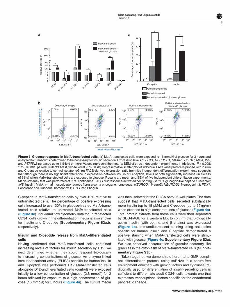
Glucose response of MafA-transfected cellsWe then investigated whether regulation of the transcript levels in the MafA-transfected cells would be sensitive to glucose treatment. D12 MafA-transfected cells were treated with 16 mmol/l glucose for 3 hours. Total RNA was then
extracted for quantitative RT-PCR where we observed posi-tive regulation in mRNA levels of the relevant transcription factors when compared with cells without glucose treatment (Figure 3a). Scatter plot fluorescence-activated cell sorting analysis (Figure 3b) demonstrated a significant increase in staining for insulin and C-peptide in MafA-transfected cells treated with glucose. Fluorescence-activated cell sorting analysis from a total of five independent experiments dem-onstrated a significant increase in expression of insulin and
Figure 2 Upregulation of key transcriptional factors in the presence of MafA saRNA. mRNA levels of (a) PDX1, (b) NEUROG3, (c) NEUROD1, (d) NKX6-1, (e) MafA, (f) GCK, (g) GLP1R, (h) SUR1 (K-ATP component (sulfonyl urea receptor, the ATP sensitive potassium channel), ABCC8, (i) PTPRN2, and (j) INS. Values are mean ± SEM of three independent experiments run in triplicate and shows expression relative to undifferentiated CD34+ cells. *P < 0.005; **P < 0.0001; ***P < 0.00001, paired Student’s t-test, two-tailed at 95% CI. GCK, glucokinase; GLP1R, glucagon-like peptide 1 receptor; INS, insulin; MafA, v-maf musculoaponeurotic fibrosarcoma oncogene homologue; NEUROD1, NeuroD; NEUROG3, Neurogenin 3; PDX1, Pancreatic and Duodenal homeobox 1; PTPRN2, Phogrin; SUR1, sulfonylurea receptor-1.
0
Undifferentiated CD34
Untransfected
MafA transfected
10
PDX1
MAFA GCK GLP1R
NEUROG3
Rel
ativ
e ex
pres
sion
0
2
4
6
8
Rel
ativ
e ex
pres
sion
0
5
10
15
20
Rel
ativ
e ex
pres
sion
ABCC8
0
5
10
15
20
Rel
ativ
e ex
pres
sion
0
2
4
6
8
10
Rel
ativ
e ex
pres
sion
Rel
ativ
e ex
pres
sion
20
***
*****
***
**
0
Undifferentiated CD34
Untransfected
MafA transfected
10
15
5
2030
0
Undifferentiated CD34
Untransfected
MafA transfected
10
NEUROD1
Rel
ativ
e ex
pres
sion
20
***
0
100
200
300
400
Undifferentiated CD34
Untransfected
MafA transfected
Undifferentiated CD34
Untransfected
MafA transfected
Undifferentiated CD34
Untransfected
MafA transfected
Undifferentiated CD34
Untransfected
MafA transfected
PTPRN2
0
2
4
6
8
10
Rel
ativ
e ex
pres
sion
**
Undifferentiated CD34
Untransfected
MafA transfected
INS
0
20
40
60
80
Rel
ativ
e ex
pres
sion
**
Undifferentiated CD34
Untransfected
MafA transfected
Undifferentiated CD34
Untransfected
MafA transfected
NKX6-1
Rel
ativ
e ex
pres
sion
***30
a b c d
e f g
i j
h
www.moleculartherapy.org/mtna
Short-activating RNA OligonucleotideReebye et al
5
C-peptide in MafA-transfected cells by over 12% relative to untransfected cells. The percentage of positive expressing cells increased to over 30% in glucose-treated MafA-trans-fected cells relative to untreated MafA-transfected cells (Figure 3c). Individual flow cytometry data for untransfected CD34+ cells grown in the differentiation media is also shown for insulin and C-peptide (Supplementary Figure S2a,b, respectively).
Insulin and C-peptide release from MafA-differentiated cellsHaving confirmed that MafA-transfected cells contained increasing levels of factors for insulin secretion by D12, we next determined whether they could respond biologically to increasing concentrations of glucose. An enzyme-linked immunoabsorbent assay (ELISA) specific for human insulin and C-peptide was performed. D12 MafA-transfected cells alongside D12-undifferentiated cells (control) were exposed initially to a low concentration of glucose (2.8 mmol/l) for 2 hours followed by exposure to a high concentration of glu-cose (16 mmol/l) for 3 hours (Figure 4a). The culture media
was then isolated for the ELISA onto 96-well plates. The data suggest that MafA-transfected cells secreted substantially more insulin (up to 18 pM/L) and C-peptide (up to 35 ηg/ml) when exposed to high concentrations of glucose (Figure 4a). Total protein extracts from these cells were then separated by SDS-PAGE for a western blot to confirm that biologically active insulin (with both α and β chains) was expressed (Figure 4b). Immunofluorescent staining using antibodies specific for human insulin and C-peptide demonstrated a positive staining when MafA-transfected cells were stimu-lated with glucose (Figure 4c; Supplementary Figure S3a). We also observed accumulation of granular secretory like granules in the cytoplasm of MafA-transfected cells (Supple-mentary Figure S3b)
Taken together, we demonstrate here that a GMP compli-ant differentiation protocol using saRNAs in a serum-free environment enriched with growth factors and cytokines tra-ditionally used for differentiation of insulin-secreting cells is sufficient to differentiate adult CD34+ cells towards one that expresses transcriptional factors specific for the endodermal pancreatic lineage.
Figure 3 Glucose response in MafA-transfected cells. (a) MafA-transfected cells were exposed to 16 mmol/l of glucose for 3 hours and analyzed for transcripts determined to be necessary for insulin secretion. Expression levels of PDX1, NEUROD1, NKX6-1, GLP1R, MafA, INS, and PTPRN2 increased up to 1.5-fold or more. Values represent the mean ± SEM of three independent experiments in triplicate. *P < 0.005; **P < 0.0001, paired Student’s t-test, two-tailed at 95% CI. (b) Representative scatter plot of individual FACS-analyzed cells probed with insulin and C-peptide relative to control isotype IgG. (c) FACS-derived expression ratio from five independent differentiation experiments suggests that although there is no significant difference in expression between insulin or C-peptide, levels of both significantly increase (in excess of 35%) when MafA-transfected cells are exposed to glucose. Results are mean and SEM of five independent differentiation experiments. Mann–Whitney test was performed at 95% confidence. FACS, fluorescence-activated cell sorting; GLP1R, glucagon-like peptide 1 receptor; INS, Insulin; MafA, v-maf musculoaponeurotic fibrosarcoma oncogene homologue; NEUROD1, NeuroD; NEUROG3, Neurogenin 3; PDX1, Pancreatic and Duodenal homeobox 1; PTPRN2, Phogrin.
0
101
101 102 103
525_50 B-A
104 105
24.47%75.53%
0.00%0.00%
14.66%61.03%
13.40%10.91%
Control: IgG isotype Untransfected cells MafA-transfected cells
102
103
AP
C-A
C-P
eptid
e-A
lexa
Flu
or 6
47 104
101
101 102 103
525_50 B-A
104 105
102
103
AP
C-A
104
20.99%46.52%
22.96%9.53%
101
101 102 103
525_50 B-A
104 105
102
103A
PC
-A
104
MafA-transfected cells +16 mmol/l glucose
11.02%9.43%
47.90%31.65%
101
101 102 103
525_50 B-A
104 105
102
103
AP
C-A
104
0C-peptide
Untransfected cells
MafA transfected
MafA transfected + 16 mmol/l glucose
Insulin
10
20
Per
cent
age
of e
xpre
ssin
g ce
lls (
%)
30
40
50
PDX1
NEUROD1
NKX6-1
GLP1R
MAFA IN
S
PTPRN2
5101520
*
**
*
**
**
**
4060
Rel
ativ
e ex
pres
sion 600
800
MafA transfected
MafA transfected +16 mmol/l glucose
Insulin-AlexaFluor 488
a
b
c

Molecular Therapy–Nucleic Acids
Short-activating RNA OligonucleotideReebye et al
6
Discussion
The primary therapeutic goal of preventing complications associated with diabetes is to maintain a stable glycemic content by reconstitution of endogenous insulin secretion. Islet transplantation therefore presents a feasible therapeutic alternative for metabolically labile patients with T1DM; how-ever, shortage of organ donors does not make this a practical option. Many attempts have been made to develop surrogate insulin-secreting cells using embryonic stem cells, organ derived stem cells or inducible pluripotent stem cells.
The basic methodology of pushing stem cells towards the pancreatic endodermal lineage has previously been described by other groups. The addition of activin A, exendin-4, noggin, EGF, and bFGF during stages of the
differentiation process is known to drive stem cells towards an insulin-expressing phenotype.12,18–20 The current challenge however is the ability to maintain this expression to comple-tion and generate cells that will respond to glucose using a GMP compliant serum-free method. This has not yet been achieved. We therefore demonstrate that a combination of GMP compliant growth factors and saRNA molecules can be used to transform human adult CD34+ cells into glucose sensitive and insulin-secreting cells. This novel use of saRNA oligonucleotides positively increases the transcript levels of MafA. Being the master switch for insulin expression in β-cells, MafA then induces expression of key endodermal endocrine factors including PDX1, Neurogenin 3 and NeuroD, MafA, glucokinase, glucagon-like peptide 1 receptor, SUR1-KATP, and phogrin. These factors are essential in supporting
Figure 4 Insulin and C-peptide release from MafA-transfected cells. (a) ELISA demonstrating MafA-transfected cells respond to glucose stimulation when compared with undifferentiated CD34+ cells (control). The culture media from control and from day 12 MafA-transfected cells were recovered following exposure of cells to low glucose (2.8 mmol/l) followed by high glucose (16 mmol/l) pulse. The media was then transferred onto immunoabsorbent ELISA plates specific for human insulin and human C-peptide. The data suggest that MafA-transfected cells are able to respond to a glucose gradient by secreting insulin. (b) Day 12 MafA-transfected cells that were treated with 16 mmol/l glucose from as mention in a were isolated for total protein extraction and SDS-PAGE separation. A western blot probed with an antibody specific to human insulin demonstrates the presence of biologically active insulin with its α and β chain. (c) Confocal microscopy of undifferentiated (control) and MafA-transfected cells with or without glucose treatment. Both control and MafA-transfected cells (with or without glucose treatment) were then costained with anti-insulin and anti C-peptide.
Actin
Insulin
38 Kda
DAPI
Con
trol
Maf
A tr
ansf
ecte
dM
afA
tran
sfec
ted
+ 1
6 m
mol
/l gl
ucos
e
Insulin C-peptide
6 Kda β chain
α chain4 Kda
Contro
l
Maf
A tran
sfecte
d
+ 16
mm
ol/l g
lucos
e
0
5
15
Contro
l
Contro
l + 2
.8 m
mol/
l gluc
ose
Contro
l + 1
6 m
mol/
l gluc
ose
Maf
A tran
sfecte
d +
2.8
mm
ol/l g
lucos
e
Maf
A tran
sfecte
d +
16 m
mol/
l gluc
ose
Maf
A tran
sfecte
d
10
20
25
pM
Insulina
0
Contro
l
Contro
l + 2
.8 m
mol/
l gluc
ose
Contro
l + 1
6 m
mol/
l gluc
ose
Maf
A tran
sfecte
d +
2.8
mm
ol/l g
lucos
e
Maf
A tran
sfecte
d +
16 m
mol/
l gluc
ose
Maf
A tran
sfecte
d
10
20
ng/m
l
30
40C-peptide
b c

www.moleculartherapy.org/mtna
Short-activating RNA OligonucleotideReebye et al
7
glucose sensitivity and processing of insulin. This observa-tion ties in with other reports by Wang et al. and Aramata et al. suggesting that the master regulatory potential of MafA can feasibly mediate transdifferentiation of progenitor cells towards an insulin secretory lineage.48,49 Moreover, our dif-ferentiation protocol also suggests a potential to upregulate sensitivity to the incretin hormone, GLP1 which is important in promoting pancreatic endocrine cell growth, in stimulat-ing insulin release in the post-prandial state and in expan-sion of β-cell mass via islet cell neogenesis.44 Therefore, our approach may be a possible avenue towards restoration of glucose sensitivity for type-2 diabetes and one that will be further investigated.
At the current stage of this promising approach to gener-ating glucose responsive cells from hematopoietic derived CD34+ cells, our procedure demonstrates that up to 35% of MafA-transfected cells responds to glucose and achieved an insulin secretion content of approximately 18 pM/L by day 12. The next challenge will be to meet the normal adult insulin requirement of 57–79 pM/L (between meals) and to estab-lish how these cells behave functionally in vivo and whether they may require encapsulation strategies or micro chamber technology to enhance cell survival beyond 12 days following transplantation.50,51 Despite the numerous hurdles ahead, the work performed here represents a significant step forward towards developing a potentially useful clinical tool for recon-stituting endogenous insulin secretion with reduced possi-bility of rejection. Although this strategy may not completely remove the requirement for pharmacological insulin replace-ment formulations, it may provide a baseline to reduce the risks associated with severe diabetes.
Materials and methods
CD34+ stem cell source. Granulocyte colony-stimulating factor–mobilized peripheral blood cells were obtained from leukaphereses processed by the Stem Cell Laboratory, Hammersmith Hospital. A total of five healthy donor patients undergoing mobilization for transplantation in allogeneic recipients were used where cells were in excess of clinical requirements. Blood samples were obtained with informed patient consent and approved by the Hammersmith Hospital Research Ethics Committee. We found that the response of the cells used in the differentiation procedure was similar for all the five healthy donors.
CD34+ stem cell isolation. Human-mobilized peripheral blood samples were diluted in a ratio of 1:4 in Hanks’ buffered saline solution (Gibco, Paisley, UK), the mononuclear cells were separated by centrifugation over a Lymphoprep (Axis-Shield, Dundee, UK) density gradient at 1,800 rpm for 30 minutes. The mononuclear cell fraction at the interface was aspirated and washed twice with HBSS, and finally with MACS buffer (Dul-becco’s phosphate-buffered saline solution (DPBS) (Gibco) at pH 7.2 supplemented with 0.5% human serum albumin and 2 mmol/l EDTA). CD34+ cells were isolated using a CD34+ isolation kit (Miltenyi Biotec, Bergisch Gladbach, Germany) according to the manufacturer’s protocol. Adherent CD34+ pro-genitor cells were isolated as previously described with modi-fication.38,39 Briefly, isolated CD34+ cells were added to 24-well
tissue culture treated dishes (Nunc, Loughborough, UK) at a density of 2.5–5 × 10⁵ cells in α-MEM medium to isolate the adherent CD34+ cell population. After 30 minutes incubation, non-adherent cells were removed and adherent cells were rinsed three times with PBS. The adherent CD34+ cell popula-tion was grown in a serum-free medium-CellGro (CellGenix, Freiburg, UK) containing three cytokines: 250 ηg/ml of stem cell factor, 250 ηg/ml of interleukin-6, and 250 ηg/ml of interleu-kin-3 (Invitrogen, Carlsbad, CA or CellGenix) in 0.5% penicil-lin/streptomycin antibiotics. Cells were incubated at 37 °C in humidified 5% CO2 air.
Design of short-activating RNA oligonucleotides. The gene sequence of MafA was selected for designing short-activating RNA molecules for its specific activation. Four parameters were used: (i) targeting gene annotations from UCSC RefSeq database; (ii) targeted sequence from anti-sense RNA; (iii) promoter selection of antisense sequences; and (iv) identification of candidate short activating RNAs. First, the method downloads information about the target’s genomic location, orientation, and transcriptional structure from available databases (RefSeq at UCSC). Second, given a database of RNA transcripts with known read direction, such as the UCSC Spliced EST track, our method searches the database for transcripts that are antisense to and in the vicinity of the target gene. More specifically, the method iden-tifies antisense transcripts that (a) overlap the target’s pro-moter and the target mRNA’s 5′ end; (b) overlap the target mRNA; (c) are at most 20-100 kb upstream of the target’s transcription start site; or (d) are at most 20-100 kb down-stream of the target’s poly-adenylation site. The method uses these four criteria as hierarchical filters such that if it finds antisense transcripts that for example satisfy criterion (a), the method does not consider the three other criteria. Third, based on the target’s transcription start site, the method downloads the antisense genomic sequence from a fixed size region upstream and downstream of the transcription start site. The typical region size used by the method is 500 nts upstream and downstream of transcription start site, but larger or smaller sizes can also be used. Fourth, the method designs siRNAs that give effective and specific downregula-tion of the antisense target sequence. The method (a) uses a siRNA design algorithm, such as GP boost,52 to identify can-didate effective siRNAs; (b) removes all candidate siRNAs with aaaa, cccc, gggg, or uuuu motifs and GC content <20% or >55%; (c) removes all candidates that have Hamming dis-tance less than two to all potential off-target transcripts; and (d) returns a given number of remaining non-overlapping siR-NAs sorted by their predicted siRNA knockdown efficacy. The method returns the two highest scoring short activating RNAs for a given antisense target sequence. Paired short activating RNA oligonucleotides were annealed using 50 mmol/l Tris–HCl, pH 8.0, 100 mmol/l NaCl and 5 mmol/l EDTA following a denaturation step at 90 °C followed by a gradual anneal step to room temperature.
Differentiation procedure. A flow diagram of the differentiation procedure is shown in (Figure 1a). The commencement of the differentiation protocol involved supplementing the adher-ent subpopulation of hematopoietic adult CD34+ cells with

Molecular Therapy–Nucleic Acids
Short-activating RNA OligonucleotideReebye et al
8
10 ηg/ml of activin A (Sigma, Poole, UK) and 2.5 mmol/l glucose (Sigma, St Louis, MO) into serum-free CellGro media (Cell-Genix) containing Stem Cell Factor (Invitrogen), 250 ηg/ml of interleukin-3 and interleukin-6 (Invitrogen) in 0.5% penicil-lin/streptomycin antibiotics for 72 hours at day 0. This was then followed by the addition of 11 mmol/l glucose, 5 ηg exendin-4 (Sigma, St Louis, MO), 50 ηg noggin (Sigma, St Louis, MO), 5 ηg bFGF (Sigma, St Louis, MO), and 5 ηg EGF (Sigma, St Louis, MO) at 72 hour intervals together with transfection 25 mmol/l of annealed short activating RNA oligonucleotide targeted to MafA (OmniCyte, London, UK) using Nanofectamine (PAA, Somerset, UK) following the manufacturer’s instructions. This process was repeated three times at days 2, 5, and 8.
Isolation of total RNA. Total RNA extraction was carried out on non-adherent differentiated cells using the RNAqueous-Micro kit (Ambion, Paisley, UK) following the manufacturer’s instruc-tions. Briefly, the cells were gently centrifuged followed by three pulses of sonication at Output 3 in Lysis buffer (Ambion). The cell lysates were then processed through an RNA bind-ing column, followed by multiple washes and elution. The total RNA isolated was quantified by a Nanodrop 2000 spectropho-tometer (Thermo Scientific, Wilmington, DE).
For semi-quantitative RT-PCR. Total RNA of 500 ηg was reversed transcribed using One Step RT-PCR (Qiagen, Hilden, Germany) following the manufacturer’s instructions. Expression for PDX1, RFX6, MafA, insulin, and for loading control, the house keeping gene actin was performed by PCR using their respective primer pairs: PDX1- F: CGG AAG AAA AAG AGC CAT TG; R: GCC AGA GGA AGA GGA GGA CT following 59 cycles at 95 °C, 20 seconds; 60 °C, 20 sec-onds; 72 °C, 40 seconds. Rfx6- F: AAT GGC AGG GAG GCC CCC AA; R: CTG GGC TTG CAG GGA TGG GC following 39 cycles at 95 °C, 30 seconds; 55 °C, 30 seconds; 65 °C, 45 seconds. MafA-F: AGC TGG TGT CCA TGT CGG TGC G; R: TGG CAC TTC TCG CTC TCC AGA ATG T following 39 cycles at 95 °C, 45 seconds; 67.4 °C, 45 seconds; 72 °C, 45 seconds. Insulin (nested cycle, previously described)42-F: GAC CTA CTG CCA CAA CAG CTA; R: GTT GCA GTA GTT CTC CAG CTG; following 40 cycles at 95 °C, 1 minute; 55 °C, 1 minute; 70 °C, 1 minute. For the nested step using F(nested): CTG TCC TTC TGC CAT GG; following 35 cycles at 50 °C, 1 minute; 95 °C, 1 minute; 58 °C, 45 seconds; 68 °C, 45 seconds in at 35 cycles. Actin-F: GAG AAA ATC TGG CAC CAC ACC; R: ATA CCC CTC GTA GAT GGG CAC following 37 cycles at 95 °C, 5 minutes; 60 °C, 30 seconds; 70 °C, 45 seconds; 72 °C, 10 minutes at 37 cycles.
For quantitative RT-PCR. Total extracted RNA of 500 ηg was processed for elimination of genomic DNA followed by reverse transcription using the QuantiTect Reverse Transcription kit from Qiagen. The first strand cDNA synthesis was then ampli-fied for quantitative analysis of PDX1 (NM_000209), Neu-rogenin 3 (NM_020999), NeuroD (NM_002500), NKX6-1 (NM_006168), MafA (NM_201589), glucokinase (NM_000162, NM_033507, NM_033508), GLP1R (NM_002062), SUR1, K-ATP component, ABCC8 (NM_000352), phogrin (protein tyrosine phosphatase receptor (N-terminal polypeptide 2),
PTPRN2 (NM_002847, NM_130842, NM_130843), pro-Insu-lin (NM_000207, NM_001185097, NM_001185098), and the reference gene, human gluceraldehyde-3-phosphate-dehy-drogenase (GAPDH) (NM_002046) using QuantiFast SYB-RGreen PCR Kit from Qiagen. Amplification was performed using Applied Biosystems 7900HT FAST-Real-Time System (Applied Biosystems, Paisley, UK) with 40 cycle conditions at 95 °C, 15 seconds and 60 °C, 45 seconds with a total volume of 25 μl per sample. Amplified products were then analyzed using Applied Biosystems RQ Manager 1.2.1. Four indepen-dent experiments were amplified in triplicates for quantitative analysis. Student’s t-test scoring was performed at 99% con-fidence intervals.
Confocal microscopy. CD34+ cells were fixed for 20 minutes with 4% paraformaldehyde onto 1 cm glass cover slips followed by permeabilization with 0.2% TritonX100 for 15 minutes. The coverslips were washed three times with PBS followed by blocking with 5% mouse or rabbit serum for 45 minutes. Mouse raised anti-human insulin (1:200) (Sigma, Poole, UK) or rabbit raised anti-human C-peptide (1:200) (Abcam, Cam-bridge, UK), was added to the cells in 5% serum for 1 hour. Cells were washed three times in PBS followed by the addi-tion of 5% serum for a further 15 minutes before incubation with the appropriate Alexa-488 conjugated or Alexa-594 secondary antibody (1:600) (Cell Signalling Technology, Hertfordshire, UK) for 1 hour. After five washes in PBS, cover-slips were mounted onto glass slides with Vectashield contain-ing 4′6′-diamidino-2-phenylindole (DAPI) (Vector Laboratories, Burlingame, CA). Slides were visualized on a Leica DM4000 at 60× magnification or Olympus epiflourescent CKX41 micro-scope. An average of five images was captured and compared with staining with the secondary conjugated antibody alone to confirm no presence of autofluorescence.
Insulin and C-peptide ELISA. MafA-transfected cells growing in suspension to day 12 were transferred to a preconditioning α-MEM media supplemented with 10 mmol/l nicotinamide, 50 ηg IGF-II, 10 ηg HGF, 25 ηg exendin-4, and 10 ηg activin A. Cells were incubated for 16 hours at 37 °C and 5% CO
2 before glucose stimulation. Glucose of 2.8 mmol/l was added for 3 hours followed by 16 mmol/l of glucose for a further 3 hours. The media was collected for total human insulin or human C-peptide ELISA (Millipore, Billerica, MA) following the manufacturer’s instructions. Positive control used was as supplied by the kit.
Fluorescence-activated cell sorting analysis. Approximately 3 × 10⁶ of day 12 differentiated CD34+ cells were fixed and permeabilized using Fix&Perm fixation medium (Caltag Lab-oratories, Burlingame, CA) following the manufacturer’s pro-tocol. Briefly, after washing the cells in PBS, 100 μl of reagent A (fixation medium) were added and the cells were incubated for 15 minutes at room temperature. At the end of the incu-bation, the cells were washed in PBS then 100 μl of reagent B (permeabilization medium) were added for 15 minutes at room temperature. The cells were then resuspended in block-ing buffer comprising PBS, with 5% fetal calf serum and 3% bovine serum albumin and incubated for 15 minutes. Cells were then incubated with antibodies against insulin (Abcam)

www.moleculartherapy.org/mtna
Short-activating RNA OligonucleotideReebye et al
9
(1:350) and C-peptide (Santa Cruz Biotechnology, Dallas, TX) (1:350) for 1 hour. After a wash in 3% FBS/1% bovine serum albumin in PBS (wash solution), cells were incubated with 1 μg/ml goat anti-mouse Alexa Fluor-488 (green), goat anti-rabbit Alexa Fluor-488 (green), and goat anti-rabbit Alexa Fluor-647 (red) (all Invitrogen) in blocking solution for 45 minutes in the dark, washed with PBS and resuspended in PBS for flow cytometric analysis. Stained cells were analyzed using the BD LSRII flow cytometer with 525_50 B-A and APC lasers to view the Alexa Fluors-488 and 647 respectively. At least 10,000 cells were collected for each test sample to ensure a sufficient number of positive stained cells. FlowJo 7.6 software (FlowJo, Ashland, OR) was used for the analy-sis of the results (Supplementary Materials and Methods).
Supplementary material
Figure S1. (a) An insulin and C-peptide ELISA assay of MafA-transfected cells.Figure S2. Individual and merged (in color) histograms of fluorescence-activated cell sorting analyzed cells probed with (a) anti-insulin or (b) anti-C-peptide.Figure S3. A high magnification immunostaining of insulin and C-peptide in MafA-transfected cells with glucose treat-ment and control cells (undifferentiated CD34+ cells).Materials and Methods
Acknowledgments. The authors declared no conflict of interest.
1. American Diabetes Association. (2011). Standards of medical care in diabetes–2011. Diabe-tes Care 34: S11–61.
2. Chen, S, Ding, J, Yu, C, Yang, B, Wood, DR and Grayburn, PA (2007). Reversal of streptozotocin-induced diabetes in rats by gene therapy with betacellulin and pancreatic duodenal homeobox-1. Gene Ther 14: 1102–1110.
3. Shapiro, AM, Lakey, JR, Ryan, EA, Korbutt, GS, Toth, E, Warnock, GL et al. (2000). Islet transplantation in seven patients with type 1 diabetes mellitus using a glucocorticoid-free immunosuppressive regimen. N Engl J Med 343: 230–238.
4. Assady, S, Maor, G, Amit, M, Itskovitz-Eldor, J, Skorecki, KL and Tzukerman, M (2001). Insulin production by human embryonic stem cells. Diabetes 50: 1691–1697.
5. Lumelsky, N, Blondel, O, Laeng, P, Velasco, I, Ravin, R and McKay, R (2001). Differentiation of embryonic stem cells to insulin-secreting structures similar to pancreatic islets. Science 292: 1389–1394.
6. Lechner, A, Nolan, AL, Blacken, RA and Habener, JF (2005). Redifferentiation of insulin-secreting cells after in vitro expansion of adult human pancreatic islet tissue. Biochem Biophys Res Commun 327: 581–588.
7. Yatoh, S, Dodge, R, Akashi, T, Omer, A, Sharma, A, Weir, GC et al. (2007). Differentiation of affinity-purified human pancreatic duct cells to beta-cells. Diabetes 56: 1802–1809.
8. Elsner, M, Jörns, A and Lenzen, S (2008). Diabetes therapy by lentiviral hepatic insulin gene expression without transformation of liver. Diabetologia 51: 694–5; author reply 696.
9. Talchai, C, Xuan, S, Kitamura, T, Depinho, RA and Accili, D (2012). Generation of functional insulin-producing cells in the gut by Foxo1 ablation. Nat Genet (doi:10.1038/ng.2215).
10. Ianus, A, Holz, GG, Theise, ND and Hussain, MA (2003). In vivo derivation of glucose-competent pancreatic endocrine cells from bone marrow without evidence of cell fusion. J Clin Invest 111: 843–850.
11. Hebrok, M, Kim, SK and Melton, DA (1998). Notochord repression of endodermal Sonic hedgehog permits pancreas development. Genes Dev 12: 1705–1713.
12. D’Amour, KA, Bang, AG, Eliazer, S, Kelly, OG, Agulnick, AD, Smart, NG et al. (2006). Production of pancreatic hormone-expressing endocrine cells from human embryonic stem cells. Nat Biotechnol 24: 1392–1401.
13. Pittenger, MF, Mackay, AM, Beck, SC, Jaiswal, RK, Douglas, R, Mosca, JD et al. (1999). Multilineage potential of adult human mesenchymal stem cells. Science 284: 143–147.
14. Jensen, J, Pedersen, EE, Galante, P, Hald, J, Heller, RS, Ishibashi, M et al. (2000). Control of endodermal endocrine development by Hes-1. Nat Genet 24: 36–44.
15. Nakhai, H, Siveke, JT, Klein, B, Mendoza-Torres, L, Mazur, PK, Algül, H et al. (2008). Conditional ablation of Notch signaling in pancreatic development. Development 135: 2757–2765.
16. Smith, SB, Qu, HQ, Taleb, N, Kishimoto, NY, Scheel, DW, Lu, Y et al. (2010). Rfx6 directs islet formation and insulin production in mice and humans. Nature 463: 775–780.
17. Aguayo-Mazzucato, C, Koh, A, El Khattabi, I, Li, WC, Toschi, E, Jermendy, A et al. (2011). Mafa expression enhances glucose-responsive insulin secretion in neonatal rat beta cells. Diabetologia 54: 583–593.
18. Jiang, J, Au, M, Lu, K, Eshpeter, A, Korbutt, G, Fisk, G et al. (2007). Generation of insulin-producing islet-like clusters from human embryonic stem cells. Stem Cells 25: 1940–1953.
19. Cho, YM, Lim, JM, Yoo, DH, Kim, JH, Chung, SS, Park, SG et al. (2008). Betacellulin and nicotinamide sustain PDX1 expression and induce pancreatic beta-cell differentiation in human embryonic stem cells. Biochem Biophys Res Commun 366: 129–134.
20. Shim, JH, Kim, SE, Woo, DH, Kim, SK, Oh, CH, McKay, R et al. (2007). Directed differentiation of human embryonic stem cells towards a pancreatic cell fate. Diabetologia 50: 1228–1238.
21. Fujikawa, T, Oh, SH, Pi, L, Hatch, HM, Shupe, T and Petersen, BE (2005). Teratoma formation leads to failure of treatment for type I diabetes using embryonic stem cell-derived insulin-producing cells. Am J Pathol 166: 1781–1791.
22. Roobrouck, VD, Vanuytsel, K and Verfaillie, CM (2011). Concise review: culture mediated changes in fate and/or potency of stem cells. Stem Cells 29: 583–589.
23. Chandra, V, Swetha, G, Muthyala, S, Jaiswal, AK, Bellare, JR, Nair, PD et al. (2011). Islet-like cell aggregates generated from human adipose tissue derived stem cells ameliorate experimental diabetes in mice. PLoS ONE 6: e20615.
24. Kim, JH, Shin, KH, Li, TZ and Suh, H (2011). Potential of nucleofected human MSCs for insulin secretion. J Tissue Eng Regen Med 5: 761–769.
25. Limbert, C, Päth, G, Ebert, R, Rothhammer, V, Kassem, M, Jakob, F et al. (2011). PDX1- and NGN3-mediated in vitro reprogramming of human bone marrow-derived mesenchymal stromal cells into pancreatic endocrine lineages. Cytotherapy 13: 802–813.
26. Paz, AH, Salton, GD, Ayala-Lugo, A, Gomes, C, Terraciano, P, Scalco, R et al. (2011). Betacellulin overexpression in mesenchymal stem cells induces insulin secretion in vitro and ameliorates streptozotocin-induced hyperglycemia in rats. Stem Cells Dev 20: 223–232.
27. Yuan, H, Li, J, Xin, N, Zhao, Z and Qin, G (2010). Expression of Pdx1 mediates differentiation from mesenchymal stem cells into insulin-producing cells. Mol Biol Rep 37: 4023–4031.
28. Chang, CF, Hsu, KH, Chiou, SH, Ho, LL, Fu, YS and Hung, SC (2008). Fibronectin and pellet suspension culture promote differentiation of human mesenchymal stem cells into insulin producing cells. J Biomed Mater Res A 86: 1097–1105.
29. Lin, HY, Tsai, CC, Chen, LL, Chiou, SH, Wang, YJ and Hung, SC (2010). Fibronectin and laminin promote differentiation of human mesenchymal stem cells into insulin producing cells through activating Akt and ERK. J Biomed Sci 17: 56.
30. Tang, XL, Xiao, R, Wang, YX, He, M, Xie, T, Zhang, C et al. (2011). [Neurogenin 3 and Paired box gene 4 promote PDX1-induced differentiation of mesenchymal stem cells into pancreatic secretory cells]. Beijing Da Xue Xue Bao 43: 421–426.
31. Wang, HW, Lin, LM, He, HY, You, F, Li, WZ, Huang, TH et al. (2011). Human umbilical cord mesenchymal stem cells derived from Wharton’s jelly differentiate into insulin-producing cells in vitro. Chin Med J 124: 1534–1539.
32. Li, LC, Okino, ST, Zhao, H, Pookot, D, Place, RF, Urakami, S et al. (2006). Small dsRNAs induce transcriptional activation in human cells. Proc Natl Acad Sci USA 103: 17337–17342.
33. Janowski, BA, Younger, ST, Hardy, DB, Ram, R, Huffman, KE and Corey, DR (2007). Activating gene expression in mammalian cells with promoter-targeted duplex RNAs. Nat Chem Biol 3: 166–173.
34. Huang, V, Qin, Y, Wang, J, Wang, X, Place, RF, Lin, G et al. (2010). RNAa is conserved in mammalian cells. PLoS ONE 5: e8848.
35. Morris, KV, Santoso, S, Turner, AM, Pastori, C and Hawkins, PG (2008). Bidirectional transcription directs both transcriptional gene activation and suppression in human cells. PLoS Genet 4: e1000258.
36. Schwartz, JC, Younger, ST, Nguyen, NB, Hardy, DB, Monia, BP, Corey, DR et al. (2008). Antisense transcripts are targets for activating small RNAs. Nat Struct Mol Biol 15: 842–848.
37. Modarresi, F, Faghihi, MA, Lopez-Toledano, MA, Fatemi, RP, Magistri, M, Brothers, SP et al. (2012). Inhibition of natural antisense transcripts in vivo results in gene-specific transcriptional upregulation. Nat Biotechnol 30: 453–459.
38. Gordon, MY (2008). Stem cells for regenerative medicine–biological attributes and clinical application. Exp Hematol 36: 726–732.
39. Gordon, MY, Levicar, N, Pai, M, Bachellier, P, Dimarakis, I, Al-Allaf, F et al. (2006). Characterization and clinical application of human CD34+ stem/progenitor cell populations mobilized into the blood by granulocyte colony-stimulating factor. Stem Cells 24: 1822–1830.
40. Voutila, J, Sætrom, P, Mintz, P, Sun, G, Alluin, J, Rossi, JJ et al. (2012). Gene Expression Profile Changes After Short-activating RNA-mediated Induction of Endogenous Pluripotency Factors in Human Mesenchymal Stem Cells. Mol Ther Nucleic Acids 1: e35.
41. Laub, O, Rall, L, Bell, GI and Rutter, WJ (1983). Expression of the human insulin gene in an alternate mammalian cell and in cell extracts. J Biol Chem 258: 6037–6042.
42. Zhukova, E, Afshar, A, Ko, J, Popper, P, Pham, T, Sternini, C et al. (2001). Expression of the human insulin gene in the gastric G cells of transgenic mice. Transgenic Res 10: 329–341.
43. Kawai, S, Mukai, T, Mori, S, Mikami, B and Murata, K (2005). Hypothesis: structures, evolution, and ancestor of glucose kinases in the hexokinase family. J Biosci Bioeng 99: 320–330.

Molecular Therapy–Nucleic Acids
Short-activating RNA OligonucleotideReebye et al
10
44. Toft-Nielsen, MB, Madsbad, S and Holst, JJ (2001). Determinants of the effectiveness of glucagon-like peptide-1 in type 2 diabetes. J Clin Endocrinol Metab 86: 3853–3860.
45. Aguilar-Bryan, L, Nichols, CG, Wechsler, SW, Clement, JP 4th, Boyd, AE 3rd, González, G et al. (1995). Cloning of the beta cell high-affinity sulfonylurea receptor: a regulator of insulin secretion. Science 268: 423–426.
46. Nichols, CG, Shyng, SL, Nestorowicz, A, Glaser, B, Clement, JP 4th, Gonzalez, G et al. (1996). Adenosine diphosphate as an intracellular regulator of insulin secretion. Science 272: 1785–1787.
47. Wasmeier, C and Hutton, JC (1999). Secretagogue-dependent phosphorylation of phogrin, an insulin granule membrane protein tyrosine phosphatase homologue. Biochem J 341 (Pt 3): 563–569.
48. Wang, H, Brun, T, Kataoka, K, Sharma, AJ and Wollheim, CB (2007). MAFA controls genes implicated in insulin biosynthesis and secretion. Diabetologia 50: 348–358.
49. Aramata, S, Han, SI and Kataoka, K (2007). Roles and regulation of transcription factor MafA in islet beta-cells. Endocr J 54: 659–666.
50. Beck, J, Angus, R, Madsen, B, Britt, D, Vernon, B and Nguyen, KT (2007). Islet encapsulation: strategies to enhance islet cell functions. Tissue Eng 13: 589–599.
51. Ludwig, B, Rotem, A, Schmid, J, Weir, GC, Colton, CK, Brendel, MD et al. (2012). Improvement of islet function in a bioartificial pancreas by enhanced oxygen supply and growth hormone releasing hormone agonist. Proc Natl Acad Sci USA 109: 5022–5027.
52. Saetrom, P and Snøve, O Jr (2004). A comparison of siRNA efficacy predictors. Biochem Biophys Res Commun 321: 247–253.
Molecular Therapy–Nucleic Acids is an open-access journal published by Nature Publishing Group. This work
is licensed under a Creative Commons Attribution-NonCommercial-NoDerivative Works 3.0 License. To view a copy of this license, visit http://creativecommons.org/licenses/by-nc-nd/3.0/
Supplementary Information accompanies this paper on the Molecular Therapy–Nucleic Acids website (http://www.nature.com/mtna)
Copyright © 2022 FDOKUMEN