A Protein Encoded by the Latency-Related Gene of Bovine Herpesvirus 1 Is Expressed in Trigeminal...
11
1998, 72(10):8133. J. Virol. Todd Holt, Alan Doster and Clinton Jones Yunquan Jiang, Ashfaque Hossain, Maria Teresa Winkler, during Productive Infection Interacts with Cyclin-Dependent Kinase 2 Neurons of Latently Infected Cattle and Expressed in Trigeminal Ganglionic Gene of Bovine Herpesvirus 1 Is A Protein Encoded by the Latency-Related http://jvi.asm.org/content/72/10/8133 Updated information and services can be found at: These include: REFERENCES http://jvi.asm.org/content/72/10/8133#ref-list-1 at: This article cites 28 articles, 14 of which can be accessed free CONTENT ALERTS more» articles cite this article), Receive: RSS Feeds, eTOCs, free email alerts (when new http://journals.asm.org/site/misc/reprints.xhtml Information about commercial reprint orders: http://journals.asm.org/site/subscriptions/ To subscribe to to another ASM Journal go to: on October 20, 2014 by guest http://jvi.asm.org/ Downloaded from on October 20, 2014 by guest http://jvi.asm.org/ Downloaded from
-
Upload
independent -
Category
Documents
-
view
0 -
download
0
Transcript of A Protein Encoded by the Latency-Related Gene of Bovine Herpesvirus 1 Is Expressed in Trigeminal...

1998, 72(10):8133. J. Virol.
Todd Holt, Alan Doster and Clinton JonesYunquan Jiang, Ashfaque Hossain, Maria Teresa Winkler, during Productive InfectionInteracts with Cyclin-Dependent Kinase 2Neurons of Latently Infected Cattle and Expressed in Trigeminal GanglionicGene of Bovine Herpesvirus 1 Is A Protein Encoded by the Latency-Related
http://jvi.asm.org/content/72/10/8133Updated information and services can be found at:
These include:
REFERENCEShttp://jvi.asm.org/content/72/10/8133#ref-list-1at:
This article cites 28 articles, 14 of which can be accessed free
CONTENT ALERTS more»articles cite this article),
Receive: RSS Feeds, eTOCs, free email alerts (when new
http://journals.asm.org/site/misc/reprints.xhtmlInformation about commercial reprint orders: http://journals.asm.org/site/subscriptions/To subscribe to to another ASM Journal go to:
on October 20, 2014 by guest
http://jvi.asm.org/
Dow
nloaded from
on October 20, 2014 by guest
http://jvi.asm.org/
Dow
nloaded from

JOURNAL OF VIROLOGY,0022-538X/98/$04.0010
Oct. 1998, p. 8133–8142 Vol. 72, No. 10
Copyright © 1998, American Society for Microbiology. All Rights Reserved.
A Protein Encoded by the Latency-Related Gene of BovineHerpesvirus 1 Is Expressed in Trigeminal Ganglionic
Neurons of Latently Infected Cattle and Interactswith Cyclin-Dependent Kinase 2
during Productive InfectionYUNQUAN JIANG, ASHFAQUE HOSSAIN,† MARIA TERESA WINKLER,
TODD HOLT, ALAN DOSTER, AND CLINTON JONES*
Department of Veterinary and Biomedical Sciences, Center for Biotechnology,University of Nebraska, Lincoln, Lincoln, Nebraska 68583-0905
Received 12 March 1998/Accepted 23 June 1998
Despite productive viral gene expression in the peripheral nervous system during acute infection, the bovineherpesvirus 1 (BHV-1) infection cycle is blocked in sensory ganglionic neurons and consequently latency isestablished. The only abundant viral transcript expressed during latency is the latency-related (LR) RNA. LRgene products inhibit S-phase entry, and binding of the LR protein (LRP) to cyclin A was hypothesized to blockcell cycle progression. This study demonstrates LRP is a nuclear protein which is expressed in neurons oflatently infected cattle. Affinity chromatography indicated that LRP interacts with cyclin-dependent kinase 2(cdk2)-cyclin complexes or cdc2-cyclin complexes in transfected human cells or infected bovine cells. Afterpartial purification using three different columns (DEAE-Sepharose, Econo S, and heparin-agarose), LRP wasprimarily associated with cdk2-cyclin E complexes, an enzyme which is necessary for G1-to-S-phase cell cycleprogression. During acute infection of trigeminal ganglia or following dexamethasone-induced reactivation,BHV-1 induces expression of cyclin A in neurons (L. M. Schang, A. Hossain, and C. Jones, J. Virol. 70:3807–3814, 1996). Expression of S-phase regulatory proteins (cyclin A, for example) leads to neuronal apoptosis. Con-sequently, we hypothesize that interactions between LRP and cell cycle regulatory proteins promote survival ofpostmitotic neurons during acute infection and/or reactivation.
Bovine herpesvirus 1 (BHV-1) is a significant viral pathogenof cattle which causes respiratory disease, abortions, genitaldisease, or occasionally encephalitis (32). Like other membersof the Alphaherpesvirinae family, BHV-1 establishes a latentinfection in sensory ganglionic neurons (reviewed in references22 and 23). Viral DNA persists in these neurons for the life-time of infected cattle but can periodically reactivate and spread.In contrast to the 70 to 80 viral genes which are expressed dur-ing productive infection of bovine cells, latency-related RNA(LR-RNA) is the only abundant viral transcript expressed inlatently infected neurons. A small fraction of LR-RNA is poly-adenylated and alternatively spliced in neurons, suggesting thisRNA is translated into an LR protein (LRP) (5, 11). LR geneproducts inhibit S-phase entry, and LRP is associated withcyclin A (24), a protein required for S-phase entry and progres-sion (reviewed in reference 9). LR gene products may enhanceneuronal survival because in a rabbit model of BHV-1 infec-tion neurons in trigeminal ganglia (TG) express cyclin A dur-ing acute infection or reactivation (24) and neurons undergoapoptosis if cell cycle regulatory proteins which promote G1- orS-phase progression are expressed (6, 20). Furthermore, inap-propriate cyclin A expression (10, 17) or certain cyclin-depen-dent kinases (cdks) (18) can induce apoptosis.
Cell cycle progression is regulated by cyclins and cdks (re-viewed in references 9, 14, and 26). D-type cyclins (cyclins D1,D2, and D3) assemble into a holoenzyme with cdk4 or cdk6,and consequently G1 cell cycle progression occurs. Phosphor-ylation of the retinoblastoma protein (Rb) by cdk4 or cdk6-cyclin D complexes is important for G1 cell cycle progression(reviewed in references 28 and 29). Late in G1, cyclin E bindsto cdk2. Cyclin E, but not cyclin D1, induces S-phase entry inthe absence of Rb, indicating these two cyclins have uniquefunctions (19, 21). Upon commitment to S phase, cdk2-cyclinA complexes are associated with replication forks (4), and cdk2is required for DNA replication (15). Rb is differentially phos-phorylated by cdk2-cyclin A, resulting in displacement of E2F(33), a transcription factor which activates expression ofgenes necessary for DNA replication (reviewed in reference1). During G2 and M, cdc2-cyclin A or cdc2-cyclin B com-plexes predominate. Although sequential phosphorylation ofRb by the appropriate cdk is important for cell cycle progres-sion, phosphorylation of other specific substrates by cdk-cyclincomplexes is also necessary. Enzymatic activity of cdk-cyclincomplexes is negatively regulated by cdk inhibitors (cdkI) (re-viewed in reference 27). Two families of cdkI exist: (i) the Inkfamily of proteins, which specifically bind cdk4 or cdk6-cyclincomplexes; and (ii) the Cip or Kip family of cdkI, which canbind all cdk-cyclin complexes in vitro. Binding of cdkI to cdk-cyclin complexes inhibits cdk activity, and thus phosphoryla-tion of substrate is blocked. A recent study demonstrated thatcdk2 activity is stimulated and Rb is phosphorylated after her-pes simplex virus type 2 (HSV-2) infection (12), suggestingalphaherpesviruses utilize certain cell cycle regulatory compo-nents to stimulate virus infection.
* Corresponding author. Mailing address: Dept. of Veterinary andBiomedical Sciences, Center for Biotechnology, University of Ne-braska, Lincoln, Fair Street at East Campus Loop, Lincoln, NE 68583-0905. Phone: (402) 472-1890. Fax: (402) 472-9690. E-mail: [email protected].
† Present address: Virus Research Institute, Cambridge, MA 02138.
8133
on October 20, 2014 by guest
http://jvi.asm.org/
Dow
nloaded from

In this study, we addressed two questions: (i) Is LRP ex-pressed in neurons of latently infected cattle? (ii) Is LRPbound to cdk-cyclin complexes? Our results demonstrated thatLRP was expressed in TG neurons of latently infected calvesand that LRP was stably associated with cdk2-cyclin E com-plexes in productively infected cells. The significance of thesefindings is discussed.
MATERIALS AND METHODS
Virus and cells. Growth and maintenance of Madin-Darby bovine kidney(MDBK) and human osteosarcoma (U2-OS) cells were described previously(24). U2-OS cells were transfected as described previously, using calcium phos-phate (24). The Cooper strain of BHV-1 was obtained from the National Vet-erinary Services Laboratory, Animal and Plant Inspection Services, Ames, Iowa.
Preparation of cellular extracts. Whole-cell lysates and nuclear extracts wereprepared as described previously (11, 12, 24).
Antibodies, immunoprecipitations, and Western blot analysis. The P2 anti-body is directed against the amino terminus of LR open reading frame 2 (11),and the immunoglobulin G (IgG) fraction in the P2 serum was purified on aprotein A column. Antibodies directed against cdk2 (sc-163), cdk4 (sc-260), cdk7(sc-529), cdc2 (sc-54), cyclin A (sc-239 and sc-437), and cyclin E (sc-198 andsc-247) were purchased from Santa Cruz Biotechnology (Santa Cruz, Calif.). Themonoclonal antibody directed against BHV-1-encoded glycoprotein D was ob-tained from S. Srikumaran (University of Nebraska).
Immunoprecipitations were performed with 150 mg of cell lysate for 3 to 4 hat 4°C; 1 mg of antibody was used. Immune complexes were precipitated withprotein A-Sepharose beads (Bio-Rad) and washed four times with washing buffer(10 mM Tris-HCl [pH 8.0], 50 mM NaCl, 1 mM EDTA, 0.5% Nonidet P-40) (12,24). Immune complexes were washed in 13 kinase buffer (50 mM Tris, 25 mMmagnesium acetate, 2.5 mM EDTA), and the pellet was resuspended in 10 ml of
FIG. 1. Expression and localization of LRP during a productive infection ofMDBK cells. MDBK cells were infected with BHV-1 (2 TCID50/cell) for theindicated times (hours postinfection). Mock-infected cells were designated time0. (A) Whole-cell lysates were prepared and Western blot analysis was performedwith the P2 antibody as described previously (11, 24). (B) At 36 hpi, cells werescraped and washed two times with PBS. Cells were then incubated in hypotonicbuffer for 10 min (lane 1), pelleted, resuspended in hypotonic buffer, and lysedby Dounce homogenization (lane 2). Nuclei were pelleted and resuspended inhypertonic buffer. After 30 min of incubation at 4°C, the nuclei were centrifugedand the nuclear extract was removed (lane 3). Nuclei were extracted two moretimes with hypertonic buffer for 30 min each time (lanes 4 and 5). The final nu-clear pellet was lysed by boiling in SDS-PAGE buffer (lane 6). Each lane containsextract from approximately 100,000 cells. Sizes are indicated in kilodaltons.
FIG. 2. Analysis of glycoprotein D in TG of calves at 90 dpi. MDBK cells infected with BHV-1 for 24 h (A) or mock-infected MDBK cells (B) were fixed and stained.Thin sections were prepared from a latently infected calf (C and D). The samples were subsequently stained with a monoclonal antibody directed against glycoproteinD. Magnifications: 359 (A to C) and 3148 (D).
8134 JIANG ET AL. J. VIROL.
on October 20, 2014 by guest
http://jvi.asm.org/
Dow
nloaded from

the same buffer. Western blot analysis was performed as described previously(11, 12, 24).
Plasmids. Plasmid pcDNA3 LRT (LRT) contains the intact 2.0-kb LR gene.pcDNA3 LRTDSph (LRTDSph) lacks a 1-kb SphI fragment containing LRPcoding sequences. These constructs are contained within pcDNA3 (Invitrogen)and are described in detail elsewhere (11, 24).
Measurement of cdk activity. cdk reaction mixtures contained 10 mCi of[g-32P]ATP, 0.1 M ATP, 1.5 mg of a glutathione S-transferase (GST)–Rb fusionprotein (Rb-SE; see below), and enzymatic activity was performed as describedpreviously (12). After 30 min at 30°C, the reaction was stopped by the additionof 25 ml of sodium dodecyl sulfate-polyacrylamide gel electrophoresis (SDS-PAGE) loading buffer per sample. Reaction products were analyzed by SDS-PAGE; the gel was dried and autoradiographed. Kinase activity was measuredwith a PhosphorImager instrument (Molecular Dynamics). The GST-Rb con-struct Rb-SE, which contains the C-terminal domain of Rb (amino acids 768 to928) fused to GST, was obtained from J. Wang (University of California, SanDiego) (30).
Precipitation of cdk complexes with affinity matrices. Cell lysate (300 mg ofprotein) or column fractions were incubated with 50 ml of p13suc1 beads (UpstateBiotechnology, Lake Placid, N.Y.) or 25 ml of p9CKShs1 agarose beads (Calbio-chem) at 4°C with shaking overnight. Beads were washed twice in washing buffer(10 mM Tris-HCl [pH 8.0], 50 mM NaCl, 1 mM EDTA, 0.5% Nonidet P-40) for5 min at 4°C. Proteins bound to beads were boiled for 5 min in SDS-PAGE bufferand subjected to SDS-PAGE (10% gel); Western blotting was performed withthe indicated antibodies.
Partial purification of LRP from infected MDBK cells. MDBK cells (a total of5 3 109 cells in 500 100-mm2 tissue culture dishes) were infected with BHV-1 for36 to 48 h, scraped with a rubber policeman, pelleted by centrifugation, andwashed twice with cold phosphate-buffered saline (PBS). Packed cells wereresuspended in hypotonic buffer (10 mM HEPES [pH 7.9], 10 mM KCl, 1.5 mMMgCl2, 0.2 mM phenylmethylsulfonyl fluoride [PMSF], 0.5 mM dithiothreitol[DTT], 0.1 mg each of soybean trypsin inhibitor, leupeptin, and aprotinin per ml),sonicated, and centrifuged. The pellet was resuspended in hypertonic buffer
(hypotonic buffer containing 0.4 M KCl, 0.2 mM EDTA, and 25% glycerol), andthe cells were sonicated. After centrifugation, the cells were resuspended inhypertonic buffer and sonication was repeated. The three supernatants obtainedfrom sonication were pooled, the salt concentration was adjusted to 100 mM KClwith hypotonic buffer, and the extract was clarified by centrifugation at 4°C(18,000 rpm for 15 min in a J2-21 Beckman centrifuge using a JA-20 rotor).
Extracts were applied to a 40 ml of DEAE-Sepharose column (Sigma) whichwas equilibrated in loading buffer (hypotonic buffer containing 100 mM KCl, 0.2mM EDTA, and 10% glycerol). After washing with loading buffer, bound pro-teins (including LRP) were eluted in 7-ml fractions with KGED buffer (10 mMKH2PO4-Na2HPO4 [pH 7.0], 0.2 mM EDTA, 0.2 mM PMSF, 0.5 mM DTT, 5%glycerol, 0.1 mg each of soybean trypsin inhibitor, leupeptin, and aprotinin perml, 250 mM KCl). The optical density at 280 nm (OD280) was determined;LRP-containing fractions were identified by Western blot analysis using the P2antibody.
LRP-containing fractions were dialyzed in KGED buffer and then loaded ontoa 5-ml Econo S column (Bio-Rad) which was equilibrated in KGED buffer. Thecolumn was washed extensively with TGED buffer (40 mM Tris-HCl [pH 7.8], 0.2mM EDTA, 0.2 mM PMSF, 0.5 mM DTT, 10% glycerol, 0.1 mg each of soybeantrypsin inhibitor, leupeptin, and aprotinin per ml). Elution of bound proteins wasperformed with TGED buffer containing 100 mM KCl, and 2-ml fractions werecollected. LRP-containing fractions were identified by Western blot analysisusing the P2 antibody.
LRP-containing fractions were dialyzed against TGED buffer and then loadedonto a 2-ml heparin-agarose affinity column (Bio-Rad) which was equilibratedwith TGED buffer. After the column was washed extensively with TGED buffer,proteins were eluted with TGED buffer containing 100 mM KCl and 1-mlfractions were collected.
Immunohistochemistry. Two calves were infected with BHV-1 as describedpreviously (25), and two uninfected calves were used as a control. At 90 dayspostinfection (dpi), calves were euthanized. TG were fixed in neutral bufferedformalin and embedded in paraffin, and thin sections (4 to 5 mm) were cut. Tissuesections were incubated in xylene for 10 min, rehydrated in graded alcohols, and
FIG. 3. Detection of LRP in TG neurons at 90 dpi. Thin sections were prepared from two different latently infected calves (A and B) or two different uninfectedcalves (C and D). IgG from P2 antiserum was used to detect LRP. Arrowheads denote neurons which have a nucleus specifically stained by the P2 antibody.Magnification: 359 for all panels.
VOL. 72, 1998 FUNCTION OF LRP OF BHV-1 8135
on October 20, 2014 by guest
http://jvi.asm.org/
Dow
nloaded from
then treated with 3% hydrogen peroxide in PBS (pH 7.4) for 20 min to inactivateendogenous peroxidase. After being washed in distilled water for 10 min, tissuesections were digested with 0.05% protease in Tris buffer (50 mM Tris buffer [pH7.6]) for 3 min at 37°C. LRP was detected by using the indirect avidin-biotincomplex system (ABC; Santa Cruz Biotechnology). Nonspecific binding wasblocked with 10% normal goat serum diluted in PBS–0.05% Tween 20–1%bovine serum albumin (pH 7.4) for 1 h at room temperature in a humidifiedchamber. Purified P2 antibody was diluted to a final concentration of 3 mg/ml inthe same buffer as used for the normal goat serum and incubated for 3 h at 4°C.Slides were then washed three times for 10 min in PBS (pH 7.4) before additionof the biotinylated goat anti-rabbit IgG diluted 1:200. After incubating for 45 minat room temperature in a humidified chamber, three washings were performedand the ABC reagent (prepared according to the manufacturer’s recommenda-tion) was added to the slides for 45 min at room temperature in a humidifiedchamber. Finally, slides were incubated with freshly prepared substrate for 5 to10 min, rinsed with distilled water, and counterstained with Mayer’s hematoxylin.
RESULTS
Expression of LRP during productive infection. To identifythe subcellular localization of LRP, MDBK cells were infectedwith BHV-1 (2 50% tissue culture infective doses [TCID50])for 4, 8, 16, 24, 36, or 48 h, and LRP was detected with anantibody directed against the amino terminus of LRP (P2)(11). A prominent band migrating near 40 kDa was detected at36 or 48 h postinfection (hpi) by the P2 antibody but did notreact with extracts prepared from mock-infected cells (Fig.1A). Longer exposures revealed a faint band was also presentat 24 hpi (13). This was consistent with previous studies whichconcluded LRP was a 40-kDa protein in infected or transfected
cells (11, 24). As described previously (11, 24), preimmuneserum does not react with a 40-kDa protein in infected cells(13). Infected cells (36 hpi) were incubated with hypotonicbuffer to lyse the cells, and cytoplasmic extracts were prepared.Crude nuclei were subsequently incubated with hypertonicbuffer, and nuclear extracts were prepared. Although LRP wasdetected in the cytoplasmic extract (Fig. 1B, lane 2), the ma-jority of LRP was detected in nuclear extracts (Fig. 1B, lanes 3to 5). After three washes with hypertonic buffer, LRP was notreadily detected in the nuclear pellet (Fig. 1B, lane 6).
Expression of LRP in TG neurons of calves. Since LR-RNAis the only abundant viral transcript expressed in latently in-fected neurons (23), we were interested in determining wheth-er LRP was expressed in TG neurons of latently infectedcalves. Two calves were infected with BHV-1 for 90 days, andthen TG were collected as described previously (25). As con-trols, TG were collected from two uninfected calves. Thinsections were prepared from paraffin-embedded TG. At 90 dpi,LR-RNA was readily detected, but infectious virus was notdetected in ocular swabs at 85 or 90 dpi. The expression ofBHV-1 glycoprotein D in TG was also examined in TG toconfirm that the calves were latently infected. High levels ofglycoprotein D expression were detected in MDBK cells whichwere infected with BHV-1 for 24 h (Fig. 2A) but not in mock-infected cells (Fig. 2B). In TG of a calf infected for 90 days,glycoprotein D was not detected in neurons (Fig. 2C and D).Identical results were obtained from the other latently infected
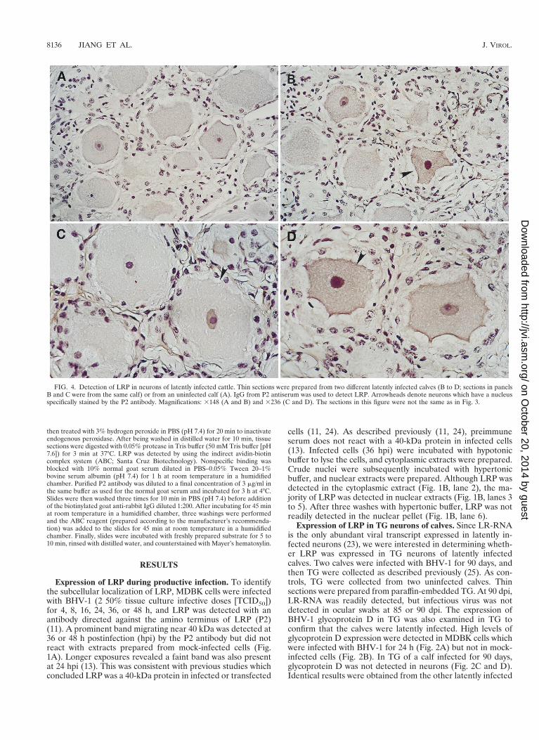
FIG. 4. Detection of LRP in neurons of latently infected cattle. Thin sections were prepared from two different latently infected calves (B to D; sections in panelsB and C were from the same calf) or from an uninfected calf (A). IgG from P2 antiserum was used to detect LRP. Arrowheads denote neurons which have a nucleusspecifically stained by the P2 antibody. Magnifications: 3148 (A and B) and 3236 (C and D). The sections in this figure were not the same as in Fig. 3.
8136 JIANG ET AL. J. VIROL.
on October 20, 2014 by guest
http://jvi.asm.org/
Dow
nloaded from
calf, and no specific staining was detected in TG prepared fromuninfected calves (31). In summary, these studies demonstrat-ed that calves which were infected with BHV-1 by ocular andnasal instillation were latently infected at 90 dpi.
The P2 antibody specifically reacted with the nucleus ofsome neurons within TG of both calves at 90 dpi (Fig. 3A andB). Approximately 10% of TG neurons from latently infectedcalves expressed LRP. In contrast, the P2 antibody did notspecifically react with thin sections of TG prepared from thetwo uninfected calves (Fig. 3C or D). Some neurons from un-infected calves have dark nuclei which are a result of counter-staining of the nucleolus and not because the antibody cross-reacts with neuronal proteins. Counterstaining of the nucleoluswas readily differentiated from P2 positive neurons by exam-ining thin sections at high or low magnification (Fig. 4). P2-positive staining resulted in reddish brown staining of the en-tire nucleus, while the nucleolus was counterstained blue orpurple. Thus, in TG of latently infected calves, LRP was de-tected in the nuclei of a subset of neurons.
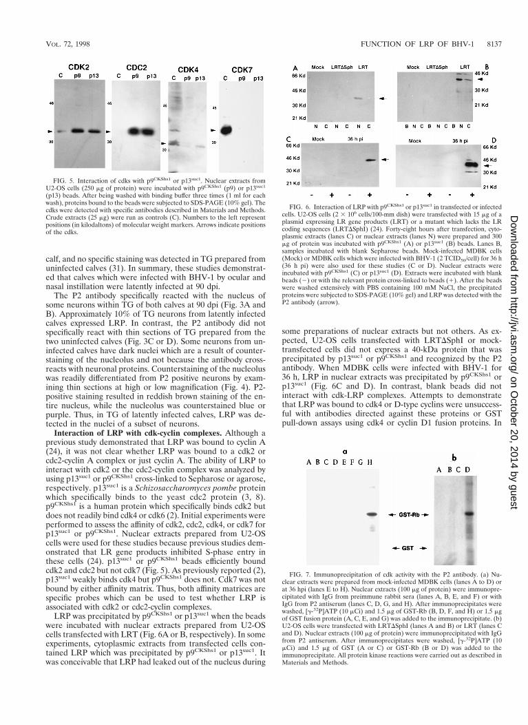
Interaction of LRP with cdk-cyclin complexes. Although aprevious study demonstrated that LRP was bound to cyclin A(24), it was not clear whether LRP was bound to a cdk2 orcdc2-cyclin A complex or just cyclin A. The ability of LRP tointeract with cdk2 or the cdc2-cyclin complex was analyzed byusing p13suc1 or p9CKShs1 cross-linked to Sepharose or agarose,respectively. p13suc1 is a Schizosaccharomyces pombe proteinwhich specifically binds to the yeast cdc2 protein (3, 8).p9CKShs1 is a human protein which specifically binds cdk2 butdoes not readily bind cdk4 or cdk6 (2). Initial experiments wereperformed to assess the affinity of cdk2, cdc2, cdk4, or cdk7 forp13suc1 or p9CKShs1. Nuclear extracts prepared from U2-OScells were used for these studies because previous studies dem-onstrated that LR gene products inhibited S-phase entry inthese cells (24). p13suc1 or p9CKShs1 beads efficiently boundcdk2 and cdc2 but not cdk7 (Fig. 5). As previously reported (2),p13suc1 weakly binds cdk4 but p9CKShs1 does not. Cdk7 was notbound by either affinity matrix. Thus, both affinity matrices arespecific probes which can be used to test whether LRP isassociated with cdk2 or cdc2-cyclin complexes.
LRP was precipitated by p9CKShs1 or p13suc1 when the beadswere incubated with nuclear extracts prepared from U2-OScells transfected with LRT (Fig. 6A or B, respectively). In someexperiments, cytoplasmic extracts from transfected cells con-tained LRP which was precipitated by p9CKShs1 or p13suc1. Itwas conceivable that LRP had leaked out of the nucleus during
some preparations of nuclear extracts but not others. As ex-pected, U2-OS cells transfected with LRTDSphI or mock-transfected cells did not express a 40-kDa protein that wasprecipitated by p13suc1 or p9CKShs1 and recognized by the P2antibody. When MDBK cells were infected with BHV-1 for36 h, LRP in nuclear extracts was precipitated by p9CKShs1 orp13suc1 (Fig. 6C and D). In contrast, blank beads did notinteract with cdk-LRP complexes. Attempts to demonstratethat LRP was bound to cdk4 or D-type cyclins were unsuccess-ful with antibodies directed against these proteins or GSTpull-down assays using cdk4 or cyclin D1 fusion proteins. In
FIG. 5. Interaction of cdks with p9CKShs1 or p13suc1. Nuclear extracts fromU2-OS cells (250 mg of protein) were incubated with p9CKShs1 (p9) or p13suc1
(p13) beads. After being washed with binding buffer three times (1 ml for eachwash), proteins bound to the beads were subjected to SDS-PAGE (10% gel). Thecdks were detected with specific antibodies described in Materials and Methods.Crude extracts (25 mg) were run as controls (C). Numbers to the left representpositions (in kilodaltons) of molecular weight markers. Arrows indicate positionsof the cdks.
FIG. 6. Interaction of LRP with p9CKShs1 or p13suc1 in transfected or infectedcells. U2-OS cells (2 3 106 cells/100-mm dish) were transfected with 15 mg of aplasmid expressing LR gene products (LRT) or a mutant which lacks the LRcoding sequences (LRTDSphI) (24). Forty-eight hours after transfection, cyto-plasmic extracts (lanes C) or nuclear extracts (lanes N) were prepared and 300mg of protein was incubated with p9CKShs1 (A) or p13suc1 (B) beads. Lanes B,samples incubated with blank Sepharose beads. Mock-infected MDBK cells(Mock) or MDBK cells which were infected with BHV-1 (2 TCID50/cell) for 36 h(36 h pi) were also used for these studies (C or D). Nuclear extracts wereincubated with p9CKShs1 (C) or p13suc1 (D). Extracts were incubated with blankbeads (2) or with the relevant protein cross-linked to beads (1). After the beadswere washed extensively with PBS containing 100 mM NaCl, the precipitatedproteins were subjected to SDS-PAGE (10% gel) and LRP was detected with theP2 antibody (arrow).
FIG. 7. Immunoprecipitation of cdk activity with the P2 antibody. (a) Nu-clear extracts were prepared from mock-infected MDBK cells (lanes A to D) orat 36 hpi (lanes E to H). Nuclear extracts (100 mg of protein) were immunopre-cipitated with IgG from preimmune rabbit sera (lanes A, B, E, and F) or withIgG from P2 antiserum (lanes C, D, G, and H). After immunoprecipitates werewashed, [g-32P]ATP (10 mCi) and 1.5 mg of GST-Rb (B, D, F, and H) or 1.5 mgof GST fusion protein (A, C, E, and G) was added to the immunoprecipitate. (b)U2-OS cells were transfected with LRTDSphI (lanes A and B) or LRT (lanes Cand D). Nuclear extracts (100 mg of protein) were immunoprecipitated with IgGfrom P2 antiserum. After immunoprecipitates were washed, [g-32P]ATP (10mCi) and 1.5 mg of GST (A or C) or GST-Rb (B or D) was added to theimmunoprecipitate. All protein kinase reactions were carried out as described inMaterials and Methods.
VOL. 72, 1998 FUNCTION OF LRP OF BHV-1 8137
on October 20, 2014 by guest
http://jvi.asm.org/
Dow
nloaded from

8138 JIANG ET AL. J. VIROL.
on October 20, 2014 by guest
http://jvi.asm.org/
Dow
nloaded from
summary, these studies demonstrated that LRP was associatedwith cdk2 or cdc2-cyclin complexes.
Nuclear extracts which were prepared from infected MDBKcells were immunoprecipitated with the P2 antibody, and thepresence of cdk activity was measured by using a GST-Rbfusion protein. Rb was used as a substrate because it is effi-ciently phosphorylated by all cdks in vitro but not by othercommon protein kinases (reviewed in references 28 and 29).The GST-Rb fusion protein (Fig. 7a, lane H) but not the GSTprotein (lane G) was phosphorylated when nuclear extractsprepared from infected MDBK cells were immunoprecipitatedby the P2 antibody. Normal rabbit sera did not immunopre-cipitate cdk activity in uninfected (lanes A and B) or infected(lanes E and F) cells. Furthermore, the P2 antibody did notimmunoprecipitate cdk activity from uninfected cells (lanes Cand D).
Nuclear extracts prepared from U2-OS cells transfected withLRT were also capable of phosphorylating GST-Rb but notGST following immunoprecipitation with the P2 antibody (Fig.7b, lanes D and C, respectively). In contrast, nuclear extractsprepared from U2-OS cells transfected with LRTDSph werenot able to phosphorylate Rb-GST or GST after immunopre-cipitation with the P2 antibody (lanes A and B). In summary,
these studies demonstrated that the P2 antibody precipitated aprotein kinase which phosphorylated Rb, a known substrate ofcdk. This finding was consistent with the conclusion that LRPwas associated with cdk2 or cdc2-cyclin complexes.
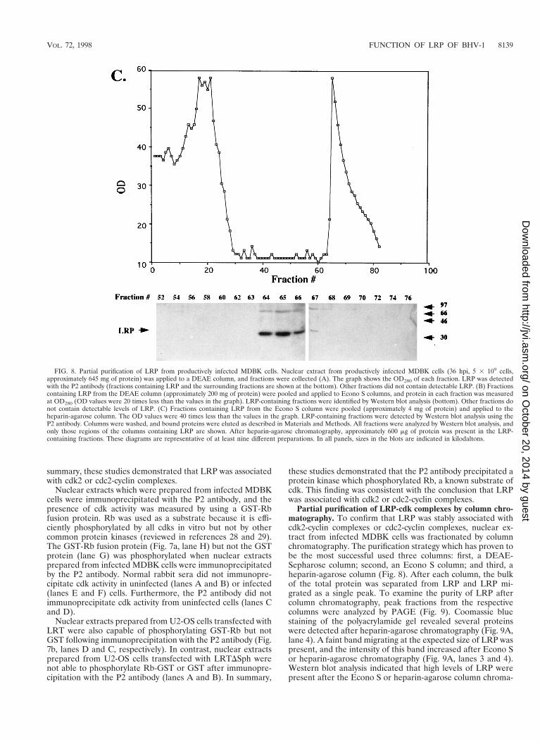
Partial purification of LRP-cdk complexes by column chro-matography. To confirm that LRP was stably associated withcdk2-cyclin complexes or cdc2-cyclin complexes, nuclear ex-tract from infected MDBK cells was fractionated by columnchromatography. The purification strategy which has proven tobe the most successful used three columns: first, a DEAE-Sepharose column; second, an Econo S column; and third, aheparin-agarose column (Fig. 8). After each column, the bulkof the total protein was separated from LRP and LRP mi-grated as a single peak. To examine the purity of LRP aftercolumn chromatography, peak fractions from the respectivecolumns were analyzed by PAGE (Fig. 9). Coomassie bluestaining of the polyacrylamide gel revealed several proteinswere detected after heparin-agarose chromatography (Fig. 9A,lane 4). A faint band migrating at the expected size of LRP waspresent, and the intensity of this band increased after Econo Sor heparin-agarose chromatography (Fig. 9A, lanes 3 and 4).Western blot analysis indicated that high levels of LRP werepresent after the Econo S or heparin-agarose column chroma-
FIG. 8. Partial purification of LRP from productively infected MDBK cells. Nuclear extract from productively infected MDBK cells (36 hpi, 5 3 109 cells,approximately 645 mg of protein) was applied to a DEAE column, and fractions were collected (A). The graph shows the OD280 of each fraction. LRP was detectedwith the P2 antibody (fractions containing LRP and the surrounding fractions are shown at the bottom). Other fractions did not contain detectable LRP. (B) Fractionscontaining LRP from the DEAE column (approximately 200 mg of protein) were pooled and applied to Econo S columns, and protein in each fraction was measuredat OD280 (OD values were 20 times less than the values in the graph). LRP-containing fractions were identified by Western blot analysis (bottom). Other fractions donot contain detectable levels of LRP. (C) Fractions containing LRP from the Econo S column were pooled (approximately 4 mg of protein) and applied to theheparin-agarose column. The OD values were 40 times less than the values in the graph. LRP-containing fractions were detected by Western blot analysis using theP2 antibody. Columns were washed, and bound proteins were eluted as described in Materials and Methods. All fractions were analyzed by Western blot analysis, andonly those regions of the columns containing LRP are shown. After heparin-agarose chromatography, approximately 600 mg of protein was present in the LRP-containing fractions. These diagrams are representative of at least nine different preparations. In all panels, sizes in the blots are indicated in kilodaltons.
VOL. 72, 1998 FUNCTION OF LRP OF BHV-1 8139
on October 20, 2014 by guest
http://jvi.asm.org/
Dow
nloaded from

tography (Fig. 9B, lanes 3 and 4). In some preparations, faintbands migrating slightly below or above LRP were detected.We hypothesized that these bands were due to proteolysis orother modifications of LRP which occurred during purificationof LRP. Based on the total protein in the peak column frac-tions and the intensity of LRP on Western blots, we estimatethat LRP was purified 300-fold.
LRP-containing fractions from the DEAE-Sepharose or he-parin-agarose column were pooled and then incubated withp13suc1 beads. After extensive washing, Western blot analysiswas performed with antibodies directed against cdk2, cdc2, orLRP. p13suc1 affinity chromatography was then used after par-tial purification of LRP because it is a stringent assay thatconfirms the stable association of LRP with cdk2 or cdc2.Fractions which did not contain LRP but were adjacent to thepeak fractions of LRP were used as a control. This approachdoes not allow us to quantify the amount of cdk-cyclin com-plexes which are associated with LRP because all of the cdk-cyclin complexes are bound by p13suc1 affinity chromatography.Since our primary goal was to prove that LRP was bound bycdk-cyclin complexes, this was not a major concern. FollowingDEAE-Sepharose chromatography, cdk2 was detected in bothpooled fractions containing LRP but cdc2 was detected in justone pooled fraction (Fig. 10A). After heparin-agarose chro-matography, cdk2 but not cdc2 was detected in the pooledfractions which contained LRP. These studies also indicatedthat a significant population of cdk2 or cdc2 was not bound toLRP following DEAE-Sepharose chromatography. Fractionscontaining LRP were also tested for the presence of cyclin A orcyclin E because cdk2 binds cyclin A or cyclin E in vivo. AfterDEAE-Sepharose chromatography, cyclin A and cyclin E werepresent in pooled fractions containing LRP (Fig. 10B). Onlycyclin E was detected after heparin-agarose column chroma-tography. Thus, in productively infected MDBK cells, LRP waspredominantly associated with cdk2-cyclin E complexes but thebulk of cdk2-cyclin complexes were not associated with LRP.
DISCUSSION
Experiments in this study demonstrated that LRP was ex-pressed in TG neurons of latently infected cattle and duringlate stages of productive infection. Further studies demonstrat-ed LRP was present in the nucleus and binds cdk2-cyclin Ecomplexes. The ability of LRP to bind cdk2-cyclin E complexesis hypothesized to have functional significance during infec-tion.
This study provided evidence that LRP was expressed in sen-sory neurons of latently infected calves. It appeared that LRPwas expressed in the nuclei of TG neurons (Fig. 3 and 4). Thisfinding was consistent with the nuclear localization of LRP inproductively infected bovine cells (Fig. 1). Although one couldargue that neurons which contain LRP were undergoing spon-taneous reactivation, this is unlikely because there were toomany neurons which express the protein. Furthermore, calvesat 90 dpi were not shedding virus in ocular swabs prior toeuthanization (31), and there was no evidence of glycoproteinD expression in TG neurons (Fig. 2). Thus, by standard criteriawhich are used to operationally define alphaherpesvirus la-tency, these calves were latently infected. Two other calves alsoexpressed LRP in sensory neurons at 60 dpi (31), and thesecalves fit the criteria of being latently infected. Our studies donot allow us to estimate what proportion of latently infected
FIG. 9. Analysis of fractions containing LRP after partial purification. Nu-clear extract from productively infected MDBK cells (36 hpi, 5 3 109 cells) waschromatographed as described for Fig. 8 and in Materials and Methods. Peakfractions containing LRP were electrophoresed in a 7.5 to 15% polyacrylamidegel, and the proteins were detected by Coomassie blue staining (A) or Westernblot analysis performed with the P2 antibody (B). Lanes: M, molecular weightmarkers (sizes of the proteins are shown in kilodaltons at the left); 1, crudenuclear extract, 400 mg of protein; 2, peak fractions of LRP from the DEAE-Sepharose column, 60 mg of protein; 3, peak fractions of LRP from the Econo Scolumn, 50 mg of protein; 4, peak fractions of LRP from the heparin-agarosecolumn, 30 mg of protein. Arrows mark the positions of LRP.
FIG. 10. Association of LRP with cdk2 or cdc2-cyclin complexes after col-umn chromatography. Nuclear extract prepared from MDBK cells infected withBHV-1 for 36 h (5 3 109 cells) was applied to a DEAE-Sepharose column, andafter extensive washing bound proteins were eluted. Fractions containing LRPwere identified by Western blotting using the P2 antibody, and these fractionswere applied to an Econo S column. After extensive washing, bound proteinswere eluted. Fractions containing LRP were identified by Western blot analysisusing the P2 antibody, and these fractions were applied to a heparin-agarose col-umn. After extensive washing, bound proteins were eluted and LRP was iden-tified. (A) Association of LRP with cdk2 or cdc2. Two pooled fractions from theDEAE-Sepharose column which did not contain detectable levels of LRP wereused as controls: fractions 19 to 21 from Fig. 8A (lane 1) and fractions 31 to 33from Fig. 8A (lane 2). Two pooled fractions from the DEAE-Sepharose columnwhich contained LRP were incubated with the affinity matrix p13suc1: fractions 22to 25 from Fig. 8A (lane 3) and fractions 26 to 30 from Fig. 8A (lane 4). Fromthe heparin-agarose column, two pooled fractions were incubated with p13suc1
beads: fractions 60 to 63 from Fig. 8C (lane 5) and fractions 64 to 67 from Fig.8C (lane 6). After extensive washing, bound proteins were subjected to SDS-PAGE and subsequently incubated with the relevant antibodies. The positions ofLRP, cdk2, and cdc2 are indicated. (B) Association of LRP with cyclin A or E.Peak fractions which contained LRP from the DEAE-Sepharose column (lanesD) or the heparin-agarose column (lanes H) were incubated with p13suc1 beads.Bound proteins were then electrophoresed, and Western blots were probed forthe presence of cyclin A (60 kDa) or cyclin E (50 kDa).
8140 JIANG ET AL. J. VIROL.
on October 20, 2014 by guest
http://jvi.asm.org/
Dow
nloaded from

neurons are expressing LRP. However, it seems unlikely thatall latently infected neurons are expressing detectable levels ofLRP.
Three lines of evidence demonstrated that LRP was boundto cdk2-cyclin complexes in transfected human cells or infectedbovine cells: (i) LRP was efficiently precipitated by affinitymatrices which bind cdk2 or cdc2 complexes, p13suc1 orp9CKShs1 (Fig. 6); (ii) antiserum directed against LRP copre-cipitated a protein kinase which phosphorylated Rb (Fig. 7), aspecific substrate for cdks, and (iii) LRP was associated withcdk2 after passage through three columns because it was re-tained by the p13suc1 beads (Fig. 10). The finding that LRP wasnot associated with cdc2 after heparin-agarose chromatogra-phy suggested it was not tightly bound to cdc2 or that low levelsof cdc2 were bound to LRP. Since LRP migrates on SDS-PAGE with a mobility of 40 kDa and cdk2 and cyclin E migrateas proteins of 35 and 50 kDa, respectively, the complex shouldhave a mass of 125 kDa. Following DEAE-Sepharose chroma-tography and gel filtration, LRP migrated between 80 and 140kDa, supporting the hypothesis that LRP was bound to otherproteins (13).
Overexpression of cyclin E initiates S phase in Rb-negativecells (21) but does not activate the Rb/E2F pathway (16). cdk2-cyclin E complexes are believed to phosphorylate DNA repli-cation factors, thus promoting initiation of DNA replication(reviewed in reference 9). Since cdk2 activity is stimulated byinfection with HSV-2 (12) or BHV-1 (13), cdk2 may be impor-tant for efficient infection and its activity may be altered byviral proteins, including LRP. The association of proteins withcdk-cyclin complexes can result in repression of cdk activity(reviewed in reference 26), changes in substrate specificity (7),or have no obvious effect on cdk activity, but its associationpromotes cell cycle progression (34). The functional impact ofLRP binding to cdk2-cyclin complexes is unknown.
What is the selective advantage for BHV-1 to express a pro-tein in postmitotic neurons that binds cdk2-cyclin complexesand inhibits S-phase entry? A previous study demonstratedthat cyclin A, a protein required for S-phase progression, is ex-pressed in TG neurons of rabbits during acute infection or af-ter dexamethasone-induced reactivation (24). Following HSV-2 (12) or BHV-1 (31) infection, certain cell cycle regulatoryproteins are activated. These observations suggest that cell cy-cle regulatory factors enhance viral replication or transcrip-tion. Activation of cell cycle regulators in neurons by BHV-1 orHSV-2 poses a dilemma for the virus because neuronal apo-ptosis is preceded by induction of proteins which promotes cellcycle progression (6). The concept that apoptosis is linked toinappropriate activation of cell cycle regulatory proteins issupported by the finding that cyclin A-dependent kinase activ-ity is stimulated during apoptosis (17), apoptosis is suppressedby dominant negative mutants of cdk2 or cdc2 (18), and cellcycle inhibitors promote survival of postmitotic neurons (20).As discussed previously (24), we believe that LRP promotesneuronal survival during infection of neurons by blocking thedeleterious effects of virus-induced activation of cell cycle fac-tors. Recent findings demonstrated that LR-RNA is alterna-tively spliced in TG during acute infection relative to latencyor productive infection of nonneural cells (5). Taken to-gether, these findings have led us to hypothesize that neuralcell-specific LRP isoforms have novel biological propertiesthat are important for specific stages of latency in cattle.Functional studies designed to test this hypothesis are inprogress.
ACKNOWLEDGMENTS
The first three authors contributed equally to this work.This research was supported by grants from the USDA (9402117 and
9702394) and the Center for Biotechnology.We thank Jean Wang (UCSD) for the plasmid encoding GST-Rb
and S. Srikumaran for the monoclonal antibody directed againstBHV-1 gD. We also thank Fernando Osorio and Charles Wood forproviding critical reviews of the manuscript.
REFERENCES
1. Adams, P. D., and W. G. Kaelin, Jr. 1996. The cellular effects of E2F over-expression. Curr. Top. Microbiol. Immunol. 208:79–83.
2. Azzi, L., L. Meijer, S. I. Reed, R. Pidikiti, and H. Y. L. Tung. 1992. Inter-action between the cell cycle control proteins p34cdc2 and p9CKShs2. Eur.J. Biochem. 203:353–360.
3. Brizuela, L., G. Draetta, and D. Beach. 1987. p13suc1 acts in the fission yeastcell division cycle as a component of the p34cdc2 protein kinase. EMBO J.6:3507–3514.
4. Cardoso, M. C., H. Leonhardt, and B. Nadal-Ginard. 1993. Reversal ofterminal differentiation and control of DNA replication: cyclin A and cdk2specificity localize at subnuclear sites of DNA replication. Cell 74:979–992.
5. Devireddy, L. R., and C. Jones. 1998. Alternative splicing of the latency-related transcript of bovine herpesvirus 1 yields RNAs containing uniqueopen reading frames. J. Virol. 72:7294–7301.
6. Freeman, R. S., S. Estus, and E. M. Johnson. 1994. Analysis of cell cycle-related gene expression in post mitotic neurons: selective induction of cyclinD1 during programmed cell death. Neuron 12:343–335.
7. Hauser, P. J., D. Agrawal, B. Chus, and W. J. Pledgers. 1997. p107 and p130associated cyclin A has altered substrate specificity. J. Biol. Chem. 272:22954–22959.
8. Hayles, J., S. Aves, and P. Nurse. 1986. Suc1 is an essential gene involved inboth the cell cycle and growth in fission yeast. EMBO J. 5:3373–3379.
9. Heichman, K. A., and J. M. Roberts. 1994. Rules to replicate by. Cell 79:557–562.
10. Hoang, A. T., K. J. Cohen, J. F. Barrett, D. A. Bergstrom, and C. V. Dang.1994. Participation of cyclin A in Myc-induced apoptosis. Proc. Natl. Acad.Sci. USA 91:6875–6879.
11. Hossain, A., L. Schang, and C. Jones. 1995. Identification of gene productsencoded by the latency-related gene of bovine herpesvirus 1. J. Virol. 69:5345–5352.
12. Hossain, A., T. Holt, J. Ciacci-Zanella, and C. Jones. 1997. Analysis of cyclindependent kinase activity after herpes simplex virus type 2 infection. J. Gen.Virol. 78:3341–3348.
13. Jiang, Y., A. Hossain, T. Holt, and C. Jones. Unpublished data.14. King, R. W., P. K. Jackson, and M. W. Kirschner. 1994. Mitosis in transition.
Cell 79:563–571.15. Krude, T., M. Jackman, J. Pines, and R. A. Laskey. 1997. Cyclin/Cdk-
dependent initiation of DNA replication in a human cell-free system. Cell88:109–119.
16. Lukas, J., T. Herzinger, K. Hansen, M. C. Moroni, D. Resnitzky, K. Helin,S. I. Reed, and J. Bartek. 1997. Cyclin E-induced S phase without activationof the pRb/2F pathway. Genes Dev. 11:1479–1492.
17. Meikrantz, W., S. Geisselbrecht, S. W. Tam, and R. Schlegel. 1994. Activa-tion of cyclin A-dependent protein kinases during apoptosis. Proc. Natl.Acad. Sci. USA 91:3754–3758.
18. Meikrantz, W., and R. Schlegel. 1996. Suppression of apoptosis by dominantnegative mutants of cyclin-dependent protein kinases. J. Biol. Chem. 271:10205–10209.
19. Ohtsubo, M., A. M. Theodoras, J. Schumaker, J. M. Roberts, and M. Pa-gano. 1995. Human cyclin E, a nuclear protein essential for the G1-to-Sphase transition. Mol. Cell. Biol. 15:2612–2624.
20. Park, D. S., S. E. Farinellis, and L. A. Greene. 1996. Inhibitors of cyclin-dependent kinases promote survival of post-mitotic neuronally differentiatedPC12 cells and sympathetic neurons. J. Biol. Chem. 14:8161–8169.
21. Resnitzky, D., and S. I. Reed. 1995. Different roles for cyclins D1 and E inregulation of the G1-to-S transition. Mol. Cell. Biol. 15:3463–3469.
22. Rock, D. L. 1993. The molecular basis of latent infections by alpha herpes-viruses. Semin. Virol. 4:157–165.
23. Rock, D. L. 1994. Latent infection with bovine herpesvirus type 1. Semin.Virol. 5:233–240.
24. Schang, L., A. Hossain, and C. Jones. 1996. The latency-related gene ofbovine herpesvirus 1 encodes a factor which inhibits cell cycle progression.J. Virol. 70:3807–3814.
25. Schang, L. M., and C. Jones. 1997. Analysis of bovine herpesvirus 1 tran-scripts during a primary infection of trigeminal ganglia of cattle. J. Virol. 71:6786–6795.
26. Sherr, C. J. 1994. G1 phase progression: cyclin on cue. Cell 79:551–555.27. Sherr, C. J., and J. M. Roberts. 1995. Inhibitors of mammalian G1 cyclin-
VOL. 72, 1998 FUNCTION OF LRP OF BHV-1 8141
on October 20, 2014 by guest
http://jvi.asm.org/
Dow
nloaded from

dependent kinases. Cell 9:1149–1163.28. Wang, J. K. J., E. S. Knudson, and P. J. Welch. 1994. The retinoblastoma
tumor suppressor protein. Adv. Cancer Res. 64:25–85.29. Weinberg, R. A. 1995. The retinoblastoma protein and cell cycle control. Cell
81:323–330.30. Welch, P. J., and J. Y. J. Wang. 1995. Disruption of retinoblastoma protein
function by coexpression of its C pocket fragment. Genes Dev. 9:31–46.31. Winkler, M. T., and C. Jones. Unpublished data.32. Wyler, R., M. Engels, and M. Schwyzer. 1989. Infectious bovine rhino-
tracheitis/vulvovaginitis (BHV-1), p. 1–72. In G. Wittman (ed.), Herpesvirusdiseases of cattle, horses, and pigs, developments in veterinary medicine.Kluwer Academic Publishers, Boston, Mass.
33. Zarkowska, T., and S. Mittnacht. 1997. Differential phosphorylation of theretinoblastoma protein by G1/S cyclin-dependent kinases. J. Biol. Chem.272:12738–12745.
34. Zhang, H., R. Kobayashi, K. Galaktionov, and D. Beach. 1995. p19Skp1 andp45Skp2 are essential elements of the cyclin A-CDK2 S phase kinase. Cell 82:915–925.
8142 JIANG ET AL. J. VIROL.
on October 20, 2014 by guest
http://jvi.asm.org/
Dow
nloaded from