A Program for the Monte Carlo Evaluation of Significance of the Extended Transmission/Disequilibrium...
15
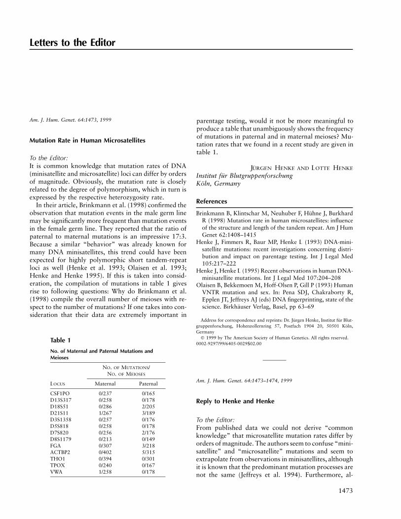
1473 Letters to the Editor Table 1 No. of Maternal and Paternal Mutations and Meioses LOCUS NO. OF MUTATIONS/ NO. OF MEIOSES Maternal Paternal CSF1PO 0/237 0/165 D13S317 0/258 0/178 D18S51 0/286 2/205 D21S11 1/267 3/189 D3S1358 0/257 0/176 D5S818 0/258 0/178 D7S820 0/256 2/176 D8S1179 0/213 0/149 FGA 0/307 3/218 ACTBP2 0/402 5/315 THO1 0/394 0/301 TPOX 0/240 0/167 VWA 1/258 0/178 Am. J. Hum. Genet. 64:1473, 1999 Mutation Rate in Human Microsatellites To the Editor: It is common knowledge that mutation rates of DNA (minisatellite and microsatellite) loci can differ by orders of magnitude. Obviously, the mutation rate is closely related to the degree of polymorphism, which in turn is expressed by the respective heterozygosity rate. In their article, Brinkmann et al. (1998) confirmed the observation that mutation events in the male germ line may be significantly more frequent than mutation events in the female germ line. They reported that the ratio of paternal to maternal mutations is an impressive 17:3. Because a similar “behavior” was already known for many DNA minisatellites, this trend could have been expected for highly polymorphic short tandem-repeat loci as well (Henke et al. 1993; Olaisen et al. 1993; Henke and Henke 1995). If this is taken into consid- eration, the compilation of mutations in table 1 gives rise to following questions: Why do Brinkmann et al. (1998) compile the overall number of meioses with re- spect to the number of mutations? If one takes into con- sideration that their data are extremely important in parentage testing, would it not be more meaningful to produce a table that unambiguously shows the frequency of mutations in paternal and in maternal meioses? Mu- tation rates that we found in a recent study are given in table 1. J U ¨ RGEN HENKE AND LOTTE HENKE Institut fu ¨ r Blutgruppenforschung Ko ¨ ln, Germany References Brinkmann B, Klintschar M, Neuhuber F, Hu ¨ hne J, Burkhard R (1998) Mutation rate in human microsatellites: influence of the structure and length of the tandem repeat. Am J Hum Genet 62:1408–1415 Henke J, Fimmers R, Baur MP, Henke L (1993) DNA-mini- satellite mutations: recent investigations concerning distri- bution and impact on parentage testing. Int J Legal Med 105:217–222 Henke J, Henke L (1995) Recent observations in human DNA- minisatellite mutations. Int J Legal Med 107:204–208 Olaisen B, Bekkemoen M, Hoff-Olsen P, Gill P (1993) Human VNTR mutation and sex. In: Pena SDJ, Chakraborty R, Epplen JT, Jeffreys AJ (eds) DNA fingerprinting, state of the science. Birkha ¨user Verlag, Basel, pp 63–69 Address for correspondence and reprints: Dr. Ju ¨ rgen Henke, Institut fu ¨ r Blut- gruppenforschung, Hohenzollernring 57, Postfach 1904 20, 50501 Ko ¨ ln, Germany q 1999 by The American Society of Human Genetics. All rights reserved. 0002-9297/99/6405-0029$02.00 Am. J. Hum. Genet. 64:1473–1474, 1999 Reply to Henke and Henke To the Editor: From published data we could not derive “common knowledge” that microsatellite mutation rates differ by orders of magnitude. The authors seem to confuse “mini- satellite” and “microsatellite” mutations and seem to extrapolate from observations in minisatellites, although it is known that the predominant mutation processes are not the same (Jeffreys et al. 1994). Furthermore, al-
-
Upload
independent -
Category
Documents
-
view
0 -
download
0
Transcript of A Program for the Monte Carlo Evaluation of Significance of the Extended Transmission/Disequilibrium...
1473
Letters to the Editor
Table 1
No. of Maternal and Paternal Mutations andMeioses
LOCUS
NO. OF MUTATIONS/NO. OF MEIOSES
Maternal Paternal
CSF1PO 0/237 0/165D13S317 0/258 0/178D18S51 0/286 2/205D21S11 1/267 3/189D3S1358 0/257 0/176D5S818 0/258 0/178D7S820 0/256 2/176D8S1179 0/213 0/149FGA 0/307 3/218ACTBP2 0/402 5/315THO1 0/394 0/301TPOX 0/240 0/167VWA 1/258 0/178
Am. J. Hum. Genet. 64:1473, 1999
Mutation Rate in Human Microsatellites
To the Editor:It is common knowledge that mutation rates of DNA(minisatellite and microsatellite) loci can differ by ordersof magnitude. Obviously, the mutation rate is closelyrelated to the degree of polymorphism, which in turn isexpressed by the respective heterozygosity rate.
In their article, Brinkmann et al. (1998) confirmed theobservation that mutation events in the male germ linemay be significantly more frequent than mutation eventsin the female germ line. They reported that the ratio ofpaternal to maternal mutations is an impressive 17:3.Because a similar “behavior” was already known formany DNA minisatellites, this trend could have beenexpected for highly polymorphic short tandem-repeatloci as well (Henke et al. 1993; Olaisen et al. 1993;Henke and Henke 1995). If this is taken into consid-eration, the compilation of mutations in table 1 givesrise to following questions: Why do Brinkmann et al.(1998) compile the overall number of meioses with re-spect to the number of mutations? If one takes into con-sideration that their data are extremely important in
parentage testing, would it not be more meaningful toproduce a table that unambiguously shows the frequencyof mutations in paternal and in maternal meioses? Mu-tation rates that we found in a recent study are given intable 1.
JURGEN HENKE AND LOTTE HENKE
Institut fur BlutgruppenforschungKoln, Germany
References
Brinkmann B, Klintschar M, Neuhuber F, Huhne J, BurkhardR (1998) Mutation rate in human microsatellites: influenceof the structure and length of the tandem repeat. Am J HumGenet 62:1408–1415
Henke J, Fimmers R, Baur MP, Henke L (1993) DNA-mini-satellite mutations: recent investigations concerning distri-bution and impact on parentage testing. Int J Legal Med105:217–222
Henke J, Henke L (1995) Recent observations in human DNA-minisatellite mutations. Int J Legal Med 107:204–208
Olaisen B, Bekkemoen M, Hoff-Olsen P, Gill P (1993) HumanVNTR mutation and sex. In: Pena SDJ, Chakraborty R,Epplen JT, Jeffreys AJ (eds) DNA fingerprinting, state of thescience. Birkhauser Verlag, Basel, pp 63–69
Address for correspondence and reprints: Dr. Jurgen Henke, Institut fur Blut-gruppenforschung, Hohenzollernring 57, Postfach 1904 20, 50501 Koln,Germany
q 1999 by The American Society of Human Genetics. All rights reserved.0002-9297/99/6405-0029$02.00
Am. J. Hum. Genet. 64:1473–1474, 1999
Reply to Henke and Henke
To the Editor:From published data we could not derive “commonknowledge” that microsatellite mutation rates differ byorders of magnitude. The authors seem to confuse “mini-satellite” and “microsatellite” mutations and seem toextrapolate from observations in minisatellites, althoughit is known that the predominant mutation processes arenot the same (Jeffreys et al. 1994). Furthermore, al-

1474 Letters to the Editor
Figure 1 Allele frequencies and mutation events at the FGAlocus.
though an influence of the degree of polymorphism ex-ists, there is no “close relation.” As we tried to show inour article (Brinkmann et al. 1998), the correlation isnot that simple.
We also disagree with the statement that the sex ratioswere to be expected from minisatellite loci. The mutationprocess is different, and other structures and loci areinvolved. In fact, the sex ratios seem to differ grosslyamong minisatellites—for example, 1:1 for D1S7 and1:8 for D7S22 (Henke and Henke 1995). It is thereforeunclear which of these ratios should be chosen for ex-trapolation to microsatellites.
We agree with the authors that application to bio-medical or forensic calculations would require sex-specific mutation rates. Therefore, their data are a val-uable contribution, especially if the same criteria havebeen met. We pooled our data for individual loci to focuson the correlation of the mutation rates with the meanlength of the affected structure. The data set was toosmall to address the question of locus-specific ratios.
Apart from sex-specific mutation rates, further factorsneed to be considered when Henke and Henke’s dataand ours are used to determine mutation rates applicableto paternity probability calculations.
First, the mutation rates of individual alleles at a givenlocus vary according to size, and, as we described in ourarticle (Brinkmann et al. 1998), longer alleles often ex-hibit higher mutation rates than shorter ones. Our cur-rent enlarged mutation database indicates that, at thelocus FGA, the mutation ratio of short versus long allelesseems to be close to an order of magnitude (fig. 1).
Second, the complexity of the allele sequence seemsto exert an influence on the mutation rate. For example,we showed that, for ACTBP2, the longer alleles exhibita lower mutation rate because of the interruption of theAAAG repeat by an AA dinucleotide.
Third, the number of undetected mutations dependson the paternity case—that is, whether one parent orboth parents are examined and whether the putativefather is included or excluded. It also depends on thegenotype distribution in the specific population. For ex-ample, in the trio mother 16/17; child 16/17; father 16/18, a paternal 16r17 or 18r17 mutation would beundetected.
Fourth, paternal age may affect the mutation rate. Wedemonstrated that the mean age of fathers in whommutations occurred was higher than the mean age of allfathers in our database. This would mean that the mu-tation rate should also be classified according to age.
Finally, it needs to be stressed that ∼10% of the mu-tation events in our study are of unknown origin. Thesemutations cannot be used directly for the calculation ofa sex-specific mutation rate without further assumptionsand elaborations.
It is clear, then, that a correct mutation rate wouldrequire knowledge of many more mutation events thanwe have observed in our laboratory or Henke and Henkehave seen. The currently available data at least allowthe definition of an overall ratio of mutations that occurin male and female gametes. We would like to have ourconsiderations understood as an incentive to others toreport their mutation events. As more data become avail-able, more certainty will be gained for the biostatisticalcalculation of paternity probability in cases of single-locus exclusions due to mutations.
B. ROLF AND B. BRINKMANN
Institut fur RechtsmedizinWestfalische Wilhelms UniversitatMunster, Germany
References
Brinkmann B, Klintschar M, Neuhuber F, Huhne J, Rolf B(1998) Mutation rate in human microsatellites: influence ofthe structure and length of the tandem repeat. Am J HumGenet 62:1408–1415
Henke J, Henke L (1995) Recent observations in human DNA-minisatellite mutations. Int J Legal Med 107:204–208
Jeffreys AJ, Tamaki K, MacLeod A, Monckton DG, Neil DL,Armour JAL (1994) Complex gene conversion events ingermline mutation at human minisatellites. Nat Genet 6:136–145
Address for correspondence and reprints: Dr. B. Brinkmann, Institut furRechtsmedizin, Westfalische Wilhelms Universitat, Von Esmarch Strasse 62,D-48149, Munster, Germany. E-mail: [email protected]
q 1999 by The American Society of Human Genetics. All rights reserved.0002-9297/99/6405-0030$02.00

Letters to the Editor 1475
Figure 1 Photograph of the affected siblings
Am. J. Hum. Genet. 64:1475–1478, 1999
Recurrent Williams-Beuren Syndrome in a SibshipSuggestive of Maternal Germ-Line Mosaicism
To the Editor:Williams syndrome (WS; MIM 194050) is a multisystemdisorder characterized by mental retardation and an out-going personality, distinctive facial features, infantile hy-percalcemia, and supravalvular aortic stenosis (Williamset al. 1961; Beuren et al. 1962). A deletion encompassingthe elastin (Ewart et al. 1993), replication factor subunit2 (Peoples et al. 1996), LIM kinase-1 (Frangiskakis etal. 1996; Tassabehji et al. 1996), wnt receptor Dro-sophila frizzled homologue FZD3 (Wang et al. 1997),WBSCR1 (Osborne et al. 1996), and syntaxin 1A (Os-borne et al. 1997b) genes at the 7q11.23 locus almostalways is found in patients with WS. Earlier reports haveindicated that a high frequency of deletions associatedwith WS result from unequal meiotic recombination, asshown by the common concurrence of these deletionswith recombination between markers proximal and dis-tal to the deletion (Dutly and Schinzel 1996; Baumer etal. 1998). Several independent studies that demonstratedduplicated genes flanking the deleted region in WS haveprovided a structural basis for the high frequency ofunequal crossover events (Robinson et al. 1996; Os-borne et al. 1997a; Perez Jurado et al. 1998). The re-currence risk of an interchromosomal rearrangement inthe sibship of a proband with WS is usually negligibleand thus is likely to account for the sporadic occurrenceof almost all cases of WS. A minority of interstitial7q11.23 deletions could, however, result from an in-trachromosomal rearrangement, occurring during or be-fore parental meiosis. In case of a premeiotic intrach-romosomal recombination, the theoretical possibility ofgonadal mosaicism for the WS deletion has been sug-gested recently by Dutly and Schinzel (1996). In agree-ment with this hypothesis, we report here the recurrenceof a 7q11.23 deletion in two siblings with WS whoseparents did not carry this rearrangement in their somaticcells.
The first affected proband, a male, was the third off-spring of unrelated parents aged 26 years (mother) and32 years (father) at the time of delivery. Both elder sistersand parents were healthy. Unilateral kidney agenesis andgrowth retardation were detected in the fetus during thethird trimester of pregnancy. The child was delivered at40 wk of gestation by cesarean section. Birth weight,length, and head circumference were 2,020 g (!10th per-centile), 43 cm (!10th percentile), and 31 cm (!10thpercentile), respectively. No hypercalcemia was noted.Supravalvular aortic stenosis and peripheral pulmonary
arterial stenosis were detected at age 3 mo. Psychomotordevelopment was markedly delayed, walking started byage 25 mo, and language developed by age 4 years. Atthe current age of 8 years, the proband’s facies is char-acteristic of WS, with a narrowed forehead, strabismuswith hypermetropia, periorbital fullness, malar hypo-plasia, and a large open mouth with an everted lowerlip (fig. 1). IQ is markedly reduced, and behavior is char-acteristic of WS, with attention deficit, loquaciousness,and hypersensitivity to sound. The second affected child,a boy currently aged 3 years, has a clinical picture similarto the one reported in the first affected proband, exceptfor the absence of supravalvular aortic stenosis (fig.1).
Informed consent was obtained from the probands’parents prior to implementation of the genetic studiesreported here. FISH was done with biotin-labeled DNAkits (Oncor), according to the protocols provided by themanufacturer. ELN and D7S427 (7q36) probes (Oncor)were used for testing the 7q11.23 locus and for chro-mosome 7 identification, respectively. The 7q11.23 de-letion was found in both affected siblings, whereas itwas not detected in either parent (data not shown). Toour knowledge, this is the first report of the recurrenceof full-blown WS secondary to a 7q11.23 deletion innontwin sibs whose parents are unaffected. The familialcases of WS with the typical 7q deletion reported so farhave involved either offspring of an affected individual(Ewart et al. 1993, 1994; Morris et al. 1993a, 1993b)or MZ twins (Castorina et al. 1997). Recurrence of WSin nontwin sibs with unaffected parents has earlier beenreported (Burn 1986), but has never been established incases with proven elastin deletion.
Both affected siblings, their healthy sisters, and theirparents were genotyped with DNA (CA)n polymor-phisms, from centromere to telomere, as follows:D7S645-D7S2415-D7S672-D7S653-D7S489B-ELN

1476 Letters to the Editor
Figure 2 Segregation of microsatellite polymorphic markers within and surrounding the elastin gene on chromosome 7q in the nuclearfamily. The markers used and approximate genetic distances are given in the inset (sex average). The (CA)n repeat, which occurs in the elastingene intron 18; marker D7S489B; and marker D7S1870 are known to be deleted in typical WS.
(intron 18 of the elastin gene)-D7S1870-D7S2470-D7S675-D7S669-D7S634-D7S660 (Genethon; Foster etal. 1993; Gyapay et al. 1994; Gilbert-Dussardier et al.1995; Dib et al. 1996; Robinson et al. 1996). DNAsamples from peripheral leukocytes were analyzed ac-cording to procedures published elsewhere (Gilbert-Dus-sardier et al. 1995). Both probands failed to inherit amaternal allele at the elastin and D7S1870 loci, thusindicating that the 7q11.23 deletion was maternal inorigin (fig. 2). Moreover, they shared a single maternalhaplotype, outside the deletion, with their eldest sister,who did not carry the WS deletion in her leukocytes, asindicated by microsatellite analysis. These data werehighly suggestive of maternal mosaicism. In the pro-bands’ mother, heterozygosity at both elastin andD7S1870 loci, consistent with the absence of deletionof chromosome 7 in her leukocytes, by FISH, indicatedthat mosaicism was likely to be restricted to germ cells.
We could not rule out conclusively that the deletionin the present cases had occurred as a result of unequal
crossover between homologous chromosomes 7 duringmaternal meiosis, because DNA of the maternal grand-parents was not available. However, our comparison ofhaplotypes, using markers centromeric and telomeric tothe deletion region, failed to suggest any recombinationevent within this region in any of the four siblings (fig.2), making the possibility of a meiotic crossover veryunlikely. On the basis of a female recombination distanceof 6 cM between the first informative markers centro-meric (D7S653) and telomeric (D7S675) to the deletion(Dib et al. 1996), the probability of a double recombi-nation event in both affected sibs was very low (∼1/7.5#104). Thus, the 7q11.23 deletion observed in bothprobands most likely resulted from a premeiotic intrach-romosomal event responsible for a gonadal mosaicismin the probands’ mother.
Apart from a gonadal mosaicism, the presence of aconstitutive structural defect of the maternal chromo-some 7 could theoretically account for the recurrence ofthe elastin deletion in sibs. A cryptic 7q11.23 rearrange-

Letters to the Editor 1477
ment in a maternal homologue, such as an inversion oran insertion, could indeed interfere with meiotic pairing,thus leading to recurrence of unequal crossover in dis-tinct meiosis and greatly enhancing the possibility of adeletion. However, high-resolution chromosome band-ing at the 500–850 bands level failed to detect any chro-mosomal abnormality in maternal leukocytes, and DNAhaplotyping data were irrelevant to an interchromoso-mal crossover, thus arguing against this hypothesis.Whereas germ-line mosaicism in this particular familyclearly results in an increased risk of WS in subsequentpregnancies, it would be necessary to determine the fre-quency of gonadal mosaicism in a large cohort of fam-ilies with WS to assess the potential impact of this phe-nomenon on the overall recurrence risk of WS.
ALI KARA-MOSTEFA,1 ODILE RAOUL,2
STANISLAS LYONNET,2 JEANNE AMIEL,2
ARNOLD MUNNICH,2 MICHEL VEKEMANS,2
SUZEL MAGNIER,3 BATOOL OSSAREH,2
AND JEAN-PAUL BONNEFONT1,2
1Biochemical Genetics Unit and 2Department ofGenetics, Hopital Necker Enfants Malades, and3Paediatric Cardiology Unit, Hopital Robert Debre,Paris
Electronic-Database Information
Accession numbers and URLs for data in this article are asfollows:
Genethon, ftp://ftp.genethon.fr/pub/Gmap/Nature-1995/data/data_chrom7 (for sequence-tagged site used for chromosome7 genotyping)
Online Mendelian Inheritance in Man (OMIM), http://www.ncbi.nlm.nih.gov/Omim (for WS [MIM194050])
References
Baumer A, Dutly F, Balmer D, Riegel M, Tukel T, Krajewska-Walasek M, Schinzel AA (1998) High level of unequal mei-otic crossovers at the origin of the 22q11.2 and 7q11.23deletions. Hum Mol Genet 7:887–894
Beuren AJ, Apitz J, Armjanz D (1962) Supravalvular aorticstenosis in association with mental retardation and facialappearance. Circulation 26:1235–1240
Burn J (1986) Williams syndrome. J Med Genet 23:389–395Castorina P, Selicorni A, Bedeschi F, Dalpra L, Larizza L (1997)
Genotype-phenotype correlation in two sets of monozygotictwins with Williams syndrome. Am J Med Genet 69:107–111
Dib C, Faure S, Fizames C, Samson D, Drouot N, Vignal A,Millasseau P, et al (1996) A comprehensive genetic map ofthe human genome based on 5,264 microsatellites. Nature380:152–154
Dutly F, Schinzel A (1996) Unequal interchromosomal rear-rangements may result in elastin gene deletions causing theWilliams-Beuren syndrome. Hum Mol Genet 5:1893–1898
Ewart AK, Jin W, Atkinson D, Morris CA, Keating MT (1994)Supravalvular aortic stenosis associated with a deletion dis-rupting the elastin gene. J Clin Invest 93:1071–1077
Ewart AK, Morris CA, Atkinson D, Jin W, Sternes K, SpalloneP, Stock AD, et al (1993) Hemizygosity at the elastin locusin a developmental disorder, Williams-Beuren syndrome.Nat Genet 5:11–16
Foster K, Ferrell R, King-Underwood L, Povey S, Attwood J,Rennick R, Humphries SE, et al (1993) Description of adinucleotide repeat polymorphism in the human elastin geneand its use to confirm assignment of the gene to chromosome7. Ann Hum Genet 57:87–96
Frangiskakis JM, Ewart AK, Morris CA, Mervis CB, BertrandJ, Robinson BF, Klein BP, et al (1996) LIM-kinase1 hemi-zygosity implicated in impaired visuospatial constructivecognition. Cell 86:59–69
Gilbert-Dussardier B, Bonneau D, Gigarel N, Le Merrer M,Bonnet D, Philip N, Serville F, et al (1995) A novel micro-satellite DNA marker at locus D7S1870 detects hemizygos-ity in 75% of patients with Williams syndrome. Am J HumGenet 56:542–544
Gyapay G, Morissette J, Vignal A, Dib C, Fizames C, Millas-seau P, Marc S, et al (1994) The 1993-1994 Genethon hu-man genetic linkage map. Nat Genet 7:246–339
Morris CA, Loker J, Ensing G, Dean Stock A (1993a) Supra-valvular aortic stenosis cosegregates with a familial 6;7translocation which disrupts the elastin gene. Am J MedGenet 46:737–744
Morris CA, Thomas IT, Greenberg F (1993b) Williams syn-drome: autosomal dominant inheritance. Am J Med Genet47:478–481
Osborne LR, Herbrick JA, Greavette T, Heng HHQ, Tsui LC,Scherer SW (1997a) PMS2-related genes flank the rearrange-ment breakpoints associated with Williams syndrome andother diseases on human chromosome 7. Genomics 45:402–406
Osborne LR, Martindale D, Scherer SW, Shi XM, HinzengaJ, Heng HHQ, Costa T, et al (1996) Identification of a 500kb region that is commonly deleted in Williams syndromepatients. Genomics 36:328–336
Osborne LR, Soder S, Shi XM, Pober B, Costa T, Scherer SW,Tsui LC (1997b) Hemizygous deletion of the syntaxin 1Agene in individuals with Williams syndrome. Am J HumGenet 61:449–452
Peoples R, Perez Jurado L, Wang YK, Kaplan P, Francke U(1996) The gene for replication factor subunit 2 (RFC2) iswithin the 7q11.23 Williams syndrome deletion. Am J HumGenet 58:1370–1373
Perez Jurado LA, Wang YK, Peoples R, Coloma A, Cruces J,Francke U (1998) A duplicated gene in the breakpointregions of the 7q11.23 Williams-Beuren syndrome deletionencodes the initiator binding protein TFII-I and BAP-135,a phosphorylation target of BTK. Hum Mol Genet 7:325–334
Robinson WP, Waslynka J, Bernasconi F, Wang M, Clark S,Kotzot D, Schinzel A (1996) Delineation of 7q11.2 deletionsassociated with Williams-Beuren syndrome and mapping ofa repetitive sequence to within and to either side of thecommon deletion. Genomics 34:17–23
Tassabehji M, Metcalfe K, Fergusson WD, Carette MJA, Dore

1478 Letters to the Editor
Figure 1 Multipoint results for chromosome 5
JK, Donnai D, Read AP, et al (1996) LIM-kinase deleted inWilliams syndrome. Nat Genet 13:272–273
Wang YK, Harryman Samos C, Peoples R, Perez Jurado LA,Nusse R, Francke U (1997) A novel human homologue ofthe Drosophila frizzled wnt receptor gene binds winglessprotein and is in the Williams syndrome deletion at 7q11.23. Hum Mol Genet 6:465–472
Williams JCP, Barrat-Boyers BG, Lowe JB (1961) Supraval-vular aortic stenosis. Circulation 24:1311–1318
Address for correspondence and reprints: Dr. Jean-Paul Bonnefont, Biochem-ical Genetics Unit and Department of Genetics, Hopital Necker-EnfantsMalades,149 rue de Sevres, 75743 Paris cedex 15, France. E-mail: [email protected]
q 1999 by The American Society of Human Genetics. All rights reserved.0002-9297/99/6405-0000$02.00
Am. J. Hum. Genet. 64:1478–1480, 1999
Localization of a Gene for Bitter-Taste Perception toHuman Chromosome 5p15
To the Editor:Some people perceive the taste of phenylthiocarbamideand its chemical relative propylthiouracil (PROP) as in-tensely bitter at low concentrations, whereas others areunable to detect them, even at high concentrations. Thistaste blindness is an inherited trait (Snyder 1931). Al-though inheritance is thought to be recessive, other pos-
sibilities have been suggested, such as multiple genes(Boyd 1950; Olson et al. 1989), incomplete dominance(Johnson et al. 1966; Bartoshuk et al. 1994; Reed et al.1995), or multiple alleles of a single gene (Rychkov andBorodina 1973). This trait is among the most-studied inhuman genetics, but the relevant gene has not been char-acterized. Therefore, we conducted a genomewide scanby using 98 nuclear families and 356 markers spaced at∼10-cM intervals.
Three hundred ninety-three adults and their parentsparticipated as research subjects. The 98 families wereoriginally recruited as part of an ongoing study of thegenetics of body weight at the University of Pennsyl-vania’s Behavioral Genetics Laboratory, and the detailsof family collection have been published elsewhere (Priceet al. 1998). The protocol was approved by the Com-mittee of Studies Involving Human Beings at the Uni-versity of Pennsylvania.
To phenotype the subjects, filter paper was soaked ina saturated PROP solution, dried, and cut into strips.Subjects were asked to place the paper in their mouthsand to rate the bitterness of taste. The scale used by thesubjects to rate the taste intensity utilized descriptivewords and is referred to as a “labeled-magnitude scale”(LMS; Green et al. 1993). Because the LMS is contin-uous, it prevents the loss of information associated withcategoric scales and therefore provides the type of dataessential for quantitative linkage analysis. In addition,the LMS minimizes ceiling effects and is better at dis-

Letters to the Editor 1479
criminating sensitive tasters from nontasters than theclassic nine-point scale (Lucchina et al. 1998b). The scaleis labeled as follows (with numeric values assigned toeach level of taste intensity): barely detectable (0), weak(6), moderate (17), strong (35), very strong (54), andstrongest imaginable (100). The scores from the LMSwere used as phenotypes for the quantitative linkageanalysis.
The LMS is also a valid instrument to classify indi-viduals as “nontasters” or “tasters.” Studies scaling thesuprathreshold bitterness of PROP with magnitude es-timation demonstrate that psychophysical functions fornontasters and tasters diverge (Bartoshuk et al. 1994).The LMS produces suprathreshold functions equivalentto magnitude estimation (Green et al. 1996). Becausethe LMS is easier for naıve subjects to use, it is replacingmagnitude estimation in studies of PROP (Snyder et al.1996; Intranuova and Powers 1998; Lucchina et al.1998a, 1998b; Schwartz et al. 1998; Prutkin et al., inpress). Thus, the LMS provides a convenient, reliable,and reasonable choice for a large-scale gene-mappingstudy.
The mean rating of suprathreshold taste intensity was31.2 5 29.3 units, near the label “strong.” As expected,the distribution of scores was kurtotic (20.454) andskewed (.73). There was no relationship between sub-jects’ ratings of PROP and height, weight, or body-massindex ( ).P 1 .05
We genotyped microsatellite markers spaced ∼10.1cM apart by using methods described by Lee et al.(1999). All half-siblings were eliminated prior to anal-ysis. Computation of descriptive statistics and correla-tion coefficients were conducted with SPSS (6.1.1.).Quantitative trait loci analysis was conducted with thecomputer program MAPMAKER/SIBS version 2.0(Kruglyak and Lander 1995). For analysis of transmis-sion disequilibrium, the quantitative data were dichot-omized into taster and nontaster categories, with all tast-ers reporting that suprathreshold concentrations ofPROP tasted “strong,” “extremely strong,” or “strong-est imaginable” ( ; 45.8%). Nontasters ratedn 5 180PROP as “barely detectable” or “weak” ( ;n 5 11529.3%). Subjects giving intermediate responses were ex-cluded from analysis ( ; 25%). These cut-off val-n 5 98ues are conservative. Transmission of alleles from het-erozygous parents to nontaster offspring was computedwith TDTLIKE version 2.1 (Terwilliger 1995), whichcorrects for multiple-allele testing.
The telomeric portion of 5p gave the strongest evi-dence for linkage (t-score 5 3.28, ; fig. 1),P 5 .0005with the peak score near D5S2505. The linkage peakspanned ∼10 cM, from D5S406 to D5S2081, and wasthe only region of the genome that had a t score > 3.0.No candidate genes are apparent in 5p15. Markers fromthe telomeric portion of chromosome 5 were then ex-
amined for transmission disequilibrium. There was sig-nificant distortion in transmission of alleles from het-erozygous parents to nontaster children for markersfrom the linked region, with D5S2505 being the mostsignificant (D5S406, ; D5S2505, ,P 5 .031 P 5 .007D5S635, ; D5S807, ; D5S2081,P 5 .017 P 5 .034 P 5
). These results are consistent with the hypothesis.012that a gene that confers the ability to taste PROP lieson the telomeric region of human chromosome 5p.
In addition to chromosome 5, there was a suggestionof linkage on chromosome 7, ∼35–40 cM centromericto the KELL locus, with a maximum t-score of 2.34( ) near D7S1789 and D7S796. Initial linkageP 5 .008studies suggested a locus was near KELL on chromo-some 7 (Chautard-Freire-Maia 1974; Conneally et al.1976), but later reports were unable to replicate thisfinding (Spence et al. 1984). The results of the currentstudy suggest that a region on chromosome 7 may alsoinfluence the taster phenotype.
Acknowledgments
The present work was supported by National Institutes ofHealth (NIH) grant R03DC03509 (to D.R.R).; NIH grantsR01DK44073, R01DK48095, and funds from Axys Phar-maceuticals and Glaxo-Wellcome (to R.A.P.); and NIH grants5M01RR00125-350922, 5R01DC00283, and 5R21DC03003to (L.M.B.). We acknowledge the generous cooperation of par-ticipating families. We thank Robin L. Kilker, Wei-Dong Li,Andrew C. Krakowski, Guang Ming Yuan, Christa L. AlberryMayr, Elizabeth Joe, Kruti Quazi, Karynn Henry, Paula Kwon,Samantha J. Weisberg, Balasahib G. Shinde, Robert BrianChin, Cassandra George, Nicole Baker, and Adam S. Crystalfor technical assistance and Joseph H. Lee for statistical advice.
DANIELLE R. REED,1 ELIZABETH NANTHAKUMAR,2
MICHAEL NORTH,2 CALLUM BELL,2
LINDA M. BARTOSHUK,3 AND R. ARLEN PRICE1
1Center for Neurobiology and Behavior, Departmentof Psychiatry, University of Pennsylvania; 2AxysPharmaceutical, La Jolla, CA; and 3Yale UniversitySchool of Medicine, New Haven, CT
References
Bartoshuk LM, Duffy VB, Miller IJ (1994) PTC/PROP tasting:anatomy, psychophysics, and sex effects. Physiol Behav 56:1165–1171 (Erratum [1995] Physiol Behav 58:203)
Boyd WC (1950) Genetics and the races of man: an intro-duction to modern physical anthropology. Little, Brown,Boston
Chautard-Freire-Maia EA (1974) Linkage relationships be-tween 22 autosomal markers. Ann Hum Genet 38:191–198
Conneally PM, Dumont-Driscoll M, Huntzinger RS, NanceWE, Jackson CE (1976) Linkage relations of the loci forKell and phenylthiocarbamide taste sensitivity. Hum Hered26:267–271

1480 Letters to the Editor
Green B, Dalton P, Cowart B, Rankin K, Higgins J (1996)Evaluating the labeled magnitude scale for measuring sen-sations of taste and smell. Chem Senses 21:323–334
Green BG, Shaffer GS, Gilmore M (1993) Derivation and eval-uation of a semantic scale of oral sensation magnitude withapparent ratio properties. Chem Senses 18:683–702
Intranuova L, Powers A (1998) The perceived bitterness ofbeer and 6-n-propylthiouracil (PROP) and taste sensitivity.In: Murphy C (ed) Olfaction and taste. Ann NY Acad Sci855:816–819
Johnson FE, Hertzog KP, Malina RM (1966) Phenylthiocar-bamide taste sensitivity and its relationship to growth var-iation. Am J Phys Anthrop 24:253–256
Kruglyak L, Lander E (1995) Complete multipoint sib-pairanalysis of qualitative and quantitative traits. Am J HumGenet 54:439–454
Lee JH, Reed DR, Li W-D, Xu W, Joo E-J, Kilker RL, Nan-thakumar E, et al (1999) Genome scan for human obesityand linkage to markers in 20q13. Am J Hum Genet 64:1063–1070
Lucchina L, Curtis O, Putnam P, Bartoshuk L (1998a) 6-n-propylthiouracil (PROP) tasters assign higher sweetness rat-ings to sucrose and high-intensity sweetners. Chem Senses23:560
Lucchina LA, Curtis OF, Putnam P, Drewnowski A, PrutkinJ, Bartoshuk LM (1998b) Psychophysical measurement of6-n-propylthiouracil (PROP) taste perception. In: MurphyC (ed) Olfaction and taste. Ann NY Acad Sci 855:816–819
Olson JM, Boehnke M, Neiswanger K, Roche AF, SiervogelRM (1989) Alternative genetic models for the inheritanceof the phenylthiocarbamide taste deficiency. Genet Epide-miol 6:423–434
Price RA, Reed DR, Lee JH (1998) Obesity related phenotypesin families selected for extreme obesity and leanness. Int JObes Relat Metab Disord 22:406–413
Prutkin J, Fast K, Lucchina L, Bartoshuk L PROP (6-n-pro-pylthiouracil) genetics and trigeminal innervation of fungi-form papillae. Chem Senses (in press)
Reed DR, Bartoshuk LM, Duffy V, Marino S, Price RA (1995)Propylthiouracil tasting: determination of underlying thresh-old distributions using maximum likelihood. Chem Senses20:529–533
Rychkov YG, Borodina SR (1973) Further investigations ofthe genetics of hypersensitivity to phenylthiocarbamide inman (experimental, population, and familial data). Genetika9:141–152
Schwartz S, Janjua T, Kveton J, Green B, Bartoshuk L (1998)Alteration in lingual somatosensation as a result of tran-section of the chorda tympani nerve (VII). Chem Senses 23:560
Snyder D, Lucchina L, Duffy V, Bartoshuk L (1996) Magnitudematching adds power to the labeled magnitude scale. ChemSenses 21:673
Snyder LH (1931) Inherited taste deficiency. Science 74:151–152
Spence MA, Falk CT, Neiswanger K, Field LL, Marazita ML,Allen FH Jr, Siervogel RM, et al (1984) Estimating the re-combination frequency for the PTC-Kell linkage. Hum Ge-net 67:183–186
Terwilliger J (1995) A powerful likelihood method for the anal-
ysis of linkage disequilibrium between trait loci and one ormore polymorphic marker loci. Am J Hum Genet 56:777–787
Address for correspondence and reprints: Dr. R. Arlen Price, Center for Neu-robiology and Behavior, University of Pennsylvania, 415 Curie Boulevard,CRB135b, Philadelphia, PA 19104 E-mail: [email protected]
q 1999 by The American Society of Human Genetics. All rights reserved.0002-9297/99/6405-0032$02.00
Am. J. Hum. Genet. 64:1480–1484, 1999
Sperm Chromosome Analysis in a Man Heterozygousfor a Paracentric Inversion of Chromosome 14(q24.1q32.1)
To the Editor:Paracentric inversions are rarely reported, since they canonly be detected by the use of banding procedures (Pet-tenati et al. 1995). The incidence is estimated to be.09–.49/1,000 (van Dyke et al. 1983; Ferguson-Smithand Yates 1984; Hook et al. 1984; Fryns et al. 1988).Some investigators have suggested that paracentric in-versions in man are generally harmless (Madan 1995);however, recombinant chromosomes have been observedin 17 cases, and the risk of viable recombinants has beenestimated to be 3.8% (Pettenati et al. 1995). Becausethe frequency of spontaneous abortions may be in-creased in carriers of paracentric inversions (Mules andStamberg 1984), chromosomally unbalanced concep-tions may be lost early in pregnancy. A direct analysisof chromosomes in gametes would overcome this po-tential loss of information. In 1986, I reported the firstanalysis of sperm chromosomes in a man heterozygousfor a paracentric inversion of chromosome 7 (Martin1986). The present report represents the second studyof sperm karyotypes in a paracentric-inversion carrier.
A paracentric inversion of chromosome 14(q24.1q32.1) was ascertained at amniocentesis per-formed because of advanced maternal age. Subsequentinvestigation of the family revealed that the inversion ispresent in the father. The couple had previously had twonormal children and one spontaneous abortion at 11 wkgestation (chromosomes were not studied). The studywas approved by the university ethics committee, andthe sperm donor provided informed consent. The spermdonor had a normal sperm profile, with a volume of 3ml, concentration of /ml, 80% motility, and6138 # 10forward progression of 8/10. A partial karyotype of thenormal and inverted chromosomes 14 in the 41-year-old father is presented in figure 1. Sperm chromosomecomplements were obtained by fusion of golden hamsteroocytes with human sperm and analysis of the Q-banded

Letters to the Editor 1481
Figure 1 Q-banded human sperm chromosomes demonstratingnormal and inverted chromosomes 14.
Figure 2 Q-banded karyotype of human sperm chromosomes,23,Y,inv(14)(q24q32).
pronuclear chromosomes. This technique has been de-scribed in detail elsewhere (Martin et al. 1994b).
A total of 120 sperm chromosome complements wereobtained. The results are summarized in the Appendix.Fifty complements (41.7%) contained a normal chro-mosome 14, whereas 70 (58.3%) had the inverted chro-mosome 14. This segregation was not significantly dif-ferent from the expected 1:1 ratio ( , ).2x 5 3.3 P 1 .05An example of a sperm chromosome complement withan inverted chromosome 14 is shown in figure 2. Thenumber of X- and Y-bearing sperm was 55 and 65, re-spectively, which was not significantly different from theexpected (60 X- and 60 Y-bearing) number.
None of the spreads contained a recombinant chro-mosome 14. There were no dicentric, acentric, or du-plicated/deficient chromosomes, as would be expected ifa crossover had occurred within the inverted segment.Abnormal sperm chromosome complements were pre-sent in 10% of the spreads. Three complements had anumeric abnormality, six had a structural abnormality,and three had both. Details of these abnormalities areprovided in the Appendix. This frequency of abnor-malities is similar to my results from 84 chromosomallynormal control donors, who had a mean frequency of12.8% abnormal sperm (Martin 1995).
In a paracentric-inversion heterozygote, pairing ofhomologues during meiosis is maximized by the for-mation of an inversion loop. If an unequal number ofcrossovers occur within this loop, dicentric and acentricchromosomes are formed. The acentric chromosomesare generally lost in subsequent cell divisions. Dicentricchromosomes tend to break, since the two centromeres
are pulled to opposite poles of the cell. Thus, the re-sulting gametes can have a variety of duplications ordeficiencies.
Pettanati et al. (1995) have reviewed 446 cases ofparacentric inversions, with 17 cases of recombinantchromosomes reported. The majority of the cases weremonocentric chromosomes with duplications or defi-ciencies. Two maternally inherited cases had offspringwith dicentric chromosomes: one case was an inversionof chromosome 9 (q22.1q34.3) (Worsham et al. 1989),and the other was an inversion of chromosome 14(q24.2q32.3), with breakpoints very similar to thoseseen in the present case (Mules and Stamberg 1984).The paracentric inversion of chromosome 14 (q24q32)appears to be relatively common in humans (Pettenatiet al. 1995), with six index cases reported, including thisreport. Two of the six cases were paternally inherited.The only case with recombinant chromosomes reportedfor this inversion is the maternally inherited dicentriccase cited above. Because this inversion is ∼30% of thelength of long arm of chromosome 14, and because chro-

1482 Letters to the Editor
mosome 14 has an average of 1.8 chiasmata (Chandley1975), one would expect that a crossover within theinverted segment would occur ∼50% of the time. Be-cause one half of the chromatids are involved in thecrossover, ∼25% of the gametes should be chromoso-mally unbalanced. However, my results from spermchromosome analysis did not detect a single recombi-nant chromosome in 120 spermatozoa. The lack of anyrecombinant chromosomes in sperm suggests either thatthe chromosomes 14 did not pair by an inversion loopor that crossing-over was suppressed within the loop.
Very few meiotic studies of inversion carriers haveshed light on these possibilities. The only meiotic anal-ysis of a paracentric inversion was performed in mice,by Poorman et al. (1981). They found that 100% ofspermatocytes at early pachytene contained a fully syn-apsed loop. There have been no cytogenetic meiotic stud-ies of paracentric inversions in humans, and the studiesof pericentric inversions have produced conflicting re-sults: some have shown homosynapsis whereas othershave demonstrated heterosynapsis and asynapsis (Ga-briel-Robez and Rumpler 1994). A recent study reportedthe analysis of meiotic recombination, by sperm typingin a man carrying a 9q32q34 inversion (Brown et al.1998). The authors concluded that there was a reducedfrequency of recombination within the inversion, sug-gesting that an inversion loop had not been formed.
Sperm karyotyping has been performed on a total ofnine inversion heterozygotes: two with paracentric in-versions (Martin 1986; present study) and seven withpericentric inversions. The other paracentric-inversioncarrier (7q11q22) also did not have any recombinantsperm, despite expectation that the estimated frequencyof recombinant sperm would be 25% (Martin 1986).Of the seven pericentric-inversion carriers, four had norecombinant sperm (Balkan et al. 1983; Jenderny et al.1992; Martin et al. 1994a; Colls et al. 1997), whereasthree demonstrated frequencies of 11%, 18%, and 31%recombinant sperm (Martin 1991, 1993; Navarro et al.1993). The inversions that produced recombinant chro-mosomes in sperm were all large inversions encompass-ing more than half the chromosome length. The inver-sions that did not produce recombinant chromosomeswere, in general, smaller, being less than one-third thelength of the chromosome. The exception to this wasthe pericentric inversion of chromosome 20 studied byJenderny et al. (1992), which failed to produce recom-binant sperm despite a relatively large size. However,this may simply be a matter of sample size, because only26 sperm were analyzed. This dependence on size of theinversion to produce recombinant chromosomes is rem-iniscent of empirical studies that have suggested that, fora pericentric-inversion heterozygote to produce live-bornchildren with a recombinant chromosome, the invertedsegment must involve one-third of the chromosome
length (Trunca and Opitz 1977). It is possible that, insmall inversions, crossing-over is suppressed within theloop or that pairing is accomplished by heterosynapsis.
Ashley (1988) has formulated a hypothesis that pre-dicts that, if G-light bands are aligned with G-lightbands, lack of homology will be recognized and an in-version loop will be formed; in contrast, if the arrange-ment aligned two G-dark bands or a G-light band witha G-dark band, then lack of homology would not berecognized and heterosynapsis would proceed. de Per-digo et al. (1989) and Gabriel-Robez and Rumpler(1994) have reviewed synaptic data and localization ofchromosomal breakpoints in human pericentric inver-sions and have determined that Ashley’s hypothesis isconsistent with the data: loops are formed when bothbreaks occur in G-light bands, whereas heterosynapsiswithout loop formation or asynapsis occurs when oneof the breaks is in a G-dark band. The results of spermchromosome–complement analysis of pericentric-inver-sion carriers are also generally consistent with this hy-pothesis. All three inversions with recombinant chro-mosomes in sperm had breakpoints in G-light areas(allowing homosynapsis, loop formation, chiasmata,and recombinant chromosomes). Of the four pericentricinversions with no recombinant chromosomes in sperm,all except the paracentric inversion of chromosome 20(p13q11.2) studied by Jenderney et al. (1992) had atleast one breakpoint in a G-dark region. However, asdiscussed above, 26 sperm complements may have beenan insufficient sample size for detection of recombinantchromosomes. Of the two paracentric inversions studiedby sperm karyotyping, both had breakpoints in G-lightareas, yet neither had recombinant chromosomes insperm. Thus, data from sperm karyotyping of the twoparacentric inversions studied to date do not agree withAshley’s hypothesis. It may be that, for paracentric in-versions, small size is a major detriment to homologouspairing and crossing-over.
The possibility of an interchromosomal effect for in-version heterozygotes has been raised, because childrenwith unrelated chromosomal abnormalities have beenborn to inversion carriers. Canki and Dutrillaux (1979)described two cases of familial paracentric inversionsassociated with sex-chromosomal aneuploidy, and Frynsand van den Berghe (1980) reported a child with trisomy21 who was born to a father heterozygous for a para-centric inversion. However, my data do not support suchan effect, because the frequency of chromosomal ab-normalities was not increased in either the case that washeterozygous for a paracentric inversion of chromosome14 or the case that was heterozygous for a paracentricinversion of chromosome 7 (Martin 1986). Similarly,previous studies of sperm chromosomes in men withpericentric inversions have not demonstrated any in-creased frequency of abnormalities in other chromo-

Letters to the Editor 1483
somes (Balkan et al. 1983; Martin 1991, 1993; Navarroet al. 1993; Colls et al. 1997). Colls et al. (1997) spe-cifically searched for an interchromosomal effect in-volving chromosome 21, by performing FISH analysisof 110,000 sperm nuclei from a case that was hetero-zygous for a paracentric inversion of chromosome 9, aswell as by analyzing 1300 sperm complements. Eventhese large sample sizes did not uncover an interchro-mosomal effect.
Further studies of sperm chromosomes in paracentric-inversion carriers are required, because only two havebeen reported. Information from these studies will pro-vide estimates of the frequency of chromosomally un-balanced gametes. These studies will also help us to elu-cidate some of the factors that influence the productionof recombinant chromosomes at meiosis.
Acknowledgments
I would like to thank Drs. I. Lange and N. Spence for re-ferring the family, Dr. J. Chernos for lymphocyte chromosomeanalysis, Evelyn Ko and Leona Barclay for expert technicalassistance, Debbie Bell for preparing the manuscript, and thefamily for their cooperation and interest in the study. Thesupport of the Medical Research Council of Canada (grantMA-7961), the Alberta Heritage Fund for Medical Research,and the Alberta Children’s Hospital Research Foundation isgratefully acknowledged.
R. H. MARTIN
Department of Medical Genetics, University ofCalgary, and Department of Genetics, AlbertaChildren’s Hospital, Calgary
Appendix
Sperm Chromosome Complements in a MaleHeterozygous for a Paracentric Inversion ofChromosome 14 (q24q32)
Normal sperm ( ):n 5 108Normal chromosome 14, n 5 44Inverted chromosome 14, n 5 64
Abnormal sperm ( ):n 5 12Normal chromosome 14, n 5 6Inverted chromosome 14, n 5 6
Details of the 12 abnormal sperm complements:Numerical:
22,Y,2622,X,21322,X,218
Structural:23,X,chrb(12)(q14or15)
23,X,chte(19;20)(q13;1or13.2;q13.3)(tr,incomplete)
23,X,chrb(12)(q12or13),[214,1inv(14)]21,Y,chte(1;3;17)(cx),[14,1inv(14)]22,X,dic(7;18)(pterrcen::p11.3rqter),
[214,1inv(14)]—,Y,MB1R,[214,1inv(14)]
Both numerical and structural:24,XiY,1ace21,X,222,dic(5;7)(p15;q35or36)1ace,
[214,1inv(14)]22,Y,217,chrg(7)(q21,chtg(9)(p21),
[214,1inv(14)]
References
Ashley T (1988) G-band position on meiotic synapsis andcrossing over. Genetics 118:307–317
Balkan W, Burns K, Martin R (1983) Sperm chromosome anal-ysis of a man heterozygous for a pericentric inversion ofchromosome number 3. Cytogenet Cell Genet 35:295–297
Brown GM, Leversha M, Hulten M, Ferguson-Smith MA, Af-fara NA, Furlong RA (1998) Genetic analysis of meioticrecombination in humans by use of sperm typing: reducedrecombination within a heterozygous paracentric inversionof chromosome 9q32-q34.3. Am J Hum Genet 62:1484–1492
Canki N, Dutrillaux B (1979) Two cases of familial paracentricinversion in man associated with sex chromosome anomaly47,XY,inv(5)(q21q32) and 45,X,inv(7)(q11.3q22.3). HumGenet 47:261–268
Chandley A (1975) Human meiotic studies. In: Emery A (ed)Modern trends in human genetics. Vol 2. Butterworths, Lon-don, Boston, pp 31–82
Colls P, Blanco J, Martinez-Pasarell O, Vidal F, Egozcue J,Marquez C, Guitart M, et al (1997) Chromosome segre-gation in a man heterozygous for a pericentric inversion,inv (9)(p11q13), analysed by using sperm karyotyping andtwo-colour fluorescence in situ hybridization on sperm nu-clei. Hum Genet 99:761–765
de Perdigo A, Gabriel-Robez O, Rumpler Y (1989) Correlationbetween chromosomal breakpoint position and synaptic be-haviour in human males heterozygous for a pericentric in-version. Hum Genet 83:274–276
Ferguson-Smith MA, Yates JR (1984) Maternal age specificrates for chromosome aberrations and factors influencingthem: report of a collaborative European study on 52965amniocenteses. Prenat Diagn 4:5–44
Fryns J, Kleczkowska A, Kubien E, van den Berghe H (1988)Structural chromosome rearrangements in couples with re-current fetal wastage: the Leuven experience. In: SandbergA (ed) The cytogenetics of mammalian autosomal rearrange-ments Vol 8. Alan R Liss, New York, pp 453–470
Fryns J, van den Berghe H (1980) Paracentric inversion in man:personal experience and review of literature. Hum Genet54:413–416
Gabriel-Robez O, Rumpler Y (1994) The meiotic pairing be-haviour in human spermatocytes carrier of chromosome

1484 Letters to the Editor
Table 1
Comparison of Different TDT Statistics Applied to Schizophreniaand D22S278
Type x2 dfAsymptotic
P ValueEmpiricalP Value
SE(P)
ETDT:Allelewise 22.025 10 .0151 .0142 .0012Genotypewise 66.228 30 .0002 .0009 .0003Goodness-of-fit 44.203 20 .0015 .0085 .0009
MATLAB:Spielman-Ewens 22.358 10 .0134 .0115 .0011Stuart 18.976 10 .0406 .0202 .0014
anomalies and their repercussions on reproductive fitness.I. Inversions and insertions: a European collaborative study.Ann Genet 37:3–10
Hook EB, Schreinemachers DM, Willey AM, Cross PK (1984)Inherited structural cytogenetic abnormalities detected in-cidentally in fetuses diagnosed prenatally: frequency, paren-tal-age associations, sex-ratio trends, and comparisons withrates of mutants. Am J Hum Genet 36:422–443
Jenderny J, Gebauer J, Rohborn G, Ruger A (1992) Spermchromosome analysis of a man heterozygous for a pericen-tric inversion of chromosome 20. Hum Genet 89:117–119
Madan K (1995) Paracentric inversions: a review. Hum Genet96:503–515
Martin RH (1986) Sperm chromosome analysis in a man het-erozygous for a paracentric inversion of chromosome 7(q11q22). Hum Genet 73:97–100
——— (1991) Cytogenetic analysis of sperm from a man het-erozygous for a pericentric inversion, inv (3) (p25q21). AmJ Hum Genet 48:856–861
——— (1993) Analysis of sperm chromosome complementsfrom a man heterozygous for a pericentric inversion, inv(8)(p23q22). Cytogenet Cell Genet 62:199–202
——— (1995) The sperm cell—genetic aspects. In: Grudzin-skas J, Yovich J, Simpson J, Chard T (eds) Cambridge re-views in human reproduction. Cambridge University Press,Cambridge, pp 104–121
Martin R, Chernos J, Lowry R, Pattinson A, Barclay L, Ko E(1994a) Analysis of sperm chromosome complements froma man heterozygous for a pericentric inversion of chromo-some 1. Hum Genet 93:135–138
Martin R, Ko E, Barclay L (1994b) Human sperm karyotypes.In: Verma R, Babu A (eds) Human chromosomes: manualof basic techniques. McGraw-Hill, New York, pp 56–65
Mules E, Stamberg J (1984) Reproductive outcomes of para-centric inversion carriers: report of a liveborn dicentric re-combinant and literature review. Hum Genet 67:126–131
Navarro J, Benet J, Martorell MR, Templado C, Egozcue J(1993) Segregation analysis in a man heterozygous for apericentric inversion of chromosome 7 (p13;q36) by spermchromosome studies. Am J Hum Genet 53:214–219
Pettenati M, Rao P, Phelan M, Grass F, Rao K, Cosper P,Carroll A, et al (1995) Paracentric inversion in humans: areview of 446 paracentric inversions with presentation of120 new cases. Am J Med Genet 55:171–187
Poorman P, Moses M, Davisson M, Roderick T (1981) Syn-aptonemal complex analysis of mouse rearrangements. III.Cytogenetic observations on two paracentric inversions.Chromosoma 83: 419–429
Trunca O, Opitz J (1977) Pericentric inversion of chromosome14 and the risk of partial duplication of 14q(14qr14qter).Am J Med Genet 1:217–228
van Dyke D, Weiss l, Roberson J, Babu R (1983) The frequencyand mutation rate of balanced autosomal rearrangements inman estimated from prenatal genetic studies for advancedmaternal age. Am J Hum Genet 35:301–308
Worsham M, Miller D, Devries J, Mitchell A, Babu V, SurliV, Weiss L, et al (1989) A dicentric recombinant 9 derivedfrom a paracentric inversion: phenotype, cytogenetics, andmolecular analysis of centromeres. Am J Hum Genet 44:115–123
Address for correspondence and reprints: Dr. R. H. Martin, Genetics De-partment, Alberta Children’s Hospital, 1820 Richmond Road SW, Calgary, Al-
berta, Canada. T2T 5C7. E-mail: [email protected] letter is dedicatedto R. Brian Lowry, clinic geneticist, on the occasion of his retirement.q 1999 by The American Society of Human Genetics. All rights reserved.0002-9297/99/6405-0033$02.00
Am. J. Hum. Genet. 64:1484–1485, 1999
A Program for the Monte Carlo Evaluationof Significance of the Extended Transmission/Disequilibrium Test
To the Editor:The extended transmission/disequilibrium test (ETDT)package (Sham and Curtis 1995a) calculates three like-lihood ratio transmission/disequilibrium test (TDT) sta-tistics for a multiallelic marker: an allelewise statisticthat assumes an allele-specific effect on transmission dis-tortion from heterozygous parents, a genotypewise sta-tistic that allows an independent effect for each parentalgenotype, and a goodness-of-fit statistic that assesses theadequacy of the allelewise model compared with thegenotypewise model. Since a marker with m alleles willhave possible heterozygous genotypes, therem(m 2 1)/2may be very few observations of certain genotypes whenm is large, so that P values based on asymptotic x2 dis-tributions may be inaccurate, especially for the latter twotests. The x2 approximation is more likely to be adequatefor the allelewise test unless there are very few obser-vations or very many alleles. Since we have proposedthe allelewise test to be the most useful test in mostcircumstances, we did not at first implement a MonteCarlo approach (e.g., Sham and Curtis 1995b; Cleves etal. 1997; Kaplan et al. 1997a; Miller 1997) in the ETDTprogram to obtain empirical P values. We now believethat it is useful to have such an option, and we havetherefore extended the ETDT package to include a pro-gram called “MCETDT,” which carries out such a pro-cedure to obtain empirical P values for all three tests.

Letters to the Editor 1485
MCETDT works by taking input to the LRTDT pro-gram of the ETDT package and then generating trans-missions at random according to the null hypothesis ofequal transmission, conditional on the observed parentalgenotypes. The three statistics are then calculated byLRTDT in the usual fashion for multiple replicates ofsimulated data, and the proportion of times each statisticis equal to or greater than the corresponding one fromthe real data provides an empirical P value for that test.If n replicates are used to estimate this P value, then thestandard error (SE) of the estimate is .ÎP (1 2 P) /n
Example application. In a large collaborative link-age study of schizophrenia, data on transmissions ofalleles at D22S278 to subjects affected with schizophre-nia were available in 574 families (Vallada et al. 1998).Asymptotic and empirical results based on 10,000 rep-licates were calculated by use of MCETDT and, for com-parison, the Spielman-Ewens (Spielman and Ewens1996) and Stuart (1955) statistics were also calculatedby use of MATLAB.
The results are shown in table 1; the asymptotic Pvalues for the genotypewise and goodness-of-fit tests areanticonservative. Nevertheless, the empirical P value forthe genotypewise test of ETDT is more highly significantthan the results of any other method of analysis andhence provides the strongest support for transmissiondistortion.
There have been extensive discussions regarding theanalysis of multiallelic TDT data (e.g., Kaplan et al.1997a, 1997b; Miller 1997; Sham 1997; Lazzeroni andLange 1998). Monte Carlo methods provide a usefulway to obtain empirical P values for tests conducted onsparse data sets. (The new program is provided as partof the ETDT package, which is freely available at http://www.gene.ucl.ac.uk/users/dcurtis/software.html).
Acknowledgments
The authors are grateful to Dr. Mike Miller for helpful dis-cussions in his analysis of GAW10 data during developmentof the program and for kindly providing his MATLAB macrosfor calculating empirical P values for the Spielman-Ewens andStuart x2 statistics. We thank Richard Spielman and an anon-ymous referee for helpful comments. Jing Hua Zhao is sup-ported by Wellcome Trust grants 043279 and 055379.
JING HUA ZHAO,1 PAK CHUNG SHAM,1 AND
DAVID CURTIS2
1Department of Psychological Medicine, Institute ofPsychiatry, De Crespigny Park, and 2AcademicDepartment of Psychological Medicine, St.Bartholomew’s and Royal London School of Medicineand Dentistry, Royal London Hospital, Whitechapel,London
References
Cleves MA, Olson JM, Jacobs KB (1997) Exact transmission/disequilibrium tests with multiallelic markers. Genet Epi-demiol 14:337–347
Kaplan NL, Martin ER, Weir BS (1997a) Power studies forthe transmission/disequilibrium tests with multiple alleles.Am J Hum Genet 60:691–702
——— (1997b) Reply to Sham. Am J Hum Genet 61:778Lazzeroni LC, Lange K (1998) A conditional inference frame-
work for extending the transmission/disequilibrium test.Hum Hered 48:67–81
Mathworks, Inc. (1993) MATLAB version 4.0, Natick,Massachusetts
Miller MB (1997) Genomic scanning and the transmission/disequilibrium test: analysis of error rates. Genet Epidemiol14:851–856
Sham PC, Curtis D (1995a) An extended transmission/dise-quilibrium test (TDT) for multiallelic marker loci. Ann HumGenet 59:323–336
——— (1995b) Monte Carlo tests for associations betweendisease and alleles at highly polymorphic loci. Ann HumGenet 59:97–105
Sham PC (1997) Transmission/disequilibrium tests for mul-tiallelic loci. Am J Hum Genet 61:774–778
Spielman RS, Ewens WJ (1996) The TDT and other family-based tests for linkage disequilibrium and association. AmJ Hum Genet 59:983–989
Stuart A (1955) A test of homogeneity of the marginal distri-bution in a two-way classification. Biometrika 42:412–416
Vallada H, Curtis D, Sham P, Kunugi H, Zhao JH, MurrayR, McGuffin P, et al (1998) A transmission disequilibriumand linkage analysis of D22S278 marker alleles in 574 fam-ilies: further support for a susceptibility locus for schizo-phrenia at 22q12. Schizophr Res 32:115–121
Address for correspondence and reprints: Dr. Jing Hua Zhao, Department ofPsychological Medicine, Institute of Psychiatry, De Crespigny Park, London SE58AF, United Kingdom. E-mail: [email protected]
q 1999 by The American Society of Human Genetics. All rights reserved.0002-9297/99/6405-0000$02.00
Am. J. Hum. Genet. 64:1485–1487, 1999
Genomewide Transmission/Disequilibrium Testing: ACorrection
To the Editor:This response is to address comments made by severalinvestigators regarding the sample sizes required for ge-nomewide transmission/disequilibrium testing (TDT) inmy earlier article (Camp 1997). There are two maincomments: first, the issue of independence of parentaltransmissions and, second, the issue of the definition forthe random variable (RV) Bi (for detailed explanations,see Risch and Merikangas 1996; Camp 1997).

1486 Letters to the Editor
Table 1
Sample Sizes Necessary to Gain 80% Power in a Genomewide TDT, for the C and R&M Definitions of RV Bi and with Consideration ofDependence between Parental Transmissions
g AND p
SAMPLE SIZE REQUIREDa
Multiplicative Additive Recessive Dominant
C; No
R&M
C; No
R&M
C; No
R&M
C; No
R&M
No Yes No Yes No Yes No Yes
4.0:.01 523 1,097 1,097 549 1,137 1,135 64.3 # 10 64.3 # 10 64.3 # 10 562 1,158 1,155.10 86 150 150 123 200 197 5,056 5,559 5,643 153 239 234.50 103 103 103 222 222 220 205 205 208 712 712 698.80 291 222 222 663 560 558 337 263 264 9,873 9,476 9,388
2.0:.01 4,154 5,817 5,817 4,154 5,817 5,817 73.8 # 10 73.9 # 10 73.9 # 10 4,317 6,014 6,008.10 533 695 695 533 695 695 43,331 44,808 45,065 766 960 954.50 340 340 340 340 340 340 949 949 959 1,861 1,861 1,840.80 750 640 640 750 640 640 976 851 855 22,728 22,126 21,995
1.5:.01 16,008 19,300 19,300 15,550 18,794 18,801 81.5 # 10 81.5 # 10 81.5 # 10 16,487 19,829 19,821.10 1,908 2,216 2,216 1,485 1,757 1,762 51.7 # 10 51.7 # 10 51.7 # 10 2,554 2,911 2,901.50 949 949 949 464 464 466 3,078 3,078 3,099 4,599 4,599 4,568.80 1,833 1,662 1,662 816 701 703 2,553 2,351 2,359 51,914 51,003 50808
a “No” denotes that dependence of parental transmissions were not accounted for; “Yes” denotes that dependence of parental transmissionswere accounted for.
Fengzhu Sun and Rong Cheng have kindly pointedout that the assumption that I made (Camp 1997) aboutthe independence of parental transmissions is incorrect.This assumption of independence is valid only under themultiplicative mode of inheritance (MOI) (Knapp et al.1993). Sun and Cheng have shown that, for the numberof samples that are required in order to perform a ge-nomewide TDT using singletons, the correct version ofthe formula given in Camp (1997) is
2
2 2Î Î ( )z 2pq 2 z S 2 2h 2t 2 112a b s s[ ]N > ,2 2( )2h 2t 2 1s s
where 2 2S 5 pq [(1 2 q ) f 1 (1 2 2pq) f 1 (1 2 p ) f ] /2 1 0
K, p is the frequency of the putative disease allele( ), is the probability that a parent will beq 5 1 2 p hs
affected, given that she or he has a single affected off-spring (SAO), is the conditional probability that thets
disease allele is transmitted, given that the parent is het-erozygous and has an SAO, and K is the populationprevalence of the disease. Correct formulas for , ,h ts s
and K can be found in Camp (1997).Both I and Sun and Cheng have recalculated, using
the revised formula given above, and have found thatthe new sample sizes are extremely similar to thoseshown in table 3 in Camp (1997). The average discrep-ancy, although not always in the same direction, was∼0.65%. I agree with Sun and Cheng that it is importantthat the correct formula be available in the literature
(hence it is included it here); however, I think that it isequally important and interesting to note that the in-correct assumption of independence of parental trans-missions that I made (Camp 1997) has little impact onthe sample size calculated (see table 1). A similar con-clusion is also true in the case of genomewide TDT withaffected sib pairs (not shown).
A second issue—and one with greater impact on sam-ple size—regards the choice of definition for the RV Bi.Recall that in Risch and Merikangas (1996) the RV Bi
takes the values , , and 0 for the cases in1 1
2 22 21(h ) 2(h )s s
which the parent is heterozygous and transmits the dis-ease allele, is heterozygous and transmits the normalallele, or is homozygous, respectively; Camp (1997) al-ternatively used , , and 0. These two
1 12 2
2 21(2pq) 2(2pq)definitions for the RV Bi result in two different TDTstatistics (in which the subscripts “R&M” and “C” de-note “Risch and Merikangas” and “Camp,” respec-tively): and2T 5 {(b 2 c) /[h (2N)]} T 5 {(b 2R&M s C
, where b and c are the standard symbols2c) /[2pq (2N)]}used, in the TDT statistic, for the number of times thatthe disease and the normal alleles, respectively, are trans-mitted from heterozygous parents and where N is thetotal number of trios collected for study.
The denominator in the true TDT statistic is( )—that is, the total number of heterozygous par-b 1 cents (M, say) within the 2N total possible parents in theN trios collected. Thus, the two different RVs for Bi areeffectively using two different estimates for M: h (2N)s
and . Now, it is true that , as2pq (2N) E(M) 5 h (2N)2

Letters to the Editor 1487
used by Risch and Merikangas. However, the calculationof includes information on the genotypic relative riskshs
(GRRs), or g, which are unknown. Hence the RV spec-ified by Risch and Merikangas (1996) could never beused to actually perform the test, since the values as-signed to the RV Bi assume knowledge of the values forthe GRRs. This was the rationale for changing the RVBi in Camp (1997) to one that was not dependent onunknown parameters. Under the null hypothesis (g 5
), ; hence, the false-positive rates under both1 h 5 2pqs
definitions will be as expected under the true TDT sta-tistic. For power, however, the definition as given byRisch and Merikangas (1996) estimates power more ac-curately (M. M. Iles, personal communication). It is alsoworth noting that, when , it is also the casep 5 q 5 .5that , and so discrepancies between the twoh 5 2pqs
methods are small when p and q are near equifrequentor when g is near 1.0, and discrepancies are larger forthose cases in which p and q are more divergent andwhen g is large. I believe that the RV Bi as used by Rischand Merikangas (1996) leads to the correct sample sizesfor a given power but that their Bi is inappropriatelyparameterized for use in a test statistic. The RV givenby Camp (1997) was an attempt to gain both a tool forpower and a valid RV for use in a TDT test statistic.Table 1 illustrates the sample sizes necessary to gain 80%power in a genomewide TDT using the two differenttypes of RV Bi defined above and for various values forg and p. For each MOI, the third column illustrates(using Bi as defined by Risch and Merikangas [1996])the sample sizes when the dependence of parental trans-missions are considered, as discussed above. The for-mula for this column is as follows:
2
′ 2Î ( )z 2 z S 2 2h 2t 2 112a b s s[ ]N > ,2( )2h 2t 2 1s s
where .′S 5 S/h2
Qualitatively, the results from all the alternatives dis-cussed above are concordant—that is, they all indicatethat genomewide TDT analysis could be useful as analternative to classical affected-sib-pair linkage analysisfor localization of genes of small effect in complex dis-ease. Quantitatively, with respect to the RV Bi, resultsdiffer for large g and extreme values for p. It is now leftto the reader to determine whether either of these issuesis worthy of further investigation.
NICOLA J. CAMP
Genetic Research, Intermountain Health Care, andGenetic Epidemiology, Department of MedicalInformatics, University of Utah, Salt Lake City
References
Camp NJ (1997) Genomewide transmission/disequilibriumtesting—consideration of the genotypic relative risks at dis-ease loci. Am J Hum Genet 61:1424–1430
Knapp M, Seuchter SA, Baur MP (1993) The haplotype-rel-ative-risk (HRR) method for analysis of association in nu-clear families. Am J Hum Genet 52:1085–1093
Risch N, Merikangas K (1996) The future of genetic studiesof complex human diseases. Science 273:1516–1517
Address for correspondence and reprints: Dr. Nicola J. Camp, GeneticResearch, 391 Chipeta Way, Suite C, Salt Lake City, UT 84108. E-mail:[email protected]
q 1999 by The American Society of Human Genetics. All rights reserved.0002-9297/99/6405-0035$02.00