OSURNIA, INN-terbinafine, florfenicol, betamethasone acetate
Upload
independentCategory
view
3download
0
Al
Sa
b
a
ARRAA
KCILBM
1
rrdctofteiosub2
t(1
0d
Biosensors and Bioelectronics 26 (2011) 3549–3554
Contents lists available at ScienceDirect
Biosensors and Bioelectronics
journa l homepage: www.e lsev ier .com/ locate /b ios
novel support for laccase immobilization: Cellulose acetate modified with ioniciquid and application in biosensor for methyldopa detection
ally K. Moccelinia, Ana C. Franzoia,∗, Iolanda C. Vieiraa, Jairton Dupontb, Carla W. Scheerenb
Departamento de Química, Laboratório de Biossensores, Universidade Federal de Santa Catarina, 88040-970 Florianópolis, SC, BrazilLaboratório de Catálise Molecular, Instituto de Química, Universidade Federal do Rio Grande do Sul, 91501-970 Porto Alegre, RS, Brazil
r t i c l e i n f o
rticle history:eceived 3 November 2010eceived in revised form 28 January 2011ccepted 31 January 2011vailable online 25 February 2011
a b s t r a c t
A material based on cellulose acetate (CA) and the room temperature ionic liquid 1-butyl-3-methylimidazolium bis(trifluoromethylsulfonyl)imide (BMI·N(Tf)2) was developed and characterized byscanning electron microscopy, electron dispersive spectroscopy and infrared analysis. Laccase (Lac) fromAspergillus oryzae was immobilized in this material to investigate the behavior of methyldopa by square-wave voltammetry. Under optimized conditions, the Lac biosensor based on CA/BMI·N(Tf)2 exhibited an
eywords:ellulose acetate
onic liquidaccaseiosensorethyldopa
excellent electrocatalytic performance: the analytical curve showed good linear range for methyldopaconcentrations from 34.8 to 370.3 �M with a detection limit of 5.5 �M. This sensor demonstrated accept-able stability (ca. 60 days; at least 350 determinations), good repeatability and reproducibility (relativestandard deviations of 1.5 and 4.3%, respectively). The recovery study of methyldopa in pharmaceuti-cal formulations ranged from 94.1 to 105.9%. The determination of this substance using the biosensorcompared favorably with that using a spectrophotometry procedure at the 95% confidence level, and
ation
indicated potential applic. Introduction
Cellulose is among the most abundant renewable organicesources and is also easily biodegradable, thus representing aelatively low contamination risk to the environment. Celluloseerivatives, such as cellulose nitrate, cellulose acetate (CA), andarboxymethyl cellulose exhibit an excellent biocompatibility andhese promising materials are thus suitable for immobilizationf biological compounds (Wu et al., 2009). The ideal supportor enzymes, for example, should be inert, stable and resistanto mechanical force. Other properties should also be consid-red: shape, distribution, pore size and expandability. Often thencreased stability, selectivity and activity of the enzyme arebtained by combining immobilization techniques and the properelection of the support. Many studies have been carried out on these of cellulose matrices for adsorption and covalent bond immo-ilization (Ikeda et al., 2002; Dalla-Vecchia et al., 2004; Wu et al.,009; Wan et al., 2010).
The immobilization of enzymes is an indispensable part ofhe development of biosensors. In this study, the enzyme laccaseLac) was immobilized on CA modified with the ionic liquid (IL)-butyl-3-methylimidazolium bis(trifluoromethylsulfonyl)imide
∗ Corresponding author. Tel.: +55 48 3721 6844; fax: +55 48 3721 6850.E-mail address: [email protected] (A.C. Franzoi).
956-5663/$ – see front matter © 2011 Elsevier B.V. All rights reserved.oi:10.1016/j.bios.2011.01.043
to methyldopa determination in pharmaceutical samples.© 2011 Elsevier B.V. All rights reserved.
BMI·N(Tf)2. ILs have been considered as a group of solvents withunique properties, such as negligible vapor pressure, thermal andchemical stability, recyclability, and nonflammability. They havealso attracted considerable attention among scientists because theyare versatile cations, anions, or combinations of the two, whichmake their properties designable according to different require-ments (Xing et al., 2010).
Currently in polymer science, ILs are not only used as the mediafor the polymerization process but also in the preparation of func-tional materials. These materials can be prepared by the covalentattachment of an IL to the support surface, which is usually silica ora polymer with the main characteristic being its porous structure(Xing et al., 2010). Moreover, the unique structure of the modifiedmaterial may have other additional advantages such as the immobi-lization of more active forms of the species with the establishmentof a porous contact region between the phases within the structure(Xu et al., 2005).
Lac (EC 1.10.3.1, p-benzenediol: oxygen oxidoreductase) is oneof the most important enzymes in terms of applicability and ver-satility in industry. It belongs to a family of multicopper oxidasesthat catalyze the oxidation of a range of inorganic and aromaticcompounds (principally phenols) with the concomitant reduction
of molecular oxygen to water (Mayer and Staples, 2002; Duránet al., 2002). Several applications of Lac have been reported, suchas in the food, textile, pulp and paper, and cosmetics industries, aswell as in the areas of nanobiotechnology, synthetic chemistry andthe construction of biosensors, in particular for the determination
3 nd Bio
oe
pudisTod
mdcqtkepeM2ttmBaaf2
Breusswtadws
2
2
keaa2
(gNssssba
550 S.K. Moccelini et al. / Biosensors a
f several phenolic compounds (Couto and Herrera, 2006; Moustyt al., 2007; Fernandes et al., 2008; Franzoi et al., 2009a,b, 2010).
Methyldopa (2-amino-3-(3,4-dihydroxyphenyl)-2-methyl-ropanoic acid) is a catechol derivative (catecholamine) widelysed as an antihypertensive agent. It can be converted to 1-methylopamine and 1-methyl norepinephrine (Talebpour et al., 2004). It
s a centrally acting �-2-adrenoceptor agonist, which reduces theympathetic tone of muscles and produces a fall in blood pressure.his catecholamine contains a chiral centre and can thereforeccur either as an S- or R-isomer, and has antihypertensive activityue to the S-isomer (Gadkariem et al., 2009).
Catecholamines are present in relatively large amounts in phar-aceutical formulations and much effort has been devoted to the
evelopment of simple, rapid, accurate and precise analytical pro-edures. Several analytical methods have been reported for theuantitative determination of methyldopa. These methods includeitrimetry (USP, 2000; Amin, 1986), fluorimetry (Salem, 1993),inetics measurements (Martinez-Lozano et al., 1991; Matthieut al., 2006), gas chromatography (Sharma et al., 1996), high-erformance liquid chromatography (Benza et al., 1996; Muzzit al., 2008), spectrophotometry (Vieira and Fatibello-Filho, 1998;adrakian et al., 2006) and reflectance spectroscopy (Ribeiro et al.,
006). Some of these methods are not simple and others areime consuming or involve procedures with rigorous control ofhe experimental conditions or suffer interference from the tablet
atrix and consequently are not suitable for routine analysis.iosensors offer an alternative means to determine methyldopand have the advantage of low cost, high sensitivity, easy operationnd the possibility for the construction of simple portable devicesor fast screening purposes (Badawy et al., 1996; Mohammadi et al.,008; Tortolini et al., 2010).
In this paper, a novel polymeric support, based on CA andMI·N(Tf2) IL, was used for Lac immobilization and was incorpo-ated into a carbon paste electrode (CPE). Herein, the synergisticffect of the CA/BMI·N(Tf)2 combination and Lac enzyme wassed to improve the electrochemical performance of the biosen-or. Scanning electron microscopy (SEM) and electron dispersivepectroscopy (EDS) analysis were used to characterize the supportith BMI·N(Tf)2. Infrared (IR) analysis was carried out to compare
he pure CA and CA/BMI·N(Tf)2. The effects of the various oper-tional parameters on the sensor response were investigated inetail to obtain the best performance of the proposed sensor, whichas then applied to methyldopa determination in pharmaceutical
amples by square-wave voltammetry (SWV).
. Experimental
.1. Chemicals and solutions
The halide-free IL BMI·N(Tf)2 was prepared according to anown procedure and dried over molecular sieves (4 A) (Cassolt al., 2006). CA (Sigma–Aldrich, 39.8 wt% of acetylation content)nd acetone (Merck, 99.8%) were used to prepare the support,ccording to the method described in the literature (Gelesky et al.,009).
Lac from Aspergillus oryzae was purchased from NovozymesDenilite® II BASE). The carbon paste was prepared usingraphite powder (Acheson 38, Fisher Scientific) and high purityujol purchased from Sigma–Aldrich. Methyldopa, citric acid,
odium chloride, sucrose, glucose, fructose, lactose, magnesium
tearate and starch and 2,2′-azino-bis-(3-ethylbenzothiazoline-6-ulphonic acid) (ABTS) were obtained from Sigma–Aldrich. A stockolution of methyldopa (0.2 mM) was prepared daily in acetateuffer solution (0.1 M, pH 5.5) with the aid of ultrasonic agitationnd this buffer was used as the supporting electrolyte through-electronics 26 (2011) 3549–3554
out the experiments. Reagents were of analytical grade and usedas received. All solutions were prepared with ultrapure water(18.2 M� cm) obtained from a Milli-Q purification system.
2.2. Preparation of the support CA/BMI·N(Tf)2
CA (10 g) was added to a reaction flask containing 90 mL ofacetone, and the mixture was allowed to sit for 24 h at room tem-perature under a dry nitrogen atmosphere. After a viscous syrupwas formed, 1.0 g of BMI·N(Tf)2 was added to 5.0 g of the syrup.The mixtures were magnetically stirred until a homogeneous phasewas obtained. The support was prepared by spreading the homo-geneous phase over a glass plate, calcined at 300 ◦C and trituratedto obtain the powder.
2.3. Characterization of CA/BMI·N(Tf)2: scanning electronmicroscopy (SEM), electron dispersive spectroscopy (EDS) andinfrared (IR) analysis
The morphology of the polymeric supports CA andCA/BMI·N(Tf)2 was observed through SEM (Philips, model XL30microscope) with an accelerating voltage of 15 kV and EDS ele-mental analysis was performed using a JEOL model JSM 5800microscope with 10 and 20 kV and magnifications of 100× and500×. The IR analysis of the materials was carried out usinga Shimadzu FTIR spectrometer, model 8300. The spectra wereobtained at room temperature with a resolution of 4 cm−1 and 100cumulative scans.
2.4. Measurement of Lac activity and its immobilization
Lac activity was calculated as the quantity of enzyme whichoxidizes 1.0 �mol of substrate per minute at 420 nm. The enzy-matic activity was determined in triplicate by measuring of theabsorbance at 420 nm of the product formed from the reactionbetween 2.8 mL of 0.5 mM ABTS solution and 0.2 mL of Lac in 0.1 Macetate buffer (pH 5.0) at 25 ◦C.
Aliquots of the homogenate containing 0.3–0.9 U mL−1 of Lacwere added to 20 mg of the CA/BMI·N(Tf)2 support. The resultingmaterial was dried at room temperature and used for constructionof the biosensor.
2.5. Biosensor construction
CA/BMI·N(Tf)2 (20 mg) containing 0.5 U mL−1 of immobilizedLac (11%, w/w) was hand mixed with 100 mg of graphite powder(56%, w/w) in a mortar for 20 min to ensure a uniform mixture. Sub-sequently, 60 mg of Nujol (33%, w/w) were added and mixed for atleast 20 min to form a homogeneous final paste. The resulting mod-ified carbon paste was tightly packed into a plastic syringe (1.0 mminternal diameter) and a copper wire was inserted to obtain theexternal electrical contact. The biosensors with unmodified CA andfree Lac were prepared in a similar way.
2.6. Instrumentation, and electrochemical and UV measurements
Electrochemical measurements (SWV) were performed usingan Autolab PGSTAT12 potentiostat/galvanostat (Eco Chemie, TheNetherlands) connected to data processing software (GPES, soft-ware version 4.9.006, Eco Chemie). A conventional three-electrodesystem was used with a biosensor as the working electrode, a plat-
inum wire as the counter electrode and Ag/AgCl (3.0 M KCl) as thereference electrode.SWV measurements were carried out in an unstirred, non de-aerated acetate buffer solution (0.1 M pH 5.5) at room temperature(25.0 ± 0.5 ◦C) and all potentials were measured and reported vs.
S.K. Moccelini et al. / Biosensors and Bioelectronics 26 (2011) 3549–3554 3551
EDS s
Awoma1osv
qtdw
2
Mrb0tswm(w
3
3
tec
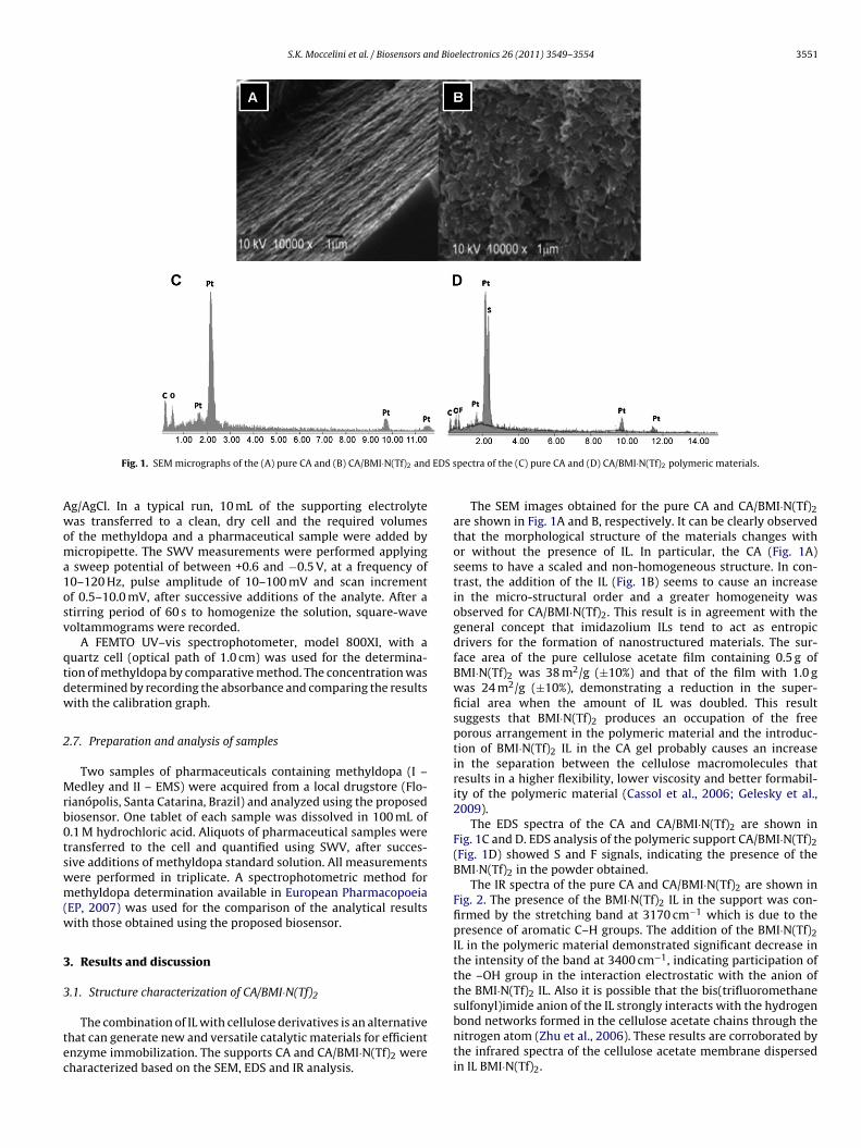
Fig. 1. SEM micrographs of the (A) pure CA and (B) CA/BMI·N(Tf)2 and
g/AgCl. In a typical run, 10 mL of the supporting electrolyteas transferred to a clean, dry cell and the required volumes
f the methyldopa and a pharmaceutical sample were added byicropipette. The SWV measurements were performed applyingsweep potential of between +0.6 and −0.5 V, at a frequency of
0–120 Hz, pulse amplitude of 10–100 mV and scan incrementf 0.5–10.0 mV, after successive additions of the analyte. After atirring period of 60 s to homogenize the solution, square-waveoltammograms were recorded.
A FEMTO UV–vis spectrophotometer, model 800XI, with auartz cell (optical path of 1.0 cm) was used for the determina-ion of methyldopa by comparative method. The concentration wasetermined by recording the absorbance and comparing the resultsith the calibration graph.
.7. Preparation and analysis of samples
Two samples of pharmaceuticals containing methyldopa (I –edley and II – EMS) were acquired from a local drugstore (Flo-
ianópolis, Santa Catarina, Brazil) and analyzed using the proposediosensor. One tablet of each sample was dissolved in 100 mL of.1 M hydrochloric acid. Aliquots of pharmaceutical samples wereransferred to the cell and quantified using SWV, after succes-ive additions of methyldopa standard solution. All measurementsere performed in triplicate. A spectrophotometric method forethyldopa determination available in European Pharmacopoeia
EP, 2007) was used for the comparison of the analytical resultsith those obtained using the proposed biosensor.
. Results and discussion
.1. Structure characterization of CA/BMI·N(Tf)2
The combination of IL with cellulose derivatives is an alternativehat can generate new and versatile catalytic materials for efficientnzyme immobilization. The supports CA and CA/BMI·N(Tf)2 wereharacterized based on the SEM, EDS and IR analysis.
pectra of the (C) pure CA and (D) CA/BMI·N(Tf)2 polymeric materials.
The SEM images obtained for the pure CA and CA/BMI·N(Tf)2are shown in Fig. 1A and B, respectively. It can be clearly observedthat the morphological structure of the materials changes withor without the presence of IL. In particular, the CA (Fig. 1A)seems to have a scaled and non-homogeneous structure. In con-trast, the addition of the IL (Fig. 1B) seems to cause an increasein the micro-structural order and a greater homogeneity wasobserved for CA/BMI·N(Tf)2. This result is in agreement with thegeneral concept that imidazolium ILs tend to act as entropicdrivers for the formation of nanostructured materials. The sur-face area of the pure cellulose acetate film containing 0.5 g ofBMI·N(Tf)2 was 38 m2/g (±10%) and that of the film with 1.0 gwas 24 m2/g (±10%), demonstrating a reduction in the super-ficial area when the amount of IL was doubled. This resultsuggests that BMI·N(Tf)2 produces an occupation of the freeporous arrangement in the polymeric material and the introduc-tion of BMI·N(Tf)2 IL in the CA gel probably causes an increasein the separation between the cellulose macromolecules thatresults in a higher flexibility, lower viscosity and better formabil-ity of the polymeric material (Cassol et al., 2006; Gelesky et al.,2009).
The EDS spectra of the CA and CA/BMI·N(Tf)2 are shown inFig. 1C and D. EDS analysis of the polymeric support CA/BMI·N(Tf)2(Fig. 1D) showed S and F signals, indicating the presence of theBMI·N(Tf)2 in the powder obtained.
The IR spectra of the pure CA and CA/BMI·N(Tf)2 are shown inFig. 2. The presence of the BMI·N(Tf)2 IL in the support was con-firmed by the stretching band at 3170 cm−1 which is due to thepresence of aromatic C–H groups. The addition of the BMI·N(Tf)2IL in the polymeric material demonstrated significant decrease inthe intensity of the band at 3400 cm−1, indicating participation ofthe –OH group in the interaction electrostatic with the anion ofthe BMI·N(Tf)2 IL. Also it is possible that the bis(trifluoromethane
sulfonyl)imide anion of the IL strongly interacts with the hydrogenbond networks formed in the cellulose acetate chains through thenitrogen atom (Zhu et al., 2006). These results are corroborated bythe infrared spectra of the cellulose acetate membrane dispersedin IL BMI·N(Tf)2.
3552 S.K. Moccelini et al. / Biosensors and Bio
3
aep(aBb
tofib
Fc
Fig. 2. Infrared spectra of the (A) pure CA and (B) CA/BMI·N(Tf)2.
.2. Lac immobilization and enzymatic reaction
A key factor in the construction of a biosensor is the need tochieve adequate and effective enzyme immobilization. The Lacnzyme has been successfully immobilized in a variety of sup-orts, including chitosan and cyclodextin, using several methodsFernandes et al., 2008; Franzoi et al., 2009b, 2010). In this study,
new material with excellent properties composed of CA andMI·N(Tf)2 IL was developed and used as a support for Lac immo-ilization.
The plasticizer effect of IL reduces the intermolecular forces
hat are usually present in the CA. This allows the N(Tf)2− anionf the IL to strongly interact with the hydrogen bond networksormed in the CA chains through the nitrogen atom. Therefore, thentroduction of the IL probably causes an increase in the distanceetween the cellulose macromolecules which results in greater
ig. 3. (A) Schematic representation of construction of Lac biosensor based on CA/BMI·Nhemical reduction on the biosensor surface.
electronics 26 (2011) 3549–3554
flexibility, lower viscosity, and better formability of the cellulosematerial (Gelesky et al., 2009). The Lac and CA/BMI·N(Tf)2 com-bination exhibits an excellent synergistic effect that enhances theactivity and stability of the biosensor when used for methyldopadetermination.
Thus, the Lac enzyme is entrapped within the interstitial spaceof the CA/BMI·N(Tf)2 and this system tries to mimic its naturalenvironment which can result in considerable stabilization of theenzyme structure, hence increasing its activity. In addition, ILsmay be responsible for the stabilization of the supported enzyme,which offers a favorable microenvironment for the enzymatic reac-tion, improving the biosensor performance (Yang and Pan, 2005).Fig. 3A shows the construction steps of the Lac biosensor based onCA/BMI·N(Tf)2, used for methyldopa determination.
Lac biosensors have been employed for the determination of abroad range of polyphenols (Fernandes et al., 2008; Franzoi et al.,2009a,b, 2010). Here, Lac catalyzed the oxidation of methyldopato quinone and this product was electrochemically reduced tomethyldopa on the biosensor surface at a potential of +0.07 V vs.Ag/AgCl, as illustrated in Fig. 3B.
3.3. Electrochemical response of Lac biosensors to methyldopa
SEM, EDS and IR analysis confirmed the presence of BMI·N(Tf)2within the CA polymeric matrix. To show the importance of theLac enzyme adsorbed on the CA/BMI·N(Tf)2 support, the elec-trochemical behavior of methyldopa using different sensors wasinvestigated by SWV in the potential range of +0.6 and −0.5 Vvs. Ag/AgCl. Fig. 4 shows the voltammograms obtained using the(a) bare CPE, (b) biosensor (free Lac), (c) biosensor//CA and (d)biosensor//CA/BMI·N(Tf)2 in 0.2 mM methyldopa in 0.1 M acetatebuffer solution (pH 5.5), with optimized parameters. The cathodic
current of o-quinone reduction to methyldopa was used as theanalytical response (cathodic current values are shown in the insetof Fig. 4).The Lac enzyme can catalyze the oxidation reaction of methyl-dopa and thus its incorporation in the electrode (curve b) causes
(Tf)2 and (B) Lac-catalyzed oxidation of methyldopa with its subsequent electro-

S.K. Moccelini et al. / Biosensors and Bioelectronics 26 (2011) 3549–3554 3553
0,80,60,40,20,0-0,2-0,4
-80
-70
-60
-50
d
c
b
a
ΔI (
μ A)
E (V) vs. Ag/AgCl
76543210
d
c
a
b
-ΔI (μA)
Fig. 4. Square-wave voltammograms obtained using (a) bare CPE, (b) biosensor (freeLac), (c) biosensor//CA and (d) biosensor//CA/BMI·N(Tf) in 0.2 mM methyldopa in0ad
aoiqefeectsibi
3c
CtwS
bpt0f
vpvb
ttarpr
0,80,60,40,20,0-0,2-0,4
-130
-120
-110
-100
-90
j
a
Δ I (
μ A)
E (V) vs. Ag/AgCl
400350300250200150100500-6
-8
-10
-12
-14
-16
-18
ΔI (
μA)
[methyldopa] (μM)
Fig. 5. Square-wave voltammograms obtained using Lac biosensor based on
2.1 M acetate buffer solution (pH 5.5); frequency 90 Hz, pulse amplitude 100 mVnd scan increment 4.0 mV. Inset: cathodic peak current values for bare CPE andifferent biosensors.
n increase in the cathodic current when compared with thatbtained for the bare CPE (curve a). The incorporation of CAn the biosensor (curve c) increased the current value of o-uinone reduction to methyldopa, since CA offers a biocompatiblenvironment for the enzyme. The greater response observedor the biosensor//CA/BMI.N(Tf)2 (curve d) is attributed to thexcellent immobilization process, resulting in a more sensitivelectrode. The novel material based on CA and BMI.N(Tf)2, whichombined the biocompatibility of the former and the conduc-ivity and enzymatic stabilization power of the latter, presentsuperior properties over unmodified CA. In addition, the compar-son of the response obtained with biosensor//CA/BMI.N(Tf)2 andiosensor//CA showed the formation of a matrix suitable for Lac
mmobilization.
.4. Optimization of the biosensor construction and analyticalonditions
To optimize the performance of the Lac biosensor based onA/BMI·N(Tf)2 for the determination of methyldopa, some parame-ers, such as Lac concentration, supporting electrolyte and pH, alongith the frequency, pulse amplitude and scan increment used in the
WV, were investigated.The effect of different Lac unit values (0.3–0.9 U mL−1) on the
iosensor response was evaluated. The analytical signals (cathodiceak currents) for 0.2 mM methyldopa in 0.1 M acetate buffer solu-ion at pH 5.5 increased with the enzyme concentration up to.5 U mL−1. Consequently, this enzyme concentration was selectedor further studies.
The influence of different 0.1 M supporting electrolytes and pHalues, including acetate buffer solution (pH 4.0–5.5) and phos-hate buffer solution (pH 6.0–8.0), were investigated. The bestoltammetric responses for methyldopa were obtained in acetateuffer solution at pH 5.5.
The SWV parameters were optimized using the best response ofhe biosensor to 0.2 mM methyldopa solutions. Frequency values in
he range of 10–120 Hz, pulse amplitudes between 10 and 100 mVnd scan increments from 0.5 to 10.0 mV were evaluated. The bestesultant peak currents were obtained using values for frequency,ulse amplitude and scan increment of 90 Hz, 100 mV and 4.0 mV,espectively.CA/BMI·N(Tf)2 for (a) blank in acetate buffer solution (0.1 M; pH 5.5) and methyldopasolutions at the following concentrations: (b) 34.8; (c) 49.5; (d) 73.9; (e) 121.9; (f)169.1; (g) 215.0; (h) 238.0; (i) 283.0 and (j) 370.3 �M; frequency 90 Hz, pulse ampli-tude 100 mV and scan increment 4.0 mV. Inset: calibration curves for methyldopa.
3.5. Analytical performance of biosensor in methyldopadetermination
Under the optimal working conditions the analytical curveswere obtained with the biosensor in the potential range of +0.6to −0.5 V vs. Ag/AgCl. The electrochemical reduction to methyldopawas obtained at a potential of +0.07 V. Fig. 5 shows the square-wavevoltammograms and analytical curve constructed from cathodiccurrent peak vs. methyldopa concentration, obtained employingthe proposed Lac biosensor based on CA/BMI·N(Tf)2. As can beobserved, the cathodic current increased with methyldopa concen-tration and the analytical curve was linear from 34.8 to 370.3 �M(�I = 0.262 (±0.041) − 3.486 × 10⁴ (±2.028 × 10²) [methyldopa];r2 = 0.9998); where �I is the cathodic peak current in �A and[methyldopa] is the concentration in �M. The detection limit (threetimes the signal blank/slope) using the biosensor was 5.5 �M. Thesatisfactory analytical performance of the proposed biosensor canbe attributed to the efficiency of the immobilization of the Lac inthe polymeric material – CA/BMI·N(Tf)2 and the high conductiv-ity of the ionic liquid facilitating electron transfer on the biosensorsurface.
3.6. Repeatability, reproducibility and stability of the biosensor
The repeatability of the current response for the proposedbiosensor in a solution containing 0.2 mM of methyldopa solutionin acetate buffer solution (0.1 M; pH 5.5) was investigated. Therelative standard deviation (RSD) was 1.5% for successive assays(n = 6).
Another important parameter that needs to be considered is thereproducibility of the biosensor which was investigated by detect-ing 0.2 mM methyldopa. Five biosensors were constructed usingthe same procedure and were independently used for detection ofmethyldopa under the optimized conditions described previously.The RSD was 4.3%, demonstrating that the biosensor has a goodreproducibility.
Additional experiments were carried out to test the stability
over a period of 60 days (every 5 days) at room temperature, intriplicate and using three concentrations of methyldopa. The sta-bility of the Lac biosensor based on CA/BMI·N(Tf)2 was measuredby the cathodic current response and 90% of its initial response to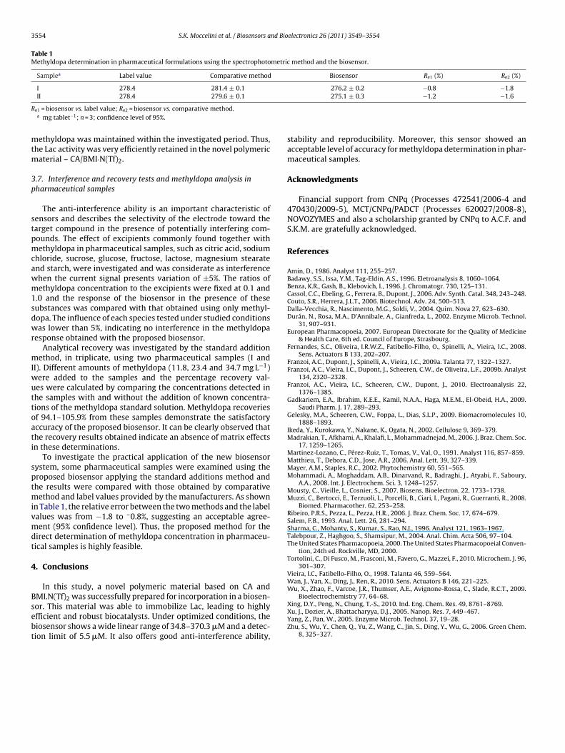
3554 S.K. Moccelini et al. / Biosensors and Bioelectronics 26 (2011) 3549–3554
Table 1Methyldopa determination in pharmaceutical formulations using the spectrophotometric method and the biosensor.
Samplea Label value Comparative method Biosensor Re1 (%) Re2 (%)
I 278.4 281.4 ± 0.1 276.2 ± 0.2 −0.8 −1.8
R
mtm
3p
stpmcawm1sdwr
mIwuttoati
sptmivmdt
4
Bsebt
II 278.4 279.6 ± 0.1
e1 = biosensor vs. label value; Re2 = biosensor vs. comparative method.a mg tablet−1; n = 3; confidence level of 95%.
ethyldopa was maintained within the investigated period. Thus,he Lac activity was very efficiently retained in the novel polymeric
aterial – CA/BMI·N(Tf)2.
.7. Interference and recovery tests and methyldopa analysis inharmaceutical samples
The anti-interference ability is an important characteristic ofensors and describes the selectivity of the electrode toward thearget compound in the presence of potentially interfering com-ounds. The effect of excipients commonly found together withethyldopa in pharmaceutical samples, such as citric acid, sodium
hloride, sucrose, glucose, fructose, lactose, magnesium stearatend starch, were investigated and was considerate as interferencehen the current signal presents variation of ±5%. The ratios ofethyldopa concentration to the excipients were fixed at 0.1 and
.0 and the response of the biosensor in the presence of theseubstances was compared with that obtained using only methyl-opa. The influence of each species tested under studied conditionsas lower than 5%, indicating no interference in the methyldopa
esponse obtained with the proposed biosensor.Analytical recovery was investigated by the standard addition
ethod, in triplicate, using two pharmaceutical samples (I andI). Different amounts of methyldopa (11.8, 23.4 and 34.7 mg L−1)
ere added to the samples and the percentage recovery val-es were calculated by comparing the concentrations detected inhe samples with and without the addition of known concentra-ions of the methyldopa standard solution. Methyldopa recoveriesf 94.1–105.9% from these samples demonstrate the satisfactoryccuracy of the proposed biosensor. It can be clearly observed thathe recovery results obtained indicate an absence of matrix effectsn these determinations.
To investigate the practical application of the new biosensorystem, some pharmaceutical samples were examined using theroposed biosensor applying the standard additions method andhe results were compared with those obtained by comparative
ethod and label values provided by the manufacturers. As shownn Table 1, the relative error between the two methods and the labelalues was from −1.8 to –0.8%, suggesting an acceptable agree-ent (95% confidence level). Thus, the proposed method for the
irect determination of methyldopa concentration in pharmaceu-ical samples is highly feasible.
. Conclusions
In this study, a novel polymeric material based on CA and
MI.N(Tf)2 was successfully prepared for incorporation in a biosen-or. This material was able to immobilize Lac, leading to highlyfficient and robust biocatalysts. Under optimized conditions, theiosensor shows a wide linear range of 34.8–370.3 �M and a detec-ion limit of 5.5 �M. It also offers good anti-interference ability,275.1 ± 0.3 −1.2 −1.6
stability and reproducibility. Moreover, this sensor showed anacceptable level of accuracy for methyldopa determination in phar-maceutical samples.
Acknowledgments
Financial support from CNPq (Processes 472541/2006-4 and470430/2009-5), MCT/CNPq/PADCT (Processes 620027/2008-8),NOVOZYMES and also a scholarship granted by CNPq to A.C.F. andS.K.M. are gratefully acknowledged.
References
Amin, D., 1986. Analyst 111, 255–257.Badawy, S.S., Issa, Y.M., Tag-Eldin, A.S., 1996. Eletroanalysis 8, 1060–1064.Benza, K.R., Gash, B., Klebovich, I., 1996. J. Chromatogr. 730, 125–131.Cassol, C.C., Ebeling, G., Ferrera, B., Dupont, J., 2006. Adv. Synth. Catal. 348, 243–248.Couto, S.R., Herrera, J.L.T., 2006. Biotechnol. Adv. 24, 500–513.Dalla-Vecchia, R., Nascimento, M.G., Soldi, V., 2004. Quim. Nova 27, 623–630.Durán, N., Rosa, M.A., D’Annibale, A., Gianfreda, L., 2002. Enzyme Microb. Technol.
31, 907–931.European Pharmacopoeia, 2007. European Directorate for the Quality of Medicine
& Health Care, 6th ed. Council of Europe, Strasbourg.Fernandes, S.C., Oliveira, I.R.W.Z., Fatibello-Filho, O., Spinelli, A., Vieira, I.C., 2008.
Sens. Actuators B 133, 202–207.Franzoi, A.C., Dupont, J., Spinelli, A., Vieira, I.C., 2009a. Talanta 77, 1322–1327.Franzoi, A.C., Vieira, I.C., Dupont, J., Scheeren, C.W., de Oliveira, L.F., 2009b. Analyst
134, 2320–2328.Franzoi, A.C., Vieira, I.C., Scheeren, C.W., Dupont, J., 2010. Electroanalysis 22,
1376–1385.Gadkariem, E.A., Ibrahim, K.E.E., Kamil, N.A.A., Haga, M.E.M., El-Obeid, H.A., 2009.
Saudi Pharm. J. 17, 289–293.Gelesky, M.A., Scheeren, C.W., Foppa, L., Dias, S.L.P., 2009. Biomacromolecules 10,
1888–1893.Ikeda, Y., Kurokawa, Y., Nakane, K., Ogata, N., 2002. Cellulose 9, 369–379.Madrakian, T., Afkhami, A., Khalafi, L., Mohammadnejad, M., 2006. J. Braz. Chem. Soc.
17, 1259–1265.Martinez-Lozano, C., Pérez-Ruiz, T., Tomas, V., Val, O., 1991. Analyst 116, 857–859.Matthieu, T., Debora, C.D., Jose, A.R., 2006. Anal. Lett. 39, 327–339.Mayer, A.M., Staples, R.C., 2002. Phytochemistry 60, 551–565.Mohammadi, A., Moghaddam, A.B., Dinarvand, R., Badraghi, J., Atyabi, F., Saboury,
A.A., 2008. Int. J. Electrochem. Sci. 3, 1248–1257.Mousty, C., Vieille, L., Cosnier, S., 2007. Biosens. Bioelectron. 22, 1733–1738.Muzzi, C., Bertocci, E., Terzuoli, L., Porcelli, B., Ciari, I., Pagani, R., Guerranti, R., 2008.
Biomed. Pharmacother. 62, 253–258.Ribeiro, P.R.S., Pezza, L., Pezza, H.R., 2006. J. Braz. Chem. Soc. 17, 674–679.Salem, F.B., 1993. Anal. Lett. 26, 281–294.Sharma, C., Mohanty, S., Kumar, S., Rao, N.J., 1996. Analyst 121, 1963–1967.Talebpour, Z., Haghgoo, S., Shamsipur, M., 2004. Anal. Chim. Acta 506, 97–104.The United States Pharmacopoeia, 2000. The United States Pharmacopoeial Conven-
tion, 24th ed. Rockville, MD, 2000.Tortolini, C., Di Fusco, M., Frasconi, M., Favero, G., Mazzei, F., 2010. Microchem. J. 96,
301–307.Vieira, I.C., Fatibello-Filho, O., 1998. Talanta 46, 559–564.Wan, J., Yan, X., Ding, J., Ren, R., 2010. Sens. Actuators B 146, 221–225.Wu, X., Zhao, F., Varcoe, J.R., Thumser, A.E., Avignone-Rossa, C., Slade, R.C.T., 2009.
Bioelectrochemistry 77, 64–68.Xing, D.Y., Peng, N., Chung, T.-S., 2010. Ind. Eng. Chem. Res. 49, 8761–8769.Xu, J., Dozier, A., Bhattacharyya, D.J., 2005. Nanop. Res. 7, 449–467.Yang, Z., Pan, W., 2005. Enzyme Microb. Technol. 37, 19–28.Zhu, S., Wu, Y., Chen, Q., Yu, Z., Wang, C., Jin, S., Ding, Y., Wu, G., 2006. Green Chem.
8, 325–327.
Copyright © 2022 FDOKUMEN