Bayesian Joint Topic Modelling for Weakly Supervised Object Localisation

PARASITOLOGY
Molecular & Biochemical Parasitology 134 (2004) 105-114
A novel form of actin in Leishmania: molecular characterisation,subcellular localisation and association with subpellicular microtubules 1:4
Amogh A. Sahasrabuddhea, Virendra K. Bajpai b, Chhitar M. Gupta a,*
a Divi.rion of Molecular and Structural Biology. Central Drug Research Institute. Lucknow 226 DOl India
b Electron Microscopy Unit. Central Drug Research Institute. Lucknow 226 001. India
Abstract
To study the occurrence and subcellular distribution of actin in trypanosomatid parasites, we have cloned and overexpressed Leishmaniadonovani actin gene in bacteria, purified the protein, and employed the affinity purified rabbit polyclonal anti-recombinant actin antibodiesas a probe to study the organisation and subcellular distribution of actin in Leishmania cells. The Leishmania actin did not cross reactwith antimammalianactinantibodiesbut was readilyrecognizedby the anti-Leishmaniaactinantibodiesin both the promastigoteandamastigote forms of the parasite. About 106copies per cell of this protein (Mr 42.05 kDa) were present in the Leishmania promastigote.Unlike other eukaryotic actins, the oligomeric forms of Leishmania actin were not stained by phalloidin nor were dissociated by actinfilament-disrupting agents, like Latrunculin Band Cytochalasin D. Analysis of the primary structure of this protein revealed that theseunusual characteristics may be related to the presence of highly diverged amino acids in the DNase I-binding loop (amino acids 40-50)and the hydrophobic plug (amino acids 262-272) regions of Leishmania actin.
The subcellular distribution of actin was studied in the Leishmania promastigotes by employing immunoelectron and immunofluorescencemicroscopies. This protein was present not only in the flagella, flagellar pocket. nucleus and the kinetoplast but it was also localized onthe nuclear, vacuolar and cytoplasmic face of the plasma membranes. Further, the plasma membrane-associated actin was colocalised withsubpellicular microtubules, while most of the actin present in the kinetoplast colocalised with the k-DNA network.
These results clearly indicate that Leishmania contains a novel form of actin which may structurally and functionally differ from othereukaryotic actins. The functional significance of these observations is discussed.
Keywords: Trypanosomatids; Actin; Microtubules; k-DNA network; Association; Cellular functions
1. Introduction
Actin is a cytoskeletal protein, which besides determin-ing the cell morphology also provides the building blocksrequired for cell motility, contractility and intracellulartransport in eukaryotic cells [1,2]. The protein exists inmonomeric as well as filamentous forms. The filaments are
organised in specific arrays that form protrusive structures
Abbreviations: DMEM, Dulbecco's modified eagle medium; L-act
Abs, monospecific polyclonal anti-Leishmania actin antibodies; M-act
Abs, polyclonal anti-human actin antibodies; DAPI, 4',5-diamidino-2-phenylindole
~ This is a communication number 6366 from the Central DrugResearch Institute, Lucknow, India.
. Corresponding author. Tel.: +91-522-2223286/2210932;fax: +91-522-2223405/2223938.
E-mail addres.fes:[email protected]@rediffmail.com(C.M. Gupta).
at the cell surface and also associate with the cytoplasmicsurface of the plasma membrane to regulate the cell shape[2,3]. Also, actin has been detected in the nucleus where ithas been implicated to play an important role in chromatinremodeling [4,5], and also in RNA processing, transport andother related processes [6,7]. Furthermore, the actin networkfunctionally associates with microtubules during processes,such as cell migration, cleavage furrow placement and vesi-cle transport [8,9], and also with the plasma membraneregions that are engaged during endocytosis [10,II J. Dueto its multifunctional roles in cell multiplication, growthand development, actin is a most abundant protein in manyeukaryotic cells and is conserved from yeast to humans [2].
Leishmania parasites are protozoan organisms whichcause the dreaded disease 'Kala-azar' and exist in two
forms, viz. promastigotes and amastigotes, which containmicrotubules as their major cytoskeletal component [12].Although these cells have been shown to carryout some of
J
Mol Biochem Parasitol,134.105-114 (2004)

the cellular activities that essentially require actin [13,14],no convincing evidence to date is available on the existenceof actin network in Leishamnia cells [12,15,16]. However,the presence of actin gene has been confirmed in the Leish-mania major genome [17].
To examine the presence of actin in Leishmania, we pre-pared affinity purified monospecific anti-recombinant Leish-mania actin antibodies and then by using these antibodiesas a probe we analysed some molecular characteristics, sub-cellular localization and association with microtubules ofactin in the Leishmania promastigotes. Our results revealedthat about 106copies of actin (Mr 42.05 kDa) per cell werepresent in form of granules, short filamentslbundles andpatches in the Leishmania promastigotes. It is further shownthat unlike conventional actins, these oligomeric forms ofLeishmania actin were neither disrupted by the actin fila-ment disruptingagents nor stained by phalloidin. In addition,it is demonstrated that this protein in these cells was local-ized not only in their cortical cytoskeleton, flagella, flagellarpocket, nucleus and kinetoplast, but also on the vacuolar andnuclear membranes, and also it is closely associated with thesubpellicular microtubules and the k-DNA network. Theseresults indicate that kinetoplastid parasites, especially Leish-mania, contain unconventional actin which may structurallyand functionally differ from other eukaryotic actins.
2. Materials and methods
2.1. Cell culture
All the Leishmania strains and mouse macrophage cellline (1774 A.I) and BHK21 cells were obtained fromthe National Institute of Immunology, New Delhi (India),and maintained in high glucose DMEM, supplementedwith 10% heat-inactivated fetal bovine serum containing40 j..Lg/mlgentamycin, at 25 and 37°C, respectively.Mono-layers of macrophage cells on coverslips were infected withLeishmania donovani (DD8 strain) promastigotes followingthe published procedure [18].
2.2. Cloning and expression of Leishmania donovani actin
Genomic DNA from the cultured promastigotes was pre-pared using standard protocols [19]. Primers were designedon the basis of the Leishmania major actin gene sequenceavailable from the Gene Bank (accession no. L16961). Puta-tive actin gene of about 1.1kb was amplified by polymerasechain reaction (PCR) using the forward primer: 5'-GGAA-TTCCATATGGCTGACAACGAGCAGAGCTCCATCG-3',reverseprimer: 5'-GGAATTCCATATGTCAGAAGCACTT-GTTATGCACGATGCT-3'and the total genomic DNA asthe template. PCR product was cloned in the prokaryoticexpression vector pET-22b (Novagen, USA) at Nde I site,sequenced by dideoxy chain termination method [20], andthen overexpressed in Escherichia coli strain BL21 (DE3).
For expression in mammalian cells (BHK21 cell line), theLeishmania actin gene was cloned in pCDNA3 vector (In-vitrogen, USA). Transfection quality DNA of this constructwas made by Qiagen Tip 100 midiprep column. BHK21cells were transfected with pCDNA3-actin construct usingLipofectamine-Plus reagent (GIBCO-BRL, USA).
2.3. Actin sample preparation for ESI-mass spectrometry
The recombinant E. coli cells obtained from the inducedcultures (5.0ml) were sonicated in water (0.5 ml) with IOpulses of 10s each with 20 s intervals on ice. The cell lysatethus obtained was centrifuged first at 500 x g for 10minto remove the cell debris and then the milky supernatantat 12000 x g for 5 min (4°C) to pellet out the aggregatedactin present in inclusion bodies. The pellet was washed withwater for six times and then dissolved in absolute formicacid (50 j..L1).It was centrifuged at 12,000 x g and the clearsupernatant was analysed by mass spectrometry.
...
2.4. Antibodies
The recombinant Leishmania donovani actin was pu-rified by SDS-polyacrylamide gel electrophoresis [21].Purified recombinant protein was injected into rabbits toraise the polyclonal antiserum. Anti-Leishmania actin an-tibodies were affinity purified by using recombinant actinafter its purification by preparative SDS-polyacrylamidegel electrophoresis followed by immobilization on theCNBr-activated Sepharose (Amersham-Pharmacia Biotech).Antibodies against a,~-tubulins (monoclonals) and human~-actin (polyclonal and monoclonal)) were procured fromICN, USA.
2.5. Western blotting
Proteins resolved on the SDS-polyacrylamide (10%) gelswere electroblotted onto the nitrocellulose membranes in
Tris-glycine buffer at 50 V for 2h [22]. After incubating theblots with primary antibodies (0.1 j..Lg/ml)at 25°C for I hfollowed by five washings with phosphate-buffered saline(PBS) containing 0.05% (v/v) Tween 20, these were fur-ther incubated with HRP conjugate of secondary antibod-ies, washed, and developed by chemiluminescence (ECL kit,Amersham Pharmacia, USA).
2.6. Immunofluorescence microscopy
Leishmania promastigotes were fixed with 4% (w/v)paraformaldehyde in 200mM HEPES buffer (pH 7.2) at25°C for 30 min, and washed two times for 5min eachwith PBS containing 0.5% (w/v) glycine. The washed cellswere allowed to adhere on the polY-L-lysine-coatedcover-slips. To analyze the Leishmania amastigotes, the mousemacrophage cells (J774A.l) were grown on coverslipsand infected with Leishmania promastigotes (1:1O ratio)
Mol Biochem Parasitol,134.105-114 (2004)

at 37°C for I h. The macrophage cells 24 h post infectionwere fixed with 4% paraformaldehyde (w/v) in PBS (pH7.2) at 25°C for 10min, and washed as above.
The fixedcells were permeabilised with 0.1% (v/v) TritonX-IOOand blocked with 10% (v/v) goat serum (AmershamPharmacia) in PBS for 30 min. The blocked cells were firstincubated with primary antibodies (10 I-Lg/ml)at 4°C andthen stained with Oregon green and/or Texas red-X-taggedsecondary antibodies (10 I-Lglml)at 4°C for 4 h in dark.Nuclei were stained with 4',5-diamidino-2-phenylindole(DAPI) (5 I-Lglml).Coverslips were mounted in the mount-ing medium (Oncogene) and images were collected using63 x 1.4 NA Plan Apochromate lense on Zeiss LSM 510Confocal Microscope. Green, red and blue images werecollected separately with excitation at 488, 543 and 364 nm,respectively, and merged for presentation.
2.7. Immunogold electron microscopy
Leishmania promastigotes were fixed with 4% (w/v)paraformaldehyde and 0.1% (w/v) glutaraldehyde in PBS.This was followed by two more changes of fixatives andsubsequent incubations for 4 h. The excess fixatives werequenched with 0.5% (w/v) glycine in PBS (pH 7.4) at 4°Cfor 10min. The fixed cells embedded in 1% agarose. Theagarose-embedded blocks were dehydrated in ascendingseries of ethanol, impregnated in LR-White resin (Sigma)and then polymerized at 60°C for 48 h. Ultrathin sec-tions (80-100 nm) were cut and collected on nickel grids.Sections were first blocked by PBS (pH 7.4) containing0.1% BSA-c (w/v) (Aurion, Germany), and then incubatedfor 4 h with monospecific anti-Leishmania actin antibod-ies (10 I-Lg/ml)in the blocking buffer. After washing fivetimes with the blocking buffer, the sections were incu-bated with 10nm gold-coupled goat anti-rabbit IgG (I: 10)at 37°C for I h, washed with blocking buffer five timesfor 5 min each, contrasted with uranyl acetate and exam-ined under FEI Philips Transmission Electron Microscope(TECNAI-12) at 80 kV. Control samples were separatelyincubated as above with normal rabbit IgG as well as poly-clonal anti-human actin antibodies rather than monospecificpolyclonal anti-Leishmania actin antibodies.
2.8. Latrunculin B treatment
Transfected or non-transfectedBHK21cells and Leishma-
nia promastigotes were treated with Latrunculin B (5 I-LM)under their growth conditions for 30 min. Cells were har-vested, washed, and then lysed in PBS containing 1% (v/v)Triton X-lOa and protease inhibitor cocktail on ice (15 min).The Iysateswere centrifuged at 12,000x g for 20 min at 4 °Cand the clear supernatants were carefully collected as sepa-rate soluble fractions. Insoluble pellets washed once with theabove buffer and washings were added to the above solublefractions. Equal volumes of both the soluble and insolublefractions were analysed for actin by Westernblotting and the
amount of actin quantitated by densitometric analysis usingthe Image Master programme (Amersham-Pharmacia).
2.9. Protein copy number determination
Estimated quantities of purified recombinant actin andIysates of known number of Leishmania promastigotes wereresolved on 10% SDS-polyacrylamide gels and electroblot-ted on nitrocellulose membrane. Blots were developed af-ter primary and secondary antibody treatment by ECL kit(Amersham Pharmacia, USA) on X-ray film.The bands wereanalysed by densitometric analysis and the amount of actinin known number of cells was determined from the standardcurve derived from purified actin. The standard curve waslinear at least upto 100ng quantity of actin.
3. Results
3.1. Molecular characterization
We cloned and over expressed the L. donovani actingene in E. coli (Fig. I) by using the primers that weredesigned on the basis of the sequence reported earlier forL. major actin gene [17]. The molecular identity of re-combinant actin was established by sequencing a portion
kD Mr 1 2 3 4
Fig. I. Over expression of Leishmania actin in E. coli. The cell Iysates
were analysed by SOS-polyacryamide gel electrophoresis (PAGE) and
the gels stained with Coomassie blue. Mr. molecular weight markers; lane
I. uninduced cell Iysates; lane 2. induced cell Iysates; lane 3. inclusionbodies; lane 4. SOS-PAGE purified recombinant Leishmania actin (Mr
42.05 kOa). The total protein in lanes 1-3 was 30 ILgllane. while lane 4contained only 10 ILg protein.
166 .--66 ---45-.
35
25
1814
Mol Biochem Parasitol,134.105-114 (2004)

kD Mr 1 2 3 4 5 6 7 8 9pr. --
116 ..66 ..45-35'-25-18.-14,-
1 2 3 4 8 9 123456789
·Iti ~I'
.
c
.
..
.
..
.
.
.
1
""'
.1....._
':; . .. I
~_f.,..(A)
-(B) (C)
Fig. 2. Specificity of anti recombinant Lei.fhmania actin antibodies. Panel A: Coomassie-blue stained SDS-PAGE (10%); panel B: Western blotting with
anti-Leishmania actin antibodies; panel C: Western blotting with anti-human actin antibodies. Mr, molecular weight markers; lane I, recombinant actin;
lane 2, L. donovani promastigote lysate; lane 3, L. major promastigote lysate; lane 4, L. tropica promastigote lysate; lane 5, T. cruzi (blood stage) lysatewhich contained about 20% mouse erythrocytes as contamination; lane 6, P falciparum lysate; lane 7, S. cerevisiae lysate; lane 8, BHK21 cells lysate;
lane 9. mouse erythrocytes lysate. Lanes 2-9 contained 30 fLg protein/lane whereas lane I contained only 0.5 fLg protein.
of its N-terminal sequence (ADNEQSSIVXD) and alsoanalyzing by mass spectrometry. The molecular mass ofrecombinant actin was 42.05 kDa which corresponded wellwith the molecular mass of eukaryotic actins [1,2]. Purerecombinant actin in monomeric form was prepared fromactin aggregates by SDS-polyacrylamide gel electrophore-sis [21], and used for immunization of rabbits for raisingthe anti-actin antisera. The immunogenicity of the recom-binant Leishmania actin was about 100 times higher thanthat of the human erythrocyte actin as judged from thelevels of antibody titres in the rabbits that were separatelyimmunized with equal quantities of Leishmania and hu-man erythrocyte actins under identical conditions (data notshown).
Monospecificpolyclonal anti-Leishmaniaactin antibodies(L-act Abs) were prepared from the rabbit antisera and usedto probe the presence of actin in Leishmania cells. Fig. 2shows that L-act Abs strongly crossreacted with a proteinof about 43 kDa in whole celllysates of various Leishmaniaspecies as well as of Trypanosomacruzi, but it failed to rec-ognize the actin band in whole cell Iysates of Plasmodiumfalciparum, Saccharomycescerevisiae,BHK21 cells and hu-man erythrocytes. Conversely,rabbit polyclonal anti-human~-actin antibodies (M-act Abs) readily recognized the actinband in whole cell Iysates of P.falciparum. S. cerevisiae,BHK21 cells and human erythrocytes but it did not crossreact with Leishmania or Trypanosomaactin. To further cor-roborate these findings, we analysed the presence of actinin L. donovani by immunofluorescencemicroscopy (Fig. 3).Whereas M-act Abs even upon using in very high concentra-tion failed to clearly detect the presence of actin in Leishma-nia promastigotes, which is not in agreement with the earlierreport [15], L-act Abs readily recognized actin in both thepromastigote and amastigote formsof L. donovani. This pro-tein was present largely in granular form not only in the mainbody of the promastigote but also in its flagella.The averagenumber of actin molecules was about I x 106copies per pro-mastigote.Further,unlikethe mammalianactin,the Leish-mania actin was not stained by rhodamine-phalloidinwhichis known to specifically stain actin in its filamentous form[23].
Significant fraction of the total cell actin exists inoligomeric forms in the eukaryotic cells [1,2]. Theseoligomers are, however, dissociated into actin monomersby treating the cells with Latrunculin B or Cytochalasins[24], but resist their solubilisation by Triton X-IOO [25].To determine the fraction of Leishmania actin that existsin the oligomeric form, we therefore treated the L. dono-vani promastigotes and BHK21 cells with Latrunculin Bin identical conditions, and then determined the fractionsof monomeric and oligomeric forms of actin prior to andafter subjecting them to the Triton X-IOO solubilisationmethod [25]. Fig. 4A and B shows that only about 30% ofthe total Leishmania actin was insoluble in the detergent,as compared to about 45% of the total BHK 21 cell actin.Further, unlike the BHK 21 actin, the oligomeric forms ofLeishmania actin were almost completely resistant to theLatrunculin B treatment. Similar results were also observedby treating the Leishmania promastigotes with Cytochalasino (data not shown). To examine whether the reduced extentof Leishmania actin oligomerisation in the promastigotes isdue to absence of some proteins [3,26] that assist/promoteactin in forming large filaments or it is due to some struc-tural differences between the mammalian and Leishmania
actins, we expressed the Leishmania actin in BHK 21 cellsand then estimated the amounts of oligomeric forms of boththe BHK 21 and Leishmania actins in the transfected cells.Results given in Fig. 4C and 0 clearly indicate that theextent of oligomerisation of Leishmania actin did not in-crease even upon expressing this protein in the mammaliancells.
These results strongly indicate that Leishmania actin sig-nificantly differs from yeast, Plasmodium and human actinsin terms of its properties. To examine the structural basis ofthis difference, we carried out the amino acid sequence align-ment analysis and homology modeling (Fig. 5). Leishmaniaactin possessed 70.93, 69.14 and 69.76% amino acid identi-ties, respectively, with yeast, Plasmodium and human actins.The major differences were confined to the amino acids (aa)1-9,40-53, 194-200,229-240,266-281 and 307-315, mostof which were located on the surface of yeast or mammalianactins, and one of these included the amino acid residues, aa
Mol Biochem Parasitol,134.105-114 (2004)

(A)
(C)
(E)
Fig. 3. Immunostaining of actin in L. donovani promastigotes and amastigotes with anti-Leishmania actin antibodies. The control cells (promastigotes)
were stained with secondary antibodies (panel A: a. phase micrograph; b, immunofluorescence micrograph), anti-human actin antibodies (panel B: a, phase
micrograph; b, immunofluorescence micrograph) and rhodamine-phalloidin (panel C: a. phase micrograph; b, fluorescence micrograph). Panel D: phase
(a) and immunofluorescence (b) micrographs of Leishmania promastigotes stained with anti-Leishmania actin antibodies. Panel E: L. donovani infected
mouse macrophages (J774A.1) doubly stained with rhodamine-phalloidin and anti-Leishmania actin antibodies. A, phase; b, rhodamine-phalloidin; c,anti-Lei.vhmania actin antibodies; d, merge of b and c. Bar, 5 jLm.
120
100
c~CJCO
ii-o-...o
80
60
-CII)CJ~II)
D..
40
20
o
Before Latrunculin B treatment. actin in pellet 0 soluble actinAfter Latrunculin B treatment. actin in pellet . soluble actin
Fig. 4. Effect of Latrunculin B on Triton X-IOO-insoluble fraction of Leishmania and mammalian actins. (A) Leishmania actin in L. donovani promastigotes;
(B) mammalian actin in BHK2l cells; (C) Leishmania actin in BHK2l cells transfected with Leishmania actin gene; (D) mammalian actin in BHK2l
cells transfected with Leishmania actin gene. Values shown are means of three independent experiments:i: standard deviation. The standard deviation was
calculatedby usingStudent's.t' test.
Mol Biochem Parasitol,134.105-114 (2004)

(A) IlADNEOS S IJVCDNGS GMJKAOF S GODAIIR HVFP $1 VGR PJCNmJMiiNOK1INKTXlfVGDEAQ leish-ad
rdSDCEQTAI VCDNGSGMlKSGFSGOD/>/'RHVFPSI VGRPKNEQJM1AGSANKKLrVGOEAQ t'YP_a..r.lOEEOVOALWONOS ONIIKAGVAGDDAP RSVFP S I VGRP KNP 01 MVCMEEKDArVGDEAO pl..m_...
. MDSEVAAL VI ONOS OM:KAGF AGOD/>/' RAVFP S I VGRP RHOGI MVGMGQKDSYVGOEAQ v a..
.MODOI AALWONOS OIvCKAOFAGDOAPRAVFP II VQRPRHOO\oM\lGMOOKDSVVODEAO h""'.n_"'"
S:k'ROVlS:LKV" C=WCI\/rN\/Il8Ot.A!i;1I:1~wTr VN!i;LRVNPEOHNVLL TEAPIu1NPt:ONAEKU l.ich_3~
AKRGVLAlKY'1 EHGI vrN\I\ODMEKWHHTrYNELRVNPESHNVLL TEAPMNPKDNREKIII try,_aotTKRCI LTLKVPI EHGI VTN\I\ODlvlGKI wnrrrVNELRAA.PEEIIPVLL TEAPLNPKGP-IRERt.I pl..m_...SKRCI LHRY" EHGI vrN\I\OOIvIEKI II'tIHTr YNEL RvAPEEHPVLL TEAPJoWjPKSP-IREKIII v...._...OKRCI LTLKVPI EIIGI vrN\I\ODt.AEKI wnrrrVNELR\H\PEEHPVLL TEJ\PLNPrJ\NREKt.I h n_...
TQltvFliiTFNVPSLYI GIQAVLSLV8CGRTTCI VLOAGDOVfI-tTVPI VIiGYiLPNAVARVO Ici~h_ZIotTQII6ETrOvr.AllA'NGI OAVlSL YSSOOTTOI VlDAGDGvrHTVPI YEGYSL PHAI RRVDtry,_a..TOI WI' .srNVPMAWAJ OAVl H VUGRTTCI VlDOGDGVSIITVPI V.CVAL PIIAI MRL D pl..m_...TQII6ETrNvrAF'NSI OAVlSL YSSOOTTOI VlDSGDGvrHWPI YAGFSL PHAI LRI 0 v a..TOI WFETfNHMAVVAJ OAVlOLYASGRTTGI 'v!oIOSGDGvrHTVPI VEGYALPllAllRLD h n_...
MA.O~OLTfVLMKlllm:tllllrTTTAet<e1 VftNVlCeQLCY\lALDreee~'II:ft~JIIlItS__rF~C1.:'Ilei~b_lIotrdAGROLTEYLMKI LMETGMTfTTSAEKEIVRNI KEOLCYVALOfOEEMTNSAKSVS.EEP tryp_actLAOAOLTEYLMKILtlEAOYOrSTSAeKelVROIMeKLCYINo NrOeEMf<TSEQaSOIeKe pl8Jm_lIftLAGROLTOYLMKI LSERGYSfSTTAEREIVROI KEKLCYVALOfEOEMOTAAQSSSIEKS yeast_a..LAORDLTDYLMKI LTEROYSITTTAEREIVRDI KEKLCYVALOrEOEMATAASSSSlEKS num.n_~
liEL"OOH~ONQRf'RCPEVLf'KP3"1:"[Jt:"u!!!...rur:r.~""uSI NKCOI O\fflR~L YONI 'V lelSrU!lOI
fELPDGNWQVGNORFRCPEALFKPALIGLOEMGHEMTFOSI NKCOIDVRROLYGNIV try,_aotU!LPOOHII TVON~R"RCpeAL"apSFlOK~A.A.01Hl'TTFtt$1KKCOVOI RKDL TUNI"V plilsm_aceYELPOGOVITI GNERFRAPEALFHPS'vI.GLESA.GI DOTTYNSIMKCOVDVRKELYGNIV V.ast act
1ELPOOOVITIGNERFRCPEALFOPSFLGMESC.GI HETTFNSIMKCDV~ RKOLVANTV numl._~
LSDIJ5T~KI'IL:r.I:IV.:MKeISNlArSSIKPKWAfI,ef'KY3VW OOSIl5SL TTrOTMVlNKlelSh_actLSGGfT~KNlPERLGKEI SHLAPSSIKPKWMPERI<YSIMI GaSI LSSLTHOShMJ T tryp_.ctLSGGfTMVEGIOERLTROI TlLAPSTM<1 KWMPERKYSIMI GOSILSSLSHOQMW T plasm_a"MSGGfT~PGIAER\otQKEI TALAPSSM<VKII MPERKYSIMI GOSILASLTTfOQMW S y.ast_a<!LSGGfTMYPGIAOR\otQKEITALAP$TM<I KII APPERKYSIMI GOSILASLSHOQMW Shuman_act
KSEYOESGPSIVHNKCFKSEYOESGPSIVHSKCrKEEYOESGPSIVHRKCFKOEYOESGPSIVHHKCrKOEYOESGPSIVHRKCF
lelsh_aCltryP.'ctprasm_a"y.HI_act."M'TIII'I_a:.1:
(B)I -
229-240\
4o-~,
t: - - - --Fig. 5. (A) Amino acid sequence alignment of Leishmania (Ieish), Trypanosoma (tryp), Plasmodium (plasm), Yeast and human actins. The shaded regionsdenote the divergent sequences in Leishmania actin. L. donovani actin gene sequence submitted to EMBL Gene Bank (AY637036). (B) G-actin model
showing the aminoacid sequences that are divergent in Leishmania actin. Homology modelling was carried out as in [38].
42-46, that constitute the major self association sites duringthe actin filament formation [2].
Subcellular localization of Leishmania actin was stud-ied by the immunoelectron and immunofluorescence mi-crocopies. Fig. 6 shows that this protein in Leishmania
promastigotes was present not only in the flagellar pocket,flagella, kinetoplast, nucleus and cytoplasm but also onthe nuclear, vacular and cytoplasmic face of plasma mem-branes. Actin in flagella was localized predominantly onits surface rather than the interior. Likewise, in the kineto-plast, this protein seemed to largely localize in the k-DNAnetwork. Similarly, in the cortical region, it appeared tolocalize mainly on the microtubular network that constitutethe cortical cytoskeleton. This apart, short filament-like
3.2. Subcellular localization and association withmicrotubules
Mol Biochem Parasitol,134.105-114 (2004)

c,
It J}. .
r<:
"
~ .-. .... .
...-.
. I
. ..
.. t
-<Ii
Fig. 6. Electron microscopy of immunogold-Iabeled actin in Leishmania promastigotes. Localization of actin on as well as in close vicinity of subpellicular
microtubules (Mt), flagella (F), flagellar pocket (FP), kinetoplast (K), nucleus (Nu), cytoplasm and nuclear membrane is clearly visible in panel A. Inaddition, presence of actin on membranes of vacuoles (V) may also be noticed in panel B. Associations of actin with kDNA network, nuclear membrane
and subpellicular microtubules may clearly be seen in panels C-E, respectively. The arrowheads in panel E mark the microtubules. The control sampleswere incubated with normal rabbit IgG as well as polyclonal anti-human actin antibodies rather than the anti-Leishmania actin antibodies. At least
120--150 fields were examined and most of them were found to have similar actin distribution patterns. Mc is mitochondria. Bar, 200nm.
structures of actin were seen in cortical as well as otherregions.
To further confirm the above findings, we examinedthe actin distribution in the Leishmania promastigotes cy-toskeletons that were largely devoid of the cytoplasm butcontained the nucleus and kinetoplast by employing im-munofluorescence microscopy.The cytoskeletons were pre-pared by treating the cells with varying amounts of NP-40or Triton X-I00 (Fig. 7) as described earlier for preparationof Trypanosoma cytoskeletons [27,28]. Whereas treatmentof the promastigotes with I% Triton X-IOOresulted in lossof structural integrity of the nucleus and kinetoplast, use ofO.1-0.5% NP-40 yielded the cytoskeletons that containedthese organelles largely in their intact form, as judged bylight fluorescence microscopy (data not shown). Therefore,only the cytoskeletons that were obtained by the 0.5%
NP-40 treatment were used for the immunofluorescencestudies to analyse the presence of actin in the nucleus andkinetoplast. Results shown in Fig. 8 clearly indicate thatbesides being localized in the posterior end, flagella, cor-tical cytoskeleton and flagellar pocket, actin was presentalso within the nucleus and kinetoplast in form of shortfilaments, bundles and beaded structures. Further, at leasta part of the actin that was localized in the nucleus andkinetoplast appeared to associate with DNA in both theorganelles. This apart, most of the kinetoplast surface wascovered by the actin network which seemed to serve as ananchor between the kinetoplast and the cell surface.
It would seem that actin in the Leishmania promastigotesassociates with cortical microtubules. To further examinethis possibility, we studied both the intact promastigotes andtheir cytoskeletons, obtained by thel% Triton X-IOOtreat-
Mol Biochem Parasitol,134.105-114 (2004)

kDMr 1 2 5 6 1
116 _66 _45
35..
25_
18_14.-
(A)
2 3 4 5 6 1 2 3 4 5 6
(8) (C)
Fig. 7. Preparation of Leishmania promastigote cytoskeletons. The cells were treated with Triton X-IOO (1%) or varying concentrations of NP-40 for
5min on ice and the Iysates centrifuged at 12,000 x g (4°C) for 10min. The pellets were washed with buffer and then analysed by SDS-PAGE. PanelA, Coommassie blue stained SDS-PAGE; panel B, Western blotting with anti-Leishmania actin antibody; Panel C, Western blotting with anti-a and
(3-tubulin. Mr. molecular weight markers; lane I, recombinant actin (0.5ILg); lane 2, 1% Triton X-IOO; lane 3, 0.5% NP-40; lane 4, 0.1% NP-40; lane5, 0.05% NP-40; lane 6, 0.025% NP-40; lanes 2-6 contained 30 ILg total protein.
ment, by immunofluorescence microscopy. Fig. 9 showsthat cortical actin in Leishmania is colocalized with micro-tubules in both the whole cells and cytoskeletons, confirm-ing its association with microtubules, at least in the corticalregion.
----A
Fig. 8. Immunofluorescence microscopy of Leishmania promastigote cy-
toskeletons prepar~d by treating the cells with O. 5% NP-40. The cy-toskeletons were doubly stained with anti-Leishmania actin antibodies
and DAPI. Panel A, phase; panel B, Leishmania actin; panel C, DNA in
nucleus and kinetoplast; panel D, merge of B and C. Arrowheads indicate
the regions where association of actin with DNA is clearly visible. Bar,
51Lm.
4. Discussion
Microtubules and actin microfilaments are the two majorcomponents of the eukaryotic cell cytoskeleton. While themicrotubule network has been well characterized in kineto-
plastid parasites [12], virtually nothing is known about theexistence and functions of the actin cytoskeleton in theseorganisms. The present study, for the first time, reports thatabout 106actin copies per cell are present in the Leishmaniapromastigote, and shows that Leishmania actin significantlydiffers from other eukaryotic actins, especially plasmodia,yeast and mammalian actins, in terms of its immunogenicand filament-forming properties. Unlike mammalian oryeast actins [1,2], Leishmania actin does not form arrays norare its oligomeric forms stained by phalloidin or dissociatedby Latrunculin B or Cytochalasin D. However, besides itslocalization in flagella, flagellar pocket and kinetoplast, itis, like in other eukaryotic cells [1,2,4,5], also present inthe cortical cytoskeleton and nucleus.
There are atleast six different isoforms of actin in mam-mals; two cytoplasmic actins, 13ad 'Y, and four muscleactins. The major differences between the mammalian actinsand Leishmania actin exist in the amino acid sequences ofwhich three sequences, viz. aa 40-53, aa 194-200 and aa266-281, include partially or wholly the sites that partici-pate in subunit-subunit contacts during the actin oligomer-ization to form filamentous actin [29,30]. Further, besidesbeing important for the subunit-subunit contacts, the aminoacid residues 40-53 also include the DNase I binding loop[29,30]. In addition, mutation (L266D) in the aa 262-272region, which constitutes the hydrophobic plug [29,30], inyeast actin has been reported to disrupt the hydrophobic in-teractions, resulting in inhibition of actin polymerisation atlow temperature [31]. We therefore conclude that Leishma-
Mol Biochem Parasitol,134.105-114 (2004)

Tubulin Actin Merge(A)
(B)
Fig. 9. Immunofluorescence microscopy of Leishmania promastigotes (A) and their cytoskeletons prepared by their treatment with ]% Triton X-]OO (B)
after doubly staining them with anti a,l3-tubulin antibodies (red) and anti-Leishmania actin antibodies (green). Association of actin with subpeIIicular
microtubules is clearly visible in both the intact cells and cytoskeletons. Bar, 5 ILm.
nia actin is a new isoform of actin which may structurallyand functionally differ from other eukaryotic actins.
Structural and functional heterogeneity in cytoskeletalproteins is not limited only to actins, but it also exists inother major members of this family of proteins, especiallytubulins, which are the key protein components of micro-tubules. Although the ex,J3-tubulinisotypes are the mainmembers of this superfamily of proteins, atleast five moremembers of this family, gamma (-y),delta (B), epsilon (~),zeta (~) and eeta (TJ),have recently been discovered in vari-ous eukaryotic organisms [32]. The kinetoplastid parasites,especially Trypanosoma, besides containing ex,J3-tubulinsisotypes also contain -y-and ~-tubulins [33], out of whichonly -y-tubulin has been functionally characterized [27].However, so far there is no evidence of the existence of-y-tubulin in Leishmania parasites but ~-tubulin has beenidentified even in the genome of L. major [32]. Such dif-ferences between Leishmania and Trypanosoma may occuralso in case of actins, since Leishmania cells appear to haveonly one actin gene, as compared to two or more genes ofthis protein present in the Trypanosoma genome [17]. Thismay further find support from the various studies reportedearlier on ciliates. The ciliate actins, like the Leishmaniaactin, are not stained by phalloidin or recognised by anti-bodies raised against other eukaryotic actins, and containdiverged amino acids in the DNase I binding loop (aa40-50) as well as aa 194-200 region [34]. Further, the pri-mary structure and the gene copy number of these actinsalso vary within this family of organisms [34].
Presence of actin has been reported in the nucleus of sev-eral eukaryotic cells, but up till recently, it was mainly as-
cribed to its thermodynamic equilibrium [4]. However, thereis now compelling evidence to suggest that actin in the nu-cleus exists in form of some yet undefined structures andnot in the monomeric form [4,5], which is further corrobo-rated by the present study. As actin oligomerization requiresan involvement of specific actin-binding proteins [3,5], wepredict that Leishmania genome must contain genes thatendocde actin-binding proteins required for retaining actinwithin the nucleus. This finds strong support from the factthat Leishmania actin contains two amino acid sequences,viz. aa 171-182 and aa 212-223, which are quite similar tothe two nuclear export signals identified earlier in the mam-malian and yeast actins [35]. Further, the presence of actinon the nuclear membrane as well as its association, at leastin part, with nuclear DNA suggests that this protein couldplay an important role in chromatin remodelling and RNAtransport. This apart, the observed association of actin withkDNA and also with the surface of the kinetoplast indicatesthat this protein might have been required by the parasitefor kDNA network remodelling [36] as well as for the kine-toplast positioning.
Microtubules, rather than the actin microfilaments, reg-ulate the cell morphology at least in trypanosomes [16].However,no convincingevidence to date is available to com-pletely rule out the role of actin in this process in Leishmaniaparasites [16,17]. Since the Leishmania cortical cytoskele-ton besides containing ex,J3-tubulinshas also been found inthe present study to contain actin, we speculate that actin inLeishmania could play a role in maintaining the cell shapeby assisting the subpellicular microtubules network in its as-sociation with the overlying plasma membrane bilayer. Fur-
Mol Biochem Parasitol,134.105-114 (2004)
ther, the microtubule-actin association may perhaps also berequired for the vesicle transport and organelle movement[8,9]. Moreover, the presence of actin in the flagellar pocket[13,14] strongly suggests its role during endocytosis [10, II].Finally, we speculate that actin in flagella may have a roleanalogous to that reported earlier in other flagellates [37].
Acknowledgements
We are grateful to our colleague, Dr. Saman Habib, forher advice and help throughout the course of this work. Weexpress our gratitude to Drs. Gyan C. Mishra and M.V.K.Sastry (NCCS, pune) and Drs. D. Salunke (NIl, New Delhi)and v.K. Chaudhary (DUSC, New Delhi) for their help, re-spectively, in the confocal microscopy studies and analysesof the N-terminal amino acid/nucleotide sequence. One ofus (AAS) is thankful to the Council of Scientific and Indus-trial Research, New Delhi, for award of the Junior/SeniorResearch Fellowships.
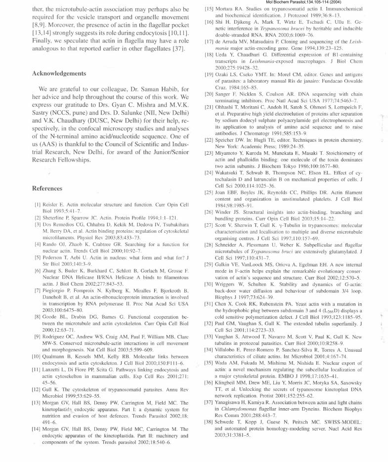
References
[II Reisler E. Actin molecular structure and function. Curr Opin CellBioI 1993;5:41-7.
[2] Sheterline P. Sparrow Jc. Actin. Protein Profile 1994; I: 1-121.[3] Dos Remedios CG, Chhabra D, Kekik M, Dedova IV, Tsubakihara
M, Berry DA, et al. Actin binding proteins: regulation of cytoskeletalmicrofilaments. Physiol Rev 2003;83:433-73.
[4] Rando OJ, Zhaob K, Crabtree GR. Searching for a function fornuclear actin. Trends Cell Bioi 2000;10:92-7.
[5] Pederson T, Aebi U. Actin in nucleus: what form and what for? JStr Bioi 2003;140:3-9.
[6] Zhang S, Buder K, Burkhard C, Schlott B, Gorlach M, Grosse F.Nuclear DNA Helicase IIIRNA Helicase A binds to filamentous
actin. J Bioi Chern 2002;277:843-53.
[7] Piegiorgio P, Fomproix N, Kylberg K, Miralles F, Bjorkroth B,
Daneholt B, et aI. An actin-ribonucleoprotein interaction is involvedin transcription by RNA polymerase II. Proc Nat Acad Sci USA2003;100:6475-80.
[8] Goode BL, Drubin DG, Barnes G. Functional cooperation be-
tween the microtubule and actin cytoskeleton. Curr Opin Cell Bioi2000;12:63-71.
[9] Rodriguez OC, Andrew WS, Craig AM, Paul F, William MB, ClareMW-S. Conserved microtubule-actin interactions in cell movement
and morphogenesis. Nat Cell Bioi 2003;5:599-609.
[10] Qualmann B, Kessels MM, Kelly RB. Molecular links betweenendocytosis and actin cytoskeleton. J Cell Bioi 2000;150:FIII-6.
[11] Lanzetti L, Di Fiore PP, Scita G. Pathways linking endocytosis andactin cytosekelton in mammalian cells. Exp Cell Res 200 I ;271:45-56.
[12] Gull K. The cytoskeleton of trypanosomatid parasites. Annu RevMicrobiol 1999;53:629-55.
[13] Morgan GV, Hall BS, Denny PW, Carrington M, Field MC. The
kinetoplastida endocytic apparatus. Part I: a dynamic system fornutrition and evasion of host defences. Trends Parasitol 2002; 18:491-6.
[14] Morgan GV, Hall BS, Denny PW, Field MC, Carrington M. The
endocytic apparatus of the kinetoplastida. Part II: machinery and
components of the system. Trends parasitol 2002;18:540-6.
[15] Mortara RA. Studies on trypanosomatid actin I. Immunochemicaland biochemical identification. J Protozool 1989;36:8-13.
[16] Shi H, Djikeng A, Mark T, Wirtz E, Tschudi C, Ullu E. Ge-
netic interference in Trypanosoma brucei by heritable and inducibledouble-stranded RNA. RNA 2000;6: 1069-76.
[]7] de Arruda MV, Matsudaira P. Cloning and sequencing of the Leish-
mania major actin-encoding gene. Gene ]994;139:23-125.
[18] Ueda Y, Chaudhuri G. Differential expression of B I-containingtranscripts in Leishmania-exposed macrophages. J Bioi Chern2000;275: 19428-32.
[19] Ozaki LS, Cseko YMT. In: Morel CM, editor. Genes and antigensof parasites: a laboratory manual Ris de janairo: Fundacao OswaldeCruz. 1984:]65-85.
[20] Sanger F, Nicklen S, Coulson AR. DNA sequencing with chainterminating inhibitors. Proc Natl Acad Sci USA 1977;74:5463-7.
[21] Ohhashi T, Moritani C, Andoh H, Satoh S, Ohmori S, Lottspeich F,et al. Preparative high yield electroelution of proteins after separation
by sodium dodecyl sulphate polyacrylamide gel electrophoresis and
its application to analysis of amino acid sequence and to raiseantibodies. J Chromatogr 199];585:153-9.
[22] Speicher DW. In: Hugli TE, editor. Techniques in protein chemistry.New York: Academic Press; 1989:24-35.
[23] Miyamoto Y, Kuroda M, Munekata E, Masaki T. Stoichiometry of
actin and phalloidin binding: one molecule of the toxin dominates
two actin subunits. J Biochem Tokyo 1986;100:1677-80.
[24] Wakatsuki T, Schwab B, Thompson NC, E]son EL. Effect of cy-
tochalasin D and latrunculin B on mechanical properties of cells. JCell Sci 2000;114:1025-36.
[25] Joan EBF, Boy]es JK, Reyno]ds CC, Phillips DR. Actin filament
content and organization in unstimulated platelets. J Cell Bio]1984;98: 1985-91.
[26] Winder JS. Structural insights into actin-binding, branching and
bundling proteins. Curr Opin Cell Bioi 2003;]5: 14-22.[27] Scott V, Sherwin T, Gull K. -y-Tubulin in trypanosomes: molecular
characterisation and localisation to multiple and diverse microtubule
organising centres. J Cell Sci 1997;110:157-69.
[28] Schneider A, Plessmann U, Weber K. Subpellicular and flagellarmicrotubu]es of Trypanosoma bruci are extensively glutamylated. JCell Sci 1997;] 10:431-7.
[29] Galkin YE, YanLoock MS, Oriova A, Egelman EH. A new internal
mode in F-actin helps explain the remarkable evolutionary conser-vation of actin's sequence and structure. Curr Bio] 2002; 12:570-5.
[30] Wriggers W, Schulten K. Stability and dynamics of G-actin:
back-door water diffusion and behaviour of subdomain 3/4 loop.
Biophys J 1997;73:624-39.[31] Chen X, Cook RK, Rubenstein PA. Yeast actin with a mutation in
the hydrophobic plug between subdomain 3 and 4 (L266D) displays acold sensitive polymerization defect. J Cell Bio] 1993;]23:1185-95.
[32] Paul GM, Vaughan S, Gull K. The extended tubulin superfamily. JCell Sci 2001;114:2723-33.
[33] Vaughan S, Attwood T, Navarro M, Scott Y, Paul K, Gull K. New
tubulins in protozoa] parasities. Curr Bioi 2000;10:R258-9.[34] Villalobo E, Perez-Romero P, Sanchez-Silva R, Torres A. Unusual
characteristics of ciliate actins. Int Microbiol 2001;4:167-74.
[35] Wada AM, Fukuda M, Mishima M, Nishida E. Nuclear export of
actin: a nove] mechanism regulating the subcellular localization ofa major cytoskeletal protein. EMBO J 1998;17:1635--41.
[36] Klingbeil MM, Drew ME, Liu y, Morris JC, Motyka SA, Saxowsky
TT, et al. Unlocking the secrets of typanosome kinetoplast DNA
network replication. Protist 200 I; 152:255-62.
[37] Yanagisawa H, Kamiya R. Association between actin and light chains
in Chlamydomonas flagellar inner-arm Dyneins. Biochem BiophysRes Comm 2001;288:443-7.
[38] Schwede T, Kopp J, Guese N, Peitsch MC. SWISS-MODEL:
and automated protein homology-modeling server. Nucl Acid Res2003;31:3381-5.
Mol Biochem Parasitol,134.105-114 (2004)
Copyright © 2022 FDOKUMEN