Immunohistochemical Localisation of PDE5 in Rat Lung during Pre- and Postnatal Development
7
Hindawi Publishing Corporation Journal of Biomedicine and Biotechnology Volume 2009, Article ID 932961, 7 pages doi:10.1155/2009/932961 Research Article Immunohistochemical Localisation of PDE5 in Rat Lung during Pre- and Postnatal Development Angela Scipioni, 1 Mauro Giorgi, 2 Valeria Nuccetelli, 2 and Stefania Stefanini 1 1 Department of Cellular and Developmental Biology, Sapienza University of Rome, Piazzale Aldo Moro 5, 00185 Rome, Italy 2 Department of Basic and Applied Biology, University of L’Aquila, Via Vetoio 10, 67010 L’Aquila, Italy Correspondence should be addressed to Mauro Giorgi, [email protected] Received 31 March 2009; Accepted 18 June 2009 Recommended by Richard Tucker In mammalian lung, at the transition to extrauterine life, NO/cGMP signal transduction system is known to play crucial roles in the regulation of vascular resistance and is supposed to act in angiogenesis. PDE5, which is the most abundant cGMP metabolizing enzyme within the lung, is highly expressed in the perinatal period, but its localisation in the different pulmonary cells is still poorly known. In our research, PDE5 immunohistochemical distribution was investigated in foetal and neonatal rat lung. The highest expression of PDE5 was found in cells randomly located in the stroma; in newborns, in particular, many cells in the intersaccular walls were heavily labelled, while much lower staining levels were shown by smooth myocytes belonging to vessels and airways. On the basis of their immunoreactivity for α-SM actin and/or desmin, most of the heavily PDE5-positive cells were identified as interstitial myofibroblasts and transitional pericytes, while only a few were interpreted as interstitial lipofibroblasts. Copyright © 2009 Angela Scipioni et al. This is an open access article distributed under the Creative Commons Attribution License, which permits unrestricted use, distribution, and reproduction in any medium, provided the original work is properly cited. 1. Introduction In mammals, pulmonary vascular resistance is known to be modulated by cGMP, which induces relaxation of vascular smooth muscle through the activation of cGMP-dependent protein kinases. In smooth muscle cells, cGMP is synthesised by nitric oxide- and natriuretic peptide-activated guanylate cyclases, while its degradation occurs through phospho- diesterases. Among the eleven phosphodiesterase families recognised in mammalian tissues [1], cGMP-binding cGMP- specific phosphodiesterase (PDE5) shows particularly strong enzymatic activities in highly vascularized organs, such as the lungs and the spleen [2–4]. Moreover, its mRNA and protein were specifically detected in vascular smooth muscle cells; so that the enzyme is supposed to play a key role in inducing vessel relaxation. In adult rat lungs, PDE5 is highly concentrated in smooth muscle cells in the medial layer of pulmonary arteries and veins [5] and its expression increases in hypoxia-induced pulmonary hypertension [6]. Moreover, in rodents as well as in humans, PDE5 specific inhibitors such as zaprinast and sildenafil markedly attenuate hypertension itself [5, 7]; consequently these substances, together with other agents that modulate intracellular cGMP, are considered as promis- ing, safe, and easy-to-administer therapies for pulmonary hypertension [8]. During the perinatal reorganization of the pulmonary vascular bed, when both constitutive endothelial nitric oxide synthase and soluble guanylate cyclase are highly expressed [9, 10], alterations in PDE5 activity, protein, and mRNA were described both in ovine and in rodent lungs [11, 12]. Moreover, increases of soluble guanylate cyclase and PDE5 were found in lambs with pulmonary hypertension [13] and zaprinast and sildenafil were effective in lowering pulmonary vascular resistance of normal and hypertensive ovine foetuses [14–16]. In hyperoxia-exposed rat pups, sildenafil, besides alleviating pulmonary hypertension, interestingly, increased pulmonary capillary density and preserved alveolar growth; preservation of endothelial network during hyperoxia was also found in vitro [17]. It was therefore suggested that the beneficial effects on vascular and alveolar growth might occur through promotion of lung angiogenesis. Although sildenafil appears as a promising agent for the management of pulmonary hypertension [18], several
-
Upload
independent -
Category
Documents
-
view
2 -
download
0
Transcript of Immunohistochemical Localisation of PDE5 in Rat Lung during Pre- and Postnatal Development

Hindawi Publishing CorporationJournal of Biomedicine and BiotechnologyVolume 2009, Article ID 932961, 7 pagesdoi:10.1155/2009/932961
Research Article
Immunohistochemical Localisation of PDE5 in Rat Lung duringPre- and Postnatal Development
Angela Scipioni,1 Mauro Giorgi,2 Valeria Nuccetelli,2 and Stefania Stefanini1
1 Department of Cellular and Developmental Biology, Sapienza University of Rome, Piazzale Aldo Moro 5, 00185 Rome, Italy2 Department of Basic and Applied Biology, University of L’Aquila, Via Vetoio 10, 67010 L’Aquila, Italy
Correspondence should be addressed to Mauro Giorgi, [email protected]
Received 31 March 2009; Accepted 18 June 2009
Recommended by Richard Tucker
In mammalian lung, at the transition to extrauterine life, NO/cGMP signal transduction system is known to play crucial roles inthe regulation of vascular resistance and is supposed to act in angiogenesis. PDE5, which is the most abundant cGMP metabolizingenzyme within the lung, is highly expressed in the perinatal period, but its localisation in the different pulmonary cells is still poorlyknown. In our research, PDE5 immunohistochemical distribution was investigated in foetal and neonatal rat lung. The highestexpression of PDE5 was found in cells randomly located in the stroma; in newborns, in particular, many cells in the intersaccularwalls were heavily labelled, while much lower staining levels were shown by smooth myocytes belonging to vessels and airways.On the basis of their immunoreactivity for α-SM actin and/or desmin, most of the heavily PDE5-positive cells were identified asinterstitial myofibroblasts and transitional pericytes, while only a few were interpreted as interstitial lipofibroblasts.
Copyright © 2009 Angela Scipioni et al. This is an open access article distributed under the Creative Commons AttributionLicense, which permits unrestricted use, distribution, and reproduction in any medium, provided the original work is properlycited.
1. Introduction
In mammals, pulmonary vascular resistance is known to bemodulated by cGMP, which induces relaxation of vascularsmooth muscle through the activation of cGMP-dependentprotein kinases. In smooth muscle cells, cGMP is synthesisedby nitric oxide- and natriuretic peptide-activated guanylatecyclases, while its degradation occurs through phospho-diesterases. Among the eleven phosphodiesterase familiesrecognised in mammalian tissues [1], cGMP-binding cGMP-specific phosphodiesterase (PDE5) shows particularly strongenzymatic activities in highly vascularized organs, such as thelungs and the spleen [2–4]. Moreover, its mRNA and proteinwere specifically detected in vascular smooth muscle cells; sothat the enzyme is supposed to play a key role in inducingvessel relaxation.
In adult rat lungs, PDE5 is highly concentrated in smoothmuscle cells in the medial layer of pulmonary arteries andveins [5] and its expression increases in hypoxia-inducedpulmonary hypertension [6]. Moreover, in rodents as wellas in humans, PDE5 specific inhibitors such as zaprinastand sildenafil markedly attenuate hypertension itself [5, 7];
consequently these substances, together with other agentsthat modulate intracellular cGMP, are considered as promis-ing, safe, and easy-to-administer therapies for pulmonaryhypertension [8].
During the perinatal reorganization of the pulmonaryvascular bed, when both constitutive endothelial nitric oxidesynthase and soluble guanylate cyclase are highly expressed[9, 10], alterations in PDE5 activity, protein, and mRNAwere described both in ovine and in rodent lungs [11, 12].Moreover, increases of soluble guanylate cyclase and PDE5were found in lambs with pulmonary hypertension [13] andzaprinast and sildenafil were effective in lowering pulmonaryvascular resistance of normal and hypertensive ovine foetuses[14–16]. In hyperoxia-exposed rat pups, sildenafil, besidesalleviating pulmonary hypertension, interestingly, increasedpulmonary capillary density and preserved alveolar growth;preservation of endothelial network during hyperoxia wasalso found in vitro [17]. It was therefore suggested thatthe beneficial effects on vascular and alveolar growth mightoccur through promotion of lung angiogenesis.
Although sildenafil appears as a promising agent forthe management of pulmonary hypertension [18], several
2 Journal of Biomedicine and Biotechnology
issues need to be addressed before advocating the clinicaluse of this and others PDE5 inhibitors. In order to evaluatetheir side effects, the distribution of the enzyme in theorgans and in the different cell types inside the organsmust be extensively described. Indeed, in many organs,PDE5 was found to be expressed by cell types differentfrom smooth muscle cells, for example, Purkinje cells[19], platelets, epithelia of renal proximal tubules andcollecting ducts, as well as pancreatic duct cells [20, 21].In adult rat lungs, the enzyme was immunolocalised inthe smooth muscle of vessels and airways, including thealveolar ducts and openings of the alveoli; its expressionwas found to accompany the distal muscularization ofpulmonary arterioles, pathognomonic of hypoxia-inducedpulmonary hypertension [5–22]. In newborn rat lungs,PDE5 mRNA transcripts were detected in the vascularsmooth muscle and in the alveolar walls cells [12]. Duringfoetal lung development, while in vivo, both in normal andhypertensive ovine foetuses the enzyme was only detectedin the vascular wall [14]; in vitro, on the other hand, ahyperoxia-inducible expression of PDE5 mRNA and proteinwas shown in foetal pulmonary artery smooth muscle cells[23].
We decided to investigate the PDE5 expression in differ-ent cell populations of the developing rat lung, utilising anaffinity purified polyclonal antibody, raised against the N-terminal region of bovine PDE5 [21], revealed by either theconventional or the amplified ABC immunohistochemicalprocedure [24]. We examined paraffin sections of lungparenchyma from animals at developmental stages com-prised between the embryonic day 15.5th and the postnatalday 14th. Moreover, in order to provide a detailed descriptionof the cell populations that specifically express PDE5 insidethe intersaccular walls in early postnatal development, westudied possible colocalization of PDE5 with the cytoskeletalmarkers α-smooth muscle actin (α-SMA) and/or desmin.
2. Material and Methods
2.1. Animals. Albino Wistar rats (Charles River, Italy) kept at20–22◦C, with a dark/light cycle of 12/12 hours. Females wereplaced with males overnight and examined the followingmorning for the presence of sperm in the vaginal smear; thesperm observation day was considered as the embryonic day0.5th (E0.5). Animals were housed and handled according tothe European Communities Council Directive of November24th 1986 (86/609/EEC) and to the Italian Health Ministryguidelines.
2.2. Fixation and Tissue Processing. Pregnant rats at gesta-tional age E15.5 and E17.5 were i.p. injected with Farmotal(Amersham Pharmacia Biotech Italia, Cologno Monzese,Milan, Italy) 100 mg/kg b.w.; the uterus was quickly removedand immersed in Ringer’s solution, individual embryos wereextracted from the decidua and fixed by immersion inBouin, for 4 hours at room temperature (RT). Foetal ageswere confirmed basing on weight and length. 10 minutesafter immersion in fixative, the embryos were cut along the
sagittal plane. Rats aging 3, 7 and 14 days (at least fouranimals for each developmental stage) were i.p. injected withFarmotal 100 mg/kg b.w. and were endotracheally instilledwith 50–80 μL of Bouin’s or Methacarn’s solutions. Thedose of fixative was chosen on the basis of preliminaryexperiments. Trachea was then ligated; lungs were removedand immersed in the same solutions. A few minutes later,lungs were cut in fragments, which were further fixed for2–4 hours. All specimens were dehydrated, embedded inparaffin, and sectioned 7 μm thick; at least three embryosfor each gestational age and several randomly chosenlung samples taken from 3, 7, and 14 day-old rats wereutilised for immunolocalisation of PDE5 and cytoskeletalmarkers.
Concerning the immunolocalisation of cytoskeletal pro-teins, since a stronger labelling was found after Methacarnfixation with respect to Bouin, the identification of cellscoexpressing PDE5 and α-SMA or PDE5 and desmin wascarried out in serial sections obtained from Methacarn-fixedlungs.
2.3. Anti-PDE5 Antibody Production and Purification. Aspreviously described in [21], a specific PDE5 antiserumwas produced in rabbits using as immunogen a fusionprotein containing glutathione S-transferase and a peptidecorresponding to the 68–239 amino acid residues of the N-terminal bovine lung PDE5 sequence. After purification byaffinity chromatography on a protein A column this antibodywas able to immunoprecipitate cGMP-PDE activity frommouse tissues.
The activity recovered in the immunoprecipitate wasidentified as PDE5 on the basis of its sensitivity to the spe-cific inhibitors zaprinast (IC50 0.6 μM) and sildenafil (IC50
4 nM). Moreover, western blotting of the immunoprecipitateshowed three specific immunoreactive bands (100, 93, and86 kDa), corresponding to known PDE5 splice variants [25].
2.4. Western Blot Analysis. Lung samples from adult andsuckling rats were homogenized in 20 mM Tris-HCl buffer,pH 7.2, containing 0.2 mM EGTA, 5 mM mercaptoethanol,1 mM PMSF, 5 mM MgCl2, and 2% (v/v) antiproteasecocktail, using a glass homogenizer (15 strokes, 4◦C).Homogenates were centrifuged at 14 000 x g for 30 minutesat 4◦C, and pellets were resuspended in the homogenizationbuffer and centrifuged at 14 000 x g for 30 minutes (4◦C).The first and second supernatants were then pooled anddenatured for an SDS-Page run. Protein determinationwas performed using the procedure of Lowry [26]. SDS-polyacrylamide gel electrophoresis was performed on 10%slab gels according to Laemmli [27], and proteins werethen transferred to nitrocellulose membranes according toTowbin et al. [28]. The blots were incubated for 3 hoursat RT with 1:1000 rabbit polyclonal anti-PDE5 and mousemonoclonal antiactin antibodies (Sigma Chemical Co., St.Louis, USA); actin band was used as reference protein indensitometric evaluation. In competition experiments, theincubation medium was supplemented with 10 μg/mL ofthe PDE5-peptide antigen used to raise PDE5 antibody.

Journal of Biomedicine and Biotechnology 3
Alkaline phosphatase-conjugated goat, antirabbit and anti-mouse IgG were used to reveal immunocomplexes. Thebands were then stained with nitro-blue tetrazolium inthe presence of 5-bromo-4-chloro-3-indolyl-phosphate. Acrude extract of N18TG2 cells, which are known to highlyexpress PDE5 [21], was utilised as a positive control.Densitometric analysis on scanned blots was performedusing the NIH ImageJ v1.29 program; the significance ofdifferences was evaluated by means of variance analysis(ANOVA).
2.5. Immunohistochemistry. Freshly deparaffinized sectionswere treated with 0.3% H2O2 in methanol, for 30 minutes atRT, to inactivate endogenous peroxidases; after rehydrationthey were transferred to PBS containing 0.2% Triton X-100and 5% nonfat dry milk, for 1 hour at RT, and then incubatedfor 24 hours at 4◦C, in the primary antibodies, diluted in PBScontaining 0.1% Triton X-100 and 2.5% nonfat dry milk.
Dilutions were: 1:200 rabbit polyclonal anti-PDE5;1:300 mouse monoclonal antihuman muscle actin (Dako,Carpinteria, CA, USA); 1:800 mouse monoclonal anti-α-SMA (Sigma Chemical Co., St. Louis, USA); 1:100 mousemonoclonal antidesmin (Sigma Chemical Co., St. Louis,USA).
For the revelation of immunocomplexes through theconventional ABC method, sections were sequentially incu-bated in
(i) 1:200 biotinylated goat antirabbit or rabbit anti-mouse IgG, for 1 hour at RT,
(ii) avidin-biotin-horseradish peroxidase complex, for 1hour at RT,
(iii) 0.05% 3,3’-diaminobenzidine (DAB) in PBS contain-ing 0.01 H2O2 for 2–5 minutes at RT, in the amplifiedABC procedure the step (ii) was followed by
(i) incubation in biotinylated tyramine, 1:100diluted in PBS containing 0.01 H2O2 for 10minutes at RT,
(ii) avidin-biotin-horseradish peroxidase complex,for 30 minutes at RT.
Microwave treatment was carried out incubating freshlyrehydrated sections with citrate buffer pH 6 (10 minutesat RT, 8 minutes in the microwave oven at 750 W, and 30minutes at RT).
For negative control, the primary antibody was omittedor substituted with preimmune serum. Moreover, somesections were incubated with the anti-PDE5 antibody pread-sorbed (overnight at 4◦C) with 150 μg/mL of the PDE5-peptide utilised as immunogen [21].
Unstained and haematoxylin counterstained sectionswere examined in a Zeiss Axioskop2, equipped with a videocamera. Representative images were electronically captured;contrast and brightness were adjusted via Adobe Photoshop6.0.
Actin
PDE5
N18 ALu P14Lu P3LuALu +peptide
0
1
2
3
PD
E5/
acti
n
ALu P14Lu P3Lu
Figure 1: PDE5 expression at different stages of rat lung develop-ment. Upper panel: representative western blot analysis of adultlung (ALu), 14 day-old lung (P14Lu), 3 day-old lung (P3Lu),adult lung incubated in presence of PDE5-peptide (ALu+peptide).N18 cell culture (N18) was utilised as standard of PDE5. Eachlane was loaded with 50 μg of protein. Lower panel: densitometricquantification of immunoreactive PDE5 and actin bands. Dataare mean ± SD of 3 different experiments. Differences were notsignificant (ANOVA test).
3. Results
3.1. Western Blot Analysis. As shown in Figure 1, bothlung preparations and neuroblastoma N18TG2 cell extractshowed the strong bands at 100 kDa, corresponding to thePDE5 main splicing variant; the less intense bands, detectedat lower molecular masses (93 and 86 kDa), were identifiedas corresponding to other known splice variants [25].The absence of reactivity shown by the sample incubatedin peptide-supplemented medium confirmed the antibodyspecificity. All these results are in agreement with previousdata obtained in experiments on mice [21]. Quantitativeexamination, carried out using actin as reference protein, didnot reveal significant differences among the three lung sam-ples taken at different developmental stages (Figure 1(b)).
3.2. PDE5 Immunohistochemistry. With all the techniquesemployed (Bouin or Methacarn fixations, microwave treat-ment against nonmicrowave treatment, standard or ampli-fied ABC methods), at every stage we examined, PDE5immunoreactivity was restricted to the cytoplasmic com-partment of specific cell types. Sections incubated withpreimmune serum instead of the primary antibody, as wellas those incubated with the antibody pre-adsorbed with thepeptide utilised as an immunogen, were free of labelling.
In all the examined developmental stages, the epitheliumof the airways was unlabelled, while a mild staining wasfound in the smooth muscle of specific parts of the

4 Journal of Biomedicine and Biotechnology
20μm
(a)
20μm
(b)
TB
s
s
s
c
c
50μm
(c)
TB
s
s
50μm
(d)
c
TBa
a
ss
s
s
50μm
(e)
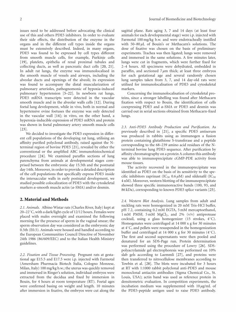
Figure 2: Rat lung. Bouin fixation. PDE5 immunohistochemistry carried out via the amplified ABC method on microwave treated sections.Haematoxylin counterstaining. (a) E15.5, (b) E17.5, (c) P3, (d) P7, (e) P14. s: saccule; a: alveolus; c: canalicular portion of the respiratoryspace; TB terminal bronchiole; LB larger bronchiole. ((a), (b)) Bar 20μm; ((c), (d), (e)) Bar 50μm. ((a), (b)) positivity is restricted to thecytoplasm of several cells, the majority of which are scattered in the stroma (arrowheads), while some are closely apposed to the respiratorytubule (arrows). The tubular epithelium is unlabelled. ((c), (d), (e)) labelling is restricted to the distal parenchyma, including sacculi, alveoli,and canalicular portions of respiratory spaces. The epithelium of terminal bronchioles is unstained.
respiratory tree (e.g., major airways in embryos, largerbronchioles in suckling rats). Vascular myocytes were verymildly labelled, and their staining intensity changed withrespect to the fixation and incubation conditions, reachingtotal negativity in Methacarn-fixed specimens.
Both in pre- and in postnatal lungs, the heaviest PDE5-positivity was shown in cells located in the pulmonarystroma. In embryos, both at pseudoglandular (E15.5) andat canalicular (E17.5) phases of lung development (Figures2(a) and 2(b)), these strongly labelled cells were numerousand randomly distributed, with only some closely adheringto the epithelial wall.
In suckling rats (Figures 2(c), 2(d), and 2(e)), heavilylabelled stromal cells were especially abundant in the wallsof distal respiratory spaces (sacculi and alveoli depend-ing on developmental stage); concerning larger airways, amild staining was present in the muscular layer of largerbronchioles, while terminal bronchioles appeared unlabelled.Neither number nor position of PDE5-expressing cellsremarkably changed among the examined developmentalstages.
3.3. Immunohistochemistry of Cytoskeletal Proteins. In allthe examined specimens, independently from the differentfixation and incubation conditions, at every examined stagethe immunoreactivity for total actin, α-SMA, and desminwas restricted to the cytoplasmic compartment of specific celltypes; all control sections were free of labelling. Concerningthe strength of labelling, the best results were obtained withMethacarn-fixed, nonmicrowave-treated specimens.
Throughout the examined postnatal development thecytoskeletal markers were immunolocalised in muscle cellsbelonging to bronchial, bronchiolar, and vessel walls aswell as in those underlying the epithelia in the canalicularportions of the respiratory spaces; epithelia were alwaysunlabelled, while a specific labelling was additionally foundin the cytoplasmic compartment of many cells of theintersaccular walls. All control sections were free of labelling.
Concerning the distribution of positivity in the intersac-cular wall, at 3, 7, and 14 days of postnatal development: (i)total actinexpressing cells were relatively few and randomlydistributed inside the interstitium; (ii) desmin-containing aswell as α-SMA-positive cells were sensibly more numerous;moreover, they were preferentially found in close proximity

Journal of Biomedicine and Biotechnology 5
c
cv
ss
20μm
(a)
c
cv
s
s
20μm
(b)
c
cv
s
s
20μm
(c)
Figure 3: Rat lungs. Postnatal day 3. Methacarn fixation.Immunolocalisation of different markers, carried out in adjacentsections. Standard ABC method. Haematoxylin counterstaining. s:saccule; c: canalicular portion of respiratory space; v: vessel. Bar20μm. (a) α-SMA immunolocalisation. Besides what found in thevascular wall, a strong labelling is found in the myocytes adheringto the epithelium in the canalicular portions of respiratory spaces,as well as in cells located in the intersaccular walls. (b) PDE5immunolocalisation. The labelling is restricted to the cytoplasm ofseveral stromal cells, located in close proximity with the saccularboundary. The myocytes of the small vessel and of respiratorychannels are unstained. (c) desmin immunolocalisation. Both themyocytes in the canalicular portions of respiratory spaces and thosebelonging to the vessel wall are strongly labelled; moreover fewlabelled cells are scattered in the intersaccular walls. Arrows indicatesome α-SMA- or desmin-containing PDE5-positive cells.
to the respiratory epithelium, especially on the tip of theintersaccular septa, and around the venules at the junctionsof the three septa.
At all the examined stages, the number and distributionof PDE5-expressing cells appeared substantially similar tothose of α-SMA-containing cells, while the correspondencewas weaker with desmin-containing elements. Finally, onlyrare and randomly located stromal cells showed the contem-porary presence of PDE5 and total actin. The developmentalstage of P3 was chosen as the most suitable for validatingthese preliminary results, and serial sections taken from 3day-old animals were therefore submitted the immunolo-calisation of PDE5, α-SMA, and desmin. Figure 3 showsa representative field, where several PDE5-expressing cells,
located in the saccular wall, are found to correspond to α-SMA-containing elements, while a lower number is foundto express desmin; fewer cells appear to express all threemarkers at the same time.
4. Discussion
PDE5-immunolocalization experiments revealed that in pre-and postnatal rat lungs stromal cells located in the inter-stitium between the respiratory units mainly expressed theenzyme. More in particular, during pseudoglandular andcanalicular phases of prenatal lung development, PDE5-expressing cells showed a preferential position close tothe tubular epithelium, while during the saccular phaseof postnatal lung development they were exclusively foundin the wall of distal respiratory spaces (canaliculi, sacculi,alveoli).
Concerning lung airways and vessels in the respiratorytree, PDE5 immunoreactivity was always absent in epithe-lium and hardly detectable in muscle of larger bronchioles,while vascular myocytes were very mildly labelled and theirstaining intensity changed with respect to fixation andincubation conditions.
Discrepancies between our results and those reportedby other authors who found stronger PDE5 expressionin vascular walls during perinatal development [11, 12]might be due to differences in the experimental method (insitu hybridization instead of immunohistochemistry) andadopted species (sheep instead of rats).
The high level of PDE5 expression shown in sucklingrat lungs by nonvascular cells of distal parenchyma supportsthe previously suggested idea [12] that in perinatal lungthe NO/cGMP signal transduction system might play keyroles not only in the regulation of vascular resistance butmore generally in tissue remodelling. In immature rodentlungs the angiogenic factor VEGF is known to induceendothelial proliferation and differentiation through bindingto Flk-1 [29], highly expressed by endothelial cells closelyapposed to the developing epithelium [30, 31]. According toresults obtained in other experimental models [32, 33], weargue that in immature lungs, Flk-1-bound VEGF stimulatesendothelial nitric oxide synthase, with consequent nitricoxide production. NO/cGMP system and cGMP modulation,therefore, appear to be crucial steps in pulmonary angiogen-esis.
Concerning the cell types that specifically express PDE5in neonatal rat lungs, we found that many of them alsocontain α-SMA, while some expressing desmin; on theother hand only few cells express all the three markers atthe same time. According to recent literature [34], the α-SMA+/desmin- cells mainly include interstitial myofibrob-lasts, with a contribute of transitional pericytes bound to pre-and postcapillary microvascular segments [35, 36], whilethe α-SMA-/desmin+ cells are presumed to be interstitiallipofibroblasts.
It is worth reminding that α-SMA-containing myofibrob-lasts, which in adult lung are only detected during idio-pathic or bleomycin-induced fibrosis [37, 38], in perinatal

6 Journal of Biomedicine and Biotechnology
development show increased α-SMA expression and collagensynthesis, together with cytoskeletal reorganization, and arepresumed crucial for adaptation to extra uterine life [39].
On the basis of our data concerning PDE5 expressionin myofibroblasts and pericytes and in agreement withresults obtained in liver and kidney samples (in which thenonselective phosphodiesterase inhibitors pentoxifylline and3-isobutyl-1-methylxanthine were found to reduce both pro-liferation rate and collagen secretion of α-SMA-expressingmyofibroblasts [40–42]), we argue that in neonatal mam-malian lung the NO/cGMP signal transduction system,besides being involved in vessel maturation, influenceshomeostasis and fibrogenic activity of myofibroblasts.
Structural and functional modifications of myofibrob-lasts and pericytes, which are both responsible for thetensile fine tuning of capillaries and compliance of inter-airspace walls [43, 44], might constitute the basis forthe alterations in extracellular matrix deposition, septalelongation and alveolar maturation found, for example,in hyperoxia-induced pulmonary perinatal hypertension.Interestingly, in this experimental condition sildenafil wasfound to increase lung capillary density and to preservealveolar growth [17]. In conclusion the possible effects ofPDE5 inhibitors on different pulmonary cell types shouldbe accurately investigated, in order to better utilise thesesubstances as therapeutic agents in neonatal chronic lungdiseases characterised by excess matrix deposition.
Abbreviations
PDE5: cGMP-binding cGMP-specific phosphodiesterase.a-SMA: a-smooth muscle actin.
References
[1] S. H. Soderling and J. A. Beavo, “Regulation of cAMP andcGMP signaling: new phosphodiesterases and new functions,”Current Opinion in Cell Biology, vol. 12, no. 2, pp. 174–179,2000.
[2] T. M. Lincoln, C. L. Hall, C. Park, and J. D. Corbin,“Guanosine 3′ : 5′ cyclic monophosphate binding proteinsin rat tissues,” Proceedings of the National Academy of Sciencesof the United States of America, vol. 73, no. 8, pp. 2559–2563,1976.
[3] J. F. Coquil, G. Brunelle, and J. Guedon, “Occurrence ofthe methylisobutylxanthine-stimulated cyclic GMP bindingprotein in various rat tissues,” Biochemical and BiophysicalResearch Communications, vol. 127, no. 1, pp. 226–231, 1985.
[4] J. D. Corbin, A. Beasley, M. A. Blount, and S. H. Francis, “Highlung PDE5: a strong basis for treating pulmonary hyper-tension with PDE5 inhibitors,” Biochemical and BiophysicalResearch Communications, vol. 334, no. 3, pp. 930–938, 2005.
[5] A. Sebkhi, J. W. Strange, S. C. Phillips, J. Wharton, andM. R. Wilkins, “Phosphodiesterase type 5 as a target forthe treatment of hypoxia-induced pulmonary hypertension,”Circulation, vol. 107, no. 25, pp. 3230–3235, 2003.
[6] M. R. Maclean, E. D. Johnston, K. M. Mcculloch, L. Pooley,M. D. Houslay, and G. Sweeney, “Phosphodiesterase isoformsin the pulmonary arterial circulation of the rat: changesin pulmonary hypertension,” Journal of Pharmacology andExperimental Therapeutics, vol. 283, no. 2, pp. 619–624, 1997.
[7] O. Pauvert, S. Bonnet, E. Rousseau, R. Marthan, and J.-P.Savineau, “Sildenafil alters calcium signaling and vasculartone in pulmonary arteries from chronically hypoxic rats,”American Journal of Physiology, vol. 287, no. 3, pp. L577–L583,2004.
[8] M. K. Steiner, I. R. Preston, J. R. Klinger, and N. S. Hill,“Pulmonary hypertension: inhaled nitric oxide, sildenafil andnatriuretic peptides,” Current Opinion in Pharmacology, vol. 5,no. 3, pp. 245–250, 2005.
[9] N. Kawai, D. B. Bloch, G. Filippov, et al., “Constitutiveendothelial nitric oxide synthase gene expression is regulatedduring lung development,” American Journal of Physiology, vol.268, no. 4, pp. L589–L595, 1995.
[10] K. D. Bloch, G. Filippov, L. S. Sanchez, M. Nakane, and S.M. de La Monte, “Pulmonary soluble guanylate cyclase, anitric oxide receptor, is increased during the perinatal period,”American Journal of Physiology, vol. 272, no. 3, pp. L400–L406,1997.
[11] K. A. Hanson, F. Burns, S. D. Rybalkin, J. W. Miller, J. Beavo,and W. R. Clarke, “Developmental changes in lung cGMPphosphodiesterase-5 activity, protein, and message,” AmericanJournal of Respiratory and Critical Care Medicine, vol. 158, no.1, pp. 279–288, 1998.
[12] L. S. Sanchez, S. M. de la Monte, G. Filippov, R. C. Jones, W. M.Zapol, and K. D. Bloch, “Cyclic-GMP-binding, cyclic-GMP-specific phosphodiesterase (PDE5) gene expression is regu-lated during rat pulmonary development,” Pediatric Research,vol. 43, no. 2, pp. 163–168, 1998.
[13] S. M. Black, L. S. Sanchez, E. Mata-Greenwood, J. M. Bekker,R. H. Steinhorn, and J. R. Fineman, “sGC and PDE5 areelevated in lambs with increased pulmonary blood flow andpulmonary hypertension,” American Journal of Physiology, vol.281, no. 5, pp. L1051–L1057, 2001.
[14] K. A. Hanson, J. W. Ziegler, S. D. Rybalkin, J. W. Miller, S. H.Abman, and W. R. Clarke, “Chronic pulmonary hypertensionincreases fetal lung cGMP phosphodiesterase activity,” Ameri-can Journal of Physiology, vol. 275, no. 5, pp. L931–L941, 1998.
[15] J. Weimann, R. Ullrich, J. Hromi, et al., “Sildenafil is apulmonary vasodilator in awake lambs with acute pulmonaryhypertension,” Anesthesiology, vol. 92, no. 6, pp. 1702–1712,2000.
[16] F. Ichinose, J. Erana-Garcia, J. Hromi, et al., “Nebulizedsildenafil is a selective pulmonary vasodilator in lambs withacute pulmonary hypertension,” Critical Care Medicine, vol.29, no. 5, pp. 1000–1005, 2001.
[17] F. Ladha, S. Bonnet, F. Eaton, K. Hashimoto, G. Korbutt,and B. Thebaud, “Sildenafil improves alveolar growth andpulmonary hypertension in hyperoxia-induced lung injury,”American Journal of Respiratory and Critical Care Medicine,vol. 172, no. 6, pp. 750–756, 2005.
[18] J. N. Travadi and S. K. Patole, “Phosphodiesterase inhibitorsfor persistent pulmonary hypertension of the newborn: areview,” Pediatric Pulmonology, vol. 36, no. 6, pp. 529–535,2003.
[19] J. Kotera, N. Yanaka, K. Fujishige, et al., “Expression of ratcGMP-binding cGMP-specific phosphodiesterase mRNA inPurkinje cell layers during postnatal neuronal development,”European Journal of Biochemistry, vol. 249, no. 2, pp. 434–442,1997.
[20] J. Kotera, K. Fujishige, and K. Omori, “Immunohistochemi-cal localization of cGMP-binding cGMP-specific phosphodi-esterase (PDE5) in rat tissues,” Journal of Histochemistry andCytochemistry, vol. 48, no. 5, pp. 685–693, 2000.

Journal of Biomedicine and Biotechnology 7
[21] D. Giordano, M. E. De Stefano, G. Citro, A. Modica, and M.Giorgi, “Expression of cGMP-binding cGMP-specific phos-phodiesterase (PDE5) in mouse tissues and cell lines usingan antibody against the enzyme amino-terminal domain,”Biochimica et Biophysica Acta, vol. 1539, no. 1-2, pp. 16–27,2001.
[22] J. Wharton, J. W. Strange, G. M. O. Møller, et al., “Antiprolifer-ative effects of phosphodiesterase type 5 inhibition in humanpulmonary artery cells,” American Journal of Respiratory andCritical Care Medicine, vol. 172, no. 1, pp. 105–113, 2005.
[23] K. N. Farrow, B. S. Groh, P. T. Schumacker, et al., “Hyperoxiaincreases phosphodiesterase 5 expression and activity in ovinefetal pulmonary artery smooth muscle cells,” CirculationResearch, vol. 102, no. 2, pp. 226–233, 2008.
[24] J. C. Adams, “Biotin amplification of biotin and horseradishperoxidase signals in histochemical stains,” Journal of Histo-chemistry and Cytochemistry, vol. 40, no. 10, pp. 1457–1463,1992.
[25] C. S. Lin, A. Lau, R. Tu, and T. F. Lue, “Expression of threeisoforms of cGMP-binding cGMP-specific phosphodiesterase(PDE5) in human penile cavernosum,” Biochemical andBiophysical Research Communications, vol. 268, pp. 628–635,2000.
[26] O. Lowry, N. J. Rosembrough, A. R. Farr, and R. J. Randall,“Protein measurement with the folin phenol reagent,” Journalof Biological Chemistry, vol. 193, pp. 265–275, 1951.
[27] U. K. Laemmli, “Cleavage of structural proteins during theassembly of the head of bacteriophage T4,” Nature, vol. 227,no. 5259, pp. 680–685, 1970.
[28] H. Towbin, T. Staehelin, and J. Gordon, “Electrophoretictransfer of proteins from polyacrylamide gels to nitrocellulosesheets: procedure and some applications,” Proceedings of theNational Academy of Sciences of the United States of America,vol. 76, no. 9, pp. 4350–4354, 1979.
[29] F. Shalaby, J. Rossant, T. P. Yamaguchi, et al., “Failure of blood-island formation and vasculogenesis in Flk-1-deficient mice,”Nature, vol. 376, no. 6535, pp. 62–66, 1995.
[30] A. J. Bhatt, S. B. Amin, P. R. Chess, R. H. Watkins, and W. M.Maniscalco, “Expression of vascular endothelial growth factorand Flk-1 in developing and glucocorticoid-treated mouselung,” Pediatric Research, vol. 47, no. 5, pp. 606–613, 2000.
[31] Y.-S. Ng, R. Rohan, M. E. Sunday, D. E. Demello, and P.A. D’Amore, “Differential expression of VEGF isoforms inmouse during development and in the adult,” DevelopmentalDynamics, vol. 220, no. 2, pp. 112–121, 2001.
[32] B.-Q. Shen, D. Y. Lee, and T. F. Zioncheck, “Vascular endothe-lial growth factor governs endothelial nitric-oxide synthaseexpression via a KDR/Flk-1 receptor and a protein kinase Csignaling pathway,” Journal of Biological Chemistry, vol. 274,no. 46, pp. 33057–33063, 1999.
[33] D. Fukumura, T. Gohongi, A. Kadambi, et al., “Predominantrole of endothelial nitric oxide synthase in vascular endothelialgrowth factor-induced angiogenesis and vascular permeabil-ity,” Proceedings of the National Academy of Sciences of theUnited States of America, vol. 98, no. 5, pp. 2604–2609, 2001.
[34] M. Yamada, H. Kurihara, K. Kinoshita, and T. Sakai, “Tem-poral expression of alpha-smooth muscle actin and drebrin inseptal interstitial cells during alveolar maturation,” Journal ofHistochemistry and Cytochemistry, vol. 53, no. 6, pp. 735–744,2005.
[35] V. Nehls and D. Drenckhahn, “Heterogeneity of microvascularpericytes for smooth muscle type alpha-actin,” The Journal ofCell Biology, vol. 113, pp. 147–154, 1991.
[36] Y. Kapanci, C. Ribaux, C. Chaponnier, and G. Gabbiani,“Cytoskeletal features of alveolar myofibroblasts and pericytesin normal human and rat lung,” Journal of Histochemistry andCytochemistry, vol. 40, no. 12, pp. 1955–1963, 1992.
[37] Y. Kapanci, A. Desmouliere, J.-C. Pache, M. Redard, and G.Gabbiani, “Cytoskeletal protein modulation in pulmonaryalveolar myofibroblasts during idiopathic pulmonary fibrosis:possible role of transforming growth factor beta and tumornecrosis factor alpha,” American Journal of Respiratory andCritical Care Medicine, vol. 152, no. 6, pp. 2163–2169, 1995.
[38] H.-Y. Zhang, M. Gharaee-Kermani, K. Zhang, S. Karmiol,and S. H. Phan, “Lung fibroblast α-smooth muscle actinexpression and contractile phenotype in bleomycin-inducedpulmonary fibrosis,” American Journal of Pathology, vol. 148,no. 2, pp. 527–537, 1996.
[39] K. Jostarndt-Fogen, V. Djonov, and A. Draeger, “Expression ofsmooth muscle markers in the developing murine lung: poten-tial contractile properties and lineal descent,” Histochemistryand Cell Biology, vol. 110, no. 3, pp. 273–284, 1998.
[40] E. Shimizu, Y. Kobayashi, Y. Oki, T. Kawasaki, T. Yoshimi,and H. Nakamura, “OPC-13013, a cyclic nucleotide phos-phodiesterase type III inhibitor, inhibits cell proliferation andtransdifferentiation of cultured rat hepatic stellate cells,” LifeSciences, vol. 64, no. 23, pp. 2081–2088, 1999.
[41] T. D. Hewitson, M. Martic, K. J. Kelynack, E. Pedagogos,and G. J. Becker, “Pentoxifylline reduces in vitro renalmyofibroblast proliferation and collagen secretion,” AmericanJournal of Nephrology, vol. 20, no. 1, pp. 82–88, 2000.
[42] D. Schuppan and Y. Porov, “Hepatic fibrosis: from bench tobedside,” Journal of Gastroenterology and Hepatology, vol. 17,supplement 3, pp. S300–S305, 2002.
[43] Y. Kapanci and G. Gabbiani, “Contractile cells in pulmonaryalveolar tissue,” in The Lung: Scientific Foundations, R. G.Crystal, J. B. West, E. R. Weibel, and P. J. Barnes, Eds., pp. 697–707, Lippincott-Raven, Philadelphia, Pa, USA, 1997.
[44] E. R. Weibel and R. G. Crystal, “Structural organization of thepulmonary interstitium,” in The Lung: Scientific Foundations,R. G. Crystal, J. B. West, E. R. Weibel, and P. J. Barnes, Eds.,pp. 685–695, Lippincott-Raven, Philadelphia, Pa, USA, 1997.