Diagnosing and Characterizing Neuropathic Pain in Dogs with ...
Upload
khangminh22Category
view
3download
0
MMG22: A Novel Bivalent Ligand for the Treatment of
Neuropathic Pain
A DISSERTATION
SUBMITTED TO THE FACULTY OF THE
UNIVERSITY OF MINNESOTA
BY
Rebecca Speltz Paiz
M.D./Ph.D. Candidate
IN PARTIAL FULFILLMENT OF THE REQUIREMENTS
FOR THE DEGREE OF
Doctor of Philosophy
ADVISED BY
Donald A Simone, Ph.D.
October 2021

ii
©
Rebecca Speltz Paiz
2021

i
Acknowledgments and Dedication
I would like to thank a number of individuals for their unending support and encouragement over the last five years. Without these individuals, this thesis would not have been possible.
First and foremost, I would like to thank my thesis advisor Dr. Donald Simone. You have made my experience in science incredibly exciting and valuable. Thank you for providing me with the right level of guidance and autonomy, allowing me to flourish. Your mentorship has given me skills that will be an asset for the rest of my life.
I would also like to thank my mentors including Dr. Martin Wessendorf, Dr. George Wilcox, Dr. Carolyn Fairbanks, Dr. Lucy Vulchanova, Dr. Sergey Khasabov and Dr. Iryna Khasabova. Dr. Wessendorf, you took me into your lab way back in 2012, while I was still an undergraduate student. Your enthusiasm for science was infectious. Your trust and support has not wavered in 8 years and I am so glad that I sent that email asking about volunteering, and that you responded! Drs Wilcox, Fairbanks and Vulchanova, the collaboration between your labs will forever be the ideal of scientific cooperation. Drs Khasabov and Khasabova, you have shown me the dedication it takes to do good science. To all of you, thank you.
I want to thank all members of the Simone lab including Dr. Guiseppe Cataldo, Dr. Samuel Erb, Dr. Sarah Shueb, Dr. Brian McAdams, Catherine Harding-Rose, Malcom Johns, and Dr. Ratan Banik.
I also want to thank members of the Portoghese lab including Dr. Phil Portoghese, Dr. Eyup Akgün, and Mary Lunzer for providing MMG22 and to Mary Lunzer in particular for her work involving MMG22 in respiratory depression and her all around general support.
I also want to thank several of my collaborators including Dr. Alex Kalyuzhny of R&D systems for his help and guidance with the RNAScope work. Dr. Henry Wong for the work he did involving the pharmacokinetics of MMG2 and Dr. Steven Graves for the self-administration studies.
I want to acknowledge the members of my thesis committee Dr. Linda McLoon (chair), Dr. George Wilcox, Dr. Martin Wessendorf and Dr. Paul Mermelstein.
I would like to thank the past and present administrative directors and supporting staff of the University of Minnesota’s Medical Scientist (MD/PhD) Training Program, Graduate Program in Neuroscience, and the Department of Diagnostic and Biological Sciences. Included in these teams, I would like to individually thank Dr. Yoji Shimizu, Susan Shurson, Nicholas Berg, Dr. Marshall Hertz, Dr. Lisa Schimmenti, Dr. Bryce Binstadt, Dr. Linda McLoon, Dr. David Redish, Dr. Robert Meisel, John Paton, Elaine McCauley, Janice Casey, and Nina Tran.
Finally, I would like to thank my family for all of their love and support including my parents (Leah and Stephen), my sisters (Laura and Andrea), my brother (Paul) and the extended Mylrea and Speltz family around the country.
I would also like to thank my funding sources including the NIH grants from the Simone lab: HL135895 and CA241627, the Portoghese lab: DA030316, and the institutional training grants including MSTP NIGMS 5T32GM008244 and NIDA Drug Abuse Grant 5T32DA007234.
I would like to dedicate this thesis to my nephew, Carson McCullough Smith, who was taken too soon from this world.
ii
Thesis Abstract
Functional interactions between the mu opioid receptor (MOR) and the
metabotropic glutamate receptor 5 (mGluR5) in pain and analgesia have been
well established. MMG22 is a bivalent ligand containing MOR agonist
(oxymorphamine) and mGluR5 antagonist (MPEP) pharmacophores tethered by
a 22-atom linker. MMG22 has been shown to produce potent analgesia in
several models of chronic inflammatory and neuropathic pain. This study
assessed the efficacy of systemic administration of MMG22 at reducing pain
behavior in the spared nerve injury (SNI) model of neuropathic pain in mice, as
well as its side effect profile and abuse potential. MMG22 reduced mechanical
hyperalgesia and spontaneous ongoing pain after SNI, with greater potency early
(10 days) as compared to late (30 days) after injury. Systemic administration of
MMG22 did not induce place preference in naïve animals, suggesting absence of
abuse liability when compared to traditional opioids. MMG22 also lacked the
central locomotor, respiratory, and anxiolytic side effects of its monomeric
pharmacophores. Evaluation of mRNA expression showed the transcripts for
both receptors were co-localized in cells in the dorsal horn of the lumbar spinal
cord and dorsal root ganglia. Teased nerve fiber recordings from the sural nerve
of SNI mice show that MMG22 reduces the firing rate of C and Aδ fiber
nociceptors evoked by suprathreshold stimuli. Thus, MMG22 reduces
hyperalgesia after injury in the SNI model of neuropathic pain by decreasing
nociceptor activity without the typical centrally mediated side effects associated
with traditional opioids.

iii
Table of Contents
Acknowledgments and Dedication ..................................................................................... i
Thesis Abstract ..................................................................................................................ii
Table of Contents .............................................................................................................. iii
List of Tables ...................................................................................................................... v
List of Figures .................................................................................................................... vi
Chapter 1 ................................................................................................................... 1
Pain and its pathways ........................................................................................................1
Pain and its manifestations .................................................................................................................... 1
An introduction to the somatosensory pathway ................................................................................... 4
Chapter 2 ................................................................................................................. 31
MMG22, a novel bivalent ligand for the treatment of chronic pain .................................... 31
Current and future treatments for neuropathic pain .......................................................................... 31
MMG22 ................................................................................................................................................ 40
Purpose and Organization of this Thesis ........................................................................... 41
Chapter 3 ................................................................................................................. 43
The anti-hyperaglesic potency of systemic MMG22 ........................................................... 43
Introduction: Research on pain and analgesia..................................................................................... 44
Methods ............................................................................................................................................... 46
Results .................................................................................................................................................. 49
Discussion ............................................................................................................................................. 54
Conclusion ............................................................................................................................................ 56
Chapter 4 ................................................................................................................. 58
MMG22 Site of Action ...................................................................................................... 58
Introduction: Potential site of action for MMG22 within the pain neuroaxis ..................................... 59
Methods ............................................................................................................................................... 60
Results .................................................................................................................................................. 62
Discussion ............................................................................................................................................. 64
Conclusion ............................................................................................................................................ 65

iv
Chapter 5 ................................................................................................................. 66
Rewarding properties of MMG22: abuse liability vs relief from spontaneous pain ............. 66
Introduction: A brief overview of reward and addiction ..................................................................... 67
Methods: .............................................................................................................................................. 71
Results .................................................................................................................................................. 74
Discussion ............................................................................................................................................. 81
Conclusion ............................................................................................................................................ 82
Chapter 6 ................................................................................................................. 84
Potential side effects of MMG22 ...................................................................................... 84
Introduction: Side effects associated with MOR agonists and mGluR5 antagonists ........................... 85
Methods ............................................................................................................................................... 87
Results .................................................................................................................................................. 90
Discussion ............................................................................................................................................. 99
Conclusion .......................................................................................................................................... 100
Chapter 7 ............................................................................................................... 101
Effects of MMG22 on the response properties of primary afferents nociceptors .............. 101
Introduction: Electrophysiology of primary afferent nociceptors ..................................................... 102
Methods ............................................................................................................................................. 110
Results ................................................................................................................................................ 114
Discussion ........................................................................................................................................... 121
Conclusion .......................................................................................................................................... 123
General discussion: summary and conclusions ......................................................... 124
Bibliography ........................................................................................................... 127
v
List of Tables
Table 1-1 Characteristics and response properties of fibers that respond to non-noxious stimuli………………………………………………………………..
9
Table 3-1 ED50 values for MMG22, Morphine, Loperamide and MPEP early and late after nerve injury…………………………………………………..
54
Table 6-1 Effects of MMG22 on respiratory function…………………………..
98
Table 7-1 Characteristics and response properties of A fiber nociceptors…. 105
Table 7-2 Characteristics and response properties of C fiber nociceptors…. 107
Table 7-3 Properties of C fibers used in recordings…………………………... 115
Table 7-4 Properties of Aδ fibers used in recordings…………………………. 117

vi
List of Figures
Figure 1.1 Allodynia and hyperalgesia………………………………………… 4
Figure 1.2 Trace of electrically stimulated Aβ, Aδ and C fiber wave forms….. 7
Figure 2.1 Design of MMG22 and theoretical heteromer binding…………….. 42
Figure 3.1 Hyperalgesia after nerve injury………………………………………. 50
Figure 3.2 Dose response curves for MMG22, morphine, loperamide and
MPEP………………………………………………………………………………… 53
Figure 4.1 Colocalization of grm5 and opmr1 in the dorsal horn and DRG….. 64
Figure 5.1 Conditioned place preference experimental timeline……………… 75
Figure 5.2 CPP and aCPP in naïve and nerve injured animals……………….. 78
Figure 5.3 Dose dependent aCPP in nerve injured animals…………………... 81
Figure 6.1 Locomotor effects of MMG22………………………………………… 94
Figure 6.2 Effects of MMG22 on respiratory minute volume…………………... 97
Figure 6.3 Effects of MMG22 on colonic motility……………………………….. 99
Figure 7.1 Effects of MMG22 on C fiber nociceptor responses……………….. 115
Figure 7.2 Effects of MMG22 on Aδ nociceptor responses……..…………...... 117
Figure 7.3 MMG22 may reduce heat responsiveness of primary afferent
nociceptors …………………..……………………………………………………... 119
Figure 7.3 MMG22 may reduce cold responsiveness of primary afferent
nociceptors …………………..……………………………………………………... 120
1
Chapter 1
Pain and its pathways
Pain and its manifestations
The IASP defines pain as:
“An unpleasant sensory and emotional experience associated with actual or
potential tissue damage, or described in terms of such damage” [619].
A study in 2016 found that ~20% of Americans suffer from chronic pain
[164], costing approximately $560 billion dollars annually in direct medical costs,
lost productivity and disability programs [327]. Pain has both sensory and
affective components; the purely sensory components of pain (such as intensity
and location) are referred to as nociception, while the emotional component is
often referred to as the distress or suffering associated with a painful stimulus.
Chronic pain conditions are commonly associated with comorbid physical and
emotional/affective disorders including decreased mobility, increased incidence
of anxiety and depression, and an overall reduced quality of life [276,277]. The
following thesis will deal with the purely nociceptive components of pain.
The different types of pain
Although pain is a nearly universal experience; pain itself can manifest in
a variety of patterns. We categorize different types of pain in several ways
including duration (acute vs chronic) and pathophysiology (nociceptive,
inflammatory or neuropathic). Acute pain can be provoked by disease or injury
and is generally self-limited. Acute pain serves a biological function and is
necessary to avoid/limit injury and promote healing after tissue damage [78]. The
importance of acute pain for survival is highlighted by the numerous pathologies
seen in individuals who suffer from congenital insensitivity to pain (CIP) [712].
Patients with CIP are unable to perceive pain, this results in repeated injuries

2
including: oral self-mutilation, biting of the fingertips, bone fractures, and burns
[712]. Clinically the transition from acute to chronic pain happens if pain persists
for three months after onset [589]; however, this definition is purely operational
and not based on pathophysiology. While acute pain is a biological necessity,
chronic pain serves no clear biological purpose and has no recognizable
endpoint.
With regard to the pathophysiology, most pain conditions fall into one of
three categories: nociceptive, inflammatory or neuropathic (although the
categories are not mutually exclusive). Nociceptive pain is defined as pain that
arises from actual or threatened damage to non-neural tissue [619] and is due to
the activation of peripheral nociceptors. Nociceptive pain can be acute and self-
limited (e.g. pain after stubbing your toe, pain from a paper cut) or chronic (e.g.
osteoarthritis). Inflammatory pain is caused by inflammatory mediators
sensitizing nociceptors and can also be acute (e.g. pain from an acute infection)
or chronic (e.g. rheumatoid arthritis). Neuropathic pain is caused by a lesion or
disease of the somatosensory system itself [619].
Neuropathic pain (NP) is associated with a variety of etiologies, all of
which lead to the increased excitability of neurons along the sensory neuroaxis
which causes sensations of pain. NP can be further separated into central and
peripheral subtypes. Central neuropathic pain results from damage to pathways
or nuclei in the central nervous system that relay and process nociceptive signals
(e.g. post stroke pain, spinal cord injury, and multiple sclerosis) [853]. Peripheral
neuropathic pain results from damage to the primary afferent fibers that transmit
information about painful stimuli to the CNS and is associated with numerous
conditions including: post herpetic neuralgia, phantom limb pain, diabetic NP
(DNP), and chemotherapy induced peripheral NP (CIPN) and trauma [268,340].
The most common symptoms of NP include ongoing burning pain, paroxysmal
electric shock-like pain and brush-evoked pain [159,819]. Peripheral
neuropathies caused by generalized damage to peripheral nerves (such as in
DNP and CIPN) typically present in a “glove and stocking” distribution, primarily
affecting the distal extremities including feet, calves and hands. This pattern is
3
characteristic of a progressive dying-back, length-dependent, process of distal to
proximal sensory loss and pain. The prevalence of NP in the general population
is estimated to be between 7 and 10% [81,293], and is expected to increase
along with the incidence of diabetes [141]. Compared to other chronic pain
etiologies, NP is associated with increased drug prescriptions and healthcare
visits [26,141,811].
Allodynia and hyperalgesia
Acute and chronic pain may be accompanied by hyperalgesia and or
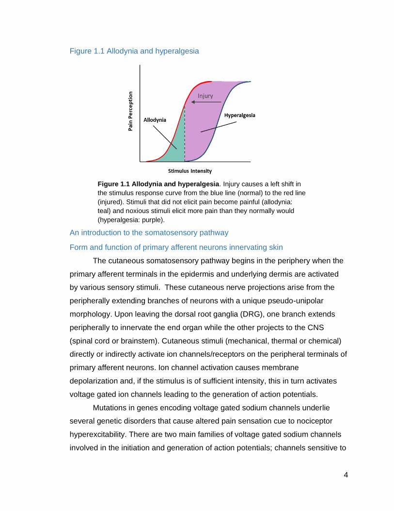
allodynia. Hyperalgesia is defined as an increased perception of pain to a
normally painful stimulus, whereas allodynia is a perception of pain to a normally
non-painful stimulus (Fig. 1.1). Examples of allodynia include the pain caused by
light touch to sun-burned skin, or the intense pain caused by clothing brushing
over the skin of people with peripheral neuropathies.
Hyperalgesia is increased pain felt from a normally painful stimulus.
Examples of hyperalgesia include the exaggerated pain response generated
when someone with painful peripheral neuropathy steps on a Lego, or the pain of
a sharp object that pokes a blister. Hyperalgesia can be primary (at the site of
injury and caused by peripheral sensitization) or secondary (in non-injured
tissues surrounding an injury and caused by sensitization of central pain
pathways). The mechanisms underlying primary and secondary hyperalgesia will
be addressed later. Functionally allodynia and hyperalgesia serve to aid in
healing by creating a state of hypervigilance surrounding the injured area (i.e.
avoid the pain that accompanies walking on a sprained ankle to prevent further
damage and promote healing). The enhanced sensitivity for pain may outlast the
initial injury. At this point pain becomes a disease in and of itself, instead of
merely a symptom of an underlying process.
4
Figure 1.1 Allodynia and hyperalgesia
An introduction to the somatosensory pathway
Form and function of primary afferent neurons innervating skin
The cutaneous somatosensory pathway begins in the periphery when the
primary afferent terminals in the epidermis and underlying dermis are activated
by various sensory stimuli. These cutaneous nerve projections arise from the
peripherally extending branches of neurons with a unique pseudo-unipolar
morphology. Upon leaving the dorsal root ganglia (DRG), one branch extends
peripherally to innervate the end organ while the other projects to the CNS
(spinal cord or brainstem). Cutaneous stimuli (mechanical, thermal or chemical)
directly or indirectly activate ion channels/receptors on the peripheral terminals of
primary afferent neurons. Ion channel activation causes membrane
depolarization and, if the stimulus is of sufficient intensity, this in turn activates
voltage gated ion channels leading to the generation of action potentials.
Mutations in genes encoding voltage gated sodium channels underlie
several genetic disorders that cause altered pain sensation cue to nociceptor
hyperexcitability. There are two main families of voltage gated sodium channels
involved in the initiation and generation of action potentials; channels sensitive to
Figure 1.1 Allodynia and hyperalgesia. Injury causes a left shift in
the stimulus response curve from the blue line (normal) to the red line
(injured). Stimuli that did not elicit pain become painful (allodynia:
teal) and noxious stimuli elicit more pain than they normally would
(hyperalgesia: purple).

5
inactivation by tetrodotoxin (TTX-S: Nav1.1, Nav1.6, and Nav1.7) and channels
resistant to tetrodotoxin (TTX-R: Nav1.8 and Nav1.9). TTX-S channels are found
on myelinated and unmyelinated primary afferents, while TTX-R channels are
found primarily on unmyelinated fibers [11] (both of which will be discussed
further below). Mutations in the gene encoding for Nav1.7 can result in two very
distinct heritable pain disorders: congenital insensitivity to pain (as discussed
earlier [152]) and primary erythromelalgia (characterized by recurring episodes of
extreme pain [904]) (for review see [281]).
The ability of a given primary afferent fiber to respond to a specific
mechanical, thermal, or chemical stimulus depends on the end organs
associated with the afferent’s terminal branches (for Aβ afferents) and the
receptors/ion channels expressed on the nerve ending (for all afferents). Once
generated, action potentials conduct down the axon, bypassing the neuron’s cell
body, transmitting information from the periphery to the CNS.
The form and function of low threshold Aβ mechanoreceptors
Cutaneous sensory fibers are broadly categorized into Aβ, Aδ, and C
fibers based on their degree of myelination, and how fast they conduct action
potentials [7]. Aβ fibers are heavily myelinated, with action potential conduction
velocities between 51-77 m/s in man [813], and 13.8-40 m/s in mouse [97] (Table
1-1). Aβ fibers mechanical thresholds are low (generally < 2mN [97,394,554]),
innervate mechanoreceptors including Merkel disks (SA-I), Meissner corpuscles
(RA-I), Ruffini endings (SA-II) and Pacinian corpuscles (RA-II) in skin, and make
up around 22% of primary afferent fibers. Aβ fibers relay information about light
touch and vibration. Merkel disks are located in the basal layer of the epidermis
and are innervated by Aβ fibers. These primary afferents have a pinpoint
receptive field and can detect displacement of the skin of less than 1µm.
Activation of a Merkel complex via low threshold mechanical stimulation induces
a slowly adapting response that relays information about pressure and texture
with high spatial resolution [2]. Meissner corpuscles are located in the dermal
papillae and consist of elongated Schwann cells, a connective tissue capsule and
a central axon. With small receptive fields, activation of this complex leads to a

6
rapidly adapting response from the associated Aβ fiber and transmits information
on texture, low frequency vibration and skin movement. Ruffini endings are
spindle-shaped cylinders composed of layers of Schwann cells and collagen
fibers with an inner core of nerve terminals surrounded by a fluid filled space.
Ruffini endings respond to lateral stretching of the skin, are slowly adapting and
have large receptive fields. Deep in the dermis, Pacinian corpuscles are
composed of interdigitating lamellar cells surrounding a central Aβ fiber. Pacinian
corpuscles are extremely sensitive, responding to movement in the nanometer
range. Despite this sensitivity, they have large receptive fields and hence poor
spatial resolution. These fibers respond to high frequency vibration up to 1000hz.
These receptors are also present in hairy skin in touch dome complexes or
innervating hair follicle shafts [934].
Figure 1.2 Trace of electrically stimulated Aβ, Aδ and C fiber wave forms.
The form and function of C and Aδ low threshold mechanoreceptors in hairy skin
A subset primary afferent C and Aδ fibers are activated by weak forces,
and slowly moving mechanical stimuli across the skin, and contribute to touch
sensation as well as emotional responses to touch [62,63,322,349,402,605,606]
(for review see [449,502]). Human psychophysical studies have shown low
threshold C (LTHC) fibers in the hairy skin of the arm, but not the glabrous skin of
Figure 1.2 Trace of electrically stimulated Aβ, Aδ and C fiber wave forms. Aβ
fibers are heavily myelinated have fast conduction velocities (51-77 m/s). Aδ fibers
are thinly myelinated and have intermediate conduction velocities (1.4–35 m/s). C
fibers are unmyelinated and slowly conducting fibers (0.5-1.4 m/s).
Aβ
Aδ
C
5ms
1V

7
the palm [440,464,828]. These fibers have thresholds between 0.3-2.5mN [828],
adapt to continuous stimulation, display fatigue and fire after discharges [586].
LTHC fibers express VGLUT3 [719] and tyrosine hydroxylase [440], make up
about 10% of the neurons in the DRG [420,457,719] and innervate the hair shafts
of zigzag and awl/auchene hairs. Aδ-LTH fibers, or D-hair fibers make up a small
group of non-peptidergic fibers that express TrkB and respond to low threshold
mechanical stimuli, bending of hair shafts and cooling of the skin [88,427,440].
Other non-noxious fibers in skin include Aδ cooling fibers and C warm fibers that
signal non-noxious cool and warm stimuli, respectively (Table 1-1).
Support for the role of TLH-C fibers in emotional touch comes from a
study that looked at patients after anterolateral cordotomies (a procedure that
severs the ascending axons carrying nociceptive information to alleviate pain in
terminal patients). Investigators noted that in addition to loss of pain and
temperature sensation, patients also reported a lack of erotic touch [409], but this
finding has not been replicated [495]. Other psychophysical studies have shown
that patients with congenital loss of C-fibers exhibit altered perception of low
threshold stimuli [480,547], whereas patients lacking Aβ afferents are able to
detect soft brush stroking of the forearm [139,604].

8
Table 1-1 Characteristics and response properties of fibers that encode non-noxious stimuli
LTM
Conduction
velocity
(m/s)
Sensory
modality
(Threshold)
End organ
(RF size)
Response
properties
SA-I 16-96 [2] Skin indentation,
pressure
Merkel cell
RF: 11 mm2 [347]
SA-II 20-100 [2] Skin stretch Ruffini ending
RF: 60 mm2 [347]
RA-I 26-91 [2] Skin movement,
flutter
Meissner
corpuscle
RF: 3-13 mm2
[249,347]
RA-II 30-90 [2] Vibration
(40-1000 hz)
Pacinian corpuscle
RF: 100 mm2 [347]
Aδ LTM/
D-hair 16-96 [2]
Hair follicle
deflection and
cooling
TH: ≤8.8mN [6]
Free nerve ending
RF: single hair to
20 mm2 [6]
Slowly or rapidly
adapting
Aδ
Cooling 5-30 [167]
Innocuous cool
Dynamic range:
10°C - 42°C
[338]
Free nerve ending
RF: 3-10 mm2
[167]
C-LTM 0.8-1.2
[860]
Low threshold
mechanical
TH: 0.3-2.5mN
[860]
Free nerve ending
RF: 1-35 mm2
[860]
Slowly or rapidly
adapting
C
Warming
0.5-2m/s
[338]
Non-noxious
heat
(30°C -48°C)
[338]
RF: <1 mm2 [412]
RF, receptive field size. RA, rapidly adapting. SA, slowly adapting
0°C
30°C
30°C
35°C
48°C
30°C

9
Form and function of nociceptors
A majority of Aδ and C fibers respond specifically to and encode noxious
stimuli. These fibers are called “nociceptors”, a term coined in the beginning of
the 20th century when Charles Sherrington theorized the existence of a special
set of nerve endings in the skin that respond to “stimuli that do the skin injury,
stimuli that in continuing to act would injure it still further” [729]. Nociceptors
respond to a variety of noxious stimuli including noxious cold, noxious heat, high
threshold mechanical stimuli (>10 mN) in humans [554,828] and mice [97] and a
variety of algesic chemicals including inflammatory mediators. The response
frequency of nociceptors to peripheral stimulation is proportional to stimulus
intensity. Electrophysiology studies have shown that around 70% of cutaneous
nociceptors respond to more than one stimuli and are termed “polymodal”
[97,424,640]. Nociceptors are not a homogenous population and can be grouped
based on several characteristics including conduction velocity, response patterns
to peripheral stimulation and expression of molecular markers.
Aδ fibers are thinly myelinated with conduction velocities of 1.5-30 m/s in
humans [6,97,752] and 1.4-13.0 m/s in mice [97]. C fibers are unmyelinated and
have slower conduction velocities (0.4-1.4 m/s in mice [97] and <1.5 m/s in man
[279,294]). Because some Aδ and C fibers can be activated by noxious stimuli,
the differences in their conduction velocities translates into two temporally distinct
perceptions of pain termed “first pain” and “second pain” respectively [61,158].
Experiments using nerve block have shown that the nociceptive sensations
associated with activation of Aδ nociceptors are qualitatively different from those
associated with C fiber activation [72,348,418,481,665,808]. The more rapidly
conducted signal transmitted by the Aδ fiber called “first pain” and is associated
with a high spatial resolution and a “pricking” quality [98]. The sensation following
C fiber activation is slower in onset, poorly localized, and described as burning
[72]. Unlike Aβ fibers, Aδ and C fibers lack the specialized receptor complexes
and terminate as free nerve endings in glabrous skin. Peripherally, Aδ fibers
terminate in 5-20 discrete sensitive spots covering an area of about 4 mm2
[702,810], whereas most C fibers have only one small but continuous RF

10
between 1-10 mm2 [294,708,807]. RFs are larger on the trunk and proximal limbs
and smaller in the fingers [708].
C and Aδ nociceptors can also be subcategorized by their response
properties to peripheral stimulation. C fibers can be divided into two groups
based on the timing of their peak response to thermal stimuli. Quick C (QC)
fibers exhibit their maximum response during the ramp phase of a heat stimulus,
while slow C (SC) fibers reach maximum firing rate during the plateau phase of a
heat stimulus [517]. Aδ fibers likewise can be grouped based on their responses
to heat and mechanical stimuli. Aδ type-I have a slowly increasing firing pattern
and have higher heat thresholds whereas Aδ type-II have lower heat thresholds
and adapt rapidly to continued stimulation [517]. The response properties of
these neurons will be discussed further in chapter 7.
Peptidergic and Non-peptidergic Nociceptors
The two main groups of molecularly defined nociceptors include
peptidergic and non-peptidergic [536,557]. Early in development, all nociceptors
depend on nerve growth factor (NGF) signaling for survival and express the NGF
receptor tyrosine kinase (TrkA) as well as the runt domain transcription factor
Runx1. Later, about ½ lose their dependence on NGF, but continue to express
Runx1 and Ret, the receptor for glial cell-derived growth factors (GDNF) [115].
Ret+ neurons develop into non-peptidergic nociceptors [536,537]. Non-
peptidergic C-fibers express the enzyme fluoride-resistant acid phosphatase
(FRAP) and bind Griffonia simplicifolia lectin IB4 [135]. Most non-peptidergic C-
fibers also express the ATP gated ion channel P2X3 [84,838], and a variety of
Mas-related GPCRs (Mrgpr) involved in transduction of stimuli including chemical
mediators like neuropeptides [207]. Non-peptidergic C- fibers primarily innervate
the epidermis [649,791] and are thought to be important for mechanical pain
[110,919]. Unlike C fibers, non-peptidergic Aδ nociceptive fibers do not bind IB4
[195].
Primary afferents which maintain their dependence on NGF/TrkA signaling
lose Runx1 expression [115] and become peptidergic nociceptors [536,537].
Peptidergic fibers (~50 % of C nociceptors and ~20% of Aδ nociceptors) stain
11
positively for the peptides substance P (SP) and/or Calcitonin Gene Related
Peptide (CGRP) [29,426,427]. Peptidergic C fibers are believed to be involved
mainly in transmitting heat pain information [110,919] while peptidergic Aδ fibers
that express the receptor for neuropeptide Y (Npy2r) are involved in pinprick
sensation and ablation of these neurons selectively impairs behavioral response
to pinprick [21]. Peptidergic neurons innervate both skin and deeper structures
including viscera and muscle [54,642]. Expression of IB4 and/or the peptides
CGRP and SP is not mutually exclusive as there is some overlap between these
two populations (more so in rat than in mouse) [667].
The function of Aβ nociceptors
Traditionally, Aβ fibers were thought to be involved in the conduction of
low threshold sensory information; however, there is a growing body of evidence
to suggest the existence of a small population of high threshold Aβ fiber
nociceptors [91,200,425,554]. Previous studies have shown that 18% of A fiber
nociceptors in monkey [816], and 12% in human [554] had CVs in the Aβ range.
Contribution of these fibers to the perception of pain is still debated.
Molecular characteristics of nociceptive neurons
The particular combination of high threshold receptors and ion channels
expressed by an individual nociceptor sets up the sensory specificity for that cell.
Many of these channels are members of a group of Transient Receptor Potential
(TRP) transmembrane proteins. Humans express 27 different TRP channels that
are grouped into 6 families including: vallinoid (TRPV), melastatin (TRPM), and
ankyrin (TRPA) [582]. Many of these channels are activated by thermal stimuli.
For instance, sensitivity to heat is conferred by a combination of TRPV1, TRPV2,
TRPV3 and TRPV4 expression [187]. Since it was first cloned in the late 1990s
[107], TRPV1 is one of the most studied channels in pain literature. TRPV1 is
activated by heat ~43°C (around the pain threshold for heat stimuli in humans
and animals [113,412]) and capsaicin [107]. TRPV1 is found in both peptidergic
and non-peptidergic nociceptors [274,333,667,803] in rats, but is confined to
peptidergic fibers in mice [109,667,719,938]. In mice, TRPV1 is selectively
expressed in a group of mechanically insensitive C fibers [424] as well as a small
12
population of Aδ fibers [558,559]. TPRV2 is activated by heat >52°C and
expressed by a group of polymodal and mechanically sensitive fibers [424]
presumably Aδ MH I [106].
The role of TRPV1 and TRPV2 in basal heat thresholds has recently been
questioned as mice lacking TRPV1 have only minor changes in heat sensitivity
[105], and primary afferents lacking both receptors have normal heat thresholds
and responses [876]. However, ablating the central terminals of all TRPV1+ fibers
causes a profound loss of heat sensitivity [110]. TRPV3 and TRPV4 are both
activated by temperatures around 30°C [273,751] and their expression is seen
mostly in keratinocytes, suggesting a role for these cells in the transduction of
heat stimuli [130,636].
Transduction of cold stimuli is similarly complex. Temperatures below
15°C cause pain sensations in glabrous skin [124,740]. TRPM8 is activated by
cold temperatures with a threshold for activation around 23°C [635]. Expression
of TRPM8 is seen in about 10-20% of all C and Aδ fibers [386,506,635] and loss
of this channel dramatically reduces behavioral responses to cold [48,137,186].
When Aδ input is blocked, noxious cold stimuli are perceived as “hot”
[243,481,843], suggesting that C-fiber input evoked by cold is normally
modulated by simultaneous activation of Aδ cold-sensitive fibers. Below 18°C
TRPA1 is activated on a small group of peptidergic fibers, 30% of which also
express TRPV1 [772]. The involvement of TRPA1 in transduction of cold stimuli
is debated as primary afferents from TRPA1 KO mice show normal responses to
cold and [406] and loss of the central terminals that express TRPV1 does not
decrease behavioral responses to cold [110].
TRPA1 has also been implicated in transduction of other stimuli including
mechanical, heat, and certain chemicals [260]. Regarding mechanical
transduction, mice lacking TRPA1 have decreased responses to punctate tactile
stimuli [405], and primary afferents lacking TRPA1 are less responsive to
mechanical stimuli [406]. TRPA1 is also activated by allyl isothiocyanate, the
pungent ingredient in mustard and wasabi [350].
The mechanisms by which mechanical stimuli are detected and

13
transduced has been heavily investigated and until recently was still unknown.
Piezo2 is a mechanically activated cation channel [147,148] expressed by a
variety of DRG neurons [920]. Conditional deletion of piezo2 in sensory neurons
impairs light touch sensation in mice [783,920]. Another mechanically activated
ion channel, TACAN, is believed to be important for the detection of high
threshold tactile stimuli and deleting this channel in non-peptidergic Mrgprd+ C
fibers decreases nocifensive behaviors to painful mechanical stimuli [49]. These
studies support earlier results suggesting that Mrgprd+ non-peptidergic C fibers
are necessary for the transduction of high threshold mechanical stimuli and the
development of inflammatory mechanical hyperalgesia [110].
As noted earlier, nociceptors express a variety of voltage gated sodium
channels including Nav1.7 and Nav1.8. The electrophysiological properties of
these channels, along with voltage sensitive and insensitive potassium channels
and calcium channels, regulate neuronal excitability. Nav1.7 is expressed in
DRG and sympathetic neurons [73] including 85% of nociceptors [201],
and loss of Nav1.7 in nociceptors results in decreases in acute thermal and
mechanical pain responses [569]. Nav1.8 is seen in 80-90% of nociceptors
[198,822], and Nav1.8 KO mice display reduced responses to noxious
mechanical [11] and cold stimuli [935]. There are many other receptors and
channels involved in the transduction of physical/thermal/chemical stimuli, for a
more thorough review see [43,177,472].
Nociceptor sensitization
Inflammation and primary hyperalgesia
Peripherally, inflammation causes primary afferent nociceptors to become
sensitized, lowering the threshold for activation and increasing the firing rate of
nociceptors causing primary hyperalgesia [235,416,417,515]. Chemical
mediators released from activated nociceptors as well as non-neuronal cells in
the vicinity of peripheral nerve endings sensitize nociceptors. These mediators,
collectively known as “inflammatory soup” include: protons (H+) [65,764], ATP
[193], peptides (substance P [562], CGRP [437], bradykinin [532]), eicosanoids
[337], neurotrophins [734], cytokines [753], and chemokines [862] (for review see

14
[43,774,881]). These mediators bind receptors or channels on the ends of
nociceptors and activate multiple signaling cascades which modulate cellular
excitability.
For example, TNF, a prototypical inflammatory cytokine produced by
macrophages [755] and Schwann cells [724,842] is involved in nociceptor
sensitization [161,852]. Expression of TNF, and its receptors (TNFR1 and
TNFR2) are elevated in various pain models including bone cancer pain [925],
inflammatory pain [325,443,623,879], and nerve injury induced pain (for review
see [433]). The involvement of TNF in peripheral sensitization is supported by
evidence that exogenous application of TNF, can activate and sensitize
nociceptors [306,352,703,757]. Potential mechanisms of TNF induced
sensitization of primary afferents include increasing expression of TRPV1 [296]
and COX [223], enhancing voltage gated sodium channel currents [120,345],
inhibiting sustained potassium currents via prostaglandin synthesis/release [454]
and increasing cytosolic calcium [655].
TRPV1 is essential for the development of thermal hyperalgesia due to
inflammation and TRPV KO animals develop less thermal hyperalgesia after
inflammatory insult [105,169,851]. Moreover, TRPV1 itself appears to be
sensitized by inflammation. Sensitization of TRPV1 is mediated in several ways
including activation of PLC by bradykinin or nerve growth factor (NGF). PLC
activation causes breakdown of PIP2 and TRPV1 disinhibition [90]. TRPV1 can
also be sensitized by direct phosphorylation via protein kinase A (PKA) [465,645]
or protein kinase C (PKC) [66,465,663,738,832]. Other studies have shown that
low pH (frequently seen during inflammation [65,764]) can sensitize TRPV1 and
lower its thermal activation threshold [803]. Bradykinin signaling and subsequent
breakdown of PIP2 also sensitizes TRPA1 channels on primary afferents
[375,850], and TRPA1 KO mice do not develop hypersensitivity after mustard oil
or bradykinin treatment [47,405].
Voltage gated sodium channels also play a role in the sensitization of
primary afferent neurons [407]. In models of inflammatory pain, Nav1.7 and 1.8
are upregulated in primary afferents [74,345,773] and rodents lacking Nav 1.7 or

15
Nav1.8 in nociceptive C fibers develop less inflammatory mediated hyperalgesia
[11,529,569]. Modulation of Nav channels by inflammatory mediators is believed
to be important for peripheral sensitization [74,190,407].
In addition to increasing excitability of normally responsive nociceptors,
inflammatory mediators can also sensitize so called “silent nociceptors”. About
half of all Aδ and 30% of C fibers nociceptors are mechanically insensitive
[170,262,287,518]. Mechanically insensitive fibers develop sensitivity to
mechanical stimuli after injury [707] are believed to be involved in sensitization
and hyperalgesia after injury or inflammation [706,720].
LTH Aδ and C fibers sensitization
Low threshold C (CLTH) and Aδ (Aδ LTH) fibers are thought to contribute
to mechanical allodynia in rodents and humans [555,719]; however, the extent of
their involvement is still unclear [466]. Studies inhibiting transmitter release from
CLTH neurons by knocking out VGLUT3 have shown reduced behavioral
sensitization after inflammation and injury [719]; however, these results were not
replicated when VGLUT3 was specifically knocked out in unmyelinated
nociceptors [466]. Aδ LTH fibers (including D-hair cells and cooling fibers) make
up about 30-40% of all Aδ fibers innervating mouse hairy skin [97]. In human
microneurography recordings, LTH fibers represent around 45% of the total Aδ
population [6].
Neurogenic Inflammation
C fiber nociceptors are also capable of efferent signaling by releasing
chemical mediators in the periphery, this process is called neurogenic
inflammation. Activation of TRPV1+ terminals in the periphery can promote the
release of bioactive substances that then act on other cell types in the vicinity
including immune cells and vascular smooth muscle cells [305,334]. The reaction
in the skin to activation of TRPV1 terminals is three fold, 1: reddening of the skin,
2: local edema (wheal) caused by substance P induced plasma protein
extravasation, and 3: arteriolar vasodilation (flare) caused by CGRP induced
vasodilation [305,855]. Substance P also causes mast cell degranulation and
histamine release [42]. The spread of the flare reaction but not the local edema is
16
dependent on action potential generation [335] and is probably mediated by
“axon reflex” [784].
How nerve injury causes pain
Wallerian degeneration
After peripheral axotomy, distal axon segments undergo Wallerian
degeneration beginning at the proximal end of the cut axon. This degeneration
begins within the first 24 hours after injury [50] and continues for 1-2 weeks [252].
The peak of the inflammatory response occurs around 4-7 days after injury [252].
Schwann cells are the ensheathing glial cells in the peripheral nervous
system and provide trophic support for axons. Myelinating Schwann cells form
multilayered membranous sheaths of myelin around large and medium sized
axons. In contrast Remak cells (non-myelinating Schwann cells) loosely surround
bundles of unmyelinated axons in groups named Remak bundles. After injury,
both types of Schwann cells begin to proliferate, secrete cytokines and actively
phagocytose axonal debris.
The cytokines secreted by Schwann cells recruit circulating macrophages
which help clear myelin and axonal debris [802]. Recruited macrophages also
release inflammatory mediators including IL-1b, IL-6, TNFα and NGF [241,724].
These cytokines and neurotrophic factors increase the excitability of primary
afferent neurons as discussed earlier. The contribution of Wallerian degeneration
to the development of neuropathic pain is supported by studies showing
decreased neuropathic pain behaviors in mice where this process is reduced or
slowed [674,840].
Wallerian degeneration provides a favorable microenvironment for axonal
regrowth by eliminating the myelin sheaths and clearing myelin associated
factors that inhibit axon growth (for review see [252,693]). Despite providing this
favorable environment for axon regeneration, not all neurons survive axotomy.
Studies have shown that around 30% of injured DRG neurons undergo apoptosis
after peripheral nerve injury [24,300,504,730,833,864,907]. In addition, if
peripheral regrowth is blocked by nerve ligation, or the mal-approximation of the

17
cut ends of the nerve sheath, the axons will sprout and a neuroma will form at the
site of injury [180].
The involvement of injured fibers in neuropathic pain
After nerve injury, there is a rapid volley of action potentials sent from the
site of injury to the spinal cord but this activity is short lived [849]. Injured primary
afferents develop ectopic activity over a period of hours to days
[179,282,455,459]. Ectopic firing can originate from both the neuroma that
develops at the site of injury, as well as neuron cell bodies in the DRG
[354,455,847]. The development of spontaneous discharge parallels the onset
tactile allodynia and spontaneous pain behaviors in animal models
[282,459,778].
The involvement of uninjured fibers in neuropathic pain
The inflammatory milieu induced by the Wallerian degeneration of injured
axons [597] also induces ectopic activity in uninjured afferents
[181,182,598,887,888,912]. Pain following nerve injury has also been shown to
correlate with spontaneous C fiber firing in uninjured fibers [199]. Uninjured
nociceptors become sensitized to peripheral stimulation [731,749,887] and
peripheral terminals of intact fibers sprout into denervated territories [183,381]
increasing the size of their receptive fields. Ectopic activity in one neuron can be
increased by stimulating the axons of neighboring neurons (more readily in
myelinated vs unmyelinated fibers) in a process called cross excitation which
occurs in the DRG [184].In patients, studies have shown that ectopic activity in
intact primary afferents correlates with pain ratings [103,266,600].
Molecular changes after peripheral nerve injury
After nerve injury, both injured and uninjured fibers show changes in gene
expression and cell signaling. For example, expression for TRPV1 is increased in
uninjured fibers [245,317]. However, the involvement of TPRV1 in nerve injury
induced pain is unclear as TRPV1 KO animals still developed thermal
hyperalgesia after nerve injury [105]. Other studies looking at the expression of
Piezo2 suggest that this channel is necessary for the development of tactile
allodynia after nerve injury [552]. Cytokines, including TNFα, are also

18
upregulated after nerve injury and are involved in the generation of neuropathic
pain (for review see [433,754]. TNFα increases Nav1.7 expression [479],
increasing nociceptor excitably.
In contrast to their involvement in inflammatory pain, expression of Nav1.7
and Nav1.8 is reduced in injured axons after nerve injury [190,263,408,924].
However, uninjured axons show increased Nav currents [263,924] and
knockdown of Nav expression has been shown to decrease pain phenotype after
injury [411,659]. Some studies suggest that channel redistribution, instead of up
or down regulation, may play a role in the development of neuropathic pain [263].
However, results from double knock out studies suggest that expression of
Nav1.7 and Nav1.8 does not affect the development of pain caused by nerve
injury [568]. A recent case study has demonstrated the development of
neuropathic pain like symptoms in a patient with congenital insensitivity to pain
(CIP) caused by a null mutation of Nav1.7 [861], and other clinical studies have
shown an increase in Nav expression in human neuromas [218]. The conflicting
evidence around the contribution of Nav 1.7 and Nav1.8 in neuropathic pain,
suggests more work needs to be done [263,281,339,411,434,503,923].
A variety of voltage gated calcium channels (VGCCs), including N, P/Q
and T types, are also expressed by primary afferent neurons. The 2 auxiliary
subunit of VGCCs, is upregulated in the DRG after peripheral nerve injury
[473,926]. This channel is the target of gabapentinoids which will be discussed
later.
Nerve injury modulates expression of hundreds of genes in primary
afferents (for review see [58,441,643]). These changes alter neuronal excitability
in general and can change the responsiveness of individual fibers to various
peripheral stimuli.
Sensitization of high threshold Aβ fibers
Although, pain has traditionally been thought of as being transduced by
unmyelinated or thinly myelinated fibers, the properties of Aβ afferents are also
changed in pathological states. After nerve injury, Aβ fibers develop lower
mechanical thresholds and prolonged discharge to stimulation [931]. Recently, a

19
study using optogenetic activation of Aβ fibers found that nerve injured animals
responded with pain like behaviors upon paw illumination [789] supporting a role
for these fibers in nerve injury induced pain.
Peripheral glia after nerve injury
Glial cells of the PNS include satellite glial cells (SGCs) and Schwann
cells (SCs). Satellite glial cells (SGCs) tightly surround the cell bodies of sensory
neurons and are connected via gap junctions [285]. Peripheral nerve injury
causes activation of SGCs [463] that is dependent of neuronal activity [682].
Activated SGCs proliferate [467,468], increase expression of GFAP [602], TNF
[603] and other effectors/receptors involved in nociceptive signaling.
Inflammation and nerve injury also increase gap junction coupling between SGCs
and neurons [210,284,286]. ATP is a major signaling molecule between neurons
and SGCs in sensory ganglia [270,922]. ATP release from neurons activates
P2X receptors on SGCs [270] causing TNFα release.
Organization and processing of pain in the CNS
Form and function of the spinal dorsal horn
The dorsal horn of the spinal cord is a complex neural circuit involving the
central projections of primary afferent neurons, intrinsic interneurons (neurons
whose axons do not project out of the spinal cord), projection neurons whose
axons ascend to the brain, and the terminals of descending axons from the brain
which modulate pain transmission. The dorsal horn not only transmits
information, but also functions to modulate nociceptive signaling. The dorsal horn
is traditionally divided into discrete layers called Rexed’s layers [683]. The
superficial dorsal horn consists of the marginal zone (lamina I) and the substantia
gelatinosa (lamina II), whereas laminae III-VI are considered the deep dorsal
horn.
Central projections of primary afferents
Although primary afferents involved in detection of noxious and non-
noxious stimuli run together in peripheral nerves, their pathways diverge once
their central processes enter the spinal cord. Central projections from low
threshold Aβ fibers bifurcate; one branch will ascend to the brainstem in the

20
ipsilateral dorsal columns (cuneate and gracile fasciculi), the other branch will
enter the dorsal horn and synapse on neurons in the deep dorsal horn (laminae
III-V). Central terminals of primary afferent Aβ sensory neurons in the dorsal horn
label with VGLUT1 [800].
The central projections of low threshold C fibers (expressing VGLUT3)
terminate in laminae I and II and overlap with PKCγ but not IB4+ terminals in
layer IIi [420,719]. Aδ low threshold terminals are located in lamina III and IV
[440,447].
Nociceptive primary afferent dorsal horn projections
Fibers carrying information about noxious stimuli enter Lissauer’s tract,
exiting to synapse in the dorsal horn within 1-3 spinal segments of their dorsal
root entry point. From there, the message is relayed through interneurons to
projection neurons in laminae I and III-V, whose axons ascend through the
contralateral spinal thalamic tract (STT) and other tracts.
The two groups of peptidergic and non-peptidergic fibers have distinct
projections into the dorsal horn. Peptidergic C fibers terminate in lamina I-II, while
peptidergic Aδ fibers terminate in laminae I, IIo, and V [259,303,304,320].
Peptidergic terminals release glutamate as well as SP and/or CGRP onto second
order neurons. Non-peptidergic IB4+ C fibers terminate mostly in the dorsal part
of lamina IIi [86,136,160,557]. Non-peptidergic Aδ fibers terminate in laminae I
and V [446].
Intrinsic spinal neurons
Over 90% of neurons in the superficial dorsal horn are interneurons [760],
including excitatory (glutamatergic) (75%) and inhibitory (GABAergic) (25%) [651]
subtypes. Lamina I neurons have two distinct response patterns to peripheral
stimulation. The first group responds almost exclusively to noxious stimuli, and
are called high threshold (HT) or nociceptive specific neurons [127]. A second
group of neurons responds in a graded fashion to innocuous and noxious stimuli
are called wide dynamic range neurons (WDR) [511,867]. WDR neurons are also
found in laminae IV-V where they receive low and high threshold inputs (directly
or indirectly) through their superficial dendritic arbors [43].
21
A subpopulation of lamina II neurons expressing PKCγ which receives
input from CLTH vglut3+ [3,420,578] responds preferentially to slow brushing of
the skin [448]. These cells are necessary for the development of tactile allodynia
[23,484,530] (for a review see [798]).
Labeled lines vs population coding?
The polymodality of nociceptors has sparked a debate in pain research
regarding the decoding of sensory input: how does the brain differentiate
between different stimuli that activate the same primary afferent neurons? The
answer to this question is beyond the scope of this thesis, but new research
highlights the importance of cross talk between different primary afferents in the
dorsal horn and population coding of sensory input [474,859].
Nociceptive signaling in the dorsal horn
Glutamate is the major neurotransmitter released by primary afferent
neurons onto second order dorsal horn neurons [69,525]. Glutamate binds to two
different receptor subtypes on postsynaptic cells, ionotropic and metabotropic.
Ionotropic receptors include α-amino-3-hydroxy-5-methyl-4-isoxazolepropionic
acid (AMPA) receptors, N-methyl-D-aspartate (NMDA) receptors, and kainate
receptors. There are several groups of metabotropic glutamate receptors
including group I (mGluR1 and mGluR5), group II (mGluR2 and mGluR3), and
group III (mGluR4 and mGluR6-8) based on the intracellular trimeric G proteins
they recruit. Normally, low threshold stimuli cause the release of glutamate from
the central terminals of primary afferents onto second order neurons. Fast
excitatory synaptic transmission is accomplished by glutamate binding to and
opening of AMPA receptor channels (calcium permeable and impermeable
[805]). A stimulus with a higher firing rate can cause the additional release of
neuropeptides like substance P and CGRP from peptidergic neurons. Substance
P binds and activates NK-1 receptors on second order neurons.
Prolonged AMPA stimulation and postsynaptic depolarization can trigger
the removal of the Mg2+ block from NMDA receptors on the postsynaptic
membrane. Glutamate binding to NMDA combined with the removal of the Mg2+
22
block causes activation of this ionotropic receptor and a subsequent influx of
calcium into the second order neuron [250].
The involvement of central sensitization in chronic pain
In addition to the peripheral mechanisms promoting hyperalgesia,
increased synaptic strength between primary afferents and their dorsal horn
targets (central sensitization) has also been shown to play a role in the
pathophysiology of hyperalgesia and allodynia [132,163,211,342,698,880].
Electrophysiologically, central sensitization is seen as increased spontaneous
activity, hyperexcitability and increased receptive field sizes of dorsal horn
neurons and can be induced by tissue or nerve damage [208,866]. There are two
main mechanisms that have been shown to increase neuronal excitability of
dorsal horn neurons in an activity dependent manor.
The first, windup is caused by repetitive, low frequency application of a C
fiber strength stimulus to the periphery. This causes dorsal horn neurons to fire
an increased number of action potentials in response to the same stimulus [512]
and the development of after discharges. Windup is primarily observed in WDR
neurons [713] and is dependent on temporal summation of C fiber evoked
synaptic potentials [745] and NMDA receptor activation [168,192,297,512].
Temporal summation of pain (the behavioral correlate of windup [215]) has also
been documented in human subjects [393,666].
Windup may lead to the development of the second form of activity
dependent increase in neuronal excitability, central sensitization. Central
sensitization is an increase in neuronal excitability following a high frequency
conditioning stimulus of sufficient strength [878,885], and is considered to be the
spinal equivalent of long term potentiation (LTP) [698]. LTP between primary
afferent C fibers and second order dorsal horn neurons can be induced following
high frequency stimulation [676], as well as noxious stimulation or nerve injury
[323,699]. Spinal LTP results in reduced activation threshold, increased
responsiveness to peripheral input and changes in receptive field sizes [143].
LTP of synapses in the dorsal horn requires the release of a combination of
excitatory amino acids (like glutamate) and peptides (such as SP and CGRP)

23
onto dorsal horn neurons [208,577,678,795,824]. Like windup, LTP is driven by
activation of C-fiber inputs [460,461,781,884] and NDMA receptor activation
[462,676]. Unlike windup, LTP increases the response of dorsal horn neurons to
both A and C fiber inputs [741,782,883].
NK-1 neurons are essential for the development of central sensitization
[372,461]. Studies have shown that chemically ablating NK-1 neurons (using
substance P-saporin conjugate) prevents the development of hyperalgesia in
models of neuropathic and inflammatory pain [489,580,780]. Studies using NK-1
KO mice have shown similar results [487]. More recently, LTP has also recently
been shown to require group I mGluRs [30,257,444].
Allodynia and secondary hyperalgesia
Peripheral sensitization can explain the onset of heat allodynia and
hyperalgesia (caused by sensitization of heat activated ion channels which
lowers the threshold for activation of heat responsive nociceptors [313]), and
primary mechanical hyperalgesia (caused by increased firing of nociceptors to
suprathreshold stimuli [36,731,749]). However, mechanical allodynia cannot be
explained by the sensitization of primary afferent neurons, as mechanical
thresholds for Aδ and C fiber afferents rarely drop so low that they would be
activated by low threshold stimuli [36,37,749]. In patients, compression nerve
block to inhibit Aβ conduction reduces tactile allodynia [100,395,414,809,933].
Other studies have shown that electrical stimulation of Aβ afferents reproduces
allodynia in patients [664]. Collectively, this evidence strongly suggests that Aβ
input is responsible for the sensation of allodynia. Recently, a study done in mice
showed that optogenetic activation of Aβ fibers in the periphery elicited allodynic
like responses in mice after nerve injury [789]. Peripheral sensitization also
cannot explain secondary hyperalgesia (pain in areas surrounding injury) as
primary afferent nociceptors innervating these areas are not sensitized [46,413].
To explain these phenomena we invoke non-Hebbian plasticity and
heterosynaptic potentiation [561]. Central sensitization and synaptic potentiation
is not only seen at activated synapses (homosynaptic) but on neighboring
synapses as well (heterosynaptic) [561]. Potentiated synapses may be found on
24
neurons that also receive input from low threshold afferents with receptive fields
that overlap those of the activated C fibers. Heterosynaptic potentiation allows
low threshold input from Aβ afferents to drive activation of nociceptive and WDR
dorsal horn neurons and is thought to be the basis of allodynia.
Heterosynaptic sensitization is also thought to be the basis for secondary
hyperalgesia (hyperalgesia in areas distant to, but surrounding an injury). The
development of secondary hyperalgesia is believed to be due to increased
excitability of dorsal horn neurons that receive input from primary afferents with
receptive fields in and surrounding the injury site [414,741,866,878]. This non-
Hebbian LTP serves to increase dorsal horn responses to stimuli in the area
surrounding the primary site of activation (secondary hyperalgesia).
Nerve injury and the induction and maintenance of central sensitization
Injured vs uninjured primary afferent drive in central sensitization
Research suggests that pain after nerve injury is caused by increased
activity in both the injured and uninjured axons, as well as altered connectivity in
the dorsal horn (for review see [99,509]. Immediately after injury, the firing rate of
dorsal horn neurons increases dramatically, and this is dependent on peripheral
input [759]. We have previously reviewed how peripheral nerve injury can cause
the development of ectopic activity in and sensitization of primary afferent
neurons. Studies have shown that ectopic discharge of primary afferents causes
sensitization of spinal HT and WDR neurons [422,646–648,697,759]. These
neurons also have expanded receptive fields [647,785].
However, there is ongoing debate as to the importance of injured vs
uninjured fibers in the development and maintenance of central sensitization.
Results from several studies have led to the development of two distinct
hypotheses: the first maintains that activity from injured fibers drives central
sensitization, the second insists that activity from uninjured fibers is more
important. Proponents of the injured afferent hypothesis maintain that central
sensitization is triggered by ectopic activity from axotomized neurons [179,459].
This belief stems from studies that have shown that eliminating the central input
from injured fibers after PNI decreases pain behaviors [659,727,912].

25
However, there is a paradox here. The incidence of spontaneous activity
in injured Aδ and C fibers is very low [80,282,431,459,520] and the overwhelming
source of ectopic activity in injured axons comes from myelinated Aβ fibers
[80,282]. Studies have also suggested the majority of this activity is from muscle
afferents [521], and that damage to nerves with muscle but not cutaneous
afferents is necessary for nerve injury induced pain [924]. However, it is well
known that C fiber input is necessary for central sensitization [393,884,885].
Because of this, some groups have focused on and the development of ectopic
activity in, and sensitization of, uninjured neighboring axons
[179,442,598,887,888].
Many studies have shown that uninjured nociceptors develop low
frequency ectopic activity after nerve injury [80,122,196,197,199,731,887], and
pain following nerve injury correlates with spontaneous C fiber firing in uninjured
fibers [199]. Uninjured nociceptors also become sensitized to peripheral
stimulation [122,731,749,887]. One study was able to induce behavioral
sensitization following low frequency stimulation of C fiber afferents, suggesting
that low frequency C fiber firing (like that seen in uninjured C fibers after nerve
injury) can induce central sensitization [888]. Support for the involvement of
uninjured fibers comes from studies that, in contrast to those mentioned earlier,
showed eliminating central input from injured axons does not decrease pain
behaviors [219,442], whereas cutting off input from uninjured fibers does [442].
Because the overwhelming source of ectopia after nerve injury comes
from myelinated Aβ afferents in the injured nerve, multiple avenues of research
have sought to examine the role of Aβ fibers in the development of central
sensitization [179,877,878]. Studies have shown that myelinated Aβ fibers
undergo phenotypic switching, as they begin to express neuropeptides like SP
and CGRP that are normally restricted to nociceptors [475–
477,483,524,579,584,647,857], but this has been challenged [318]. Support for
Aβ input in central sensitization comes also from studies showing that ablating C
fibers fails to prevent the development of nerve injury induced pain behaviors
[371,556,612].
26
However, these studies all used the TRPV1 agonist resiniferatoxin (RTX)
to ablate unmyelinated primary afferent neurons, but as 70% of primary afferents
are C fibers and only 30% of all neurons in the DRG express TRPV1
[105,109,667], there would be a significant number of C fibers remaining after
RTX treatment in the adult. One study looking at electrophysiologically defined C
fibers found that only 11 out of 88 stained positive for TRPV1 [424]. Hence, RTX
treatment in adult animals would only ablate between 25 and 50% of C fibers and
would not be expected to eliminate hyperalgesia in mice.
The role of phenotypic switching of myelinated Aβ fibers in the induction of
central sensitization has been supported by evidence showing central sprouting
of Aβ terminals into the more superficial layers of the dorsal horn after nerve
injury [389,432,486,563,882,932]. This sprouting would allow low threshold input
access to the nociceptive circuitry. However, others studies insist that central
sprouting of low threshold fibers does not happen [38,319,728,806], or that Aβs
already have projections into the superficial dorsal horn [77,875].
Although the ability of injured Aβ afferents to induce or maintain central
sensitization is still incompletely understood, the ability to induce neuropathic
pain in mice after elimination of all Nav1.8 expressing primary afferents brings
into question the necessity of C fiber input for central sensitization [1].
Regardless of their role in central sensitization, the evidence that residual
(uninjured) Aβ afferents are responsible for transmitting stimuli that produce
tactile allodynia is well established [100,143,266,393,664,809].
Ectopic activity of injured afferents correlates with tactile allodynia early,
but not late after nerve injury [459,778] suggesting that this activity may be
important for the development but not the maintenance of neuropathic pain
[459,778,894]. The relative importance of activity in injured vs uninjured fibers in
the development and maintenance is still heavily debated and the development
of tactile allodynia after PNI probably involves both [261,336,516,686] (for review
see [99]).
27
Sympathetically maintained pain
There is some evidence that certain neuropathic pain conditions are
maintained through the involvement of the sympathetic nervous system. Studies
have shown that peripheral nerve injury induces noradrenergic sprouting into
local DRGs [507]. However, sympathectomy has had varied effects on
neuropathic pain [685,911]. Recent studies looking at the role of various Nav
channels have shown that mice lacking Nav1.7 in sensory neurons develop
neuropathic pain normally [529,569] while mice lacking Nav1.7 in sensory and
sympathetic neurons show reduced nerve injury induced pain behaviors [529].
The role of central glia in neuropathic pain
Activation of glia (astrocytes and microglia) within the dorsal horn of the
spinal cord and the subsequent production of cytokines (including TNFα), plays
an important role in the development of chronic pain [205,527,711]. Centrally,
TNFα is secreted primarily by activated microglia [288]. Exogenous TNFα can
induce LTP at C fiber synapses only after nerve injury, indicating enhanced
responsiveness of dorsal horn neurons to inflammatory cytokines [463]. Inhibiting
microglial activation can attenuate the development but not the maintenance of
neuropathic pain [673] (for review see [341]).
Gate theory of pain
In 1965, Melzack and Wall developed a theory involving input from low
threshold mechanoreceptors activating inhibitory interneurons that would then
“close the gate” by presynaptically inhibiting nociceptive terminals in the dorsal
horn [510,846]. Conversely, high threshold input would act to disinhibit
nociceptive projection neurons to “open the gate”. The balance between
excitation and inhibition in the dorsal horn is thus essential for maintaining normal
sensory processing in the dorsal horn. Under pathological conditions, the “gate”
would falter leading to the sensitization of dorsal horn neurons such that low
threshold input could over-ride the parallel inhibitory signals to activate
nociceptive spinal neurons.
Contemporary views of this gate mechanism implicate two populations of
interneurons; PKCγ expressing excitatory interneurons in inner lamina II [23,85]

28
and inhibitory interneurons in the superficial dorsal horn. The PKCγ population of
neurons receives low threshold input and are necessary for injury induced
allodynia [3,484,530]. Loss of tonic inhibition of these neurons may open the pain
“gate” and allow low threshold input access to the nociceptive circuitry [85,469].
The mechanism by which this loss of inhibition might occur is still unclear but
may involve inhibitory cell loss or decreased inhibitory tone in the dorsal horn.
Several studies have shown that peripheral nerve injury results in the loss of
inhibitory interneurons in the dorsal horn [213,321,482,587,744] or a decrease in
inhibitory tone [329,540,744] however, other studies have not shown this
[652,653] (for review see [267]).
Characteristics of dorsal horn projection neurons
Projection neurons are located primarily in laminae I with a second
population scattered throughout laminae III-VIII. These neurons make up the
spinothalamic tract [153]. Although only 5% of neurons in lamina I are projection
neurons, 80% of those express NK-1, the receptor for substance P [496,801] and
may correspond to HT neurons [372], whereas only 30% of projection neurons
from deeper laminae express NK-1 [496,801].
Form and function of supraspinal pain pathways
Some non-noxious information is relayed to the brain through the dorsal
column medial lemniscus pathway. After ascending through the dorsal columns,
primary afferent neurons synapse in the dorsal column nuclei in the brainstem.
From there, secondary afferents cross the midline in the medulla forming the
medial lemniscus which projects to several nuclei in the thalamus including the
ventral posterior medial (VPM), ventral posterior lateral (VPL), central lateral (CL)
and intralaminar nuclei. From the thalamus, information is relayed to several
cortical areas including the primary somatosensory cortex, insular cortex and
cingulate cortex (for review see [43,870]). From the dorsal horn through to the
cortex, information in this pathway maintains a somatotopic arrangement.
The spinal thalamic tract (STT) is the main relay of nociceptive information
from the spinal cord to the brain and is more developed in primates than other
vertebrates [869]. In primates, about half the STT neurons come from lamina I,

29
while ¼ come from lamiae IV-V and the other ¼ from laminae VII-VIII [869].
Functionally, about 55% of STT neurons are WDR, 32% of NS, 11% respond to
stimulation of deep tissues and 2% are activated exclusively by innocuous tactile
stimulation [61]. Projection neurons cross the midline through the anterior white
commissure and ascend in the anterolateral spinal cord white matter. As STT
axons ascend through the brainstem towards the thalamus they send collaterals
to reticular and mesencephalic nuclei including the dorsal reticular nucleus,
lateral parabrachial nucleus, and the periaqueductal gray (PAG)
[14,15,654,760,801] (for review see [613,799]) sites important for descending
modulation of pain.
Projection neurons from laminae I, IV and V synapse in the lateral
thalamus (VPL and VPM), have discrete receptive fields and are thought to carry
information on the sensory-discriminative aspects of pain [867,869,870].
Whereas, projection neurons from deep dorsal horn terminate more medially in
the thalamus (CL and intralaminar), have very large receptive fields [260] and are
thought to carry information related to the motivational-affective components of
pain [870].
The role of the thalamus and cortex in pain signaling
Lateral thalamocortical afferents project to the primary somatosensory
cortex (SI) where sensory information from the contralateral body is
somatotopically organized [637,638]. From there, information is passed to other
cortical areas involved in sensory processing including the secondary
somatosensory cortex (SII). Medial thalamocortical afferents project to the insular
cortex, and anterior cingulate cortex [815]. The somatosensory cortex is believed
to be involved in processing the sensory/discriminative aspects of pain, while the
anterior cingulate cortex and other limbic structures respond more to the
affective/motivational components of pain perception [92,615].
The periaqueductal gray (PAG) / rostral ventromedial medulla (RVM) in
descending modulation
Spinal processing of nociceptive signals is modulated by descending
facilitatory and inhibitory projections from the brainstem. Early studies found that

30
electrical stimulation in the PAG caused antinociception in animals [684].
Neurons in the PAG receive input from the cortex and ascending STT collaterals
and in turn project to the rostral ventromedial medulla (RVM).
The RVM also gets input from the thalamus, parabrachial area, and the
locus coeruleus [615]. There are two main populations of neurons in the RVM
that are involved in the descending modulation of pain: on-cells and off-cells. Off-
cells tonically active GABAergic cells [230,544] and are turned off by noxious
stimulation, whereas on-cells are constitutively silent and turned on by noxious
stimulation [230]. Both populations project to the superficial spinal dorsal horn
[44]. On-cells enhance spinal nociceptive transmission whereas off-cells
suppress it [295]. Studies have suggested that an imbalance between the
inhibitory and facilitatory activity within the RVM may underlie pathological pain
states (For review see [295]).
31
Chapter 2
MMG22, a novel bivalent ligand for the treatment of chronic
pain
Current and future treatments for neuropathic pain
Currently, pharmacological treatments available to treat neuropathic pain
are largely ineffective and plagued by adverse CNS side effects [231]. First line
therapies for neuropathic pain include calcium channel blockers (like Gabapentin
and Pregabalin) as well as antidepressants including SNRIs (like duloxetine or
venlafaxine) and Tricyclics. Second line therapies include 8% capsaicin patches,
lidocaine patches, and tramadol. Subcutaneous botulinum toxin A injections and
strong opioids are considered third line therapies (for review see [231]).
Of the first line treatments for neuropathic pain, gabapentin and pregabalin
seem to have the most efficacy. As calcium channel blockers, these drugs have
high affinity for the 2-1 subunit of several voltage gated calcium channels
[253,492]. The expression of this subunit is fairly ubiquitous but importantly it is
found presynaptically in the dorsal horn on the central terminals of primary
afferent neurons [45,435,436,792]. The 2-1 subunit is upregulated in ipsilateral
DRG after nerve injury [45], and hyperalgesia is delayed in KO animals after
nerve injury [627]. However, in clinical trials only ~35% of patients reported
substantial pain relief [541,865].
Various antidepressants including tricyclics, selective serotonin reuptake
inhibitors (SSRIs), and serotonin norepinephrine reuptake inhibitors (SNRIs)
have been used in the treatment of neuropathic pain. These drugs are believed
to reduce pain by enhancing descending inhibitory controls in the brainstem, but
probably work through other mechanisms as well [328,743]. Meta-analysis has
shown that tricyclics and SNRIs reduce pain in around 45% of pain patients with
32
peripheral neuropathy, where SSRIs are much less effective, reducing pain in
less than 15% patients with painful diabetic neuropathy [743]. Side effects like
dry mouth, seating, dizziness, fatigue, nausea and vomiting limit the clinical utility
of antidepressants for the treatment of neuropathic pain. Despite our increasing
knowledge about the pathophysiology of neuropathic pain, less than 50% of
patients receive adequate pain relief from pharmacological treatments [231,357].
The effects of opioids on nociception
Opium, extracted from poppies, was the originate opiate from which all
natural opioids are derived (including morphine and codeine). Opium has been
used for various purposes including pain relief beginning some six to eight
thousand years ago [87,937]. Traditional opioids act as agonists at the opioid
receptor (MOR). MOR is one of a family of opioid receptors including δ opiate
receptors (DOR), and κ opiate receptors (KOR) [52]. Endogenous ligands for
these receptors include β-endorphin, enkephalins and dynorphins, respectively
(for review see [52]). While potent analgesics, opioid agonists also induce
respiratory depression, nausea, sedation, constipation, tolerance, and can be
incredibly addictive [56] which limits their clinical utility. Mirroring their efficacy in
patients, MOR agonists have been shown to decrease pain behaviors in various
rodent models including bone cancer pain [363,471,528,550], inflammatory in
[343,471], arthritis pain [290,526], peripheral neuropathies like CIP and DIPN
[149,625] and nerve injury induced NP [272,820].
However, recent research has shown that opioids have reduced analgesic
efficacy in neuropathic pain patients [22,53,123,403] as well as in rodent models
of neuropathic pain, including nerve injury [378,491,565,594–596,614,679,779],
diabetic neuropathy [35,116,149,226,360,551], and chemotherapeutic induced
peripheral neuropathy [237]. Previously, opioids were considered first or second
line therapies for the treatment of chronic pain, the demotion of strong opioids to
third line therapies was in large part due to their decreased efficacy over time
[500,548,906], the increasing awareness of the risk of addiction [696,837], and
the recent epidemic of opioid misuse and overdose related deaths [111,231]. The

33
efficacy and use of opioids for patients with NP is still debated [144,364,508],
while their efficacy in the treatment of acute pain is well known.
opioid receptor signaling pathways
opioid receptors are G-protein coupled receptors that couple with
inhibitory trimeric G proteins (Gi/o) when activated [189]. The Gα subunit inhibits
adenylyl cyclase [121] resulting in a decrease in the production of cyclic
adenosine monophosphate (cAMP). Reduced cAMP causes a decrease in the
activity of PKA, which in turn decreases the ion conduction of membrane
channels including TRPV1 and ASIC [96,217]. The βγ subunit of the trimeric G
protein has been shown to open G-protein-gated inwardly rectifying K+ (GIRK)
channels [324,423], and inhibit N-type, P/Q-type and L-type calcium channels
[535,620,715,716,890] (for review see [423]). Together these actions decrease
neuronal excitability and neurotransmitter release.
MOR agonists can also engage a β-arrestin dependent pathway, which
mediates receptor desensitization, internalization, and activation of mitogen-
activated protein kinase (MAPK) signaling cascades [13,185]. Individual MOR
agonists may preferentially induce one signaling pathway over another in what is
referred to as biased agonism (for review see [367]).
Expression of MOR and effects of opioids in the CNS
MOR is expressed at various sites in the central nervous system, and its
expression patterns parallel the various effects of opioids [176]. For example
MOR is expressed at various levels of the pain neur-axis including the dorsal
horn of the spinal cord, RVM, and PAG [44,176], which account for the analgesic
effects of agonists administered in those locations. Opioids (endogenous or
exogenous) disinhibit off-cells in the RVM indirectly by inhibiting GABAergic cells
in the PAG. Disinhibition of RVM off-cells leads to suppression of dorsal horn
nociceptive transmission[228–230,868]. Opioids directly inhibit on-cells in the
RVM, reducing their normally facilitatory effect on the dorsal horn neurons (for
review see [227,295,421]).
Intrathecal (i.t.) morphine has been shown to decrease pain behaviors in
rodent models [900]. In the spinal cord, 30% of MOR expression is localized
34
postsynaptically on excitatory interneurons [368] and projection neurons while
70% is located presynaptically on the central terminals of primary afferent
nociceptors [60]. In the dorsal horn, activation of presynaptic MORs decreases
the release of glutamate, SP, and CGRP from primary afferent neurons
[31,118,119,307,396,400,786,899] and decreases the excitability of A and C
fiber terminals [104,700,796]. Postsynaptically, MOR agonists decrease GIRK
channel potassium conductance to hyperpolarize dorsal horn neurons
[265,493,494,913]. Electrophysiologically, i.t. morphine reduces the firing of
nociceptive dorsal horn neurons [191]. Studies have shown that opioids do not
affect the development of windup or maintenance of LTP [191,694]. However, in
MOR KO mice lower levels of electrical stimulation are able to induce windup in
WDR neurons, suggesting opioids may modulate the development of central
sensitization [271].
Studies have shown that peripherally restricted MOR antagonists do not
greatly reduce the analgesia provided by systemically administered opioids
[522,794], (however see [767]) which suggests that spinal and supraspinal sites
of action are the major mediators of opioid analgesia. However, this does not
exclude peripheral opioid receptors from mediating analgesia.
MOR is also expressed in the nucleus of the solitary tract, nucleus
ambiguous, and parabrachial nucleus [176]. These sites are all involved in the
control of respiration [238] and the locations underlying opioid induced
respiratory depression. MOR expression in the dopaminergic mesolimbic system,
including the ventral tegmental area (VTA) has been implicated in both natural
reward and addictive behaviors [872,873], emphasizing the addictive properties
of opioid agonists.
Expression of MOR and effects of opioids in the PNS
Although it is believed that most of the analgesic activity of opioid
agonists occurs in the CNS, MOR is also found in the peripheral nervous system.
Activation of peripheral MORs has been shown to contribute to opioid analgesia
in rodents models of pain [133,134,365,769,770] and in patients [194,356,793].
Peripherally restricted opioids have also been shown to produce anti-
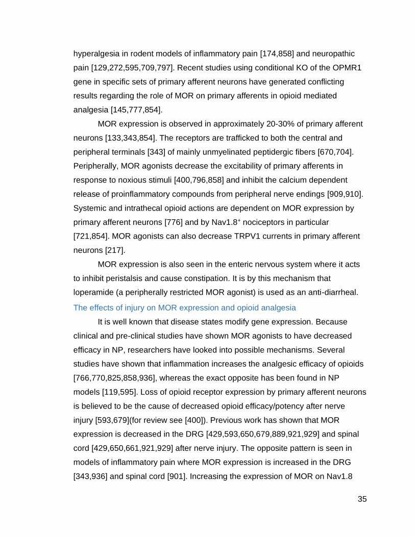
35
hyperalgesia in rodent models of inflammatory pain [174,858] and neuropathic
pain [129,272,595,709,797]. Recent studies using conditional KO of the OPMR1
gene in specific sets of primary afferent neurons have generated conflicting
results regarding the role of MOR on primary afferents in opioid mediated
analgesia [145,777,854].
MOR expression is observed in approximately 20-30% of primary afferent
neurons [133,343,854]. The receptors are trafficked to both the central and
peripheral terminals [343] of mainly unmyelinated peptidergic fibers [670,704].
Peripherally, MOR agonists decrease the excitability of primary afferents in
response to noxious stimuli [400,796,858] and inhibit the calcium dependent
release of proinflammatory compounds from peripheral nerve endings [909,910].
Systemic and intrathecal opioid actions are dependent on MOR expression by
primary afferent neurons [776] and by Nav1.8+ nociceptors in particular
[721,854]. MOR agonists can also decrease TRPV1 currents in primary afferent
neurons [217].
MOR expression is also seen in the enteric nervous system where it acts
to inhibit peristalsis and cause constipation. It is by this mechanism that
loperamide (a peripherally restricted MOR agonist) is used as an anti-diarrheal.
The effects of injury on MOR expression and opioid analgesia
It is well known that disease states modify gene expression. Because
clinical and pre-clinical studies have shown MOR agonists to have decreased
efficacy in NP, researchers have looked into possible mechanisms. Several
studies have shown that inflammation increases the analgesic efficacy of opioids
[766,770,825,858,936], whereas the exact opposite has been found in NP
models [119,595]. Loss of opioid receptor expression by primary afferent neurons
is believed to be the cause of decreased opioid efficacy/potency after nerve
injury [593,679](for review see [400]). Previous work has shown that MOR
expression is decreased in the DRG [429,593,650,679,889,921,929] and spinal
cord [429,650,661,921,929] after nerve injury. The opposite pattern is seen in
models of inflammatory pain where MOR expression is increased in the DRG
[343,936] and spinal cord [901]. Increasing the expression of MOR on Nav1.8

36
primary afferent fibers reduces neuropathic pain behaviors and restores the
analgesic efficacy of MOR agonists [383]. Functionally, the decrease of MOR
expression after nerve injury results in agonists being less able to decrease SP
release in the dorsal horn after noxious peripheral stimulation [119].
Analgesic tolerance, opioid dependence, and opioid induced hyperalgesia
Repeated administration of opioids leads to the development of tolerance
to its analgesic effects, such that higher doses are needed to attain a similar level
of pain relief. Mechanisms of analgesic tolerance involve changes at the
molecular, cellular, and circuit level. Acute tolerance can be seen as a decrease
in the ability of MOR agonists to decrease cAMP production and is most likely
caused by receptor desensitization, via GPCR Kinase (GRKs) phosphorylation
and subsequent β-arrestin dependent internalization [423,768,937]. Not all
agonists induce receptor internalization, and internalization has been shown to
reduce opioid tolerance suggesting that this process may enable resensitization
of receptors after phosphorylation [387,388]. The involvement of internalization in
tolerance is not fully understood and is likely agonist dependent [555].
Downregulation of receptor expression also plays a role in the development of
tolerance [763]. High efficacy opioids induce opioid downregulation more readily
than low efficacy opioids and hence are more likely to induce tolerance via this
mechanism [626,632].
Continuous use of opioids over long periods of time produces physical
dependence. Once dependence is manifest, cessation of opioid use leads to
withdrawal symptoms including anxiety, restlessness, diarrhea, and alternating
hot flashes and shaking chills [216]. Under some conditions opioids can
paradoxically cause activation of pronociceptive systems and lead to increased
pain sensitivity in a process known as opioid induced hyperalgesia (OIH) [258].
Long term opioid use has been linked to dysregulation in the balance between
descending inhibition and facilitation tipping the scales towards the latter
[513,829]. A recent study has also suggested a role for MOR expression in
primary afferent neurons in the development of OIH [777].

37
Analgesic tolerance in the CNS
Studies have shown that the PAG/RVM and spinal cord are involved in the
development of opioid induced analgesic tolerance [175,222,470]. The
development of opioid tolerance in the spinal cord, but not the PAG/RVM, is
dependent on NMDA and NOS signaling [222,470,543]. In the PAG/RVM
tolerance has been linked to cholecystokinin and microglial signaling
[379,514,689,892].
Analgesic tolerance in the PNS
The contribution of peripheral MOR signaling to analgesic tolerance is still
debated. Using Nav1.8 Cre mice to delete MOR expression in primary afferent
nociceptors, two groups have demonstrated opposing results regarding the role
of peripheral opioid receptors in the generation of tolerance [145,854]. However,
studies showing that repetitive use of peripherally restricted opioid loperamide
promotes the development of tolerance, suggest that the peripheral system is
also susceptible to tolerance [291].
Glutamate signaling as an analgesic target
Recently there has been an increase in research surrounding the role of
glutamate in nociceptive signaling and the use of glutamate receptor
agonists/antagonists as potential analgesics (for review see [75,610]).
Metabotropic glutamate receptors have received a lot of attention, specifically the
role of the metabotropic glutamate receptor 5 (mGluR5) in peripheral and spinal
nociceptive pathways [391,575,844,916,930].
The role of mGluR5 in nociception
mGluR5 (a group I mGluR) is a GPCR and its activation initiates signaling
cascades that modulate cellular excitability. Early studies using the mGluR5
agonist CHPG showed that mGluR5 activation induces pain behaviors in rodents
when injected into the paw [351,844] or intrathecally [280,452,681]. Contrastingly,
mGluR5 antagonists have been shown to decrease pain behaviors in various
rodent models including: CCI [203,233,234,385,758,930], SNL
[316,451,830,928,930], CFA [456,844,930], the second phase of the formalin test

38
[330,362,538,701,930], visceral pain [68,157,312,453,917], chemotherapeutic
induced peripheral neuropathy [930], and Bone cancer pain [681].
A novel photoactivatable mGluR5 antagonist was able to decrease CCI
induced mechanical hyperalgesia upon paw illumination [239]. Despite its anti-
nociceptive properties, mGluR5 antagonists do not alter responses to acute
noxious stimuli in naïve animals [722,844]. Similarly, mGluR5 KO animals exhibit
decreased inflammatory bladder pain [157], and decreased formalin evoked pain
behaviors [311,392,538] but display normal baseline thresholds [311,895].
The expression of mGluR5 in the pain neuraxis
mGluR5 expression is seen in the dorsal horn of the spinal cord post
synaptic to primary afferent fibers [16,59,344,690,788,827,834], in small diameter
DRG neurons [155,827], as well as in the central and peripheral terminals of
primary afferent nociceptors [362,377,844,891]. Pre-treatment of the skin with
capsaicin prevents the hyperalgesia caused by intraplantar administration of the
mGluR5 agonist CHPG, suggesting that the receptor is expressed by TRPV1+
primary afferents [351].
The expression of mGluR5 in pain conditions
mGluR5 expression is upregulated in the dorsal horn in animal models of
bone cancer pain [681], inflammatory pain [204,648], chemotherapeutic pain
[891], diabetic pain [804,927], and in models of nerve injury induced pain
including: CCI [592,611], and SNL [308,410,450]. In the DRG mGluR5 expression
is also upregulated in models of chemotherapeutic neuropathy [891], diabetic
neuropathy [439], CCI [385], SNI[316], and after painful whiplash injury [206].
Local extracellular concentrations of glutamate, the endogenous ligand for
mGluR5, have also been shown to be increased in the spinal cord
[12,202,326,401,747,891,903] and peripherally [173,345,608] in various pain
conditions. Stimulation of presynaptic mGluR5 itself increases glutamatergic input
in the spinal cord [891], and receptor antagonists prevent the induction of LTP at
primary afferent synapses [444]. The upregulation of receptor expression
combined with the increased concentration of endogenous ligand in conditions of

39
pain may explain why mGluR5 antagonists do not decrease behavioral responses
to acute noxious stimuli in naïve animals [722,844].
mGluR5 signaling pathways
mGluR5 is a GPCR which couples to a Gq/11 trimeric G protein and
activates several pathways including activation of phospholipase C (PLC). PLC
activation promotes the hydrolysis of phosphoatidylinositol-4,5-bisphosphate
(PIP2) to form inositol-1,4,5-triphosphate (IP3) and diacylglycerol (DAG) [188].
The subsequent reduction of PIP2 disinhibits TRPV1 channel, increasing its
excitability [128]. IP3 can also bind calcium release channels on the endoplasmic
reticulum opening the channel and causing an increase in cytosolic calcium.
Indeed, mGluR5 activation causes calcium transients in cultured DRG neurons
[155]. Increased cytosolic calcium combined with DAG both serve to activate
protein kinase C (PKC) which directly phosphorylates AMPA and NMDA
channels, increasing their conductance [76,275,577]. PKC also directly
phosphorylates MORs leading to receptor desensitization and TRPV1 causing
decreased thresholds and increased membrane expression of this receptor
[66,131,154,310,542,663,738]. In 2009, Kim et al. was able to show that TRPV1
is transactivated by mGluR5 via DAG in the presynaptic terminals of primary
afferents in the spinal cord [377]. mGluR5 activation also leads to the activation of
ERK (via PKC), and ERK in turn decreases Kv4.2 A-type potassium currents,
further increasing cell excitability [5,309].
It has also been shown that inflammation induced central sensitization is
dependent on group I mGluR-NMDAR coupling [275,902]. mGluR5 is structurally
linked to the NMDA receptor via a protein scaffold [560] and functional
interactions have been demonstrated between the two receptors [27]. Allosteric
interaction of mGluR5 via a covalent linkage with the NR2 subunit of the NMDAR
has been shown to modulate neuronal excitability [76,83,641].
Behavioral and cellular interactions between MOR and mGluR5
Earlier studies demonstrated that co-administration of MOR agonists with
mGluR5 antagonists enhances the antinociceptive effects of opioids
[609,644,928] and reduces the development of analgesic tolerance

40
[246,398,567,750,897,898,928]. Similarly, mGluR5 KO mice do not develop
analgesic tolerance to the same extent as normal animals [314], and knockdown
of mGluR5 reduces development of analgesic tolerance [896].
Given the pharmacological interaction between MOR and mGluR5,
Schroder and colleagues investigated the potential for these two receptors to
interact on a cellular level. They found that, in cells expressing both receptors,
mGluR5 antagonists reduce opioid induced MOR desensitization,
phosphorylation and internalization. They also discovered that delivering both
pharmacophores increased the physical association between the two receptors
[714].
Given the pharmacological interaction between MOR and mGluR5,
Schroder and colleagues investigated the potential for these two receptors to
interact on a cellular level. They found that, in cells expressing both receptors,
mGluR5 antagonists reduce opioid induced MOR desensitization,
phosphorylation and internalization. They also discovered that delivering both
pharmacophores to these cells increased the physical association between the
two receptors [714].
MMG22
The functional interaction between MOR and mGluR5, and evidence that
MOR/mGluR5 can form heteromers [714], led to the development of MMG22.
MMG22 is a bivalent ligand that combines a MOR agonist: oxymorphamine, with
an mGluR5 antagonist: 2-methyl-6-(phenylethynyl)-pyridine (MPEP) [9]. The two
pharmacophores of MMG22 are tethered together with a 22-atom linker and is
designed to bind the putative MOR/mGluR5 heteromer (Figure 2.1).
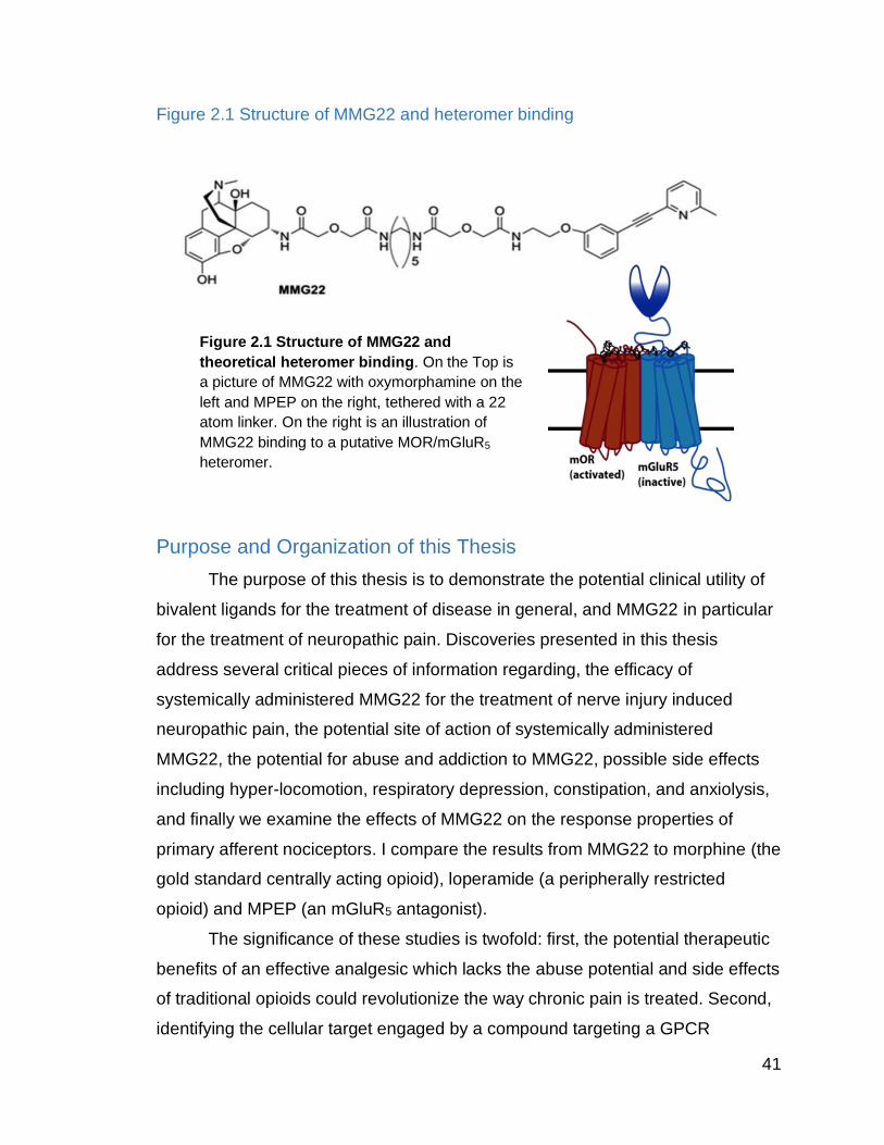
41
Figure 2.1 Structure of MMG22 and heteromer binding
Purpose and Organization of this Thesis
The purpose of this thesis is to demonstrate the potential clinical utility of
bivalent ligands for the treatment of disease in general, and MMG22 in particular
for the treatment of neuropathic pain. Discoveries presented in this thesis
address several critical pieces of information regarding, the efficacy of
systemically administered MMG22 for the treatment of nerve injury induced
neuropathic pain, the potential site of action of systemically administered
MMG22, the potential for abuse and addiction to MMG22, possible side effects
including hyper-locomotion, respiratory depression, constipation, and anxiolysis,
and finally we examine the effects of MMG22 on the response properties of
primary afferent nociceptors. I compare the results from MMG22 to morphine (the
gold standard centrally acting opioid), loperamide (a peripherally restricted
opioid) and MPEP (an mGluR5 antagonist).
The significance of these studies is twofold: first, the potential therapeutic
benefits of an effective analgesic which lacks the abuse potential and side effects
of traditional opioids could revolutionize the way chronic pain is treated. Second,
identifying the cellular target engaged by a compound targeting a GPCR
Figure 2.1 Structure of MMG22 and
theoretical heteromer binding. On the Top is
a picture of MMG22 with oxymorphamine on the
left and MPEP on the right, tethered with a 22
atom linker. On the right is an illustration of
MMG22 binding to a putative MOR/mGluR5
heteromer.

42
heteromer would add to the current understanding of how GPCR heteromers
work and support the development of bivalent ligands targeting GPCR
heteromers in drug design.

43
Chapter 3
The anti-hyperaglesic potency of systemic MMG22
Chapter reprinted with permission from PAIN, modified from:
Speltz, Rebecca, Lunzer, Mary M., Shueb, Sarah S., Akgün, Eyup, Reed, Rachelle, Kalyuzhny,
Alex, Portoghese, Philip S., Simone, Donald A. 2020. The bivalent ligand, MMG22, reduces
neuropathic pain after nerve injury without the side effects of traditional opioids. PAIN. (in press)

44
Introduction: Research on pain and analgesia
Previous research with MMG22
Previous studies have shown that intrathecal (i.t.) but not
intracerebroventricular administration of MMG22 was orders of magnitude more
potent at reducing hyperalgesia in mice with inflammatory pain and bone cancer
pain than compounds with shorter or longer linker lengths [9,748] or a mixture of
the monovalents [9]. Inflammation also causes a dramatic left shift in the dose
response curve of MMG22 [9]. The importance of linker length to the potency of
MMG22 may depend on several factors including the pain model and its
underlying mechanisms, and route of administration. For example, i.t.
administration of the MMG bivalent with 10 or 22 atom linker lengths were
equipotent in the spared nerve injury (SNI) model of neuropathic pain [644]. The
potency of MMG22 was significantly greater in mice with inflammatory pain, than
in naïve mice, suggesting the importance of pain and or inflammation in its
mechanism of action [9,10]. Importantly, repeated administration of MMG22 did
not promote the development of acute [748] or chronic [735] analgesic tolerance.
Because of its exceptional potency, the total amount of MMG22 that
needs to be given in order to obtain optimal efficacy is unusually small (in LPS
mice the ED50 for MMG22 and morphine are ~9 femtomoles and ~35,000
femtomoles respectively). The ability to give such small doses of MOR agonist
and get such a robust analgesic response decreases the likelihood of off-target
effects like respiratory depression, constipation, and sedation.
Animal models of neuropathic pain
Animal models have been created to study neuropathic pain conditions
including diabetic neuropathy [240,549], chemotherapy induced peripheral
neuropathy [28,108,302,656], and injury induced neuropathies. A majority of the
peripheral nerve injury models involve damage to the sciatic nerves or its
branches. The spinal nerve ligation (SNL) model involves ligating and cutting of
L5 (and L6) spinal nerves of the rat or mouse [376]. In the chronic constriction
injury (CCI) model, 3-4 chromic gut ligatures are loosely placed around the

45
sciatic nerve [55]. The spared nerve injury (SNI) model (where the common
peroneal and tibial branches of the sciatic nerve to ligated and cut, sparing the
sural nerve) has recently gained favor as it creates a long lasting hyperalgesia
that does not resolve over time [172]. For review of the various neuropathic pain
models see [331]. These models induce changes in rodent behavior that are
thought to correspond to spontaneous pain, allodynia and hyperalgesia- the
dominant symptoms experienced by patients with neuropathic pain.
Behavioral assessment of pain in animals
Direct assessment of pain in rodents is problematic, so researchers use a
variety of measures behavior to indirectly assess spontaneous pain as well as
hyperalgesia and allodynia. Spontaneous pain behaviors that can be observed
and quantified include spontaneous foot lifting [199], autotomy [848], and facial
grimace [533]. Mechanical hyperalgesia and allodynia can be measured in
several ways. Dynamic mechanical allodynia can be asses by brushing the skin
with a cotton swab or paintbrush to elicit withdrawal. Punctate hyperalgesia and
allodynia are commonly tested by applying calibrated von Frey hairs to the
affected area in order to evoke a withdrawal response [114]. Randall-selitto [677]
test is used to assess mechanical hyperalgesia. Tests of heat sensitivity include
the tail flick test [162], hot plate test [591,886], and Hargreaves [289]. A drop of
acetone applied to the skin can be used to assess cold allodynia [911] (for review
see [419]).
An earlier study examined the effects of intrathecal MMG22 in mice after
SNI [644], however; we wanted to see if systemically administered MMG22
would be effective. We assessed the ability of different doses of subcutaneously
administered MMG22 to decrease mechanical hypersensitivity in mice after
nerve injury. We also compared how the effects of MMG22 compared to the
effects of the traditional monovalent opioid morphine, the peripherally restricted
opioid loperamide, as well as the monovalent mGluR5 antagonist MPEP.

46
Methods
Animals
Adult (5-8 months) male and female C57/B6 mice (Charles River) were
housed 4 (males) or 5 (females) to a cage and maintained on a 12-hour light/dark
cycle with ad libitum access to food and water. An equal number of male and
female mice were used for each experiment and no sex differences were seen
for any of the parameters measured. All procedures were carried out during the
light cycle. 8-12 mice (an equal number of male and female mice) were used for
each experiment unless otherwise specified. All procedures were approved by
the Institutional Animal Care and Use Committee of the University of Minnesota.
Spared nerve injury
Mice were anesthetized with 2.5 % isoflurane. Spared nerve injury (SNI) to
the sciatic nerve was performed as described previously [82,172,644]. Briefly,
after exposing the three branches of the sciatic nerve, the tibial and common
peroneal branches were tightly ligated with 5.0 silk suture and cut 2 mm distal to
the suture. Care was taken not to disturb the sural nerve. Sham surgeries
followed the same procedure without manipulation of the sciatic nerve or distal
branches.
Drugs
The bivalent ligand MMG22 was synthesized as described previously [9].
MMG22, morphine, 2-methyl-6-(phenylethynyl)pyridine (MPEP), (Mallinckrodt
Inc, Hazelwood, MO) and loperamide (Sigma, St. Louis, MO) were diluted in 1%
DMSO (vehicle). Loperamide was diluted in near boiling 1% DMSO daily. All
drugs were administered subcutaneously in a volume of 250 µl, between the
shoulders.
Behavior
In SNI, sensitivity seen in territory of non-injured sural nerve on the lateral
hind paw [82,172,644]. Mechanical hyperalgesia was defined as an increase in
frequency of paw withdrawal evoked by a von Frey monofilament as we
described previously [373,374]. Mice were placed on an elevated mesh platform
under glass enclosures and allowed to habituate for 30 minutes prior to initial

47
testing. A calibrated von Frey monofilament with a bending force of 5.9 mN (0.6g)
(StoeltingCo, Woodale, IL) was applied to the lateral portion of the plantar
surface of each hind paw 10 times, with an interval of approximately 10 seconds
between applications, and the frequency of withdrawal responses were
determined. Mice were tested a minimum of three times prior to surgery. After
surgery, mice were tested once a day for 3 days starting on day 7 to establish a
stable post-surgical baseline before testing any of the analgesics. Drug-induced
reduction of mechanical hyperalgesia was assessed early (10 days) and late (30
days) after surgery. 10 days was chosen as the early time point because full
development of hyperalgesia occurs around 7 days after nerve injury [82,172]
and 3 days of stable post-surgical baselines were desired prior to analgesic
testing. We chose 30 days for the late time point because at this time, the
inflammatory response to nerve injury has subsided [17,523].
Dose response curves
To determine dose-response functions, mice were injected with escalating
doses of MMG22, morphine, loperamide or MPEP (s.c.). Separate groups of
mice were used for each drug and time point. Starting dose of MMG22, morphine
and loperamide was 0.1mg/kg, starting dose of MPEP was 1mg/kg. Doses were
increased as follows: 0.1mg/kg, 0.3mg/kg, 1mg/kg, 3mg/kg and so on. The
frequency of withdrawal evoked by 10 applications of a von Frey monofilament
was determined 30 minutes after each injection. Mice were returned to their
home cage after each injection and placed back on the mesh platform 10
minutes prior to testing. The percent maximal possible effect (%MPE) was
calculated using the following standard formula:
%𝑀𝑃𝐸 =(𝑃𝑜𝑠𝑡 𝐷𝑟𝑢𝑔 𝑉𝑎𝑙𝑢𝑒 − 𝑃𝑟𝑒 𝐷𝑟𝑢𝑔 𝑉𝑎𝑙𝑢𝑒)
(𝑃𝑟𝑒 𝑆𝑢𝑟𝑔𝑖𝑐𝑎𝑙 𝐵𝑎𝑠𝑒𝑙𝑖𝑛𝑒 𝑉𝑎𝑙𝑢𝑒 − 𝑃𝑟𝑒 𝐷𝑟𝑢𝑔 𝑉𝑎𝑙𝑢𝑒) × 100
Only doses that resulted in a behavioral response >0% were included in the
analysis, The final dose included in the analysis was either the first dose to give a
100% MPE or the largest dose tested. The dose that reduced the withdrawal
frequency by 50% compared to baseline (ED50) was determined by non-linear
regression of the %MPE data carried out in Prism 8.00 (GraphPad Software, San

48
Diego, CA). Data collectors were blinded to drug and absolute (but not relative)
dose by a third party
Data analysis
Data are expressed as mean ± SEM, except where otherwise noted.
GraphPad Prism 8.0 (Graphpad software Inc. La Jolla, CA, USA) was used for
statistical analyses and calculation of ED50 values. All behavioral data were
analyzed via one- or two-way ANOVA with repeated measures. Post-hoc
comparisons were done with Bonferroni tests. A P value of <0.05 was considered
significant.

49
Results
SNI induces sustained hyperalgesia in mice
Consistent with earlier reports [82,172,644] SNI produced robust
mechanical hyperalgesia. We compared the percent withdrawal frequency to von
Frey in SNI and sham operated mice before and after surgery (two-way ANOVA;
time: [F(3,16)=75; P<0.0001], surgery: [F(1,16)=476; P<0.0001], time × surgery:
[F(3,16)=54, P<0.0001]. Before surgery the groups were not different (P>0.999),
but on days 10, 20 and 30 after surgery, SNI mice had withdrawal frequencies
higher than sham mice (P<0.0001) (Fig. 3.1).
Figure 3.1 Hyperalgesia after nerve injury
Figure 3.1 Hyperalgesia after spared nerve injury. The percent
withdrawal frequency increased in mice after SNI surgery (open circles)
but not in mice after sham surgery (closed circles). Data presented as
mean ± SEM, n=5 per group. ****P<0.0001 compared to sham.
Baseline 10 20 30
0
25
50
75
100
Days Post Injury
% W
ith
dra
wa
l
Fre
qu
en
cy
Sham
SNI
**** ****
****

50
MMG22 decreased mechanical hyperalgesia dose-dependently in neuropathic
mice when delivered subcutaneously
We evaluated the ability of MMG22, morphine, and loperamide to
decrease mechanical hyperalgesia early (10 days after surgery / during the
initiation phase) and late (30 days after surgery / during the maintenance phase)
after nerve injury. MMG22 did not alter percent withdrawal frequency of naïve
animals or animals after sham surgery at any of the doses tested (data not
shown). Subcutaneous administration of MMG22, morphine, and loperamide
dose-dependently reduced mechanical hyperalgesia in nerve-injured mice (Fig.
3.2A-C). Data are shown separately in mg/kg and nmoles/mouse for ease of
comparison, but all data were analyzed together. We compared the ED50s of
MMG22, morphine, and loperamide early and late after nerve injury (two-way
ANOVA; drug: [F(2,183)= 4.8; P=0.0095], time: [F(1,183) = 27.88; P<0.0001], time
× drug: [F(2,183)=12.54; P<0.0001] n=8-14). The potency of MMG22 was
decreased late after nerve injury compared to early after nerve injury (P<0.0001),
causing a rightward shift in the dose response curve (Fig. 3.2A, Table 3-1).
Morphine did not exhibit the same shift in potency and was equipotent at
reducing mechanical hyperalgesia early and late after nerve injury based on ED50
values (Fig. 3.2B).
As discussed earlier, MOR expression is known to decrease in the DRG
following peripheral nerve injury [429,438,593,650,679,889,929] and this
reduction has been linked to a concomitant loss of opioid responsiveness of
DRG neurons [390]. To determine if loss of peripheral MOR expression after
nerve injury was involved in the decrease in potency for MMG22, we repeated
this experiment with the peripherally restricted MOR agonist loperamide.
Loperamide does not penetrate the blood brain barrier at the doses tested
[32,174,588,725], but has been shown to decrease hyperalgesia after systemic
administration [129,272,732]. Consistent with previous data [272], there was no
decrease in potency late after nerve injury as compared with the earlier time
point (Fig. 3.2C). It has been shown that the bulk of MOR downregulation in the
DRG occurs within the first week after nerve injury [429,889] which suggests that

51
any effects of MOR down-regulation on peripheral opioid potency would already
be completed 10 days after nerve injury.
We converted the mg/kg doses into nmoles per mouse for direct
comparison between drugs. Early after nerve injury, MMG22 was 40 times as
potent as morphine or loperamide at reducing mechanical hyperalgesia (Fig.
3.2D, Table 3-1). Late after nerve injury however, all agonists were equipotent
(Fig. 3.2E).
As one of the pharmacophores of MMG22 is MPEP, an mGluR5
antagonist, we evaluated the ability of MPEP to decrease mechanical
hyperalgesia early and late after nerve injury. At the highest dose, MPEP was
unable to decrease mechanical hyperalgesia more than 50% early after nerve
injury (Fig. 3.2F). This is consistent with previous reports showing that mGluR5
antagonists alone have little effect [234,830,930] or no effect [316,330,844] on
mechanical hyperalgesia after nerve injury. The ED50 for MPEP was increased
late after nerve injury compared to the earlier time point (unpaired t-test; t=2.73,
P<0.01, n=8-12) (Fig. 3.2F, Table 3.1). The change in potency of MPEP over
time after nerve injury suggests that this may contribute to the similar decrease in
potency seen with MMG22.
In this study MMG22 also did not alter the responses in naïve or sham
animals (data not shown). Results from previous studies with LPS mice used
radiant tail flick assay to create their dose response curve, MMG22 did increase
tail flick latency in naïve mice, but was much less potent than in LPS mice [9].
Using percent withdrawal frequency to mechanical stimuli, MMG22 did not
decrease the percent withdrawal in naïve mice. We used a light filament that
gives an average of 1-2 withdrawals per 10 applications in naïve mice, but 8-10
withdrawals in SNI mice in order to increase the dynamic range of the assay.
Because of this, there may be a floor effect prohibiting us from seeing any
MMG22 induced analgesia in naïve mice

52
Figure 3.2 Dose response curves for MMG22, morphine, loperamide and MPEP
Figure 3.2 Cumulative dose response functions for reducing tactile hyperalgesia after
spared nerve injury. Mice were given subcutaneous injections of drug in increasing doses,
early (10 days/open circles) or late (30 days/filled circles) after nerve injury. Paw withdrawal
frequency to a 5.9 mN (0.6g) von Frey hair was measured 30 minutes after each injection
and %MPE calculated based on pre-surgical baseline values. (A) MMG22 (blue) dose
dependently reduced mechanical hyperalgesia in mice. The dose response curve is right
shifted late after nerve injury compared to the earlier time point. (B,C) Morphine (red) and
loperamide (purple) also dose dependently reduced mechanical hyperalgesia after nerve
injury; however, no change in potency was observed over time. (D) Early (10 days) after
nerve injury, MMG22 is more potent at reducing mechanical hyperalgesia than similar doses
of morphine or loperamide. (E) Late (30 days) after nerve injury, the dose response curves
for MMG22, morphine, and loperamide are overlapping. (F) MPEP (green) weakly reduced
mechanical hyperalgesia early after nerve injury. There is a rightward shift in the dose
response curve late after nerve injury. Data are presented on graphs as means ± SEM, n =
8-14 per group.
0.01 0.1 1 10 100
0
25
50
75
100
125MMG22
Dose (mg/kg)
% M
PE
Early
Late
A
0.
1 1 10 10
0
0
25
50
75
100
125Morphine
Dose (mg/kg)
Late
Early
B
0.1 1 10 10
0
0
25
50
75
100
125Loperamide
Dose (mg/kg)
Early
Late
C
1 10 100
1000
0
25
50
75
100
125Early
Dose (nmol/mouse)
%M
PE
D1 10 100
1000
0
25
50
75
100
125Late
Dose (nmol/mouse)
E
1 10 100
0
25
50
75MPEP
Dose (mg/kg)
Early
Late
F
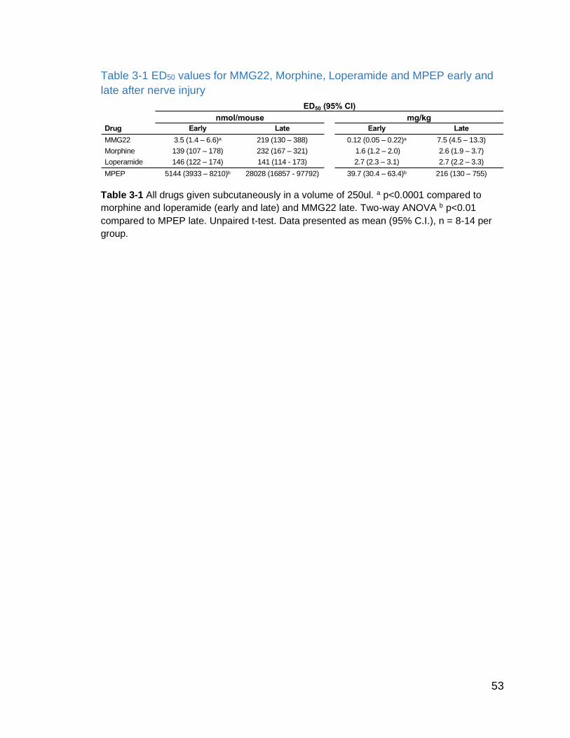
53
Table 3-1 ED50 values for MMG22, Morphine, Loperamide and MPEP early and
late after nerve injury
Table 3-1 All drugs given subcutaneously in a volume of 250ul. a p<0.0001 compared to
morphine and loperamide (early and late) and MMG22 late. Two-way ANOVA b p<0.01
compared to MPEP late. Unpaired t-test. Data presented as mean (95% C.I.), n = 8-14 per
group.
ED50 (95% CI)
nmol/mouse mg/kg
Drug Early Late Early Late
MMG22 3.5 (1.4 – 6.6)a 219 (130 – 388) 0.12 (0.05 – 0.22)a 7.5 (4.5 – 13.3)
Morphine 139 (107 – 178) 232 (167 – 321) 1.6 (1.2 – 2.0) 2.6 (1.9 – 3.7)
Loperamide 146 (122 – 174) 141 (114 - 173) 2.7 (2.3 – 3.1) 2.7 (2.2 – 3.3)
MPEP 5144 (3933 – 8210)b 28028 (16857 - 97792) 39.7 (30.4 – 63.4)b 216 (130 – 755)

54
Discussion
Our results show that, in agreement with previous studies [644], MMG22
potently reduced mechanical hyperalgesia after nerve injury. For the first time,
we show that MMG22 was able to reduce spontaneous pain, and importantly that
systemic MMG22 lacked the rewarding properties and other centrally mediated
side effects associated with traditional opioids. The bivalent design of MMG22
was intended to activate MOR and to inhibit mGluR5 and to target a putative
MOR-mGluR5 heteromer. mGluR5 has been characterized as a promising new
target for chronic pain [575,576,844,845]. Activation of mGluR5 produces pain
[280,681,845] and mGluR5 antagonists reduce pain behaviors in various pain
models [203,233,362,539,830,844,930] without altering responses to acute
noxious stimuli in naïve animals [722,844].
The exceptionally potent antinociception produced by MMG22 is due to
the activation of MOR combined with antagonism of the mGluR5 and its co-
receptor, NMDAR. Allosteric interaction of mGluR5 via a covalent linkage with the
NR2 subunit of the NMDAR has been shown to modulate neuronal excitability
[76,83,641]. Pre-treatment of inflamed mice with an irreversible MOR antagonist
(β-FNA) or the NMDAR antagonist (MK801) reduces the antinociception of
MMG22 [10] indicating a contribution of both pharmacophores of MMG22.
MMG22 was designed to target a putative MOR-mGluR5 heteromer, which
was supported by the relation between its linker length and optimal potency
[9,748]. Both MOR and mGluR5 can form heteromers with other GPCRs
[94,225,787] and MOR-mGluR5 heteromers have been reported in vitro [714].
Bivalent ligands have increased affinity and selectivity for their targets [485,662],
and antagonizing one receptor can enhance agonist-induced signaling at its
heteromeric protomer [283]. Research has also shown that heteromer formation
can be modulated by pathological states [264], and the formation of heteromers
can alter signal transduction [299].
Early after nerve injury, MMG22 was 40 times more potent than morphine,
whereas late after nerve injury the two were equipotent. Contrastingly, in a bone
cancer pain model the potency of MMG22 increased in parallel with tumor growth

55
and hyperalgesia [735,748]. The differences in potency of MMG22 may be
explained by the timing and duration of the inflammatory response following
nerve injury or tumor implantation. After nerve injury, there is an early pro-
inflammatory response that is rapidly resolved after 2-3 weeks [17,523,812],
whereas the inflammatory response after tumor implantation remains elevated
over time [269,458,488,905]. The potency of MMG22 to decrease hyperalgesia
mirrors the time course of inflammation in bone cancer pain [735,748] and after
nerve injury (in this study). A study using i.t. MMG22 after nerve injury did not
report any statistically significant differences in potency over time after nerve
injury; however, the ED50 was lowest at 7 and 17 days after injury and higher at a
later time point, and also parallel to the time course of inflammation [644]. The
combination of transient inflammation and persistent pain after nerve injury,
allowed us to demonstrate that the potency of MMG22 induced analgesia is
almost certainly dependent on ongoing inflammation, and not on ongoing pain.
The decrease in potency of MPEP late after injury also agrees with previous
studies that suggest MPEP is a more potent analgesic in inflammatory pain
models vs neuropathic models [316,330,639,844].
The importance of inflammation in the development of neuropathic pain is
supported by several studies showing that neonatal rats do not develop nerve
injury induced pain phenotypes until the default neuroimmune response switches
from anti-inflammatory to pro-inflammatory around P28 [156,236,505]. Prior to
this, nerve injury promotes an anti-inflammatory response instead of the typical
pro-inflammatory response seen in the adult [505]. Blockade of the anti-
inflammatory cytokines or exogenous application of TNFα reveals neuropathic
like pain behaviors after nerve injury in the neonatal rat [505].
A recent study showed a connection between the pro-inflammatory
cytokine, tumor necrosis factor α (TNFα), and mGluR5 upregulation after nerve
injury [410]. TNFα contributes to pain hypersensitivity [705,756,757,841]. A
decrease in TNFα levels reduced mGluR5 expression and hyperalgesia, while
intrathecal administration of TNFα had the opposite effect [410]. The importance
of TNFα for the development of neuropathic pain is consistent with previous
56
studies showing a transient increase in TNFα in the lumbar DRG
[430,519,601,695] and spinal cord [430] after nerve injury. Upregulation of
mGluR5 may follow a similar timeline; with peak expression around 1-2 weeks
post injury [385,592]. The potential involvement of inflammation in the potency of
MMG22 is consistent with previous data showing that blocking astrocytes
reduced the potency of intrathecal MMG22 [10]. The involvement of central
astrocytes in the intrathecal potency of MMG22 suggests that MMG22 has the
ability to promote analgesia through central route, by modulating central
neuroinflammation.
Although our results show reduced potency for MMG22 late after nerve
injury, many neuropathic pain conditions area accompanied by more chronic
inflammatory changes [657,754], suggesting that MMG22 may be a viable
therapeutic for such conditions. For example, a recent study done by the
Portoghese lab has shown that MMG22 reduced hyperalgesia produced by
chemotherapy, which has a persistent inflammatory component [918], with no
change in potency over time (manuscript in preparation),. Future studies are
needed to determine the types of neuropathic pain conditions that best respond
to MMG22.
Conclusion
Previous research has shown that MMG22 is potent at reducing
hyperalgesia when given intrathecally in models of bone cancer pain,
inflammatory pain, and nerve injury induced neuropathic pain. Systemic
administration has also shown MMG22 to be effective in reducing bone cancer
induced pain. However, whether systemic administration is capable of reducing
neuropathic pain is not yet known. As neuropathic pain is thought to be less
opioid sensitive than other forms of pain, we decided to investigate whether
systemic administration of MMG22 would decease pain caused by nerve injury.
MMG22 was able to reduce hyperalgesia early but not late after nerve injury. The
reduction in potency of MMG22 late as compared to early after nerve injury
parallels the loss of potency for the mGluR5 antagonist MPEP over the same time
period, whereas neither morphine nor loperamide showed the same reduction in

57
potency. This pattern of potency is opposite from what has been shown for bone
cancer pain, suggesting the differing pathophysiology involved in these two types
of pain may play a part in the analgesia produced by MMG22.

58
Chapter 4
MMG22 Site of Action
Chapter reprinted with permission from PAIN, modified from:
Speltz, Rebecca, Lunzer, Mary M., Shueb, Sarah S., Akgün, Eyup, Reed, Rachelle, Kalyuzhny,
Alex, Portoghese, Philip S., Simone, Donald A. 2020. The bivalent ligand, MMG22, reduces
neuropathic pain after nerve injury without the side effects of traditional opioids. PAIN. (in press)

59
Introduction: Potential site of action for MMG22 within the pain neuroaxis
Heteromer formation
Opioid receptors have been shown to form heteromers with a number of
different GPCR receptor sub-types [146,244]. The ability for GPCRs to form
heteromers has been well established over the last 30 years [278]. Although
controversial, evidence for the existence and physiological significance of such
hetromers in vivo is mounting [224]. Receptor dimerization can affect receptor
function, ligand pharmacology, signal transduction, and cellular trafficking [299].
Targeting GPCR dimers with bivalent ligands may result in more potent and
selective compounds that act selectively on cells that express both receptors
[315] minimizing potential off-target effects [553]. It has also been suggested that
the propensity for different GPCRs to form heteromers may be modulated in
pathological states [264], making the targeting of GPCR heteromers not only cell
specific, but potentially disease state specific as well.
In order for a bivalent ligand to target a receptor heteromer, both receptors
must be expressed on individual neurons. Previous research has shown that
both receptors are expressed by primary afferent neurons as well as neurons in
the dorsal horn (as previously outlined), but it has never been shown that both
receptors are expressed by individual cells. Therefore, we set out to investigate
this possibility.
60
Methods
Animals
Adult (5-8 months) male and female C57/B6 mice (Charles River) were
housed 4 (males) or 5 (females) to a cage and maintained on a 12-hour light/dark
cycle with ad libitum access to food and water, except as otherwise noted for
constipation studies. All procedures were carried out during the light cycle. All
procedures were approved by the Institutional Animal Care and Use Committee
of the University of Minnesota.
Spared nerve injury
Mice were anesthetized with 2.5 % isoflurane. Spared nerve injury (SNI) to
the sciatic nerve was performed as described previously [82,172,644]. Briefly,
after exposing the three branches of the sciatic nerve, the tibial and common
peroneal branches were tightly ligated with 5.0 silk suture and cut 2 mm distal to
the suture. Care was taken not to disturb the sural nerve. Sham surgeries
followed the same procedure without manipulation of the sciatic nerve or distal
branches.
RNAScope
RNAscope® in situ hybridization (ISH) (a probe based non-radioisotopic
RNA ISH approach for detecting target RNAs in tissue) was used to determine
whether MOR and mGluR5 transcripts co-localized in dorsal root ganglia (DRG)
or spinal neurons. Ten days after SNI surgery, mice were given an intraperitoneal
injection of Euthasol (sodium pentobarbital, 390 mg/mL and phenytoin sodium,
90 mg/mL) and transcardially perfused with saline followed by 4%
paraformaldehyde. L3-L5 DRGs and L3-L5 spinal cord segments were collected,
post fixed in 4% paraformaldehyde for 3 hours and then placed in 30% sucrose
in phosphate-buffered saline overnight at 4°C. Isolated and fixed DRGs and
spinal cord segments were embedded into a tissue microarray using OTC media,
frozen in dry ice and methanol, sectioned (7-10um thickness) with a cryostat, and
thaw mounted onto slides. Sections were stored at -80°C.
RNAscope® ISH was performed on fixed, frozen sections of DRGs and
lumbar spinal cord with probes for mouse Oprm1 (Cat No. 315848) and Grm5

61
(Cat No. 423631) purchased from ACD Bio. RNAscope®. ISH for sections was
performed following manufacturer’s written protocol with only one modification to
the protease digestion (ACD, RNAscope® Multiplex Fluorescent Detection
Reagents v2, Cat No. 323110). Tissue digestion with Protease IV was done for
15 min at room temperature. Probes were hybridized for 2 h at 40 °C and washed
twice in wash buffer (RNAscope® Wash Buffer Reagents, 310091). Amplification
steps were performed by incubating with v2Amp1 (30 min), v2Amp2 (30 min) and
v2Amp3 (15 min) at 40 °C with washes of 2 × 2 min in between steps. Sections
were incubated with v2-HRP-C1 for 15 min at 40 °C and washed twice in wash
buffer for 2 min. Tyramide signal amplification (TSA)-conjugated fluorophores
were diluted 1:1,500 in TSA buffer (RNAscope® Multiplex TSA Buffer, 322809)
and incubated for 30 min at 40 °C followed by 2 washes of 2 min and HRP
blocker incubation for 30 min at 40 °C. The last steps were performed
subsequently for v2-HRP-C2. Images were collected on Olympus FluoView
FV1000 confocal microscope.
62
Results
Previous studies have shown the MOR and mGluR5 are expressed in
primary afferent neurons [67,134,362], as well as in superficial dorsal horn
neurons in the spinal cord [16,344,546,771,826,834]. To examine if both
receptors are expressed together in the same neurons, we performed an
RNAScope assay on lumbar spinal cord and DRG sections. Ten days after SNI
surgery there was good expression of both MOR (OPMR1) and mGluR5 (GRM5)
mRNAs in the lumbar dorsal horn (Fig. 4.1A). Higher magnification images show
that individual cells in the superficial (Fig. 4.1B) and deep (Fig. 4.1C) dorsal horn
express both receptors. Individual cells in the DRG (Fig. 4.1D) also expressed
the mRNAs for both receptors. High-resolution examination of the nuclear
morphology (dapi stained) suggested the cells in question were neuronal (large
nuclei, prominent nucleolar fading and folded nuclear membranes [248];
however, co-staining with cell type specific markers is needed for confirmation.

63
Figure 4.1 Colocalization of grm5 and opmr1 in the dorsal horn and DRG
Figure 4.1 MOR (Opmr1) and mGluR5 (Grm5) mRNAs co-expressed in dorsal horn and
DRG 10 days after SNI. (A-C) Examples of staining in the dorsal horn of SNI mice. Nuclei
are stained using dapi (blue). (A) Low magnification image of the lumbar dorsal horn stained
for MOR (OPRM1, right/red) and mGluR5 (GRM5, middle/green), and overlay (right). Dashed
line shows the grey-white matter boundary of the dorsal horn. (B) Higher magnification
image of the superficial dorsal horn; OPMR1 (left/red), GRM5 (middle/green), overlay (right).
Arrows indicate individual cells that co-express MOR and mGluR5. (C) Higher magnification
image of the deep dorsal horn; OPMR1 (left/red), GRM5 (middle/green), and overlay (right).
Arrow indicates a cell that highly expresses both receptors. (D) High magnification image of
a lumbar DRG; OPMR1 (left/red), GRM5 (middle/green) and overlay (right). Nuclei are
stained using dapi (blue). Arrows indicate individual cells that express both receptors. Scale
bar: 50 um for all images.

64
Discussion
MMG22 was designed to target a putative MOR-mGluR5 heteromer.
Heteromer binding is supported by the relationship between linker length and
optimal anti-hyperagesic potency [9,748]. Both MOR and mGluR5 can form
heteromers with other GPCRs [94,225,787] and MOR-mGluR5 heteromers have
been reported in vitro [714]. Bivalent ligands are hypothesized to have increased
affinity and selectivity for their targets [315,662], and it has been shown that
antagonizing one receptor can enhance agonist-induced signaling at its
heteromeric protomer [283]. Heteromer formation can be modulated by
pathological states [264], and can alter signal transduction [299].
For the first time, we have shown that mRNAs for both MOR and mGluR5
are co-expressed in neurons in the lumbar spinal cord and DRG early after nerve
injury; suggesting potential targets for i.t. and systemic administration of MMG22
respectively. The co-expression of both receptors supports the potential for
MOR-mGluR5 heteromer formation in vivo. Previously, a non-overlapping pattern
of MOR and mGluR5 expression in the dorsal horn was found 8 weeks after
nerve injury [644]. Once translated, the receptors may be trafficked to different
cellular compartments. However, the pre and postsynaptic location of the
individual receptors would argue against this [25,60,344,788,834]. Alternatively,
consistent with the 60-fold decrease in potency of MMG22 from early to late after
injury (Table 3.1), the co-localization of its target receptors, and their
heteromerization, may be transient and not present late after nerve injury. These
possibilities may not be mutually exclusive, as studies have demonstrated that
axonal targeting of mGluR5 is dependent on the expression of Homer1a (an
immediate early gene) [20], which is only transiently upregulated early after nerve
injury [531]. More research is needed to quantify the degree of colocalization
over time after nerve injury.
This is the first study to demonstrate the colocalization of mRNAs for both
target receptors in vivo. Co-localization by itself does not prove that the receptors
form heteromers, but previous research has shown that the two receptors
physically associate in HEK cells [714]. Further support for the existence of

65
heteromers comes from pharmacological studies showing that MMG ligands with
shorter (10) or longer (24) linker lengths are less potent than MMG22 [9,748].
Conclusion
In order for MMG22 to target and bind heteromers, the two receptors must
be expressed in the same cells. Previous research has shown the receptors are
both expressed in the spinal dorsal horn and dorsal root ganglia, but little
research has been done to look at both receptors simultaneously. Here we show
that mRNAs for both receptors are expressed together in cells of the dorsal horn
and dorsal root ganglia early after nerve injury.

66
Chapter 5
Rewarding properties of MMG22: abuse liability vs relief
from spontaneous pain
Chapter reprinted with permission from PAIN, modified from:
Speltz, Rebecca, Lunzer, Mary M., Shueb, Sarah S., Akgün, Eyup, Reed, Rachelle, Kalyuzhny,
Alex, Portoghese, Philip S., Simone, Donald A. 2020. The bivalent ligand, MMG22, reduces
neuropathic pain after nerve injury without the side effects of traditional opioids. PAIN. (in press)

67
Introduction: A brief overview of reward and addiction
Addiction and the brain
Addiction is a chronic, relapsing brain disease characterized by loss of
control regarding the use of a substance, compulsive use despite negative
consequences, and the emergence of a negative emotional state upon withdrawn
[397,823]. Drugs that are associated with addiction in humans include cocaine
(and other psychostimulants), alcohol, nicotine, and opioids. All of these drugs
directly or indirectly cause dopamine release in the nucleus accumbens (NAc)
[836].
Generally, addiction is thought to consist of three stages:
binge/intoxication, withdrawal/negative affect, and preoccupation/anticipation.
The brain regions believed to be involved in these stages include the basal
ganglia, amygdala, and prefrontal cortex (respectively) [397]. During the
binge/intoxication stage, positively reinforcing stimuli work by activating
dopaminergic neurons in the ventral tegmental area (VTA) [572] and the
subsequent release of dopamine into the NAc [397]. The withdrawal/negative
affect stage involves a cessation of the positive reinforcement which results in a
decrease in the activity in the mesolimbic dopamine projection, which causes a
decrease in dopamine release, an elevation of reward thresholds, and increased
anxiety. Removal of the withdrawal associated negative affect becomes the basis
for negative reinforcement and drug seeking behavior [397]. The
preoccupation/anticipation stage of addiction involves glutamatergic projections
from the prefrontal cortex (PFC) to the ventral striatum. The PFC is an area of
the brain that mediates executive function and decreased activity in this area
interferes with decision making and inhibitory control. Animal models of the
positive reinforcing effects of drugs include self-administration and conditioned
place preference [397].
68
Opioid addiction
One of the most prominent disadvantages regarding the use of opioids to
treat chronic pain is the abuse and addiction liability associated with these drugs.
Over the last few decades the incidence of opioid abuse and addiction has
skyrocketed leading to what many are calling an “opioid epidemic” [746]. In 2017,
the US department of Health and Human Services declared the opioid crisis a
public health emergency [178]. Prescriptions for opioids have increased
dramatically since 1999, peaking in 2012 with more than 255 million opioid
prescriptions written [112]. Over the same time period, the number of opioid
related overdose deaths has increased 200% [616], and opioids now kill more
people than motor vehicle accidents in the US [570]. The number of patients
prescribed opioids for non-cancer pain who misuse them is estimated to be as
high as 30%, with around 10% developing opioid use disorders [723]. Withdrawal
from opioids causes feelings of dysphoria, anxiety, and irritability, a combination
of symptoms collectively termed “kyperkatifeia” [736]. Opioid withdrawal can also
cause hyperalgesia and physical malaise [397].
There is evidence suggesting that chronic pain patients develop addiction
at a lower rate than the general population [232] and animal studies indicate that
chronic pain is accompanied by reduction in opioid induced reward
[64,220,221,499,617,618,839]. However, the use of prescription opioids for
chronic non-cancer pain is a strong risk factor for the development of opioid use
disorders [214,837,871]. Nearly half of pain patients with a diagnosed substance
use disorder involving opioids were first prescribed opioids for pain control [332]
and 1 in 10 patients receiving opioids for chronic pain will develop an opioid
addiction [837].
Effects of mGluR5 antagonists on addiction
mGluR5 has been shown to be involved in the reward pathway for many
drugs of abuse, and antagonizing mGluR5 reduces reward seeking behavior for
alcohol [70,151,301,717], nicotine [370,445,628], ketamine [359] and cocaine
[369,370]. mGluR5 KO mice show decreased reward to cocaine [125], and do not
69
exhibit cocaine mediated LTP in the VTA (known to be important for the
acquisition of reward seeking behavior) [71].
Regarding opioids, mGluR5 antagonists have been shown in previous
studies to inhibit morphine induced conditioned place preference [658,691,831]
and decreased self-administration of morphine [89] and heroin [359], although
there are conflicting reports [298,358].
Animal models of addiction
The conditioned place preference (CPP) assay is used to evaluate the
rewarding properties of drugs in animals [39,821]. A typical CPP assay involves
differentially pairing contextual cues with stimuli of interest. Conditioning involves
repeated administration of a stimulus of interest (unconditioned stimulus / US) in
a particular environment (conditioned stimulus / CS), intermixed with exposure to
a different environment without the US. Following conditioning is a choice test
where the animal has access to both environments without the US. An increase
in time spent in the environment previously paired with the US vs the unpaired
environment is taken as evidence that the US is rewarding (for review see [39])
Research has shown that using drugs known to be addictive in people as the US
reliably produces CPP in mice and rats including morphine [40,607], cocaine
[585], nicotine [733], and alcohol [680].
Another tool used to assess the rewarding properties of drugs is self-
administration. In this assay, animals are given access to a drug of abuse
(generally administered when the animal pushes a lever) and their intake is
monitored. There are two main variations of this assay, continuous and
intermittent reinforcement. In the continuous administration paradigm drug is
given every time the animal pushes a lever. For intermittent reinforcement, the
animal must push the lever a certain number of times before drug is given. For a
more in depth look at various parameters used in self-administration see [621].
Most drugs that promote self-administration, also promote conditioned place
preference [39].
70
Interactions between pain and Reward
Negative reinforcement from pain relief also involves the activation of
dopaminergic neurons in the VTA and dopamine release in the NAc [573]. Pain
relief has been shown to be rewarding in animal models [571,572,660]. A
variation on the traditional CPP assay has been developed to take advantage of
this analgesic CPP (aCPP). This assay can be used to measure the relief from
ongoing spontaneous pain that is otherwise difficult to measure. Previous studies
have shown that in the context of pain animals will show place preference when
paired with normally non-rewarding analgesics like MPEP [428,835], lidocaine
[292,380], bupivacaine [165], clonidine [171,292,380], and loperamide [797].
Analgesics including clonidine [498] and suprofen [142] have also been shown to
promote self-administration in animal models of pain.
The incredible potency of MMG22 is concerning when it comes to the
potential for abuse and addiction. In the following studies we used a conditioned
place preference assay in naïve and nerve injured animals to determine if
MMG22 was rewarding in naïve animals and if MMG22 could decrease
spontaneous pain in nerve injured animals.

71
Methods:
Animals
Adult (5-8 months) male and female C57/B6 mice (Charles River) were
housed 4 (males) or 5 (females) to a cage and maintained on a 12-hour light/dark
cycle with ad libitum access to food and water. An equal number of male and
female mice were used for each experiment and no sex differences were seen
for any of the parameters measured. All procedures were carried out during the
light cycle. All procedures were approved by the Institutional Animal Care and
Use Committee of the University of Minnesota.
Spared nerve injury
Mice were anesthetized with 2.5 % isoflurane. Spared nerve injury (SNI) to
the sciatic nerve was performed as described previously [82,172,644]. Briefly,
after exposing the three branches of the sciatic nerve, the tibial and common
peroneal branches were tightly ligated with 5.0 silk suture and cut 2 mm distal to
the suture. Care was taken not to disturb the sural nerve. Sham surgeries
followed the same procedure without manipulation of the sciatic nerve or distal
branches.
Drugs
The bivalent ligand MMG22 was synthesized as described previously [9].
MMG22, morphine, 2-methyl-6-(phenylethynyl)pyridine (MPEP), (Mallinckrodt
Inc, Hazelwood, MO) and loperamide (Sigma, St. Louis, MO) were diluted in 1%
DMSO (vehicle). Loperamide was diluted in near boiling 1% DMSO daily. All
drugs were administered subcutaneously in a volume of 250 µl.
Conditioned place preference
The CPP apparatus consisted of a two chambered box (28 x 28 x 20 cm)
made from Plexiglas lined with 16 infrared photobeam emitters and detectors
(Med Associates, St. Albans, VT). Alternate sides of the box were lined with
vertical or horizontal black and white stripes (1.2 cm thick) but were otherwise
identical. The two sides of the box were separated by one of two plexiglass
partitions; a closed partition, or an open partition with a centrally located opening
to allow access to both chambers. Both partitions had the same black and white

72
striped patterns. On day 1, mice were placed into the middle of the box (with the
open partition) and allowed to move freely between the two chambers for 30
minutes. Movement was tracked by software (Med Associates, activity monitor)
that recorded infrared beam interruptions to locate the mouse position in the box.
The program recorded the time spent in each chamber on days 1 and 5 of
testing.
Day 1 established if mice exhibited a preferred side of the chamber at
baseline. Mice that spent more than 70% of the 30 minutes in one chamber were
excluded to avoid preconditioning bias (1 mouse). On days 2-4 the remaining
mice were subjected to two separate 30 minute chamber/treatment pairings per
day. In the morning, mice were given a s.c. injection of vehicle and then placed in
one side of the chamber 30 minutes later. In the afternoon, the same mice were
given a s.c. injection of drug (at the same volume) and placed in the alternate
(drug-paired) side of the chamber 30 minutes later. The drug paired chamber
was pseudo-randomly assigned to each mouse such that some mice received
drug in the chamber with vertical stripes, and some in the chamber with
horizontal stripes, while maintaining a baseline average of ~40% of time spent in
-in the drug paired chamber (biased design). During these sessions, the closed
partition was used to separate the two chambers so that mice only had access to
one side of the box. On day 5 (post-conditioning) the open partition replaced the
closed partition and the mice were allowed to move freely between to two
chambers for 30 minutes. The time spent on each side was again recorded.
Preference scores were generated by subtracting the amount of time (sec) mice
spent in drug paired chamber during pre-conditioning from the time mice spent in
the drug paired chamber post-conditioning. Each mouse was only used for one
conditioning experiment. Data collectors were blinded to drug by a third party.
Data analysis
Data are expressed as mean ± SEM, except where otherwise noted.
GraphPad Prism 8.0 (Graphpad software Inc. La Jolla, CA, USA) was used for
statistical analyses and calculation of ED50 values. All behavioral data were
analyzed via one- or two-way ANOVA with repeated measures. Post-hoc

73
comparisons were done with Bonferroni tests. A p value of <0.05 was considered
significant.

74
Results
CPP in naïve and nerve injured animals
To determine the potential rewarding properties of MMG22 we used the
traditional CPP assay in naïve mice [39,821]. To examine the ability of MMG22 to
promote reward by decreasing spontaneous ongoing pain, we used the variant
aCPP assay in nerve-injured mice (both early and late after nerve injury)
[171,292,380,775]. As a known drug of abuse, morphine was used as a positive
control for naïve and nerve-injured mice. Loperamide and MPEP were used as
positive controls for aCPP as they have both been shown to produce preference
in nerve-injured animals but not in naïve or sham control animals [428,797,835].
Although SNI has been shown to decrease motor coordination in mice, it does
not decrease overall motility [726] and therefore would not affect the ability of
mice to develop a preference for one chamber or another.
Figure 5.1 Conditioned place preference experimental timeline
We tested the ability of 10 mg/kg s.c. MMG22 to induce CPP in naïve
mice and aCPP in mice early and late after nerve injury. We compared the time
mice spent in the drug paired chamber before and after conditioning (two-way
ANOVA with repeated measures: injury: [F(2,21)=5.39; P<0.05], conditioning:
[F(1,21)=5.77; P<0.05], injury X conditioning: [F(2,21)=11.94; P<0.001], n=8-12).
After 3 days of pairing with MMG22, naïve mice showed no preference for either
chamber (P=0.73). The inability for MMG22 to induce place preference in naïve
mice suggests that MMG22 may lack the addictive properties of traditional
Day 0
SNI
6
Pre-
Conditionin
g
test
7-9
Condition
AM: Vehicle
PM: Dru
g
10
Post-
Conditionin
g
test
Early Group
26
Pre-
Conditionin
g
test
27-29
Condition
AM: Vehicle
PM: Dru
g
30
Post-
Conditionin
g
test
Late Group

75
opioids. Early after nerve injury, mice spent more time in the drug paired
chamber after conditioning with MMG22 (10 mg/kg) (P<0.0001). The ability for
MMG22 to produce aCPP was limited to early after nerve injury as mice
conditioned with MMG22 late after injury showed no increase in time spent in the
drug paired chamber (P>0.99) (Fig. 5.2A). The ability of MMG22 to induce aCPP
early after nerve injury, but not late, mirrors its decrease in anti-hyperalgesic
potency over the same time period. A dose of 10 mg/kg MMG22 was also unable
to induce place preference in sham operated animals early (10 days) after
surgery (data not shown). Comparing the preference scores (one-way ANOVA;
[F(2,25)=12.41; P<0.001]) indicated that mice conditioned with 10 mg/kg MMG22
early after nerve injury had higher preference scores than naïve mice (P<0.001)
as well as mice late after nerve injury (P=0.01) (Fig. 5.2B). There was no
difference between the preference scores of naïve mice and mice tested late
after nerve injury (P=0.24). The ability of MMG22 to induce aCPP only early after
nerve injury suggests it is able to reduce spontaneous/ongoing pain early, but not
late after nerve injury. The results parallel the decrease in potency of MMG22 for
reducing mechanical hyperalgesia late after nerve injury.
Unlike MMG22, morphine at 10 mg/kg induced CPP in naïve mice
(P<0.0001), as well as early (P<0.0001), and late (P<0.0001) after nerve injury
(Fig. 5.2C) (two-way ANOVA with repeated measures; conditioning: [F(1,21) =
0.11; P<0.0001], injury: [F(2,21)=0.62; P=0.55], injury × conditioning: [F(2,21)=0.32;
P=0.73]). Comparison of the preference scores also showed no difference
between morphine’s ability to produce place preference in naïve or nerve-injured
mice (one-way ANOVA; [F(2,22)=0.22; P=0.81]) (Fig. 5.2D). That morphine
produced place preference equally well in naïve and nerve-injured mice (both
early and late), suggests that at 10 mg/kg, the rewarding properties of morphine
overshadow any increased reward the mice might get from the reduction of
spontaneous ongoing pain.
We also compared the ability of MPEP and loperamide to induce CPP in
naïve mice and aCPP in nerve-injured mice. The ability of MPEP to induce place
preference differed between naïve and nerve-injured mice conditioned early and

76
late after injury (two-way ANOVA with repeated measures; conditioning:
[F(1,24)=6.77; P<0.05], conditioning × injury [F(2,24)=3.84; P<0.05], injury:
[F(2,24)=1.86; P=0.188]). Similar to previous studies [428, 835], 30 mg/kg MPEP
produced aCPP in mice conditioned early after nerve injury (P<0.01), but not
CPP in naïve mice (P>0.99). Interestingly, 30mg/kg MPEP was also unable to
produce aCPP in mice tested late after nerve injury (P>0.99) (Fig. 5.2E).
Preference scores (one-way ANOVA; [F(2,28)=5.9; P<0.01]) were higher for mice
early after nerve injury than for naïve mice (P<0.05), or for mice late after nerve
injury (P<0.05) (Fig. 5.2F). There was no difference between the preference
scores of naïve mice and mice late after nerve injury (P>0.99). The temporary
ability of MPEP to induce analgesic place preference after injury is consistent
with the dose response data showing a significant decrease in potency of MPEP
late after injury compared to the earlier time point.
The ability of loperamide (10 mg/kg) to induce place preference also
differed among the groups (two-way ANOVA with repeated measures;
conditioning: [F(1,24)=10.47; P<0.01], injury: [F(2,24)=0.13; P=0.88], conditioning ×
injury: [F(2,24)=3.04; P=0.07]). Loperamide produced aCPP in mice tested early
after nerve injury (P<0.05) as well as mice tested late after nerve injury (P<0.05).
The same dose of loperamide was unable to produce CPP in naïve mice
(P>0.99) (Fig. 5.2G). Comparing the preference scores (one-way ANOVA;
[F(2,29)=4.82; P<0.05]) revealed a difference between naïve mice and both groups
of injured mice (P<0.05) but no difference between groups of mice after nerve
injury (P>0.99) (Fig. 5.2H). The inability for loperamide to induce CPP in naïve
mice is consistent with previous results [8]. Loperamide has also been shown to
induce aCPP two weeks after nerve injury in rats [797]. That loperamide retained
its ability to produce aCPP late after nerve injury also mirrors behavioral data
showing no change in analgesic potency early vs late after nerve injury.

77
Figure 5.2 CPP and aCPP in naïve and nerve injured animals
Figure 5.2 Traditional and analgesic conditioned place preference in naïve and nerve-
injured mice. (A) Naïve mice show no difference in the time spent in the chamber paired
with 10 mg/kg MMG22 before and after conditioning (pre: 837±66s vs post: 1183±89s;
P=0.73). When paired with the same dose, nerve-injured mice spent significantly more time
in the drug-paired chamber when conditioned early after nerve injury (pre: 788±40s vs post:
709±56s; P<0.0001) but not late after nerve injury (pre: 840±48s vs post: 1843±81s;
P>0.99). (B) Mice had higher preference scores when paired with MMG22 early after nerve
injury (322±56s) than naïve mice (-115±82; P<0.001), and mice late after nerve injury
(49±52; P=0.01). Preference scores for naïve mice and mice conditioned late after nerve
injury were not different (P=0.24). (C) Naïve mice spent more time in the drug paired
chamber after conditioning with 10 mg/kg morphine (pre: 821±57s vs post: 1224±71s;
P<0.0001). Nerve-injured mice also spent more time in the drug paired chamber after
conditioning with 10 mg/kg morphine both early (pre: 802±58s vs post: 1147±54s; P<0.0001)
and late (pre: 776±52s vs post: 1122±40s; P<0.0001) after nerve injury. (D) There is no
difference between the preference scores of mice paired with morphine regardless of injury
or timing (naïve: 403±62, early: 379±68, late: 346±48; P<0.99 for all comparisons). (E) Early
after nerve injury, mice paired with 30 mg/kg MPEP mice spent more time in the drug paired
chamber (pre: 795±71s vs post: 1045±40s, P<0.01). Naïve mice (pre: 854±40s vs post:
888±57s), and mice conditioned late after nerve injury (pre: 784±65s vs post: 799±68s) spent
equal time in both chambers when conditioned with 30 mg/kg MPEP (P>0.99 for both). (F)

78
Mice conditioned early after nerve injury (247±68) show higher preference scores for the
drug paired chamber than naïve mice (-12±65; P<0.05) or mice conditioned late after nerve
injury (5±39; P<0.05). There was no difference in preference scores between naïve mice and
mice conditioned late after nerve injury (P>0.99). (G) Mice spent more time in the chamber
paired with 10 mg/kg loperamide when conditioned early (pre: 757±31s vs post: 1025±76s;
P<0.05) and late (pre: 774±43 vs post: 1001±51s; P<0.05) after nerve injury. Naïve mice
showed no preference for the loperamide paired chamber (pre: 873±33s, vs post: 862±79s;
P>0.99). (H) Mice conditioned early (268±84) and late (257±55) after nerve injury had larger
preference scores for loperamide paired chamber than naïve mice (4±66; P<0.05 for both).
Preference scores were not different for mice paired early or late after nerve injury (P>0.99).
Data are presented as means ± SEM, n=8-12 per group.

79
Dose dependency of analgesic place preference
We also examined the ability of different doses of MMG22 and morphine
(1, 3 and 10 mg/kg) to induce analgesic place preference early after nerve injury.
We first compared the time spent in the drug paired chamber before and after
conditioning with MMG22 (two-way ANOVA with repeated measures; dose:
[F(2,21)=9.25; P=0.001], conditioning: [F(1,21)=21.03; P<0.001], dose × conditioning:
[F(2,21)=8.29; P<0.01]). Early after nerve injury, mice paired with 3 mg/kg or 10
mg/kg MMG22 spent more time in the drug paired chamber (P=0.01, P<0.001
respectively). Mice paired with 1mg/kg MMG22 showed no preference for either
chamber (P>0.99) (Fig. 5.3A). Similarly, preference scores also differed with
dose (one-way ANOVA; [F(2,23)=8.13; P<0.01]. Preferences scores of mice given
3 mg/kg MMG22 and 10 mg/kg MMG22 - were greater than those produced by 1
mg/kg MMG22 (P<0.05 and P<0.01 respectively). There was no significant
difference between preference scores of mice given 3 and 10mg/kg MMG22
(P>0.99) (Fig. 5.3B).
Morphine at doses of 1, 3, and 10 mg/kg also produced place preference
early after nerve injury (two-way ANOVA with repeated measures; dose:
[F(2,21)=0.15; P=0.87], conditioning: [F(1,21)=53.7; P<0.0001], dose × conditioning:
[F(2,21)=0.25; P=0.79]). (Fig. 5.3C). Morphine increased the time mice spent on
the drug paired chamber at 10 mg/kg (P<0.001) as well as at 3mg/kg (P<0.01).
At 1mg/kg (~1/2 the ED50) morphine was also equally effective (P=<0.01) at
producing place preference in mice early after nerve injury. Examining the
preference scores (one-way ANOVA: [F(2,22)=0.61; P=0.55]) revealed no
differences in preference scores between mice paired with any of the doses of
morphine tested (P>0.99 for all comparisons) (Fig. 5.3D).

80
Figure 5.3 Dose dependent aCPP in nerve injured animals
Figure 5.3 Analgesic place preference is dose dependent for MMG22, but not for
morphine early after nerve injury. (A) Early after nerve injury, mice spent more time in the
drug paired chamber after conditioning with 3 mg/kg (pre: 895±31s vs post: 1173±75s;
P=<0.01) or 10 mg/kg (pre: 837±66s vs post: 1182±89s,; P<0.001) or MMG22. Mice paired
with 1 mg/kg MMG22 showed no preference for either chamber (pre: 757±72s vs post:
711±57s; P>0.99). (B) Mice paired with 3 mg/kg (278±82) and 10 mg/kg (322±56) MMG22
showed higher preference scores than mice paired with 1 mg/kg MMG22 (-46±73; P<0.05
and P<0.01 respectively). Preference scores for mice paired with 3 and 10 mg/kg MMG22
were not different from each other (P>0.99). (C) Early after nerve injury, mice spent more
time in the drug paired chamber after conditioning with 1 mg/kg (pre: 829±7s vs post:
1106±6s; P=<0.01), 3 mg/kg (pre: 857±61s v post: 1149±58s; p<0.01), or 10 mg/kg
morphine (pre: 802±58s vs post: 1147±54; p<0.001) (D) There were no differences in
preference scores of mice paired with 1, 3, or 10 mg/kg of morphine (276±57, 292±88,
379±68 respectively; P>0.99 for all comparisons). Data presented as means ± SEM, n = 8-
12 for all groups.
1 mg/kg 3 mg/kg 10 mg/kg
500
750
1000
1250
1500
1750
Tim
e in
dru
g-pa
ired
cham
ber
(sec
onds
)
MMG22
Pre-Conditioning
Post-Conditioning
✱✱ ✱✱✱
1 mg/k
g
3 mg/k
g
10 mg/k
g
-400
-200
0
200
400
600
800
Pre
fere
nce
Sco
re
✱
✱✱
1 mg/kg 3 mg/kg 10 mg/kg
500
750
1000
1250
1500
1750
Tim
e in
dru
g-pa
ired
cham
ber
(sec
onds
)
Morphine
Pre-Conditioning
Post-Conditioning
✱✱ ✱✱ ✱✱✱
1 mg/k
g
3 mg/k
g
10 mg/k
g
-200
0
200
400
600
800
Pre
fere
nce
Sco
re
ns
A
C
B
D

81
Discussion
The conditioned place preference assay, which assesses the rewarding
attributes of a drug, is used as an initial screening tool for the addictive potential
of drugs [10,145,146]. Morphine, but not MMG22, MPEP or loperamide,
produced place preference in naïve animals. This strongly suggests that MMG22
may not have the abuse potential associated with traditional opioids. This is the
first study to show that MMG22 is not rewarding in naïve mice. Recent data from
the Graves lab (department of neuroscience) indicates that MMG22 also does
not induce self-administration in naïve mice (unplublished data).
MMG22 did produce analgesic place preference in animals conditioned
early after nerve injury. The ability for an analgesic to produce place preference
in injured animals is thought to indicate its ability to decrease ongoing,
spontaneous pain [171,292,380,775], which may be caused by spontaneous
activity in the injured nerves [671]. The minimum dose of MMG22 required to
produce aCPP was 3 mg/kg, a much higher dose than its ED50 for reducing
mechanical hyperalgesia (0.12 mg/kg), indicating that a higher dose of MMG22 is
needed to decrease spontaneous pain as compared to evoked pain. MMG22
was also unable to produce aCPP late after nerve injury, when MMG22 showed
a marked decrease in anti-hyperalgesic potency. The ability of MMG22 to
produce place preference only early after nerve injury, when it is most potent at
reducing hyperalgesia, supports MMG22 being an effective pain reliever that
lacks abuse potential. That MMG22 only induced aCPP when its anti-
hyperalgesic potency was high, strongly suggests that the development of place
preference for analgesics is related to the negative reinforcement of pain relief
[380,775].
Unlike MMG22, morphine (at the doses tested) produced place preference
in nerve-injured and naïve mice equally well. Some studies have shown that low
doses of morphine can induce aCPP in nerve injured animals but are unable to
produce CPP in naïve animals [95,775]. Other studies suggest that chronic pain
decreases the rewarding properties of opioid analgesics in animal models
[64,500,839]. These studies propose that chronic pain suppresses the ability of

82
the endogenous reward system to respond to opioids [41]. In support of this,
chronic pain has been shown to decrease opioid induced dopamine release in
the VTA [581]. However, the use of prescription opioids by patients with chronic
non-cancer pain is a strong risk factor for the development of opioid use
disorders [214,837,871]. Whether chronic pain patients have an increased or
decreased risk of opioid abuse and addiction than the general population is still
debated; however, between 35 and 75% of chronic non-cancer pain patients
receive opioid prescriptions [138,668] and one factor that is an absolute pre-
requisite for opioid addiction is opioid exposure.
Studies in mice have shown conflicting results regarding the role of pain in
opioid addiction. Several studies have shown that pain reduces the rewarding
properties of opioids in ICR mice [566,618,839], whereas other studies have
shown no changes or even enhancement of opioid reward in C57/BL6 mice
[353,574,590]. The possibility that there are inherent strain differences in the
processing of reward signals under conditions of pain warrants further
exploration.
MMG22 induced dose dependent aCPP early after nerve injury but did not
promote place preference in naïve mice. Given the current opioid crisis, novel
non-addictive treatments for chronic pain are more urgent than ever. Previous
studies have also shown that MMG22 does not induce development of acute
[748] or chronic [735] analgesic tolerance, which is paramount for treatment of
chronic pain.
Conclusion
The rewarding properties of opioids causes these drugs to be abused and
detracts from their powerful analgesic effects. Previous studies have shown the
MMG22 is incredibly potent at reducing hyperalgesia but have not characterized
its properties with regards to abuse and addiction. Here we show that MMG22 is
not rewarding in naïve animals but does produce negative reinforcement as
shown by the acquisition of place preference in the setting of pathological pain,
suggesting its ability to reduce spontaneous pain. MMG22’s ability to produce
negative reinforcement from pain relief parallels its ability to reduce tactile

83
hyperalgesia, in that it no longer induces place preference when given to animals
later after nerve injury.

84
Chapter 6
Potential side effects of MMG22
Chapter reprinted with permission from PAIN, modified from:
Speltz, Rebecca, Lunzer, Mary M., Shueb, Sarah S., Akgün, Eyup, Reed, Rachelle, Kalyuzhny,
Alex, Portoghese, Philip S., Simone, Donald A. 2020. The bivalent ligand, MMG22, reduces
neuropathic pain after nerve injury without the side effects of traditional opioids. PAIN. (in press)
85
Introduction: Side effects associated with MOR agonists and mGluR5 antagonists
Off-target effects of MOR agonists
Abuse and addiction are not the only deterrents to the use of opioids to
manage chronic pain, and other off-target effects include the development of
analgesic tolerance, constipation, sedation, respiratory depression, as well as
nausea and vomiting [57]. Results from a meta-analysis of 11 different double-
blinded, placebo controlled, randomized trials looking at the use of opioids in
chronic non-cancer pain patients reported that over 80% of patients treated with
opioids experienced at least one adverse event including constipation (41%),
nausea (32%), somnolence (29%), dizziness (20%), vomiting (15%) and itching
(15%), significantly more than patients treated with placebo [355]. While side
effects were prevalent, the average reduction in pain scores was only 30% [355].
Respiratory depression (RD) is the main cause of opioid related overdose
death [863]. Opioids induce changes in breathing patterns such that respiration
becomes slow and irregular. A decrease in minute and tidal volume results in
hypercapnia and hypoxia (for review see [630]). RD is mediated by inhibition of
rhythm generating respiratory centers in the CNS including the pre-Bötzinger
complex in the ventrolateral medulla [490,675,718]. Unfortunately, tolerance to
the respiratory effects of opioids develops more slowly than tolerance to the
analgesic effects [534], leaving those for whom analgesic tolerance has
developed in a dangerous situation: as increasing the dose of opioid in order to
gain pain relief comes with the risk dying from respiratory depression.
It is well known that high doses of MOR agonists induce hyperlocomotion
[51,126,564,629] and thigmotaxis or “wall hugging” [19] in mice, while they
produce sedation and somnolence in patients [914]. While hyperlocomotion is not
a clinically relevant behavior, it is a centrally mediated effect that is often used as
a behavioral marker in addiction studies [765]. Opioid induced sedation in
patients is also a centrally mediated effect [914] and not seen with peripherally
restricted opioids [404].
Opioid receptors expressed by enteric neurons in the gastrointestinal tract
are responsible for most of the consitpatory effects of systemic opioids [545].

86
MOR agonists delay transit through the small and large intestine by stimulating
tonic contraction of smooth muscle and inhibiting peristaltic contractions [874]
causing constipation. Tolerance to these effects develops in the small intestine,
but not the colon [692]. A similar but more severe effect of opioids can lead to
opioid bowl dysfunction (OBD). The symptoms of OBD can be severe and can
cause patients to discontinue opioid therapy despite the recurrence of pain
[57,622].
Off-target effects of mGluR5 antagonists
The mGluR5 antagonists, including MPEP and fenobam, readily pass the
blood brain barrier and produce centrally mediated effects on rodent behavior
[34,251,330]. While there are conflicting data regarding the ability of mGluR5
antagonists to alter locomotor behavior [501,538,930], they are generally
considered to be anxiolytic [330,538,761,762,790,830] in rodents, with mixed
results in clinical trials [242,633,634]. Other central effects of mGluR5 antagonists
include deficits in hippocampal dependent spatial learning [330]; however, there
are conflicting reports (for review see [742]).
In the following studies we investigated the potential locomotor effects of
MMG22 and compare them to the effects of morphine, loperamide and MPEP.
We also compare the effects MMG22 and morphine on respiratory function and
constipation.

87
Methods
Animals
Adult (5-8 months) male and female C57/B6 mice (Charles River) were
housed 4 (males) or 5 (females) to a cage and maintained on a 12-hour light/dark
cycle with ad libitum access to food and water, except as otherwise noted for
constipation studies. An equal number of male and female mice were used for
each experiment and no sex differences were seen for any of the parameters
measured. All procedures were carried out during the light cycle. All procedures
were approved by the Institutional Animal Care and Use Committee of the
University of Minnesota.
Drugs
The bivalent ligand MMG22 was synthesized as described previously [9].
MMG22, morphine, 2-methyl-6-(phenylethynyl)pyridine (MPEP), (Mallinckrodt
Inc, Hazelwood, MO) and loperamide (Sigma, St. Louis, MO) were diluted in 1%
DMSO (vehicle). Loperamide was diluted in near boiling 1% DMSO daily. All
drugs were administered subcutaneously in a volume of 250 µl.
Locomotor activity, sedation and anxiety
To determine if treatment with MMG22 shared any of the motor side
effects common to centrally acting opioids we measured the effects of MMG22
on motor behavior. Mice were placed in an activity chamber box 30 minutes after
drug administration and motor activity was recorded for the subsequent 30
minutes. Total distance traveled, average velocity, and total ambulatory time
were recorded and analyzed.
Antagonists to mGluR5 are known to decrease basal measures of anxiety in
mice [330,762,830]. To determine if MMG22 shared any of these effects we used
an open-field assay to look for center avoidance. Center avoidance is considered
anxiety-like behavior in rodents [669]. Pharmacological studies have supported
this interpretation as anxiolytics have been shown to increase both the total time
mice spend in the center area as well as the number of entries mice will make
into the center area during a given time [669,737,818]. Data sets were analyzed
by defining a central area to be one-half the size of the full chamber, with

88
identical central coordinates. Total time spent in the center, time spent
ambulatory in the center, distance traveled in the center, and the number of
entries into the center zone were recorded and analyzed. Data collectors were
blinded to drug by a third party.
Drug-induced constipation
The colonic bead expulsion test [692] was used to compare the effects of
chronic MMG22 and morphine on constipation, a common side effect of opioids.
Mice were given twice daily s.c. injections of vehicle or drug (MMG22 or
morphine at 1 or 10 mg/kg) for 8 days. At 24 hours before testing, mice were
placed in cages with raised mesh wire to suspend them above their bedding and
prevent ingestion of feces or bedding. Mice were then fasted for 24 hours with
free access to water; to maintain caloric intake and to avoid hypoglycemia, mice
had access to a sugar water solution of 5% dextrose for the first 8 h of the fasting
period. 30 minutes prior to testing, mice were given a final s.c. injection of vehicle
or drug (MMG22 or morphine at 1 or 10 mg/kg). 30 min after injection, mice were
anesthetized with 2.5% isoflurane (1–2 min) and a single 2 mm glass bead was
inserted 3 cm into the distal colon. Bead insertion was accomplished by pushing
the bead into the end of a 10 cm long piece of 2 mm plastic tubing. The tubing
was inserted into the rectum 3 cm and then an internal plunger was advanced
just past the tip of the tube to ensure bead ejection. After bead insertion, mice
were placed in large glass beakers and the time to bead expulsion was recorded.
Mice were monitored for a maximum of 5 hours. Data collectors were blinded to
drug by a third party.
Respiratory depression
Whole body plethysmography experimental setup was adapted from
Young et al. to determine the effects of morphine (3, 10 and 30 mg/kg) and
MMG22 (3, 10, 30 mg/kg) on respiratory function [915]. Mice were allowed to
acclimate for two days in the same room as the plethysmography before testing.
Mice were habituated to the testing environment by placing them in the chambers
for 30 minutes for two consecutive days before testing. The mice were observed
by the experimenter outside the room via camera for any adverse effects. On
89
testing day, baseline respiratory data was collected for 30 minutes. Mice were
given drug or vehicle and placed back in the chamber. Measurements of
respiratory function were taken every 10 minutes for 60 minutes. Each mouse
was utilized for three measurements on three separate days: Day 1: mice were
injected with vehicle, Day 2: mice were given the lowest dose of either MMG22 or
morphine, Day 3: mice received a higher dose of either MMG22 or morphine.
Data collectors were blinded to drug by a third party.
Data analysis
Data are expressed as mean ± SEM, except where otherwise noted.
GraphPad Prism 8.0 (Graphpad software Inc. La Jolla, CA, USA) was used for
statistical analyses and calculation of ED50 values. All behavioral data were
analyzed via one- or two-way ANOVA with repeated measures. Post-hoc
comparisons were done with Bonferroni tests. A p value of <0.05 was considered
significant.

90
Results
MMG22 did not alter locomotor activity in mice
It is well known that MOR agonists induce hyper-locomotion
[51,126,564,629] and thigmotaxis or “wall hugging” [19] in mice. However, there
are conflicting data regarding the ability of mGluR5 antagonists to alter locomotor
behavior [501,538,930] and anxiolytic behavior [330,538,762,830] in rodents. We
investigated whether s.c. MMG22 (10 mg/kg) altered either locomotor behavior or
anxiety-like behavior in naïve mice and compared its effects to vehicle, morphine
(10 mg/kg), MPEP (30 mg/kg) and loperamide (10 mg/kg). General locomotor
activity indicated by the distance mice traveled during a 30 minute period in an
activity chamber differed between groups (one-way ANOVA; [F(4,35)=100.2;
P<0.0001], Bonferroni post hoc pairwise against vehicle). Morphine caused an
increase in distance traveled (P<0.0001) while loperamide had the opposite
effect (P<0.05). Neither MMG22 nor MPEP had a significant effect on distance
traveled (P>0.99, P=0.11 respectively) (Fig. 6.1A). The reverse pattern was
observed for velocity (one-way ANOVA; [F(4,40)=28.32; P<0.0001]) where
morphine caused a decrease in velocity (P<0.0001) and loperamide increased
velocity (P<0.05). Average velocities for mice after MMG22 and MPEP were not
different from vehicle (P>0.99 for both) (Fig. 6.1B). We also examined the total
time mice spent ambulatory during the same 30-minute sessions. Comparing
treatments (one-way ANOVA; [F(4,40)=116.3; P<0.0001]), morphine increased the
total time mice spent ambulatory (P<0.0001) whereas loperamide had the
opposite effect (P<0.05) (Fig. 6.1C). Total time spent ambulatory after MMG22 or
MPEP were not different from vehicle (P>0.99, P=0.22 respectively).
We next examined the effect of each treatment on normal (non-
pathological) anxiety-like behavior. Anxiolysis produced by drug treatment can be
evaluated via the open-field test; anxiolytics will increase the amount of time
rodents spend in the center region of an open field [669,739], as well as the
number of entries made into the center region [669,737]. Because mGluR5
antagonists have been shown to reduce anxiety like behavior in rodents
[93,330,366,384,538,762,830], we determined if MMG22 produced anxiolytic
91
activity. The amount of time mice spent in the central zone of the activity
chamber differed among the treatment groups (one-way ANOVA; [F(4,40)=27.30;
P<0.0001]). Morphine reduced the time mice spent in the center of the chamber
(P<0.001) as did loperamide (P<0.01). In agreement with previous studies,
MPEP increased the amount of time mice spent in the center of the chamber
(P<0.0001). Treatment with MMG22 did not differ from vehicle (P=0.32) (Fig.
6.1D). The number of times mice entered into the center zone of the chamber
(one-way ANOVA; [F(4,40)=24.25; P<0.0001]) was also decreased by morphine
(P<0.001) and increased by MPEP (P=0.0001). Center zone entries after
treatment with MMG22 or loperamide were not different from vehicle treated mice
(P=0.3, P=0.06 respectively) (Fig. 6.1E).
Finally, to control for effects of hyper- or hypo-locomotion on the time
spent in the center zone we compared the distance mice traveled in the center
zone as a percent of total distance traveled after drug treatment (one-way
ANOVA; [F(4,40)=60.40, P<0.0001]). Compared to vehicle, morphine decreased
the percentage of total distance traveled in the center of the chamber
(P<0.0001). MPEP has the opposite effect and increased the percentage of total
distance mice traveled in the center (P<0.0001). Treatment with MMG22 and
loperamide were not different from control (P>0.99 for both) (Fig. 6.1F). The time
mice spent ambulatory in the center zone (as a percent of total ambulatory time)
followed the same pattern (data not shown).
Previous studies evaluating the effects of opioids on open field activity
have shown that morphine does induce thigmotaxis “wall hugging” [19]; however,
the confounding hyper-locomotion induced by this dose of morphine makes
interpreting this behavior difficult. The decrease in time spent in the central zone
after loperamide was also unexpected. However, loperamide did not affect the
distance mice traveled in the center area (as a percent of total distance traveled),
suggesting that the decreased time spent in the center zone after loperamide
may reflect the reduction in total time spent ambulatory and total distance
traveled as opposed to anxiety related behavior. A representative trace of
ambulatory data is shown in Fig. 6.1G. Over the course of the experiments it was

92
also noted that MMG22, unlike morphine, did not induce Straub tail at any dose
tested (data not shown). Cumulatively, the data suggests that MMG22 (at 10
mg/kg s.c.) does not affect locomotor behavior like traditional opioids, nor does it
have the anxiolytic activity of mGluR5 antagonists.

93
Figure 6.1 Locomotor effects of MMG22
Figure 6.1 Drug effects on locomotor activity and anxiety related behavior. (A)
Morphine increased the total distance traveled by mice over 30 minutes when compared to
vehicle (P<0.0001). Loperamide had the opposite effect, decreasing the distance traveled by
mice when compared to vehicle (P<0.001). Administration of MMG22 or MPEP had no effect
on distance mice traveled compared to vehicle (P>0.99, P=0.11). (B) Morphine decreased
the average velocity of mice compared to mice treated with vehicle (P<0.0001), whereas
loperamide increased the average velocity slightly (P<0.05). The average velocity of mice
treated with MMG22 and MPEP was not different than control mice (P>0.99 for both). (C)
Morphine increased the amount of time mice spent ambulatory over a 30 minute period when
compared to vehicle treated mice (P<0.0001). Treatment with loperamide had the opposite
effect, showing a reduction in time mice spent ambulatory (P<0.05). MMG22 and MPEP had
no effect of the time mice spent ambulatory (P>0.99, P=0.22 respectively). (D) Both
morphine and loperamide decreased the time mice spent in the center zone of the chamber
compared to vehicle treated animals (P<0.001 and P<0.05 respectively). MPEP increased
the time mice spent in the center zone (P<0.0001). Treatment with MMG22 was not different
MorphineVehicle MMG22 MPEP LoperamideVeh
icle
MM
G22
Mor
phin
e
MPEP
Lope
ram
ide
0
5
10
15
20
25
Tim
e A
mb
ula
tory
(m
in)
✱✱✱✱
✱
C
Veh
icle
MM
G22
Mor
phin
e
MPEP
Lope
ram
ide
0
5
10
15
20
25
Dis
tan
ce
(m
)
✱✱✱✱
✱
A
Veh
icle
MM
G22
Mor
phin
e
MPEP
Lope
ram
ide
0
10
20
30
40
50
Ve
locity (
cm
/se
c)
✱✱✱✱
✱
B
D
Veh
icle
MM
G22
Mor
phin
e
MPEP
Lope
ram
ide
0
2
4
6
8
Tim
e in
C
en
ter
(min
)
✱✱✱✱
✱✱✱✱
E
Veh
icle
MM
G22
Mor
phin
e
MPEP
Lope
ram
ide
0
50
100
150
200
250
En
trie
s in
to C
en
ter
✱✱✱
✱✱✱ F
Veh
icle
MM
G22
Mor
phin
e M
PEP
Lope
ram
ide
0
10
20
30
40
50
Dis
tan
ce
tra
ve
led
in
ce
nte
r
(% o
f to
tal)
****
****
G
94
from vehicle with respect to the amount of time mice spent in the center zone of the chamber
(P=0.32). (E) When compared to vehicle, morphine caused a significant decrease in the
number of entries mice made into the center zone (P<0.01). MPEP increased the number of
times mice entered the center zone (P<0.001). Mice treated with MMG22 or loperamide
entered the center zone the same number of times as vehicle treated mice (P=0.3, P=0.06
respectively). (F) Compared to vehicle, morphine decreased the distance traveled in the
center zone (as a percentage of total distance traveled) (P<0.0001) while MPEP increased it
(P<0.0001). Neither MMG22 nor loperamide had any effect on the distance mice traveled in
the center zone (P>0.99 for both). (G) Representative traces of the locomotor behavior of
mice over a 30 minute period after s.c. injection of vehicle, 10mg/kg MMG22, 10mg/kg
morphine, 30mg/kg MPEP, or 10mg/kg Loperamide. Data presented as mean ± SEM, n = 9
per group.

95
MMG22 does not cause respiratory depression
Whole body plethysmography was used to examine the effects of 3, 10
and 30 mg/kg of s.c. MMG22 and morphine on minute volume, respiratory
frequency, and tidal volume in naïve mice (Fig. 6.2A-C, Table 6-1). Analysis of
the AUC for minute volume normalized to baseline showed a significant effect of
drug treatment (one-way ANOVA; [F(6,35)= 4.77; P=0.001]). Post hoc comparison
(pairwise against vehicle) showed a decrease in minute volume after systemic
administration of 30 mg/kg morphine (P<0.05) (Fig. 6.2C). No other doses or
treatments were different from vehicle. We also compared the effects of each
drug on respiratory frequency (two-way ANOVA with repeated measures; drug:
[F(6,35)=10.53; P<0.0001], time: [F(1,35)=29.03; P<0.0001], drug × time:
[F(6,35)=4.12; P=0.003]). Morphine decreased respiratory frequency at 3 mg/kg
(P<0.05), 10 mg/kg (P<0.001) and at 30 mg/kg (P<0.001). Neither vehicle nor
MMG22 had any effect on respiratory frequency (P>0.99 for vehicle and all doses
of MMG22) (Table 6-1). Along with a decrease in respiratory frequency,
morphine also caused a compensatory increase in tidal volume at 10mg/kg
(P<0.001) and 30 mg/kg (P<0.001), but not at 3 mg/kg (P=0.24). MMG22 and
vehicle had no effect on tidal volume (P>0.5) (two-way ANOVA with repeated
measures; drug: [F(6,35)= 1.77; P=0.15], time: [F(1,35)=0.39; P=0.54], drug × time:
[F(6,35)=9.05; P<0.0001]) (Table 6-1).

96
Figure 6.2 Effects of MMG22 on respiratory minute volume
Figure 6.1 MMG22 did not decrease respiratory minute volume. (A) Minute volume
(frequency x tidal volume) before and after s.c. administration of 3, 10 and 30 mg/kg
morphine or MMG22. (B) Minute volume normalized to pre-drug baseline. Morphine dose
dependently decreases minute volume in naïve mice. (C) AUC for minute volume is reduced
after 30 mg/kg morphine (*p<0.05), but not MMG22. (n = 6 per group, data presented as
mean ± SEM.) BL, baseline; AUC, area under the curve; s.c., subcutaneous.
50
75
100
125
150
10 20 30
Time (min)
Min
ute
volu
me
(ml/m
in)
BL
50
75
100
125
150
10 20 30
Time (min)
Min
ute
vol
ume
(%
pre
-dru
g le
vel)
BL
30 mg/kg
10 mg/kg
30 mg/kg
10 mg/kg
Vehicle
3 mg/kg
3 mg/kg
Morphine
MMG22
Vehicle 3 mg/kg 10 mg/kg 30 mg/kg
-1500
-1000
-500
0
500
1000
AU
C (
time
x %
cha
nge
in m
inut
e vo
lum
e)
MMG22 Morphine
✱
A
C
B
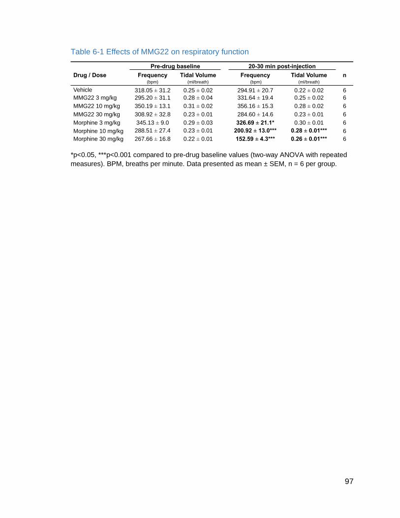
97
Table 6-1 Effects of MMG22 on respiratory function
*p<0.05, ***p<0.001 compared to pre-drug baseline values (two-way ANOVA with repeated
measures). BPM, breaths per minute. Data presented as mean ± SEM, n = 6 per group.
Pre-drug baseline 20-30 min post-injection
Drug / Dose Frequency Tidal Volume Frequency Tidal Volume n(bpm) (ml/breath) (bpm) (ml/breath)
Vehicle 318.05 ± 31.2 0.25 ± 0.02 294.91 ± 20.7 0.22 ± 0.02 6
MMG22 3 mg/kg 295.20 ± 31.1 0.28 ± 0.04 331.64 ± 19.4 0.25 ± 0.02 6
MMG22 10 mg/kg 350.19 ± 13.1 0.31 ± 0.02 356.16 ± 15.3 0.28 ± 0.02 6
MMG22 30 mg/kg 308.92 ± 32.8 0.23 ± 0.01 284.60 ± 14.6 0.23 ± 0.01 6
Morphine 3 mg/kg 345.13 ± 9.0 0.29 ± 0.03 326.69 ± 21.1* 0.30 ± 0.01 6
Morphine 10 mg/kg 288.51 ± 27.4 0.23 ± 0.01 200.92 ± 13.0*** 0.28 ± 0.01*** 6
Morphine 30 mg/kg 267.66 ± 16.8 0.22 ± 0.01 152.59 ± 4.3*** 0.26 ± 0.01*** 6
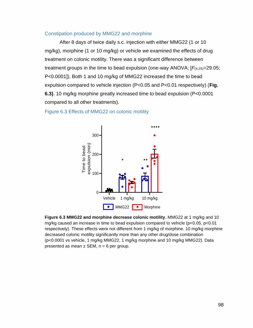
98
Constipation produced by MMG22 and morphine
After 8 days of twice daily s.c. injection with either MMG22 (1 or 10
mg/kg), morphine (1 or 10 mg/kg) or vehicle we examined the effects of drug
treatment on colonic motility. There was a significant difference between
treatment groups in the time to bead expulsion (one-way ANOVA; [F(4,25)=29.05;
P<0.0001]). Both 1 and 10 mg/kg of MMG22 increased the time to bead
expulsion compared to vehicle injection (P<0.05 and P<0.01 respectively) (Fig.
6.3). 10 mg/kg morphine greatly increased time to bead expulsion (P<0.0001
compared to all other treatments).
Figure 6.3 Effects of MMG22 on colonic motility
Figure 6.3 MMG22 and morphine decrease colonic motility. MMG22 at 1 mg/kg and 10
mg/kg caused an increase in time to bead expulsion compared to vehicle (p<0.05, p<0.01
respectively). These effects were not different from 1 mg/kg of morphine. 10 mg/kg morphine
decreased colonic motility significantly more than any other drug/dose combination
(p<0.0001 vs vehicle, 1 mg/kg MMG22, 1 mg/kg morphine and 10 mg/kg MMG22). Data
presented as mean ± SEM, n = 6 per group.
Vehicle 1 mg/kg 10 mg/kg
0
100
200
300
Tim
e t
o b
ea
d
exp
uls
ion
(m
in)
MMG22 Morphine
✱ ✱✱
✱✱✱✱

99
Discussion
Systemic administration of MMG22 produced robust analgesia without the
centrally mediated side effects of traditional opioids, including hyper-locomotion
[51,629] and respiratory depression [79,631]. Earlier studies demonstrated that
MMG22 had no effect on locomotor coordination, and mice treated with MMG22
did not display naloxone precipitated withdrawal symptoms frequently seen with
centrally acting opioids [735]. MMG22 also did not produce anxiolytic behavior
seen with the mGluR5 antagonist MPEP. That MMG22 does not affect
hippocampal dependent spatial learning can be inferred from its ability to induce
aCPP in nerve injured mice (chapter 5). However, more studies need to be done
regarding MMG22 and spatial learning as MPEP was equally able to induce
aCPP early after nerve injury.
Like traditional opioids, MMG22 caused constipation, a peripherally
mediated side effect [874]. That MMG22 induced constipation, but none of the
centrally mediated side effects associated with monovalent MOR agonists or
mGluR5 antagonists suggests that MMG22 may not pass the blood brain barrier
when given systemically, and hence may be working peripherally to decrease
mechanical hyperalgesia. A similar efficacy vs. side effect profile was also seen
for the combination of the peripherally restricted MOR agonist loperamide and
the opioid agonist oxymorphindole [90], also supporting the possibility that
MMG22 targets peripheral over central receptors. A simple reason for s.c.
administration of MMG22 targeting peripheral rather than spinal or supraspinal
receptors is its relatively high molecular weight (852 Daltons [9]) and a high
hydrogen bond capacity (>20). Lipid-mediated diffusion across the BBB is limited
by size (<400daltons) and low hydrogen bond capacity (<7) [624]. Recent studies
done by Henry Wong at the University of Minnesota have demonstrated that
levels of MMG22 in the brain and spinal cord after systemic delivery are around
8% of plasma levels, supporting the behavioral evidence that MMG22 is
minimally centrally available (unpublished data). For comparison, levels of
morphine in the brain after systemic administration are around 50% of plasma
levels [893].

100
Another potential mechanism by which MMG22 might confer the anti-
hyperalgesic effects of traditional opioids while lacking the less desirable off-
target effects could involve heteromer based biased agonism [247]. It is known
that heteromer formation can alter the signaling pathways engaged by particular
ligands [299]. It is believed that the analgesic effects of MOR agonists are due to
the engagement of trimeric G protein signaling pathway, while the development
of constipation, and respiratory depression may primarily be mediated by
recruitment of the β-arrestin dependent pathways [166,367]. This is supported by
the decrease in opioid mediated respiratory depression and constipation in β-
arrestin KO mice [672]. The high potency of MMG22 suggests that even at 8% or
plasma levels, the amount of MMG22 getting to the CNS may be enough to
produce analgesia. In which case, the lack of centrally mediated side effects may
instead be due to biased agonism. Cell signaling assays will be required to
explore this possibility.
Conclusion
The use of opioids is plagued with off-target effects including respiratory
depression, constipation, nausea and vomiting, sedation (in patients), and
hyperlocomotion (in rodents). mGluR5 antagonists have also been shown to
reduce anxiety like behavior in rodents. The side effects from systemic MMG22
have not been fully characterized. Here we have shown that MMG22 does not
induce respiratory depression or hyperlocomotion in rodents (both centrally
mediated side effects of opioids). MMG22 does induce a degree of constipation
in rodents, although less than morphine at the same concentration (a largely
peripherally mediated side effect). MMG22 also does not produce anxiolysis (a
central action of mGluR5 antagonists. The absence of centrally mediated side
effects combined with the presence of a peripherally mediated side effect,
suggests a predominantly peripheral mechanism of action for systemically
administered MMG22.

101
Chapter 7
Effects of MMG22 on the response properties of primary
afferents nociceptors

102
Introduction: Electrophysiology of primary afferent nociceptors
Response properties of A fiber nociceptors
Aδ fibers are thinly myelinated and conduct action potentials at a speed
ranging from 1.4 – 35 m/s in man [61] and from 1.4 – 13.6 in mice [97]. Aδ fibers
can respond to either high threshold or low threshold mechanical stimulation
(HTM and LTM respectively). In the mouse, about 35% of cutaneous Aδ fibers
are LTM and 65% are HTM [97,394]. In human the numbers are similar with 47%
being LTM, and 53% being HTM [6]. Aδ fibers terminate as free nerve endings in
glabrous skin with receptive field sizes ranging from pinpoint to 255 mm2 in the
primate [256] and pinpoint in mice [97].
There are two main types of nociceptive Aδ mechanoheat receptors
(AMH) which respond to both mechanical and heat stimuli applied to their
receptive fields [349,515,687,817]. Type I AMH receptors exhibit an delayed but
sustained response to noxious heat, where type II AMH receptors respond to
noxious heat with an initial burst of activity that is noticed in hairy skin as a fast
“pricking” sensation [817]. In addition to heat and mechanical stimuli, type I and II
AMHs also sometimes respond to cold stimuli [97,256] and are considered
polymodal (see Table 7-1).
Heat sensitivity in type I AMHs is probably mediated by TRPV2 since this
channel has a similar high heat activation threshold of ~52°C and 80% of cells
expressing this channel also stain positive for neurofilament, a marker of
myelinated neurons [106]. Type I AMH fibers also sensitize when repeatedly
activated by a heat stimulus and after burn injury [515,814].
Type II AMH have lower thermal thresholds, similar to the activation
threshold of TRPV1 channels and may contribute to pain caused by cutaneous
application of capsaicin to the skin [687]. Lack of type II receptors in glabrous
skin correlates with the lack of first heat pain sensation in the human hand
[98,814]. Other populations of Aδ nociceptors include fibers only responsive to
mechanical stimulation (Aδ-M) and those responsive to mechanical and cold
stimuli (Aδ-MC). Most Aδ fibers also respond to extreme cold [740].

103
The average size of an Aβ HTMR receptive field is ~25 mm2 in human
skin [554]. These fibers make up about 12% of A fibers and may be involved in
the perception of noxious stimulation [554].
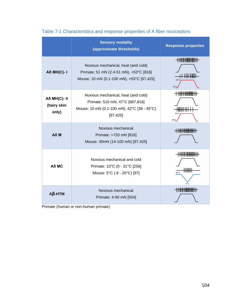
104
Table 7-1 Characteristics and response properties of A fiber nociceptors
Sensory modality
(approximate thresholds) Response properties
Aδ MH(C)- I
Noxious mechanical, heat (and cold)
Primate: 51 mN (2.4-51 mN), >53°C [816]
Mouse: 10 mN (0.1-100 mN), >53°C [97,425]
Aδ MH(C)- II
(hairy skin
only)
Noxious mechanical, heat (and cold)
Primate: 510 mN, 47°C [687,816]
Mouse: 10 mN (0.1-100 mN), 42°C (39 - 45°C)
[97,425]
Aδ M
Noxious mechanical
Primate: >720 mN [816]
Mouse: 30mN (14-100 mN) [97,425]
Aδ MC
Noxious mechanical and cold
Primate: 10°C (0 - 31°C [256]
Mouse: 5°C (-8 - 20°C) [97]
Aβ-HTM Noxious mechanical
Primate: 4-60 mN [554]
Primate (human or non-human primate)
30°C
53°C
0°C
30°C
30°C
53°C
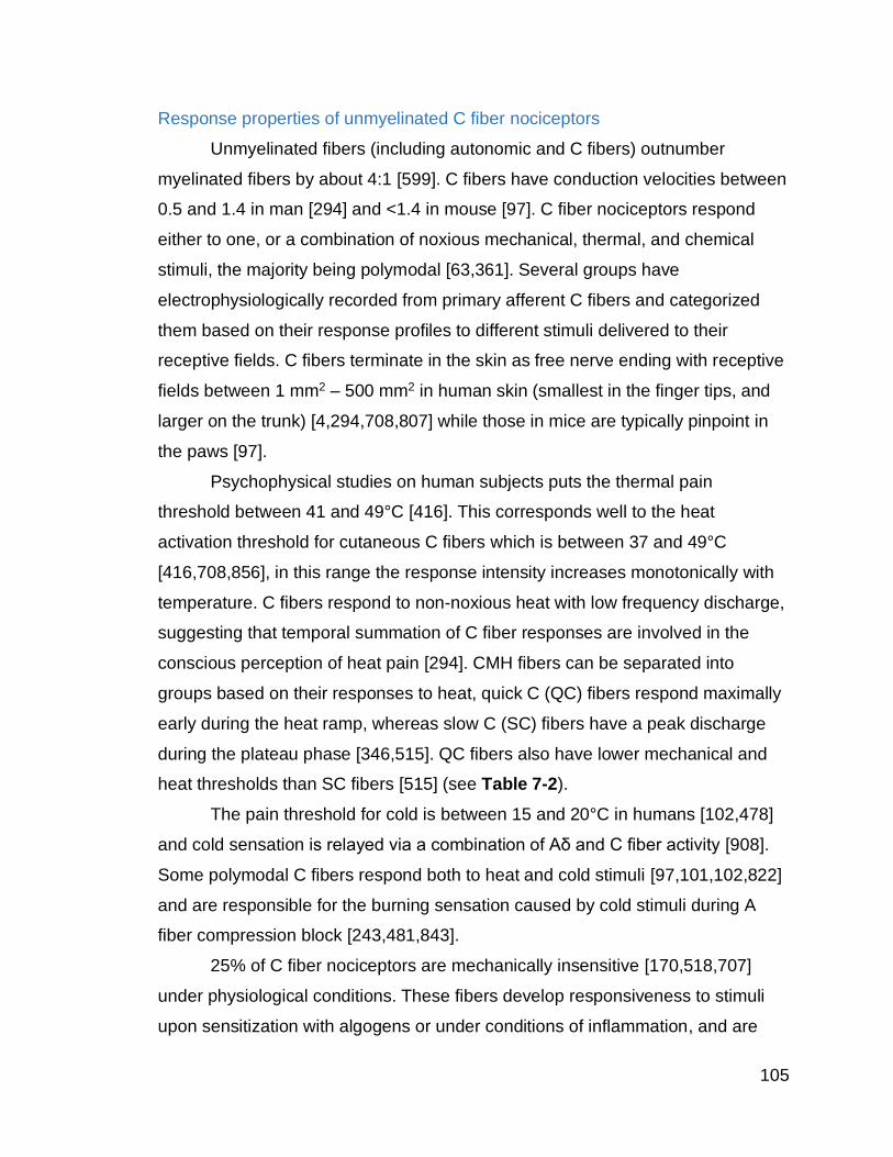
105
Response properties of unmyelinated C fiber nociceptors
Unmyelinated fibers (including autonomic and C fibers) outnumber
myelinated fibers by about 4:1 [599]. C fibers have conduction velocities between
0.5 and 1.4 in man [294] and <1.4 in mouse [97]. C fiber nociceptors respond
either to one, or a combination of noxious mechanical, thermal, and chemical
stimuli, the majority being polymodal [63,361]. Several groups have
electrophysiologically recorded from primary afferent C fibers and categorized
them based on their response profiles to different stimuli delivered to their
receptive fields. C fibers terminate in the skin as free nerve ending with receptive
fields between 1 mm2 – 500 mm2 in human skin (smallest in the finger tips, and
larger on the trunk) [4,294,708,807] while those in mice are typically pinpoint in
the paws [97].
Psychophysical studies on human subjects puts the thermal pain
threshold between 41 and 49°C [416]. This corresponds well to the heat
activation threshold for cutaneous C fibers which is between 37 and 49°C
[416,708,856], in this range the response intensity increases monotonically with
temperature. C fibers respond to non-noxious heat with low frequency discharge,
suggesting that temporal summation of C fiber responses are involved in the
conscious perception of heat pain [294]. CMH fibers can be separated into
groups based on their responses to heat, quick C (QC) fibers respond maximally
early during the heat ramp, whereas slow C (SC) fibers have a peak discharge
during the plateau phase [346,515]. QC fibers also have lower mechanical and
heat thresholds than SC fibers [515] (see Table 7-2).
The pain threshold for cold is between 15 and 20°C in humans [102,478]
and cold sensation is relayed via a combination of Aδ and C fiber activity [908].
Some polymodal C fibers respond both to heat and cold stimuli [97,101,102,822]
and are responsible for the burning sensation caused by cold stimuli during A
fiber compression block [243,481,843].
25% of C fiber nociceptors are mechanically insensitive [170,518,707]
under physiological conditions. These fibers develop responsiveness to stimuli
upon sensitization with algogens or under conditions of inflammation, and are

106
believed to be involved in hyperalgesia [150,399,518,707]. In general, fibers for a
given stimulus, the discharge rates are lower for C-fibers than for A-δ [6]. The
discharge rate of C-fibers is also subject to saturation and fatigue with high
intensity and long-lasting stimulation [807].

107
Table 7-2 Characteristics and response properties of C fiber nociceptors
Sensory modality
(approximate threshold) Response properties
CMHC
(polymodal)
Noxious mechanical, heat and cold
Primate: 30 mN (3-750 mN) [61,209,294]
Mouse: 20 mN (1-175 mN) [97,209,425]
CMH/CMiH
Noxious heat (± mechanical)
Primate: 41°C (37-49°C) [708,855]
Mouse: 42°C (37-49)°C [97,424]
CMC/CC
Noxious Cold (± mechanical)
Primate: 19°C (10-26°C) [415]
Mouse: 10°C (-12-16°C) [97]
CM
Noxious mechanical only
Primate: 30 mN [209]
Mouse: 20 mN (1-175 mN) [97,209,425]
C-MiHi “Silent” [856]
Very high mechanical threshold, chemical
Primate (human or non-human primate)
0°C
30°C
30°C
53°C
30°C
53°C
Quick C
Slow C

108
Effects of MOR agonists on primary afferent nociceptors
MOR expression is observed in approximately 20-30% of primary afferent
neurons [133,343,854]. Peripherally, MOR agonists decrease the excitability of
primary afferents in response to noxious stimuli [18,400,688,710,796,858].
Opioids have been shown to inhibit the calcium dependent release of
proinflammatory compounds from peripheral nerve endings [909,910] and
decrease TRPV1 currents in primary afferent neurons [217] (for review see
[768]).
Effects of mGluR5 agonists and antagonists on pain signaling
The effects of mGluR5 agonists/antagonists on primary afferent responses
have not been studied directly, however it has been shown that intraplantar
administration of mGluR5 agonists increases the firing of WDR neurons in the
dorsal horn [844] suggesting that activation of mGluR5 excites the peripheral
terminals of primary afferents. When applied to the cord, mGluR5 agonists mimic
the response pattern of high frequency stimulation induced LTP at C fiber
synapses [444]. Contrastingly, mGluR5 antagonists like MPEP prevent the
induction of LTP at C fiber synapses [444].
DHPG (a group I mglur agonist) increases the frequency of miniature
excitatory post synaptic currents (EPSCs) and reduces the paired pulse ratio of
evoked EPSCs in lamina II neurons in rats, suggesting enhanced excitability of
primary afferents. These effects were blocked by MPEP, demonstrating the
involvement of mGluR5 [891]. MPEP also normalizes the frequency of mEPSCs,
and the amplitude of monosynaptically evoked EPSCs in the dorsal horn of rats
with paclitaxel-induced neuropathy [891], and diabetic neuropathy [439]. These
results suggest that glutamatergic input from primary afferents is modulated by
mGluR5 signaling in these conditions.
Effects of nerve injury on the electrophysiological properties of primary afferent
nociceptors
Nerve injury causes sensitization of neighboring primary afferent
nociceptors. Previous studies of nerve injury induced neuropathic pain have

109
shown that uninjured fibers develop spontaneous activity, have reduced
thresholds, and increased firing to suprathreshold stimuli [80,117,199,731,887].
If MMG22 works peripherally to reduce hyperalgesia, it should decrease
the responsiveness of primary afferent fibers. To test this, we decided to record
from sural nerve axons in nerve injured animals before and after systemic
administration of MMG22.

110
Methods
Animals
Adult (5-8 months) male and female C57/B6 mice (Charles River) were
housed 4 (males) or 5 (females) to a cage and maintained on a 12-hour light/dark
cycle with ad libitum access to food and water, except as otherwise noted for
constipation studies. All procedures were carried out during the light cycle. All
procedures were approved by the Institutional Animal Care and Use Committee
of the University of Minnesota.
Spared nerve injury
Mice were anesthetized with 2.5 % isoflurane. Spared nerve injury (SNI) to
the sciatic nerve was performed as described previously [82,172,644]. Briefly,
after exposing the three branches of the sciatic nerve, the tibial and common
peroneal branches were tightly ligated with 5.0 silk suture and cut 2 mm distal to
the suture. Care was taken not to disturb the sural nerve. Sham surgeries
followed the same procedure without manipulation of the sciatic nerve or distal
branches.
Drugs
The bivalent ligand MMG22 was synthesized as described previously [9].
MMG22, oxymorphamine, and 2-methyl-6-(phenylethynyl)pyridine (MPEP),
(Mallinckrodt Inc, Hazelwood, MO) were diluted in 1% DMSO (vehicle). All drugs
were administered subcutaneously in a volume of 250 µl.
Teased fiber electrophysiology
The surgical procedure to isolate and record for teased nerve fibers of the
sural nerve in vivo has been described in rat [117]. 7-15 days after SNI, mice
were anesthetized using inhaled isoflurane (2.5% induction, 1-2% maintenance)
and placed on a feedback controlled heated stage to maintain core temperature
at 37°C. The level of anesthesia was monitored by toe pinch on the contralateral
side. A cutaneous incision was made at the level of the mid thigh, and the
quadriceps femoris and biceps femoris were separated using blunt dissection to
expose the previously ligated tibial and common peroneal nerves as well as the
spared sural nerve. Part of the biceps femoris muscle was carefully removed

111
down to the where the sural nerve enters the gastrocnemius muscle. The skin
around the incision was loosened from the muscle below by blunt dissection and
sutured to a ring platform to create a small basin. To prevent leakage of oil from
the recording basin, and fix the hind limb in position, a rubber-based polysulfide
impression material (COE-FLEX, GC America) was applied around the ring to the
skin of the hind limb and allowed to set. The sural nerve was then dissected from
connective tissue, delicately separated from its vascular bundle, and placed on a
small, mirrored platform for separation of nerve fibers. The epineurium of the
sural nerve was opened using a miniature scalpel, and small fascicles were cut
to allow the proximal ends to be spread out on the platform for separation with
fine jewelers forceps. Nerve fascicles were teased apart, and fine filaments were
placed on a silver-wire recording electrode maneuvered by a micromanipulator.
Extracellular recordings were obtained only from single fibers that could be easily
discriminated according to amplitude and shape. Action potentials from individual
fibers were amplified, audiomonitored, and visualized on an oscilloscope and
personal computer using Spike 2.0 software (CED, Cambridge, UK). Evoked
responses were analyzed off-line using a data analysis program (Spike 2). An
amplitude window discriminator was used to separate action potentials of the
fiber under study from those of other fibers and/or from background noise.
However, recordings usually consisted of one afferent fiber.
Identification of primary afferent fibers
The receptive fields (RFs) of cutaneous afferent fibers were identified
using mechanical stimuli. Mechanical stimulation proceeded by a graduated
approach beginning with large and soft stimulation with a cotton swab or the
experimenter’s fingers, followed by mild pinching with forceps. Once a fiber was
isolated, the location of its RF was identified using a suprathreshold von Frey
monofilament. The RF location was then marked on the skin with a felt-tip pen
and reconstructed on a drawing of the mouse hind paw.
Conduction velocity
The conduction velocity of each fiber was determined by electrically
stimulating the RF, before recording the conduction latency between the RF and

112
the recording electrode. Two fine needle electrodes (32 gauge acupuncture
needles) were inserted into the skin on opposite sides adjacent to the RF.
Square-wave pulses (duration, 0.2 ms, 0.5 Hz) were delivered at a stimulating
voltage 1.5 times the voltage required to evoke a threshold response. Fibers with
CV of 1.2 m/s or less were classified as C-fibers, whereas those with CV
between 1.2 and 13.6 m/s were classified as Aδ-fibers and those with CV >13.6
were classified as Aβ-fibers. C- and Aδ-fiber nociceptors were preferentially
studied.
Electrophysiological responses of nociceptors
For each nociceptor, the rate of spontaneous activity was determined for a
period of 2 min before any testing. Mechanical response threshold (mN) was
obtained using von Frey monofilaments. The RF was stimulated multiple times
with von Frey filaments of increasing force. Response threshold was defined as
the lowest force eliciting a response on at least ½ of the trials. Responses
evoked by suprathreshold mechanical stimuli were determined using a single
suprathreshold von Frey monofilament that delivered a force of 147 mN. This
monofilament was applied three times, each for 5 seconds with an inter-stimulus
interval of 60 seconds, and the evoked response was defined as the mean
number of impulses from the three trials.
A Peltier device (contact area 25 mm2) was used to deliver cold and heat
stimuli to the skin. The probe was maintained at a base temperature of 32°C, and
cold stimuli from 28°C to 0°C was applied for 10 seconds, with a 60 second
interstimulus interval. Heat stimuli from 34°C to 50°C was applied for 5 seconds
with at least 5 minutes between applications. Each fiber was tested before and at
30 and 60 minutes after subcutaneous delivery of vehicle or 10mg/kg MMG22.
Data analysis
Data are expressed as mean ± SEM, except where otherwise noted.
GraphPad Prism 8.0 (Graphpad software Inc. La Jolla, CA, USA) was used for
statistical analyses. Mean CVs, mean baseline mechanical thresholds, and the
average number of days post surgery were compared using independent t-tests,
The mean number of impulses evoked by the 147-mN von Frey stimulus before

113
and after drug treatment were compared using repeated-measures ANOVA. The
number of recordings from nociceptors that responded to thermal stimuli was not
large enough for statistical analysis. Post-hoc comparisons were done with
Bonferroni tests. A P value of <0.05 was considered significant. All evoked
responses were determined by subtracting spontaneous activity that occurred 5–
10 s before the onset of the stimulus.
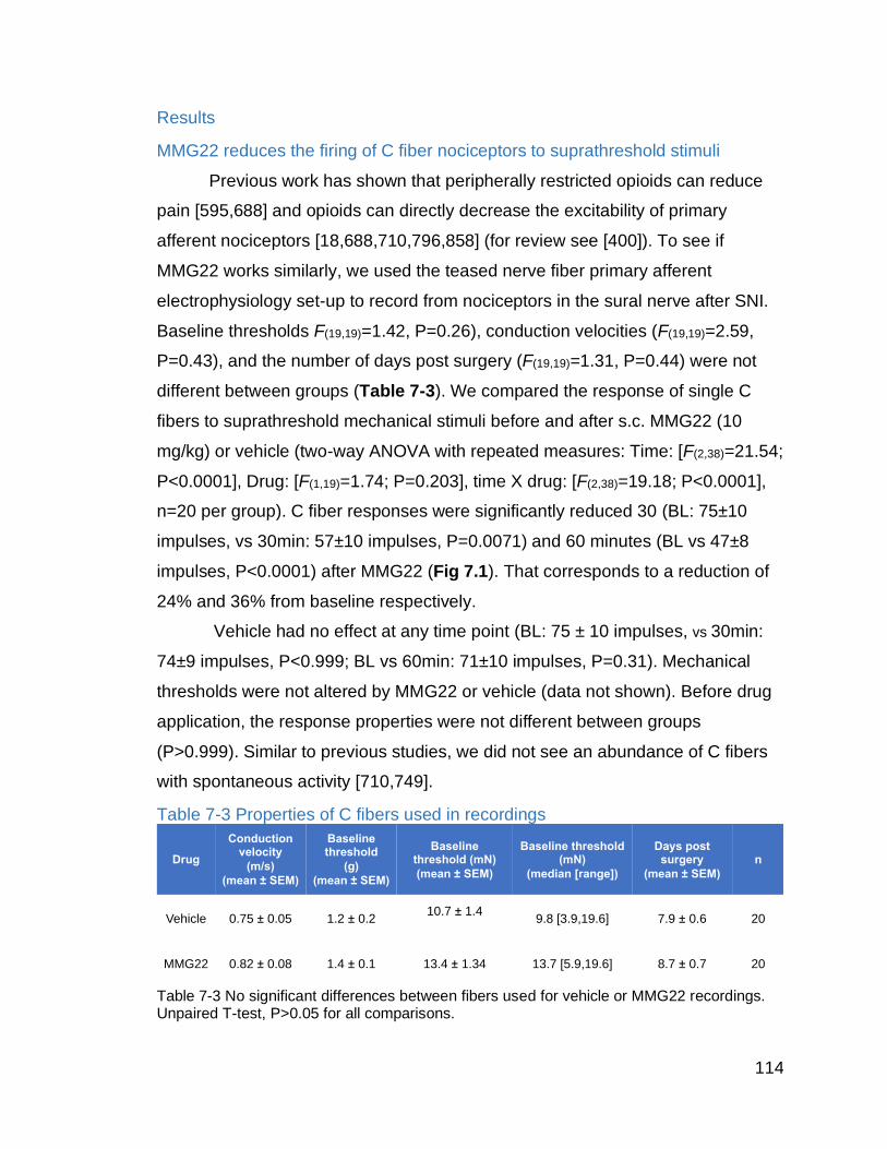
114
Results
MMG22 reduces the firing of C fiber nociceptors to suprathreshold stimuli
Previous work has shown that peripherally restricted opioids can reduce
pain [595,688] and opioids can directly decrease the excitability of primary
afferent nociceptors [18,688,710,796,858] (for review see [400]). To see if
MMG22 works similarly, we used the teased nerve fiber primary afferent
electrophysiology set-up to record from nociceptors in the sural nerve after SNI.
Baseline thresholds F(19,19)=1.42, P=0.26), conduction velocities (F(19,19)=2.59,
P=0.43), and the number of days post surgery (F(19,19)=1.31, P=0.44) were not
different between groups (Table 7-3). We compared the response of single C
fibers to suprathreshold mechanical stimuli before and after s.c. MMG22 (10
mg/kg) or vehicle (two-way ANOVA with repeated measures: Time: [F(2,38)=21.54;
P<0.0001], Drug: [F(1,19)=1.74; P=0.203], time X drug: [F(2,38)=19.18; P<0.0001],
n=20 per group). C fiber responses were significantly reduced 30 (BL: 75±10
impulses, vs 30min: 57±10 impulses, P=0.0071) and 60 minutes (BL vs 47±8
impulses, P<0.0001) after MMG22 (Fig 7.1). That corresponds to a reduction of
24% and 36% from baseline respectively.
Vehicle had no effect at any time point (BL: 75 ± 10 impulses, vs 30min:
74±9 impulses, P<0.999; BL vs 60min: 71±10 impulses, P=0.31). Mechanical
thresholds were not altered by MMG22 or vehicle (data not shown). Before drug
application, the response properties were not different between groups
(P>0.999). Similar to previous studies, we did not see an abundance of C fibers
with spontaneous activity [710,749].
Table 7-3 Properties of C fibers used in recordings
Table 7-3 No significant differences between fibers used for vehicle or MMG22 recordings. Unpaired T-test, P>0.05 for all comparisons.
Drug
Conductionvelocity
(m/s)
(mean ± SEM)
Baseline threshold
(g)
(mean ± SEM)
Baseline threshold (mN)
(mean ± SEM)
Baseline threshold (mN)
(median [range])
Days post surgery
(mean ± SEM)
n
Vehicle 0.75 ± 0.05 1.2 ± 0.210.7 ± 1.4
9.8 [3.9,19.6] 7.9 ± 0.6 20
MMG22 0.82 ± 0.08 1.4 ± 0.1 13.4 ± 1.34 13.7 [5.9,19.6] 8.7 ± 0.7 20

115
Figure 7.1 Effects of MMG22 on C fiber nociceptor responses
Figure 7.1 Effects of MMG22 on C fiber responses to suprathreshold mechanical stimuli.
(A) Primary afferent teased nerve fiber electrophysiology set up. (B) 30 and 60 minutes after
injection, MMG22 (10mg/kg s.c.) but not vehicle caused a decrease in the number of
impulses evoked from C fibers by a 15g (147mN) von Frey hair applied to their receptive
fields for 5 seconds. (C) Representative example of C fiber firing before and after application
of vehicle. Trace on left shows the latency to electrical stimulation, foot inset shows receptive
field. (D) Representative example of C fiber firing before and after application of MMG22
(10mg/kg) **P<0.01, ****P<0.0001, data presented as mean ± SEM, n = 20. CV, conduction
velocity. BL, baseline.
BL 30 60 BL 30 60
0
25
50
75
100
Minutes post injection
# o
f im
pu
lses
Vehicle
MMG22
ns
******
BA
L4 L5 L6 Sciatic n.
Sural n.
MMG22
5 Seconds
15 g von Frey
60 min
30 min
Baseline
Latency: 65 ms
Distance: 23 mm
CV: 0.35 m/s
D
5 Seconds
15 g von FreyVehicle
30 min
Baseline
60 minLatency: 35 ms
Distance: 12 mm
CV: 0.34 m/s
C

116
MMG22 reduces the firing of Aδ fiber nociceptors to suprathreshold stimuli
We repeated these experiments with Aδ fiber nociceptors. Conduction
velocities (F(9,9)=2.5, P=0.6), baseline mechanical thesholds (F(9,9)=1.9, P=0.4)
and number of days post surgery (F(9,9)=1.6, P=0.6) also did not differ between
groups (unpaired T-test) (Table 7-4). We tested the ability of MMG22 (10 mg/kg)
to decrease Aδ responses to suprathreshold mechanical stimuli compared to
vehicle (two-way ANOVA, Time: [F(2,18)=2.32; P=0.13], Drug: [F(1,9)=0.41; P=054],
time X drug: [F(2,18)=4.1; P<0.034], n=10 per group). MMG22 decreased the
suprathreshold responses of Aδ nociceptors at 30 (BL: 84.2±18.8 impulses vs 30
min: 66.5±15.5 impulses, P=0.016) and 60 minutes after treatment (BL vs 60
min: 66.8±18.2 impulses, P=0.018) (Figure 7.2). That corresponds to a 20%
reduction in firing at both time points.
Vehicle had no effect at any time point (BL: 83.5 ±17.3 impulses, vs 30
min: 86.2±16.9 impulses; BL vs 60 min: 87.7±18.6 impulses, P>0.99 for all).
Thresholds were not changed after vehicle of MMG22 (data not shown). Baseline
responses were not different between groups (P>0.99).
Table 7-4 Properties of Aδ fibers used in recordings
Table 7.4 No significant differences between the Aδ fibers used for vehicle or MMG22
recording. Unpaired T-test, P>0.05 for all comparisons.
Drug
Conductionvelocity
(m/s)
(mean ± SEM)
Baseline threshold
(g)
(mean ± SEM)
Baseline threshold (mN)
(mean ± SEM)
Baseline threshold (mN)
(median [range])
Days post surgery
(mean ± SEM)n
Vehicle 3.25 ± 0.77 2.0 ± 0.5 19.8 ± 5.5 19.6 [1.6,39.2] 7.3 ± 0.9 10
MMG22 4.02 ± 1.21 1.5 ± 0.4 16.8 ± 4.5 19.6 [3.9,39.2] 8.1 ± 1.1 10

117
Figure 7.2 Effects of MMG22 on Aδ nociceptor responses
Figure 7.2. Effects of MMG22 on Aδ fiber responses to suprathreshold mechanical
stimuli. (A) Primary afferent teased nerve fiber electrophysiology set up. (B) 30 and 60
minutes after injection, MMG22 (10mg/kg s.c.) but not vehicle caused a decrease in the
number of impulses evoked from Aδ fibers by a 15g (147nM) von Frey hair applied to the
receptive field for 5 seconds. (C) Representative example of an Aδ fiber firing before and
after application of vehicle. Trace on left shows the latency to electrical stimulation, foot inset
shows location of the receptive field. (D) Representative example of Aδ fiber firing before
and after application of MMG22 (10mg/kg). *P<0.05, data presented as mean ± SEM, n = 10.
CV, conduction velocity. BL, baseline.
5 Seconds
15 g von FreyVehicle
30 min
Baseline
60 min
Latency: 13 ms
Distance: 20 mm
CV: 1.54 m/s
MMG22
5 Seconds
15 g von Frey
60 min
30 min
Baseline
Latency: 12 ms
Distance: 22 mm
CV: 1.83 m/s
BL 30 60 BL 30 60
0
25
50
75
100
125
Minutes post injection
# o
f im
pu
lses Vehicle
MMG22
* *
ns
L4 L5 L6 Sciatic n.
Sural n.
A B
C D

118
MMG22 may also reduce the firing of nociceptors to noxious heat and cold
stimuli
Nerve injury has also been shown to sensitize nociceptors to thermal
stimuli [731], and opioids can also reduce the responses of sensitized
nociceptors to noxious heat and cold stimuli [858]. In an attempt to determine if
MMG22 would do the same we also tested the heat and cold evoked responses
from several isolated fibers. Not all fibers responded reliably to heat or cold
stimuli but we were able to record from a few thermally responsive fibers
(Figures 7.3 and 7.4). Vehicle did not decrease the response of a C fiber evoked
by a 5 second heat ramp from 32°C to 50°C, but MMG22 (10 mg/kg) reduced the
firing of one C fiber by 30% and one Aδ fiber by 65% to the same heat stimulus
(Figure 7.3).
We also tested a few cold responsive C fibers (Figure 7.4). Vehicle did
not decrease the number of action potentials evoked by a 10 second cold ramp
from 32°C to 0°C, while MMG22 (10 mg/kg) reduced the firing of one C fiber 70%
compared to baseline (Figure 7.4). As we were only able to reliably record from
1 fiber in each condition no statistical evaluation was possible.
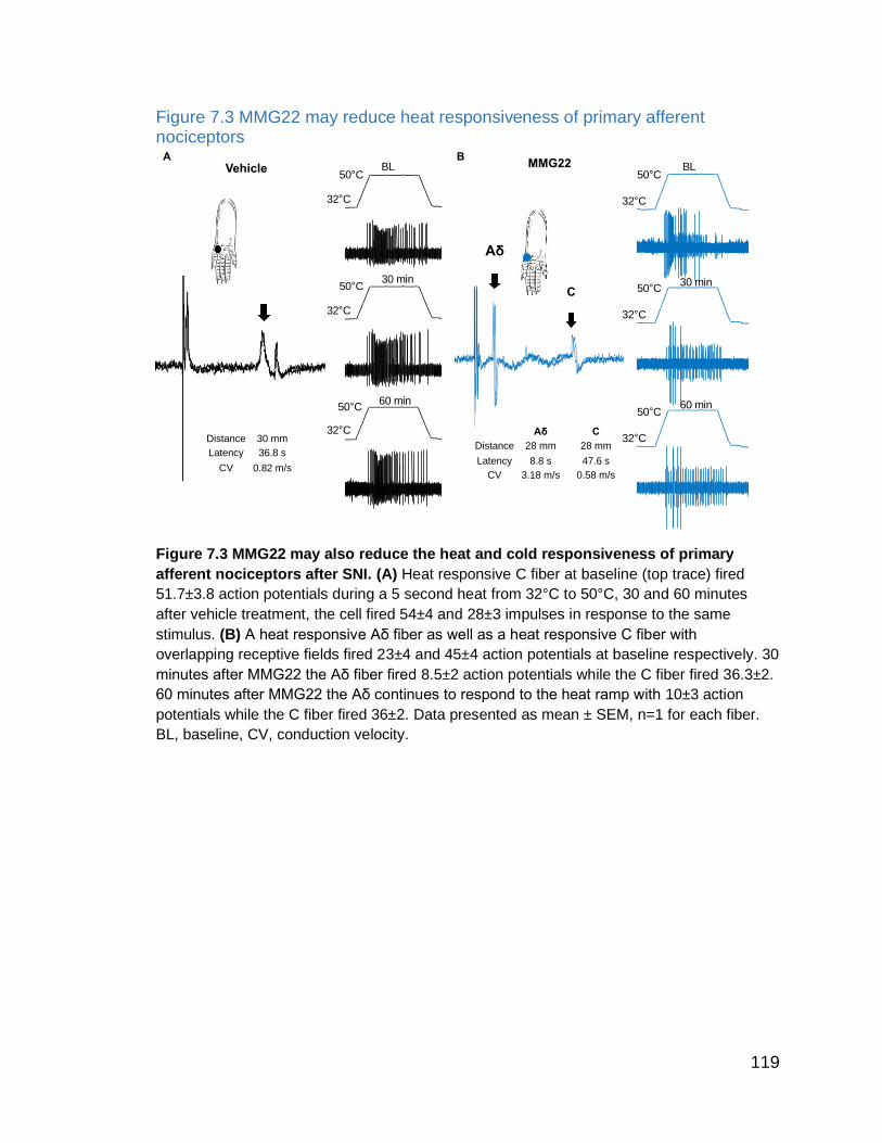
119
Figure 7.3 MMG22 may reduce heat responsiveness of primary afferent nociceptors
Figure 7.3 MMG22 may also reduce the heat and cold responsiveness of primary
afferent nociceptors after SNI. (A) Heat responsive C fiber at baseline (top trace) fired
51.7±3.8 action potentials during a 5 second heat from 32°C to 50°C, 30 and 60 minutes
after vehicle treatment, the cell fired 54±4 and 28±3 impulses in response to the same
stimulus. (B) A heat responsive Aδ fiber as well as a heat responsive C fiber with
overlapping receptive fields fired 23±4 and 45±4 action potentials at baseline respectively. 30
minutes after MMG22 the Aδ fiber fired 8.5±2 action potentials while the C fiber fired 36.3±2.
60 minutes after MMG22 the Aδ continues to respond to the heat ramp with 10±3 action
potentials while the C fiber fired 36±2. Data presented as mean ± SEM, n=1 for each fiber.
BL, baseline, CV, conduction velocity.
32°C
50°C
32°C
50°C
32°C
50°C
32°C
50°C
32°C
50°C
32°C
50°C
Aδ
C
Aδ C
Distance 28 mm 28 mm
Latency 8.8 s 47.6 s
CV 3.18 m/s 0.58 m/s
Distance 30 mm
Latency 36.8 s
CV 0.82 m/s
A BVehicle MMG22BL BL
30 min 30 min
60 min 60 min

120
Figure 7.4 MMG22 may reduce cold responsiveness of primary afferent nociceptor
Figure 7.4 MMG22 reduced the cold responsiveness of a C fiber nociceptor. At baseline
a cold sensitive C fiber fired 14.5±2 action potentials in response to a 10 second cold ramp.
30 and 60 minutes after vehicle this fiber responded with 17±1 and 16±1 action potentials
respectively. (B) A cold sensitive C fiber responded to the cold ramp with 14±1 action
potentials at baseline. 30 and 60 minutes after MMG22 this C fiber responded to the same
heat from with 7±2 and 3±1 action potentials respectively. Data presented as mean ± SEM,
n=1 for each fiber. BL, baseline, CV, conduction velocity.
0°C
32°C
0°C
32°C
0°C
32°C
a
0°C
32°C
0°C
32°C
0°C
32°C
Distance 20 mm
Latency 27 s
CV 1.35 m/s
Distance 32 mm
Latency 29 s
CV 1.1 m/s
A BVehicle MMG22
BLBL
30 min30 min
60 min
60 min

121
Discussion
Similar to the results obtained with other nerve injury models
[122,197,731], uninjured primary afferents show enhanced responsiveness to
suprathreshold stimuli after SNI [749]. In a study done by Smith et al., C and Aδ
and fibers in SNI animals fired an average at of 28% and 22% more action
potentials per second to mechanical stimuli compared sham operated animals
[749]. In the experiments just reviewed, MMG22 decreased suprathreshold firing
of C and Aδ fibers by 30% and 20% respectively, very similar to the increase in
firing shown by Smith et al. Previous research has shown that MMG22 is
incredibly less potent in naïve animals [9,748], and in our studies MMG22 did not
alter baseline thresholds in naïve mice (unpublished observations). The ability of
MMG22 to decrease the suprathreshold mechanical responses of C and Aδ
nociceptors after injury to the same extend that they were seen previously to be
elevated confirms MMG22 possess anti-hyperalgesic activity without causing
frank analgesia. Preliminary studies suggest that MMG22 may decrease the heat
and cold responses of C and Aδ nociceptors but more studies are needed to
confirm this finding.
As mentioned earlier, the behavioral methods employed by this study
were not suited to study analgesia beyond anti-hyperalgesia (the dynamic range
of our assay was maximally sensitive to detect a return to baseline, and unlikely
to detect any further reduction in responsivity). More studies looking at the effects
of MMG22 on acute nociceptive responses (both behaviorally and via
electrophysiology) would be worth pursuing. Mechanistically, the low basal
expression of mGluR5 in the spinal cord and DRG [206,308,410,681] compared
to expression levels in animal with chronic pain, may be involved.
Similar to previous studies, we did not observe a large amount of
spontaneous firing in uninjured primary afferent nociceptors after SNI [117,749].
In contrast, most studies using other models of nerve injury suggest that injured
and uninjured afferents do develop spontaneous activity and uninjured afferents
show reduced thresholds compared to control animals [122,197,516,731,887].
Spontaneous activity in nociceptors is thought to cause spontaneous pain

122
sensations and microneurography studies have shown that nociceptors from
patients with neuropathic pain have an increased amount of ectopic activity [382].
Most models of nerve injury induced pain have shown that injured fibers develop
ectopic activity [179,282,455,459,778]. So, although the uninjured axons of the
spared sural nerve do not develop spontaneous activity after SNI [117,749], it is
probable that the injured fibers in the tibial and common peroneal nerves do.
Studies have shown that ectopic activity in injured fibers is highest early after
nerve injury and decreases steadily over time [179,282,455,459,778]. The
development of spontaneous activity after nerve injury has been ascribed to the
inflammatory milieu produced by Wallerian degeneration [888]. Like spontaneous
activity, the inflammatory response after nerve injury is transient and resolves
rapidly after 2-3 weeks [17,523,812]. Early after nerve injury (when both
inflammation and spontaneous activity are high), MMG22 was able to reduce
both evoked and spontaneous pain. The decreased potency of MMG22 4 weeks
after nerve injury parallels the decrease in the inflammatory response
[254,255,430](for review see [241]).
Lack of spontaneous firing in uninjured fibers after SNI has been ascribed
to the minimal co-mingling of injured and non-injured fibers compared to other
nerve injury models [117,749]. The portion of the injured tibial and common
peroneal nerves that undergo Wallerian degeneration in the SNI model are distal
to the trifurcation where the sural nerve exits the sciatic. This means that a large
portion of the inflammatory response to axon degeneration is physically
separated from the spared axons in the sural nerve. However, other studies have
found that spontaneous activity originates in the DRG [354], where there is
considerable comingling of injured and uninjured fibers in the SNI model [408]. In
our studies, the portion of the axon connecting the peripheral terminals to the
DRG was acutely cut prior to recording. Thus, any spontaneous activity coming
from the proximal axon or DRG would not have been detected.

123
Conclusion
Systemic delivery of MMG22 reduces evoked responses of C and Aδ
nociceptors to suprathreshold mechanical stimuli after nerve injury. The degree
to which MMG22 reduced these responses was similar to increased responsivity
of C and Aδ fibers in SNI animals found in a previous study [749]. The ability of
MMG22 to reduce the excitability of primary afferent nociceptors in the periphery,
supports the peripheral action of systemically delivered MMG22. MMG22 given
intrathecally may also decrease the responsiveness of dorsal horn neurons.
Although the central availability of systemically delivered MMG22 has been
shown to be quite low (~8%), the incredibly potency of MMG22 may enable it to
accumulate in sufficient amounts to have an effect on dorsal horn neurons as
well. Studies looking at this possibility are currently underway.
124
General discussion: summary and conclusions
Chronic pain is a prevalent and disabling condition which has far reaching
consequences for those who suffer with it [212,276]. Current pharmacological
therapies offer suboptimal pain relief [497] and neuropathic pain is particularly
difficult to manage [231]. Opioids are the most potent and effective analgesics
available today; however, neuropathic pain is considered less sensitive to opioid
management [22,53,123,403,500]. In addition, many chronic pain patients will
discontinue opioid use due to inadequate pain control or an inability to tolerate
the side effects [583]. The decreased efficacy in reducing neuropathic pain
combined with the need for long term management for these patients increases
the risk of addiction to opioids as higher doses are needed to manage
neuropathic pain and long term use is associated with the development of
analgesic tolerance, which also leads to dose escalation [33,140].
Pre-clinical research has shown that mGluR5 antagonists enhance the
analgesic effects of opioids [609,644,928], reduce the development of opioid
induced analgesic tolerance [246,398,567,750,897,898,928], and decrease
opioid reward seeking [89,359,658,691,831]. In vitro studies have shown mGluR5
antagonists reduce opioid induced MOR desensitization, phosphorylation and
internalization, and suggest that the receptors may physically associate in the
form of a heteromer [714].
A novel bivalent ligand, MMG22, was synthesized in an attempt to take
advantage of the unique pharmacological and signaling interactions
demonstrated between MOR and mGluR5 [9]. Previous studies have shown that
MMG22 is orders of magnitude more potent at reducing hyperalgesia in mice
with inflammatory pain and bone cancer pain than compounds with shorter or
longer linker lengths [9,748] or a mixture of the monovalents [9]. The importance
of linker length for anti-hyperalgesic potency supports the idea that MMG22 binds
to a MOR-mGluR5 heteromer; however, another study using the same nerve
injury model employed here was unable to replicate this [644]. The studies

125
described here were aimed at characterizing both the potency and potential side
effects of systemically administered MMG22.
We have shown that MMG22 reduces pain (evoked and spontaneous)
early after nerve injury. The transient inflammatory response after nerve injury
may be responsible for loss of potency late after nerve injury. In support of this,
previous studies have shown that the potency of MMG22 increases over time
after tumor implantation [9,735] in parallel with the development of inflammation
[269,458,488,905]. A recent study in the Portoghese lab, using chemotherapy
induced peripheral neuropathy has shown that the potency of MMG22 does not
change over time (manuscript in preparation), again reflecting the long lasting
inflammatory response in this condition [657,754,918]. So, while MMG22 may not
be the best therapeutic option for the treatment of traumatic neuropathies, it may
provide significant pain relief to patients with pain conditions that promote long
lasting inflammation.
Using RNAScope, we were able to show that early after nerve injury, the
target receptors for MMG22 were co-expressed in dorsal horn neurons as well as
primary afferent DRG neurons. That the receptors are found together supports
their ability to form heteromers in vivo. Further research looking at the temporal
dynamics of inflammation and mGluR5 expression in various pain conditions in
relation to the potency of MMG22 would further support the importance of
inflammation for MMG22 mediated analgesia.
We have also shown that MMG22 does not induce place preference in
naïve mice, suggesting a reduced risk for abuse and addiction compared to
traditional opioids. MMG22 also lacks the respiratory depressant effects of
traditional opioids, reducing the risk of overdose death. Other centrally mediated
side effects of either monovalent (mGluR5 antagonist or MOR agonist) including
hyperlocomotion and anxiolysis were also not exhibited by MMG22 treated
animals. Peripherally, MMG22 did cause constipation, although to a lesser extent
than morphine. That MMG22 was able to slow colonic transit supports that
systemically delivered MMG22 is pharmacologically active.

126
Finally, we show that systemic MMG22 decrease the responses of primary
afferent nociceptors to suprathreshold stimuli. That MMG22 can act directly on
nociceptors, proves that at least part of the anti-hyperalgeisc effects of MMG22
are peripherally mediated.
MMG22 potently reduces hyperalgesia with none of the centrally mediated
effects of monovalent MOR agonists or mGLuR5 antagonists. A potent, non-
addictive pain medication that does not promote the development of analgesic
tolerance has the potential to be a game changer for pain management. Future
clinical trials will show if MMG22 can fill that role.

127
Bibliography
[1] Abrahamsen B, Zhao J, Asante CO, Cendan CM, Marsh S, Martinez-Barbera JP, Nassar MA, Dickenson AH, Wood JN. The cell and molecular basis of mechanical, cold, and inflammatory pain. Science (80- ) 2008;321:702–705.
[2] Abraira VE, Ginty DD. The sensory neurons of touch. Neuron 2013;79:618–639.
[3] Abraira VE, Kuehn ED, Chirila AM, Springel MW, Toliver AA, Zimmerman AL, Orefice LL, Boyle KA, Bai L, Song BJ, Bashista KA, O’Neill TG, Zhuo J, Tsan C, Hoynoski J, Rutlin M, Kus L, Niederkofler V, Watanabe M, Dymecki SM, Nelson SB, Heintz N, Hughes DI, Ginty DD. The Cellular and Synaptic Architecture of the Mechanosensory Dorsal Horn. Cell 2017;168:295-310.e19.
[4] Ackerley R, Watkins RH. Microneurography as a tool to study the function of individual C-fiber afferents in humans: Responses from nociceptors, thermoreceptors, and mechanoreceptors. J Neurophysiol 2018;120:2834–2846.
[5] Adams JP, Anderson AE, Varga AW, Dineley KT, Cook RG, Pfaffinger PJ, Sweatt JD. The A-type potassium channel Kv4.2 is a substrate for the mitogen-activated protein kinase ERK. J Neurochem 2000;75:2277–2287.
[6] Adriaensen H, Gybels J, Handwerker HO, Van Hees J. Response properties of thin myelinated (A-δ) fibers in human skin nerves. J Neurophysiol 1983;49:111–122.
[7] Adrian, E D, Zotterman Y, Adrian ED, Zotterman Y, Adrian, E D, Zotterman Y. The impulses produced by sensory nerve-endings: Part 4. Impulses from pain receptors. J Physiol 1926;61:33–51.
[8] Ågmo A, Rojas J, Vázquez P. Inhibitory effect of opiates on male rat sexual behavior may be mediated by opiate receptors outside the central nervous system. Psychopharmacology (Berl) 1992;107:89–96.
[9] Akgün E, Javed MI, Lunzer MM, Smeester BA, Beitz AJ, Portoghese PS. Ligands that interact with putative MOR-mGluR5 heteromer in mice with inflammatory pain produce potent antinociception. Proc Natl Acad Sci U S A 2013;110:11595–11599.
[10] Akgün E, Lunzer MM, Portoghese PS. Combined Glia Inhibition and Opioid Receptor Agonism Afford Highly Potent Analgesics without Tolerance. ACS Chem Neurosci 2019;10:2004–2011.
[11] Akopian AN, Sivilotti L, Wood JN. A tetrodotoxin-resistant voltage-gated sodium channel expressed by sensory neurons. Nature 1996;379:257–262.
[12] Al-Ghoul WM, Volsi GL, Weinberg RJ, Rustioni A. Glutamate immunocytochemistry in the dorsal horn after injury or stimulation of the sciatic nerve of rats. Brain Res Bull 1993;30:453–459.
[13] Al-Hasani R, Bruchas MR. Molecular mechanisms of opioid receptor-dependent signaling and behavior. Anesthesiology 2011;115:1363–1381.
[14] Al-Khater KM, Kerr R, Todd AJ. A quantitative study of spinothalamic neurons in laminae I, III and IV in lumbar and cervical segments of the rat spinal cord. J Comp Neurol 2008;511:1–18.
[15] Al-Khater KM, Todd AJ. Collateral projections of neurons in laminae I, III, and IV of rat spinal cord to thalamus, periaqueductal gray matter, and lateral parabrachial area. J Comp Neurol 2009;515:629–646.
[16] Alvarez FJ, Villalba RM, Carr PA, Grandes P, Somohano PM. Differential distribution of metabotropic glutamate receptors 1a, 1b, and 5 in the rat spinal cord. J Comp Neurol 2000;422:464–467.

128
[17] Andrade P, Visser-Vandewalle V, Hoffmann C, Steinbusch HWM, Daemen MA, Hoogland G. Role of TNF-alpha during central sensitization in preclinical studies. Neurol Sci 2011;32:757–771.
[18] Andreev N, Urban L, Dray A. Opioids suppress spontaneous activity of polymodal nociceptors in rat paw skin induced by ultraviolet irradiation. Neuroscience 1994;58:793–798.
[19] Andrew Mickley G, Mulvihill MA, Postler MA. Brain μ and δ opioid receptors mediate different locomotor hyperactivity responses of the C57BL/6J mouse. Psychopharmacology (Berl) 1990;101:332–337.
[20] Ango F, Pin JP, Tu JC, Xiao B, Worley PF, Bockaert J, Fagni L. Dendritic and axonal targeting of type 5 metabotropic glutamate receptor is regulated by Homer1 proteins and neuronal excitation. J Neurosci 2000;20:8710–8716.
[21] Arcourt A, Gorham L, Dhandapani R, Prato V, Taberner FJ, Wende H, Gangadharan V, Birchmeier C, Heppenstall PA, Lechner SG. Touch Receptor-Derived Sensory Information Alleviates Acute Pain Signaling and Fine-Tunes Nociceptive Reflex Coordination. Neuron 2017;93:179–193.
[22] Arnér S, Meyerson BA. Lack of analgesic effect of opioids on neuropathic and idiopathic forms of pain. Pain 1988;33:11–23.
[23] Artola A, Voisin D, Dallel R. PKCγ interneurons, a gateway to pathological pain in the dorsal horn. J Neural Transm 2020;127:527–540.
[24] Arvidsson J, Ygge J, Grant G. Cell loss in lumbar dorsal root ganglia and transganglionic degeneration after sciatic nerve resection in the rat. Brain Res 1986;373:15–21.
[25] Arvidsson U, Riedl M, Chakrabarti S, Lee JH, Nakano AH, Dado RJ, Loh HH, Law PY, Wessendorf MW, Elde R. Distribution and targeting of a μ-opioid receptor (MOR1) in brain and spinal cord. J Neurosci 1995;15:3328–3341.
[26] Attal N, Lanteri-Minet M, Laurent B, Fermanian J, Bouhassira D. The specific disease burden of neuropathic pain: Results of a French nationwide survey. Pain 2011;152:2836–2843.
[27] Attucci S, Carlá V, Mannaioni G, Moroni F. Activation of type 5 metabotropic glutamate receptors enhances NMDA responses in mice cortical wedges. Br J Pharmacol 2001;132:799–806.
[28] Authier N, Balayssac D, Marchand F, Ling B, Zangarelli A, Descoeur J, Coudore F, Bourinet E, Eschalier A. Animal Models of Chemotherapy-Evoked Painful Peripheral Neuropathies. Neurotherapeutics 2009;6:620–629.
[29] Averill S, McMahon SB, Clary DO, Reichardt LF, Priestley J V. Immunocytochemical Localization of trkA Receptors in Chemically Identified Subgroups of Adult Rat Sensory Neurons. Eur J Neurosci 1995;7:1484–1494.
[30] Azkue JJ, Liu XG, Zimmermann M, Sandkühler J. Induction of long-term potentiation of C fibre-evoked spinal field potentials requires recruitment of group I, but not group II/III metabotropic glutamate receptors. Pain 2003;106:373–379.
[31] Baillie LD, Schmidhammer H, Mulligan SJ. Peripheral μ-opioid receptor mediated inhibition of calcium signaling and action potential-evoked calcium fluorescent transients in primary afferent CGRP nociceptive terminals. Neuropharmacology 2015;93:267–273.
[32] Baker DE. Loperamide: A pharmacological review Therapy Update for Metastatic Castration-Resistant Prostate Cancer View project. Rev Gastroenterol Disord 2007;7:S11–S18.
[33] Ballantyne JC, LaForge KS. Opioid dependence and addiction during opioid treatment of chronic pain. Pain 2007;129:235–255.

129
[34] Ballard TM, Woolley ML, Prinssen E, Huwyler J, Porter R, Spooren W. The effect of the mGlu5 receptor antagonist MPEP in rodent tests of anxiety and cognition: A comparison. Psychopharmacology (Berl) 2005;179:218–229.
[35] Balogh M, Zádor F, Zádori ZS, Shaqura M, Király K, Mohammadzadeh A, Varga B, Lázár B, Mousa SA, Hosztafi S, Riba P, Benyhe S, Gyires K, Schäfer M, Fürst S, Al-Khrasani M. Efficacy-based perspective to overcome reduced opioid analgesia of advanced painful diabetic neuropathy in rats. Front Pharmacol 2019;10.
[36] Banik RK, Brennan TJ. Sensitization of primary afferents to mechanical and heat stimuli after incision in a novel in vitro mouse glabrous skin-nerve preparation. Pain 2008;138:380–391.
[37] Banik RK, Brennan TJ. Spontaneous discharge and increased heat sensitivity of rat C-fiber nociceptors are present in vitro after plantar incision. Pain 2004;112:204–213.
[38] Bao L, Wang HF, Cai HJ, Tong YG, Jin SX, Lu YJ, Grant G, Hökfelt T, Zhang X. Peripheral axotomy induces only very limited sprouting of coarse myelinated afferents into inner lamina II of rat spinal cord. Eur J Neurosci 2002;16:175–185.
[39] Bardo MT, Bevins RA. Conditioned place preference: What does it add to our preclinical understanding of drug reward? Psychopharmacology (Berl) 2000;153:31–43.
[40] Bardo MT, Miller JS, Neisewander JL. Conditioned place preference with morphine: The effect of extinction training on the reinforcing CR. Pharmacol Biochem Behav 1984.
[41] Barrot M. When chronic pain can suppress the opioid kick: New insights from preclinical research. Anesthesiology 2011;114:493–494.
[42] Basbaum A, Jessell T. The perception of pain. In: Kandel E, Schwartz J, Jessell T, Siegelbaum S, Hudspeth A, editors. Principles of neural science. New York, 2013. p. 530-555.
[43] Basbaum AI, Bautista DM, Scherrer G, Julius D. Cellular and Molecular Mechanisms of Pain. Cell 2009;139:267–284.
[44] Basbaum AI, Fields HL. Endogenous Pain Control Systems: Brainstem Spinal Pathways and Endorphin Circuitry. Annu Rev Neurosci 1984;7:309–338.
[45] Bauer CS, Nieto-Rostro M, Rahman W, Tran-Van-Minh A, Ferron L, Douglas L, Kadurin I, Ranjan YS, Fernandez-Alacid L, Millar NS, Dickenson AH, Lujan R, Dolphin AC. The increased trafficking of the calcium channel subunit α2δ-l to presynaptic terminals in neuropathic pain is inhibited by the α2δ ligand pregabalin. J Neurosci 2009;29:4076–4088.
[46] Baumann TK, Simone DA, Shain CN, LaMotte RH. Neurogenic hyperalgesia: The search for the primary cutaneous afferent fibers that contribute to capsaicin-induced pain and hyperalgesia. J Neurophysiol 1991;66:212–227.
[47] Bautista DM, Jordt SE, Nikai T, Tsuruda PR, Read AJ, Poblete J, Yamoah EN, Basbaum AI, Julius D. TRPA1 Mediates the Inflammatory Actions of Environmental Irritants and Proalgesic Agents. Cell 2006;124:1269–1282.
[48] Bautista DM, Siemens J, Glazer JM, Tsuruda PR, Basbaum AI, Stucky CL, Jordt SE, Julius D. The menthol receptor TRPM8 is the principal detector of environmental cold. Nature 2007;448:204–208.
[49] Beaulieu-Laroche L, Christin M, Donoghue A, Agosti F, Yousefpour N, Petitjean H, Davidova A, Stanton C, Khan U, Dietz C, Faure E, Fatima T, MacPherson A, Mouchbahani-Constance S, Bisson DG, Haglund L, Ouellet JA, Stone LS, Samson J, Smith MJ, Ask K, Ribeiro-da-Silva A, Blunck R, Poole K, Bourinet E, Sharif-Naeini R. TACAN Is an Ion Channel Involved in Sensing Mechanical Pain. Cell 2020;180:956-967.e17.

130
[50] Beirowski B, Adalbert R, Wagner D, Grumme DS, Addicks K, Ribchester RR, Coleman MP. The progressive nature of Wallerian degeneration in wild-type and slow Wallerian degeneration (WldS) nerves. BMC Neurosci 2005;6.
[51] Belknap JK, Riggan J, Cross S, Young ER, Gallaher EJ, Crabbe JC. Genetic determinants of morphine activity and thermal responses in 15 inbred mouse strains. Pharmacol Biochem Behav 1998;59:353–360.
[52] Benarroch EE. Endogenous opioid systems: Current concepts and clinical correlations. Neurology 2012;79:807–814.
[53] Benedetti F, Vighetti S, Amanzio M, Casadio C, Oliaro A, Bergamasco B, Maggi G. Dose-response relationship of opioids in nociceptive and neuropathic postoperative pain. Pain 1998;74:205–211.
[54] Bennett DLH, Dmietrieva N, Priestley J V., Clary D, McMahon SB. trkA, CGRP and IB4 expression in retrogradely labelled cutaneous and visceral primary sensory neurones in the rat. Neurosci Lett 1996;206:33–36.
[55] Bennett GJ, Xie YK. A peripheral mononeuropathy in rat that produces disorders of pain sensation like those seen in man. Pain 1988;33:87–107.
[56] Benyamin R, Trescot AM, Datta S, Buenaventura R, Adlaka R, Sehgal N, Glaser SE, Vallejo R. Opioid complications and side effects. Pain Physician 2008;11:S105-120.
[57] Benyamin R, Trescot AM, Datta S, Buenaventura R, Adlaka R, Sehgal N, Glaser SE, Vallejo R. Opioid complications and side effects. Pain Physician 2008;11.
[58] Berta T, Poirot O, Pertin M, Ji RR, Kellenberger S, Decosterd I. Transcriptional and functional profiles of voltage-gated Na+ channels in injured and non-injured DRG neurons in the SNI model of neuropathic pain. Mol Cell Neurosci 2008;37:196–208.
[59] Berthele A, Boxall SJ, Urban A, Anneser JMH, Zieglgänsberger W, Urban L, Tölle TR. Distribution and developmental changes in metabotropic glutamate receptor messenger RNA expression in the rat lumbar spinal cord. Dev Brain Res 1999;112:39–53.
[60] Besse D, Lombard MC, Zajac JM, Roques BP, Besson JM. Pre- and postsynaptic distribution of μ, δ and κ opioid receptors in the superficial layers of the cervical dorsal horn of the rat spinal cord. Brain Res 1990;521:15–22.
[61] Besson JM, Chaouch A. Peripheral and spinal mechanisms of nociception. Physiol Rev 1987;67:67–186.
[62] Bessou P, Burgess PR, Perl ER, Taylor CB. Dynamic properties of mechanoreceptors with unmyelinated (C) fibers. J Neurophysiol 1971;34:116–131.
[63] Bessou P, Perl ER. Response of Cutaneous Sensory Units with Unmyelinated Fibers to Noxious. J Neurophysiol 1969;32:1025–1043.
[64] Betourne A, Familiades J, Lacassagne L, Halley H, Cazales M, Ducommun B, Lassalle JM, Zajac JM, Frances B. Decreased motivational properties of morphine in mouse models of cancerous- or inflammatory-chronic pain: Implication of supraspinal neuropeptide FF2 receptors. Neuroscience 2008;157:12–21.
[65] Bevan S, Geppetti P. Protons: small stimulants of capsaicin-sensitive sensory nerves. Trends Neurosci 1994;17:509–512.
[66] Bhave G, Hu HJ, Glauner KS, Zhu W, Wang H, Brasier DJ, Oxford GS, Gereau IV RW, - Juan Hu H, Glauner KS. Protein kinase C phosphorylation sensitizes but does not activate the capsaicin receptor transient receptor potential vanilloid 1 (TRPV1). Neuron 2002;35:12480–12485.
[67] Bhave G, Karim F, Carlton SM, Gereau Iv RW. Peripheral group I metabotropic glutamate receptors modulate nociception in mice. Nat Neurosci 2001;4:417–423.
131
[68] Bianchi R, Rezzani R, Borsani E, Rodella L. mGlu5 receptor antagonist decreases Fos expression in spinal neurons after noxious visceral stimulation. Brain Res 2003;960:263–266.
[69] De Biasi S, Rustioni A. Glutamate and substance P coexist in primary afferent terminals in the superficial laminae of spinal cord. Proc Natl Acad Sci U S A 1988;85:7820–7824.
[70] Bird MK, Kirchhoff J, Djouma E, Lawrence AJ. Metabotropic glutamate 5 receptors regulate sensitivity to ethanol in mice. Int J Neuropsychopharmacol 2008;11:765–774.
[71] Bird MK, Reid CA, Chen F, Tan HO, Petrou S, Lawrence AJ. Cocaine-mediated synaptic potentiation is absent in VTA neurons from mGlu5-deficient mice. Int J Neuropsychopharmacol 2010;13:133–141.
[72] Bishop GH, Landau WM, Jones MH. Evidence for a double peripheral pathway for pain. Science (80- ) 1958;128:712–714.
[73] Black JA, Frézel N, Dib-Hajj SD, Waxman SG. Expression of Nav1.7 in DRG neurons extends from peripheral terminals in the skin to central preterminal branches and terminals in the dorsal horn. Mol Pain 2012;8.
[74] Black JA, Liu S, Tanaka M, Cummins TR, Waxman SG. Changes in the expression of tetrodotoxin-sensitive sodium channels within dorsal root ganglia neurons in inflammatory pain. Pain 2004;108:237–247.
[75] Bleakman D, Alt A, Nisenbaum ES. Glutamate receptors and pain. Semin Cell Dev Biol 2006;17:592–604.
[76] Bleakman D, Rusin KI, Chard PS, Glaum SR, Miller RJ. Metabotropic glutamate receptors potentiate ionotropic glutamate responses in the rat dorsal horn. Mol Pharmacol 1992;43:192–196.
[77] Boada MD, Woodbury CJ. Myelinated skin sensory neurons project extensively throughout adult mouse substantia gelatinosa. J Neurosci 2008;28:2006–2014.
[78] Bonavita V, De Simone R. Pain as an evolutionary necessity. Neurol Sci 2011;32.
[79] Boom M, Niesters M, Sarton E, Aarts L, W. Smith T, Dahan A. Non-Analgesic Effects of Opioids: Opioid-induced Respiratory Depression. Curr Pharm Des 2012;18:5994–6004.
[80] Boucher TJ, Okuse K, Bennett DLH, Munson JB, Wood JN, McMahon SB. Potent analgesic effects of GDNF in neuropathic pain states. Science (80- ) 2000;290:124–127.
[81] Bouhassira D, Lantéri-Minet M, Attal N, Laurent B, Touboul C. Prevalence of chronic pain with neuropathic characteristics in the general population. Pain 2008;136:380–387.
[82] Bourquin AF, Süveges M, Pertin M, Gilliard N, Sardy S, Davison AC, Spahn DR, Decosterd I. Assessment and analysis of mechanical allodynia-like behavior induced by spared nerve injury (SNI) in the mouse. Pain 2006;122:14.e1-14.
[83] Boxall SJ, Berthele A, Tölle TR, Zieglgänsberger W, Urban L. mGluR activation reveals a tonic NMDA component in inflammatory hyperalgesia. Neuroreport 1998;9:1201–1203.
[84] Bradbury EJ, Burnstock G, McMahon SB. The expression of P2X3 purinoreceptors in sensory neurons: Effects of axotomy and glial-derived neurotrophic factor. Mol Cell Neurosci 1998;12:256–268.
[85] Braz J, Solorzano C, Wang X, Basbaum AI. Transmitting Pain and Itch Messages: A Contemporary View of the Spinal Cord Circuits that Generate Gate Control. Neuron 2014;82:522–536.
[86] Braz JM, Nassar MA, Wood JN, Basbaum AI. Parallel “pain” pathways arise from subpopulations of primary afferent nociceptor. Neuron 2005;47:787–793.
[87] Brook K, Bennett J, Desai SP. The Chemical History of Morphine: An 8000-year Journey,
132
from Resin to de-novo Synthesis. J Anesth Hist 2017;3:50–55.
[88] Brown AG, Iggo A. A quantitative study of cutaneous receptors and afferent fibres in the cat and rabbit. J Physiol 1967;193:707–733.
[89] Brown RM, Stagnitti MR, Duncan JR, Lawrence AJ. The mGlu5 receptor antagonist MTEP attenuates opiate self-administration and cue-induced opiate-seeking behaviour in mice. Drug Alcohol Depend 2012;123:264–268.
[90] Bruce DJ, Peterson CD, Kitto KF, Akgün E, Lazzaroni S, Portoghese PS, Fairbanks CA, Wilcox GL. Combination of a δ-opioid Receptor Agonist and Loperamide Produces Peripherally-mediated Analgesic Synergy in Mice. Anesthesiology 2019;131:649–663.
[91] Burgess PR, Perl ER. Myelinated afferent fibres responding specifically to noxious stimulation of the skin. J Physiol 1967;190:541–562.
[92] Bushnell MC, Čeko M, Low LA. Cognitive and emotional control of pain and its disruption in chronic pain. Nat Rev Neurosci 2013;14:502–511.
[93] Busse CS, Brodkin J, Tattersall D, Andersen JJ, Warren N, Tehrani L, Bristow LJ, Varney MA, Cosford NDP. The behavioral profile of the potent and selective mGlu5 receptor antagonist 3-[(2-methyl-1,3-thiazol-4-yl)ethynyl] pyridine (MTEP) in rodent models of anxiety. Neuropsychopharmacology 2004;29:1971–1979.
[94] Cabello N, Gandía J, Bertarelli DCG, Watanabe M, Lluís C, Franco R, Ferré S, Luján R, Ciruela F. Metabotropic glutamate type 5, dopamine D2 and adenosine A 2a receptors form higher-order oligomers in living cells. J Neurochem 2009;109:1497–1507.
[95] Cahill CM, Xue L, Grenier P, Magnussen C, Lecour S, Olmstead MC. Changes in morphine reward in a model of neuropathic pain. Behav Pharmacol 2013;24:207–213.
[96] Cai Q, Qiu CY, Qiu F, Liu TT, Qu ZW, Liu YM, Hu WP. Morphine inhibits acid-sensing ion channel currents in rat dorsal root ganglion neurons. Brain Res 2014;1554:12–20.
[97] Cain DM, Khasabov SG, Simone DA. Response properties of mechanoreceptors and nociceptors in mouse glabrous skin: An in vivo study. J Neurophysiol 2001;85:1561–1574.
[98] Campbell JN, LaMotte RH. Latency to detection of first pain. Brain Res 1983;266:203–208.
[99] Campbell JN, Meyer RA. Mechanisms of Neuropathic Pain. Neuron 2006;52:77–92.
[100] Campbell JN, Raja SN, Meyer RA, Mackinnon SE. Myelinated afferents signal the hyperalgesia associated with nerve injury. Pain 1988;32:89–94.
[101] Campero M, Baumann TK, Bostock H, Ochoa JL. Human cutaneous C fibres activated by cooling, heating and menthol. J Physiol 2009;587:5633–5652.
[102] Campero M, Serra J, Bostock H, Ochoa JL. Slowly conducting afferents activated by innocuous low temperature in human skin. J Physiol 2001;535:855–865.
[103] Campero M, Serra J, Marchettini P, Ochoa JL. Ectopic impulse generation and autoexcitation in single myelinated afferent fibers in patients with peripheral neuropathy and positive sensory symptoms. Muscle and Nerve 1998.
[104] Carstens E, Tulloch I, Zieglgänsberger W, Zimmermann M. Presynaptic excitability changes induced by morphine in single cutaneous afferent C- and A-fibers. Pflügers Arch Eur J Physiol 1979;379:143–147.
[105] Caterina MJ, Leffler A, Malmberg AB, Martin WJ, Trafton J, Petersen-Zeitz KR, Koltzenburg M, Basbaum AI, Julius D. Impaired nociception and pain sensation in mice lacking the capsaicin receptor. Science (80- ) 2000;288:306–313.
[106] Caterina MJ, Rosen TA, Tominaga M, Brake AJ, Julius D. A capsaicin-receptor

133
homologue with a high threshold for noxious heat. Nature 1999;398:436–441.
[107] Caterina MJ, Schumacher MA, Tominaga M, Rosen TA, Levine JD, Julius D. The capsaicin receptor: A heat-activated ion channel in the pain pathway. Nature 1997;389:816–824.
[108] Cavaletti G, Tredici G, Braga M, Tazzari S. Experimental peripheral neuropathy induced in adult rats by repeated intraperitoneal administration of taxol. Exp Neurol 1995;133:64–72.
[109] Cavanaugh DJ, Chesler AT, Bráz JM, Shah NM, Julius D, Basbaum AI. Restriction of transient receptor potential vanilloid-1 to the peptidergic subset of primary afferent neurons follows its developmental downregulation in nonpeptidergic neurons. J Neurosci 2011;31:10119–10127.
[110] Cavanaugh DJ, Lee H, Lo L, Shields SD, Zylka MJ, Basbaum AI, Anderson DJ. Distinct subsets of unmyelinated primary sensory fibers mediate behavioral responses to noxious thermal and mechanical stimuli. Proc Natl Acad Sci U S A 2009;106:9075–9080.
[111] Centers for Disease Control. Drug Overdose Deaths | Drug Overdose | CDC Injury Center. Centers Dis Control Prev 2018. Available: https://www.cdc.gov/drugoverdose/data/statedeaths.html. Accessed 2 Apr 2020
[112] Centers for Disease Control. U.S. Opioid Prescribing Rate Maps. n.d. Available: https://www.cdc.gov/drugoverdose/maps/rxrate-maps.html. Accessed 2 Apr 2020.
[113] Cesare P, Mcnaughton P. A novel heat-activated current in nociceptive neurons and its sensitization by bradykinin. Proc Natl Acad Sci U S A 1996;93:15435–15439.
[114] Chaplan SR, Bach FW, Pogrel JW, Chung JM, Yaksh TL. Quantitative assessment of tactile allodynia in the rat paw. J Neurosci Methods 1994;53:55–63.
[115] Chen CL, Broom DC, Liu Y, De Nooij JC, Li Z, Cen C, Samad OA, Jessell TM, Woolf CJ, Ma Q. Runx1 determines nociceptive sensory neuron phenotype and is required for thermal and neuropathic pain. Neuron 2006;49:365–377.
[116] Chen SR, Pan HL. Antinociceptive Effect of Morphine, but not μ Opioid Receptor Number, Is Attenuated in the Spinal Cord of Diabetic Rats. Anesthesiology 2003;99:1409–1414.
[117] Chen W, Chi YN, Kang XJ, Liu QY, Zhang HL, Li ZH, Zhao ZF, Yang Y, Su L, Cai J, Liao FF, Yi M, Wan Y, Liu FY. Accumulation of Cav 3.2 t-type calcium channels in the uninjured sural nerve contributes to neuropathic pain in rats with spared nerve injury. Front Mol Neurosci 2018;11.
[118] Chen W, Ennes HS, McRoberts JA, Marvizón JC. Mechanisms of μ-opioid receptor inhibition of NMDA receptor-induced substance P release in the rat spinal cord. Neuropharmacology 2018;128:255–268.
[119] Chen W, McRoberts JA, Marvizón JCG. μ-Opioid receptor inhibition of substance P release from primary afferents disappears in neuropathic pain but not inflammatory pain. Neuroscience 2014;267:67–82.
[120] Chen X, Pang RP, Shen KF, Zimmermann M, Xin WJ, Li YY, Liu XG. TNF-α enhances the currents of voltage gated sodium channels in uninjured dorsal root ganglion neurons following motor nerve injury. Exp Neurol 2011;227:279–286.
[121] Chen Y, Mestek A, Liu J, Hurley A, Yu L. Molecular cloning and functional expression of a mu opioid receptor from rat brain. Regul Pept 1994;53.
[122] Chen Z, Wang T, Fang Y, Luo D, Anderson M, Huang Q, He S, Song X, Cui H, Dong X, Xie Y, Guan Y, Ma C. Adjacent intact nociceptive neurons drive the acute outburst of pain following peripheral axotomy. Sci Rep 2019;9:1–12.
134
[123] Cherny NI, Thaler HT, Friedlander-Klar H, Lapin J, Foley KM, Houde R, Portenoy RK. Opioid responsiveness of cancer pain syndromes caused by neuropathic or nociceptive mechanisms: A combined analysis of controlled, single-dose studies. Neurology 1994;44:857–861.
[124] Chéry-Croze S. Relationship between noxious cold stimuli and the magnitude of pain sensation in man. Pain 1983;15:265–269.
[125] Chiamulera C, Epping-Jordan MP, Zocchi A, Marcon C, Cottiny C, Tacconi S, Corsi M, Orzi F, Conquet F. Reinforcing and locomotor stimulant effects of cocaine are absent in mGluR5 null mutant mice. Nat Neurosci 2001;4:873–874.
[126] Di Chiara G, Alan North R. Neurobiology of opiate abuse. Trends Pharmacol Sci 1992;13:185–193.
[127] Christensen BN, Perl ER. Spinal neurons specifically excited by noxious or thermal stimuli: marginal zone of the dorsal horn. J Neurophysiol 1970;33:293–307.
[128] Chuang HH, Prescott ED, Kong H, Shields S, Jordt SE, Basbaum AI, Chao M V., Julius D. Bradykinin and nerve growth factor release the capsaicin receptor from PtdIns(4,5)P2-mediated inhibition. Nature 2001;411:957–962.
[129] Chung C, Carteret AF, McKelvy AD, Ringkamp M, Yang F, Hartke T V., Dong X, Raja SN, Guan Y. Analgesic properties of loperamide differ following systemic and local administration to rats after spinal nerve injury. Eur J Pain (United Kingdom) 2012;16:1021–1032.
[130] Chung MK, Lee H, Mizuno A, Suzuki M, Caterina MJ. TRPV3 and TRPV4 mediate warmth-evoked currents in primary mouse keratinocytes. J Biol Chem 2004;279:21569–21575.
[131] Chung MK, Lee J, Joseph J, Saloman J, Ro JY. Peripheral group i metabotropic glutamate receptor activation leads to muscle mechanical hyperalgesia through TRPV1 phosphorylation in the rat. J Pain 2015;16:67–76.
[132] Coderre TJ, Katz J, Vaccarino AL, Melzack R. Contribution of central neuroplasticity to pathological pain: review of clinical and experimental evidence. Pain 1993;52:259–285.
[133] Coggeshall RE, Carlton SM. Receptor localization in the mammalian dorsal horn and primary afferent neurons. Brain Res Rev 1997;24:28–66.
[134] Coggeshall RE, Zhou S, Carlton SM. Opioid receptors on peripheral sensory axons. Brain Res 1997;764:126–132.
[135] Coimbra A, Magalhães MM, Sodré-Borges BP. Ultrastructural localization of acid phosphatase in synaptic terminals of the rat substantia gelatinosa Rolandi. Brain Res 1970;22:142–146.
[136] Coimbra A, Sodré-Borges BP, Magalhães MM. The substantia gelatinosa Rolandi of the rat. Fine structure, cytochemistry (acid phosphatase) and changes after dorsal root section. J Neurocytol 1974;3:199–217.
[137] Colburn RW, Lubin M Lou, Stone DJ, Wang Y, Lawrence D, D’Andrea MRR, Brandt MR, Liu Y, Flores CM, Qin N. Attenuated Cold Sensitivity in TRPM8 Null Mice. Neuron 2007;54:379–386.
[138] Coldstream A, Noyes J, Groom J, Angwin K, Davis E, Mishra S. Opioid prescribing in chronic non-malignant pain. Anaesthesia 2018;73:34.
[139] Cole J, Bushnell MC, McGlone F, Elam M, Lamarre Y, Vallbo Å, Olausson H. Unmyelinated tactile afferents underpin detection of low-force monofilaments. Muscle and Nerve 2006;34:105–107.
[140] Collett B-J. Opioid tolerance: the clinical perspective. Br J Anaesth 1998;81:58–68.
135
[141] Colloca L, Ludman T, Bouhassira D, Baron R, Dickenson AH, Yarnitsky D, Freeman R, Truini A, Attal N, Finnerup NB, Eccleston C, Kalso E, Bennett DL, Dworkin RH, Raja SN. Neuropathic pain. Nat Rev Dis Prim 2017;3:17002.
[142] Colpaert FC, De Witte P, Maroli AN, Awouters F, Niemegeers CJE, Janssen PAJ. Self-administration of the analgesic suprofen in arthritic rats: Evidence of Mycobaterium butyricum-induced arthritis as an experimental model of chronic pain. Life Sci 1980;27:921–928.
[143] Cook AJ, Woolf CJ, Wall PD, Mcmahon SB. Dynamic receptive field plasticity in rat spinal cord dorsal horn following C-primary afferent input. Nature 1987;325:151–153.
[144] Cooper TE, Chen J, Wiffen PJ, Derry S, Carr DB, Aldington D, Cole P, Moore RA. Morphine for chronic neuropathic pain in adults. Cochrane Database Syst Rev 2017;2017:CD011669.
[145] Corder G, Tawfik VL, Wang D, Sypek EI, Low SA, Dickinson JR, Sotoudeh C, Clark JD, Barres BA, Bohlen CJ, Scherrer G. Loss of μ opioid receptor signaling in nociceptors, but not microglia, abrogates morphine tolerance without disrupting analgesia. Nat Med 2017;23:164–173.
[146] Costantino CM, Gomes I, Stockton SD, Lim MP, Devi LA. Opioid receptor heteromers in analgesia. Expert Rev Mol Med 2012;14.
[147] Coste B, Mathur J, Schmidt M, Earley TJ, Ranade S, Petrus MJ, Dubin AE, Patapoutian A. Piezo1 and Piezo2 are essential components of distinct mechanically activated cation channels. Science (80- ) 2010;330:55–60.
[148] Coste B, Xiao B, Santos JS, Syeda R, Grandl J, Spencer KS, Kim SE, Schmidt M, Mathur J, Dubin AE, Montal M, Patapoutian A. Piezo proteins are pore-forming subunits of mechanically activated channels. Nature 2012;483:176–181.
[149] Courteix C, Bardin M, Chantelauze C, Lavarenne J, Eschalier A. Study of the sensitivity of the diabetes-induced pain model in rats to a range of analgesics. Pain 1994;57:153–160.
[150] Coutaux A, Adam F, Willer JC, Le Bars D. Hyperalgesia and allodynia: Peripheral mechanisms. Jt Bone Spine 2005;72:359–371.
[151] Cowen MS, Djouma E, Lawrence AJ. The metabotropic glutamate 5 receptor antagonist 3-[(2-methyl-1,3-thiazol- 4-yl)ethynyl]-pyridine reduces ethanol self-administration in multiple strains of alcohol-preferring rats and regulates olfactory glutamatergic systems. J Pharmacol Exp Ther 2005;315:590–600.
[152] Cox JJ, Reimann F, Nicholas AK, Thornton G, Roberts E, Springell K, Karbani G, Jafri H, Mannan J, Raashid Y, Al-Gazali L, Hamamy H, Valente EM, Gorman S, Williams R, McHale DP, Wood JN, Gribble FM, Woods CG. An SCN9A channelopathy causes congenital inability to experience pain. Nature 2006;444:894–898.
[153] Craig AD. How do you feel? Interoception: The sense of the physiological condition of the body. Nat Rev Neurosci 2002;3:655–666.
[154] Crandall M, Kwash J, Yu W, White G. Activation of protein kinase C sensitizes human VR1 to capsaicin and to moderate decreases in pH at physiological temperatures in Xenopus oocytes. Pain 2002;98:109–117.
[155] Crawford JH, Wainwright A, Heavens R, Pollock J, Martin DJ, Scott RH, Seabrook GR. Mobilisation of intracellular Ca2+ by mGluR5 metabotropic glutamate receptor activation in neonatal rat cultured dorsal root ganglia neurones. Neuropharmacology 2000;39:621–630.
[156] Crisp T, Giles JR, Cruce WLR, McBurney DL, Stuesse SL. The effects of aging on thermal hyperalgesia and tactile-evoked allodynia using two models of peripheral

136
mononeuropathy in the rat. Neurosci Lett 2003;339:103–106.
[157] Crock LW, Stemler KM, Song DG, Abbosh P, Vogt SK, Qiu CS, Lai HH, Mysorekar IU, Gereau IV RW. Metabotropic glutamate receptor 5 (mGluR5) regulates bladder nociception. Mol Pain 2012;8.
[158] CROSS SA. Pathophysiology of Pain. Mayo Clin Proc 1994;69:375–383.
[159] Cruccu G, Truini A. A review of Neuropathic Pain: From Guidelines to Clinical Practice. Pain Ther 2017;6:35–42.
[160] Csillik B, Knyihár E. Biodynamic plasticity in the rolando substance. Prog Neurobiol 1978;10:203–230.
[161] Cunha FQ, Poole S, Lorenzetti BB, Ferreira SH. The pivotal role of tumour necrosis factor α in the development of inflammatory hyperalgesia. Br J Pharmacol 1992;107:660–664.
[162] D’Amour FE, Smith DL. A Method for Determining Loss of Pain Sensation. J Pharmacol Exp Ther 1941;72:74–79.
[163] D’Mello R, Dickenson AH. Spinal cord mechanisms of pain. Br J Anaesth 2008;101:8–16.
[164] Dahlhamer JM, Lucas J, Zelaya C, Nahin R, Mackey S, Debar L, Kerns R, Von Korff M, Porter L, Helmick C. Prevalence of chronic pain and high-impact chronic pain among adults — United States, 2016. Morb Mortal Wkly Rep 2018;67:1001–1006.
[165] Dalm BD, Reddy CG, Howard MA, Kang S, Brennan TJ. Conditioned place preference and spontaneous dorsal horn neuron activity in chronic constriction injury model in rats. Pain 2015;156:2562–2571.
[166] Darcq E, Kieffer BL. Opioid receptors: Drivers to addiction? Nat Rev Neurosci 2018;19:499–514.
[167] Darian-Smith I, Johnson KO, Dykes R. “Cold” fiber population innervating palmar and digital skin of the monkey: responses to cooling pulses. J Neurophysiol 1973;36:325–346.
[168] Davies SN, Lodge D. Evidence for involvement of N-methylaspartate receptors in “wind-up” of class 2 neurones in the dorsal horn of the rat. Brain Res 1987;424:402–406.
[169] Davis JB, Gray J, Gunthorpe MJ, Hatcher JP, Davey PT, Overend P, Harries MH, Latcham J, Clapham C, Atkinson K, Hughes SA, Rance K, Grau E, Harper AJ, Pugh PL, Rogers DC, Bingham S, Randall A, Sheardown SA. Vanilloid receptor-1 is essential for inflammatory thermal hyperalgesia. Nature 2000;405:183–187.
[170] Davis KD, Meyer RA, Campbell JN. Chemosensitivity and sensitization of nociceptive afferents that innervate the hairy skin of monkey. J Neurophysiol 1993;69:1071–1081.
[171] Davoody L, Quiton RL, Lucas JM, Ji Y, Keller A, Masri R. Conditioned place preference reveals tonic pain in an animal model of central pain. J Pain 2011;12:868–874.
[172] Decosterd I, Woolf CJ. Spared nerve injury: An animal model of persistent peripheral neuropathic pain. Pain 2000;87:149–158.
[173] DeGroot J, Zhou S, Carlton SM. Peripheral glutamate release in the hindpaw following low and high intensity sciatic stimulation. Neuroreport 2000;11:497–502.
[174] Dehaven-Hudkins DL, Cortes Burgos L, Cassel JA, Daubert JD, Dehaven RN, Mansson E, Nagasaka H, Yu G, Yaksh T. Loperamide (ADL 2-1294), an opioid antihyperalgesic agent with peripheral selectivity. J Pharmacol Exp Ther 1999;289:494–502.
[175] Delander GE, Takemori AE. Spinal antagonism of tolerance and dependence induced by systemically administered morphine. Eur J Pharmacol 1983;94:35–42.

137
[176] Delfs JM, Kong H, Mestek A, Chen Y, Yu L, Reisine T, Chesselet M ‐F. Expression of Mu
opioid receptor mRNA in rat brain: An in situ hybridization study at the single cell level. J Comp Neurol 1994;345:46–68.
[177] Delmas P, Hao J, Rodat-Despoix L. Molecular mechanisms of mechanotransduction in mammalian sensory neurons. Nat Rev Neurosci 2011;12:139–153.
[178] Department of Health and Human Services. Declaration That a Public Health Emergency Exists. Dep Heal Hum Serv 2017:1. Available: https://www.phe.gov/emergency/news/healthactions/phe/Pages/opioids.aspx%0Ahttps://www.hhs.gov/sites/default/files/opioid PHE Declaration-no-sig.pdf%0Ahttps://www.hhs.gov/sites/default/files/opioid PHE Declaration-no-sig.pdf. Accessed 2 Apr 2020.
[179] Devor M. Ectopic discharge in Aβ afferents as a source of neuropathic pain. Exp Brain Res 2009;196:115–128.
[180] Devor M. Neuropathic Pain: Pathophysiological Response of Nerves to Injury. Wall and Melzack’s Textbook of Pain, 6th ed. Philadelphia: Elsevier/Saunders, 2013. pp. 861–887.
[181] Devor M. Pain mechanisms. Neuroscientist 1996;2:233–244.
[182] Devor M. Sodium channels and mechanisms of neuropathic pain. J Pain 2006;7:S3–S12.
[183] Devor M, Schonfeld D, Seltzer Z, Wall PD. Two modes of cutaneous reinnervation following peripheral nerve injury. J Comp Neurol 1979;185:211–220.
[184] Devor M, Wall PD. Cross-excitation in dorsal root ganglia of nerve-injured and intact rats. J Neurophysiol 1990;64:1733–1746.
[185] DeWire SM, Ahn S, Lefkowitz RJ, Shenoy SK. β-Arrestins and Cell Signaling. Annu Rev Physiol 2007;69:483–510.
[186] Dhaka A, Murray AN, Mathur J, Earley TJ, Petrus MJ, Patapoutian A. TRPM8 Is Required for Cold Sensation in Mice. Neuron 2007;54:371–378.
[187] Dhaka A, Viswanath V, Patapoutian A. Trp Ion Channels and Temperature Sensation. Annu Rev Neurosci 2006;29:135–161.
[188] Dhami GK, Ferguson SSG. Regulation of metabotropic glutamate receptor signaling, desensitization and endocytosis. Pharmacol Ther 2006;111:260–271.
[189] Dhawan BN, Cesselin F, Raghubir R, Reisine T, Bradley PB, Portoghese PS, Hamon M. International Union of Pharmacology. XII. Classification of opioid receptors. Pharmacol Rev 1996;48:567–592.
[190] Dib-Hajj SD, Cummins TR, Black JA, Waxman SG. Sodium Channels in Normal and Pathological Pain. Annu Rev Neurosci 2010;33:325–347.
[191] Dickenson AH, Sullivan AF. Electrophysiological studies on the effects of intrathecal morphine on nociceptive neurones in the rat dorsal horn. Pain 1986;24:211–222.
[192] Dickenson AH, Sullivan AF. Evidence for a role of the NMDA receptor in the frequency dependent potentiation of deep rat dorsal horn nociceptive neurones following c fibre stimulation. Neuropharmacology 1987;26:1235–1238.
[193] Ding Y, Cesare P, Drew L, Nikitaki D, Wood JN. ATP, P2X receptors and pain pathways. J Auton Nerv Syst 2000;81:289–294.
[194] Dionne RA, Lepinski AM, Gordon SM, Jaber L, Brahim JS, Hargreaves KM. Analgesic effects of peripherally administered opioids in clinical models of acute and chronic inflammation. Clin Pharmacol Ther 2001;70:66–73.
[195] Djouhri L. Aδ-fiber low threshold mechanoreceptors innervating mammalian hairy skin: A review of their receptive, electrophysiological and cytochemical properties in relation to
138
Aδ-fiber high threshold mechanoreceptors. Neurosci Biobehav Rev 2016;61:225–238.
[196] Djouhri L. L5 spinal nerve axotomy induces sensitization of cutaneous L4 Aβ-nociceptive dorsal root ganglion neurons in the rat in vivo. Neurosci Lett 2016;624:72–77.
[197] Djouhri L, Fang X, Koutsikou S, Lawson SN. Partial nerve injury induces electrophysiological changes in conducting (uninjured) nociceptive and nonnociceptive DRG neurons: Possible relationships to aspects of peripheral neuropathic pain and paresthesias. Pain 2012;153:1824–1836.
[198] Djouhri L, Fang X, Okuse K, Wood JN, Berry CM, Lawson SN. The TTX-resistant sodium channel Nav 1.8 (SNS/PN3): Expression and correlation with membrane properties in rat nociceptive primary afferent neurons. J Physiol 2003;550:739–752.
[199] Djouhri L, Koutsikou S, Fang X, McMullan S, Lawson SN. Spontaneous pain, both neuropathic and inflammatory, is related to frequency of spontaneous firing in intact C-fiber nociceptors. J Neurosci 2006;26:1281–1292.
[200] Djouhri L, Lawson SN. Aβ-fiber nociceptive primary afferent neurons: A review of incidence and properties in relation to other afferent A-fiber neurons in mammals. Brain Res Rev 2004;46:131–145.
[201] Djouhri L, Newton R, Levinson SR, Berry CM, Carruthers B, Lawson SN. Sensory and electrophysiological properties of guinea-pig sensory neurones expressing Nav 1.7 (PN1) Na+ channel α subunit protein. J Physiol 2003;546:565–576.
[202] Dmitrieva N, Rodríguez-Malaver AJ, Pérez J, Hernández L. Differential release of neurotransmitters from superficial and deep layers of the dorsal horn in response to acute noxious stimulation and inflammation of the rat paw. Eur J Pain 2004;8:245–252.
[203] Dogrul A, Ossipov MH, Lai J, Malan TP, Porreca F. Peripheral and spinal antihyperalgesic activity of SIB-1757, a metabotropic glutamate receptor (mGLUR5) antagonist, in experimental neuropathic pain in rats. Neurosci Lett 2000;292:115–118.
[204] Dolan S, Kelly JG, Monteiro AM, Nolan AM. Up-regulation of metabotropic glutamate receptor subtypes 3 and 5 in spinal cord in a clinical model of persistent inflammation and hyperalgesia. Pain 2003;106:501–512.
[205] Dominguez E, Rivat C, Pommier B, Mauborgne A, Pohl M. JAK/STAT3 pathway is activated in spinal cord microglia after peripheral nerve injury and contributes to neuropathic pain development in rat. J Neurochem 2008;107:50–60
[206] Dong L, Quindlen JC, Lipschutz DE, Winkelstein BA. Whiplash-like facet joint loading initiates glutamatergic responses in the DRG and spinal cord associated with behavioral hypersensitivity. Brain Res 2012;1461:51–63.
[207] Dong X, Han S kyou, Zylka MJ, Simon MI, Anderson DJ. A diverse family of GPCRs expressed in specific subsets of nociceptive sensory neurons. Cell 2001;106:619–632.
[208] Dougherty PM, Palecek J, Paleckova V, Sorkin LS, Willis WD. The role of NMDA and non-NMDA excitatory amino acid receptors in the excitation of primate spinothalamic tract neurons by mechanical, chemical, thermal, and electrical stimuli. J Neurosci 1992;12:3025–3041.
[209] Dubin AE, Patapoutian A. Nociceptors : the sensors of the pain pathway Find the latest version : Review series Nociceptors : the sensors of the pain pathway. J Clin Invest 2010;120:3760–3772.
[210] Dublin P, Hanani M. Satellite glial cells in sensory ganglia: Their possible contribution to inflammatory pain. Brain Behav Immun 2007;21:592–598.
[211] Dubner R, Ruda MA. Activity-dependent neuronal plasticity following tissue injury and inflammation. Trends Neurosci 1992;15:96–103.
139
[212] Dueñas M, Ojeda B, Salazar A, Mico JA, Failde I. A review of chronic pain impact on patients, their social environment and the health care system. J Pain Res 2016;9:457–467.
[213] Eaton MJ, Plunkett JA, Karmally S, Martinez MA, Montanez K. Changes in GAD- and GABA- immunoreactivity in the spinal dorsal horn after peripheral nerve injury and promotion of recovery by lumbar transplant of immortalized serotonergic precursors. J Chem Neuroanat 1998;16:57–72.
[214] Edlund MJ, Martin BC, Russo JE, Devries A, Braden JB, Sullivan MD. The role of opioid prescription in incident opioid abuse and dependence among individuals with chronic noncancer pain: The role of opioid prescription. Clin J Pain 2014;30:557–564.
[215] Eide PK. Wind-up and the NMDA receptor complex from a clinical perspective. Eur J Pain 2000;4:5–15.
[216] Enck RE. Understanding tolerance, physical dependence and addiction in the use of opioid analgesics. Am J Hosp Palliat Care 1991;8:9–11.
[217] Endres-Becker J, Heppenstall PA, Mousa SA, Labuz D, Oksche A, Schäfer M, Stein C, Zöllner C. μ-opioid receptor activation modulates Transient Receptor Potential Vanilloid 1 (TRPV1) currents in sensory neurons in A model of inflammatory pain. Mol Pharmacol 2007;71:12–18.
[218] England JD, Happel LT, Kline DG, Gamboni F, Thouron CL, Liu ZP, Levinson SR. Sodium channel accumulation in humans with painful neuromas. Neurology 1996;47:272–276.
[219] Eschenfelder S, Häbler HJ, Jänig W. Dorsal root section elicits signs of neuropathic pain rather than reversing them in rats with L5 spinal nerve injury. Pain 2000;87:213–219.
[220] Ewan EE, Martin TJ. Analgesics as reinforcers with chronic pain: Evidence from operant studies. Neurosci Lett 2013;557:60–64.
[221] Ewan EE, Martin TJ. Opioid facilitation of rewarding electrical brain stimulation is suppressed in rats with neuropathic pain. Anesthesiology 2011;114:624–632.
[222] Fairbanks CA, Wilcox GL. Spinal plasticity of acute opioid tolerance. J Biomed Sci 2000;7:200–212.
[223] Fehrenbacher JC, Burkey TH, Nicol GD, Vasko MR. Tumor necrosis factor α and interleukin-1β stimulate the expression of cyclooxygenase II but do not alter prostaglandin E 2 receptor mRNA levels in cultured dorsal root ganglia cells. Pain 2005;113:113–122.
[224] Ferré S, Casadó V, Devi LA, Filizola M, Jockers R, Lohse MJ, Milligan G, Pin JP, Guitart X. G protein-coupled receptor oligomerization revisited: Functional and pharmacological perspectives. Pharmacol Rev 2014;66:413–434.
[225] Ferré S, Karcz-Kubicha M, Hope BT, Popoli P, Burgueño J, Gutiérrez MA, Casadó V, Fuxe K, Goldberg SR, Lluis C, Franco R, Ciruela F. Synergistic interaction between adenosine A2A and glutamate mGlu5 receptors: Implications for striatal neuronal function. Proc Natl Acad Sci U S A 2002;99:11940–11945.
[226] Field MJ, McCleary S, Hughes J, Singh L. Gabapentin and pregabalin, but not morphine and amitriptyline, block both static and dynamic components of mechanical allodynia induced by streptozocin in the rat. Pain 1999;80:391–398.
[227] Fields H. State-dependent opioid control of pain. Nat Rev Neurosci 2004;5:565–575.
[228] Fields HL, Basbaum AI. Brainstem Control of Spinal Pain-Transmission Neurons. Annu Rev Physiol 1978;40:217–248.
[229] Fields HL, Basbaum AI, Clanton CH, Anderson SD. Nucleus raphe magnus inhibition of

140
spinal cord dorsal horn neurons. Brain Res 1977;126:441–453.
[230] Fields HL, Vanegas H, Hentall ID, Zorman G. Evidence that disinhibition of brain stem neurones contributes to morphine analgesia. Nature 1983;306:684–686.
[231] Finnerup NB, Attal N, Haroutounian S, McNicol E, Baron R, Dworkin RH, Gilron I, Haanpää M, Hansson P, Jensen TS, Kamerman PR, Lund K, Moore A, Raja SN, Rice ASC, Rowbotham M, Sena E, Siddall P, Smith BH, Wallace M. Pharmacotherapy for neuropathic pain in adults: A systematic review and meta-analysis. Lancet Neurol 2015;14:162–173.
[232] Fishbain DA, Cole B, Lewis J, Rosomoff HL, Rosomoff RS. What percentage of chronic nonmalignant pain patients exposed to chronic opioid analgesic therapy develop abuse/addiction and/or aberrant drug-related behaviors? a structured evidence-based review. Pain Med 2008;9:444–459.
[233] Fisher K, Fundytus ME, Cahill CM, Coderre TJ. Intrathecal administration of the mGluR compound, (S)-4CPG, attenuates hyperalgesia and allodynia associated with sciatic nerve constriction injury in rats. Pain 1998;77:59–66.
[234] Fisher K, Lefebvre C, Coderre TJ. Antinociceptive effects following intrathecal pretreatment with selective metabotropic glutamate receptor compounds in a rat model of neuropathic pain. Pharmacol Biochem Behav 2002;73:411–418.
[235] Fitzgerald M, Lynn B. The sensitization of high threshold mechanoreceptors with myelinated axons by repeated heating. J Physiol 1977;265:549–563.
[236] Fitzgerald M, McKelvey R. Nerve injury and neuropathic pain - A question of age. Exp Neurol 2016;275:296–302.
[237] Flatters SJL, Bennett GJ. Ethosuximide reverses paclitaxel- and vincristine-induced painful peripheral neuropathy. Pain 2004;109:150–161.
[238] Flórez J, Hurlé MA. Opioids in Respiration and Vomiting. In: Herz A, Akil H, Simon E, editors. Opioids II: Handbook of Experimental Pharmacology. Springer-Verlag Berlin Heidelberg, 1993. pp. 263–292.
[239] Font J, López-Cano M, Notartomaso S, Scarselli P, Di Pietro P, Bresolí-Obach R, Battaglia G, Malhaire F, Rovira X, Catena J, Giraldo J, Pin JP, Fernández-Dueñas V, Goudet C, Nonell S, Nicoletti F, Llebaria A, Ciruela F. Optical control of pain in vivo with a photoactive mGlu5 receptor negative allosteric modulator. Elife 2017;6:1–20.
[240] Forman LJ, Estilow S, Lewis M, Vasilenko P. Streptozocin diabetes alters immunoreactive β-endorphin levels and pain perception after 8 wk in female rats. Diabetes 1986;35:1309–1313.
[241] Fregnan F, Muratori L, Simões AR, Giacobini-Robecchi MG, Raimondo S. Role of inflammatory cytokines in peripheral nerve injury. Neural Regen Res 2012;7:2259–2266.
[242] Friedmann CTH, Davis LJ, Ciccone PE, Rubin RT. Phase II double-blind controlled study of a new anxiolytic, fenobam (McN-3377) vs placebo. Curr Ther Res - Clin Exp 1980;27:144–151.
[243] Fruhstorfer H. Thermal sensibility changes during ischemie nerve block. Pain 1984;20:355–361.
[244] Fujita W, Gomes I, Devi LA. Revolution in GPCR signalling: Opioid receptor heteromers as novel therapeutic targets: IUPHAR Review 10. Br J Pharmacol 2014;171:4155–4176.
[245] Fukuoka T, Tokunaga A, Tachibana T, Dai Y, Yamanaka H, Noguchi K. VR1, but not P2X3, increases in the spared L4 DRG in rats with L5 spinal nerve ligation. Pain 2002;99:111–120.
[246] Gabra BH, Kessler FK, Ritter JK, Dewey WL, Smith FL. Decrease in N-methyl-D-aspartic

141
acid receptor-NR2B subunit levels by intrathecal short-hairpin RNA blocks group I metabotropic glutamate receptor-mediated hyperalgesia. J Pharmacol Exp Ther 2007;322:186–194.
[247] Gaitonde SA, González-Maeso J. Contribution of heteromerization to G protein-coupled receptor function. Curr Opin Pharmacol 2017;32:23–31.
[248] García-Cabezas MÁ, John YJ, Barbas H, Zikopoulos B. Distinction of Neurons, Glia and Endothelial Cells in the Cerebral Cortex: An Algorithm Based on Cytological Features. Front Neuroanat 2016;10:107.
[249] Gardner EP, Martin JH. Coding of Sensory Information Sensory. In: Kandel E, Schwartz J, Jessell T, editors. Principles of neural science. New York: McGraw-Hill, 2013. pp. 411–429.
[250] Garry EM, Fleetwood-Walker SM. A new view on how AMPA receptors and their interacting proteins mediate neuropathic pain. Pain 2004;109:210–213.
[251] Gasparini F, Lingenhöhl K, Stoehr N, Flor PJ, Heinrich M, Vranesic I, Biollaz M, Allgeier H, Heckendorn R, Urwyler S, Varney MA, Johnson EC, Hess SD, Rao SP, Sacaan AI, Santori EM, Veliçelebi G, Kuhn R. 2-Methyl-6-(phenylethynyl)-pyridine (MPEP), a potent, selective and systemically active mGlu5 receptor antagonist. Neuropharmacology 1999;38:1493–1503.
[252] Gaudet AD, Popovich PG, Ramer MS. Wallerian degeneration: Gaining perspective on inflammatory events after peripheral nerve injury. J Neuroinflammation 2011;8:1–13.
[253] Gee NS, Brown JP, Dissanayake VUK, Offord J, Thurlow R, Woodruff GN. The novel anticonvulsant drug, gabapentin (neurontin), binds to the α2δ subunit of a calcium channel. J Biol Chem 1996;271:5768–5776.
[254] George A, Buehl A, Sommer C. Wallerian degeneration after crush injury of rat sciatic nerve increases endo- and epineurial tumor necrosis factor-alpha protein. Neurosci Lett 2004;372:215–219.
[255] George A, Schmidt C, Weishaupt A, Toyka K V., Sommer C. Serial determination of tumor necrosis factor-alpha content in rat sciatic nerve after chronic constriction injury. Exp Neurol 1999;160:124–132.
[256] Georgopoulos AP. Functional properties of primary afferent units probably related to pain mechanisms in primate glabrous skin. J Neurophysiol 1976;39:71–83.
[257] Gerber G, Youn DH, Hsu CH, Isaev D, Randić M. Spinal dorsal horn synaptic plasticity: Involvement of group I metabotropic glutamate receptors. Prog Brain Res 2000;129:115–134.
[258] Ghelardini C, Di Cesare Mannelli L, Bianchi E. The pharmacological basis of opioids. Clin Cases Miner Bone Metab 2015;12:219–221.
[259] Gibson SJ, Polak JM, Bloom SR, Wall PD. The distribution of nine peptides in rat spinal cord with special emphasis on the substantia gelatinosa and on the area around the central canal (laminaX). J Comp Neurol 1981;201:65–79.
[260] Giesler GJ, Yezierski RP, Gerhart KD, Willis WD. Spinothalamic tract neurons that project to medial and/or lateral thalamic nuclei: Evidence for a physiologically novel population of spinal cord neurons. J Neurophysiol 1981;46:1285–1308.
[261] Gold MS. Spinal nerve ligation: What to blame for the pain and why. Pain 2000;84:117–120.
[262] Gold MS, Gebhart GF. Nociceptor sensitization in pain pathogenesis. Nat Med 2010;16:1248–1257.
[263] Gold MS, Weinreich D, Kim CS, Wang R, Treanor J, Porreca F, Lai J. Redistribution of

142
Nav1.8 in uninjured axons enables neuropathic pain. J Neurosci 2003;23:158–166.
[264] Gomes I, Fujita W, Chandrakala M V., Devi LA. Disease-specific heteromerization of G-protein-coupled receptors that target drugs of abuse. Prog Mol Biol Transl Sci 2013;117:207–265.
[265] González-Rodríguez S, Hidalgo A, Baamonde A, Menéndez L. Spinal and peripheral mechanisms involved in the enhancement of morphine analgesia in acutely inflamed mice. Cell Mol Neurobiol 2010;30:113–121.
[266] Gracely RH, Lynch SA, Bennett GJ. Painful neuropathy: altered central processing maintained dynamically by peripheral input. Pain 1992;51:175–194.
[267] Gradwell MA, Callister RJ, Graham BA. Reviewing the case for compromised spinal inhibition in neuropathic pain. J Neural Transm 2020;127:481–303.
[268] Gray P. Acute neuropathic pain: Diagnosis and treatment. Curr Opin Anaesthesiol 2008;21:590–595.
[269] Gu X, Zheng Y, Ren B, Zhang R, Mei F, Zhang J, Ma Z. Intraperitoneal Injection of Thalidomide Attenuates Bone Cancer Pain and Decreases Spinal Tumor Necrosis Factor-α Expression in a Mouse Model. Mol Pain 2010;6.
[270] Gu Y, Chen Y, Zhang X, Li GW, Wang C, Huang LYM. Neuronal soma-satellite glial cell interactions in sensory ganglia and the participation of purinergic receptors. Neuron Glia Biol 2010;6:53–62.
[271] Guan Y, Borzan J, Meyer RA, Raja SN. Windup in dorsal horn neurons is modulated by endogenous spinal μ-opioid mechanisms. J Neurosci 2006;26:4298–4307.
[272] Guan Y, Johanek LM, Hartke T V., Shim B, Tao Y-XX, Ringkamp M, Meyer RA, Raja SN. Peripherally acting mu-opioid receptor agonist attenuates neuropathic pain in rats after L5 spinal nerve injury. Pain 2008;138:318–329.
[273] Güler AD, Lee H, Iida T, Shimizu I, Tominaga M, Caterina M. Heat-evoked activation of the ion channel, TRPV4. J Neurosci 2002;22:6408–6414.
[274] Guo A, Vulchanova L, Wang J, Li X, Elde R. Immunocytochemical localization of the vanilloid receptor 1 (VR1): relationship to neuropeptides, the P2X 3 purinoceptor and IB4 binding sites. Eur J Neurosci 1999;11:946–958.
[275] Guo W, Wei F, Zou S, Robbins MT, Sugiyo S, Ikeda T, Tu JC, Worley PF, Dubner R, Ren K. Group I metabotropic glutamate receptor NMDA receptor coupling and signaling cascade mediate spinal dorsal horn NMDA receptor 2B tyrosine phosphorylation associated with inflammatory hyperalgesia. J Neurosci 2004;24:9161–9173.
[276] Gureje O, Von Korff M, Kola L, Demyttenaere K, He Y, Posada-Villa J, Lepine JP, Angermeyer MC, Levinson D, de Girolamo G, Iwata N, Karam A, Luiz Guimaraes Borges G, de Graaf R, Browne MO, Stein DJ, Haro JM, Bromet EJ, Kessler RC, Alonso J. The relation between multiple pains and mental disorders: Results from the World Mental Health Surveys. Pain 2008;135:82–91.
[277] Gureje O, Von Korff M, Simon GE, Gater R. Persistent pain and well-being: A World Health Organization study in primary care. J Am Med Assoc 1998;280:147–151.
[278] Gurevich V V., Gurevich E V. How and why do GPCRs dimerize? Trends Pharmacol Sci 2008;29:234–240.
[279] Gybels J, Handwerker HO, Van Hees J. A comparison between the discharges of human nociceptive nerve fibres and the subject’s ratings of his sensations. J Physiol 1979;292:193–206..
[280] Hama AT. Acute activation of the spinal cord metabotropic glutamate subtype-5 receptor leads to cold hypersensitivity in the rat. Neuropharmacology 2003;44:423–430.
143
[281] Hameed S. Nav1.7 and Nav1.8: Role in the pathophysiology of pain. Mol Pain 2019;15:1744806919858801.
[282] Han HC, Lee DH, Chung JM. Characteristics of ectopic discharges in a rat neuropathic pain model. Pain 2000;84:253–261.
[283] Han Y, Moreira IS, Urizar E, Weinstein H, Javitch JA. Allosteric communication between protomers of dopamine class a GPCR dimers modulates activation. Nat Chem Biol 2009;5:688–695.
[284] Hanani M. Intercellular communication in sensory ganglia by purinergic receptors and gap junctions: Implications for chronic pain. Brain Res 2012;1487:183–191.
[285] Hanani M. Satellite glial cells in sensory ganglia: From form to function. Brain Res Rev 2005;48:457–476.
[286] Hanani M, Huang TY, Cherkas PS, Ledda M, Pannese E. Glial cell plasticity in sensory ganglia induced by nerve damage. Neuroscience 2002;114:279–283.
[287] Handwerker HO, Kilo S, Reeh PW. Unresponsive afferent nerve fibres in the sural nerve of the rat. J Physiol 1991;435:229–242.
[288] Hanisch UK. Microglia as a source and target of cytokines. Glia 2002;40:140–155.
[289] Hargreaves K, Dubner R, Brown F, Flores C, Joris J. A new and sensitive method for measuring thermal nociception in cutaneous hyperalgesia. Pain 1988;32:77–88.
[290] Haywood AR, Hathway GJ, Chapman V. Differential contributions of peripheral and central mechanisms to pain in a rodent model of osteoarthritis. Sci Rep 2018;8:1–12.
[291] He SQ, Yang F, Perez FM, Xu Q, Shechter R, Cheong YK, Carteret AF, Dong X, Sweitzer SM, Raja SN, Guan Y. Tolerance develops to the antiallodynic effects of the peripherally acting opioid loperamide hydrochloride in nerve-injured rats. Pain 2013;154:2477–2486.
[292] He Y, Tian X, Hu X, Porreca F, Wang ZJ. Negative reinforcement reveals non-evoked ongoing pain in mice with tissue or nerve injury. J Pain 2012;13:598–607.
[293] Van Hecke O, Austin SK, Khan RA, Smith BH, Torrance N. Neuropathic pain in the general population: A systematic review of epidemiological studies. Pain 2014;155:654–662.
[294] Van Hees J, Gybels J. C nociceptor activity in human nerve during painful and non painful skin stimulation. J Neurol Neurosurg Psychiatry 1981;44:600–607.
[295] Heinricher MM, Tavares I, Leith JL, Lumb BM. Descending control of nociception: Specificity, recruitment and plasticity. Brain Res Rev 2009;60:214–225.
[296] Hensellek S, Brell P, Schaible HG, Bräuer R, Segond von Banchet G. The cytokine TNFα increases the proportion of DRG neurones expressing the TRPV1 receptor via the TNFR1 receptor and ERK activation. Mol Cell Neurosci 2007;36:381–391.
[297] Herrero JF, Laird JMA, Lopez-Garcia JA. Wind-up of spinal cord neurones and pain sensation: Much ado about something? Prog Neurobiol 2000;61:169–203.
[298] Herzig V, Schmidt WJ. Effects of MPEP on locomotion, sensitization and conditioned reward induced by cocaine or morphine. Neuropharmacology 2004;47:973–984..
[299] Hiller C, Kühhorn J, Gmeiner P. Class A G-protein-coupled receptor (GPCR) dimers and bivalent ligands. J Med Chem 2013;56:6542–6559.
[300] Himes BT, Tessler A. Death of some dorsal root ganglion neurons and plasticity of others following sciatic nerve section in adult and neonatal rats. J Comp Neurol 1989;284:215–230.

144
[301] Hodge CW, Miles MF, Sharko AC, Stevenson RA, Hillmann JR, Lepoutre V, Besheer J, Schroeder JP. The mGluR5 antagonist MPEP selectively inhibits the onset and maintenance of ethanol self-administration in C57BL/6J mice. Psychopharmacology (Berl) 2006;183:429–438.
[302] Höke A, Ray M. Rodent models of chemotherapy-induced peripheral neuropathy. ILAR J 2014;54:273–281.
[303] Hökfelt T, Elde R, Johansson O, Luft R, Nilsson G, Arimura A. Immunohistochemical evidence for separate populations of somatostatin-containing and substance P-containing primary afferent neurons in the rat. Neuroscience 1976;1:131–136.
[304] Hökfelt T, Kellerth JO, Nilsson G, Pernow B. Substance P: Localization in the central nervous system and in some primary sensory neurons. Science (80- ) 1975;190:889–890.
[305] Holzer P. Local effector functions of capsaicin-sensitive sensory nerve endings: Involvement of tachykinins, calcitonin gene-related peptide and other neuropeptides. Neuroscience 1988;24:739–768.
[306] Homma Y, Brull SJ, Zhang JM. A comparison of chronic pain behavior following local application of tumor necrosis factor α to the normal and mechanically compressed lumbar ganglia in the rat. Pain 2002;95:239–246.
[307] Hori Y, Endo K, Takahashi T. Presynaptic inhibitory action of enkephalin on excitatory transmission in superficial dorsal horn of rat spinal cord. J Physiol 1992;450:673–685.
[308] Hsieh MC, Ho YC, Lai CY, Chou D, Wang HH, Chen G Den, Lin T Bin, Peng HY. Melatonin impedes Tet1-dependent mGluR5 promoter demethylation to relieve pain. J Pineal Res 2017;63:1–17.
[309] Hu HJ, Alter BJ, Carrasquillo Y, Qiu CS, Gereau IV RW. Metabotropic glutamate receptor 5 modulates nociceptive plasticity via extracellular signal-regulated kinase-Kv4.2 signaling in spinal cord dorsal horn neurons. J Neurosci 2007;27:13181–13191.
[310] Hu HJ, Bhave G, Gereau IV RW. Prostaglandin and protein kinase A-dependent modulation of vanilloid receptor function by metabotropic glutamate receptor 5: Potential mechanism for thermal hyperalgesia. J Neurosci 2002;22:7444–7452.
[311] Hu JH, Yang L, Kammermeier PJ, Moore CG, Brakeman PR, Tu J, Yu S, Petralia RS, Li Z, Zhang PW, Park JM, Dong X, Xiao B, Worley PF. Preso1 dynamically regulates group i metabotropic glutamate receptors. Nat Neurosci 2012;15:836–844.
[312] Hu Y, Dong L, Sun B, Guillon MA, Burbach LR, Nunn PA, Liu X, Vilenski O, Ford APDW, Zhong Y, Rong W. The role of metabotropic glutamate receptor mGlu5 in control of micturition and bladder nociception. Neurosci Lett 2009;450:12–17.
[313] Huang J, Zhang X, McNaughton P. Inflammatory Pain: The Cellular Basis of Heat Hyperalgesia. Curr Neuropharmacol 2006;4:197–206.
[314] Huang M, Luo L, Zhang Y, Wang W, Dong J, Du W, Jiang W, Xu T. Metabotropic glutamate receptor 5 signalling induced NMDA receptor subunits alterations during the development of morphine-induced antinociceptive tolerance in mouse cortex. Biomed Pharmacother 2019;110:717–726.
[315] Hubner H, Schellhorn T, Gienger M, Schaab C, Kaindl J, Leeb L, Clark T, Moller D, Gmeiner P. Structure-guided development of heterodimer-selective GPCR ligands. Nat Commun 2016;7:12298.
[316] Hudson LJ, Bevan S, Mcnair K, Gentry C, Fox A, Kuhn R, Winter J. Metabotropic Glutamate Receptor 5 Upregulation in A-Fibers after Spinal Nerve Injury: 2-Methyl-6-(Phenylethynyl)-Pyridine (MPEP) Reverses the Induced Thermal Hyperalgesia. J Neurosci 2002;22:2660–2668.

145
[317] Hudson LJ, Bevan S, Wotherspoon G, Gentry C, Fox A, Winter J. VR1 protein expression increases in undamaged DRG neurons after partial nerve injury. Eur J Neurosci 2001;13:2105–2114.
[318] Hughes DI, Scott DT, Riddell JS, Todd AJ. Upregulation of substance P in low-threshold myelinated afferents is not required for tactile allodynia in the chronic constriction injury and spinal nerve ligation models. J Neurosci 2007;27:2035–2044.
[319] Hughes DI, Scott DT, Todd AJ, Riddell JS. Lack of Evidence for Sprouting of Aβ Afferents into the Superficial Laminas of the Spinal Cord Dorsal Horn after Nerve Section. J Neurosci 2003;23:9491–9499.
[320] Hunt SP, Kelly JS, Emson PC, Kimmel JR, Miller RJ, Wu JY. An immunohistochemical study of neuronal populations containing neuropeptides or γ-aminobutyrate within the superficial layers of the rat dorsal horn. Neuroscience 1981;6:1883–1898.
[321] Ibuki T, Hama AT, Wang XT, Pappas GD, Sagen J. Loss of GABA-immunoreactivity in the spinal dorsal horn of rats with peripheral nerve injury and promotion of recovery by adrenal medullary grafts. Neuroscience 1996;76:845–858.
[322] Iggo A. Cutaneous mechanoreceptors with afferent C fibres. J Physiol 1960;152:337–353.
[323] Ikeda H, Heinke B, Ruscheweyh R, Sandkühler J. Synaptic plasticity in spinal lamina I projection neurons that mediate hyperalgesia. Science (80- ) 2003;299:1237–1240.
[324] Ikeda K, Kobayashi T, Ichikawa T, Usui H, Abe S, Kumanishi T. Comparison of the three mouse G-protein-activated K+ (GIRK) channels and functional couplings of the opioid receptors with the GIRK1 channel. Ann N Y Acad Sci 1996;801:95–109.
[325] Inglis JJ, Nissim A, Lees DM, Hunt SP, Chernajovsky Y, Kidd BL. The differential contribution of tumour necrosis factor to thermal and mechanical hyperalgesia during chronic inflammation. Arthritis Res Ther 2005;7:R807.
[326] Inquimbert P, Bartels K, Babaniyi OB, Barrett LB, Tegeder I, Scholz J. Peripheral nerve injury produces a sustained shift in the balance between glutamate release and uptake in the dorsal horn of the spinal cord. Pain 2012;153:2422–2431.
[327] IOM (Intstitute of Medicine). Relieving Pain in America: A Blueprint for Transforming Prevention, Care, Education, and Research. 2011 p.
[328] Ito S, Suto T, Saito S, Obata H. Repeated Administration of Duloxetine Suppresses Neuropathic Pain by Accumulating Effects of Noradrenaline in the Spinal Cord. Anesth Analg 2018;126:298–307.
[329] Iura A, Takahashi A, Hakata S, Mashimo T, Fujino Y. Reductions in tonic GABAergic current in substantia gelatinosa neurons and GABAA receptor δ subunit expression after chronic constriction injury of the sciatic nerve in mice. Eur J Pain (United Kingdom) 2016;20:1678–1688.
[330] Jacob W, Gravius A, Pietraszek M, Nagel J, Belozertseva I, Shekunova E, Malyshkin A, Greco S, Barberi C, Danysz W. The anxiolytic and analgesic properties of fenobam, a potent mGlu5 receptor antagonist, in relation to the impairment of learning. Neuropharmacology 2009;57:97–108.
[331] Jaggi AS, Jain V, Singh N. Animal models of neuropathic pain. Fundam Clin Pharmacol 2011;25:1–28.
[332] Jamison RN, Kauffman J, Katz NP. Characteristics of methadone maintenance patients with chronic pain. J Pain Symptom Manage 2000;19:53–62.
[333] JancsÓ G, Kiraly E, JancsÓ-Gábor A. Pharmacologically induced selective degeneration of chemosensitive primary sensory neurones. Nature 1977;270:741–743.

146
[334] JANCSÓ N, JANCSÓ-GÁBOR A, SZOLCSÁNYI J. Direct Evidence for Neurogenic Inflammation and Its Prevention By Denervation and By Pretreatment With Capsaicin. Br J Pharmacol Chemother 1967;31:138–151.
[335] JANCSÓ N, JANCSÓ-GÁBOR A, SZOLCSÁNYI J. the Role of Sensory Nerve Endings in Neurogenic Inflammation Induced in Human Skin and in the Eye and Paw of the Rat. Br J Pharmacol Chemother 1968;33:32–41.
[336] Jang JH, Kim KH, Nam TS, Lee WT, Park KA, Kim DW, Leem JW. The role of uninjured C-afferents and injured afferents in the generation of mechanical hypersensitivity after partial peripheral nerve injury in the rat. Exp Neurol 2007;204:288–298.
[337] Jang Y, Kim M, Hwang SW. Molecular mechanisms underlying the actions of arachidonic acid-derived prostaglandins on peripheral nociception. J Neuroinflammation 2020;17:1–27.
[338] Jänig W. Peripheral thermoreceptors in innocuous temperature detection. Handb Clin Neurol 2018;156:47–56.
[339] Jarvis MF, Honore P, Shieh CC, Chapman M, Joshi S, Zhang XF, Kort M, Carroll W, Marron B, Atkinson R, Thomas J, Liu D, Krambis M, Liu Y, McGaraughty S, Chu K, Roeloffs R, Zhong C, Mikusa JP, Hernandez G, Gauvin D, Wade C, Zhu C, Pai M, Scanio M, Shi L, Drizin I, Gregg R, Matulenko M, Hakeem A, Gross M, Johnson M, Marsh K, Wagoner PK, Sullivan JP, Faltynek CR, Krafte DS. A-803467, a potent and selective Nav1.8 sodium channel blocker, attenuates neuropathic and inflammatory pain in the rat. Proc Natl Acad Sci U S A 2007;104:8520–8525.
[340] Jensen TS, Finnerup NB. Allodynia and hyperalgesia in neuropathic pain: Clinical manifestations and mechanisms. Lancet Neurol 2014;13:924–935.
[341] Ji RR, Berta T, Nedergaard M. Glia and pain: Is chronic pain a gliopathy? Pain 2013;154:S10–S28.
[342] Ji RR, Kohno T, Moore KA, Woolf CJ. Central sensitization and LTP: Do pain and memory share similar mechanisms? Trends Neurosci 2003;26:696–705.
[343] Ji RR, Zhang Q, Law PY, Low HH, Elde R, Hökfelt T. Expression of μ-, δ-, and κ-opioid receptor-like immunoreactivities in rat dorsal root ganglia after carrageenan-induced inflammation. J Neurosci 1995;15:8156–8166.
[344] Jia H, Rustioni A, Valtschanoff JG. Metabotropic glutamate receptors in superficial laminae of the rat dorsal horn. J Comp Neurol 1999;410:627–642.
[345] Jin YH, Nishioka H, Wakabayashi K, Fujita T, Yonehara N. Effect of morphine on the release of excitatory amino acids in the rat hind instep: Pain is modulated by the interaction between the peripheral opioid and glutamate systems. Neuroscience 2006;138:1329–1339.
[346] Johanek LM, Meyer RA, Friedman RM, Greenquist KW, Shim B, Borzan J, Hartke T, Lamotte RH, Ringkamp M. A Role for Polymodal C-Fiber Afferents in Nonhistaminergic Itch. J Neurosci 2008;28:7659–7669.
[347] Johansson RS, Vallbo ÅB. Spatial properties of the population of mechanoreceptive units in the glabrous skin of the human hand. Brain Res 1980;184:353–366.
[348] Jones MH. Second Pain : Fact or Artifact ? Science (80- ) 1956;124:442–443.
[349] Joong Woo Leem, Willis WD, Jin Mo Chung. Cutaneous sensory receptors in the rat foot. J Neurophysiol 1993;69:1684–1699.
[350] Jordt SE, Bautista DM, Chuang HH, McKemy DD, Zygmunt PM, Högestätt ED, Meng ID, Julius D. Mustard oils and cannabinoids excite sensory nerve fibres through the TRP channel ANKTM1. Nature 2004;427:260–265.

147
[351] Jung SS, Sung KW, Lee SE, Shin HK. Capsaicin prevents the hyperalgesia induced by peripheral group I mGluRs activation. Neurosci Lett 2011;500:197–201.
[352] Junger H, Sorkin LS. Nociceptive and inflammatory effects of subcutaneous TNFα. Pain 2000;85:145–151.
[353] Kai Y, Li Y, Sun T, Yin W, Mao Y, Li JJ, Xie W, Chen S, Wang L, Li JJ, Zhang Z, Tao W. A medial prefrontal cortex-nucleus acumens corticotropin-releasing factor circuitry for neuropathic pain-increased susceptibility to opioid reward. Transl Psychiatry 2018;8:100.
[354] Kajander KC, Wakisaka S, Bennett GJ. Spontaneous discharge originates in the dorsal root ganglion at the onset of a painful peripheral neuropathy in the rat. Neurosci Lett 1992;138:225–228.
[355] Kalso E, Edwards JE, Moore RA, McQuay HJ. Opioids in chronic non-cancer pain: Systematic review of efficacy and safety. Pain 2004;112:372–380.
[356] Kalso E, Smith L, McQuay HJ, Andrew Moore R. No pain, no gain: Clinical excellence and scientific rigour - Lessons learned from IA morphine. Pain 2002;98:269–275.
[357] Kalso Prof. E, Aldington DJ, Moore RA. Drugs for neuropathic pain. BMJ 2013;347:f7339.
[358] van der Kam EL, De Vry J, Tzschentke TM. 2-Methyl-6(phenylethynyl)-pyridine (MPEP) potentiates ketamine and heroin reward as assessed by acquisition, extinction, and reinstatement of conditioned place preference in the rat. Eur J Pharmacol 2009;606:94–101.
[359] Van Der Kam EL, De Vry J, Tzschentke TM. Effect of 2-methyl-6-(phenylethynyl) pyridine on intravenous self-administration of ketamine and heroin in the rat. Behav Pharmacol 2007;18:717–724.
[360] Kamei J, Ohhashi Y, Aoki T, Kawasima N, Kasuya Y. Streptozotocin-induced diabetes selectively alters the potency of analgesia produced by μ-opioid agonists, but not by δ- and κ-opioid agonists. Brain Res 1992;571:199–203.
[361] Kandel, E., Schwartz, J., Jessell, T., Siegelbaum, S., Hudspeth AJ. Principles of Neural Science. 5th ed. McGraw Hill Professional, 2012 p.
[362] Karim F, Bhave G, Gereau IV RW. Metabotropic glutamate receptors on peripheral sensory neuron terminals as targets for the development of novel analgesics. Mol Psychiatry 2001;6:615–617.
[363] Kato A, Minami K, Ito H, Tomii T, Matsumoto M, Orita S, Kihara T, Narita M, Suzuki T. Oxycodone-induced analgesic effects in a bone cancer pain model in mice. Oncology 2008;74:55–60.
[364] Katz N, Benoit C. Opioids for neuropathic pain. Curr Pain Headache Rep 2005;9:153–160.
[365] Kayser V, Chen YL, Guilbaud G. Behavioural evidence for a peripheral component in the enhanced antinociceptive effect of a low dose of systemic morphine in carrageenin-induced hyperalgesic rats. Brain Res 1991;560:237–244.
[366] Keck TM, Zou MF, Zhang P, Rutledge RP, Newman AH. Metabotropic glutamate receptor 5 negative allosteric modulators as novel tools for in vivo investigation. ACS Med Chem Lett 2012;3:544–549.
[367] Kelly E. Efficacy and ligand bias at the μ-opioid receptor. Br J Pharmacol 2013;169:1430–1446.
[368] Kemp T, Spike RC, Watt C, Todd AJ. The μ-opioid receptor (MOR1) is mainly restricted to neurons that do not contain GABA or glycine in the superficial dorsal horn of the rat spinal cord. Neuroscience 1996;75:1231–1238.

148
[369] Kenny PJ, Boutrel B, Gasparini F, Koob GF, Markou A. Metabotropic glutamate 5 receptor blockade may attenuate cocaine self-administration by decreasing brain reward function in rats. Psychopharmacology (Berl) 2005;179:247–254.
[370] Kenny PJ, Paterson NE, Boutrel B, Semenova S, Harrison AA, Gasparini F, Koob GF, Skoubis PD, Markou A. Metabotropic Glutamate 5 Receptor Antagonist MPEP Decreased Nicotine and Cocaine Self-Administration but Not Nicotine and Cocaine-Induced Facilitation of Brain Reward Function in Rats. Ann N Y Acad Sci 2003;1003:415–418.
[371] Khan GM, Chen SR, Pan HL. Role of primary afferent nerves in allodynia caused by diabetic neuropathy in rats. Neuroscience 2002;114:291–299.
[372] Khasabov SG, Rogers SD, Ghilardi JR, Peters CM, Mantyh PW, Simone DA. Spinal neurons that possess the substance P receptor are required for the development of central sensitization. J Neurosci 2002;22:9086–9098.
[373] Khasabova IA, Khasabov S, Paz J, Harding-Rose C, Simone DA, Seybold VS. Cannabinoid type-1 receptor reduces pain and neurotoxicity produced by chemotherapy. J Neurosci 2012;32:7091–7101.
[374] Khasabova IA, Khasabov SG, Harding-Rose C, Coicou LG, Seybold BA, Lindberg AE, Steevens CD, Simone DA, Seybold VS. A decrease in anandamide signaling contributes to the maintenance of cutaneous mechanical hyperalgesia in a model of bone cancer pain. J Neurosci 2008;28:11141–11152.
[375] Kim D, Cavanaugh EJ, Simkin D. Inhibition of transient receptor potential A1 channel by phosphatidylinositol-4,5-bisphosphate. Am J Physiol - Cell Physiol 2008;295:C92–C99.
[376] Kim SH, Chung JM, Ho Kim S, Mo Chung J. An experimental model for peripheral neuropathy produced by segmental spinal nerve ligation in the rat. Pain 1992;50:355–363.
[377] Kim YH, Park CK, Seung KB, Lee CJ, Se JH, Yong CB, Heung SN, Joong SK, Sung JJ, Seog BO, Kim YH, Park CK, Back SK, Lee CJ, Hwang SJ, Bae YC, Na HS, Kim JS, Jung SJ, Oh SB. Membrane-delimited coupling of TRPV1 and mGluR5 on presynaptic terminals of nociceptive neurons. J Neurosci 2009;29:10000–10009.
[378] Kimura M, Obata H, Saito S. Peripheral nerve injury reduces analgesic Effectsof systemic morphine via spinal 5-hydroxytryptamine 3 receptors. Anesthesiology 2014;121:362–371.
[379] King T, Ossipov MH, Vanderah TW, Porreca F, Lai J. Is paradoxical pain induced by sustained opioid exposure an underlying mechanism of opioid antinociceptive tolerance? NeuroSignals 2005;14:194–205.
[380] King T, Vera-Portocarrero L, Gutierrez T, Vanderah TW, Dussor G, Lai J, Fields HL, Porreca F. Unmasking the tonic-aversive state in neuropathic pain. Nat Neurosci 2009;12:1364–1366.
[381] Kinnman E, Aldskogius H, Johansson O, Wiesenfeld-Hallin Z. Collateral reinnervation and expansive regenerative reinnervation by sensory axons into “foreign” denervated skin: an immunohistochemical study in the rat. Exp Brain Res 1992;91:61–72.
[382] Kleggetveit IP, Namer B, Schmidt R, Helås T, Rückel M, Orstavik K, Schmelz M, Jorum E. High spontaneous activity of C-nociceptors in painful polyneuropathy. Pain 2012;153:2040–2047.
[383] Klein AH, Mohammad HK, Ali R, Peper B, Wilson SP, Raja SN, Ringkamp M, Sweitzer S. Overexpression of μ-opioid receptors in peripheral afferents, but not in combination with enkephalin, decreases neuropathic pain behavior and enhances opioid analgesia in mouse. Anesthesiology 2018;128:967–983.
[384] Klodzinska A, Tatarczyńska E, Chojnacka-Wójcik E, Nowak G, Cosford NDP, Pilc A.

149
Anxiolytic-like effects of MTEP, a potent and selective mGlu5 receptor agonist does not involve GABAA signaling. Neuropharmacology 2004;47:342–350.
[385] Ko MH, Hsieh YL, Hsieh ST, Tseng TJ. Nerve demyelination increases metabotropic glutamate receptor subtype 5 expression in peripheral painful mononeuropathy. Int J Mol Sci 2015;16:4642–4665. [386] Kobayashi K, Fukuoka T, Obata K, Yamanaka H, Dai Y, Tokunaga A, Noguchi K. Distinct expression of TRPM8, TRPA1, and TRPV1 mRNAs in rat primary afferent neurons with Aδ/C-fibers and colocalization with Trk receptors. J Comp Neurol 2005;493:596–606.
[387] Koch T, Höllt V. Role of receptor internalization in opioid tolerance and dependence. Pharmacol Ther 2008;117:199–206.
[388] Koch T, Widera A, Bartzsch K, Schulz S, Brandenburg LO, Wundrack N, Beyer A, Grecksch G, Höllt V. Receptor endocytosis counteracts the development of opioid tolerance. Mol Pharmacol 2005;67:280–287.
[389] Kohama I, Ishikawa K, Kocsis JD. Synaptic reorganization in the substantia gelatinosa after peripheral nerve neuroma formation: Aberrant innervation of lamina II neurons by Aβ afferents. J Neurosci 2000;20:1538–1549.
[390] Kohno T, Ji RR, Ito N, Allchorne AJ, Befort K, Karchewski LA, Woolf CJ. Peripheral axonal injury results in reduced μ opioid receptor pre- and post-synaptic action in the spinal cord. Pain 2005;117:77–87.
[391] Kolber BJ. mGluRs Head to Toe in Pain. Mol Cell Biol Pain 2015;131:281–324.
[392] Kolber BJ, Montana MC, Carrasquillo Y, Xu J, Heinemann SF, Muglia LJ, Gereau IV RW. Activation of metabotropic glutamate receptor 5 in the amygdala modulates pain-like behavior. J Neurosci 2010;30:8203–8213.
[393] Koltzenburg M, Handwerker HO. Differential ability of human cutaneous nociceptors to signal mechanical pain and to produce vasodilatation. J Neurosci 1994;14:1756–1765.
[394] Koltzenburg M, Stucky CL, Lewin GR. Receptive properties of mouse sensory neurons innervating hairy skin. J Neurophysiol 1997;78:1841–1850.
[395] Koltzenburg M, Torebjörk HE, Wahren LK. Nociceptor modulated central sensitization causes mechanical hyperalgesia in acute chemogenic and chronic neuropathic pain. Brain 1994;117:579–591.
[396] Kondo I, Marvizon JCG, Song B, Salgado F, Codeluppi S, Hua XY, Yaksh TL. Inhibition by spinal μ- and δ-opioid agonists of afferent-evoked substance P release. J Neurosci 2005;25:3651–3660.
[397] Koob GF, Arends MA, McCracken M, Le Moal M. Neurobiological theories of addiction. 2019 p.
[398] Kozela E, Pilc A, Popik P. Inhibitory effects of MPEP, an mGluR5 antagonist, and memantine, an N-methyl-D-aspartate receptor antagonist, on morphine antinociceptive tolerance in mice. Psychopharmacology (Berl) 2003;165:245–251.
[399] Kress M, Koltzenburg M, Reeh PW, Handwerker HO. Responsiveness and functional attributes of electrically localized terminals of cutaneous C-fibers in vivo and in vitro. J Neurophysiol 1992;68:581–595.
[400] Kumamoto E, Mizuta K, Fujita T. Opioid actions in primary-afferent fibers-Involvement in analgesia and anesthesia. Pharmaceuticals 2011;4:343–365.
[401] Kumar N, Laferriere A, Yu JSC, Poon T, Coderre TJ. Metabotropic glutamate receptors (mGluRs) regulate noxious stimulus-induced glutamate release in the spinal cord dorsal horn of rats with neuropathic and inflammatory pain. J Neurochem 2010;114:281–290.
[402] Kumazawa T, Perl ER. Primate cutaneous receptors with unmyelinated (C) fibers and

150
their projection to the substantia gelatinosa. J Physiol (Paris) 1977;73:287–304.
[403] Kupers RC, Konings H, Adriaensen H, Gybels JM. Morphine differentially affects the sensory and affective pain ratings in neurogenic and idiopathic forms of pain. Pain 1991;47:5–12.
[404] Kurnik D, Sofowora GG, Donahue JP, Nair UB, Wilkinson GR, Wood AJJ, Muszkat M. Tariquidar, a selective P-glycoprotein inhibitor, does not potentiate loperamide’s opioid brain effects in humans despite full inhibition of lymphocyte P-glycoprotein. Anesthesiology 2008;109:1092–1099.
[405] Kwan KY, Allchorne AJ, Vollrath MA, Christensen AP, Zhang DS, Woolf CJ, Corey DP. TRPA1 Contributes to Cold, Mechanical, and Chemical Nociception but Is Not Essential for Hair-Cell Transduction. Neuron 2006;50:277–289.
[406] Kwan KY, Glazer JM, Corey DP, Rice FL, Stucky CL. TRPA1 modulates mechanotransduction in cutaneous sensory neurons. J Neurosci 2009;29:4808–4819.
[407] Laedermann CJ, Abriel H, Decosterd I. Post-translational modifications of voltage-gated sodium channels in chronic pain syndromes. Front Pharmacol 2015;6.
[408] Laedermann CJ, Pertin M, Suter MR, Decosterd I. Voltage-gated sodium channel expression in mouse DRG after SNI leads to re-evaluation of projections of injured fibers. Mol Pain 2014;10:1–11.
[409] Lahuerta J, Bowsher D, Campbell J, Lipton S. Clinical and instrumental evaluation of sensory function before and after percutaneous anterolateral cordotomy at cervical level in man. Pain 1990;42:23–30.
[410] Lai CY, Hsieh MC, Ho YC, Wang HH, Chou D, Wen YC, Yang PS, Cheng JK, Peng HY. Spinal RNF20-mediated histone H2B monoubiquitylation regulates mGluR5 transcription for neuropathic allodynia. J Neurosci 2018;38:9160–9174.
[411] Lai J, Gold MS, Kim CS, Biana D, Ossipov MH, Hunterc JC, Porreca F. Inhibition of neuropathic pain by decreased expression of the tetrodotoxin-resistant sodium channel, NaV1.8. Pain 2002;95:143–152.
[412] LaMotte RH, Campbell JN. Comparison of responses of warm and nociceptive C-fiber afferents in monkey with human judgments of thermal pain. J Neurophysiol 1978;41:509–528.
[413] LaMotte RH, Lundberg LE, Torebjörk HE. Pain, hyperalgesia and activity in nociceptive C units in humans after intradermal injection of capsaicin. J Physiol 1992;448:749–764.
[414] LaMotte RH, Shain CN, Simone DA, Tsai EFP. Neurogenic hyperalgesia: Psychophysical studies of underlying mechanisms. J Neurophysiol 1991;66:190–211.
[415] LaMotte RH, Thalhammer JG. Response properties of high-threshold cutaneous cold receptors in the primate. Brain Res 1982;244:279–287.
[416] LaMotte RH, Thalhammer JG, Robinson CJ. Peripheral neural correlates of magnitude of cutaneous pain and hyperalgesia: A comparison of neural events in monkey with sensory judgments in human. J Neurophysiol 1983;50:1–26.
[417] Lamotte RH, Thalhammer JG, Torebjork HE, Robinson CJ. Peripheral neural mechanisms of cutaneous hyperalgesia following mild injury by heat. J Neurosci 1982;2:765–781.
[418] Landau W, Bishop GH. Pain from dermal, periosteal, and fascial endings and from inflammation: Electrophysiological Study Employing Differential Nerve Blocks. Arch Neurol Psychiatry 1953;69:490–504.
[419] Larson CM, Wilcox GL, Fairbanks CA. The Study of Pain in Rats and Mice. Comp Med 2019;69:555–570.

151
[420] Larsson M, Broman J. Synaptic organization of VGLUT3 expressing low-threshold mechanosensitive C fiber terminals in the rodent spinal cord. eNeuro 2019.
[421] Lau BK, Vaughan CW. Descending modulation of pain: The GABA disinhibition hypothesis of analgesia. Curr Opin Neurobiol 2014;29:159–164.
[422] Lavertu G, Côté SL, De Koninck Y. Enhancing K-Cl co-transport restores normal spinothalamic sensory coding in a neuropathic pain model. Brain 2014;137:724–38.
[423] Law P-Y, Wong YH, Loh HH. Molecular Mechanisms and Regulation of Opioid Receptor Signaling. Annu Rev Pharmacol Toxicol 2000;40:389–430.
[424] Lawson JJ, McIlwrath SL, Woodbury CJ, Davis BM, Koerber HR. TRPV1 Unlike TRPV2 Is Restricted to a Subset of Mechanically Insensitive Cutaneous Nociceptors Responding to Heat. J Pain 2008;9:298–308.
[425] Lawson SN. Phenotype and function of somatic primary afferent nociceptive neurones with C-, Aδ- or Aα/β-fibres. Exp Physiol 2002;87:239–244.
[426] Lawson SN, Crepps B, Perl ER. Calcitonin gene-related peptide immunoreactivity and afferent receptive properties of dorsal root ganglion neurone in guinea-pigs. J Physiol 2002;540:989–1002.
[427] Lawson SN, Crepps BA, Perl ER. Relationship of substance P to afferent characteristics of dorsal root ganglion neurones in guinea-pig. J Physiol 1997;505:177–191.
[428] Lax NC, George DC, Ignatz C, Kolber BJ. The mGluR5 antagonist fenobam induces analgesic conditioned place preference in mice with spared nerve injury. PLoS One 2014;9:e103524.
[429] Lee CY, Perez FM, Wang W, Guan X, Zhao X, Fisher JL, Guan Y, Sweitzer SM, Raja SN, Tao YX. Dynamic temporal and spatial regulation of mu opioid receptor expression in primary afferent neurons following spinal nerve injury. Eur J Pain 2011;15:669–675.
[430] Lee HL, Lee KM, Son SJ, Hwang SH, Cho HJ. Temporal expression of cytokines and their receptors mRNAs in a neuropathic pain model. Neuroreport 2004;15:2807–2811.
[431] Leem JW, Gwak YS, Nam TS, Paik KS. Involvement of α2-adrenoceptors in mediating sympathetic excitation of injured dorsal root ganglion neurons in rats with spinal nerve ligation. Neurosci Lett 1997;234:39–42.
[432] Lekan HA, Carlton SM, Coggeshall RE. Sprouting of Aβ fibers into lamina II of the rat dorsal horn in peripheral neuropathy. Neurosci Lett 1996;208:147–150.
[433] Leung L, Cahill CM. TNF-α and neuropathic pain - a review. J Neuroinflammation 2010;7:1–11.
[434] Levinson SR, Luo S, Henry MA. The role of sodium channels in chronic pain. Muscle and Nerve 2012;46:155–165.
[435] Li CY, Song YH, Higuera ES, Luo ZD. Spinal dorsal horn calcium channel α2δ-1 subunit upregulation contributes to peripheral nerve injury-induced tactile allodynia. J Neurosci 2004;24:8494–8499.
[436] Li CY, Zhang XL, Matthews EA, Li KW, Kurwa A, Boroujerdi A, Gross J, Gold MS, Dickenson AH, Feng G, Luo ZD. Calcium channel α2δ1 subunit mediates spinal hyperexcitability in pain modulation. Pain 2006;125:20–34.
[437] Li D, Ren Y, Xu X, Zou X, Fang L, Lin Q. Sensitization of Primary Afferent Nociceptors Induced by Intradermal Capsaicin Involves the Peripheral Release of Calcitonin Gene-Related Peptide Driven by Dorsal Root Reflexes. J Pain 2008;9:1155–1168.
[438] Li JL, Kaneko T, Mizuno N. Effects of peripheral nerve ligation on expression of μ-opioid receptor in sensory ganglion neurons: An immunohistochemical study in dorsal root and

152
nodose ganglion neurons of the rat. Neurosci Lett 1996;214:91–94.
[439] Li JQ, Chen SR, Chen H, Cai YQ, Pan HL. Regulation of increased glutamatergic input to spinal dorsal horn neurons by mGluR5 in diabetic neuropathic pain. J Neurochem 2010;112:162–172.
[440] Li L, Rutlin M, Abraira VE, Cassidy C, Kus L, Gong S, Jankowski MP, Luo W, Heintz N, Koerber HR, Woodbury CJ, Ginty DD. The functional organization of cutaneous low-threshold mechanosensory neurons. Cell 2011;147:1615–1627.
[441] Li S, Xue C, Yuan Y, Zhang R, Wang Y, Wang Y, Yu B, Liu J, Ding F, Yang Y, Gu X. The transcriptional landscape of dorsal root ganglia after sciatic nerve transection. Sci Rep 2015;5:16888.
[442] Li Y, Dorsi MJ, Meyer RA, Belzberg AJ. Mechanical hyperalgesia after an L5 spinal nerve lesion in the rat is not dependent on input from injured nerve fibers. Pain 2000;85:493–502.
[443] Li Y, Ji A, Weihe E, Schäfer MKH. Cell-specific expression and lipopolysaccharide-induced regulation of tumor necrosis factor α (TNFα) and TNF receptors in rat dorsal root ganglion. J Neurosci 2004;24:9623–9631.
[444] Liang YC, Huang CC, Hsu K Sen. Characterization of long-term potentiation of primary afferent transmission at trigeminal synapses of juvenile rats: Essential role of subtype 5 metabotropic glutamate receptors. Pain 2005;114:417–428.
[445] Liechti ME, Markou A. Interactive effects of the mGlu5 receptor antagonist MPEP and the mGlu2/3 receptor antagonist LY341495 on nicotine self-administration and reward deficits associated with nicotine withdrawal in rats. Eur J Pharmacol 2007;554:164–174.
[446] Light AR, Perl ER. Reexamination of the dorsal root projection to the spinal dorsal horn including observations on the differential termination of coarse and fine fibers. J Comp Neurol 1979;186:117–131.
[447] Light AR, Perl ER. Spinal termination of functionally identified primary afferent neurons with slowly conducting myelinated fibers. J Comp Neurol 1979;186:133–150.
[448] Light AR, Trevino DL, Perl ER. Morphological features of functionally defined neurons in the marginal zone and substantia gelatinosa of the spinal dorsal horn. J Comp Neurol 1979;186:151–171.
[449] Liljencrantz J, Olausson H. Tactile C fibers and their contributions to pleasant sensations and to tactile allodynia. Front Behav Neurosci 2014;8:37.
[450] Lin T Bin, Hsieh MC, Lai CY, Cheng JK, Chau YP, Ruan T, Chen G Den, Peng HY. Fbxo3-dependent fbxl2 ubiquitination mediates neuropathic allodynia through the TRAF2/TNIK/GluR1 cascade. J Neurosci 2015;35:16545–16560.
[451] Lin T Bin, Lai CY, Hsieh MC, Wang HH, Cheng JK, Chau YP, Chen G Den, Peng HY. VPS26A-SNX27 interaction-dependent MGLUR5 recycling in dorsal horn neurons mediates neuropathic pain in rats. J Neurosci 2015;35:14943–14955.
[452] Lin YR, Chen HH, Lin YC, Ko CH, Chan MH. Antinociceptive actions of honokiol and magnolol on glutamatergic and inflammatory pain. J Biomed Sci 2009;16.
[453] Lindström E, Brusberg M, Hughes PA, Martin CM, Brierley SM, Phillis BD, Martinsson R, Abrahamsson C, Larsson H, Martinez V, Blackshaw LA. Involvement of metabotropic glutamate 5 receptor in visceral pain. Pain 2008;137:295–305.
[454] Liu B-G, Dobretsov M, Stimers JR, Zhang J-M. Tumor Necrosis Factor-α Suppresses Sustained Potassium Currents in Rat Small Diameter Sensory Neurons. Open Pain J 2008;1:1–7.
[455] Liu CN, Wall PD, Ben-Dor E, Michaelis M, Amir R, Devor M. Tactile allodynia in the

153
absence of C-fiber activation: Altered firing properties of DRG neurons following spinal nerve injury. Pain 2000.
[456] Liu MG, Matsuura S, Shinoda M, Honda K, Suzuki I, Shibuta K, Tamagawa T, Katagiri A, Kiyomoto M, Ohara K, Furukawa A, Urata K, Iwata K. Metabotropic glutamate receptor 5 contributes to inflammatory tongue pain via extracellular signal-regulated kinase signaling in the trigeminal spinal subnucleus caudalis and upper cervical spinal cord. J Neuroinflammation 2012;9.
[457] Liu Q, Vrontou S, Rice FL, Zylka MJ, Dong X, Anderson DJ. Molecular genetic visualization of a rare subset of unmyelinated sensory neurons that may detect gentle touch. Nat Neurosci 2007;10:946–948.
[458] Liu S, Liu YP, Song WB, Song XJ. EphrinB-EphB receptor signaling contributes to bone cancer pain via Toll-like receptor and proinflammatory cytokines in rat spinal cord. Pain 2013;154:2823–2835.
[459] Liu X, Eschenfelder S, Blenk KH, Jänig W, Häbler HJ. Spontaneous activity of axotomized afferent neurons after L5 spinal nerve injury in rats. Pain 2000;84:309–318.
[460] Liu XG, Sandkühler J. Activation of spinal N-methyl-D-aspartate or neurokinin receptors induces long-term potentiation of spinal C-fibre-evoked potentials. Neuroscience 1998;86:1209–1216.
[461] Liu XG, Sandkühler J. Characterization of long-term potentiation of C-fiber-evoked potentials in spinal dorsal horn of adult rat: Essential role of NK1 and NK2 receptors. J Neurophysiol 1997;78:1973–1982.
[462] Liu XG, Sandkühler J. Long-term potentiation of C-fiber-evoked potentials in the rat spinal dorsal horn is prevented by spinal N-methyl-d-aspartic acid receptor blockage. Neurosci Lett 1995;191:43–46.
[463] Liu YL, Zhou LJ, Hu NW, Xu JT, Wu CY, Zhang T, Li YY, Liu XG. Tumor necrosis factor-α induces long-term potentiation of C-fiber evoked field potentials in spinal dorsal horn in rats with nerve injury: The role of NF-kappa B, JNK and p38 MAPK. Neuropharmacology 2007;52:708–715.
[464] Löken LS, Wessberg J, Morrison I, McGlone F, Olausson H. Coding of pleasant touch by unmyelinated afferents in humans. Nat Neurosci 2009;12:547–548.
[465] Lopshire JC, Nicol GD. The cAMP transduction cascade mediates the prostaglandin E2 enhancement of the capsaicin-elicited current in rat sensory neurons: Whole-cell and single-channel studies. J Neurosci 1998;18:6081–6092.
[466] Lou S, Duan B, Vong L, Lowell BB, Ma Q. Runx1 controls terminal morphology and mechanosensitivity of VGLUT3-expressing C-Mechanoreceptors. J Neurosci 2013;33:870–882.
[467] Lu X, Richardson PM. Inflammation near the nerve cell body enhances axonal regeneration. J Neurosci 1991;11:972–978.
[468] Lu X, Richardson PM. Responses of macrophages in rat dorsal root ganglia following peripheral nerve injury. J Neurocytol 1993;22:334–341.
[469] Lu Y, Dong H, Gao Y, Gong Y, Ren Y, Gu N, Zhou S, Xia N, Sun YY, Ji RR, Xiong L. A feed-forward spinal cord glycinergic neural circuit gates mechanical allodynia. J Clin Invest 2013;123:4050–4062.
[470] Lueptow LM, Fakira AK, Bobeck EN. The contribution of the descending pain modulatory pathway in opioid tolerance. Front Neurosci 2018;12:886.
[471] Luger NM, Sabino MAC, Schwei MJ, Mach DB, Pomonis JD, Keyser CP, Rathbun M, Clohisy DR, Honore P, Yaksh TL, Mantyh PW. Efficacy of systemic morphine suggests a fundamental difference in the mechanisms that generate bone cancer vs. inflammatory

154
pain. Pain 2002;99:397–406.
[472] Lumpkin EA, Caterina MJ. Mechanisms of sensory transduction in the skin. Nature 2007;445:858–865.
[473] Luo ZD, Chaplan SR, Higuera ES, Sorkin LS, Stauderman KA, Williams ME, Yaksh TL. Upregulation of dorsal root ganglion α2δ calcium channel subunit and its correlation with allodynia in spinal nerve-injured rats. J Neurosci 2001;21:1868–1875.
[474] Ma Q. Labeled lines meet and talk : population coding of somatic sensations Find the latest version : Review series Labeled lines meet and talk : population coding of somatic sensations. J Clin Invest 2010;120:3773–3778.
[475] Ma W, Bisby MA. Increase of calcitonin gene-related peptide immunoreactivity in the axonal fibers of the gracile nuclei of adult and aged rats after complete and partial sciatic nerve injuries. Exp Neurol 1998;152:137–149.
[476] Ma W, Bisby MA. Increase of preprotachykinin mRNA and substance P immunoreactivity in spared dorsal root ganglion neurons following partial sciatic nerve injury. Eur J Neurosci 1998;10:2388–2399.
[477] Ma W, Ramer MS, Bisby MA. Increased calcitonin gene-related peptide immunoreactivity in gracile nucleus after partial sciatic nerve injury: Age-dependent and originating from spared sensory neurons. Exp Neurol 1999;159:459–473.
[478] MacDonald DI, Wood JN, Emery EC. Molecular mechanisms of cold pain. Neurobiol Pain 2020;7:100044.
[479] De Macedo FHP, Aires RD, Fonseca EG, Ferreira RCM, Machado DPD, Chen L, Zhang FX, Souza IA, Lemos VS, Romero TRL, Moutal A, Khanna R, Zamponi GW, Cruz JS. TNF-α mediated upregulation of NaV1.7 currents in rat dorsal root ganglion neurons is independent of CRMP2 SUMOylation. Mol Brain 2019;12.
[480] Macefield VG, Norcliffe-Kaufmann L, Löken L, Axelrod FB, Kaufmann H. Disturbances in affective touch in hereditary sensory & autonomic neuropathy type III. Int J Psychophysiol 2014;93:56–61.
[481] Mackenzie RA, Burke D, Skuse NF, Lethlean AK, Hospital PH, Bay L. Fibre function and perception during cutaneous nerve block. J Neurol Neurosurg Psychiatry 1975;38:865–873.
[482] Maione S, Siniscalco D, Galderisi U, De Novellis V, Uliano R, Di Bernardo G, Berrino L, Cascino A, Rossi F. Apoptotic genes expression in the lumbar dorsal horn in a model neuropathic pain in rat. Neuroreport 2002;13:101–106.
[483] Malcangio M, Ramer MS, Jones MG, McMahon SB. Abnormal substance P release from the spinal cord following injury to primary sensory neurons. Eur J Neurosci 2000;12:397–399.
[484] Malmberg AB, Chen C, Tonegawa S, Basbaum AI. Preserved acute pain and reduced neuropathic pain in mice lacking PKCγ. Science (80- ) 1997;278:279–283.
[485] Manglik A, Lin H, Aryal DK, McCorvy JD, Dengler D, Corder G, Levit A, Kling RC, Bernat V, Hübner H, Huang XP, Sassano MF, Giguère PM, Löber S, Duan D, Scherrer G, Kobilka BK, Gmeiner P, Roth BL, Shoichet BK. Structure-based discovery of opioid analgesics with reduced side effects. Nature 2016;537:185–190.
[486] Mannion RJ, Doubell TP, Coggeshall RE, Woolf CJ. Collateral sprouting of uninjured primary afferent A-fibers into the superficial dorsal horn of the adult rat spinal cord after tropical capsaicin treatment to the sciatic nerve. J Neurosci 1996;16:5189–5195.
[487] Mansikka H, Sheth RN, DeVries C, Lee H, Winchurch R, Raja SN. Nerve injury-induced mechanical but not thermal hyperalgesia is attenuated in neurokinin-1 receptor knockout mice. Exp Neurol 2000;162:343–349.

155
[488] Mantyh PW. Bone cancer pain: From mechanism to therapy. Curr Opin Support Palliat Care 2014;8:83–90.
[489] Mantyh PW, Rogers SD, Honore P, Allen BJ, Ghilardi JR, Li J, Daughters RS, Lappi DA, Wiley RG, Simone DA. Inhibition of hyperalgesia by ablation of lamina I spinal neurons expressing the substance P receptor. Science (80- ) 1997;278:275–279.
[490] Manzke T, Guenther U, Ponimaskin EG, Haller M, Dutschmann M, Schwarzacher S, Richter DW. 5-HT4(a) receptors avert opioid-induced breathing depression without loss of analgesia. Science (80- ) 2003;301:226–229.
[491] Mao J, Price DD, Mayer DJ. Experimental mononeuropathy reduces the antinociceptive effects of morphine: implications for common intracellular mechanisms involved in morphine tolerance and neuropathic pain. Pain 1995;61:353–364.
[492] Marais E, Klugbauer N, Hofmann F. Calcium channel α2δ subunits - Structure and gabapentin binding. Mol Pharmacol 2001;59:1243–1248.
[493] Marker CL, Luján R, Colón J, Wickman K. Distinct populations of spinal cord lamina II interneurons expressing G-protein-gated potassium channels. J Neurosci 2006;26:12251–12259.
[494] Marker CL, Luján R, Loh HH, Wickman K. Spinal G-protein-gated potassium channels contribute in a dose-dependent manner to the analgesic effect of μ- and δ-but not κ-opioids. J Neurosci 2005;25:3551–3559.
[495] Marshall AG, Sharma ML, Marley K, Olausson H, McGlone FP. Spinal signalling of c-fiber mediated pleasant touch in humans. Elife 2019;8.
[496] Marshall GE, Shehab SAS, Spike RC, Todd AJ. Neurokinin-1 receptors on lumbar spinothalamic neurons in the rat. Neuroscience 1996;72:255–263.
[497] Martel MO, Finan PH, Dolman AJ, Subramanian S, Edwards RR, Wasan AD, Jamison RN. Self-reports of medication side effects and pain-related activity interference in patients with chronic pain: A longitudinal cohort study. Pain 2015;156:1092–1100.
[498] Martin TJ, Ewan E. Chronic Pain Alters Drug Self-Administration: Implications for Addiction and Pain Mechanisms. Exp Clin Psychopharmacol 2008;16:357–366.
[499] Martin TJ, Kim SA, Buechler NL, Porreca F, Eisenach JC. Opioid self-administration in the nerve-injured rat: Relevance of antiallodynic effects to drug consumption and effects of intrathecal analgesics. Anesthesiology 2007;106:312–322.
[500] Martínez-Navarro M, Maldonado R, Baños J-EE. Why mu-opioid agonists have less analgesic efficacy in neuropathic pain? Eur J Pain (United Kingdom) 2019;23:435–454.
[501] Mcgeehan AJ, Janak PH, Olive MF. Effect of the mGluR5 antagonist 6-methyl-2-(phenylethynyl)pyridine (MPEP) on the acute locomotor stimulant properties of cocaine, D-amphetamine, and the dopamine reuptake inhibitor GBR12909 in mice. Psychopharmacology (Berl) 2004;174:266–273.
[502] McGlone F, Wessberg J, Olausson H. Discriminative and Affective Touch: Sensing and Feeling. Neuron 2014;82:737–755.
[503] McGowan E, Hoyt SB, Li X, Lyons KA, Abbadie C. A peripherally acting Nav1.7 sodium channel blocker reverses hyperalgesia and allodynia on rat models of inflammatory and neuropathic pain. Anesth Analg 2009;109:951–958.
[504] McKay Hart A, Brannstrom T, Wiberg M, Terenghi G. Primary sensory neurons and satellite cells after peripheral axotomy in the adult rat: Timecourse of cell death and elimination. Exp Brain Res 2002;142:308–318.
[505] McKelvey R, Berta T, Old E, Ji R-R, Fitzgerald M. Neuropathic pain is constitutively suppressed in early life by anti-inflammatory neuroimmune regulation. J Neurosci

156
2015;35:457–66.
[506] McKemy DD, Neuhausser WM, Julius D. Identification of a cold receptor reveals a general role for TRP channels in thermosensation. Nature 2002;416:52–58.
[507] McLachlan EM, Jänig W, Devor M, Michaelis M. Peripheral nerve injury triggers noradrenergic sprouting within dorsal root ganglia. Nature 1993;363:543–546.
[508] McNicol ED, Midbari A, Eisenberg E. Opioids for neuropathic pain. Cochrane database Syst Rev 2013;2017:CD006146.
[509] Meacham K, Shepherd A, Mohapatra DP, Haroutounian S. Neuropathic Pain: Central vs. Peripheral Mechanisms. Curr Pain Headache Rep 2017;21.
[510] MELZACK R, WALL P. Pain Mechanisms: a New Theory a Gate Control System Modulates Sensory Input From the Skin Before It Evokes Pain Perception and Response. Surv Anesthesiol 1972;16:583–600.
[511] Mendell LM. Physiological properties of unmyelinated fiber projection to the spinal cord. Exp Neurol 1966;16:316–332.
[512] Mendell LM, Wall PD. Responses of single dorsal cord cells to peripheral cutaneous unmyelinated fibres [23]. Nature 1965;206:97–99.
[513] Meng ID, Harasawa I. Chronic morphine exposure increases the proportion of on-cells in the rostral ventromedial medulla in rats. Life Sci 2007;80:1915–1920.
[514] Mercadante S, Arcuri E, Santoni A. Opioid-Induced Tolerance and Hyperalgesia. CNS Drugs 2019;33:943–955.
[515] Meyer RA, Campbell JN. Myelinated nociceptive afferents account for the hyperalgesia that follows a burn to the hand. Science (80- ) 1981;213:1527–1529.
[516] Meyer RA, Ringkamp M. A role for uninjured afferents in neuropathic pain. Sheng li xue bao 2008;60:605–609.
[517] Meyer RA, Ringkamp M, Campbell JN, Raja SN, Campbell JN, Meyer RA. Peripheral mechanisms of cutaneous nociception. Wall and Melzack’s Textbook of Pain. Philadelphia: Elsevier/Saunders, 2013. pp. 3–34.
[518] Meyer RARA, Davis KDKD, Cohen RHRH, Treede RDRD, Campbell JNJN. Mechanically insensitive afferents (MIAs) in cutaneous nerves of monkey. Brain Res 1991;561:252–261.
[519] Miao P, Madec K, Gong Y, Shen H, Eisenstat D, Melanson M, Gu X, Leong C, Klowak M, Namaka M. Axotomy-induced up-regulation of tumor necrosis factor-alpha in the dorsal root ganglia. Neurol Res 2008;30:623–631.
[520] Michaelis M, Blenk KH, Janig W, Vogel C. Development of spontaneous activity and mechanosensitivity in axotomized afferent nerve fibers during the first hours after nerve transection in rats. J Neurophysiol 1995;74:1020–1027.
[521] Michaelis M, Liu X, Jänig W. Axotomized and intact muscle afferents but no skin afferents develop ongoing discharges of dorsal root ganglion origin after peripheral nerve lesion. J Neurosci 2000;20:2742–2748.
[522] Michna E, Blonsky ER, Schulman S, Tzanis E, Manley A, Zhang H, Iyer S, Randazzo B. Subcutaneous methylnaltrexone for treatment of opioid-induced constipation in patients with chronic, nonmalignant pain: A randomized controlled study. J Pain 2011;12:554–562.
[523] Mietto BS, Mostacada K, Martinez AMB. Neurotrauma and Inflammation: CNS and PNS Responses. Mediators Inflamm 2015;2015:251204.
[524] Miki K, Fukuoka T, Tokunaga A, Noguchi K. Calcitonin gene-related peptide increase in

157
the rat spinal dorsal horn and dorsal column nucleus following peripheral nerve injury: Up-regulation in a subpopulation of primary afferent sensory neurons. Neuroscience 1997;82:1243–1252.
[525] Miller KE, Hoffman EM, Sutharshan M, Schechter R. Glutamate pharmacology and metabolism in peripheral primary afferents: Physiological and pathophysiological mechanisms. Pharmacol Ther 2011;130:283–309.
[526] Miller RE, Ishihara S, Bhattacharyya B, Delaney A, Menichella DM, Miller RJ, Malfait A-M. Chemogenetic Inhibition of Pain Neurons in a Mouse Model of Osteoarthritis. Arthritis Rheumatol 2017;69:1429–1439.
[527] Milligan ED, Watkins LR. Pathological and protective roles of glia in chronic pain. Nat Rev Neurosci 2009;10:23–36.
[528] Minami K, Hasegawa M, Ito H, Nakamura A, Tomii T, Matsumoto M, Orita S, Matsushima S, Miyoshi T, Masuno K, Torii M, Koike K, Shimada S, Kanemasa T, Kihara T, Narita M, Suzuki T, Kato A. Morphine, oxycodone, and fentanyl exhibit different analgesic profiles in mouse pain models. J Pharmacol Sci 2009;111:60–72.
[529] Minett MS, Nassar MA, Clark AK, Passmore G, Dickenson AH, Wang F, Malcangio M, Wood JN. Distinct Nav1.7-dependent pain sensations require different sets of sensory and sympathetic neurons. Nat Commun 2012;3.
[530] Miracourt LS, Dallel R, Voisin DL. Glycine inhibitory dysfunction turns touch into pain through PKCgamma interneurons. PLoS One 2007;2:e116.
[531] Miyabe T, Miletic G, Miletic V. Loose ligation of the sciatic nerve in rats elicits transient up-regulation of Homer1a gene expression in the spinal dorsal horn. Neurosci Lett 2006;398:296–299.
[532] Mizumura K, Sugiura T, Katanosaka K, Banik RK, Kozaki Y. Excitation and sensitization of nociceptors by bradykinin: What do we know? Exp Brain Res 2009;196:53–65.
[533] Mogil J. Mice Show Pain on Their Faces. Nature 2010;May 9. Available: http://www.wired.com/wiredscience/2010/05/mouse-pain-expression/.
[534] Mohammed W, Alhaddad H, Marie N, Tardy F, Lamballais F, Risède P, Noble F, Baud FJ, Mégarbane B. Comparison of tolerance to morphine-induced respiratory and analgesic effects in mice. Toxicol Lett 2013;217:251–259.
[535] Moises HC, Rusin KI, Macdonald RL. μ-Opioid receptor-mediated reduction of neuronal calcium current occurs via a G(o)-type GTP-binding protein. J Neurosci 1994;14:3842–3851.
[536] Molliver DC, Radeke MJ, Feinstein SC, Snider WD. Presence or absence of TrKA protein distinguishes subsets of small sensory neurons with unique cytochemical characteristics and dorsal horn projections. J Comp Neurol 1995;361:404–416.
[537] Molliver DC, Wright DE, Leitner ML, Parsadanian AS, Doster K, Wen D, Yan Q, Snider WD. IB4-binding DRG neurons switch from NGF to GDNF dependence in early postnatal life. Neuron 1997;19:849–861.
[538] Montana MC, Cavallone LF, Stubbert KK, Stefanescu AD, Kharasch ED, Gereau IV RW. The metabotropic glutamate receptor subtype 5 antagonist fenobam is analgesic and has improved in vivo selectivity compared with the prototypical antagonist 2-methyl-6-(phenylethynyl)-pyridine. J Pharmacol Exp Ther 2009;330:834–843.
[539] Montana MC, Conrardy BA, Cavallone LF, Kolber BJ, Rao LK, Greco SC, Gereau RW. Metabotropic glutamate receptor 5 antagonism with fenobam: Examination of analgesic tolerance and side effect profile in mice. Anesthesiology 2011;115:1239–1250.
[540] Moon HC, Park YS. Reduced GABAergic neuronal activity in zona incerta causes neuropathic pain in a rat sciatic nerve chronic constriction injury model. J Pain Res

158
2017;10:1125–1134.
[541] Moore RA, Wiffen PJ, Derry S, McQuay HJ. Gabapentin for chronic neuropathic pain and fibromyalgia in adults. Cochrane Database Syst Rev 2011.
[542] Morenilla-Palao C, Planells-Cases R, García-Sanz N, Ferrer-Montiel A. Regulated exocytosis contributes to protein kinase C potentiation of vanilloid receptor activity. J Biol Chem 2004;279:25665–25672.
[543] Morgan MM, Bobeck EN, Ingram SL. Glutamate modulation of antinociception, but not tolerance, produced by morphine microinjection into the periaqueductal gray of the rat. Brain Res 2009;1295:59–66.
[544] Morgan MM, Whittier KL, Hegarty DM, Aicher SA. Periaqueductal gray neurons project to spinally projecting GABAergic neurons in the rostral ventromedial medulla. Pain 2008;140:376–386.
[545] Mori T, Shibasaki Y, Matsumoto K, Shibasaki M, Hasegawa M, Wang E, Masukawa D, Yoshizawa K, Horie S, Suzuki T. Mechanisms that underlie μ-opioid receptor agonist-induced constipation: Differential involvement of μ-opioid receptor sites and responsible regions. J Pharmacol Exp Ther 2013;347:91–99.
[546] Morris BJ, Herz A. Distinct distribution of opioid receptor types in rat lumbar spinal cord. Naunyn Schmiedebergs Arch Pharmacol 1987;336:240–243.
[547] Morrison I, Löken LS, Minde J, Wessberg J, Perini I, Nennesmo I, Olausson H. Reduced C-afferent fibre density affects perceived pleasantness and empathy for touch. Brain 2011;134:1116–1126.
[548] Morrone L, Scuteri D, Rombola L, Mizoguchi H, Bagetta G. Opioids Resistance in Chronic Pain Management. Curr Neuropharmacol 2017;15:444–456.
[549] Morrow TJ. Animal Models of Painful Diabetic Neuropathy: The STZ Rat Model. Curr Protoc Neurosci 2004;29.
[550] Mouedden M El, Meert TF. Pharmacological evaluation of opioid and non-opioid analgesics in a murine bone cancer model of pain. Pharmacol Biochem Behav 2007;86:458–467.
[551] Mousa SA, Shaqura M, Khalefa BI, Zöllner C, Schaad L, Schneider J, Shippenberg TS, Richter JF, Hellweg R, Shakibaei M, Schäfer M. Rab7 silencing prevents μ-opioid receptor lysosomal targeting and rescues opioid responsiveness to strengthen diabetic neuropathic pain therapy. Diabetes 2013;62:1308–1319.
[552] Murthy SE, Loud MC, Daou I, Marshall KL, Schwaller F, Kühnemund J, Francisco AG, Keenan WT, Dubin AE, Lewin GR, Patapoutian A. The mechanosensitive ion channel Piezo2 mediates sensitivity to mechanical pain in mice. Sci Transl Med 2018;10.
[553] Mustafa S, Ayoub MA, Pfleger KDG. Uncovering GPCR heteromer-biased ligands. Drug Discov Today Technol 2010;7.
[554] Nagi SS, Marshall AG, Makdani A, Jarocka E, Liljencrantz J, Ridderström M, Shaikh S, O’Neill F, Saade D, Donkervoort S, Foley AR, Minde J, Trulsson M, Cole J, Bönnemann CG, Chesler AT, Bushnell MC, McGlone F, Olausson H, O’Neill F, Saade D, Donkervoort S, Reghan Foley A, Minde J, Trulsson M, Cole J, Bönnemann CG, Chesler AT, Catherine Bushnell M, McGlone F, Olausson H. An ultrafast system for signaling mechanical pain in human skin. Sci Adv 2019;5:1–11.
[555] Nagi SS, Rubin TK, Chelvanayagam DK, Macefield VG, Mahns DA. Allodynia mediated by C-tactile afferents in human hairy skin. J Physiol 2011;589:4065–4075.
[556] Nagy JI, Buss M, Mallory B. Autotomy in rats after peripheral nerve section: Lack of effect of topical nerve or neonatal capsaicin treatment. Pain 1986;24:75–86.

159
[557] Nagy JI, Hunt SP. Fluoride-resistant acid phosphatase-containing neurones in dorsal root ganglia are separate from those containing substance P or somatostatin. Neuroscience 1982;7:89–97.
[558] Nagy JI, Hunt SP. The termination of primary afferents within the rat dorsal horn: Evidence for rearrangement following capsaicin treatment. J Comp Neurol 1983;218:145–158.
[559] Nagy JI, Iversen LL, Goedert M, Chapman D, Hunt SP. Dose-dependent effects of capsaicin on primary sensory neurons in the neonatal rat. J Neurosci 1983;3:399–406.
[560] Naisbitt S, Eunjoon K, Tu JC, Xiao B, Sala C, Valtschanoff J, Weinberg RJ, Worley PF, Sheng M. Shank, a novel family of postsynaptic density proteins that binds to the NMDA receptor/PSD-95/GKAP complex and cortactin. Neuron 1999;23:569–582.
[561] Naka A, Gruber-Schoffnegger D, Sandkühler J. Non-Hebbian plasticity at C-fiber synapses in rat spinal cord lamina i neurons. Pain 2013;154:1333–1342.
[562] Nakamura-Craig M, Smith TW. Substance P and peripheral inflammatory hyperalgesia. Pain 1989;38:91–98.
[563] Nakamura S ichiro, Myers RR. Myelinated afferents sprout into lamina II of L3-5 dorsal horn following chronic constriction nerve injury in rats. Brain Res 1999;818:285–290.
[564] Narita M, Mizuo K, Mizoguchi H, Sakata M, Narita M, Tseng LF, Suzuki T. Molecular evidence for the functional role of dopamine D 3 receptor in the morphine-induced rewarding effect and hyperlocomotion. J Neurosci 2003;23:1006–1012.
[565] Narita M, Nakamura A, Ozaki M, Imai S, Miyoshi K, Suzuki M, Suzuki T. Comparative pharmacological profiles of morphine and oxycodone under a neuropathic pain-like state in mice: Evidence for less sensitivity to morphine. Neuropsychopharmacology 2008;33:1097–1112.
[566] Narita M, Oe K, Kato H, Shibasaki M, Narita M, Yajima Y, Yamazaki M, Suzuki T. Implication of spinal protein kinase C in the suppression of morphine-induced rewarding effect under a neuropathic pain-like state in mice. Neuroscience 2004;125:545–551.
[567] Narita MM, Suzuki M, Narita MM, Niikura K, Nakamura A, Miyatake M, Aoki T, Yajima Y, Suzuki T. Involvement of spinal metabotropic glutamate receptor 5 in the development of tolerance to morphine-induced antinociception. J Neurochem 2005;94:1297–1305.
[568] Nassar MA, Levato A, Stirling LC, Wood JN. Neuropathic pain develops normally in mice lacking both Nav 1.7 and Nav 1.8. Mol Pain 2005;1.
[569] Nassar MA, Stirling LC, Forlani G, Baker MD, Matthewst EA, Dickenson AH, Wood JN. Nociceptor-specific gene deletion reveals a major role for Nav 1.7 (PN1) in acute and inflammatory pain. Proc Natl Acad Sci U S A 2004;101:12706–12711.
[570] National Safety Council. Injury Facts. Natl Saf Counc Itasca, n.d. Available: https://injuryfacts.nsc.org/all-injuries/preventable-death-overview/odds-of-dying/. Accessed 2 Apr 2020.
[571] Navratilova E, Porreca F. Reward and motivation in pain and pain relief. Nat Neurosci 2014;17:1304–1312.
[572] Navratilova E, Xie JY, King T, Porreca F. Evaluation of reward from pain relief. Ann N Y Acad Sci 2013;1282:1–11.
[573] Navratilova E, Xie JY, Okun A, Qu C, Eyde N, Ci S, Ossipov MH, King T, Fields HL, Porreca F. Pain relief produces negative reinforcement through activation of mesolimbic reward–valuation circuitry. Proc Natl Acad Sci U S A 2012;109:20709–20713.
[574] Neelakantan H, Ward SJ, Walker EA. Effects of paclitaxel on mechanical sensitivity and morphine reward in male and female C57Bl6 mice. Exp Clin Psychopharmacol

160
2016;24:485–495.
[575] Neugebauer V. Metabotropic glutamate receptors: Novel targets for pain relief. Expert Rev Neurother 2001;1:207–224.
[576] Neugebauer V. Peripheral metabotropic glutamate receptors: Fight the pain where it hurts. Trends Neurosci 2001;24:550–552.
[577] Neugebauer V, Lücke T, Schaible H ‐G. Requirement of Metabotropic Glutamate
Receptors for the Generation of Inflammation‐evoked Hyperexcitability in Rat Spinal Cord
Neurons. Eur J Neurosci 1994;6:1179–1186.
[578] Neumann S, Braz JM, Skinner K, Llewellyn-Smith IJ, Basbaum AI. Innocuous, not noxious, input activates PKCγ interneurons of the spinal dorsal horn via myelinated afferent fibers. J Neurosci 2008;28:7936–7944.
[579] Neumann S, Doubell TP, Leslie T, Woolf CJ. Inflammatory pain hypersensitivity mediated by phenotypic switch in myelinated primary sensory neurons. Nature 1996;384:360–364.
[580] Nichols ML. Transmission of Chronic Nociception by Spinal Neurons Expressing the Substance P Receptor. Science (80- ) 1999;286:1558–1561.
[581] Niikura K, Narita M, Narita M, Nakamura A, Okutsu D, Ozeki A, Kurahashi K, Kobayashi Y, Suzuki M, Suzuki T. Direct evidence for the involvement of endogenous β-endorphin in the suppression of the morphine-induced rewarding effect under a neuropathic pain-like state. Neurosci Lett 2008;435:257–262.
[582] Nilius B, Owsianik G. The transient receptor potential family of ion channels. Genome Biol 2011;12.
[583] Noble M, Treadwell JR, Tregear SJ, Coates VH, Wiffen PJ, Akafomo C, Schoelles KM. Long-term opioid management for chronic noncancer pain. Cochrane Database Syst Rev 2010;2010:CD006605.
[584] Noguchi K, Kawai Y, Fukuoka T, Senba E, Miki K. Substance P induced by peripheral nerve injury in primary afferent sensory neurons and its effect on dorsal column nucleus neurons. J Neurosci 1995;15:7633–7643.
[585] Nomikos GG, Spyraki C. Cocaine-induced place conditioning: importance of route of administration and other procedural variables. Psychopharmacology (Berl) 1988;94:119–125.
[586] Nordin M. Low‐threshold mechanoreceptive and nociceptive units with unmyelinated (C)
fibres in the human supraorbital nerve. J Physiol 1990;426:229–240.
[587] De Novellis V, Siniscalco D, Galderisi U, Fuccio C, Nolano M, Santoro L, Cascino A, Roth KA, Rossi F, Maione S. Blockade of glutamate mGlu5 receptors in a rat model of neuropathic pain prevents early over-expression of pro-apoptotic genes and morphological changes in dorsal horn lamina II. Neuropharmacology 2004;46:468–79.
[588] Nozaki-Taguchi N, Yaksh TL. Characterization of the antihyperalgesic action of a novel peripheral mu-opioid receptor agonist - loperamide. Anesthesiology 1999;90:225–234.
[589] Nugraha B, Gutenbrunner C, Barke A, Karst M, Schiller J, Schäfer P, Falter S, Korwisi B, Rief W, Treede RD. The IASP classification of chronic pain for ICD-11: Functioning properties of chronic pain. Pain 2019;160:88–94.
[590] Nwaneshiudu CA, Shi XY, Clark JD. Incisional Injury Modulates Morphine Reward and Morphine-Primed Reinstatement: A Role of Kappa Opioid Receptor Activation. Anesth Analg 2020;130:248–257.
[591] O’Callaghan JP, Holtzman SG. Quantification of the analgesic activity of narcotic antagonists by a modified hot plate procedure. J Pharmacol Exp Ther 1975;192:497–505.
161
[592] Obara I, Goulding SP, Hu JH, Klugmann M, Worley PF, Szumlinski KK. Nerve injury-induced changes in Homer/glutamate receptor signaling contribute to the development and maintenance of neuropathic pain. Pain 2013;154:1932–1945.
[593] Obara I, Gunduz Cinar O, Starowicz K, Benyhe S, Borsodi A, Przewlocka B. Agonist-dependent attenuation of μ-opioid receptor-mediated G-protein activation in the dorsal root ganglia of neuropathic rats. J Neural Transm 2010;117:421–429.
[594] Obara I, Makuch W, Spetea M, Schütz J, Schmidhammer H, Przewlocki R, Przewlocka B. Local peripheral antinociceptive effects of 14-O-methyloxymorphone derivatives in inflammatory and neuropathic pain in the rat. Eur J Pharmacol 2007;558:60–67.
[595] Obara I, Parkitna JR, Korostynski M, Makuch W, Kaminska D, Przewlocka B, Przewlocki R. Local peripheral opioid effects and expression of opioid genes in the spinal cord and dorsal root ganglia in neuropathic and inflammatory pain. Pain 2009;141:283–291.
[596] Obara I, Przewlocki R, Przewlocka B. Local peripheral effects of μ-opioid receptor agonists in neuropathic pain in rats. Neurosci Lett 2004;360:85–89.
[597] Obata K, Noguchi K. MAPK activation in nociceptive neurons and pain hypersensitivity. Life Sciences. Life Sci, 2004, Vol. 74. pp. 2643–2653.
[598] Obata K, Yamanaka H, Fukuoka T, Yi D, Tokunaga A, Hashimoto N, Yoshikawa H, Noguchi K. Contribution of injured and uninjured dorsal root ganglion neurons to pain behavior and the changes in gene expression following chronic constriction injury of the sciatic nerve in rats. Pain 2003;101:65–77.
[599] Ochoa J, Mair WGP. The normal sural nerve in man - I. Ultrastructure and numbers of fibres and cells. Acta Neuropathol 1969;13:197–216.
[600] Ochoa JL, Campero M, Serra J, Bostock H. Hyperexcitable polymodal and insensitive nociceptors in painful human neuropathy. Muscle and Nerve 2005;32:459–472.
[601] Ogawa N, Kawai H, Terashima T, Kojima H, Oka K, Chan L, Maegawa H. Gene therapy for neuropathic pain by silencing of TNF-α expression with lentiviral vectors targeting the dorsal root ganglion in mice. PLoS One 2014;9:e92073.
[602] Ohara PT, Vit JP, Bhargava A, Romero M, Sundberg C, Charles AC, Jasmin L. Gliopathic pain: When satellite glial cells go bad. Neuroscientist 2009;15:450–463.
[603] Ohtori S, Takahashi K, Moriya H, Myers RR. TNF-α and TNF-α receptor type 1 upregulation in glia and neurons after peripheral nerve injury: Studies in murine DRG and spinal cord. Spine (Phila Pa 1976) 2004;29:1082–1088.
[604] Olausson H, Cole J, Rylander K, McGlone F, Lamarre Y, Wallin BG, Krämer H, Wessberg J, Elam M, Bushnell MC, Vallbo Å. Functional role of unmyelinated tactile afferents in human hairy skin: Sympathetic response and perceptual localization. Exp Brain Res 2008;184:135–140.
[605] Olausson H, Lamarre Y, Backlund H, Morin C, Wallin BG, Starck G, Ekholm S, Strigo I, Worsley K, Vallbo B, Bushnell MC. Unmyelinated tactile afferents signal touch and project to insular cortex. Nat Neurosci 2002;5:900–904.
[606] Olausson H, Wessberg J, Morrison I, McGlone F, Vallbo Å. The neurophysiology of unmyelinated tactile afferents. Neurosci Biobehav Rev 2010;34:185–191.
[607] Olmstead MC, Franklin KBJ. The development of a conditioned place preference to morphine: Effects of microinjections into various CNS sites. Behav Neurosci 1997.
[608] Omote K, Kawamata T, Kawamata M, Namiki A. Formalin-induced nociception activates a monoaminergic descending inhibitory system. Brain Res 1998;814:194–198.
[609] Osikowicz M, Mika J, Makuch W, Przewlocka B. Glutamate receptor ligands attenuate allodynia and hyperalgesia and potentiate morphine effects in a mouse model of

162
neuropathic pain. Pain 2008;139:117–126.
[610] Osikowicz M, Mika J, Przewlocka B. The glutamatergic system as a target for neuropathic pain relief. Exp Physiol 2013;98:372–384.
[611] Osikowicz M, Skup M, Mika J, Makuch W, Czarkowska-Bauch J, Przewlocka B. Glial inhibitors influence the mRNA and protein levels of mGlu2/3, 5 and 7 receptors and potentiate the analgesic effects of their ligands in a mouse model of neuropathic pain. Pain 2009;147:175–186.
[612] Ossipov MH, Bian D, Malan TP, Lai J, Porreca F. Lack of involvement of capsaicin-sensitive primary afferents in nerve-ligation injury induced tactile allodynia in rats. Pain 1999;79:127–133.
[613] Ossipov MH, Dussor GO, Porreca F. Central modulation of pain. J Clin Invest 2010;120:3779–3787.
[614] Ossipov MH, Lopez Y, Nichols ML, Bian D, Porreca F. The loss of antinociceptive efficacy of spinal morphine in rats with nerve ligation injury is prevented by reducing spinal afferent drive. Neurosci Lett 1995;199:87–90.
[615] Ossipov MH, Morimura K, Porreca F. Descending pain modulation and chronification of pain. Curr Opin Support Palliat Care 2014;8:143–151.
[616] Ostling PS, Davidson KS, Anyama BO, Helander EM, Wyche MQ, Kaye AD. America’s Opioid Epidemic: a Comprehensive Review and Look into the Rising Crisis. Curr Pain Headache Rep 2018;22:32.
[617] Ozaki S, Narita M, Narita M, Iino M, Miyoshi K, Suzuki T. Suppression of the morphine-induced rewarding effect and G-protein activation in the lower midbrain following nerve injury in the mouse: Involvement of G-protein-coupled receptor kinase 2. Neuroscience 2003;116:89–97.
[618] Ozaki S, Narita MM, Narita MM, Ozaki M, Khotib J, Suzuki T. Role of extracellular signal-regulated kinase in the ventral tegmental area in the suppression of the morphine-induced rewarding effect in mice with sciatic nerve ligation. J Neurochem 2004;88:1389–97.
[619] Pain terms: a list with definitions and notes on usage. Recommended by the IASP Subcommittee on Taxonomy. Pain 1979;6:249.
[620] Pan HL, Wu ZZ, Zhou HY, Chen SR, Zhang HM, Li DP. Modulation of pain transmission by G-protein-coupled receptors. Pharmacol Ther 2008;117:141–161.
[621] Panlilio L V., Goldberg SR. Self-administration of drugs in animals and humans as a model and an investigative tool. Addiction 2007;102:1863–1870.
[622] Pappagallo M. Incidence, prevalence, and management of opioid bowel dysfunction. Am J Surg 2001;182:S11–S18.
[623] Parada CA, Yeh JJ, Joseph EK, Levine JD. Tumor necrosis factor receptor type-1 in sensory neurons contributes to induction of chronic enhancement of inflammatory hyperalgesia in rat. Eur J Neurosci 2003;17:1847–1852.
[624] Pardridge WM. Drug transport across the blood-brain barrier. J Cereb Blood Flow Metab 2012;32:1959–1972.
[625] Park BY, Park SH, Kim WM, Yoon MH, Lee HG. Antinociceptive Effect of Memantine and Morphine on Vincristine-induced Peripheral Neuropathy in Rats. Korean J Pain 2010;23:179.
[626] Patel MB, Patel CN, Rajashekara V, Yoburn BC. Opioid agonists differentially regulate μ-opioid receptors and trafficking proteins in vivo. Mol Pharmacol 2002;62:1464–1470.
163
[627] Patel R, Bauer CS, Nieto-Rostro M, Margas W, Ferron L, Chaggar K, Crews K, Ramirez JD, Bennett DLH, Schwartz A, Dickenson AH, Dolphin AC. α2δ-1 gene deletion affects somatosensory neuron function and delays mechanical hypersensitivity in response to peripheral nerve damage. J Neurosci 2013;33:16412–16426.
[628] Paterson NE, Semenova S, Gasparini F, Markou A. The mGluR5 antagonist MPEP decreased nicotine self-administration in rats and mice. Psychopharmacology (Berl) 2003;167:257–264.
[629] Patti CL, Frussa-Filho R, Silva RH, Carvalho RC, Kameda SR, Takatsu-Coleman AL, Cunha JLS, Abílio VC. Behavioral characterization of morphine effects on motor activity in mice. Pharmacol Biochem Behav 2005;81:923–927.
[630] Pattinson KTS. Opioids and the control of respiration. Br J Anaesth 2008;100:747–758.
[631] Pattinson KTS, Governo RJ, MacIntosh BJ, Russell EC, Corfield DR, Tracey I, Wise RG. Opioids depress cortical centers responsible for the volitional control of respiration. J Neurosci 2009;29:8177–8186.
[632] Pawar M, Kumar P, Sunkaraneni S, Sirohi S, Walker EA, Yoburn BC. Opioid agonist efficacy predicts the magnitude of tolerance and the regulation of μ-opioid receptors and dynamin-2. Eur J Pharmacol 2007;563:92–101.
[633] Pecknold JC, McClure DJ, Appeltauer L. Fenobam in anxious out-patients. Curr Ther Res - Clin Exp 1980;27:119–123.
[634] Pecknold JC, McClure DJ, Appeltauer L, Wrzesinski L, Allan T. Treatment of anxiety using fenobam (A nonbenzodiazepine) in a double-blind standard (diazepam) placebo-controlled study. J Clin Psychopharmacol 1982;2:129–133.
[635] Peier AM, Moqrich A, Hergarden AC, Reeve AJ, Andersson DA, Story GM, Earley TJ, Dragoni I, McIntyre P, Bevan S, Patapoutian A. A TRP channel that senses cold stimuli and menthol. Cell 2002;108:705–715.
[636] Peier AM, Reeve AJ, Andersson DA, Moqrich A, Earley TJ, Hergarden AC, Story GM, Colley S, Hogenesch JB, McIntyre P, Bevan S, Patapoutian A. A heat-sensitive TRP channel expressed in keratinocytes. Science (80- ) 2002;296:2046–2049.
[637] Penfield W, Boldrey E. Somatic motor and sensory representation in the cerebral cortex of man as studied by electrical stimulation. Brain A J Neurol 1937;60:389–443.
[638] Penfield W, Rasmussen T. The Cerebral Cortex of Man. New York: Macmillian, 1952 p.
[639] Pereira V, Goudet C. Emerging trends in pain modulation by metabotropic glutamate receptors. Front Mol Neurosci 2019;11:464.
[640] Perl ER. Cutaneous polymodal receptors: Characteristics and plasticity. Prog Brain Res 1996;113:21–37.
[641] Perroy J, Raynaud F, Homburger V, Rousset MC, Telley L, Bockaert J, Fagni L. Direct interaction enables cross-talk between ionotropic and group I metabotropic glutamate receptors. J Biol Chem 2008;283:6799–6805.
[642] Perry MJ, Lawson SN. Differences in expression of oligosaccharides neuropeptides, carbonic anhydrase and neurofilament in rat primary afferent neurons retrogradely labelled via skin, muscle or visceral nerves. Neuroscience 1998;85:293–310.
[643] Persson AK, Gebauer M, Jordan S, Metz-Weidmann C, Schulte AM, Schneider HC, Ding-Pfennigdorff D, Thun J, Xu XJ, Wiesenfeld-Hallin Z, Darvasi A, Fried K, Devor M. Correlational analysis for identifying genes whose regulation contributes to chronic neuropathic pain. Mol Pain 2009;5:7.
[644] Peterson CD, Kitto KF, Akgün E, Lunzer MM, Riedl MS, Vulchanova L, Wilcox GL, Portoghese PS, Fairbanks CA. Bivalent ligand that activates mu opioid receptor and

164
antagonizes mGluR5 receptor reduces neuropathic pain in mice. Pain 2017;158:2431–2441.
[645] De Petrocellis L, Harrison S, Bisogno T, Tognetto M, Brandi I, Smith GD, Creminon C, Davis JB, Geppetti P, Di Marzo V. The vanilloid receptor (VR1)-mediated effects of anandamide are potently enhanced by the cAMP-dependent protein kinase. J Neurochem 2001;77:1660–1663.
[646] Pitcher GM, Henry JL. Cellular mechanisms of hyperalgesia and spontaneous pain in a spinalized rat model of peripheral neuropathy: Changes in myelinated afferent inputs implicated. Eur J Neurosci 2000;12:2006–2020.
[647] Pitcher GM, Henry JL. Nociceptive response to innocuous mechanical stimulation is mediated via myelinated afferents and NK-1 receptor activation in a rat model of neuropathic pain. Exp Neurol 2004;186:173–197.
[648] Pitcher MH, Ribeiro-Da-Silva A, Coderre TJ. Effects of inflammation on the ultrastructural localization of spinal cord dorsal horn group I metabotropic glutamate receptors. J Comp Neurol 2007;505:412–423.
[649] Plenderleith MB, Snow PJ. The plant lectin Bandeiraea simplicifolia I-B4 identifies a subpopulation of small diameter primary sensory neurones which innervate the skin in the rat. Neurosci Lett 1993;159:17–20.
[650] Pol O, Murtra P, Caracuel L, Valverde O, Puig MM, Maldonado R. Expression of opioid receptors and c-fos in CB1 knockout mice exposed to neuropathic pain. Neuropharmacology 2006;50:123–132.
[651] Polgár E, Durrieux C, Hughes DI, Todd AJ. A Quantitative Study of Inhibitory Interneurons in Laminae I-III of the Mouse Spinal Dorsal Horn. PLoS One 2013;8.
[652] Polgár E, Gray S, Riddell JS, Todd AJ. Lack of evidence for significant neuronal loss in laminae I-III of the spinal dorsal horn of the rat in the chronic constriction injury model. Pain 2004;111:144–150.
[653] Polgár E, Hughes DI, Riddell JS, Maxwell DJ, Puskár Z, Todd AJ. Selective loss of spinal GABAergic or glycinergic neurons is not necessary for development of thermal hyperalgesia in the chronic constriction injury model of neuropathic pain. Pain 2003;104:229–239.
[654] Polgár E, Wright LL, Todd AJ. A quantitative study of brainstem projections from lamina I neurons in the cervical and lumbar enlargement of the rat. Brain Res 2010;1308:58–67.
[655] Pollock J, McFarlane SM, Connell MC, Zehavi U, Vandenabeele P, MacEwan DJ, Scott RH. TNF-α receptors simultaneously activate Ca2+ mobilisation and stress kinases in cultured sensory neurones. Neuropharmacology 2002;42:93–106.
[656] Polomano RC, Mannes AJ, Clark US, Bennett GJ. A painful peripheral neuropathy in the rat produced by the chemotherapeutic drug, paclitaxel. Pain 2001;94:293–304.
[657] Pop-Busui R, Ang L, Holmes C, Gallagher K, Feldman EL. Inflammation as a Therapeutic Target for Diabetic Neuropathies. Curr Diab Rep 2016;16:1–10.
[658] Popik P, Wróbel M. Morphine conditioned reward is inhibited by MPEP, the mGluR5 antagonist. Neuropharmacology 2002;43:1210–1217.
[659] Porreca F, Lai J, Bian DI, Wegert S, Ossipov MH, Eglen RM, Kassotakis L, Novakovic S, Rabert DK, Sangameswaran L, Hunter JC. A comparison of the potential role of the tetrodotoxin-insensitive sodium channels, PN3/SNS and NaN/SNS2, in rat models of chronic pain. Proc Natl Acad Sci U S A 1999;96:7640–7644.
[660] Porreca F, Navratilova E. Reward, motivation, and emotion of pain and its relief. Pain 2017;158:S43–S49.

165
[661] Porreca F, Tang QB, Bian D, Riedl M, Eide R, Lai J. Spinal opioid mu receptor expression in lumbar spinal cord of rats following nerve injury. Brain Res 1998;795:197–203.
[662] Portoghese PS. Bivalent ligands and the message-address concept in the design of selective opioid receptor antagonists. Trends Pharmacol Sci 1989;10:230–235.
[663] Premkumar LS, Ahern GP. Induction of vanilloid receptor channel activity by protein kinase C. Nature 2000;408:985–990.
[664] Price DD, Bennett GJ, Rafii A. Psychophysical observations on patients with neuropathic pain relieved by a sympathetic block. Pain 1989;36:273–288.
[665] Price DD, Dubner R. Mechanisms of first and second pain in the peripheral and central nervous systems. J Invest Dermatol 1977;69:167–171.
[666] Price DD, Mao J, Frenk H, Mayer DJ. The N-methyl-d-aspartate receptor antagonist dextromethorphan selectively reduces temporal summation of second pain in man. Pain 1994;59:165–174.
[667] Price TJ, Flores CM. Critical Evaluation of the Colocalization Between Calcitonin Gene-Related Peptide, Substance P, Transient Receptor Potential Vanilloid Subfamily Type 1 Immunoreactivities, and Isolectin B4 Binding in Primary Afferent Neurons of the Rat and Mouse. J Pain 2007;8:263–272.
[668] Prunuske JP, Hill CAS, Hager KD, Lemieux AM, Swanoski MT, Anderson GW, Lutfiyya MN. Opioid prescribing patterns for non-malignant chronic pain for rural versus non-rural US adults: A population-based study using 2010 NAMCS data. BMC Health Serv Res 2014;14.
[669] Prut L, Belzung C. The open field as a paradigm to measure the effects of drugs on anxiety-like behaviors: A review. Eur J Pharmacol 2003;463:3–33.
[670] Przewlocki R, Przewlocka B. Opioids in chronic pain. Eur J Pharmacol 2001;429:79–91.
[671] Qu C, King T, Okun A, Lai J, Fields HL, Porreca F. Lesion of the rostral anterior cingulate cortex eliminates the aversiveness of spontaneous neuropathic pain following partial or complete axotomy. Pain 2011;152:1641–1648.
[672] Raehal KM, Walker JKL, Bohn LM. Morphine side effects in β-arrestin 2 knockout mice. J Pharmacol Exp Ther 2005;314:1195–1201.
[673] Raghavendra V, Tanga F, Deleo JA. Inhibition of microglial activation attenuates the development but not existing hypersensitivity in a rat model of neuropathy. J Pharmacol Exp Ther 2003;306:624–630.
[674] Ramer MS, French GD, Bisby MA. Wallerian degeneration is required for both neuropathic pain and sympathetic sprouting into the DRG. Pain 1997;72:71–78.
[675] Ramirez J. The human pre-Bötzinger complex identifiedtle. Brain 2011;134:8–10.
[676] Ranclic M, Jiang MC, Cerne R. Long-term potentiation and long-term depression of primary afferent neurotransmission in the rat spinal cord. J Neurosci 1993;13:5228–5241.
[677] RANDALL LO, SELITTO JJ. A method for measurement of analgesic activity on inflamed tissue. Arch Int Pharmacodyn thérapie 1957;111:409–419.
[678] Randić M, Hećimović H, Ryu PD. Substance P modulates glutamate-induced currents in acutely isolated rat spinal dorsal horn neurones. Neurosci Lett 1990;117:74–80.
[679] Rashid H, Inoue M, Toda K, Ueda H, Rashid MH. Loss of Peripheral Morphine Analgesia Contributes to the Reduced Effectiveness of Systemic Morphine in Neuropathic Pain. J Pharmacol Exp Ther 2004;309:380–387.
[680] Reid LD, Hunter GA, Beaman CM, Hubbell CL. Toward understanding ethanol’s capacity

166
to be reinforcing: A conditioned place preference following injections of ethanol. Pharmacol Biochem Behav 1985;22:483–487.
[681] Ren BX, Gu XP, Zheng YG, Liu CL, Wang D, Sun YE, Ma ZL. Intrathecal injection of metabotropic glutamate receptor subtype 3 and 5 agonist/antagonist attenuates bone cancer pain by inhibition of spinal astrocyte activation in a mouse model. Anesthesiology 2012;116:122–132.
[682] Ren K, Dubner R. Neuron-glia crosstalk gets serious: Role in pain hypersensitivity. Curr Opin Anaesthesiol 2008;21:570–579.
[683] Rexed B. The cytoarchitectonic organization of the spinal cord in the cat. J Comp Neurol 1952;96:415–495.
[684] Reynolds D V. Surgery in the rat during electrical analgesia induced by focal brain stimulation. Science (80- ) 1969;164:444–445.
[685] Ringkamp M, Eschenfelder S, Grethel EJ, Häbler HJ, Meyer RA, Jänig W, Raja SN. Lumbar sympathectomy failed to reverse mechanical allodynia- and hyperalgesia-like behavior in rats with L5 spinal nerve injury. Pain 1999;79:143–153.
[686] Ringkamp M, Meyer RA. Injured versus uninjured afferents: Who is to blame for neuropathic pain? Anesthesiology 2005;103:221–223.
[687] Ringkamp M, Peng YB, Wu G, Hartke T V., Campbell JN, Meyer RA. Capsaicin responses in heat-sensitive and heat-insensitive A-fiber nociceptors. J Neurosci 2001;21:4460–4468.
[688] Ringkamp M, Tal M, Hartke T V., Wooten M, McKelvy A, Turnquist BP, Guan Y, Meyer RA, Raja SN. Local loperamide injection reduces mechanosensitivity of rat cutaneous, nociceptive C-fibers. PLoS ONE. PLoS One, 2012, Vol. 7. p. e42105.
[689] Roeckel LA, Le Coz GM, Gavériaux-Ruff C, Simonin F. Opioid-induced hyperalgesia: Cellular and molecular mechanisms. Neuroscience 2016;338:160–182.
[690] Romano C, Sesma MA, McDonald CT, O’Malley K, Van den Pol AN, Olney JW. Distribution of metabotropic glutamate receptor mGluR5 immunoreactivity in rat brain. J Comp Neurol 1995;355:455–69.
[691] Roohi N, Sarihi A, Shahidi S, Zarei M, Haghparast A. Microinjection of the mGluR5 antagonist MTEP into the nucleus accumbens attenuates the acquisition but not expression of morphine-induced conditioned place preference in rats. Pharmacol Biochem Behav 2014;126:109–115.
[692] Ross GR, Gabra BH, Dewey WL, Akbarali HI. Morphine tolerance in the mouse ileum and colon. J Pharmacol Exp Ther 2008;327:561–72.
[693] Rotshenker S. Wallerian degeneration: The innate-immune response to traumatic nerve injury. J Neuroinflammation 2011;8.
[694] Rygh LJ, Green M, Athauda N, Tjølsen A, Dickenson AH. Effect of spinal morphine after long-term potentiation of wide dynamic range neurones in the rat. Anesthesiology 2000;92:140–146.
[695] Sacerdote P, Franchi S, Trovato AE, Valsecchi AE, Panerai AE, Colleoni M. Transient early expression of TNF-α in sciatic nerve and dorsal root ganglia in a mouse model of painful peripheral neuropathy. Neurosci Lett 2008;436:210–213.
[696] Salsitz EA. Chronic Pain, Chronic Opioid Addiction: a Complex Nexus. J Med Toxicol 2016;12:54–57.
[697] Salter MW. Deepening understanding of the neural substrates of chronic pain. Brain 2014;137:651–653.
167
[698] Sandkühler J, Gruber-Schoffnegger D. Hyperalgesia by synaptic long-term potentiation (LTP): An update. Curr Opin Pharmacol 2012;12:18–27.
[699] Sandkühler J, Liu X. Induction of long-term potentiation at spinal synapses by noxious stimulation or nerve injury. Eur J Neurosci 1998;10:2476–2480.
[700] Sastry BR. Morphine and met-enkephalin effects of sural aδ- afferent terminal excitability. Eur J Pharmacol 1978;50:269–273.
[701] Satow A, Maehara S, Ise S, Hikichi H, Fukushima M, Suzuki G, Kimura T, Tanaka T, Ito S, Kawamoto H, Ohta H. Pharmacological effects of the metabotropic glutamate receptor 1 antagonist compared with those of the metabotropic glutamate receptor 5 antagonist and metabotropic glutamate receptor 2/3 agonist in rodents: Detailed investigations with a selective allost. J Pharmacol Exp Ther 2008;326:577–586.
[702] Schady WJL, Torebjörk HE, Ochoa JL. Peripheral projections of nerve fibres in the human median nerve. Brain Res 1983;277:249–261.
[703] Schäfers M, Svensson CI, Sommer C, Sorkin LS. Tumor necrosis factor-α induces mechanical allodynia after spinal nerve ligation by activation of p38 MAPK in primary sensory neurons. J Neurosci 2003;23:2517–2521.
[704] Scherrer G, Imamachi N, Cao YQ, Contet C, Mennicken F, O’Donnell D, Kieffer BL, Basbaum AI. Dissociation of the Opioid Receptor Mechanisms that Control Mechanical and Heat Pain. Cell 2009;137:1148–1159.
[705] Scherrer G, Imamachi N, Cao YQ, Contet C, Mennicken F, O’Donnell D, Kieffer BL, Basbaum AI. Dissociation of the Opioid Receptor Mechanisms that Control Mechanical and Heat Pain. Cell 2009;137:1148–1159.
[706] Schmelz M. Encoding of burning pain from capsaicin-treated human skin in two categories of unmyelinated nerve fibres. Brain 2000;123:560–571.
[707] Schmidt R, Schmelz M, Forster C, Ringkamp M, Torebjork E, Handwerker H. Novel classes of responsive and unresponsive C nociceptors in human skin. J Neurosci 1995;15:333–341.
[708] Schmidt R, Schmelz M, Ringkamp M, Handwerker HO, Torebjörk HE. Innervation territories of mechanically activated C nociceptor units in human skin. J Neurophysiol 1997;78:2641–2648.
[709] Schmidt Y, Labuz D, Heppenstall PA, Machelska H. Cutaneous nociceptors lack sensitisation, but reveal μ-opioid receptor-mediated reduction in excitability to mechanical stimulation in neuropathy. Mol Pain 2012;8.
[710] Schmidt Y, Labuz D, Heppenstall PA, Machelska H. Cutaneous nociceptors lack sensitisation, but reveal μ-opioid receptor-mediated reduction in excitability to mechanical stimulation in neuropathy. Mol Pain 2012;8:81.
[711] Scholz J, Woolf CJ. The neuropathic pain triad: Neurons, immune cells and glia. Nat Neurosci 2007;10:1361–1368.
[712] Schon K, Parker A, Woods C. Congenital Insensitivity to Pain Overview. 2018:1–17.
[713] Schouenborg J, Sjolund BH. Activity evoked by A- and C-afferent fibers in rat dorsal horn neurons and its relation to a flexion reflex. J Neurophysiol 1983;50:1108–1121.
[714] Schröder H, Wu DF, Seifert A, Rankovic M, Schulz S, Höllt V, Koch T. Allosteric modulation of metabotropic glutamate receptor 5 affects phosphorylation, internalization, and desensitization of the μ-opioid receptor. Neuropharmacology 2009;56:768–778.
[715] Schroeder JE, Fischbach PS, Zheng D, McCleskey EW. Activation of μ opioid receptors inhibits transient high- and low-threshold Ca 2+ currents, but spares a sustained current. Neuron 1991;6:13–20.
168
[716] Schroeder JE, McCleskey EW. Inhibition of Ca2+ currents by a μ-opioid in a defined subset of rat sensory neurons. J Neurosci 1993;13:867–873.
[717] Schroeder JP, Overstreet DH, Hodge CW. The mGluR5 antagonist MPEP decreases operant ethanol self-administration during maintenance and after repeated alcohol deprivations in alcohol-preferring (P) rats. Psychopharmacology (Berl) 2005;179:262–270.
[718] Schwarzacher SW, Rub U, Deller T. Neuroanatomical Characteristics of the Human pre-Bötzinger Complex and Its Involvement in Neurodegenerative Brainstem Diseases. Brain 2011;134:24–35.
[719] Seal RP, Wang X, Guan Y, Raja SN, Woodbury CJ, Basbaum AI, Edwards RH. Injury-induced mechanical hypersensitivity requires C-low threshold mechanoreceptors. Nature 2009;462:651–655.
[720] Serra J, Campero M, Bostock H, Ochoa J. Two types of C nociceptors in human skin and their behavior in areas of capsaicin-induced secondary hyperalgesia. J Neurophysiol 2004;91:2770–2781.
[721] Severino A, Chen W, Hakimian JK, Kieffer BL, Gaveriaux-Ruff C, Walwyn W, Marvizón JCG. Mu-opioid receptors in nociceptive afferents produce a sustained suppression of hyperalgesia in chronic pain. Pain 2018;159:1607–1620.
[722] Sevostianova N, Danysz W. Analgesic effects of mGlu1 and mGlu5 receptor antagonists in the rat formalin test. Neuropharmacology 2006;51:623–630.
[723] Shah A, Hayes CJ, Martin BC. Characteristics of initial prescription episodes and likelihood of long-term opioid use – United States, 2006–2015. Morb Mortal Wkly Rep 2017;66:265–269.
[724] Shamash S, Reichert F, Rotshenker S. The cytokine network of wallerian degeneration: Tumor necrosis factor-α, interleukin-1α, and interleukin-1β. J Neurosci 2002;22:3052–3060.
[725] Shannon HE, Lutz EA. Comparison of the peripheral and central effects of the opioid agonists loperamide and morphine in the formalin test in rats. Neuropharmacology 2002;42:253–261.
[726] Sheahan TD, Siuda ER, Bruchas MR, Shepherd AJ, Mohapatra DP, Gereau RW, Golden JP. Inflammation and nerve injury minimally affect mouse voluntary behaviors proposed as indicators of pain. Neurobiol Pain 2017;2:1–12.
[727] Sheen K, Chung JM. Signs of neuropathic pain depend on signals from injured nerve fibers in a rat model. Brain Res 1993;610:62–68.
[728] Shehab SAS, Spike RC, Todd AJ. Do central terminals of intact myelinated primary afferents sprout into the superficial dorsal horn of rat spinal cord after injury to a neighboring peripheral nerve? J Comp Neurol 2004;474:427–437.
[729] Sherrington CS. Qualitative difference of spinal reflex corresponding with qualitative difference of cutaneous stimulus. J Physiol 1903;30:39–46.
[730] Shi TJ, Tandrup T, Bergman E, Xu ZQ, Ulfhake B, Hökfelt T. Effect of peripheral nerve injury on dorsal root ganglion neurons in the C57 BL/6J mouse: Marked changes both in cell numbers and neuropeptide expression. Neuroscience 2001;105:249–263.
[731] Shim B, Kim DW, Kim BH, Nam TS, Leem JW, Chung JM. Mechanical and heat sensitization of cutaneous nociceptors in rats with experimental peripheral neuropathy. Neuroscience 2005;132:193–201.
[732] Shinoda K, Hruby VJ, Porreca F. Antihyperalgesic effects of loperamide in a model of rat neuropathic pain are mediated by peripheral δ-opioid receptors. Neurosci Lett 2007;411:143–146.
169
[733] Shoaib M, Stolerman IP, Kumar RC. Nicotine-induced place preferences following prior nicotine exposure in rats. Psychopharmacology (Berl) 1994;113:445–452.
[734] Shu XQ, Mendell LM. Neurotrophins and hyperalgesia. Proc Natl Acad Sci U S A 1999;96:7693–7696.
[735] Shueb SS, Erb SJ, Lunzer MM, Speltz R, Harding-Rose C, Akgün E, Simone DA, Portoghese PS. Targeting MOR-mGluR5 heteromers reduces bone cancer pain by activating MOR and inhibiting mGluR5. Neuropharmacology 2019;160:107690.
[736] Shurman J, Koob GF, Gutstein HB. Opioids, Pain, the Brain, and Hyperkatifeia: A Framework for the Rational Use of Opioids for Pain. Pain Med 2010;11:1092–1098.
[737] Sibille E, Pavlides C, Benke D, Toth M. Genetic inactivation of the serotonin(1A) receptor in mice results in downregulation of major GABA(A) receptor α subunits, reduction of GABA(A) receptor binding, and benzodiazepine-resistant anxiety. J Neurosci 2000;20:2758–2765.
[738] Sikand P, Premkumar LS. Potentiation of glutamatergic synaptic transmission by protein kinase C-mediated sensitization of TRPV1 at the first sensory synapse. J Physiol 2007;581:631–647.
[739] Simon P, Dupuis R, Costentin J. Thigmotaxis as an index of anxiety in mice. Influence of dopaminergic transmissions. Behav Brain Res 1994;61:59–64.
[740] Simone DA, Kajander KC. Responses of cutaneous A-fiber nociceptors to noxious cold. J Neurophysiol 1997;77:2049–2060.
[741] Simone DA, Sorkin LS, Oh U, Chung JM, Owens C, LaMotte RH, Willis WD. Neurogenic hyperalgesia: Central neural correlates in responses of spinothalamic tract neurons. J Neurophysiol 1991;66:228–246.
[742] Simonyi A, Schachtman TR, Christoffersen GRJ. Metabotropic glutamate receptor subtype 5 antagonism in learning and memory. Eur J Pharmacol 2010;639:17–25.
[743] Sindrup SH, Otto M, Finnerup NB, Jensen TS. Antidepressants in the treatment of neuropathic pain. Basic Clin Pharmacol Toxicol 2005;96:399–409.
[744] Sivilotti L, Woolf CJ. The contribution of GABA(A) and glycine receptors to central sensitization: Disinhibition and touch-evoked allodynia in the spinal cord. J Neurophysiol 1994;72:169–179.
[745] Sivilotti LG, Thompson SWN, Woolf CJ. Rate of rise of the cumulative depolarization evoked by repetitive stimulation of small-caliber afferents is a predictor of action potential windup in rat spinal neurons in vitro. J Neurophysiol 1993;69:1621–1631.
[746] Skolnick P. The Opioid Epidemic: Crisis and Solutions. Annu Rev Pharmacol Toxicol 2018;58:143–159.
[747] Sluka KA, Westlund KN. An experimental arthritis model in rats: The effects of NMDA and non-NMDA antagonists on aspartate and glutamate release in the dorsal horn. Neurosci Lett 1993;149:99–102.
[748] Smeester BA, Lunzer MM, Akgün E, Beitz AJ, Portoghese PS. Targeting putative mu opioid/metabotropic glutamate receptor-5 heteromers produces potent antinociception in a chronic murine bone cancer model. Eur J Pharmacol 2014;743:48–52.
[749] Smith AK, O’Hara CL, Stucky CL. Mechanical sensitization of cutaneous sensory fibers in the spared nerve injury mouse model. Mol Pain 2013;9:1–6.
[750] Smith FL, Smith PA, Dewey WL, Javed RR. Effects of mGlu1 and mGlu5 metabotropic glutamate antagonists to reverse morphine tolerance in mice. Eur J Pharmacol 2004;492:137–142.

170
[751] Smith GD, Gunthorpe MJ, Kelsell RE, Hayes PD, Reilly P, Facer P, Wright JE, Jerman JC, Walhin JP, Ooi L, Egerton J, Charles KJ, Smart D, Randall AD, Anand P, Davis JB. TRPV3 is a temperature-sensitive vanilloid receptor-like protein. Nature 2002;418:186–190.
[752] Sneddon LU. Comparative physiology of nociception and pain. Physiology 2018;33:63–73.
[753] Sommer C, Kress M. Recent findings on how proinflammatory cytokines cause pain: Peripheral mechanisms in inflammatory and neuropathic hyperalgesia. Neurosci Lett 2004;361:184–187.
[754] Sommer C, Leinders M, Üçeyler N. Inflammation in the pathophysiology of neuropathic pain. Pain 2018;159:595–602.
[755] Sommer C, Schäfers M. Painful mononeuropathy in C57BL/Wld mice with delayed Wallerian degeneration: Differential effects of cytokine production and nerve regeneration on thermal and mechanical hypersensitivity. Brain Res 1998;784:154–162.
[756] Sorkin LS, Doom CM. Epineurial application of TNF elicits an acute mechanical hyperalgesia in the awake rat. J Peripher Nerv Syst 2000;5:96–100.
[757] Sorkin LS, Xiao WH, Wagner R, Myers RR. Tumour necrosis factor-α induces ectopic activity in nociceptive primary afferent fibres. Neuroscience 1997;81:255–262.
[758] Sotgiu ML, Bellomi P, Biella GEM. The mGluR5 selective antagonist 6-methyl-2-(phenylethynyl)-pyridine reduces the spinal neuron pain-related activity in mononeuropathic rats. Neurosci Lett 2003;342:85–88.
[759] Sotgiu ML, Biell G, Biell G, Riva L. A study of early ongoing activity in dorsal horn units following sciatic nerve constriction. Neuroreport 1994;5:2609–2612.
[760] Spike RC, Puskár Z, Andrew D, Todd AJ. A quantitative and morphological study of projection neurons in lamina I of the rat lumbar spinal cord. Eur J Neurosci 2003;18:2433–2448.
[761] Spooren W, Lesage A, Lavreysen H, Gasparini F, Steckler T. Metabotropic glutamate receptors: Their therapeutic potential in anxiety. Curr Top Behav Neurosci 2010;2:391–413.
[762] Spooren WPJM, Vassout A, Neijt HC, Kuhn R, Gasparini F, Roux S, Porsolt RD, Gentsch C. Anxiolytic-like effects of the prototypical metabotropic glutamate receptor 5 antagonist 2-methyl-6-(phenylethynyl)pyridine in rodents. J Pharmacol Exp Ther 2000;295:1267–1275.
[763] Stafford K, Gomes A, Shen J, Yoburn BC. μ-Opioid receptor downregulation contributes to opioid tolerance in vivo. Pharmacol Biochem Behav 2001;69:233–237.
[764] Steen KH, Steen AE, Reeh PW. A dominant role of acid pH in inflammatory excitation and sensitization of nociceptors in rat skin, in vitro. J Neurosci 1995;15:3982–3989.
[765] Steidl S, Wasserman DI, Blaha CD, Yeomans JS. Opioid-induced rewards, locomotion, and dopamine activation: A proposed model for control by mesopontine and rostromedial tegmental neurons. Neurosci Biobehav Rev 2017;83:72–82.
[766] Stein C. Targeting pain and inflammation by peripherally acting opioids. Front Pharmacol 2013;4 SEP.
[767] Stein C, Jagla C. Methylnaltrexone and opioid analgesia. Pain 2014;155:2722–2723.
[768] Stein C, Machelska H. Modulation of peripheral sensory neurons by the immune system: Implications for pain therapy. Pharmacol Rev 2011;63:860–881.
[769] Stein C, Machelska H, Binder W, Schäfer M. Peripheral opioid analgesia. Curr Opin

171
Pharmacol 2001;1:62–65.
[770] Stein C, Millan MJ, Yassouridis A, Herz A. Antinociceptive effects of μ- and κ-agonists in inflammation are enhanced by a peripheral opioid receptor-specific mechanism. Eur J Pharmacol 1988;155:255–264.
[771] Stevens CW, Lacey CB, Miller KE, Elde RP, Seybold VS. Biochemical characterization and regional quantification of μ, δ and κ opioid binding sites in rat spinal cord. Brain Res 1991;550:77–85.
[772] Story GM, Peier AM, Reeve AJ, Eid SR, Mosbacher J, Hricik TR, Earley TJ, Hergarden AC, Andersson DA, Hwang SW, McIntyre P, Jegla T, Bevan S, Patapoutian A. ANKTM1, a TRP-like channel expressed in nociceptive neurons, is activated by cold temperatures. Cell 2003;112:819–829.
[773] Strickland IT, Martindale JC, Woodhams PL, Reeve AJ, Chessell IP, McQueen DS. Changes in the expression of NaV1.7, NaV1.8 and NaV1.9 in a distinct population of dorsal root ganglia innervating the rat knee joint in a model of chronic inflammatory joint pain. Eur J Pain 2008;12:564–572.
[774] Su Y-S. Molecular mechanism of inflammatory pain. World J Anesthesiol 2014;3:71.
[775] Sufka KJ. Conditioned place preference paradigm: a novel approach for analgesic drug assessment against chronic pain. Pain 1994;58:355–366.
[776] Sun J, Chen S-RR, Chen H, Pan H-LL. μ-Opioid receptors in primary sensory neurons are essential for opioid analgesic effect on acute and inflammatory pain and opioid-induced hyperalgesia. J Physiol 2019;597:1661–1675.
[777] Sun J, Chen SR, Pan HL. μ-Opioid receptors in primary sensory neurons are involved in supraspinal opioid analgesia. Brain Res 2020;1729:146623.
[778] Sun Q, Tu H, Xing GG, Han JS, Wan Y. Ectopic discharges from injured nerve fibers are highly correlated with tactile allodynia only in early, but not late, stage in rats with spinal nerve ligation. Exp Neurol 2005;191:128–136.
[779] Suzuki R, Chapman V, Dickenson AH. The effectiveness of spinal and systemic morphine on rat dorsal horn neuronal responses in the spinal nerve ligation model of neuropathic pain. Pain 1999;80:215–228.
[780] Suzuki R, Rahman W, Rygh LJ, Webber M, Hunt SP, Dickenson AH. Spinal-supraspinal serotonergic circuits regulating neuropathic pain and its treatment with gabapentin. Pain 2005;117:292–303.
[781] Svendsen F, Tjolsen A, Hole K. AMPA and NMDA receptor-dependent spinal LTP after nociceptive tetanic stimulation. Neuroreport 1998;9:1185–1190.
[782] Svendsen F, Tjølsen A, Hole K. LTP of spinal Aβ and C-fibre evoked responses after electrical sciatic nerve stimulation. Neuroreport 1997;8:3427–3430.
[783] Szczot M, Liljencrantz J, Ghitani N, Barik A, Lam R, Thompson JH, Bharucha-Goebel D, Saade D, Necaise A, Donkervoort S, Foley AR, Gordon T, Case L, Bushnell MC, Bönnemann CG, Chesler AT. PIEZO2 mediates injury-induced tactile pain in mice and humans. Sci Transl Med 2018;10:eaat9892.
[784] Szolcsányi J. Antidromic vasodilatation and neurogenic inflammation. Agents Actions 1988;23:4–11.
[785] Takaishi K, Eisele JH, Carstens E. Behavioral and electrophysiological assessment of hyperalgesia and changes in dorsal horn responses following partial sciatic nerve ligation in rats. Pain 1996;66:297–306.
[786] Takasusuki T, Yaksh TL. Regulation of spinal substance P release by intrathecal calcium channel blockade. Anesthesiology 2011;115:153–164.

172
[787] Tao YM, Yu C, Wang WS, Hou YY, Xu XJ, Chi ZQ, Ding YQ, Wang YJ, Liu JG. Heteromers of μ opioid and dopamine D1 receptors modulate opioid-induced locomotor sensitization in a dopamine-independent manner. Br J Pharmacol 2017;174:2842–2861.
[788] Tao YX, Li YQ, Zhao ZQ, Johns RA. Synaptic relationship of the neurons containing a metabotropic glutamate receptor, MGluR5, with nociceptive primary afferent and GABAergic terminals in rat spinal superficial laminae. Brain Res 2000;875:138–143.
[789] Tashima R, Koga K, Sekine M, Kanehisa K, Kohro Y, Tominaga K, Matsushita K, Tozaki-Saitoh H, Fukazawa Y, Inoue K, Yawo H, Furue H, Tsuda M. Optogenetic activation of non-nociceptive aβ fibers induces neuropathic pain-like sensory and emotional behaviors after nerve injury in rats. eNeuro 2018;5:ENEURO.0450-17.2018.
[790] Tatarczyńska E, Kłodzińska A, Chojnacka-Wójcik E, Pałucha A, Gasparini F, Kuhn R, Pilc A. Potential anxiolytic- and antidepressant-like effects of MPEP, a potent, selective and systemically active mGlu5 receptor antagonist. Br J Pharmacol 2001;132:1423–1430.
[791] Taylor AMW, Peleshok JC, Ribeiro-Da-Silva A. Distribution of P2X3-immunoreactive fibers in hairy and glabrous skin of the rat. J Comp Neurol 2009;514:555–566.
[792] Taylor CP, Garrido R. Immunostaining of rat brain, spinal cord, sensory neurons and skeletal muscle for calcium channel alpha2-delta (α2-δ) type 1 protein. Neuroscience 2008;155:510–521.
[793] Tegeder I, Meier S, Burian M, Schmidt H, Geisslinger G, Lötsch J. Peripheral opioid analgesia in experimental human pain models. Brain 2003;126:1092–1102.
[794] Thomas J, Karver S, Cooney GA, Chamberlain BH, Watt CK, Slatkin NE, Stambler N, Kremer AB, Israel RJ. Methylnaltrexone for opioid-induced constipation in advanced illness. N Engl J Med 2008;358:2332–2343.
[795] Thompson SWN, Dray A, Urban L. Injury-induced plasticity of spinal reflex activity: NK1 neurokinin receptor activation and enhanced A- and C-fiber mediated responses in the rat spinal cord in vitro. J Neurosci 1994;14:3672–3687.
[796] Tian YL, Guo Y, Cao DY, Zhang Q, Wang HS, Zhao Y. Local application of morphine suppresses glutamate-evoked activities of C and Aδ afferent fibers in rat hairy skin. Brain Res 2005;1059:28–34.
[797] Tiwari VVV, Anderson M, Yang F, Tiwari VVV, Zheng Q, He SQ, Zhang T, Shu B, Chen X, Grenald SA, Stephens KE, Chen Z, Dong X, Raja SN, Guan Y. Peripherally Acting μ-Opioid Receptor Agonists Attenuate Ongoing Pain-associated Behavior and Spontaneous Neuronal Activity after Nerve Injury in Rats. Anesthesiology 2018;128:1220–1236.
[798] Todd AJ. Identifying functional populations among the interneurons in laminae I-III of the spinal dorsal horn. Mol Pain 2017;13:1744806917693003.
[799] Todd AJ. Neuronal circuitry for pain processing in the dorsal horn. Nat Rev Neurosci 2010;11:823–836.
[800] Todd AJ, Hughes DI, Polgár E, Nagy GG, Mackie M, Ottersen OP, Maxwell DJ. The expression of vesicular glutamate transporters VGLUT1 and VGLUT2 in neurochemically defined axonal populations in the rat spinal cord with emphasis on the dorsal horn. Eur J Neurosci 2003;17:13–27.
[801] Todd AJ, McGill MM, Shehab SAS. Neurokinin 1 receptor expression by neurons in laminae I, III and IV of the rat spinal dorsal horn that project to the brainstem. Eur J Neurosci 2000;12:689–700.
[802] Tofaris GK, Patterson PH, Jessen KR, Mirsky R. Denervated Schwann cells attract macrophages by secretion of leukemia inhibitory factor (LIF) and monocyte

173
chemoattractant protein-1 in a process regulated by interleukin-6 and LIF. J Neurosci 2002;22:6696–6703.
[803] Tominaga M, Caterina MJ, Malmberg AB, Rosen TA, Gilbert H, Skinner K, Raumann BE, Basbaum AI, Julius D. The cloned capsaicin receptor integrates multiple pain-producing stimuli. Neuron 1998;21:531–543.
[804] Tomiyama M, Furusawa KI, Kamijo M, Kimura T, Matsunaga M, Baba M. Upregulation of mRNAs coding for AMPA and NMDA receptor subunits and metabotropic glutamate receptors in the dorsal horn of the spinal cord in a rat model of diabetes mellitus. Mol Brain Res 2005;136:275–281.
[805] Tong CK, Macdermott AB. Both Ca2+-permeable and -impermeable AMPA receptors contribute to primary synaptic drive onto rat dorsal horn neurons. J Physiol 2006;575:133–144.
[806] Tong YG, Wang HF, Ju G, Grant G, Hökfelt T, Zhang X. Increased uptake and transport of cholera toxin B-subunit in dorsal root ganglion neurons after peripheral axotomy: Possible implications for sensory sprouting. J Comp Neurol 1999;404:143–158.
[807] Torebjörk HE, Hallin RG. Identification of afferent C units in intact human skin nerves. Brain Res 1974;67:387–403.
[808] Torebjörk HE, Hallin RG. Perceptual changes accompanying controlled preferential blocking of A and C fibre responses in intact human skin nerves. Exp Brain Res 1973;16:321–332.
[809] Torebjörk HE, Lundberg LE, LaMotte RH. Central changes in processing of mechanoreceptive input in capsaicin‐induced secondary hyperalgesia in humans. J
Physiol 1992;448:765–780.
[810] Torebjörk HE, Ochoa JL. New method to identify nociceptor units innervating glabrous skin of the human hand. Exp Brain Res 1990;81:509–514.
[811] Torrance N, Smith BH, Bennett MI, Lee AJ. The Epidemiology of Chronic Pain of Predominantly Neuropathic Origin. Results From a General Population Survey. J Pain 2006;7:281–289.
[812] Tracey DJ, Walker JS. Pain due to nerve damage: Are inflammatory mediators involved? Inflamm Res 1995;44:407–411.
[813] Tran TD, Lam K, Hoshiyama M, Kakigi R. A new method for measuring the conduction velocities of Aβ-, Aδ- and C-fibers following electric and CO2 laser stimulation in humans. Neurosci Lett 2001;301:187–190.
[814] Treede R, Meyer R, Raja S, Campbell JN. Evidence for two different heat transduction mechanisms in nociceptive primary afferents innervating monkey skin Rolf-Detlef. J Physiol 2009:747–758.
[815] Treede RD, Kenshalo DR, Gracely RH, Jones AKP. The cortical representation of pain. Pain 1999;79:105–111.
[816] Treede RD, Meyer RA, Campbell JN. Myelinated mechanically insensitive afferents from monkey hairy skin: Heat-response properties. J Neurophysiol 1998;80:1082–1093.
[817] Treede RD, Meyer RA, Raja SN, Campbell JN. Evidence for two different heat transduction mechanisms in nociceptive primary afferents innervating monkey skin. J Physiol 1995;483:747–758.
[818] Treit D, Fundytus M. Thigmotaxis as a test for anxiolytic activity in rats. Pharmacol Biochem Behav 1988;31:959–962.
[819] Truini A, Garcia-Larrea L, Cruccu G. Reappraising neuropathic pain in humans - How symptoms help disclose mechanisms. Nat Rev Neurol 2013;9:572–582.

174
[820] Truong W, Cheng C, Xu QG, Li XQ, Zochodne DW. μ Opioid receptors and analgesia at the site of a peripheral nerve injury. Ann Neurol 2003;53:366–375.
[821] Tzschentke TM. Measuring reward with the conditioned place preference paradigm: A comprehensive review of drug effects, recent progress and new issues. Prog Neurobiol 1998;56:613–672.
[822] Uhelski ML, Bruce DJ, Séguéla P, Wilcox GL, Simone DA. In vivo optogenetic activation of Nav1.8+ cutaneous nociceptors and their responses to natural stimuli. J Neurophysiol 2017;117:2218–2223.
[823] Umberger W, Gaddis L. The Science of Addiction Through the Lens of Opioid Treatment for Chronic Noncancer Pain. Pain Manag Nurs 2020;21:57–64.
[824] Urban L, Thompson SWN, Nagy I, Dray A. Hyperexcitabilty in the Spinal Dorsal Horn: Cooperation of Neuropeptides and Excitatory Amino Acids. Cellular Mechanisms of Sensory Processing.1994.
[825] Vadivelu N, Mitra S, Hines RL. Peripheral opioid receptor agonists for analgesia: A comprehensive review. J Opioid Manag 2011;7:55–68.
[826] Valerio A, Paterlini M, Boifava M, Memo M, Spano PF. Metabotropic glutamate receptor mRNA expression in rat spinal cord. Neuroreport 1997;8:2695–2699.
[827] Valerio A, Rizzonelli P, Paterlini M, Moretto G, Knöpfel T, Kühn R, Memo M, Spano PF. MGluR5 metabotropic glutamate receptor distribution in rat and human spinal cord: A developmental study. Neurosci Res 1997;28:49–57.
[828] Vallbo ÅB, Olausson H, Wessberg J. Unmyelinated afferents constitute a second system coding tactile stimuli of the human hairy skin. J Neurophysiol 1999;81:2753–2763.
[829] Vanderah TW, Suenaga NMH, Ossipov MH, Malan J, Lai J, Porreca F. Tonic descending facilitation from the rostral ventromedial medulla mediates opioid-induced abnormal pain and antinociceptive tolerance. J Neurosci 2001;21:279–286.
[830] Varty GB, Grilli M, Forlani A, Fredduzzi S, Grzelak ME, Guthrie DH, Hodgson RA, Lu SX, Nicolussi E, Pond AJ, Parker EM, Hunter JC, Higgins GA, Reggiani A, Bertorelli R. The antinociceptive and anxiolytic-like effects of the metabotropic glutamate receptor 5 (mGluR5) antagonists, MPEP and MTEP, and the mGluR1 antagonist, LY456236, in rodents: A comparison of efficacy and side-effect profiles. Psychopharmacology (Berl) 2005;179:207–217.
[831] Veeneman MMJ, Boleij H, Broekhoven MH, Snoeren EMS, Guitart Masip M, Cousijn J, Spooren W, Vanderschuren LJMJ. Dissociable roles of mGlu5 and dopamine receptors in the rewarding and sensitizing properties of morphine and cocaine. Psychopharmacology (Berl) 2011;214:863–876.
[832] Vellani V, Mapplebeck S, Moriondo A, Davis JB, McNaughton PA. Protein kinase C activation potentiates gating of the vanilloid receptor VR1 by capsaicin, protons, heat and anandamide. J Physiol 2001;534:813–825.
[833] Vestergaard S, Tandrup T, Jakobsen J. Effect of permanent axotomy on number and volume of dorsal root ganglion cell bodies. J Comp Neurol 1997;388:307–312.
[834] Vidnyanszky Z, Hamori J, Negyessy L, Ruegg D, Knopfel T, Kuhn R, Gorcs T j, Vidnyánszky Zó, Hámori J, Négyessy L, RÜegg D, KnÖpfel T, Kuhn R, GÖrcs TJ. Cellular and subcellular localization of mGluR5a metabotropic glutamate receptor in rat spinal cord. Neuroreport 1994;6:209–213.
[835] Vincent K, Cornea VM, Jong YJI, Laferriere A, Kumar N, Mickeviciute A, Fung JST, Bandegi P, Ribeiro-Da-Silva A, O’Malley KL, Coderre TJ. Intracellular mGluR5 plays a critical role in neuropathic pain. Nat Commun 2016;7:10604.
[836] Volkow ND, Michaelides M, Baler R. The neuroscience of drug reward and addiction.
175
Physiol Rev 2019;99:2115–2140.
[837] Vowles KE, McEntee ML, Julnes PS, Frohe T, Ney JP, Van Der Goes DN. Rates of opioid misuse, abuse, and addiction in chronic pain: A systematic review and data synthesis. Pain 2015;156:569–576.
[838] Vulchanova L, Reidl MS, Shuster SJ, Stone LS, Hargreaves KM, Buell G, Surprenant A, North RA, Elde R. P2X 3 is expressed by DRG neurons that terminate in inner lamina II. Eur J Neurosci 1998;10:3470–3478.
[839] Wade CL, Krumenacher P, Kitto KF, Peterson CD, Wilcox GL, Fairbanks CA. Effect of chronic pain on fentanyl self-administration in mice. PLoS One 2013;8:e79239.
[840] Wagner R, Heckman HM, Myers RR. Wallerian degeneration and hyperalgesia after peripheral nerve injury are glutathione-dependent. Pain 1998;77:173–179.
[841] Wagner R, Myers RR. Endoneurial injection of TNF-α produces neuropathic pain behaviors. Neuroreport 1996;7:2897–2901.
[842] Wagner R, Myers RR. Schwann cells produce tumor necrosis factor alpha: Expression in injured and non-injured nerves. Neuroscience 1996;73:625–629.
[843] Wahren LK, Torebjörk E, Jörum E. Central suppression of cold-induced C fibre pain by myelinated fibre input. Pain 1989;38:313–319.
[844] Walker K, Bowes M, Panesar M, Davis A, Gentry C, Kesingland A, Gasparini F, Spooren W, Stoehr N, Pagano A, Flor PJ, Vranesic I, Lingenhoehl K, Johnson EC, Varney M, Urban L, Kuhn R. Metabotropic glutamate receptor subtype 5 (mGlu5) and nociceptive function - I. Selective blockade of mGlu5 receptors in models of acute, persistent and chronic pain. Neuropharmacology 2001;40:1–9.
[845] Walker K, Reeve A, Bowes M, Winter J, Wotherspoon G, Davis A, Schmid P, Gasparini F, Kuhn R, Urban L. mGlu5 receptors and nociceptive function II. mGlu5 receptors functionally expressed on peripheral sensory neurones mediate inflammatory hyperalgesia. Neuropharmacology 2001;40:10–19.
[846] Wall PD. The gate control theory of pain mechanisms: A re-examination and re-statement. Brain 1978;101:1–18.
[847] Wall PD, Devor M. Sensory afferent impulses originate from dorsal root ganglia as well as from the periphery in normal and nerve injured rats. Pain 1983;17:321–339.
[848] Wall PD, Devor M, Inbal R, Scadding JW, Schonfeld D, Seltzer Z, Tomkiewicz MM. Autotomy following peripheral nerve lesions: experimental anesthesia dolorosa. Pain 1979;7:103–113.
[849] Wall PD, Waxman S, Basbaum AI. Ongoing activity in peripheral nerve: Injury discharge. Exp Neurol 1974;45:576–589.
[850] Wang S, Dai Y, Fukuoka T, Yamanaka H, Kobayashi K, Obata K, Cui X, Tominaga M, Noguchi K. Phospholipase C and protein kinase A mediate bradykinin sensitization of TRPA1: A molecular mechanism of inflammatory pain. Brain 2008;131:1241–1251.
[851] Watanabe M, Ueda T, Shibata Y, Kumamoto N, Ugawa S. The role of TRPV1 channels in carrageenan-induced mechanical hyperalgesia in mice. Neuroreport 2015;26:173–178.
[852] Watkins LR, Goehler LE, Relton J, Brewer MT, Maier SF. Mechanisms of tumor necrosis factor-alpha (TNF-alpha) hyperalgesia. Brain Res 1995;692:244–50.
[853] Watson JC, Sandroni P. Central Neuropathic Pain Syndromes. Mayo Clin Proc 2016;91:372–385.
[854] Weibel R, Reiss D, Karchewski L, Gardon O, Matifas A, Filliol D, Becker JAJJ, Wood JN, Kieffer BL, Gaveriaux-Ruff C. Mu Opioid Receptors on Primary Afferent Nav1.8 Neurons

176
Contribute to Opiate-Induced Analgesia: Insight from Conditional Knockout Mice. PLoS One 2013;8:e74706.
[855] Weidner C, Klede M, Rukwied R, Lischetzki G, Neisius U, Skov PS, Petersen LJ, Schmelz M. Acute effects of substance P and calcitonin gene-related peptide in human skin - A microdialysis study. J Invest Dermatol 2000;115:1015–1020.
[856] Weidner C, Schmelz M, Schmidt R, Hansson B, Handwerker HO, Torebjörk HE. Functional attributes discriminating mechano-insensitive and mechano- responsive C nociceptors in human skin. J Neurosci 1999;19:10184–10190.
[857] Weissner W, Winterson BJ, Stuart-Tilley A, Devor M, Bove GM. Time course of substance P expression in dorsal root ganglia following complete spinal nerve transection. J Comp Neurol 2006;497:78–87.
[858] Wenk HN, Brederson JD, Honda CN. Morphine directly inhibits nociceptors in inflamed skin. J Neurophysiol 2006;95:2083–2097.
[859] Wercberger R, Basbaum AI. Spinal cord projection neurons: a superficial, and also deep analysis. Curr Opin Physiol 2019;11:109–115.
[860] Wessberg J, Olausson H, Fernström KW, Vallbo ÅB. Receptive field properties of unmyelinated tactile afferents in the human skin. J Neurophysiol 2003;89:1567–1575.
[861] Wheeler DW, Lee MCH, Harrison EK, Menon DK, Woods CG. Case Report: Neuropathic pain in a patient with congenital insensitivity to pain. F1000Research 2015;3:135.
[862] White FA, Bhangoo SK, Miller RJ. Chemokines: Integrators of pain and inflammation. Nat Rev Drug Discov 2005;4:834–844.
[863] White JM, Irvine RJ. Mechanisms of fatal opioid overdose. Addiction 1999;94:961–972.
[864] Wiberg R, Novikova LN, Kingham PJ. Evaluation of apoptotic pathways in dorsal root ganglion neurons following peripheral nerve injury. Neuroreport 2018;29:779–785.
[865] Wiffen PJ, Derry S, Bell RF, Rice ASC, Tölle TR, Phillips T, Moore RA. Gabapentin for chronic neuropathic pain in adults. Cochrane Database Syst Rev 2017;2017.
[866] Willis WD. Mechanical allodynia. A role for sensitized nociceptive tract cells with convergent input from mechanoreceptors and nociceptors? APS J 1993;2:23–30.
[867] Willis WD. Nociceptive pathways: anatomy and physiology of nociceptive ascending pathways. Philos Trans R Soc London Ser B Biol Sci 1985;308:253–270.
[868] Willis WD, Haber LH, Martin RF. Inhibition of spinothalamic tract cells and interneurons by brain stem stimulation in the monkey. J Neurophysiol 1977;40:968–981.
[869] Willis WD, Kenshalo DR, Leonard RB. The cells of origin of the primate spinothalamic tract. J Comp Neurol 1979;188:543–73.
[870] Willis WD, Westlund KN. Neuroanatomy of the pain system and of the pathways that modulate pain. J Clin Neurophysiol 1997;14:2–31.
[871] Wilson‐Poe AR, Morón JA. The dynamic interaction between pain and opioid misuse. Br
J Pharmacol 2018;175:2770–2777. [872] Wise R. Brain Dopamine And Reward. Annu Rev Psychol 1989;40:191–225.
[873] Wise RA, Bozarth MA. Action of drugs of abuse on brain reward systems: An update with specific attention to opiates. Pharmacol Biochem Behav 1982;17:239–243.
[874] Wood JD, Galligan JJ. Function of opioids in the enteric nervous system. Neurogastroenterol Motil 2004;16:17–28..
[875] Woodbury CJ, Kullmann FA, McIlwrath SL, Koerber HR. Identity of myelinated cutaneous sensory neurons projecting to nocireceptive laminae following nerve injury in adult mice.

177
J Comp Neurol 2008;508:500–509.
[876] Woodbury CJ, Zwick M, Wang S, Lawson JJ, Caterina MJ, Koltzenburg M, Albers KM, Koerber HR, Davis BM. Nociceptors lacking TRPV1 and TRPV2 have normal heat responses. J Neurosci 2004;24:6410–6415.
[877] Woolf CJ. Central sensitization: Implications for the diagnosis and treatment of pain. Pain 2011;152:S2-15.
[878] Woolf CJ. Evidence for a central component of post-injury pain hypersensitivity. Nature 1983;306:686–688.
[879] Woolf CJ, Allchorne A, Safieh-Garabedian B, Poole S. Cytokines, nerve growth factor and inflammatory hyperalgesia: The contribution of tumour necrosis factor α. Br J Pharmacol 1997;121:417–424.
[880] Woolf CJ, Fitzgerald M. The properties of neurones recorded in the superficial dorsal horn of the rat spinal cord. J Comp Neurol 1983;221:313–328.
[881] Woolf CJ, Ma Q. Nociceptors-Noxious Stimulus Detectors. Neuron 2007;55:353–364.
[882] Woolf CJ, Shortland P, Reynolds M, Ridings J, Doubell T, Coggeshall RE. Reorganization of central terminals of myelinated primary afferents in the rat dorsal horn following peripheral axotomy. J Comp Neurol 1995;360:121–134.
[883] Woolf CJ, Shortland P, Sivilotti LG. Sensitization of high mechanothreshold superficial dorsal horn and flexor motor neurones following chemosensitive primary afferent activation. Pain 1994;58:141–155.
[884] Woolf CJ, Thompson SWN. The induction and maintenance of central sensitization is dependent on N-methyl-d-aspartic acid receptor activation; implications for the treatment of post-injury pain hypersensitivity states. Pain 1991;44:293–299.
[885] Woolf CJ, Wall PD. Relative effectiveness of C primary afferent fibers of different origins in evoking a prolonged facilitation of the flexor reflex in the rat. J Neurosci 1986;6:1433–1442.
[886] Woolfe G, MacDonald AL. the Evaluation of the Analgesic Action of Pethidine Hydrochloride (Demerol). J Pharmacol Exp Ther 1944;80:300–307.
[887] Wu G, Ringkamp M, Hartke T V., Murinson BB, Campbell JN, Griffin JW, Meyer RA. Early onset of spontaneous activity in uninjured C-fiber nociceptors after injury to neighboring nerve fibers. J Neurosci 2001;21:1–5.
[888] Wu G, Ringkamp M, Murinson BB, Pogatzki EM, Hartke T V., Weerahandi HM, Campbell JN, Griffin JW, Meyer RA. Degeneration of myelinated efferent fibers induces spontaneous activity in uninjured C-fiber afferents. J Neurosci 2002;22:7746–7753.
[889] Wu S, Marie Lutz B, Miao X, Liang L, Mo K, Chang Y-J, Du P, Soteropoulos P, Tian B, Kaufman AG, Bekker A, Hu Y, Tao Y-X. Dorsal root ganglion transcriptome analysis following peripheral nerve injury in mice. Mol Pain 2016;12:1–14.
[890] Wu ZZ, Chen SR, Pan HL. Differential sensitivity of N- and P/Q-type Ca2+ channel currents to a μ opioid in isolectin B4-positive and -negative dorsal root ganglion neurons. J Pharmacol Exp Ther 2004;311:939–947.
[891] Xie JD, Chen SR, Pan HL. Presynaptic mGluR5 receptor controls glutamatergic input through protein kinase C-NMDA receptors in paclitaxelinduced neuropathic pain. J Biol Chem 2017;292:20644–20654.
[892] Xie JY, Herman DS, Stiller CO, Gardell LR, Ossipov MH, Lai J, Porreca F, Vanderah TW. Cholecystokinin in the rostral ventromedial medulla mediates opioid-induced hyperalgesia and antinociceptive tolerance. J Neurosci 2005;25:409–416.

178
[893] Xie R, Hammarlund-Udenaes M, De Boer AG, De Lange ECM. The role of P-glycoprotein in blood-brain barrier transport of morphine: Transcortical microdialysis studies in mdr1a (-/-)and mdr1a (+/+) mice. Br J Pharmacol 1999;128:563–568.
[894] Xie W, Strong JA, Meij JTA, Zhang JM, Yu L. Neuropathic pain: Early spontaneous afferent activity is the trigger. Pain 2005;116:243–256.
[895] Xu, J, Zhu, Y, Contractor, A, Heinemann S. metabotropic glutamate receptor 5 (mGluR5) has a critical role in inhibitory learning. J Neurosci Res 2009;29:3676–3684.
[896] Xu T, Chen M, Zhou Q, Xue Y, Wang L, De Arce VJB, Zhang X, Jiang W. Antisense oligonucleotide knockdown of mGlu5 receptor attenuates the antinociceptive tolerance and up-regulated expression of spinal protein kinase C associated with chronic morphine treatment. Eur J Pharmacol 2012;683:78–85.
[897] Xu T, Jiang W, Du D, Xu Y, Hu Q, Shen Q. Role of spinal metabotropic glutamate receptor subtype 5 in the development of tolerance to morphine-induced antinociception in rat. Neurosci Lett 2007;420:155–159.
[898] Xu T, Jiang W, Du D, Xu Y, Zhou Q, Pan X, Lou Y, Xu L, Ma K. Inhibition of MPEP on the development of morphine antinociceptive tolerance and the biosynthesis of neuronal nitric oxide synthase in rat spinal cord. Neurosci Lett 2008;436:214–218.
[899] Yaksh TL, Jessell TM, Gamse R, Mudge AW, Leeman SE. Intrathecal morphine inhibits substance P release from mammalian spinal cord in vivo. Nature 1980;286:155–157.
[900] Yaksh TL, Rudy TA. Studies on the direct spinal action of narcotics in the production of analgesia in the rat. J Pharmacol Exp Ther 1977;202:411–428.
[901] Yamamoto J, Kawamata T, Niiyama Y, Omote K, Namiki A. Down-regulation of mu opioid receptor expression within distinct subpopulations of dorsal root ganglion neurons in a murine model of bone cancer pain. Neuroscience 2008;151:843–853.
[902] Yang K, Takeuchi K, Wei F, Dubner R, Ren K. Activation of group i mGlu receptors contributes to facilitation of NMDA receptor membrane current in spinal dorsal horn neurons after hind paw inflammation in rats. Eur J Pharmacol 2011;670:509–518.
[903] Yang LC, Marsala M, Yaksh TL. Characterization of time course of spinal amino acids, citrulline and PGE2 release after carrageenan/kaolin-induced knee joint inflammation: A chronic microdialysis study. Pain 1996;67:345–354.
[904] Yang Y, Wang Y, Li S, Xu Z, Li H, Ma L, Fan J, Bu D, Liu B, Fan Z, Wu G, Jin J, Ding B, Zhu X, Shen Y. Mutations in SCN9A, encoding a sodium channel alpha subunit, in patients with primary erythermalgia. J Med Genet 2004;41:171–174.
[905] Yang Y, Zhang J, Gao Q, Bo J, Ma Z. Etanercept attenuates thermal and mechanical hyperalgesia induced by bone cancer. Exp Ther Med 2017;13:2565–2569.
[906] Yekkirala AS, Roberson DP, Bean BP, Woolf CJ. Breaking barriers to novel analgesic drug development. Nat Rev Drug Discov 2017;16:545–564.
[907] Ygge J. Neuronal loss in lumbar dorsal root ganglia after proximal compared to distal sciatic nerve resection: a quantitative study in the rat. Brain Res 1989;478:193–195.
[908] Yin K, Zimmermann K, Vetter I, Lewis RJ. Therapeutic opportunities for targeting cold pain pathways. Biochem Pharmacol 2015;93:125–140.
[909] Yonehara N, Imai Y, Chen JQ, Takiuchi S, Inoki R. Influence of opioids on substance P release evoked by antidromic stimulation of primary afferent fibers in the hind instep of rats. Regul Pept 1992;38:13–22.
[910] Yonehara N, Takiuchi S. Involvement of calcium-activated potassium channels in the inhibitory prejunctional effect of morphine on peripheral sensory nerves. Regul Pept 1997;68:147–153.

179
[911] Yoon C, Young Wook Y, Heung Sik N, Sun Ho K, Jin Mo C. Behavioral signs of ongoing pain and cold allodynia in a rat model of neuropathic pain. Pain 1994;59:369–376.
[912] Yoon YW, Na HS, Chung JM. Contributions of injured and intact afferents to neuropathic pain in an experimental rat model. Pain 1996;64:27–36.
[913] Yoshimura M, North RA. Substantia gelatinosa neurones hyperpolarized in vitro by enkephalin. Nature 1983;305:529–530.
[914] Young-McCaughan S, Miaskowski C. Definition of and mechanism for opioid-induced sedation. Pain Manag Nurs 2001;2:84–97.
[915] Young A, Viswanath A, Kalladka M, Khan J, Eliav E, Diehl SR. Mouse model demonstrates strain differences in susceptibility to opioid side effects. Neurosci Lett 2018;675:110–115.
[916] Young MR, Fleetwood-Walker SM, Mitchell R, Munro FE. Evidence for a role of metabotropic glutamate receptors in sustained nociceptive inputs to rat dorsal horn neurons. Neuropharmacology 1994;33:141–144.
[917] Young RL, Page AJ, O’Donnell TA, Cooper NJ, Blackshaw LA. Peripheral versus central modulation of gastric vagal pathways by metabotropic glutamate receptor 5. Am J Physiol - Gastrointest Liver Physiol 2007;292:501–511.
[918] Zajaczkowską R, Kocot-Kępska M, Leppert W, Wrzosek A, Mika J, Wordliczek J. Mechanisms of chemotherapy-induced peripheral neuropathy. Int J Mol Sci 2019;20:1451.
[919] Zhang J, Cavanaugh DJ, Nemenov MI, Basbaum AI. The modality-specific contribution of peptidergic and non-peptidergic nociceptors is manifest at the level of dorsal horn nociresponsive neurons. J Physiol 2013;591:1097–1110.
[920] Zhang M, Wang Y, Geng J, Zhou S, Xiao B. Mechanically Activated Piezo Channels Mediate Touch and Suppress Acute Mechanical Pain Response in Mice. Cell Rep 2019;26:1419-1431.e4.
[921] Zhang X, Bao L, Shi TJ, Ju G, Elde R, Hökfelt T. Down-regulation of μ-opioid receptors in rat and monkey dorsal root ganglion neurons and spinal cord after peripheral axotomy. Neuroscience 1997;82:223–240.
[922] Zhang X, Chen Y, Wang C, Huang LYM. Neuronal somatic ATP release triggers neuron-satellite glial cell communication in dorsal root ganglia. Proc Natl Acad Sci U S A 2007;104:9864–9869.
[923] Zhang XF, Shieh CC, Chapman ML, Matulenko MA, Hakeem AH, Atkinson RN, Kort ME, Marron BE, Joshi S, Honore P, Faltynek CR, Krafte DS, Jarvis MF. A-887826 is a structurally novel, potent and voltage-dependent Nav1.8 sodium channel blocker that attenuates neuropathic tactile allodynia in rats. Neuropharmacology 2010;59:201–207.
[924] Zhang XF, Zhu CZ, Thimmapaya R, Choi WS, Honore P, Scott VE, Kroeger PE, Sullivan JP, Faltynek CR, Gopalakrishnan M, Shieh CC. Differential action potentials and firing patterns in injured and uninjured small dorsal root ganglion neurons after nerve injury. Brain Res 2004;1009:147–158.
[925] Zhao D, Han DF, Wang SS, Lv B, Wang X, Ma C. Roles of tumor necrosis factor-α and interleukin-6 in regulating bone cancer pain via TRPA1 signal pathway and beneficial effects of inhibition of neuro-inflammation and TRPA1. Mol Pain 2019;15:1744806919857981.
[926] Zhou C, Luo ZD. Nerve injury-induced calcium channel alpha-2-delta-1 protein dysregulation leads to increased pre-synaptic excitatory input into deep dorsal horn neurons and neuropathic allodynia. Eur J Pain (United Kingdom) 2015;19:1267–1276.
[927] Zhou CH, Zhang MX, Zhou SS, Li H, Gao J, Du L, Yin XX. SIRT1 attenuates neuropathic
180
pain by epigenetic regulation of mGluR1/5 expressions in type 2 diabetic rats. Pain 2017;158:130–139.
[928] Zhou Q, Wang J, Zhang X, Zeng L, Wang L, Jiang W. Effect of metabotropic glutamate 5 receptor antagonists on morphine efficacy and tolerance in rats with neuropathic pain. Eur J Pharmacol 2013;718:17–23.
[929] Zhou XL, Yu LN, Wang Y, Tang LH, Peng YN, Cao JL, Yan M. Increased methylation of the MOR gene proximal promoter in primary sensory neurons plays a crucial role in the decreased analgesic effect of opioids in neuropathic pain. Mol Pain 2014;10:1–14.
[930] Zhu CZ, Wilson SG, Mikusa JP, Wismer CT, Gauvin DM, Lynch JJ, Wade CL, Decker MW, Honore P. Assessing the role of metabotropic glutamate receptor 5 in multiple nociceptive modalities. Eur J Pharmacol 2004;506:107–118.
[931] Zhu YF, Henry JL. Excitability of Aβ sensory neurons is altered in an animal model of peripheral neuropathy. BMC Neurosci 2012;13:15.
[932] Zhu YF, Kwiecien JM, Dabrowski W, Ungard R, Zhu KL, Huizinga JD, Henry JL, Singh G. Cancer pain and neuropathic pain are associated with Aβ sensory neuronal plasticity in dorsal root ganglia and abnormal sprouting in lumbar spinal cord. Mol Pain 2018;14:1744806918810099.
[933] Ziegler EA, Magerl W, Meyer RA, Treede RD. Secondary hyperalgesia to punctate mechanical stimuli. Central sensitization to A-fibre nociceptor input. Brain 1999;122:2245–2257.
[934] Zimmerman A, Bai L, Ginty DD. The gentle touch receptors of mammalian skin. Science (80- ) 2014;346:950–954.
[935] Zimmermann K, Leffler A, Babes A, Cendan CM, Carr RW, Kobayashi JI, Nau C, Wood JN, Reeh PW. Sensory neuron sodium channel Nav1.8 is essential for pain at low temperatures. Nature 2007;447:855–858.
[936] Zöllner C, Shaqura MA, Bopaiah CP, Mousa S, Stein C, Schäfer M. Painful inflammation-induced increase in μ-opioid receptor binding and G-protein coupling in primary afferent neurons. Mol Pharmacol 2003;64:202–210.
[937] Zöllner C, Stein C. Opioids. Handb Exp Pharmacol 2007;177:31–63.
[938] Zwick M, Davis BM, Woodbury CJ, Burkett JN, Koerber HR, Simpson JF, Albers KM. Glial Cell Line-Derived Neurotrophic Factor is a Survival Factor for Isolectin B4-Positive, but not Vanilloid Receptor 1-Positive, Neurons in the Mouse. J Neurosci 2002;22:4057–4065.
Copyright © 2022 FDOKUMEN