A model for the Escherichia coli FtsB/FtsL/FtsQ cell division complex
15
RESEARCH ARTICLE Open Access A model for the Escherichia coli FtsB/FtsL/FtsQ cell division complex Felipe Villanelo 1 , Alexis Ordenes 1 , Juan Brunet 2 , Rosalba Lagos 1 and Octavio Monasterio 1* Abstract Background: Bacterial division is produced by the formation of a macromolecular complex in the middle of the cell, called the divisome, formed by more than 10 proteins. This process can be divided into two steps, in which the first is the polymerization of FtsZ to form the Z ring in the cytoplasm, and then the sequential addition of FtsA/ZipA to anchor the ring at the cytoplasmic membrane, a stage completed by FtsEX and FtsK. In the second step, the formation of the peptidoglycan synthesis machinery in the periplasm takes place, followed by cell division. The proteins involved in connecting both steps in cell division are FtsQ, FtsB and FtsL, and their interaction is a crucial and conserved event in the division of different bacteria. These components are small bitopic membrane proteins, and their specific function seems to be mainly structural. The purpose of this study was to obtain a structural model of the periplasmic part of the FtsB/FtsL/FtsQ complex, using bioinformatics tools and experimental data reported in the literature. Results: Two oligomeric models for the periplasmic region of the FtsB/FtsL/FtsQ E. coli complex were obtained from bioinformatics analysis. The FtsB/FtsL subcomplex was modelled as a coiled-coil based on sequence information and several stoichiometric possibilities. The crystallographic structure of FtsQ was added to this complex, through protein-protein docking. Two final structurally-stable models, one trimeric and one hexameric, were obtained. The nature of the protein-protein contacts was energetically favourable in both models and the overall structures were in agreement with the experimental evidence reported. Conclusions: The two models obtained for the FtsB/FtsL/FtsQ complex were stable and thus compatible with the in vivo periplasmic complex structure. Although the hexameric model 2:2:2 has features that indicate that this is the most plausible structure, the ternary complex 1:1:1 cannot be discarded. Both models could be further stabilized by the binding of the other proteins of the divisome. The bioinformatics modelling of this kind of protein complex, whose function is mainly structural, provide useful information. Experimental results should confirm or reject these models and provide new data for future bioinformatics studies to refine the models. Background Bacterial cell division is performed at the middle of the cell, after duplication and segregation of the genetic material into the daughter nucleoids. In Escherichia coli, this process requires at least 12 essential proteins, loca- lized at the constriction site at the cell equator. These proteins coordinate the invagination of the cytoplasmic membrane and guide the inward growth of the peptido- glycan to produce the daughter cells. The proteins FtsZ, FtsA, ZipA, FtsE/FtsX, FtsK, FtsQ, FtsB/FtsL, FtsW, FtsI and FtsN have been identified mainly through micro- scopy observation of GFP-protein fusions and deletions of the corresponding gene (reviewed in [1] and [2]). The E. coli divisome, the macromolecular complex composed of the aforementioned proteins, is assembled in an almost sequential way. FtsZ polymerization is the leading event, recruiting proteins such as FtsA and ZipA that attach the polymer to the inner face of the cyto- plasmic membrane. The proteins are recruited in the following order: FtsZ > FtsA/ZipA > FtsE/FtsX > FtsK > FtsQ > FtsB/FtsL > FtsW > FtsI > FtsN [3-5]. The recruiting mechanism, the binding characteristics, and the exact function of some of these proteins is still unknown. * Correspondence: [email protected] 1 Laboratorio de Biología Estructural y Molecular, Departamento de Biología, Facultad de Ciencias, Universidad de Chile. Chile Full list of author information is available at the end of the article Villanelo et al. BMC Structural Biology 2011, 11:28 http://www.biomedcentral.com/1472-6807/11/28 © 2011 Villanelo et al; licensee BioMed Central Ltd. This is an Open Access article distributed under the terms of the Creative Commons Attribution License (http://creativecommons.org/licenses/by/2.0), which permits unrestricted use, distribution, and reproduction in any medium, provided the original work is properly cited.
-
Upload
independent -
Category
Documents
-
view
0 -
download
0
Transcript of A model for the Escherichia coli FtsB/FtsL/FtsQ cell division complex
RESEARCH ARTICLE Open Access
A model for the Escherichia coli FtsB/FtsL/FtsQcell division complexFelipe Villanelo1, Alexis Ordenes1, Juan Brunet2, Rosalba Lagos1 and Octavio Monasterio1*
Abstract
Background: Bacterial division is produced by the formation of a macromolecular complex in the middle of thecell, called the divisome, formed by more than 10 proteins. This process can be divided into two steps, in whichthe first is the polymerization of FtsZ to form the Z ring in the cytoplasm, and then the sequential addition ofFtsA/ZipA to anchor the ring at the cytoplasmic membrane, a stage completed by FtsEX and FtsK. In the secondstep, the formation of the peptidoglycan synthesis machinery in the periplasm takes place, followed by celldivision. The proteins involved in connecting both steps in cell division are FtsQ, FtsB and FtsL, and theirinteraction is a crucial and conserved event in the division of different bacteria. These components are smallbitopic membrane proteins, and their specific function seems to be mainly structural. The purpose of this studywas to obtain a structural model of the periplasmic part of the FtsB/FtsL/FtsQ complex, using bioinformatics toolsand experimental data reported in the literature.
Results: Two oligomeric models for the periplasmic region of the FtsB/FtsL/FtsQ E. coli complex were obtainedfrom bioinformatics analysis. The FtsB/FtsL subcomplex was modelled as a coiled-coil based on sequenceinformation and several stoichiometric possibilities. The crystallographic structure of FtsQ was added to thiscomplex, through protein-protein docking. Two final structurally-stable models, one trimeric and one hexameric,were obtained. The nature of the protein-protein contacts was energetically favourable in both models and theoverall structures were in agreement with the experimental evidence reported.
Conclusions: The two models obtained for the FtsB/FtsL/FtsQ complex were stable and thus compatible with thein vivo periplasmic complex structure. Although the hexameric model 2:2:2 has features that indicate that this isthe most plausible structure, the ternary complex 1:1:1 cannot be discarded. Both models could be furtherstabilized by the binding of the other proteins of the divisome. The bioinformatics modelling of this kind of proteincomplex, whose function is mainly structural, provide useful information. Experimental results should confirm orreject these models and provide new data for future bioinformatics studies to refine the models.
BackgroundBacterial cell division is performed at the middle of thecell, after duplication and segregation of the geneticmaterial into the daughter nucleoids. In Escherichia coli,this process requires at least 12 essential proteins, loca-lized at the constriction site at the cell equator. Theseproteins coordinate the invagination of the cytoplasmicmembrane and guide the inward growth of the peptido-glycan to produce the daughter cells. The proteins FtsZ,FtsA, ZipA, FtsE/FtsX, FtsK, FtsQ, FtsB/FtsL, FtsW, FtsI
and FtsN have been identified mainly through micro-scopy observation of GFP-protein fusions and deletionsof the corresponding gene (reviewed in [1] and [2]).The E. coli divisome, the macromolecular complex
composed of the aforementioned proteins, is assembledin an almost sequential way. FtsZ polymerization is theleading event, recruiting proteins such as FtsA and ZipAthat attach the polymer to the inner face of the cyto-plasmic membrane. The proteins are recruited in thefollowing order: FtsZ > FtsA/ZipA > FtsE/FtsX > FtsK >FtsQ > FtsB/FtsL > FtsW > FtsI > FtsN [3-5]. Therecruiting mechanism, the binding characteristics, andthe exact function of some of these proteins is stillunknown.
* Correspondence: [email protected] de Biología Estructural y Molecular, Departamento de Biología,Facultad de Ciencias, Universidad de Chile. ChileFull list of author information is available at the end of the article
Villanelo et al. BMC Structural Biology 2011, 11:28http://www.biomedcentral.com/1472-6807/11/28
© 2011 Villanelo et al; licensee BioMed Central Ltd. This is an Open Access article distributed under the terms of the CreativeCommons Attribution License (http://creativecommons.org/licenses/by/2.0), which permits unrestricted use, distribution, andreproduction in any medium, provided the original work is properly cited.
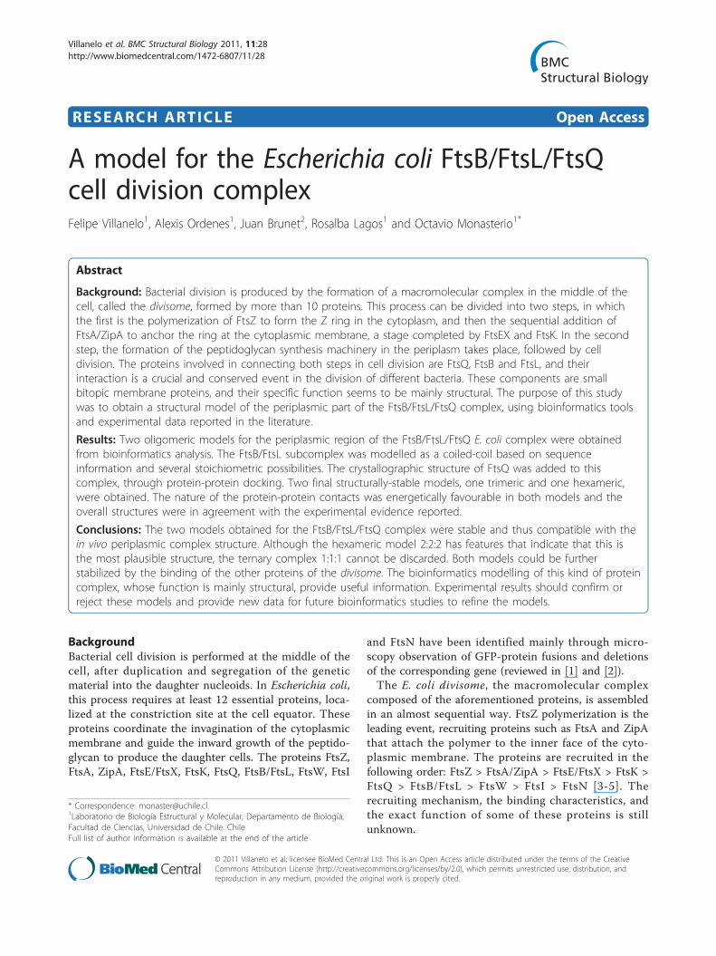
FtsQ is a low abundance periplasmic protein in E. coli(~22 copies per cell) [6], composed of 276 residues witha bitopic membrane topology (Figure 1). The structureincludes a short cytoplasmic N-terminal tail, a mem-brane-spanning helix, and a longer 226-residue periplas-mic section indispensable for the division process [7,8].This protein seems to have a central role in divisomeformation, but its exact functional properties remainunknown. FtsQ localization in the divisome depends onFtsK [9], and drives the localization of the subsequentproteins, including FtsB, FtsL, FtsI, FtsW and FtsN[10-14]. The periplasmic part of FtsQ consists of twodomains called alpha and beta [PDB:2VH1] as describedin the crystal structure of the E. coli and Yersinia entero-colitica proteins [15]. The alpha domain corresponds toa POTRA domain presumably involved in chaperone-like functions that was first predicted from sequenceanalysis [16]. The beta domain, includes a regioninvolved in the interactions with FtsB/FtsL, whose struc-ture was determined by NMR for the Geobacillus stear-othermophilus FtsQ-homologue DivIB [17]. The last 30-40 C-terminal residues are non-structured and theyseem to be determinant for the interaction with FtsB/FtsL in E. coli [18,19]. This sequence was proposed as adomain from limited proteolysis analysis [17,20]. How-ever this is not a separated domain, as shown by thecrystal structure, because these residues are part of thelong, extented beta sheet comprising the beta domain.The alpha domain, immediately after the transmem-
brane helix, is very similar to a POTRA domain of otherreported structures, confirming the predictions of San-chez-Pulido et al. [16]. This domain could protect FtsB/FtsL from denaturation or degradation although a cha-perone-activity has not been probed. Site directed muta-tions of FtsQ (V92D, Q108L, V111G, K113D, E125K)indicate that this domain is the FtsK-interacting part ofthe protein, allowing its correct localization in the divi-some [15,19,21]. The second domain is structured as abeta sheet surrounded by two twisted helices. Mutationsin this domain (Q232R, D237N, A252P and L259S) pre-vent the recruitment of FtsB/FtsL [15,19,22]. The third
non-structured domain proposed for the G. stearother-mophilus FtsQ homologue DivIB [17] is part of the sec-ond domain in the crystallographic E. coli and Y.enterocolitica structures, rather than as an independentdomain [15].FtsB and FtsL are short proteins (121 and 103 residues
respectively) with a topology similar to FtsQ (Figure 1).Both proteins have a leucine heptad in their periplasmicregion, suggesting the presence of a leucine zippermotif, typical of coiled-coil proteins [10,23]. The homo-logues of these proteins in Gram-positive bacteria, FtsLand DivIC respectively, share these characteristics[24,25]. FtsL associates with itself forming unstabledimers in vitro and as heterocomplexes with FtsB andFtsQ at the division site or elsewhere [10,23,26]. Therecruitment of FtsB and FtsL to the E. coli divisomedepends on their mutual interaction [26]. These specificinteractions have been mapped using protein truncationexperiments, highlighting the importance of the C-term-inal regions of FtsB and FtsL for the interaction withFtsQ, and the need of the FtsL cytoplasmic tail for theinteraction with FtsW [27,28]. In Bacillus subtilis DivIC(FtsB) and FtsL are unstable proteins which are stabi-lized when in contact with each other and with DivIB atthe septum (FtsQ homologue) [29,30]. It has been pro-posed that B. subtilis FtsL instability could be a controlpoint in divisome formation, and that the membranemetalloprotease YluC is involved in the degradation ofthe complex [31]. This membrane protease, which parti-cipates in the regulated intramembrane proteolysis (RIP)process, has a homologue in E. coli called RseP, whichcan hydrolyze unstable membrane-spanning domains insome proteins [32,33].The FtsB/FtsL/FtsQ heterocomplex in E. coli exists as
a late recruitment event, together with proteins involvedin cell wall synthesis [3,26]. Corresponding genes havebeen identified in the genomes of many different bac-teria [28]. This complex was isolated in E. coli by co-immunoprecipitation [26] and the interaction has beenconfirmed by other methods [4,5,34]. The structure ofthis complex is a subject of interest in many labora-tories, because it seems to have a crucial structural func-tion in cell division. Masson et al. [18] proposed a lowresolution structure of this ternary complex based onanalysis by NMR, surface plasmon resonance, small-angle neutron and X-ray scattering using the proteinhomologues from Streptococcus pneumoniae (FtsL,DivIC and DivIB). In this study, the POTRA domain ofFtsQ appears loosely-structured and the main interac-tions between these proteins are through the C-terminalregion, leaving the coiled-coil part of DivIC(FtsB)/FtsLfree to interact with other proteins. The extracellularregions of DivIC(FtsB) and FtsL do not interact in vitroso this interaction was forced by fusing each protein to
Figure 1 Topology of FtsB, FtsL and FtsQ. Schematic topology ofFtsB, FtsL and FtsQ, with their transmembrane regions (TM) aligned.The POTRA-like domain of FtsQ is greyed. The scheme is not drawnto scale.
Villanelo et al. BMC Structural Biology 2011, 11:28http://www.biomedcentral.com/1472-6807/11/28
Page 2 of 15
a coiled-coil peptide, leading to the formation of a puta-tive heterodimer complex with a 1:1 DivIC/FtsL ratio[18,34]. The three-protein complex could then beformed independent of FtsK [26] and its formation isprobably crucial in order to stabilize FtsL and FtsBthrough the interaction with FtsQ, like in B. subtilis[29,35]. The binding of FtsQ to the FtsB/FtsL complexcould also be important for its stoichiometry [1,15].In this work, we propose the most probable atomic
structures of the ternary FtsB/FtsL/FtsQ complex, itsstoichiometry and the nature of their interactions. Themost probable and stable models were the trimeric andhexameric complexes, with proteins in a 1:1:1 ratio.These models could help to understand a crucial step indivisome formation, the importance of this complex inthe sequential binding of the other proteins and theirrole in the control of the division process. The theoreti-cal construction of this complex could be useful todesign experiments in order to confirm these modelsand to make progress in the understanding of the bac-terial division process.
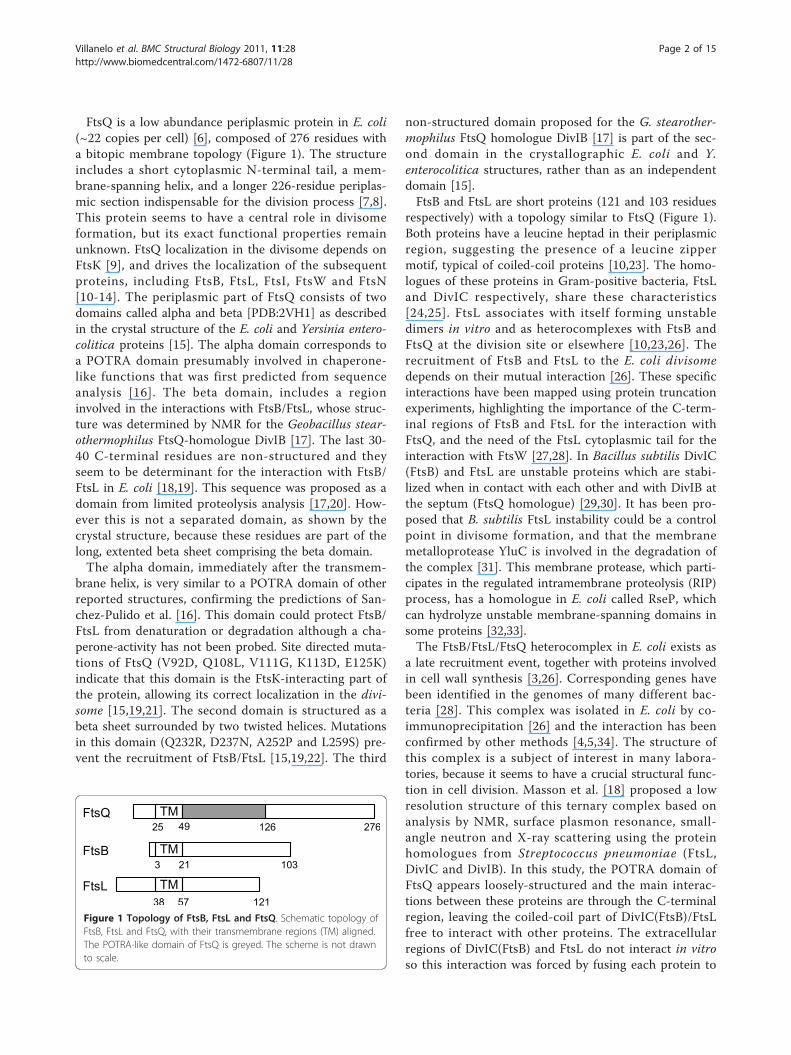
Results and discussionFtsB and FtsL monomer modellingThese division proteins were previously reported as pos-sible coiled-coil proteins, as predicted by the COILSprogram and the presence of a leucine heptad fragmentin the periplasmic region, which could form a leucinezipper motif [10,23]. Secondary structure predictionsshow that both proteins have a long helix between themembrane-spanning region and the C-terminal region,
which could include a loop (Figure 2). The sequence ofboth proteins is not well conserved in other bacteria,but the leucine zipper motif and other structural charac-teristics are conserved [28]. It is important to mentionthe conservation of the leucine residues and the highproportion of charged residues (Additional File 1: Fig-ures S1 and S2).In FtsL, the leucine zipper motif extends from close to
the membrane-spanning helix to approximately residue92 [28]. Here two of four secondary structure predictionsshow a break in the helix (Figure 2), where the residuesGDHS could form a turn, followed by a second helix(residues 96 to 105). This observation is not conclusivebecause the residues in the proposed helix-breaking turnare not fully conserved (Additional File 1: Figure S2).Besides, one of the other two secondary structure predic-tion (SOMPA) shown in Figure 2, has a high reliabilityfor the helix prediction in these residues. After these resi-dues, neither leucine nor other hydrophobic residues areobserved in the correct position in the helix to form thecoiled-coil interaction. The presence of this possible turnis important because in the hexameric FtsB/FtsL/FtsQmodel, a helix-break is indeed modelled in FtsL in orderto fit correctly with the other structures. However, forthe other models this is not necessary because a straighthelix is sufficient to model FtsL. In FtsB, leucine residuesare present in the distal region with respect to the mem-brane-spanning helix, from position 46 to 75. In this pro-tein, the secondary structure prediction shows a helixconfiguration from the transmembrane helix to residue78. For both proteins, the helix prediction includes the
Figure 2 Secondary structure prediction of the periplasmic region of FtsB and FtsL. The prediction was made with the Jpred3, SOMPA,PSIpred and HNN servers. The leucine residues involved in the zipper motif are indicated with arrowhead. Seq: primary sequence; Server name:secondary structure prediction (H: helix; E: extended; -: neither helix nor extended); Rel: reliability of the prediction averaged between thenormalized reliability reported by each server used.
Villanelo et al. BMC Structural Biology 2011, 11:28http://www.biomedcentral.com/1472-6807/11/28
Page 3 of 15
leucine zipper motif, but in the case of FtsB, it is muchlonger (Figure 2).The last 12 residues of FtsL (109-121) were not mod-
elled due to the null helical tendency and therefore thelack of an adequate template structure in the PDB data-base for homology modelling. These C-terminal residuesin the model include residues defined as necessary forthe FtsQ interaction [28]. In the last 20 amino acids ofFtsB (83-103), the secondary structure prediction issimilar to FtsL, the helix tendency is lost and there is noadequate template in the PDB database, although astrand structure is predicted between residues 83 and87. This part of the FtsB model includes the residuesreported to interact with FtsQ [27]. The crystallographicstructures selected for both proteins as template camefrom threading searches using the secondary structuredescribed before (details in Methods section).
Modelling different stoichiometriesThere is a lack of experimental data regarding the stoi-chiometry of the FtsB/FtsL/FtsQ complex. FtsB/FtsLproteins form a complex prior to the binding of FtsQthrough coiled-coil interactions [10,23,26], and for thisreason the FtsB/FtsL complex was modelled first, andthen FtsQ was added. Different oligomeric possibilitiesobserved in coiled-coil multimers were assayed for theFtsB/FtsL complex e.g. dimer, trimer and tetramer. Thepentamer was not considered due to the lack of bulkyhydrophobic residues, such as tryptophan or tyrosinenecessary to stabilize this type of protein-protein inter-action [36,37]. In FtsB or FtsL, there are few such resi-dues and they are not in an appropriate position tomake a possible interacting interface between helices.The strategy used for the stoichiometry analysis was to
model several oligomeric complexes and then to deter-mine their structural stability by molecular dynamicssimulations. The leucine heptad is crucial in order toform the zipper motif during the modelling of the com-plex; notwithstanding, there are other important resi-dues in this type of binding and in the stoichiometry ofthe multimer [38]. FtsB has the leucine residues in itsdistal periplasmic region, and FtsL in its proximal regionwith respect to the lipid bilayer, but this coiled-coilmotif could expand along the helix because the leucinesthat form the zipper motif can be replaced by otherhydrophobic residues such as isoleucine or valine. Thisfact is important for modelling complexes with differentstoichiometries, but sequence analysis was not consid-ered for the stoichiometry, because of the random distri-bution of important interaction residues reported forcoiled-coil interactions, except for the leucines. In sev-eral studies, a pattern of residues has been identified inparticular stoichiometries of coiled-coil folds [39-42] butmultiple alignments of several FtsB and FtsL sequences
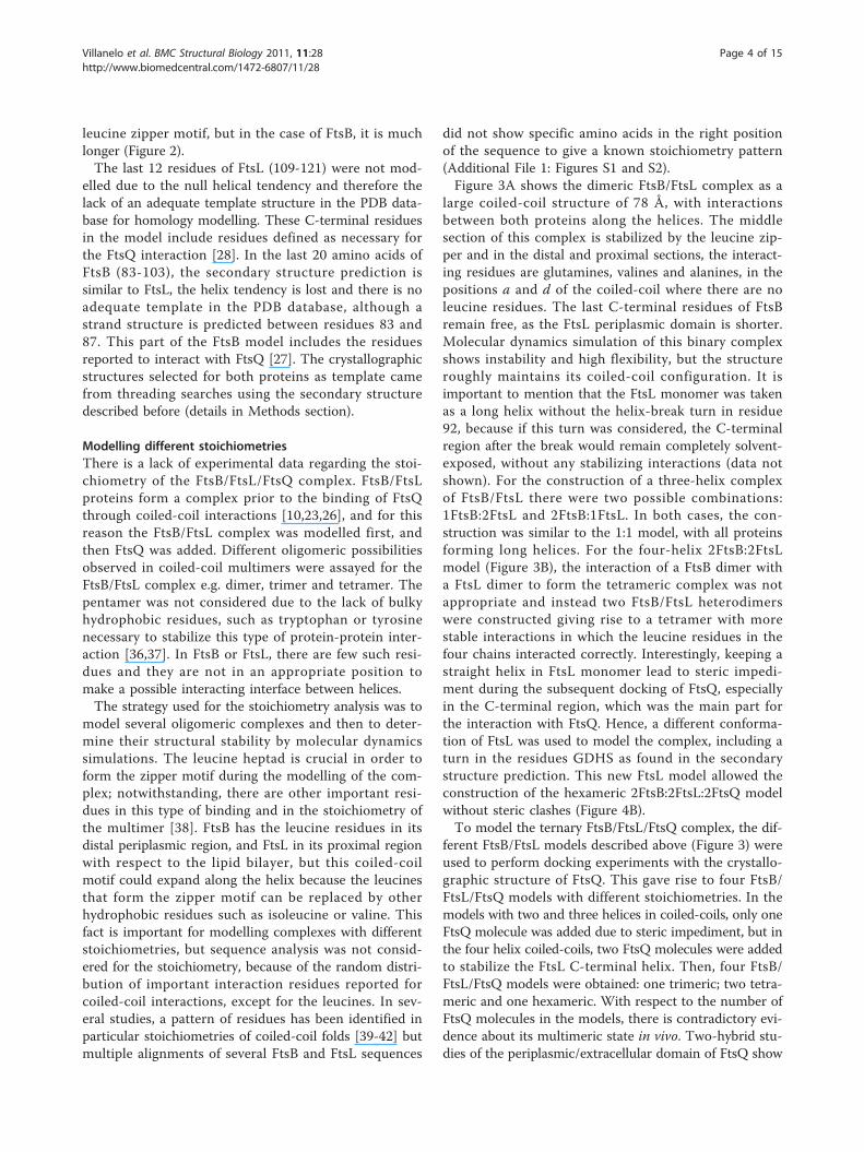
did not show specific amino acids in the right positionof the sequence to give a known stoichiometry pattern(Additional File 1: Figures S1 and S2).Figure 3A shows the dimeric FtsB/FtsL complex as a
large coiled-coil structure of 78 Å, with interactionsbetween both proteins along the helices. The middlesection of this complex is stabilized by the leucine zip-per and in the distal and proximal sections, the interact-ing residues are glutamines, valines and alanines, in thepositions a and d of the coiled-coil where there are noleucine residues. The last C-terminal residues of FtsBremain free, as the FtsL periplasmic domain is shorter.Molecular dynamics simulation of this binary complexshows instability and high flexibility, but the structureroughly maintains its coiled-coil configuration. It isimportant to mention that the FtsL monomer was takenas a long helix without the helix-break turn in residue92, because if this turn was considered, the C-terminalregion after the break would remain completely solvent-exposed, without any stabilizing interactions (data notshown). For the construction of a three-helix complexof FtsB/FtsL there were two possible combinations:1FtsB:2FtsL and 2FtsB:1FtsL. In both cases, the con-struction was similar to the 1:1 model, with all proteinsforming long helices. For the four-helix 2FtsB:2FtsLmodel (Figure 3B), the interaction of a FtsB dimer witha FtsL dimer to form the tetrameric complex was notappropriate and instead two FtsB/FtsL heterodimerswere constructed giving rise to a tetramer with morestable interactions in which the leucine residues in thefour chains interacted correctly. Interestingly, keeping astraight helix in FtsL monomer lead to steric impedi-ment during the subsequent docking of FtsQ, especiallyin the C-terminal region, which was the main part forthe interaction with FtsQ. Hence, a different conforma-tion of FtsL was used to model the complex, including aturn in the residues GDHS as found in the secondarystructure prediction. This new FtsL model allowed theconstruction of the hexameric 2FtsB:2FtsL:2FtsQ modelwithout steric clashes (Figure 4B).To model the ternary FtsB/FtsL/FtsQ complex, the dif-
ferent FtsB/FtsL models described above (Figure 3) wereused to perform docking experiments with the crystallo-graphic structure of FtsQ. This gave rise to four FtsB/FtsL/FtsQ models with different stoichiometries. In themodels with two and three helices in coiled-coils, only oneFtsQ molecule was added due to steric impediment, but inthe four helix coiled-coils, two FtsQ molecules were addedto stabilize the FtsL C-terminal helix. Then, four FtsB/FtsL/FtsQ models were obtained: one trimeric; two tetra-meric and one hexameric. With respect to the number ofFtsQ molecules in the models, there is contradictory evi-dence about its multimeric state in vivo. Two-hybrid stu-dies of the periplasmic/extracellular domain of FtsQ show
Villanelo et al. BMC Structural Biology 2011, 11:28http://www.biomedcentral.com/1472-6807/11/28
Page 4 of 15
that it is a protein capable of self-interaction [3,4,19], butthese observations are indirect and have been refuted bymore direct experiments, such as co-immunoprecipitationof E. coli FtsQ [5], multiangle laser scattering of G. stear-othermophilus DivIB [17] and analytical ultracentrifugationof various bacterial FtsQ and DivIB [15]. The presence ofthe membrane-spanning domain could affect this situationin vivo and promote oligomerization. Besides, the very lim-ited amount of FtsQ molecules in E. coli, reinforces theproposal that FtsQ is a protein that does not self-interact,but that it does form complexes with other proteins, likeFtsB/FtsL.To date, the experimental data on the FtsB/FtsL inter-
action came mainly from two- or three-hybrid studies,for Gram-positive as well as for Gram-negative bacterialproteins [4,19,30,35] and were confirmed by co-immuno-precipitation of these proteins from E. coli [10,26-28].The immunoprecipitation experiments do not provideuseful information on the binding stoichiometry, in con-trast with the studies involving the direct observation of
homolog proteins from S. pneumoniae [18,34], whichstrongly suggest the formation of heterodimers of FtsL/DivIC(FtsB) in a 1:1 ratio. These observations could betrue for E. coli proteins, but the low conservation ofsequence between them lead us to consider other possi-bilities. Besides, in the NMR structure of the periplasmicdomain of DivIB (FtsQ), the POTRA domain appearsloosely-structured, a feature very unlikely under physiolo-gical conditions. The authors attribute this to a marginalconformational stability of the protein in some species,so this domain is not stable in conditions that differ fromthose in vivo [18]. This explanation could be extended tothe stoichiometry observed, so the 1:1 FtsL/DivIC(FtsB)heterodimer could be distinct in the in vivo complex ofE. coli. This reasoning lead us to consider different ratiosbetween the studied proteins.
FtsB/FtsL/FtsQ modelsOnce the possible FtsB/FtsL stoichiometry has beendefined, the FtsQ crystallographic structure can be
Figure 3 FtsB/FtsL complex models. Modelled structures of the FtsB/FtsL complex before the addition of FtsQ. In blue FtsB, in green FtsL. A.Heterodimer FtsB/FtsL; B. Heterotetramer 2FtsB/2FtsL. The insets are top views (from C-terminal to N-terminal) of each model. The residues ofthe position a in the coiled-coil are shown as sticks, coloured by atom type (carbon in grey, oxygen in red, nitrogen in blue). The majority ofthese residues are leucines (see the text for explanation). The range of residues modelled is 25 to 88 for FtsB, and 61 to 109 for FtsL. N- and C-terminals are indicated.
Villanelo et al. BMC Structural Biology 2011, 11:28http://www.biomedcentral.com/1472-6807/11/28
Page 5 of 15
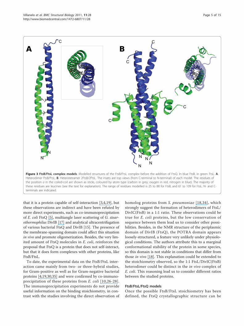
added through a docking procedure (details in Methodssection) using experimental data to restrict the numberof models obtained. In spite of the abundant informa-tion in the literature about mutants in the ftsQ gene, themutations used were those studied in van den Ent et al.[15] because these are point mutations which generate aknown phenotype and are concordant with othersreported before [19,22]. These residues are Q232, D237and L259, all in the C-terminal domain, required forFtsB/FtsL recruitment. The FtsB/FtsL/FtsQ modelsobtained in this way were long and planar, mainly dueto the elongated form of both the FtsQ and the FtsB/
FtsL coiled-coil folds, being between 75 and 80 Å long(Figure 4). The models were subjected to a moleculardynamic routine to analyse their stability: energy mini-mization, simulation annealing, equilibration dynamic,and production dynamic of 10 ns. On one hand, the tri-meric and hexameric models maintained their structureand showed sufficient stability through the process. Onthe other hand, for both tetrameric models, whichinclude three helices in the coiled-coil fold, the confor-mation was lost at some point of the simulation, andthe structure was not maintained during the process(data not shown). In the course of the simulation of
Figure 4 FtsB/FtsL/FtsQ complex models. Representative structures of FtsB/FtsL/FtsQ complex models produced during dynamics simulation.A. Trimeric 1:1:1 model. FtsB are shown in blue, FtsL in green and FtsQ in red. The interacting residues described in the text are in stick form,coloured by atom type and grouped by the pair of proteins involved in the interaction. Leucines in the zipper between FtsB and FtsL are shownin grey sticks. N indicates the N-terminal residue of each protein. The C-terminals are omitted for clarity, but all three are placed at the top ofthe figure. B. Hexameric 2:2:2 model. The colour coding is the same as explained in A. C. Solvent accessible surface of the trimeric model,showing the regions identified previously in the proteins as crucial for the interaction. In purple, the FtsQ POTRA domain (residues 58 to 126); inburgundy, the FtsQ C-terminal domain (residues 127 to 145); in red, the FtsQ residues reported to be essential for FtsB/FtsL recruitment (245 to260); in pale green, the FtsL coiled-coil region (residues 61 to 95); in green, the FtsL residues reported to be crucial for interaction with FtsQ (96to 109); in marine blue, the FtsB coiled-coil region (region 25 to 67); in light blue, the FtsB region not identified to participate in any interaction(residues 68 to 84); in blue, the FtsB region identified to interact with FtsQ (residues 84 to 88); in grey, the leucine residues involved in theleucine-zipper between FtsB and FtsL. D. Solvent accessible surface of the hexameric model, with the same colour coding as above. In C and D,the left panels correspond to the same view as A and B, and the right panels correspond to a 180° rotated view.
Villanelo et al. BMC Structural Biology 2011, 11:28http://www.biomedcentral.com/1472-6807/11/28
Page 6 of 15
these models, the solvent molecules diffused into theprotein interface, disassembling the complex almostcompletely and just some secondary structure elementsremained. These observations led us to discard the tet-rameric models and some minimal calculations wereused to confirm this determination. The work detailedbelow thus relates to the trimeric and hexameric FtsB/FtsL/FtsQ complexes.In order to simulate the biological conditions, position
restraint (an algorithm for maintaining a group of atomsin a fixed position) was applied for the N-terminal mainchain atoms in the first residue of all peptide chains,due to the lack of transmembrane helices in the model.With this approach, these residues were fixed to a vir-tual membrane zone in all the dynamics. During thesimulation annealing (going from 200 to 300K), somestructural changes occurred but it was not clear if thesechanges were due to the artificial forces applied in thedescribed restraint or if they corresponded to a realdynamic process. The FtsB and FtsL helices shouldrotate and translate freely, however the movements wererestricted by the rigidity imposed by artificial forcesapplied that fix the polypeptide chain at the amino ter-minus. In the real environment, the entire complexshould have translational and rotational movement. Inorder to simulate this movement, a distance restraintshould have been applied between the main chain atomsof N-terminal residues of the different chains, but thiswas not possible with the software used.After the 10 ns of molecular dynamics simulation at
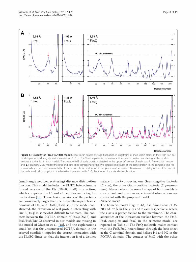
NpT conditions (number or particles, temperature andpressure are maintained constant), the overall structureof the tertiary FtsB/FtsL/FtsQ complex remained stablein the trimeric and hexameric models with a RMS about1-2 Å with respect to the initial model (Figure 5). TheFtsQ molecules were flexible mainly in the loops thatconnect the secondary structure elements. FtsB and FtsLretained their structure in the 1-2 Å range with somedifferences between the two models, especially in FtsB(Figure 5). Although FtsL was not modelled with thesame structure in the trimeric and hexameric models, itwas very stable in both cases. In both the trimeric andhexameric models, the C-terminal residues of FtsQ andFtsB spontaneously formed a beta-like interaction,although in the hexameric complex, this region was lessdefined than in the trimeric model. In both cases, theseinteractions stabilized the exposed residues, loweringtheir mobility throughout the dynamic process. Hence,the complex FtsB/FtsL binds FtsQ in a specific way.However, in these complexes the C-terminal region wasflexible, mainly stabilized by hydrogen bonds that couldbe disrupted by the solvent due to the polar nature ofthis interaction. Although conserved aromatic residuessuch as Phe84 and Tyr85 in FtsB, and Trp256 and
Tyr258 in FtsQ could be important in accounting forthe binding strength, the simulation showed only vander Waals and hydrogen interactions, with no electrondynamics such as aromatic π-π interactions that couldbe important (the simulation, based on classicalmechanics, does not consider electron dynamics). In thezone of the complexes proximal to the membrane, thehexameric model showed more interactions than the tri-meric complex, where hydrogen bonds and salinebridges maintained the sandwich-like position of FtsQwith respect to the coiled-coil fold. These interactionsbetween FtsB/FtsL coiled-coils and the FtsQ POTRAdomain were not reported previously as crucial[18,19,22]. However, they could help to stabilize thecomplex once the mandatory C-terminal interactionshave been formed. In the previous model of the S. pneu-moniae DivIB(FtsQ)/FtsL/DivIC(FtsB) complex [18],these interactions could have been missed due to thelow structured conformation of the POTRA domain atthe conditions used in the experiments. Nevertheless, itis important to bear in mind that the stability could bereinforced by the possible interaction of the membrane-spanning helices of these proteins. Thus, the membranehelices of the three proteins could contribute to theinteractions, as shown for FtsQ [43]. In our work, themovement restriction imposed by the membrane wassimulated through artificial forces (position restraint),but the use of explicit membrane lipid dynamics wouldbe very useful. The dynamic of the lipid bilayer and theresidues around the phospholipid headgroups could sig-nificantly influence the interactions and stabilitiesobserved in this work and this fact would be relevant toselect one of the models.The elongated forms of both FtsB/FtsL/FtsQ models
are in agreement with the model from S. pneumoniaeDivIB(FtsQ)/FtsL/DivIC(FtsB) [18] where the C-terminaldomain (b domain) of DivIB(FtsQ) is the region respon-sible for the binding of the FtsL/DivIC(FtsB) forced het-erodimer (named KL/EC). In the NMR spectrum, thePOTRA domain and the g domain (the last 35 residues)of DivIB(FtsQ) do not change upon FtsL/DivIC(FtsB)binding, but this could be due to the lack of structure ofthese domains in the assayed conditions. The C-terminalregions of KL/EC interact with the b domain of DivIB(FtsQ), and considering the likely sizes of these proteinslet the authors to propose a tilt in the helical coiled-coilinteraction. This tilt could exist in the coiled-coil folditself or at the interface between coiled-coil and trans-membrane helices. The latter option is considered to bemore likely because the secondary structure predictionof these proteins shows an absence of helical tendencyin this region (Figure 2). These conclusions came fromthe ab initio model of the b domain of DivIB(FstQ)interacting with KL/EC, constructed from the SANS
Villanelo et al. BMC Structural Biology 2011, 11:28http://www.biomedcentral.com/1472-6807/11/28
Page 7 of 15
(small-angle neutron scattering) distance distributionfunction. This model includes the KL/EC heterodimer, aforced version of the FtsL/DivIC(FtsB) interaction,which comprises the k5 and e5 peptides and a tag forpurification [18]. These fusion versions of the proteinsare considerably larger than the extracellular/periplasmicdomains of FtsL and DivIC(FtsB), so in the model con-structed, the extension of real protein interacting withDivIB(FtsQ) is somewhat difficult to estimate. The con-tacts between the POTRA domain of FtsQ(DivIB) andFtsL/FtsB(DivIC) observed in our models are missing inthe model of Masson et al. [18], and the explanationscould be: that the unstructured POTRA domain in theassayed condition impedes the correct interaction withthe KL/EC dimer or; that the interaction is of a distinct
nature in the two species, one Gram-negative bacteria(E. coli), the other Gram-positive bacteria (S. pneumo-niae). Nevertheless, the overall shape of both models isconcordant, and previous experimental observations areconsistent with the proposed model.Trimeric modelThe trimeric model (Figure 4A) has dimensions of 35,20 and 79 Å in the x, y and z-axis respectively, wherethe z-axis is perpendicular to the membrane. The char-acteristics of the interaction surface between the FtsB/FtsL complex and FtsQ in the trimeric model arereported in Table 1. The FtsQ molecule makes contactwith the FtsB/FtsL heterodimer through the beta sheetat the C-terminal domain and helices H1 and H2 in thePOTRA domain. The contact of FtsQ with the other
Figure 5 Flexibility of FtsB/FtsL/FtsQ models. Root mean square average fluctuation in angstroms of main chain atoms in the FtsB/FtsL/FtsQmodels produced during dynamics simulation of 10 ns. The X-axis represents the amino acid sequence position numbering in the models(residue 1 is the first in each model). The average RMS of each protein is detailed in the upper left corner of each box. A. Trimeric 1:1:1 modeland B. Hexameric 2:2:2 model (the blue and pink lines correspond to the two different molecules of the same protein in the complex). The redarrows indicate the maximum mobility of FtsB; in A, a helix break is located at position 66 whereas in B maximum mobility occurs at the end ofthe coiled-coil helix and prior to the beta-like interaction with FtsQ. See the text for a detailed explanation.
Villanelo et al. BMC Structural Biology 2011, 11:28http://www.biomedcentral.com/1472-6807/11/28
Page 8 of 15
proteins occurs via its C-terminal domain and alsothrough the POTRA domain. The contacts identified as“hotspots” are reported (Additional File 2: Table S1).There are several hydrophobic residues of FtsL exposedto the solvent, however the main contact with FtsQ is atthe C-terminal region of FtsL (residues 88 to 109) viapolar interactions. The specific residues involved in theionic network at the C-terminal region were: Arg99(FtsL)-Glu190(FtsQ); Glu103(FtsL)-Lys208(FtsQ); Lys45(FtsB)-Glu150(FtsQ) and Arg49(FtsB)-Asp134(FtsQ).There is a little discordance between the model and theexperimental data regarding FtsL/FtsQ contacts (Figure4C), because in the trimeric model, the interactingregion of FtsL was displaced towards the N-terminalwith respect to the previously-identified region (residues100 to 114 in FtsL) [28].A beta-sheet-like conformation formed spontaneously
between the last C-terminal residues of FtsB (76-88) andthe last b-strand of FtsQ (251-258), stabilized by hydro-gen bonds. This region in FtsB adopted a twisted betaconfiguration, establishing several hydrogen bonds withFtsQ. During the rest of the simulation, this beta-likehydrogen interaction remained stable and diminishedthe flexibility of this zone (Figure 5A). This was aninteresting observation because FtsB and FtsQ C-term-inal residues were previously-reported as being crucialin this interaction (Figure 4C) [27]. A couple of polarinteractions were detected between FtsB and thePOTRA domain of FtsQ, such as Glu83(FtsQ)-His27(FtsB) and Gln76(FtsQ)-Asp34(FtsB), whilst the Pro84(FtsQ)-Thr30(FtsB)-Leu70(FtsQ) interaction wasmediated by van der Waals forces. These interactionswere not reported in Masson et al. [18], as explainedabove. Another interesting feature found in the dynamicsimulation of the FtsB/FtsL/FtsQ complex is a break inthe helix of FtsB. This conformational change occurs inresidues 65-67, just 5 positions upstream of a break pre-dicted by the secondary structure (Figure 2) that wasnot considered for the modelling of the FtsB monomer.This break in the helix could be related to the proposed
tilt in the coiled-coil fold proposed by Masson et al [18]in order to fit the C-terminal regions.Hexameric modelThe dimensions of the hexameric FtsB/FtsL/FtsQ modelwere 72, 20 and 90 Å for the x, y and z axis respectively,with a symmetrical distribution of protein chains onboth sides (Figure 4B). The side of FtsQ facing the FtsB/FtsL heterotetramer is slightly displaced towards helicesH4 and H5 in the C-terminal domain, with respect tothe trimeric model. The total number of hydrogenbonds found between the FtsB/FtsL complex and FtsQin the hexameric model were around 27, and the num-ber of salt bridges around 50 (Table 1). This modelshows that two molecules of FtsQ were located at oppo-site sides of the four-helix coiled-coil FtsB/FtsL com-plex. The most important contacts identified as“hotspots” are reported (Additional File 2: Table S1).FtsL molecules interact with FtsQ along their longitu-
dinal axis, mainly through the C-terminal helices (resi-dues 94 to 104) that were accommodated betweenhelices H3 and H4 of the C-terminal domain of eachFtsQ molecule. This interaction was mediated by hydro-gen bonds and salt bridges. The loop between the FtsLhelices (residues 89-92), absent in the trimeric model,established several ionic contacts with loops in FtsQ,such as residues 127-158 in the C-terminal domain.One of the FtsL monomers lost the interaction withFtsQ during the dynamic simulation by disruption ofthe short distal helix during the molecular dynamicssimulation. These facts are unexpected due to thereported importance of residues 100-114 of FtsL in thebinding of FtsQ [28], but not of the residues in the 89-92 range (Figure 4D). This contradiction between ourmodel and the reported experimental data could beattributable to the unknown conformation of FtsL, thatcould present a helix-loop-helix conformation (like inthe hexameric model) or a straight helix from the mem-brane-spanning domain to the C-terminal residues (likein the trimeric model). We cannot distinguish betweenthese conformations with the tools used here, and the
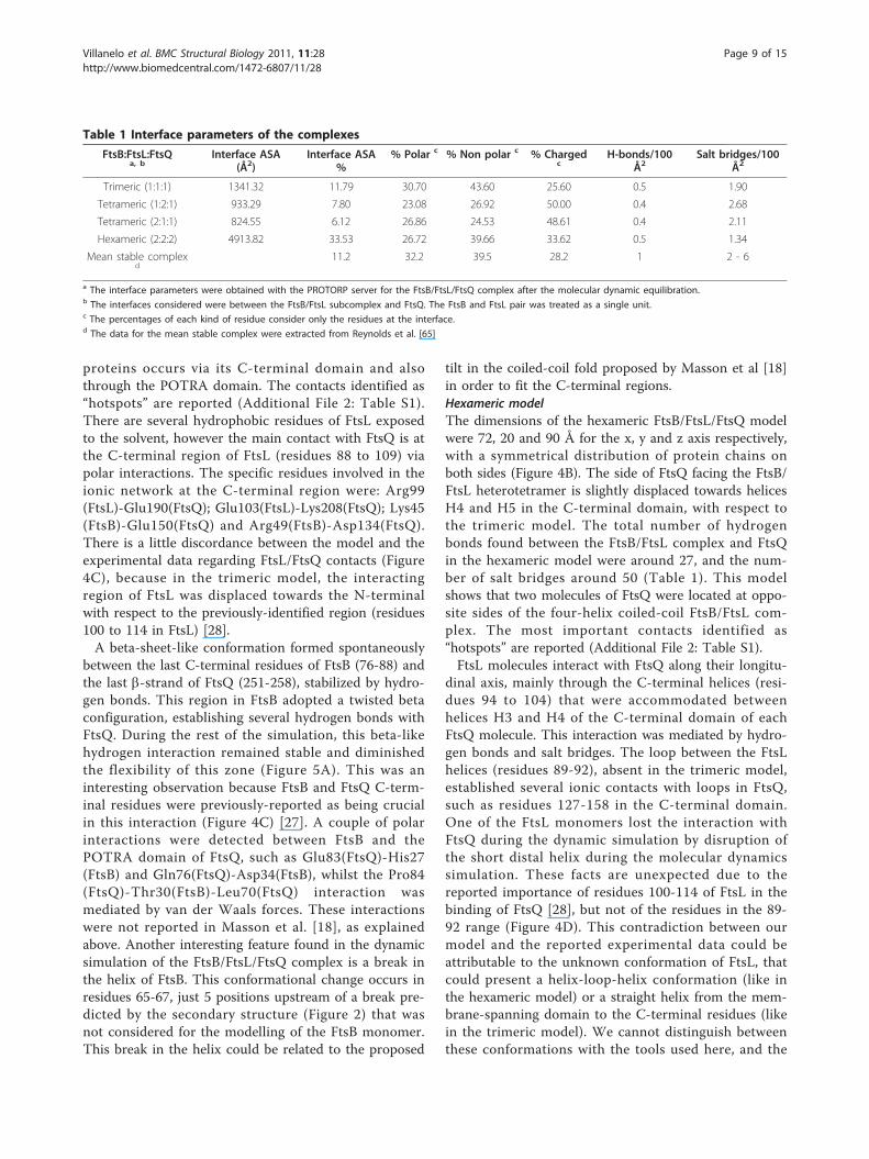
Table 1 Interface parameters of the complexes
FtsB:FtsL:FtsQa, b
Interface ASA(Å2)
Interface ASA%
% Polar c % Non polar c % Chargedc
H-bonds/100Å2
Salt bridges/100Å2
Trimeric (1:1:1) 1341.32 11.79 30.70 43.60 25.60 0.5 1.90
Tetrameric (1:2:1) 933.29 7.80 23.08 26.92 50.00 0.4 2.68
Tetrameric (2:1:1) 824.55 6.12 26.86 24.53 48.61 0.4 2.11
Hexameric (2:2:2) 4913.82 33.53 26.72 39.66 33.62 0.5 1.34
Mean stable complexd
11.2 32.2 39.5 28.2 1 2 - 6
a The interface parameters were obtained with the PROTORP server for the FtsB/FtsL/FtsQ complex after the molecular dynamic equilibration.b The interfaces considered were between the FtsB/FtsL subcomplex and FtsQ. The FtsB and FtsL pair was treated as a single unit.c The percentages of each kind of residue consider only the residues at the interface.d The data for the mean stable complex were extracted from Reynolds et al. [65]
Villanelo et al. BMC Structural Biology 2011, 11:28http://www.biomedcentral.com/1472-6807/11/28
Page 9 of 15
solving of this divergence would allow us to correctlyposition the C-terminal residues of FtsL in the model,which could be concordant with the experimental evi-dence [28].FtsB molecules interact with FtsQ mainly in two areas:
one close to the membrane region, and the other at theC-terminal region. These interactions are the most rele-vant within the hexameric model. The C-terminal regionof FtsB (residues 76 to 88), as explained for the trimericmodel, was initially almost unstructured. This situationchanged in the equilibration dynamic simulation tendingto form a beta-like structure that interacted with the C-terminal beta sheet of FtsQ. However, this beta-likestructure was less stable than in the trimeric model dur-ing the dynamic simulation, with different behaviour oneither side of the hexameric model. On the other hand,the interaction between FtsB and the POTRA domain ofFtsQ at the boundaries of the virtual transmembraneregion includes salt bridges, hydrogen bonds and vander Waals interactions, the last of which could contri-bute to maintain the stability of the structure. Thesehydrophobic contacts could indicate an important inter-action missed in the model of Masson et al. [20], butnot crucial for interaction [27] (Figure 4D). These vander Waals contacts were made by Leu60(FtsQ), Tyr68(FtsQ), Val127(FtsQ), Ile129(FtsQ), Ile85(FtsL), Leu86(FtsL), Val97(FtsL), Ile100(FtsL) and Leu105(FtsL). Inthe middle section of the FtsB molecule (residues 46 to78), there were only van der Waals interactions withtheir FtsL partners which stabilized the heterotetramericinterface via the amino acid residues leucine 60, 67 and75 of the zipper motif.The molecular dynamic simulation showed that during
the equilibration process, the central part of one FtsBmolecule presents a break in the helical configuration,at position 45, probably caused by the position restraintof the N-terminal residues. However, this helix disrup-tion did not interfere with the stability of the complex.The same occurred with FtsL, specifically in one mole-cule, resulting in a twisted and disordered helix thatinteracts in a different way with FtsQ. The break in thehelical conformations during the simulations is due tothe need to fit the positions between the C-terminalregions of FtsB and FtsQ. The POTRA domain of FtsQshowed more flexibility than in the trimeric model,being different for the two monomers of this molecule(Figure 5B). The flexibility of the POTRA domain couldbe in agreement with the observations for the S. pneu-moniae FtsQ homologue DivIB, where this domainappears loosely-structured [20]. Nevertheless, this aug-mented flexibility is marginal, and the contactsdescribed between FtsQ and FtsB were the most stablepart within the model. The region of the hexamericcomplex located around the POTRA domain in FtsQ
showed that the tetrameric coiled-coil structure betweenFtsB and FtsL was important for the interactions withFtsQ (Figure 4B). Given the predicted ΔΔG of ionicpairs, these are not the strongest interactions, but wererelatively stable in the 10 ns of the dynamic simulation.
Interface analysisThe FtsB/FtsL subcomplexes without the FtsQ molecule(s) have hydrogen bonds and salt bridges to stabilize theinterface, but the main contribution to stability wasalways the hydrophobic contacts between leucine resi-dues in the zipper motif. However, the number of stabi-lizing interactions was small for these long complexes,because the hydrophobic interactions came almostexclusively from the a position in the heptad. The posi-tion d, which theoretically must be hydrophobic, hadseveral polar substitutions, resembling a multimericcoiled-coil motif [38-40]. In the simulated annealingstep of the FtsB/FtsL models (without FtsQ), instabilityof these structures was found. The rmsd of the finalstructure was 6-10 Å (depending on the stoichiometry),with a weakening of van der Waals energies withrespect to the initial structure. The polar interactions(hydrogen bonds and electrostatic) also seem to dimin-ish during the course of the dynamic simulation (datanot shown). This is in agreement with experimentaldata in B. subtilis proteins that show that the FtsB/FtsLinteraction is not spontaneous when only the periplas-mic/extracellular domains are used [34,44].The interface analysis of the FtsB/FtsL/FtsQ models
showed different results. For the unfavoured tetramericmodels, presenting a three-helix coiled-coil fold betweenFtsB and FtsL (2:1:1 and 1:2:1 FtsB:FtsL:FtsQ), 20% of theinterface residues and 40-50% of residues exposed to thesolvent were hydrophobic (Tables 1 and 2). Additionally,there are few hydrogen bonds and salt bridges that stabi-lize the surface contact between the FtsB/FtsL subcom-plex and FtsQ, especially in the 2FtsB:1FtsL:1FtsQconformation, where the percentage of ASA (accesible
Table 2 Surface distribution of amino acid residues
FtsB:FtsL:FtsQ a,b % Polar c % Non polar c % Charged c
Trimeric (1:1:1) 30.63 38.38 31.00
Tetrameric (1:2:1) 34.10 42.30 23.60
Tetrameric (2:1:1) 32.50 45.80 21.70
Hexameric (2:2:2) 30.41 36.33 33.27
Mean stable complex d 31.90 38.00 30.00a The interface parameters were obtained with the PROTORP server for theFtsB/FtsL/FtsQ complex after the molecular dynamic equilibration.b The interfaces considered were between the FtsB/FtsL subcomplex andFtsQ. The FtsB and FtsL pair was treated as a single unit.c The percentages of each kind of residue consider only the residues at theexposed surface.d The data for the mean stable complex were extracted from Reynolds et al.[65]
Villanelo et al. BMC Structural Biology 2011, 11:28http://www.biomedcentral.com/1472-6807/11/28
Page 10 of 15

surface area) in the interface was only 6%. These resultsconfirm the conclusion derived from the moleculardynamic simulation showing that the tetrameric com-plexes that contain FtsQ and heterotrimeric FtsB/FtsL(2:1 or 1:2) could be less stable and less soluble due tothe large number of surface hydrophobic residues andthe small area of interaction, and would explain the nullstability of the models in the molecular dynamic proce-dure [45,46].In the more stable trimeric and hexameric FtsB/FtsL/
FtsQ models, the areas of the interface between FtsB/FtsL and FtsQ, were ~1.300 and ~5.000 Å2, respectively.In both models, a major proportion of polar and non-polar residues were found to be responsible for thehydrogen bonds and contacts between the proteins(Table 1 and 2). The number of these interactions andthe proportion of non-polar, polar and charged residuesat the interface of these models resemble that of a stabletransient heteromultimer [45,47], suggesting that theycould be close to the minimum energy structure.Another important feature of the trimeric and hex-
americ FtsB/FtsL/FtsQ models is the proportion of non-polar residues at the interfaces, in both cases near 40%,a value found in many stable complexes. It has beenshown that the strength of binding in transient dimersis related to the proportion of polar and non-polar resi-dues at the interface of the proteins [45]. The “weak”interaction, in which the proteins exist as monomers ordimers at physiological concentrations, has a higher pro-portion of polar residues at the interface, and in general,a geometrically-planar surface of interaction is found.The “strong” interaction in a dimer involves a higherproportion of non-polar residues, and a more complexgeometry of surface contact. The trimeric and hexame-ric models of FtsB/FtsL/FtsQ showed that the character-istic residue distribution in the surface between FtsB/FtsL and FtsQ is closer than that found in a strong tran-sient interaction (Table 1, Table 2, Figure 4). It is tempt-ing to speculate that the other polar residues and thehydrophobic residues exposed to the solvent could beuseful for interaction with the other proteins of the divi-some such as FtsW, FtsI or FtsN, because these percen-tages would be unusual for a very stable solublemultimeric complex [45]. Attending these criteria, thetrimeric FtsB/FtsL/FtsQ model would be more plausiblebecause of the higher percentage of non-polar residuesin the exposed surface, compared to the hexamericmodel.Besides the observed overall proportions of non-polar
residues at the interfaces, there are zones in the com-plexes that showed a substantial presence of polar inter-actions rather than hydrophobic ones. The N-terminalzone that should be proximal to the membrane, is richin salt bridges. This is mainly due to the amino acid
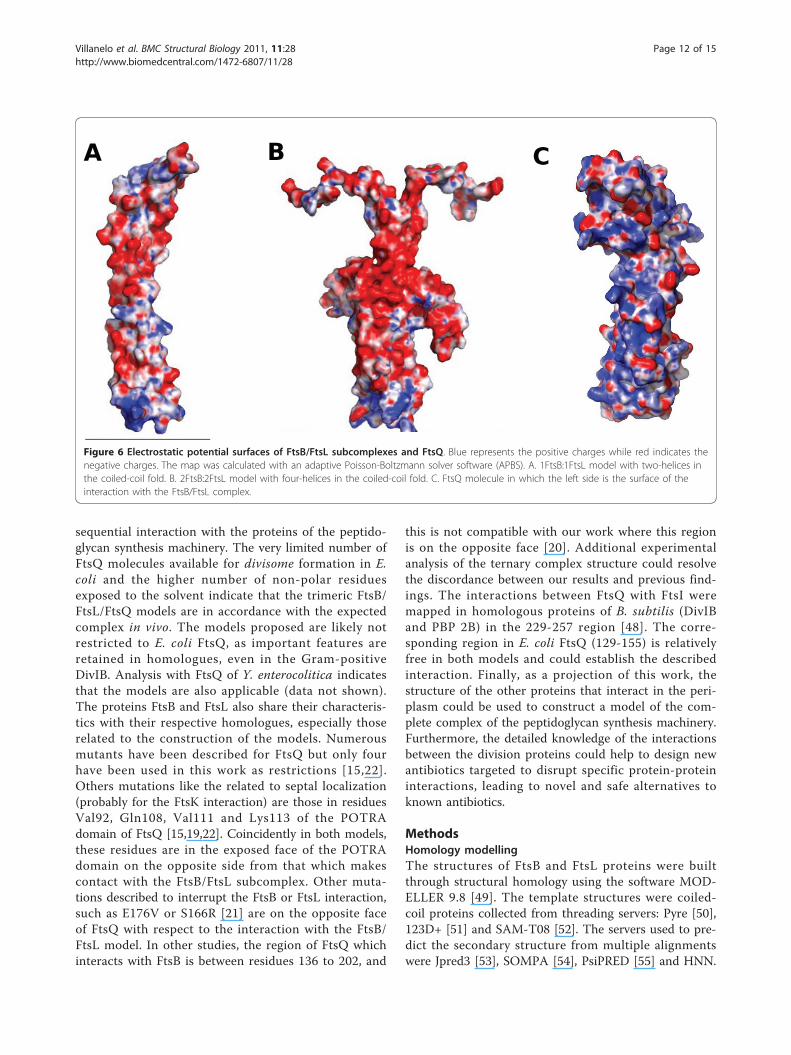
contribution of FtsB and FtsL. Both proteins are rich incharged residues (30% and 26% respectively) with bothpositive and negative charges. The distribution ofcharges in the rest of the sequence is rather random inboth proteins, but there is a zone in the middle sectionof the coiled-coil fold, between residues 50-70 of FtsBand 85-95 in FtsL where the predominant charge isnegative. This “negative patch” in the structure of thecomplex is visible in the electrostatic surface of both tri-meric and hexameric models, although it is more evi-dent in the hexameric model (Figure 6). FtsQ alsopresented a high level of charged residues (24%), how-ever it is more difficult to interpret this fact due to themore complex pattern of secondary and tertiary struc-ture. The electrostatic potential surface showed clearlythat one side of FtsQ is positively charged (Figure 6C),and that this side in both models faces and interactswith the FtsB/FtsL subcomplex, where the charge com-plementation helps to form the ternary complex. Theoverall level of conservation in the three proteins is low,but the relatively high presence of charged residues is aconserved feature, as is the “negative patch” in FtsB andFtsL (See Additional File 1: Figure S1 and S2). The elec-trostatic surface in the N-terminal of the modelled com-plexes tends to be slightly positive (Figure 6),complementing the negatively-charged surface of theouter leaflet of the E. coli inner membrane.In many proteins complexes, there are contact points
between pairs of residues that are crucial for the wholecomplex interactions, and that are identified by thechange in binding energy in a mutation-by-alaninesimulation. In both models of the FtsB/FtsL/FtsQ com-plex, specific and critical point interactions were absent.The search for “hot spots” in the interactions betweenthe proteins by the alanine-mutation simulation resultedin a large number of contacts of discrete binding energy(Additional File 2: Table S1). This could be explained bythe fact that FtsL and FtsB proteins have low residuesimilarity, preserving just the membrane topology, thegenomic context and the leucine heptad [28]. The speci-ficity of protein-protein interactions could be repre-sented by the overall interface interaction rather thanfew contacts, and the surface topology of each proteincould convert an unspecific interaction, such as a chargeattraction (Figure 6), into a specific interaction. Thebinding strength could be reinforced by the transmem-brane helices and the other proteins present in thedivisome.
ConclusionsThe proposed FtsB/FtsL/FtsQ complex models are plau-sible because of the stability and the important numberof hydrophobic residues accessible at the surfacesexposed to the solvent which could be available for the
Villanelo et al. BMC Structural Biology 2011, 11:28http://www.biomedcentral.com/1472-6807/11/28
Page 11 of 15
sequential interaction with the proteins of the peptido-glycan synthesis machinery. The very limited number ofFtsQ molecules available for divisome formation in E.coli and the higher number of non-polar residuesexposed to the solvent indicate that the trimeric FtsB/FtsL/FtsQ models are in accordance with the expectedcomplex in vivo. The models proposed are likely notrestricted to E. coli FtsQ, as important features areretained in homologues, even in the Gram-positiveDivIB. Analysis with FtsQ of Y. enterocolitica indicatesthat the models are also applicable (data not shown).The proteins FtsB and FtsL also share their characteris-tics with their respective homologues, especially thoserelated to the construction of the models. Numerousmutants have been described for FtsQ but only fourhave been used in this work as restrictions [15,22].Others mutations like the related to septal localization(probably for the FtsK interaction) are those in residuesVal92, Gln108, Val111 and Lys113 of the POTRAdomain of FtsQ [15,19,22]. Coincidently in both models,these residues are in the exposed face of the POTRAdomain on the opposite side from that which makescontact with the FtsB/FtsL subcomplex. Other muta-tions described to interrupt the FtsB or FtsL interaction,such as E176V or S166R [21] are on the opposite faceof FtsQ with respect to the interaction with the FtsB/FtsL model. In other studies, the region of FtsQ whichinteracts with FtsB is between residues 136 to 202, and
this is not compatible with our work where this regionis on the opposite face [20]. Additional experimentalanalysis of the ternary complex structure could resolvethe discordance between our results and previous find-ings. The interactions between FtsQ with FtsI weremapped in homologous proteins of B. subtilis (DivIBand PBP 2B) in the 229-257 region [48]. The corre-sponding region in E. coli FtsQ (129-155) is relativelyfree in both models and could establish the describedinteraction. Finally, as a projection of this work, thestructure of the other proteins that interact in the peri-plasm could be used to construct a model of the com-plete complex of the peptidoglycan synthesis machinery.Furthermore, the detailed knowledge of the interactionsbetween the division proteins could help to design newantibiotics targeted to disrupt specific protein-proteininteractions, leading to novel and safe alternatives toknown antibiotics.
MethodsHomology modellingThe structures of FtsB and FtsL proteins were builtthrough structural homology using the software MOD-ELLER 9.8 [49]. The template structures were coiled-coil proteins collected from threading servers: Pyre [50],123D+ [51] and SAM-T08 [52]. The servers used to pre-dict the secondary structure from multiple alignmentswere Jpred3 [53], SOMPA [54], PsiPRED [55] and HNN.
Figure 6 Electrostatic potential surfaces of FtsB/FtsL subcomplexes and FtsQ. Blue represents the positive charges while red indicates thenegative charges. The map was calculated with an adaptive Poisson-Boltzmann solver software (APBS). A. 1FtsB:1FtsL model with two-helices inthe coiled-coil fold. B. 2FtsB:2FtsL model with four-helices in the coiled-coil fold. C. FtsQ molecule in which the left side is the surface of theinteraction with the FtsB/FtsL complex.
Villanelo et al. BMC Structural Biology 2011, 11:28http://www.biomedcentral.com/1472-6807/11/28
Page 12 of 15

In each modelling routine, 100 structures were gener-ated without any restriction or special condition applied.For the construction of FtsB dimers, two coiled-coil
DNA-binding proteins were used [PDB:1T2K,PDB:1HKB]. For the trimeric models, several coiled-coilproteins were used [PDB:1AQ5, PDB:1M7L, PDB:1CE0].For the tetramer, a very stable tetrameric form ofmutant GCN4 was used [PDB:1GCL]. In all cases thealignment between the query and the templatesequences was constructed manually, carefully locatingleucine and other residues following the general rulesobserved in coiled-coil proteins [38,39].To select the correct models, several methods were
used. The statistical potentials DOPE software [56],included in the MODELLER suite, was used for a firstclassification. Then, an empiric residue pair potentialmatrix was used to evaluate the models. This matrixwas constructed following the studies of Moont [57],but using 53 non-redundant structures from the coiled-coil family in the SCOP database. The reasoning behindthis method was to apply a biological filter, reflectingthe typical interaction in this kind of proteins. Throughthe combined use of these two scoring systems, tenmodels were chosen. These models were further ana-lysed with Verify3D [58], PROCHECK [59] and PROSAII [60], selecting one model.
Docking of FtsQ to the FtsB/FtsL complexFor protein-protein docking, the program 3D-Dock wasused, which is based on the FTDock algorithm [61].This software searches the right combination of posi-tions through rigid-body surface complementation gen-erating 10.000 possible models, and uses an empiricresidue pair potential matrix to evaluate the models.Finally, two biological restrictions were used to selectthe final structure: the correct N-terminal-membraneposition of all molecules and a distance restraint of 6 Åbetween the residues identified as interacting in FtsQ(Q232, D237, L259) and FtsB [15]. For FtsQ crystalavailable, the monomer structure was used, because thedimeric FtsQ structure observed was an artefact [15].
Molecular dynamicsIn all this work the GROMACS 4.0 software [62] wasused. To perform MD simulation, the structure of themodelled complex was set to the GROMOS96 43a1 forcefield with explicit hydrogen atoms in the aromatic rings.The simulation cell was created in a triclinic periodic boxwith a minimum distance of 1.5 Å between the proteinand the box walls. The complexes were solvated withapproximately 40,000 water molecules, using the simplepoint charges (SPC) of the water model. The state of tri-trable residues was defined with the H++ web server [63]and the necessary ionic species were added to neutralize
the net charge. Electrostatic interactions were calculatedusing the Particle-Mesh Ewald method (PME) with a gridwidth of 1.2 Å and fourth-order spline interpolations. Acut-off distance of 9 Å was applied for Lennard-Jonesinteractions. The peptides and water were coupledtogether in a temperature bath with a v-rescale thermo-stat. When required, the pressure was coupled with anisotropic Berendsen barostat at a reference pressure of 1bar. A 2 fs time step was used and an harmonic positionrestraint with a force constant of 1000 kJ mol-1 nm-2 inthe z-axis and 500 kJ mol-1 nm-2 in the x and y-axis wasapplied to the heavy atoms of the N-terminal residues ofall peptides. This position restraint was used for main-taining the correct membrane oriented proteins(described in the text). For equilibration of the complexin the aqueous media, the following procedure was per-formed: (1) 1.000 steps of steepest descent energy mini-mization in vacuo; (2) 1.000 steps of conjugated gradientenergy minimization in water; (3) 1200 ps of simplesimulated annealing from 200 K to 300 K; (4) 200 ps ofNvT condition for correct adjustment of temperature; (5)500 ps of NpT for density adjusting. The equilibratedstructure obtained was subjected to 10 ns of NpT pro-duction dynamic at a coupled temperature of 310 K and1 bar of pressure. All the bond lengths were constrainedwith the LINCS algorithm. The initial velocities of theatoms were taken from a Maxwell distribution at 298 K.
Surface and Interface analysisTo analyse the properties and interactions at the inter-face of the distinct protein molecules of the complexes,two web servers were used. 1) the Alanine Scanning ofRobetta server [64] was employed for the identificationof “hot spots” in interfaces. It is based on a simpleenergy function developed primarily with empirical dataextracted from crystallographic complexes deposited atPDB. 2) the PROTORP web-based server [65] was used.This determines the interface area by subtracting theaccessible surface area (ASA) of the complex from thatof the monomers, divided by two. Changes greater than1.0 Å2 indicate an interface residue. The hydrogenbonds were calculated with the program HBPLUS [66],and the salt bridges were calculated by selecting oppo-sitely-charged atoms at least 4 Å apart [67]. The electro-static potential surface was calculated with APBS andPDB2PQR software [68,69]. All the structures shown aredisplayed with PyMOL software (DeLano Scientific).
Additional material
Additional File 1: Sequence logo of the periplasmic region of FtsBand FtsL. Figure S1: Sequence logo of the periplasmic region of FtsB.Around 100 sequences of FtsB and its Gram-positive bacteria equivalent,DivIC were aligned with Muscle software and the logo was obtained
Villanelo et al. BMC Structural Biology 2011, 11:28http://www.biomedcentral.com/1472-6807/11/28
Page 13 of 15
with HMMER software. The height of the symbols in the logo representsthe level of conservation in the alignment. The upper symbol is therepresentative residue for that specific position. Figure S2: Sequence logoof the periplasmic region of FtsL. Around 100 sequences of FtsL werealigned with Muscle software and the logo was obtained with HMMERsoftware. The height of the symbols in the logo represents the level ofconservation in the alignment. The upper symbol is the representativeresidue for that specific position.
Additional File 2: Table S1: Selected Hot spot residues involved inbinding contacts. Hot spot residues were identified with the AlaScanserver and the values of ΔΔG (kJ/mol) are reported. The type ofinteraction is shown: hydrogen bond (HB), salt bridge (SB) or van derWaals contact (VW) in the complex after the molecular dynamicequilibration. The letter in parenthesis (trimeric model) and the letterplus number in parenthesis (hexameric model) are the interactingpartner chain identifier.
AcknowledgementsWe thank the Math Modelling Center (CMM) of Universidad de Chile, forallowing us to perform the molecular dynamics simulations with theircomputer facilities. Special thanks to Dr. C. Baeza for his advice and Dr. M.Handford for critically reading the manuscript. This work was supported bygrant No 1095121 from FONDECYT and EC FP7 # 223431 DIVINOCELL.
Author details1Laboratorio de Biología Estructural y Molecular, Departamento de Biología,Facultad de Ciencias, Universidad de Chile. Chile. 2Instituto de Química,Facultad de Ciencias, Pontificia Universidad Católica de Valparaíso. Chile.
Authors’ contributionsFV performed the work. FV and OM analysed results and drafted themanuscript. AO helped to analyse results and correct the manuscript. JB, RLand OM conceived the study, and participated in its design andcoordination. All authors read and approved the final manuscript anddeclare no conflict of interest.
Received: 28 January 2011 Accepted: 14 June 2011Published: 14 June 2011
References1. Errington J, Daniel RA, Scheffers DJ: Cytokinesis in bacteria. Microbiol Mol
Biol Rev 2003, 67:52-65.2. Vicente M, Rico IA, Martinez-Arteaga R, Mingrorance J: Septum
enlightenment: assembly of bacterial division proteins. J Bacteriol 2006,188:19-27.
3. Di Lallo G, Fagioli GM, Barionovi D, Ghelardini P, Paolozzi L: Use of a two-hybrid assay to study the assembly of a complex multicomponentprotein machinery: bacteria septosome differentiation. Microbiology 2003,149:3353-3359.
4. Karimova G, Dautin N, Ladant D: Interaction network among Escherichiacoli membrane proteins inved in cell division as revealed by bacterialtwo-hybrid analysis. J Bacteriol 2005, 187:2233-2243.
5. Goehring NW, Gonzalez MD, Beckwith J: Premature targeting of a celldivision proteins to midcell reveals hierarchies of protein interactionsinvolved in divisome assembly. Mol Microbiol 2006, 61:33-45.
6. Carson MJ, Barondess J, Beckwith J: The FtsQ protein of Escherichia coli:membrane topology, abundance, and cell division phenotypes due tooverproduction and insertion mutations. J Bacteriol 1991, 173:2187-2195.
7. Guzman LM, Weiss DS, Beckwith J: Domain-swapping analysis of FtsI, FtsL,and FtsQ, bitopic membrane proteins essential for cell division inEscherichia coli. J Bacteriol 1997, 179:5094-5103.
8. Chen JC, Weiss DS, Ghigo JM, Beckwith J: Septal localization of FtsQ, anessential cell division protein in Escherichia coli. J Bacteriol 1999,181:521-530.
9. Chen JC, Beckwith J: FtsQ, FtsL, and FtsI require FtsK, but not FtsN, forco-localization with FtsZ during Escherichia coli cell division. MolMicrobiol 2001, 42:395-413.
10. Buddelmeijer N, Judson N, Boyd D, Mekalanos JJ, Beckwith J: YgbQ, a celldivision protein in Esherichia coli and Vibrio cholerae, localizes incodependent fashion with FtsL to the divisome. Proc Natl Acad Sci USA2002, 99:6316-6321.
11. Ghigo JM, Weiss DS, Chen JC, Yarrow JC, Beckwith J: Localization of FtsLto the Escherichia coli septal ring. Mol Microbiol 1999, 31:725-737.
12. Pastoret S, Fraipont C, den Blaauwen T, Wolf B, Aarsman MEG, Piette A,Thomas A, Brasseur R, Nguyen-Distèche M: Functional analysis of the celldivision protein FtsW of Escherichia coli. J Bacteriol 2004, 186:8370-8379.
13. Weiss DS, Chen JC, Ghigo JM, Boyd D, Beckwith J: Localization of FtsI(PBP3) to the septal ring requires its membrane anchor, the Z ring, FtsA,FtsQ, and FtsL. J Bacteriol 1999, 181:508-520.
14. Addinall SG, Cao C, Lutkenhaus J: FtsN, a late recruit to the septum inEscherichia coli. Mol Microbiol 1997, 25:303-309.
15. van den Ent F, Vinkenvleugel TM, Ind A, West P, Veprintsev D, Nanninga N,den Blaauwen T, Löwe J: Structural and mutational analysis of the celldivision protein FtsQ. Mol Microbiol 2008, 68(1):110-23.
16. Sánchez-Pulido L, Devos D, Genevrois S, Vicente M, Valencia A: POTRA: aconserved domain in the FtsQ family and a class of beta-barrel outermembrane proteins. Trends Biochem Sci 2003, 2:523-526.
17. Robson SA, King GF: Domain architecture and structure of the bacterialcell division protein DivIB. Proc Natl Acad Sci USA 2006, 103:6700-6705.
18. Masson S, Kern T, Le Gouëllec A, Guistini C, Simorre JP, Cllow P, Vernet T,Gabel F, Zapun A: Central domain of DivIB caps the C-terminal region ofthe FtsL/DivIC coiled-coil rod. J Biol Chem 2009, 284(40):27687-27700.
19. D’Ulisse V, Fagioli M, Ghelardini P, Paolozzi L: Three functional subdomainsof the Escherichia coli FtsQ protein are involved in its interaction withthe other division proteins. Microbiology 2007, 153:124-138.
20. Chen JC, Minev M, Beckwith J: Analysis of ftsQ mutant alleles inEscherichia coli: complementation, septal localization, and recruitmentof downstream cell division proteins. J Bacteriol 2002, 184:695-705.
21. Grenga L, Guglielmi G, Melino S, Ghelardini P, Paolozzi L: FtsQ interactionmutants: a way to identify new antibacterial targets. New Biotechnol2010, 27:870-881.
22. Goehring NW, Petrovska I, Boyd D, Beckwith K: Mutants, suppressors, andwrinkled colonies: mutant alleles of the cell division gene ftsQ point tofunctional domains in FtsQ and a role for the domain 1C of FtsA indivisome assembly. J Bacteriol 2007, 189:633-645.
23. Ghigo JM, Beckwith J: Cell division in Escherichia coli: role of FtsLdomains in septal localization, function, and oligomerization. J Bacteriol2000, 182:116-129.
24. Daniel RA, Harry EJ, Katis VL, Wake RG, Errington J: Characterization of theessential cell division gene ftsL (yllD) of Bacillus subtilis and its role inthe assembly of the division apparatus. Mol Microbiol 1998, 29:593-604.
25. Katis VL, Harry EJ, Wake RG: The Bacillus subtulis division protein DivIC isa highly abundant membrane-bound protein that localizes to thedivision site. Mol Microbiol 1997, 26:1047-1055.
26. Buddelmeijer N, Beckwith J: A complex of the Escherichia coli cell divisionproteins FtsL, FtsB and FtsQ forms independently of its localization tothe septal region. Mol Microbiol 2004, 52:1315-1327.
27. Gonzalez MD, Beckwith J: Divisome under construction: Distinct domainsof the small membrane protein FtsB are necessary for interaction withmultiple cell division proteins. J Bacteriol 2009, 191(8):2815-2825.
28. Gonzalez MD, Akbay EA, Boyd D, Beckwith J: Multiple interaction domainsin FtsL, a protein component of the widely conserved bacterial FtsLBQcell division complex. J Bacteriol 2010, 192:2757-2768.
29. Daniel RA, Errington J: Intrinsic instability of the essential cell divisionprotein FtsL of Bacillus subtilis and a role for DivIB protein in FtsLturnover. Mol Microbiol 2000, 36:278-289.
30. Robson SA, Michie KA, Mackay JP, Harry E, King GF: The Bacillus subtiliscell division proteins FtsL and DivIC are intrinsically unstable and do notinteract with one another in the absence of other septasomalcomponents. Mol Microbiol 2002, 44:663-674.
31. Bramkamp M, Weston L, Daniel RA, Errington J: Regulated intramembraneproteolysis of FtsL protein and the control of cell division in Bacillussubtilis. Mol Microbiol 2006, 62(2):580-91.
32. Akiyama Y, Kanehara K, Ito K: RseP (YaeL), an Escherichia coli RIPprotease, cleaves transmembrane sequences. EMBO J 2004, 23:4434-4442.
33. Koide K, Ito K, Akiyama Y: Substrate recognition and binding by RseP, enEscherichia coli intramembrane protease. J Biol Chem 2008,283:9562-9570.
Villanelo et al. BMC Structural Biology 2011, 11:28http://www.biomedcentral.com/1472-6807/11/28
Page 14 of 15
34. Noirclerc-Savoye M, Le Gouellec A, Morlot C, Dideberg O, Vernet T,Zapun A: In vitro reconstruction of a trimeric complex of DivIB, DivICand FtsL, and their transcient co-localization at the division site inStreptococcus pneumoniae. Mol Microbiol 2005, 55:413-424.
35. Daniel RA, Noirot-Gros MF, Noirot P, Errington J: Multiple interactionsbetween the transmembrane division proteins of Bacillus subtilis androle of FtsL instability in divisome assembly. J Bacteriol 2006,188:7396-7404.
36. Liu J, Yong W, Deng Y, Kallenbach NR, Lu M: Atomic structure of atryptophan-zipper pentamer. Proc Natl Acad Sci USA 2004,101(46):16156-61.
37. Liu J, Zheng Q, Deng Y, Kallenbach NR, Lu M: Conformational transitionbetween four and five-stranded phenylalanine zippers determined by alocal packing interaction. J Mol Biol 2006, 361(1):168-79.
38. Krylov D, Mikhailenko I, Vinson C: A thermodynamic scale for leucinezipper stability and dimerization specificity: e and g interhelicalinteractions. EMBO J 1994, 13(12):2849-2861.
39. Fairman R, Chao H, Lovoie TB, Villafranca JJ, Matsueda GR, Novotny J:Design of heterotetrameric coiled coils: Evidence of increasedstabilization by Glu-Lys ion pair interaction. Biochemistry 1996,35:2824-2829.
40. Vu C, Robblee J, Werner KM, Fairman R: Effect of changed aminoacid at band c heptad positions on specificity and stability of four-chain coiledcoils. Prot Sci 2001, 10:631-637.
41. Akey DL, Malashkevich VN, Kim PS: Buried polar residues in coiled-coilinterfaces. Biochemistry 2001, 40:6352-6360.
42. Keating AE, Malashkevich VN, Tidor B, Kim PS: Side-chain repackingcalculation for predicting structures and stabilities of heterodimericcoiled coils. PNAS 2001, 98(26):14825-14830.
43. Scheffer DJ, Robichon C, Haan GJ, den Blaauken T, Konigstein G, vanBloois E, Beckwith J, Luirink J: Contribution of the FtsQ transmembranesegment to localization to the cell division site. J Bacteriol 2007,189(20):7273-7280.
44. Sievers J, Errington J: The Bacillus subtilis cell division protein FtsLlocalizes to sites of septation and interacts with DivIC. Mol Microbiol2000, 36:846-855.
45. Nooren IM, Thornton JM: Structural characterisation and functionalsignificance of transient protein-protein interactions. J Mol Biol 2003,325(5):991-1018.
46. Shanahan HP, Thornton JM: Amino acid architecture and the distributionof polar atoms on the surfaces of proteins. Biopolymers 2005,78(6):318-28.
47. Valdar WS, Thornton JM: Protein-protein interfaces: analysis of amino acidconservation in homodimers. Proteins 2001, 42(1):108-24.
48. Rowland SL, worth KD, Robson SA, Robichon C, Beckwith J, King GF:Evidence from artificial septal targeting and site-directed mutagenesisthat residues in the extracytoplasmic domain of DivIB mediate itsinteraction with the divisomal transpeptidase PBP 2B. J Bacteriol 2010,192:6116-6125.
49. Martí-Renom MA, Stuart AC, Fiser A, Sanchez R, Melo F, Sali A: Comparativeprotein structure modelling of genes and genomes. Ann Rev BiomolStruct 2000, 29:291-325.
50. Bennett-Lovsey RM, Hebert AD, Sternberg MJE, Kelley LA: Exploring theextremes of sequence/structure space with ensemble fold recognition inthe program Phyre. Proteins: structure, function, bioinformatics 2008,70(3):611-625.
51. Alexandrov NN, Nussinov R, Zimmer RM: Fast protein fold recognition viasequence to structure alignment and contact capacity potentials. PacificSymposium on Biocomputing; Singapore World Scientific Publishing Co;1996, 53-72.
52. Karplus K, Barrett C, Hughey R: Hidden Markov models for detectingremote protein homologies. Bioinformatics 1998, 14(10):846-56.
53. Cole C, Barber JD, Barton GJ: The Jpred 3 secondary structure predictionserver. Nucleic Acids Res 2008, 36:W197-W201.
54. Geourjon C, Deleage G: SOPMA: Significant improvements in proteinsecondary structure prediction by consensus prediction from multiplealignments. Comput Appl Biosci 1995, 11(6):681-684.
55. McGuffin LJ, Bryson K, Jones DT: The PSIPRED protein structure predictionserver. Bioinformatics 2000, 16(4):404-5.
56. Shen MY, Sali A: Statistical potential for assessment and prediction ofprotein structures. Protein Sci 2006, 15(11):2507-24.
57. Moont G, Gabb HA, Sternberg MJ: Use of Pair Potentials Across ProteinInterfaces in Screening Predicted Docked Complexes. Proteins 1999,35(3):364-73.
58. Lüthy R, Bowie JU, Eisenberg D: Assessment of protein models withthree-dimensional profiles. Nature 1992, 356(6364):83-5.
59. Laskowski RA, Moss DS, Thornton JM: Main-chain bond lengths and bondangles in protein structures. J Mol Biol 1993, 231(4):1049-67.
60. Wiederstein M, Sippl MJ: ProSA-web: interactive web service for therecognition of errors in three-dimensional structures of proteins. NucleicAcids Res 2007, 35:W407-10.
61. Gabb HA, Jackson RM, Sternberg MJ: Modelling protein docking usingshape complementarity, electrostatics and biochemical information. JMol Biol 1997, 272(1):106-20.
62. van der Spoel D, Lindahl E, Hess B, Groenhof G, Mark AE, Berendsen HJC:GROMACS: Fast, Flexible and Free. J Comp Chem 2005, 26:1701-1718.
63. Gordon JC, Myers JB, Folta T, Shoja V, Heath LS, Onufriev A: H++: a serverfor estimating pKas and adding missing hydrogens to macromolecules.Nucleic Acids Res 2005, 33:W368-71.
64. Kortemme T, Kim DE, Baker D: Computational alanine scanning ofprotein-protein interfaces. Nucleic Acids Res 2004, 32(17):5147-62.
65. Reynolds C, Damerell D, Jones S: ProtorP: a protein-protein interactionanalysis server. Bioinformatics 2008, 25(3):413-4.
66. McDonald IK, Thornton JM: Satisfying hydrogen bonding potential inproteins. J Mol Biol 1994, 238(5):777-93.
67. Barlow DJ, Thornton JM: Ion-pairs in proteins. J Mol Biol 1983,168(4):867-885.
68. Baker NA, Sept D, Joseph S, Holst MJ, McCammon JA: Electrostatics ofnanosystems: application to microtubules and the ribosome. Proc NatlAcad Sci USA 2001, 98(18):10037-10041.
69. Dolinsky TJ, Nielsen JE, McCammon JA, Baker NA: PDB2PQR: an automatedpipeline for the setup, execution, and analysis of Poisson-Boltzmannelectrostatics calculations. Nucleic Acids Res 2004, 32:W665-W667.
doi:10.1186/1472-6807-11-28Cite this article as: Villanelo et al.: A model for the Escherichia coli FtsB/FtsL/FtsQ cell division complex. BMC Structural Biology 2011 11:28.
Submit your next manuscript to BioMed Centraland take full advantage of:
• Convenient online submission
• Thorough peer review
• No space constraints or color figure charges
• Immediate publication on acceptance
• Inclusion in PubMed, CAS, Scopus and Google Scholar
• Research which is freely available for redistribution
Submit your manuscript at www.biomedcentral.com/submit
Villanelo et al. BMC Structural Biology 2011, 11:28http://www.biomedcentral.com/1472-6807/11/28
Page 15 of 15