The impact of water droplet vibration on corona inception on ...
Upload
independentCategory
view
0download
0
Hindawi Publishing CorporationBioMed Research InternationalVolume 2013, Article ID 807982, 14 pageshttp://dx.doi.org/10.1155/2013/807982
Research ArticleA Lys49 Phospholipase A2, Isolated fromBothrops asper Snake Venom, Induces Lipid Droplet FormationinMacrophagesWhich Depends on Distinct SignalingPathways and the C-Terminal Region
Karina Cristina Giannotti,1 Elbio Leiguez,1 VanessaMoreira,1 Neide Galvão Nascimento,1
Bruno Lomonte,2 José Maria Gutiérrez,2 Robson Lopes deMelo,3 and Catarina Teixeira1
1 Laboratory of Pharmacology, Butantan Institute, Avenida Vital Brazil, 05503-900 São Paulo, SP, Brazil2 Clodomiro Picado Institute, School of Microbiology, University of Costa Rica, 2060 San José, Costa Rica3 Center for Applied Toxinology (CAT), Butantan Institute, 05503-900 São Paulo, SP, Brazil
Correspondence should be addressed to Catarina Teixeira; [email protected]
Received 20 September 2012; Accepted 11 October 2012
Academic Editor: Luis A. Ponce Soto
Copyright © 2013 Karina Cristina Giannotti et al. is is an open access article distributed under the Creative CommonsAttribution License, which permits unrestricted use, distribution, and reproduction in any medium, provided the original work isproperly cited.
MT-II, a Lys49PLA2 homologue devoid of catalytic activity from B. asper venom, stimulates in�ammatory events in macrophages.�e investigated the ability ofMT-II to induce formation of lipid droplets (LDs), key elements of in�ammatory responses, in isolatedmacrophages and participation of protein kinases and intracellular PLA2s in this effect. In�uence of MT-II on PLI�2 recruitmentand expression was assessed, and the effects of some synthetic peptides on LD formation were further evaluated. At noncytotoxicconcentrations,MT-II directly activatedmacrophages to formLDs.is effect was reproduced by a synthetic peptide correspondingto the C-terminal sequence 115–129 of MT-II, evidencing the critical role of C-terminus for MT-II-induced effect. Moreover,MT-II induced expression and recruitment of PLI�2. Pharmacological interventions with speci�c inhibitors showed that PKC,PI3K, ERK1/2, and iPLA2, but not P38
MAPK or cPLA2, signaling pathways are involved in LD formation induced by MT-II. issPLA2 homologue also induced synthesis of PGE2 that colocalized to LDs. In conclusion, MT-II is able to induce formation of LDscommitted to PGE2 formation in a process dependent on C-terminal loop engagement and regulated by distinct protein kinasesand iPLA2. LDs may constitute an important in�ammatory mechanism triggered by MT-II in macrophages.
1. Introduction
Phospholipases A2s (PLA2; EC 3.1.1.4) constitute a family oflipolytic enzymes with key roles in several cellular processesby regulating the release of arachidonic acid and lysophos-pholipids from cell membrane phospholipids. Venoms fromsnakes of the Viperidae family contain group IIA phospho-lipases A2 (PLA2s), which share structural and functionalfeatures with PLA2s found in in�ammatory exudates inmammals [1, 2]. A number of Bothrops snake venom PLA2shave been shown to induce in�ammatory events such as
edema and leukocyte in�ltration and to directly activatein�ammatory cell functions [3–6].
Basic PLA2s are considered the most important venomcomponents responsible for the severe local myotoxicity andin�ammation characteristic of the envenomation induced byBothrops genus snakes [7].ese enzymes are further dividedinto two subgroups, namely, catalytically active variants,presenting a conserved aspartic acid residue at position49 (Asp49PLA2s), and catalytically inactive homologues,known as Lys49PLA2s, which present various substitutionsin residues of the Ca2+ binding loop, as well as at position

2 BioMed Research International
49, where Lys replaces the highly conserved Asp [8, 9]. Suchmodi�cations drastically affect the catalytic ability of theseproteins rendering these homologues enzymatically inac-tive [10]. Interestingly, Lys49PLA2 homologues are highlymyotoxic, bactericidal, and proin�ammatory [9], evidencingthat phospholipid hydrolysis is not strictly required for theseactivities. Studies on synthetic peptides and site-directedmutagenesis identi�ed the C-terminal region of Lys49PLA2sas essential for their biological activities [10, 11]. us,Lys49PLA2 homologues constitute interesting models toinvestigate a series of cellular effects which do not depend onmembrane phospholipid hydrolysis.
In the Bothrops asper snake venom three myotoxic Lys49-PLA2s have been identi�ed, namedMT-II,MT-IV, andM1-3-3, and reported in UNIPROT database. Besides myotoxicity,MT-II, the most studied Lys49PLA2 homologue, has beenreported to induce in�ammation in vivo [5, 12] and toactivate some in�ammatory functions of macrophages invitro, increasing phagocytosis, respiratory burst, and releasein�ammatory mediators [4] at noncytotoxic concentrations.However, the knowledge on the effects of this Lys49PLA2 inmacrophages functions, is still fragmentary.
Macrophages play key roles in a wide variety of pro-cesses associated with tissue maintenance, antigen presenta-tion, in�ammation, and tissue repair [13]. Upon in�amma-tory stimuli, quiescent macrophages become activated andpresent increased number of lipid rich cytoplasmic organellesnamed lipid droplets (LDs), also known as lipid bodies.ese organelles are functionally involved in biosynthesis,transport, and catabolism of lipids [14, 15] as well as biosyn-thesis and accumulation of in�ammatory mediators, suchas eicosanoids and cytokines [16, 17]. Moreover, leukocyteLDs associated with in�ammatory responses have beenshown to compartmentalize signaling proteins involved incellular activation and structural proteins, mainly perilipin2 (PLIN2), also named adipophilin (Adipose differentiation-related protein, ADRP), which has an important role inLD assembly and formation of foam macrophages [18, 19],which are markers of atherosclerotic plaques [19]. Increasednumbers of lipid droplets are described in distinct popu-lations of leukocytes during in�ammatory and infectiousprocesses [20, 21]. Recently, MT-III, a catalytically activevariant Asp49PLA2 from B. asper venom, has been shownto activate macrophages to form increased amounts of LDs[22], but no such effect has been described for the actionof Lys49PLA2s. erefore, it is relevant to assess the effectsof MT-II on macrophages in terms of LD formation. Suchmacrophage activation might play a relevant role in thescenario of the local pathological alterations induced bysnake venom toxins. Based on these information, in thepresent study the ability of MT-II to induce LD formationin macrophages was evaluated and the mechanisms involvedin this effect were analyzed in terms of recruitment andexpression of PLIN2, participation of intracellular PLA2s(cPLA2 and iPLA2) and signaling protein kinases. In lightof the absence of catalytic activity in MT-II, the effects ofsome synthetic peptides related to distinct regions of thisLys49PLA2 molecule on lipid droplet formation were furtherevaluated in macrophages.
2. Materials andMethods
2.1. Chemicals and Reagents. MTT and L-glutamine wereobtained from USB Corporation (Cleveland, OH, USA).H7, LY294002, SB202190, PD98059, and Pyr-2 were pur-chased from Calbiochem-Novabiochem (La Jolla, CA, USA).Racemic mixture of BEL and anti-mouse PGE2 was obtainedfrom Cayman Chemical (Ann Arbor, MI, USA). Guineapig polyclonal antibody anti-mouse PLIN2 and FITC-conjugated donkey anti-guinea pig antibody were obtainedfrom Research Diagnostics Inc. (Flanders, NJ, USA). Sec-ondary antibodies anti-mouse and anti-guinea pig conju-gated to horseradish peroxidase and nitrocellulose mem-brane were obtained from GEHealthcare (Buckinghamshire,UK). Gentamicin was purchased from Schering-Plough, NJ,USA). DMSO and BSA were obtained from Amresco (Solon,OH, USA). Mouse monoclonal antibody anti-𝛽𝛽-actin, NileRed, RPMI-1640, thiocarbohydrazide, OsO4, and EDACwere purchased from Sigma Aldrich Co. (St. Louis, MO,USA). PFAwas purchased fromElectronMicroscopy Science(USA). Alexa Fluor 488 Goat Anti-mouse IgG was purchasedfrom Life Technologies (Grand Island, NY, USA). DAPIand �uoromount G were purchased from Molecular Probes(Eugene, OR, USA). Donkey serum was obtained fromJackson ImmunoResearch Laboratories (PA, USA). Triton-X was obtained from Union Carbide Corporation (Danbury,USA). GA, thioglycolate, and all salts used were obtainedfromMerck (Darmstadt, Germany).
2.2. Animals. Male Swiss mice (18–20 g) were obtained fromButantan Institute (São Paulo, Brazil). Animals were housedin a temperature-controlled room (22–24∘C) with a 12 hlight-dark cycle and fresh water and food ad libitum untilused.is study was approved by the Butantan Institute Ani-mal Experimentation Ethics Committee (reference number760/10) in accordance with the procedures laid down by theUniversities Federation for Animal Welfare.
2.3. Phospholipase A2. e Lys49PLA2 homologue (MT-II)was isolated from Bothrops asper venom by ion-exchangechromatography on CM-Sephadex C-25 as described [23],followed by RP-HPLC on a C8 semipreparative column(10 × 250mm; Vydac) eluted at 2.0mL/min with a 0–70%acetonitrile gradient containing 0.1% tri�uoroacetic acid,during 30min, on an Agilent 1200 instrument monitoredat 215 nm. Homogeneity was assessed by analytical reverse-phase HPLC on a C4 column using a gradient of 0–60%acetonitrile in 0.1% tri�uoroacetic acid (v/v). e absenceof endotoxin contamination in the MT-II preparation wasdemonstrated by the quantitative Limulus amebocyte lysate(LAL) test [24], which revealed undetectable levels of endo-toxin (<0.125 EU/mL).
2.4. Synthetic Peptides. e following synthetic peptideswere synthesized and used for biological assays: (a) pep-tide 115–129 (KKYRYYLKPLCKK) corresponding to theoriginal sequence 115–129 of MT-II from B. asper snakevenom; (b) peptide p115-W3 (KKWRWWLKPLCKK) cor-responding to the triple tyrosine-to-tryptophan substitution

BioMed Research International 3
of p115-129; (c) peptide pEM-2 (KKWRWWLKALAKK),in which the proline and cysteine residues of p115-W3were each replaced by an alanine residue; (d) peptide 60–71(KKDRYSYSWKDK) corresponding to a central regionsequence of MT-II; (e) peptide p-Scr (FKFKYKKACKKYK)corresponding to a scrambled peptide version of the sequence115–129 of ACL myotoxin, a Lys49 PLA2 homologue fromthe venom of the snake Agkistrodon contortrix laticinctus.
Synthetic peptides were prepared in automated bench-top simultaneous multiple solid-phase synthesizer (PSSM8 system from Shimadzu Co.) using solid-phase peptidessynthesis by the Fmoc procedure [25�. Brie�y, sequentialcouplings of protected amino acids were performedwith HOBt, TBTU and NMM on Fmoc-Lys(Boc)-Wangresin (Merck KGaA, Germany). Fmoc group cleavagewas performed with 30% piperidine (v/v) in DMF. eresin-bound peptides were cleaved/deprotected withTFA/thioanisole/EDT/phenol/water (82.5 : 5: 2.5 : 5 v/v/v/v)at room temperature for 4 h. Aer �ltration, the �ltratewas concentrated under argon stream and precipitated withdiethyl ether. All crude peptides were puri�ed by reversed-phase chromatography (Shim-pack Prep-ODS, ShimadzuCo.) semipreparative HPLC, and the purity and identity ofthe peptide were con�rmed by mass spectrometry and byanalytical HPLC.
2.5. Harvesting of Macrophages. Peritoneal macrophageswere harvested 4 days aer i.p. injection of 1mL of 3%thioglycolate. Animals were killed under CO2 atmosphereand cells were harvested by washing peritoneal cavitieswith 3mL of PBS, pH 7.2, containing 10 IU/mL heparin.Aliquots of the washes were used for total cell counts in aNeubauer chamber aer dilution (1 : 20, v/v) in Turk solution(0.2% crystal violet dye in 30% acetic acid). Differential cellcounts were performed on smears stained with Hema3. Morethan 95% of the cell population consisted of macrophages,as determined by conventional morphological criteria. eremainingwash volumeswere centrifuged at 500×g for 6min(4∘C) and the cell pellets were used for subsequent studiesaer suitable dilutions.
2.6. Cytotoxicity Assay. Cytotoxicity of MT-II towardselicited macrophages was evaluated using the MTT assay.In brief, 2 × 10⁵ macrophages/well in RPMI-1640 mediumsupplemented with 40 𝜇𝜇g/mL gentamicin sulfate and 2mML-glutamine were plated in 96-well plates and incubated with100 𝜇𝜇L of selected concentrations of MT-II (0.4–0.8 𝜇𝜇M)diluted in medium or with the same volume of mediumalone (control) for 1, 6, 12, and 24 h at 37∘C in a humidi�edatmosphere (5% CO2). MTT (5mg/mL) was dissolved inPBS and �ltered for sterilization and removal of a smallamount of insoluble residue present in some batches ofMTT. Stock MTT solution (10% in culture medium) wasadded to all wells in each assay, and plates were incubated at37∘C for 3 h. One hundred 𝜇𝜇L of DMSO were added to allwells and mixed thoroughly at room temperature for 30min.Absorbances at 540 nm were then recorded in a microtiterplate reader. Results were expressed as percentage of viable
cells, considering control cells incubated with medium aloneas 100% viable.
2.7. Stimulation and Treatment of Macrophages. Macro-phages were plated on glass coverslips in 24-well plates at adensity of 2 × 10⁵ cells/coverslip and allowed to attach for30min at 37∘C under a 5% CO2 atmosphere. Non-adherentcells were removed by washing with PBS. Cell monolayerswere cultured for 1 h in RPMI-1640 supplemented with40 𝜇𝜇g/mL gentamicin sulfate and 2mM L-glutamine at 37∘Cand 5% CO2 and were then challenged with selected con-centrations of MT-II (0.2–1.2𝜇𝜇M) or synthetic peptides(250 𝜇𝜇g/mL) or medium (control). Where appropriate, thefollowing inhibitors were used: 1𝜇𝜇M SB202190, inhibitor ofp38MAPK; 1 𝜇𝜇M LY294002, inhibitor of PI3K; 6 𝜇𝜇M H7-Dihydro, inhibitor of PKC; 25 𝜇𝜇M PD98059, inhibitor ofERK1/2; 1 𝜇𝜇M Pyr-2 (Pyrrolidine-2), inhibitor of cPLA2 and2 𝜇𝜇M BEL (bromoenol lactone) an inhibitor of iPLA2. Allstock solutions were prepared in DMSO and stored at −20∘C.Aliquots were diluted in RPMI-1640 to the required concen-tration immediately before use. e �nal DMSO concentra-tion was always lower than 1% and had no effect on lipidbody numbers. All pharmacological inhibitors were addedbetween 30 and 60min before stimulation of macrophageswith MT-II or medium (control). Cells treated with theinhibitors were analyzed for viability by the tetrazolium-based (MTT) colorimetric assay. No signi�cant changes incell viability were registered with any of the above agents orvehicle at the concentrations used (data not shown).
2.�. �ipid �ody Staining and �uanti�cation. Analysis of lipidbody numbers was performed in osmium-stained cells. Inbrief, macrophages (2 × 10⁵ cells) adhered to glass cov-erslips were �xed in 4% PFA in 0.1M phosphate buffer,pH 7.2, for 15min, and stained with OsO4. e coverslipswere then rinsed in 0.1M phosphate buffer, stained in 1%OsO4 (30min), rinsed in deionized H2O, immersed in 1.0%thiocarbohydrazide (5min), rinsed again in 0.1M phosphatebuffer, restained with 1% OsO4 (3min), rinsed with H2O,and then dried and mounted. e morphology of the �xedcells was observed and round osmiophilic structures wereidenti�ed as lipid droplets, which were then counted underphase-contrast microscopy using the 100x objective lens in50 consecutively scanned leukocytes in each coverslip. Forassays with �uorescent-labeled lipid droplets, macrophages(2 × 10⁵ cells) adhered to glass coverslips were incubatedwith Nile Red staining solution freshly prepared in 0.1Mphosphate buffer (10 𝜇𝜇g/mL) for 20min at room temperatureand washed with phosphate buffer. Aer several washes thecoverslips were mounted with �uoromount G and examinedunder a �uorescence microscope equipped with the appro-priate �lter (�eiss LSM 510 Meta).
2.9. Electron Microscopy. A standardized protocol for elec-tronmicroscopy procedure was developed for ultrastructuralanalysis of lipid droplets. Macrophages (5 × 10⁶ cells) incu-bated with either MT-II (0.8 𝜇𝜇M) or medium alone for 1 hwere �xed in a diluted mixture of freshly prepared aldehydes

4 BioMed Research International
(4% PFA/1% GA in 0.1M phosphate buffer) containing 3.5%sucrose for 2 h at room temperature and then washed threetimes in 0.1M phosphate buffer. Cells were centrifuged at129×g, and cell pelletswere post�xedwithOsO4 (1% in phos-phate buffer at room temperature) followed by three washeswith saline solution. Uranyl acetate in aqueous solution wasthen added for 2 h at room temperature before dehydrationin a graded series of ethanol (70, 95 and 100% twice for10min each). For embedding, aliquots of propylene oxidewere added twice for 10 minutes, followed by Spurr resindiluted in propylene oxide (1 : 1) and undiluted Spurr resinfor 12 h. Polymerization was carried out for 48 h at 70∘C.Samples were then incubated with 4% uranyl acetate and leadcitrate for contrast. in sections were examined with LEO906E and Zeiss EM109 transmission electron microscopes.
2.10. Immunodetection of PLIN2 and PGE2. Detection ofPLIN2 in MT-II-stimulated macrophages was performedby PLIN2 immunostaining. In brief, macrophages attachedto coverslips and stimulated for 3 h with MT-II (0.8 𝜇𝜇M)were �xed in 2% paraformaldehyde (PFA). e cells werepermeabilized with 0.2% Triton-X 100 in 0.1M phosphatebuffer and blocked with 0.5% normal donkey serum in 0.1Mphosphate buffer for 90min. Aer PBS washes, macrophageswere incubated for 1 h with guinea pig polyclonal anti-mousePLIN2 (1 : 2000) diluted in 0.1M phosphate buffer with 0.2%Triton-X 100. Aer three washes with PBS (10min each), thepreparations were incubated for 1 h with secondary FITC-conjugated donkey anti-guinea pig antibody (1 : 500) in thedark for 1 h. Aer the washes, the slides were mountedwith �uoromount G and examined under confocal laserscanning microscope (Zeiss LSM 510 Meta). For analysis ofPGE2 immunostaining, the cell were �xed and permeabilizedin 1% N-ethyl-N�-(3-dimethylaminopropyl) carbodiimidehydrochloride (EDAC) in HBSS−/−. e macrophages wereblocked with 0.5% normal donkey serum in 0.1M phosphatebuffer for 60min. Next, the macrophages were washed withHBSS−/− and incubated for 1 hwith anti-PGE2 (1 : 100). Aerfurther washes, cells were incubated with biotinylated rabbitanti-mouse IgG secondary Ab (1 : 250) and Nile red solution(1 : 250) in the dark for 1 h. e cover slips were then washedthree times and mounted with �uoromount G containingDAPI (Vector Laboratories, Burlingame, CA) and examinedunder confocal laser scanning microscope (Zeiss LSM 510Meta).
2.11. Western Blotting of PLIN2. Aliquots of MT-II-stim-ulated and -nonstimulated cells (2 × 10⁶ cells) were lysedwith 100 𝜇𝜇L of sample buffer (0.5M Tris-HCl, pH 6.8,20% SDS, 1% glycerol, 1M 𝛽𝛽-mercaptoethanol, and 0.1%bromophenol blue) and boiled for 10min. Samples wereresolved by SDS polyacrylamide gel electrophoresis (SDS-PAGE) on 10% bis-acrylamide gels overlaid with a 5%stacking gel. Proteins were then transferred to nitrocellulosemembrane (GE Healthcare, Buckinghamshire, UK) usinga Mini Trans-Blot (Bio-Rad Laboratories, Richmond, CA,USA). e membranes were blocked for 1 h with 5% nonfatdry milk in TTBS (20mM Tris, 100mM NaCl and 0.5%
Tween 20) and incubated with primary antibodies againstPLIN2 (1 : 2000 dilution) and 𝛽𝛽-actin (1 : 3000) for 1 h.ey were then washed and incubated with the appropriatesecondary antibody conjugated to horseradish peroxidase.Detection was by the enhanced chemiluminescence (ECL)method according to the manufacturer’s instructions (GEHealthcare, Buckinghamshire, UK). Band densities werequanti�ed with a GS 800 Densitometer (Bio-Rad Laborato-ries, Richmond, CA) using the image analysis soware fromMolecular Analyst (Bio-Rad Laboratories, Richmond, CA,USA).
2.12. Statistical Analysis. Data are expressed as the mean ±standard error of mean (SEM) of at least three independentexperiments. Multiple comparisons among groups were per-formed by one-way analysis of variance (ANOVA) followedbyTukey’s test. Values of probability lower than 5% (𝑃𝑃 𝑃 0𝑃0𝑃)were considered signi�cant.
3. Results
3.1. Effect of MT-II on Macrophage Viability. Initially, theeffect of MT-II on isolated-elicited macrophage viability wasassessed by the tetrazolium-based (MTT) colorimetric assay.To this purpose the effect of 24 h incubation with two distinctconcentrations of MT-II (0.8 and 1.6𝜇𝜇M) were evaluated. Asshown in Figure 1, incubation of macrophages with MT-II ata concentration of 0.8 𝜇𝜇Mdid not affectmacrophage viability.At a concentration of 1.6 𝜇𝜇M, the sPLA2 homologue partiallydecreased (𝑃𝑃 𝑃 0𝑃0𝑃) macrophage viability.
3.2. MT-II Induces LD Formation in Macrophages. To deter-mine whether stimulation of peritoneal macrophages withMT-II would lead to LDs formation, these cells were incu-bated with selected concentrations of MT-II (0.2–1.6 𝜇𝜇M)for 1 h. As demonstrated in Figure 2(a), incubation ofmacrophages with MT-II at concentrations from 0.8 to1.6 𝜇𝜇M, but not from 0.2 and 0.4 𝜇𝜇M, for 1 h induced asigni�cant increase (𝑃𝑃 𝑃 0𝑃0𝑃) in the number of LDs in com-parison with control cells incubated with culture mediumalone. Maximal LD numbers were observed at 1.6 𝜇𝜇M MT-II. To determine the time-course of LD formation inducedby MT-II, a submaximal concentration of this Lys49PLA2was used (0.8 𝜇𝜇M), and the number of LDs aer 1–24 h ofincubation was determined. As shown in Figure 2(b), MT-IIcaused a signi�cant increase (𝑃𝑃 𝑃 0𝑃0𝑃) in the numbers ofLDs aer 1–24 h incubation compared with control cells.ehighest number of LDs was detected aer 24 h incubation. Asillustrated in Figure 2(c), control macrophages stained withOsO4 showed very few osmiophilic inclusions in the cyto-plasm. In contrast, MT-II-stimulated macrophages exhibiteda cytoplasm packed with the osmiophilic organelles, whichcan be seen as dark punctate structures in Figures 2(d), 2(e),and 2(f).
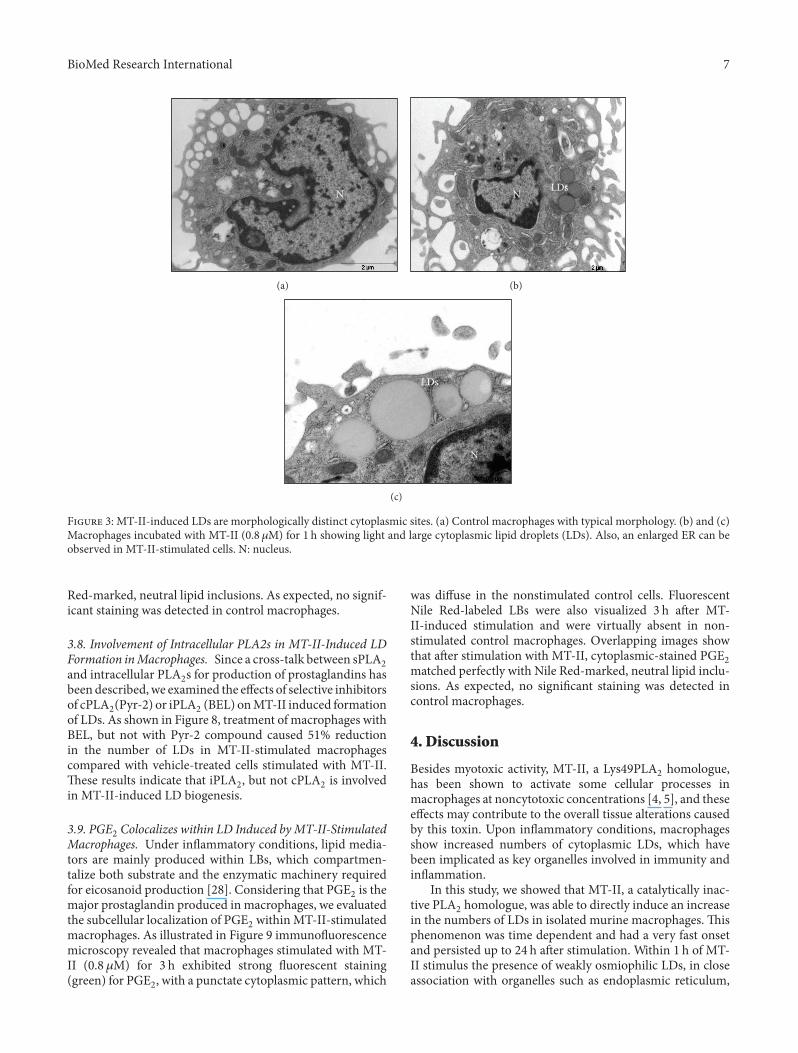
3.3. Ultrastructural Analysis of LDs Induced by MT-II. Tofurther investigate the stimulatory effect of MT-II leadingto LDs formation in macrophages, ultrastructural analysis

BioMed Research International 5
0
50
100
150
RPMI
MT-II 0.8 μM
MT-II 1.6 μM
24 h
∗
Met
abo
lic
acti
vity
(%
)
F 1: Effect of MT-II on cell viability. e cells were incubatedwith MT-II (0.8 and 1.6 𝜇𝜇M) or RPMI (control) for 24 h, and cyto-toxicity was assessed by the tetrazolium-based (MTT) colorimetricassay. Values represent the mean ± SEM from four animals. ∗𝑃𝑃 𝑃0.05 compared with control (RPMI).
of LDs was performed using a standardized procedure forTEM. As seen in Figure 3(a) control macrophages showedsmall, non-membrane-bound, light cytoplasmic LDs. Aer1 h incubation, MT-II-stimulated macrophages showed lightcytoplasmatic LDs that were present in markedly greaternumbers than in the control cells butmorphologically similarto LDs in these cells (Figure 3(b)). Also, an enlarged ERwas observed in MT-II-stimulated cells in incubation periodtested as showed in Figure 3(c).
3.4. Effects of Peptides Corresponding to Selected Regionsof MT-II Molecule on LD Formation. e effects of syn-thetic peptides derived from distinct regions of MT-II pro-tein on LDs formation were investigated in macrophages.Experiments were carried out with macrophages stimulatedwith peptides corresponding to distinct regions of MT-IImolecule for 3 h and then treated as necessary for lipid�xation and stained with OsO4. Figure 4(a) demonstratesthat incubation of macrophages with the C-terminal peptidep115-129 induced a signi�cant increase (𝑃𝑃 𝑃 0.05) in thenumber of LDs aer 3 h of incubation in comparison withnonstimulated control cells. e effect induced by this C-terminal peptide did not differ from that observed in cellsstimulated with MT-II native protein for 3 h, which causeda signi�cant increase in the number of LDs in comparisonwith control cells. On the other hand, neither a scrambledversion of the residue sequence used as a control, p-Scr, northe peptide comprising amino acid residues 60–71 of centralregion of MT-II modi�ed the basal numbers of LDs aer3 h of incubation as compared with RPMI-treated controlcells. As a positive control macrophages were incubatedwith MT-II native protein for 3 h. In this case, a signi�cantincrease in LD number was detected in comparison with
RPMI-treated control cells. All peptides tested were usedin a concentration (250 𝜇𝜇g/mL) previously demonstrated inliterature as effective to induce biologic effects (32), butwithout toxic effect on the viability of macrophages aer 3 hof exposure. Figure 4(b) demonstrates that incubation of cellswith peptides p115–129, pScr, and p60–71 at a concentrationof 250 𝜇𝜇g/mL did not affect macrophages viability, whereasincubation of cells with peptides pEM2 (150 𝜇𝜇g/mL) andp115-W3 (150 𝜇𝜇g/mL) signi�cantly (𝑃𝑃 𝑃 0.05) reduced theviability of macrophages making these peptides unsuitablefor the present study. Although MT-II is recognized as asPLA2 devoid of catalytic activity, we investigated whethera possible residual enzyme activity of MT-II would leadto formation of LDs in macrophages. As shown in Figure4(c) incubation of macrophages with MT-II (0.8𝜇𝜇M) for1 h in the presence of a Ca2+-containing medium induceda signi�cant increase (𝑃𝑃 𝑃 0.05) in the number of LDs.is MT-II-induced effect was not modi�ed in the presenceof Ca2+-free, EGTA- and Sr+2 -containing medium, with asigni�cant increase of LD numbers observed in comparisonwith respective control group.
3.5. LD Formation Triggered by MT-II Is Dependent onDistinct Signaling Pathways. To assess the role of kinases inthe described actions of MT-II, we determined the effectsof the speci�c inhibitors of p38, PI3K, PKC, and ERK1/2(SB202190, LY294002, H7-Dihydro, and PD98059, resp.) onMT-II-induced LDs in macrophages. As seen in Figure 5(a),the PI3K and PKC inhibitors abolished the LDs formationin MT-II-stimulated macrophages compared with vehicle-treated macrophages stimulated with MT-II. e ERK1/2inhibitor, in turn, caused 49% reduction in the number ofLDs in MT-II-stimulated macrophages when compared withvehicle-treated macrophages stimulated with MT-II (Figure5(b)). In contrast, the preincubation of macrophages withp38MAPK inhibitor did not change the number of LDsinduced by MT-II, in comparison to cells stimulated withMT-II only (Figure 5(b)).
3.6. MT-II Upregulates PLIN2 Protein Expression in MT-II-Stimulated Macrophages. PLIN2 expression can be inducedby a variety of in�ammatory cells and has been associatedwith increased numbers of LDs [26, 27]. erefore, weinvestigated whether MT-II induces expression of this LDstructural protein. Levels of PLIN2 protein expression wereanalyzed by western blotting in cells incubated and notincubated with MT-II for selected time periods.is analysisrevealed increased expression of PLIN2 protein in cellsstimulated with MT-II as early as 3 h of incubation, whichwas sustained up to 12 h. PLIN2 was minimally expressed orabsent in control nonstimulated macrophages (Figures 6(a)and 6(b)).
3.7. PLIN2 Colocalizes to LDs in MT-II-Stimulated Macro-phages. To better understand the stimulatory effect of MT-II on macrophages that leads to LD formation, cells exposedto MT-II were immunostained with speci�c antibodies thatrecognize PLIN2 or neutral lipids from the LD core. As

6 BioMed Research International
∗
Lip
id b
od
ies/
cell
0
1
2
3
MT-II ( M)
0.2 0.4 0.8 1.6
#
∗#
(a)
0
2
4
6
RPMI
MT-II 0.8 M
1 3 6 12 24
Time (h)
∗
∗∗∗
∗
+
Lip
id b
od
ies/
cell
(b)
(c) (d)
(e) (f)
F 2: MT-II induces formation of LDs in peritoneal macrophages in culture. (a) Effect of selected concentrations of MT-II on formationof LDs in macrophages which were incubated with various concentrations of MT-II or with RPMI (Control) for 1 h. (b) Time-course of MT-II-induced LD formation. Macrophages were incubated with MT-II (0.8 𝜇𝜇M) or RPMI (Control) for 1, 3, 6, 12, or 24 h. LDs were �uanti�edusing light microscopy aer osmium staining. LDs aer osmium staining observed in control (c) or in cells stimulated with MT-II (0.8 𝜇𝜇M)for 1 h (d), 12 h (e), or 24 h (f). Each bar represents themean ± S.E.M. of the number of LDs/cell in 50 counted cells. Values represent means ±S.E.M. for three to �ve animals. ∗𝑃𝑃 𝑃 𝑃𝑃𝑃𝑃 compared with control cells; #𝑃𝑃 𝑃 𝑃𝑃𝑃𝑃 compared with cells stimulated by 0.2 or 0.4 𝜇𝜇M of MT-II;+𝑃𝑃 𝑃 𝑃𝑃𝑃𝑃 compared with cells stimulated by MT-II (0.8 𝜇𝜇M) for 1, 3, 6 and 12 h.
illustrated in Figure 7 macrophages stimulated with MT-II(0.8𝜇𝜇M) for 3 h e�hibited strong �uorescent staining (green)for PLIN2, with a punctate cytoplasmic pattern, whichwas absent in the nonstimulated control cells. Fluorescent
Nile Red-labeled LDs were also visualized 3 h aer MT-II-induced stimulation and were virtually absent in nonstim-ulated control macrophages. Aer stimulation with MT-II,cytoplasmic-stained PLIN2 matched perfectly with Nile
BioMed Research International 7
N
(a)
LDsN
(b)
LDs
N
(c)
F 3: MT-II-induced LDs are morphologically distinct cytoplasmic sites. (a) Control macrophages with typical morphology. (b) and (c)Macrophages incubated with MT-II (0.8 𝜇𝜇M) for 1 h showing light and large cytoplasmic lipid droplets (LDs). Also, an enlarged ER can beobserved in MT-II-stimulated cells. N: nucleus.
Red-marked, neutral lipid inclusions. As expected, no signif-icant staining was detected in control macrophages.
3.8. Involvement of Intracellular PLA2s in MT-II-Induced LDFormation inMacrophages. Since a cross-talk between sPLA2and intracellular PLA2s for production of prostaglandins hasbeen described,we examined the effects of selective inhibitorsof cPLA2(Pyr-2) or iPLA2 (BEL) onMT-II induced formationof LDs. As shown in Figure 8, treatment of macrophages withBEL, but not with Pyr-2 compound caused 51% reductionin the number of LDs in MT-II-stimulated macrophagescompared with vehicle-treated cells stimulated with MT-II.ese results indicate that iPLA2, but not cPLA2 is involvedin MT-II-induced LD biogenesis.
3.9. PGE2 Colocalizes within LD Induced byMT-II-StimulatedMacrophages. �nder in�ammatory conditions, lipid media-tors are mainly produced within LBs, which compartmen-talize both substrate and the enzymatic machinery requiredfor eicosanoid production [28]. Considering that PGE2 is themajor prostaglandin produced in macrophages, we evaluatedthe subcellular localization of PGE2 withinMT-II-stimulatedmacrophages. As illustrated in Figure 9 immuno�uorescencemicroscopy revealed that macrophages stimulated with MT-II (0.8 𝜇𝜇M) for 3 h exhibited strong �uorescent staining(green) for PGE2, with a punctate cytoplasmic pattern, which
was diffuse in the nonstimulated control cells. FluorescentNile Red-labeled LBs were also visualized 3 h aer MT-II-induced stimulation and were virtually absent in non-stimulated control macrophages. Overlapping images showthat aer stimulation with MT-II, cytoplasmic-stained PGE2matched perfectly with Nile Red-marked, neutral lipid inclu-sions. As expected, no signi�cant staining was detected incontrol macrophages.
4. Discussion
Besides myotoxic activity, MT-II, a Lys49PLA2 homologue,has been shown to activate some cellular processes inmacrophages at noncytotoxic concentrations [4, 5], and theseeffects may contribute to the overall tissue alterations causedby this toxin. �pon in�ammatory conditions, macrophagesshow increased numbers of cytoplasmic LDs, which havebeen implicated as key organelles involved in immunity andin�ammation.
In this study, we showed that MT-II, a catalytically inac-tive PLA2 homologue, was able to directly induce an increasein the numbers of LDs in isolated murine macrophages. isphenomenon was time dependent and had a very fast onsetand persisted up to 24 h aer stimulation. Within 1 h of MT-II stimulus the presence of weakly osmiophilic LDs, in closeassociation with organelles such as endoplasmic reticulum,

8 BioMed Research International
0
0.5
1
1.5
2
2.5
RPMI
MT-II
p115–129
pScr
p60–71
3 h
Lip
id d
rop
lets
/cel
l
∗∗
(a)
0
50
100
150
RPMIp115–129
pEM2
p115-W3
pScrp60− 71
Met
abo
lic
acti
vity
(%
)
3 h
∗∗
(b)
0
1
2
3
Ca2+ medium Ca2+ free + Sr2+
+ EGTA medium
Lip
id d
rop
lets
/cel
l ∗∗
RPMI
MT-II
(c)
F 4: Effects of peptides corresponding to selected regions of MT-II molecule on LD formation. (a) Peritoneal macrophages wereincubated with p115-129 (250 𝜇𝜇g/mL) or pScr (250 𝜇𝜇g/mL) or p60–71 (250 𝜇𝜇g/mL) or RPMI (control) for 3 h. LDs were �uanti�ed usinglight microscopy aer osmium staining; (b) cells were incubated with p115–129 (250 𝜇𝜇g/mL) or pScr (250 𝜇𝜇g/mL) or p60–71 (250 𝜇𝜇g/mL)or PEM2 (150 𝜇𝜇g/mL) or p115–W3 (150 𝜇𝜇g/mL) or RPMI (control) for 3 h, aer which cytotoxicity was assessed by the tetrazolium-based(MTT) colorimetric assay; (c) effect of MT-II (0.8 𝜇𝜇M) on formation of LDs in macrophages in a Ca2+ containing medium or Ca2+-free,EGTA (200 𝜇𝜇M)-Sr2+-containing medium. LDs were �uanti�ed using light microscopy aer osmium staining. Each bar represents the mean± S.E.M. of the number of LDs/cell in 50 counted cells. Values represent means ± S.E.M. for three to �ve animals. ∗𝑃𝑃 𝑃 𝑃𝑃𝑃𝑃 compared withcontrol cells.
were evidenced by the ultrastructural analysis. Accordingto the current model of LD biogenesis, these organellesarise from endoplasmic reticulum, where the enzymes thatsynthesize lipids reside [15, 29]. erefore, endoplasmicreticulum may play a role in LDs biogenesis induced byMT-II.
Because LDs have been associated to regulated in�am-matory mediator synthesis with roles in in�ammatory andinfectious conditions, and macrophages are central elementsin the innate immune response it is plausible to considerthat biogenesis of LDs induced by MT-II demonstrated
herein represents an important mechanism by which thisPLA2 homologue displays an in�ammatory response andleads to production and release of in�ammatory mediators.Moreover, considering that basic PLA2s comprise around30% of B. asper venom [30], the fact that MT-II elicited a�ey in�ammatory event inmacrophages clearly indicates thatthis sPLA2 homologue contributes to the local in�ammatoryresponse triggered by the whole venom.
In addition, our data demonstrated that the absenceof Ca2+ and presence of Sr2+ in culture medium did notalter LDs formation induced by MT-II, con�rming that

BioMed Research International 9
0
1
2
3
Vehicle Dyhidro-H7
MT-II
RPMI
LY294002
Lip
id d
rop
lets
/cel
l
∗
##
(a)
Lip
id d
rop
lets
/cel
l
0
1
2
3
4
Vehicle SB202190 PD98059
∗
∗
#
(b)
F 5: Signaling pathways involved in MT-II-induced LD for-mation. Peritoneal macrophages were incubated with one of thefollowing: (a) PKC inhibitor H7 Dihidro (6 𝜇𝜇M) for 1 h or the PI3Kinhibitor LY294002 (1 𝜇𝜇M) for 1 h; (b) the p38MAPK or ERK1/2inhibitors SB202190 (1 𝜇𝜇M) or PD98059 (25 𝜇𝜇M) for 1 h beforestimulation with MT-II (0.8 𝜇𝜇M) for 1 h. LDs were counted usinglight microscopy aer osmium staining. Each bar represents themean ± SEM of the number of LDs/cell in 50 counted cells. Valuesrepresent means ± SEM from three to �ve animals. ∗𝑃𝑃 𝑃 𝑃𝑃𝑃𝑃compared with control cells; #𝑃𝑃 𝑃 𝑃𝑃𝑃𝑃 compared with MT-II-stimulated cells.
the catalytic activity is not an essential requirement toenhancement of LDs biogenesis by MT-II. A number ofexperimental evidences suggested that a stretch of residues,located at the C-terminus of the MT-II protein molecule,and involving cationic and hydrophobic amino acids areresponsible for myotoxic and cytotoxic effects of this [9, 31,32] and other Lys-49sPLA2 homologues [31, 33]. Based onthese and other studies, a model of Lys49PLA2s-membraneinteraction was proposed by Lomonte et al. [9] in whichthe action of Lys49PLA2s is based on the interaction ofthe C-terminal positive residues with membrane anionicphospholipids. So far, in the present study we found thatthe synthetic peptides 115–129 corresponding to MT-II C-terminus peptide induced LD formation in macrophages
similarly to the parent protein. is �nding indicates for the�rst time the speci�c region of MT-II molecule responsiblefor activation of macrophages, and gives support to thenotion that the effects of MT-II in leukocytes are not relatedto the PLA2 enzymatic activity. e membrane target(s) andthe mechanisms by which this C-terminal peptide triggersmacrophages activation to form LDs were not addressed inthis study, although perturbation of the membrane phospho-lipid bilayer is likely to be involved.
Perilipin 2 is a protein ubiquitously expressed in anumber of cell types, including macrophages, as a majorcomponent of intracellular LDs [34, 35]. It has fatty acid-binding properties, contributes to cytoplasmic trafficking ofnewly synthesized lipids, and plays an important role inassembly of LDs as well as in foam cell formation [35–37].PLIN2 expression can be induced by a variety of in�amma-tory stimuli and has been associated with increased numbersof LD [26, 27]. Consistent with its properties, PLIN2 has beenconsidered as a marker of LDs assembly and lipid loading inin�ammatory cells, such as macrophages. Accordingly, ourresults showed that PLIN2 protein expression is upregulatedbyMT-II, given support to data demonstrating LD formationupon stimulus by this PLA2 homologue. Furthermore, PLIN2clustering co-localized to LDs was seen indicating that MT-IIis also able to recruit this protein from its constitutive poolsinto LDs, and suggesting a role for PLIN2 as a nucleation sitefor the assembly of lipids to form new LDs under the stimulusof this Lys49PLA2.
LD biogenesis in leukocytes is a highly regulated process.Studies of the intracellular signaling pathways committedto this process in leukocytes have revealed that distinctpathways can trigger LD biogenesis in a stimulus-dependentmanner [38]. To better understand the stimulatory effectsof MT-II on LD formation, we herein used pharmacologicalapproaches to identify the critical downstream signalingproteins involved in LD formation induced by this PLA2homologue and focused on major downstream signalingmolecules that have previously been shown to participatein LD biogenesis which follows in�ammatory stimuli, suchas PKC [39, 40], PI3K [16, 26] and MAPKs (p38MAPK andERK1/2) [16, 41]. As a marked LD formation was observedaer 3 h of incubation, the effects of pharmacological com-pounds were evaluated at this time interval. We foundthat MT-II-induced LD formation is regulated by speci�csignaling pathways and that PKC, PI3K, ERK1/2, but notp38MAPK are involved in the formation of LD induced by thisPLA2 homologue.
�ur �nding thatmacrophage activation byMT-II to formLDs is largely dependent on the PKC agrees with previousreports that PKC activation is implicated in LD formationinduced by cys-fatty acid and PAF in leukocytes [39, 42].Considering that activation of PKC has been associated withincreased expression of PLIN2 inmacrophages [43], it is pos-sible to suggest that in the present experimental conditions,PKC signaling pathway is important to MT-II-induced up-regulation of PLIN2, and thus to the increased formation ofLDs. Moreover, our observation that LDs formation inducedby MT-II requires activation of the PI3K pathway is in line

10 BioMed Research International
RPMI
MT-II
3 6 12 3 6 12 (h)
(a)
MT-II
RPMI
0
0.2
0.4
0.6
0.8
3 6 12
Time (h)
∗∗
∗
PL
IN2
(a.
u.)
(b)
F 6: MT-II induces upregulation of PLIN2 expression in macrophages. Peritoneal macrophages were incubated with MT-II (0.8 𝜇𝜇M)or RPMI (Control) for 3, 6, and 12 h. (a) Western blotting of PLIN2 and 𝛽𝛽-actin (loading control) in macrophage extracts. (b) Densitometricanalysis of the band intensities of immunoreactive PLIN2. e densities (in arbitrary units) were normalized with those of 𝛽𝛽-actin. Resultsare expressed as mean ± S.E.M. from three experiments. ∗𝑃𝑃 𝑃 𝑃𝑃𝑃𝑃 compared with controls.
PLIN2 Lipid droplets Merged DIC
RPMI
MT-II
F 7: PLIN2 and LD colocalize in macrophages stimulated by MT-II. Macrophages incubated with RPMI (control) or MT-II (0.8 𝜇𝜇M)for 3 h were labeled for LDs (�uorescent Nile Red) and for PLIN2 (FITC-con�ugated immunocomplex). Merged image shows colocalizationof PLIN2 to LDs. Cell nuclei are observed by DIC. e pictures are representative of three independent experiments.
with reports of participation of this signaling protein inprocesses related to lipid accumulation [26, 44, 45] and inthe regulation of PLIN2 which has been largely associated tolipid accumulation into LDs, and to atherosclerosis [26, 45].Furthermore, our results implicating ERK1/2 signaling in theMT-II effect that leads to LD formation in macrophages areconsistent with previous studies demonstrating the involve-ment of ERK1/2 in LDs biogenesis induced by cytokinesand saturated fatty acids in macrophages [16, 46]. Moreover,evidences of the involvement of ERK1/2 in regulation of
PLIN2 expression and the growth of LDs [41] give support toour �ndings of increased protein expression of PLIN2 seenunder MT-II stimulus, and is in line with the involvement ofERK1/2 in biogenesis of LDs induced by this PLA2 homo-logue. Conversely, the speci�c inhibitor of p38MAPK failedto inhibit MT-II-induced LD formation, implying that thisMAPK element does not contribute to this MT-II-inducedeffect. Taken together, the above results evidenced that MT-II-induced LD formation is a regulated process associatedto activation of selected downstream signaling pathways in

BioMed Research International 11L
ipid
dro
ple
ts/c
ell
0
1
2
3
4
∗
∗
#
MT-II
RPMI
Vehicle Pyr-2 BEL
F 8: Effects of inhibitors of c cPLA2 and iPLA2 on MT-IIinduced LDs formation. Peritoneal macrophages were incubatedwith Pyr-2 (1 𝜇𝜇M) or BEL (2 𝜇𝜇M) compounds for 30min andthen with MT-II (0.8 𝜇𝜇M) for 3 h. LDs were quanti�ed using lightmicroscopy aer osmium staining. Each bar represents the mean± sem LDs/cell in 50 counted cells. Values represent means ± SEMfrom 3–5 animals. ∗𝑃𝑃 𝑃 𝑃𝑃𝑃𝑃 compared with control group; #𝑃𝑃 𝑃𝑃𝑃𝑃𝑃 compared with MT-II-stimulated cells.
macrophages. Of note, despite the lack of enzyme activity,MT-II triggers signaling pathways almost similar to those thatsignal increased formation of LDs induced by the catalyticallyactive sPLA2 MT-III in macrophages [22], thus providing anadditional evidence of functional similarities between thesetwo venom sPLA2variants.
It has beendemonstrated that LDs are involved in produc-tion of in�ammatory mediators [28] and to act as platformsfor enhanced PGE2 synthesis during infection conditions[47, 48]. Moreover, a number of enzymes and signalingproteins were shown to be associated with LDs, includingthe prostaglandin-forming enzymes named cyclooxygenases[47]. Our �ndings that MT-II caused an increase of PGE2intracellular pools, which colocalized to LDs in macrophagesrepresent the �rst evidence that a sPLA2 homologue is ableto induce synthesis and compartmentalization of a lipidmediator in LDs.ese �ndings suggest that macrophage LDconstitutes a relevant site for the synthesis and accumulationof eicosanoids underMT-II stimuli andmay represent a rapidand alternative mechanism for PGE2 production by whichmacrophages react to activation by this sPLA2 homologue.Moreover, given the importance of PGE2 in several in�am-matory settings, due to its hyperalgesic and edematogenicproperties [49, 50], it is reasonable to suggest that LD-derived PGE2 may have implications for the in�ammatoryeffects of MT-II. is hypothesis is in line with the viewthat LDs are dynamic organelles integrating lipidmetabolism,in�ammatory mediator production, membrane tra�cking,and intracellular signaling [27, 47, 51].
A number of studies have demonstrated that secretedPLA2s crosstalk with the intracellular PLA2s (cPLA2 and
iPLA2) to produce arachidonic acid- (AA-) derived in�am-matory mediators, such as prostaglandins, in several patho-physiological conditions [52, 53]. cPLA2 is recognized asa key regulator of stimulus-coupled cellular AA release[6, 54]. e iPLA2 in turn has no substrate speci�city forthe fatty acid residue at sn-2 position, playing a minor rolein eicosanoid synthesis. is intracellular enzyme, however,has a role in membrane phospholipid remodeling throughdeacylation/reacylation reactions [55]. In this context, cPLA2and iPLA2 were demonstrated to be involved in LD bio-genesis induced by stress in CHO-K1 cells [56] and by theAsp49PLA2 MT-III in macrophages [22]. Taking the aboveinformation into account we investigated the participationof both intracellular PLA2 isoforms in MT-II-induced LDformation. We found that treatment of macrophages with,compound Pyr-2, a speci�c inhibitor of cPLA2 failed toinhibit MT-II-induced effect, indicating that cPLA2 is notrequired for LD formation under MT-II stimulus. is�nding is in accordance with our observation that p38MAPK
is not involved in LD formation induced by MT-II, asupstream p38MAPK is critical for phosphorylation and acti-vation of cPLA2 [57]. In contrast, inhibition of iPLA2 by BELcompound signi�cantly reduced LD formation, thus imply-ing iPLA2 in the mechanisms involved in MT-II-inducedLD formation. is �nding is supported by recent studiesdemonstrating participation of iPLA2s in the metabolism offatty acids and triacylglycerol formation, which are involvedin LD formation [56]. We believe that our results are the�rst demonstration that a sPLA2 devoid of catalytic activityrecruits an intracellular PLA2 (iPLA2) to induce a cellularevent, such as LD formation in macrophages. However, thecellular steps involved in such a protein crosstalk were notaddressed in the present study and deserve further studies.
5. Conclusions
Taken together, our data show that the venom group IIAsPLA2 homologue MT-II directly activates murine macro-phages to form LDs by a mechanism independent on enzy-matic activity. is effect is related to the C-terminal loop ofthe MT-II molecule since a synthetic peptide correspondingto region 115–129-induced LD formation similarly to MT-II. Moreover, MT-II-induced LD formation is related toincreased expression and recruitment of PLIN2 from its con-stitutive pools and regulated by distinct signaling pathwaysthat include PKC, PI3K, ERK1/2, and iPLA2. In addition,MT-II induced synthesis and compartmentalization of PGE2within LDs. erefore, LDs may represent and importantplatform for the synthesis and accumulation of lipid media-tors underMT-II stimulus that takes place in themechanismswhereby this Lys49PLA2 triggers in�ammation.
Finally, considering that catalytically inactive PLA2s havebeen also described in mammalian tissues under normaland pathological conditions [58, 59], this study may shedlight on the possible activities of similar proteins on amore general scope, providing insights into the possibleroles of human catalytically inactive PLA2 homologues in

12 BioMed Research International
Lipid droplets Merged DIC
RPMI
MT-II
PGE2
F 9: Cytoplasmic lipid droplets compartmentalize PGE2. Macrophages incubated with RPMI (control) or MT-II (0.8 𝜇𝜇M) for 3 h werelabeled for LBs (�uorescent Nile Red) and for PGE2 (Cayman Chemical). Merged image shows colocalization of PGE2 to LDs. Cell nuclei areobserved by DIC. e pictures are representative of three independent experiments.
in�ammatory conditions as it has been demonstrated for thesnake venom Lys49PLA2.
Authors’ Contribution
K. C. Giannotti and E. Leiguez contributed equally to thiswork.
Acknowledgments
e authors thank Renata Hage do Amaral for technicalassistance and Alexsander Seixas de Souza for confocal laserscanning microscopy analysis assistance. is paper wassupported by research grant from Fundação de Amparo àPesquisa do Estado de São Paulo (FAPESP) Brazil (Grant2011/21341-5). C. Teixeira is a recipient of aNational Councilfor Scienti�c and Technological Development�Bolsa PQ(CNPq-PQ), Grant 306099/2008-0; K. C. Giannotti is a recip-ient of M. S. fellowship from CAPES; E. Leiguez is a recipientof a Ph.D. fellowship from FAPESP (Grant 10/06345-1); V.Moreira is recipient of Postdoctoral fellowship from FAPESP(Grant 07/03337-5);N.G.Nascimento is a recipient of a Ph.D.fellowship from CAPES. J. M. Gutiérrez and B. Lomontereceived support from the Vicerrectoría de Investigación,Universidad de Costa Rica.
References
[1] I. I. Kaiser, J. M. Gutiérrez, D. Plummer, S. D. Aird, and G. V.Odell, “e amino acid sequence of a myotoxic phospholipasefrom the venom of Bothrops asper,”Archives of Biochemistry andBiophysics, vol. 278, no. 2, pp. 319–325, 1990.
[2] R. M. Kini, Venom Phospholipase A2 Enzymes: Structure, Func-tion and Mechanisms, Wiley, Nova York, NY, USA, 1997.
[3] E. C. T. Landucci, R. C. Castro, M. F. Pereira et al., “Mast celldegranulation induced by two phospholipase A2 homologues:
dissociation between enzymatic and biological activities,” Euro-pean Journal of Pharmacology, vol. 343, no. 2-3, pp. 257–263,1998.
[4] J. P. Zuliani, C. M. Fernandes, S. R. Zamuner, J. M. Gutiérrez,and C. F. P. Teixeira, “In�ammatory events induced by Lys-49 and Asp-49 phospholipases A2 isolated from Bothrops aspersnake venom: role of catalytic activity,” Toxicon, vol. 45, no. 3,pp. 335–346, 2005.
[5] J. P. Zuliani, J. M. Gutiérrez, L. L. Casais e Silva, S. C.Sampaio, B. Lomonte, and C. Teixeira, “Activation of cellularfunctions in macrophages by venom secretory Asp-49 and Lys-49 phospholipases A2,” Toxicon, vol. 46, no. 5, pp. 523–532,2005.
[6] V. Moreira, J. M. Gutiérrez, A. M. Soares, S. R. Zamunér, E.Purgatto, and C. Teixeira, “Secretory phospholipases A2 iso-lated from Bothrops asper and from �rotalus durissus terri�cussnake venoms induce distinct mechanisms for biosynthesis ofprostaglandins E2 and D2 and expression of cyclooxygenases,”Toxicon, vol. 52, no. 3, pp. 428–439, 2008.
[7] J. M. Gutiérrez and C. L. Ownby, “Skeletal muscle degenerationinduced by venom phospholipases A2: insights into the mech-anisms of local and systemic myotoxicity,” Toxicon, vol. 42, no.8, pp. 915–931, 2003.
[8] C. L. Ownby, H. S. Selistre de Araujo, S. P. White, and J. E.Fletcher, “Lysine 49 phospholipase A2 proteins,” Toxicon, vol.37, no. 3, pp. 411–445, 1999.
[9] B. Lomonte, Y. Angulo, and L. Calderón, “An overview oflysine-49 phospholipase A2 myotoxins from crotalid snakevenoms and their structural determinants of myotoxic action,”Toxicon, vol. 42, no. 8, pp. 885–901, 2003.
[10] R. J. Ward, L. Chioato, A. H. C. de Oliveira, R. Ruller, and J.M. Sá, “Active-site mutagenesis of a Lys49-phospholipase A2:biological andmembrane-disrupting activities in the absence ofcatalysis,” Biochemical Journal, vol. 362, no. 1, pp. 89–96, 2002.
[11] L. Chioato, E. A. Aragão, T. Lopes Ferreira, A. I. de Medeiros,L. H. Faccioli, and R. J. Ward, “Mapping of the structuraldeterminants of arti�cial and biological membrane damagingactivities of a Lys49 phospholipase A2 by scanning alanine

BioMed Research International 13
mutagenesis,” Biochimica et Biophysica Acta—Biomembranes,vol. 1768, no. 5, pp. 1247–1257, 2007.
[12] B. Lomonte, A. Tarkowski, and L. Å. Hanson, “Host responseto Bothrops asper snake venom. Analysis of edema formation,in�ammatory cells, and cytokine release in a mouse model,”In�ammation, vol. 17, no. 2, pp. 93–105, 1993.
[13] S. J. Galli, N. Borregaard, and T. A. Wynn, “Phenotypic andfunctional plasticity of cells of innate immunity: macrophages,mast cells and neutrophils,”Nature Immunology, vol. 12, no. 11,pp. 1035–1044, 2011.
[14] L. Kuerschner, C. Moessinger, and C. iele, “Imaging of lipidbiosynthesis: how a neutral lipid enters lipid droplets,” Traffic,vol. 9, no. 3, pp. 338–352, 2008.
[15] T. C. Walther and R. V. Farese Jr., “Lipid droplets and cellularlipid metabolism,” Annual Review of Biochemistry, vol. 81, pp.687–714, 2012.
[16] P. Pacheco, A. Vieira-de-Abreu, R. N. Gomes et al.,“Monocyte chemoattractant protein-1/CC chemokine ligand2 controls microtubule-driven biogenesis and leukotrieneB4-synthesizing function of macrophage lipid bodies elicitedby innate immune response,” Journal of Immunology, vol. 179,no. 12, pp. 8500–8508, 2007.
[17] T. Araújo-Santos, D. B. Prates, B. B. Andrade et al., “Lut-zomyia longipalpis saliva triggers lipid body formation andprostaglandin E2 production in murine macrophages,” PLoSNeglected Tropical Diseases, vol. 4, no. 11, article e873, 2010.
[18] H. Robenek, M. J. Robenek, I. Buers et al., “Lipid dropletsgain PAT family proteins by interaction with specialized plasmamembrane domains,” Journal of Biological Chemistry, vol. 280,no. 28, pp. 26330–26338, 2005.
[19] Y. Yuan„ P. Li, and J. Ye, “Lipid homeostasis and the formation ofmacrophage-derived foam cells in atherosclerosis,” Protein Cell,vol. 3, no. 3, pp. 173–181, 2012.
[20] G. Schmitz and M. Grandl, “Lipid homeostasis in macro-phages—implications for atherosclerosis,” Reviews of Physiol-ogy, Biochemistry and Pharmacology, vol. 160, pp. 93–125, 2008.
[21] K. A. Mattos, H. D’Avila, L. S. Rodrigues et al., “Lipid dropletformation in leprosy: toll-like receptor-regulated organellesinvolved in eicosanoid formation and mycobacterium lepraepathogenesis,” Journal of Leukocyte Biology, vol. 87, no. 3, pp.371–384, 2010.
[22] E. Leiguez, J. P. Zuliani, A. M. Cianciarullo, C. M. Fernandes, J.M. Gutiérrez, and C. Teixeira, “A group IIA-secreted phospho-lipase A2 from snake venom induces lipid body formation inmacrophages: the roles of intracellular phospholipases A2 anddistinct signaling pathways,” Journal of Leukocyte Biology, vol.90, no. 1, pp. 155–166, 2011.
[23] B. Lomonte and J. M. Gutiérrez, “A new muscle damagingtoxin, myotoxin II, from the venom of the snake Bothrops asper(terciopelo),” Toxicon, vol. 27, no. 7, pp. 725–733, 1989.
[24] K. Takayama, D. H. Mitchell, Z. Z. Din, P. Mukerjee, C. Li,and D. L. Coleman, “Monomeric re lipopolysaccharide fromescherichia coli is more active than the aggregated form inthe limulus amebocyte lysate assay and in inducing Egr-1mRNA inmurine peritonealmacrophages,” Journal of BiologicalChemistry, vol. 269, no. 3, pp. 2241–2244, 1994.
[25] E. Atherton and R. C. Sheppard, Solid Phase Peptide Synthe-sis—A Practical Approach, IRL Press, Oxford, UK, 1989.
[26] C. M. Maya-Monteiro, P. E. Almeida, H. D’Ávila et al., “Lep-tin induces macrophage lipid body formation by a phos-phatidylinositol 3-kinase- andmammalian target of rapamycin-dependent mechanism,” Journal of Biological Chemistry, vol.283, no. 4, pp. 2203–2210, 2008.
[27] A. R. Silva, P. Pacheco, A. Vieira-de-Abreu et al.,“Lipid bodies in oxidized LDL-induced foam cells areleukotriene-synthesizing organelles: a MCP-1/CCL2 regulatedphenomenon,” Biochimica et Biophysica Acta—Molecular andCell Biology of Lipids, vol. 1791, no. 11, pp. 1066–1075, 2009.
[28] P. T. Bozza, K. G. Magalhães, and P. F. Weller, “Leukocyte lipidbodies—biogenesis and functions in in�ammation,” Biochimicaet Biophysica Acta—Molecular and Cell Biology of Lipids, vol.1791, no. 6, pp. 540–551, 2009.
[29] K. K. Buhman, H. C. Chen, and R. V. Farese Jr., “e enzymesof neutral lipid synthesis,” Journal of Biological Chemistry, vol.276, no. 44, pp. 40369–40372, 2001.
[30] J. Gutiérrez and B. Lomonte, “Phospholipase A2 myotoxinsfrom Bothrops snake venoms,” Toxicon, vol. 33, no. 11, pp.1405–1424, 1995.
[31] B. Lomonte, Y. Angulo, and E. Moreno, “Synthetic peptidesderived from the C-terminal region of Lys49 phospholipaseA2 homologues from viperidae snake venoms: biomimeticactivities and potential applications,” Current PharmaceuticalDesign, vol. 16, no. 28, pp. 3224–3230, 2010.
[32] L. Calderón and B. Lomonte, “Immunochemical characteriza-tion and role in toxic activities of region 115–129 of myotoxinII, a Lys49 phospholipase A2 fromBothrops asper snake venom,”Archives of Biochemistry and Biophysics, vol. 358, no. 2, pp.343–350, 1998.
[33] L. Chioato and R. J. Ward, “Mapping structural determinantsof biological activities in snake venom phospholipases A2 bysequence analysis and site directed mutagenesis,” Toxicon, vol.42, no. 8, pp. 869–883, 2003.
[34] H. W. Heid, R. Moll, I. Schwetlick, H. R. Rackwitz, and T. W.Keenan, “Adipophilin is a speci�c marker of lipid accumulationin diverse cell types and diseases,” Cell and Tissue Research, vol.294, no. 2, pp. 309–321, 1998.
[35] C. Londos, C. Sztalryd, J. T. Tansey, and A. R. Kimmel, “Role ofPAT proteins in lipid metabolism,” Biochimie, vol. 87, no. 1, pp.45–49, 2005.
[36] A. Paul, B. H. J. Chang, L. Li, V. K. Yechoor, and L. Chan, “De�-ciency of adipose differentiation-related protein impairs foamcell formation and protects against atherosclerosis,” CirculationResearch, vol. 102, no. 12, pp. 1492–1501, 2008.
[37] J. Gao and G. Serrero, “Adipose differentiation related pro-tein (ADRP) expressed in transfected COS-7 cells selectivelystimulates long chain fatty acid uptake,” Journal of BiologicalChemistry, vol. 274, no. 24, pp. 16825–16830, 1999.
[38] P. T. Bozza, R. C. N. Melo, and C. Bandeira-Melo, “Leukocytelipid bodies regulation and function: contribution to allergy andhost defense,” Pharmacology and erapeutics, vol. 113, no. 1,pp. 30–49, 2007.
[39] P. F. Weller, S. W. Ryeom, S. T. Picard, S. J. Ackerman, and A.M.Dvorak, “Cytoplasmic lipid bodies of neutrophils: formationinduced by cis-unsaturated fatty acids and mediated by proteinkinase C,” Journal of Cell Biology, vol. 113, no. 1, pp. 137–146,1991.
[40] P. T. Bozza, W. Yu, J. Cassara, and P. F. Weller, “Pathways foreosinophil lipid body induction: differing signal transductionin cells from normal and hypereosinophilic subjects,” Journal ofLeukocyte Biology, vol. 64, no. 4, pp. 563–569, 1998.

14 BioMed Research International
[41] L. Andersson, P. Boström, J. Ericson et al., “PLD1 and ERK2regulate cytosolic lipid droplet formation,” Journal of CellScience, vol. 119, no. 11, pp. 2246–2257, 2006.
[42] P. T. Bozza, J. L. Payne, J. L. Goulet, and P. F. Weller,“Mechanisms of platelet-activating factor-induced lipid bodyformation: requisite roles for 5-lipoxygenase and de novoprotein synthesis in the compartmentalization of neutrophillipids,” Journal of Experimental Medicine, vol. 183, no. 4, pp.1515–1525, 1996.
[43] J. S. Chen, A. S. Greenberg, Y. Z. Tseng, and S. M. Wang,“Possible involvement of protein kinase C in the induction ofadipose differentiation-related protein by sterol ester in RAW264.7 macrophages,” Journal of Cellular Biochemistry, vol. 83,no. 2, pp. 187–199, 2001.
[44] A. Fougerat, S. Gayral, N. Malet, F. Briand-Mesange, M.Breton-Douillon, and M. Laffargue, “Phosphoinositide 3-kinases and their role in in�ammation: potential clinical targetsin atherosclerosis?” Clinical Science, vol. 116, no. 11-12, pp.791–804, 2009.
[45] L. Barberis and E. Hirsch, “Targeting phosphoinositide 3-kinase 𝛾𝛾 to �ght in�ammation and more,” rombosis andHaemostasis, vol. 99, no. 2, pp. 279–285, 2008.
[46] L. S. Moreira, B. Piva, L. B. Gentile et al., “Cytosolic phospholi-pase A2-driven PGE2 synthesis within unsaturated fatty acids-induced lipid bodies of epithelial cells,” Biochimica et BiophysicaActa—Molecular and Cell Biology of Lipids, vol. 1791, no. 3, pp.156–165, 2009.
[47] M. T. Accioly, P. Pacheco, C. M. Maya-Monteiro et al.,“Lipid bodies are reservoirs of cyclooxygenase-2 and sitesof prostaglandin-E2 synthesis in colon cancer cells,” CancerResearch, vol. 68, no. 6, pp. 1732–1740, 2008.
[48] H. D’Avila, N. R. Roque, R. M. Cardoso, H. C. Castro-Faria-Neto, R. C. N. Melo, and P. T. Bozza, “Neutrophils recruited tothe site of mycobacterium bovis BCG infection undergo apop-tosis and modulate lipid body biogenesis and prostaglandin E2production by macrophages,” Cellular Microbiology, vol. 10, no.12, pp. 2589–2604, 2008.
[49] J. P. Portanova, Y. Zhang, G. D. Anderson et al., “Selec-tive neutralization of prostaglandin E2 blocks in�ammation,hyperalgesia, and interleukin 6 production in vivo,” Journal ofExperimental Medicine, vol. 184, no. 3, pp. 883–891, 1996.
[50] R. F. Claudino, C. A. L. Kassuya, J. Ferreira, and J. B. Cal-ixto, “Pharmacological and molecular characterization of themechanisms involved in prostaglandin E2-induced mouse pawedema,” Journal of Pharmacology and Experimental erapeu-tics, vol. 318, no. 2, pp. 611–618, 2006.
[51] H. D’Avila, R. C. N. Melo, G. G. Parreira, E. Werneck-Barroso,H. C. Castro-Faria-Neto, and P. T. Bozza, “Mycobacteriumbovis bacillus calmette-guérin induces TLR2-mediated for-mation of lipid bodies: intracellular domains for eicosanoidsynthesis in vivo,” Journal of Immunology, vol. 176, no. 5, pp.3087–3097, 2006.
[52] M. W. Anthonsen, A. Solhaug, and B. Johansen, “Func-tional Coupling between secretory and cytosolic phospholipaseA2 modulates tumor necrosis factor-𝛼𝛼- and interleukin-1𝛽𝛽 -inducedNF-𝜅𝜅BActivation,” Journal of Biological Chemistry, vol.276, no. 32, pp. 30527–30536, 2001.
[53] S. Chakraborti, “Phospholipase A2 isoforms: a perspective,”Cellular Signalling, vol. 15, no. 7, pp. 637–665, 2003.
[54] I. Kudo and M. Murakami, “Phospholipase A2 enzymes,”Prostaglandins and Other Lipid Mediators, vol. 68-69, pp. 3–58,2002.
[55] M. V. Winstead, J. Balsinde, and E. A. Dennis, “Calcium-independent phospholipase A2: structure and function,”Biochimica et Biophysica Acta—Molecular and Cell Biology ofLipids, vol. 1488, no. 1-2, pp. 28–39, 2000.
[56] A. Gubern, M. Barceló-Torns, D. Barneda et al., “JNK andceramide kinase govern the biogenesis of lipid droplets throughactivation of group IVA phospholipase A2,” Journal of BiologicalChemistry, vol. 284, no. 47, pp. 32359–32369, 2009.
[57] R. M. Kramer, E. F. Roberts, S. L. Um et al., “p38 mitogen-activated protein kinase phosphorylates cytosolic phospholi-pase A2 (cPLA2) in thrombin-stimulated platelets. Evidencethat proline-directed phosphorylation is not required for mobi-lization of arachidonic acid by cPLA2,” Journal of BiologicalChemistry, vol. 271, no. 44, pp. 27723–27729, 1996.
[58] M. Rouault, J. G. Bollinger, M. Lazdunski, M. H. Gelb, and G.Lambeau, “Novel mammalian group XII secreted phospholi-pase A2 lacking enzymatic activity,” Biochemistry, vol. 42, no.39, pp. 11494–11503, 2003.
[59] M. Guan, L. Qu,W. Tan, L. Chen, and C.W.Wong, “Hepatocytenuclear factor-4 alpha regulates liver triglyceride metabolismin part through secreted phospholipase A2 GXIIB,”Hepatology,vol. 53, no. 2, pp. 458–466, 2011.
Copyright © 2022 FDOKUMEN