A Large Scale Test of Computational Protein Design: Folding and Stability of Nine Completely...
12
A Large Scale Test of Computational Protein Design: Folding and Stability of Nine Completely Redesigned Globular Proteins Gautam Dantas 1 †, Brian Kuhlman 1 †, David Callender 1 Michelle Wong 1 and David Baker 1,2 * 1 Department of Biochemistry University of Washington Seattle, WA 98195, USA 2 Howard Hughes Medical Institute, University of Washington, Seattle, WA 98195, USA A previously developed computer program for protein design, RosettaDesign, was used to predict low free energy sequences for nine naturally occurring protein backbones. RosettaDesign had no knowledge of the naturally occurring sequences and on average 65% of the residues in the designed sequences differ from wild-type. Synthetic genes for ten completely redesigned proteins were generated, and the proteins were expressed, purified, and then characterized using circular dichroism, chemical and temperature denaturation and NMR experiments. Although high-resolution structures have not yet been determined, eight of these proteins appear to be folded and their circular dichroism spectra are similar to those of their wild-type counterparts. Six of the proteins have stabilities equal to or up to 7 kcal/mol greater than their wild-type counterparts, and four of the proteins have NMR spectra consistent with a well-packed, rigid structure. These encouraging results indicate that the computational protein design methods can, with significant reliability, identify amino acid sequences compatible with a target protein backbone. q 2003 Elsevier Ltd. All rights reserved. Keywords: computational protein design; protein engineering; protein stability; protein thermodynamics *Corresponding author Introduction The ultimate goal of protein design is the creation of novel proteins that perform specified tasks. A necessary requirement for meeting this goal is the ability to identify sequences that fold with sufficient stability into a target structure. Towards this end several laboratories have developed computer programs for identifying amino acid sequences compatible with a given protein. 1–8 A rigorous test for these models is the complete redesign of naturally occurring proteins. In such a test, the only information given to the method is the backbone coordinates of the protein to be redesigned. Although there has been con- siderable recent success in the field of compu- tational protein design, 9–12 the pioneering zinc finger redesign by Mayo and co-workers is the only published report in which automated pro- cedures have been used to completely redesign a naturally occurring protein backbone. 1 Previously we demonstrated that our method for protein design, RosettaDesign, produces native- like sequences when run on a large test set of naturally occurring protein backbones. 13 On average, 30% of the residues were identical with their wild-type counterpart, and in the core the level of identity was 50%. These results suggested that RosettaDesign was performing well, but they did not indicate whether the design sequences would actually fold into the target structures. The RosettaDesign method has been applied success- fully to the redesign of protein folding pathways, 14 backbone conformations, 15 and oligomerization states. 16 Here, to more rigorously test Rosetta- Design and, more generally, to assess the consist- ency with which modern computational protein design methodology can completely redesign the sequences of small proteins, we make and charac- terize complete redesigns of nine globular proteins. Like all automated procedures for protein 0022-2836/$ - see front matter q 2003 Elsevier Ltd. All rights reserved. † G. D. and B. K. contributed equally to this work. Present address: B. Kuhlman, Department of Biochemistry and Biophysics, University of North Carolina, Chapel Hill, NC 27599, USA. E-mail address of the corresponding author: [email protected] doi:10.1016/S0022-2836(03)00888-X J. Mol. Biol. (2003) 332, 449–460
-
Upload
independent -
Category
Documents
-
view
0 -
download
0
Transcript of A Large Scale Test of Computational Protein Design: Folding and Stability of Nine Completely...
A Large Scale Test of Computational Protein Design:Folding and Stability of Nine Completely RedesignedGlobular Proteins
Gautam Dantas1†, Brian Kuhlman1†, David Callender1
Michelle Wong1 and David Baker1,2*
1Department of BiochemistryUniversity of WashingtonSeattle, WA 98195, USA
2Howard Hughes MedicalInstitute, University ofWashington, Seattle, WA98195, USA
A previously developed computer program for protein design,RosettaDesign, was used to predict low free energy sequences for ninenaturally occurring protein backbones. RosettaDesign had no knowledgeof the naturally occurring sequences and on average 65% of the residuesin the designed sequences differ from wild-type. Synthetic genes for tencompletely redesigned proteins were generated, and the proteins wereexpressed, purified, and then characterized using circular dichroism,chemical and temperature denaturation and NMR experiments. Althoughhigh-resolution structures have not yet been determined, eight of theseproteins appear to be folded and their circular dichroism spectra aresimilar to those of their wild-type counterparts. Six of the proteins havestabilities equal to or up to 7 kcal/mol greater than their wild-typecounterparts, and four of the proteins have NMR spectra consistent witha well-packed, rigid structure. These encouraging results indicate thatthe computational protein design methods can, with significant reliability,identify amino acid sequences compatible with a target protein backbone.
q 2003 Elsevier Ltd. All rights reserved.
Keywords: computational protein design; protein engineering; proteinstability; protein thermodynamics*Corresponding author
Introduction
The ultimate goal of protein design is thecreation of novel proteins that perform specifiedtasks. A necessary requirement for meeting thisgoal is the ability to identify sequences that foldwith sufficient stability into a target structure.Towards this end several laboratories havedeveloped computer programs for identifyingamino acid sequences compatible with a givenprotein.1 – 8 A rigorous test for these models is thecomplete redesign of naturally occurring proteins.In such a test, the only information given to themethod is the backbone coordinates of the proteinto be redesigned. Although there has been con-siderable recent success in the field of compu-tational protein design,9 – 12 the pioneering zinc
finger redesign by Mayo and co-workers is theonly published report in which automated pro-cedures have been used to completely redesign anaturally occurring protein backbone.1
Previously we demonstrated that our method forprotein design, RosettaDesign, produces native-like sequences when run on a large test set ofnaturally occurring protein backbones.13 Onaverage, 30% of the residues were identical withtheir wild-type counterpart, and in the core thelevel of identity was 50%. These results suggestedthat RosettaDesign was performing well, but theydid not indicate whether the design sequenceswould actually fold into the target structures. TheRosettaDesign method has been applied success-fully to the redesign of protein folding pathways,14
backbone conformations,15 and oligomerizationstates.16 Here, to more rigorously test Rosetta-Design and, more generally, to assess the consist-ency with which modern computational proteindesign methodology can completely redesign thesequences of small proteins, we make and charac-terize complete redesigns of nine globular proteins.
Like all automated procedures for protein
0022-2836/$ - see front matter q 2003 Elsevier Ltd. All rights reserved.
† G. D. and B. K. contributed equally to this work.Present address: B. Kuhlman, Department of
Biochemistry and Biophysics, University of NorthCarolina, Chapel Hill, NC 27599, USA.
E-mail address of the corresponding author:[email protected]
doi:10.1016/S0022-2836(03)00888-X J. Mol. Biol. (2003) 332, 449–460
design, RosettaDesign has two main components:an energy function that ranks the relative fitnessof various amino sequences for a given proteinstructure and a search function for rapidly scan-ning sequence space. The energy function used byRosettaDesign is dominated by Lennard–Jonesinteractions, an orientation dependent hydrogenbonding potential,17 and an implicit solvationmodel.18 The Lennard–Jones term favors atomsbeing closely packed, but not too close to eachother and therefore provides the steric informationneeded to correctly pack a protein core. Theimplicit solvation model penalizes the burial ofpolar atoms and therefore favors hydrophobicresidues in the core of the protein and hydrophilicresidues on the protein surface. The hydrogenbond term offsets the implicit solvation model byrewarding buried polar groups that form goodhydrogen bonds. Amino acid specific referenceenergies approximate the average free energy ofeach of the amino acids in the denatured state.
Even for a small protein the size of sequencespace is enormous and therefore it is not feasibleto explicitly calculate the energy of every possiblesequence. RosettaDesign uses a simple MonteCarlo optimization to identify low energysequences. Starting from a completely randomsequence, single amino acid substitutions areaccepted or rejected using the Metropolis criterion.To make the search discrete, side-chain confor-mations are restricted to the backbone torsionangle dependent rotamer conformations in
Dunbrack’s library.19 This optimization procedureconverges to very similar sequences (70–80%identity) when multiple runs are started withdifferent random sequences. Unlike the commonlyused dead-end elimination algorithm, the MonteCarlo protocol does not guarantee that the finalsequence will be at the global energy minimum,but the convergence observed from multiple runsstrongly suggests that the search is not gettingtrapped in local minima. An advantage of theMonte Carlo protocol is that it is very fast, a typicalsearch for a 100 residue protein takes approxi-mately five minutes on a desktop computer.
Results
RosettaDesign was used to design sequences fornine globular proteins: the src SH3 domain,lambda repressor, U1A, protein L, tenascin, procar-boxypeptidase, acylphosphatase, S6, and FKBP12(Figure 1). For protein L, two sequences werechosen for experimental study. On average, theredesigned protein sequences are 35% identicalwith the wild-type sequence over all residues and50% identical for the core residues (Table 1). Ingeneral, the overall amino acid composition in theredesigns is similar to that of the wild-typeproteins, although a few of the redesigns are morehydrophobic than the wild-type protein. Theredesign of S6 has the most dramatic change, 59%of the residues are non-polar in the redesign while
Figure 1. Ribbon diagrams of thenine redesigned structures. ThePDB codes and respective residuenumbers are: acylphosphatase(2acy, 1–98), lambda repressor(1lmb, 6–92), U1A (1urn, 2–97),procarboxypeptidase (1aye, 10–79),C-Src SH3 (1fmk, 83–142), tenascin(1ten, 803–890), FKB12 (1fkb, 1–107), protein L (1hz5, 1–62), S6(1ris, 1–94).
450 The Automated Design of Nine Globular Proteins
only 49% of the residues are non-polar in the wild-type protein.
Synthetic genes which place each of the tenprotein sequences under the control of the T7promoter, with a C-terminal 6£ His tag, and acodon usage optimal for Escherichia coli wereobtained from BlueHeron Biotechnologies. Follow-ing induction in E. coli, each of the proteins wasclearly visible on Coomassie-stained SDS/poly-
acrylamide gels, and it was possible to purify allten proteins to reasonable homogeneity usingnickel affinity chromatography.
The folding and stability of each of theredesigned proteins was assessed using a batteryof biophysical techniques. The extent of secondarystructure in the completely redesigned proteinswas assessed by circular dichroism spectroscopy(Figure 2). Size-exclusion chromatography was
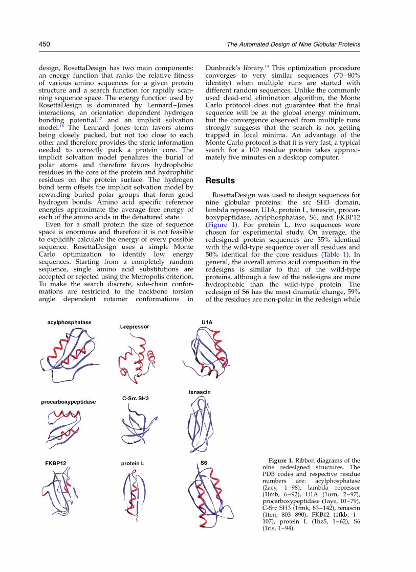
Table 1. Sequence alignments comparing the wild-type sequences (WT) to the design sequences (D)
The Automated Design of Nine Globular Proteins 451
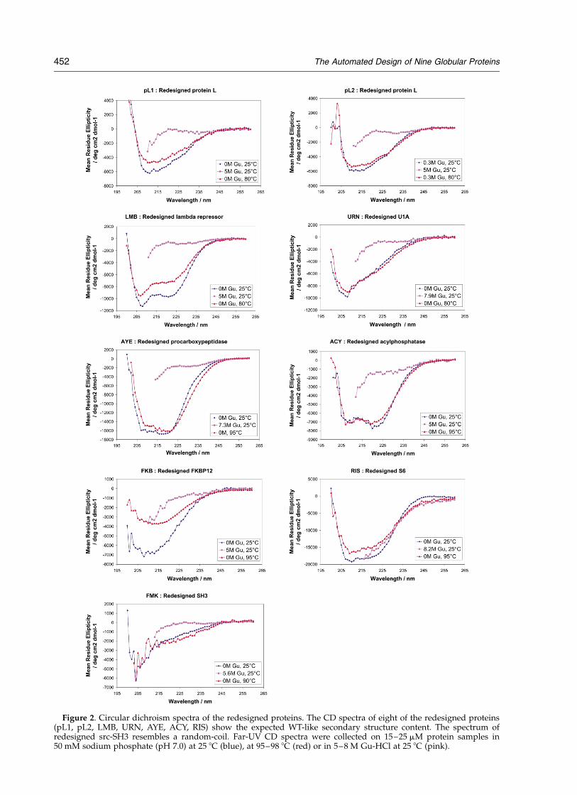
Figure 2. Circular dichroism spectra of the redesigned proteins. The CD spectra of eight of the redesigned proteins(pL1, pL2, LMB, URN, AYE, ACY, RIS) show the expected WT-like secondary structure content. The spectrum ofredesigned src-SH3 resembles a random-coil. Far-UV CD spectra were collected on 15–25 mM protein samples in50 mM sodium phosphate (pH 7.0) at 25 8C (blue), at 95–98 8C (red) or in 5–8 M Gu-HCl at 25 8C (pink).
452 The Automated Design of Nine Globular Proteins
used to determine if the proteins were monomeric(data not shown). Chemical (Figure 3) and thermal(Figure 4) denaturation experiments were used toconfirm that the proteins were folded and to deter-mine their stabilities. One-dimensional 1H NMRexperiments (Figure 5) were used to further con-
firm that the proteins were folded and to probethe rigidity of their structures. On the basis of theresults from these experiments we were able toplace the proteins into different categories:unfolded versus folded, lower or higher than WTstability, and more or less rigid (Table 2).
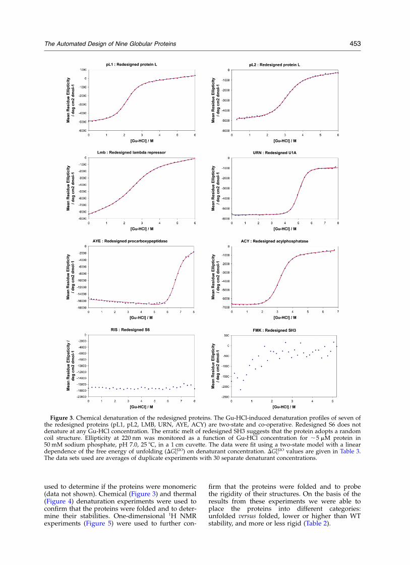
Figure 3. Chemical denaturation of the redesigned proteins. The Gu-HCl-induced denaturation profiles of seven ofthe redesigned proteins (pL1, pL2, LMB, URN, AYE, ACY) are two-state and co-operative. Redesigned S6 does notdenature at any Gu-HCl concentration. The erratic melt of redesigned SH3 suggests that the protein adopts a randomcoil structure. Ellipticity at 220 nm was monitored as a function of Gu-HCl concentration for ,5 mM protein in50 mM sodium phosphate, pH 7.0, 25 8C, in a 1 cm cuvette. The data were fit using a two-state model with a lineardependence of the free energy of unfolding (DGU
H2O) on denaturant concentration. DGUH2O values are given in Table 3.
The data sets used are averages of duplicate experiments with 30 separate denaturant concentrations.
The Automated Design of Nine Globular Proteins 453
Only one of the proteins, the SH3 redesign, isclearly unfolded. The CD spectra of redesignedSH3 (Figure 2) is typical of a random coil and the1D 1H NMR spectrum (Figure 5) shows sharplines and very little dispersion, strongly indicativeof an unfolded protein.
Three of the proteins were multimeric even at
low concentration. The tenascin redesign visiblyaggregates at low concentrations and could not befurther characterized. Size-exclusion chromato-graphy of the FKBP12 and S6 redesignssuggest they form oligomers, but the two proteinsdo not form extensive aggregates and their CDspectra are similar to their naturally occurring
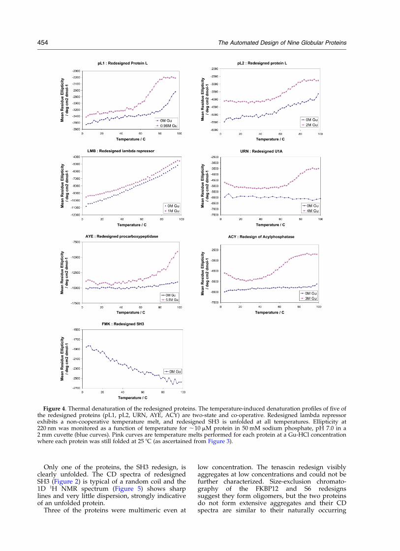
Figure 4. Thermal denaturation of the redesigned proteins. The temperature-induced denaturation profiles of five ofthe redesigned proteins (pL1, pL2, URN, AYE, ACY) are two-state and co-operative. Redesigned lambda repressorexhibits a non-cooperative temperature melt, and redesigned SH3 is unfolded at all temperatures. Ellipticity at220 nm was monitored as a function of temperature for ,10 mM protein in 50 mM sodium phosphate, pH 7.0 in a2 mm cuvette (blue curves). Pink curves are temperature melts performed for each protein at a Gu-HCl concentrationwhere each protein was still folded at 25 8C (as ascertained from Figure 3).
454 The Automated Design of Nine Globular Proteins
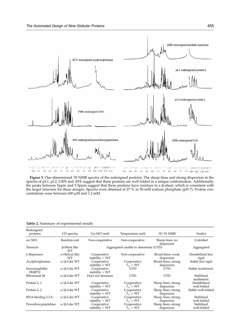
Figure 5. One-dimensional 1H NMR spectra of the redesigned proteins. The sharp lines and strong dispersion in thespectra of pL1, pL2, URN and AYE suggest that these proteins are well folded in a unique conformation. Additionally,the peaks between 5ppm and 5.5ppm suggest that these proteins have residues in a b-sheet, which is consistent withthe target structure for these designs. Spectra were obtained at 27 8C in 50 mM sodium phosphate (pH 7). Protein con-centrations were between 600 mM and 1.2 mM.
Table 2. Summary of experimental results
Redesignedproteins CD spectra Gu-HCl melt Temperature melt 1D 1H NMR Verdict
src SH3 Random-coil Non-cooperative Non-cooperative Sharp-lines; nodispersion
Unfolded
Tenascin b-Sheet likeWT
Aggregated unable to determine (UTD) Aggregated
l-Repressor a-Helical likeWT
Cooperativestability , WT
Non-cooperative Broad-lines; weakdispersion
Destabilised lessrigid
Acylphosphatase a/b-Like WT Cooperativestability ¼ WT
CooperativeTm . WT
Broad-lines; strongdispersion
Stable less rigid
ImmunophillinFKBP12
a/b-Like WT Cooperativestability ¼ WT
UTD UTD Stable multimeric
Ribosomal S6 a/b-Like WT Does not denature UTD UTD Stabilisedmultimeric
Protein L 1 a/b-Like WT Cooperativestability , WT
CooperativeTm . WT
Sharp lines; strongdispersion
Destabilisedwell-folded
Protein L 2 a/b-Like WT Cooperativestability ¼ WT
CooperativeTm . WT
Sharp lines; strongdispersion
Stable well-folded
RNA-binding U1A a/b-Like WT Cooperativestability . WT
CooperativeTm . WT
Sharp lines; strongdispersion
Stabilisedwell-folded
Procarboxypeptidase a/b-Like WT Cooperativestability . WT
CooperativeTm . WT
Sharp lines; strongdispersion
Stabilisedwell-folded
The Automated Design of Nine Globular Proteins 455
counterparts, suggesting that they may adopt thetarget structures. While the FKBP12 redesigndenatured at high guanidine concentration, asevidenced by the change in the CD spectrum(Figure 2), the CD spectrum of redesigned S6 wasremarkably resistant to both temperature andchemical denaturant (Figure 2); hence redesignedS6 may be stabilized by intermolecular as well asintramolecular interactions. Clearly the compu-tational design method, in addition to optimizingthe stability of a given structure, needs to takeinto account solubility issues as well. This mayperhaps be achieved by negative design againstpossible intermolecular interactions, for exampleby placing inwardly pointing charged amino acidsin edge beta strands as suggested by Richardson& Richardson.20
The six remaining redesigned proteins appearmonomeric and folded as evidenced by size-exclusion chromatography, CD spectra, and chemi-cal denaturation experiments. The proteins chro-matographed as monomers by gel-filtrationchromatography, and comparison of their CDspectra (Figure 2) to previously published CDspectra of their naturally occurring counterpartssuggested a very similar distribution of secondarystructures. Chemical denaturation data fit well toa simple two-state folding model (Figure 3), andfor the designed proteins with buried tryptophanresidues, unfolding transitions monitored by CDand by intrinsic fluorescence (data not shown)were coincident, further supporting the two-statemodel typical for small naturally occurringproteins. The free energies of unfolding and theirdenaturant dependencies (m values) for theredesigned proteins were estimated from the fitsof the chemical denaturation data (Figure 3) to thetwo-state model. The m values of the designedproteins are in the range of those of naturallyoccurring small proteins; of the four cases wheredirect comparisons are possible, two of thedesigned proteins have smaller m values thantheir wild-type counterparts, one has a largervalue, and one a very similar value (Table 3). Of
the six proteins, two, the redesigned lambdarepressor and one of the two protein L redesigns(pL1), are clearly less stable than their naturallyoccurring counterparts (Figure 3 and Table 3). Theredesigned acylphosphatase and the secondprotein L redesign (pL2) have roughly the samestability as their naturally occurring counterparts(Table 3). In contrast, the U1A and procarboxy-peptidase redesigns were significantly more stablethan the naturally occurring proteins, redesignedU1A by ,2 kcal/mol and redesigned procarboxy-peptidase by a striking ,7 kcal/mol.
Two common features of many naturally occur-ring proteins are cooperative thermal denaturationtransitions and NMR spectra with strong dis-persion and sharp lines. Both of these featuresappear to be linked to the rigidity of the proteinstructure. In a rigid protein each atom is locatedin a well-defined environment and therefore onlyone sharp NMR peak is observed for eachresonance. In contrast, if the structure is moremolten then the atom may be in multiple differentenvironments on a time-scale relevant to the NMRmeasurement and therefore broad NMR lines areobserved. A highly cooperative thermal transitionindicates a large change in enthalpy upon unfold-ing and is consistent with a change from a rigidfolded protein to a dynamic unfolded protein.
To assess the rigidity of the redesigned proteins,1D NMR spectra and temperature melts wereobtained. Redesigned lambda repressor does nothave a cooperative thermal melt or an NMR spec-trum with sharp lines, suggesting that it may bemore flexible than the other redesigns. Redesignedacylphosphatase has a cooperative thermal melt,but the NMR spectrum has broad lines, whichmay in this case reflect some intermolecular associ-ation at high concentration (and hence slowertumbling times and broader NMR lines).
Remarkably, four of the beta sheet-containingprotein redesigns appear to be as rigid as mostnaturally occurring proteins. The two protein Lredesigns and the redesigns of U1A and pro-carboxypeptidase have cooperative thermal melts
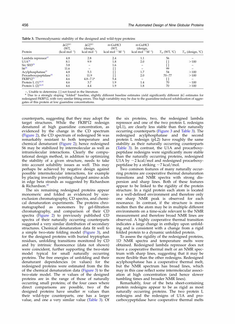
Table 3. Thermodynamic stability of the designed and wild-type proteins
Protein
DGUH2O
(WT,(kcal mol21)
DGUH2O
(design,kcal mol21)
m-GuHCl(WT,
kcal mol21 M21)
m-GuHCl(design,
kcal mol21 M21) Tm (WT, 8C) Tm (design, 8C)
Lambda repressor27 4.8 2.8 2.4 1.1 56 –U1A28 8.1 9.9 1.8 2.0 [ ] .100Src SH329 3.8 – 1.6 – [ ] –S630 11.6 – [ ] – 99 –Acylphosphatase31 4.8 5.3 [ ] 1.7 54 .100Procarboxypeptidase32 4.1 11.9 [ ] 2.0 70–77 .100FKBP1233 4.6 4.8–7.1a 5.4 – [ ] –Protein L (1)34,35 4.6 3.7 1.9 1.4 70 ,100Protein L (2)34,35 4.6 4.4 1.9 1.8 70 .100
–, Unable to determine. [ ] not found in the literature.a Due to a strongly sloping “folded” baseline, slightly different baseline estimates yield significantly different DG estimates for
redesigned FKBP12, with very similar fitting errors. This high variability may be due to the guanidine-induced solubilization of aggre-gates of this protein at low guanidine concentrations.
456 The Automated Design of Nine Globular Proteins
(Figure 4) and NMR spectra with relatively sharplines and good dispersion (Figure 5). In addition,the NMR spectra for these proteins have smallpeaks just downfield of the water (5–5.5ppm) thatare probably from Ca protons on the backboneand are strongly indicative of a b-sheet.21
Another hallmark of naturally occurring proteinsis a large change in heat capacity upon folding.Two of the proteins, redesigned U1A and acyl-phosphatase, cold-denature at intermediate con-centrations of guanidine and estimates of DC8p
could be obtained from fits of temperaturedenaturation experiments to the Gibbs–Hemholtzequation. For both proteins the DC8p per residue isapproximately 10 cal deg21 mol21, which fallswithin the range of DC8p per residue valuesreported for natural proteins of this size.22
Discussion
Here we have shown that RosettaDesign canreliably predict sequences that fold to stablestructures, and that the designed proteins oftenhave features typical of naturally occurringproteins. Half of the folded designs have NMRspectra and temperature melts typical of tightlypacked proteins. These findings significantlyextend the pioneering successful completeredesign of the 25 residue zinc finger Zif2681 to abroad range of considerably larger proteins.
Since so many mutations were made to eachprotein it is difficult to determine why somedesigns were more successful than others, butthere are some trends. Three redesigns were sig-nificantly more stable than their wild-type versions(the redesigns of S6, U1A, and procarboxypepti-dase) and two redesigns were less stable (one ofthe protein L redesigns and the redesign of lambdarepressor). In each of the cases where the designswere more stable, the design sequence had agreater fraction of hydrophobic amino acids thanthe wild-type protein. In the two cases where thedesign was less stable, the redesigned sequenceswere less hydrophobic than those of the wild-typeprotein. These results are consistent with thenotion that the burial of hydrophobic groups isone of the driving forces of protein folding.23
More detailed comparison of the wild-type andredesigned proteins must await high-resolutiondetermination of the structures of the redesignedproteins.
Six of the ten design sequences were soluble andmonomeric at NMR concentrations (1mM) asjudged by gel-filtration, while one was unfolded.Why do the remaining three proteins selfassociate? Redesigned tenascin was visibly aggre-gated even at low concentrations, and therefore itwas not possible to determine if it was folded.One possibility is that it aggregates to such a highdegree because it is unfolded and therefore itshydrophobic core residues are exposed to inter-actions with other molecules. Redesigned S6 and
FKBP12 do not visibly aggregate at low (CD) orhigh (NMR) concentrations, but are multimeric.This lower degree of association, when comparedto redesigned tenascin, is probably due to associ-ation of folded monomers. Indeed, the redesignsof S6 and FKBP12 have an increase in the fractionof non-polar accessible (i.e. “sticky”) surface areacompared to the wild-type counterparts; thiscriterion could be used as a filter to ensure thatfuture designs are soluble. To test this we are cur-rently constructing variants of the S6 and FKBP12redesigns that have fewer hydrophobic residueson their surface. Additionally, redesigned tenascinand FKBP12 seem to lack any of the “aggregationpreventors” that native proteins employ with theiredge beta strands, namely strand kinks or inwardpointing charged residues.20 Since designing akink in any strand would change the redesignbackbone from its native target, we are testing thefeasibility of the second anti-aggregation strategyby constructing variants of redesigned tenascinand FKBP12 that replace edge-strand partially sur-face-exposed hydrophobic residues with chargedresidues.
In only one case, the redesign of the src SH3domain, was the designed protein clearlyunfolded. To examine why this designed sequencewas so unstable, we used the program Probe24 tolook for clashes in our model of the designed pro-tein. Probe identified a large clash between Ile26and Ala39. An examination of the multiplesequence alignment for SH3 domains showed thatthese amino acid residues are often seen at thesepositions, but typically not together.25 There is astrong preference for Ile26 to be paired withGly39, and Leu26 to be paired with Ala39. Theatomic radii used in our simulations are scaled by0.95 relative to CHARMM 19 radii in order to com-pensate for the use of fixed rotamers. If the radiiare increased to their full size, then RosettaDesignshows a strong preference for a Leu-Ala pair. Cur-rently we are testing these findings by mutatingIle26 to Leu or Ala39 to Gly. This is a case whereusing reduced radii can be costly, and suggests theneed for more realistic radii coupled with a bettersampling of side-chain conformational space.
The large-scale test described here establishesthat RosettaDesign can redesign naturally occur-ring proteins with a reasonable chance of success.These encouraging results suggest that the pro-gram is ready to attack the next big challenge inthe field of protein design, the creation of proteinswith novel structures.
Materials and Methods
Computational procedure
Our computational model for protein design, Rosetta-Design, is largely unchanged from that described.13
RosettaDesign contains two main components: an energyfunction that ranks the relative fitness of amino
The Automated Design of Nine Globular Proteins 457
sequences for a given protein structure and a MonteCarlo optimization procedure for rapidly searchingsequence space. The energy function is a linear combi-nation of a 12–6 Lennard–Jones potential, the Lararidis–Karplus implicit solvation model,18 an empirical hydro-gen bonding potential,17 backbone dependent rotamerprobabilities,19 amino acid probabilities for particularregions of phi,psi space, and a simple electrostatics termderived from the probability that two types of polaramino acid residues are found near each other in thePDB.26 In addition, each amino acid has a unique refer-ence energy that controls the frequency that it is placedduring design. Except for the hydrogen bonding term,all of these energies were computed as described pre-viously (see the Supplementary Material for a detaileddescription). The new hydrogen bonding potential wasderived from hydrogen bonding geometries in high-resolution protein structures,17 and is consistent withquantum mechanics calculations on formamide and acet-amide dimers (A. Morozov, & D.B., unpublished results).Environment dependent hydrogen bond weights wereused to roughly account for the reduced dielectric in theprotein interior and the loss of side-chain entropy uponformation of side-chain–side-chain hydrogen bonds onthe surface.17
The Monte Carlo optimization procedure used to scansequence space started with a random sequence. Theside-chain conformations of each amino acid weremodeled using Dunbrack’s backbone dependent rotamerlibrary.19 Only rotamers observed more than 3% of thetime were considered. Each round of Monte Carlo con-sisted of replacing one rotamer, evaluating the energychange, and accepting the change if it passed the Metro-polis criterion. A rotamer replacement may or may notinvolve changing amino acid identity. A typical run con-sisted of a few hundred thousand rotamer replacements,at which point the energy had typically plateaued.
Two rounds of optimization were used for each pro-tein that was redesigned. The first round consisted of100 independent runs in which all 20 amino acidresidues were allowed at each position. Dunbrack’s stan-dard rotamer library was used for this round. During thesecond round the amino acid residues considered at eachsequence position were restricted to those observed atthat position in the results from the first round. Typicallybetween one and five amino acid residues were con-sidered at each position in the second round. Becausethere were fewer amino acid residues being consideredin the second round it was possible to use an expandedrotamer library. In addition to the standard Dunbrackrotamers, new rotamers were constructed with chi anglesplus one and minus one standard deviation away fromthe most commonly observed chi angles. These new rota-mers were given a small energy penalty to account forthe fact that they are sub-optimal. As in the first round,100 independent runs were performed for each proteinin the second round. From these runs, the lowest energysequence was chosen for experimental study. In general,it is not clear if using a second round of design withmore rotamers was helpful. The average identitybetween the design sequences and the native sequencedid not increase from round one to round two.
Protein expression and purification
Genes corresponding to the computationally selectedprotein sequences were purchased from BlueHeronBiotechnologies. The gene constructs were cloned in
plasmid pet29b(þ) (Novagen) and expressed in theBL21(DE3)pLysS strain of E. coli. A 6£ histidine tag atthe C terminus of each construct allowed for the single-step purification of the expressed proteins on a Niþ
affinity column (Pharmacia Biotech). Column-purifiedprotein was dialysed 104-fold against 50 mM sodiumphosphate (pH 7.0), which is the buffer used in all sub-sequent experiments. Protein identity and purity wasdetermined by SDS-PAGE and ESI-MALDI mass spec-troscopy. Protein concentrations were determined byUV absorbance at 280 nm with extinction coefficients cal-culated using the ExPASy Protparam tool†.
Circular dichroism (CD)
CD data were collected on an Aviv 62A DS spec-trometer. Far-UV CD wavelength scans (260–200 nm) atvarying protein concentrations (15–25 mM), guani-dinium hydrochloride (Gu-HCl) concentrations(0–8.3 M), and temperatures (0–98 8C) were collected ina 1 mm pathlength cuvette. Gu-HCl-induced proteindenaturation was followed by change in ellipticity at220 nm in a 1 cm pathlength cuvette, using a Microlabtitrator (Hamilton) for denaturant mixing. Temperaturewas maintained at 25 8C with a Peltier device. All CDdata were converted to mean residue ellipticity. Tem-perature-induced protein denaturation was followed bythe change in ellipticity at 220 nm in a 2 mm pathlengthcuvette. To obtain a value for DGU
H2O, chemical denatura-tion curves were fit by non-linear least-squares analysisusing the linear extrapolation model as applied bySantoro & Bolen. To obtain a value for DC8p, thermaldenaturation curves were fit using the Gibbs–Hemholtzequation in the form:
f ¼ ff þðfu 2 ffÞ
1 þ e2DG8
RT
2DG8 ¼ DH8 1 2T
Tm
� �þ DC8p T 2 Tm 2 T ln
T
Tm
� �� �
where f is CD signal, ff and fu are the estimated CD sig-nal for the folded and unfolded states, respectively, R isthe gas constant, T is temperature, Tm is the temperaturewhere 50% of the protein is folded, DG8 is the change inthe Gibbs free energy for the unfolding reaction, DH8 isthe change in enthalpy, and DC8p is the change in heatcapacity.
Size-exclusion (gel-filtration) chromatography
Size-exclusion chromatography was carried out usingan analytical Superdex-75 column (AmershamPharmacia) with the Pharmacia FPLC system (GP-250gradient programmer, P-500 Pump). Protein samples atNMR concentrations (600 mM–1.2mM) and CD concen-trations (10–40 mM) were equilibrated in 20 mM EDTA,50 mM sodium phosphate (pH 7.0 at 25 8C) and run onthe Superdex-750 column at 1 ml/minute.
Nuclear magnetic resonance
One-dimensional spectra were obtained on a BrukerAMX500 using water presaturation. Spectra wereobtained at 27 8C in 50mM sodium phosphate (pH 7).
† http://us.expasy.org/tools/protparam.html
458 The Automated Design of Nine Globular Proteins
Protein concentrations were between 600 mM and1.2mM.
Solvent-accessible surface area
Solvent-accessible surface area of non-polar atoms wascalculated using the program Whatif†.
Acknowledgements
We thank Lynne R. Spencer, Peter Brzovic, PonniRajagopol, Jennifer Keefe and Rachel Klevitt for aidin obtaining the NMR spectra. B.K. was partiallysupported by a Damon Runyon–Walter WinchellFoundation fellowship. This work was supportedby a grant from the NIH.
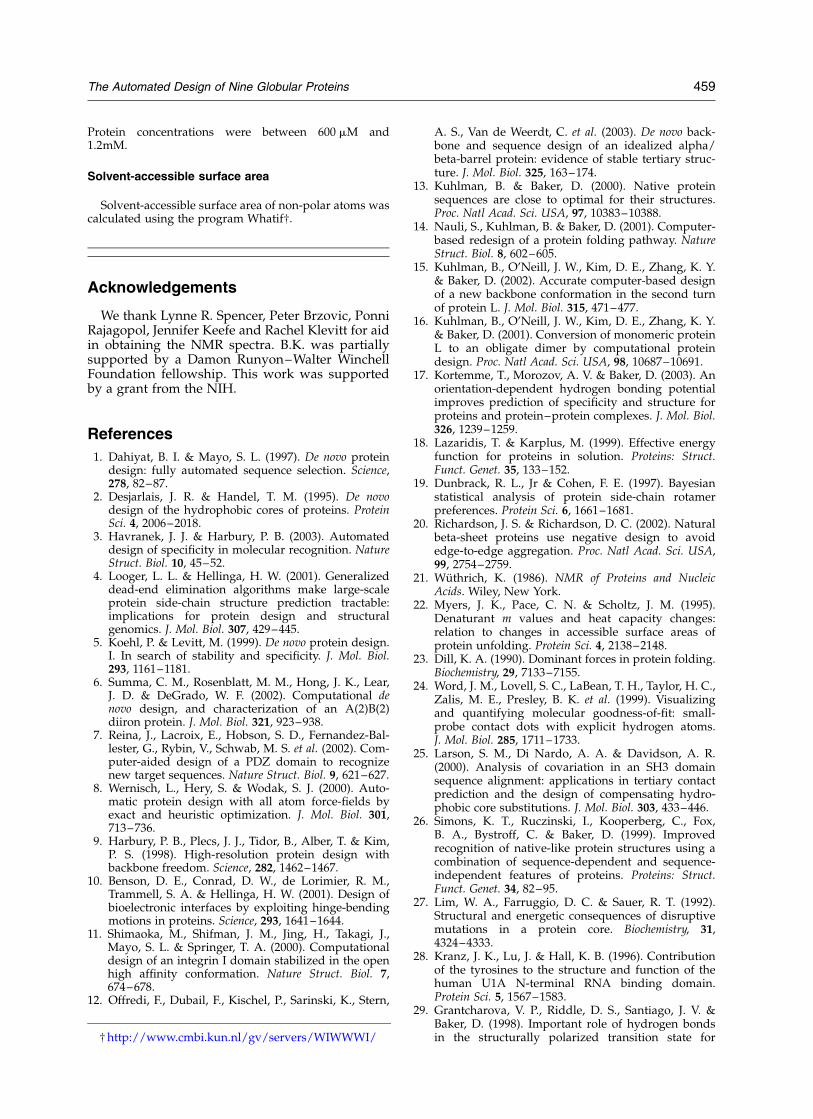
References
1. Dahiyat, B. I. & Mayo, S. L. (1997). De novo proteindesign: fully automated sequence selection. Science,278, 82–87.
2. Desjarlais, J. R. & Handel, T. M. (1995). De novodesign of the hydrophobic cores of proteins. ProteinSci. 4, 2006–2018.
3. Havranek, J. J. & Harbury, P. B. (2003). Automateddesign of specificity in molecular recognition. NatureStruct. Biol. 10, 45–52.
4. Looger, L. L. & Hellinga, H. W. (2001). Generalizeddead-end elimination algorithms make large-scaleprotein side-chain structure prediction tractable:implications for protein design and structuralgenomics. J. Mol. Biol. 307, 429–445.
5. Koehl, P. & Levitt, M. (1999). De novo protein design.I. In search of stability and specificity. J. Mol. Biol.293, 1161–1181.
6. Summa, C. M., Rosenblatt, M. M., Hong, J. K., Lear,J. D. & DeGrado, W. F. (2002). Computational denovo design, and characterization of an A(2)B(2)diiron protein. J. Mol. Biol. 321, 923–938.
7. Reina, J., Lacroix, E., Hobson, S. D., Fernandez-Bal-lester, G., Rybin, V., Schwab, M. S. et al. (2002). Com-puter-aided design of a PDZ domain to recognizenew target sequences. Nature Struct. Biol. 9, 621–627.
8. Wernisch, L., Hery, S. & Wodak, S. J. (2000). Auto-matic protein design with all atom force-fields byexact and heuristic optimization. J. Mol. Biol. 301,713–736.
9. Harbury, P. B., Plecs, J. J., Tidor, B., Alber, T. & Kim,P. S. (1998). High-resolution protein design withbackbone freedom. Science, 282, 1462–1467.
10. Benson, D. E., Conrad, D. W., de Lorimier, R. M.,Trammell, S. A. & Hellinga, H. W. (2001). Design ofbioelectronic interfaces by exploiting hinge-bendingmotions in proteins. Science, 293, 1641–1644.
11. Shimaoka, M., Shifman, J. M., Jing, H., Takagi, J.,Mayo, S. L. & Springer, T. A. (2000). Computationaldesign of an integrin I domain stabilized in the openhigh affinity conformation. Nature Struct. Biol. 7,674–678.
12. Offredi, F., Dubail, F., Kischel, P., Sarinski, K., Stern,
A. S., Van de Weerdt, C. et al. (2003). De novo back-bone and sequence design of an idealized alpha/beta-barrel protein: evidence of stable tertiary struc-ture. J. Mol. Biol. 325, 163–174.
13. Kuhlman, B. & Baker, D. (2000). Native proteinsequences are close to optimal for their structures.Proc. Natl Acad. Sci. USA, 97, 10383–10388.
14. Nauli, S., Kuhlman, B. & Baker, D. (2001). Computer-based redesign of a protein folding pathway. NatureStruct. Biol. 8, 602–605.
15. Kuhlman, B., O’Neill, J. W., Kim, D. E., Zhang, K. Y.& Baker, D. (2002). Accurate computer-based designof a new backbone conformation in the second turnof protein L. J. Mol. Biol. 315, 471–477.
16. Kuhlman, B., O’Neill, J. W., Kim, D. E., Zhang, K. Y.& Baker, D. (2001). Conversion of monomeric proteinL to an obligate dimer by computational proteindesign. Proc. Natl Acad. Sci. USA, 98, 10687–10691.
17. Kortemme, T., Morozov, A. V. & Baker, D. (2003). Anorientation-dependent hydrogen bonding potentialimproves prediction of specificity and structure forproteins and protein–protein complexes. J. Mol. Biol.326, 1239–1259.
18. Lazaridis, T. & Karplus, M. (1999). Effective energyfunction for proteins in solution. Proteins: Struct.Funct. Genet. 35, 133–152.
19. Dunbrack, R. L., Jr & Cohen, F. E. (1997). Bayesianstatistical analysis of protein side-chain rotamerpreferences. Protein Sci. 6, 1661–1681.
20. Richardson, J. S. & Richardson, D. C. (2002). Naturalbeta-sheet proteins use negative design to avoidedge-to-edge aggregation. Proc. Natl Acad. Sci. USA,99, 2754–2759.
21. Wuthrich, K. (1986). NMR of Proteins and NucleicAcids. Wiley, New York.
22. Myers, J. K., Pace, C. N. & Scholtz, J. M. (1995).Denaturant m values and heat capacity changes:relation to changes in accessible surface areas ofprotein unfolding. Protein Sci. 4, 2138–2148.
23. Dill, K. A. (1990). Dominant forces in protein folding.Biochemistry, 29, 7133–7155.
24. Word, J. M., Lovell, S. C., LaBean, T. H., Taylor, H. C.,Zalis, M. E., Presley, B. K. et al. (1999). Visualizingand quantifying molecular goodness-of-fit: small-probe contact dots with explicit hydrogen atoms.J. Mol. Biol. 285, 1711–1733.
25. Larson, S. M., Di Nardo, A. A. & Davidson, A. R.(2000). Analysis of covariation in an SH3 domainsequence alignment: applications in tertiary contactprediction and the design of compensating hydro-phobic core substitutions. J. Mol. Biol. 303, 433–446.
26. Simons, K. T., Ruczinski, I., Kooperberg, C., Fox,B. A., Bystroff, C. & Baker, D. (1999). Improvedrecognition of native-like protein structures using acombination of sequence-dependent and sequence-independent features of proteins. Proteins: Struct.Funct. Genet. 34, 82–95.
27. Lim, W. A., Farruggio, D. C. & Sauer, R. T. (1992).Structural and energetic consequences of disruptivemutations in a protein core. Biochemistry, 31,4324–4333.
28. Kranz, J. K., Lu, J. & Hall, K. B. (1996). Contributionof the tyrosines to the structure and function of thehuman U1A N-terminal RNA binding domain.Protein Sci. 5, 1567–1583.
29. Grantcharova, V. P., Riddle, D. S., Santiago, J. V. &Baker, D. (1998). Important role of hydrogen bondsin the structurally polarized transition state for† http://www.cmbi.kun.nl/gv/servers/WIWWWI/
The Automated Design of Nine Globular Proteins 459

folding of the src SH3 domain. Nature Struct. Biol. 5,714–720.
30. Uversky, V. N., Abdullaev, Z. Kh., Arseniev, A. S.,Bocharov, E. V., Dolgikh, D. A., Latypov, R. F. et al.(1999). Structure and stability of recombinant proteindepend on the extra N-terminal methionine residue:S6 permutein from direct and fusion expressionsystems. Biochim. Biophys. Acta, 1432, 324–332.
31. Taddei, N., Chiti, F., Paoli, P., Fiaschi, T., Bucciantini,M., Stefani, M. et al. (1999). Thermodynamics andkinetics of folding of common-type acylphosphatase:comparison to the highly homologous muscle iso-enzyme. Biochemistry, 38, 2135–2142.
32. Villegas, V., Azuaga, A., Catasus, L., Reverter, D.,Mateo, P. L., Aviles, F. X. & Serrano, L. (1995).Evidence for a two-state transition in the foldingprocess of the activation domain of human pro-carboxypeptidase A2. Biochemistry, 34, 15105–15110.
33. Veeraraghavan, S., Holzman, T. F. & Nall, B. T. (1996).Autocatalyzed protein folding. Biochemistry, 35,10601–10607.
34. Scalley, M. L., Yi, Q., Gu, H., McCormack, A., Yates,J. R., III & Baker, D. (1997). Kinetics of folding of the
IgG binding domain of peptostreptococcal proteinL. Biochemistry, 36, 3373–3382.
35. Yi, Q., Scalley, M. L., Simons, K. T., Gladwin, S. T. &Baker, D. (1997). Characterization of the free energyspectrum of peptostreptococcal protein L. Fold. Des.2, 271–280.
Edited by B. Honig
(Received 29 January 2003; received in revised form3 July 2003; accepted 8 July 2003)
Supplementary Material comprising text andone Table is available on Science Direct
460 The Automated Design of Nine Globular Proteins