Hepatic Atrophy-Hypertrophy Complex Due to Echinococcus granulosus
Upload
independentCategory
view
0download
0
A Family of Diverse Kunitz Inhibitors from Echinococcusgranulosus Potentially Involved in Host-ParasiteCross-TalkSilvia Gonzalez1., Martın Flo1,2., Mariana Margenat1, Rosario Duran3, Gualberto Gonzalez-Sapienza1,
Martın Grana4, John Parkinson5, Rick M. Maizels6, Gustavo Salinas1, Beatriz Alvarez2, Cecilia
Fernandez1*
1 Catedra de Inmunologıa, Facultad de Quımica, Universidad de la Republica, Montevideo, Uruguay, 2 Laboratorio de Enzimologıa, Facultad de Ciencias, Universidad de la
Republica, Montevideo, Uruguay, 3 Unidad de Bioquımica y Proteomica Analıticas, Institut Pasteur de Montevideo and Instituto de Investigaciones Biologicas Clemente
Estable, Uruguay, 4 Unidad de Bioinformatica, Institut Pasteur de Montevideo, Montevideo, Uruguay, 5 Program in Molecular Structure and Function, Hospital for Sick
Children, Toronto, Canada, 6 Institute of Immunology and Infection Research, University of Edinburgh, Edinburgh, United Kingdom
Abstract
The cestode Echinococcus granulosus, the agent of hydatidosis/echinococcosis, is remarkably well adapted to its definitivehost. However, the molecular mechanisms underlying the successful establishment of larval worms (protoscoleces) in thedog duodenum are unknown. With the aim of identifying molecules participating in the E. granulosus-dog cross-talk, wesurveyed the transcriptomes of protoscoleces and protoscoleces treated with pepsin at pH 2. This analysis identified amultigene family of secreted monodomain Kunitz proteins associated mostly with pepsin/H+-treated worms, suggestingthat they play a role at the onset of infection. We present the relevant molecular features of eight members of the E.granulosus Kunitz family (EgKU-1 – EgKU-8). Although diverse, the family includes three pairs of close paralogs (EgKU-1/EgKU-4; EgKU-3/EgKU-8; EgKU-6/EgKU-7), which would be the products of recent gene duplications. In addition, we describethe purification of EgKU-1 and EgKU-8 from larval worms, and provide data indicating that some members of the family(notably, EgKU-3 and EgKU-8) are secreted by protoscoleces. Detailed kinetic studies with native EgKU-1 and EgKU-8highlighted their functional diversity. Like most monodomain Kunitz proteins, EgKU-8 behaved as a slow, tight-bindinginhibitor of serine proteases, with global inhibition constants (KI
*) versus trypsins in the picomolar range. In sharp contrast,EgKU-1 did not inhibit any of the assayed peptidases. Interestingly, molecular modeling revealed structural elementsassociated with activity in Kunitz cation-channel blockers. We propose that this family of inhibitors has the potential to actat the E. granulosus-dog interface and interfere with host physiological processes at the initial stages of infection.
Citation: Gonzalez S, Flo M, Margenat M, Duran R, Gonzalez-Sapienza G, et al. (2009) A Family of Diverse Kunitz Inhibitors from Echinococcus granulosusPotentially Involved in Host-Parasite Cross-Talk. PLoS ONE 4(9): e7009. doi:10.1371/journal.pone.0007009
Editor: Clotilde K. S. Carlow, New England Biolabs, United States of America
Received July 3, 2009; Accepted August 3, 2009; Published September 17, 2009
Copyright: � 2009 Gonzalez et al. This is an open-access article distributed under the terms of the Creative Commons Attribution License, which permitsunrestricted use, distribution, and reproduction in any medium, provided the original author and source are credited.
Funding: This work was supported by The Wellcome Trust (http://www.wellcome.ac.uk/) International Traveling Research Fellowship to CF sponsored by RMM(Ref 061168); the Uruguayan Research Council (http://www.dicyt.gub.uy/), Grant PDT 54/173 to CF; and PEDECIBA, Programa para el Desarrollo de las CienciasBasicas Uruguay (http://www.pedeciba.edu.uy/). The funders had no role in study design, data collection and analysis, decision to publish, or preparation of themanuscript.
Competing Interests: The authors have declared that no competing interests exist.
* E-mail: [email protected]
. These authors contributed equally to this work.
Introduction
Echinococcus granulosus is a member of a major, though neglected
class of helminth parasites, the cestodes. It is the agent of a
medically and economically important cosmopolitan zoonosis,
with endemic foci in every inhabited continent [1]. This organism
requires two mammals for completion of its life cycle: in
intermediate hosts (various ungulates, mostly domestic cattle and
also humans), eggs develop into metacestodes (hydatid cysts)
containing larval worms (protoscoleces) at visceral sites. When the
canine definitive host ingests infected flesh, protoscoleces evaginate
and attach to the mucosa of the dog duodenum, where they
develop to hermaphroditic adult worms producing eggs over a
period of several weeks. In dogs, the infection is referred to as
echinococcosis.
E. granulosus is extremely well adapted to its definitive host: it can
reside in the dog gut for long periods without causing any apparent
damage; the dog, in turn, usually develops an immune response that
has little effect on the parasite [2,3]. Specific anatomical structures
allow a very close contact at the canid-worm interface; indeed, the
intimacy of this contact has led E. granulosus to be regarded as both a
tissue and a luminal parasite [4]. At the onset of infection, freshly
evaginated protoscoleces attach to the mucosa at the base of a crypt
of Lieberkhun by means of suckers, with a rostellum pushed deeply
into the crypt (occasionally, even reaching the lamina propria). The
apical end of the scolex contains the rostellar gland, whose secretion
is thought to be important for protoscolex development [5]. The
specific molecular mechanisms by which larval worms establish a
successful infection in the hostile environment of the dog duodenum
are, however, largely unknown.
PLoS ONE | www.plosone.org 1 September 2009 | Volume 4 | Issue 9 | e7009

With the aim of identifying molecules participating in the E.
granulosus–dog cross-talk, we surveyed the genes expressed by
protoscoleces and protoscoleces treated with pepsin at pH 2.
Because the larval worms are naturally exposed to these signals
immediately after being ingested by the dog, the rationale was that
pepsin/H+ treatment would induce the expression of relevant
genes for parasite establishment in the definitive host [6]. Analysis
of the larval worm transcriptome (Parkinson J, Maizels RM,
Fernandez C, unpublished) revealed the existence of a multigene
family of Kunitz inhibitors expressed mostly in pepsin/H+-treated
protoscoleces, suggesting that these molecules play a role at the
initial phases of infection. Kunitz inhibitors are a class of serine
protease inhibitors present in all metazoa, whose prototype is the
bovine pancreatic inhibitor of trypsin (BPTI; family I2 of the
MEROPS database [7,8]). They are competitive inhibitors acting
in a substrate-like manner, that form very stable complexes of 1:1
stoichiometry with their target enzymes, devoid of activity [9].
Kunitz inhibitors are also frequent components of the venoms
from poisonous animals (snakes [10]; sea anemones [11,12]; cone
snails [13]; spiders [14]); in such cases, they are referred to as
‘‘Kunitz-type toxins’’. Interestingly, some Kunitz-type toxins
display a different activity besides serine protease inhibition: they
block various types of cation permeating channels. Furthermore,
several examples exist of Kunitz-type toxins acting solely as
channel blockers; some neurotoxins present in the venoms of
mamba snakes (‘‘dendrotoxins’’), whose function is to paralyze the
prey, are the best characterized example [15].
In this article, we present the relevant molecular features of the
E. granulosus family of Kunitz-type inhibitors which, to date,
includes eight members: EgKU-1 to EgKU-8 (E. granulosus Kunitz
protein 1 to 8). In addition, we describe the purification to
homogeneity of EgKU-1 and EgKU-8 from larval worms and
provide evidence of the occurrence of some members of the family
in protoscolex secretions. We also present the results of detailed
kinetic studies of the purified inhibitors with a panel of serine
proteases that highlight their functional diversity: EgKU-8 is a slow
tight-binding inhibitor of trypsins; whereas EgKU-1 does not
inhibit any of the assayed peptidases. Interestingly, molecular
modeling reveals that structural elements associated with activity
of a-dendrotoxin, which is a selective blocker of specific voltage-
activated K+-channels, are also present in EgKU-1. Considered
globally, our results allow us to propose that the expression of this
gene family is a strategy that allows E. granulosus to control host
processes and contribute to initiate a successful infection in the dog
duodenum.
Results
Protoscoleces express a family of diverse Kunitzinhibitors
In the context of a strategy to identify molecules participating in
the host-parasite cross-talk in hydatid infections, we undertook an
EST-based transcriptome analysis of E. granulosus larval stages [6].
A major feature of the protoscolex transcriptome was the
identification of seven members of the Kunitz family of inhibitors
that we named EgKU-1-EgKU-7 (Figure 1A and Table S1).
Interestingly, 5 of these transcripts were associated with the
pepsin/H+-treated parasites; furthermore, ESTs from cDNAs
encoding Kunitz inhibitors represented about 1% of the total
derived from the treated protoscolex cDNA library (16 out of
1500), but only 0.1% (2 out of 1500) from the library of untreated
parasites. Notably, two transcripts (EgKU-1 and EgKU-2) were
among the products with highest representation in the treated
protoscolex library (see Table S1).
It is predicted from sequence analyses that these proteins are
secreted and the corresponding mature peptides contain a single
‘‘Kunitz domain’’: about 50 amino acids forming a compact a + bstructure (two short segments of a-helix located at the N and C-
terminal ends of the domain + two b strands), cross-linked by three
disulfide bonds between the conserved Cys residues, arranged in
the canonical topology 1:6, 2:4 and 3:5. As usual among members
of the Kunitz/I2 family, similarity of the E. granulosus proteins is
higher towards the C-terminal half of the domain, whereas the
antiproteinase site (the P1 position, 15 in Figure 1A, and
neighboring residues - notation of Schetcher and Berger, [16]) is
within its most variable region. While all showing the architecture
of a signal peptide followed by a single Kunitz domain, an
extended C-terminal region is seen in some proteins (EgKU-2,
EgKU-6 and EgKU-7). In addition, they differ in isoelectric point:
EgKU-3 and EgKU-7 are acidic, EgKU-6 neutral; the remaining
basic or very basic. Perhaps most significantly, the homologs differ
in the residue present at the reactive P1 site; consequently, if
behaving as serine protease inhibitors, diverse specificities could be
expected: an Arg in P1 (as in EgKU-4 – EgKU-8) is associated with
activity towards trypsin-like peptidases; Trp (EgKU-2) and Leu
(EgKU-3) towards chymotrypsin-like enzymes; whereas Gln
(EgKU-1) -although rare at P1 sites- is compatible with activity
towards various serine proteases [17,18] (Figure 1A and Table S1).
A detailed inspection of the sequences indicated another
interesting feature of the family, namely, that EgKU-1/EgKU-4
and EgKU-6/EgKU-7 represent two pairs of close paralogs, which
possess highly similar predicted mature proteins and, in the case of
EgKU-1/EgKU-4, almost identical signal peptides (Figure 1A).
When the Kunitz domains of EgKU-1 to EgKU-7 were
compared with protein domain databases, good similarity was
observed with homologous domains in other proteins (present in
either single or multi-, homo as well as hetero- domain proteins)
with high levels of identity, except for EgKU-2 (Table S1).
Furthermore, the Gly12, Phe33, Gly37 and Gly40 residues, which
are conserved in the whole Kunitz/I2 family, are also present in
the E. granulosus proteins, except in EgKU-2 (where they are
substituted by Ala12 and Ser at the other positions, see Figure 1A).
These observations highlighted that EgKU-2 is a rather atypical
Kunitz protein.
Because substantial sequence information is now available from
platyhelminth species previously under-represented in databases,
appropriate searches were carried out using EgKU-1 to EgKU-7 as
queries against EST databases (dbEST and also databases
associated with specific sequencing projects). This led to the
identification of highly similar sequences among other platyhel-
minths with likely orthologs of EgKU-3 and EgKU-4, and of
EgKU-2 respectively, among ESTs from E. multilocularis [19] and
Taenia solium [20]. In the case of E. multilocularis, for which full-
length cDNAs are available, the corresponding ‘‘EmKU-3’’ and
‘‘EmKU-4’’ predicted proteins were more than 90% identical to
EgKU-3 and EgKU-4 over the whole sequence, including the
signal peptide. In the case of T. solium ‘‘TsKU-2’’, identity was
about 85% and also extended to the available partial signal
peptide sequence. Moreover, this analysis allowed cDNAs
encoding additional Kunitz inhibitors similar to the E. granulosus
proteins to be identified. Indeed, a second cDNA related to EgKU-
3 (70% identity) was found to be present in E. multilocularis; and a
close homolog of EgKU-6 (75% identity) as well as several cDNAs
related to EgKU-4 (about 60% identity) in T. solium. Finally,
sequences bearing significant similarity with the Kunitz domain of
EgKU-5 were recognized among ESTs from the free-living
planarians Dugesia ryukyvensis and Schmidtea mediterranea (up to 60%
identity) (Figure 1B and Table S1).
E. granulosus Kunitz Proteins
PLoS ONE | www.plosone.org 2 September 2009 | Volume 4 | Issue 9 | e7009

Sequence alignment of the newly-identified proteins highlighted
the striking (though not unprecedented) level of identity between
putative orthologs of both Echinococcus species, qualitatively similar
at the nucleotide level. In addition, it revealed that the two E.
multilocularis molecules similar to EgKU-3 would constitute a pair
of close paralogs, as previously noted for EgKU-1/EgKU-4 and
EgKU-6/EgKU-7. We thus hypothesized that the second member
of the pair would also be expressed in E. granulosus and attempted
to isolate the corresponding full-coding cDNA with a set of
oligonucleotide primers designed on the basis of the E. multilocularis
sequence. RT-PCR using RNA from pepsin/H+ treated proto-
scoleces yielded a product migrating as a single band in agarose-
gel electrophoresis. Sequencing of the cloned PCR product
revealed an open reading frame of 228 nt encoding a 75 amino
acids polypeptide, differing from the E. multilocularis amino acid
sequence in a single residue (position 46 was Glu in E. granulosus
and Gly in E. multilocularis; identity at the nucleotide level was also
very high, 225/228). This new member of the E. granulosus Kunitz
family was named EgKU-8 (Figure 1A and Table S1).
Phylogenetic analysis of the Kunitz domains from E. granulosus
and related platyhelminth sequences (see Table S1) confirmed the
relatedness among sequences from different species and, also, that
the family includes three pairs of close paralogs that would be the
products of recent gene duplications. In the case of the ‘‘KU-3/
KU-8’’ pair, two genes were already present in the common
ancestor of the two Echinococcus species (Figure S1). This analysis
also emphasized that EgKU-2 is an atypical Kunitz protein;
interestingly, the same unusual substitutions of conserved residues
(Gly12Ala, Phe33Ser, Gly37Ser and Gly40Ser) were observed in
the putative ortholog identified in T. solium (Figure 1B).
Kunitz inhibitors may be purified from protoscoleces anddetected in their secretions
As part of an independent strategy aimed at isolating positively
charged molecules from E. granulosus larval worms, a soluble
extract was fractionated by cationic exchange chromatography at
pH 7. Column elution with a linear NaCl gradient yielded two
peaks that were analyzed by non-reducing Tricine-SDS-PAGE.
Interestingly, despite the fact that the starting material was a crude
preparation, the profile of the fractions corresponding to the minor
peak (fractions 20 to 23 in Figure 2A) was extremely simple: they
contained a major band of approximately 7 kDa. This band was
submitted to N-terminal sequencing, and the 21 determined
residues found to correspond to the mature form of EgKU-1, the
most abundant Kunitz inhibitor identified in the protoscolex
transcriptome (see Figures 1A and 2A).
Although the N-terminal amino acids were unambiguously
assigned and only one signal was obtained in each sequencing
cycle, further analysis of the fractions containing EgKU-1 by mass-
spectrometry revealed two peaks (Figure 2B) of m/z 6601.9 and
6521.2, matching the predicted monocharged molecular ion mass
Figure 1. The E. granulosus Kunitz family and related cestode proteins. (A) Comparison of the full-length amino acid sequences of EgKU-1 –EgKU-8. The alignment was constructed using Clustal W2 [70] and manually refined to separate predicted signal peptides and mature proteins (theputative signal peptidase cleavage site is marked with an arrow). Conserved Cys residues are highlighted in grey and the canonical topology ofdisulfide bonds indicated; numbers refer to mature BPTI (58 amino acids), as well as the elements of secondary structure specified on top of thealignment to provide a global view of the domain fold. Residues present throughout are marked with (*) and conservative replacements with (:).Amino acids at position 15 (corresponding to the P1 site of serine protease inhibitors) are in white with black shading, and unusual substitutions inEgKU-2 (at positions 12, 33, 37 and 40) in white with dark grey shading. (B) Comparison of E. granulosus and related cestode Kunitz proteins.Alignments were constructed as in (a) with the three pairs of close E. granulosus paralogs – EgKU-1/EgKU-4, EgKU-3/EgKU-8 and EgKU-6/EgKU-7 – andEgKU-2, together with highly similar proteins predicted from E. multilocularis and T. solium ESTs. EmKU-3, EmKU-4 and EmKU-8 correspond,respectively, to XvEMa04137, XvMa03312 and XvMa16368 in Full Echinococcus [19]; Ts-1, Ts-2 and Ts-3 were deduced from sequences EL763407,EL746785 and EL743839 in dbEST (refer to Table S1 for further details). Unusual substitutions in the Kunitz domains of EgKU-2 (conserved in Ts-2) areindicated as in (A); the substitution of the second conserved Cys in Ts-1 is similarly marked.doi:10.1371/journal.pone.0007009.g001
E. granulosus Kunitz Proteins
PLoS ONE | www.plosone.org 3 September 2009 | Volume 4 | Issue 9 | e7009

Figure 2. Purification of EgKU-1 and EgKU-8 from a protoscolex extract. (A) Fractionation of cationic protoscolex proteins. Left panel,Chromatography profile (MonoS column; pH 7); elution was with increasing NaCl concentrations. Right panel, Tricine SDS-PAGE of eluted fractionsunder non-reducing conditions; the gel was Coomassie-stained. N-terminal sequencing of the 7 kDa band identified EgKU-1 in fractions 21 and 22(see Figure 1A). Conserved Cys are in grey and the putative P1 site marked with an asterisk. (B) MALDI-TOF MS of EgKU-1containing fractions. Thepeak at m/z 6601.9 matched the MH+ value predicted for EgKU-1 (6600.5 Da); similarly, the 6521.2 signal indicated that the fractions also containedEgKU-8 (MH+ = 6520.4 Da). MW estimation was from cDNA predicted sequences, considering that conserved Cys form disulfide bonds (see Table S1).(C) Separation of EgKU-1 and EgKU-8 by rpHPLC. A pool of the ion exchange fractions containing the 7 kDa band was loaded onto a C8 column. Amajor and a minor peak were eluted with acetonitrile (ACN) in 0.07% trifluoroacetic acid, at about 16% (*) and 24% (**) ACN. By MALDI-TOF MS, eachpeak was found to contain one predominant component corresponding, respectively, to EgKU-1 and EgKU-8, as specified in (B).doi:10.1371/journal.pone.0007009.g002
E. granulosus Kunitz Proteins
PLoS ONE | www.plosone.org 4 September 2009 | Volume 4 | Issue 9 | e7009

(MH+) for the mature forms of EgKU-1 (6600.5 Da) and EgKU-8
(6520.4 Da), respectively (see Table S1; note that both inhibitors
would be positively charged at pH 7). The two components were
separated by rpHPLC, as confirmed by MALDI-TOF MS
(Figure 2C). Peptide finger-printing of the component recovered
in the second rpHPLC peak (whose molecular mass matched the
MW of EgKU-8) allowed confirmation of its identity and its
unequivocal assignment to EgKU-8 (Figure S2). EgKU-1 and
EgKU-8 were thus purified to homogeneity from a protoscolex
extract using a combination of cation exchange and reverse phase
chromatography.
To approach the question of whether Kunitz inhibitors are,
indeed, secreted to the parasite-host interface, we analyzed the
supernatants from cultured protoscoleces by mass spectrometry.
Aliquots from freshly isolated parasites were either left untreated
or treated with pepsin/H+ prior to the culture. Figure 3 shows
representative MALDI-TOF MS profiles (5000 – 12000 Da) of
supernatants from short-term (3 h) cultures of the larval worms.
Two major peaks of m/z 6406.9 and 6520.7, matching the
predicted MH+ value for EgKU-3 (6406.4 Da) and EgKU-8
(6520.4 Da), respectively (Figure 3A), were observed in the
supernatant from untreated worms. The secretions from pepsin/
H+-treated protoscoleces were considerably more complex and
included signals of m/z 6405.3 and 6519.6, also consistent with
the presence of EgKU-3 and EgKU-8 (Figure 3B). A peak of m/z
6594.1 was detected as well; while close, this does not accurately
match the MH+ predicted for EgKU-1 (within about 1 Da as was
the case for the other molecules and also for EgKU-1 in the ion
exchange eluate, Figure 2B). Although the identity of the peaks
needs further confirmation, the results indicate that members of
the Kunitz family would be present in protoscolex secretions at the
onset of infection. It is worth noting that EgKU-3 is an acidic
protein that would be negatively charged at pH 7; thus, even if it
had been present in the starting protoscolex extract, it would not
Figure 3. Detection of members of the Kunitz family in protoscolex secretions. Analysis by MALDI-TOF MS of supernatants from short-termcultures of (A) untreated and (B) pepsin/H+-treated protoscoleces. The spectra highlight the different complexity of the two samples. Signals whosem/z values could derive from the presence of E. granulosus Kunitz inhibitors are indicated, together with the MH+ predicted for each protein based ontranslation of the corresponding cDNA, considering that the six conserved Cys residues form disulfide bonds (seeTable S1).doi:10.1371/journal.pone.0007009.g003
E. granulosus Kunitz Proteins
PLoS ONE | www.plosone.org 5 September 2009 | Volume 4 | Issue 9 | e7009

have bound to the cation exchange column used for the isolation
of EgKU-1 and EgKU-8.
EgKU-8 is a high affinity trypsin inhibitor whereas noinhibition of serine protease activity was detected withEgKU-1
A preliminary screening (not shown) of serine protease
inhibitory activity was carried out with purified native EgKU-1
and EgKU-8. Taking into account the usual inhibition profiles of
proteins from the Kunitz family, we assayed pancreatic enzymes
(bovine cationic trypsin and chymotrypsin A; and porcine elastase)
and serine proteases of the coagulation cascade.
EgKU-8 displayed dose-dependent inhibitory activities against
trypsin and chymotrypsin, whereas no inhibition of elastase was
detected, even at high concentrations of the parasite protein. As
expected for Kunitz inhibitors which are extremely stable proteins
[21,22], the activity was heat-resistant: around 80% and 65% of
the inhibitory capacity towards trypsin and chymotrypsin
respectively, was retained after 20 min at 100uC. Activity towards
serine proteases of the coagulation cascade was tested through
measurement of prothrombin time and partial thromboplastin
time; two functional assays which are highly sensitive for factors X,
VII and II, and factors XII, XI, X, IX and II, correspondingly. No
increase of either time was observed using normal human plasma,
indicating that EgKU-8 did not inhibit these enzymes.
In view of the behavior of EgKU-8 with bovine trypsin and
chymotrypsin, and assuming that the host digestive enzymes could
be physiological targets of the parasite molecule, we next analyzed
its activity against trypsin and chymotrypsin purified from dog
pancreas, i. e. anionic and cationic trypsins and chymotrypsin B
(chymotrypsin A is absent from dogs, see S01.001 at MEROPS -
http://merops.sanger.ac.uk). In parallel, we also assayed the
bovine enzymes (i. e. cationic trypsin and chymotrypsin A). To
obtain global inhibition constants (KI*) for EgKU-8, data of vi versus
[I] were fit to equation (1). Representative results for canine
anionic trypsin are shown in Figure 4A, and the values of KI* for
each enzyme in Table 1. EgKU-8 behaved as a very efficient
inhibitor of both canine trypsins, with KI* of 1764 and 2268 pM
for the cationic and anionic enzymes, respectively (Table 1). It also
inhibited bovine trypsin although to a lower extent than the canine
counterparts, since KI* was ,3-fold higher. In contrast, no
appreciable inhibition of chymotrypsin B was detected, while the
KI* obtained for chymotrypsin A was in the nanomolar range, two
orders of magnitude higher than the KI* for trypsins. Results of
EgKU-8 with chymotrypsins reproduced those obtained with
BPTI, which was also found to inhibit chymotrypsin A but not the
B isoforms of the protease, from either bovine [23] or from canine
(our unpublished observations) origin.
The progress curves for the inhibition, as shown for anionic
trypsin in Figure 4B, indicated that, in the presence of EgKU-8,
the rate of substrate hydrolysis reached the inhibited steady-state
rate in a time scale of minutes, suggesting that the formation of the
enzyme-inhibitor complex is a slow process and that EgKU-8 is a
slow-binding inhibitor as defined by Morrison [24]. The
interaction of EgKU-8 with all three trypsins was reversible, since
progress curves reached appreciable slopes even at higher than
stoichiometric inhibitor concentrations. This is the behavior
expected for Kunitz-type inhibitors [9].
Similarly, plots of the apparent rate constant (kobs) versus EgKU-8
concentration were hyperbolical (Figure 4C), in accordance with
the mechanism shown in Scheme 1. The kinetic constants of
EgKU-8 binding to the bovine and canine trypsins obtained from
analyses of the progress curves are shown in Table 2. The values of
k2 and k22 were in the order of 1022 and 1024 s21, consistent with
Figure 4. Inhibition studies with EgKU-8: results for canineanionic trypsin. (A) Inhibition of canine anionic trypsin. The enzyme(0.30 nM) was preincubated for 15 min with EgKU-8 (0.05–1.0 nM) andmixed with substrate (N-t-BOC-Ile-Glu-Gly-Arg-AMC, 5 mM) in 50 mMTris-HCl, pH 8.0, 0.01% Triton X-100, at 37uC. K*
I, app values atequilibrium were determined from the remaining activity usingequation (1) for tight binding inhibitors as described in Materials andMethods. The solid line represents the best fit to this equation. (B)Progress curves for the inhibition of canine anionic trypsin. The enzyme(0.05 nM) was added to reaction mixtures containing the substrate (N-t-BOC-Ile-Glu-Gly-Arg-AMC, 5 mM) and increasing concentrations of EgKU-8 (0, 0.1, 0.25, 0.5, 1, 2, and 3 nM, gray traces) in 50 mM Tris-HCl, pH 8.0,0.01% Triton X-100, at 37uC. The black traces represent the best fits toequation 3, from which kobs were obtained. (C) Dependence of kobs onthe concentration of inhibitor for canine anionic trypsin. The enzymewas added to reaction mixtures containing the substrate (N-t-BOC-Ile-Glu-Gly-Arg-AMC, 5 mM) and increasing concentration of EgKU-8 in50 mM Tris-HCl, pH 8.0, 0.01% Triton X-100, at 37uC. The enzymeconcentrations were: 0.05 nM for 0.1–1 nM of EgKU-8, 0.2 nM for 1–5 nM of EgKU-8, and 0.6 nM for 5–8 nM of EgKU-8. kobs values wereobtained from time course experiments according to equation 3 andcorrespond to the average of at least two independent determinations.The black trace represents the best fit to equation (4) in agreement withscheme 1.doi:10.1371/journal.pone.0007009.g004
E. granulosus Kunitz Proteins
PLoS ONE | www.plosone.org 6 September 2009 | Volume 4 | Issue 9 | e7009

the fact that EgKU-8 behaved as a slow inhibitor. KI, the
equilibrium dissociation constant of the initial loose complex, was
in the nanomolar range. Remarkably, KI was 2–3-fold higher for
bovine trypsin than for the canine enzymes. The values of k2/KI,
the apparent second order rate constants for complex formation
(kon), were in the order of 106 M21 s21. Although slight differences
in k2 and KI were observed between the canine trypsins, the ratio
k2/KI was similar for both isoforms. In turn, as KI was 2–3-fold
higher for the bovine trypsin compared to the canine enzymes, the
ratio k2/KI was accordingly lower. The fact that KI was higher for
the bovine than for both canine isoforms may explain the
differences observed in KI*, according to equation 6. The values of
KI*, calculated from the kinetic constants (Table 2), compared very
well with the values obtained through the fit of steady-state data to
the Morrison equation (Table 1).
In sharp contrast with EgKU-8, EgKU-1 did not inhibit any of
the assayed proteases (i. e. trypsin, chymotrypsin, elastase and
serine proteases from the coagulation cascade).
Discussion
A central theme of parasite adaptation is the study of host-
parasite interfaces and the interactions between molecules from
both organisms that, ultimately, underlie a successful parasitism.
The products secreted by infective stages are especially relevant in
this context: parasite establishment relies, to a great extent, on the
capacity of these molecules to give rise to fast, high affinity
interactions with their host counterparts. Such finely tuned host-
parasite cross-talk is the result of a co-evolutionary process where
each molecular partner has been selected in response to changes in
the other (see, for example, [25]). Serine protease inhibitors have
been recognized as key components of parasite secretions and have
been implicated in parasite survival through their capacity to
inhibit host enzymes, either normally present in the microenvi-
ronment and/or secreted by immune effector cells (see, for
example, [26]).
In the present work, we describe a family of eight Kunitz-type
inhibitors from E. granulosus protoscoleces, which is preferentially
expressed after exposure to signals such as those encountered in
their definitive host. In addition, we have provided evidence that
some of these proteins are synthesized prior to infection of dogs
(EgKU-1 and EgKU-8) and secreted by the larval worms (EgKU-3
and EgKU-8). A detailed analysis of the time course of the
synthesis and secretion of all the members of the family as well as
of the signals regulating these processes is ongoing; nevertheless,
the results presented herein point to a role of these molecules at the
onset of echinococcosis. The most notable features of the family at
the molecular level are its diversity and the existence of several
pairs of close paralogs (Figure 1 and Table S1). These two features
are consistent with an accelerated evolution of the family, and it is
to be expected that the targets of inhibition -as counterparts-
reproduce a similar pattern of diversity. One member, EgKU-2, is
only distantly related to the rest (,25% pair-wise identity); the
other members have 35% to 45% pair-wise identities, rising to
.70% between close paralogs (71% between EgKU-1/EgKU-4;
76% for EgKU-6/EgKU-7; and 82% for EgKU-3/EgKU-8).
Consistent with the idea of accelerated evolution to generate
functional diversity is the fact that, in two pairs of paralogs (EgKU-
1/EgKU-4 and EgKU-3/EgKU-8), identity in the signal peptides
is higher than in the Kunitz domains.
Interestingly, our exhaustive search for platyhelminth Kunitz
inhibitors in EST databases indicated that, among parasites, the
expression of ‘‘families’’ of these proteins would be a distinctive
trait of cestodes. Indeed, at least three putative orthologs of the E.
granulosus molecules were identified in each of the other two
medically important cestodes, E. multilocularis and T. solium;
whereas no cDNAs coding for proteins with a similar molecular
Table 1. Inhibition constants (KI*) of EgKU-8 acting on
digestive serine proteases.
Target enzyme KI* (pM)a
Bovine
Cationic trypsin 60613
Chymotrypsin A 20506170
Canine
Anionic trypsin 1764
Cationic trypsin 2268
Chymotrypsin B NIb
aKI*, the global equilibrium dissociation constants, were calculated from
inhibition assays (see Figure 4A) according to equation 1 for tight-bindinginhibitors and minimally corrected for the effect of substrate concentrationaccording to equation 2. Values correspond to averages of independentmeasurements 6 the standard error (n$3).
bNI, not inhibited.doi:10.1371/journal.pone.0007009.t001
Table 2. Inhibitory kinetics of EgKU-8 on bovine and canine trypsins.
Kinetic constant Cationic bovine trypsin Anionic canine trypsin Cationic canine trypsin
k261022 (s21)a 2.660.4 2.860.6 1.560.1
KI (nM)a 10.263.4 4.360.5 2.961.0
k2/KI6106 (M21 s21)a 2.760.5 7.061.2 6.062.0
k2261024 (s21)b 2.360.3c 1.160.7 1.160.5
KI* (pM)d NDe 1663 2166
ak2, KI and k2/KI were calculated from time course experiments (see Figures 4B and 4C) according to the fit to equation 4 of kobs versus [I] plots. Values are averages ofindependent measurements 6 the standard error (n$2).
bk22 were calculated from time course experiments according to equation 5. Values are averages of independent measurements 6 the standard deviation (n = 15).cCalculated from equation 6 using the value of KI
* determined in steady state inhibition experiments (Table 1). The value is the average of independent measurements6 the standard error (n = 2).
dKI* were calculated from equation 6 using the values of k2, KI and k22 obtained from time course experiments. The values are averages of independent measurements
6 the standard error (n$3).eND, not determined.doi:10.1371/journal.pone.0007009.t002
E. granulosus Kunitz Proteins
PLoS ONE | www.plosone.org 7 September 2009 | Volume 4 | Issue 9 | e7009

architecture (an N-terminal signal peptide followed by a single
Kunitz domain) were spotted in the large collection of trematode
ESTs, which includes extensive surveys from the transcriptomes of
several species (in particular, from practically all life-cycle stages of
Schistosoma mansoni [27], and several from S. japonicum [28]).
Although the transcriptomes of the ‘‘activated’’ infective stages
from other cestodes have not yet been analyzed, it is tempting to
speculate that the secretion of Kunitz inhibitors is an evolved
strategy to establish in the duodenum of their definitive hosts. As
could be expected from the extremely high similarity of both
Echinococcus, putative orthologs of EgKU-1 – EgKU-8 were
identified when searching the recently assembled E. multilocularis
genome (at the Sanger Institute BLAST server: http://www.
sanger.ac.uk/cgi-bin/blast/submitblast/Echinococcus). This
search also highlighted that the ‘‘cestode Kunitz family’’ likely
includes more than eight members.
cDNAs encoding Kunitz inhibitors were also identified among
ESTs from the free-living planarians. A search of the S. mediterranea
genome database (SmedGD, [29]) indicated that this organism has
quite a large set of predicted single domain Kunitz proteins (at
least 20) of which 14 are known to be transcribed from EST data.
The fact that these molecules appear to be present in planarians
and cestodes but virtually absent from trematodes highlights that
the evolution of the Kunitz family is lineage specific within
platyhelminths, of special interest if, as we suggest, it would be
related to the parasitic way of life of cestodes.
In the case of nematodes, the other phylum of helminths, which
also includes parasites and free-living organisms, a search of
NEMBASE [30] for proteins predicted to contain Kunitz domains
identified more than 100 EST clusters in species from all
nematode clades, including the free-living Rhabditoidea. A
majority of these domains (all of them in the case of Caenorhabditis
elegans [8]) appear to be present in a diverse group of molecules
containing Kunitz and other domains, with one to twelve Kunitz
domains in the same protein [31,32]. However, it is noteworthy
that about 25 clusters corresponded to putative Kunitz inhibitors
(i. e. proteins with a signal peptide and a single Kunitz domain)
from animal and plant parasites (clades V and VI, respectively),
with several clusters derived from just a few species. Thus, the
presence of ‘‘families’’ of Kunitz inhibitors would also be
associated with a parasitic way of life in some nematodes.
Interestingly, a second family of canonical serine protease
inhibitors is expressed by these organisms (Ascaris/I8 family at
the MEROPS database, see [26]); a number of parasitic
nematodes (such as Ancylostoma spp.) express members from each
of the two families (I8 [33] as well as I2 [34]).
The other major finding from our work derives from functional
data showing that EgKU-1 could inhibit neither bovine trypsin nor
chymotrypsin, whereas EgKU-8 behaved as a powerful inhibitor of
both enzymes (Figure 4, Tables 1 and 2). The results with EgKU-8
complement detailed studies of the interaction of BPTI and its
mutants with classical serine proteases [18,35], which have
elucidated the structure-function relationship and broad specificity
of the Kunitz family. The antiprotease site is primarily formed by
the canonical loop stabilized by the Cys14-Cys38 disulfide bond:
about 8 amino acids surrounding the P1 residue (residues P4-P49,
positions 12–19 in BPTI) are in direct contact with the protease.
Major players in this interaction are the residue in P1, which is
highly complementary to the active site of the enzyme (S1 pocket)
and establishes 50% of the interactions; and also those in P3, P19,
P29 and P49. In other molecules, like the first Kunitz domain of
human tissue factor pathway inhibitor-2 [TFPI-2 (1)], residues in
P6 and P59 are also important determinants of the specificity of
inhibition [36].
The activity of EgKU-8 as a strong tight-binding inhibitor of all
three assayed trypsins and a less potent inhibitor of chymotrypsin
A (Tables 1 and 2) is consistent with the residues of its canonical
loop, similar to those in BPTI and, even more, in TFPI-2 (1), a
strong inhibitor of trypsin and plasmin [36] (Figure 5A). This
observation indicates that other trypsin-like proteases as well as
trypsin, present in the protoscolex establishment scenario, could be
the molecular counterparts of EgKU-8. The interaction between
EgKU-8 and trypsins is slow, tight and reversible, resembling other
Kunitz/I2 inhibitors with their target enzymes [21,37,38]. The
stability of the corresponding inhibitor-enzyme complexes is
reflected by the small rate constants for their dissociation, in the
order of 1024 s21. The values of k2/KI, the apparent second order
rate constants for complex formation (kon), in the order of 106
M21 s21, are in good agreement with reports for other members
of the family, including BPTI with bovine trypsin [39]. In turn, the
values of k22, 1024 s21, are several orders faster than those
reported for BPTI (1028 s21 with bovine trypsin [39]).
The fact that the inhibition constants for canine trypsins were
three-fold lower than the value for the bovine enzyme (Tables 1
and 2) is especially interesting in the context of this work. In
principle, it is possible to consider it as an indication that the dog
enzymes could be physiological targets of EgKU-8. However, the
result should be interpreted with caution because a similar pattern
was observed in the values of KM for the substrate used in the
assays (see Materials and Methods) and comparable values were
reported with another substrate [40,41]. In view of these
observations, we looked for variations in amino acids known to
be critical for the interaction with substrates and inhibitors in the
three enzymes. The S1 trypsin binding site is formed by residues
189–195 (loop 1) and 214–220 (loop 2) (chymotrypsin numbering
[42]). The three proteins are identical over these loops except for
the residue at position 217, which is Ser in the bovine, Ala in the
cationic and Tyr in the anionic canine enzymes. Interestingly, the
anionic trypsin from Atlantic salmon also has a Tyr217, and the
presence of this residue was used to explain discrepancies in the
behavior of the salmon and bovine trypsins, similar to the ones
arising from our data [43,44,45]. The differences were considered
to derive from variations in the network of hydrogen bonds at the
S1 pocket of the respective enzyme-substrate/inhibitor complexes:
direct hydrogen bonds are formed with the salmon enzyme [46],
which are not observed in the complexes with bovine trypsin
(instead, they are water mediated due to the Ser217 [47]). The
differences we found could be similarly explained.
Regarding the results with EgKU-1, it is not clear why it did not
inhibit the assayed peptidases. Comparison of its putative
antiprotease loop with positions P6-P59 of known inhibitors
showed that analogous amino acids are present in active
molecules: the chymotrypsin inhibitor from Bungarus fasciatus has
Asn in P1 and is similar over the P-side of the loop; the trypsin
inhibitor from Daboia russelli bears equivalent residues in P29 and
P49 (His and Arg) (Figure 5A). Thus, the occurrence of these amino
acids does not explain, by itself, the lack of antiprotease activity of
EgKU-1. It is worth noting that it has also been complex to explain
why other Kunitz domains whose structures have been thoroughly
analyzed are not active against proteases [48,49]. Interestingly, the
inactive Kunitz domain of human collagen VI was rendered as
active as BPTI by mutating two amino acids from its antiprotease
loop and one from the hydrophobic core of the domain [50],
reinforcing the view that target recognition in the Kunitz family
relies on the conformation of the chain segment to which the
interactive side-chains are attached [51]. However, given the
known high conformational flexibility of the antiprotease loop, the
effects are subtle and often hard to predict.
E. granulosus Kunitz Proteins
PLoS ONE | www.plosone.org 8 September 2009 | Volume 4 | Issue 9 | e7009
With these caveats concerning the antiprotease loop, and in order
to further discern among possible functions of EgKU-1, we
attempted to get an unbiased structural insight. For this, the best
templates, as defined by sequence identity criteria, were retrieved
from the Protein Data Bank. Next, the Modeller program [52] was
simultaneously fed with spatial restraints coming from thirteen
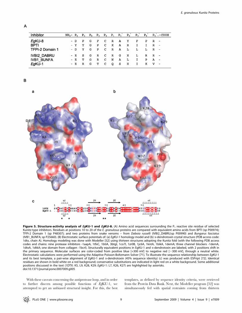
Figure 5. Structure-activity analysis of EgKU-1 and EgKU-8. (A) Amino acid sequences surrounding the P1 reactive site residue of selectedKunitz-type inhibitors. Residues at positions 10 to 20 of the E. granulosus proteins are compared with equivalent amino acids from BPTI (sp P00974);TFPI-2 Domain 1 (sp P48307); and two proteins from snake venoms – from Daboia russelli (IVBI2_DABRU;sp P00990) and Bungarus fasciatus(IVB1_BUNFA; sp P25660). (B) Electrostatic surface potentials of: (a) EgKU-1 homology model and (b) a-dendrotoxin crystal structure (PDB access code:1dtx, chain A). Homology modeling was done with Modeller [52] using thirteen structures adopting the Kunitz fold (with the following PDB accesscodes and chains: nine protease inhibitors -1aapA, 1tfxC, 1btiA, 3btgI, 1co7I, 1zr0B, 1jc6A, 1kntA, 1bikA, 1demA; three channel blockers -1demA,1dtxA, 1dtkA; one domain from collagen -1bcrI). Structurally equivalent positions in EgKU-1 and a-dendrotoxin are labeled, with 2 positions shift inthe primary sequence. Molecular surfaces are color-coded from positive blue [+300 mV] to negative red [2300 mV], through a neutral white.Electrostatic calculations were performed using the Adaptive Poisson-Boltzmann Solver [71]. To illustrate the sequence relationship between EgKU-1and its best template, a pair-wise alignment of EgKU-1 and a-dendrotoxin (45% sequence identity) (c) was produced with ESPript [72]. Identicalresidues are shown in bold white on a red background; conservative substitutions are indicated in light red on a white background. Some additionalpositions discussed in the text (1DTX: K5, L9, K28, K29; EgKU-1: L7, K26, K27) are highlighted by asterisks.doi:10.1371/journal.pone.0007009.g005
E. granulosus Kunitz Proteins
PLoS ONE | www.plosone.org 9 September 2009 | Volume 4 | Issue 9 | e7009
functionally diverse structure templates (nine protease inhibitors,
three cation channel blockers and the above mentioned Kunitz
domain from human collagen VI). Interestingly, the best overall
template was found to be a-dendrotoxin, the extensively charac-
terized blocker of specific voltage-activated K+-channels [15]. We
subsequently intended to check whether the structural elements
associated with channel-blocking activity were also present in the
reached consensus model of EgKU-1. Although not so clearly
delineated as the antiprotease site, the channel-blocking site of a-
dendrotoxin and related toxins is formed by residues from the N-
terminus and the b-turn region of the Kunitz domain, brought close
to each other by the conserved Cys5-Cys55 bond. Key residues in
the interaction appear to be a protruding Lys and a close
hydrophobic amino acid; whereas an enrichment of basic side
chains at sites forming an interface with the channel has also been a
consistent finding [53,54,55,56]. Analysis of the electrostatic surface
potential of EgKU-1 indicated that residues from its N-terminus
(Lys8) and b-turn (Arg25, Lys26 and Lys27) define highly cationic
protuberances that line an elongated crevasse (Figure 5B). Further-
more, a comparison with a-dendrotoxin showed that both
molecules share several amino acids found to be important for
activity (Leu7 and Lys27 in EgKU-1 are equivalent to Leu9 and
Lys29 in a-dendrotoxin); a residue comparable to the Lys5 is,
though, absent from the parasite molecule. These observations
would support the notion that EgKU-1 is a putative cation-channel
blocker. Interestingly, Leu7 and Lys27 are conserved in EgKU-4
(the closest paralog of EgKU-1) and also in the closest homologs
from E. multilocularis and T. solium (Figure 1B).
Taken altogether, our results suggest that the secretion of Kunitz
proteins is a strategy evolved by E. granulosus to block, through high
affinity interactions, the function of host proteins (such as serine
proteases and, possibly, K+-channels) present at the site of
establishment of the larval worms. From a more general perspective,
if the predicted K+-channel blocking activity is confirmed, this
family of secreted cestode proteins would provide a striking example
of protein evolution, similar to the one described in animal toxin
multigene families, where natural selection has acted to diversify the
coding sequences of duplicated genes, allowing the emergence not
only of specific inhibitors of particular enzymes (i. e. paralogous
genes whose products block paralogous proteins), but also of a new
function associated with the same molecular scaffold [10,57].
Taking into account that the genes coding for parasite secretions
and predator toxins arise from an arms race between different
organisms, it is interesting to consider that both sets of molecules
display similar evolutionary patterns. Thus, the concept of
‘‘exogenome’’ (the part of the genome whose products are targeted
exogenously [58]) coined for toxin genes, may also be applied to the
genes encoding parasite secretions.
Materials and Methods
Analysis of protoscolex transcriptomesHydatid cysts (G1 genotype) from the lungs of naturally infected
bovines were obtained from slaughterhouses in Uruguay. Proto-
scoleces were recovered under aseptic conditions, and extensively
washed in phosphate-buffered saline to remove dead larval worms.
They were stored at 270uC in TRIzol reagent (Invitrogen) until
RNA extraction. One fraction of freshly isolated larval worms was
incubated with 0.5 mg/ml of pepsin (Sigma) at pH 2 for 3 h at
37uC, prior to treatment with TRIzol. The processing of parasite
materials and the construction and sequencing of full-length
enriched cDNA libraries were previously described [6]. A detailed
account of the bioinformatics analysis of ESTs will be provided in a
separate manuscript (Parkinson J, Maizels RM, Fernandez C,
unpublished). In brief, sequence processing was performed using the
PartiGene pipeline [59]. Low quality, vector, host (bovine), linking
and poly(dA) sequences were removed from raw sequence trace
data. The resulting sequences were annotated by comparison to the
protein non-redundant database (UniProt - [60]) using BLAST and
submitted to dbEST [61]. Sequences were subsequently collated
and clustered on the basis of BLAST similarity to derive groups of
sequences, which putatively derive from the same gene using the
software package - CLOBB [62]. EST clusters were thus verified as
originating from distinct transcripts and not from sequencing errors.
Identification of platyhelminth cDNAs related to EgKUsThe full-length sequences predicted for EgKU-1 to EgKU-7
were subjected to BLAST analysis for similarity to ‘‘non-human,
non-rodent ESTs’’ (option ‘‘est_others’’ at the NCBI server), and
E. multilocularis ESTs available at ‘‘Full-Echinococcus’’ (http://
fullmal.hgc.jp/em/index.html [19]). As of August 2009, dbEST
contains 24,790 ESTs from Taenia solium, generated from cDNA
libraries of whole larva (cysticercus) and whole tapeworm; 74,915
from Schmidtea mediterranea, derived from various sources including
libraries from juvenile and sexually mature worms of the
hermaphroditic strain; and 16,350 from Dugesia spp. (8,988 from
whole adults of D. ryukyuensis; and 7,362 from the head of D.
japonica). Full-Echinococcus includes 10,966 ESTs, generated from
a library of hydatid cysts developed in cotton-rats (one-third of
ESTs represent host genes).
Cloning of EgKU-8 cDNAAbout 1 mg of total RNA isolated from pepsin/H+-treated
protoscoleces preserved in TRIzol was used to prepare cDNA using
PowerScript reverse transcriptase (Clontech) and an oligo(dT) primer.
The full coding sequence of EgKU-8 was amplified from an aliquot of
cDNA using forward and reverse primers designed on the basis of the
sequence from the putative E. multilocularis ortholog [XvEMa16368 in
Full-Echinococcus (http://fullmal.hgc.jp/em/index.html)]: KU8F:
59-ATG GTT GCC GCC TTT GCG C-39; and KU8R: 59-AAA
GCT TAC TTA GTG ACC GCA C-39. The PCR was initiated
with a touch down (5 cycles at 94uC/0.5 min, 60uC to 55uC/1 min,
75uC/1.5 min), and followed by 30 cycles at 94uC/0.5 min, 55uC/
1 min, 75uC/1.5 min, using Vent DNA polymerase (New England
Biolabs). The single product thus obtained was purified from an
agarose gel, A-tailed with Taq DNA polymerase (Fermentas), cloned
into pGEM-T-easy (Promega) and sequenced using vector primers.
The corresponding cDNA sequence was admitted to GenBank with
accession number: FJ031017.
Accession numbersE. granulosus sequence data reported in this manuscript is
available from GenBank and the corresponding accession
numbers indicated in Table S1. Accession numbers for other
platyhelminth sequences are also indicated in Table S1, together
with the database from which they were retrieved. Accession
numbers in Swiss-Prot or TrEMBL of Kunitz domain proteins
used for comparison of EgKUs are specified in Table S1 and in the
legend to Figure 5A. The Protein Data Bank access codes for the
templates of the homology modeling of EgKU-1 are provided in
the legend to Figure 5B.
Purification of native Kunitz inhibitors fromprotoscoleces
EgKU-1 and EgKU-8 were purified to homogeneity from a
protoscolex lysate by cation exchange followed by reverse-phase
chromatography. Freshly isolated protoscoleces were homoge-
E. granulosus Kunitz Proteins
PLoS ONE | www.plosone.org 10 September 2009 | Volume 4 | Issue 9 | e7009
nized by sonication in 50 mM phosphate buffer, pH 7. The
homogenate was centrifuged at 10,0006g, and the recovered
supernatant was first subjected to FPLC on a Mono S HR 5/5
column (GE Healthcare). After loading the extract, the column
was washed with 10 volumes of binding buffer (50 mM phosphate,
pH 7); bound proteins were eluted with a linear NaCl gradient (0–
0.4 M in 10 min, at a flow rate of 1 ml/min) in the same buffer.
Elution was monitored at 280 nm; 1 ml fractions were collected
and analyzed by non-reducing 15% Tricine-SDS-PAGE [63] and
for serine protease inhibitory activity. Active fractions were pooled
and applied to an Aquapor RP-300 (100621 mm, Perkin Elmer)
reverse phase HPLC column (rpHPLC). Bound proteins were
eluted with a linear gradient of acetonitrile in 0.07% trifluoroacetic
acid (0–40% in 60 min, at a flow rate of 0.4 ml/min), and
monitored at 220 nm. Eluted proteins were lyophilized and
dissolved in 50 mM Tris-HCl, pH 8, 0.01% Triton X-100 (v/v).
The Mono S fractions showing serine protease inhibitory
activity and the rpHPLC peaks were analyzed by matrix-assisted
LASER desorption ionization time-of-flight mass spectrometry
(MALDI-TOF MS) using a Voyager DE-PRO instrument
(Applied Biosystems). Mass spectra of whole proteins were
acquired on linear mode using a matrix solution of a-cyano-4-
hydroxycinnamic acid in 0.2% trifluoroacetic acid in acetonitrile-
H2O (50%, v/v), and were externally calibrated using a mixture of
peptide standards (Applied Biosystems).
N-terminal amino acid sequencing of EgKU-1 was carried out by
automatic Edman degradation on a pulsed liquid-phase sequencer
(Applied Biosystems), at the laboratory of Dr Ulf Hellman (Ludwig
Institute for Cancer Research, Uppsala Branch - Sweden).
Peptide mass fingerprinting of EgKU-8 was performed by in-gel
trypsin (Sequencing-grade, Promega) treatment of an SDS-PAGE
band of the purified inhibitor followed by MALDI-TOF MS of the
tryptic digest (4800 MALDI TOF-TOF Analyzer System, Applied
Biosystems). EgKU-8 was reduced and alkylated with iodoaceta-
mide prior to treatment with the enzyme and peptides were
extracted from the gel in 60% acetonitrile in 0.2% trifluoroacetic
acid, concentrated by vacuum-drying and desalted using C18
reverse phase micro-columns (OMIX Pipette tips, Varian).
Confirmation of the sequence of selected peptides was performed
by collision-induced dissociation MS/MS experiments.
Analysis of protoscolex secretionsAliquots of about 100 ml of freshly isolated protoscoleces (roughly
50,000 larval worms of .95% viability, estimated by eosin exclusion)
were incubated in 1 ml of RPMI 1640 containing penicillin/
streptomycin (Sigma) for 3 h at 37uC with gentle agitation. Some
aliquots were treated with 0.5 mg/ml of pepsin (Sigma) at pH 2 for
30 min at 37uC, washed and then incubated with RPMI for 3 h.
Parasite viability was checked at the end of the cultures and found to
have remained unchanged. The supernatants containing parasite
secretions were kept at 270uC and analyzed by MALDI-TOF MS
as described. The samples were concentrated and desalted by
adsorption onto a reverse phase micro-column; and were eluted with
matrix solution directly on the MALDI sample plate.
Assays of protease inhibitionThe inhibitory activity of purified EgKU-1 and EgKU-8 was
tested against bovine and canine chymotrypsins (EC 3.4.21.1) and
trypsins (EC 3.4.21.4). Bovine enzymes were obtained from Sigma
whereas canine proteases were purified from the pancreas of a dog
that had passed away due to an accidental cause, according to the
procedure of Waritani et al.[64]. The following peptidases were thus
assayed (MEROPS - http://merops.sanger.ac.uk - identifiers are
indicated in brackets): from Bos taurus, chymotrypsin A (S01.001) and
trypsin 1 (cationic, S01.151); from Canis familiaris, trypsins 1 (cationic,
S01.151) and 2 (anionic, S01.120), and chymotrypsin B (S01.152).
Prior to inhibition studies, proteolytic activity in enzyme
preparations was determined with fluorogenic substrates using
initial steady-state rate conditions at 37uC and pH 8. Assays
(200 ml) were performed in black 96-well microplates (Costar,
Corning Life Sciences). Enzymes and substrates were dissolved in
50 mM Tris-HCl, pH 8.0 containing 0.01% Triton X-100 (v/v),
and reactions were initiated by the addition of enzyme. The
changes in fluorescence intensity, corresponding to the formation
of the hydrolysis product 7-amino-4-methylcoumarin (AMC), were
registered at excitation and emission wavelengths of 390 and
460 nm, respectively, with a microplate fluorescence reader
(FLUOstar* OPTIMA, BMG Labtechnologies). For trypsin
activity, the artificial substrate N-t-BOC-Ile-Glu-Gly-Arg-AMC
was used and for chymotrypsin, Suc-Ala-Ala-Pro-Phe-AMC.
Calibration curves using AMC were carried out in each
experiment. Initial steady-state rates of substrate hydrolysis were
calculated from the linear portion of product (AMC) versus time
plots when less than 10% of substrate had been consumed.
Protein concentrations of enzyme preparations and purified
inhibitors were determined with the BCA reagent (Pierce) using
bovine serum albumin as standard; and the active site concentration
of trypsins and bovine chymotrypsin A by specific titration with the
high affinity inhibitor BPTI [39,65]. The active site concentration of
canine chymotrypsin could not be estimated because, similar to
bovine chymotrypsin B [23], it was not inhibited by BPTI.
The kinetic parameters for substrate and enzyme pairs were
calculated from the non-linear fitting to the Michaelis-Menten
equation. The values determined with the substrates specified
above were, for canine proteases: KM = 2563 mM and
kCat = 3862 s21 for anionic trypsin; KM = 3164 mM and
kCat = 4362 s21 for cationic trypsin; and KM = 39 mM62 s21 for
chymotrypsin B (kCat was not calculated because of the unknown
active site concentration). And for the bovine enzymes:
KM = 8569 mM and kCat = 5066 s21 for cationic trypsin and
KM = 3062 mM and kCat = 1962 s21 for chymotrypsin A.
For inhibition studies, each of the enzymes was incubated with
purified EgKU-1 or EgKU-8 for 15 min at 37uC prior to the
addition of the appropriate fluorogenic substrate, to allow for the
equilibration of the enzyme-inhibitor complexes. The substrate
concentration (5 mM) was chosen so as to be well below the
corresponding KM, as specified above.
Inhibition studies with EgKU-8Tight-binding kinetics. In order to determine the inhibition
constants (KI*) of EgKU-8 towards canine trypsins (anionic and
cationic) and bovine trypsin and chymotrypsin A, the initial
steady-state rates of substrate hydrolysis in the presence of
increasing concentrations of EgKU-8 were measured after pre-
incubation of the enzyme with inhibitor. The inhibition constants
were calculated by nonlinear fitting to the Morrison equation for
tight binding inhibitors [66,67]:
vi ~v
2 E½ � E½ � - I½ � - KI�
app
� �(
z
ffiffiffiffiffiffiffiffiffiffiffiffiffiffiffiffiffiffiffiffiffiffiffiffiffiffiffiffiffiffiffiffiffiffiffiffiffiffiffiffiffiffiffiffiffiffiffiffiffiffiffiffiffiffiffiffiffiffiffiffiffiffiffiffiffiffiffiffiffiI½ �z KI
�app - E½ �
� �2
- 4 KI�
app E½ �q ) ð1Þ
where KI*app is the apparent global dissociation constant of the
enzyme-inhibitor complex, vi is the inhibited steady-state rate, v is
the uninhibited rate, [I] is the total EgKU-8 concentration and [E]
E. granulosus Kunitz Proteins
PLoS ONE | www.plosone.org 11 September 2009 | Volume 4 | Issue 9 | e7009

is the total enzyme concentration. The true inhibition constants,
KI*, were corrected from KI
*app according to the equation 2 for
competitive inhibitors:
K�I ~K�I app
1zS½ �
KM
ð2Þ
Slow-binding kinetics. The decrease in the rate of product
formation during the first minutes after mixing the enzyme with
EgKU-8 and substrate (5 mM) was studied for increasing inhibitor
concentrations. Progress curves were analyzed using the equation
3 [68] that describes the slow establishment of equilibrium
between the enzyme and the inhibitor according to:
P ~ vi t zvo { við Þ 1 - e-kobst
� �kobs
ð3Þ
where P is the concentration of AMC produced by hydrolysis of
the substrate, vo is the initial rate, vi is the final steady-state rate and
kobs represents the apparent first order rate constant. Computer
fitting of progress curves estimated values for vo, vi and kobs.
For Kunitz inhibitors that bind to the enzyme rapidly and
reversibly forming an initial ‘‘loose’’ complex EI that isomerizes
slowly to the final complex EI*, the reaction mechanism can be
represented by the following scheme:
In this mechanism, the value of the apparent rate constant (kobs) is
related to the kinetic constants k2 and k22 and to the equilibrium
dissociation constant of the initial loose complex KI (KI = k21/k1), by
equation 4 [69]:
kobs ~ k-2 zk2 I½ �
I½ �z KI 1 z S½ �=KMð Þ ð4Þ
The constants k2 and KI were determined from plots of kobs vs
[I], by computer fitting to equation 4. Because k-2 was too small to
be accurately estimated from these plots, it was determined using
equation 5 [24] and data from situations where the ratio vi/vo was
higher than 0.05:
k-2 ~ kobsvi
vo
ð5Þ
The values of k22, k2 and KI thus determined allowed to
corroborate the inhibition constant KI*, according to equation 6:
KI� ~KI
k-2
k2zk-2ð6Þ
Data analysis. Computer fitting to non-linear equations was
performed using the software Origin (OriginLab). All experiments
were carried out at least two to three independent times and results
shown are averages 6 the standard error unless otherwise specified.
Supporting Information
Table S1 The E. granulosus Kunitz protein family.
Found at: doi:10.1371/journal.pone.0007009.s001 (0.04 MB
PDF)
Figure S1 Phylogenetic analysis of E. granulosus and related
Kunitz proteins from platyhelminths. The mature protein
sequences predicted for EgKU-1 - EgKU-8, together with those
identified among E. multilocularis, T. solium and planarian (D.
ryukuyensis and S. mediterranea) ESTs were aligned with Clustal W2
[70]. A neighbor joining tree was constructed using MEGA4 [73]
with default parameters. E. multilocularis (Em) and T. solium (Ts)
sequences are as in Figure 1B; Sm-1 was deduced from DN300487
and DN307650 (derived from the same transcript); and Dr-1, from
BW635664 in dbEST (refer to Table S1 for further details).
Found at: doi:10.1371/journal.pone.0007009.s002 (0.89 MB TIF)
Figure S2 Confirmation of EgKU-8 as the component of the
minor rpHPLC peak. A pool of the fractions eluting around 24%
ACN was resolved by SDS-PAGE and Coomassie-stained; the 7
kDa band was in-gel digested with trypsin, after reduction and
alkylation with iodoacetamide. Tryptic fragments identified by
MALDI-TOF MS provided 73% coverage (42/57 amino acids) of
the sequence predicted for mature EgKU-8. Peptides 1-15 and 16–
20 were further verified by MS/MS experiments. The spectrum of
the 16–20 peptide (m/z 653.33) is shown: signals from N-terminal
(b ions) and C-terminal (y ions) fragments confirmed the sequence
AYFPR. P, F, R and Y indicate signals from the immoniun ions of
the corresponding amino acids.
Found at: doi:10.1371/journal.pone.0007009.s003 (0.45 MB TIF)
Acknowledgments
We thank Madelon Portela for excellent technical assistance with MS
analysis; and Ana M Ferreira and Alvaro J Dıaz for their advice and
encouragement. The E. multilocularis genome sequence data mentioned in
the discussion was produced by the Pathogen Sequencing Group of the
Wellcome Trust Sanger Institute (Program of Helminth Sequencing;
project manager: Dr Matt Berriman).
Author Contributions
Conceived and designed the experiments: SG MF RMM GS BA CF.
Performed the experiments: SG MF MM JP CF. Analyzed the data: SG
MF MM RD GGS MG RMM GS BA CF. Contributed reagents/
materials/analysis tools: RD GGS MG JP. Wrote the paper: GS BA CF.
References
1. Craig PS, Larrieu E (2006) Control of cystic echinococcosis/hydatidosis: 1863–
2002. Adv Parasitol 61: 443–508.
2. Heath DD (1986) Immunobiology of Echinococcus infections. In:
Thompson RCA, ed. The biology of Echinococcus and hydatid disease. London:
George Allen & Unwin. pp 164–188.
3. Heath DD (1995) Immunology of Echinococcus infections In: Thompson RCA,
Lymbery A, eds. Echinococcus and hydatid disease. Wallingford: CAB
International. pp 183–200.
4. Smyth JD, McManus DP (1989) The physiology and biochemistry of cestodes:
Cambridge University Press.
5. Thompson RCA (1995) Biology and systematics of Echinococcus. In:
Thompson RCA, Lymbery A, eds. Echinococcus and hydatid disease.
Wallingford: CAB International. pp 1–50.
6. Fernandez C, Gregory WF, Loke P, Maizels RM (2002) Full-length-
enriched cDNA libraries from Echinococcus granulosus contain separate
populations of oligo-capped and trans-spliced transcripts and a high level
of predicted signal peptide sequences. Mol Biochem Parasitol 122: 171–
180.
7. Rawlings ND, Tolle DP, Barrett AJ (2004) Evolutionary families of peptidase
inhibitors. Biochem J 378: 705–716.
E. granulosus Kunitz Proteins
PLoS ONE | www.plosone.org 12 September 2009 | Volume 4 | Issue 9 | e7009

8. Rawlings ND, Morton FR, Kok CY, Kong J, Barrett AJ (2008) MEROPS: thepeptidase database. Nucleic Acids Res 36: D320–325.
9. Laskowski M Jr, Kato I (1980) Protein inhibitors of proteinases. Annu RevBiochem 49: 593–626.
10. Fry BG (2005) From genome to ‘‘venome’’: molecular origin and evolution ofthe snake venom proteome inferred from phylogenetic analysis of toxin
sequences and related body proteins. Genome Res 15: 403–420.
11. Schweitz H, Bruhn T, Guillemare E, Moinier D, Lancelin JM, et al. (1995)
Kalicludines and kaliseptine. Two different classes of sea anemone toxins forvoltage sensitive K+ channels. J Biol Chem 270: 25121–25126.
12. Andreev YA, Kozlov SA, Koshelev SG, Ivanova EA, Monastyrnaya MM, et al.(2008) Analgesic compound from sea anemone Heteractis crispa is the first
polypeptide inhibitor of vanilloid receptor 1 (TRPV1). J Biol Chem 283:23914–23921.
13. Bayrhuber M, Vijayan V, Ferber M, Graf R, Korukottu J, et al. (2005)Conkunitzin-S1 is the first member of a new Kunitz-type neurotoxin family.
Structural and functional characterization. J Biol Chem 280: 23766–23770.
14. Yuan CH, He QY, Peng K, Diao JB, Jiang LP, et al. (2008) Discovery of a
distinct superfamily of Kunitz-type toxin (KTT) from tarantulas. PLoS ONE 3:e3414.
15. Harvey AL (2001) Twenty years of dendrotoxins. Toxicon 39: 15–26.
16. Schechter I, Berger A (1968) On the size and the active site in proteases. I.
Papain. Biochem Biophys Res Commun 27: 157–162.
17. Kamei S, Petersen LC, Sprecher CA, Foster DC, Kisiel W (1999) Inhibitory
properties of human recombinant Arg24–.Gln type-2 tissue factor pathwayinhibitor (R24Q TFPI-2). Thromb Res 94: 147–152.
18. Krowarsch D, Zakrzewska M, Smalas AO, Otlewski J (2005) Structure-functionrelationships in serine protease-bovine pancreatic trypsin inhibitor interaction.
Protein Pept Lett 12: 403–407.
19. Watanabe J, Wakaguri H, Sasaki M, Suzuki Y, Sugano S (2007) Comparasite: a
database for comparative study of transcriptomes of parasites defined by full-length cDNAs. Nucleic Acids Res 35: D431–438.
20. Aguilar-Diaz H, Bobes RJ, Carrero JC, Camacho-Carranza R, Cervantes C, etal. (2006) The genome project of Taenia solium. Parasitol Int 55 Suppl:
S127–130.
21. Moses E, Hinz HJ (1983) Basic pancreatic trypsin inhibitor has unusual
thermodynamic stability parameters. J Mol Biol 170: 765–776.
22. Chen C, Hsu CH, Su NY, Lin YC, Chiou SH, et al. (2001) Solution structure of
a Kunitz-type chymotrypsin inhibitor isolated from the elapid snake Bungarusfasciatus. J Biol Chem 276: 45079–45087.
23. Wu FC, Laskowski M (1955) Action of the naturally occurring trypsin inhibitorsagainst chymotrypsins alpha and beta. J Biol Chem 213: 609–619.
24. Morrison JF (1982) The slow-binding and slow, tight-binding inhibtion ofenzyme-catalyzed reactions. Trends Biochem Sci 7: 102–105.
25. Bell G, Maynard Smith J (1987) Short-term selection for recombination amongmutually antagonistic species. Nature 328: 66–68.
26. Zang X, Maizels RM (2001) Serine proteinase inhibitors from nematodes andthe arms race between host and pathogen. Trends Biochem Sci 26: 191–197.
27. Verjovski-Almeida S, DeMarco R, Martins EA, Guimaraes PE, Ojopi EP, et al.(2003) Transcriptome analysis of the acoelomate human parasite Schistosoma
mansoni. Nat Genet 35: 148–157.
28. Hu W, Yan Q, Shen DK, Liu F, Zhu ZD, et al. (2003) Evolutionary and
biomedical implications of a Schistosoma japonicum complementary DNAresource. Nat Genet 35: 139–147.
29. Robb SM, Ross E, Sanchez Alvarado A (2008) SmedGD: the Schmidteamediterranea genome database. Nucleic Acids Res 36: D599–606.
30. Parkinson J, Whitton C, Schmid R, Thomson M, Blaxter M (2004) NEMBASE:a resource for parasitic nematode ESTs. Nucleic Acids Res 32: D427–430.
31. Hawdon JM, Datu B, Crowell M (2003) Molecular cloning of a novelmultidomain Kunitz-type proteinase inhibitor from the hookworm Ancylostoma
caninum. J Parasitol 89: 402–407.
32. Kooyman FN, van Balkom BW, de Vries E, van Putten JP (2009) Identification
of a thrombospondin-like immunodominant and phosphorylcholine-containingglycoprotein (GP300) in Dictyocaulus viviparus and related nematodes. Mol
Biochem Parasitol 163: 85–94.
33. Cappello M, Vlasuk GP, Bergum PW, Huang S, Hotez PJ (1995) Ancylostoma
caninum anticoagulant peptide: a hookworm-derived inhibitor of humancoagulation factor Xa. Proc Natl Acad Sci U S A 92: 6152–6156.
34. Milstone AM, Harrison LM, Bungiro RD, Kuzmic P, Cappello M (2000) Abroad spectrum Kunitz type serine protease inhibitor secreted by the hookworm
Ancylostoma ceylanicum. J Biol Chem 275: 29391–29399.
35. Ascenzi P, Bocedi A, Bolognesi M, Spallarossa A, Coletta M, et al. (2003) The
bovine basic pancreatic trypsin inhibitor (Kunitz inhibitor): a milestone protein.Curr Protein Pept Sci 4: 231–251.
36. Chand HS, Schmidt AE, Bajaj SP, Kisiel W (2004) Structure-function analysis ofthe reactive site in the first Kunitz-type domain of human tissue factor pathway
inhibitor-2. J Biol Chem 279: 17500–17507.
37. Broze GJ, Jr, Miletich JP (1987) Characterization of the inhibition of tissue factor
in serum. Blood 69: 150–155.
38. Huang ZF, Wun TC, Broze GJ, Jr (1993) Kinetics of factor Xa inhibition by
tissue factor pathway inhibitor. J Biol Chem 268: 26950–26955.
39. Vincent JP, Lazdunski M (1972) Trypsin-pancreatic trypsin inhibitor association.
Dynamics of the interaction and role of disulfide bridges. Biochemistry 11:2967–2977.
40. Ohlsson K, Tegner H (1973) Anionic and cationic dog trypsin. Isolation andpartial characterization. BBA 317: 328–337.
41. Woodard SL, Mayor JM, Bailey MR, Barker DK, Love RT, et al. (2003) Maize(Zea mays)-derived bovine trypsin: characterization of the first large-scale,
commercial protein product from transgenic plants. Biotechnol Appl Biochem38: 123–130.
42. Perona JJ, Craik CS (1997) Evolutionary divergence of substrate specificity
within the chymotrypsin-like serine protease fold. J Biol Chem 272:29987–29990.
43. Outzen H, Berglund GI, Smalas AO, Willassen NP (1996) Temperature and pHsensitivity of trypsins from Atlantic salmon (Salmo salar) in comparison with
bovine and porcine trypsin. Comp Biochem Physiol B Biochem Mol Biol 115:33–45.
44. Smalas AO, Heimstad ES, Hordvik A, Willassen NP, Male R (1994) Coldadaption of enzymes: structural comparison between salmon and bovine
trypsins. Proteins 20: 149–166.
45. Krowarsch D, Dadlez M, Buczek O, Krokoszynska I, Smalas AO, et al. (1999)
Interscaffolding additivity: binding of P1 variants of bovine pancreatic trypsin
inhibitor to four serine proteases. J Mol Biol 289: 175–186.
46. Helland R, Leiros I, Berglund GI, Willassen NP, Smalas AO (1998) The crystal
structure of anionic salmon trypsin in complex with bovine pancreatic trypsininhibitor. Eur J Biochem 256: 317–324.
47. Huber R, Kukla D, Bode W, Schwager P, Bartels K, et al. (1974) Structure ofthe complex formed by bovine trypsin and bovine pancreatic trypsin inhibitor.
II. Crystallographic refinement at 1.9 A resolution. J Mol Biol 89: 73–101.
48. Skarzynski T (1992) Crystal structure of alpha-dendrotoxin from the green
mamba venom and its comparison with the structure of bovine pancreatictrypsin inhibitor. J Mol Biol 224: 671–683.
49. Arnoux B, Merigeau K, Saludjian P, Norris F, Norris K, et al. (1995) The 1.6 Astructure of Kunitz-type domain from the alpha 3 chain of human type VI
collagen. J Mol Biol 246: 609–617.
50. Kohfeldt E, Gohring W, Mayer U, Zweckstetter M, Holak TA, et al. (1996)Conversion of the Kunitz-type module of collagen VI into a highly active trypsin
inhibitor by site-directed mutagenesis. Eur J Biochem 238: 333–340.
51. Pritchard L, Dufton MJ (1999) Evolutionary trace analysis of the Kunitz/BPTI
family of proteins: functional divergence may have been based on conforma-tional adjustment. J Mol Biol 285: 1589–1607.
52. Sali A, Blundell TL (1993) Comparative protein modelling by satisfaction ofspatial restraints. J Mol Biol 234: 779–815.
53. Gasparini S, Danse JM, Lecoq A, Pinkasfeld S, Zinn-Justin S, et al. (1998)Delineation of the functional site of alpha-dendrotoxin. The functional
topographies of dendrotoxins are different but share a conserved core with
those of other Kv1 potassium channel-blocking toxins. J Biol Chem 273:25393–25403.
54. Harvey AL, Robertson B (2004) Dendrotoxins: structure-activity relationshipsand effects on potassium ion channels. Curr Med Chem 11: 3065–3072.
55. Katoh E, Nishio H, Inui T, Nishiuchi Y, Kimura T, et al. (2000) Structural basisfor the biological activity of dendrotoxin-I, a potent potassium channel blocker.
Biopolymers 54: 44–57.
56. Smith LA, Reid PF, Wang FC, Parcej DN, Schmidt JJ, et al. (1997) Site-directed
mutagenesis of dendrotoxin K reveals amino acids critical for its interaction withneuronal K+ channels. Biochemistry 36: 7690–7696.
57. Kordis D, Gubensek F (2000) Adaptive evolution of animal toxin multigenefamilies. Gene 261: 43–52.
58. Olivera BM (2006) Conus peptides: biodiversity-based discovery and exoge-
nomics. J Biol Chem 281: 31173–31177.
59. Parkinson J, Anthony A, Wasmuth J, Schmid R, Hedley A, et al. (2004)
PartiGene–constructing partial genomes. Bioinformatics 20: 1398–1404.
60. (2009)The Universal Protein Resource (UniProt) 2009. Nucleic Acids Res 37:
D169–174.
61. Boguski MS, Lowe TM, Tolstoshev CM (1993) dbEST - database for ‘‘expressed
sequence tags’’. Nat Genet 4: 332–333.
62. Parkinson J, Guiliano DB, Blaxter M (2002) Making sense of EST sequences by
CLOBBing them. BMC Bioinformatics 3: 31.
63. Schagger H, von Jagow G (1987) Tricine-sodium dodecyl sulfate-polyacrylamide
gel electrophoresis for the separation of proteins in the range from 1 to 100 kDaAnal Biochem 166: 368–379.
64. Waritani T, Okuno Y, Ashida Y, Tsuchiya R, Kobayashi K, et al. (2001)Development and characterization of monoclonal antibodies against canine
trypsin. Vet Immunol Immunopathol 80: 333–338.
65. Vincent JP, Lazdunski M (1973) The interaction between alpha-chymotrypsin
and pancreatic trypsin inhibitor (Kunitz inhibitor). Kinetic and thermodynamic
properties. Eur J Biochem 38: 365–372.
66. Morrison JF (1969) Kinetics of the reversible inhibition of enzyme-catalysed
reactions by tight-binding inhibitors. Biochim Biophys Acta 185: 269–286.
67. Greco WR, Hakala MT (1979) Evaluation of methods for estimating the
dissociation constant of tight binding enzyme inhibitors. J Biol Chem 254:12104–12109.
68. Williams JW, Morrison JF (1979) The kinetics of reversible tight-bindinginhibition. Methods Enzymol 63: 437–467.
69. Morrison JF, Walsh CT (1988) The behavior and significance of slow-bindingenzyme inhibitors. Adv Enzymol Relat Areas Mol Biol 61: 201–301.
70. Larkin MA, Blackshields G, Brown NP, Chenna R, McGettigan PA, et al. (2007)Clustal W and Clustal X version 2.0. Bioinformatics 23: 2947–2948.
E. granulosus Kunitz Proteins
PLoS ONE | www.plosone.org 13 September 2009 | Volume 4 | Issue 9 | e7009

71. Baker NA, Sept D, Joseph S, Holst MJ, McCammon JA (2001) Electrostatics of
nanosystems: application to microtubules and the ribosome. Proc Natl Acad
Sci U S A 98: 10037–10041.
72. Gouet P, Robert X, Courcelle E (2003) ESPript/ENDscript: Extracting and
rendering sequence and 3D information from atomic structures of proteins.Nucleic Acids Res 31: 3320–3323.
73. Tamura K, Dudley J, Nei M, Kumar S (2007) MEGA4: Molecular Evolutionary
Genetics Analysis (MEGA) software version 4.0. Mol Biol Evol 24: 1596–1599.
E. granulosus Kunitz Proteins
PLoS ONE | www.plosone.org 14 September 2009 | Volume 4 | Issue 9 | e7009
Copyright © 2022 FDOKUMEN