A Dissertation on KILPAUK MEDICAL COLLEGE CHENNAI
114
A Dissertation on EVALUATION OF THE RISK FACTORS AND CLINICAL FEATURES OF STROKE IN CORRELATION WITH CT SCAN FINDINGS Submitted to THE TAMILNADU DR. M.G.R. MEDICAL UNIVERISTY CHENNAI – 600 032 In partial fulfillment of the regulations For the Award of the Degree of M.D. (GENERAL MEDICINE) BRANCH -1, PART - II KILPAUK MEDICAL COLLEGE CHENNAI – 600 010 MARCH - 2007
-
Upload
khangminh22 -
Category
Documents
-
view
0 -
download
0
Transcript of A Dissertation on KILPAUK MEDICAL COLLEGE CHENNAI

A Dissertation on
EVALUATION OF THE RISK FACTORS AND CLINICAL
FEATURES OF STROKE IN CORRELATION WITH
CT SCAN FINDINGS
Submitted to
THE TAMILNADU DR. M.G.R. MEDICAL UNIVERISTY
CHENNAI – 600 032
In partial fulfillment of the regulations
For the Award of the Degree of
M.D. (GENERAL MEDICINE)
BRANCH -1, PART - II
KILPAUK MEDICAL COLLEGE
CHENNAI – 600 010
MARCH - 2007


ACKNOWLEDGMENT
I greatfully acknowledge and sincerely thank
Prof. THIAGAVALLI KIRUBAKARAN, M.D., Dean, Govt. Kilpauk Medical
College and Prof. D.S. SOMASEKAR, M.D., Superintendent Govt. Royapettah
Hospital Chennai, for permitting me to utilize the facilities available in the
institution for my study.
I owe my sincere gratitude to Prof. S.R. SAKUNTHALA, M.D., Professor
and Head, Department of Internal Medicine, Kilpauk Medical College, for her
constant encouragement and guidance in all stages of this study.
I whole heartedly express my sincere thanks to my respected chief Prof.
G. RAJENDRAN, M.D., Reader in Medicine, Chief IInd Medical Unit, Govt.
Royapettah Hospital, Kilpauk Medical College, for his valuable guidance and
support throughout my dissertation work.
I wish to thank Dr. T. RAVINDRAN, M.D., Dr. A. NASREEN BEGUM,
M.D., and Dr. T. RAMESHKUMAR, M.D., Assistant Professors, Department of
Internal Medicine, Kilpauk Medical College, for their valuable suggestions and
help rendered throughout this work.
I also extend my thanks to Dr. AMUDHAN, Assistant Professor,
Department of Radiology, Govt. Royapettah Hospital, for his kind and timely
support.

I also thank my colleagues, house surgeons, staffs of our hospital for their
contribution to the study.
Last but not least, with sincere gratitude, I thank all the patients who
contributed so much to this study, without whom the study could not have been
possible.

CONTENTS
S.NO. TITLE PAGE NO.
1. INTRODUCTION 1
2. AIM OF THE STUDY 3
3. REVIEW OF LITERATURE 4
4. MATERIALS AND METHODS 43
5. OBSERVATIONS AND RESULTS 46
6. ANALYSIS OF DATA AND DISCUSSION 52
7. CONCLUSION 67
BIBLIOGRAPHY
PROFORMA
ABBREVIATIONS
MASTER CHART

Introduction

INTRODUCTION
Cerebrovascular accident or stroke is one among the three leading causes
of death, surpassed only by ischemic heart disease and malignancy. Stroke is
also a common cause of physical disability, which imposes a substantial burden
to the community in the foreseeable future. It is estimated that the incidence of
stroke is likely to increase by about 20% in the next 20 years. In a developing
nation like India, Rheumatic heart disease forms a significant risk factor for
stroke. The high incidence and serious consequences make it one of the most
important challenges faced by medical profession today.
Atleast 50 percent of the neurological disorders in a general hospital are
due to stroke. As remarked by a renowned neurologist C.M. Fisher, neurology is
learnt “Stroke by Stroke”. The advent of imaging procedures such as
Computerized Tomography, Magnetic Resonance Imaging, Carotid Doppler and
Magnetic Resonance Angiography have led to better evaluation of stroke and its
risk factors.
A risk factor is a characteristic for an individual or for a population,
indicating that the individual or population has an increased risk of stroke
compared with one without that characteristic. Such an association does not
imply causality which is determined more by strength of the association, the
consistency of the association in different studies and population and the
presence of a dose response relationship.

The last decade has witnessed exciting advances in the field of stroke,
both in terms of enhanced understanding and in the availability of a rich panoply
of therapeutic options, most important being thrombolytic therapy. Several
studies in the recent years, have proved beyond doubt, the role of thrombolytic
therapy in acute cerebral infarction. The success of this modality of treatment
rests in early recognition of stroke and institution of therapy, for which a good
knowledge of risk factors and clinical presentation of stroke and investigative
modalities available is crucial.
Inspite of revolutionary changes in the management of ischemic stroke,
the ultimate goal should be its prevention in order to decrease the incidence, for
which, a thorough understanding of the etiopathogenesis and risk factors seems
essential.
Hence, the present study on stroke was undertaken.

Aim of the
Study

AIM OF THE STUDY
1. To study the age and gender distribution.
2. To evaluate the risk factors in cerebrovascular disease.
3. To study the mode of presentation of different types of stroke.
4. To study the pathogenesis of stroke with the aid of CT Scan.
5. Correlative study of the above modalities.

Review of
Literature

REVIEW OF LITERATURE
STROKE (Synonyms: Cerebrovascular Accident, Apoplexy)
DEFINITIONS
Stroke
WHO defined stroke as “rapidly developed clinical signs of focal or
global disturbance of brain function; lasting more than 24 hrs or leading to death,
with no apparent cause other than vascular origin”.
Completed Stroke
It is the term applied to the temporal profile of the stroke syndrome in
which the deficit is prolonged and often permanent, causing demonstrable
parenchymatous changes. Most completed strokes reach the maximum of
neurological dysfunction within an hour of onset.
Stroke in Evolution
This describes the temporal profile in which the neurological deficit
occurs in a stepwise or progressive fashion culminating in a major deficit in the
absence of treatment. In the carotid arterial system, this progression may go upto
24 hrs. If the vertebrobasilar system is the site of ischemia, the deficit may
progress for upto 72 hours.
Transient Ischemic Attack (TIA)

It is an acute loss of focal cerebral or monoocular function with
symptoms lasting less than 24 hours, which after adequate investigation is
presumed to be due to thrombotic or embolic vascular disease.
Reversible Ischemic Neurological Deficit (RIND)
It is the term applied to the temporal profile of the stroke syndrome in
which the focal neurological ischemic deficit lasts longer than 24 hours but
resolves within 3 weeks.
Stuttering Hemiplegia
It is characterized by repeated episodes of TIA or crescendo TIAs
followed by fully evolved stroke. Internal carotid artery lesions produce this
event.
Lacunar Stroke
It is secondary to lipohyalinosis or microatheroma of the small
perforating branches within the brain substance producing small deep infarcts
less that 1.5 cm in diameter.

Pathological types of stroke
1. Cerebral infarction – Cerebral thrombosis / embolism
2. Primary Intracerebral haemorrhage
3. Subarachnoid haemorrhage.
EPIDEMIOLOGY OF STROKE
Stroke epidemiology has lagged behind coronary artery disease, since
strokes are not only less frequent, but occur later in life.7 Stroke epidemiology is
hampered by :
� Diagnosis still being largely a matter of clinical skill, without help of
many confirmatory investigations.
� Being a disorder of late middle age and elderly, where other diseases
frequently co-exist.
� A low post-mortem rate.
� Inaccuracy of death certificates.
� Being more pathologically diverse.
Incidence can only be reliably assessed in prospective community based
studies using obsessional methodology to identify all possible patients. Hospital
based studies are subject to referral bias since very mild and rapidly fatal strokes
are least likely to be brought to the hospital. In any case, the incidence of stroke
rises rapidly with age, about a quarter occur below the age of 65, and about a
half below the age of 75.

Prevalence is difficult to measure because a large representative sample
has to be identified as the denominator, a large proportion of that sample must be
questioned and many past stroke episodes are forgotten by patients8.
Prevalence depends on long-term survival which may be changing. In any
event, stroke prevalence dose not even capture the burden of stroke since some
patients die early after the onset and many of the survivors are not disabled at all.
The mortality of stroke is on the decline in the recent years. However, it is
unlikely that treatment of hypertension is responsible since the decline had
started earlier and treatment cannot explain more than 25% of the decline9.
Treatment of TIAs is also unlikely, largely because only a small proportion of
strokes are preceded by TIAs10. Based on retrospective analysis of subjects
admitted in urban hospitals in India, strokes constitute 2% of all hospital cases
and 20% of neurological admissions. Incidence in younger population (below 40
years) is 13-22%.

Seasonal and Diurnal Variation
Stroke incidence and mortality is higher in winter months11, probably
secondary to high blood pressure and complications like pneumonia being more
fatal in winter12.
Cerebral infarction occurs more frequently in an hour or two after waking
in the morning13. Sub-arachnoid haemorrhage is unlikely to occur during sleep,
whereas intracerebral haemorrhage is more likely to occur during strenuous
activity.
RISK FACTORS
Stroke places a tremendous burden on health resources throughout the
world. Vast majority of stroke patients have well recognized risk factors, and
reducing their prevalence should make a considerable impact on stroke
incidence. Nonmodifiable risk factors include age, sex, race, genetic factors and
family history.
Age
Age is the strongest risk factor for stroke. Risk of stroke in people aged
75-84 yrs is 25 times the risk in people age 45-54 yrs14.

Gender
There is a small male excess of strokes most prominent in middle age and
disappearing in the very elderly and probably absent in the young12. However the
difference is much less than that observed in myocardial infarction and
peripheral vascular disease. It is more common in blacks than whites.
Genetic factors
Some genetically determined diseases are vascular anomalies like
malformations, aneurysms, connective tissue disorders such as Ehler – Danlos
Syndrome, Marfan’s syndrome and hereditary causes of hypercholesterolemia.
Several genetic determinants contribute to stroke risk. Of these, carotid intimal-
medial wall thickness (IMT) is particularly relevant, because it is a surrogate
measure of subclincial atherosclerosis and a strong predictor of future ischemic
stroke. Studies of twins, siblings, and families have provided significant
evidence for heritability, but the genes involved have not been identified. Some
researchers have reported that IMT is high in people with functional variants of
genes related to matrix deposition (MMP3), inflammation (interleukin 6), and
lipid metabolism (hepatic lipase, APOE, CETP, and PONI)15.
Family history
Sometimes stroke is clearly familial in some cases it is a simple
mendelian pattern of inheritance (eg. Hemophilia). Family history of stroke is an
independent risk factor for ischemic stroke with onset before age 70 years16.

Modifiable / Preventable Risk Factors
Systemic Hypertension
Systemic hypertension is strongly associated with stroke risk, probably by
increasing the extent and severity of atheroma, and the prevalence of
microvascular disease in the small penetrating arteries. Stroke risk doubles with
each 7.5 mmHg increase in diastolic blood pressure. A reduction of blood
pressure 10-12 mm systolic, 5-10 mmHg diastolic is associated with 38%
decrease in stroke17. Isolated systolic hypertension (ISH) and isolated diastolic
hypertension (IDH) both are independent predictors of stroke. Patients with
systolic and diastolic hypetension both are at the highest risk of stroke and
should be treated more aggressively18.
The strength of association of blood pressure and stroke is strong
consistent, biologically plausible and treatment reduces stroke risk. Hence one
can conclude that hypertension is a causal risk factor19,20. Hypertension and type
2 diabetes increase stroke risk independently, and their combination increases
the risk drastically. A significant proportion of the risk of stroke assumed to be
related to hypertension may be attributable to concomitant diabetes21.
Diabetes Mellitus
Diabetes doubles the risk of stroke compared to non-diabetics.20 In
addition patients with diabetic retinopathy and autonomic neuropathy are at
increased risk of ischemic stroke.22 Raised Hb A1c is an independent risk factor

for stroke in people with and without diabetes23. Impaired glucose tolerance is an
independent risk factor for future stroke in nondiabetic patients with TIA or
minor ischemic stroke24.
Hyperlipidemia
Increased levels of plasma total cholesterol, LDL cholesterol and
decreasing levels of HDL cholesterol are strong risk factors for coronary artery
disease25. Although their relationship to stroke is less clearcut compared to
ischemic heart disease, there is almost certainly some association. This may be
due to lipid levels being less associated with vascular events in the elderly
(where more strokes occur) than in younger people (where coronary events are
more common26). Cholesterol levels are also negatively associated with
haemorrhage27.
Smoking
Smoking is a strong risk factor for subarachnoid haemorrhage28 (relative
risk 3.0), and for cerebral infarction (relative risk 2.0) and there appears to be no
association with parenchymal haemorrhage. Males and females are equally
affected, but the association seems to be weaker in the elderly29. Stroke risk
decreases 5 yrs after cessation of smoking. There is a positive association
between history of smoking among spouses with the incidence of stroke30.
Alcohol

Heavy consumption is an independent risk factor, while moderate
consumption may be protective31. However, apoE genotype may modify this
association, and even moderate alcohol intake may be associated with an
increased risk of ischemic stroke among apoE4-positive older adults32. Drinking
pattern and beverage type may also be important. Intake of more than 2 drinks
per day may be associated with a higher risk for ischemic stroke33. Alcohol
consumption also raises the blood pressure34, affects blood lipids35, increases the
incidence of atrial fibrillation36 and cardiomyopathy which predispose to stroke.
Heart Disease
Coronary artery disease is clearly associated with stroke. The risk for
stroke is markedly increased after MI, particularly early after MI, compared with
the expected risk in population without MI. Stroke is associated with a large
increase in the risk for death after MI37. Cardiac failure, ECG changes and rapid
heart rate further increase the risk. Left ventricular dysfunction even of mild
degree, is independently associated with an increased risk of ischemic stroke38.
The most frequent potential cardiac cause of embolism is atrial fibrillation,
usually non-rheumatic in developed countries, rheumatic in developing
nations39.
Transient Ischemic Attacks
TIAs are ischemic strokes, recovering in 24 hrs. A TIA patient has an
increased risk of stroke about 5–10 times greater than that of a non TIA patient
of the same age. The risk of stroke after initial stroke / TIA is higher than the
risk of cardiac events. The propensity after stroke / TIA to have the first
recurrent ischemic event in the brain, rather than in the heart, has implications
for prophylactic therapy selection40.

Carotid Stenosis
Cervical carotid bruit is a risk factor for stroke, but not necessarily in the
same arterial territory, because stenosis in one artery is likely to be associated
with disease of other arteries41. Less than 75% occlusion has 1.3% annual
incidence of stroke, while more than 75% occlusion has a risk of 10.5% per year.
Ulcerated, echoluscent, heterogenous plaques with soft core are at higher risk.
Aortic arch atheroma is an important independent risk factor for stroke, studies
indicate a four times greater odds of stroke in patients with severe arch
atheroma42.
Haematocrit
Cerebral blood flow is strongly related to haematocrit. Increased
haemotocrit in association with smoking, hypertension and plasma fibrinogen
increase the risk of stroke.
Hormones
Although strokes are uncommon in females of the reproductive age
group, attributing this to endogenous sex hormones is difficult43, since high does
estrogen given to elderly men with prostate cancer, increases risk of vascular
event44. The use of oral contraceptive pills triples the risk of stroke in women
especially smokers aged more than 30 years.

Hormone replacement therapy seems to have a protective effect45. It has
been stated that natural menopause has no effect on stroke while surgical
menopause without estrogen replacement doubles the risk.
Obesity
The relationship between obesity and stroke is seldom studied, and if
present is probably secondary to its association with hypertension and diabetes.
The risk of stroke is increased in men with metabolic syndrome, in the absence
of past history of stroke, diabetes and cardiovascular disease at baseline46.
Diet
High salt intake47, decreased intake of fruits, vegetables48, deficiency of
selenium and vitamin E are associated with increased risk, while high potassium
diet by reducing blood pressure reduces risk of stroke49.

Other Risk Factors
1. High serum uric acid levels are strong risk factor for stroke50.
2. Severe obstructive sleep apnea syndrome increases the risk of ischemic
stroke in elderly population and the increase is independent of other risk
factors, including hypertension51, 52.
3. Physical inactivity is associated with an increased risk of stroke. A high
level of leisure time physical activity reduces the risk of all subtypes of
stroke53.
4. Chagas Disease is a risk factor for stroke, independent of systolic
dysfunction or presence of cardiac arrhythmias54.
5. Fabry Disease is also one of the cause of unexplained stroke in young
patients, especially in those with the combination of infarction in the
vertebrobasilar artery system and proteinuria55.
6. Moyamoya Disease is a risk factor for stroke56.
7. Higher serum ferritin concentrations in postmenopausal women are
associated with an increased risk of ischemic stroke57.
8. A high Lp(a) concentration is associated with a higher incidence of
ischemic stroke in blacks and white women, but not in white men58.

9. Peripheral arterial disease is a strong marker of multifocal atherosclerotic
disease. Individuals with intermittent claudication and asymptomatic
peripheral vascular disease (as defined by an abnormal low ankle-brachial
systolic pressure index) are at excess risk of stroke59.
10. Raised plasma factor VII coagulant activity, raised tissue plasminogen
activator antigen, low blood fibrinolytic activity and raised von
Willebrand factor are risk factors for coronary artery disease and may also
be risk factors for stroke60.
11. Hyperhomocyteinaemia is a risk factor for stroke61.
12. Oral contraceptives increase the risk of ischemic stroke, and less so,
haemorrhagic strokes62.
13. Arterial dissection
14. Antiphospholipid syndrome
15. Protein C, protein S deficiencies
16. Other less documented risk factors include.
a. Sickle cell disease
b. Drug abuse
c. Low socioeconomic factors
d. Stress
e. Alpha (1) antichymotrypsin polymorphism

f. Boiled and unfiltered coffee consumption
g. Pregnancy and puerperium
h. Infection – Chlamydia pneumoniae, periodontal disease,
meningeo vascular syphilis, Helicobacter pylori.
i. Cholesterol embolisation syndrome
j. Trauma
k. Fibromuscular dysplasia
l. Irradiation

PATHOPHYSIOLOGIC CLASSFICATION OF STROKE
I. ISCHEMIC – 85%
Thrombotic Embolic
Lacunar 20-25% Cardioembolic 20%
Large Vessel 1-5% Artery – artery 15%
Cryptogenic 30%
Others 10%
II. HAEMORRHAGIC – 15%
Intraparenchymal - 10%
Subarachnoid - 1-2%
Subdural - <1%
Epidural - <1%
ISCHEMIC STROKE
Ischemic stroke is caused by a sudden occlusion of an artery supplying
the brain or less often by low flow distal to an already occluded artery. The
causes of ischemic stroke include:
a. Thrombosis
Atherosclerosis
Vasculitis
Collagen vascular disease: Temporal (Giant cell) arteritis, polyarteritis
nodosa, Wegener’s granulomatosis, Takayasu’s arteritis, syphilis.

Meningitis: Tuberculous, fungi, syphilis, bacteria, herpes zoster
Arterial dissection: Carotid, vertebral, intracranial arteries.
Haematological disorders: Polycythemia, thrombocytosis, thrombotic
thrombocytopenic purpura, DIC, dysproteinemias, haemoglobinopathies
(sickle cell disease).
Miscellaneous: Cocaine, amphetamine, moyamoya disease, fibromuscular
dysplasia, Binswanger’s disease.
b. Embolism
Cardiac Source
Dysrhythmia – Atrial fibrillation, sick sinus syndrome
Coronary artery disease
Rheumatic heart disease
Cardiomyopathy
Prosthetic valve
Congenital heart disease - MVP, patent foramen ovale
Infective endocarditis
Atrial myxoma

Non-bacterial thrombotic endocarditis.
Atherothrombotic arterial disease
Bifurcation of common carotid artery, distal vertebral artery and aortic
arch.
Unknown source
May be associated with a hypercoagulable state
c. Vasoconstriction
Following subarachnoid haemorrhage, migraine, eclampsia etc.
d. Venous occlusion
Dehydration, post-partum, systemic cancer etc.
Brain is an obligate aerobe and obtains energy from oxidative metabolism
of glucose. Since the brain glucose stores are negligible, a decrease in cerebral
blood flow causes ischemia. The normal cerebral blood flow is 50 ml/100 g/min.
When this falls below a critical value of 20 ml/100 g/min, there is loss of
neuronal electronic function which is a reversible stage. When it decreases to
less than 10 ml/100 g/min, then aerobic mitochondrial metabolism fails and
anaerobic metabolism leads to lactic acidosis63. As a sequel, sodium and water
enters the cell, potassium leaks out, due to failure of energy dependent
intracellular homeostasis, leading to irreversible cell death.

In man, it is not known exactly how long and how severe focal ischemia
has to be before complete recovery of function is impossible. The ischemic and
infarcted brain cannot autoregulate. Therefore modest increase in cerebral
perfusion pressure could cause hyperemia, increased cerebral blood flow, edema
and haemorrhagic infarction which in turn increases intracranial tension and
decreases cerebral perfusion pressure. On the other hand, a decrease in cerebral
perfusion pressure may exacerbate ischemia.
Ischemic penumbra is an area around an infarcted tissue, that is in the
reversible state of electrical failure, where flow is decreased, function depressed,
oxygen extraction fraction is high, but recovery is still possible. Thrombolytic
agents are used in this window time to salvage the ischemic penumbra zone.
Ischemic cerebral edema is partly cytotoxic and partly vasogenic.
Cytotoxic edema starts within minutes and affects grey matter. Vasogenic edema
starts several hours later and affects more white matter, since the damaged blood
brain barrier allows plasma to enter the extracellular space.
Differentiating signs of Thrombosis and Embolism
Thrombosis Embolism Preceding brief shot gun like TIA
Single or infrequent but longer –
lasting TIA or strokes
TIAs all in same vascular territory
Infarcts in multiple vascular territories

Onset of stroke after sleep Onset during activity or sudden strain,
cough or sneeze.
Absence of distal embolus by
angiography
Presence of distal intra arterial
embolus by angiography or
transcranial doppler
Infarct on CT or MRI near border
zone of affected artery
Infarct on CT or MRI in heart of
vascular territory wedge shaped and
abutting on cortical surface
Presence of risk factors for
atherosclerosis
Presence of known cardiac, arterial or
venous source of embolus
Occlusion or severe stenosis of a
large artery shown by USG or
angiography
Haemorrhagic cerebral infarct on CT

HAEMORRHAGIC STROKE
Intracranial haemorrhage accounts for approximately 15% strokes. The
overall mortality for this subtype of stroke is from 25% to 60%. In nearly 70% of
patients hypertension is the commonest cause. Other causes include
arteriovenous malformation, aneurysm, coagulopathy, drugs, amyloid
angiopathy, metastatic tumours, cavernous angiomas, dural arterial-venous
fistula, capillary telangiectasia –etc.
Location of hypertensive haemorrhage :
Putamen - 60 – 65%
Thalamus - 15 - 25%
Pons - 5 - 10%
Cerebellum - 1 – 5%
Subcortical white matter - 1 – 2%
Clinical presentation of intracranial haemorrhage are mainly due to
symptoms of raised intracranial tension and specific for the location of
haematoma. Characteristically haemorrhage presents with progression of deficits
over a period of hours. Seizures at the time of presentation occurs usually in
cortical haemorrhage.
CT continues to be the gold standard for diagnosis of acute intracranial
haemorrhage and evaluating its prognosis. MRI appears normal in the first 24
hrs, but is more specific than CT in determining the age of haemorrhage. Acute

haemorrhage appears as a hyperdense area having attenuation values of 50-100
Hounsfeld units. They become hypodense in the ensuring couple of weeks. The
periphery of the clot is absorbed at a rate of 0.7 mm/day.
SUBARACHNOID HAEMORRHAGE [SAH]
The incidence of SAH increases with age being more common in women.
Causes include rupture of a saccular aneurysm, bleeding from vascular anomaly,
extension into subarachnoid space from primary intracerebral haemorrhage.
Idiopathic SAHs are localized to perimesencephalic cisterns and are benign64.
Chronic, recurrent bleed produces superficial hemosiderosis of central nervous
system65.
SAH usually presents as excruciating headache followed by loss of
consciousness. Focal neurologic deficits such as hemiparesis, aphasia, abulia
may occur. Delayed neurological deficits may be due to
re-rupture, hydrocephalus, vasospasm and hyponatremia.
Hall mark of SAH is blood in CSF. High quality, non-contrast CT
localizes blood in 95% of cases within 72 hrs. Lumbar puncture is indicated only
if CT scan is not available. Four-vessel conventional
X-ray angiography is performed to localize, define anatomic details for
interventional therapy.
INTRACRANIAL VENOUS THROMBOSIS
Thrombosis in dural sinuses /cerebral veins is much less common than
arterial thrombosis. However, it should be suspected in high risk groups mainly
postpartum and malignancies. Venous thrombosis can result from diseases that
alter clotting factor or cellular constituents of blood.
CAUSES
Local Systemic
Head injury Dehydration
Intracranial surgery Septicaemia
Sepsis Oral contraceptives
Subdural empyema Hypercoagulable states
Meningitis Drugs
Tumor invasion of sinuses
Catheterization of jugular vein
Non- metastatic effect of
intracranial malignancy
Symptoms depend on structure involved, extent and rapidity of
thrombosis and collaterals. Headache, nausea, vomiting, convulsions, depressed
alertness are frequent. Focal signs may be subtle or absent, but papilledema and
meningeal signs are often found. Cavernous sinus thrombosis following
infection of orbit, paranasal sinus produce orbital signs. Cerebral angiography is
the definitive indication. Mortality is 15–30%, mainly results from cerebral

edema and secondary haemorrhagic infarction. Heparinization improves
prognosis.
STROKE IN THE YOUNG
Ischemic stroke is often considered as a disease of middle aged and
elderly persons but it does occur in patients aged 40 years or less. This may be
due to improved recognition and increased prevalence of risk factors.
Predominant causes are:
• Cardiovascular: Rheumatic heart disease, infective endocarditis,
prosthetic valve, myxoma, mitral valve prolapse etc.
• Non–atherosclerotic vasculopathies: Tuberculosis, syphilis, collagen
vascular disease, dissection, trauma, migraine etc.
• Hypercoagulable states: Ranging from disturbances in blood
components, coagulation factors, platelet functions and deficiencies of
clotting inhibitors or fibrinolytic system predispose to stroke.
• Atherosclerosis: May occur prematurely secondary to earlier onset of
risk factors such as hypercholesterolemia, smoking, diabetes mellitus,
hypertension.
Blood supply to the brain95
The blood supply to the brain is delivered by the two internal carotid and
two vertebral arteries which anastomose at the base of the brain to form the
circle of Willis. The carotid artery system supplies the anterior two-thirds of the

brain (hence it is called the anterior circulation) (Fig.1). The vertebrobasilar
arterial system supplies the posterior third of the brain (hence it is called the
posterior circulation) (Fig.2).

FIGURE - 1
The anatomy of the arterial blood supply to the brain. Sites that are most often
affected by atherosclerosis are shown as dark indentations in the arterial
lumen.

FIGURE – 2
Diagrammatic representation of the circle of Willis at the base of the
brain, as seen from below in relation to the optic chiasma.

STROKE SYNDROMES 95
Internal carotid artery territory (or) anterior cir culation stroke95
Branches Region supplied Syndrome caused by ischemia
Ophthalmic artery Retina and optic nerve Monoocular blindness or altitudinal field detect
Anterior choroidal artery
Globus pallidus internal capsule choroid plexus
Contralateral hemiparesis, hemisensory loss and homonymous hemianopia
Middle cerebral artery
Frontal lobe Contralateral facial weakness, hemiparesis and hemisensory loss (arm>leg), homonymous hemianopia, and global aphasia (dominant hemisphere) or visual-spatial – perceptual dysfunction (non-dominant hemisphere)
Medial lenticulostriate artery
Internal capsule Contralateral pure motor hemiparesis
Lateral lenticulostriate artery
Putamen, globus pallidus, caudate nucleus, internal capsule, corona radiata
Contralateral hemiparesis, dysphasia (dominant hemispehre) or visual –spatial – perceptual dysfunction (non-dominant hemisphere)
Superior division of middle cerebral artery
Prerolandic branch
Frontal and anterior parietal lobes
Contralateral central facial weakness, hemiparesis and hemisensory loss, ipsilateral deviation of head and eyes, and global or motor aphasia (dominant hemisphere) Contralateral face and

Branches Region supplied Syndrome caused by ischemia
Rolandic branch
Anterior parietal
branch
arm weakness, and motor aphasia (dominant hemisphere) Contralateral central facial weakness, hemiparesis and hemisensory loss, and dysarthria (resembling lacunar syndrome) conduction aphasia and bilateral ideomotor apraxia
Inferior division of middle cerebral artery
Posterior parietal branch
Angular branch
Posterior temporal branch
Anterior temporal branch
Temporal polar branch
Inferior parietal and lateral temporal lobes
Homonymous hemianopia, Wernicke’s aphasia or agitated confusional state (dominant hemisphere), left visual neglect (right-sided lesion)
Anterior cerebral artery
Anterior and superior medial frontal lobe
Contralateral foot and leg weakness or hemiparesis (leg>arm), abulia, incontinence, grasp reflexes

Vertebrobasilar territory (or) posterior circulatio n stroke95
Branches Region supplied
Syndrome caused by ischemia
Vertebral and basilar artery
Brainstem and cerebellum
Various syndromes including: diplopia, ophthalmoplegia or gaze palsies; vertigo, nausea and nystagmus; dysarthria, dysphagia and bulbar weakness; ipsilateral facial sensory loss and weakness (nuclear or infranuclear); hiccups and respiratory failure contralateral hemiparesis or tetraparesis; contralateral or bilateral sensory loss; coma
Basilar artery
Top of basilar artery
Rostral midbrain
Part of thalamus
Inferior temporal occipital lobes
Variable pupillary abnormalities
Ptosis or lid retraction
Supranuclear vertical gaze paresis
Somnolence
Hemiballismus
Amnesia
Cortical blindness
Superior cerebellar artery
Midbrain (dorsolateral)
Superior cerebellar peduncle
Superior cerebellum
Ipsilateral Horner’s syndrome
Ipsilateral limb ataxia and tremor
Contralateral spinothalamic sensory loss
Contralateral central facial weakness
Sometimes contralateral IVth nerve palsy
Anterior inferior cerebellar artery
Base of pons Rostral medulla
Inferior cerebellum
Cochlea
Ipsilateral Horner’s syndrome
Ipsilateral facial sensory loss
(pain, temperature)
Ipsilateral nuclear facial and abducens palsy

Branches Region supplied
Syndrome caused by ischemia
Vestibule abducens palsy
Ipsilateral deafness and tinnitus
Vertigo, nausea, vomiting and nystagmus
Ipsilateral ataxia of limbs and dysarthria
Posterior inferior cerebellar artery
Lateral medulla Inferior cerebellum
Ipsilateral Horner’s syndrome
Ipsilateral facial sensory loss (pain, temp)
Vertigo, nausea, vomiting and nystagmus
Ipsilateral paralysis of palate (dysphagia)
Ipsilateral paralysis of larynx (dysphonia)
Ipsilateral ataxia of limbs
Contralateral hemisensory loss below neck
Paramedian branches Paramedian pons
Any of the lacunar syndromes:
Pure motor hemiparesis
Pure hemisensory loss
Hemiparesis – hemisensory loss
Ataxic hemiparesis
Internuclear ophthalmoplegia
Locked- in syndrome, if bilateral
Thalamic – subthalamic (Thalamoperforating)
Posteromedial thalamus inferiorly
Hemisensory loss, amnesia
Paramedian mesencephalic arteries
Rostral medial midbrain
Hemisensory-motor loss

Branches Region supplied
Syndrome caused by ischemia
Posterior cerebral artery Occipital lobe inferior temporal lobe
Contralateral homonymous hemianopia
Cortical blindness if bilateral Amnesia (especially if bilateral)
Thalamogeniculate Ventrolateral thalamus
Pure hemisensory loss
Posterior choroidal arteries
Anterior and posterior thalamus
Hemisensory loss, amnesia
Posterior communicating artery.
Polar arteries (tuberothalamic)
Anterior lateral thalamus
Hemisensory loss, amnesia

Lacunar Stroke Syndromes
Infarct Location Clinical Features
Posterior limb of internal capsule,
crus cerebri midbrain or basis pontis
Pure motor hemiparesis face, arm,
leg, foot and toes are almost always
involved.
Ventrolateral thalamus Pure sensory stroke
Base of the pons Ataxic hemiparesis
Base of the pons or genu of the
internal capsule
Dysarthria and a clumsy hand or arm
Genu and anterior limb of internal
capsule and adjacent white matter of
corona radiata
Pure motor hemiparesis with motor
aphasia

COMPUTED TOMOGRAPHY
CT was invented by Godfrey N. Hounsfield.
The typical radiation dose for a plain head CT is 40 to 60 milligray.
Densities of various tissues in CT brain
Tissue Density (Hounsfield units)
Gray Scale
Air -1000 Black (↓↓↓)
Fat -100 Black (↓↓)
CSF 0 Black (↓)
Brain 30 Gray (-)
Extravasated Blood 100 White (↑↑)
Contrast Medium Enhancement
100 White (↑↑)
Bone 1000 White (↑↑↑)
Note:
(↓↓↓) - marked hypoattenuation (↓↓)- moderate hypoattenuation
(↓) - mild hypoattenuation (-) Isoattenuation to brain
(↑↑) - moderate hyperattenuation
(↑↑↑) - marked hyperattenuation.
An early CT is advisable in all stroke patients due to two reasons.
a) To differentiate ischemic stroke from haemorrhage, as it could change
the management.

b) To identify the presence of an underlying tumor, vascular
malformation, haematoma that could mimic stroke.
Though acute infarcts are more frequently visible on MRI than on CT, the
latter is preferred as it is widely available, can be done on an emergency basis
and it fulfills the basic needs for immediate management. Also patient
cooperation is less of a problem in CT. Contrast enhancement on CT helps to
pick up additional cases of acute infarction by showing relative hypodensity of
involved area as compared to normal enhancing brain or by showing evidence of
luxury perfusion.
It is also essential in patients with stroke who may require treatment with
tissue plasminogen activator.
CT is also helpful in identifying patients at higher risk for haemorrhage. If
an infarction is clearly visible and is atleast one half the size of the MCA
territory, then the age of the infarct is likely to be >3 hrs or severe damage has
already occurred. In such patients the risk of haemorrhage is high and these
patients should not be treated with tissue plasminogen activator.
CT scan exposes the patient to minimal radiation and can be repeated if
there is clinical suspicion of new infarction or recurrent haemorrhage (or)
haemorrhage into an infarct (especially in patient on anticoagulation therapy).
CT findings in cerebral infarction

State Findings
Hyperacute (<12 hrs) Normal (50-60%)
Hyperdense artery (25-50%)
Obscuration of lentiform nuclei
Acute (12-24 hrs) Low density basal ganglia
Loss of grey-white matter interface (insular ribbon sign) Sulcal effacement
1-3days Mass effect
Wedge shaped low density area involving grey and white matter.
Haemorrhagic transformation, gyral enhancement
4-7 days Gyral enhancement
Mass effect, edema persists
1-7weeks Contrast enhancement persists
Mass effect resolves
Months to years Encephalomalacic change,
Volume loss
Rarely calcification
Early signs of cerebral edema
a. Effacement of sulci and the sylvian fissure
b. Decrease in size of the ipsilateral ventricle.
Cerebral edema involves both the gray and white matter and is restricted
to the zone of infarction. The edema pattern may be helpful in differentiation
from brain tumor which rarely involves the gray matter.
The use of contrast agents may be necessary when the diagnosis is
unclear or a neoplasm is suspected. Contrast agents must be used only in
selected cases as they may adversely affect prognosis or increase edema.

CT brain in Intracerebral haemorrhage
Among laboratory methods for the diagnosis of intracerebral
haemorrhage the CT scan occupies the foremost position. This procedure has
proved totally reliable in the detection of haemorrhages that are 1.0 cm or more
in diameter.
If the volume of haematoma from CT is less than 30 ml it favours a better
outcome. If volume of haematoma calculated from the CT brain is ≥ 60 ml the
prognosis is poor.
Lacunar infarcts in internal capsule, thalamus, brainstem and cerebellum
are difficult to be detected by CT.
The major limitation of CT is in the posterior fossa where linear artifacts
appear because bone selectively attenuates the X-ray beam. The resulting beam
hardening creates dense or lucent streaks that project across the brainstem and
may obscure underlying lesions.
Helical CT can be done in an uncooperative patient as time require to
perform it is less. There is some loss of resolution with this technique as
scanning time decrease such that in routine brain imaging for stroke patients,
helical scanning is not employed.
MAGNETIC RESONANCE IMAGING (MRI)
MRI offers greater sensitivity and specificity in the detection of acute
infarcts than CT. This is due to its substantially better soft tissue resolution,

grey-white matter differentiation and its ability to image in multiple planes. MRI
is usually not indicated for emergency diagnosis, reasons being :
• Not easily available
• Monitoring of ill patients difficult within the MRI machine
• Time required to perform MRI is greater than CT
• Acute SAH can be easily missed on MRI.
MRI is presently used where available as a problem solving modality and
is specifically useful in the diagnosis of ischemic stroke involving brainstem and
cerebellum. MRI is preferred as the first modality in patients with suspected
posterior fossa infarcts and in patients with TIAs.
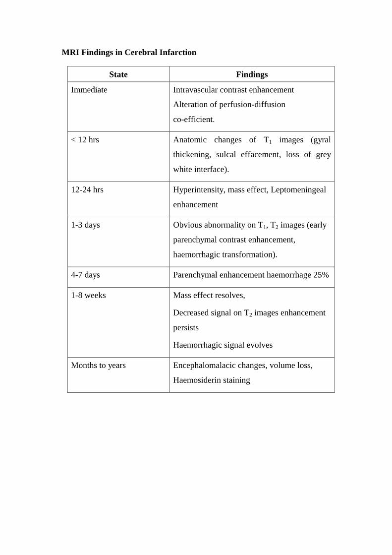
MRI Findings in Cerebral Infarction
State Findings
Immediate Intravascular contrast enhancement
Alteration of perfusion-diffusion
co-efficient.
< 12 hrs Anatomic changes of T1 images (gyral
thickening, sulcal effacement, loss of grey
white interface).
12-24 hrs Hyperintensity, mass effect, Leptomeningeal
enhancement
1-3 days Obvious abnormality on T1, T2 images (early
parenchymal contrast enhancement,
haemorrhagic transformation).
4-7 days Parenchymal enhancement haemorrhage 25%
1-8 weeks Mass effect resolves,
Decreased signal on T2 images enhancement
persists
Haemorrhagic signal evolves
Months to years Encephalomalacic changes, volume loss,
Haemosiderin staining

ELECTROCARDIOGRAPHIC CHANGES IN STROKE
Cardiovascular regulation is carried out by descending pathways from
forebrain which converge in hypothalamus, hence cerebrovascular accident can
produce changes in the ECG. These changes are often misdiagnosed as organic.
Stroke can cause ST elevation or depression and ‘Q’ waves but plasma CPK
levels do not rise. These changes normalize in 2 weeks time. Commonest
changes observed are T wave inversion, sinus tachycardia. Other changes
include ST depression, sinus QT prolongation, conduction disturbances and
ectopic rhythms.
THROMBOLYTIC THERAPY IN STROKE
In the recent years, the use of thrombolytic therapy in cerebral infarction
has been studied extensively66. The European co-operative acute stroke study
(ECASS) tested intravenous recombinant tissue plasminogen activator (rtPA :
1.1 mg/kg to a max. 100 mg; 10% as bolus, the remainder over 60 min) within 6
hrs of onset of symptoms.
The National institute of neurological disorders and stroke (NINDS)
study tested rtPA(0.9 mg/kg to a 90 mg max; 10% as bolus and the remainder
over 60 min) within 3 hours of onset of symptoms. Improved clinical outcome
and decreased bleeding hazard observed in NINDS study was probably due to
lower dose of rtPA and earlier institution of therapy.

A recent trial of the fibrinolytic agent ancrod in ischemic stroke is being
evaluated. Recent trials PROACT I and II (Prolyse in acute cerebral
thromboembolism) using intra-arterial thrombolysis upto the sixth hour showed
benefit. However intra-arterial thrombolysis is not approved by the FDA and
remains experimental.
REHABILITATION
Proper rehabilitation of stroke patient includes, early physical,
occupational and speech therapy. It is directed towards educating the patient and
family about the neurological deficit, preventing complications of immobility,
and providing encouragement and instruction in overcoming the deficit. The
goal of rehabilitation is to return the patient to home early and to maximize
recovery by providing a safe regimen suited to the individual patient.

Materials and
Methods

MATERIALS AND METHODS
Patients with cerebrovascular accident satisfying the inclusion criteria,
admitted in our hospital during the period from January 2005 to December 2005
were studied.
Inclusion Criteria
Patients with a clinical diagnosis of stroke and on whom CT has been
done were included. The various types of stroke in this group were:
� Ischemic stroke
� Haemorrhagic stroke
o Intraparenchynal
o Subarachnoid haemorrhage
� Cortical vein thrombosis
Exclusion Criteria
� Neurological deficit lasting for less than 24 hrs.
� Cases presenting 48 hrs after onset.
� Space Occupying lesions
� Subdural haemorrhage
� Extradural haemorrhage
� Patients with history of head injury.
� Patients in whom CT Scan brain could not be performed.

A detailed history in each case regarding onset, predisposing factors and
nature of stroke was recorded, followed by a thorough clinical examination.
The major risk factors included in the study were hypertension, diabetes
mellitus, hyperlipidemia along with other factors such as age, sex, menopause,
puerperium which were evaluated. History of TIA was noted. In young patients
family history of stroke and history of exposure to sexually transmitted disease
were recorded. Habituations to smoking, alcohol was noted. Special clinical
evaluation of cardiovascular system giving importance to rhythm disturbances,
cardiac failure, valvular heart disease, prosthetic valve was made.
Clinical neurological examination was done and the patients were
grouped into those having cortical involvement, internal capsular involvement or
brainstem involvement delineating the major blood vessels involved in the
process. Ophthalmoscopic examination of the fundus was done. Patients with
blood pressure readings of ≥160/90 mm Hg on two occasions was taken as
hypertension.
Baseline investigations included complete blood count, blood sugar, urea,
creatinine, electrolytes, cholesterol, urine analysis and chest x-ray. ECG was
taken at admission, after 3 days and at discharge. CT Brain was done for all
patients. Echocardiography and CSF analysis were done in relevant cases.
The results of all data are expressed in tabular forms for analysis. For the
analysis of ECG, those patients giving a definite history or evidence of diabetes

mellitus, coronary artery disease, rheumatic heart diseases, electrolyte
disturbances or those on anti-arrhythmatic drugs were avoided.

Observations and Results

OBSERVATIONS AND RESULTS
TABLE – 1
AGE DISTRIBUTION
Age in Year Number (n=100) Percentage
< 30 8 8
31 – 50 22 22
51 - 60 30 30
> 61 40 40
TABLE – 2
SEX DISTRIBUTION
Age in Year Male Female
Less than 50 15 15
More than 50 47 23
Total 62 38

0
5
10
15
20
25
30
35
40
Per
cen
tag
e
<30 31-50 51-60 >61
AGE IN YEAR
GRAPH -1AGE DISTRIBUTION
0
5
10
15
20
25
30
35
40
45
50
Per
cen
tag
e
Less than 50 More than 50
AGE IN YEAR
GRAPH -2SEX DISTRIBUTION
Male Female

TABLE – 3
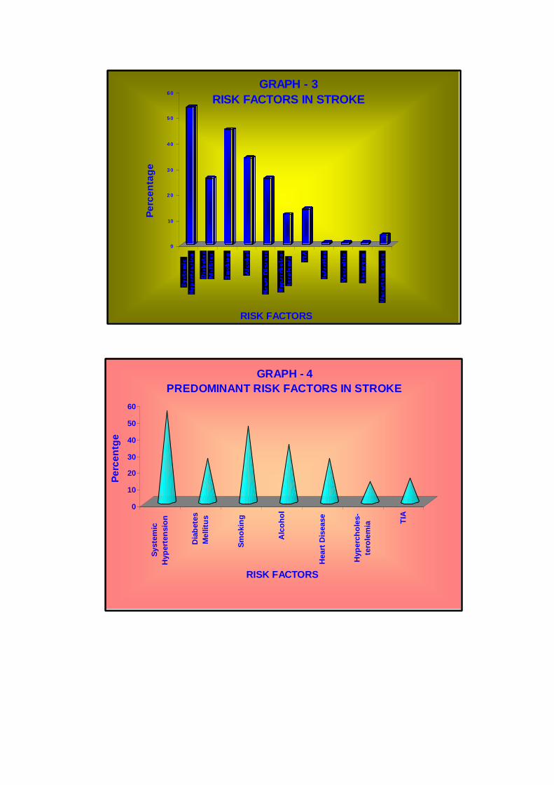
RISK FACTORS IN STROKE
S.No. Risk Factors Number Percentage
1. Systemic Hypertension 54 54
2. Diabetes Mellitus 26 26
3. Smoking 45 45
4. Alcohol 34 34
5. Heart Disease 26 26
6. Hypercholesterolemia 12 12
7. Transient Ischemic Attack 14 14
8. Infection 1 1
9. Vasculitis 1 1
10. Aneurysm 1 1
11. Uncertain cause 4 4
TABLE – 4
INCIDENCE OF CARDIAC DISEASES ASSOCIATED WITH STROK E
Disease Number (n=26)
Percentage
Coronary Artery Disease 15 57.7
Rheumatic Heart Disease 7 26.9
Mitral Valve Prolapse 4 15.4
0
10
2 0
3 0
4 0
5 0
6 0
Per
cen
tag
e
RISK FACTORS
GRAPH - 3RISK FACTORS IN STROKE
0
10
20
30
40
50
60
Per
cen
tge
Sys
tem
ic
Hyp
erte
nsi
on
Dia
bet
esM
ellit
us
Sm
oki
ng
Alc
oh
ol
Hea
rt D
isea
se
Hyp
erch
ole
s-te
role
mia T
IA
RISK FACTORS
GRAPH - 4PREDOMINANT RISK FACTORS IN STROKE

TABLE – 5
CLINICAL PRESENTATION
S.No. Factors Number Percentage
1. Hemiparesis 91 91
2. Cranial nerve Involvement 89 89
3. Aphasia 31 31
4. Loss of consciousness 26 26
5. Sensory symptoms 7 7
6. Cerebellar 3 3
7. Headache 14 14
8. Vomiting 23 23
9. Convulsions 5 5
TABLE – 6
APHASIAS
Factors Number
(n=31) Percentage
Global 22 71
Motor 7 22.6
Sensory 2 6.4

0
10
20
30
40
50
60
70
80
90
100
Per
cent
age
Hem
ipar
esis
CN
invo
lvem
ent
Ap
has
ia
LO
C
Sen
sory
sym
pto
ms
Cer
ebel
lar
Hea
dac
he
Vo
mit
ing
Co
nvu
lsio
ns
FACTORS
GRAPH - 5CLINICAL PRESENTATION
PIE CHART - 1APHASIAS
71
22.6
6.4
Global Motor Sensory

TABLE 7
TIME OF OCCURRENCE OF STROKE
Thrombotic (n=70)
Embolic (n=12) Haemorrhagic (n=18) Time
No % No % No %
4 am – 8 am 31 44.3 5 41.7 8 44.4
8 am – 12 pm 10 14.3 4 33.3 4 22.2
12 pm - 4 am 29 41.4 3 25.0 6 33.3
TABLE 8
SIGNIFICANCE OF ACTIVITY
Activity Sleep Factor
No % No %
Thrombotic (n=70) 30 42.9 40 57.1
Embolic (n=12) 9 75 3 25.0
Haemorrhagic (n=18) 14 77.8 4 22.2
TABLE 9
NATURE OF STROKE
S.No. Stroke No Percentage
1. Ischemic 82 82
2. Haemorrhagic 18 18

0
5
10
15
2 0
2 5
3 0
3 5
4 0
4 5
Per
cen
tag
e
T hromb ot ic N -70
Embo lic N - 12 Haemo rrhag ic N =18
TIME
GRAPH -6TIME OF OCCURRENCE OF STROKE
4 AM - 8 AM 8 AM - 12 PM 12PM- 4 AM
0102 03 04 0506 0708 0
Per
cen
tag
e
Thrombo t ic N = 70
Emb olic N=12 Haemo rrhag icN = 18
A ct ivit y
Sleep
FACTOR
GRAPH - 7SIGNIFICANCE OF ACTIVITY
Activity Sleep
0
20
40
60
80
100
Per
cern
tag
e
Ischemic Haemorrhagic
STROKE
GRAPH - 8NATURE OF STROKE

TABLE 10
AREA OF THE BRAIN INVOLVED IN ISCHEMIC STROKE
S.No. Factor No Percentage
1. Subcortical 42 51.2
2. Cortical 25 30.5
3. Cortical and Subcortical 13 15.9
4. Brainstem /Cerebellar 2 2.4
TABLE 11
DISTRIBUTION OF ARTERIAL TERRITORY IN ISCHEMC STROK E
S.No. Artery Involved No
(n=82) Percentage
1. MCA 73 89
2. PCA 4 4.9
3. Multiple 2 2.4
4. MCA, PCA 1 1.2
5. MCA, ACA 1 1.2
6. ACA 1 1.2

PIE CHART - 2AREA OF THE BRAIN INVOLVED IN ISCHEMIC
STROKE
51.2
30.5
15.92.4
Subcortical corticalCortical and Subcortical Brainstem / Cerebellar
PIE CHART - 3DISTRIBUTION OF ARTERIAL TERRITORY IN
ISCHEMIC STROKE
89
4.9
1.21.22.41.2
MCA PCA MCA, PCA MCA, ACA Multiple ACA

TABLE 12
LOCATION OF HAEMORRHAGE
S.No. Factor No (n=18) Percentage
1. Putamen 7 38.9
2. Cortical 4 22.2
3. Putamen, Thalamus 3 16.7
4. Pons 2 11.1
5. Cerebellum 1 5.5
6. Subarachnoid Haemorrhage 1 5.5
TABLE 13
ECG CHANGES IN STROKE
S.No. ECG No (n=25) Percentage
1. T wave Inversion 10 40
2. Sinus Tachycardia 8 32
3. Sinus Bradycardia 4 16
4. ST Depression 3 12
5. Conduction Disturbance 2 8
6. VPC 1 4
7. Normal 2 8
TABLE 14
LEVEL OF CONSCIOUSNESS AND PROGNOSIS
LOC (n=26) Death Factor
No % No %
Ischemic Stroke 11 42.3 5 45.5
Haemorrhagic Stroke 15 57.7 13 86.7

0
5
10
15
20
25
30
35
40
Per
cen
tag
e
Putamen Cortical Putamen,Thalamus
Pons Cerebellum SAH
SITE OF HAEMORRHAGE
GRAPH - 9LOCATION OF HAEMORRHAGE
0
5
10
15
20
25
30
35
40
Per
cen
tag
e
T wave inversion
SinusTachycardia
SinusBradycardia
ST. Depression ConductionDisturb
VPC Normal
ECG CHANGES
GRAPH - 10ECG CHANGES IN STROKE

CT SCAN PICTURES
CT SCAN PICTURES
Right MCA Infarct Left ACA and MCA Infarct
Left PCA Infarct Left Cerebellar Infarct
Large Right Intra Cerebral Bleed With Intra Ventricular
Extension
Left Cerebellar Haemorrhage With Fourth Ventricular
Extension

Corti cal Venous Thrombosis Subarachnoid Haemorrhage

Analysis of Data
and Discussi
on

ANALYSIS OF DATA AND DISCUSSION
Age
The maximum incidence of stroke in this study was observed in the above
60 yrs age group (40%). According to Banford and Sandercock et al., 1987,
maximum incidence of stroke was in the above 70 years age group. The
Framingham study (1979) showed that 80% of patients, were above 65 years of
age. Compared to Framingham study, a lower incidence of stroke in this study
could be explained by:
• Many cases of old stroke not being brought to the hospital.
• A high mortality rate in cases of old stroke.
• Higher prevalence of rheumatic heart disease in India compared to
western population.
• Majority of young strokes being hospitalized.
In this study incidence of stroke in below 50 years age group was 30%,
and above 50 yrs age group was 70%. This corelates well with the Vellore study
by Abraham J. Inbaraj et al., where the incidence below 40 years was 27% and
above 40 years was 73%, and the Rohtak rural study, where the prevalence of
stroke below 40 yrs, was 28.8%. However in the Kashmir rural study, the
prevalence below 40 years was 41.1% (Koul RL et al.).
Sex

With regard to sex distribution, the male to female ratio in this study was
1.6:1. In the below 50 years age, ratio was equal and in the above 50 years age
group ratio was 2:1. Higher female preponderance in the younger age group
could be attributed to higher prevalence of rheumatic heart disease.
In a study by Das Gupta et al., in 1984 male to female ratio was 1.3 : 1,
which is almost similar to this study67. In the Madurai study by Aleem MA et al.,
1996, the male to female ratio in young strokes was 1.1:1, which corelates well
with this study68. Study done by Jotideb Mukhopadhyay et al., 1998, concluded
an equal incidence in males and females69.
An epidemiological study by K.R. Dhamija et al., 1998, recorded a male
to female ratio of 1.7 : 1, which is in accordance with this study70. In a study
published in Kashmir (1989), males constituted 64% of stroke patients. In the
Saudi study by Qari FA et al., 2000, the male to female ratio was 3.4:1.

RISK FACTORS
(i) Systemic Hypertension
Hypertension was the commonest predisposing factor in this study,
present in 54% of the patients. Among the patients with haemorrhagic stroke,
77.8% were hypertensives. The Framingham 18 years follow up study shows
that hypertension was the most powerful precursor of stroke in both infarction
and haemorrhage, which tallies with this study. In a population study of stroke
patients in Richmond by Williams CA et al., 2003, 58.3% of patients were
hypertensives, which corelates well with this study71. In Oxford Shire
community stroke project, hypertension was present in 52% patients and Review
of stroke studies by Dalal and Dalal gives a figure of 51%, which is similar to
this study.
In a study by Rahman KMN et al., 2002, 78.82% of stroke patients were
hypertensives72, and a study by Aszal C et al., 2002, documented hypertension in
75% of patients73. Risk factors analysis by Jotideb et al., recorded 76% as
hypertensives69, while an epidemiological study by K.R. Dhamija et al., showed
34.7% as hypertensives70. Studies by Nielson W.B. et al., and Hart CL et al.,
2002 state that elevation of both systolic and diastolic blood pressure is
associated with increased risk of stroke74.
ii) Diabetes Mellitus

Diabetes mellitus was present in 26% of stroke patients in this study. In
the Oxford Shire community stroke project, the incidence of diabetes mellitus in
stroke was 28% which corelates well with this study. According to a study
Manorama Devi in. “The incidence of stroke among Indian Nationals in UAE”
diabetes was present in 20% of patients.
The incidence and severity of stroke are increased by the presence of
diabetes and the outcome from stroke are poorer, according to a study by Baird
TA et al., 200275. Study by Wannamathe et al., 1999 confirms diabetes mellitus
as an independent risk factor for stroke76. Similar conclusions were drawn from
the study of Hark CL et al., in 20009.
iii. Smoking
Smoking was a risk factor in 45% of patients in this study. None of the
females were smokers. According to a study by Williams CA et al., 2003, 33.3%
of ischemic stroke patients and 26.3% of haemorrhagic stroke patients were
smokers71. In a study by Ansari K et al., 2001, 29% of elderly stroke patients
were smokers. Study on multiple cerebral infarcts by B Reddy et al., recorded a
figure of 62%77.
Christensen et al., 2001, concluded that stroke patients who smoke are on
an average nine years younger than non-smokers78. Study done by Bonita R et
al., 1999 demonstrated that active as well as passive smoking increases the risk
of stroke79.

iv. Alcohol consumption
Alcohol consumption was present in 34% of patients in this study. Study
done by William CA et al., 2003 concluded that 41.6% of ischemic stroke
patients and 21.1% of haemorrhagic stroke patients were alcoholics71. A study
on multiple cerebral infarcts done by B Reddy et al., showed 30% of stroke
patients were alcoholics77.
Studies by Reynolds K et al., 200380 and Wannamathe SG et al., 1996
state that light or moderate alcohol consumption may be protective, while heavy
alcohol consumption increases risk of stroke largely mediated through blood
pressure76. Relationship of alcohol and stroke can be due to non-valvular atrial
fibrillation or cardiomyopathy (Lancet – 1995).
v. Heart disease
26% of patients in this study had cardiac disease associated with stroke.
Among the cardiac diseases, coronary artery disease was present in 15 patients
(57.7%), rheumatic heart disease in 7 patients (26.9%) and mitral valve prolapse
in 4 patients (15.4%). Study by Ansari K et al., 2001, showed that 23% of stroke
patients had cardiac disease. Studies by B Reddy et al.,77 Kundu TN et al.,81
Aleem M.A. et al.,68 documented an incidence of 36%, 35% and 30.8%
respectively.
The incidence of cardiac causes of embolic stroke in this study was 11%,
of which 7% contributed by rheumatic heart disease with atrial fibrillation and

4% mitral valve prolapse with infective endocarditis. This shows that rheumatic
heart disease with atrial fibrillation is the predominant cause of cardioembolic
stroke. This is in accordance with studies by V.K. Gupta in Kashmir and A.
Panagriya, Jaipur et al.,
The incidence of coronary artery disease in stroke patients in this study
was 15%. Among them, one patient developed stroke 4 days after the onset of
acute myocardial infarction. Studies by Mooe T et al., 199982 and Errikcson P et
al.,83 1997 state that the risk for stroke is highest in the first 5 days after
myocardial infarction, predictors being atrial fibrillation, ST elevation and
previous stroke. A study by Loh E et al. 1997, state that decreased ejection
fraction and older age are predictors for risk, while anti-coagulation appears to
have a protective effect against stroke after acute myocardial infarction84.
vi. Hypercholesterolemia
In this study 12% of patients had elevated total cholesterol levels,
indicating a causal relationship between stroke and lipid profile. Similar
observations were also made by Shridharan, Apollo Hospitals, Chennai, H.
Jacobs, D.R. Wentworth (1999), and Prospective studies collaboration the
Lancet 199585. The literature does not give a clear relation between serum lipids
and stroke. Studies by NA Rajwade et al., (1996)86 Sarti C et al., (2000)87 state
that the lipid levels in stroke patients were observed to be higher but not

significant and their associations are relatively weak. A study by Wannamathe
SG et al., (2000) concludes that elevated total cholesterol showed a weak
positive association, while elevated HDL cholesterol was associated with a
significant decrease in the risk of stroke88. However studies by Nair
M. Radhakrishnan et al., at Kerala and Jotideb et al., West Bengal, suggested
that hyperlipidemia is an important risk factor for stroke69. It may be concluded
that hypercholesterolemia is a risk factor for stroke, although the risk imparted is
lower than that for myocardial infarction.
vii. Transient Ischemic Attack and Other Factors
In this study, past history of stroke or Transient ischemic attacks were
present in 14% of patients. According to a study by William CA et al. (2003)
history of prior neurovascular disease was present in 36.2%71. MM Singh et al.,
(1996) in his study stated that transient ischemic attack is associated with mild to
moderate stenosis of internal carotid artery and is an important predictor of
ischemic stroke. Other etiological factors in this study include infective
endocarditis, tuberculous meningitis and subarachnoid haemorrhage secondary
to aneurysms.
CLINICAL PRESENTATION
• Hemiparesis was the commonest presentation in this study, present in
91% of patients. This is in accordance with the study by Rahman KM et
al., (2002) where hemiplegia was present in 88.24%72.

• 26% of patients presented with loss of consciousness. 83.3% of
haemorrhagic stroke patients and 22% of ischemic stroke patients had
altered consciousness level. Study by Rahman KM et al., suggested that
54.84% of haemorrhagic stroke patients present with altered
consciousness72.
• Headache and vomiting were present in 14 and 23 patients respectively.
Incidence of headache and vomiting were more in the haemorrhagic
stroke patients and among the ischemic stroke patients, it was more
common in posterior circulation stroke.
• 5 patients presented with convulsions, out of which 2 had embolic stroke,
1 patient had thrombotic stroke in the cortical territory and 2 had
haemorrhagic stroke.
• Language disturbances were observed in 31 patients. 22 had global
aphasia, 7 had motor aphasia and 2 patients had sensory aphasia. These
presentations correlated well with involvement of cortical territory.
• 7 patients had sensory disturbances, out of which 3 had thalamic infarct,
one patient had thalamic haemorrhage, one had lateral medullary
syndrome, one had brainstem stroke and one cortical infarct.
• Cerebellar involvement was observed in 3 patients. One had lateral
medullary syndrome, one had cerebellar haemorrhage and the last one had
brainstem stroke with cerebellar involvement.
Time of occurrence

In this study, 44.3% of thrombotic stokes, 41.7% of embolic strokes and
44.4% of haemorrhagic strokes occurred in the early morning hours between 4
am and 8 am. A study by Lago A et al., (1998) observed a higher frequency of
strokes during the day and lower frequency in the last hours of the evening in all
types of stroke, which is in accordance with this study89. Similar observations,
were made by Chaturvedi S. et al., (1999)90 and Casetta I et al., (2002) in
ischemic stroke91. They suggest the circadian variability in blood pressure and a
concurrent morning hypercoagulability as possible determinants of this pattern.
57.1% of thrombotic strokes occur during sleep (40% in Michael Reel
Stroke Registry)92. 75% of embolic strokes and 77.8% of haemorrhagic strokes
occurred during activity in this study (68% and 64% respectively in Michael
Reel stroke registry).
CT SCAN CORRELATION
• CT scan is the single most important non invasive investigation to
distinguish infarction from cerebral haemorrhage.
• After 6 hrs, CT brain may show lesion in <1/3 of patients with cerebral
infarct and at 12 hrs 50% show evidence of infarction. Over 90%
supratentorial infarctions (moderate to large) can be detected within 24
hrs.
• In this study, out of the 100 patients studied, CT Scan showed ischemic
stroke in 82% and haemorrhage in 18%. Studies by K.R.Dhamija et al.,

(1998)70 and Quari FA et al., (2000) observed cerebral infarction in
82.6% and 77% respectively, which tallies with this study.
• Regarding the distribution of arterial territory in ischemic stroke, MCA
was involved in 89%, PCA in 4.9%, ACA in 1.2%. Involvement of more
than one arterial territory was observed in 4.8%. Study by K. Srinivasa
Rao et al., documented 54% in the MCA, 4.5% in the PCA, 4.5% in the
ACA, and 27% at multiple sites.
• With respect to the area of the brain involved in ischemic stroke, 51.2%
had subcortical and 30.5% cortical infarcts, combination of both was
observed in 15.9%. Brainstem and cerebellar lesions found in 2.4%. In a
study by Sandya Purohit et al., (1999) cortical infarcts were found in
18%.
• Among the 18 patients with haemorrhagic stroke, 7 patients had putamen
haemorrhage (38.9%), cortical haemorrhage in 4 (22.2%), thalamic
haemorrhage in 3 (16.7%). 2 patients had pontine haemorrhage and both
of them expired. Cerebellar and subarachnoid haemorrhage were
documented in one patient each. According to a study by K. Srinivasa
Rao et al., (1998), 40% haemorrhages occurred in basal ganglia, and 27%
in the thalamic region.
• Among laboratory methods for diagnosis of intracerebral haemorrhage
the CT scan occupies the foremost position. This procedure has proved
totally reliable in the detection of haemorrhages that are 1.0 cm or more
in diameter. If the volume of haematoma from CT is less than 30ml it

favours a better prognosis. If volume of haematoma calculated from the
CT brain is ≥ 60 ml the prognosis is poor.
• The distinct appearance of the fresh haemorrhage changes with time,
disappearing slowly in the subsequent days to weeks, depending on the
size of the haemorrhage. Thereafter the haemorrhage appears as a region
of low densities on CT scan, which can be mistaken for an old infarct. So
if CT imaging is delayed for more than a few weeks after stroke, it may
not possible to reliably distinguish between ischemic and haemorrhagic
stroke
• Hypertension was the commonest predisposing factor in this study,
present in 54% of patients. Among patients with haemorrhagic stroke as
evidenced by CT scan 86.6% of them were hypertensives.
• Diabetes mellitus was present in 26% of stroke patients in this study.
Among the patients with diabetes mellitus CT scan showed increased
incidence of infarct than haemorrhage.
• Other important risk factors such as alcohol consumption,
hypercholesterolemia, TIA were present in 34%, 12%, 14% of the
patients respectively: with CT scan showing increased preponderance to
infarct pattern.
• Cardiac causes namely RHD, MVPS increases the incidence of ischemic
embolic stroke which in this study was 11% as shown by the infarcts in
CT scan.

• Haemorrhagic stroke as evidenced from CT scan showed increased
incidence of headache and vomiting. However among the infarct patients
it was more associated with posterior circulation strokes.
• Convulsions occurred in 11% of patients with haemorrhagic stroke and
0.4% of ischemic stroke. Among the patients with ischemic strokes
convulsions was more common in cortical infarcts.
• Among the haemorrhagic stroke revealed by CT scan, 83.3% presented
with loss of consciousness where as it was 22% in ischemic stroke.
• Sensory disturbances were more common in patients with thalamic and
cortical infarcts as revealed by CT scan.
• Patients with language disturbances had more incidence of cortical
territory infarct in CT scan.
• In this study, CT scan showed cerebellar involvement in 3 patients, one
had lateral medullary syndrome, one had cerebellar haemorrhage and
other one had brainstem stroke with cerebellar involvement.
ECG CHANGES IN STROKE
For the analysis of ECG, patients with coronary artery disease, rheumatic
heart disease, those with definite history of diabetes and hypertension, renal
failure patients, those on anti-arrythmic drugs were excluded. Out of the 100
patients, 25 satisfying the criteria were selected and their ECGs were analysed.
The commonest change observed was T wave inversion and sinus
tachycardia present in 40% and 32% respectively. Sinus bradycardia in 16% and

ST depression in 12%. 8% had conduction disturbance, Ventricular premature
contractions observed in 4% and 8% had normal ECG.

PROGNOSIS
Loss of consciousness at the time of admission is considered to be an
adverse prognostic factor. In this study, 5 out of the 11 ischemic stroke patients
and 13 out of the 15 haemorrhagic stroke patients, presenting with loss of
consciousness expired. On an average, 69.2% of patients presenting with loss of
consciousness died. Studies of M Das et al., (1999) and IS Gambhir et al., (1998)
state that level of consciousness at admission by Glasgow coma scale is a very
important prognostic factor and 82.14% of patients with a score less than 8
expired.
Similar observations were made by Gambir IS et al.,93 (1998) and Joshi
PP et al ., (1999)94. Study by Masquardsen et al., states that nearly all patients
who were deeply comatosed at the time of admission died within 24 hrs.
Semicomatosed patients also fared badly. The above results were comparable to
this study.

Conclusi
on

CONCLUSIONS
• The risk factors were present in more than 90% of cases.
• The incidence of stroke was more common in the above 60 years age
group.
• The incidence in males was one and a half times more than in the females,
with the difference being negligible or absent in the less than 50. This is
attributed to increased prevalence of rheumatic heart disease in young
females.
• Hypertension was the commonest risk factor in both ischemic and
haemorrhagic strokes, present in more than half of the patients, followed
by smoking, and diabetes mellitus.
• There is a weak positive association between hypercholesterolemia and
stroke.
• Heart disease was the predominant risk factor in the young, of which
rheumatic mitral stenosis with atrial fibrillation was found to be the most
common, followed by mitral valve prolapse with infective endocarditis.
• The presence of atherosclerosis increases the risk of stroke.
• Risk for ischemic stroke doubles for smokers. In smokers, stroke occurs
after an average of 10-20 years of smoking.
• Only about 12% of patients experienced transient ischemic attack.
• Other important causes of stroke in the young were vasculitis and
aneurysmal rupture.

• Hemiparesis was the commonest presenting feature in both ischemic and
haemorrhagic stroke followed by cranial nerve involvement, aphasia and
loss of consciousness.
• Headache and vomiting were more common in haemorrhagic stroke. In
ischemic stroke, it was more common in the posterior circulation stroke.
• Convulsions were more common in embolic stroke involving cortical
territory.
• Peak incidence of stroke occurred between 4 am to 8 am irrespective of
the type of stroke.
• More than half of thrombotic strokes occur during sleep. Nearly three
fourths of embolic and haemorrhagic strokes occur during activity.
• Majority of strokes were found to be infarcts, commonest being the
middle cerebral artery territory followed by posterior cerebral artery.
• Among ischemic strokes, involvement of sub-cortical structures i.e.
Internal capsule, putamen, basal ganglia were more common compared to
cortical structure.
• Among haemorrhagic strokes, putamen involvement was the commonest
followed by cortical haemorrhage.
• Commonest ECG change observed was T wave inversion and sinus
tachycardia.
• Loss of consciousness is an adverse prognostic factor with a mortality rate
of 69%.

• Computed Tomography is likely to remain the principal imaging
technique for stroke patients in foreseeable future, not only because it
excludes several non-stroke pathologies and reliably distinguishes
haemorrhage from infarction, but because it is far more widely available,
relatively less expensive, easier and safer to use in acutely ill stroke
patients.
• CT scan is also essential in patients with stroke who require thrombolytic
treatment with tissue plasminogen activator.

Bibliography

BIBLIOGRAPHY
1. Harrison’s Principles of Internal Medicine - 16th Edition,
Volume 2.
2. Adams and Victors Principles of Neurology - 7th Edition.
3. Brains diseases of the Nervous system - 11th Edition.
4. Oxford Text book of Medicine - 5th Edition.
5. Alagappan Manual of Practical Medicine - 2nd Edition.
6. API text book of Medicine - 7th Edition.
7. Corwin, L.I., Wolf, P.A., Kannel, W.B., and McNamara, P.M. Accuracy
of death certification of stroke: the Framingham study. Stroke 13: 818,
1982.
8. Garraway, W.M., Whisnant, J.P., and Drury, I. The changing pattern of
survival following stroke. Stroke 14: 699, 1983.
9. Bonita, R., and Beaglehole, R., Increased treatment of hypertension does
not explain the decline in stroke mortality in the United States, 1970-
1980. Hypertension 13: I-69, 1989.
10. Whisnant, J.P., The role of neurologist in the decline of stroke. Ann.
Neurol. 14: 1, 1983.
11. Knox, E.G. Meteorological associations of cerebrovascular disease
mortality in England and Wales. J. Epidemiol. Comm. Hlth 35: 220,
1981.

12. Haberman, S., Geographical Variation in Cerebrovascular disease
mortalty in England and Wales. Neuroepidemiology 3: 207, 1984.
13. Wroe, S.J., Sandercock, P., Bamford, J. , et al., Diurnal Variation in the
incidence of Stroke. The Oxfordshire Community Stroke Project, Br.
Med. J. 304: 155, 1992.
14. Bamford, J. Sandercock, P., Dennis, M.I. et al., A Prospective study of
acute cerebrovascular disease in the community. The Oxford Shire
community stroke project, 1981-86. 2. Incidence, case fatality rates and
overall outcome at one year of cerebral infarction, primary intracerebral
and subarachnoid haemorrhage. J. Neurol. Neurosurg. Psychiat, 53: 16,
1990.
15. Prof. Steve E. Humphries, Laleh Morgan B, Genetic risk factors for
stroke and carotid atherosclerosis. Lancet 3: 227-235, 2004.
16. Katarina Jood. Claes Ladenvall, Family History in ischemic stroke before
70 years of age. Stroke 36: 1383, 2005.
17. MacMohan, S.W., Peto, R., Cutler, J., et al., Blood Pressure, stroke, and
coronary artery disease. Part I, prolonged differences in blood pressure:
Prospective observational studies corrected for the regression dilution
bias. Lancet 335: 765, 1990.
18. Xiang-Hua Fang, Xin-Hua Zhang, Subtype Hypertension and risk of
stroke in middle-Aged and Older Chinese. Stroke 37 : 38, 2006.
19. Collins, R. Peto, R., MacMohan, S. et al., Blood Pressure, stroke,
coronary Artery disease. Part 2: Short term reductions in blood pressure.
Overview of randomized drug trials in their epidemiological context
Lancet 335: 827, 1990.

20. Per Harmsen, Georg Lappas, et al. Long-Term Risk Factors for stroke.
Stroke 37 : 1663, 2006.
21. Gang Hu, Cinzia Satri, et al., The impact of History of Hypertension and
type 2 diabetes at Baseline on the incidence of stroke and stroke
mortality. Stroke 36: 2538, 2005.
22. Kannel, W.B., and McGee, D.L. Diabetes and Cardiovascular disease.
The Framingham study. J. Am. Med. Ass. 241: 2035, 1979.
23. Elizabeth selvin, Eyal Shahar, et al., Glycaemia (Haemoglobin A1c) and
incident ischemic stroke. Lancet 4: 821-826, 2005.
24. Sarah vermeer, Willemijn Sandee, et al., IGT increases stroke risk in
Nondiabetic patients with TIA. Stroke 37 : 1413, 2006.
25. Martin, M.J., Hulley, S.B., Browner, W.S., et al., Serum cholesterol,
blood pressure and mortality; implications from a cohort of 361,622 men.
Lancet 2: 933, 1986.
26. Qizilbash, N., Jones, L., Warlow, C., and Mann, J., Fibrinogen and lipids
as risk factors for transient ischemic attacks and minor ischemic strokes.
Br. Med. J. 303: 605, 1991.
27. Tanak,H. Ueda, Y. Hayashi, M., et al., Risk factors for cerebral
haemorrhage and cerebral infarction in a Japanese rural community.
Stroke 13: 62, 1983.
28. Valery Feigin, Varsha Parag, et al., Smoking and Elevated Blood pressure
are the most important risk factors for subarachnoid Haemorrhage in the
Asia-Pacific Region. Stroke 36: 1360, 2005.

29. Shinton, R., and Beevers G., Meta analysis of relationship between
cigarette smoking and stroke, Br. Med. J. 298: 789, 1989.
30. Adan I. Qureshi, M. Fareed; K. Sari, Cigarette smoking Among spouses.
Stroke 36: e74, 2005.
31. Kozararevic, D.J., McGee D., Vojvodic, N. et al., Frequency of alcohol
consumption and morbidity and mortality. The Yugoslavia cardiovascualr
disease study. Lancet 1: 613, 1980.
32. Kenneth J. Mukamal., Hyoju chung., Alcohol use and risk of ischemic
stroke among older adults. Stroke, 36: 1830, 2005.
33. Alberto Ascherio, Murray A. Mittleman, et al., Alcohol and Risk for
ischemic stroke in men. Stroke 142:1 Pages 11-19, 2005.
34. Malhotra, H., Mehta, S.R. Mathur, D., and Khandelwal, P.D., Pressor
effects of alcohol in normotensive and hypertensive subjects. Lancet, 1:
584, 1985.
35. Gordon, T., Kannel, W.B., Drinking habits and Cardiovascular disease.
The Framingham Study. Am. Heart, J., 105: 667, 1983.
36. Lancet (1985). Alcohol and atrial fibrillation. Lancet 1: 1374, 1985.
37. Brandi J. Witt, Robert D. Brown. A community – Based study of stroke
incidence after myocardial infarction. Stroke 143: 11, pages 785-792,
2005.
38. Allison G. Hays, Ralph L. Sacco, Left ventricular systolic dysfunction
and risk of ischemic stroke in multiethnic population. Storke 37: 1715,
2006.

39. Aberg, H. Atrial Fibrillation. A study of atrial thrombosis and systemic
embolism in a necropsy material. Acta Med. Scand. 185 : 373, 1969.
40. Devin L. Brown, Lynda D. Lisabeth, Recurrent stroke risk is higher than
cardiac event risk after initial stroke / TIA. Stroke 36: 1285, 2005.
41. Wiebers, D.O., Whisnant, J.P., Sandok, B.A., and O’Fallon, W.M.
Prospective comparison of a cohort with asymptomatic carotid bruit and a
population based cohort without carotid bruit. Stroke 21: 984, 1990.
42. Dr. Malcolm R. Macleod, Pierre Amarenco d, Atheroma of the aortic arch
: an important and poorly recognised factor in the aetiology of stroke.
Lancet 3: 308-414, 2004.
43. Bayer, D.P., and Corle, D.K., Hormone therapy for prostate cancer:
results of the Veterans Administration Co-operative Urological Research
group studies. NCI Monogr. 7: 165, 1988.
44. Stadel, B.V. Oral Contraceptives and cardiovascular diseases. Part I, New
Engl. J. Med. 305: 612, 1981.
45. Beaglehole R., Oestrogens and Cardiovascular disorder.
Br. Med. J. 297: 571, 1988.
46. Sudhir Kurl : Jari A. Laukkanen, Metabolic Syndrome and the risk factors
in middle-Aged Men. Stroke 37 : 806, 2006.
47. Law, M.R., Frost, C.D. and Wald, N.J. By how much does dietary salt
reduction lower blood pressure? I- Analysis of observational data among
populations. Br. Med. J. 301: 811, 1991.

48. Acheson, R.M. and Williams, D.R.R. Does consumption of fruits and
vegetables protect against stroke. Lancet 1: 1191, 1983.
49. Khaw, K.T., and Barrett – Connor, E., Dietary Potassium and Stroke
associated mortality. A 12 year prospective population study. New Engl.
J. Med. 316, 235, 1987.
50. Michiel J. Bos, Peter J. Kouds taal. Uric acid is a risk factor for MI and
stroke. Stroke 37: 1503, 2006.
51. Klar. H. Yoggi, John concato, Obstructive sleep apnea as risk fator for
stroke and death. Stroke, 353: 2034-2041, 2005.
52. Roberto Munoz, Joaquin Duran – Cantolla, Severe Sleep Apnea and risk
of ischemic stroke in the elderly. Stroke 37: 2317, 2006.
53. Gang Hu, Cinzia Sarti, Leisure Time, occupational and commuting
physical activity and the risk of stroke. Stroke 36: 1994, 2005.
54. Jamary Oliveira Filho, Leila C. Viana, Chagas disease is an independent
risk factor for stroke. Stroke, 36: 2015, 2005.
55. Arndt Rolfs, Marlies Zschiesche, Prevalence of Fabry disease in patients
with Cryptogenic stroke, Lancet 366: 1794-1796, 2005.
56. Satoshi Kuroda, Tatsuya Ishikawa, Incidence and clinical features of
disease progression in Adult Moyamoya disease. Stroke 36: 2148, 2005.
57. Daphne L. Vander, Diederick E. Grobbee, Serum ferritin is a risk factor
for stroke in post menopausal women. Stroke 36: 1637, 2005.

58. Tetsuya Ohira, Pamela J. Schreiner, Lipoprotein (a) and Incident
inchemic stroke. Stroke 37: 1407, 2006.
59. Leng GC, Fowkes FGR, Lee AJ et al. Use of ankle brachial pressure
index to predict cardiovascualr events and death: a cohort study. Br
Med., J. 313: 1440-1444, 1996.
60. Macko RF, Kittner SJ, Epstein A et al. Elevated tissue plasminogen
activator antigen and stroke risk. The stroke prevention in young women
study. Stroke; 30: 7-11, 1999.
61. Hankey GJ, Eikelboom JW. Homocysteine and vascular disease. Lancet,
354: 407-413, 1999.
62. Gillum LA, Mamidipudi SK, Johnston SC. Ischemic stroke risk with oral
contraceptives. A meta–analysis. JAMA, 284: 72-78, 2000.
63. Wise, R.J.S. Bernardi S., Frackowiak, R.S.J. et al., Serial observations on
the pathophysiology of acute stroke. The transition from ischemia to
infarction as reflected in regional oxygen extraction. Brain 106: 197,
1983.
64. Van Gijn, J., Van Dongen, K.J., Vermullen, M. and Hijdra A.,
Perimesencephalic haemorrhage: A non-aneurysmal and benign form of
subarachnoid Haemorrhage. Neurology,
35: 493, 1985.
65. Tomlinson, B.E., and Walton, J.N. Superficial Hemosiderosis of the
central nervous system. J. Neurol. Neurosurg. Psychiat, 27: 332, 1964.

66. Albers, GW et al.,: Anti-thrombotic and thrombolytic therapy for
ischemic stroke. Fifth ACCP consensus conference on Anti - thrombotic
therapy. Chest 114: 683S, 1998.
67. Dasgupta S, Hazra SC, Ghosal S. et al., A comparative study of stroke in
1984 and 1994. JAPI 46: 1, 1998.
68. Aleem M.A., Manivannan, R., Sahubar Sadious S., Rajendran P,
Ramasubramanian D et al., Analysis of risk factors in young stroke
patients. JAPI 46: 1, 1998.
69. Jotideb Mukhopadhyay,Kiran Mitra ND Dutta, S. Jana, P. Bose, Study of
risk factors in cerebrovascular accident cases in a rural medical college of
West Bengal. JAPI 46: 1, 1998.
70. K. Rajinder Dhamija, AK Sood, S Lal, A Dua, SB Dhamija. An
epidemiological study of cerebrovascular disease in rural Haryana. JAPI
46: 1, 1998.
71. Williams CA, Sheppard T, Maruffo M et al., A Brief descriptive analysis
of stroke features in a population of patients from a large urban Hospital
in richmond, Virginia, a city within the stroke belt. Neuroepidemiol, 22 :
31-36, 2003.
72. Rahman KM, Sarker CB, Mohammad QD, Dhar DK, Hossain MS, et al.,
Risk factors and clinical presentations – a study of eight five hospital
admitted stroke cases. Mymensingh Med. J. July 11, 113-5, 2002.

73. Aszal CZ, Barsi P, Vitrai J, Nagy Z. Hypertension and clusters of risk
factors in different stroke subtypes. J. Hum. Hypertens. July 16: 495-
500, 2002.
74. Hart CL, Hole DJ, Smith G.D. Comparison of risk factors for stroke
incidence and mortality in 20 years of follow up in men and women in the
Renfrew/paisley study in Scotland. Stroke Aug. 31 : 1893-6, 2000.
75. Baird TA, Parsons MW, Alan Barber et al., The influence of diabetes
mellitus and hyperglycemia on stroke incidence and outcome. J. Clin.
Neuroscio Nov.9, 618-26, 2002.
76. Wannamathe SG, Perry J et al., Non-Fasting serum glucose and insulin
concentrations and the risk of stroke. Stroke Sep. 30: 1780-6, 1999.
77. Reddy B, Reddy C.S. A study of multiple cerebral infarcts. JAPI 47: 1,
1999.
78. Christensen HK, Guassora AD et al., Ischemic stroke occurs among
young smokers. Ugeskr Laegr Dec. 163: 7057-9, 2001.
79. Bonita R., Duncan J et al., Passive smoking as well as active smoking
increase the risk of acute stroke. Tob control summer 8 : 156-160, 1999.
80. Reynolds K, Lewis LB, Nolen JD et al., Alcohol consumption and risk of
stroke a meta analysis, JAMA Feb. 289: 579-88, 2003.
81. Kundu TN, Ganguly PK, Maiti B et al., A clinical study on stroke in
Young. JAPI 46: 1, 1998.
82. Mooe T., Olofsoon BO. Stegmayr B et al., Ischemic stroke. Impact of a
recent myocardial infarction. Stroke May 30: 997 – 1001, 1999.

83. Erikkson P, Mooe T et al., Ischemic stroke after acute myocardial
infarction – A population based study. Stroke April 28 : 762-7, 1997.
84. Loh E, Sutton MS, Wun CC et al., Ventricualr dysfunction and the risk of
stroke after myocardial infarction. N. Engl. J. Med. Jan. 336 : 251-7,
1997.
85. Cholesterol, Diastolic blood pressure and stroke. 13,000 strokes in
450,000 people in 45 prospective cohorts. Prospective studies
collaboration. Lancet Dec. 23-30, 346 : 1647-53, 1995.
86. Rajwade NA, Desai NK, Gupta KC Evaluation of serum lipids in patients
of acute stroke. JAPI 44 8 : 1996.
87. Sartie, Kaarisalo M, et al., The relationship between cholesterol and
stroke: implications for anti-hyperlipidemic therapy in older patients.
Drug Aging Jul 17: 33-51, 2000.
88. Wannamathe SG, Shaper AG, Ebrahim S HDL-Cholesterol, total
cholesterol, and the risk of stroke in middle aged British Men. Stroke
Aug. 31, 1882-8, 2000.
89. Lago A, Geffner D, Tembl J et al., Circadian variation in acute ischemic
stroke a hospital based study. Stroke Sep. 29; 1873-5, 1998.
90. Chaturvedi S, Adams HP, Woolson RF, Circadian variation in ischemic
stroke subtypes. Stroke, Sep. 30: 1792-5, 1999.
91. Casetta I, Granieri E, Fallica E et al., Patient demographic and clinical
features and circadian variation in onset of ischemic stroke. Arch.
Neurol. Jan. 59: 48-53, 2002.

92. Bornstein NM, Gur, AY, Fainshtein P et al., Stroke during Sleep:
epidemiological and clinical features cerebrovasc. Dis. Nov – Dec. 9:
320-2, 1999.
93. Gambhir IS, Yadav A, Shukla RC et al., Prognostic significance of initial
glasgow coma scale in patients of spontaneous intracerebral haemorrhage.
JAPI 46: 1, 1998.
94. Joshi PP, Nair MK, Holay MP, Fusey SM. Predictors of Mortality after
haemorrhagic stroke, JAPI 47: 1, 1999.
95. Stroke: Your Questions Answered Graeme J. Hankey. First Indian
edition, page. 61-66, 2006.

Proforma

PROFORMA
Name : Study No. : Age/Sex : IP No. : Occupation : DOA : Place : DOD : HISTORY
PRESENT HISTORY
• Weakness of limbs • Sensory disturbance • Cranial Nerve palsies • Language disturbance Global / Motor / Sensory aphasia • Memory disturbance • Cerebellar symptoms • Headache, Vomiting, Convulsions • Loss of Consciousness • Autonomic nervous system symptoms • Activity/ Sleep, Time of onset • Trauma, Fever
PAST HISTORY
• Transient ischemic attack • Stroke • Diabetes mellitus • Systemic hypertension • CAD • Rheumatic heart disease • Intermittent claudication • H/O oral contraceptive intake
FAMILY HISTORY Stroke / Diabetes mellitus / Systemic hypertension / CAD PERSONAL HISTORY
• Smoker - / Day Years • Alcoholic - / Day Years
MENSTRUAL HISTORY
• Pre – Menopausal • Post – Menopausal
GENERAL EXAMINATION

Febrile / Afebrile Anemia / Cyanosis / Clubbing / Pedal edema.
Pulse : Peripheral pulses :
BP : Respiration :
CVS : S1 S2, murmurs
RS : NVBS , added sounds
P/A : Bowel sounds, organomegaly
CNS
• Handedness • Conscious/Semi–conscious / Unconsciousness
• Orientation • Speech-Normal/Aphasia-Global/Motor/Sensory
• Cranial nerve palsies • Motor weakness
• Sensory abnormalities • Cerebellum
• Carotids • Fundus
• Gait • Involuntary movements
• Signs of meningeal irritation
CLINICAL DIAGNOSIS: INVESTIGATIONS TC DC Hb%
PCV ESR Platelet count
Blood - Sugar
Urea Urine Complete
Creatinine
Electrolytes CSF analysis
Serum Cholesterol
Chest X-ray ECG
CT Brain MRI
Echocardiography Carotid Doppler
FINAL DIAGNOSIS:

Abbreviations

ABBREVIATIONS ACA - Anterior Cerebral Artery
AF - Atrial Fibrillation
ASMI - Anteroseptal Myocardial Infarction
BG - Basal Ganglia
BS - Brainstem
C - Cortical
CAD - Coronary Artery Disease
CHO - Serum Cholesterol
CN - Cranial Nerve
CT - Computed Tomography
DIC - Disseminated Intravascular Coagulation
DM - Diabetes Mellitus
E - Expired
ECG - Electrocardiogram
ECHO - Echocardiogram
EM - Embolic
G - Global Aphasia
H - Haemorrhage
I - Infarction
IE - Infective Endocarditis
IVCD - Intra Ventricular Conduction Defect
IWMI - Inferior Wall Myocardial Infarction
LH - Left Hemiparesis
LMS - Lateral Medullary Syndrome
LOC - Loss of Consciousness
M - Motor Aphasia
MCA - Middle Cerebral Artery
MI - Myocardial Infarction
MR - Mitral Regurgitation

MS - Mitral Stenosis
MVP - Mitral Valve Prolapse
N - Normal
P - Putamen
PCA - Posterior Cerebral Artery
PH - Past History of Stroke
PHT - Pulmonary Hypertension
R - Recovered
RBBB - Right Bundle Branch Block
RH - Right Hemiparesis
RHD - Rheumatic Heart Disease
S - Sensory Aphasia
SAH - Sub Arachnoid Haemorrhage
SB - Sinus Bradycardia
SC - Subcortical
SHT - Systemic Hypertension
ST - Sinus Tachycardia
ST↓ - ST Depression
T - Thalamus
T↓ - T wave Inversion
TBM - Tuberculous Meningitis
Th - Thrombotic
TIA - Transient Ischemic Attack
VEG - Vegetation

Master Chart

MASTER CHART
CT S.No.
Age Sex
Time of
occurrence
Activity/
Sleep
Weak ness
Cranial Nerve Aphasia Sensory Headache
/Vomiting LOC/Fits SHT/DM Smoking /Alcohol TIA/PH CAD
/RHD CHO ECG ECHO Clinical
Diagnosis I/H Artery
Cortical/ Subcortic
al
Outcome
1 55 M 7.00PM S RH 7 - - - - DM S/A - - 120 T ↓ - I-TH I MCA SC R
2 65 M 11.00PM S RH 7 G - - - SHT S/A - - 176 ST DD I-TH I MCA C,SC R
3 50 M 3.00PM S LH 7 - - - - SHT S/A TIA - 133 ST - I-TH I MCA SC R
4 58 M 4.00PM A LH 7 - - - - - S/A - - 225 VPC - I-TH I MCA C R
5 85 F 7.00AM S LH 7 M - - - - - PH - 234 T↓ - I-TH I MCA SC R
6 65 M 2.00PM A RH 7 G - - - - S - - 177 T↓ - I-TH I MCA C R
7 45 F 2.00AM S RH 7 G - - LOC/F - - TIA/PH RHD 176 RAD, LAE, AF
MS. MR, PHT
RHD, I-EM
I MCA C E
8 62 M 11.00PM S LH - - - - - SHT - PH - 142 ST - I-TH I MCA SC R
9 68 F 9.00PM A LH - - - - - SHT/DM - - - 205 LVH - I-TH I MCA SC R
10 48 M 3.00PM A RH 7 - - - - SHT - - - 136 N - I-TH I MCA SC R
11 60 M 11.00AM A LH 7 - - H/V LOC SHT/DM S - - 148 IVCD, LVH
- H H - P,T,IC E
12 70 M 12.00AM S RH 7 - - - - SHT/DM - - - 153 N - I-TH H - P,BG,T,IC R
13 36 F 12.00PM A RH 7 G - H - - - - - 172 LVH MVP, MR,
VEG+ I-EM I MCA C,SC E
14 70 F 11.00AM S RH 7 G - - F SHT/DM - - - 156 N - I-TH I MCA C R
15 72 M 3.00PM A RH 7 - - V LOC SHT S/A - - 175 ST - H H - C E
16 49 M 6.00AM A LH 7 - - V LOC SHT S/A - - 165 N - I-TH I MCA C R
17 75 F 5.00AM A RH 7 - + - LOC SHT - - - 172 N DD, LVH
I-TH H - P,BG,T,IC E
18 60 M 6.00AM, S LH 7 - - - - SHT S - - 185 ST - I-TH I MCA SC R
19 60 M 7.00AM A RH 7 G - - - SHT S/A - - 192 IVCD - I-TH I MCA SC R
20 58 M 2.00PM S RH 7 G - - - - S - - 210 ST - I-TH I MCA C R
21 54 M 9.00PM A RH 7 - - - - - S/A - - 175 SB - I-TH I MCA SC R
22 81 M 9.00PM A LH 7 - - - LOC DM S/A - - 180 ST, ST
↓ - I-TH I MCA SC R

23 48 M 10.00AM A RH 7 - - - - - S - - 186 SB, LVH
MVP, MR
I-EM I MCA SC R
24 70 F 7.00AM S LH 7 - - - - SHT - - - 157 N DD, LVH
I-TH I MCA SC R
25 78 F 2.00PM S RH 7 G - V - - - - - 142 T ↓ - I-TH I MCA C,SC R
26 18 F 8.00AM S LH 7 G - H - - - - - 150 ST - I-TH-EBM I MCA C E
27 65 F 7.00AM A LH 7 - - H LOC/F SHT - - - 126 N - H H - P,BG R
28 52 M 8.00PM A Cerebell
ar 9,10,5 - + H - SHT/DM S/A - - 134 N - I-TH I PCA LMS E
29 50 M 6.00A A LH 7 - - V LOC SHT A - CAD 136 OLD IWMI
- H H - P.BG E
30 73 M 11.00PM A - - - + - LOC DM S/A - CAD 164 ST T - H I PCA C E
31 75 M 10.00AM S RH 7 G - - - DM - - - 200 T ↓ - I-TH I MCA C R
32 85 F 7.00AM S LH 7 - - - - SHT - - - 140 LVH, ST, ST↓
DD, LVH
I-TH I MCA SC R
33 48 M 7.00AM S RH 7,12 - - - - SHT/DM S - - 188 N - I-TH I MCA SC R
34 60 F 4.00PM A LH 7 - - V - SHT/DM - TIA - 136 ST - I-TH I ACA,M
CA C R
35 40 F 4.00PM A LH 7 - - - - - - - RHD 148
LAD, AF,
RBBB T
MS, PHT
I-EMB, RHD I MCA SC R
36 80 F 6.00AM S LH 7 - - - - SHT - - CAD 150 LBBB LAD
- I-TH I MCA SC R
37 36 M 11.00AM S LH 7 - - - - - S/A - - 140 ST, T ↓ - I-TH I MCA SC R
38 75 M 6.00 AM S RH 7 G - - - SHT/DM S/A - - 158 ST DD I-TH I MCA C,SC R
39 60 F 6.00AM S - 7,8,9,10 - + - - SHT/DM - - - 174 T LVH - I-TH I PCA C,SC R
40 60 F 6.00PM A Cerebell
ar - - - - - SHT/DM - - - 144 LVH - I-TH H - Cerebellar R
41 80 F 8.00PM A LH 7 - - H LOC SHT - - - 196 T ST↓ - H H - SC-P, BG E
42 60 F 4.00AM S LH 7 - - - - SHT - - - 198 N - I-TH I MCA SC R
43 65 M 8.00PM A - - - - V LOC SHT S/A - - 174 LVH - H H - C E
44 55 F 6.00AM S RH 7 S - - LOC SHT - - - 124 LVH - I-TH H - C E
45 70 F 6.00AM S RH 7 - - - - SHT/DM - - - 176 ST DD I-TH I MCA SC R
46 73 M 3.00AM S RH 7 - - - - SHT S - - 186 T↓ DD, LVH
I-TH I MCA SC R
47 52 M 10.00AM A RH 7 G - - - SHT/DM S - - 146 ST - I-TH I MCA C,SC R

48 58 M 3.00PM A LH 7 - - - - SHT S - - 152 ST - I-TH I MCA SC R
49 55 F 10.00AM A LH 7 - - - - - - - - 164 T↓, ST ↓
- I-TH I MCA SC R
50 58 F 7.00AM S - - - - H/V LOC SHT/DM - PH - 178 ST T ST ↓
DD H I MCA, PCA C,SC E
51 48 F 2.00PM A - - - - - LOC/F SHT - PH CAD 182 ST T - H H - PONTINE E
52 45 M 8.00AM A RH 7 M - - - - S/A - - 234 IVCD ST
↓ RBBB - I-TH I MCA SC R
53 55 M 6.00AM S LH 7 - - - - - S/A - - 114 SB - I-TH I MCA SC R
54 64 M 2.00PM S RH 7 - - - - SHT - - - 163 T↓ DD I-TH I MCA SC R
55 50 M 4.00PM S RH 7 G + - - - S/A - CAD 162 T↓ - I-TH I MCA C,SC R
56 36 F 5.00AM A - - - - - LOC - - PH - 124 ST MVP, VEG+ AML
I-EM, MVP IE
I MCA C E
57 53 M 5.00AM S RH 7 G - - LOC SHT S/A - CAD 134 ST, T↓ - I-TH I MCA C R
58 65 M 8.00PM S LH 7 - - - - SHT S/A - - 208 N - I-TH I MCA SC R
59 60 M 7.00PM S RH - - - - LOC DM S/A - - 222 RBBB - H H - PONTINE E
60 20 F 6.00AM S RH 7 - - H/V - - - - RHD 154 RAD, LAE, AF
MS, PHT
RHD, I-EM
I MCA C E
61 70 M 4.00AM S RH 7 G - - - SHT S/A - - 170 T↓ - I-TH I MCA C R
62 75 M 4.00PM S RH 7 M - - - SHT S/A - - 128 T↓ DD, LVH
I-TH I MCA C R
63 55 M 6.00AM S LH 7 - - H - - S/A - - 182 ST, T↓ - I-TH I MCA SC R
64 55 F 8.00AM A RH 7 S - - - SHT/DM - - - 194 IVCD - I-TH I MCA C R
65 65 M 5.00AM S LH 7 - - - - DM - - - 136 ST,
IVCD - I-TH I MCA SC R
66 38 M 8.00AM A RH 7 M - - LOC SHT A - CAD 196 T↓ - I-TH I MCA C,SC R
67 65 M 2.00PM A RH 7 - - - - SHT - - - 142 N - I-TH I MCA SC R
68 40 F 4.00AM S LH 7 - - - - - - - - 210 N MVP, VEG+
I-EM, MVP IE
I MCA SC E
69 29 F 1.00PM A RH 7 - - V F - - PH RHD 156 LAE, RAD, ST
MS, PHT
I-EM, RHD
I MCA SC R
70 30 F 2.00PM A LH 7 - - V - - - - RHD 132 LAE, RAD, AF
MS, MR
I-EM, RHD
I MCA SC R
71 72 M 7.00AM S RH 7 G - - - SHT - - - 194 N - I-TH I MCA C R

72 80 M 8.00AM S LH 7 - - - - DM - - CAD 126 ST - I-TH I MCA SC R
73 68 M 2.00PM A LH 7 - - V - SHT - - - 187 N DD I-TH I MCA SC R
74 28 F 4.00PM A RH 7 G - V - - - - RHD 146 LAE, RAD, AF
MS RHD, I-EM
I MCA C E
75 70 M 9.00PM A LH 7 - - - - DM - - CAD 134 T↓ - I-TH I MCA SC R
76 74 M 8.00AM S RH 7 - - - - SHT - - - 158 DD - I-TH I MCA SC R
77 30 F 6.00PM A LH 7 - - V - - - PH RHD 128 RAD, LAE, AF
MS,MR,PHT
RHD, I-EM
I MCA SC R
78 40 M 8.00AM A - - - - H/V LOC SHT S/A - - 148 N - H SAH - - E
79 39 M 8.00AM S RH+Cerebellar
7,8,9,10 - + - - - S/A - CAD 136 ST - I I PCA BS E
80 40 M 10.00AM A RH 7 M - - - DM S/A - CAD 160 T↓ - I-TH I MCA C R
81 52 M 2.00AM A RH 7 M - - - DM S/A TIA CAD 145 ST, T↓ - I-TH I MCA C R
82 73 M 1.00A S LH 7 - - H/V LOC SHT S PH - 134 ST, T↓ DD, LVH
I-TH I MULTI
PLE C,SC E
83 60 M 8.00PM A RH 7 G - - LOC SHT A - - 146 ST - I-TH I MCA C R
84 42 M 4.00PM A LH 7 - - H/V LOC - - - - 154 ST↓ - I-TH H - C E
85 60 M 5.00PM A RH 7 G - - - - S - - 183 T↓ - I-TH I MCA C R
86 50 F 6.00AM A RH 7 - - V LOC SHT - - - 138 N - I-TH I MCA SC,C R
87 62 F 8.00AM S RH 7 M - V - SHT - - - 152 T↓ - H I MCA C R
88 50 M 12.00AM S RH 7 G + H/V - SHT S - - 170 LVH - I-TH I MULTI
PLE C,SC E
89 58 M 8.00AM A LH 7 - - - - - S - - 139 T↓ - I-EM I MCA SC R
90 82 F 4.00AM S RH 7 - - V - SHT - - - 217 LVH DD, LVH I-TH H - P,BG R
91 38 M 10.00PM A LH 7 - - - - - S - - 182 ST - I-TH I MCA SC R
92 73 M 6.00AM A LH 7,6 - - - - SHT S - - 210 ST DD, LVH
I-TH I MCA SC R
93 60 M 6.00AM A LH 7 - - - - - S - - 163 T↓ - H I MCA SC R
94 73 M 8.00AM A RH 7 - - V LOC DM S/A - CAD 161 ASMI - I-TH H - BG E
95 68 M 6.00AM S RH 7 G - H/V - DM A PH CAD 145 ST - I-TH I MCA C,SC R
96 60 M 11.00PM A RH - - - - - - A - CAD 174 T↓ - I-TH I ACA SC R
97 30 F 10.00AM A RH 7 G - - LOC - - - - 132 T↓ - H H - P E

98 68 F 7.00AM A LH 7 - - - LOC SHT - - - 118 N - H H - P,BG R
99 60 F 7.00PM S RH 7 - - - - - - - - 230 SB - I-TH I MCA SC R
100
69 M 2.00PM A LH 7 - - - - - S/A TIA - 150 ST - I-TH I MCA C,SC R