A Dissertation on A STUDY ON ULTRASOUND SCORING ...
110
A Dissertation on A STUDY ON ULTRASOUND SCORING PREDICTING THE CONVERSION OF LAPAROSCOPIC TO OPEN CHOLECYSTECTOMY Dissertation submitted to THE TAMILNADU Dr. MGR MEDICAL UNIVERSITY CHENNAI. in partial fulfilment of the regulations for the Award of the degree of M.S. (General Surgery) Branch – I THE TAMILNADU DR.M.G.R.MEDICAL UNIVERSITY CHENNAI APRIL 2016
-
Upload
khangminh22 -
Category
Documents
-
view
0 -
download
0
Transcript of A Dissertation on A STUDY ON ULTRASOUND SCORING ...
A Dissertation on
A STUDY ON ULTRASOUND SCORING PREDICTING THE
CONVERSION OF LAPAROSCOPIC TO OPEN
CHOLECYSTECTOMY
Dissertation submitted to
THE TAMILNADU Dr. MGR MEDICAL UNIVERSITY
CHENNAI.
in partial fulfilment of the regulations for the Award of the degree of
M.S. (General Surgery)
Branch – I
THE TAMILNADU DR.M.G.R.MEDICAL UNIVERSITY
CHENNAI
APRIL 2016

BONAFIDE CERTIFICATE
Certified that this dissertation is the bonafide work of Dr. D.JEGADHES
KUMAR on “A STUDY ON ULTRASOUND SCORING PREDICTING THE
CONVERSION OF LAPAROSCOPIC TO OPEN CHOLECYSTECTOMY”
during his M.S. (General Surgery) course from June 2013 to June 2016 at the Madras
Medical College and Rajiv Gandhi Government General Hospital, Chennai in partial
fulfillment of the rules and regulations laid down by The Tamil Nadu Dr. MGR
Medical University, Chennai, Tamil Nadu.
Prof. P.RAGUMANI, M.S
Professor & Director of the Institute of General Surgery Madras Medical College & Rajiv Gandhi Government General Hospital, Chennai – 600 003
Prof. R. A. PANDYA RAJ., MS., FRCS(GLAS)., FICS., FIMSA(GENEVA)., FMSA(LAP)., FMMC,
Professor of General Surgery Institute of General Surgery Madras Medical College & Rajiv Gandhi Government General Hospital, Chennai – 600 003
Prof. R.Vimala, M.D ,DEAN Madras Medical College & Rajiv Gandhi Government General Hospital Chennai – 600 003
DECLARATION
I, declare that this dissertation titled “A STUDY ON ULTRASOUND
SCORING PREDICTING THE CONVERSION OF LAPAROSCOPIC TO
OPEN CHOLECYSTECTOMY” represents a genuine work of mine. The
contributions of any supervisors to the research are consistent with normal supervisory
practice, and are acknowledged, I also affirm that this bonafide work or part of this
work was not submitted by me or any others for any award, degree or diploma to any
other University board, either in India or abroad. This is submitted to The Tamil Nadu
Dr. M.G.R. Medical University, Chennai in partial fulfillment of the rules and
regulations for the award of Master of Surgery Degree Branch I (General Surgery).
Date :
Place:
Dr. D.JEGADHES KUMAR Post Graduate MS General Surgery MMC & RGGGH Chennai

ACKNOWLEDGEMENT
At the outset, I would like to place on record my deep sense of gratitude
to the Dean, Madras Medical College and also Professor and Director of the
Institute of General Surgery, Madras Medical College, for allowing me to
undertake this study on “A STUDY ON ULTRASOUND SCORING
PREDICTING THE CONVERSION OF LAPAROSCOPIC TO OPEN
CHOLECYSTECTOMY" IN RAJIV GANDHI GOVT. GENERAL
HOSPITAL, CHENNAI with much avidity. In keeping with the maxim, “All
is well that ends well”, I was able to carry out my study to my fullest
satisfaction, thanks to guidance, encouragement, motivation and constant
supervision extended to me, by Prof. Dr. R. A. PANDYA RAJ, M.S., my
chief and Professor of General Surgery. Hence my profuse thanks are due to
him. I would be failing in my duty if I don’t place on record my sincere thanks
to those patients who were the subjects of my study. I am bound by ties of
gratitude to my respected Assistant Professors, Dr.J. SELVARAJ, M.S., Dr. R.
MANIVANNAN, M.S., and Dr. D.MANIVANNAN, M.S.,
Dr. T.E.SATHISH KUMAR,M.S., Dr.D. VINODH, M.S., in general, for
placing and guiding me on the right track from the very beginning of my career
in surgery and till this day. I am fortunate to have my colleague PGs of S5 Unit
for their invaluable suggestions, relentless help for shouldering my
responsibilities. Simply words cannot express its depth for their unseen
contributions. . Lastly, my lovable thanks to my family for their moral support.

ABSTRACT
BACK GROUND AND OBJECTIVES:
The aim of this study is to preoperatively predict the conversion of Laparoscopic
to open cholecystectomy by using ultrasound scoring the following factors GB
status, GB wall thickness, number of stones , pericholecystic collection , stone size
METHODOLOGY:
A total of 50 cases with gall stones admitted in RGGGH, madras medical college during
the period of January 2015 to September 2015 and planned for laparoscopic
cholecystectomy were analysed in this study , they are subjected to detailed history,
clinical examination , name , age , sex and findings of USG abdomen were recorded ,
All these patients were subjected to laparoscopic cholecystectomy.
RESULT:
Out of 50 patients,
GB status - 35 patients GB status was normal, of the remaining 15 patients in which
GB distended, 5 cases were converted to open , the statistical analysis also shows this
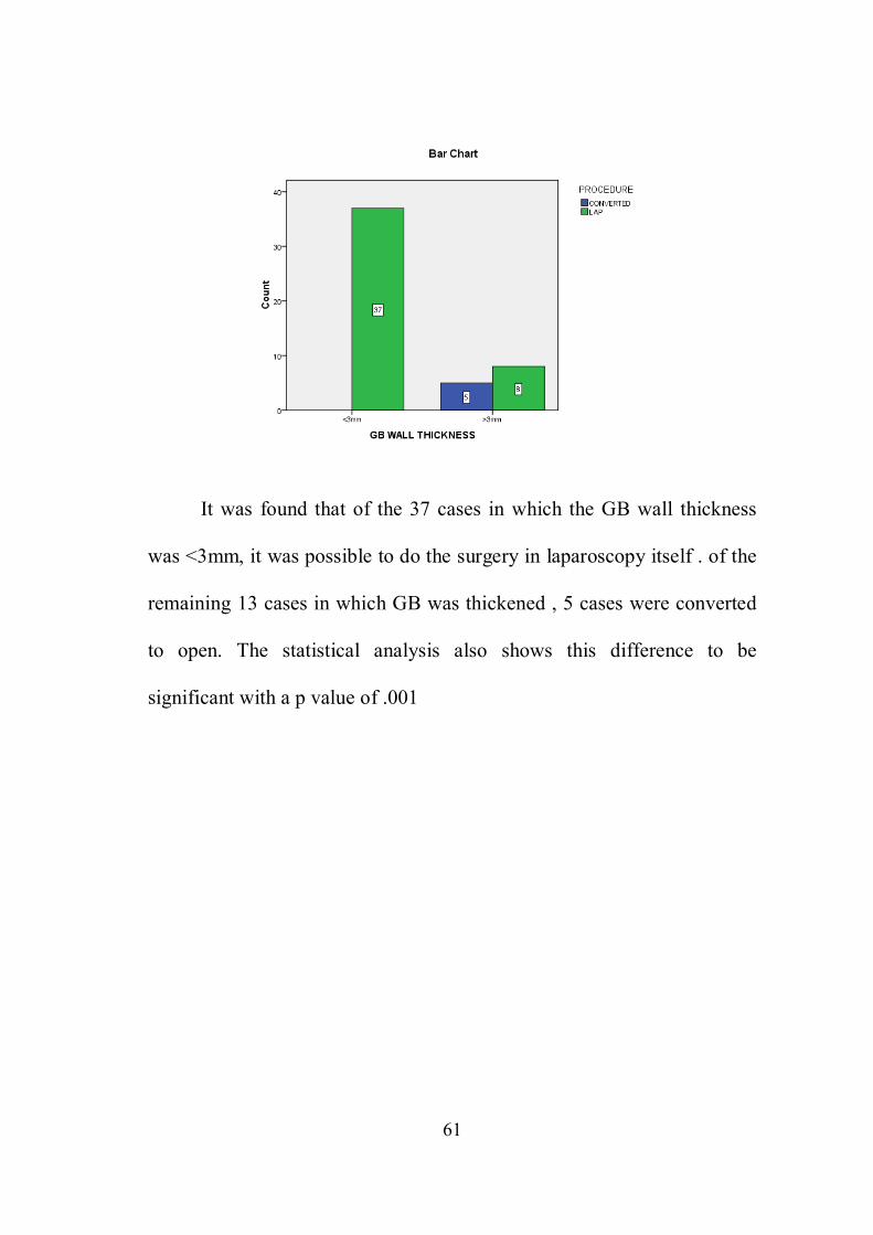
difference to be significant with a p value of 0.001, GB wall thickness – 37 cases in
which the GB wall thickness was < 3mm , it was possible to do surgery in laparoscopic
itself. Of the remaining 13 cases in which GB was thickened , 5 cases were converted
to open , the statistical analysis also shows this difference to be significant with a p
value of 0.001, number of stones alone is not significant factor to predict conversion
into open, pericholecystic collection – 39 cases in which pericholecystic collection was
not there , it was possible to do surgery in laparoscopic itself, of the remaining 11 cases
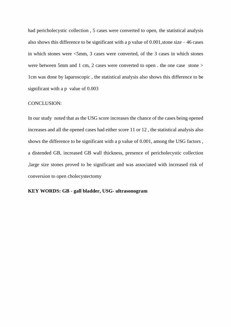
had pericholecystic collection , 5 cases were converted to open, the statistical analysis
also shows this difference to be significant with a p value of 0.001,stone size – 46 cases
in which stones were <5mm, 3 cases were converted, of the 3 cases in which stones
were between 5mm and 1 cm, 2 cases were converted to open . the one case stone >
1cm was done by laparoscopic , the statistical analysis also shows this difference to be
significant with a p value of 0.003
CONCLUSION:
In our study noted that as the USG score increases the chance of the cases being opened
increases and all the opened cases had either score 11 or 12 , the statistical analysis also
shows the difference to be significant with a p value of 0.001, among the USG factors ,
a distended GB, increased GB wall thickness, presence of pericholecystic collection
,large size stones proved to be significant and was associated with increased risk of
conversion to open cholecystectomy
KEY WORDS: GB - gall bladder, USG- ultrasonogram
CONTENTS
Sl. No. TOPICS Page No.
1 INTRODUCTION 1
2 AIMS OF THE STUDY 2
3 REVIEW OF LITERATURE 3
4 MATERIALS AND METHODS 47
5 RESULTS 50
6 DISCUSSIONS 70
7 CONCLUSIONS 76
8 BIBLIOGRAPHY
9 ANNEXURES
A MASTER CHART
B ABBREVIATIONS
C PROFORMA

INTRODUCTION
1
INTRODUCTION
“Laparoscopic cholecystectomy (LC) has become the treatment of
choice for gallstones. Laparoscopiccholecystectomy is established as the
primary procedure forthe vast majority of patients with benign
gallbladder disease.
Conversion to open cholecystectomy is occasionally needed to avoid or
repair injury, delineate confusing anatomic relationships, or treat
associated conditions6.”
“Conversion to open cholecystectomy has been associated with
increased overall morbidity, surgical site and pulmonary infections, and
longer hospital stays.The ability to accurately identify an individual
patient’s risk for conversion based on preoperative information can result
in more meaningful and accurate preoperative counselling, improved
operating room scheduling and efficiency, stratification of risk for
technical difficulty, may improve patient safety by minimizing time to
conversion, and also helps to identify patients in who a planned open
cholecystectomy is indicated12.”
“In our study we evaluated various clinical, haematological and
USG factors to preoperatively predict the conversion of laparoscopic
cholecystectomy to open cholecystectomy".

AIM OF THIS STUDY

2
AIMS OF THE STUDY
The aim of this study is to preoperatively predict the conversion of
Laparoscopic to open cholecystectomy by using ultrasound scoring the
following factors:
1. GB Status
2. GB Wall thickness
3. No of stones
4. Pericholecystic collection
5. Stone size
· The above USG scoring factors are analysed in this study to
preoperatively predict the conversion of Laparoscopic to Open
cholecystectomy
REVIEW OF LITERATURE
3
REVIEW OF LITERATURE
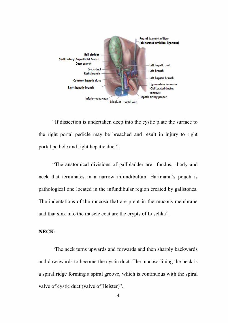
SURGICAL ANATOMY OF GALL BLADDER:
“A Gallbladder is a flask shaped organ attached to the common bile
duct by the cystic duct. It lies on the visceral surface of the liver at the
junction of quadrate lobe and right lobe of the liver along the line of Rex.
It usually lies in a shallow fossa in the liver parenchyma covered by the
peritoneum continued from liver surface. The gall bladder lies on a
fibrous or a cystic plate which is referred to as the hilar plate which is a
part of the perihilar system of the fibrous tissue. The cystic plate attaches
directly on to the anterior surface of right portal pedicle. The hepatic
parenchyma lies deep to the cystic plate through which small bile duct
may penetrate to enter the gall bladder. These ducts of Luschka consists
of accessory ducts less than 1 mm in diameter. During dissection of gall
bladder from the liver the posterior surface of the cystic artery and bile
duct will be reached when the areolar tissue is left on the cystic plate”.
4
“If dissection is undertaken deep into the cystic plate the surface to
the right portal pedicle may be breached and result in injury to right
portal pedicle and right hepatic duct”.
“The anatomical divisions of gallbladder are fundus, body and
neck that terminates in a narrow infundibulum. Hartmann’s pouch is
pathological one located in the infundibular region created by gallstones.
The indentations of the mucosa that are prent in the mucous membrane
and that sink into the muscle coat are the crypts of Luschka”.
NECK:
“The neck turns upwards and forwards and then sharply backwards
and downwards to become the cystic duct. The mucosa lining the neck is
a spiral ridge forming a spiral groove, which is continuous with the spiral
valve of cystic duct (valve of Heister)”.
5
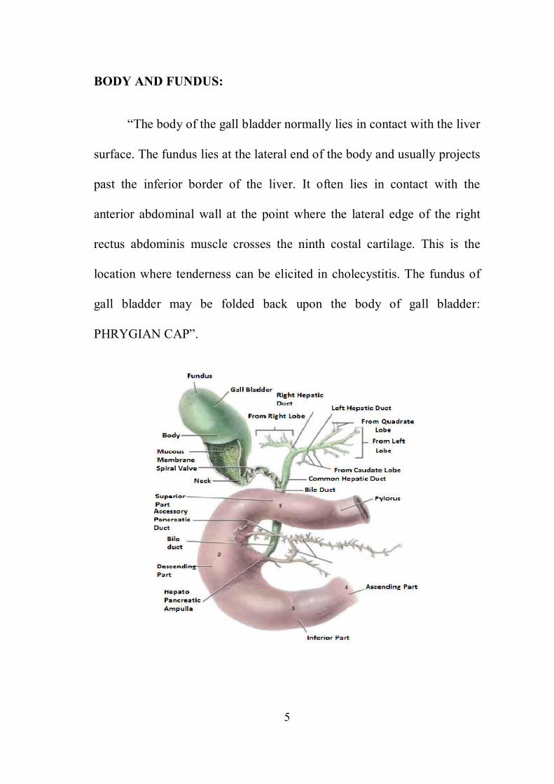
BODY AND FUNDUS:
“The body of the gall bladder normally lies in contact with the liver
surface. The fundus lies at the lateral end of the body and usually projects
past the inferior border of the liver. It often lies in contact with the
anterior abdominal wall at the point where the lateral edge of the right
rectus abdominis muscle crosses the ninth costal cartilage. This is the
location where tenderness can be elicited in cholecystitis. The fundus of
gall bladder may be folded back upon the body of gall bladder:
PHRYGIAN CAP”.

6
CYSTIC DUCT:
“A cystic duct is about 3 to 4 cm in length and joins with the
common hepatic duct forming the common bile duct. It runs parallel to it
and is adherent to the common hepatic duct for a short distance before
joining it. The cystic duct mucosa is arranged in numerous spiral folds
called the ‘valves of Heister’ and the wall is surrounded by a sphincteric
structure known as the sphincter of Lütkens’. The cystic duct drains at an
acute angle into the common bile duct. There are a number of anatomic
variations in insertion of the cystic duct, including into the right hepatic
duct”.
ANATOMICAL VARIATIONS OF CYSTIC DUCT:

7
HEPATIC DUCT:
“The Common Hepatic duct is formed by the union of right and
left hepatic duct in the portahepatis. This descents for about 3 cm before
joining the cystic duct at an acute angle to form the common bile duct.
The hepatic artery lies to the left of common hepatic duct and portal vein
lies posterior to it”.
COMMON BILE DUCT:
“Common hepatic duct and cystic duct joins to form the Common
Bile duct.. It is about 6 to 8 cm in length and about 6mm in diameter. It
lies anterior and to the right of portal vein and to the right of hepatic
artery. It passes behind the first part of duodenum with the
gastroduodenal artery on its left and then runs in groove on the
superolateral part of posterior surface of pancreas”.
HEPATOPANCREATIC AMPULLA (OF VATER):
“It is formed by the union of CBD and pancreatic duct before
entering the 2nd part of the duodenum. Circular muscles usually surround
the lower part of the CBD(bile duct sphincter),and frequently also
surround the terminal part of the main pancreatic duct(pancreatic duct
sphincter)and the hepatopancreatic ampulla(sphincter of oddi)”.
8
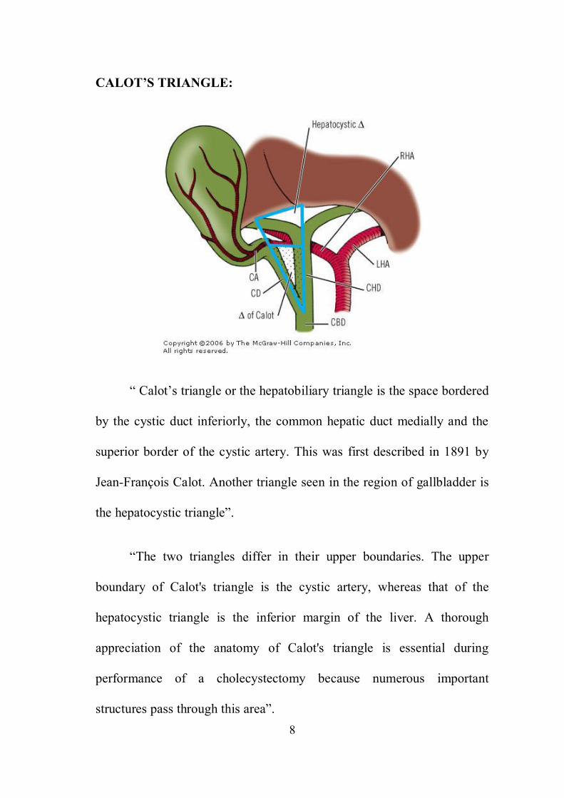
CALOT’S TRIANGLE:
“ Calot’s triangle or the hepatobiliary triangle is the space bordered
by the cystic duct inferiorly, the common hepatic duct medially and the
superior border of the cystic artery. This was first described in 1891 by
Jean-François Calot. Another triangle seen in the region of gallbladder is
the hepatocystic triangle”.
“The two triangles differ in their upper boundaries. The upper
boundary of Calot's triangle is the cystic artery, whereas that of the
hepatocystic triangle is the inferior margin of the liver. A thorough
appreciation of the anatomy of Calot's triangle is essential during
performance of a cholecystectomy because numerous important
structures pass through this area”.
9
CONTENTS OF CALOTS TRIANGLE:
1) Cystic artery as it approaches the GB.
2) Cystic lymphnode.
3) Lymphatics from the GB.
4) 1 or 2 small cystic veins.
5) Autonomic nerves running to the GB.
6) Some adipose tissue.
In most instances, the cystic artery arises as a branch of the right
hepatic artery within the hepatocystic triangle. A replaced or aberrant
right hepatic artery arising from the superior mesenteric artery usually
courses through the medial aspect of the triangle, posterior to the cystic
duct. Aberrant or accessory hepatic ducts also may pass through Calot's
triangle before joining the cystic duct or common hepatic duct. During
performance of a cholecystectomy, clear visualization of the hepatocystic
triangle is essential with accurate identification of all structures within
this triangle.
“The area where the hepatic artery takes a tortuous course in front
of the origin of the cystic duct is the most dangerous anomaly. The right
10
hepatic artery is tortuous and the cystic artery short. This tortuosity is
known as the ‘caterpillar turn’ or ‘Moynihan hump’. This is the main
reason for difficult cholecystectomy”.
BLOOD SUPPLY:
“Gallbladder is supplied by Cystic artery. The cystic artery arises
from the right hepatic artery, which can pass posterior or anterior to the
common bile duct to supply the gallbladder. Similar to the variability of
the cystic duct, the cystic artery may arise from the right hepatic, left
hepatic, proper hepatic, common hepatic, gastroduodenal, or superior
mesenteric artery. Although variable, the cystic artery generally lies
superior to the cystic duct and is usually associated with a lymph node,
known as Calot’s node. Because this node provides some of the
lymphatic drainage of the gallbladder, it can be enlarged in the setting of
gallbladder pathology, whether inflammatory or neoplastic”.
11
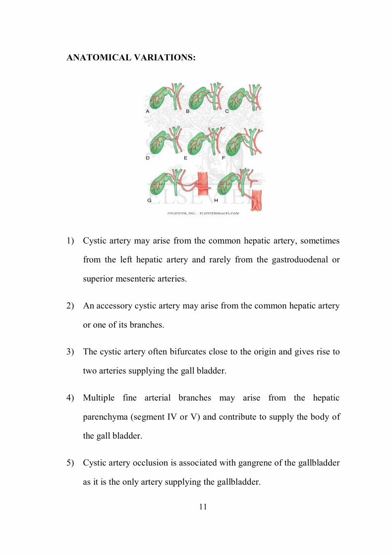
ANATOMICAL VARIATIONS:
1) Cystic artery may arise from the common hepatic artery, sometimes
from the left hepatic artery and rarely from the gastroduodenal or
superior mesenteric arteries.
2) An accessory cystic artery may arise from the common hepatic artery
or one of its branches.
3) The cystic artery often bifurcates close to the origin and gives rise to
two arteries supplying the gall bladder.
4) Multiple fine arterial branches may arise from the hepatic
parenchyma (segment IV or V) and contribute to supply the body of
the gall bladder.
5) Cystic artery occlusion is associated with gangrene of the gallbladder
as it is the only artery supplying the gallbladder.
12
VENOUS DRAINAGE:
Veins that drain the gallbladder are called Cystic veins. Those
arising from the superior surface of the body and neck and lie in the
areolar tissue between the gall bladder and liver and enters the liver
parenchyma to drain into the segmental portal veins. The remaining
cystic veins drains into the liver directly or after joining the veins
draining the hepatic duct.
LYMPHATIC DRAINAGE:
“The cystic lymph node of Lund (the sentinel lymph node) is the
main lymphatic drainage of gallbladder, which lies at the junction of the
cystic and common hepatic ducts. Efferent vessels from this lymph node
reach the hilum of the liver, and from there to the coeliac lymph nodes”.
“The subserosal lymphatic vessels of the gall bladder also connect with
the subcapsular lymph channels of the liver, and this accounts for the
frequent spread of carcinoma of the gall bladder to the liver”.
INNERVATION:
“The gallbladder and the extra hepatic biliary tree are innervated
by the branches from the hepatic plexus. The retroduodenal part of the
13
CBD also receives contribution from the pyloric branch of the Vagus. It
also innervates of the smooth muscles of the hepato pancreatic ampulla.
foregut. This outgrowth, the hepatic diverticulum or the hepatic bud
consists of rapidly dividing cells that penetrate the septum transversum,
that is the mesodermal plate between the pericardial cavity and the stalk
of the yolk sac.
GALL STONES – PREVALENCE:
“Cholelithiasis is a common disease throughout the Western world.
Gallstones can be found in 10% to 20% of the western population at some
stage of life. In both sexes the prevalence increases with age; however,
overall gallstones are more common in females than in males with a ratio
of 4:1. Obesity and family history are also significant risk factors. The
prevalence of gallstones is related to many factors like age, gender,
ethnicity. Many factors predispose to the development of gallstones. They
include obesity, pregnancy, dietary factors, Crohn's disease, terminal ileal
resection, gastric surgery, hereditary spherocytosis, sickle cell disease,
and thalassemia”.
14
NATURAL HISTORY:
“Most patients with gallstones remain asymptomatic throughout
life. Around 3% of asymptomatic individuals become symptomatic per
year and develop biliary colic. Once symptomatic they develop recurrent
episodes of biliary colic. Only few patients without biliary symptoms
develop complications. Prophylactic cholecystectomy for asymptomatic
cholelithiasis is rarely indicated. Elderly patients with diabetes mellitus,
individuals who will be isolated from medical care for a prolonged period
of time, Gall bladder polyp > 1 cm are indications for prophylactic
cholecystectomy.
ETIOLOGY:
“Gallstones are classified by their cholesterol content as cholesterol
stones and Pigment stones. Pigment stones are further classified into
black and brown pigment stones. In the United States and Europe, 80 per
cent are cholesterol or mixed stones, whereas in Asia, 80 per cent are
pigment stones. Cholesterol or mixed stones contain 51–99 per cent pure
cholesterol plus an admixture of calcium salts, bile acids, bile pigments
and phospholipids”.
15
CLINICAL PRESENTATION:
“Most patients remain asymptomatic from their gallstones.
Although mechanism unclear some develop symptomatic gall stones,
with biliary colic caused by stone obstructing the cystic duct. Only 1 to
2% of the asymptomatic individuals with gallstone will develop serious
symptoms or complications related to their gall stone per year. Therefore
only 1% require cholecystectomy. Once symptomatic patients tend to
have recurring symptoms, usually repeated episodes of biliary colic. Non-
specific gastrointestinal symptoms will develop in 10 to 30% of patients
and 5 to 10% develop classic biliary symptoms”.
BILIARY COLIC
“Acute obstruction of gall bladder by calculi results in biliary colic,
a common misnomer because the pain is not colicky in the epigastrium or
right upper quadrant”.
“Biliary colic is a constant pain that builds in intensity and can
radiate to the back, interscapular area or to the right shoulder. The pain is
described as a band like tightness of upper abdomen, that may be
associated with nausea and vomiting. This is due to a normal gallbladder
contracting against a luminal obstruction, such as a gallstone impacted at
16
the neck of gallbladder, the cystic duct or the CBD. The pain is most
commonly trigerred by fatty foods, but it can also be initiated by other
kind of foods or even occur spontaneously. An association with meals is
present only in 50% of patients.
REFERRED PAIN:
In common with other structures of foregut origin, pain from
stretch of CBD or gallbladder is referred to the central epigastrium.
involvement of overlying somatic peritoneum produces pain which is
more localized to the right quadrant.
INVESTIGATION:
ULTRASOUND:
“Transabdominal ultrasound is a sensitive, reliable and
inexpensive test to evaluate most of the biliary tract and to separate
medical jaundice patients from those with surgical jaundice. It does not
submit the patient to radiation and is useful in critically ill patients.
Gallbladder diseases are usually diagnosed by ultrasound, because of its
superficial location with no overlying bowel gas enables its evaluation by
sound waves thus making it the investigation of choice for gallstones.
Ultrasound has a high specificity and sensitivity of more than 90%

17
forcholelithiasis, or gallstones. The density of gallstones allows crisp
reverberation of the sound wave, showing an echogenic focus with a
characteristic shadowing behind the stone. The patient has acute
cholecystitis if edema is seen within the wall of the gallbladder or
between the gallbladder and liver with tenderness. Pathologic changes
seen in gallbladder diseases can beidentified by ultrasound. For example,
the gallbladder wallthickening and pericholecystic fluid collection seen in
cholecystitis can be detected by ultrasound. Porcelain or calcified
gallbladder appear as a curvilinear echogenic focus along the entire
gallbladder wall with posterior shadowing”.
ULTRASOUND SHOWING GALLSTONES
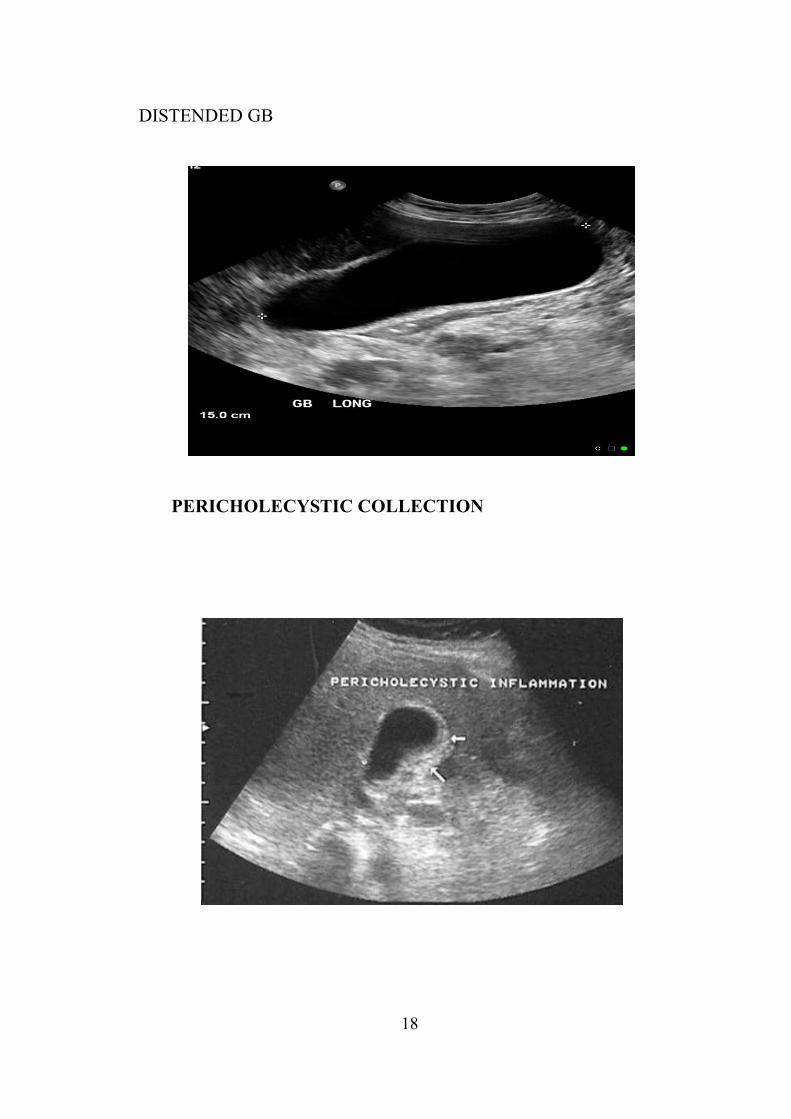
18
DISTENDED GB
PERICHOLECYSTIC COLLECTION
19
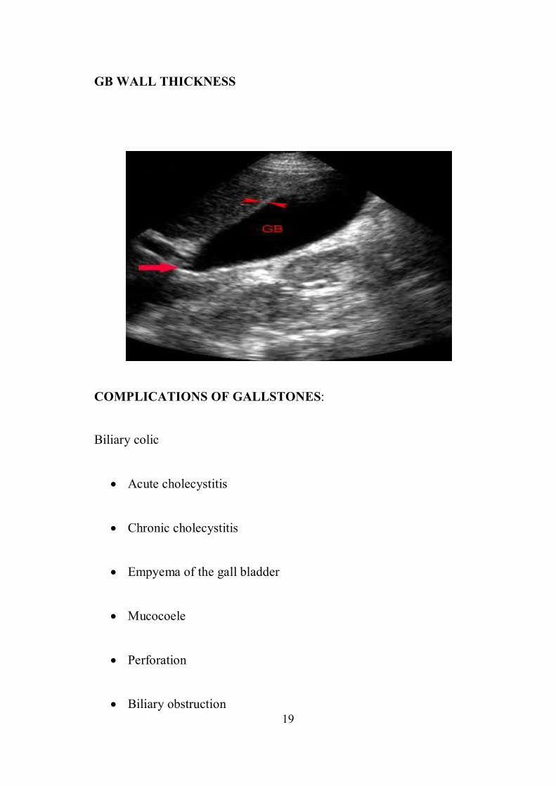
GB WALL THICKNESS
COMPLICATIONS OF GALLSTONES:
Biliary colic
· Acute cholecystitis
· Chronic cholecystitis
· Empyema of the gall bladder
· Mucocoele
· Perforation
· Biliary obstruction

20
· Acute cholangitis
· Acute pancreatitis
· Intestinal obstruction (gallstone ileus)
MANAGEMENT OF CHOLELITHIASIS
SURGICAL MANAGEMENT :
LAPAROSCOPIC CHOLECYSTECTOMY:
“Laparoscopic cholecystectomy is one of the most common
surgeries performed and has replaced open cholecystectomy. In 1992,
The National Institute of Health (NIH) consensus development
conference stated that laparoscopic cholecystectomy “provides a safe and
effective treatment for most patients with symptomatic gallstones17”.
INDICATIONS OF LAPAROSCOPIC CHOLECYSTECTOMY:
a) SYMPTOMATIC CHOLELITHIASIS30:
i) Biliary colic: Once the patient experience symptoms, there is a
greater than 80% chance that they will continue to have
symptoms. There is also a finite risk of disease related
21
complications such as acute cholecystitis, gallstone pancreatitis
and choledocholithiasis.
ii) Acute cholecystitis.
iii) Gallstone pancreatitis.
b) ASYMPTOMATIC CHOLELITHIASIS30:
“Patient with asymptomatic gallstone have less than 20% chance of
ever developing symptoms, and the risks associated with prophylactic
operation outweigh the potential benefit of surgery in most patients.
Therfore prophylactic cholecystectomy is recommended in:
i) Sickle cell disease: Patients with sickle cell disease often have
hepatic and vasoocclusive crisis that can be difficult to
differentiate from acute cholecystitis.
ii) Total parenteral nutrition
iii) Chronic immunosuppression: In transplant patients, there is a
concern that immunosuppression may mask the signs and
symptoms of inflammation until overwhelming infection
occurred.
22
iv) No immediate access to health care facilities (eg: missionaries,
military” personal, peace corps workers, relief workers)
v) Incidental cholecystectomy for patients undergoing procedures
for other indications.
c) Acalculouscholecystitis or biliary akinesia
d) Gallbladder polyps >1 cm in diameter.
e) Porcelain gallbladder”.
TECHNIQUES OF LAPAROSCOPIC CHOLECYSTECTOMY:
“The techniques of Laparoscopic cholecystectomy has been
changing over years. The conventional Laparoscopic cholecystectomy is
done by four ports: a 10 mm optical port at umbilicus, a 10 mm and a
5mm port in epigastrium and in midclavicularline respectively and
another 5 mm port in the midaxillary line at the level of umbilicus for the
assistant to retract the fundus of gall bladder”.
“Over years with increasing experience, laparoscopic
cholecystectomy has undergone many changes including reduction in
port size and number. Some surgeons tried laparoscopic cholecystectomy
through two ports. This required the introduction of transabdominal
sutures through the anterior abdominal wall for retracting the gall bladder
during dissection34.
23
APPROACH
A) NORTH AMERICAN APPROACH
“The patient is kept in supine in reverse trendelenberg position (15
degree head up tilt) with left lateral tilt (15-20 degree).this ensures that
the bowel and Omentum falls down and medially, away from the
operative site. The operating surgeon and camera surgeon stand on the
left of the patient while the assistant surgeon stands on the right of the
patient”.
PREOPERATIVE PREPARATION32:
1) Blood coagulation should be normalized in patients with prior, by
giving vitamin K (IM in 3 doses)
2) A prophylactic antibiotic preferably a second generation
cephalosporin is given at the time of anaesthesia induction.
3) To prevent deep venous thrombosis, subcutaneous heparin or
antiembolic stocking are used.
24
PROCEDURE OF LAPAROSCOPIC CHOLECYSTECTOMY35:
“The North American approach is usually followed. The patient is
placed on the operating table with the surgeon standing on the left side of
the patient and the first assistant standing on the patients right. Following
induction of general endotracheal anaesthesia, an orogastric tube is
inserted to decompress the stomach. Abdomen is painted from nipple to
midthigh35”.
“Pneumoperitoneum created with carbondioxide provides the
working space. The surgeon needs this working space for operating
within the abdominal cavity. Carbondioxide is non-combustible. It is
rapidly absorbed from the abdominal cavity. However in patients with
severe cardiopulmonary disease it can lead to hypercarbia32”.
“Pneumoperitoneum is created either by open technique or by
closed technique.Initially, a small incision is made in the upper edge of
the umbilicus. In the Closed technique,CO2 is insufflated into the
peritoneal cavity through a Veress needle, which is subsequently replaced
with a laparoscopic port, placed blindly into the abdominal cavity. In the
Open (HASSON) technique, a small incision is made and a laparoscopic
port is created under vision into the peritoneal cavity33”.

25
“A 10-mm laparoscope is inserted into the abdomen through the
periumbilical port and the abdominal cavity is inspected. The patient is
placed in a Anti-Trendelenburg position of 30 degrees while rotating the
table to the left by 15 degrees. This position allows the duodenum and
colon to fall away from the liver edge. The liver and falciformligament
are examined closely for abnormalities26”.
“Two small accessory subcostal ports are then placed under direct
vision. The first 5-mm trocar is placed along the right anterior axillary
line between the 12th rib and the iliac crest. A second 5-mm port is
inserted in the right subcostal area in the midclavicular line. Grasping
forceps are placed through these two ports to secure the gallbladder. The
assistant manipulates the lateral grasping forceps, which are used to grasp
the fundus and elevate the liver”.
“The fourth working port is then inserted through an incision in the
midline of the epigastrium. This trocar is usually inserted approximately
5 cm below the xiphoid process, but the precise position and angle
depends on the location of the gallbladder as well as the size of the
medial segment of the left lobe of the liver”.
“Dissecting forceps are then inserted and directed toward the
gallbladder neck. The surgeon uses a dissecting forceps to raise a serosal
fold of the most dependent portion of the fundus. The assistant’s heavy

26
grasping forceps are then locked onto this fold using either a spring or
ratchet device. With these axillary grasping forceps, the fundus of the
gallbladder is then pushed in a lateral and cephalad direction, rolling the
entire right lobe of the liver cranially”.
DISSECTION OF THE CHOLECYSTOHEPATIC
TRIANGLE(CALOTS
TRIANGLE):
“The dissection starts at the junction of the gallbladder and the
cystic duct. A helpful anatomic landmark is the cystic artery lymph
node.The infundibulumis grasped, placing traction on the Gallbladder in a
lateral direction to distract the cystic duct from the CBD. Fine-tipped
dissecting forceps (Maryland) areused to dissect and seperate the
overlying fibroareolar membrane from the gallbladder”.
“The dissection should not begin from an unknown area but it
should begin from the gallbladder,, to avoid damage to the underlying
structures such as a bile duct or hepatic artery. The dissection initially
commences 4 or 5 cm proximal to the neck of the gallbladder and
proceeds distally, such that a modified “top-down” technique is
employed25”
27
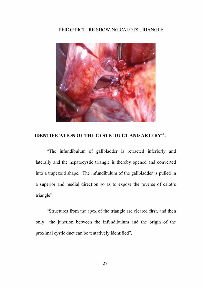
PEROP PICTURE SHOWING CALOTS TRIANGLE.
IDENTIFICATION OF THE CYSTIC DUCT AND ARTERY34:
“The infundibulum of gallbladder is retracted inferiorly and
laterally and the hepatocystic triangle is thereby opened and converted
into a trapezoid shape. The infundibulum of the gallbladder is pulled in
a superior and medial direction so as to expose the reverse of calot’s
triangle”.
“Structures from the apex of the triangle are cleared first, and then
only the junction between the infundibulum and the origin of the
proximal cystic duct can be tentatively identified”.

28
“The peritoneal strands, lymphatic strands and neurovascular tissue
over the cystic duct are stripped away to clear a segment from the
surrounding tissue. Curved dissecting forceps are used to create a
window around the posterior aspect of the cystic duct to skeletonize the
cystic duct”.
“By similar blunt dissection cystic artery is also separated from
surrounding structures. Thus the neck of the gallbladder is dissected
away from the liver bed, leaving a large window at its base through
which the liver parenchyma is visualized. There should be two, and only
two, structures (the cystic duct and artery) crossing this window—this is
the “critical view of safety,” which should be demonstrated prior to
clipping or cutting any tubular structures”.
“The cystic duct is clipped using an endoscopic clip applier and
divided using scissors. Two clips are placed proximally on the cystic duct
and one clip is placed toward the gallbladder. For cystic ducts that are
large or friable, a preformed endoloop is preferable for ligating the distal
cystic duct”.
“After the duct is divided, the cystic artery is dissected from the
surrounding tissue for an adequate distance to permit placement of three
clips.Electrocautery should not be used for this division, as the current
29
may be transmitted to the proximal clips leading to subsequent necrosis
and haemorrhage”.
“The ligated stumps of the cystic duct and the artery are then
examined to ensure that there is no leakage of either bile or blood and that
the clips are placed securely and compress the entire lumen of the
structures without impinging on adjacent tissues. A suction-irrigation
catheter is used to remove any debris or blood that has accumulated
during the dissection”.
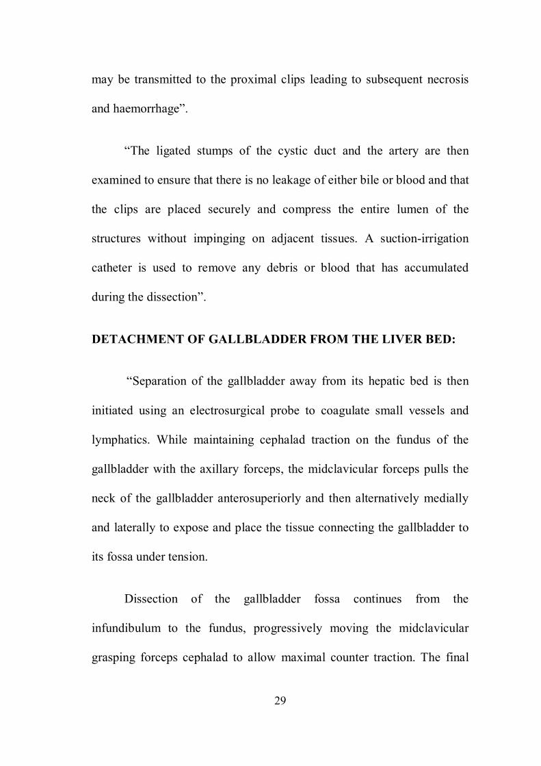
DETACHMENT OF GALLBLADDER FROM THE LIVER BED:
“Separation of the gallbladder away from its hepatic bed is then
initiated using an electrosurgical probe to coagulate small vessels and
lymphatics. While maintaining cephalad traction on the fundus of the
gallbladder with the axillary forceps, the midclavicular forceps pulls the
neck of the gallbladder anterosuperiorly and then alternatively medially
and laterally to expose and place the tissue connecting the gallbladder to
its fossa under tension.
Dissection of the gallbladder fossa continues from the
infundibulum to the fundus, progressively moving the midclavicular
grasping forceps cephalad to allow maximal counter traction. The final
30
attachments of the gallbladder are divided, and the liver edge is again
examined for hemostasis”.
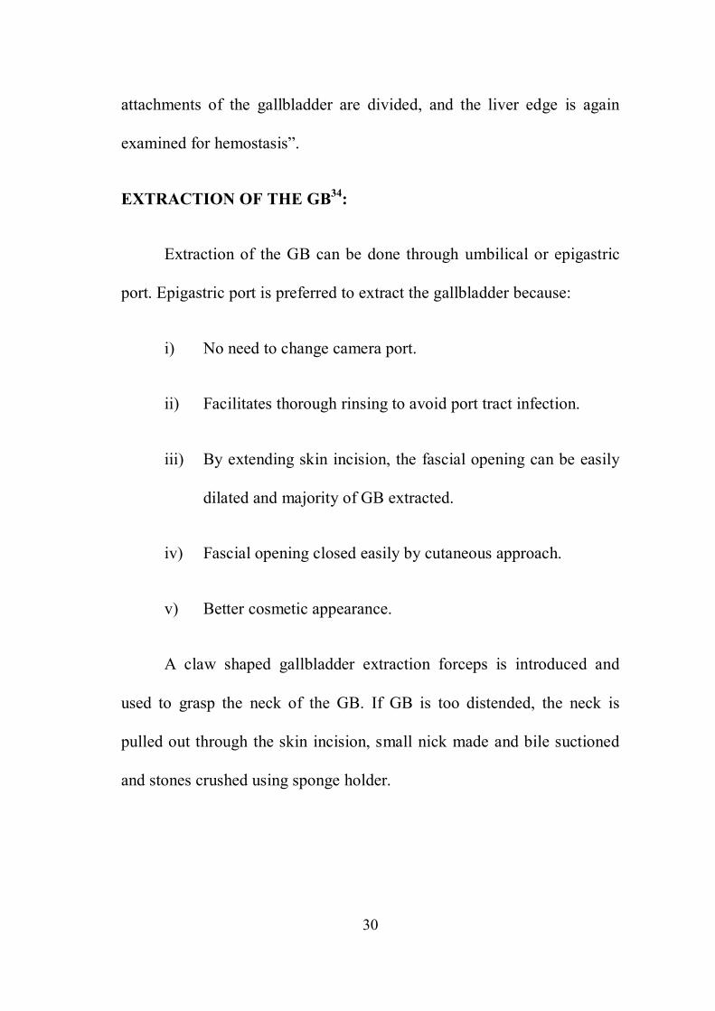
EXTRACTION OF THE GB34:
Extraction of the GB can be done through umbilical or epigastric
port. Epigastric port is preferred to extract the gallbladder because:
i) No need to change camera port.
ii) Facilitates thorough rinsing to avoid port tract infection.
iii) By extending skin incision, the fascial opening can be easily
dilated and majority of GB extracted.
iv) Fascial opening closed easily by cutaneous approach.
v) Better cosmetic appearance.
A claw shaped gallbladder extraction forceps is introduced and
used to grasp the neck of the GB. If GB is too distended, the neck is
pulled out through the skin incision, small nick made and bile suctioned
and stones crushed using sponge holder.
31
If the GB is thick preventing its extraction the fascial incision is
enlarged using a closed Robert’s clamp or extending it. Infected or
necrotic GB or a GB with suspicion of carcinoma is placed in a sterile
bag before extraction to reduce port site infection”.
If drain is needed a 14 F Redivac tube is placed through 5 mm
trocar site lateral most port. Trocars are removed under direct vision to
check for bleeding from trocar site. Pneumopritoneum evacuated and 10
mm ports closed with vicryl. The skin incisions at port sites is closed with
absorbable sutures, skin closure adhesives.

32
PORT SITES PORT PLACEMENT
DISSECTION OF CALOTS TRIANGLE
CLIPPING OF CYSTIC DUCT
33
COMPLICATIONS35:
The complications of Laparoscopic cholecystectomy are:
a) HEMORRHAGE:
i) TROCAR SITE BLEEDING:
Trocar site bleeding can be prevented by control of bleeding
following skin incision and before inserting trocar.
Management: Pressure over the site of bleeding by tilting the trocar.
Injection of epinephrine 1:10000 in the vacinity of the bleeding site.
ii) HEMORRHAGE DUE TO BLUNT DISSECTION OF ADHESIONS
can be managed with electrocautery.
iii) SUDDEN AND PULSATILE BLEEDING IN CALOT’S
TRIANGLE
Bleeding in the calot’s triangle can be prevented by careful
dissection and proper application of clip to cystic artery.
34
iv) GALLBLADDER FOSSA BLEEDING
GB fossa bleeding can be controlled by electrocautery, packing the
site with hemlock soacked gel foam, figure of eight stitch in case of
spurter from liver parenchyma.
b) PERFORATION OF GALLBLADDER:
“GallBladder perforation seen in acute cholecystitis and while
detaching GallBladder from the liver bed. This can be prevented by
confining to the areolar tissue between the GallBladder and the liver bed
during dissection and decompression of the gall bladder if distended. The
likelihood of a complication when gallstone spillage occurred was 2.3%
which was increased to 7.0% when unretrieved peritoneal gallstones were
documented”.
c) DIFFICULTY IN EXTRACTION OF THE GALLBLADDER
“Difficulty in extraction of the gallbladder is seen in gallbladder
containing large stones and those with thick wall. In GallBladder
containing large stones, the GallBladder is placed in an endobag, the neck
retrieved out through the abdomen and stones are crushed and removed.
In GallBladder with thickened wall ,theGallBladder is placed in an
endobag and extracted”.
35
d) OCCULT CARCINOMA
In cases suspected to have carcinoma intraoperatively, frozen
section is sent and if frozen section is positive for carcinoma, then
conversion to open technique is considered and radical surgery
e) POST OPERATIVE BILE LEAK
“Post operative bile leak can occur due to injury to the CBD, the
right hepatic duct or accessory bile duct. In case of acute inflammation,
the clip applied to the cystic duct may become loose once the edema
subsides and subsequently slip off”.
“This can be prevented by correct identification of the cystic duct
and artery, minimum use of electrocautery in calot’s triangle dissection
and appropriate choice of laparoscopic subtotal cholecystectomy.in the
setting of acute cholecystitis, when tight application of the clip is in
doubt, it may be advisable to use a pre-tied suture loop or intra-corporeal
suturing to occlude the cystic duct”.
“Postoperative bile leak should be suspected in patients with fever,
tachycardia and upper abdominal pain and tenderness persisting or
appearing unexpectedly. The diagnosis can be confirmed by USG or
ERCP. If drain is placed most of the minor leak will heal with expectant
36
management. In some persistent cases, it may be advisable to decrease
the intraductal pressure by nasobiliary drainage, endoscopic
spincterotomy or transpapillary stenting”.
f) BILE DUCT INJURY
Incidence of CBD injury during Laparoscopic cholecystectomy
exceeds that of open cholecystectomy ie 0.5% vs 0.2%.21 Reasons for the
increase in injury during Laparoscopic cholecystectomy included loss of
haptic information, incorrect traction forces to the gallbladder, and
injudicious use of cautery inside of the triangle of calot. Risk factors that
increase the risk of CBD injury include acute cholecystitis, aberrant
anatomy. The most common anatomic variant is an aberrant right hepatic
duct.
PREVENTION:
i) use a 30 degree laparoscope and high-quality imaging equipment.
ii) Apply firm cephalic traction to the fundus and lateral traction to
the infundibulum so that the cystic duct is perpendicular to the
CBD.
iii) Dissect the cystic duct where it joins the gallbladder.
37
iv) Expose the “critical view of safety” prior to dividing the cystic
duct.18
v) Convert to open procedure if the infundibulum cannot be
mobilized or bleeding or inflammation obscures the triangle of
calot.
vi) Perform routine intraoperative cholangiography. A recent study
using an American Medicare database found a reduction in CBD
injuries with routine use of Intraoperative Cholangiography (from
0.58% to 0.39%).
g) BOWEL INJURY
Injury to bowel can occur during trocar insertion or dissection in
the right upper quadrant, especially when using electrosurgical devices.
The jejunum, ileum and colon can be injured by veress needle and trocars
while duodenum is likely to be injured during dissection. Any structure
fixed to the undersurface of the umbilicus like the urachus or a meckel’s
diverticulum is more susceptible to injury during access. The rate of
bowel injury between 0 and 0.4% has been reported in various studies.
Deziel et al carried out retrospective analysis and found that mortality
38
rate following all bowel injuries during laparoscopic cholecystectomy
was 4.6% while it was 8.3% for duodenal injuries.
h) WOUND INFECTION AND INCISIONAL HERNIA
“The risk of wound infection following laparoscopic
cholecystectomy is less than 1% and the risk of incisional hernia is 0.5%.
Use of a retrieval bag for extraction of GB and closure of all port sites
larger than 8mm may avoid these complications”.
i) DIAPHRAGMATIC INJURY
Diaphragmatic injury may be due to either cautery or by
mechanical puncture by an instrument while retracting the fundus
cranially with excessive force.
j) PNEUMOPERITONEUM RELATED COMPLICATIONS34:
“Pneumoperitoneum can cause complications like carbon dioxide
embolism, vasovagal reflex, cardiac arrhythmias and hypercapnia
induced acidosis.Hypercapnia and acidosis are due to absorption of
carbon dioxide from the peritoneal cavity. Sudden increases in Paco2
may be related to port slippage and extraperitoneal or subcutaneous
diffusion of CO2. It is managed by desufflating the abdomen for 10 to 15
39
min. If reinsufflation results in recurrent hypercapnia, then change the
insufflations gas or convert to open”.
“Carbon dioxide embolism is characterized by unexplained
hypotension and hypoxia. Characteristic millwheel murmur is detected on
auscultation. This is produced due to the contraction of right ventricle
against the blood gas interface. There is an exponential decrease in end
tidal CO2 due to complete right ventricular outflow obstruction. It is
managed by immediate evacuation of pneumoperitoneum and placement
of the patient in left lateral decubitus, head down position. This allows the
CO2 bubble to float to the apex of the right ventricle, where it is less
likely to cause right ventricular outflow obstruction. Patient is
hyperventilated with 100% oxygen”.
OPEN CHOLECYSTECTOMY:
INDICATIONS FOR OPEN CHOLECYSTECTOMY35:
“i) Poor pulmonary or cardiac reserve
ii) Suspected or known gallbladder cancer
iii) Cirrhosis and portal hypertension
iv) Third-trimester pregnancy
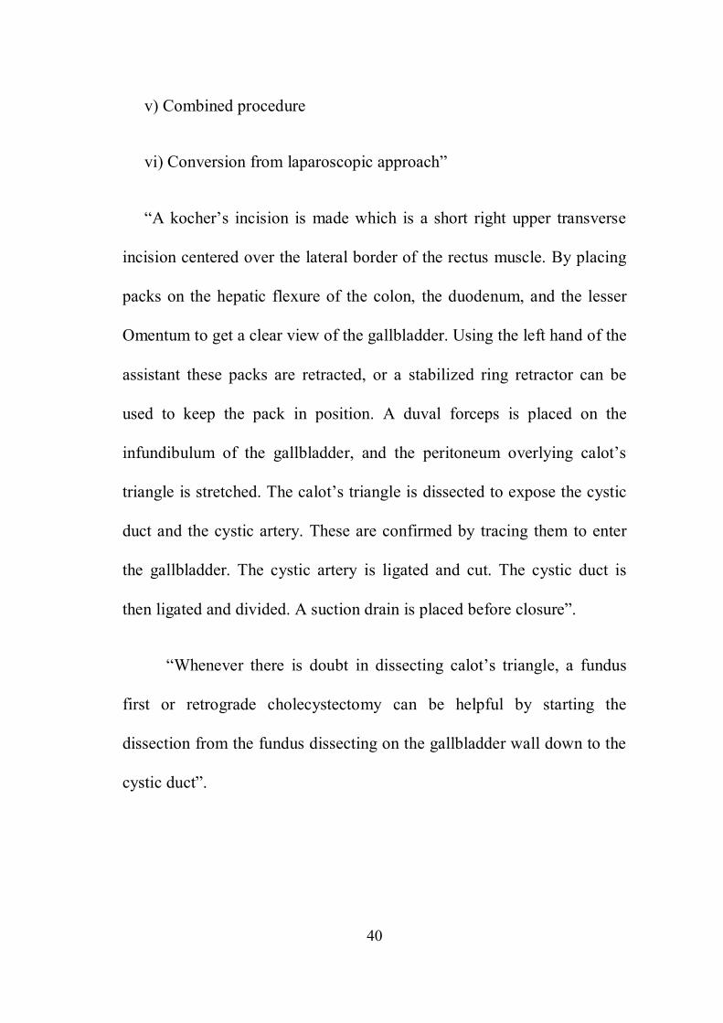
40
v) Combined procedure
vi) Conversion from laparoscopic approach”
“A kocher’s incision is made which is a short right upper transverse
incision centered over the lateral border of the rectus muscle. By placing
packs on the hepatic flexure of the colon, the duodenum, and the lesser
Omentum to get a clear view of the gallbladder. Using the left hand of the
assistant these packs are retracted, or a stabilized ring retractor can be
used to keep the pack in position. A duval forceps is placed on the
infundibulum of the gallbladder, and the peritoneum overlying calot’s
triangle is stretched. The calot’s triangle is dissected to expose the cystic
duct and the cystic artery. These are confirmed by tracing them to enter
the gallbladder. The cystic artery is ligated and cut. The cystic duct is
then ligated and divided. A suction drain is placed before closure”.
“Whenever there is doubt in dissecting calot’s triangle, a fundus
first or retrograde cholecystectomy can be helpful by starting the
dissection from the fundus dissecting on the gallbladder wall down to the
cystic duct”.

41
ADVANTAGES34:
“The advantages of Laparoscopic cholecystectomy over Open
cholecystectomy are:
· Less pain
· Smaller incisions
· Less intestinal ileus
· Shorter Hospital stay
· Better cosmesis
· Earlier return to normal activity”
DISADVANTAGES34:
“The disadvantages of Laparoscopic cholecystectomy are:
· Lack of depth perception
· Adhesions/Inflammation limit its use
· More difficult to control haemorrhage
· Potential CO2 insufflation complications
· Decreased tactile discriminations”
42
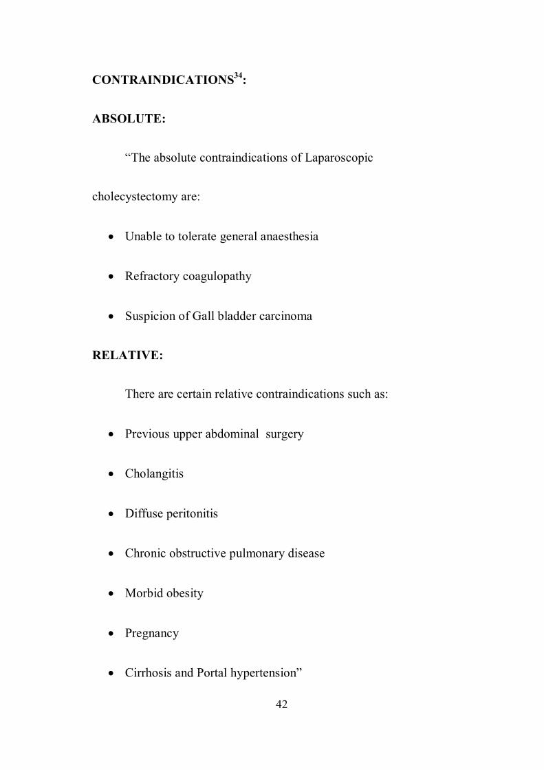
CONTRAINDICATIONS34:
ABSOLUTE:
“The absolute contraindications of Laparoscopic
cholecystectomy are:
· Unable to tolerate general anaesthesia
· Refractory coagulopathy
· Suspicion of Gall bladder carcinoma
RELATIVE:
There are certain relative contraindications such as:
· Previous upper abdominal surgery
· Cholangitis
· Diffuse peritonitis
· Chronic obstructive pulmonary disease
· Morbid obesity
· Pregnancy
· Cirrhosis and Portal hypertension”
43
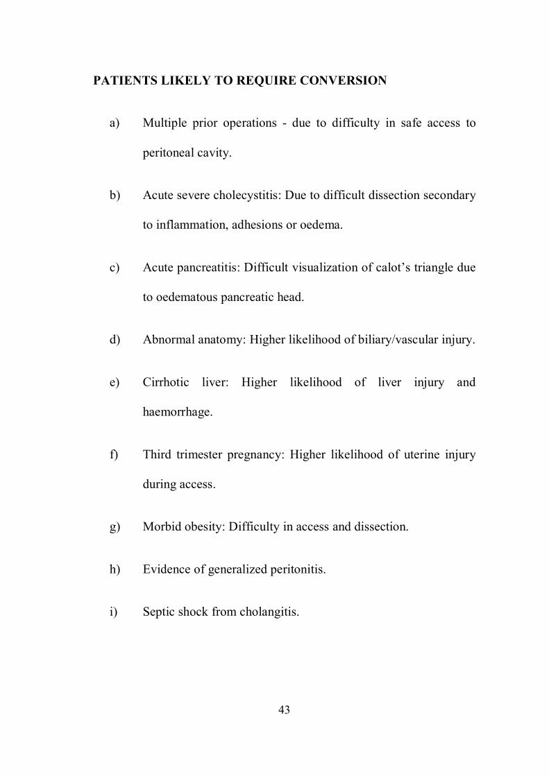
PATIENTS LIKELY TO REQUIRE CONVERSION
a) Multiple prior operations - due to difficulty in safe access to
peritoneal cavity.
b) Acute severe cholecystitis: Due to difficult dissection secondary
to inflammation, adhesions or oedema.
c) Acute pancreatitis: Difficult visualization of calot’s triangle due
to oedematous pancreatic head.
d) Abnormal anatomy: Higher likelihood of biliary/vascular injury.
e) Cirrhotic liver: Higher likelihood of liver injury and
haemorrhage.
f) Third trimester pregnancy: Higher likelihood of uterine injury
during access.
g) Morbid obesity: Difficulty in access and dissection.
h) Evidence of generalized peritonitis.
i) Septic shock from cholangitis.
44
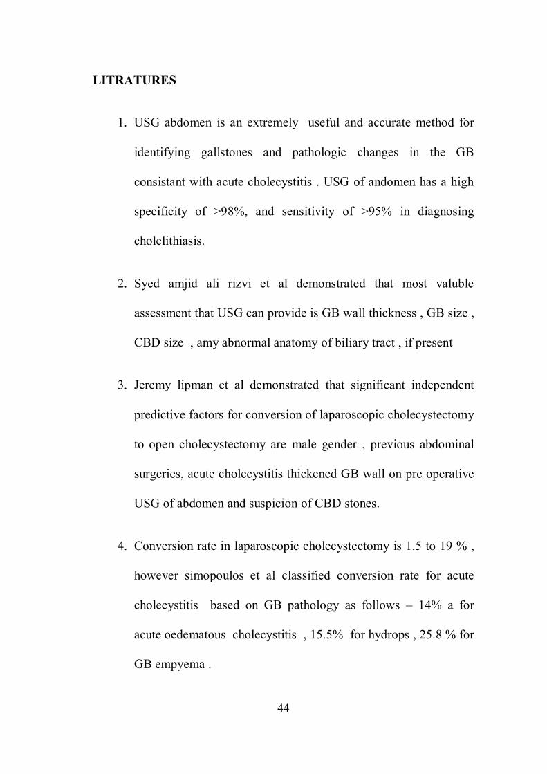
LITRATURES
1. USG abdomen is an extremely useful and accurate method for
identifying gallstones and pathologic changes in the GB
consistant with acute cholecystitis . USG of andomen has a high
specificity of >98%, and sensitivity of >95% in diagnosing
cholelithiasis.
2. Syed amjid ali rizvi et al demonstrated that most valuble
assessment that USG can provide is GB wall thickness , GB size ,
CBD size , amy abnormal anatomy of biliary tract , if present
3. Jeremy lipman et al demonstrated that significant independent
predictive factors for conversion of laparoscopic cholecystectomy
to open cholecystectomy are male gender , previous abdominal
surgeries, acute cholecystitis thickened GB wall on pre operative
USG of abdomen and suspicion of CBD stones.
4. Conversion rate in laparoscopic cholecystectomy is 1.5 to 19 % ,
however simopoulos et al classified conversion rate for acute
cholecystitis based on GB pathology as follows – 14% a for
acute oedematous cholecystitis , 15.5% for hydrops , 25.8 % for
GB empyema .
45
5. First described in 1882 by lagenbuch , open cholecystectomy has
been the primary treatment gall stone diseases for most of the
past century
6. In 1985 , the first documented laparoscopic cholecystectomy was
performed by ERICH MUHE in GERMANY in 1985, in 1987 ,
PHILIPE MOURET , perform the first laparoscopic
cholecystectomy in LYONS , FRANCE using video technique .
7. A difficult laparoscopic cholecystectomy that requires conversion
to open procedure can be predicted by pre operative
ultrasonography ., pawan lal, md, pn agarwal md, and al
chakravarthi, md JSLS 2002.,ncbi,nlm.nih.gov . Results were pre
operative USG abdomen good predictor of difficulty in lap
cholecystectomy
8. Risk factors resulting in conversion of laparoscopic
cholecystectomy to open surgery , N.A.kama, mdoganay , m
dolapci, e,reis, m.atli, m .kologlu. surgical endocopy, September
2001, volume 15, issue 9 ,pp 965-968., Results the most common
reason for conversion was inability to defined anatomy patients
with inflamed GB, male gender, previous abdominal surgery,
thickened GB wall on pre op USG and suspicion of CBD stones

46
9. The role of pre operative investigations in predicting difficult
laparoscopic cholecystectomies., surgical endoscopy , august
1996, volume 10,issue 8 , pp 791-793 ., results USG findings
relate to difficulty of lap procedure more closely than the other
pre operative investigations
10. A comprehensive predictive scoring method for difficult
laparoscopic cholecystectomy . journal of minimal access
surgery. By makm vivek 2014, www.ncbi,nlm .nih.gov. results
this sudy demonstrate that a scoring system predicting the
difficulty in laparoscopic is feasible .
11. USG in GB disease prediction of difficult laparoscopic
cholecystectomy., IJSR,ISSN ONLINE : 2319-7064, IMPACT
FATOR 2012: 3.358 , results USG is good predictor of difficult
laparoscopic cholecystectomy
12. Prediction of difficulty of laparoscopic cholecystectomy by pre
operative USG : a randomized control trail , global journal inc,
(USA), online ISSN 229-4618, thick GB wall is a finding which
may show that more adhesions may be found in surgery, reason
for conversion dense adhesions and bleeding from cystic artery.

MATERIALS AND
METHODS

47
MATERIALS AND METHODS
A total of 50 cases with gallstones admitted in RGGGH during the
period from January 2015 to September 2015 and planned for
Laparoscopic cholecystectomy were analysed in this study. They were
subjected to a detailed history, clinical examination and then to blood
investigations and radiological investigations. Their Name, Age, Sex,
and findings of USGof Abdomen were recorded. All these patients were
subjected to Laparoscopic cholecystectomy .the laparoscopic surgery was
performed by surgeons at our unit experienced in laparoscopic surgery,
therefore , the learning curve statistics do not apply to this study, the
operating surgeon was blinded to these findings ,the operative findings
were objectively graded as difficult or easy laparoscopic
cholecystectomy from insertion of the veress needle or trocar until the
extraction of the gall bladder was considered a difficult laparoscopic
cholecstectomy, tear of GB, spillage of bile and stones considered a
difficult procedure , > 30 minutes taken to dissect GB from GB bed was
considered a difficult procedure , > 20 minutes taken to dissect calots
triangle was considered difficult procedure,any laparoscopic
cholecystectomy converted to the open procedure was considered a
difficult laparoscopic cholecystectomy.

48
INCLUSION CRITERIA:
· The patients presenting with symptom and sign of
cholelithiasis/ diagnosed by ultrasound abdomen.
· Age 20 -70 yrs
EXCLUSION CRITERIA:
· Patients below 20yrs age
· Previous abdominal surgery
· Patients with CBD calculus, raised ALP , dilated CBD,
where CBD exploration needed.
· Patients with features of obstructive jaundice
· Suspected malignant gall bladder disease
· Patient medically unfit for laparoscopic
EQUIPMENTS REQUIRED FOR LAPAROSCOPIC
CHOLECYSTECTOSMY:
a) High-quality videoscope with a 300 w light source be coupled to
two high resolution monitors.
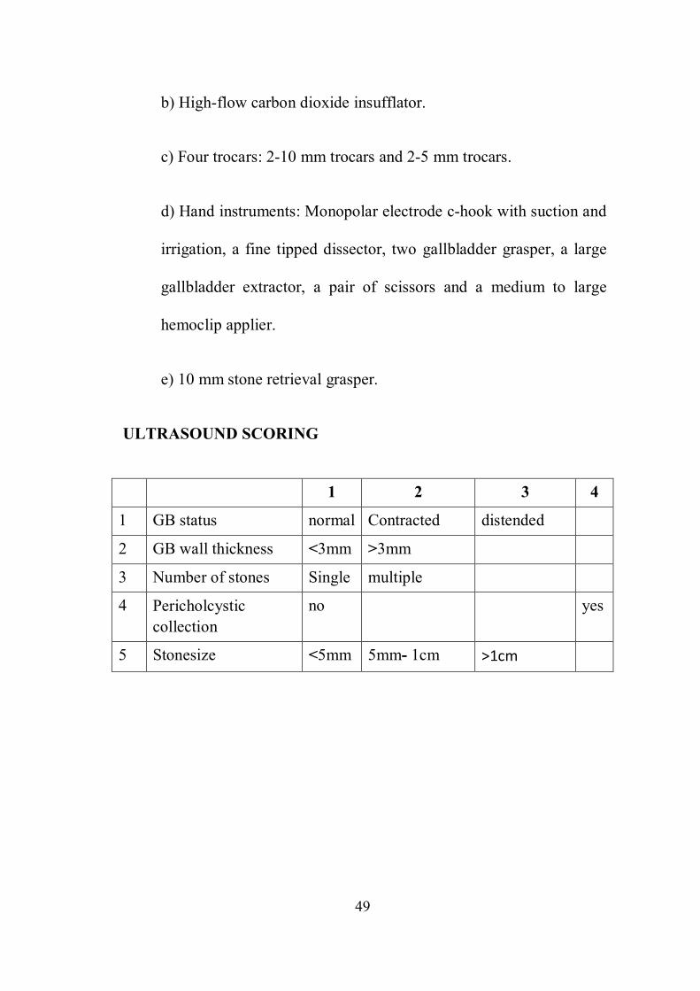
49
b) High-flow carbon dioxide insufflator.
c) Four trocars: 2-10 mm trocars and 2-5 mm trocars.
d) Hand instruments: Monopolar electrode c-hook with suction and
irrigation, a fine tipped dissector, two gallbladder grasper, a large
gallbladder extractor, a pair of scissors and a medium to large
hemoclip applier.
e) 10 mm stone retrieval grasper.
ULTRASOUND SCORING
1 2 3 4
1 GB status normal Contracted distended
2 GB wall thickness <3mm >3mm
3 Number of stones Single multiple
4 Pericholcystic collection
no yes
5 Stonesize <5mm 5mm- 1cm >1cm

RESULTS
50
RESULTS
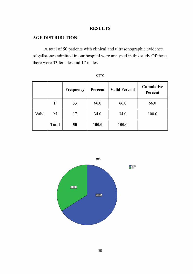
AGE DISTRIBUTION:
A total of 50 patients with clinical and ultrasonographic evidence of gallstones admitted in our hospital were analysed in this study.Of these there were 33 females and 17 males
SEX
Frequency Percent Valid Percent Cumulative
Percent
Valid
F 33 66.0 66.0 66.0
M 17 34.0 34.0 100.0
Total 50 100.0 100.0
51
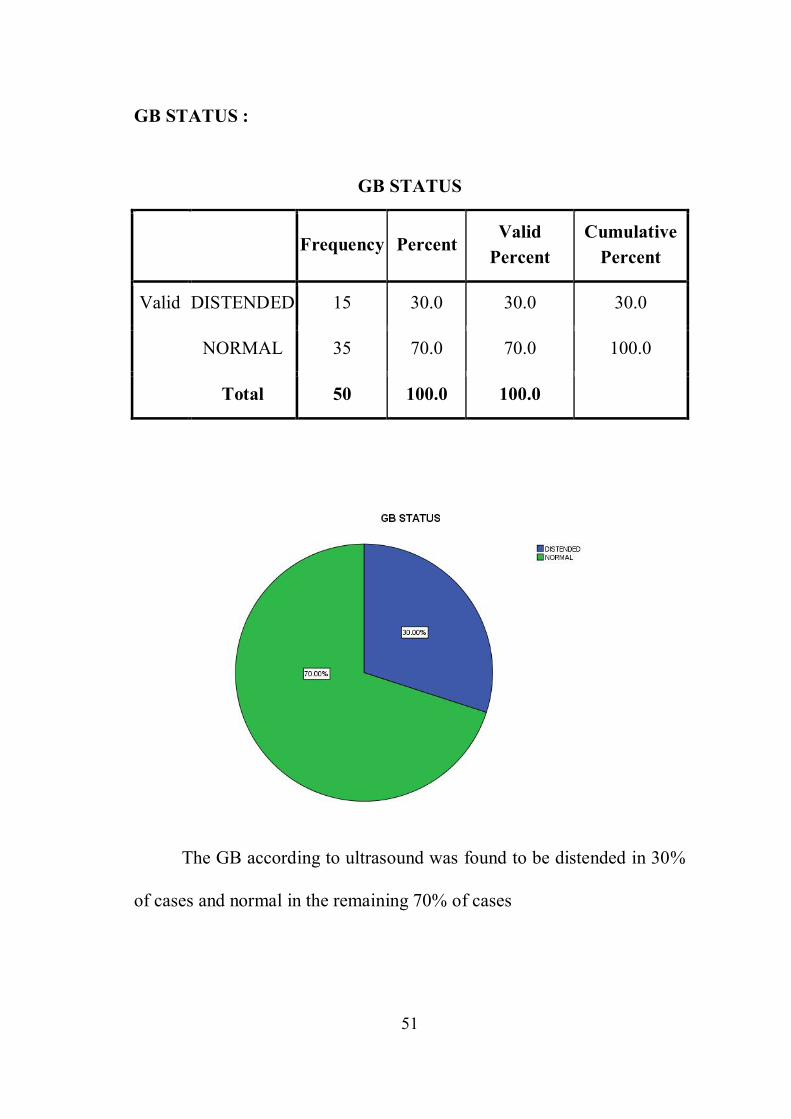
GB STATUS :
GB STATUS
Frequency Percent
Valid Percent
Cumulative Percent
Valid DISTENDED 15 30.0 30.0 30.0
NORMAL 35 70.0 70.0 100.0
Total 50 100.0 100.0
The GB according to ultrasound was found to be distended in 30%
of cases and normal in the remaining 70% of cases

52
GB WALL THICKNESS
GB WALL THICKNESS
Frequency Percent
Valid Percent
Cumulative Percent
Valid NO 37 74.0 74.0 74.0
YES 13 26.0 26.0 100.0
Total 50 100.0 100.0

53
NO OF STONES :
The ultrasound showed multiple stones in 90% of cases and single
stones were found in 10% of cases
NO OF STONES
Frequency Percent Valid Percent
Cumulative Percent
Valid MULTI 45 90.0 90.0 90.0
SINGLE 5 10.0 10.0 100.0
Total 50 100.0 100.0
10%
90%
54
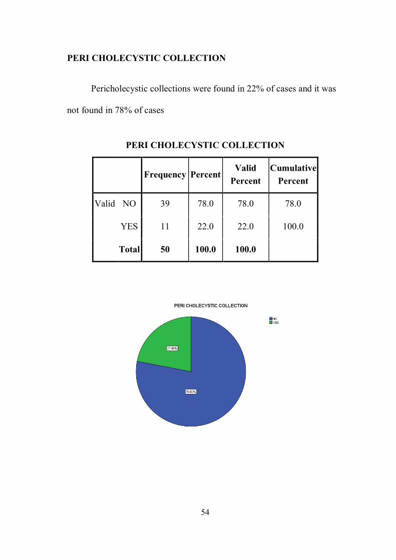
PERI CHOLECYSTIC COLLECTION
Pericholecystic collections were found in 22% of cases and it was
not found in 78% of cases
PERI CHOLECYSTIC COLLECTION
Frequency Percent
Valid Percent
Cumulative Percent
Valid NO 39 78.0 78.0 78.0
YES 11 22.0 22.0 100.0
Total 50 100.0 100.0
55
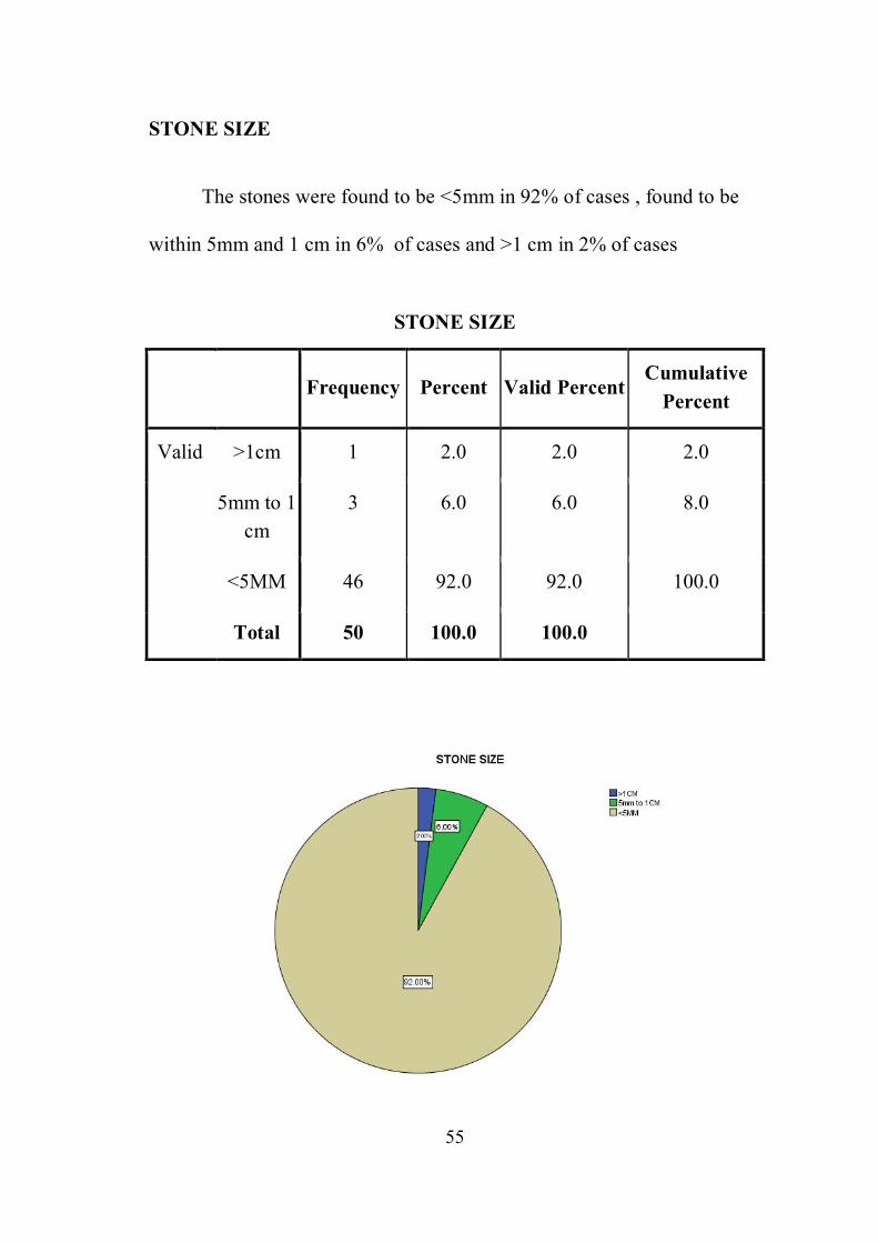
STONE SIZE
The stones were found to be <5mm in 92% of cases , found to be
within 5mm and 1 cm in 6% of cases and >1 cm in 2% of cases
STONE SIZE
Frequency Percent Valid Percent
Cumulative Percent
Valid >1cm 1 2.0 2.0 2.0
5mm to 1 cm
3 6.0 6.0 8.0
<5MM 46 92.0 92.0 100.0
Total 50 100.0 100.0
56
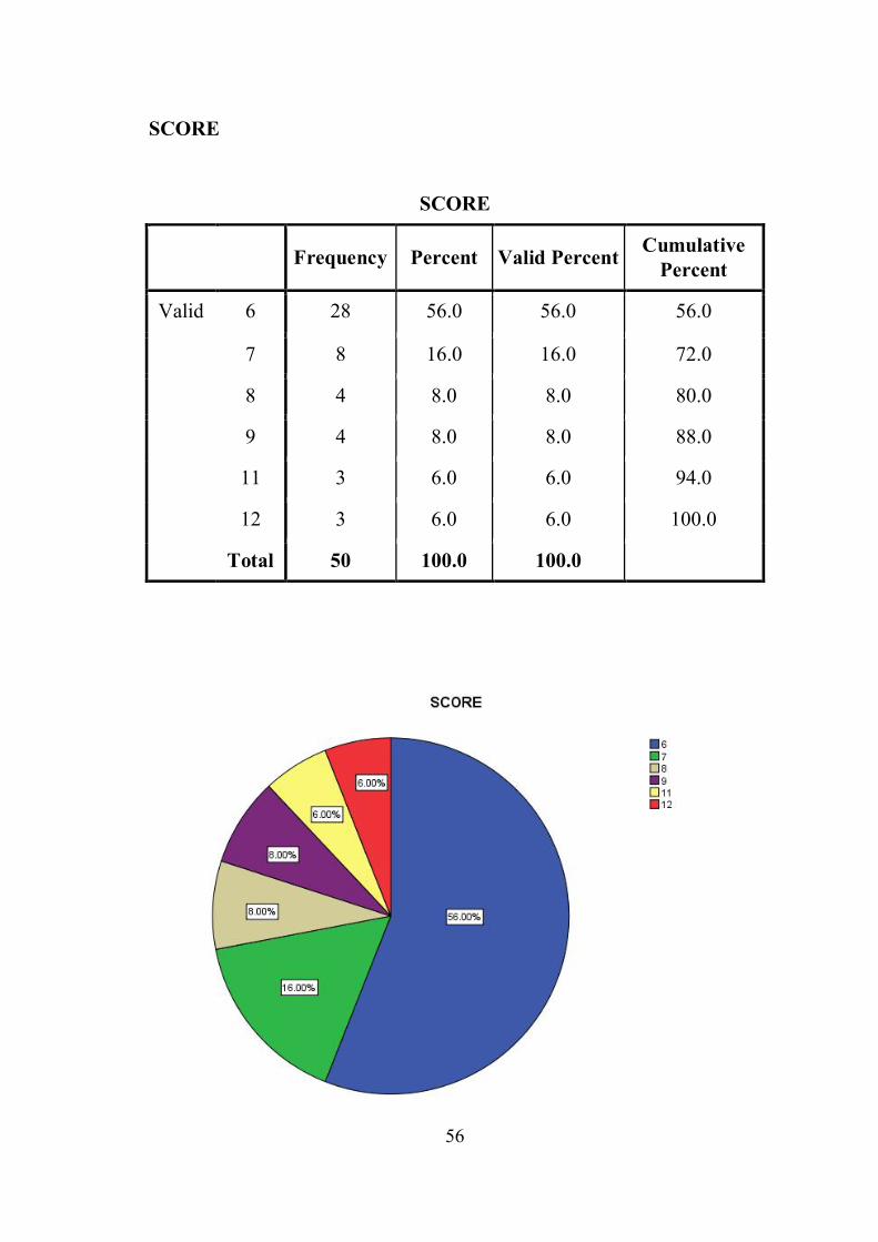
SCORE
SCORE
Frequency Percent Valid Percent Cumulative Percent
Valid 6 28 56.0 56.0 56.0
7 8 16.0 16.0 72.0
8 4 8.0 8.0 80.0
9 4 8.0 8.0 88.0
11 3 6.0 6.0 94.0
12 3 6.0 6.0 100.0
Total 50 100.0 100.0

57
PROCEDURE
PROCEDURE
Frequency Percent
Valid Percent
Cumulative Percent
Valid CONVERTED 5 10.0 10.0 10.0
LAP 45 90.0 90.0 100.0
Total 50 100.0 100.0
58
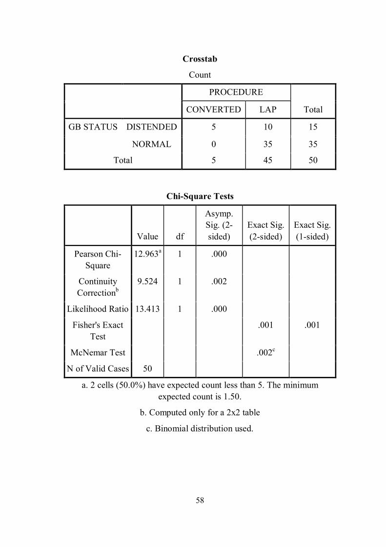
Crosstab
Count
PROCEDURE
Total CONVERTED LAP
GB STATUS DISTENDED 5 10 15
NORMAL 0 35 35
Total 5 45 50
Chi-Square Tests
Value df
Asymp. Sig. (2-sided)
Exact Sig. (2-sided)
Exact Sig. (1-sided)
Pearson Chi-Square
12.963a 1 .000
Continuity Correctionb
9.524 1 .002
Likelihood Ratio 13.413 1 .000
Fisher's Exact Test .001 .001
McNemar Test .002c
N of Valid Cases 50
a. 2 cells (50.0%) have expected count less than 5. The minimum expected count is 1.50.
b. Computed only for a 2x2 table
c. Binomial distribution used.

59
It was found that of the 35 cases in which the GB status was
normal, it was possible to do the surgery in laparoscopy itself . of the
remaining 15 cases in which GB was distended, 5 cases were converted
to open. The statistical analysis also shows this difference to be
significant with a p value of .001

60
Crosstab
Count
PROCEDURE
Total CONVERTED LAP
GB WALL THICKNESS
<3 mm 0 37 37
>3mm 5 8 13
Total 5 45 50
Chi-Square Tests
Value df Asymp. Sig. (2-sided)
Exact Sig. (2-sided)
Exact Sig. (1-sided)
Pearson Chi-Square
15.812a 1 .000
Continuity Correctionb
11.827 1 .001
Likelihood Ratio 15.185 1 .000
Fisher's Exact Test .001 .001
McNemar Test .c
N of Valid Cases 50
a. 2 cells (50.0%) have expected count less than 5. The minimum expected count is 1.30.
b. Computed only for a 2x2 table
c. Both variables must have identical values of categories.
61
It was found that of the 37 cases in which the GB wall thickness
was <3mm, it was possible to do the surgery in laparoscopy itself . of the
remaining 13 cases in which GB was thickened , 5 cases were converted
to open. The statistical analysis also shows this difference to be
significant with a p value of .001
62
Crosstab
Count
PROCEDURE
Total CONVERTED LAP
NO OF STONES
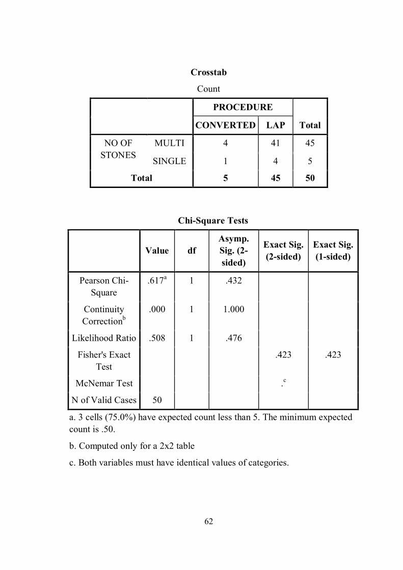
MULTI 4 41 45
SINGLE 1 4 5
Total 5 45 50
Chi-Square Tests
Value df
Asymp. Sig. (2-sided)
Exact Sig. (2-sided)
Exact Sig. (1-sided)
Pearson Chi-Square
.617a 1 .432
Continuity Correctionb
.000 1 1.000
Likelihood Ratio .508 1 .476
Fisher's Exact Test .423 .423
McNemar Test .c
N of Valid Cases 50
a. 3 cells (75.0%) have expected count less than 5. The minimum expected count is .50.
b. Computed only for a 2x2 table
c. Both variables must have identical values of categories.
63
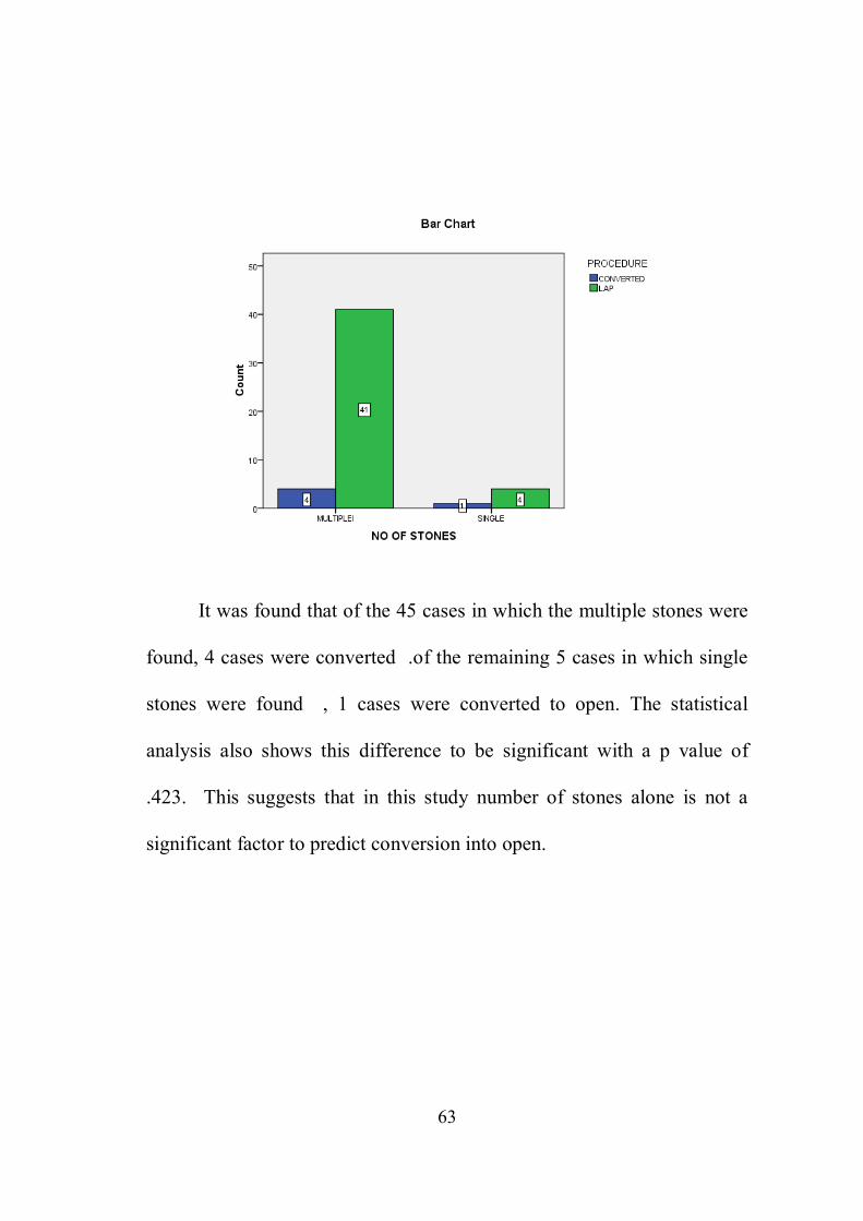
It was found that of the 45 cases in which the multiple stones were
found, 4 cases were converted .of the remaining 5 cases in which single
stones were found , 1 cases were converted to open. The statistical
analysis also shows this difference to be significant with a p value of
.423. This suggests that in this study number of stones alone is not a
significant factor to predict conversion into open.
64
Crosstab
Count
PROCEDURE
Total CONVERTED LAP
PERI CHOLECYSTIC COLLECTION
NO 0 39 39
YES 5 6 11
Total 5 45 50
Chi-Square Tests
Value df
Asymp. Sig. (2-sided)
Exact Sig. (2-sided)
Exact Sig. (1-sided)
Pearson Chi-Square
19.697a 1 .001
Continuity Correctionb
14.970 1 .001
Likelihood Ratio 17.350 1 .001
Fisher's Exact Test .001 .001
McNemar Test .c
N of Valid Cases 50
a. 2 cells (50.0%) have expected count less than 5. The minimum expected count is 1.10.
b. Computed only for a 2x2 table
c. Both variables must have identical values of categories.

65
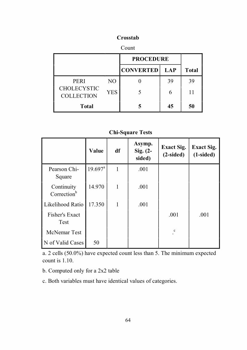
It was found that of the 39 cases in which pericolecystic collection
was not there , it was possible to do the surgery in laparoscopy itself . of
the remaining 11 cases had pericholecystic collection , 5 cases were
converted to open. The stastistical analysis also shows this difference to
be significant with a p value of .001
66
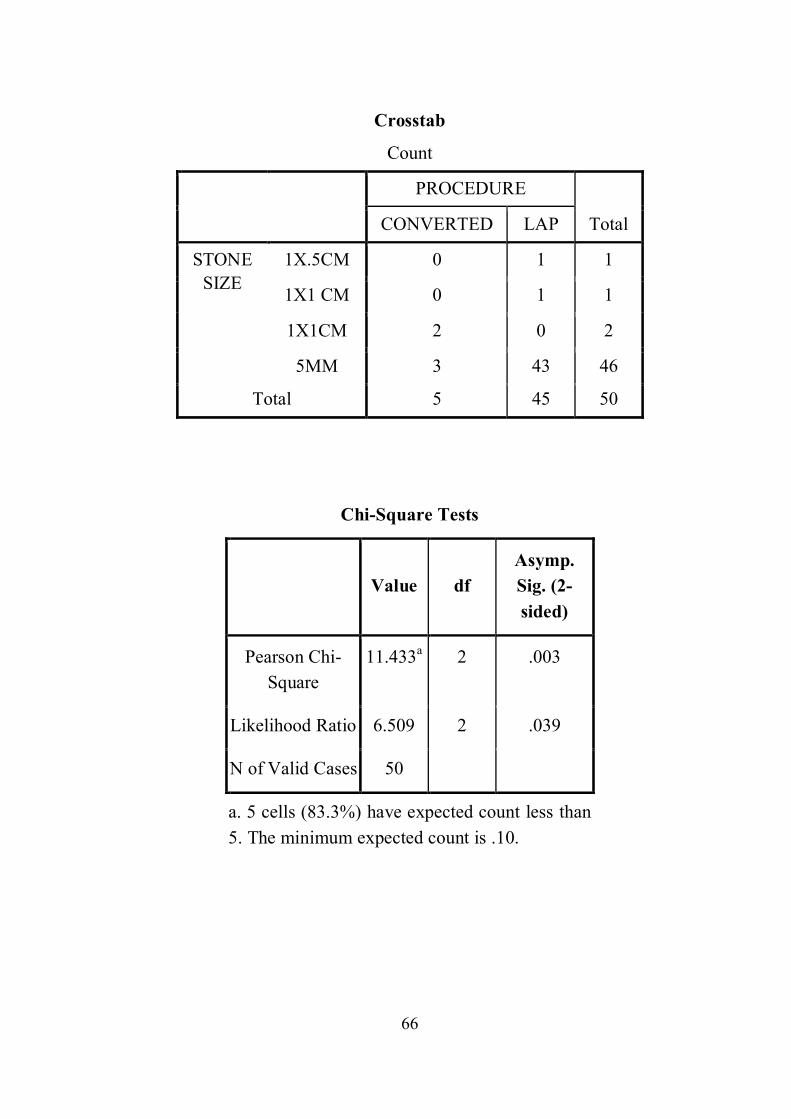
Crosstab
Count
PROCEDURE
Total CONVERTED LAP
STONE SIZE
1X.5CM 0 1 1
1X1 CM 0 1 1
1X1CM 2 0 2
5MM 3 43 46
Total 5 45 50
Chi-Square Tests
Value df
Asymp. Sig. (2-sided)
Pearson Chi-Square
11.433a 2 .003
Likelihood Ratio 6.509 2 .039
N of Valid Cases 50
a. 5 cells (83.3%) have expected count less than 5. The minimum expected count is .10.
67
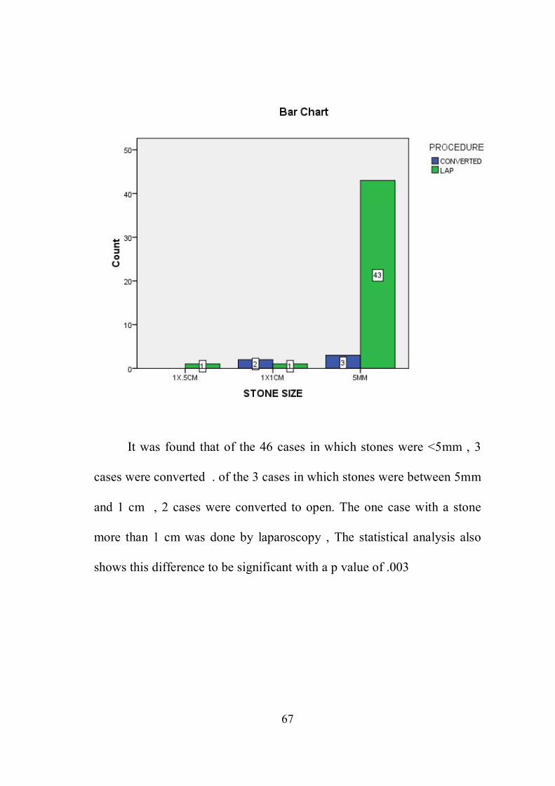
It was found that of the 46 cases in which stones were <5mm , 3
cases were converted . of the 3 cases in which stones were between 5mm
and 1 cm , 2 cases were converted to open. The one case with a stone
more than 1 cm was done by laparoscopy , The statistical analysis also
shows this difference to be significant with a p value of .003

68
Crosstab
Count
PROCEDURE
Total CONVERTED LAP
SCORE 6 0 28 28
7 0 8 8
8 0 4 4
9 0 4 4
11 3 0 3
12 2 1 3
Total 5 45 50
Chi-Square Tests
Value df
Asymp. Sig. (2-sided)
Pearson Chi-Square
42.593a 5 .001
Likelihood Ratio 28.689 5 .001
McNemar-Bowker Test
. . .b
N of Valid Cases 50
a. 10 cells (83.3%) have expected count less than 5. The minimum expected count is .30.
b. Computed only for a PxP table, where P must be greater than 1.

69
It can be noted that as the score increases the chance of the case
being opened increases and all the opened cases had score of either 11(3
cases) or 12(2 cases). The statistical analysis also shows the difference to
be significant with a p value of .001

DISCUSSION

70
DISCUSSION
Laparoscopic cholecystectomy has become the gold standard
treatment for patients with gallstones due to less morbidity, lesser
hospital stay and early return to normal activities. The difficult
gallbladder is the most common ‘difficult’ laparoscopic surgery being
performed by general surgeons all over the world and the potential one
that places the patient at significant risk12. Previous reports have
promulgated the use of scoring systems to predict conversion to open
cholecystectomy. However, these systems presented incongruent data
points, evaluated a limited number of factors, included subjective
variables, and some were formulated early in the course of laparoscopic
cholecystectomy before the operation became uniformly established16. So
we planned to analyse USG factors to predict the conversion of
laparoscopic cholecystectomy to open cholecystectomy.
In our study, 50 patients diagnosed with gallstones were taken for
Laparoscopic cholecystectomy. Among the 50 patients, 33 patients( i.e.
66%) were female and 17 patients (i.e. 34%) were male. Of the 50
patients, 5 patients were converted to open cholecystectomy.
71
GB status
It was found that of the 35 cases in which the GB status was normal , it
was possible to do surgery in laparoscopic itself , of the remaining 15
cases in which GB was distended , 5 cases were converted to open , the
statistical analysis also shows this difference to be significant with a p
value of 0.001
GB wall thickness
It was found 37 cases with GB wall thickness was <3mm possible to do
laparoscopic itself , remaining 13 cases GB wall thickness was > 3mm ,
of which 5 cases were converted to open, the statistical analysis also
shows this difference to be significant with a p value of 0.001
Number of stones
it was found 45 casesin which the multiple stones found, 4 cases
were converted, of the remaining 5 cases in which single stones were
found, 1 cases were converted to open, the statistical analysis also shows
this difference to be significant with a pvalue of 0.423 this suggest that in
this study number of stones alone is not a significant factor to predict
conversion to open
72
Pericholecystic collection
It was found that of the 39cases in which pericholecystic collection
was not there , it was possible to do the surgery in laparoscopic itself.
Of the remaining 11 cases had pericholecystic collection , of
which 5 cases were converted to open The statistical analysis also shows
this difference to be significant with a P value of 0.001.
Stone size
It was found that of the 46 cases in which stones were <5mm,
3 cases were converted.
Of the 3 cases in which stones were between 5mm and 1 cm,
2 cases converted to open.
The one case with a stone more than 1 cm was done by laparoscopy , the
statistical analysis also shows this difference to be significant with a P
value of 0.003
73
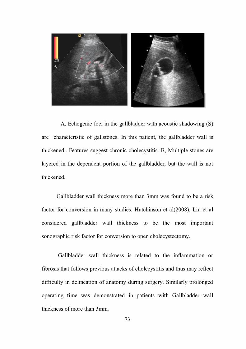
A, Echogenic foci in the gallbladder with acoustic shadowing (S)
are characteristic of gallstones. In this patient, the gallbladder wall is
thickened.. Features suggest chronic cholecystitis. B, Multiple stones are
layered in the dependent portion of the gallbladder, but the wall is not
thickened.
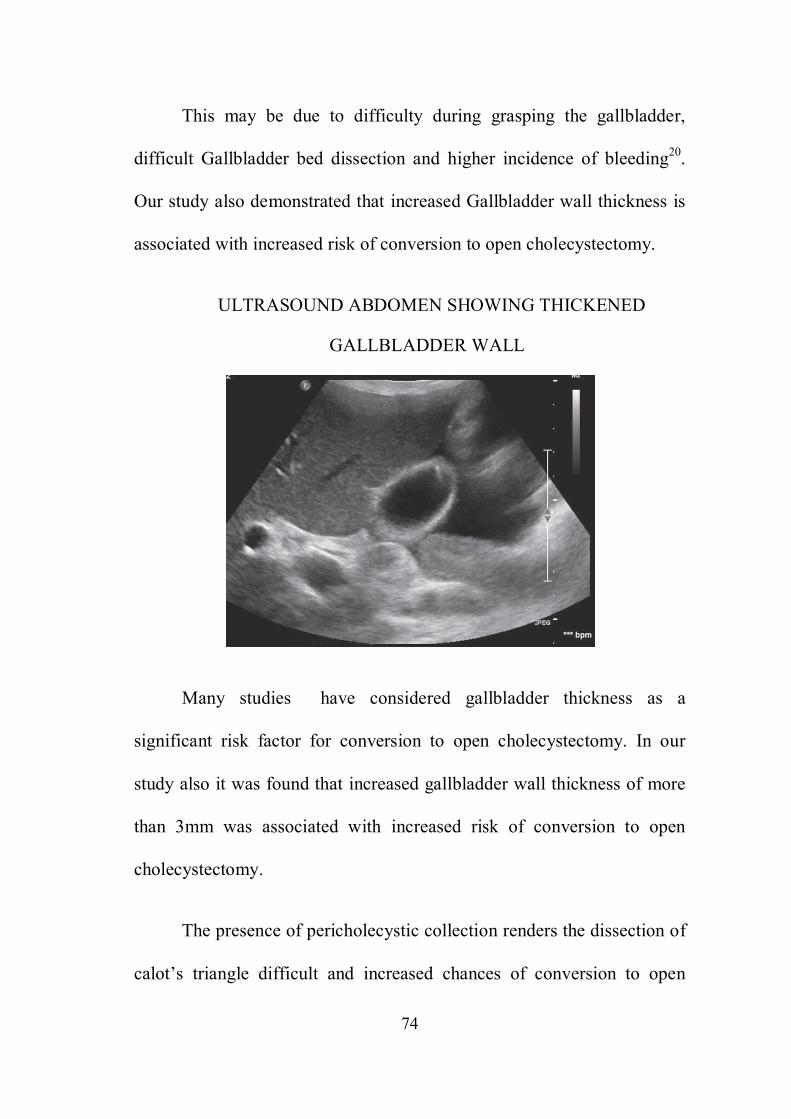
Gallbladder wall thickness more than 3mm was found to be a risk
factor for conversion in many studies. Hutchinson et al(2008), Liu et al
considered gallbladder wall thickness to be the most important
sonographic risk factor for conversion to open cholecystectomy.
Gallbladder wall thickness is related to the inflammation or
fibrosis that follows previous attacks of cholecystitis and thus may reflect
difficulty in delineation of anatomy during surgery. Similarly prolonged
operating time was demonstrated in patients with Gallbladder wall
thickness of more than 3mm.
74
This may be due to difficulty during grasping the gallbladder,
difficult Gallbladder bed dissection and higher incidence of bleeding20.
Our study also demonstrated that increased Gallbladder wall thickness is
associated with increased risk of conversion to open cholecystectomy.
ULTRASOUND ABDOMEN SHOWING THICKENED
GALLBLADDER WALL
Many studies have considered gallbladder thickness as a
significant risk factor for conversion to open cholecystectomy. In our
study also it was found that increased gallbladder wall thickness of more
than 3mm was associated with increased risk of conversion to open
cholecystectomy.
The presence of pericholecystic collection renders the dissection of
calot’s triangle difficult and increased chances of conversion to open

75
cholecystectomy11 .
In our study we analysed USG factors such as GB status, GB wall
thickness, number of stones, pericholecystic collection, size of stone as
the USG score increases the chance of the case being opened increases
and all the opened cases had either 11 0r 12 ,
The statistical analysis also shows the difference to be significant
with a p value of 0.001

CONCLUSION
76
CONCLUSION
Laparoscopic cholecystectomy has gradually replaced open
cholecystectomy in the treatment of patients with benign gallbladder
disease. With the advancement in equipment and experience in
laparoscopic surgery, most of the difficult gallbladder can be dealt
laparoscopically.
Preoperative USG examination of the GB is a good predictor of
difficult cholecystectomy in majority of cases and should be used pre
operatively as a routine screening tool to delineate biliary tree anatomy
and pathology, pre operative risk factor can help to predict difficult
gallbladder and conversion to other type of cholecystectomy.
In our study we analysed USG factors such as GB status, GB wall
thickness, number of stones, pericholecystic collection, size of stone.
In our study noted that as the USG score increases the chance of
the case being opened increases and all the opened cases had either 11 0r
12, The statistical analysis also shows the difference to be significant with
a p value of 0.001
Among USG factors, a distended GB, increased GB wall
thickness, presence of pericholecystic collection ,large size of stone
77
proved to be significant and was associated with increased risk of
conversion to open cholecystectomy.
Our results demonstrate that an a accurate and easily derived
estimation of risk factor predicting conversion from laparoscopic
cholecystectomy to open cholecystectomy can be obtained from USG
score , increase in score can predict difficulty to be encountered during
laparoscopic cholecystectomy and help in making a decision for
conversion thus shortening the duration of surgery thereby preventing
unnecessary complications.

BIBLIOGRAPHY

BIBLIOGRAPHY
1. Rakesh Tendon, “ Diseases of gallbladder and biliary tract”. API text book of
medicine, Dr.Siddarth N Shah, 7th edition, 2003, PP 642 – 644.
2. Conference, N C. Gallstones and laparoscopic cholecystectomy: JAMA 1992;
269: 1018-1024.
3. Ravi S Chari, MD AndShinul A Shah, MD. Biliary system, Sabiston textbook
of surgery; Courtney M Townsend, R Laniel Beauchamp, B. Mark Evers,
Kenneth L Mattox. 18th edition , Saunders Elsevier, vol 2, 2009. chapter 54,
PP: 1547-1588.
4. Boni L, et al. Infective complication of laparoscopic surgery. Surg infect
(Larchmt), 2006; 7 suppl 2: S109-11.36.Ravi S. Chari,MD and Shinul A.
Shah,MD. Biliary system, Chapter 54 Sabiston textbook of surgery, Volume 2,
18thedition, Courtney M. Town Send, R. Laniel Beauchamp, B. Mark Evers,
Kenneth.L. Mattox, p. 1547-1588.
5. Syed amjad ali et al, Forecast of difficult laparoscopy by sonography-an added
advantage Biomedical Research 2012 : 425 429.
6. Jeremy lipman MD, Jeffery A Claridge MD, et al. Pre operative findings
predicting conversion from laparoscopic to open cholecystectomy. Presented
at 64th meeting of Central surgical association.Chicago, Illionois, March 8-10,
2007.
7. Simopoulos et al Risk factors for conversion of laparoscopic cholecystectomy
to open cholecystectomy Surg Endoscopy 2005: 19(7) 905-909.
8. Trownbridge R L, Rutkowski N K, Shojania K G: Does this patient have acute
cholecystitis? JAMA 289; 80-86, 2003.
9. Alexander P Nagle, Nathaniel J Soper, James R Hines; Colecystectomy (open
and laparoscopic).Michael J Zinner, Stanley W Ashley; Maingot’s Abdominal
Operations; 11th edition, McGraw Hill, 2007. Chapter 32, PP:847-864.
10. Prediction of difficulty of laparoscopic cholecystectomy by pre operative USG
: a randomized control trail , global journal inc, (USA), online ISSN 229-4618
11. Risk factors resulting in conversion of laparoscopic cholecystectomy to open
surgery, N.A.kama, mdoganay, m dolapci, e,reis, m.atli, m .kologlu. surgical
endocopy, September 2001, volume 15, issue 9 ,pp 965-968.,
12. The role of pre operative investigations in predicting difficult laparoscopic
cholecystectomies., surgical endoscopy, august 1996, volume 10,issue 8 , pp
791-793 .,
13. A comprehensive predictive scoring method for difficult laparoscopic
cholecystectomy. journal of minimal access surgery. By makm vivek 2014,
www.ncbi,nlm .nih.gov
14. USG in GB disease prediction of difficult laparoscopic cholecystectomy.,
IJSR,ISSN ONLINE : 2319-7064, IMPACT FATOR 2012: 3.358
15. Prediction of difficulty of laparoscopic cholecystectomy by pre operative USG
: a randomized control trail , global journal inc, (USA), online ISSN 229-4618
16. Sopr N. Laparoscopic cholecystectomy. CurrProblSurg 199; 28: 585-655.
17. Strasburg S M. The “Hidden cystic duct” syndrome and the infundibular
technique of laparoscopic cholecystectomy – the danger of the false
infundibulum. J Ann CollSurg, 2000; 191(6): 661-7.
18. Strasburg S M, Hertl M, Soper N S. An analysis of the problem of biliary
injury during laparoscopic cholecystectomy. J Ann CollSurg 1995; 180: 101-
125.
19. T Satish Kumar, A P Saklani, R Vinayagam, R L Blackett. Spilled gallstones
during laparoscopic cholecystectomy: a review of the literature. Post grad Med
J 2004; 80: 77-79.
20. Cullen J. Laparoscopic cholecystectomy: Avoiding complications. In: Birkett D
H, Ronsky J L, Stiegmann G V. the SAGES manual- Fundamentals of
Laparoscopic and GI Endoscopy.Springer, 2003: 137- 142.
21. Way L W, Stewart L, Gantert W, et al. Causes and prevention of laparoscopic
bile duct injuries: analysis of 252 cases from a human factors and cognitive
psychology perspective. Ann Surg 2003; 4:460.
22. Deziel D, Millikan K, Economou S, et al. Complication of laparoscopic
cholecystectomy: a national survey of 4292 hospitals and analysis of 77604
cases. Am J Surg 1993; 165: 9-14.
23. The southern surgeons club. A prospective analysis of 1518 laparoscopic
cholecystectomies. N Engl J Med 1991. 324: 1073-1078.
24. Seiler C, Glattly A, Metzger A, Czerniak A. Injuries to the diaphragm and its
repair during laparoscopic cholecystectomy. SurgEndosc 1995; 9: 193-4.
25. Armstrong P, Miller S, Brown G. Diaphragmatic hernia seen as a late
complication of laparoscopic cholecystectomy. SurgEndosc 1999: 13: 817-818.
26. Kama N A, Dogary M, Dolapa M. Reise, Attli M, et al! Risk factors resulting
in conversion of laparoscopic cholecystectomy to open cholecystectomy.
Surgical endoscopy, Springer New York; V15 : 965-968.
27. Daradkeh S, laparoscopic cholecystectomy: What are the factors determining
difficulty? Hepatogastroenterology. 2001 Jan-Feb; 48(37): 76-78.
28. Jorgensen J O, Hunt D R: laparoscopic cholecystectomy. A prospective
analysis of the potential causes of failure. Surglaparosendosc 3: 49- 53, 1993.
29. Pastulka P S, Bistrian B R, Benotti P N, et al: The risks of surgery in obese
patients. Ann intern med 104: 551-556, 1985.
30. Polk H C Jr. Carcinoma and the calcified gallbladder. Gastroentrology 1966;
50: 582-585.
31. Nadu A, Gallilli Y, Soffer D, Kluger Y: Disruption of Cholecystoenteric fistula
induced by minor blunt trauma. J Trauma 1996; 41: 914-915.
32. J. S. Randhawa . A. K. Pujahari, preoperative prediction of difficult lap chole: a
scoring method. Indian Journal of Surgery, volume 71, number 4, July- August
2009, PP:198-201.
33. Bailey and Love’s Short Practice of Surgery; 26th ed
34. Sabiston Textbook of Surgery; 18th ed
35. Maingot’s Abdominal Operations, 12thed
ANNEXURES
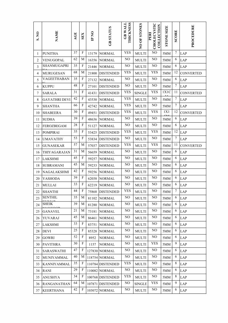
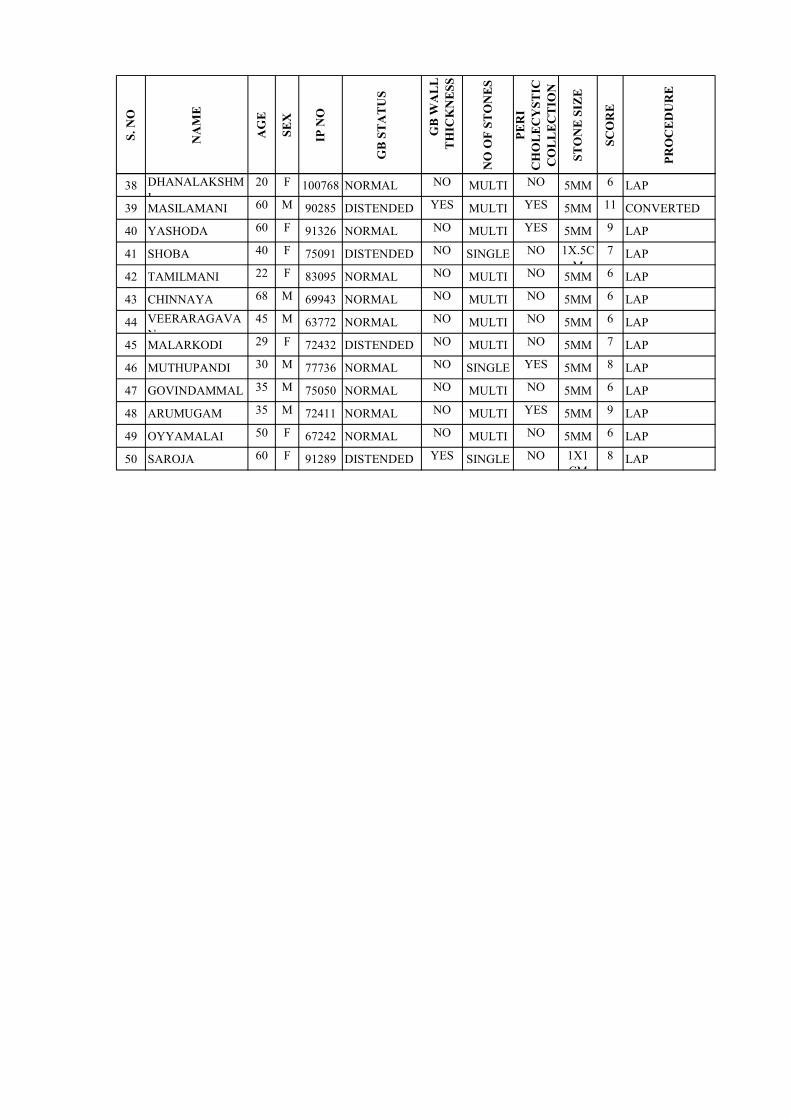
S. N
O
NA
ME
AG
E
SEX
IP N
O
GB
ST
AT
US
GB
WA
LL
T
HIC
KN
ESS
NO
OF
STO
NE
S
PER
I C
HO
LE
CY
STIC
C
OL
LE
CT
ION
STO
NE
SIZ
E
SCO
RE
PRO
CE
DU
RE
1 PUNITHA 37 F 13179 NORMAL YES MULTI NO 5MM 7 LAP
2 VENUGOPAL 62 M 16356 NORMAL NO MULTI NO 5MM 6 LAP
3 SHANMUGAPRIYA
35 F 21446 NORMAL NO MULTI NO 5MM 6 LAP
4 MURUGESAN 68 M 21800 DISTENDED YES MULTI YES 5MM 12 CONVERTED
5 VAGEETHABANU
35 F 27132 NORMAL NO MULTI NO 5MM 6 LAP
6 KUPPU 48 F 27101 DISTENDED NO MULTI NO 5MM 7 LAP
7 SARALA 31 F 41431 DISTENDED YES SINGLE YES 1X1CM
11 CONVERTED
8 GAYATHRI DEVI 42 F 43530 NORMAL YES MULTI NO 5MM 7 LAP
9 SHANTHA 66 F 42742 NORMAL YES MULTI NO 5MM 7 LAP
10 SHABEERA 30 F 49451 DISTENDED YES MULTI YES 1X1 CM
12 CONVERTED
11 SUDHA 39 F 48636 NORMAL NO MULTI NO 5MM 6 LAP
12 FEROZBEGAM 28 F 51127 NORMAL NO MULTI NO 5MM 6 LAP
13 POMPIRAI 35 F 53425 DISTENDED YES MULTI YES 5MM 12 LAP
14 UMAVATHY 53 F 53834 DISTENDED NO MULTI NO 5MM 7 LAP
15 GUNASEKAR 57 M 57037 DISTENDED YES MULTI YES 5MM 11 CONVERTED
16 THIYAGARAJAN 31 M 56659 NORMAL NO MULTI NO 5MM 6 LAP
17 LAKSHMI 45 F 59257 NORMAL NO MULTI NO 5MM 6 LAP
18 SUBRAMANI 45 M 59233 NORMAL NO MULTI NO 5MM 6 LAP
19 NAGALAKSHMI 42 F 59256 NORMAL NO MULTI NO 5MM 6 LAP
20 YASHODA 39 F 62038 NORMAL NO MULTI NO 5MM 6 LAP
21 MULLAI 33 F 62219 NORMAL NO MULTI NO 5MM 6 LAP
22 SHANTHI 44 F 75868 DISTENDED YES MULTI NO 5MM 7 LAP
23 SENTHIL KUMAR
35 M 81102 NORMAL NO MULTI NO 5MM 6 LAP
24 SHEIK MANSOOR
39 M 81280 NORMAL NO MULTI NO 5MM 6 LAP
25 GANAVEL 21 M 73181 NORMAL NO MULTI NO 5MM 6 LAP
26 YUVARAJ 45 M 86461 NORMAL NO MULTI NO 5MM 6 LAP
27 LAKSHMI 33 F 83751 NORMAL NO MULTI NO 5MM 6 LAP
28 DEVI 25 F 85328 NORMAL NO MULTI NO 5MM 6 LAP
29 GOWRI 52 F 8952 NORMAL NO MULTI NO 5MM 6 LAP
30 PAVITHRA 30 F 1157 NORMAL NO MULTI YES 5MM 9 LAP
31 SARASWATHI 47 F 127830 NORMAL NO MULTI NO 5MM 6 LAP
32 MUNIYAMMAL 40 M 118754 NORMAL NO MULTI NO 5MM 6 LAP
33 KANNIYAMMAL 35 F 110784 DISTENDED YES MULTI NO 5MM 8 LAP
34 RANI 29 F 110082 NORMAL NO MULTI NO 5MM 6 LAP
35 ANUSHYA 34 F 100768 DISTENDED YES MULTI NO 5MM 8 LAP
36 RANGANATHAN 64 M 107871 DISTENDED NO SINGLE YES 5MM 9 LAP
37 KEERTHANA 42 F 103072 NORMAL NO MULTI NO 5MM 6 LAP
S. N
O
NA
ME
AG
E
SEX
IP N
O
GB
ST
AT
US
GB
WA
LL
T
HIC
KN
ESS
NO
OF
STO
NE
S
PER
I C
HO
LE
CY
STIC
C
OL
LE
CT
ION
STO
NE
SIZ
E
SCO
RE
PRO
CE
DU
RE
38 DHANALAKSHMI
20 F 100768 NORMAL NO MULTI NO 5MM 6 LAP
39 MASILAMANI 60 M 90285 DISTENDED YES MULTI YES 5MM 11 CONVERTED
40 YASHODA 60 F 91326 NORMAL NO MULTI YES 5MM 9 LAP
41 SHOBA 40 F 75091 DISTENDED NO SINGLE NO 1X.5CM
7 LAP
42 TAMILMANI 22 F 83095 NORMAL NO MULTI NO 5MM 6 LAP
43 CHINNAYA 68 M 69943 NORMAL NO MULTI NO 5MM 6 LAP
44 VEERARAGAVAN
45 M 63772 NORMAL NO MULTI NO 5MM 6 LAP
45 MALARKODI 29 F 72432 DISTENDED NO MULTI NO 5MM 7 LAP
46 MUTHUPANDI 30 M 77736 NORMAL NO SINGLE YES 5MM 8 LAP
47 GOVINDAMMAL 35 M 75050 NORMAL NO MULTI NO 5MM 6 LAP
48 ARUMUGAM 35 M 72411 NORMAL NO MULTI YES 5MM 9 LAP
49 OYYAMALAI 50 F 67242 NORMAL NO MULTI NO 5MM 6 LAP
50 SAROJA 60 F 91289 DISTENDED YES SINGLE NO 1X1 CM
8 LAP
ABBEREVIATIONS
GB - gall bladder
CBD - common bile duct
LAP - laparoscopic
USG - ultrasonogram
LFT - liver function test
INFORMATION SHEET
TITLE : “A STUDY ON ULTRA SOUND SCORING PREDICTTING
CONVERSION FROM LAP TO OPEN CHOLECYSTECTOMY ”
Name of Investigator : Dr.D.JEGADHES KUMAR. Name of Participant :
Purpose of Research :The purpose of the study is the role of radiological
factors predicting difficult laparoscopic cholecystectomy
Study Design :Prospective Observational Study
Study Procedures :Patient will be subjected to routine investigations & USG
ABDOMEN. The patients are then, if willing undergo surgical treatment.
Possible Risks :No risks to the patient
Possible benefits to the patient :Patient is provided an alternative modality of
treatment which will help in remission of their disease.
To doctor & to other people : If this study gives positive results, it can help
determine the most effective treatment and the treatment protocol for patients
with chronic right lower quadrant pain. This will help in providing better and
complete treatment to other patients in future.
Confidentiality of the information obtained from you :The privacy of the
patients in the research will be maintained throughout the study. In the event of
any publication or presentation resulting from the research, no personally
identifiable information will be shared

Can you decide to stop participating in the study :Taking part in this study is
voluntary. You are free to decide whether to participate in this study or to
withdraw at any time
How will your decision to not participate in the study affect you :Your
decision will not result in any loss of benefits to which you are otherwise
entitled.
Signature of Investigator Signature of Participant
Date :
Place :

PATIENT CONSENT FORM
Study Detail : “A STUDY ON ULTRA SOUND SCORING
PREDICTTING CONVERSION FROM
LAPAROSCOPIC TO OPEN
CHOLECYSTECTOMY ”
Study Centre : Rajiv Gandhi Government General Hospital, Chennai.
Patient’s Name :
Patient’s Age :
In Patient Number :
Patient may check (☑) these boxes
I confirm that I have understood the purpose of procedure for the above
study. I have the opportunity to ask question and all my questions and
doubts have been answered to my complete satisfaction. o
I understand that my participation in the study is voluntary and that I am
free to withdraw at any time without giving reason, without my legal
rights being affected. o
I understand that sponsor of the clinical study, others working on the
sponsor’s behalf, the ethical committee and the regulatory authorities
will not need my permission to look at my health records, both in
respect of current study and any further research that may be conducted
in relation to it, even if I withdraw from the study I agree to this access.
However, I understand that my identity will not be revealed in any
information released to third parties or published, unless as required
under the law. I agree not to restrict the use of any data or results that o

arise from this study.
I agree to take part in the above study and to comply with the instructions
given during the study and faithfully cooperate with the study team and
to immediately inform the study staff if I suffer from any deterioration
in my health or well being or any unexpected or unusual symptoms. o
I hereby consent to participate in this study
o
I hereby give permission to undergo complete clinical examination and
diagnostic tests including hematological, biochemical, radiological tests
and to undergo treatment o
Signature/thumb impression
Patient’s Name and Address:
Signature of Investigator
Study Investigator’s Name:
Dr.D.JEGADHES KUMAR.,
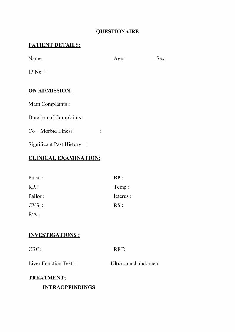
QUESTIONAIRE
PATIENT DETAILS:
Name: Age: Sex:
IP No. :
ON ADMISSION:
Main Complaints :
Duration of Complaints :
Co – Morbid Illness :
Significant Past History :
CLINICAL EXAMINATION:
Pulse : BP :
RR : Temp :
Pallor : Icterus :
CVS : RS :
P/A :
INVESTIGATIONS :
CBC: RFT:
Liver Function Test : Ultra sound abdomen:
TREATMENT;
INTRAOPFINDINGS