a cross-sectional study of gastrointestinal nematodiasis, gross ...
159
„<rTY OF f • 14 A CROSS-SECTIONAL STUDY OF GASTROINTESTINAL NEMATODIASIS, GROSS SKIN CONDITIONS AND ECTOPARASITES OF DONKEYS IN MWINGI DISTRICT, KENYA. 0 > ,.% ^ , BY CHRISTOPHER KYESWA, B. V. M. (M.U.K). This thesis was submitted in partial fulfilment of the degree of Master of science in Veterinary Epidemiology and Economics at the Faculty of Veterinary Medicine, University of Nairobi, Department of Public Health, Pharmacology and Toxicology, Faculty of Veterinary Medicine, College of Agriculture and Veterinary Sciences. 1996.
-
Upload
khangminh22 -
Category
Documents
-
view
1 -
download
0
Transcript of a cross-sectional study of gastrointestinal nematodiasis, gross ...

„ < rT Y OF
f •
14A CROSS-SECTIONAL STUDY OF GASTROINTESTINAL
N E M A T O D IA SIS, G ROSS SKIN C O NDITIO NS AND
ECTOPARASITES OF DONKEYS IN MWINGI DISTRICT,
KENYA.
0 > , . %
^ , BY
CHRISTOPHER KYESWA, B. V. M. (M.U.K).
This thesis was submitted in partial fulfilment of the degree of Master of science in
Veterinary Epidemiology and Economics at the Faculty of Veterinary Medicine,
University of Nairobi, Department of Public Health, Pharmacology and Toxicology,
Faculty of Veterinary Medicine, College of Agriculture and Veterinary Sciences.
1996.

11
DECLARATIONS.
This is my original work. It has not been presented for a degree in any other University.i
j . \? n ?
CHRISTOPHER KYESWA. (B.V.M).
This thesis was submitted with our approval as University supervisors.
%
Signed ................D . O S
DR. M. N. KYULE., B.V.M, MSc., M.P.V.M., PhD.
Signed .. • Dale
PROFESSOR. J. M. GATHUMA., B.V.Sc., M.Sc., PhD.
Signed.. .....J S & , .............. Date.
DR. J. M. NDUNG’U., B.V.M., PhD.

Ill
DEDICATION
This work is dedicated to my guardian, Mr. James Semakula Kagenda, who made seiness
sacrifice for me.

IV
ACKNOWLEDGEMENTS
I would like to gratefully acknowledge my supervisors Dr. M. N. Kyule,
Professor J. M. Gathuma and Dr. J. M. Ndung’u for their suggestions, guidance and
constructive criticisms, without which this work would not have been completed.
My sincere gratitude goes to Dr. I. Mbithi (Mwingi District veterinary officer),
the field veterinary staff in Mwingi District and Dr. J. B. Githiori of KETRJ- Muguga
for the invaluable assistance during data collection.
I’m also very grateful to Dr. T.A. Ngatia of the Department of Veterinary
Pathology and Microbiology, University of Nairobi, for allowing me to use facilities in
the Parasitology Laboratory. I thank the technical staff in the Parasitology Laboratory for
their assistance during analysis of the study samples.
The friendly environment for learning as well as the encouragement I received
from my classmates namely Sakwa, Wekare, Njue, Njiru, Githaiga, Tito Tipo,
Maichomo and Wanja are highly appreciated.
I acknowledge my sponsors, the German Academic Exchange Service (DAAD)
for offering me a scholarship to pursue postgraduate studies at the University of Nairobi.
My thanks also go to the Kenya Trypanosomiasis research Institute and the
International Donkey Protection Trust for co-sponsoring the research project.
V
TABLE OF CONTENTS
PAGE NUMBER
TITLE........................................................................................................................................ J
Declarations......................................................................................................................... II
Dedication................................................................................................................................Ill
Acknowledgements................................................................................................................ IV
Table of Contents....................................................................................................................V
List of Tables......................................................................................................................... XI
List of Figures.................................................................................................................... -XIV
List of Appendices............................................................................................................... XV
Abstract................................................................................................................................XVI
CHAPTER ONE................................................................................................................... 1
1.0 INTRODUCTION....................................................................................................... 1
CHAPTER TWO.................................................................................................................. 4
2.0 Literature review........................................................................................ 4
2.1 Aetiology of donkey nematodiasis................................................................ 4
2.1.1 The life cycles of equine nematodes............................................................ 4
2.1.2 Distribution of donkey nematodiasis............................................................ 8
2.1.2.1 Factors influencing the distribution of donkey
nematodiasis.....................................................................................................^
2.1.2.1.1 Occurrence of donkey nematodiasis............................................................. 8
2.1.2.1.2 Climatic factors...............................................................................................^

2.1.2.1.3. Host factors...............................................................................................10
2.1.2.1.3.1 Host species.............................................................................................. 10
2.1.2.1.3.2 Age and sex.............................................................................................. 10
2.1.2.1.3.3 Host Immunity..........................................................................................10
2.1.2.1.4 Donkey management.................................................................................14
2.1.3 Pathogenesis and clinical manifestation of donkey
nematodiasis.................................................................................................15
2.1.4 Impact of donkey nematodiasis................................................................17
2.1.5 Diagnosis of donkey nematodiasis..........................................................17
2.1.5.1 Clinical signs..............................................................................................18
2.1.5.2 Blood and faecal examination.................................................................. 18
2.1.5.2.1 Qualitative techniques of faecal examination.........................................19
2.1.5.2.1.1 Direct smear.............................................................................................. 19
2.1.5.2.1.2 Concentration techniques........................................................................ 19
2.1.5.2.2 Quantitative techniques of faecal examination..................................... 20
2.1.5.3 Post mortem examination......................................................................... 21
2.1.6 Management of donkey nematodiasis....................................................22
2.2 Aetiology of donkey ectoparasitosis....................................................... 24
2.2.1 Life cycles of the ectoparasites of donkeys.......................................... 24
2.2.2 Distribution of donkey ectoparasites......................................................25
2.2.2.1 Factors which influence the occurrence and
distribution of donkey ectoparasitosis.................................................... 25
VI

VII
2.2.2.1.1 Host factors.................................................................................................25
2.2.2.1.2 Management factors..................................................................................27
2.2.2.1.3 Climatic factors......................................................................................... 27
2.2.3 Pathogenesis and clinical manifestations of donkey
ectoparasitosis.............................................................................................27
2.2.4 Importance of donkey ectoparasitosis................................................... 28
2.2.5 Diagnosis of donkey ectoparasitosis....................................................... 29
2.2.6 Management of donkey ectoparasitosis................................................. 29
2.3 Donkey dermatomycosis............................................................................30
2.3.1 Aetiology and distribution of donkey dermatomycosis..................... 30
2.3.2 Pathogenesis and clinical manifestations of donkey
dermatomycosis......................................................................................... 31
2.3.3 Importance of donkey dermatomycosis................................................. 31
2.3.4 Diagnosis of donkey dermatomycosis...................................................32
2.3.5 Management of donkey dermatomycosis.............................................. 33
2.4 Donkey wounds..........................................................................................33
2.4.1 Aetiology of donkey wounds................... 33
2.4.2 Importance of donkey wounds............................................................... 34
2.4.3 Management of donkey wounds.............................................................34
CHAPTER THREE........................................................................................................35
3.0 MATERIALS AND METHODS........................................................... 35
3.1 Study area................................................................................................... 35

VIII
3.2 Study population........................................................................................ 36
3.2.1 Sampling of study donkeys..................................................................... 37
3.2.1.1 Sampling technique...................................................................................38
3.3 Collection of samples................................................................................ 38
3.3.1 Faecal samples...........................................................................................39
3.3.2 Ectoparasites and skin scrapings............................................................ 40
3.4 Laboratory analyses of samples.............................................................. 40
3.4.1 Analysis of faecal samples...................................................................... 40
3.4.1.1 Preparation of floatation fluid.................................................................40
3.4.1.2 Egg floatation and identification............................................................. 41
3.4.1.3 Faecal culturing.........................................................................................42
3.4.1.4 Recovery of larvae....................................................................................43
3.4.1.5 Identification of larvae.............................................................................. 43
3.4.2 Identification of ectoparasites...................................................................44
3.4.2.1 Ticks........................................................................................................... 44
3.4.2.2 Skin scrapings............................................................................................44
3.4.2.2.1 Recovery and identification of mites......................................................44
3.4.2.2.2 Presence of fungi......................................................................................4^
3.4.2.2.2.1 Direct smear...............................................................................................4^
3.4.2.2.2.2 Laboratory culture.................................................................................... 4^
3.5 Administration of questionnaires.............................................................46
3.6 Administration and evaluation of IvermectinR...................................... 47

IX
3.7 Data management.......................................................................................49
3.8 Data analysis............................................................................................... 49
3.8.1 Case definition............................................................................................ 50
CHAPTER FOUR...........................................................................................................52
4.0 RESULTS................................................................................................... 52
4.1 Descriptive statistics and frequency distributions of
questionnaire-derived variables................................................................52
4.1.1 Ownership of donkeys..............................................................................52
4.1.2 Characteristics of donkey herds..............................................................52
4.1.3 Presence of ectoparasites......................................................................... 57
4.1.4 Distribution of gross lesions of the skin and skin
derivatives................................................................................................... 52
4.1.5 Levels of hygiene in the donkey "Bomas"........................................... 60
4.1.6 Donkey feeds............................................................................................. 61
4.1.6.1 Feeding management of donkeys............................................................. 63
4.1.7 Sources of water for donkey herds........................................................ 64
4.1.8 Deworming of donkeys............................................................................65
4.1.9 Availability of veterinary support services.............................................66
4.1.10 Breeding of donkeys................................................................................
4.1.11 Uses of donkeys....................................................................................... 68
4.2 Results of laboratory analyses................................................................. 70
4.2.1 Skin scrapings............................................................................................ 70
X
4.2.2 Faecal samples........................................................................................... 71
4.2.2.1 Faecal egg count.......................................................................................71
4.2.2.2 Characterisation of larvae from the survey faecal
samples.........................................................................................................72
4.3 Results of statistical analyses................................................................... 73
4.3.1 Descriptive statistics and pair-wise comparisons
of the division-specific nematode egg counts.......................................... 73
4.3.2 Logistic regression................................................................................... 76
4.3.3 Discriminant analysis...............................................................................79
4.4 Evaluation of the effect of lvermectinR on donkey
gastro-intestinal nematodiasis in Central and Kyuso
divisions, 1995............................................................................................ 84
4.4.1 Pre-treatment faecal egg counts.............................................................. 84
4.4.2 Drug efficacy........................................................................................... 86
4.4.3 Characterisation of nematodes adult worms and
larvae from pre-treatment faecal samples of donkeys in
Central and Kyuso divisions.....................................................................91
4.4.4 Ectoparasites............................................................................................. 92
CHAPTER FIVE............................................................................................................93
5.0 Discussion and conclusions..................................................................... 93
6.0 References................................................................................................
7.0 Appendices 130

XI
Table I: The division-specific annual rainfall totals (1994/95)
in Mwingi District.................................................................................36
Table 11(a): Summary of the descriptive analyses of donkey herd
characteristics in Mwingi District categorised by
divisions in 1995......................................................................................... 54
Table 11(b): Summary of the analysis of variance of the Division-
specific mean donkey ages, Mwingi District, 1995.............................55
fable 11(c): Summary of Tukey’s Honest Significance Difference
test (HSD) of the absolute mean age differences of
donkeys in Mwingi District, 1995...........................................................56
Table III: Summary of the diseases/conditions as seen by the
donkey owners in Mwingi District, 1995.............................................. 57
Table IV: Summary of the descriptive analyses of the overall
and division-specific tick species of donkeys in
Mwingi District, 1994...............................................................................58
Table V: Summary of the descriptive analyses of the gross
lesions of the skin and skin derivatives in Mwingi
District, 1994/95.........................................................................................60
Table VI: Summary of the descriptive analysis of the hygienic
status of donkey "Bomas" in Mwingi District, 1995...........................61
Table VII Summary of the descriptive analyses of donkey feeds
in Mwingi District, 1995...........................................................................63
Table VIII: Summary of the descriptive analyses of the sources
of water for donkeys in Mwingi District, 1995.................................... 65
LIST OF TABLES

XII
Table IX: Summary of the analysis of deworming of donkey herds
in Mwingi District, 1995..........................................................................66
Table X: Summary of the analysis of the availability of
veterinary services to donkey herds in Mwingi District..................... 67
Table XI: Summary of the analysis of donkey functions in Mwingi
District, 1995.............................................................................................. 69
Table XII: Summary of the analysis of the distribution of daily
loads carried by donkeys in Mwingi District, 1995.............................70
Table XIII: Summary of the percent frequency distribution of the
number of nematode eggs per gram of faeces from donkeys
in Mwingi District, 1994......................................................................... 72
Table XIV: Summary of laboratory characterisation of nematode larvae
from donkey faecal samples pooled according to egg counts
and division of origin, in Mwingi District, 1994............................... 73
Table XV: Summary of the analysis of the overall and division-
specific faecal strongyle egg count per gram of faeces
from donkeys in Mwingi District, 1994...............................................74
Table XVI(a): Summary of the analysis of variance for division-
specific mean egg counts from donkeys in four divisions
of Mwingi District, 1994.......................................................................... 75
Table XVI(b): Tukey’s pair-wise comparisons of the division-specific
means of faecal egg counts from donkeys in four divisions
of Mwingi District..................................................................................... 76
Table XVII: Summary of overall unweighted logistic regression of
nematodiasis on the study variables for Mwingi District....................78

XIII
Table XVIII: Summary results from the stepwise discriminant analysis
for the classification of donkeys into the four divisions
of Mwingi District, 1994 ...................................................................... 81
Table XIX: Results of Jackknifed classification of cases of
nematodiasis in Mwingi District, 1994..................................................82
Table XX(a): Distribution of the division-specific pre-treatment
status of nematodiasis based on the epg in the study
herds in Central and Kyuso divisions.................................................... 84
Table XX(b): A summary distribution of the pair-wise comparisons of
the division-specific means of epg for 1994 and 1995
for donkey herds in Central and Kyuso divisions,
Mwingi District.......................................................................................... 85
Table XXI: A summary of the pair-wise comparison of log MEPG
pre-treatment and log MEPG post-treatment for donkeys
in Central Division, Mwingi District, 1995.......................................... 88
Table XXII Summary of the pair-wise comparison of log MEPG
pre-treatment and log MEPG post-treatment for donkeys
in Kyuso Division, Mwingi District, 1995............................................. 89
Table XXIII: A student’s paired t-test the differences between the
within division-specific pre-treatment and post
treatment means of faecal egg counts per gram of faeces
from donkeys, 1995................................................................................... 90
Table XXIV: Summary of the Wilcoxon signed rank test for differences
between median egg counts per gram of faeces before and
after treatment with ivermectin R (1995)..................................................91

XIV
Figure 1: A scatter plot showing the classification of herd
cases of nematodiasis in Mwingi District in 1994.................................83
Figure 2: Graphical presentation of epg changes in donkeys
in Central and Kyuso divisions of Mwingi District
after ivermectinR treatment in 1994.........................................................87
LIST OF FIGURES

XV
Appendix I: Sample size determination.................................................................130
Appendix II: Body condition scoring of donkeys....................................................130
Appendix III: Estimation of donkey ages using teeth eruption and
teeth wear............................................................................................ 131
Appendix IV: On-farm visit questionnaire............................................................... 132
Appendix V(a): Overall frequency density distribution of strongyle
egg counts per gram of faeces from donkeys in Mwingi
District, 1994..................................................................................... 135
Appendix V(b): Overall Wilk-Shapiro/Rankit plot of Strongyle
egg counts per gram of faeces from donkeys in
Mwingi District, 1994...................................................................... 136
Appendix V(c): Overall distribution of the natural logarithms of
Strongyle egg counts per gram of faeces from donkeys
in Mwingi District, 1994.................................................................. 137
Appendix V(d): Overall Wilk-Shapiro/Rankit plot of the natural
logarithms of the strongyle egg counts per gram of
faeces from donkeys in Mwingi District, 1994............................ 138
Appendix VI: Summary from Mumoni-Kyuso sub-set logistic regression
of nematodiasis in donkeys, 1994................................................... 139
Appendix VII: Summary from Mwingi-Migwani sub-set logistic regression
of nematodiasis in Donkeys, 1994..................................................140
Appendix VIII: Summary of the study variables in the discriminant
functions which best classified the herd cases into the
four divisions (overall) and between the divisions
whose mean egg counts differed statistically, 1994....................141
LIST OF APPENDICES
XVI
ABSTRACT.
This study was designed to investigate the prevalence gastro-intestinal nematodes,
gross skin conditions and ectoparasites in Mwingi District, Kenya. The associations
between known management and husbandry risk factors and donkeys nematodiasis were
assessed. The effectiveness of ivermectinR against the nematodes was evaluated.
Faecal sampling was done on 254 donkeys randomly selected from 186 herds.
These donkeys were also examined for presence of ectoparasites and gross skin lesions
and their body conditions and ages determined. Information on donkey management and
husbandry practices on 168 farms was gathered using on-farm visit questionnaires.
McMaster technique was used to determine the nematode egg counts per gram
(epg) of faeces. Samples positive for nematode eggs were cultured and the larvae
characterised. Similarly, all the ticks were characterised. The skin scrapings were
examined for mites and cultured for fungi.
Fifty and thirty donkey farms in Kyuso and Mwingi divisions respectively were
used in evaluating the effectiveness of Ivermectin R. Pre-treatment faecal sampling was
done before administering Ivermectin R subcutaneously at 200 micrograms per kilogram
body weight. Drug effectiveness was determined as percent faecal egg reduction between
the pre- and post-treatment egg counts.
A donkey herd was considered positive for nematodiasis if the epg for at least one
of its donkeys was above five hundreds. Descriptive statistics, analysis of variance, t-test,
Mann-Whitney test, logistic regression and discriminant analyses were performed on the
data.

XVII
Eighty three percent of the herds had nematodiasis. The mean egg counts for
Mwingi and Migwani statistically (P < 0.05) differed from those of Kyuso and Mumoni.
There was no statistical ( P > 0.05 ) difference between the division-specific means of
epg for Mwingi and Migwani and between those of Kyuso and Mumoni.
The sex of the owner, average age of the herd, deworming status, the level of
hygiene in the holding premises and farm location (division) were marginally (P < 0.1)
associated with nematodiasis. Donkey herds that belonged to women or were kept in
"dirty" bomas or were not dewormed had high risks for nematodiasis, with odds ratios
of 14.6, 3.92, and 3.82 respectively. Donkey herds whose average age was above two
years had a marginal (OR = 2.3) risk for nematodiasis. An overall correct classification
of 67% of herds having nematodiasis herds was achieved using discriminant analysis.
The herd cases in Mwingi, Migwani and Mumoni overlapped on the scatter plot while
those in Kyuso were clearly differentiated from the rest.
Strongylus vulgaris, S. edentatus, S. equinus, Cyanthostomum coronatum, C.
tetracanum, C. radius, Strongyloides species and ascarids were the gastro-intestinal
nematodes that affected the donkeys.
The pre-treatment mean epgs for Kyuso and Mwingi did not statistically (P >
0.05) differ. Ivermectin R was highly effective ( > 99%) in both divisions. Results of
both t- and Mann-Whitney tests for assessing the within division drug efficacy were
highly significant (P = 0.00001). There were no statistical (P > 0.05) differences
between the drug effectiveness for the pre- and post-treatment epgs at the division and
herd levels.

XVIII
Twenty three percent of the study herds did not have ectoparasites. Overall herd
tick prevalence was 77%. Seventy four percent of these herds had Rhipicephalus
pulchellus, 13% had Rh. appendiculatus and 13 % had both Rh. pulchellus and Rh.
appendiculatus. No other ectoparasites were observed. Wounds, overgrown hooves,
alopecia and combinations of these were the gross skin lesions observed. Overall, twenty
out of twenty two (96%) skin scrapings were positive for fungi. Of these fungi, 80%
were Trichophyton spp, 5% Epidermophyton spp, 5% Microsporum species.
Based on these findings, prospective studies should be designed to establish the
bionomics and infection patterns of donkey gastro-intestinal nematode larvae, determine
the impact of gastro-intestinal nematodes on donkey health and performance, clinically
evaluate other affordable antihelmintics and attempt to isolate the active ingredients in
the herbal preparations used to treat donkey worms in Mwingi District.

1
CHAPTER ONE
1.1. INTRODUCTION.
The lack ot adequate transport of both people and goods is a major constraint to«
efficient production and development in the rural areas of Africa, such as Mwingi
District in Kenya. The provision of efficient transport in these areas is difficult because
ot lack ot capital to purchase or even hire motorised vehicles, poor terrain and very
narrow roads (Fielding, 1987). Thus, animals such as horses, mules, donkeys and oxen
provide an alternative environmentally friendly, reliable and renewable source of draught
power.
Although Mwingi District is a semi-arid area (Pratt and Gwaynne, 1977), it has
a high agricultural potential. The fertile soils in most parts of the district and the
rangelands can support considerable livestock and wildlife production. However, the
little, unreliable and erratic rainfall constrains the full realisation of this potential since
it can only support subsistence crop and livestock agriculture. The economic returns from
this level of production are too meagre to create substantial competition for the scarce
national resources towards provision of good feeder roads and purchase of motorised
vehicles.
The lack of good access roads coupled with the inability of the majority of the
people to afford motorised transport leave animal power as the best choice of transport
in most parts of Mwingi District. Furthermore, the harsh daily temperatures as well as
the prolonged scarcity of forage and water do not favour efficient use of other
draught animals apart from donkeys (Equus asinus). This is because donkeys have

2
relatively low leed and water requirements and are less selective feeders than mules and
horses (Weipers, 1978). The slow bacterial digestion of roughage stored in the spacious
colon and caecum enables donkeys to survive on adequate quantities of rough, coursei
forage even in prolonged periods of lack of water (Dilj et al., 1980; McCarthy, 1989).
Like camels, goats and sheep, donkeys absorb fluids and electrolytes more efficiently
than Zebu cattle (Maloiy and Clemens, 1980). Donkeys adapt more easily to adverse
conditions and tend to perform better than the other equine species (Dill et al., 1980).
It allowed to acclimatise, donkeys can do well in temperatures between 0 and 30°C and
relative humidity of 30 to 70 percent (Sainsbury, 1989). Although temperatures above
22°C reduce feed intake in donkeys, they do not affect digestibility (Maloiy, 1973).
Donkeys are docile, easy to manage, relatively easier to train and can work for longer
hours than oxen (Maloiy et al., 1980). They are inherently resistant to rinderpest, foot
and mouth disease and rarely suffer from African horse sickness (Fielding, 1987). In
addition, unlike machines, donkeys can adapt to environmental changes, require less
capital input and lower operational costs. The offsprings provide cheaper replacements
(Ramanswany, 1985).
Donkeys in Mwingi District are mainly used to fetch water and to carry farm
produce to homes and to marketing centres. They are also used to transport firewood,
charcoal and building materials. Socially, they are used to pay dowry and are often sold
and the money used to pay school fees.
The ratio of the donkey population to human population engaged in agriculture
in Africa is declining (Fielding, 1987). This decline has been attributed to poor nutrition,
3
specific diseases and lack of adequate health care (Ramanswany, 1985). Wells (1985)
reported that helminths and ectoparasites are the greatest limitation to maximum output
from working donkeys. Stress from overwork, excessive travel, wounds, overgrownj
hooves, hoot ulcers, pododermatitis, dermatitis, muscle and tendon strains as well as
nutritional deficiencies also negatively affect donkey performance (Falvey, 1985; Bolbol
and Saleh, 1987).
Although Mtitilodze and Hutchnison (1988) did a controlled study on the
bionomics of equine nematode larvae in the dry tropical area of Australia, quantitative
information on equine helminth infection in the developing tropical and sub-tropical areas
is still lacking (Bliss, 1989; Sewell, 1991). Apart from postmortem reports for example
by Ngatia and Kui;ia (1991), there is insufficient information on the type and level of
donkey gastro-intestinal nematodiasis in Kenya. This information is necessary in
determining the pathogenic effects and the most cost-beneficial and effective management
measures to adopt in combating donkey helminthiasis.
This study was therefore designed with the following objectives:
1) To investigate the major causes of donkey gastro-intestinal nematodiasis and
ectoparasitosis in Mwingi District.
2) To assess the associations between known risk factors and the gastro
intestinal nematodiasis.
3) To evaluate the effectiveness of Ivermectin R against the gastro-intestinal
nematodiasis in donkeys.
4) To investigate the gross skin conditions and ectoparasites of donkeys.

4
CHAPTER TWO
2.0. LITERATURE REVIEW
2.1. Aetiology of donkey nematodiasis.
The commonest nematodes that affect donkeys belong to the genera Strongylus
and Trichonema (Dunn, 1978; Fowler, 1989). However, Ascarids, Trichostrongylus axei,
Parascaris, Oxyuris equi and Dictyocaulus arnifieldi also affect donkeys (Dunn, 1978;
Gothe and Heil, 1984; Fowler, 1989; Pandey and Eysker, 1990; Mattioli et al., 1994).
The nematodes that affect domestic equine in Kenya have been given as Strongylus asini,
Strongylus vulgaris, Strongylus edentatus, Triodontophorus serratus, Trichonema
alveatum, Trichonema coronatum, Cyclocodontophorus bicoronatum, Dipetalonema sp.
and Onchocerca spp (Round, 1962). Setaria equina, Strongylus vulgaris, S. equinus, S.
edentatus, Trichonema (cyclostomum) spp and Tricostomum spp have been recovered
from donkeys at Kabete (Ngatia and Kuria, 1991).
2.1.1. The life cycles of equine nematodes.
Female nematodes either lay eggs or pass out larvae (Dunn, 1978). The thin-
shelled eggs vary greatly in shape and size (Dunn, 1978; Soulsby, 1989) and are passed
out of an infected host in faeces (Hansen and Perry, 1990). The life cycle of a nematode
is either direct or indirect depending upon whether the first stage larvae (L I’s) live freely
in the environment or undergo development and adjustment inside an intermediate host
i
5
(Dunn, 1978).
Strongylids and Trichostrongylids or they may not hatch but develop to L i’s which moult
to L2’s and the infective L3’s as occurs in Parascaris (Dunn, 1978). L i’s of
Sirongyloides species develop through L2’s to L3’s which are either infective (homogenic
cycle) or not infective (heterogenetic cycle) depending on availability of favourable
environmental conditions (Dunn, 1978; Soulsby, 1989). In the heterogenic cycle, L3’s
first develop into free-living males and females that later produce infective L3’s. This
cycle predominates in favourable environmental conditions (Soulsby, 1989).
Following disintegration of the faecal matter having the larvae on pasture by, for
example, rain and/or coprophagous beetles, the ensheathed L3’s disseminate both
horizontally and vertically about the faecal pat (English, 1979b). This translocation is
seasonal in the tropics (Hutchinson et al., 1989).
Horizontal dissemination occurs by both active and passive migrations. These are
aided by coprophagous beetles and other dung-inhabiting organisms (English, 1979a;
Mfitilodze and Hutchinson, 1988). The larvae rarely migrate beyond 30 cm from the
faecal pat, with the majority moving 15 cm away (English, 1979b). This puts animals
grazing within this radius at a great risk of ingesting very high numbers of infective L3’s
(Hutchinson et al., 1989).
Vertical dissemination of larvae up the grass blades occurs only in the early
morning, in the evening and during dull weather (Soulsby, 1989), in the presence of a
In the direct life cycle, the eggs either hatch into L I’s which live freely and moult
through second stage larvae (L2’s) to the infective third stage larvae (L3’s) as seen in4

6
thin film of moisture (Croll, 1975; English, 1979a,b; Hansen and Perry, 1990). The
larvae climb the grass blades to quest for suitable hosts to infect (English, 1979a; Hansen
and Perry, 1990).4
In both the strongylid and parascaris, the definitive host gets infected through
ingestion ot the infective L3’s on forage. In strongyloides, the definitive host gets
infected either by ingestion of L3’s, or the L3’s penetrate the host’s integument or
vertically through milk in suckling foals (Lyons etal., 1973). The ingested L3’s exsheath
inside the lumen of the gut of the host.
All equine strongyles invade tissues (Round, 1968). Those that penetrate the gut
and wander through other organs in the body form the "migratory" group. They include
the large strongyles viz Strongylus vulgaris, S.edentatus, and S.equinus and ascarids. The
"non-migratory" nematodes remain localised in the digestive tract. They include the
Cyanthostomes and Triodontophorus spp (Dunn, 1978). In the horse, the larvae of
migratory strongylids penetrate the small and large intestines within a few days following
infection and wander through the abdominal cavity and organs (Duncan and Dargie,
1975; Soulsby, 1989). After tissue migration, the larvae return to the gut to mature,
copulate and for the females to lay eggs. The prepatent period of the migratory
strongyles (from infection with L3’s to appearance of eggs in the faeces) ranges from six
months to a year (Round, 1968; Duncan, 1974; Duncan and Dargie, 1975a; Duncan and
Pirie, 1975).
The infective L3’s of strongyloides nematodes that penetrate the host skin enter
blood capillaries and venules and are carried to the lungs (Lyons et al., 1973). Similarly,
7
the ingested L3’s of Strongyloides penetrate the gut wall into the blood stream to the
lungs. In both cases the larvae break through the lung alveoli, migrate up the respiratory
tract and are swallowed into the intestines where they mature and lay eggs (Lyons et al.,i
1973). Systemic infection with Strongyloides does pot occur following prenatal and
colostral infections (Lyons et al., 1973).
The exsheathed L3’s of Trichonemas and Triodontophorus species invade the
tubular glands of the large intestine and later emerge and moult to L5’s (Ogbourne, 1975,
1978). The growth of L3’s of Cyanthostomes in the cysts in the wall of the caecum and
colon is inhibited by presence of mixed adult worm infection in the gut lumen (Fowler,
1989). Infections with small strongyles take a minimum of eight weeks in horses (Round,
1968) and about two weeks in donkeys (Fowler, 1989) to become patent. Several species
of Trichonema may be involved in a single infection (Round, 1968).
In the indirect life cycle, development of the infective L3’s occurs in the
intermediate hosts such as snails, insects and earth worms (Soulsby, 1989). The definitive
host gets infected by ingesting the intermediate host(s) or through inoculation when the
larvae break through the proboscis of the intermediate hosts when the latter are feeding
(Dunn, 1978; Soulsby, 1989). The nematodes which go through this type of life cycle
include Spiruroides, Metastrongyloides and Filaroides.

8
2.1.2. Distribution of donkey ncmatodiasis.
2.1.2.1. Factors that influence the occurrence and distribution of donkey
nematodiasis:4
2.1.2.1.1. Occurrence of donkey nematodiasis.
Generally, donkey nematodiasis occurs in all areas of the world where equines
especially zebras and horses are kept (Round, 1968; Duncan, 1989). Donkey
nematodiasis due to Trichonemas is ubiquitous and does not vary from area to area
(Fowler, 1989; Sewell, 1991). In natural gastro-nematodiasis in donkeys both large and
small strongyles occur (Round, 1968; Bliss, 1989).
2.1.2.1.2. Climatic factors.
The epidemiology of donkey GIT nematodiasis depends largely upon favourable
moisture and temperature (22-26°C) for the survival and development of the pre-parasitic
eggs and free living larvae on pasture (English, 1979a,b; Bliss, 1989). In the wet and
warm seasons, hatching of strongylid eggs begins within one week of being passed out
in faeces and is complete by the end of two weeks (English, 1979b). In higher
temperatures, hatching proceeds at a faster rate but the survival rates of the larvae are
significantly reduced due to shortage of food in the reserves (Rogers, 1940 cited by
English, 1979a). This reduces the number of potential infective larvae on pasture
(Duncan, 1974; Hutchinson el al., 1989). Low temperatures delay both the hatching of
eggs and the development of larvae (Dunn, 1978; English, 1979a; Hansen and Perry,
1990) but favour relatively larger populations of the potential infective L3’s on pasture.
9
Larval development on pasture lasts one week to several months depending upon
availability of favourable humidity, temperature and adequate shade (English, 1979a,b;
Hansen and Perry, 1990). During hot weather, the larvae migrate back to the ground and*
into the soil (English, 1979b). High environmental temperatures cause desiccation and
kill the larvae (English, 1979b; Soulsby, 1989). The free-living L3’s of nematodes retain
their cuticle, making them highly resistant to such adverse environmental temperatures
and desiccation. Vegetation and faecal matter promote the survival of the larvae through
sheltering the latter from the lethal ultra violet light and heat from the sun (Michel, 1969;
Nansen, 1987; Hansen and Perry, 1990). The vegetation cover promotes survival of
larvae at the ground level by providing lower daily maximum temperatures, higher
relative humidity and calm air (Geiger, 1965 cited by English, 1979b). Heavy rain
washes the larvae off the grass blades, breaks the faeces and exposes the larvae to the
lethal ultra-violet light (Hutchinson et al., 1989; Hansen and perry, 1990).
Rainfall is the most important factor in the distribution of nematode parasites
(Hansen and Perry, 1990), particulary in the tropics (Hutchinson et al., 1989). The
distribution of vegetation cover, important for the survival of the free-living larval stages,
tends to vary directly with rainfall (Round, 1968). In general, similar species of
nematodes tend to occur in a wide range of climatic zones (Sewell, 1991).

10
2.1.2.1.3. Host factors.
2.1.2.1.3.1. Host species.
Zebras and horses share the same species of nematodes with donkeys and
disseminate the worms wherever they are raised (Dunn, 1978; Sewell, 1991). They are
also alternate hosts ot donkey nematodes (Soulsby, 1989). Nematodes are generally not
highly host-specific and animals sharing the same environment tend to acquire similar
parasites (Dunn, 1978). However, domestic ruminants are not infected by donkey
nematodes (Mattioli et al. 1994).
2.1.2.1.3.2. Age and sex.
Although donkeys of all ages and sex are equally susceptible to both large and
small strongyles (Round, 1968; Duncan, 1974), the young growing donkeys are often
more severely affected than the adults (Dunn, 1978; Sewell, 1991). Older donkeys
develop tolerance to nematodes and some may carry heavy infections without serious
effects to their general health (Soulsby, 1989). Strongyloides and Parascaris are
predominantly found in foals (Sewell, 1991). Strongyloides tends to clear spontaneously
at six months of age (Fowler, 1989).
2.1.2.1.3.3. Host immunity.
All stages of helminths, whether migratory or not, secrete and excrete
glycoproteins and small molecular proteinous antigens which stimulate both humoral and
cellular immune responses in the host (Carson et al., 1975; Kay, 1979; Capron et al.,
11
1980).
In the humoral response, IgE predominates (Jarret and Miller, 1982), but IgG and
IgM are also produced. The host tries to eliminate the nematodes that do not penetratei
the intestinal wall by mounting acute inflammatory responses, in the gut mucosa, against
the antigens released by these worms into the host gut (Befus and Bienestock, 1982). The
absorbed antigens access the lymphocytes and macrophages below the epithelium which
process them (Smith and Peacock, 1980; Owen et al., 1981; Befus and Bienenstock,
1982). Proliferation and differentiation of IgA, IgE and at times IgG and IgM-producing
cells as well as T-lymphocytes may occur in the draining lymph nodes. The sensitised
cells in the blood circulation later localise in the intestinal lamina propria, epithelium and
other mucosae foranti-parasitic and immuno-regulatory activities (Bienenstock and Befus,
1980; Stokes et al., 1980; Furhrmann and Cebra, 1981; Richman et al., 1981; Wakelin,
1984). Production of interferon is enhanced (Herberman et al., 1979) and mast cells
proliferate in the mucosae of resistant hosts (Befus et al., 1979; Handlarger and
Rothwell, 1981). The IgA that is produced by mucosal plasma cells and released into the
gut lumen by epithelial cells, together with systemic IgA secreted by the liver in bile
protects the host by binding the helminth antigens and prevents them from being absorbed
(Walker, 1975; Fisher et al., 1979; Peppard et al., 1981). The antibody-worm antigen
immune complexes may stimulate increased secretion of protective mucus by the goblet
cells of the gut mucosa (Walker and Block, 1977; Musoke et al., 1978). Worm expulsion
from the lumen of the intestine follows re-exposure of worm antigens to the sensitised
mast cells. The interaction between IgE molecules on the plasma membranes of sensitised

12
these cells and the specific nematode antigens stimulates the onset of anaphylactic
reactions which are characterised by increased mucus secretion, oedema of the mucosa
and increased peristaltic movements (Stewart, 1965; Dobson, 1967; Harsh and Race,y
1975; Befus and Bienenstock, 1982). The worms detach from the mucosa and are
removed from the host by the rapid peristaltic movement of the fluid-filled gut contents
(Wakelin, 1978). This "self-cure" mechanism is not species-specific since challenges by
unrelated worms lead to expulsion of all the worms present (Stewart, 1965; Mogbel and
Wakelin, 1979). The worms are expelled when they are still alive (Stoll cited by
Wakelin, 1978). The histamine and serotonin that are released during the inflammatory
reaction have a direct lethal effect on the worms (Befus and Bienestock, 1982).
The antigens of the gut-penetrating nematodes are accessible to the macrophages
in the lamina propria and to the systemic circulation (Wakelin, 1978). When these
antigens are presented to sensitised mast cells, the cells degranulate and release
pharmacological mediators of inflammation such as eosinophilic chemotactic factor,
which stimulate further infiltration, proliferation and localisation of eosinophils,
basophils, neutrophils, macrophages and lymphocytes in the area of infection (Miller,
1980; Jarret and Miller, 1982). The pharmacological mediators also activate the Fc
receptors on eosinophils thereby enhancing the cytotoxic capacity of the latter (Kay,
1979; Capron et al., 1980; Jarret and Miller, 1982).
In case of cellular response, it is only eosinophils, macrophages and neutrophils
that participate in antibody dependent cytotoxicity (ADCC) against parasites (Capron et
al., 1981), since cells of the lymphoid series, including natural killer cells, lack the
13
capacity to damage the outer membrane of nematodes (Jarret and Miller, 1982). The
contents ot eosinophil granules, including the major basic protein, peroxidase,
Instaminase, phospholipase D and arylsulphatasc B, attack and kill the parasite (McLareni
e ta l. , 1978; Butterworth etal., 1979; Henderson eial.%%1980; Capron elal., 1982; Roit,
1991). Peptide chcmotactic factors released by eosinophils and other polymorph cells
during the allergic reaction cause target tissues to generate secondary mediators such as
prostaglandin E and the slow reacting substance of anaphylaxis which oamage the worms
(Goezt etal . , 1979).
In general, host immunity against nematodes is partial and specics-spccifie (Befus
and Bienenstock, 1982). Consequently, infected animals tend to develop chronic and
intense gastro-intestinal infection in natural conditions whereby small continuous or
interrupted infections slowly build up to heavy worm burdens (Wakclin, 1978). The
chronically infected donkeys disseminate the parasite and arc sources of infection to the
unexposed animals. Pregnant and lactating animals arc particularly more susceptible to
helminth infection due to depressed immunity (Wakclin, 1978). Rcproductively active
females are an important source of infection for the susceptible young who, because they
are yet to develop immunity, respond ineffectively and become chronically infected
(Duncan, 1974; Wakelin, 1978). Duncan (1974) observed an increased faecal egg output
in mares after foaling although the increase was not closely related to foaling or lactation.

14
2.1.2.1.4. Donkey management.
A high population density of donkeys generally favours wide distribution of
nematodiasis through provision of susceptible hosts and, if infected, contamination of ai
wide area of the environment (Wakelin, 1978; Anderson and May, 1980). The fibrous
donkey faecal pat persists for long without complete disintegration. Thus, it provides
shelter to infective L3’s and makes the stocking rate of donkeys a risk factor for
nematodiasis in "unhygienic" premises (Thomas, 1982).
Accumulated donkey faecal matter acts as growth media for nematode larvae. This
creates a source of heavy worm challenge to the donkeys and favours the occurrence of
nematodiasis (Bliss, 1989; Mattioli et al. 1994). At the population level, natural
helminthiasis tends to over disperse, with the majority of parasites occurring in few hosts
and a few parasites occurring in the majority of hosts (Wakelin, 1978). Routine use of
antihelminthics may limit the occurrence of some nematode species such as Strongylus
vulgaris (Duncan, 1974; Herd, 1986b). Communal grazing, grazing along road sides,
fence rows, hill sides and cultivated gardens favour wide dissemination of nematodes
(Bliss, 1989). These, together with poor feeding and work overload, highly predispose
donkeys to helminthiasis. Such donkeys tend to harbour heavy worm burdens and
contaminate pastures from which other donkeys get infected (Gibson, 1963; Mattioli et
al, 1994).
IS
2.1.3. Pathogenesis and clinical manifestations of donkey nematodiasis. IA
the adults of the super family Strongylinae parasitise the large intestine of donkeys
(Dunn, 1978). They arc plug feeders and attach themselves to the intestinal wall by4
drawing a mass of intestinal mucosa into their buccal capsules (Duncan and Dargie,
1975; Duncan and Piric, 1975; Dunn, 1978). The worms secrete enzymes which digest
away the mucosa. The amount of damage by an individual worm is directly related to the
size of its buccal capsule which determines the size of plug drawn (Dunn, 1978). The
worms create crater-like ulcers which bleed and through which plasma proteins including
albumin are lost (Duncan and Dargie, 1975). In severe eases, penetrating ulcers may
develop in the gut with subsequent life threatening peritonitis and bleeding (Blood and
Radostits, 1989). There is villus atrophy, cryptic hyperplasia, formation of granulomas,
intestinal hypertrophy, alterations in intestinal absorption and fluid secretion as well as
changes in myo-electric activity and pancreatic secretion (Duncan and Dargie, 1975;
Schanbacher et al., 1978; Brasilus, 1979; Castro et al., 1979; Dembinsk et al., 1979).
Whereas Trichonematidae cause desquamative catarrhal enteritis in the affected donkey
(Ogbourne, 1978), the female adults of Strongyloides species bury themselves in the
mucosa of the host intestines and reduce the intestinal absorptive surface area (Soulsby,
1989).
The migratory larval stages of the large strongylcs cause more wide spread and
considerable pathological changes in the abdominal organs including the liver, lungs,
kidneys, pancreas and the wall of the gut (Dunn, 1978; Fowler, 1989; Blood and
Radostits, 1989; Symons, 1989) than do the adult large strongyles in the gut. Strongylus

16
vulgaris larvae in the anterior mesenteric artery and its branches damage the walls of the
blood vessels, creating aneurisms and forming emboli and thrombi Ogbourne and
Duncan, 1977; Blood and Radostits, 1989). The emboli, particulary formed by L4’s, may*
block the blood supply to the epithelium of the large intestines leading to localised
ischaemia and formation of abscesses in the gut mucosa (Dunn, 1978; Blood and
Radostits, 1989; Soulsby, 1989). Involvement of the iliac arteries leads to lameness
(Soulsby, 1989; Blood and Radostis, 1989).
In foals, Parascaris larvae penetrate the gut wall and the majority migrate to and
damage the liver (Dunn, 1978). The larvae get carried by blood to mainly the heart and
lungs but also to the spleen and kidneys where they get arrested in the capillaries and
cause infarction (Dunn, 1978; Soulsby, 1989).
Tissue stages of nematodes resist destruction by the host immune system by
having a multi-layered cuticle made up of unique proteins that are not affected by cell
mediated immunity (Lumsden, 1975). They also release secretions which cleave IgG
molecules (Mazingue et al., 1980; Auriault et al., 1981). This nematode resistance may
lead to chronic inflammatory reactions and formation of granulomas in the infected
tissues (Jarret and Miller, 1982; Roit, 1991).
The clinical manifestations of donkey nematodiasis include loss of appetite,
unthriftiness, pyrexia, anaemia, diarrhoea, rough coat, oedema of the intermandibular
and lower abdominal regions and development of a pot belly particulary in the young
donkeys (Duncan and Dargie, 1975; Blood and Radostits, 1989; Fowler, 1989).
Diarrhoea is more common in infections with small strongyles than it is with large

17
strongyles (Dunn, 1978). Rectal prolapse (Dhoble et al., 1990), as well as irritation in
the perineal area accompanied with rubbing against solid objects (Soulsby, 1989; Blood
and Radostits, 1989; Fowler, 1989) occur in infestations with Oxyuris equi.ii
2.1.4. Impact of donkey neinatodiasis.
Nematodiasis causes unthriftiness and reduced production in the affected donkeys
(Clayton, 1986; Drudge and Lyons, 1986; Reinemeyer, 1986). For example, P. equorum
has been reported to impair growth of young donkeys resulting in small-sized and weak
adult donkeys (Clayton and Duncan, 1978). It also reduces draught and reproductive
efficiency in working donkeys (Duncan and Dargie, 1975). The reproductive
inefficiency, reduced growth rate and mortality keep the donkey population below
demand (Fielding, 1987). There is reduced availability of animal protein and farm fuel
(Orev and Abu-Rabia, 1989) as well as loss of income from sale of donkey dung
(Svendsen, 1989). Svendsen (1989) has reported helminthiasis to be the major cause of
death of donkeys in the developing world.
2.1.5. Diagnosis of donkey nematodiasis.
Diagnosis of nematodiasis in donkeys is based on clinical signs, blood and faecal
examination as well as post mortem examination. These are briefly reviewed below:

18
2.1.5.1. Clinical signs.
The use ot clinical signs in the diagnosis of nematodiasis is subjective and largely
dependent on experience (Round, 1968). The sensitivity and specificity of clinical signsi
as a diagnostic test for nematodiasis are very low since the clinical signs often overlap
with those of malnutrition and other chronic infectious diseases (Morgan and Hawkins.
1960; Blood and Radostits, 1989). Donkeys in clinically good condition may have low
levels of gastro-intestinal nematodiasis to which they may eventually succumb (Round,
1968).
2.1.5.2. Blood and faecal examination.
This may be done on live or dead animals. In the use of blood examination for
diagnosis of nematodiasis, the haematocrit and haemoglobin levels as well as the total and
differential white blood cell counts are evaluated. The average cutoff values for these
parameters in donkeys have been given by Fowler (1989). The parameters tend to vary
a lot between individuals (Round, 1968; Fowler, 1989). For example, high eosinophilia
is indicative of high exposure to infection and/or systemic larval migration but severely
affected animals may have a low eosinophil count when the bone marrow is exhausted
(Round, 1968).
Faeces are examined both macroscopically and microscopically for presence of
eggs, larvae and whole or parts of worms (Hansen and Perry, 1990; Soulsby, 1989).
Whole or parts of worms are examined using a direct smear [Ministry of Agriculture
Fisheries and Food (M. A. F. F)., 1986; Soulsby, 1989j.

19
I he qualitative and quantitative diagnostic techniques based on presence of worm
eggs in faeces ( M. A. F. F , 1986; Soulsby, 1989) are briefly reviewed below:
4
2.1.5.2.1. Qualitative techniques of faecal examination.
These include direct smear and concentration techniques (Soulsby, 1989). They
are for rapid diagnosis but are not used to determine the severity of nematodiasis (M. A.
F. F., 1986; Soulsby, 1989).
2.1.5.2.1.1. Direct smear.
A small amount of faeces is mounted on a glass slide and examined directly under
a microscope. Tl>is technique has poor sensitivity in low grade helminthiasis (Soulsby,
1989).
2.1.5.2.1.2. Concentration techniques.
These are used to detect low grade infections in a relatively short time (Soulsby,
1989). The suspected faecal sample is mixed with a solution of either Sodium chloride,
Zinc sulphate, Sucrose or Magnesium sulphate which has a specific gravity greater than
that of nematode eggs (Soulsby, 1989). The floating eggs are picked up with a cover slip
for microscopic examination.

20
2.1.5.2.2. Quantitative techniques.
The quantitative techniques involve counting of eggs and expressing the number
in terms ot eggs per gramme (epg) of faeces to determine the severity of infectionJ
(Round, 1968; M. A. F. F., 1986; Soulsby, 1989; Hansen and Perry, 1990). The
techniques include Stoll’s dilution method and the more commonly used McMaster egg
counting technique (Dunn, 1978; M. A. F. F., 1986; Soulsby, 1989). A g u i d e
proposed by Soulsby (1989) is used to interpret the faecal egg counts of equine
nematodes. An epg of 500 shows a mild infection, epg between 800 and 1000 is a
moderate infection while one of 1500 and above is a severe infection. However, proper
interpretation of the results requires experience and should always be related to the
clinical picture of the affected animal (Soulsby, 1989).
There are general limitations to using faecal egg counts to quantify the severity
of helminthiasis in donkeys. For example, the absence of eggs in faeces may not mean
absence of infection as the worms may either be still immature and not laying eggs
(Round, 1968) or their fecundity may be low. Severely infected donkeys tend to expel
mature and fertile adult worms. But it has been observed that the population of immature
worms in the colon and caecum of those donkeys remains large (Round, 1968). In
contrast, the presence of eggs does not give the degree of infection since worms are a
normal occurrence in many animals and carrier status is common (Hansen and Perry,
1990). Thus, finding a few worm eggs in donkey faeces may not necessarily indicate
presence of disease. Furthermore, there is a general lack of correlation between epg and
the number of adult nematodes in the donkey (Round, 1968).

21
y, the use ot faecal examination in the diagnosis of ncmatodiasis is
affected by various factors. That is, the uneven distribution of eggs throughout the
the amount of faeces passed out, the season of the year, strong host immunity
ulucli increases the prepatent period and lowers egg output, the sensitivity of the test
being used and the competence of the individual carrying out the examination
mticantly influence the accuracy and outcome of the diagnosis (Round, 1968; Hansen
and Perry, 1990). In addition, the protocol of carrying out the test influences the results.
I or example, the accuracy ot the McMaster technique is highly influenced by the dilution
ot the floatation fluid and the amount of time a loaded slide is left to stand for the
nematode eggs to float (Dunn and Keymer, 1986).
Since each of these diagnostic techniques has shortcomings associated with it, it
is safer to combine clinical diagnosis with faecal and blood examination in order to make
a more accurate diagnosis of donkey nematodiasis (Round, 1968).
2.1.5.3. Post mortem examination.
If systematically done, post mortem examination is the "gold standard" for the
tests described in the preceding subsections. Its sensitivity and specificity approximate
100%. It is the most reliable method of diagnosing helminthiasis both qualitatively and
quantitatively (Dunn, 1978; Hansen and perry, 1990). It is, however, of little use in live
animals. Gastro-intestinal nematodiasis causes similar pathological lesions in horses and
donkeys (Ngatia and Kuria, 1991).

22
2.1.6. Management of donkey nematodiasis.
A balanced diet coupled with proper pasture management help to prevent
occurrence of nematodiasis in donkeys (Duncan, 1975; Falvey, 1985; Scot. 1989).
I lie control ot nematodiasis in donkeys involve* both chemical and non-chemical
methods (Duncan and Dargie, 1975). The non-chemical methods include regular removal
ni donkey faeces from pasture to increase the area of herbage free of infective larvae
(Herd, 1986a; Hutchinson et al., 1989) and hence lower the risk of infection to the
grazing donkeys (Herd, 1986a).
C omposting the manure reduces the oxygen potential around the buried larvae
while the fermentation process increases the temperature in the faeces thereby killing the
larvae (Herd, 1986a). Coprophagous beetles reduce the number of potential infective
larvae through burying the faeces, ingestion of eggs and larvae as well as burrowing
through and causing rapid desiccation of the faeces (Mfitilodzc and Hutchinson, 1988;
Hutchinson et al., 1989). Desiccation of the faecal pat is particulary important in the
control of worms in the semi-arid areas. This is because the dry environmental conditions
remove moisture from the faeces to below critical survival level for the larvae
(Hutchinson et al., 1989).
Chemical control methods involve strategic administration of anthclminthics at the
right time and frequency to break the life cycle ot the worms (Dunn, 1978; Soulsby,
1989; Hutchnison et al., 1989). In the tropics, equities are dewormed during the dry
season when the pasture is "helminthologically sterile" to minimise pasture contamination
during or just after the onset of the wet season (Hutchnison et al. , 1989). Several types

23
of anthelmintic preparations with varying efficacies are used. These include Ivermectin®,
Pyrantel, Benzimidazole (Fenbendazole and Oxibendazole) and Dichlorvos (Herd,
1986a,b, Bliss, 1989). Administration of sub-optimal levels of the drugs commonly
causes drug resistance (Herd, 1986b) especially by the encysted late third and fourth
stage larvae of Cyanthostomes (Reinemeyer, 1986).
IvermectinR is in the avermectin group of drugs produced by Streptomyces
(ivermectins. It is a 22,23 dihydroavermcctins Bl, a derivative of avermectin.
Ivermectin8 acts by potentiating the inhibitory effect of gamma-aminobutyric acid
(GABA) upon neural transfer at the interneurone-motor neurone junctions, causing flaccid
paralysis of the nematodes (Merck Veterinary Manual, 1986). The paralysed worms are
removed from the Jumen of the gut by peristaltic movements as they can no longer secure
themselves on the intestinal mucosa. Ivermectin8 is administered either sub-cutaneously
or per os (as a paste) at a rate of 200 micrograms/Kg body weight. Both routes are
equally effective against a broad range of internal and external animal parasites, but the
sub-cutaneous route is more effective against ectoparasites (Merck veterinary manual,
1986).
Lihua et al. (1994) found encysted late third stage and fourth stage Cyanthostome
larvae completely resistant to Ivermectin8. Moxidcctin8 which is in the same chemical
group as Ivermectin R has a low efficacy (63 to 79%) against these larvae (Lihua et al.,
1994). Although Benzimidazoles have been found to be effective against encysted third
and fourth stage Cyanthostome larvae, their efficacy tends to tall with intensive use
(Lihua et al, 1994).

24
Fenbendazole given at 10 mg/kg body weight is effective against both small and
large adult strongyles but it has a variable effect against the larvae. Malan and Reinecke
(1979) found out that it was ineffective against encysted larvae while Mcbeath et al.i
(1978) reported a complete efficacy. DichlorvosR al 31 mg/kg body weight is also
ineffective against encysted L4’s (Reinecke et al., 1980).
2.2. Aetiology of donkey ectoparasitosis.
The ectoparasites of donkeys include flies such as Musca domestica, tsetse flies
and Stomoxys, as well as Habronema (screw worms) (Georgi, 1974; Fowler, 1989).
Blood or lymph-sucking parasites such as ticks, lice (El-Gawward etal., 1987) and mites
[sarcoptes, psoroptes and chorioptes (Fowler, 1989)] also affect donkeys.
2.2.1. Life cycles of the ectoparasites of donkeys.
Lice are single host and they undergo incomplete metamorphosis whereby the
adults lay eggs which hatch into nymphs resembling the adult stages (Soulsby, 1989).
The young ones go through three nymphal stages before they mature into adults (Soulsby,
1989; Fowler, 1989). Ticks and mites lay eggs which hatch into larvae. The larvae moult
into nymphs which in turn develop into adults. The life cycle of Ixodidae ticks may be
single host, two host or three host depending on the number of times the tick drops oft
and re-attaches onto its host as it develops through the different stages. Hard ticks, unlike
soft ticks, attach and feed on their hosts for prolonged periods of time before they fall
off to lay eggs (Wakelin, 1984; Soulsby, 1989). All the other ectoparasites of donkeys

25
undergo complete metamorphosis whereby they lay eggs which hatch into active and
feeding larvae. The larvae in turn undergo ecdysis to become dormant pupae which later
moult into adults (Soulsby, 1989).il
2.2.2. Distribution of donkey ectoparasites.
Most ectoparasites o f horses affect donkeys as well (Blood and Radostits, 1989)
and they are cosmopolitan in distribution (Soulsby, 1989). They are also found on wild
equine such as the zebra (Onyango, 1990).
2.2.2.1. Factors that influence the occurrence and distribution of donkey
ectoparasitosis.
2.2.2.1.1. Host factors.
Young donkeys and those on very poor plane of nutrition often have ineffective
immunity and this highly predisposes them to ectoparasitosis (Soulsby, 1989). In contrast,
immune competent animals resist ectoparasites by mounting protective inflammatory
reactions (Brown and Askenase, 1983) against the potent protein immunogens which are
secreted by the ectoparasites into the bite wounds during the feeding process (Tatchell,
1969; Wakelin, 1984). These localised skin reactions involve interactions between T-
lymphocytes, basophils and eosinophils (Wikel, 1980; Askenase, 1980; Askenase et al.,
1982), with the T-lymphocytes as the effective cells (Wakelin, 1984). The basophils
degranulate and release pharmacological mediators of inflammation which cause
vasodilatation, increase permeability of blood vessels and attract eosinophils and

26
macrophages into the area (Berenberg et al., 1972).
I lie antigen-antibody complexes tormed at the dermal-epidermal junction in the
vicinity of the feeding tick activate the complement system which in turn attracts morei
basophils and neutrophils to the area (Berenberg et al.„ 1972; Brown and Knap, 1980)
to further resist the parasites.
The host skin is adapted to respond to antigens from ectoparasites. It is richly
vascularised and this promotes delivery of both humoral and cellular defence components
to the site of infestation (Tatchell and Moorhouse, 1968). The resident mast cells and
cells of langerhans hasten the onset and progress of immune reactions (Wakelin, 1984).
The manifestation of immunity varies with both the host and specific ectoparasites
(Brown and Askenase, 1983). The effects range from simple rejection of the parasites,
with no or little damage to it, through interference with feeding time for parasites such
as ticks which attach on hosts for relatively long periods of time, reduction in
engorgement weights, inhibition of egg laying, decreased egg viability to death of the
parasites (Willadsen, 1980; Brown and Askenase, 1983).
The alterations in the tissues at the feeding sites change the quality and quantity
of blood available to the ectoparasites leading to reduced uptake of nutrients and reduced
metabolism in the parasites (Willadsen, 1980; Wakelin, 1984). The parasites mainly die
from starvation and desiccation (Brown and Askenase, 1983). The donkey response
mostly affects ectoparasites such as ticks which feed for long periods of time (Tatchell,
1969).
Wildlife including zebras, rats and squirrels are alternate hosts of donkey ticks

27
(Onyango, 1990; Soulsby, 1989) and they disseminate and maintain them in the absence
of donkeys.
»2.2.2.1.2. Management factors.
Donkeys that are not sprayed tend to harbour ticks and sometimes lice (Canacoo,
1991). Communally grazed donkeys pick a lot of parasites from each other and from
pasture (Fowler, 1989).
2.2.2.1.3. Climatic factors.
Spatial and temporal distributions of the ectoparasites of donkeys are dependent
on the availability-of a favourable amount of moisture and ambient temperature for the
parasites to survive and multiply (Blood and Radostits, 1989; Soulsby, 1989).
2.2.3. Pathogenesis and clinical manifestations of donkey ectoparasitosis.
The localised host reaction to the immunogens of ectoparasites is characterised
by oedema, increased neutrophil infiltration, haemorrhage and collagen necrosis
(Berenberg et al., 1972). Often there is pruritis, alopecia and skin induration at the site
of attachment of the ectoparasite (Brown and Knap, 1980). It is worthy noting that the
structure of the mouth parts of ixodidae ticks is well adapted for incising the skin and
sucking blood (Wakelin, 1984).
Mites penetrate the skin to suck lymph and in the process cause localised
dermatitis resulting in thickening and wrinkling of the skin, pruritis, oozing of lymph,

28
formation of scabs and alopecia (Tuzer et al., 1988; Fowler, 1989; Soulsby, 1989). The
lesions may involve the whole body as seen in Sarcoptes and Psoroptes mange or just the
legs as in Chorioptes mange (Fowler, 1989).4
The adult Gasterophylus flies, mosquitoes, ticks, Tabanus, Chrysops,
Haematopota, Hypobosca equina, Pangonia and Stomoxys cause a lot of worry to the
allected donkeys leading to interruption of feeding and hence reduced donkey
productivity (Tuzer et al., 1988; Soulsby, 1989). Screw worms cause tissue haemorrhage
while ticks such as Rhipicephalus pulchellus, the louse Haematopinus asini, mosquitoes
and flies including tsetse flies, Stomoxys and Haematopota suck blood (Soulsby, 1989).
This may cause anaemia. Damalinia equi, Chorioptes equi, Psoroptes equi and Sarcoptes
scabies equi cause localised intense itching which makes the donkey scratch and bite
itself resulting in alopecia and painful wounds (Tuzer et al., 1988; Soulsby, 1989).
2.2.4. Importance of donkey ectoparasitosis.
Ticks, such as Hyalomma spp, and biting flies for example Tabanus and
mosquitoes disseminate bacterial, viral and protozoal diseases of donkeys (Linthicum et
al., 1992; Turell et al., 1992). The diseases may cause reduced conception rates,
abortion and death thereby hindering growth of the donkey population (Starkey, 1990).
The wounds caused by auto-mutilation, the reduced feed intake, anaemia and diseases
transmitted by ectoparasites negatively affect the health and work output of the affected
donkeys (Falvey, 1985; Starkey, 1990). Tuzer et al. (1988) reported death of a donkey
from severe sarcoptic mange.

o F NAIROBI L I0 P ARY
29
2.2.5. Diagnosis of donkey ectoparasitosis.
Mature ticks and some ot the biting and nuisance flies may, with experience, be
grossly identified. Clinical signs such as scratching and biting of body parts by the4
donkey may be suggestive of mange (Blood and Radostits, 1986). Confirmatory diagnosis
ot donkey ectoparasitosis involves microscopic examination of the parasites and/or skin
scrapings (Soulsby, 1989).
The biting flies are trapped with a net and fixed with 70 % alcohol in stoppable
bottles while ticks are picked off the body and kept on moist cotton wool (Soulsby,
1989). The samples are then processed for macroscopic and microscopic examination.
Diagnosis of mange involves scrapping the infected skin site with a scalpel blade
followed by digestion of tissue debris in the suspected sample with 10% potassium
hydroxide to free the parasites (Benbrook and Sloss, 1961). The parasites are then
identified microscopically.
2.2.6. M anagement of donkey ectoparasitosis.
The most effective management of donkey ectoparasitosis starts with a thorough
survey of the location and bionomics of the parasites, followed by strategically designed
control techniques (Wilson et al., 1977). Ectoparasite control strategies include keeping
donkeys indoors to prevent ectoparasites from reaching the donkey, manipulating the
environment to destroy the breeding sites of the parasite, mechanical trapping of the
parasites, use of natural predators of the parasites as well as use of chemicals (Wilson
et al., 1977; Sainsbury, 1989). The chemicals used include IvermectinR [which is
effective against mange (Abu-Samra, et al., 1987) and ticks (Wilson, 1993)], Lindane

30
and Bacticol (Canacoo, 1991). These control methods are usually integrated to achieve
the most cost effective ectoparasite control (Wilson et al., 1977; Soulsby, 1989).
i2.3. Donkey demiatomycosis.
2.3.1. Aetiology and distribution of donkey demiatomycosis.
1 he commonest cause ot donkey dermatomycosis is Trichophyton mentagrophytes
(Blood and Radostits, 1989). Microsporum spp, Chysosporium keratinophilum and
Epidermophyton spp also affect donkeys (Blood and Radostits, 1989). Affected animals
develop a species-specific immunity and the nutritional status of the donkey determines
the duration and severity of infection (Blood and Radostits, 1989). Dermatomycosis has
a world wide distribution, although some species may be limited to some geographical
areas (Austwick, 1972; Bagy, 1986). Very young donkeys whose immunity is not yet
fully developed as well as the elderly and unthrifty donkeys with waning or compromised
immune systems are highly susceptible to dermatomycosis (Pascoe, 1981). Slightly
alkaline pH of the skin and warm and humid atmosphere predispose donkeys to
dermatomycosis (Pascoe, 1981; Blood and Radostits, 1989).
Dermatomycosis spreads directly by body to body contacts and through contact
with contaminated fomites (Austwick, 1972; Blood and Radostits, 1989). Presence of
susceptible hosts in large numbers coupled with their behavioural characteristics such as
licking augments the spread of the disease (Edwardson and Andrews, 1979). Wind-borne
fungal spores spread widely thereby infecting susceptible donkeys that may be far from
the source of infection (Austwick, 1972). Tick-bite wounds are portals of entry for fungal

31
spores and therefore favour occurrence of fungal infection (Austwick, 1972).
2.3.3. Pathogenesis and clinical manifestation of donkey dermatomycosis.j
Fungal arthrospores gain entry into the skin through skin wounds and abrasions
(Austwick, 1972) and lodge in the necks of the hair follicles, beneath epidermal scales
or beneath blood and serum on superficial wounds (Austwick, 1972). Generally, the
fungi attack the stratum corneum of the skin and the hair fibres (Pascoe, 1981; Blood and
Radostits, 1989). The fibre structure of the affected hair undergoes autolysis and the hair
breaks off the body (Blood et al., 1989). Type I and III inflammatory reactions occur in
the affected epidermal layers (Austwick, 1972) and produce either mild erythema or
severe vesicular and heavily crusted, sometimes granulomatous lesions (Austwick, 1972).
Sequestration occurs in chronic infections and the skin debris, hair and fungal hyphae
produce crusts (Austwick, 1972). Ring worm fungi are strictly aerobic and die out in the
anaerobic crusts in the centre of the lesion (Blood and Radostits, 1989). Thus, the lesion
progresses at the periphery where there is enough oxygen. Fungal infection may be
asymptomatic or extremely itchy and painful. Suppuration occurs if secondary bacterial
infection is involved (Austwick, 1972).
2.3.4. Importance of donkey dermatomycosis.
The automutilation wounds that result from itching and scratching may be so
painful that the donkey is reluctant to work (Austwick, 1972). This is particularly so in
donkeys that carry loads on the backs. If the wounds are deep and infected, the bacteria

32
may become systemic (Blood el al., 1989). If Microsporum infection becomes systemic,
it may cause abortion (Garg and Machanda, 1986). Reduced output and occasionally
death may occur in severe cases.4
i
2.3.5. Diagnosis of donkey dennatomycosis.
Diagnosis of donkey dermatomycosis may be made clinically from the
characteristic macroscopic lesions. But these must be differentiated in the laboratory from
lesions due to mange (Blood and Radostits, 1989). A direct smear prepared from the
suspected skin scrapings or hair from the affected skin is microscopically examined for
the presence of hyaline, septate branched hyphae and arthrospores (Austwick, 1972).
Ultra violet light'may be used to increase the sensitivity of the diagnosis (Libero and
Pandhye, 1975). The fungi are isolated by culturing of skin scrapings or hair on
Sabouraud’s Dextrose glucose Agar (SDA) at room temperature for about four weeks
(Austwick; 1972; Libero and Pandhye, 1975). During this period, the culture is regularly
observed for presence and rate of growth, texture, topography and colour changes
(Libero and Pandhye, 1975). Definite diagnosis is made by mounting some of the growth
on a glass slide, staining the preparation with lactophenol blue and microscopically
examining it for presence and morphology of hyphae, macroconidia and microconidia
(Austwick, 1972; Libero and Pandhye, 1975).
33
2.3.6. Management of donkey dermatomycosis.
Provision ot qualitative and quantitative nutrition and use of infection-free
harnesses prevent introduction of dermatomycosis in uninfected donkeys (Blood and4
Radostits, 1989). Supplementation of malnourished animals with vitamin A also helps to
prevent dermatomycosis (Blood and Radostits, 1989). Control strategies against donkey
dermatomycosis include isolation and treatment of infected donkeys to contain further
spread of infection (Blood and Radostits, 1989). Vaccination against dermatomycosis has
been tried with limited success in horses (Andrews and Edwardson, 1981).
Treatment of dermatomycosis may be localised or systemic depending upon the
extent of infection (Blood and Radostits, 1989). Localised treatment involves physical
removal of the crusts and rubbing the medication such as borotanic acid or natamycin,
vigorously into the lesion (Blood and Radostits, 1989). Bordeaux mixture may be used
in form of a spray (Oldenkamp, 1979). Potassium iodide preparations are used to treat
systemic infections (Blood and Radostits, 1989).
2.4. Wounds.
2.4.1. Aetiology of donkey wounds.
Donkey wounds result mainly from miss-use of harnesses, use of poorly made
harnesses, biting, fights, malice and pressure and abrasion from rough surfaced loads
(Canacoo, 1991). Ectoparasites such as ticks and lice as well as automutilation when the
donkey bites or rubs itself against objects to ease irritation also cause wounds (Austwick,
1972; Blood and Radostits, 1989).

34
2.4.2. Importance of donkey wounds.
Donkey wounds bleed easily (Jordan, 1989) and depending on their severity, they
affect donkey health and performance to varying degrees (Yilma et al., 1991). If left
unattended, the wounds attract flies leading to fly worty and myiasis. The wounds often
get secondary infection resulting in abscessation, and in chronic cases, growth of
granulation tissue (Yilma et al., 1991). Wounds along the back line and points of contact
between the body and the harness become so painful and reduce donkey output (Canacoo,
1991).
2.4.3. Management of donkey wounds.
Donkey wounds can be prevented by cushioning the points of contact between the
harness/load and the body of the donkey (Canacoo, 1991). Castration of unwanted males
to reduce fighting (Yilma et al., 1991) and tethering of males until they are required to
serve female donkeys would reduce the frequency of wounds (Canacoo, 1991; Yilma et
al., 1991).
Treatment of donkey wounds may be either localised, systemic or both depending
upon the severity of the wound. Small wounds are treated with topical antibiotics coupled
with healing oil to prevent fly worry (Canacoo, 1991). Severe wounds require surgical
intervention and parenteral antibiotic given to prevent systemic infection (Jordan, 1989).

35
CHAPTER THREE
3.0. MATERIALS AND METHODSi
3.1 Study area. j
I'll is study was carried out in Mwingi District, which is located in the Eastern
Province of Kenya. The district lies within the semi-arid zones (Pratt and Gwaynne,
1977). It is situated between 513 metres and 1740 metres above sea level. The upper
areas consist of undulating hills while the lower areas are relatively plain. The
temperatures are variable, ranging between 25 °C and 34°C. February and September are
the hottest months of the year while July is the coolest. The vegetation is mainly shrubs
of acacia species and wooded grassland which provides the main source of feed to
donkeys, cattle and goats. The district receives bimoda! rainfall. The long rains begin in
October/November while the short rains fall in March/April. The rains are erratic and
unreliable, ranging between 300mm and 700mm, with an average of 500 mm per year.
The amount of rain received follows landscape features. Table I stratifies the rainfall on
the basis of divisions.

36
TABLE I: The division-specific annual rainfall totals (1994/95) in
Mwingi District.
Division Rainfall (npn)
Central 1306.0
Migwani 1625.7
Mumoni 639.4
Kyuso 436.4
Source: Mwingi District Agricultural Extension office, 1995.
The very high rate of evapotranspiration makes many rivers, streams and dams
dry up shortly after the rains. The soils are either sandy, clay or clay-loam. Extensive
sand beds, bare rocks, gravel and wide gulleys created across roads by surface run off
render most areas of the district inaccessible by motor vehicles except motor cycles.
These make donkeys the best alternative means of transport in the district. In 1994,
Mwingi District was estimated to have a population of 37000 donkeys (Mwingi District
Veterinary Office-unpublished data).
3.2. Study population.
The study population consisted of all donkeys in Mwingi District. The sampled
population consisted of donkeys in Kyuso, Mumoni, Central and Migwani divisions of

37
Mwingi District. The tour divisions were conveniently chosen to constitute the study
areas. In each ot the divisions, the donkey owners were mobilised by veterinary staff and
local chiefs. The owners and their donkeys were registered as they arrived at the pre-y
designated centres. The lists consisted of the name of the owner, the numbers and names
of donkeys and the villages.
Each of the four divisions was visited on separate days and a total of 679 donkeys
were registered. To make the owners interested and participate in the study, barazas
were first held during which the Divisional Veterinary Officer or the Locational Chief
advised them on the aims and benefits of the study. They were co-operative throughout
the study. Because of logistic constraints, only donkeys within 15 to 20 kilometres from
the locational headquarters of Kyuso, Khatse, Central, Kakuyu and Migwani were
studied.
3.2.1. Sampling of study donkeys.
Neither the prevalence of gastro-intestinal nematodes nor that of ectoparasites
affecting donkeys in Kenya was known apriori. Therefore, the appropriate sample size
(400 donkeys) was estimated (Appendix I) using an allowable error of five percent and
assuming an initial fifty percent prevalence of gastro-intestinal nematodes. The fifty
percent prevalence was assumed in order to estimate the largest sample size possible. The
sample size was adjusted downwards after the initial analyses of some of the faecal
samples revealed a prevalence of nematode eggs of eighty percent.

38
3.2.1.1. Sampling technique.
selected from Kyuso, 62 from Mumoni, 36 from Migwani and 52 from Central. A simple
random sampling, using Random Number Tables, was done and 254 out of the 679
donkeys were selected for this study. The individual donkeys were the units of interest.
Thirty four of the selected donkeys were not sampled because the owners had taken them
away before they were sampled.
3.3. Collection of samples.
There were no animal handling facilities in four of the five gathering centres. One
of the hind legs of each of the randomly selected donkeys was securely tethered with a
strong rope to a strong tree. The donkeys were muzzled and held by the ears and the
chin to prevent them from biting and to further restrain them. The donkey owners were
at all times close-by during sample collection to help in restraining and for the donkeys
to feel secure and be at a minimal level of excitement.
Prior to picking the samples, each donkey was observed from a distance for its
body condition, demeanour and presence of any gross lesions on the skin and skin
derivatives. An appropriate body condition score (McCarthy, 1989, Appendix II) of 1
(good), 2 (average) or 3 (poor) was assigned to the donkey and recorded. The demeanour
was described as either dull or bright. The gross lesions of interest included wounds,
alopecia, tissue growths, lameness, overgrown hooves and any other hoof condition
Single-stage cluster sampling was done, with donkey herds forming the clusters.
The number of donkey herds sampled per division were as follows: 38 herds werej
39
thought to affect the health of the donkey.
The donkey was then approached and examined for presence of ectoparasites. The
lower and upper lips ot the mouth were parted to expose the lower incisor teeth and at
tore finger introduced through the gap between the teeth of the lower and upper jaws
onto the hard palate. Gentle pressure was then exerted onto the hard palate to make the
donkey open its mouth and expose the occlusal surfaces of the lower incisor teeth. Teeth
eruption and teeth wear (Fowler, 1989) were used to age donkeys (Appendix III).
Donkeys that were apprehensive and could not be aged at the time of sample collection
were aged when administering the questionnaire.
3.3.1. Faecal samples.
Faecal samples were, whenever possible, obtained directly from the rectum of
each selected donkey. The tart of the donkey was held at its base and raised to create
ample space in the perineal area. Obstetric gloves were used to protect both hands against
abrasions and Clostridium tetani micro-organisms. Little water was used to moisten the
gloves to facilitate entry into the rectum. One hand was then gently introduced into the
rectum. Faecal material was removed to the outside and grossly observed in day light for
its physical appearance and presence of whole or parts of worms. The findings were
recorded immediately. Entire worms or worm segments when present were preserved in
normal saline.
A faecal sample large enough to completely fill the screw-capped plastic bottle
was then picked, tightly packed and closed in the bottle to prevent circulation of oxygen

40
and hatching ot worm eggs. Faecal samples were collected from the ground as soon as
the selected donkeys passed stool before they were examined. The samples were labelled
with the name of the owner, the name and number of the donkey and were kept in ai
refrigerator before transporting them over ice to the laboratory.
3.3.2. Ectoparasites and skin scrapings.
Thumb forceps were used to pick some or all ticks off the bodies of donkeys. The
parasites were preserved in 70 % alcohol in screw-capped glass bottles. In addition, skin
scrapings were obtained using a sharp scalpel blade and transported to the laboratory in
dry screw-capped plastic bottles.
%
3.4. Laboratory analyses of samples.
3.4.1. Analysis of faecal samples.
3.4.1.1 Preparation of floatation fluid.
Five litres of tap water were boiled in a saucepan. Two and a half killogrammes
of sodium chloride were weighed out using a balance (Ohaus scale Corp. N.l, New
York-USA) and added to the boiling water with vigorous stirring until it could not
dissolve any more. A supersaturated solution with a specific gravity ranging between
1.12 and 1.21 was made. The solution was left to cool slowly to room temperature
before it was filtered into a plastic urn with a tap. Smaller quantities of this solution were
drawn off into plastic water bottles whenever required.

41
3.4.1.2. Egg floatation and identification.
Twenty eight millilitres of the cool supersaturated solution of Sodium chloride
were measured out and placed into calibrated 50-ml dilution containers. Faecal materialj
was then added to the fluid using a spatula to make a tptal volume of 30 millilitres of a
dilution of 1 in 15. Stirring was done using a spatula to disintegrate the faeces and
release the worm eggs. The mixture was then filtered through an ordinary tea sieve to
remove the large fibrous material. While stirring, some of the filtrate was drawn with
a plastic dropper and used to charge a clean McMaster (Sterling Projects Ltd, UK)
counting chamber. A separate plastic dropper was used to charge each of the two sides
of the slide. The charged slide was left to stand on the table for about three minutes to
allow the eggs to*rise up to the inner surfaces of the upper limits of the counting
chambers. In-between charging, the McMaster slide was washed with clean tap water and
dried using cotton gauze and blotting paper.
Eggs and coccidial oocyst, when present, were identified under a microscope
(Leitz Wetzcar, German), at a magnification of xlOO. Strongyle eggs were identified
from their ovoid shape, thin egg shell and presence of morulae. (M. A. A. F., 1986;
Soulsby, 1989). Strongyloides eggs were identified from the ovoid shape and presence
o f larvae inside them. Eggs that were touching the top and left margins of the slide cells
were considered "in" and were counted, whereas those eggs touching the bottom and
right hand margins of the cells were considered "out" and were not counted. A
differential egg count of both sides of the slide was done using a laboratory hand counter
(Clay-Adam, USA). The counts from each side of the chamber were then multiplied by
42
100 (computed from the volume and dilution of the filtrate in the chamber) to get the
total egg count per gramme (epg) of faeces for each of the egg types.
Whole or segments of worms that were recovered during faecal sampling werej
picked with forceps and mounted on glass slides, .covered with cover slips and
microscopically identified on the basis of the morphological features on their heads and
the tails. A taecal sample was considered positive for worms if whole or part of adult
worms and/or if it had worm eggs.
3.4.1.3. Faecal culturing.
To find out whether or not the worms which affected donkeys in all the four
divisions were of Jhe same species, the division-specific study samples were handled
separately. For the same reason, the within division samples with epg less than 1000
were separated from those with epg equal to or greater than 1000 and separately pooled.
The fibrous faecal material was loosened using a mortar and pestle and thoroughly
mixed to have a fairly uniform distribution of the eggs when present. Portions of the
mixed materials were picked with a spatula and put in separate half-litre plastic
containers with screw stoppers and compacted to prevent them from pouring during the
harvesting of larvae. The inner sides of the plastic containers were then wiped clean with
moist cotton gauze. The moisture left on the sides of the containers was to promote
growth and upward migration of the hatched larvae. The lids of the containers were
loosely replaced to allow for gaseous exchange. Each of the containers was labelled with
the name of the division from where the samples were picked, the epg category and the

43
expected incubation period before they were placed in an incubator (National appliances
Co, USA) at 27°C for fifteen days.
j3.4.1.4. Recovery of larvae.
The larvae were harvested by the jar-over-petri dish method as described in the
M. A. A. F. (1986) manual. The samples were removed from the incubator and the lids
removed. Each of the containers was filled with tap water up to the brim and an empty
petri dish inverted over it. The containers were then turned upside down on the petri
dishes and warm normal saline added to the petri dish to promote migration of the
larvae. The preparations were left to stand at room temperature for ten hours for the
larvae to migrate into the petri dishes. The normal saline containing the larvae was
transferred from the petri dishes into glass bottles and kept in a refrigerator until
microscopic examination.
3.4.1.5. Identification of larvae.
Two to three drops of well mixed normal saline suspected of having larvae were
transferred to a glass slide using a plastic dropper. One or two drops of Lugol’s Iodine
were added to kill, stretch and stain the larvae to ease identification. The larvae were
killed to stop them from moving about on the slides. A drop of lactic acid was added to
make the internal organs of the larvae more visible. A coverslip was then laid over
gently (to prevent air bubbles from forming under it) and a light microscope (Loborlux
12 (Leitz)-German] used in larval identification. Pathogenic larvae were differentiated

44
from non-pathogenic ones by the differential intensity of Iodine stain and the absence ol
a rhabdititorm oesophagus. A key by Thienpont et al. (1986) was used to identify the
pathogenic larvae on the basis of morphological features such as presence or absence ofi
a sheath, the length of the tail sheath in relation to the* entire body size, the shape of the
head as well as the morphology of the oesophagus.
3.4.2. Identification of ectoparasites.
3.4.2.1. Ticks.
Ticks were picked from the sample bottles using thumb forceps and placed on a
glass petri dish. They were examined using a dissection microscope (Leitz, German) at
X10 magnification. They were then characterised using a key described in Hoogstraal
(1956).
3.4.2.2. Skin scrapings.
3 .4 .2 .2 .I. Recovery and identification of mites.
Small amounts of the skin scrapings were picked using a pair of thumb forceps
and put on glass slides. Two drops of 10 % Potassium hydroxide were added to each
slide and the tip of a scalpel blade used to break up the skin scabs to hasten digestion of
the tissue debris to free the parasites. The preparations were left to stand at room
temperature for at least 15 minutes. They were examined with a light microscope at a
magnification of X10 and X40 for the presence of mites.

45
3.4.2.2.2. Presence of fungi.
Analysis of the skin scrapings for the presence of fungi was done in two ways:
3.4.2.2.2.1. Direct smear./
A small amount of each of the samples of the sj<in scrapings was picked using a
pair ot flame-sterilised thumb forceps and mounted on a glass slide. The preparation was
stained with lactophenol blue and a cover slip laid on top. It was examined under a light
microscope for presence of arthrospores.
3.4.2.2.2.2. Laboratory culture:
The skin scrapings were cultured in the laboratory for fungi. Twenty millilitres
of constituted Sabouraud’s dextrose glucose agar (SDA) were put in each of twenty two
petri dishes and left to solidify at room temperature. A pair of sterilised thumb forceps
was used to nucleate the SDA with the suspected skin scrapings. The petri dishes were
then covered to prevent fungal contamination from the environment and left to stand at
room temperature for at least 14 days. They were observed at least once every three days
for presence and rate of growth of colonies. The other characteristics of the fungal
colonies looked for included the colour at the front and back of the petri dish, texture,
topography and whether or not the growth was at the same level as the agar.
A mounted needle sterilised over a flame was used to pick a small amount of the
fungal growth from the centre of the colony onto a glass slide. One to two drops of
lactophenol blue stain were added and the wet mount properly teased before gently
putting a covers!ip to spread the hyphae. The slide was examined under a microscope,

46
first at X35 and then at X400 tor presence and morphology of hyphae, macroconidia and
microconidia. A guide described by Libero et al. (1975) was used to identify the fungi.
The hyphae were either septate or non-septate. The shape, nature of margin and thej
number of microconidia inside each macroconidia were used in characterising the fungi.
3.5. Administration of questionnaires.
Questionnaires (Appendix IV) were administered to gather information on the
general management and husbandry practices of donkey farms in the study divisions. The
questionnaires were administered in form of interviews with the farm owners or any
other person who had stayed on the farm for a long time period to ably give reliable
information. The majority of the respondents in the study area could only communicate
well in the local ethnic language. To overcome this barrier, each of the animal health
assistants in the four divisions was trained on how to appropriately fill the questionnaires
in order to obtain the required information. The donkey owners were visited without
prior warning to avoid changing the usual conditions in which the donkeys were kept.
The questionnaire were administered to each of the 168 farms visited.
Generally, the information derived from the questionnaires included the particulars
of the herd owner, the size of herd, donkey functions, management and husbandry
practices. During questionnaire administration all the donkeys in a herd were observed
for general body condition and gross lesions such as wounds, alopecia and overgrown
hooves. The donkey "Bomas" were inspected for the level of hygiene and rated as clean
or dirty.
47
3.6. Administration and evaluation of Ivermectin*.
I his part ot the study was carried out eleven months after the cross-sectional
study. The herd owners were mobilised as described in subsection 3.2. A total of eight/
hundred donkeys were brought to the designated centres. It was only the herds in Central
and Kyuso that were used to determine the effectiveness of IvermectinR. The
considerations tor this choice included the differences in the mean egg counts, farming
systems and climatic and geographical factors between the two divisions. Furthermore,
the divisions were readily accessible. Priority was given to herds that were used in the
previous cross-sectional study. However, these could not provide enough sample sizes.
Thus, random numbers were used to select "new" herds. Fifty and thirty herds were
selected for follow up in Kyuso and Central divisions respectively.
Prior to administering the drug, the selected donkeys were restrained, their body
conditions assessed and scored and the body weights as well as clinical conditions
determined. The body weight was estimated using a weigh band (Farmer’s Boy R) put
around the thorax of the donkey, as close to the fore legs as possible, and pulled tight
upon the body to measure the heart girth. The height of the donkey at the withers was
measured by fixing the tape on the ground and stretching it parallel to the shoulder of the
animal. A foot ruler was placed horizontally on the back line of the donkey and used to
read off the height of the latter from the band. Both the heart girth and height
measurements were used to determine the approximate body weight of the donkey from
the nomogram (Eley and French, 1993). For donkeys which were less than two years or
those whose height was less than 90 cm, only the heart girth was used to estimate the
48
body weight.
The clinical conditions of the donkeys were assessed by taking the pulse rates and
rectal temperatures, palpating the superficial lymph nodes for their sizes, determining thei
respiratory rates and auscultation of the heart and the chest. The clinical examination was
done to rule out presence of signs of ill health in the pre-treated donkeys.
The animals were inspected for ticks which if present, were picked and preserved
in 70 % alcohol. A pre-treatment faecal sample was obtained from the rectum of each
of the animals as described in subsection 3.2.2(a).
Ivermectin* (MSD-AGVET-Holland) was administered sub-cutaneously in front
o f the shoulder at a dosage rate of 200 microgram per kilogram body weight to all the
randomly selected donkeys using gauge-eighteen needles. Before the animals were taken
away by the owners, they were observed for up to ten minutes for any side effects due
to the drug. All treated donkeys in the selected herds in Kyuso and Central divisions
were followed until their epg fell below five hundreds. The donkeys were kept by the
owners in their usual environmental conditions and on the same diet.
The epg for each faecal sample was determined using the modified McMaster
technique described in subsection 3.2.3.1(b). Since the herd sizes were too small, each
treated donkey acted as its own control. Fresh faecal samples were picked from the
treated donkeys at three and four day-intervals in Central and Kyuso divisions
respectively and their egg counts determined. The donkeys were also each time observed
for the presence of ectoparasites.
The division-specific and herd-specific percent effectiveness of the drug against
49
worms was determined using the function described by Duncan et al. (1988). That is. the
difference between the pre-treatment and post-treatment epg (day eight for Kyuso and day
nine for Central) was expressed as a percentage of the pre-treatment epg. A guide byi
Duncan et al. (1988) was used to scale the efficacy o f the drug as highly effective (>
90 %), moderately effective (80-90 %), low effect (60-80 %) and ineffective (< 60 %).
The pre-treatment positive faecal samples as well as the post treatment negative
samples were incubated as described in subsection 3.2.3.1(c). This was done to be able
to: (i) characterise the gastro-intestinal nematode species that affect donkeys in Mwingi
District, represented by Kyuso and Central divisions and (ii) find out whether the drug
had completely eliminated all the egg-laying nematodes.
%
3.7. Data management
The questionnaire-derived information was synthesised into appropriate variables,
coded and stored in a data base file (Dbase IV -Ashton-Tate, 1988; CALIFORNIA,
USA). A separate data file was created to store the information on the name of the herd
owner as well as the names, location, gross lesions, general health and body conditions
of the pre-treatment donkeys. The data on faecal egg counts both at the time of
administering IvermectinR and during the follow up were also stored in this file.
3.8. Data analysis.
Descriptive statistics of the strongyle faecal egg counts and the questionnaire-
derived variables were computed using Statistix version 4.0 (1992) analytical software.

50
The statistics were computed at the district and divisional levels. Pair-wise comparisons
o f the means were done using Tukey’s Honest Significant difference (HSD) test. The
student’s t-test (parametric) and the Mann-Whitney (non-parametric) test were used toj
compare the pre-treatment and post-treatment epg* to determine the efficacy of
lvermectink statistically.
3.8.1. Case definition.
The interpretation of the egg counts was done as described for equine
nematodiasis in Soulsby (1989). For this analysis, a herd was considered to be positive
for gastro-intestinal nematodiasis (coded 1) if the epg of faeces from at least one ot its
donkeys was above five hundreds. The herd was coded zero if found negative for
nematodiasis.
Logistic regression analysis was done to assess any association between herds
having nematodiasis and the questionnaire-derived variables. This analysis was done
using Biomedical programmes (BMDP-Program Inc., 1993 namely, Stepwise logistic
regression) statistical software. An overall logistic model for the district was computed.
However, Division-specific logistic models to investigate whether or not there were
similar statistically significant explanatory variables for nematodiasis among the divisions
could not be computed because ot the small sample sizes relative to the large within herd
variations. Data for divisions whose mean epg did not ditter was pooled and logistic
models computed.
Stepwise logistic discriminant analysis (BMDP program Inc., 1993: Program 7)

was done using thirteen questionnaire-derived independent variables and "division" as the
dependent variable. The aim of the analysis was to obtain a variate that could maximally
differentiate the donkey herds at division level.
51
i

52
CHAPTER FOUR
4.0. RESULTSl
4.1. DESCRIPTIVE STATISTICS AND FREQUENCY DISTRIBUTIONS OF
QUESTIONNAIRE-DERIVED VARIABLES.
4.1.1. Ownership of donkey herds.
Twenty nine percent of the herds visited belonged to women and seventy one
percent to men. However, all donkeys were looked after and used by women. The time
periods over which these owners had donkeys ranged from two months to fifty years,
with an average of sixteen years. The ages of the owners ranged between twenty two to
seventy years, with a mean of forty six years. There were no significant (P = 0.3450)
inter-divisional differences in the mean ages of the owners as well as in the mean number
of years they had owned donkeys.
4.1.2. Characteristics of donkey herds.
The distribution of donkey herd characteristics is summarised in Table 11(a). The
division-specific sex distribution was 47.7% females and 52.3% males in Central, 42.9%
females, 57.1% males in Migwani, 50.9% females, 49.1% males in Kyuso and 67.8%
females, 32.2% males in Mumoni. Overall, 53.2% of the donkeys were females while
46.8% were males. Seventeen percent of the sampled donkeys were castrates. The sex
ratio of the donkeys was one female to one male in Central, Migwani and Kyuso
divisions and two females to one male in Mumoni.
Migwani Division had the largest proportion (85.7%) of herds with two or less
53
donkeys followed by Central with 80.4%, Kyuso with 73.2% and Mumoni with 46.6%.
Mumoni Division with 53.4% had the largest number of herds with three or more
donkeys. The proportions for the other divisions were 26.8% in Kyuso, 19.5% in Central
and 14.3% in Migwani. The largest herd had seven donkeys and was in Mumoni
Division. On the whole, the majority (66.4%) of the herds consisted of two or less
donkeys while the other 33.6% had three or more donkeys.
The two oldest donkeys in this study were each forty seven years and were in
Central Division. Donkey herds whose average age was equal to or less than two years
accounted for 2.1% of all those visited in Migwani, 9.3% in Kyuso, and 11.8% in
Mumoni. Central Division did not have any herds in this age category. Overall, 6.5%
of the herds had an average age equal to or less than two years. Central Division had the
largest proportion (85.7%) of herds whose average ages ranged between two and fifteen
years. It was followed by Migwani with 85.4%, Mumoni with 80.9% and Kyuso with%
79.1 %. Overall, 82.6% of all the donkeys were in this age category. The average ages
of 14.3% of the herds in Central, 12.5% in Migwani, 11.6% in Kyuso and 7.3% in
Mumoni were 16 years and above. On the whole, 10.9% of all the herds fell in this age
group.
At the division level, 8.7% of the donkeys in Central, 15.9% in Migwani, 8.8%
in Kyuso and 16.1% in Mumoni were in good body condition. These were 13% of all
the donkey herds. On the whole, 40.1% of the study donkeys were in average body
condition. Of these, 41.3% were in Central, 44.4% in Migwani, 43.9% in Kyuso and•*
34.5% in Mumoni. Fifty percent of the donkeys in Central, 39.7% in Migwani, 47.4%

5 4
Table 11(a): Summary of the descriptive analyses of donkey herd characteristics in
Mwingi District categorised by divisions, 1995.
Division
Central Migwani Kyuso Mumoni Overall
Character FR % FR % FR % FR % FR %
Sex F 21 47.7 27 42.9 29 50.9 59 67.8 134 53.2
M 23 52.3 36 57.1 28 49.1 28 32 .2 118 46.8
Herd 1-3 37 80.4 54 85.7 36 73.2 41 46 .6 168 66.4
size > 3 9 19.6* 9 14.3 21 26.8 47 53.4 85 33.6
Age < 2 0 0 1 2.1 4 9.3 % 8 11.8 13 6.5
(yrs) 2-15 36 85.7 41 85.4 34 79.1 55 80.9 166 82.6
15 6 14.3 6 12.5 5 11.6 5 7.3 22 10.9
BCS 1 1 4 8.7 10 15.9 5 8.8 14 16.1 33 13.0
2 b 19 41.3 28 44.4 25 43.9 30 34.5 102 40.3
3 e 23 50.0 25 39.7 27 47.4 43 49.4 118 46.7
K E Y : BCS: Body condition score, a: good, b: average, c: poor.F: female. M: male. FR: frequency. > : greater than. < : less than.

5 5
The division-specific mean ages (years) of donkey were 10.55 in Central, 11.9
in Migwani, 8.9 in Kyuso and 8.06 in Mumoni. In general, the donkey ages ranged
between 0.3 and 47 years, with a mean of 9.67 years. The results of the analysis of
variance of these division-specific mean ages of donkeys in Mwingi District are shown
in Table 11(b). The overall F-test was highly significant (P = 0.0002). This showed that
at least two of these means were statistically different.
TABLE 11(b): Summary of the analysis of variance of the division-specific
mean donkey ages, Mwingi District, 1995.
Source DF SS MS VR P-value
Between Divisions 3 448.509 149.503 5.1)9 0.0002
Within Divisions 197 5787.82 29.3798
Total 200 6236.33
KEY: DF: Degrees of Freedom. SS: Sums of Squares. MS: Mean of squares. P-value: Probability value.
The summary results of Tukey’s Honest Significant Difference (USD) test are «
shown in Table 11(c). At five percent level of significance, it was only the mean ages ofi*
donkeys in Migwani and Mumoni that were significantly different.

56
TABLE 11(c): Summary of Tukey’s Honest Significant Difference (HSD) test of the
absolute mean age differences of donkeys in Mwingi District, 1995.
Absolute differences between the divisional means
Division Mean
Division
Migwani Central Kyuso Mumoni
Migwani 11.830 - 1.282 2.7845 3.7859*
Central 10.548 - 1.5025 2.5039
Kyuso 9.0455 - 1.0014
Mumoni 8.0441 -
KEY: *: This difference was significant at a = 0.05.
The diseases/conditions that were reported by the owners to affect donkeys in
Mwingi District are summarised in Table III. They included helminthiasis, wounds, skin
cracks, abortion, unthriftiness, stunted growth and an unspecified disease reported to
have clinical signs which were similar to those of Trypanosomiasis ("Trypanosomiasis").
Helminthiasis was the single "commonest" disease reported in all the four divisions. It
affected 25% of the herds in Central, 15.4% in Migwani, 51.4% in Kyuso and 62.1%
in Mumoni. Overall, 41.1% of the herds "commonly" had helminthiasis. Wounds were
"common" in Central (2.8%) and Mumoni (1.7%) only. Skin cracks, abortion, stunted
growth, unthriftiness and "Trypanosomiasis" affected 8.3% of the herds in Central,

57
5.7% in Kyuso and 10.3% in Mumoni. These diseases/conditions affected 6.5% of all
the herds visited. The majority (51.2 %) of owners did not report any "common" donkey
health problems in their herds.
TABLE III: Summary of the diseases/conditions as seen by donkey owners in
Mwingi District, 1995.
Disease/
Condition
Central Migwani
Division
Kyuso Mumoni Overall
FR % FR % FR % FR % FR %
Helminths 9 25 6 15.4 18 51.4 36 62.1 69 41.1
Wounds 1 2.8 - - - - 1 1.7 2 1.2
Others 3 8.3 - - 2 5.7 6 10.3 11 6.5
None 23 63.9 33 84.6 15 42.9 15 25.9 86 51.2
KEY: Others included skin cracks, abortion, "trypanosomiasis", unthriftiness and stunted growth.
FR: Frequency.Helminths: Helminthiasis.
4.1.3. Presence of ectoparasites
Ticks were the only ectoparasites that infested donkeys in the study area. The tick
distribution in the study herds is summarised in Table IV. The division-specific
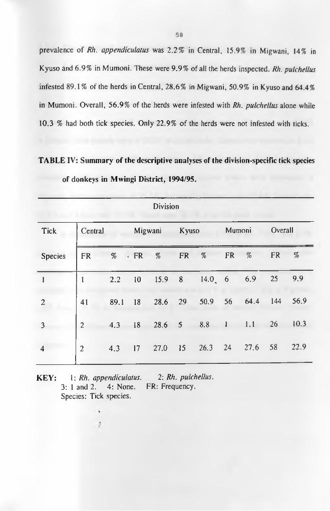
58
prevalence of Rh. appendiculatus was 2.2% in Central, 15.9% in Migwani, 14% in
Kyuso and 6.9% in Mumoni. These were 9.9% of all the herds inspected. Rh. pulchellus
infested 89.1% of the herds in Central, 28.6% in Migwani, 50.9% in Kyuso and 64.4%
in Mumoni. Overall, 56.9% of the herds were infested with Rh. pulchellus alone while
10.3 % had both tick species. Only 22.9% of the herds were not infested with ticks.
TABLE IV: Summary of the descriptive analyses of the division-specific tick species
of donkeys in Mwingi District, 1994/95.
Division
Tick Central Migwani Kyuso Mumoni Overall
Species FR % FR % FR % FR % FR %
1 1 2.2 10 15.9 8 14.0 % 6 6.9 25 9.9
2 41 89.1 18 28.6 29 50.9 56 64.4 144 56.9
3 2 4.3 18 28.6 5 8.8 1 1.1 26 10.3
4 2 4.3 17 27.0 15 26.3 24 27.6 58 22.9
KEY: 1: Rh. appendiculatus. 2: Rh. pulchellus.3: 1 and 2. 4: None. FR: Frequency.Species: Tick species.

59
4.1.4. Distribution of gross lesioas of the skin and skin derivatives.
A summary of the analyses of the distributions of the gross lesions of the skin and
skin derivatives of donkeys in Mwingi District is shown in Table V. The lesions included
wounds, alopecia and overgrown hooves. The within-division wound prevalences, in
descending order, were 11.1 % in Migwani, 10.3% in Mumoni, 8.8% in Kyusoand 8.7%
in Central. Skin wounds were in 10.0% of all the herds. Alopecia was observed in 2.2%
of the herds in Central, 1.6% in Migwani, 3.5% in Kyuso and 5.7% in Mumoni.
Generally, 3.6% of the herds had alopecia. Overgrown hooves were commonest in
Migwani, with a prevalence of 38.1%, followed by Mumoni with 27.6%, Central with
23.9% and Kyuso with 19.3%. These were 28.0% of all the herds visited.
A combination of wounds and alopecia affected 6.5%, 4.8%, 7.0%, and 9.2%
of the herds in Central, Migwani, Kyuso and Mumoni respectively. Overall, this
combination was observed in 7.2% of all the herds studied. The prevalence of a%
combination of overgrown hooves and wounds was 10.9% in Central, 1.6% in Migwani,
3.5% Kyuso, 2.3% in Mumoni and 4.0% in the whole of Mwingi District. Twenty
percent of the donkeys with overgrown hooves were lame due to either foot rot or hoof
"slippers".
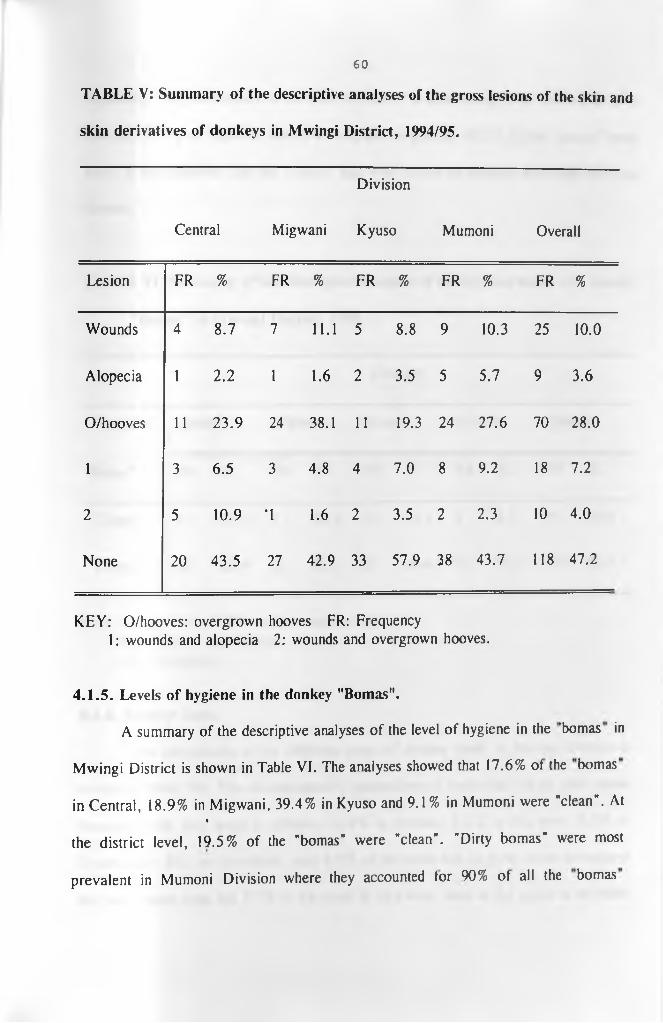
60
TABLE V: Summary of the descriptive analyses of the gross lesions of the skin and
skin derivatives of donkeys in Mwingi District, 1994/95.
Division
Central Migwani Kyuso Mumoni Overall
Lesion FR % FR % FR % FR % FR %
Wounds 4 8.7 7 11.1 5 8.8 9 10.3 25 10.0
Alopecia 1 2.2 1 1.6 2 3.5 5 5.7 9 3.6
O/hooves 11 23.9 24 38.1 11 19.3 24 27.6 70 28.0
1 3 6.5 3 4.8 4 7.0 8 9.2 18 7.2
2 5 10.9 T 1.6 2 3.5 2 2.3 10 4.0
None 20 43.5 27 42.9 33 57.9 38 43.7 118 47.2
KEY: O/hooves: overgrown hooves FR: Frequency1: wounds and alopecia 2: wounds and overgrown hooves.
4.1.5. Levels of hygiene in the donkey "Bomas".
A summary of the descriptive analyses of the level of hygiene in the 'bomas in
Mwingi District is shown in Table VI. The analyses showed that 17.6% ot the bomas
in Central, 18.9% in Migwani, 39.4% in Kyuso and 9.1% in Mumoni were "clean". At %
the district level, 19.5% of the "bomas" were "clean". "Dirty bomas" were most
prevalent in Mumoni Division where they accounted tor 90% ot all the bomas

61
inspected. The prevalences in the other divisions, in descending order, were 82.4% in
Central, 81.1% in Migwani and 60% in Kyuso. In general, 80.5% of the "bomas" were
dirty. It was observed that the "bomas" that were shared by donkeys and cattle were the
dirtiest.
TABLE VI: Summary of the descriptive analysis of the hygiene status of donkey
"Bomas” in Mwingi District, 1995.
Division
"Hygiene Central Migwani Kyuso Mumoni Overall
Status" FR % FR % FR % FR % FR %
"Clean" 6 17.6 *7 18.9 13 39.4 5 9.1 31 19.5
"Dirty" 28 82.4 30 81.1 20 60.6 ' 50 90.0 128 80.5
KEY: Clean: "boma" did not have manure in it. Dirty: "boma" had manure.
FR: Frequency.
4 .1 .6 . Donkey feeds.
The distribution of the different types of donkey feeds in Mwingi District is
shown in Table VII. The division-specific proportions ot herds that fed on grass alone
throughout the year were as follows: 32.4% in Central, 5.1% in Migwani, 6.1% in
Kyuso and 1.8%. On the whole, only 9.9% of the herds fed on grass alone throughout
the year. Apart from the 7.7% of the herds in Migwani, none ot the herds in the other
62
divisions fed on shrubs alone. Grass and farm crop by-products provided feed to 61.8%
of the herds in Central, 87.2%-in Migwani, 18.2% in Kyuso and 23.6% in Mumoni
divisions, and to 46% of the herds in the entire district. The division-specific proportions
of herds that fed on combinations of grass, farm crop by-products and shrubs were 5.9%
in Central, 75.8% in Kyuso and 74.5% in Mumoni. This combination was not used in%
Migwani, although 42.2% of the herds in Mwingi District fed on it.
At the district level, 51.6% of the study herds had "adequate" feed throughout the
year. These included 58.8% of the herds in Central 16.7% in Migwani, 24.2% in Kyuso
and 83.6% in Mumoni divisions. In contrast, 48.4% of the herds had minimal feed
particulary during dry seasons. Within the divisions, 41.2% of the herds in Central,
83.3% in Migwani, 75.8% in Kyuso and 16.4% in Mumoni did not have adequate forage
throughout the year.

63
TABLE VII: Summary of the descriptive analyses of donkey feeds in Mwingi
District, 1995.
Division
Feed Central Migwani Kyuso Mumoni Overall
Type FR % FR % FR % FR % FR %
1 11 32.4 2 5.1 2 6.1 1 1.8 16 9.9
2 - - 3 7.7 - - - - 3 1.9
3 21 61.8 34 87.2 6 18.2 13 23.6 74 46.0
4 2 5.9 - - 25 75.8 41 74.5 68 42.2
Adequate 20 58.8 * 6 16.7 8 24.2 46 83.6 80 51.6
Minimal 14 41.2 30 83.3 25 75 /8 9 16.4 78 48.4
KEY: 1: Grass. 3: Grass and crop residues. FR: Frequency. 2: Shrubs 4: Grass, shrubs and crop residues.
4.1.6.1. Feeding management of donkeys.
Generally, twenty percent of the donkeys grazed freely, 67% were tethered while
13% were both tethered and grazed freely. In all the divisions, donkeys were left to fend
for themselves in the farms during the dry season following crop harvest. All the farms %
allowed young donkeys to graze with adults. The foals were not weaned until the dams
stopped suckling them.

64
Fifty two percent of the donkeys did not share forage with other large domestic
animal species. Twenty nine percent of the herds shared forage with both cattle and goats
while 15 % and 4% shared with only cattle and goats respectively. Ninety four percent
of the farms did not give mineral supplements while 6 % irregularly gave common salt.
4.1.7. Sources of water for donkey herds.
The sources of water for donkeys in Mwingi District are summarised in Table
VIII. These included watering holes, earth dams, rock catchment areas, bore holes and
combinations of all these. Watering holes were sources of water to 85.3% of the herds
in Central, 73% in Migwani, 22.6% in Kyuso and 98.2% in Mumoni. On the whole,
74.5% of the herds in Mwingi District got water from watering holes. Earth dams
provided water to 5.9% of the herds in Central, 18.9% in Migwani and 3.2% in Kyuso.
None of the herds in Mumoni got water from earth dams. At the district level, 6.4% of%
the herds got water from earth dams. The herds whose water source were bore were
2.9% in Central, 8.1% in Migwani and none in Kyuso and Mumoni divisions. Bore hole
water was used by 2.5% of all the visited herds. Water from rock catchment areas was
used by 5.1% of all the study herds. They were all from Kyuso Division.
Combinations of watering holes, earth dams, boreholes and rock catchment areas
were sources of water to 5.9% of the herds in Central, 48.4% in Kyuso and 1.8% in
Mumoni. These combinations provided water to 11.5% of the herds m Mwingi District.
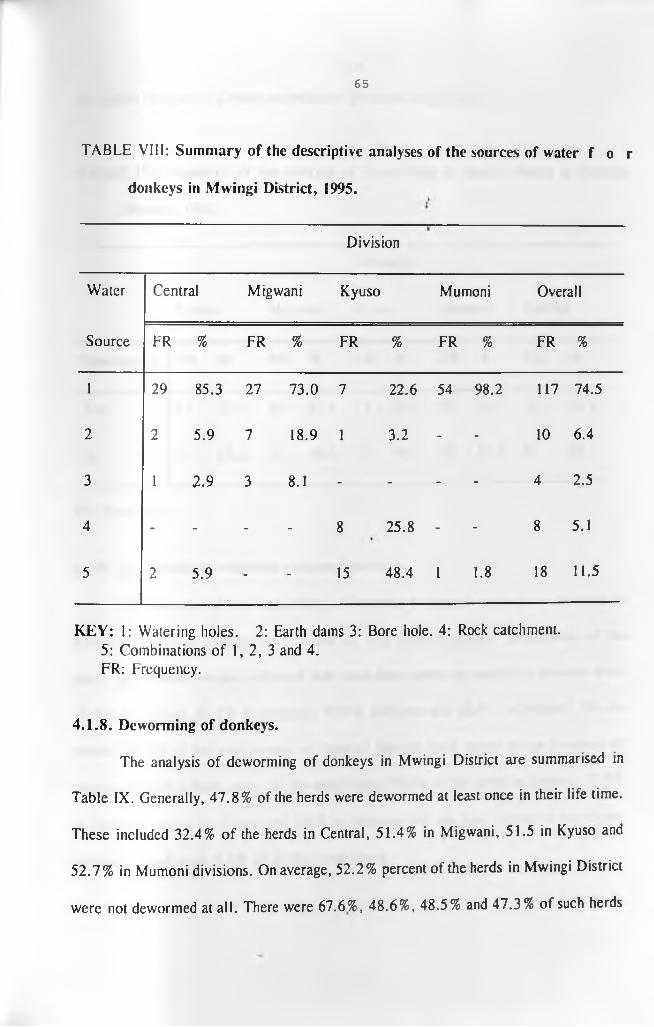
65
T A B L E V I I I : Summary of the descriptive analyses of the sources of water f o r
donkeys in Mwingi District, 1995.
Division
Water Central Migwani Kyuso Mumoni Overall
Source FR % FR % FR % FR % FR %
1 29 85.3 27 73.0 7 22.6 54 98.2 117 74.5
2 2 5.9 7 18.9 1 3.2 - - 10 6.4
3 1 2.9 3 8.1 - - - - 4 2.5
4 - - - - 8 25.8 - - 8 5.1
5 2 5.9 - - 15 48.4 1 1.8 18 11.5
KEY: l: Watering holes. 2: Earth dams 3: Bore hole. 4: Rock catchment. 5: Combinations of 1, 2, 3 and 4.FR: Frequency.
4.1.8. Deworming of donkeys.
The analysis of deworming of donkeys in Mwingi District are summarised in
Table IX. Generally, 47.8% of the herds were dewormed at least once in their life time.
These included 32.4% of the herds in Central, 51.4% in Migwani, 51.5 in Kyuso and
52.7% in Mumoni divisions. On average, 52.2% percent of the herds in Mwingi District
were not dewormed at all. There were 67.6%, 48.6%, 48.5% and 47.3% of such herds

66
in Central, Migwani, Kyuso and Mumoni divisions respectively.
TABLE IX: Summary of the analysis of deworming of donkey herds in Mwingi
District, 1995.
Division
Central Migwani Kyuso Mumoni Overall
Deworming FR % FR % FR % FR % FR %
Yes 11 32.4 19 51.4 17 51.5 29 52.7 76 47.8
No 23 67.6 18 48.6 16 48.5 26 47.3 83 52.2
FR: Frequency.
4.1.9. Availability of veterinary support services.
The analysis of the availability of veterinary support services to donkeys in
Mwingi District is summarised in Table X. The division-specific proportions of the
study herds whose owners indicated that could have access to veterinary services were
79.4% in Central, 89.2% in Migwani, 93.9% in Kyuso and 96.4% in Mumoni. On the
whole, 90.6% of the herd owners in Mwingi District were aware of the presence of
veterinary staff in their areas. On the other hand, 20.6% of the herds in Central, 10.8%
in Migwani, 6.1% In Kyuso and 3.6% in Mumoni did not have access to veterinary•*
services. These were 9.4% of all the study herds.

67
TABLE X: Summary of the analysis of the availability of Veterinary services to
donkey herds in Mwingi District, 1995.
Division
Central Migwani Kyuso Mumoni Overall
Vet. FR % FR % FR % FR % FR %
Yes 27 79.4 33 89.2 31 93.9 53 96.4 144 90.6
No 7 20.6 4 10.8 2 6.1 2 3.6 15 9.4
KEY: Vet: Availability of veterinary support services.Yes: Donkeys with access to veterinary services No: Donkeys without access to veterinary services.FR: Frequency.
4.1.10. Breeding of donkeys.%
There was no donkey breeding programme at all in Mwingi District. The donkeys
bred randomly at watering holes, in markets and in "bomas". The age-at-first-breeding
for 22% of the surveyed herds ranged from 2 to 10 years. Sixty eight percent of the herd
owners did not know the age at which they first bred their donkeys nor the average
gestation periods of these herds. Furthermore, 43% ot the herds had a foaling-to-first-
conception interval of between two to sixty months. Donkeys in 34% ot the herds took
thirty six months to conceive after foaling.

68
4.1.11. Uses of donkeys.
The major uses of donkeys in Mwingi District are summarised in Table XI.
Generally, 85.7% of the herds were used for transport only. The corresponding division-
specific proportions were 96.9% in Central, 82.4% in Migwani, 89.6% in Kyuso and
81% in Mumoni. The donkeys mainly transported water for domestic use and for sale
in towns and shopping centres. The other items transported included sand and stones for
construction purposes, farm produce such as maize corn, green grams, sorghum, cassava,
mangoes and sugar canes, firewood, charcoal, as well as sick people to health centres.
One percent of all the donkeys were used in ploughing alone and were all from Mumoni
Division.
Within the divisions, 17.6% of the herds in Migwani and 16.5% in Mumoni were
used for both transport and ploughing. These were 10.5% of the total number of herds
surveyed. Overall, 0.5% of the herds were used for transport and breeding.%
Thirty nine percent of the farms used donkey manure in the gardens. Dry donkey
dung was used to light fire and to smoke away bees and poultry ectoparasites. The
filtrate from faecal matter boiled in water was used to "treat" fever and oedema in
children. Sixty one percent of the farms did not have any use for the manure. Five farms
in Kyuso Division bred donkeys for sale and for paying dowry.
t

69
TABLE XI: Summary of the analysis of the functions of donkeys in Mwingi District,
1995.
Division
Central Migwani Kyuso Mumoni Overall
Function FR % FR % FR % FR % FR %
1 31 96.9 42 82.4 43 89.6 64 81.0 180 85.7
2 - - - - - - 2 2.5 2 1.0
3 - - 9 17.6 - - 13 16.5 22 10.5
4 1 3.1 - - 5 10.4 - - 6 2.9
KEY: 1: Transport 3: Transport and ploughing2: Ploughing 4: Transport and breeding for sale.
FR: Frequency.
The summary of the analysis of the estimated daily loads, in kilogrammes, carried
by donkeys in Mwingi District is shown in Table XII. In general, 33.8% of the donkeys
carried forty or less kilogrammes per day. These included 21.9% of the herds in Central,
54.2% in Migwani, 28.9% in Kyuso and 28.6% in Mumoni. On the other hand, 59.4%
of the herds in Central, 43.7% in Migwani, 46.6% in Kyuso, 28.6% in Mumoni and
41.6% in the whole district carried luggage which weighed between 40 and 80 Kgs. %
Mumoni Division had the largest proportion (42.8%) of herds that carried loads ranging
between 80 and 120 Kgs, followed by Kyuso with 17.8%, Central with 3.1% and

70
Migwani with 2.1 %. At the district level, 20.5% of the donkeys carried loads within this
weight category. Only Central <15.6%) and Kyuso (6.7%) divisions had donkeys that
carried more than 120 Kgs daily. These were only 4.1% in the district.
TABLE XII: Summary of the analysis of the distribution of the daily loads (Kgs)
carried by donkeys in Mwingi District, 1995.
Division
Central Migwani Kyuso Mumoni Overall
Load(kgs) FR % FR % FR % FR % FR %
10-40 7 21.9 26 54.2 13 28.9 20 28.6 66 35.8
41-80 19 59.4 21 43.7 21 46.6 20 28.6 81 43.3
81-120 1 3.1 1 2.1 8 17.8 ' 30 42.8 40 20.5
> 120 5 15.6 - - 3 6.7 - - 8 4.1
KEY: FR: Frequency.
4.2. RESULTS OF LABORATORY ANALYSES.
4.2.1. Skin scrapings.
None of the ,22 skin scrapings was positive for mange. Twenty one out of the
twenty two (95.5%) skin scrapings were positive for fungal growth. Seventeen of the

71
donkeys that were less than two years of age.
4.2.2. Faecal samples.
4.2.2.1. Faecal egg count.
A summary of the frequency distribution of the number of eggs per gram (epg)
of faeces categorised into 0 to 500 and more than 500 is shown in Table XIII.
Donkeys with egg counts that were less than 500 per gram of faeces were 4.7% in
Central, 4.2% in Migwani, 25% in Kyuso, 17.6% in Mumoni and 13.3% in the
whole district. Using epg values greater than 500 as an indicator of nematodiasis, it
was observed that helminthiasis was commonest in Migwani Division with a
prevalence of 95.7%. The prevalence in the other divisions was as follows: 95.3%
in Central, 82.4% in Mumoni and 75% in Kyuso. It was 82.7% at the district level.

72
TABLE XIII: Summary of the percent frequency distribution of the number of
nematode eggs per grain in Mwingi District, 1994.
Division
Epg Central Migwani Kyuso Mumoni Overall
0-500 4.7 4.2 25.0 17.6 13.3
>500 95.3 95.7 75.0 82.4 82.7
KEY: Epg: eggs per gram.
4.2.2.2. Characterisation of larvae from the donkey faecal samples.
Only larvae belonging to the genera Strongylus and Trichonema (Cyanthostome)
were recovered from the pooled samples from all the. divisions. There were no
differences in the types of strongyles infecting donkeys in the four divisions. The
intestinal cells of the larvae failed to stain adequately. Therefore, it was not possible
to characterise the larvae to species level. A summary of the results of the
characterisation of the larvae is shown in Table XIV. The larvae of Strongylus spp
were more, per microscopic field, than those of Trichonema spp at all but two levels
of epg. The two epg levels were epg > 1000 in Central and epg < 1000 in Migwani.
No larvae were recovered from the less than 1000 epg category from Central
Division.

73
TABLE XIV: Summary of the laboratory characterisation of nematode larvae from
donkey faecal samples pooled according to egg count and division of origin
in Mwingi District, 1994.
Division Epg category Strongylus larvae Trichonema larvae
Central <1000 - -
>1000 + + + + +
Migwani >1000 + + +
<1000 + + + + +
Kyuso <1000 + + +
>1000 + + + + +
Mumoni <1000 + + +
>1000 + + + + +
KEY: + : three or less larvae per microscope field.+ + : between four and eight larvae per microscope field.
+ + + : more than eight larvae per microscope field.Epg: Eggs per gram.
4.3. Results of statistical analyses.
4.3.1. Descriptive statistics and pair-wise comparisons of the division-specific
egg counts.
A summary of the descriptive statistics of the egg counts is shown in Table XV.

74
The division-specific egg counts ranged from zero to 17500 with a mean of 6265.1 for
the donkey herds in Central, zero to 18700 with a mean of 6006.1 in Migwani, zero to
28000 with an average of 3483.8 in Mumoni and zero to 13900 with a mean of 3060.5
in Kyuso. At the district level, the egg count per gram of faeces ranged from zero to
28000 with a mean of 4592.1. The median egg counts were less than the means of the
egg counts in all the divisions. The standard deviations about the mean egg counts were
large.
TABLE XV: Summary of the analysis of the overall and division-specific nematode
strongyle egg count per gram of faeces from donkeys in four divisions in
Mwingi District, 1994.
N a Mepg b S D C Range Median
Central 43 6265.1 4847.8 6.0 - 17500 5100
Migwani 49 6006.1 4569.2 0.0 - 18700 5300
Mumoni 68 3483.8 4949.4 0.0 - 28000 1500
Kyuso 43 3060.5 3234.9 0 .0 - 13900 2100
Overall 203 4592.1 4702.7 0.0 - 28000 3000
KEY: a: Number ,of donkeys sampled c: Standard deviation.b: Mean eggs per gram
The overall plot of egg counts per gram of faeces was skewed to the right

75
[Appendix V (a)]. Similarly, all the division-specific histograms of epg were skewed to
the right. The histograms for Central and Migwani divisions were less skewed than those
of Mumoni and Kyuso divisions. The Wilk-Shapiro/Rankit plot [Appendix V(b)J was
concave, with a level of 0.8328. A natural logarithm transformation normalised the data
[Appendix V(c)] and achieved a Wilk-Shapiro/Rankit level of 0.9802 [Appendix V(d)J.
The summary of the analysis of variance between the division-specific mean epgs
are shown in Table XVI(a). At five percent level of significance, the overall F-statistic
was highly significant (P = 0.0004). Thus, at least two of the means statistically differed
from each other.
TABLE XVI(a): Summary of the analysis of variance for division-specific mean
faecal egg counts from donkeys in four divisions of Mwingi District, 1994.
SOURCE DF SS MS Variance
ratio
P-value
Between Divisions 3 4.027 + 08 1.342+08 6.57 0.0004
Within Divisions 199 4.027+09 2.043+07
Total 202 4.467+09
DF: Degrees of Freedom, SS: Sums of Square.MS: Mean of square?, P-Value: Probability value.
A summary of the results of Tukey’s Pair-wise Honest Significant Difference
(HSD) test to determine which pair(s) of these means differed are shown in Table

76
XVI(b). There was no significant (p > 0.05) difference between the mean epg for
Central (6265.1) and that for Migwani (6006.1). Similarly, the mean epg for Kyuso
(3060.5) did not differ (p >0.05) from that of Mumoni (3443.8). However, each of the
means of Central and Migwani divisions differed statistically (P <0.05) from that of
Kyuso and Mumoni divisions.
TABLE XVI(b): Tukey’s pair-wise Comparisons of the division-specific means of
faecal egg counts from donkeys in four divisions in Mwingi District, 1994.
Absolute differences between Divisional means
Division Mean epg Central
Divsion
Migwani Mumoni Kyuso
Central 6265.1 - 259 2781.2* 3204.6*
Migwani 6006.1 - 2522.3* 2945.6*
Mumoni 3443.8 - 423.3
Kyuso 3060.5 -
* = These differences were significant at a= 0.05.
%4.3.2. Logistic regression.
t*The summary of the results of logistic regression analysis of presence of
nematodiasis in donkey herds on the study variables is shown in I able XVII. The

77
deviance associated with the model was 86.6, with 144 degrees of freedom and a
probability value of 1.0000. At 10% level of significance, the sex (male) of the owner,
the average age of the donkey herd (less than 2 years), deworming status (dewormed) of
the herd, level of hygiene (dirty) in the donkey "boma" and the division were associated
with nematodiasis. The herd size, availability of veterinary support services, the source
of water for the donkeys and availability of forage were not associated with nematodiasis.
The sex of the donkey owner, deworming of donkeys and the division were
negatively correlated with nematodiasis. On the other hand, the herd average age and the
level of hygiene in the "boma” were positively correlated with nematodiasis.
Belonging to a male person, having been dewormed and division had a protective
effect (O R < 1.0) against nematodiasis. Dirty "bomas" (OR = 3.92) had a strong causal
association with nematodiasis. The upper limit for the odds ratios associated with the
availability of veterinary services (23.87) and "boma" hygiene (OR = 15.67) were high%
while those associated with the size of the donkey herd (2.31) and availability of forage
(OR = 2.83) were marginal.
A backward stepwise logistic analysis based on change of deviance selected only
the sex of the owner, the age of the donkey, level of "boma" hygiene and the division.
Further statistical analysis revealed that donkeys which belonged to women were 14.58
(1.12, 189.7) times more likely to have nematodiasis than those owned by men. Donkeys
that were kept in dirty "bomas" were 5.07 times more at risk of having nematodiasis than %
those in relatively clean "bomas". Non-dewormed herds were 3.82 more likely to haver
nematodiasis than those that were dewormed. Compared to the donkeys in Kyuso

78
Division, the donkeys in Central Division were at the greatest risk (OR = 3768) of
nematodiasis followed by those in Migwani (OR = 14.5) and Mumoni (OR = 4.62).
Kyuso was used as the reference division because it had the lowest mean epg of faeces.
TABLE XVII: Summary of overall unweighted logistic regression of nematodiasis
on the study variables for Mwingi District.
Variables Est. SE. P-value. Odds ratio.
Constant 2.62989 2.26074 0.2447
Sex of donkey owner -2.21471 1.13270 0.0505 0.11 (0.01, 1.01)b
Number in the herd 0.37276 0.23723 0.1161 1.45 (0.91, 2.31)
Donkey age 0.14471 0.07032 0.0396 1.16 (1.01, 1.33)
Deworming status -1.05037 0.59384 0.0769 0.35 (0.11, 1.12)
Vet. availability 0.56536 1.35118
%0.6756 1.76 (0.12, 23.87)
"Boma" hygiene 1.36516 0.70757 0.0537 3.92 (1.34, 15.67)
Water source 0.02200 0.15650 0.8882 1.02 (0.75, 1.39)
Feed availability -0.40062 0.73491 0.5857 0.67 (0.16, 2.83)
Division -0.76838 0.38093 0.0437 0.460 (0.22, 0.98)
Deviance: 86.60; P = 1.000. 144 degrees o f freedom.
K E Y : Est.: Estimate of the variable. SE: Standard error.P-value: Probability.Value.Division: Division where the donkey herd was located, b: 95 % confidence interval about the odds ratio.
79
On the basis of absence of significant difference between the division-specific
means of epg, the data from Central were pooled with those from Migwani and those
from Kyuso with those from Mumoni. A logistic regression analysis was performed on
both subsets of data. The model for the combined Kyuso and Mumoni data (Appendix
VII) had the sex of the herd owner, the average age and the division of origin of the
donkeys as well as the source of water marginally (P < 0.1) associated with
nematodiasis. Donkeys from Mumoni Division were 6.64 times more likely to have
nematodiasis than those from Kyuso that were used as the references. Donkey herds
whose average ages exceeded two years were 2.3 times more at risk of nematodiasis than
younger herds. There was marginal risk (OR = 1.5) of nematodiasis to donkeys that
drunk water from watering holes and earth dams compared to those which got water
from boreholes and rock catchment areas.
The estimates of the parameters in the combined model for Central and Migwani%
(Appendix VIII) were large but not statistically (P > 0.05) related to nematodiasis.
Backward stepwise logistic regression revealed that availability of grass increased the
potential risk for nematodiasis by 22.6 times compared to when grass was scarce.
4.3.3. Discriminant analysis.
The results of the discriminant analysis are summarised in Table XVIII. At live
percent level of significance, there were significant (P < 0.05) differences between the
pairs of canonical variable means for Migwani and Central (6.85), Kyuso and Central
(26.55), Kyuso and Migwani (35.53), Mumoni and Central (8.16), Mumoni and Migwani
80
(17.14) and Mumoni and Kyuso (30.08). Five out of thirteen study variables entered the
discriminant function. These were herd size, type of feed, feed availability, source of
water and the faecal egg count. The intercepts for all the division-specific functions were
negative.
The results of the classification of the herds that had nematodiasis are summarised
in Table XIX. An overall percent correct classification of 67.1 percent was achieved. The
division-specific classification functions raised the prior probabilities for correct case
classification from 25%, which is expected by chance, to 44.1% in Central, 80.6% in
Migwani, 71.0% in Kyuso and 70.4% in Mumoni. The overall case misclassification was
thirty three percent. The misclassified cases from Central Division were distributed
almost equally between Migwani and Mumoni divisions.
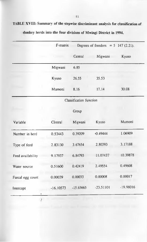
81
TABLE XVIII: Summary of the stepwise discriminant analysis for classification of
donkey herds into the four divisions of Mwingi District in 1994.
F-matrix Degrees of freedom = 5 147 (2.21).
Central Migwani Kyuso
Migwani 6.85
Kyuso 26.55 35.53
Mumoni 8.16 17.14 30.08
Classification function
Group
Variable Central Migwani Kyuso Mumoni
Number in herd 0.53443 0.39209 0.49444 1.06909
Type of feed 2.83130 3.67654 2.80393 3.17188
Feed availability 9.17937 6.84793 11.07437 10.39878
Water source 0.51600 0.42419 2.49554 0.49608
Faecal egg count 0.00039 0.00033 0.00008 0.00017
Intercept -16.10573 -15.63665 -23.51101 -19.98016
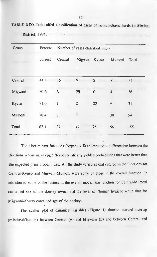
82
TABLE XIX: Jackknifed classification of cases of nematodiasis herds in Mwingi
District, 1994.
Group Percent
correct
Number of cases classified into
Central Migwan Kyuso
i
Mumoni Total
Central 44.1 15 9 2 8 34
Migwani 80.6 3 29 0 4 36
Kyuso 71.0 1 2 22 6 31
Mumoni 70.4 8 7 1 38 54
Total 67.1 27 47 25 56 155
The discriminant functions (Appendix IX) computed to differentiate between the
divisions whose mean epg differed statistically yielded probabilities that were better than
the expected prior probabilities. All the study variables that entered in the functions for
Central-Kyuso and Migwani-Mumoni were some of those in the overall function. In
addition to some of the factors in the overall model, the function for Central-Mumoni
contained sex of the donkey owner and the level of "boma" hygiene while that for
Migwani-Kyuso contained age of the donkey.
The scatter plot of canonical variables (Figure 1) showed marked overlapi
(misclassification) between Central (A) and Migwani (B) and between Central and

83
Mumoni (D) divisions. Kyuso Division (C) was well differentiated from the other
divisions. The positions of the mean values of the canonical variables for the four
divisions, that is, 1, 2, 3, and 4, are shown on the scatter plot. The mean value for
Kyuso (3) was farther away from those of the other three divisions, which were close to
each other.
Figure 1: A scatter plot showing the classification ofherd cases of nematodiasis in Mwingi District in 1994.
.... + ..... + ......+ ......+ ......+ ......+ ......+ ..
C3 + C
cm - C .D C ABBBB B*B0)pH
- C BBC2**BU3Cfl•H - C C C C3 A A BA**B« 0 > + cc c cc D 1**pHcfl - c B* *** Do•Ha - c c * D4D09 - AD*Do ★ ★
-3 +
+
+
+
.... + ..... + ...... + ......+ ......+ ......+ ......+ • •- 6 - 2 2 6
- 4 0 4
, Canonical variable 1
N O T E : Overlaps of different divisions are indicated by an asterisk!*) .KEY: A: Central, B: Migwani, C: Kyuso, D: Mumoni.

84
4.4. Evaluation of the effect of Ivermectin” on donkey gastro-intestinal nematodiasis
in Centra] and Kyuso divisions in 1995.
4.4.1. Pre-treatment faecal egg counts.
The frequency distribution of the status of nematodiasis in the pre-treatment
donkey herds is shown in Table XX(a). Nematodiasis (epg greater than 500) was
observed in 92.9% and 91.1% of the herds in Central and Kyuso respectively, with an
overall prevalence of 91.8%. On the other hand, 7.1 % of the herds in Central and 8.9%
in Kyuso were negative for nematodiasis. These accounted for 8.2% of all the herds in
this clinical trial.
TABLE XX(a): Distributions of the division-specific pre-treatment status of
nematodiasis based on the epg in the study herds in Central and Kvuso
divisions, Mwingi District, 1995.
Central
Division
Kyuso Overall
Nematodiasis FR % FR % FR %
Positive (epg >500) 39 92.9 51 91.1 90 91.8
Negative (epg 0-500 3 7.1 5 8.9 8 8.2
KEY: epg: eggs per gram. FR: Frequency.
The means and pair-wise comparisons of the means of epg for the donkey herds

85
in Kyuso and Central divisions that were surveyed in 1994 and later selected for
evaluation of the effectiveness' of IvermectinR are shown in Table XX(b). The pre-
treatment mean epg for donkeys in Central was 5826.3 while for those in Kyuso was
5160.0. The two means of epg were not statistically (P > 0.05) different. At five percent
level of significance, there was a statistical difference between the 1994 means of epg of
donkeys in Central and Kyuso divisions. The 1995 means of epg in the two divisions
were not statistically (P >0.05) different. Furthermore, the within-division mean epg
values for 1994 in both divisions were not significantly (P > 0.05) different from the
pre-treatment means of epg in 1995. However, at ten percent level of significance, the
1994 mean epg for Kyuso Division marginally differed from that of 1995.
TABLE XX(b): A summary of the pair-wise comparisons of the division-specific
means of epg for 1994 and 1995 for donkey herds in Central and Kyuso%
divisions, Mwingi District in 1995.
Mepg Central94 Central95 Kyuso95 Kyuso94
Central94 6923.5 - 1763.5 1097.2 4419.0*
Central95 5826.3 - 666.3 2655.5
Kyuso95 5160.0 - 3321.8
Kyuso94 2504.5 -
KEY: * : This difference was significant at a = 0.03.Mepg: Mean eggs per gram.

86
4.4.2. Drug efficacy
The fall in the means of egg counts per gram (epg) of faeces in Central and
Kyuso divisions following IvermectinR treatment is shown in Figure 2. The mean epg for
Central Division fell to under 200 in ninety percent of the donkeys by the sixth day. The
mean epg values fell at a fast rate during the first three days before they slowed down
over the following three days to near zero by the ninth day post-treatment. In Kyuso
Division, there was a fast rate of fall in mean epg in the first four days post-treatment.
The rate slowed down over the subsquent four days. By the fourth day following
treatment, only 20 percent of the donkeys had egg counts above 500.
The natural logarithms of the mean egg counts per gram (MEPG) for the pre
treatment (day 0) and post treatment donkey faecal samples from Central and Kyuso
divisions are shown in Table XXI. The log mean epg of donkeys in Central before
treatment was 7.8571 while that on day nine was 4.333. A pair-wise comparison of the%
log mean epg for before and days after treatment revealed that log MEPG pre-treatment
significantly (p <0.05) differed from log MEPG on days three, six and nine post-
treatment. Log MEPG on day three post-treatment did not differ significantly (P >0.05)
from log MEPG for days six and nine.

87
FIGURE 2: GRAPHICAL PRESENTATION OF EPG CHANGES IN DONKEYS OF CENTRAL AND KYUSO DIVISIONS OF MWINGI DISTRICT AFTER IVERMECTIN* TREATMENT IN 1995.
Days post-treatment.
“"'CENTRAL DIVISION +KYUS0 DIVISION
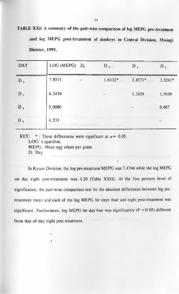
86
TABLE XXI: A summary of the pair-wise comparison of log MEPG pre-treatment
and log MEPG post-treatment of donkeys in Central Division, Mwingi
District, 1995.
DAY LOG (MEPG) D0 D , D , D ,
D„ 7.8571 1.6132* 2.8571* 3.5241*
D , 6.2439 - 1.2439 1.9109
d 6 5.0000 - 0.667
D , 4.333 -
KEY: * : These differences were significant at a= 0.05.LOG: Logarithm.MEPG: Mean egg count per gram.D: Day.
%
In Kyuso Division, the log pre-treatment MEPG was 7.4746 while the log MEPG
on day eight post-treatment was 4.20 (Table XXII). At the five percent level of
significance, the pair-wise comparison test for the absolute differences between log pre-
treatment mean and each of the log MEPG for days four and eight post-treatment was
significant. Furthermore, log MEPG for day four was significantly (P <0.05) different
from that of day eight post-treatment.
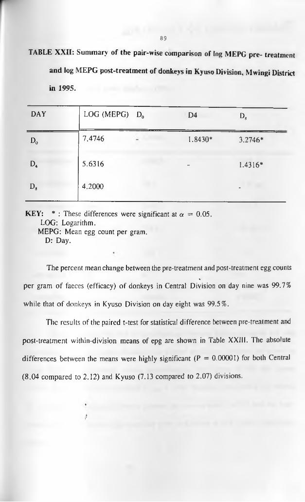
89
TABLE XXII: Summary of the pair-wise comparison of log MEPG pre- treatment
and log MEPG post-treatment of donkeys in Kyuso Division, Mwingi District
in 1995.
DAY LOG (MEPG) D0 D4 D,
D0 7.4746 1.8430* 3.2746*
d4 5.6316 - 1.4316*
D, 4.2000 -
KEY: * : These differences were significant at a = 0.05.LOG: Logarithm.
MEPG: Mean egg count per gram.D: Day.
The percent mean change between the pre-treatment and post-treatment egg counts%
per gram of faeces (efficacy) of donkeys in Central Division on day nine was 99.7%
while that of donkeys in Kyuso Division on day eight was 99.5%.
The results of the paired t-test for statistical difference between pre-treatment and
post-treatment with in-division means of epg are shown in Table XXIII. The absolute
differences between the means were highly significant (P = 0.00001) for both Central
(8.04 compared to 2.12) and Kyuso (7.13 compared to 2.07) divisions.

l Y Gr^NAiKOBl LIBRARY
TABLE XXIII: A student's paired t-test for the differences between the within
division pre-treatment and post-treatment means of faecal egg counts per
gram of faeces from donkeys, 1995.
90
Central
Division
Kyuso
Pre-treatment mean 6369.8 5028.3
Post-treatment mean 20.9 22.8
Test mean difference 6348.9 5021.1
Standard error 789.5 704.47
T-test value 8.04 7.13
Degrees of freedom 42 % 56
Probability value 0.00001 0.00001
The summary of the test of the differences between the pre-treatment and post
treatment median egg counts of donkeys in Central and Kyuso divisions are shown in
Table XXIV. The estimated normal approximations for Central (5.705) and Kyuso
(6.705) were greater than the critical Z (005) = 1.96. Therefore, there was a very strong
(P = 0.00001) statistical difference between the pre-treatment (MED,,) and the post-•*
treatment (MED*) median egg counts per gram in donkeys in both Central and Kyuso
divisions.
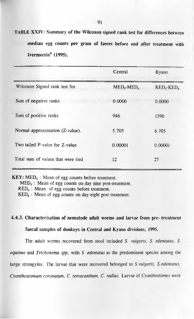
91
TABLE XXIV: Summary of the Wilcoxon signed rank test for differences between
median egg counts per gram of faeces before and after treatment with
IvermectinR (1995).
Central Kyuso
Wilcoxon Signed rank test for m e d 0-m e d 9 k e d 0-k e d 8
Sum of negative ranks 0.0000 0.0000
Sum of positive ranks 946 1596
Normal approximation (Z-value). 5.705 6.705
Two tailed P-value for Z-value 0.00001 0.00001
Total sum of values that were tied 12 27
KEY: MED0 : Mean of egg counts before treatment.MED9 : Mean of egg counts on day nine post-treatment.
KED0 : Mean of egg counts before treatment.KED8 : Mean of egg counts on day eight post treatment.
4.4.3. Characterisation of nematode adult worms and larvae from pre- treatment
faecal samples of donkeys in Central and Kyuso divisions, 1995.
The adult worms recovered from stool included S. vulgaris, S. edentatus, 5.
equinus and Trichonema spp, with S. edentatus as the predominant species among the
large strongyles. The larvae that were recovered belonged to S. vulgaris, S. edentatus,
Cyanthostomum coronatum, C. tetracanthum, C. radius. Larvae of Cyanthostomes were

92
predominant. The pool of samples that were negative for nematode eggs following
treatment did not yield any larvae even after incubation.
4.4.4. Ectoparasites.
All the donkeys examined in Kyuso Division and 42% from Central Division
were infested with Rh. pulchellus ticks. It was not possible to quantify the level of tick
infestation during the follow up period but it appeared to decrease markedly following
IvermectinR treatment.

93
CHAPTER FIVE
DISCUSSION AND CONCLUSIONS
Over eighty percent of the homesteads in Mwingi District had at least one donkey
each (Mwingi District livestock production office). However, the number of donkeys in
the district appeared to fall short of the local needs. This shortage was exemplified by
the high initial cost of acquiring donkeys, the constant and widespread overloading of the
available donkeys and the large number of people, mostly women and children, who still
carried heavy loads on their heads whenever they failed to hire donkeys.
The cost of a one-year old donkey was four thousand Kenya shillings while an
adult female could cost up to ten thousand shillings. Liquid cash was often difficult to
get. Thus, donkeys were mostly acquired through battering cattle. The unbalanced
donkey ownership ratio (2:1) between men and women in Mwingi District could be
attributed to the high cost of acquiring a donkey since most women did not have cattle
and therefore could not afford donkeys. Those women who owned donkeys mostly had
males because they were relatively cheaper to acquire. Financially poor homes could not
afford a donkey.
There was a tendency for relatively richer farmers with large pieces of land to
own more donkeys than the poorer ones. The increased need for donkeys on such farms
was mainly associated with increased farm workloads. This general observation was also
recorded by Wilson (1981) in sub-Saharan Africa. Kakuyu Location in Mumoni Division
94
had the largest percentage of herds with at least three donkeys each. This was because
the location had the poorest road network and received relatively more rainfall because
of its location on the windward side of the Mumoni hills. This high rainfall supported
more crop agriculture and hence the increased need for donkey transport to take farm
produce to homesteads and marketing centres.
Wilson (1981) recommended that the maximum load carried on backs of donkeys
should not exceed one third of the body weight of the donkey. Most adult donkeys in
Mwingi District weighed between 120 kgs and 150 kgs and carried at least 80 kgs of load
on their backs. This meant that the donkeys were overloaded. Similarly, Aluja and Lopez
(1991) had observed such overloading of donkeys in Mexico. Donkeys in Mwingi District
pulled carts whose weight exceeded that recommended by Inns (1980). The two possible
reasons why donkeys were often overloaded were that the donkey herds were relatively
small for the amount of work they had to do per day coupled with the owners avoiding
many trips since the distances to and from markets and/or water sources were usually
very long. Despite the fact that the majority of donkeys worked for two to three hours,
work overload coupled with extensive movement exerted considerable stress upon them.
Overloading and poor health have been reported to cause retarded growth, gradual
reduction in productivity, early withdrawal from work and early mortalities in donkeys
(Aluja and Lopez, 1991; Mohammed, 1991; Khallaayoune, 1991). Thus, these could
probably be the causes of reduced average donkey life expectancy, of less than ten years,%
recorded in the present study. This average life expectancy was relatively low compared
to thirty seven years reported for donkeys in the United Kingdom (Svendsen, 1989) and
95
eleven years in Turkey, Egypt, Jamaica, Peru and Ecuador (Svendsen etal., 1985 cited
by Bliss, 1989). In this study, - the significant division-specific mean donkey age
difference recorded in Mumoni and Migwani divisions was probably due to the relatively
short survival rates of donkeys in Khatse Location in Mumoni Division. A combination
of heavy helminthiasis, underfeeding and lack of access to veterinary support services
could explain the low donkey average life expectancy of seven years reported in Khatse
Location. The locational animal health assistant could not possibly cover much of the
area because o f logistic constraints, with transport as the major limitation.
The finding that 82.7% of the study herds had gastro-intestinal nematodiasis and
yet only 46.7% of them showed poor signs of health supported the observations by
Soulsby (1989) who noted that donkeys developed tolerance towards these gastro
intestinal nematodes. However., the lush grass and shrubs, which were plenty during the
rainy season when this cross-sectional survey was conducted, probably boosted the
nutritional status and hence the immunity of the donkeys to these parasites. This kept
many of them in average-to-good body conditions inspite of the heavy worm burdens and
stress from work overload.
All the donkeys in the district fed on grass throughout the year. However, forage
became scarce during the dry season and farm crop residues such as maize stover, maize
cobs and/or shrubs were fed to donkeys to offset the deficit. The high human settlement
in Central and Migwani divisions, compared to Kyuso and parts of Mumoni, reduced the *
communal grazing areas available to the donkeys. Thus, there were high proportions otr
herds with inadequate forage in the former divisions despite the high rainfall. The

96
scarcity of forage associated with dry seasons and lack of enough grazing areas could
explain the high proportion of donkey herds in Mwingi District reported to have
inadequate feed availability throughout the year. The fact that sixty percent of the herds
remained in average body condition during the dry season which followed when the
forage was very scarce and of poor quality, reflected the hardy nature of donkeys which
has already been reported by Dill et al. (1980).
Although most of the herd owners visited had on average owned and looked after
donkeys for over sixteen years, they all lacked the basic skills in donkey management
and husbandry. As a result, donkeys had severe wounds, heavy tick infestation and low
reproductive efficiencies. The latter were characterised by delayed ages at puberty, two
to ten years instead of the optimal 18 months (Weipers, 1978), long foaling-to-conception
(up to three years) intervals in thirty seven percent of the herds visited and low
percentages of herds having donkeys of two years and below. None of the surveyed
donkeys from Central Division was less than two years of age and this was further
evidence of low reproductive efficiencies and/or low survival of the young ones.
Aluja and Lopez (1991) and Mohammed (1991) had reported that inadequate
nutrition caused lowered reproductive performance in donkeys in Mexico and Ethiopia
respectively. Thus, in Kyuso Division, which received the lowest mean annual rainfall
and had the least amount of forage, donkeys foaled once in every five to six years
compared to those in Mumoni which had more forage and foaled every three to four
years. In Mwingi District, these health problems, especially nutritional deficiencies and
the related conditions, probably resulted from lack of awareness about donkey husbandry

97
and management principles such as proper feeding and health care. Kumwenda et al.
(1991) blamed ignorance to be responsible for the high prevalences of such preventable
diseases and conditions in donkeys in Malawi.
Although only 1.2% of the study donkey herds ii? Mwingi District were reported
by the owners to "commonly" have wounds, this was an understatement since as high as
21.2% ot the herds had wounds at the time of this survey. The wounds were mainly
(60%) caused by fights and biting among the donkeys during mating and when in market
centres. The other causes included poorly strapped, rough and heavy uncushioncd
luggage packed in synthetic sacs, scratching by thorny vegetation, tethering, cruelty and
malice. Poor loading methods were also reported by Yilma et al. (1991) to cause wounds
in donkeys in Ethiopia. Scratches caused by vegetation were common during the
harvesting season when the donkeys walked through narrow paths. The increased
prevalence of bite wounds during the rainy season (Mwingi District veterinary office)
could be attributed to redundancy since most donkeys did not work when the owners
tilled the gardens. The donkey wound problem occurred equally in all the divisions as
there were no significant differences noted in its prevalences.
Contrary to reports by Onyango (1990), tick infestation, particulary by
Rhipicephallus pulchellus, was present in donkeys in Mwingi District throughout the
year. The prevalence increased during the rainy seasons. However, the ticks were not
known to spread diseases among the donkeys (Mwingi District veterinary office-
Unpublished information). It was not possible to establish why tick prevalences in general
and of Rh. pulchellus in particular, in Central, Mumoni and Kyuso herds were higher
98
than that in Migwani herds.
Austwick (1972) associated ectoparasite bite wounds with infection with fungi.
This observation could explain the close similarities in the distribution of attachment sites
of ticks and alopecia lesions from which dermatophytes were recovered. It was also
possible that the breaks in the skin caused by scratches from thorny vegetation during and
soon after the rainy seasons created portals for fungal infection. The relatively higher
prevalence of Trichophyton species than the other fungal species in donkeys in Mwingi
District was in line with the findings by Blood and Radostits (1989) that Trichophyton
spp were the commonest fungi that affect donkeys. Fungal infections were predominantly
(90.5 %) observed in donkeys that were at least fifteen years of age. These findings agree
well with those of'Pascoe (1981) who reported that fungal infections were common in
unthrifty and old donkeys. The percent rate of 9.5 % infection in the younger, stunted and
unthrifty donkeys (less than two years) could be associated with the poorly developed
and/or compromised immune system (Blood and Radostits, 1989).
The relatively higher weighted prevalence of overgrown hooves in donkeys in
Mumoni Division compared to the prevalences in the other three divisions in Mwingi
District could not be explained from this study. However, the stony paths/roads could
have predisposed the donkeys to laminitis, resulting in overgrown hooves ( Blood and
Radostits, 1989).
Donkeys that were older than two years were given 15 to 20 litres of water per
day while the younger ones got half this amount. Only one percent of tne herds surveyed
had unlimited access to water sources. Water from river beds and bore holes was salty

99
while that from earth dams was very dirty and sometimes smelly. This meant that most
donkeys did not have enough water to drink.
Donkeys in Mwingi District were used for transport, ploughing and breeding. Asi
for ploughing, the donkeys cleared only one third of an acre per day due to poor health
resulting from underfeeding, heavy parasitism and overwork. Some of the donkeys would
refuse to plough to nib grass. Furthermore, the donkeys first fetched overloads of water
from long distances before they ploughed. Donkeys were muzzled to prevent them from
grazing during work. This restriction further stressed the donkeys and reduced their work
output. There was a gradual reduction in work output which started when the donkeys
reached twelve years of age.
The high prevalence of nematode eggs recorded during the cross-sectional survey
and the follow up later compared very well with the observations by Falvey (1985),
Wells (1985) and Bolbol and Saleh (1987), that helminthiasis was a real and constant
threat to the health of working donkeys. Inspite of Mwingi District being dry for the
greater part of the year, there was a general build up of gastro-intestinal nematodes in
the donkeys. The likely reasons for this build up included failure to deworm the infected
herds which contaminated the environment and, as Wakelin (1984) and Pandey and
Eysker (1989) reported, became constant sources of re-infection to the donkeys, poor
hygiene and very low levels of nutrition (Scot, 1988; Mattioli et al., 1994).
Kyuso Division, which received low rainfall, had a relatively low level ot
infection as indicated by the low mean of the epg. These results seemed to be consistent
with the observations made earlier by Round (1968), English (1979b) and Soulsby (1989)
100
that desiccation promoted death of free-living infective larvae of gastro-mtest.nal
nematodes and hence led to low levels of infection.
The high prevalence of nematodiasis in donkeys within fifteen to twenty
kilometres from the divisional/locational headquarters of the study areas suggested a
worse picture of the disease in the deeper areas of Mwingi District. The reasons to
support this hypothesis included poor infrastructure and lack of transport which rendered
some donkey herds almost inaccessible to animal health workers, the general lack of
interest/adequate training in donkey health among the local veterinary staff, ignorance
among the herd owners that donkeys could suffer from heavy inapparent worm burdens
and the belief amongst them that donkeys could never heal even after treatment.
Since this was a prevalence study, it was not possible to establish the patterns of
passing out of gastro-intestinal nematode eggs from infected donkeys, for the same
reason, it was not possible to establish the actual risk factors which could explain the
statistical differences observed between the means of the egg counts in C cntral and
Migwani divisions and those from Kyuso and Mumoni divisions. This was because the
levels of donkey management and husbandry practices in all the divisions were s irtually
the same. However, the high amount of rainfall in Central and Migwani divisions which
favoured development of more infective nematode larvae could account for these
differences. Similar hypotheses have been advanced by Hansen and ferr\ (1^90) a
Hutchinson et al (1989). It is worthy noting that the means of epg of the di\ isions
received almost the same amount of rainfall per year did not differ sta
Furthermore, there were more herds with high egg counts in Kakuvu Loca

received more rainfall than in Khatse Location, which was in a rain shadow. However,
a well designed prospective study is required to verify this observation.
The large standard deviations about the means of epg which were observed in this
study were consistent with the over dispersion of helrrpnth infections in populations as
suggested by Wakelin (1984). The skewness to the right in the overall and division-
specific distributions of epgs bore out the findings by Anderson and May (1980) that the
majority of parasites in natural parasitic infections tended to occur in a few hosts and a
few parasites to occur in the majority of hosts in populations.
Since postmortem examination was not done, it was not possible to establish why,
within the same herds, some donkeys had egg counts of zero while others had very high
epg values. However, Round (1968), Dunn (1978), and Soulsby (1989) have suggested
that this could be due to one or a combination of factors. These factors include
fluctuations in worm egg output, presence of non-egg-laying stages of worms, differences
in individual susceptibility as well as in the consistency and volume of faeces passed.
Furthermore, Round (1968) found out that these differences could be caused by the "time
period over which the eggs were allowed to float in the McMaster slide".
Strongyloides and Parascaris spp eggs were observed in faeces of foals and not
in faeces from adult donkeys. This differential age susceptibility to these nematodes
agreed with the results described by Lyons et al. (1973). Similarly, the diversity ot
Strongylus and Trichonema spp recorded in this study was comparable to that reported
in Chad (Graber, 1970), Zimbabwe (Pandey and Eysker, 1989) and Morocco
(Khallaayoune, 1991). The finding of these gastro-intestinal nematode species in donkeys
101

102
in all the four divisions of Mwingi District was in line with observations by Fowler
(1989), Sewell (1991) and Yilma et al. (1991) that donkey nematodes, particularly
Cyanthostomes, did not generally differ much over wide geographical areas.
Correctively, the higher numerical representation of Strongylus spp than Trichonema spp
observed following incubation of faeces was similar to what Yilma etal. (1991) reported
in donkeys in Ethiopia. Although Trichostrongylus was not obtained in this survey, it has
been reported to commonly occur in donkeys in Zimbabwe (Pandey and Eysker, 1991).
The positive associations obtained between gastro-intestinal nematodiasis and each
of donkey age and dirty "boma" showed that if all the other factors in the model were
held constant, nematodiasis increased by 0.14471 and 1.36516 for a unit increase in
donkey age and level of dirtiness of the "boma" respectively. The marginal potential risk
(OR = 1.89) of nematodiasis in donkeys whose average age was at least two years
compared to the younger ones was consistent with the findings by Round (1968) and
Duncan (1974) that equine strongyle infections were not age specific.
Donkeys kept in unhygienic "bomas" were 3.92 times more likely to suffer from
gastro-intestinal nematodiasis than those in relatively clean "bomas". This compared well
with the findings of Dunn (1978), Herd (1986a) and Hutchinson et al. (1989) who
concluded that keeping equine in dirty premises predisposed them to nematodiasis. In this
study, sixty five percent of the farms visited did not clear donkey premises ot manure
while only thirty five percent did so irregularly and inadequately. The manure was piled %
at the bases of the ."boma" hedges. This manure was probably the major source oti*
nematode infections to donkeys since Thomas (1982) and Mattioli et al. (1994) reported
103
ihat accumulated faecal material promoted development and survival of nematode larvae.
Although the movements of the nematode larvae from the filth in the "bomas" to the lush
vegetation along the perimeter of the "bomas" were not examined in this study, it was
likely that infective larvae climbed and quested for donkeys on this vegetation. It is
highly probable that donkeys got infected when they grazed on this vegetation.
Deworming donkeys was negatively associated with nematodiasis. This inverse
relationship was expected since proper deworming of donkeys has been reported to
reduce contamination of pasture with nematode eggs (Dunn, 1978; Herd, 1986a,b). This
in turn reduced the risk of infection and re-infection of donkeys. In this survey, donkey
herds that were not dewormed were 3.82 times more likely to suffer from gastro
intestinal nematodiasis than those that were dewormed. Lack of deworming as a risk
factor for equine nematodiasis is well documented by Dunn (1978), Herd (1986b),
Hutchinson et al. (1989) and Fowler, (1989). However, forty eight percent of all the
surveyed herds were dewormed and yet 82.7% of them had gastro-intestinal
nematodiasis (epg>500). Similarly, Migwani, Kyuso and Mumoni divisions had high
prevalences of nematodiasis despite the high percentage of herd owners who indicated
that they dewormed their donkeys. This unexpected scenario could be attributed to
irregular deworming, underdosing and/or drug resistance. Donkeys were only dewormed
when they looked sick and/or passed worms. Yet, it was observed during this survey that
several donkeys carried heavy worm burdens without necessarily looking unhealthy or
passing worms in the faeces. Furthermore, only twenty percent of the herds were«'
dewormed by animal health staff while the other eighty percent were treated by the

104
owners using drugs bought from the veterinary staff or from local drug shops. This
meant that many donkeys were probably underdosed since their body weights were not
properly estimated nor were their conditions assessed before treatment.
The significance of donkeys belonging to women as a potential risk factor (OR
= 14.6) for nematodiasis was possibly a proxy for poor hygiene in the "bomas" and/or
inadequate grazing areas fro the donkeys. This is because it was observed during this
survey that such donkeys lacked enough grazing areas and were therefore rotationally
tethered in the same ''soiled'' areas after very short rest periods. This probably created
areas of intense larval challenge and hence the associated high risk for nematodiasis.
The positive associations that were observed between gastro-intestinal
nematodiasis with each of Central, Migwani and Mumoni, while negatively associated
with Kyuso, implied that donkeys in these divisions had increased levels of nematodiasis
compared to those in Kyuso. This was also reflected by the high risk potential for
nematodiasis for herds in Central (OR = 3768), Migwani (OR = 14.5) and Mumoni
(OR = 4.62) compared to those in Kyuso Division. Similarly, donkey herds in Mumoni
Division were 6.64 times at risk of nematodiasis than those in Kyuso. These observations
could possibly be explained by the differences in climatic factors, particulary rainfall,
between the divisions. Further studies need to be designed to delineate the ettects of
these hypothesised factors on gastro-intestinal nematodiasis.
The probability values associated with each of herd size, availability of veterinary
support services and,feed were insignificant while the odds ratios for nematodiasis had
wide confidence intervals with significant upper limits. The latter suggested a strong

simple biological association between these factors and nematodiasis in the herds.
Thomas (1982) reported high stocking density to be a risk factor for helminthiasis in
animals (such as donkeys) which passed fibrous faecal material. The faecal material took
long to disintegrate completely and thus, sheltered the infective larvae and favoured their
survival. This could explain the observed positive association (OR = 1.49 (0.91,2.31)]
between herd size and nematodiasis.
The positive correlation between availability of veterinary support services and
nematodiasis suggested that as the services became available, the level of nematodiasis
increased. This was logical since eighty percent of the herd owners who knew about the
availability of these services in the areas were either not aware that the veterinary staff
could attend to donkeys or they could not afford the veterinary charges. Moreover, most
owners sought veterinary assistance only when the donkeys passed worms and/or were
visibly unthrifty. Furthermore, some of the animal health staff lacked adequate and%
effective skills in treating donkey diseases. Thus, there was no effective veterinary
attention towards the donkeys.
The high potential risk (OR = 22.6) for nematodiasis in herds in Central and
Migwani divisions when grass was plentiful (during the rainy seasons) probably reflected
the protection provided by grass cover, to the infective larvae against the harmful ultra
violet rays from the sun, which increased their survival (English, 1979a). By grazing on
this grass, the donkeys were at a high risk of getting gastro-intestinal nematodiasis.
Although a good level of classification of the case herds was achieved int
discriminant analysis, there was an overlap of donkey herds from Central, Migwani and
105

106
Mumoni. This suggested that these herds shared some of the study characteristics. Since
ail the components of the discriminant functions were either directly or indirectly related
to climatic factors, particulary rainfall, it seemed reasonable to conclude that the clear
differentiation of Kyuso from the other divisions showed that it had peculiar variables.
For example, it received the least amount of rainfall. The similarity of climatic factors
for Kyuso Division and Khatse Location in Mumoni Division could probably explain the
six herds from Kyuso which were misclassified into Mumoni Division.
Evaluation of Ivermectin*-
During the follow-up of the herds initially surveyed in Mwingi District, an
evaluation of the effectiveness of IvermectinR against gastro-intestinal nematodes was
carried out in Central and Kyuso divisions. Low responses of forty one percent in Kyuso
Division and forty four percent in Central Division, of the previous contact donkey herds
were achieved. This was either due to lack of proper flow of information or the herd
owners were working in their gardens since it was the planting season. In addition, four
previous contact donkeys died of unconfirmed causes. They all had very high faecal egg
counts at the time of the initial survey.
It was not possible to establish whether the Ascarid, tape worm as well as the
Gasterophillus spp infections observed in the pre-treatment donkeys were missed at the
tim e o f the initial cross-sectional survey or were acquired thereafter. The 2% prevalence
o f Gasterophilus infection that was observed in the present study was much lower than
the 97.5 % reported by Pandey et al. (1992) in Morocco.

107
Nematodiasis was a bigger problem in the pre-treatment donkeys in Central
Division than in those in Kyuso Division. This was indicated by the higher pre-treatment
overall mean of epg for donkeys in the former than in the latter division. This was
despite the lower number of donkeys that was sampled in Central Division.
The lower mean egg count per gram of faeces for Central Division after eleven
months following the initial survey probably resulted from the awareness that was created
among the herd owners. This is because there was an increased tendency to deworm after
the owners had been shown worms in the donkey faeces during the initial survey and/or
received the results of the laboratory faecal analysis. The significant (P < 0.1) increase
in the mean epg for Kyuso over the same period, despite similar sensitisation, could not
be explained. However, the higher amount of rainfall (Mwingi District agriculture office)
that was received compared.to the previous year probably favoured increased survival
o f the infective larvae and hence the higher levels of gastro-intestinal nematodiasis.%
The high efficacy of IvermectinR against egg-laying gastro-intestinal nematodes
observed in this study was similar to that reported by Pereira et al. (1991) and Lihua et
a t. (1994). However, the average 8.5 days it took the epg to fall below 100 in this study
w as markedly less than the 8.9 weeks reported by Lihua et al. (1994). This faster action
o f IvermectinR was due to lack of previous exposure of the gastro-intestinal nematodes
in Mwingi District to the drug. This in turn rendered them fully susceptible to
Iverm ectinR. The passing of whole parasites in faeces within three days following %
treatm ent agreed with earlier observations by Slocombe and Cote (1984).
T he uneven rate of fall of mean epg post-treatment in Central and Kyuso divisions

r
could either be due to differences in the rate at which effective levels of the drug diffused
into the gut lumen to paralyse and dislodge the egg-laying nematodes (Merck veterinary
manual, 1986) or due to the frequency of faecal sampling. Differential susceptibilities of
the nematodes to the drug were not likely causes since the worms had not previously
been exposed to IvermectinR.
The similar effectiveness of lvermectinR against the gastro-intestinal nematodes
at the division level meant that its efficacy was not affected by the differences in
husbandry and management levels as well as in geographical factors between the two
divisions. The lack of difference in efficacy at the herd level was probably because the
gastro-intestinal nematodes were IvermectinR-naive.
It was not possible to follow up donkeys for long enough to find out whether
IvermectinR was effective against encysted Cyanthostome larvae. For, whereas Love et
al. (1995) found Cyanthostome larvae fully susceptible to ivemectinR, Eysker et al.%
(1992), Klei et al. (1993) and Lihua et al. (1994) reported resistance of these larvae to
the normal and higher therapeutic doses of IvermectinR.
None of the donkeys was sacrificed to find out whether or not there were gut
luminal strongyles that had resisted the drug. However, since the pooled post-treatment
faecal samples that were negative for worm eggs did not yield any larvae on incubation,
it could be concluded that IvermectinR probably cleared all the egg-laying nematodes.
The large and small gastro-intestinal nematode species that were observed in the %
present study were similar to those Ngatia and Kuria (1991) recovered from donkeys at
K abete, Kenya. The predominance of S. edentatus among the large strongyles had also
108

109
been reported in Zimbabwe (Pandey and Eysker, 1989). Contrary to an earlier study by
Love et al. (1992) in the United kingdom, none of the pre-treatment herds with gastro
intestinal nematodiasis in the present study had diarrhoea although Cyanthostomes
predominated. Considering the high epg values and the potentially fatal lesions that were
previously observed by Ngatia et al. (1991) in donkeys infected with similar parasites,
the absence of diarrhoea did not mean that the donkeys in Mwingi District were any
safer.
CONCLUSIONS.
Within the limits of the present study, the diseases/conditions that afflicted
donkeys in Mwingi District were ranked in descending order as helminthiasis,
underfeeding, tick infestation, skin wounds, overgrown hooves and fungal infections. It
w as not possible to establish the effects of S. edentatus, S. vulgaris, S. equinus,%
Cyanthostome coronatum, C. trancathum, C. radius, Strongyloides, Ascarid spp.
Gasterophilus intestinalis, coccidial oocyst, tape worms, Rh. pulchellus and Rh.
appendiculatus on the health and productivity of the donkeys in the district.
Donkeys were given a relatively low economic and social status by the veterinary
s ta f f in Mwingi District, and were therefore often excluded from the livestock production
and health programmes for the other domestic species. Given the social economic
im portance of donkeys highlighted in the present study, a Donkey Health Programme %
focusing on their breeding and survival should be initiated at the district and country
level to correct this negative attitude of veterinarians and owners towards donkeys. The

w
no
veterinarians and owners should be educated about proper donkey management and
husbandry practices in order to minimise the occurrence ol the preventable
diseases/conditions that afflict donkeys.
Soft loans should be extended to women (the main beneficiaries) in the district to
enable them acquire and regularly deworm donkeys. This would promote a high
multiplication and increased survival of donkeys, which would in turn make them cheaper
to acquire.
Prospective studies should be designed in order to:
(i) Establish the bionomics of gastro-intestinal nematode larvae.
(ii) Establish the patterns of infections of the donkeys with these nematodes.
(iii) Determine the effect of gastro-intestinal nematodes on the health and performance
of donkeys in Mwingi.District.
(iv) Clinically evaluate other affordable and readily available antihelmintics such as%
levamisole since IvermectinR is very expensive.
(v) Attempt to isolate the active ingredients in the herbal preparations used to treat
worms in Mwingi District.

CHAPTER SIX
REFERENCES.Abu-Samra. M .T., Abbas. B., Ibrahim. K.E.E. (1987). Five cases of
Psoroptic mange in the domestic donkey (Equus asinus) and treatment with
Ivermectin. Equine Veterinary Journal. 19: 143-144.
Aluja. A. S., Lopez. F. (1991). Donkeys in Mexico. Proceedings of the colloquium on
donkeys, mules and horses in tropical development. 3rd-6th Sept. 1991. (Eds.
Fielding. D., Pearson. R. A.). Centre for Tropical Veterinary Medicine,
University of Edinburgh, Scotland, pp 1-7.
Anderson. R. M ., May. R. M. (1980). Population biology of infectious diseases.I.
Nature. 280: 361-367.
A ndrew s. A.H., Edwardson. J. (1981). Treatment of ringworm in calves using
griseofulvin. Veterinary Record. 108: 498.
A skenase. P. W. (1980). Immunopathology of parasitic disease: Involvement ofmast
cellsand basophils. Springer Sermin Immunopathology. 2: 417-442.
A skenase. P. W ., Bagnal. B. G., Worms. M. i. (1982). Cutaneous basophil-associated
resistance to ectoparasites (ticks). I. Transfer with immune serum or immune
cells. Immunology. 45: 501-511.
A u riau lt. C., Pestel. J., Joseph. M., Dessaint. P., Capron. A. (1981). Interaction
between macrophages and S. mansoni schistosomula: the role of IgG peptides and
aggregates on modulation of B-glucuronidase release and cytotoxicity against
schistosomula. Cell Immunology. 62: 15.

112
Austwick. P.K.C. (1972). Microbial pathogenicity in man and animals:Pathogenicity of
fungi. Ini Proceedings of the twenty second symposium of the society for general
microbiology. Imperial college London. Cambridge University Press, pp 251-268.
Bagy. M.M.K. (1986). Fungi on the hair of large mammals in Egypt. Mycopathologia
93 (2): 73-75.
Befus. A.D., Johnson. N., Bienenstock. J. (1979). N. brasiliensis: mast cells and
histamine levels in tissues of infected and normal rats. Explanatory parasitology.
48: 1-8.
Befus. A.D., Bienenstock. J. (1982). Factors involved in symbiosis and host resistance
at the mucosal-parasite interface. Progress in allergy, 31: 76.
Benbrook. E. A ., Sloss. W. M. (1961). Identification of mites of the skin and internal
organs. Ini Veterinary clinical parasitology. 3rd edn. pp 152. Iowa state
University press.%
Berenberg. K.L., Ward. P.A., Sonenshine. D.E. (1972). Tick bite injury: mediation by
complement-derived chemotactic factor. Journal o f Immunology. 109: 451-456.
Bienenstock. J., Befus. A.D. (1980). Review: mucosal immunology. Progress in allergy,
41: 249.
Bliss. H.D. (1989). Parasites Abroad. In: The professional handbook of the donkey. Ed
E. D. Svendsen. 2nd edn. Sidmouth. pp 94-100.
%
Blood. D .C., Radbstits. O. D. (1989). Veterinary medicine: a text book of diseases of
cattle, sheep, pigs, goats and horses. 7th edn. Bailliere Tindal.

Bolbol. A. E., Saleh. A. S. (1987). Survey on equine hoof affections in upper Egypt.
Journal o f Egyptian Veterinary Medical Association. 47 (1/2): 545-553.
Brasilus. T. A. (1979). Parasites and malabsorption. American Journal of Medicine. 67:
1058-1065.
Brown. S. J., Knap. F. W. (1980). Amblyomma americanum: Sequential histological
analysis of larval and nymphal feeding sites on Guinea Pigs. Explanatory
Parasitology. 49: 188-205.
Brown. S. J., Akenase. P.W. (1983). Immune rejection of ectoparasites (ticks) by T.
Cell and IgG antibody recruitment of basophils and eosinophils, federation
proceedings. 42: 1744.
Butterworth. A. E ., Wassom. D. L., Vadas. M. A., Dessein. A., Hogan. M., Sherry.
B., Gleich. J., Dand. J. R. (1979). Interactions between human eosinophils and
schistosomules of S. mansoni. II. The mechanism of irreversible adherence.
Journal o f Explanatory Medicine. 150: 1456.
Canacoo. E. A. (1991). Management of the donkey: a southern Ghana experience.
Proceedings of the colloquium on donkeys, mules and horses in tropical
agricultural development. 3-6th Sept. 1990. Centre for Tropical Vete.inary
Medicine, University of Edinburgh, Scotland, pp 189-193.
C apron. A., Dessaint. J. P., Capron. M., Joseph. M., Pestel. J. (1980). Role of
anaphylactic antibodies in immunology to schistosome. American Journal of
Tropical Medicine and Hygiene. 29: 849-857.
113
114
Capron. A., Dessaint. J. P., Haque. A., Capron. M. (1981), Antibody
dependent cell mediated cytotoxicity against parasites. Progress in Allergy. 31:
234-267.
Capron. A., Dessaint. J. P., Capron. M., Joseph. M., Torpia. G. (1982). Effector
mechanisms of immunity to schistosome and their regulation. Immunological
Review. 61: 41.
Carson. D., Metzger. H., Block. K. J. (1975). Serum IgE levels during potentiated
reagin response to egg albumin in rats infected with N. brasiliensis. Journal o f
Immunology. 114: 521-523.
Castro. G. A., Hessel. J. J., Whalen. G. (1979). Altered intestinal fluid movement in
response to Trichnella spiralis in immunised rats. Parasite immunology. 1: 259-
266.
Clayton. H. M., Duncan. J. L. (1978). Clinical signs associated with P. equorum
infection in worm-free pony foals and yearlings. Veterinary Parasitology. 4: 69-
78.
C layton. H. M. (1986). Ascarids: Recent advances. The veterinary clinics of north
America. Equine Practice. 2: 313-328.
C ro ll. N. A. (1975). Behaviourial analysis of nematode movement. Advances in
Parasitology. 71-122.
Dem binsk A. B., Johnson. L. R., Castro. G. A. (1979). Influence ot parasitism on
secretion-inhibited gastric secretion. American Journl o f Tropical Medicine and
Hygiene. 28: 854-859.

jp T V E R S IT Y OF NAIROBI' LlBf^115
p y
Dhoble. R. L., Sharema. S. K., Vashista. N. K., Singh-Mohinder., Singh-
Madhumit.(1990). Rectal prolapse in a donkey associated with heavy strongyle
infection. Indian Veterinary Journal. 67 (11): 1082.
Dill. D. B., Youse. M. K., Cox. C. R., Barton. R. G.(1980). Hunger against thirst in
the burro (Equus asinus). Physiology and behaviour. 24 (5): 975-978.
Dobson. C. (1967). Changes in the protein content of the serum andintestinal mucus of
sheep with reference to the histology of the gut and immune response to
Oesophagostomum colombianum infection. Parasitology. 201-219.
Drudge. J. H., Lyons. E. T. (1986). Large strongyles: recent advances. The veterinary
clinics of N. America. Equine practice 2: 263-280.
Duncan. J. L. (1974). Field studies on the epidemiology of mixed strongyle infection in
the horse. Veterinary Record. 94: 337-345.
Duncan. J. L. (1975). Studies on the epidemiology of S. Vulgaris infection of the horse.
International Journal o f Parasitology. 5: 423-426.
D uncan. J. L., Dargie. J. D. (1975). The pathogenesis and control of strongyle infection
in the horse. Journal o f South African Veterinary Association. 46 (I): 81-85.
D uncan. J.L ., Pirie. H. M. (1975). The pathogenesis of single experimental infections
with S. vulgaris in foals. Research in veterinary science. 18: 82-93.
D uncan . J. L., Arundel. J. H., Drudge. J. H., Malczewski. A., Slocombe. J.O. D.
(1988). Guidelines for evaluating the efficacy of equine anthelmintic [World
association for the advancement of veterinary parasitology (WAAV P)J. Veterinary
parasitology. 30: 57-72.

Duncan. J. L. (1989). Comment: Parasites in United kingdom donkeys, [n: The
professional handbook of the donkey. Edn. E. D. Svendsen. 2nd edn. Sidmouth.
pp 93.
Dunn. A. (1978). Veterinary helminthology. 2nd edn. William Heinman medical books
ltd. London.
Dunn. A., Keymer. A. (1986). Factors affecting the reliability of the McMaster
technique. Journal o f Helminthology. 60: 260-262.
El-Gawward. A. M. A., Derli. F. S., Nassar. A. M. (1987). A morphological study on
lice attacking the donkey (Equus asinus) in Egypt. Journal o f Egyptian Veterinary
Medical Association. 47 (1/2): 321-332.
English. A. W. (1979a). The epidemiology of equine strongylosis in southern
Queensland: the biorfomics of the free-living stages in faeces and on pasture.
Australian Veterinary Journal. 55: 299-305.%
English. A. W. (1979b). The epidemiology of equine strongylosis in southern
Queensland: the survival and migration of infective larvae on herbage. Australian
Veterinary Journal. 55: 306-309.
Edwardson. J., Andrews. A. H. (1979). An outbreak of ring worm in a group ot young
cattle. Veterinary Record. 104: 474-477.
E ley . J. L., French. J. M. (1993). Estimating the body weight of donkeys. Veterinary
Record. 132:» 250.
r
Eysker. M., Pandy. V. S. (1989). Small strongyle infections in donkeys from the high
veld in Zimbabwe. Veterinary Parasitology. 30: 345-349.

117
Eysker M., Booersema. J. H., Kooyman. F. N. J. (1992). The effect of Ivermectin*
treatment against inhibited early third, late third and fourth stage larvae and adult
stages of Cyanthostomes in Shetland ponies and spontaneous expulsion of these
helminths. Veterinary Parasitology. 42: 295-302.
Falvey. J. L. L. (1985). Asses and mules. Inj. An introduction to working animals. MPW
Australia Melbourne 300 Australia, pp 137-142.
Fielding. D. (1987). Donkey power in rural African transport. World animal review. 64:
23-30.
Fisher. M. M., Nagy. B., Bazin. H., Underdown. B.J. (1979). Biliary transport of IgA:
the role of secretory component. Proceedings o f Natural Academy of Sciences,
USA. 76: 2008-2812.
Fow ler. W. J. (1989). Parasites in United Kingdom donkeys. In: The professional
handbook of the donkey. Edn. E. D. Svendsen. 2nd edn. Sidmouth pp 85.
Furhrmann. J. A., Cebra. J. J. (1981). Special features of priming process tor secretory
IgA response. Journal o f Explanatory Medicine. 153: 534-544.
G arg . D. N ., Machanda. V. P. (1986). Prevalence and aetiology of equine abortion.
Indian Journal o f Animal Science. 56 (7): 730-735.
G eo rg i. J. R. (1974). Fundamentals of parasitology. Im Parasitology for Veterinarians.
W. B Saunders co. Philadelphia London Toronto.
G ib so n . T. E.(1963). The influence of nutrition on the relationship between gastero-
intestinal parasites and their hosts. Proceedings ot Nutrition Society. 22. 15 20.

Goetz. E J., Weller. P. F., Valone. F. H. (1979). Biochemical and functional bases of
the regulatory and protective roles of the human eosinophils. Advances in
inflammatory research. 1: 157-167.
Gothe. R., Heil. G. (1984). Helminth of the donkey in Germany: Seasonal dynamics of
eggs and larvae excretion. Veterinary Bulletin. 54:1726.
Graber. M. (1970). Helminthes et helmintiases des eqiudes (anes et chevaux) de la
republic du tchad. Revue d ’Elevage et de Medecine Veterinaire des pays
Tropicaux 23: 207-222.
Handlarger. J. H., Rothwell. T. 1. W. (1981). Studies of responses of basophil and
eosinophil leucocytes and mast cells to the nematode Trichostrongylus
colubriformis: a comparison of cell populations in parasite resistant and
susceptible Guinea pigs. International Journal o f Parasitology. 11: 67-70.
H ansen . J., Perry. B. (1990). The epidemiology, diagnosis and control of gastero-
intestinal parasites of ruminants in Africa. ILRAD.
H a rsh . J. E., Race. G. J. (1975). Inflammation as a hypothesis for the expulsion of
worms from tissues : A review. Explanatory Parasitology. 36: 251-266.
118
H ernderson . W. R., Jong. E. C., Klebanoff. S. J. (1980). Binding eos:nophil peroxidase
to mast cell granules with retention of perodixic activity. Journal o f Immunology.
124: 13.
H erb am an . R.B., Ortaldo. J.R., Bounard. G. D. (1979). Augmentation of interferon ot
human natural and antibody dependent cell mediated cytotoxicity. Nature. 211.
221-223.
119
Herd. R. P. (1986a). Pasture hygiene: a non-chemical approach to equine endoparasite
control. Modern Veterinary Practice. 67: 36-38.
Herd. R. P. (1986b). Epidemiology and control of equine strongylosis at Newmarket.
Equine veterinary Journal. 18 (6): 447-452.
Hoogstraal. H. (1956). African Ixodidae: Ticks of the Sudan. Department of Navy, Bur.
Medicine and Surgery. Washington D.C. Vol. I pp 39.
Hutchinson. G. W., Abba. S. A., Mfitilodze. M. W. (1989). Seasonal translation ot
equine strongyle infective larvae to herbage in tropical Australia. Veterinary
Parasitology. 33: 251-263.
Inns. F. M. (1980). Animal power in agricultural production with special reference to
Tanzania. World anifnal review. 34: 2-10.
Jo rdan . W. J. (1989). Surgery. ]ni The professional handbook of the donkey. Ed.
E.D.Svendsen. 2nd. edn. Sidmouth. pp 110.
Ja rre t. E. E. E., Miller. H. R. P. (1982). Production and activities of IgE in helminth
infection. Progress in Allergy. 31: 178-233.
K ay . A. B. (1979). The role of the Eosinophil. Journal o f Clinical Immunology. 64: 90-
1 0 4 .

jiallaayoune. K. (1991). Benefits of a strategic deworming programme in working
donkeys in Morocco. Proceed, colloquium on donkeys, mules and horses in
tropical agricultural development. 3rd-6th Sept. 1990. Centre for Tropical
Veterinary Medicine, University of Edinburgh, Scotland, pp 179- 180. (Eds D.
Fielding and E. A. Pearson).
\Iei. T. R., Chapman. M. R., French. D. D., Taylor. H. W. (1993). Evaluation of
ivermectinR at an elevated dose against encysted equine cyanthostome larvae.
Veterinary Parasitology. 47: 295-302.
Kumwenda. W., Mateyo. L. (1991). The potential and utilisation of the donkey in
Malawi. Proceed, colloquium on donkeys, mules and horses in tropical
agricultural development. 3rd-6th Sept. 1990. Centre for Tropical Vet. Medicine,
University of Edinburgh, Scotland, pp 28-32. (Eds D. Fielding and E. A.
Pearson).
L ibero. A., Padhye. A. A. (1975). Dermatophytes and agents of superficial mycoses. Ini
Manual of clinical mycology. 2nd edn. American society for microbiology,
Washington D.C. pp 469-481.
L inthicum . K. J., Gordon. S. W., Monath. T. P. (1992). Comparative infections of
epizootic and enzootic strains of Venezuelan equine encephalomyelitis virus in
Amblyomma cajennense (Acari: Ixodidae). Journal of Medical Entomology. 29
(5): 827-831.
L ih u a . X ., Herd. R. P ., Majewski. G. A. (1994). Comparative efficacy of moxidectinR
and ivermectinR against hypobiotic and encysted cyanthostomes and other equine
parasites. Veterinary Parasitology. 53: 83-90.

121
j Love. T. S., Mair. M. H., Hillyer. (1992). Chronic diarrhoea in horses: a review of 52
refered cases. Veterinary Record. 130: 217-219.
Love. S., Duncan. J. L., Parry. J. M., Grimshaw. W. T. R. (1995). Efficacy of oral
ivermectinR paste against mucosal stages of cyanthostomes. Veterinary Record.
136: 18-19.
Lumsden. R. D. (1975). Surface ultrastructure and cytochemistry of helminth.
Parasitological review. Explanatory Parasitology. 37: 267.
Lyons. E. T., Drudge. J. H., Tolliver. S. C. (1973). On the life cycle of Strongyloides
westeri in equine. Journal o f Parasitology. 59 (5): 780-787.
M alan. F. S., Reinecke. R. K. (1979). Anthelmintic efficiency of Fenbendazole in
equine. Journal o f South African Veterinary Association. 50 (4): 255-259.
M aloiy. G. M. O. (1973). The effect of dehydration and heat stress on intake and
digestion of food in Somali donkey. Environmental Physiology and Biochemistry.
3 (1): 36-39.
M alo iy . G. M. O., Clemens E. T. (1980). Colonic absorption and secretion of
electrolytes as seen in five East African herbivorous mammals. Comparative
biochemistry and physiology. 67 (1): 21-25.
M attio li. R. C., Zinsstag. J., Pfister. K. (1994). Frequency of Trypanosomiasis and
gastero-intestinal parasites in draught donkeys in the Gambia in relation to animal
husbandry. Bulletin of Tropical Animal Health and Production. 26: 102-108.

V122
Mazingue. C., Camus. D., Dessaint. J. P., Capron. A., Capron. M. (1980). In-vivo
inhibition of mast cell degranulation by a factor from S. mansoni. Immunology.
63: 178.
McBeath. D. G., Best. J. M. J., Preston. N. K. (1978). Studies on faecal egg output of
horses after treatment with Fenbendazole. Equine Veterinary Journal 10 (1): 5-8.
McCarthy.. G. (1989). Donkey Nutrition: Digestive physiology and nutrient requirements
o f donkeys. ]ni The professional handbook of the donkey. Ed. E. D. Svendsen.
2nd. edn. Sid mouth.
M claren. D. J., Ramallo-Pinto. F. J., Smithers. S. R. (1978). Ultrastructural evidence
o f complement and antibody dependent damage toschisotosomula of S. mansoni
by rat eosinophil in vitro. Parasitology. 77: 313.
M erck veterinary manual (1986). Chemotherapeutics: anthelmintic compounds. Merck
and company incorporated. Rahway, N. J. USA. 6th edn. pp 1576-1577.
M fitilodze. M. W., Hutchnison. G. W. (1988). Development of free-living stages of
equine strongyles in faeces in tropical environment. Veterinary Parasitology. 26:
285-296.
M ich e l. J. F. (1969). The epidemiology and control of some nematode infections of
grazing animals. Advances in parasitology. 7: 211-282.
M ille r . H. R. P. (1980). Structure and origin of mucosal mast cells: A briet review.
Biology o f the Cell. 39: 229-232.
M in is try of Agriculture, Fisheries and Food (M. A. F. F), (1986). Manual of veterinary
laboratory techniques. 3rd edn. Her Majesty s stationery office. London.
Mogbel. R., Wakelin. D. (1979). T. spiralis and Strongyloides ratti: immune interaction
in adult rats. Explanatory Parasitology. 47: 65-72.
Mohammed. A. (1991). Management and breeding aspects of donkeys around Awass,
Ethiopia. Proceed, colloquium on donkeys, mules and horses in tropical
agricultural development. 3rd-6th sept, 1990. Centre for Tropical Veterinary
Medicine University of Edinburgh, Scotland. Ed. Fielding. D., Pearson. R. A.
Morgan. B. B., Hawkins. P. A. (1960). Diagnosis of helminth infections, in Veterinary
helminthology. Burgess pub. co. pp 346-364.
Musoke. A. J., Williams J. F., Leid. R. W. (1978). Immunological response of the rat
to infection with T. taeniaformis. VI. The role of immediate hypersensitivity in
resistance to infection. Immunology. 34: 565-570.
Nansen. P. (1987). Production losses and control of helminths in ruminants of temperate
regions. International Journal o f Parasitology. 17: 425-433.
N gatia. T. A., Kuria. J. K. N. (1991). Some diseases of donkeys as seen during post
mortem examination. Bulletin o f Animal health and Production in Africa. 37: 237-
240.
O gbourne. C. P. (1975). Epidemiological studies on horses infected with nematodes of
the family Trichonematidae (Witenberg, 1975). Journal o f International
Parasitology. 5: 667-672.
O g b o u rn e . C. P., ’Duncan. J. L. (1977). S. vulgaris in the horse: Its biology and
veterinary importance. Commonwealth Agricultural Bureaux, Farnham Royal,
England.
Ogbourne. C. P. (1978). Pathogenesis of Cyanthostome (Trichonema) infections of the
horse. A Review. Commonwealth institute of Helminthology. Miscelineous
Publications 4.
Oldenkamp. E. P. (1979). Treatment of ringworm in horses with Natamycin. Equine
Veterinary Journal. 11: 36-38.
Onyango. O. S. (1990). Animal Traction for Agricultural Development. Proceedings of
the third Workshop of West African Animal Traction Network. 7th-12th July,
1988. (Eds. P. Starkey and A. Faye), pp 455-459.
Orev. Y., Abu-Rabia. A. (1989). Donkeys as draught animals. In: The professional
handbook of the donkey. Ed. E. D. Svendsen. 2nd. edn. pp 260-264. Sidmouth.
Owen. R. L., Allen. C. 1., Stevens. D. P. (1981). Sequential uptake of horse radish
peroxidase by lymphbid follicle epithelium of Peyer’s patches in normal
unobstructed mouse intestine. An ultrastructural study. Gasteroentorology. 72.
440-451.
Pandey. V. S., Eysker. M. (1990). Internal parasites of donkeys from the highveld of
Zimbabwe. Zimbabwe Vet. J. 21 (1): 27-31.
Pandey . V. S., Eysker. M. (1991). Internal parasites of Equine in Zimbabwe.
Proceedings of the colloquium on donkeys, mules and horses in tropical
development. 3rd-6th sept. 1990. Centre for Tropical Veterinary Medicine
University of Edinburgh, Scotland. Ed. Fielding. D.,Pearson. A. R.
%
P a n d e y . V. S., Ouh^lli. H., Verhulst. A. (1992). Epidemiological observations on
Gasterophilus intestinalis and G. nasalis in donkeys from Morocco. Veterinary
parasitology. 41: 285-292.

125
Pascoe. R. R. (1981). The epidemiology of ring worm in race horses caused by T.
equinum var. Autotrophicum. Australian Veterinary Journal. 55 (9): 403-407.
Peppard. J., Orlans. E., Pyne. A.W. R., Andrew. E.(1981). Elimination of circulating
immune complexes containing polymeric IgA by excretion in bile. Immunology.
42: 83-89.
Pereira. M. C., Kohek. I. Jr., Campos. R., Lima. S. B., Foz. R. P. P. (1991). A field
evaluation of antihelmintics for control of Cyanthostomes of horses in Brazil.
Veterinary Parasitology. 38: 121-129.
Pratt. D. J., Gwaynne. M. D. (1977). Rangeland management and Ecology in East
Africa. Robert. E. Krieger publishing Co. Huntington New York.
Ramanswany. R. N. (1985). Draught Animal Power: Socio-economic factors indraught
animal power production. Proceed, international workshop. James Cook
University, Townsville, Queensland, Australia.
Reinecke. R. K., Loots. L. J., Reinecke. P. M. (1980). The anthelmintic activity and
toxicity of 2,2-Dichlorovinyl Dimethyl phosphate (Dichlorvos R) in equine.
Journal o f South African Veterinary Association. 51 (1): 21-24.
R einm eyer. C. R. (1986). Small strongyles: Recent advances. Equine Practice. 2: 281-
312.
R ich m an . L. K., Graeff. A. S., Yarchoan. R., Strober. W. (1981). Simultaneous
induction of antigenic-specific IgA helper cells and IgG suppressor T. cells in the
immune murirfe Peyer’s patches after protein feeding. Journal of Immunology.
126: 2079-2082.

126
Roit. I. M. (1991). Essential immunology. 6th Edn. pp 18. Blackwell scientific publications, (ELBS).
Round. M. C. (1962). Helminth parasites of domestic animals in Kenya. Journal o f
Helminthology. 34: 375-449.
Round. M. C. (1968). The diagnosis of helminthiasis in horses. Veterinary Record. 82
(2): 39-42.
Sainsbury. W. B. D. (1989). Housing the donkey. In: The Professional Handbook of the
Donkey, 2nd edn. Ed. E. D. Svendsen. Sidmouth.
Schanbacher. C. M ., Nations. J. k., Weisbrodt. N. W., Castro. G. A. (1978). Intestinal
myoelectric activity in parasitised dogs. American Journal o f Physiology. 234
(12): 189-195.
Scot. C. (1989). Pasture management for donkeys. Im The Professional Handbook of the
Donkey, 2nd edn. Ed. E. D. Svendsen. Sidmouth. pp 18.
Sewell. M. M. H. (1991). Uniformity and contrasts of the helminth diseases of equids.
Proceedings of the colloquium on donkeys, mules and horses in tropical
agricultural development. 3rd-6th Sept. 1990. Centre for Tropical Veterinary
Medicine, University of Edinburgh, Scotland, pp 141-151. Eds. E. A. Pearson
and D. Fielding.
S iocom be. J. O. D., Cote J. F. (1984). Effectiveness of ivermectinR paste for removal
of nematodes in the horse. Canadian Veterinary Journal. 25: 386-388.
127
Smiih. M. W., Peacock. M. A. (1980). "NT-cell distribution in the follicle associated
with the epithelium of the Peyer’s patches. American Journal o f Anatomy. 159:
167-175.
Soulsby. E. J. L. (1989). Helminths, Arthropods and Protozoa of Domestic Animals. 7th
edn, Bailliere Tindal.
Starkey. P. (1990). Animal Traction for Agricultural Development in W est Africa:
Production, impact, profitability and constraints. Proceedings o f the third
workshop. 7th- 12th July. Saly, Senegal, pp 90-113. (Eds P. Starkey and A.
Faye).
Stewart. D. F. (1965). Self cure in nematode infestations of sheep. Nature. 176: 1273-
1274.
Stokes. C. R., Swarbrick. E. T ., Soothill. J. F. (1980). immune elimination and
enhanced antibody responses: functions of circulating IgA. Immunology. 40:
455-458.
Svendsen. E. D. (1989). Donkeys abroad. In: Professional handbook of the donkey. Ed.
E. E. Svendsen. 2nd edn. Sidmouth.
Symons. L. E. A. (1989). Pathophysiology of endoparasitic infection compared with
ectoparasitic infestation and microbial infection. Academic press, Australia.
Tatchell. R. J., Moorhouse. D. E. (1968). The feeding process of the cattle tick
Boophilus microplus (Canestrini). II. The sequence of host-tissue changes.
Parasitology. 58: 441-459.

128
Tatchell. J. (1969). Host-parasite interactions and the feeding of blood-sucking
arthropods. Parasitology. 59: 93-104.
Thienpont. D., Rochette. F., Van parijs. O. F. J. (1986). Diagnosis of helminthiasis by
coprological examination. Janssen research foundation, Beer, Belgium. 2nd edn.
pp 69-89.
Thomas. R. J. (1982). The ecological basis of parasite control: nematodes. Veterinary
Parasitology. 11: 9-24.
Turell. M. J., Ludwig. G. V., Bearman. J. R. (1992). Transmission of Venezuela equine
encephalitis virus by Aedes taenirynchus and A. solicitans (Diptera: culicidae).
Journal o f Medical Entomology. 29 (1): 62-65.
%
Tuzer. E., Gosko. K., Aynoglu. H., Akkaya. H. (1988). Cases of Sarcoptic mange in
camels in Instanbull. Veteriner Fakultesi Dergisi Intanbull. 14 (1): 7-16.
Wakelin. D. (1978). Immunity to intestinal nematodes. Nature. London. 273: 617-620.
Wakelin. D. (1984). How animals control parasites. Ectoparasitic arthropods: immunity
at the surface of the body. In: Immunity to parasites. Edward Arnold (Autralia)
Pty. Ltd. pp 134.
Walker. W. A., Wu. M., Block. K. J., Isselbachar. K. J. (1975). Intestinal uptake of
macromolecules. III. Studies on the mechanisms by which immunisation interferes
with antigen uptake. Journal of Immunology. 115: 854-861.
Walker. W. A., Wu. M., Block. K. J. (1977). Stimulation by immune complexes of
mucus release from goblet cells of rat small intestine. Science. 197: 370-372.
129
Weipers. M. (1978). The care and management of farm animals. W.N Scott Ed. 2nd
edn. pp 167. Bailliere Tindal London.
Wells. E. G. (1985). Health in working animals. Introduction to working animals. MPW
Australia Melbourne 300. pp 50-57.
Wikel. S. K. (1980). Host resistance to tick-borne pathogens by virtue of resistance to
infestation. Annuals o f Tropical Medical Parasitology. 74: 103.
Willadsen. P. (1980). Immunity to ticks. Advances in Parasitology. 18:293.
Wilson. M. C ., Bennet. G. W ., Provonsha. A. U. (1977). Insects of man’s household
and health. Wareland press inc. Prospect height, Illinois, USA. pp 68-108.
Wilson. R. T. (1981). Distribution and importance of donkeys in circum Saharan Africa.
Singapore Journal of Tropical Geography. 2 (2): 136-143.
Wilson. M. L. (1993). Avermetins in arthropod vector management-prospects and
pitfalls. Parasitology Today. 9(3): 83-87.
Yilma. J. M ., Feseha. G. D., Svendsen. E. D., Mohamed. A. (1991). Health problems
of working donkeys in Debre-zeit and Menagesha regions of Ethiopia.
Proceedings of the colloquium on donkeys, mules and horses in tropical
agricultural development. 3rd-6th Sept. 1990. Centre for tropical veterinary
medicine, University of Edinburgh, Scotland, pp 151-155. (Eds. D. Fielding and
E. A. Pearson).
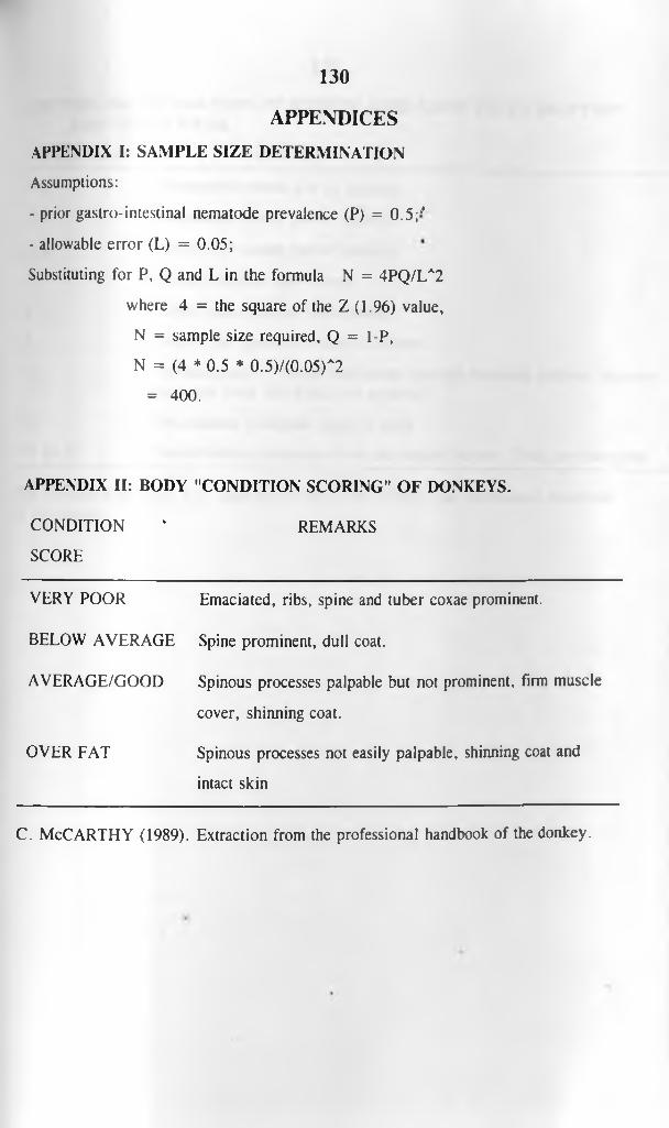
130
APPENDICES
APPENDIX I: SAMPLE SIZE DETERMINATION
Assumptions:
- prior gastro-intestinal nematode prevalence (P) = 0 .5 ;'
- allowable error (L) = 0.05;
Substituting for P, Q and L in the formula N = 4PQ/LA2
where 4 = the square of the Z (1.96) value,
N = sample size required, Q = 1-P,
N = (4 * 0.5 * 0.5)/(0.05)A2
= 400.
APPENDIX II: BODY "CONDITION SCORING" OF DONKEYS.
CONDITION ' REMARKS
SCORE
VERY POOR Emaciated, ribs, spine and tuber coxae prominent.
BELOW AVERAGE Spine prominent, dull coat.
AVERAGE/GOOD Spinous processes palpable but not prominent, firm muscle
cover, shinning coat.
OVER FAT Spinous processes not easily palpable, shinning coat and
intact skin
C. McCARTHY (1989). Extraction from the professional handbook of the donkey.

131
APPENDIX III: ESTIMATION OF DONKEY AGES USING TEETH ERUPTION AND TEETH WEAR.
AGE (YEARS) REMARKS
2.5 Permanent central pair of Incisors.
3.5 Permanent lateral pair of Incisors.
4.5 Permanent corner pair of Incisors.
5 Corner Incisors just in wear.
6 Teeth show shallow infundibulum.
8 Dental star appears in central Incisors.
11 infundibulum of central and middle Incisors becoming shallow. Incisors are quite long and dental star apparent.
14 Pronounced triangular shape of teeth
15 to 20 Infundibulum disappears from the central Incisors. Teeth are triangular.
The Donkey Sanctuary, UK. cited by Fowler J. W. (1989); The professional handbook of the donkey.
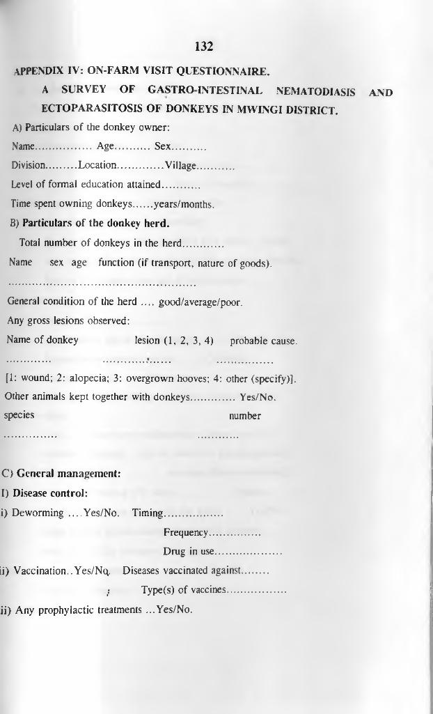
132
APPENDIX IV: ON-FARM VISIT QUESTIONNAIRE.
A SURVEY OF GASTROINTESTINAL NEMATODIASIS AND
ECTOPARASITOSIS OF DONKEYS IN MWINGI DISTRICT.
A) Particulars of the donkey owner:
Name..................... Age............... Sex...............
Division........... Location.................. Village...............
Level of formal education attained..............
Time spent owning donkeys.......years/months.
B) Particulars of the donkey herd.
Total number of donkeys in the herd...............
Name sex age function (if transport, nature of goods).
General condition of the herd .... good/average/poor.
Any gross lesions observed:
Name of donkey lesion (1, 2, 3, 4) probable cause.
[1: wound; 2: alopecia; 3: overgrown hooves; 4: other (specify)].
Other animals kept together with donkeys................. Yes/No.
species number
C) General management:
I) Disease control:
i) Deworming ....Yes/No. Timing......................
Frequency............
Drug in use..........
ii) Vaccination.. Yes/Nq. Diseases vaccinated against
r: Type(s) of vaccines.....
ii) Any prophylactic treatments ...Yes/No.

133
Types and dosages.................
iv) Availability of veterinary services.............
Common diseases observed and treatment given.......
II) Hygiene
i) Waste/manure disposal ......Yes/no.
disposal site.......................
use(s) of manure...................
ii) Feeding site.................... v.clean/average/dirty.
iii) Sleeping/holding site.............clean/dirty.
Do the young ones share premises with adult....yes/No.
Any other animals sharing the site with donkeys..Yes/No.
Species and approximate age of such animals.................
III) Breeding:
Source of sires.....................
Approximate age at first breeding..............(Years).
Approximate gestation period........... (Months).
Foaling interval........... (Months).
Foaling to first breeding.......... (Days/Weeks/Months).
IV) Feeding:
Diet.........Grass/crop residues/other (please specify).
Method of feeding....left on own/Zero grazed/tethered.
Time of feeding.....................morning/aftemoon/evening.
Time spent feeding (Per day)........................(hours).
Do the young ones graze with the adults..........Yes/No.
Other animal species sharing forage with donkeys......
Feed availability throughout the year..................
M ineral supplementation: ........................... Yes/No.
Type and freqqency..............................................
VI) W atering:

134
Source(s) and quality..................................................
Quantity given (Itrs)— Adults.............. Young.
D) Other information:
Approximate working hours per day.............
Approximate workload per day..........(Kgs/Acres).
Usual retirement age and fate of donkey........
Nature of roads/paths used by donkey..(Stony/Sandy/Soft).
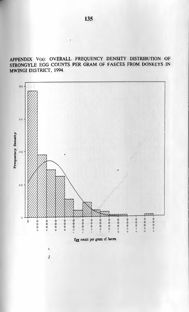
135
APPENDIX V(a): OVERALL FREQUENCY DENSITY DISTRIBUTION OF STRONGYLE EGG COUNTS PER GRAM OF FAECES FROM DONKEYS IN MWINGI DISTRICT, 1994.
Egg count per gram of faeces.

136
APPENDIX V(b): OVERALL WILK-SHAPIRO/RANKIT PLOT OF STRONGYLE EGG COUNTS PER GRAM OF FAECES FROM DONKEYS IN MWINGI DISTRICT, 1994.
A p p r o * i mat <* W i l l : - S h a p i r o 0 0 ' J ? 8
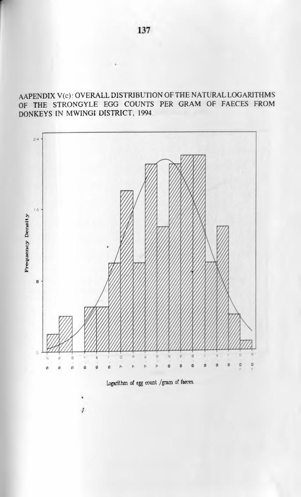
AAPENDIX V(c): OVERALL DISTRIBUTION OF THE NATURAL LOGARITHMS OF THE STRONGYLE EGG COUNTS PER GRAM OF FAECES FROM DONKEYS IN MWINGI DISTRICT, 1994.
logarithm of egg count /gram of faecea

138
APPENDIX V(d): OVERALL WILK-SHAPIRO/RANKIT PLOT OF THE NATURAL LOGARITHMS OF STRONGYLE EGG COUNTS PER GRAM OF FAECES FROM DONKEYS IN MWINGI DISTRICT, 1994.
RankitsApproximate * i 11- Ship 1ro 0 9802

139
APPENDIX VI: SUMMARY RESULTS FROM MUMONI-KYUSO SUB SET
LOGISTIC REGRESSION OF NEMATODIASIS IN DONKEYS IN 1994
PREDICTOR EST. STD.
ERROR
P-
VALUE
ODDS RATIO
CONSTANT -7.14670 4.12857 0.0834
SEX -1.94828 1.16140 0.0934 0.14 (0.01,1.39)
NUM. HERD 0.24160 0.22040 0.2730 1.27 (0.83,1.96)
DONKEY AGE 0.12699 0.07319 0.0827 1.14 (0.98,1.31)
DIVISION 2.59983 1.07425 0.0155 13.5 (1.64,111)
DEWORMING -1.02327 0.66453 0.1236 0.36 (0.10,1.32)
VET -0.96880 1.75377 0.5807 0.36 (0.01,11.81)
FEED -0.26230 0.87406 0.7641 0.77 (0.14,4.27)
W/SOURCE 0.42964 0.21865 0.0494 1.54 (1.0,2.36)
DEVIANCE: 66.81 .DEGREES OF FREEDOM: 76.
P : 0.7651.
K EY : EST : ESTIMATE.NUM/HERD: NUMBER IN HERD.VET : AVAILABILITY OF VETERINARY SUPPORT SERVICES. FEED: FEED AVAILABILITY.W/SOURCE: WATER SOURCE.STD.ERROR: STANDARD ERROR.P-VALUE: PROBABILITY VALUE.
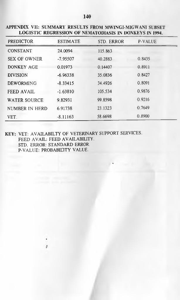
140
APPENDIX VII: SUMMARY RESULTS FROM MVVINGI-MIGVVAM SUBSET LOGISTIC REGRESSION OF NEMATODIASIS IN DONKEYS IN 1994.
PREDICTOR ESTIMATE STD. ERROR P-VALUE
CONSTANT 24.0094 115.863
SEX OF OWNER -7.95507 40.2883 0.8435
DONKEY AGE 0.01973 0.14407 0.8911
DIVISION -6.96338 35.0836 0.8427
DEWORMING -8.33415 34.4926 0.8091
FEED AVAIL -1.63810 105.534 0.9876
WATER SOURCE 9.82931 99.8598 0.9216
NUMBER IN HERD 6.91738 23.1323 0.7649
VET. -8.11163 58.6698 0.8900
KEY: VET: AVAILABILTY OF VETERINARY SUPPORT SERVICES. FEED AVAIL: FEED AVAILABILITY.STD. ERROR: STANDARD ERROR.P-VALUE: PROBABILITY VALUE.
t

141
APPENDIX V I I I : SUMMARY OF THE STUDY VARIABLES IN THE DISCRIMINANT FUNCTIONS WHICH BEST CLASSIFIED THE HERD CASES INTO THE FOUR (OVERALL) D IV ISIO N S (MWINGI, MIGWANI, KYUSO AND MUMONI) AND BETWXXV
D IV IS IO N S WHOSE MEAN EGG COUNTS DIFFERED STATISTICALLY IN 1 994 .
STUDY VARIABLE OVERALL CENTRAL-
KYUSO
CENTRAL-
MUMONI
MIGWANI-
KYUSO
MIGWANI-
MUMONI
HERD SIZE + - ■f - ♦
DIET + - - ♦ -
FEED AVAILABILITY + - + ♦ ♦
WATER SOURCE + - - •f -
EGG COUNT + + + - ♦
SEX OF DONKEY OWNER - - + - -
"BOMA" HYGIENE - - ■f - -
DONKEY AGE - - ♦ -
PERCENT CORRECT
CLASSIFICATION 6 7 . 1 8 1 . 5 7 3 . 5 8 3 . 6 8 2 . 2
N . B : + : v a r i a b l e e n t e r e d .- : v a r i a b l e n o t e n t e r e d .