Effects of affiliation and power motivation arousal on salivary progesterone and testosterone
Upload
independentCategory
view
1download
0
Clinica Chimica Acta 413 (2012) 262–268
Contents lists available at SciVerse ScienceDirect
Clinica Chimica Acta
j ourna l homepage: www.e lsev ie r .com/ locate /c l inch im
A competitive immunochromatographic strip assay for 17-α-hydroxyprogesterone using colloidal gold nanoparticles
Vinay Tripathi a,b,c,⁎, Seema Nara d, Kamya Singh b, Harpal Singh c, Tulsidas G. Shrivastav b
a Amity school of Engineering and Technology, Amity University, Noida, Uttar Pradesh-201303, Indiab Department of Reproductive Biomedicine, National Institute of Health and Family Welfare, Munirka, New Delhi-110067, Indiac Center for BioMedical Engineering, Indian Institute of Technology Delhi, New Delhi-110016, Indiad Department of Applied Mechanics (Biotechnology), Motilal Nehru National Institute of Technology Allahabad-211004, India
⁎ Corresponding author at: Amity school of EngineUniversity, Noida, Uttar Pradesh-201303, India. Tel.: +
E-mail address: [email protected] (V. Tripathi).
0009-8981/$ – see front matter © 2011 Elsevier B.V. Alldoi:10.1016/j.cca.2011.10.016
a b s t r a c t
a r t i c l e i n f oArticle history:
Received 22 July 2011Received in revised form 8 October 2011Accepted 11 October 2011Available online 25 October 2011Keywords:Immunochromatographic stripColloidal gold17-α-Hydroxy progesteroneCompetitive assayAntibody
Background: A rapid and competitive immunochromatographic strip (ICS) test has been developed using col-loidal gold as nanoprobe for measuring 17-α-hydroxy progesterone in serum which is a marker for congen-ital adrenal hyperplasia, an inborn error of metabolism.Methods: Colloidal gold nanoparticles (NPs) were prepared by reducing chloroauric acid with sodium citrate.The corresponding protein conjugate was prepared by passive adsorption of 17-α-hydroxy progesterone-3-carboxymethyl-bovine serum albumin (17-OHP-3-CMO-BSA) immunogen on the colloidal gold surface. Bothcolloidal gold and its protein conjugates were characterized using standard techniques like transmissionelectron microscopy (TEM) and atomic force microscopy (AFM) in addition to the optical characterization.The test line antibody was raised against 17-α-hydroxy progesterone-BSA immunogen and control line anti-body was raised against BSA in New Zealand white rabbits.Results: This test antibody showed high immunoreactivity and specificity when characterized by ELISA for 17-
α-hydroxy progesterone on microtiter plate. The primary antibody was coated on the nitrocellulose mem-brane as test line and anti-BSA antibody was coated as control line. The ICS test was developed in competitiveassay format. The sensitivity of the present immunochromatographic strip assay was reported to be 2.5 μg/lby visual observation. The signal at the test line was easily readable to the naked eye even at higher concen-trations of 17-α-hydroxy progesterone normally associated with congenital adrenal hyperplasia.Conclusion: The present immunoassay takes 15 min for completion. There has not been any other lateral flowimmunoassay for 17-α-hydroxy progesterone reported.© 2011 Elsevier B.V. All rights reserved.
1. Introduction
17-α-Hydroxy-progesterone (17-α-OH-P) is a clinically impor-tant corticosteroid hormone which is routinely measured in serumor plasma for the diagnosis and management of an inborn error ofmetabolism known as congenital adrenal hyperplasia (CAH). Normalreference range of serum 17-α-OH-P is 0.2–2.3 μg/l. 90% of all cases ofCAH are due to deficiency of 21-hydroxylase enzyme required duringadrenal steroidogenesis resulting in abnormally increased concentra-tions of 17-α-OH-P. Incidence ranges from 1/10,000 to 1/15,000 livebirths. This disorder is fatal if not diagnosed early and treated accord-ingly [1]. Various biochemical techniques like GC–MS, LC-MS, HPLCand immunoassays including ELISA and RIA have been used for 17-α-OH-P estimation in serum [2–6]. But the need for sample proces-sing, cleanup steps, use of costly equipments, well trained personneland the environmental hazards (in RIA) put certain limitations on
ering and Technology, Amity91 9911089108.
rights reserved.
their use. Moreover, these tests are far from the reach of poor popula-tion either because of their high costs or because the laboratories invillages or far-flung areas are not well equipped to meet the stan-dards of these tests. These limitations can be addressed by simpleand rapid ICS tests which do not require any trained personnel/costlyequipments and where the results can also be read easily.
The ICS tests have been a popular platform for diagnostic tests sincetheir introduction in the late 1980s. The main application driving theearly development of this solid phase, rapid-test technology was thehuman pregnancy test [7] after which these tests have gained impor-tance in various areas of diagnostics like veterinary testing, agriculturalapplications, environmental testing, and product quality evaluation.The development of labels or markers for ICS test has gone hand inhandwith the advances in detectionmethodology and instrumentation.Various reporters such as colloidal gold, silver, carbon black, latex, dye,dyed polystyrene, phosphor, and dye-encapsulating liposomes havebeen used for developing ICS test for different analytes. Colloidal goldhowever is the most commonly used detector reagents in ICS tests forvisualization of signals. Unlikefluorescence or enzyme tags, gold probesare more stable and do not require time consuming procedures such as

263V. Tripathi et al. / Clinica Chimica Acta 413 (2012) 262–268
incubation, washing steps and enzymatic reactions to generate a signal.Due to accumulation of colloidal gold, a red-purple color can be rapidlyobserved visually. These characteristics significantly shorten the analy-sis time and make it ideally suited for on-site testing even by untrainedpersonnel.
Though ICS tests have been used for variety of analytes, their ap-plication for detection of steroids has been somewhat restricted.This may be attributed to the nanomolar concentrations of steroidsin the biological fluids coupled with the fact that they generally con-tain a single epitope. ICS tests have been reported only for progester-one, cortisol and oestrone sulfate detection in various biological fluidslike serum or plasma, milk and saliva [8–12]. The use of ICS test fordetection of 17-α-OH-P has not been reported.
2. Experimental
2.1. Materials
All solvents, chemicals and salts used in the present study were ofanalytical grade. 17-α-Hydroxy-progesterone-3-carboxymethyloxime (17-α-OH-P-3-CMO), bovine serum albumin (BSA), N-hydroxysuccinimide (NHS), 1-ethyl-3-(3-dimethylaminopropyl) car-bodiimide hydrochloride (EDAC), Tween-20 and horse radish peroxi-dase (HRP) were purchased from Sigma Chemical Company, St. Louis,MO. Polyethylene glycol 20,000 (carbowax) was from SpectrochemPvt. Ltd, Bombay (India). Hydrogen tetra-chloroaurate hydrate(HAuCl4) was from SISCO Research Laboratories Pvt. Ltd, Mumbai(India). Tri-sodium citrate 2-hydrate was from Merc (India) Pvt. Ltd.Nitrocellulose membrane 5 μmol/l, sample pad, absorbent pad andreservoir matrix were from MDI Advanced Microdevices (Pvt.) Ltd,Ambala cantt (India). 0.2 μmol/l cellulose nitrate filters were fromWhatman. All glasswares used were siliconized with sigmacotefrom Sigma. The buffer used for storage of 17-OHP-3CMO-BSA-goldconjugate was 10 mmol/l phosphate buffer saline (PBS) pH 7.4 with0.01% Tween-20.
2.2. Preparation of colloidal gold nanoparticles
Colloidal gold nanoparticles were prepared by controlled reductionof gold chloride (HAuCl4) with trisodium citrate following the proce-dure of Frens [13], with slight modification. 0.2 mmol/l chloroauricacid was added in 0.2 μmol/l filtered double distilled water in a conicalflask and allowed to boil with constant stirring under reflux. This wasfollowed by addition of 2.5 ml of trisodium citrate (38 mmol/l) undercontinuous stirring. The color of the solution changes to black in 2 minand wine red in 5 min. The reaction was allowed to take place for10 min. The flask was removed from heating and allowed to coolunder stirring for 10 min. 0.2 mmol/l HAuCl4 was again added to theflask and boiled for 10 min followed by addition of 2.5 ml of trisodiumcitrate. The reaction mixture was boiled for 10 min and cooled furtherfor another 10 min. These steps were repeated again after 3rd additionof HAuCl4 and trisodium citrate. The solution was stirred without heat-ing at RT for 30 min and prepared colloidal gold was stored in silicon-ized bottle at 4 °C with sodium azide as preservative.
2.3. Preparation of 17-α-OH-P-3-CMO-BSA conjugate/immunogen
17-α-OH-P derivative is a low molecular weight compound; hence,it is non-immunogenic and also, cannot be easily conjugated with col-loidal gold. Therefore, 17-α-OH-P was first coupled with BSA using N-hydroxysuccinimide chemistry with modification [14]. The preparedconjugate was kept at 4 °C to be used as immunogen and for labelingwith colloidal gold.
2.4. Optimization of conditions for labeling 17-α-OH-P-BSA with goldNPs
The labeling of any protein with colloidal gold nanoparticles pre-dominantly involves electrostatic interactions since colloidal goldNPs are negatively charged over a large range of pH. Therefore, a par-tially protonated protein can form ionic bonds with NP's surface butthe interactions largely depends on the pH of the labeling mixture.Thus to establish an optimum pH for labeling of 17-α-OH-P-3-CMO-BSA with colloidal gold, a titration was carried out by adjusting thepH of 500 μl gold sol from 5.5 to 10 with an increment of 0.5 using2 mol/l NaOH. To each tube, a constant minimum amount of 17-α-OH-P-3-CMO-BSA was added;vortex-mixed and allowed to react for10 min at RT. This was followed by addition of 2.5 M NaCl to eachtube. Another titration was carried out to determine the minimumprotecting amount of 17-α-OH-P-3-CMO-BSA conjugate by addingincreasing amount of conjugate (1 mg/ml) in different tubes, rangingfrom 0 to 80 μg with an increment of 2 μg. The volume in each tubewas adjusted to 80 μl with filtered distilled water. To each tube,500 μl of colloidal gold was added, vortex-mixed and allowed toreact for 10 min at RT. After adding 2.5 mol/l NaCl to the solutions, ab-sorbance was taken at λmax of gold NPs, 580 and 600 nm for each tubeof both titrations. The ratio of absorbance at λmax:580 nm is a mea-sure of stability of gold sol whereas, the ratio of absorbance at600 nm:λmax is a measure of its polydispersity [15].
2.5. Labeling of 17-OH-P-3CMO-BSA with gold NPs
To prepare gold labeled 17-α-OH-P-BSA, minimum stabilizingamount of 17-α-OH-P-3-CMO-BSA as determined earlier was addedto colloidal gold solution with constant stirring at optimum pH. Theabsorption of protein on gold NPs surface was allowed to take placeat RT with constant stirring for 20 min. One hundred microliters ofPEG-20,000 (0.05 mmol/l) was added to block nonspecific sites andallowed to react for 20 min at RT with stirring. This was followed bythe addition of 10 μl of Tween-20 (1%) for further stabilization ofthe gold–protein conjugate. The solution was stirred for 10 min atRT and then centrifuged at 9160× for 30 min at 4 °C. The soft pelletwas resuspended to its original volume in washing buffer, allowedto equilibrate for 30–40 min at RT and centrifuged again at 9160×gfor 30 min at 4 °C to remove unbound 17-α-OH-P-3-CMO-BSA fromthe solution. The soft pellet was dissolved in 100 μl of phosphate buff-er and stored at 4 °C for future use. At 100 μl of this gold conjugate, 6%sucrose was added before using it for strip test.
2.6. Characterization of colloidal gold and its protein conjugate
2.6.1. Particle size and zeta potentialThe particle size of prepared gold NPS was determined by dynamic
light scattering using Malvern zetasizer (Nano S) from Malvern In-strument Inc. Worcestershire, U.K. The measurements were carriedout at 25 °C with a count rate of 193.7 kcps at a scattering angle of173°. Zeta potential of the gold NPs was measured with the zetasizerNano ZS90 (Malvern) using laser doppler velocimetry which mea-sures the velocity at which charged particles more on the applicationof voltage towards the oppositely charged electrode. The zeta poten-tial was calculated by determining the electrophoretic mobility andthen applying the Henry's equation.
2.6.2. Optical characterizationThe absorption spectrum of gold nanoparticles (NPs) and its pro-
tein conjugate was recorded with Tecan ELISA spectrophotometerhaving continuous wavelength filters. The nanoparticles and thegold protein conjugate were scanned in the wavelength range of450–600 nm.

Fig. 1. a) Stability and polydispersity of gold conjugate at different pH. b) Titrationcurve for determining minimum protecting amount of 17-α-OH-P-3-CMO-BSA.
264 V. Tripathi et al. / Clinica Chimica Acta 413 (2012) 262–268
2.6.3. High resolution-transmission electron microscopy (HR-TEM)The shape of the NPs before and after coupling with the protein
was analyzed using HRTEM from Technai G2, Holland. The electronsource was LaB6 and the measurements were done at 200 kV. Sam-ples were prepared by placing small drop of NP's solution and its pro-tein conjugate on formvar coated copper grids and allowing thesolvent to slowly evaporate at room temperature.
2.6.4. Atomic force microscopy (AFM)The topographic images of the gold NPs and its protein conjugate
were obtained using AFM from Digital Instruments Nanoscope(U.S.A.). The measurements were done in contact mode at a constantforce (nN).
2.7. Optimization of antibody coating on the immunochromatographicstrip
The anti-17-α-OH-P-3-CMO-BSA antibody was coated on the ni-trocellulose strip at various temperatures i.e. room temperature,37 °C, and 55 °C in order to determine the optimum temperature forantibody coating. Thereafter, the strip was incubated for varioustime durations i.e., 10, 15, 20 and 30 min at the optimized tempera-ture to determine the optimum time of incubation for coating. Lastly,different concentrations of antibody viz 1, 2, and 5 g/l were immobi-lized on the NC membrane to determine the minimum concentrationof antibody that can produce maximum color intensity at the test line.All the assays were run with 0 μg/l standards so that darkest spotcould be obtained for this standard.
2.8. Preparation and immobilization of immunoreagents
Anti-17-α-OH-P-3-CMO-BSA antibody and anti-BSA antibodywere used as test and control line capture reagents respectively. Poly-clonal antibodies were raised in New Zealand white rabbits using 17-α-OH-P-3-CMO-BSA and bovine serum albumin immunogens respec-tively [14]. Total immunoglobulin fraction was purified from theraised antiserum by first precipitating with 33% and 18% ammoniumsulfate followed by DEAE sephadex column chromatography. The ap-propriate concentration of test and capture line antibodies, werecoated on the NC strip at optimized temperature, and time. The teststrips were stored under dry conditions until used. The reservoir ma-trix was coated with standard reaction mixture which consists of10 μl each of various 17-α-OH-P standards (0, 2.5, 10, 50, 100, and250 μg/l) and 17-α-OH-P-protein–gold conjugate. The conjugatepad was dipped in 10 μl of this solution and air dried. The differentcomponents of test strip viz NC membrane laminate, sample pad, res-ervoir matrix and absorbent pad were assembled and this ready-to-use test strip was tested with unknown serum samples for the analy-sisof their 17-α-OH-P concentration. The immunoreagent coated teststrips were found stable even after 2 months of their storage at 4 °Cand thus has a good shelf life.
2.9. Immunochromatographic assay procedure
The assay is in competitive format where the gold labeled 17-α-OH-P and the analyte (17-α-OH-P in standard/sample) compete forthe limited number of binding sites of the immobilized anti-17-α-OH-P antibody at the test zone. The intensity of the red color at testzone is inversely proportional to the concentration of the analyte inthe sample i.e. the color intensity decreases with increasing concen-tration of free 17-α-OH-P in sample. The control line will alwaysshow color which indicates that gold conjugate is functional.
Ten microliters each of 6 17-α-OH-P standards and 17-α-OH-P-protein–gold conjugate was mixed in the microtiter well by pipetting.This mixture is called as standard reactionmixture. The conjugate padwas dipped in 10 μl of this mixture and air dried. For running the
unknown sample in the test 10 μl of unknown sample is mixed withthe 10 μl of 17-α-OH-P-protein–gold conjugate and is known as sam-ple reaction mixture. The standard reaction solution and sample reac-tion mixtures coated on the conjugate pad are allowed to migratealong the test strip by dispensing approximately 100 μl of PBS onsample pad. After 5–7 min a clear pattern of color development wasobserved. The intensity of the color at test capture zone was inverselyproportional to the 17-α-OH-P concentration in the sample.
2.10. Testing of serum samples
About thirty serum samples which were previously analyzed withELISA and correlated with RIA [14] were also analyzed for 17-α-OH-Pby using the developed strip. A range of probable concentration of 17-OHP in unknown serum samples was deduced by comparing the colorintensity at the test line for unknown serum samples with color in-tensity of the standard strips. This comparison can be assumed to beanalogous to the pH determining strips. The probable range of 17-OHP concentration for unknown samples as predicted visually bythe test strip matched with the 17-OHP concentration as analyzedby ELISA and RIA.
3. Results and discussion
3.1. Optimization of conditions for labeling 17-α-OH-P-BSA with goldNPs
Fig. 1a depicts the graph showing the stability (λmax/580 nm) andpolydispersity (600 nm/λmax) of gold conjugate with changing pH.The colloidal gold conjugate is least polydispersed and most stableat pH 5.5, near isoelectric point of BSA (5.1) which is the pH of elec-troneutrality of protein. At this pH the electrostatic interactions arereduced to minimum and maximum protein adsorption occurs on

Fig. 2. Representative DLS spectrum of gold NPs showing an average dia.=36 nm.
Fig. 3. Visible spectra of colloidal gold and gold-17-α-OH-P-BSA conjugate.
265V. Tripathi et al. / Clinica Chimica Acta 413 (2012) 262–268
the negatively charged and hydrophobic colloidal gold surface due tohydrophobic interactions with the protein [16,17]. Therefore, the la-beling reaction was carried out at pH 5.5.
The titration curve to determine the minimum amount of proteinwhich stabilizes colloidal gold is shown in Fig. 1b. The ratio of absor-bance at λmax and 580 nm is the measure of stability of gold sol. Thegraph shows the absorbance value at λmax against increasing
Fig. 4. HR-TEM image of a) gold NPs
concentrations of 17-α-OH-P-3-CMO-BSA. The minimum amount ofprotein required to stabilize colloidal gold against 10% NaCl wasfound to be 40 μg/500 μl of gold. This result was also supported by an-other experiment wherein a substantial decrease in the intrinsic tryp-tophan fluorescence of BSA was observed at 340 nm (data notshown) on addition of minimum protecting amount of 17-α-OH-P-3-CMO-BSA in gold sol at pH 5.5. This minimum protecting amountwas used to prepare gold labeled 17-α-OH-P-3-CMO-BSA. The effi-ciency of labeling reaction was analyzed by estimating the unboundprotein (17-α-OH-P-3-CMO-BSA) content in the supernatant at twocentrifugation steps using Bradford assay. The concentration of pro-tein was almost negligible in the supernatants collected after firstand second centrifugation. This confirms that the conjugation effi-ciency of 17-α-OH-P-3-CMO-BSA with gold NPs was approximately100%.
3.2. Characterization of colloidal gold and its protein conjugate
Characterization of both gold NPS and protein conjugated gold solwas done with standard techniques.
3.2.1. Particle size and zeta potentialFig. 2 shows that the DLS spectrum of prepared Gold NPs. The av-
erage diameter of the gold NPs is 36 nm. Zeta potential is the overallcharge a particle acquires in a specific medium. The magnitude of thezeta potential gives an indication of the potential stability of the col-loidal system. If all the particles have a large negative or positivezeta potential they will repel each other and there is dispersion stabil-ity. If the particles have low zeta potential values then there is noforce to prevent the particles from coming together and there is dis-persion instability. Particles with zeta potentials more positive than+30 mV and more negative than −30 mV are normally consideredstable. The synthesized gold NPs have a zeta potential of −44.1 mVconfirming their stability. The NPs have an electrophoretic mobilityof −3.46 (μm cm/vs) and conductivity of 1.67 ms/cm.
3.2.2. Optical characterizationThe absorption spectrum of gold nanoparticles (NPs) and 17-OHP-
3CMO-BSA-gold conjugate was recorded with Tecan ELISA spectro-photometer in the range of 450–600 nm. Gold nanoparticles exhib-ited strong absorption of electromagnetic waves in the visible range,due to surface plasmon resonance (SPR) which is due to collective os-cillations of the conduction electrons of gold nanoparticles upon irra-diation with visible light. The prepared gold NPs showed a peak at522 nm. Bioconjugation of gold NPs with 17-α-OH-P-3-CMO-BSA
b) 17-α-OH-P-3-CMO-BSA-gold.

266 V. Tripathi et al. / Clinica Chimica Acta 413 (2012) 262–268
displayed a red shift in the peak wavelength due to interaction of pro-tein with colloidal gold as shown in Fig. 3.
3.2.3. High resolution-transmission electron microscopy (HR-TEM)HR-TEM images of the unconjugated and protein conjugated gold
NPs are displayed in Fig. 4a and b respectively. The gold NPs arespherical in shape and are well dispersed even after conjugationwith 17-α-OH-P-3-CMO-BSA. This confirms that even after conjuga-tion of NPs with the protein, the gold NPs were well dispersed, in
Fig. 5. AFM images of the surface of a) gold NPs b) 17
shape and are stable as the aggregates were not observed in thesolution.
3.2.4. Atomic force microscopy (AFM)The representative surface topography of conjugated and uncon-
jugated gold NPs is shown in Fig. 5a and b, respectively. The rough-ness analysis of the surface shows that the root mean square of thesurface roughness was 1.384 nm for gold NPs while it increased to7.861 nm after protein conjugation. The change in the surface
-α-OH-P-protein–gold conjugate in 3-dimension.
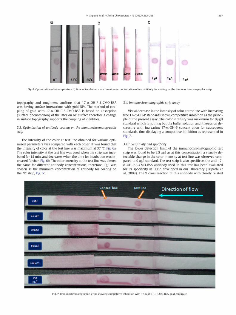
Fig. 6. Optimization of a) temperature b) time of incubation and c) minimum concentration of test antibody for coating on the immunochromatographic strip.
267V. Tripathi et al. / Clinica Chimica Acta 413 (2012) 262–268
topography and roughness confirms that 17-α-OH-P-3-CMO-BSAwas having surface interactions with gold NPs. The method of cou-pling of gold with 17-α-OH-P-3-CMO-BSA is based on adsorption(surface phenomenon) of the later on NP surface therefore a changein surface topography supports the coupling of 2 entities.
3.3. Optimization of antibody coating on the immunochromatographicstrip
The intensity of the color at test line obtained for various opti-mized parameters was compared with each other. It was found thatthe intensity of color at the test line was maximum at 37 °C, Fig. 6a.The color intensity at the test line was good when the strip was incu-bated for 15 min, and decreases when the time for incubation was in-creased further, Fig. 6b. The color intensity at the test line was almostthe same for different antibody concentrations, therefore 1 g/l waschosen as the minimum concentration of antibody for coating onthe NC strip, Fig. 6c.
Fig. 7. Immunochromatographic strips showing competitive
3.4. Immunochromatographic strip assay
Visual decrease in the intensity of color at test line with increasingfree 17-α-OH-P standards shows competitive inhibition as the princi-ple of the present assay. The color intensity was maximum for 0 μg/lstandard which is nothing but the buffer solution and it keeps on de-creasing with increasing 17-α-OH-P concentration for subsequentstandards, thus displaying a competitive inhibition as represented inFig. 7.
3.4.1. Sensitivity and specificityThe lower detection limit of the immunochromatographic test
strip was found to be 2.5 μg/l as at this concentration, a visually de-tectable change in the color intensity at test line was observed com-pared to 0 μg/l standard. The test strip is also specific as the anti-17-α-OH-P-3-CMO-BSA antibody used in this test has been evaluatedfor its specificity in ELISA developed in our laboratory (Tripathi etal., 2008). The % cross reaction of this antibody with closely related
inhibition with 17-α-OH-P-3-CMO-BSA-gold conjugate.

268 V. Tripathi et al. / Clinica Chimica Acta 413 (2012) 262–268
steroids, progesterone (2.1%) and pregnenolone (0.24%), was accept-ably low.
3.4.2. Testing of serum samplesAbout 30 serum samples which were analyzed with ELISA and cor-
related with RIA (Tripathi et al., 2008) were also analyzed for 17-α-OH-P by using the developed strip. The color intensity at the test linefor unknown samples was compared with that on the standard stripsand a visual indication of the range of concentration, where the sample17-α-OH-Pwas deduced. The strip test results of 87% of the tested sam-ples for 17-OHP were found to lie in the concentration range thatmatches with the results of ELISA for the same samples. Out of these87% matched samples, 48% samples were in concentration range0–2.5 μg/l, 36% in 2.5–10 μg/l and 3% in 10–50 μg/l. The mismatchedsamples lie in the concentration range >50 μg/l.
4. Conclusion
A rapid colloidal gold NPs based competitive ICS assay for 17-α-OH-P in serum. The test is semi-quantitative in nature as it is possible to es-tablish for unknown or test samples, a probable range of their 17-α-OH-P concentration by visually comparing the intensity of the test spotwiththat of the standard strips just like the pH strips. The lower detectionlimit of the test strip was 2.5 μg/l by visual observation. Therefore, thistest could serve well in screening of the CAH patients with generallyhigh 17-α-OH-P values. The anti-17-α-OH-P-3-CMO-BSA antibodyused for developing the strip was also specific and displayed lowcross-reactivity with analogous steroids. The immunoreagent coatedtest strips were found stable even after two months of their storage at4 °C and thus has a good shelf life. Colloidal gold nanoparticles synthe-sized for developing the strip were of 36 nm diameter and found to bemonodisperse, spherical and stable. This strip assay also differs fromother reported competitive strip assays for steroids because here colloi-dal gold is coupled with the immunogen (17-α-OH-P-3-CMO-BSA)whereas other assays have used anti-steroid antibody for couplingwith colloidal gold. The use of immunogen is more economical thanusing the antibody because the production cost of antibody is alwayshigh. Secondly, coupling of colloidal gold with any protein involveshydrophobic interactions in addition to the electrostatic interactions[16–17]. If antibody is used for labeling with the colloidal gold, thehydrophobic residues in the antigen binding site (antigen binding siteof an anti-steroid antibody is mainly composed of hydrophobic aminoacid residues) of antibody could interact with colloidal gold surfaceresulting in masking of the antigen binding pocket. This possibilitydoes not exist if immunogen is employed for labeling with gold NPs.Therefore, in this paper, the use of immunogen for preparing a label isrecommended for developing ICS test for steroids. Duration of the pre-sent immunoassay is 15 min which is much less than the plate based
ELISA where it takes around 80min for test completion. The developedICS assay is easy to perform and could be cost-effective as all its reagentswere prepared in the laboratory. The test strip has been tested withsome serum samples but further work is to be carried out for evaluatinglarge number of serum samples for its proper validation.
Acknowledgement
This study was supported by the National Institute of Health andFamily Welfare, New Delhi, India. We thank Prof. Deoki Nandan,Prof. N. K. Sethi, and Prof. K. Kalaivani for their keen interest and encour-agement throughout the study.
References
[1] White PC, New MI, Dupont BN. Congenital adrenal hyperplasia. Engl J Med1987;316:1519–24.
[2] Chong H, Cheah S, Raghavan M, Johgalingam V. Development of an indirect en-zyme immunoassay using monoclonal antibodies for the measurement of 17α-hydroxyprogesterone in human serum. J Immuno Immuno 2009;30:166.
[3] Hofman LF, Klaniecki JE, Smith EK. Direct solid-phase radioimmunoassay for screen-ing 17 alpha-hydroxyprogesterone in whole-blood samples from newborns. ClinChem 1985;31:1127–30.
[4] Lacey JM, Minutti CZ, Magera MJ, Tausher AL, Casetta B, McCann M, Lymph J, HahnSH, Rinaldo P, Matern D. Improved specificity of newborn screening for congenitaladrenal hyperplasia by second-tier steroid profiling using tandemmass spectrom-etry. Clin Chem 2004;50:621–5.
[5] Wong T, Shackleton CHL, Covey TR, Ellis G. Identification of the steroids in neona-tal plasma that interfere with 17 alpha-hydroxyprogesterone radioimmunoassay.Clin Chem 1992;38:1830–7.
[6] Wudy SA, Hartmann M, Homoki J. Hormonal diagnosis of 21-hydroxylase defi-ciency in plasma and urine of neonates using benchtop gas chromatography–mass spectrometry. J Endocrinol 2000;165:679–83.
[7] Vaitukaitis JL, Braunstein GD, Ross GT. A radioimmunoassay which specificallymeasures human chorionic gonadotropin in the presence of human luteinizinghormone. Am J Obstet Gynecol 1972;113:751–8.
[8] Laitinen MPA, Vuento M. Immunochromatographic assay for quantitation of milkprogesterone. Acta Chem Scand 1996;50:141–5.
[9] Henderson K, Stewart J. A dipstick immunoassay to rapidly measure serum oes-trone sulfate concentrations in horses. J Reprod Fertil Dev 2000;12:183–9.
[10] LeungW, Chan P, Bosgoed F, Lehmann K, Renneberg I, Lehmann M, Rennenberg R.One-step quantitative cortisol dipstick with proportional reading. J ImmunolMethods 2003;281:109–18.
[11] Lane J, Flint J, Danks C. The development of a rapid diagnostic test for cortisol inthe saliva of pigs. Int J App Res Vet Med 2004;2:62–7.
[12] Yamaguchi M, Yoshikawa S, Tahara Y. Point-of-use measurement of salivary cor-tisol levels. IEEE SENSORS Conference 2009:343–6.
[13] Frens G. Controlled nucleation for the regulation of the particle size in monodis-perse gold suspensions. Nat Phys Sci 1973;241:20–1.
[14] Tripathi V, Nara S, Chaube SK, Rangari K, Kariya KP, Singh H, Shrivastav TG. Devel-opment of rapid and sensitive one-step direct enzyme linked immunosorbentassay for 17-α-OH-progesterone in serum. J Immuno Immuno 2008;29:117–27.
[15] Englebienne P. Immune and receptor assays in theory and practice. New York:CRC press; 2000.
[16] Norde W. Adsorption of proteins from solution at the solid–liquid interface. AdvColloid Inter Sci 1986;25:267–340.
[17] Oliver C. Conjugation of colloidal gold to proteins. Meth Mol Biol 1999;115:331–4.
Copyright © 2022 FDOKUMEN