CH C C C C C C C C C C C C C C C C C N C C C C C C C C N N C C C C C C
Upload
khangminh22Category
view
0download
0
�����������������
Citation: Lee, J.-S.; Kim, J.-H.
Hemostatic Analysis of Simulated
Gloydius ussuriensis Envenomation
Using Canine Blood: A Comparison
of Thromboelastography and
Classical Coagulation Tests. Animals
2022, 12, 226. https://doi.org/
10.3390/ani12030226
Academic Editor: Mandy Paterson
Received: 6 December 2021
Accepted: 17 January 2022
Published: 18 January 2022
Publisher’s Note: MDPI stays neutral
with regard to jurisdictional claims in
published maps and institutional affil-
iations.
Copyright: © 2022 by the authors.
Licensee MDPI, Basel, Switzerland.
This article is an open access article
distributed under the terms and
conditions of the Creative Commons
Attribution (CC BY) license (https://
creativecommons.org/licenses/by/
4.0/).
animals
Article
Hemostatic Analysis of Simulated Gloydius ussuriensisEnvenomation Using Canine Blood: A Comparison ofThromboelastography and Classical Coagulation TestsJong-Sun Lee and Jung-Hyun Kim *
Department of Veterinary Internal Medicine, College of Veterinary Medicine, Konkuk University,Seoul 05029, Korea; [email protected]* Correspondence: [email protected]; Tel.: +82-2-450-3715
Simple Summary: Snake bites in companion animals are a major issue and cause life-threateninghemorrhage. Classical coagulation tests are performed to diagnose snake venom-induced coagu-lopathy; however, these tests often fail to detect early coagulopathy. This in vitro study using snakevenom and canine blood aimed to explore the efficacy of thromboelastography, which enabled acomprehensive assessment of the coagulation process, compared with classical coagulation tests. Ourresults identified the usefulness of thromboelastography in detecting early venom-induced coagu-lopathy compared with classical coagulation tests. This finding may facilitate decision making inrelation to the immediate initiation of anti-venom treatment or suspending unnecessary anti-venomadministration, which often causes adverse reactions.
Abstract: Snake envenomation may lead to venom-induced consumptive coagulopathy (VICC),usually diagnosed by classical coagulation tests (CCTs), such as prothrombin time (PT) and activatedpartial thromboplastin time (aPTT). However, the results of CCTs are frequently normal in the initialstages, which may delay anti-venom treatments. Thromboelastography (TEG) is a point-of-careand real-time diagnostic tool that enables a comprehensive assessment of the coagulation process.This in vitro study aimed to determine concentration-dependent changes in canine blood caused byGloydius ussuriensis (G. ussuriensis) envenomation using TEG and CCTs. Lyophilized G. ussuriensisvenom was reconstructed using mouse intravenous lethal dose 50 (LD50iv) and serially diluted to25% LD50iv, 50% LD50iv, and 75% LD50iv to reproduce VICC at different concentrations. Normalsaline was used for the control. We compared TEG values of the reaction time (R), kinetic time (K),rate of clot formation (α-angle), maximum amplitude (MA), fibrinolysis at 30 min (LY30), and globalstrength of the clot (G) with those of PT, aPTT, fibrinogen, and platelet counts (PLTs). Most TEGparameters, except R and LY30, demonstrated statistically significant changes compared with thecontrol at all concentrations. CCTs, except PLTs, revealed significant changes at ≥50% LD50iv. Thus,TEG could be a useful diagnostic strategy for early VICC and preventing treatment delay.
Keywords: venom-induced consumptive coagulopathy; snake envenomation; thromboelastography;in vitro study; veterinary sciences
1. Introduction
Of the 3000 snake species worldwide, 30% are venomous [1]. According to the WorldHealth Organization (WHO), 7400 snake bites are reported daily, thus resulting in anaverage of 300 casualties globally [2]. According to the big data availed by the HealthInsurance Review & Assessment Service in South Korea, an average of 2580 venomoussnake bites occur annually in South Korea [3]. However, there are no official reports onsnake bites in veterinary medicine in South Korea. In North America, snake bites in dogsand cats are approximately 30-fold the number of human cases [4,5]. Contrarily, almost
Animals 2022, 12, 226. https://doi.org/10.3390/ani12030226 https://www.mdpi.com/journal/animals

Animals 2022, 12, 226 2 of 13
equal numbers of snake bites in humans and companion animals have been reported inAustralia [6]. Taken together, the estimated number of snake bites in dogs and cats in SouthKorea is at least 2580 annually. This simple comparison may be subject to the statisticalerror; however, the gravity of snake bites in companion animals must not be neglected.
Four species of venomous snakes are found in South Korea, including Gloydius ussurien-sis, Gloydius brevicaudus, and Gloydius intermedius of the Viperidae family and Rhabdophistigrinus of the Colubridae family [7]. Snake venom is a bioactive mixture composed ofseveral enzymatic and non-enzymatic proteins, peptides, and organic and inorganic com-pounds [8], which can induce neurotoxic, cytotoxic, myotoxic, and coagulopathic effects [9].Furthermore, the venom composition may vary between genera and species in a particularfamily owing to different environmental factors, such as the prey, weather, and geographicallocation [10].
The coagulopathic effect is the most concerning aspect of the Gloydius species venom [11].Venom injection through the fang into the bloodstream leads to the concurrent inhibitionand depletion of coagulation factors, resulting in an increased bleeding tendency [12]. Thisphenomenon is termed venom-induced consumptive coagulopathy (VICC) because of itsshared similarity with disseminated intravascular coagulopathy (DIC); however, it does nottrigger severe organ damage or systemic thrombosis, which is common in DIC [13]. Animalswith VICC usually exhibit pathological abnormalities, such as prolonged prothrombintime (PT), activated partial thromboplastin time (aPTT), low fibrinogen and antithrombinconcentrations, elevated D-dimer levels, and thrombocytopenia [14–17]. However, laboratoryresults of classical coagulation tests (CCTs) are frequently normal in the initial stages andgradually become abnormal within a few hours to days [14,18,19]. This increases the needfor novel tests to detect VICC during the aforementioned period.
Thromboelastography (TEG) is a point-of-care and real-time diagnostic tool that en-ables a comprehensive assessment of the coagulation process from clot generation tofibrinolysis [20]. Its usefulness ranges from transfusion in trauma patients to monitor-ing hemostasis during liver transplantation and cardiac surgeries [21,22]. In veterinarymedicine, researchers have reported on the use of TEG during the diagnosis and treatmentof VICC along with CCTs [14–17]. However, they have not compared the efficacy of TEGwith that of CCTs.
The present in vitro study aimed to determine concentration-dependent changes incanine blood following envenomation with Gloydius ussuriensis (G. ussuriensis) venom usingTEG and CCTs and to evaluate the sensitivity of each test for identifying results beyond thenormal reference limits (NRLs).
2. Materials and Methods2.1. Animals
This study was approved by the Institutional Animal Care and Use Committee ofthe Konkuk University (approval No. KU20217), Seoul, South Korea. All dogs wereprospectively recruited at the Konkuk University Veterinary Medical Teaching Hospitalfrom February 2021 to May 2021. We enrolled 10 healthy, client-owned dogs after acquiringinformed consent from their owners. Before the experiment, all the dogs underwent aTEG test and were included in this study after normal results of the test were obtained forall parameters. To avoid the effects of external coagulation disturbances, we consideredthe following exclusion criteria: a history of anemia or bleeding disorder, a precedentuse of steroids or anticoagulants in the past 30 days, a precedent use of non-steroidalanti-inflammatory drugs in the past 7 days, or a history of significant medical comorbidity.
2.2. Blood Collection
Four milliliters of blood were collected twice from each dog through jugular venipunc-ture in blood tubes containing 3.2% sodium citrate (1.8 mL, BD Vacutainer®; BD VacutainerSystems, Plymouth, UK). All the blood samples were agitated on a roller to prevent prema-ture clotting, followed by coagulation tests performed within 2 h of blood collection.

Animals 2022, 12, 226 3 of 13
2.3. Preparation of Venom Solution and its Addition to Blood
We stored the lyophilized venom of G. ussuriensis (Latoxan, Portes-lès-Valence, France)at −80 ◦C until the day of reconstitution with normal saline. The venom solution wasaliquoted and re-stored at −20 ◦C to prevent the denaturation of the venom proteins andpeptides. A standard point for measuring the extent of coagulation was set by usingmouse intravenous lethal dose 50 (LD50iv, 1.139 mg/kg) to prepare venom solutions,according to the manufacturer’s guidelines. The calculations of 14 µg/mL were based onthe estimated distribution of an 80 mL/kg blood volume in canines [23]. Thereafter, thevenom solution was serially diluted to 25% LD50iv (3.5 µg/mL), 50% LD50iv (7 µg/mL),and 75% LD50iv (10.5 µg/mL) to reproduce the venom-induced coagulation disorder usingdifferent concentrations. Next, we added a venom volume equivalent to 20% of the bloodto the blood sample and an equal amount of normal saline to the control sample (Figure 1).Thereafter, we centrifuged the blood sample to perform CCTs (PT, aPTT, and fibrinogen)and prepared the slides for the manual platelet count. Simultaneously, blood samplesfor TEG were left for stabilization on the roller for 30 min [24,25]. CCTs were performedimmediately after centrifugation. Subsequently, we carried out TEG after stabilization for30 min, and the platelets were counted manually at the end.
Figure 1. Timeline of the experimental design. The venoms of different concentrations were addedto the blood samples in citrated tubes, whereas an equal amount of normal saline was added tothe control blood sample. Each sample was assessed using CCTs and TEG, followed by statisticalanalyses. CCT, classical coagulation test; TEG, thromboelastography.
2.4. TEG
We performed TEG coagulation assessment using a TEG® 5000 Thromboelastograph®
Hemostasis Analyzer system (Haemonetics Corporation, Boston, MA, USA). The TEGmachine comprised a holder with a cup maintained at 37 ◦C. The cup was filled witha 340 µL blood sample containing 1 mL citrated blood and 1% kaolin that were gentlyinverted five times and 20 µL of CaCl2 for recalcification to initiate the examination. Apin connected to the TEG machine by a torsion wire was positioned into the cup. Uponblood clotting, the resistance from rotational movement was sent to the torsion wire andconverted into an electrical signal. A trace line was displayed on a computer monitorthrough the TEG program supplied by the manufacturer. We measured several parameters,including the reaction time (R), kinetic time (K), global values (G), α-angle, maximumamplitude (MA), and the percentage of fibrinolysis at 30 min following MA (LY30). R is theduration from the initiation of the assessment until the detection of clots showing a clotamplitude of 2 mm is reached. K is the duration from the end of R to a clot amplitude of20 mm and represents a certain level of clot firmness. The α-angle denotes the tangent of

Animals 2022, 12, 226 4 of 13
a curve representing the clot formation rate. MA is the length of the widest amplitude atwhich the clot strength reaches its peak. G is the log derivation calculated from the MAvalue representing the global strength of the clot.
2.5. Classical Coagulation Tests
PT, aPTT, and fibrinogen were assessed using a coagulation analyzer CG02NV (A&TCorporation, Oshu, Japan). First, the reagent test card was inserted into the analyzer. Subse-quently, a mixture of 25 µL of citrated plasma and a diluent (provided by the manufacturer)was applied to the test card using a pipette. Eventually, the results were displayed ona touch panel. PT and aPTT below the NRLs were considered normal. Platelet counts(PLTs) were determined manually under a microscope (OLYMPUS CX31RTSF, OlympusCorporation, Tokyo, Japan) in an oil-immersion field (OIF) to avoid the effects of plateletaggregation inducers isolated from the venom of the Viperidae family [26]. On visualizingthe aggregated platelets in the OIF, each platelet was counted from the clumps, and theaverage values are presented.
2.6. Statistical Analyses
Statistical analyses were performed using Microsoft Excel (Microsoft Corporation,Redmond, WA, USA) and Prism 9.1.0 software (GraphPad Software, San Diego, CA,USA). Descriptive statistics are presented as interquartile ranges with the median, anddifferences between the 75th and 25th quartiles higher than 1.5-fold were defined as outliers.Data were tested for normality using the Shapiro–Wilk test. To determine the statisticaldifferences between the control and venom concentrations evaluated with TEG and CCTs,we performed one-way repeated measure analysis of variance (RM-ANOVA) with pairwisecomparisons to identify the difference between the groups. Statistical significance wasset at p < 0.05. We obtained the NRLs for TEG parameters from a previous study [27],except for LY30 and G. Thus, G was calculated from MA based on a previously usedequation as follows: G = (5000 × A) ÷ (100 − A) [28]. The sensitivities of detecting samplesout of normal diagnostic ranges for TEG and CCTs were presented as a percentage andcategorized into three groups with different sensitivities as follows: <70% indicated the lowsensitivity; between 70% and 89% indicated the medium sensitivity; and >89% indicated thehigh sensitivity. Considering the absence of the NRL for LY30, we excluded the sensitivityand ANOVA results of LY30 from the analysis.
3. Results3.1. Descriptive Analyses
Coagulation parameters of TEG and CCTs are illustrated in box-and-whisker plots(Figures 2 and 3). The mean values of TEG and CCTs parameters are listed in SupplementaryTables S1 and S2. The data distribution assessment using the Shapiro–Wilk test revealed thefollowing normally distributed data: R in the control and at 25% LD50iv and 50% LD50iv; Kin the control; α-angle in the control and at all concentrations; MA in the control and atall concentrations; G in the control and at 25% LD50iv and 50% LD50iv; PT in the controland at 50% LD50iv; aPTT in the control and at 25% LD50iv and 50% LD50iv; fibrinogenin the control and at 25% LD50iv; and PLTs in the control and at all concentrations. Asthe data were not normally distributed, and the sphericity was violated across the venomconcentration, we used the non-parametric methods of RM-ANOVA and the Durbin–Conover test for pairwise comparisons. One value of LY30 was missing owing to the abrupthalt of TEG during the analysis for an unknown reason. None of the LY30 measures werenormally distributed.

Animals 2022, 12, 226 5 of 13
Figure 2. Box-and-whisker plots of the classical coagulation tests parameters: (a) PT (b) aPTT(c) fibrinogen (d) PLTs. Horizontal lines inside the boxes indicate the median. The upper boundaryof the boxes indicates the top quartile, whereas the lower indicates the bottom quartile. An asterisk(*) indicates the venom concentration (% intravenous lethal dose 50 (LD50iv)) displaying a significantchange compared with the control (p < 0.05). Differences between the 75th and 25th quartiles higherthan 1.5-fold were considered as outliers and indicated by a black dot (•). PT, prothrombin time;aPTT, activated partial thromboplastin time; PLTs, platelet counts.

Animals 2022, 12, 226 6 of 13
Figure 3. Box-and-whisker plots of TEG parameters: (a) R (b) K (c) α-angle (d) MA (e) LY30 (f) G.Horizontal lines inside the boxes indicate the median. The upper boundary of the boxes indicatesthe top quartile, whereas the lower boundary indicates the bottom quartile. An asterisk (*) indicatesvenom concentration (% LD50iv) displaying a significant change compared with the control (p < 0.05).Differences between the 75th and 25th quartiles higher than 1.5-fold are considered as outliers andindicated by a black dot (•). R, reaction time; K, kinetic time; α-angle, the rate of clot formation; MA,maximum amplitude; LY30, fibrinolysis at 30 min; and G, global strength of the clot.
3.2. Concentration-Dependent Changes3.2.1. Classical Coagulation Tests
PT and aPTT demonstrated statistically significant changes compared with the controlat 50% LD50iv (p = 0.013, p < 0.001, respectively) and above (p < 0.001 and p < 0.001,respectively) (Figure 2). Fibrinogen displayed a statistically significant change only at75% LD50iv (p = 0.003; Figure 2). All PLTs were in the NRL and did not demonstratesignificant changes at any concentration, compared with the control (Figure 2).
3.2.2. TEG
TEG parameters demonstrated statistically significant changes compared with thecontrol, as the venom concentration increased. The following statistically significantchanges were observed in the parameters at the respective concentrations: K (p < 0.001 atall concentrations), α-angle (p = 0.001 at 25% LD50iv, p < 0.001 at 50% LD50iv, and above

Animals 2022, 12, 226 7 of 13
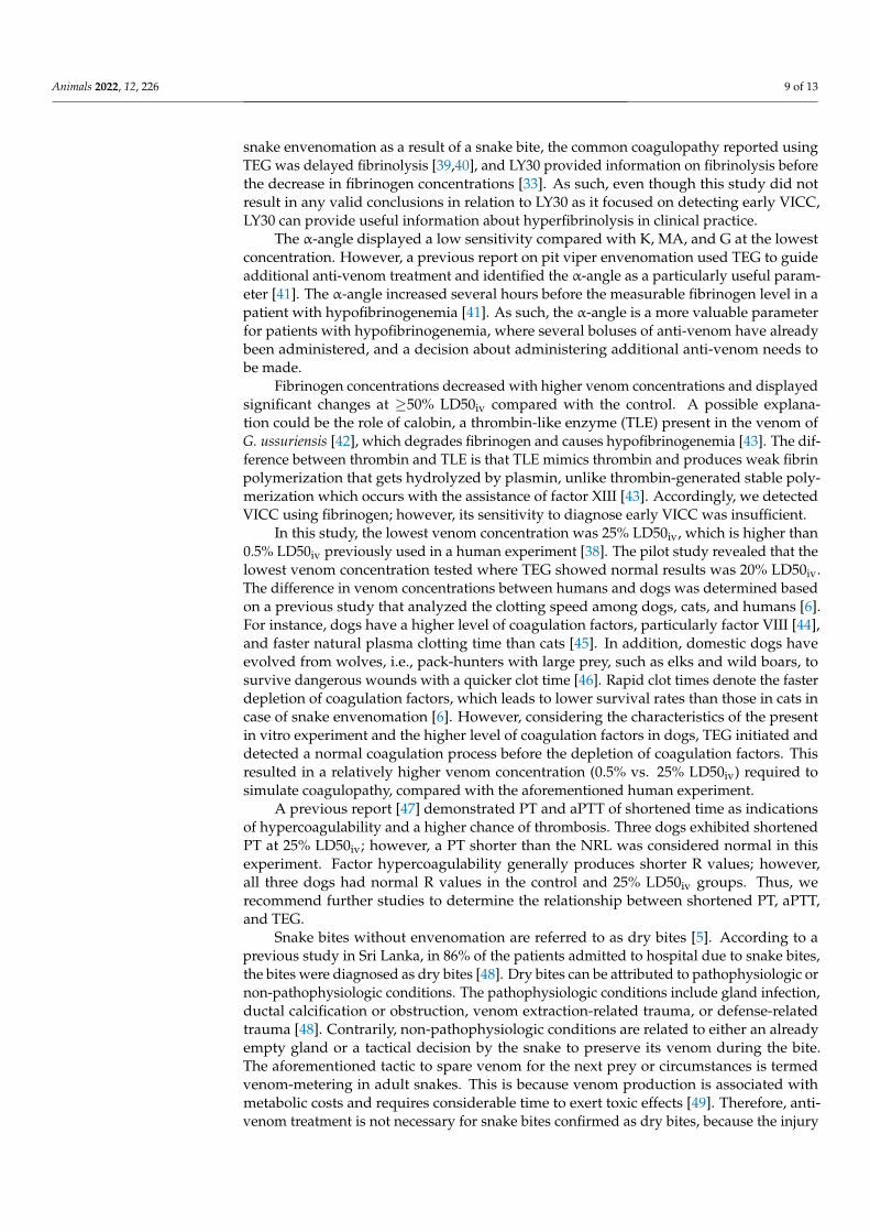
p < 0.0001), MA (p < 0.0001 at all concentrations), and G (p < 0.0001 at 25% and 50% LD50iv,p = 0.002 at 75% LD50iv, Figure 3). However, R and LY30 did not exhibit statisticallysignificant differences at any concentration (Figure 3). Figure 4 depicts typical TEG tracelines displaying concentration-dependent changes from one of the dogs.
Figure 4. Typical TEG trace lines suggesting concentration-dependent changes from one of the dogs.The trace lines of the control and different venom concentrations (% LD50iv) are placed on the similarcentral axis for an easier comparison.
3.3. Diagnostic Thresholds and Sensitivities
We determined the diagnostic thresholds (DHs) for mean values beyond the NRL.At 25% LD50iv, DHs were detected for K, MA, and G. At 50% LD50iv, we determined theDH for the α-angle, compared with those for R, PT, aPTT, and fibrinogen at 75% LD50iv.Moreover, we evaluated the sensitivities of each parameter, except for LY30 (Table 1). PT,PLTs, and R demonstrated a low sensitivity across the concentration range. Contrarily,aPTT and fibrinogen displayed low and medium sensitivities at concentrations of ≤50%LD50iv and 75% LD50iv, respectively (Table 1). The α-angle displayed low, medium, andhigh sensitivities at 25% LD50iv, 50% LD50iv, and 75% LD50iv, respectively (Table 1).Meanwhile, K, MA, and G demonstrated a high sensitivity across the entire concentrationrange (Table 1).
Table 1. Sensitivity of classical coagulation tests and TEG parameters: percentages of samples beyondnormal reference limits.
Measure Normal Range% LD50iv Venom Concentration
25% 50% 75%
CCTsPT 7.1–8.4 s 0% 30% 50%
aPTT 13.7–25.6 s 10% 50% 70%Fibrinogen 113–385 mg/dL 0% 50% 70%
PLTs 148–484 K/µL 10% 50% 50%

Animals 2022, 12, 226 8 of 13
Table 1. Cont.
Measure Normal Range% LD50iv Venom Concentration
25% 50% 75%
TEGR 1.8–8.6 min 0% 10% 50%K 1.3–5.7 min 90% 100% 100%
α-angle 36.9–74.6 degrees 30% 70% 90%MA 42.9–67.9 mm 100% 100% 100%LY30 NA NA NA NA
G 3.2–9.6 Kdynes/cm2 100% 100% 100%CCTs, classical coagulation tests; PT, prothrombin time; aPTT, activated partial thromboplastin time; PLTs, plateletcounts; TEG, thromboelastography; R, reaction time; K, kinetic time; MA, maximum amplitude; LY30, clot lysis at30 min; G, global strength of the clot; NA, not applicable; s, second; min, minutes.
4. Discussion
We intended to evaluate the efficacies of TEG versus CCTs in determining coagulationparameters following canine blood envenomation using simulated G. ussuriensis venom.Through the serial dilution of venom, we determined concentration-dependent changesand sensitivities. Our results were consistent with VICC caused by natural pit viperenvenoming. However, in contrast to TEG and the rest of CCTs, PLTs did not demonstratethe coagulopathic effects of G. ussuriensis venom.
Conventionally, researchers perform coagulation tests such as PT, aPTT, 20 min wholeblood clotting test, Lee–White clotting time, D-dimer, fibrinogen, fibrin degradation prod-uct (FDP), and PLTs, to diagnose VICC [12–17,29]. A previous report identified FDP asthe most sensitive marker for detecting early VICC [30], whereas another confirmed PTand aPTT as useful diagnostic tools for VICC [31]. However, the aforementioned testsoccasionally generated results within the NRL in envenomated patients, thereby indicatingthat CCTs may lead to false-negative results and/or delay in anti-venom treatments [32–34].PT and aPTT are easily accessible in most laboratories and have the advantage of a shortturnaround evaluation time of approximately 10 min. Nonetheless, certain characteristics ofcoagulation tests lead to limitations. For instance, the results of PT and aPTT are achievedwithin approximately 10 min before fibrin polymerization. In other words, these testsneither analyze the status of thrombin stability and fibrinolysis nor the circumstances lack-ing in protein C or antithrombin [12,35,36]. Since PT and aPTT evaluate the clotting timein the in vitro setting, they do not represent the in vivo interaction between coagulationfactors and platelets [37]. Moreover, delays in PT and aPTT imply a specific coagula-tion factor deficiency during the coagulation cascade, thus necessitating further tests tospecify the cause [37]. Similarly, fibrinogen, PLTs, and FDP produce only partial resultsat a specific point during coagulation, thereby warranting further tests [13]. Therefore,CCTs are unable to completely detect VICC characterized by platelet dysfunction and/orcoagulation deficiencies.
The performance of TEG in the present experiment supposedly surpassed the abilityof CCTs to detect VICC in the early stages. Of the TEG parameters, K, MA, and G displayeda high sensitivity even at the lowest venom concentration of 25% LD50iv, compared withthe low sensitivity of the CCTs at 25% LD50iv. However, R displayed no significantchanges at any concentration, and the sensitivity was low at all concentrations, therebyindicating its limited ability to detect coagulopathy caused by snake venom. Contrarily, theR-value tended to increase with the venom concentration (Figure 4). The experiment with100% LD50iv during the pilot study did not produce TEG traces, eventually leaving a flatline that indicated coagulation failure.
In relation to LY30, we did not observe statistically significant changes at any concen-tration, consistent with previously published results of in vitro experiments using humanblood [38]. The current in vitro study could not entirely replicate the interactions in compli-cated living organisms; thus, hyperfibrinolysis was excluded [38]. In patients with natural
Animals 2022, 12, 226 9 of 13
snake envenomation as a result of a snake bite, the common coagulopathy reported usingTEG was delayed fibrinolysis [39,40], and LY30 provided information on fibrinolysis beforethe decrease in fibrinogen concentrations [33]. As such, even though this study did notresult in any valid conclusions in relation to LY30 as it focused on detecting early VICC,LY30 can provide useful information about hyperfibrinolysis in clinical practice.
The α-angle displayed a low sensitivity compared with K, MA, and G at the lowestconcentration. However, a previous report on pit viper envenomation used TEG to guideadditional anti-venom treatment and identified the α-angle as a particularly useful param-eter [41]. The α-angle increased several hours before the measurable fibrinogen level in apatient with hypofibrinogenemia [41]. As such, the α-angle is a more valuable parameterfor patients with hypofibrinogenemia, where several boluses of anti-venom have alreadybeen administered, and a decision about administering additional anti-venom needs tobe made.
Fibrinogen concentrations decreased with higher venom concentrations and displayedsignificant changes at ≥50% LD50iv compared with the control. A possible explana-tion could be the role of calobin, a thrombin-like enzyme (TLE) present in the venom ofG. ussuriensis [42], which degrades fibrinogen and causes hypofibrinogenemia [43]. The dif-ference between thrombin and TLE is that TLE mimics thrombin and produces weak fibrinpolymerization that gets hydrolyzed by plasmin, unlike thrombin-generated stable poly-merization which occurs with the assistance of factor XIII [43]. Accordingly, we detectedVICC using fibrinogen; however, its sensitivity to diagnose early VICC was insufficient.
In this study, the lowest venom concentration was 25% LD50iv, which is higher than0.5% LD50iv previously used in a human experiment [38]. The pilot study revealed that thelowest venom concentration tested where TEG showed normal results was 20% LD50iv.The difference in venom concentrations between humans and dogs was determined basedon a previous study that analyzed the clotting speed among dogs, cats, and humans [6].For instance, dogs have a higher level of coagulation factors, particularly factor VIII [44],and faster natural plasma clotting time than cats [45]. In addition, domestic dogs haveevolved from wolves, i.e., pack-hunters with large prey, such as elks and wild boars, tosurvive dangerous wounds with a quicker clot time [46]. Rapid clot times denote the fasterdepletion of coagulation factors, which leads to lower survival rates than those in cats incase of snake envenomation [6]. However, considering the characteristics of the presentin vitro experiment and the higher level of coagulation factors in dogs, TEG initiated anddetected a normal coagulation process before the depletion of coagulation factors. Thisresulted in a relatively higher venom concentration (0.5% vs. 25% LD50iv) required tosimulate coagulopathy, compared with the aforementioned human experiment.
A previous report [47] demonstrated PT and aPTT of shortened time as indicationsof hypercoagulability and a higher chance of thrombosis. Three dogs exhibited shortenedPT at 25% LD50iv; however, a PT shorter than the NRL was considered normal in thisexperiment. Factor hypercoagulability generally produces shorter R values; however,all three dogs had normal R values in the control and 25% LD50iv groups. Thus, werecommend further studies to determine the relationship between shortened PT, aPTT,and TEG.
Snake bites without envenomation are referred to as dry bites [5]. According to aprevious study in Sri Lanka, in 86% of the patients admitted to hospital due to snake bites,the bites were diagnosed as dry bites [48]. Dry bites can be attributed to pathophysiologic ornon-pathophysiologic conditions. The pathophysiologic conditions include gland infection,ductal calcification or obstruction, venom extraction-related trauma, or defense-relatedtrauma [48]. Contrarily, non-pathophysiologic conditions are related to either an alreadyempty gland or a tactical decision by the snake to preserve its venom during the bite.The aforementioned tactic to spare venom for the next prey or circumstances is termedvenom-metering in adult snakes. This is because venom production is associated withmetabolic costs and requires considerable time to exert toxic effects [49]. Therefore, anti-venom treatment is not necessary for snake bites confirmed as dry bites, because the injury

Animals 2022, 12, 226 10 of 13
is analogous to a non-venomous snake bite [50]. However, it is difficult to differentiatebetween a dry or wet bite, because the bite site may display signs of inflammation andtissue edema [5].
Anti-venom is considered the most effective treatment for snake envenomation, consid-ering it is the only antidote to snake venom. However, the WHO recommends anti-venomonly for patients expected to gain benefits that exceed the risk. This can be attributed tothe unstable and expensive supply of anti-venom [51]. According to the WHO guidelines,anti-venom is preferably used in envenomated patients with hemostatic abnormalities,cardiovascular problems, abnormal neurologic signs, kidney failure, and worsening localtissue swelling [51]. Moreover, anti-venom can cause adverse reactions, such as ana-phylactic shock, pyrogenic reactions, and serum sickness, thus necessitating cautiousadministration [51]. In addition, performing a skin test before anti-venom administration iscontroversial because of its extremely low sensitivity and time requirement, which delaysanti-venom administration [52].
Under the aforementioned circumstances, distinguishing dry from wet bites or diag-nosing early hemostatic abnormalities is the key to treating snake envenomation. TEGproduces faster results, appears more suitable than CCTs in detecting early VICC andcould be useful in diagnosing and monitoring the patient after anti-venom treatment [38].We recommend an additional TEG evaluation with CCTs during follow-up for patientsadmitted to hospital with normal TEG results. Moreover, the unnecessary administrationof anti-venom should be suspended. Conversely, clinicians should initiate immediateanti-venom treatment in the event of abnormal TEG results, and in the case of hospitalizedpatients with hypofibrinogenemia, an abnormal LY30 result would assist the decisionmaking regarding the administration of additional anti-venom boluses. However, the costof TEG is relatively higher than that of CCTs. This need for expensive infrastructure maylimit the applicability of TEG, considering that most of the snake envenomation cases occurin resource-poor environments [12].
This study has a few limitations. First, our in vitro study analyzed the results froma single time point using a small number of dogs, and the results were interpreted basedon the NRL values from in vivo studies reported in the literature; therefore, the currentfindings should be generalized with caution. Second, we could not perform TEG withPlateletMapping® due to financial constraints. An experiment involving platelet inhibitioncould have provided a better understanding of the venom-induced inhibition of the coag-ulation factor. In addition, experimentally induced coagulopathy with a stabilization of30 min may have created a relatively calcium-free environment; therefore, the possibility ofcoagulopathy caused by low calcium rather than the inhibition of coagulation factor cannotbe excluded.
5. Conclusions
Through the serial dilution of simulated G. ussuriensis venom, we verified concentration-dependent changes in canine blood coagulation parameters. The results were consistentwith VICC caused by natural pit viper envenoming. The platelet count did not detectthe coagulopathic effects of G. ussuriensis venom, unlike TEG and the rest of the CCTs.Nonetheless, TEG effectively identified abnormal coagulation profiles compared with CCTsin in vitro simulated pit viper envenomation. Thus, TEG could be a useful diagnosticstrategy for early VICC and preventing treatment delay.
Supplementary Materials: The following supporting information can be downloaded at: https://www.mdpi.com/article/10.3390/ani12030226/s1, Table S1: Mean values of classical coagulationtests parameters for the control and increasing venom concentrations (% LD50iv) with p-values andstatistical results; Table S2: Mean values of thromboelastography parameters for the control andincreasing venom concentrations (% LD50iv) with p-values and statistical results.

Animals 2022, 12, 226 11 of 13
Author Contributions: Conceptualization, methodology, validation, investigation, formal analysis,data curation, and visualization, J.-S.L. and J.-H.K.; resources, J.-H.K.; writing—original draft prepara-tion, J.-S.L.; writing—review and editing, J.-H.K.; supervision, J.-H.K.; project administration, J.-H.K.All authors have read and agreed to the published version of the manuscript.
Funding: This research received no external funding.
Institutional Review Board Statement: This study was conducted according to the guidelines of theDeclaration of the Helsinki and approved by the Institutional Review Board of Konkuk UniversityInstitutional Animal Care and Use Committee (approval number: KU20217). Written informedconsent was obtained from the owners for the participation of their animals in this study.
Informed Consent Statement: Informed consent was obtained from owners of all animals involvedin the study.
Data Availability Statement: The data that support the findings of this study are available from thecorresponding author upon reasonable request.
Conflicts of Interest: The authors declare no conflict of interest.
References1. Al-Sadoon, M.K.; Fahad, A.M.; Ahamad, P.B.; Rahman, A.A. Envenomation and the Bite Rate by Venomous Snakes in the
Kingdom of Saudi Arabia over the Period (2015–2018). Saudi J. Biol. Sci. 2021, 28, 582–586. [CrossRef]2. World Health Organization. Snakebite Envenoming: A Strategy for Prevention and Control; WHO Document Production Services:
Geneva, Switzerland, 2019.3. Bigdata Distributed by the Health Insurance Review & Assessment Service in Korea. Available online: http://opendata.hira.or.
kr/op/opc/olap4thDsInfo.do (accessed on 27 August 2021).4. Peterson, M.E. Snake Bite: Pit Vipers. Clin. Tech. Small Anim. Pract. 2006, 21, 174–182. [CrossRef]5. Pucca, M.B.; Knudsen, C.; Oliveira, I.S.; Rimbault, C.; Cerni, F.A.; Wen, F.H.; Sachett, J.; Sartim, M.A.; Laustsen, A.H.; Monteiro,
W.M. Current Knowledge on Snake Dry Bites. Toxins 2020, 12, 668. [CrossRef]6. Zdenek, C.N.; Llinas, J.; Dobson, J.; Allen, L.; Dunstan, N.; Sousa, L.F.; Moura da Silva, A.M.; Fry, B.G. Pets in Peril: The
Relative Susceptibility of Cats and Dogs to Procoagulant Snake Venoms. Comp. Biochem. Physiol. Part C Toxicol. Pharmacol. 2020,236, 108769. [CrossRef] [PubMed]
7. Moon, J.M.; Koo, Y.J.; Chun, B.J.; Park, K.H.; Cho, Y.S.; Kim, J.C.; Lee, S.D.; Min, Y.R.; Park, H.S. The Effect of Myocardial Injuryon the Clinical Course of Snake Envenomation in South Korea. Clin. Toxicol. 2020, 59, 286–295. [CrossRef]
8. Ramos, O.H.P.; Selistre-De-Araujo, H.S. Snake Venom Metalloproteases—Structure and Function of Catalytic and DisintegrinDomains. Comp. Biochem. Physiol. C Toxicol. Pharmacol. 2006, 142, 328–346. [CrossRef]
9. Ainsworth, S.; Slagboom, J.; Alomran, N.; Pla, D.; Alhamdi, Y.; King, S.I.; Bolton, F.M.S.; Gutiérrez, J.M.; Vonk, F.J.; Toh, C.H.; et al.The Paraspecific Neutralisation of Snake Venom Induced Coagulopathy by Antivenoms. Commun. Biol. 2018, 1, 1–14. [CrossRef]
10. Lim, H.; Kang, H.G.; Kim, K.H.; Goo Kang, H.; Hwan Kim, K.; Kang, H.G.; Kim, K.H. Antivenom for Snake Bite in Korea. J.Korean Med. Assoc. 2013, 56, 1091–1103. [CrossRef]
11. Debono, J.; Bos, M.H.A.; Do, M.S.; Fry, B.G. Clinical Implications of Coagulotoxic Variations in Mamushi (Viperidae: Gloydius)Snake Venoms. Comp. Biochem. Physiol. Part C Toxicol. Pharmacol. 2019, 225, 108567. [CrossRef] [PubMed]
12. Park, E.J.; Choi, S.; Kim, H.H.; Jung, Y.S. Novel Treatment Strategy for Patients with Venom-Induced Consumptive Coagulopathyfrom a Pit Viper Bite. Toxins 2020, 12, 295. [CrossRef]
13. White, J. Snake Venoms and Coagulopathy. Toxicon Off. J. Int. Soc. Toxinol. 2005, 45, 951–967. [CrossRef] [PubMed]14. Atamna, R.; Kelmer, E.; Aroch, I.; Klainbart, S. Echis Coloratus Envenomation in a Dog: Clinical, Hemostatic and Thromboelasto-
metric Findings and Treatment. Clin. Toxicol. 2021, 59, 639–643. [CrossRef] [PubMed]15. Kopke, M.A.; Botha, W.J. Thromboelastographic Evaluation of 2 Dogs with Boomslang (Dispholidus Typus) Envenomation. J. Vet.
Emerg. Crit. Care 2020, 30, 712–717. [CrossRef] [PubMed]16. Lieblick, B.A.; Bergman, P.J.; Peterson, N.W. Thromboelastographic Evaluation of Dogs Bitten by Rattlesnakes Native to Southern
California. Am. J. Vet. Res. 2018, 79, 532–537. [CrossRef]17. Nagel, S.S.; Schoeman, J.P.; Thompson, P.N.; Wiinberg, B.; Goddard, A. Hemostatic Analysis of Dogs Naturally Envenomed by
the African Puffadder (Bitis Arietans) and Snouted Cobra (Naja Annulifera). J. Vet. Emerg. Crit. Care 2014, 24, 662–671. [CrossRef][PubMed]
18. Olives, T.D.; Topeff, J.M.; Willhite, L.A.; Kubic, V.L.; Keyler, D.E.; Cole, J.B. Complete Clinical Course of Envenomation byProtobothrops Mangshanensis: Delayed Coagulopathy and Response to Trimeresurus Albolabris Antivenom. Clin. Toxicol. 2016, 54,290–292. [CrossRef]
19. Witham, W.R.; McNeill, C.; Patel, S. Rebound Coagulopathy in Patients With Snakebite Presenting With Marked Initial Coagu-lopathy. Wilderness Environ. Med. 2015, 26, 211–215. [CrossRef]

Animals 2022, 12, 226 12 of 13
20. Fry, W.; Lester, C.; Etedali, N.M.; Shaw, S.; DeLaforcade, A.; Webster, C.R.L. Thromboelastography in Dogs with ChronicHepatopathies. J. Vet. Intern. Med. 2017, 31, 419–426. [CrossRef]
21. Park, M.S.; Martini, W.Z.; Dubick, M.A.; Salinas, J.; Butenas, S.; Kheirabadi, B.S.; Pusateri, A.E.; Vos, J.A.; Guymon, C.H.; Wolf,S.E.; et al. Thromboelastography as a Better Indicator of Hypercoagulable State after Injury than Prothrombin Time or ActivatedPartial Thromboplastin Time. J. Trauma Inj. Infect. Crit. Care 2009, 67, 266–275. [CrossRef] [PubMed]
22. Thakur, M.; Ahmed, A.B. A Review of Thromboelastography. Int. J. Perioper. Ultrasound Appl. Technol. 2012, 1, 25–29. [CrossRef]23. Vos, J.J.; Scheeren, T.W.L.; Loer, S.A.; Hoeft, A.; Wietasch, J.K.G. Do Intravascular Hypo- and Hypervolaemia Result in Changes in
Central Blood Volumes? Br. J. Anaesth. 2016, 116, 46–53. [CrossRef] [PubMed]24. Morris, B.R.; DeLaforcade, A.; Lee, J.; Palmisano, J.; Meola, D.; Rozanski, E. Effects of in Vitro Hemodilution with Crystalloids,
Colloids, and Plasma on Canine Whole Blood Coagulation as Determined by Kaolin-Activated Thromboelastography. J. Vet.Emerg. Crit. Care 2016, 26, 58–63. [CrossRef] [PubMed]
25. Williams, P.; Yang, K.; Kershaw, G.; Wong, G.; Dunkley, S.; Kam, P.C.A. The Effects of Haemodilution with Hydroxyethyl Starch130/0.4 Solution on Coagulation as Assessed by Thromboelastography and Platelet Receptor Function Studies in Vitro. Anaesth.Intensive Care 2015, 43, 734–739. [CrossRef]
26. Teng, C.M.; Huang, T.F. Snake Venom Constituents That Affect Platelet Function. Platelets 1991, 2, 77–87. [CrossRef]27. Wiinberg, B.; Kristensen, A.T. Thromboelastography in Veterinary Medicine. Semin. Thromb. Hemost. 2010, 36, 747–756. [CrossRef]28. Hochleitner, G.; Sutor, K.; Levett, C.; Leyser, H.; Schlimp, C.J.; Solomon, C. Revisiting Hartert’s 1962 Calculation of the Physical
Constants of Thrombelastography. Clin. Appl. Thromb. Hemost. 2017, 23, 201. [CrossRef] [PubMed]29. Wedasingha, S.; Isbister, G.; Silva, A. Bedside Coagulation Tests in Diagnosing Venom-Induced Consumption Coagulopathy in
Snakebite. Toxins 2020, 12, 583. [CrossRef]30. Lee, B.J.; Hong, S.; Kim, H.; Kim, T.H.; Lee, J.H.; Kim, H.; Ryu, B.; Kim, H. Hematological Features of Coagulopathy and the
Efficacy of Antivenin Therapy for a Korean Snakebite. Ann. Surg. Treat. Res. 2007, 72, 18–26.31. Pongpit, J.; Limpawittayakul, P.; Juntiang, J.; Akkawat, B.; Rojnuckarin, P. The Role of Prothrombin Time (PT) in Evaluating
Green Pit Viper (Cryptelytrops Sp) Bitten Patients. Trans. R. Soc. Trop. Med. Hyg. 2012, 106, 415–418. [CrossRef]32. Coggins, A.; Symes, E.; Cheeseman, C.; Salter, M. Thromboelastography for the Detection of Acute Anticoagulant Coagulopathy
Associated with Black Snake Envenomation. Emerg. Med. Australas. 2019, 31, 900–902. [CrossRef] [PubMed]33. Leffers, P.; Ferreira, J.; Sollee, D.; Schauben, J. Thromboelastography in the Management of Snakebiteinduced Coagulopathy: A
Case Series and Literature Review. Blood Coagul. Fibrinolysis 2018, 29, 656–660. [CrossRef]34. Nag, I.; Datta, S.S.; De, D.; Pal, P.; Das, S.K. Role of Thromboelastography in the Management of Snake Bite: A Case Report from
India. Transfus. Apheresis Sci. 2017, 56, 127–129. [CrossRef] [PubMed]35. Bolliger, D.; Görlinger, K.; Tanaka, K.A. Pathophysiology and Treatment of Coagulopathy in Massive Hemorrhage and Hemodilu-
tion. Anesthesiology 2010, 113, 1205–1219. [CrossRef] [PubMed]36. Bolliger, D.; Seeberger, M.D.; Tanaka, K.A. Principles and Practice of Thromboelastography in Clinical Coagulation Management
and Transfusion Practice. Transfus. Med. Rev. 2012, 26, 1–13. [CrossRef] [PubMed]37. Chee, Y.L. Coagulation. J. R. Coll. Physicians Edinb. 2014, 44, 42–45. [CrossRef] [PubMed]38. Fortner, G.A.; Devlin, J.J.; McGowan, A.J.; Boboc, M.; Natarajan, R.; Zarow, G.J. Comparison of Thromboelastography versus
Conventional Coagulation Tests in Simulated Crotalus Atrox Envenomation Using Human Blood. Toxicon 2020, 175, 19–27.[CrossRef]
39. Armentano, R.A.; Bandt, C.; Schaer, M.; Pritchett, J.; Shih, A. Thromboelastographic Evaluation of Hemostatic Function in DogsTreated for Crotalid Snake Envenomation. J. Vet. Emerg. Crit. Care 2014, 24, 144–153. [CrossRef]
40. Larréché, S.; Jean, F.X.; Benois, A.; Mayet, A.; Bousquet, A.; Vedy, S.; Clapson, P.; Dehan, C.; Rapp, C.; Kaiser, E.; et al.Thromboelastographic Study of the Snakebite-Related Coagulopathy in Djibouti. Blood Coagul. Fibrinolysis 2018, 29, 196–204.[CrossRef]
41. Rushton, W.F.; Rivera, J.V.; Brown, J.; Kurz, M.C.; Arnold, J. Utilization of Thromboelastograms in Management of CrotalusAdamanteus Envenomation. Clin. Toxicol. 2020, 59, 256–259. [CrossRef]
42. Cho, S.Y.; Hahn, B.S.; Yang, K.Y.; Kim, Y.S. Purification and Characterization of Calobin II, a Second Type of Thrombin-likeEnzyme from Agkistrodon Caliginosus (Korean Viper). Toxicon 2001, 39, 499–506. [CrossRef]
43. Pradniwat, P.; Rojnuckarin, P. Snake Venom Thrombin-like Enzymes. J. Toxicol. Toxin Rev. 2014, 33, 16–22. [CrossRef]44. Giles, A.; Tinlin, S.; Greenwood, R. A Canine Model of Hemophilic (Factor VIII:C Deficiency) Bleeding. Blood 1982, 60, 727–730.
[CrossRef]45. Shea, G.M. Three Western Australian Snake Venoms on Blood Coagulation of the Dog, Cat, Horse and Wallaby. Aust. Vet. J. 1986,
63, 352. [CrossRef] [PubMed]46. Leonard, J.A.; Wayne, R.K.; Wheeler, J.; Valadez, R.; Guillén, S.; Vilà, C. Ancient DNA Evidence for Old World Origin of New
World Dogs. Science 2002, 298, 1613–1616. [CrossRef] [PubMed]47. Song, J.; Drobatz, K.J.; Silverstein, D.C. Retrospective Evaluation of Shortened Prothrombin Time or Activated Partial Thrombo-
plastin Time for the Diagnosis of Hypercoagulability in Dogs: 25 Cases (2006–2011). J. Vet. Emerg. Crit. Care 2016, 26, 398–405.[CrossRef] [PubMed]

Animals 2022, 12, 226 13 of 13
48. Kularatne, K.; Budagoda, S.; Maduwage, K.; Naser, K.; Kumarasiri, R.; Kularatne, S. Parallels between Russell’s Viper (DaboiaRusselii) and Hump–Nosed Viper (Hypnale Species) Bites in the Central Hills of Sri Lanka amidst the Heavy Burden ofUnidentified Snake Bites. Asian Pac. J. Trop. Med. 2011, 4, 564–567. [CrossRef]
49. Hayes, W.K.; Herbert, S.S.; Rehling, G.C.; Gennaro, J.F. Factors That Influence Venom Expenditure in Viperids and Other SnakeSpecies during Predatory and Defensive Contexts. In Biology of the Vipers; Eagle Mountain Publishing: Eagle Mountain, UT, USA,2002; pp. 207–233.
50. Laustsen, A.H.; María Gutiérrez, J.; Knudsen, C.; Johansen, K.H.; Bermúdez-Méndez, E.; Cerni, F.A.; Jürgensen, J.A.; Ledsgaard,L.; Martos-Esteban, A.; Øhlenschlæger, M.; et al. Pros and Cons of Different Therapeutic Antibody Formats for RecombinantAntivenom Development. Toxicon 2018, 146, 151–175. [CrossRef] [PubMed]
51. WHO Regional Office for South-East Asia. Guidelines for the Management of Snakebites, 2nd ed.; WHO Regional Office for South-EastAsia: New Delhi, India, 2016; pp. 128–135.
52. Shim, J.S.; Kang, H.; Cho, Y.; Shin, H.; Lee, H. Adverse Reactions after Administration of Antivenom in Korea. Toxins 2020, 12, 507.[CrossRef] [PubMed]
Copyright © 2022 FDOKUMEN