A case for optimising fracture healing through inverse dynamization
10
1 A case for optimising fracture healing through inverse dynamization Epari DR 1 , Wehner T 2 , Ignatius A 2 , Schuetz MA 1 , Claes LE 2 1 Institute of Health & Biomedical Innovation, Queensland University of Technology, 5 Brisbane, Queensland, Australia 2 Institute for Orthopaedic Research and Biomechanics, University of Ulm, Ulm, Germany 10 Corresponding author: 15 Devakara R. Epari Institute of Health and Biomedical Innovation Queensland University of Technology 60 Musk Avenue, Kelvin Grove, Queensland 4059, Australia E-Mail: [email protected] 20 Conflict of interest/role of funding source: The authors have no conflict of interest to declare. This work received no funding. Keywords: bone healing, dynamization, fixation stiffness, interfragmentary movement 25
Transcript of A case for optimising fracture healing through inverse dynamization

1
A case for optimising fracture healing through inverse dynamization
Epari DR1, Wehner T2, Ignatius A2, Schuetz MA1, Claes LE2
1Institute of Health & Biomedical Innovation, Queensland University of Technology, 5
Brisbane, Queensland, Australia
2Institute for Orthopaedic Research and Biomechanics, University of Ulm, Ulm, Germany
10
Corresponding author: 15
Devakara R. Epari
Institute of Health and Biomedical Innovation
Queensland University of Technology
60 Musk Avenue, Kelvin Grove, Queensland 4059, Australia
E-Mail: [email protected] 20
Conflict of interest/role of funding source:
The authors have no conflict of interest to declare. This work received no funding.
Keywords: bone healing, dynamization, fixation stiffness, interfragmentary
movement 25

2
ABSTRACT
The mechanical conditions in the repair tissues are known to influence the outcome
of fracture healing. These mechanical conditions are determined by the stiffness of
fixation and limb loading. Experimental studies have shown that there is a range of 30
beneficial fixation stiffness for timely healing and that fixation stiffness that is either
too flexible or too stiff impairs callus healing. However, much less is known about
how mechanical conditions influence the biological processes that make up the
sequence of bone repair and if indeed mechanical stimulation is required at all
stages of repair. Secondary bone healing occurs through a sequence of events 35
broadly characterised by inflammation, proliferation, consolidation and remodelling. It
is our hypothesis that a change in fixation stiffness from very flexible to stiff can
shorten the time to healing relative to constant fixation stiffness. Flexible fixation has
the benefit of promoting greater callus formation and needs to be applied during the
proliferative stage of repair. The greater callus size helps to stabilize the fragments 40
earlier allowing mineralization to occur faster. Together with stable/rigid fixation
applied during the latter stage of repair to ensure mineralization of the callus. The
predicted benefits of inverse dynamization are shortened healing in comparison to
very flexible fixation and healing time comparable or faster than optimum (stable)
fixation with greater callus stiffness. 45

3
INTRODUCTION
The majority of fractures heal with the formation of an external callus. This process,
known as secondary bone healing, occurs when there is relative movement between
the fracture fragments or interfragmentary movement (IFM) [1]. This process of 50
repair can be divided into several overlapping stages. Healing begins with
inflammation and formation of a haematoma. Proliferation follows during which the
haematoma is converted to fibrous tissue and cartilage and the formation of hard
callus by intramembranous ossification takes place. During the consolidation phase,
the soft cartilaginous callus undergoes mineralisation via endochondral ossification. 55
Once the fracture is bony bridged, the callus is remodelled and finally resorbed
returning the bone to its original state [2].
Adequate blood supply and stable fixation are a necessity for timely healing.
Generally, the size of the external callus produced is related to the flexibility of
fixation [3]. Overly rigid fixation suppresses callus formation [4], whereas instability 60
leads to formation of large callus that fails to bridge, also known as a hypertrophic
non-union [5]. Investigations of the influence of controlled micro-motions on the
healing of bone fractures have determined that moderate axial IFMs reliably produce
a timely healing outcome [4,6-8].
The magnitude of IFM is determined by the stiffness of fixation, the degree of limb 65
loading and the stiffness of the healing tissues. In the normal course of healing IFMs
are largest during the initial stage of healing, when the callus is filled with
haematoma and soft fibrous tissue [8,9]. As the callus increases in size and the
tissues mature callus stiffness increases and IFMs reduce in magnitude until finally
bony bridging can occur (Figure 1). Following bony bridging callus stiffness 70
continues to increase as remaining areas of the callus are mineralized [10] and

4
remodelled replacing woven bone with lamellar bone [11,12]. A maximum in callus
stiffness is reached as the balance between callus remodelling and resorption shifts
in the favour of the latter [13].
Whilst the influence of mechanics on the healing outcome is well established, it is 75
not clear which processes and stages of repair are mechano-sensitive. Therefore, it
is not known if mechanical stimulation is needed during all stages of repair nor how
the optimal IFM may differ at various stages of repair.
Histology from an ovine model of bone healing can provide insights into the influence
of mechanics on the formation of the mineralized callus during the early proliferative 80
stage of healing. The tibial osteotomy stabilized with an unilateral external fixator
produced distinct differences in the mineralized callus formation on the medial and
lateral aspects of the bone after two weeks of healing (Figure 2). Under unilateral
external fixation, the predominant mode of deformation is axial compressing with
superimposed bending due to the eccentric location of the fixation, resulting in 85
greater amounts of compressive interfragmentary movement occurring on the far
cortex compared to the fixation near cortex. This example illustrates two important
elements. The amount of hard callus formed is related to the local mechanical
conditions and within the same time frame a larger external callus can be formed
when larger movements are present. 90
Comparing healing under varying degree of stability, it is known that overly flexible
fixation delays healing in terms of time to bridging and leads to formation of a larger
callus. Furthermore, comparing the histological evolution of osteotomies stabilized
under varying degree of fixation, it was concluded the later chondral phase of
healing was prolonged under more flexible conditions [14]. It might be inferred by 95
this observation that mineralization of the callus and bridging is impaired by

5
excessive tissue loading or interfragmentary movement and that bridging requires
stability. Hence, it may be beneficial to stiffen fixation during the callus consolidation
phase to reduce the IFM and resulting tissue strains enabling endochondral and
intramembranous ossification and permit bony bridging to occur and to eliminate 100
potentially disruptive loading events.
Therefore, flexible fixation, which is capable to stimulate a larger callus, may also
during the later stages of healing make callus bridging vulnerable to high loads.
Given that an individual stumbling without falling can easily produce loads of up to 9
times body weight in the lower limb [15]. These forces arising largely from muscle 105
contraction could easily produce IFMs capable to disrupt the healing tissues and
delay healing.
HYPOTHESIS
The ability to modify the stiffness of fixation has the potential to enable mechanical
stimulation during periods of healing when they are needed and to shield the tissues 110
from potentially disruptive loading and IFMs when stimulation is not required. Based
on the influences theorized above, we hypothesize that the optimum fixation of a
fracture is flexible fixation during the early phases of healing and rigid fixation during
the later stages of repair. A procedure referred to here as inverse dynamization.
CONSEQUENCES OF THE HYPOTHESIS AND DISCUSSION 115
The predicted benefits of inverse dynamization on bone healing in terms of IFM and
callus stiffness compared to stable and flexible fixation is depicted in Figure 3. Stable
fixation is defined here as the “theoretical optimum” fixation stiffness (unchanged
over course of healing) that results in the shortest time to healing in terms of
reduction of IFM and time to bony bridging. Flexible fixation is less stiff than the 120
6
stable fixation defined above, but is within the range that healing still occurs. Inverse
dynamization constitutes a change from flexible fixation to stable fixation.
Considering first the time to bridging as indicated by the IFM. Flexible fixation
produces larger IFMs that take longer to reduce compared to the stable fixation
stiffness. We predict that inverse dynamization will lead to a shorter or comparable 125
time to bony bridging compared to the “theoretical optimum” stable fixation. This
acceleration of healing is achieved through a combination of factors that result in low
IFMs and strains in the soft-tissues permitting faster mineralization via both
intramembranous and endochondral ossification. Firstly, a larger mineralized callus,
resulting from flexible fixation during the early proliferative stage, acts to increase the 130
load-sharing area and thereby reducing the tissue strains. Secondly, the stiffening of
fixation in the later healing phase will reduce IFM and thus strains in the healing
tissues. The combination of these two factors reduces strains in the soft-tissues to
below levels from stable fixation alone permitting an accelerated mineralization of
the soft callus and bridging. 135
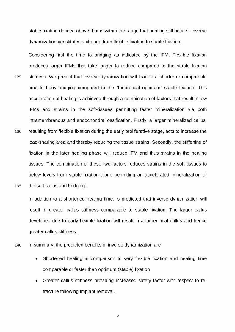
In addition to a shortened healing time, is predicted that inverse dynamization will
result in greater callus stiffness comparable to stable fixation. The larger callus
developed due to early flexible fixation will result in a larger final callus and hence
greater callus stiffness.
In summary, the predicted benefits of inverse dynamization are 140
Shortened healing in comparison to very flexible fixation and healing time
comparable or faster than optimum (stable) fixation
Greater callus stiffness providing increased safety factor with respect to re-
fracture following implant removal.

7
REFERENCES 145
[1] Willenegger H, Perren SM, Schenk R. Primary and secondary healing of bone fractures. Chirurg 1971;42:241–52.
[2] Claes LE, Recknagel S, Ignatius A. Fracture healing under healthy and inflammatory conditions. Nat Rev Rheumatol 2012:1–11.
[3] Claes LE, Wolf S, Augat P. Mechanische Einflüsse auf die Callusheilung. 150 Chirurg 2000;71:989–94.
[4] Goodship AE, Kenwright J. The influence of induced micromovement upon the healing of experimental tibial fractures. J Bone Joint Surg Br 1985;67:650–5.
[5] Muller J, Schenk R, Willenegger H. Experimental studies on the development 155
of reactive pseudarthroses on the canine radius [German]. Helv Chir Acta 1968;35:301–8.
[6] Claes LE, Heigele C, Neidlinger-Wilke C, Kaspar D, Seidl W, Margevicius K, Augat P. Effects of mechanical factors on the fracture healing process. Clin Orthop Relat Res 1998;355 Suppl:S132–47. 160
[7] Epari DR, Kassi J-P, Schell H, Duda GN. Timely fracture-healing requires optimization of axial fixation stability. J Bone Joint Surg Am 2007;89:1575–85.
[8] Claes LE, Augat P, Suger G, Wilke H. Influence of size and stability of the osteotomy gap on the success of fracture healing. J Orthop Res 165
1997;15:577–84. [9] Klein P, Opitz M, Schell H, Taylor WR, Heller MO, Kassi J-P, Kandziora F,
Duda GN. Comparison of unreamed nailing and external fixation of tibial diastases--mechanical conditions during healing and biological outcome. J Orthop Res 2004;22:1072–8. 170
[10] Richardson J, Cunningham JL, Goodship AE, O'Connor B, Kenwright J. Measuring stiffness can define healing of tibial fractures. J Bone Joint Surg Br 1994;76:389–94.
[11] Manjubala I, Liu Y, Epari DR, Roschger P, Schell H, Fratzl P, Duda GN. Spatial and temporal variations of mechanical properties and mineral content 175 of the external callus during bone healing. Bone 2009;45:185–92.
[12] Liu Y, Manjubala I, Schell H, Epari DR, Roschger P, Duda GN, Fratzl P. Size and habit of mineral particles in bone and mineralized callus during bone healing in sheep. J Bone Miner Res 2010;25:2029–38.
[13] Schell H, Epari DR, Kassi J-P, Bragulla H, Bail HJ, Duda GN. The course of 180 bone healing is influenced by the initial shear fixation stability. J Orthop Res 2005;23:1022–8.
[14] Epari DR, Schell H, Bail HJ, Duda GN. Instability prolongs the chondral phase during bone healing in sheep. Bone 2006;38:864–70.
[15] Bergmann G, Graichen F, Rohlmann A. Hip joint contact forces during 185 stumbling. Langenbecks Arch Surg 2004;389:53–9.

8
FIGURES 190
Figure 1 Over the normal course of bone healing, interfragmentary movement (red) decreases whilst callus stiffness (green) increases. The cessation of interfragmentary movement coincides approximately with bony bridging of the callus. 195 Following bony bridging callus stiffness continues to increase as the remainder of the callus is mineralised and remodelled. Callus stiffness reaches a maximum as resorptive activity becomes dominant returning the bone to close to its original anatomy. Time to healing indicated by interfragmentary movement (dashed vertical red line) occurs earlier than maximum callus stiffness (dashed vertical green line). 200

9
Figure 2 Histological section taken after two weeks of healing in an ovine osteotomy model of bone healing. The fragments were stabilized with external fixation on the 205 medial side of the tibia (right side in above). After two weeks greater callus size and mineralisation can be clearly seen on the lateral side (far cortex), where the greatest interfragmentary movement can be expected to occur under uni-lateral external fixation [14].
210

10
Figure 3 Illustrates the hypothesized benefit of inverse dynamization on interfragmentary movement (left) and callus stiffness (right) over the course of 215
healing. Inverse dynamization shortens the time taken for IFMs to fall compared to flexible fixation and stable fixation. Additionally, inverse dynamization results in a callus with a higher stiffness than that resulting from stable fixation and almost as high as that resulting from flexible fixation. Curves are illustrative only.
220