5. OA_Anil edit - Academia International Journals
7
Asian Journal of Medical Research ¦ Volume 7 ¦ Issue 1 ¦ January-March 2018 1 Section: Orthopaedics Abstract Comparative Study of Intramedullary Nailing Versus Locking Compression Plating In Adult Diaphyseal Fractures of Forearm Bones Gaurav Jain 1 , Anil Kumar Singh 2 , Lalit Kumar 3 , Irwin Garg 4 1 Associate Professor, Muzaffar Nagar Medical College, Muzaffar Nagar (U.P.), 251001, 2 Assistant Professor, Muzaffar Nagar Medical College, Muzaffarnagar (U.P.), 3 Professor, Muzaffar Nagar Medical College, Muzaffarnagar (U.P.), 4 Professor & H.O.D, Muzaffar Nagar Medical College, Muzaffar Nagar ( U.P.) Background: “Natural forces within us are true healers of disease” Hippocrates Diaphyseal fractures of forearm bones are very common orthopaedic injury. A range of products are available for its internal fixation. This study was conducted with an aim to determine whether ORIF with plates or intramedullary nailing, is able to produce better clinical outcome. The outcome was assesed on the basis of operating time, union time, functional recovery, complication and cost to patient. Subjects and Methods: The study was conducted in Muzaffar Nagar medical college, Shyamal Trauma & child care and Vedanti Hospital Muzaffar Nagar (U.P.), from 2009 to June 2017. Results: In all 117 patients were treated with intramedullary nailing or plating of adult diaphyseal fracture of fore arm both bone and single bone. Of this 17 were discarded for various reasons and 100 were considered for the present study. Biomechanically, unlocked intramedullary nail (IM) attain stability by a curvature mismatch between bone and the nail, inducing a longitudinal interference fit. If curvature misfit is large reaming is required. Conclusion: Open reduction and internal fixation (ORIF) at present is thought to be superior method to treat such fractures. We have used commonly available locking compression plates and square intramedullary nail of various diameters. Keywords: Fore arm fractures, skeletally mature, intramedullary nail, plate osteosynthesis. Corresponding Author: Anil Kumar Singh, Assistant Professor, Muzaffar Nagar Medical College, Muzaffarnagar (U.P.) Email ID: [email protected] Received: January 2018 Accepted: March 2018 Introduction “As to the disease make a habit of two things: to help or to do at least no harm.” Hippocrates Recent advances in fracture management in fracture BBFA (both bone forearm) have focused on minimally invasive fracture stabilization techniques. As forearm supination and pronation movements are initiated from the proximal and distal radioulnar joints, therefore Radius and Ulna have an important role in the movement of not only forearm but whole upper extremity. [1] Over the last 40 years, anatomical reduction with plate stabilisation has become the standard in adult patients with diphyseal fractures of Radius and Ulna. When operative fixation has been indicated in skeletally mature patients with these fractures a variety of techniques have been reported with Intramedullary Nail (IM) fixation becoming increasingly acceptable. [2] There is currently significant variability in treatment of adolescent with fore arm fracture. [2] Fracture of forearm are classified according to level of fracture, the pattern of fracture, the presence or absence of comminution or segmental, and whether they are open or close. Each of these factors may have some bearing on the type of treatment to be selected and ultimate prognosis. [3] For descriptive purposes, it is useful to divide forearm into thirds, based on linear dimension of Radius and Ulna. Disruption of proximal and distal radioulnar joints with diphyseal is of greater significance to treatment and prognosis but were not taken into consideration in present study and were discarded. Conservative methods are not recommended for these fractures. The deforming effect of muscle strength, continuity of Radial incline and interosseous membrane damage are significant factors that affect the stabilization and maintenance of reduction. [4] It is almost impossible to achieve sustainable and stable fixed reduction in conservative treatment. If there is rotatory malalignment it restricts forearm rotation, simultaneously wrist and elbow joint movement are affected negatively. So due to functional and anatomic features, forearm diphyseal fractures are different from diphyseal fracture of other long bones, and must be evaluated as intra articular fracture with treatment planned accordingly. [5] Open reduction and internal fixation is a widely used and accepted treatment method that is associated with high rate of union and satisfactory functional results. However, ORIF ISSN (0): 2347-3398; ISSN (P): 2277-7253 Original Article
-
Upload
khangminh22 -
Category
Documents
-
view
1 -
download
0
Transcript of 5. OA_Anil edit - Academia International Journals
Asian Journal of Medical Research ¦ Volume 7 ¦ Issue 1 ¦ January-March 2018
1
Section: Orthopaedics
Abstract
Comparative Study of Intramedullary Nailing Versus Locking Compression Plating In Adult Diaphyseal Fractures of Forearm Bones
Gaurav Jain1, Anil Kumar Singh2, Lalit Kumar3, Irwin Garg4
1Associate Professor, Muzaffar Nagar Medical College, Muzaffar Nagar (U.P.), 251001, 2Assistant Professor, Muzaffar Nagar Medical College, Muzaffarnagar (U.P.), 3Professor, Muzaffar Nagar Medical College, Muzaffarnagar (U.P.), 4Professor & H.O.D, Muzaffar Nagar Medical College, Muzaffar Nagar ( U.P.)
Background: “Natural forces within us are true healers of disease” Hippocrates Diaphyseal fractures of forearm bones are very common orthopaedic injury. A range of products are available for its internal fixation. This study was conducted with an aim to determine whether ORIF with plates or intramedullary nailing, is able to produce better clinical outcome. The outcome was assesed on the basis of operating time, union time, functional recovery, complication and cost to patient. Subjects and Methods: The study was conducted in Muzaffar Nagar medical college, Shyamal Trauma & child care and Vedanti Hospital Muzaffar Nagar (U.P.), from 2009 to June 2017. Results: In all 117 patients were treated with intramedullary nailing or plating of adult diaphyseal fracture of fore arm both bone and single bone. Of this 17 were discarded for various reasons and 100 were considered for the present study. Biomechanically, unlocked intramedullary nail (IM) attain stability by a curvature mismatch between bone and the nail, inducing a longitudinal interference fit. If curvature misfit is large reaming is required. Conclusion: Open reduction and internal fixation (ORIF) at present is thought to be superior method to treat such fractures. We have used commonly available locking compression plates and square intramedullary nail of various diameters. Keywords: Fore arm fractures, skeletally mature, intramedullary nail, plate osteosynthesis. Corresponding Author: Anil Kumar Singh, Assistant Professor, Muzaffar Nagar Medical College, Muzaffarnagar (U.P.) Email ID: [email protected]
Received: January 2018 Accepted: March 2018
Introduction
“As to the disease make a habit of two things: to help or to do at least no harm.” Hippocrates Recent advances in fracture management in fracture BBFA (both bone forearm) have focused on minimally invasive fracture stabilization techniques. As forearm supination and pronation movements are initiated from the proximal and distal radioulnar joints, therefore Radius and Ulna have an important role in the movement of not only forearm but whole upper extremity.[1] Over the last 40 years, anatomical reduction with plate stabilisation has become the standard in adult patients with diphyseal fractures of Radius and Ulna. When operative fixation has been indicated in skeletally mature patients with these fractures a variety of techniques have been reported with Intramedullary Nail (IM) fixation becoming increasingly acceptable.[2] There is currently significant variability in treatment of adolescent with fore arm fracture.[2] Fracture of forearm are classified according to level of fracture, the pattern of fracture, the presence or absence of comminution or segmental, and whether they are open or
close. Each of these factors may have some bearing on the type of treatment to be selected and ultimate prognosis.[3] For descriptive purposes, it is useful to divide forearm into thirds, based on linear dimension of Radius and Ulna. Disruption of proximal and distal radioulnar joints with diphyseal is of greater significance to treatment and prognosis but were not taken into consideration in present study and were discarded. Conservative methods are not recommended for these fractures. The deforming effect of muscle strength, continuity of Radial incline and interosseous membrane damage are significant factors that affect the stabilization and maintenance of reduction.[4] It is almost impossible to achieve sustainable and stable fixed reduction in conservative treatment. If there is rotatory malalignment it restricts forearm rotation, simultaneously wrist and elbow joint movement are affected negatively. So due to functional and anatomic features, forearm diphyseal fractures are different from diphyseal fracture of other long bones, and must be evaluated as intra articular fracture with treatment planned accordingly.[5] Open reduction and internal fixation is a widely used and accepted treatment method that is associated with high rate of union and satisfactory functional results. However, ORIF
ISSN (0): 2347-3398; ISSN (P): 2277-7253
Original Article


Asian Journal of Medical Research ¦ Volume 7 ¦ Issue 1 ¦ January-March 2018
5
Jain et al; Intramedullary Nailing Versus Locking Compression Plating
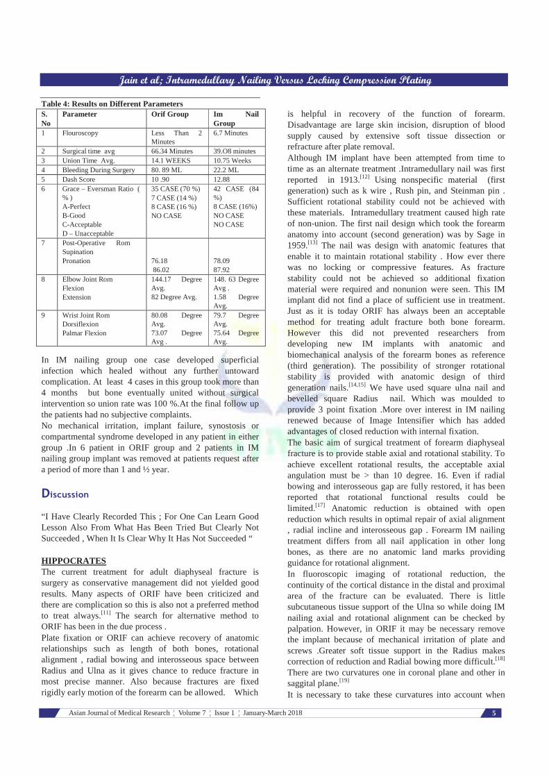
Table 4: Results on Different Parameters S. No
Parameter Orif Group Im Nail Group
1 Flouroscopy Less Than 2 Minutes
6.7 Minutes
2 Surgical time avg 66.34 Minutes 39.O8 minutes 3 Union Time Avg. 14.1 WEEKS 10.75 Weeks 4 Bleeding During Surgery 80. 89 ML 22.2 ML 5 Dash Score 10 .90 12.88 6 Grace – Eversman Ratio (
% ) A-Perfect B-Good C-Acceptable D – Unacceptable
35 CASE (70 %) 7 CASE (14 %) 8 CASE (16 %) NO CASE
42 CASE (84 %) 8 CASE (16%) NO CASE NO CASE
7 Post-Operative Rom Supination Pronation
76.18 86.02
78.09 87.92
8 Elbow Joint Rom Flexion Extension
144.17 Degree Avg. 82 Degree Avg.
148. 63 Degree Avg . 1.58 Degree Avg.
9 Wrist Joint Rom Dorsiflexion Palmar Flexion
80.08 Degree Avg. 73.07 Degree Avg .
79.7 Degree Avg. 75.64 Degree Avg.
In IM nailing group one case developed superficial infection which healed without any further untoward complication. At least 4 cases in this group took more than 4 months but bone eventually united without surgical intervention so union rate was 100 %.At the final follow up the patients had no subjective complaints. No mechanical irritation, implant failure, synostosis or compartmental syndrome developed in any patient in either group .In 6 patient in ORIF group and 2 patients in IM nailing group implant was removed at patients request after a period of more than 1 and ½ year.
Discussion
“I Have Clearly Recorded This ; For One Can Learn Good Lesson Also From What Has Been Tried But Clearly Not Succeeded , When It Is Clear Why It Has Not Succeeded “ HIPPOCRATES The current treatment for adult diaphyseal fracture is surgery as conservative management did not yielded good results. Many aspects of ORIF have been criticized and there are complication so this is also not a preferred method to treat always.[11] The search for alternative method to ORIF has been in the due process . Plate fixation or ORIF can achieve recovery of anatomic relationships such as length of both bones, rotational alignment , radial bowing and interosseous space between Radius and Ulna as it gives chance to reduce fracture in most precise manner. Also because fractures are fixed rigidly early motion of the forearm can be allowed. Which
is helpful in recovery of the function of forearm. Disadvantage are large skin incision, disruption of blood supply caused by extensive soft tissue dissection or refracture after plate removal. Although IM implant have been attempted from time to time as an alternate treatment .Intramedullary nail was first reported in 1913.[12] Using nonspecific material (first generation) such as k wire , Rush pin, and Steinman pin . Sufficient rotational stability could not be achieved with these materials. Intramedullary treatment caused high rate of non-union. The first nail design which took the forearm anatomy into account (second generation) was by Sage in 1959.[13] The nail was design with anatomic features that enable it to maintain rotational stability . How ever there was no locking or compressive features. As fracture stability could not be achieved so additional fixation material were required and nonunion were seen. This IM implant did not find a place of sufficient use in treatment. Just as it is today ORIF has always been an acceptable method for treating adult fracture both bone forearm. However this did not prevented researchers from developing new IM implants with anatomic and biomechanical analysis of the forearm bones as reference (third generation). The possibility of stronger rotational stability is provided with anatomic design of third generation nails.[14,15] We have used square ulna nail and bevelled square Radius nail. Which was moulded to provide 3 point fixation .More over interest in IM nailing renewed because of Image Intensifier which has added advantages of closed reduction with internal fixation. The basic aim of surgical treatment of forearm diaphyseal fracture is to provide stable axial and rotational stability. To achieve excellent rotational results, the acceptable axial angulation must be > than 10 degree. 16. Even if radial bowing and interosseous gap are fully restored, it has been reported that rotational functional results could be limited.[17] Anatomic reduction is obtained with open reduction which results in optimal repair of axial alignment , radial incline and interosseous gap . Forearm IM nailing treatment differs from all nail application in other long bones, as there are no anatomic land marks providing guidance for rotational alignment. In fluoroscopic imaging of rotational reduction, the continuity of the cortical distance in the distal and proximal area of the fracture can be evaluated. There is little subcutaneous tissue support of the Ulna so while doing IM nailing axial and rotational alignment can be checked by palpation. However, in ORIF it may be necessary remove the implant because of mechanical irritation of plate and screws .Greater soft tissue support in the Radius makes correction of reduction and Radial bowing more difficult.[18] There are two curvatures one in coronal plane and other in saggital plane.[19] It is necessary to take these curvatures into account when
Asian Journal of Medical Research ¦ Volume 7 ¦ Issue 1 ¦ January-March 2018
6
Jain et al; Intramedullary Nailing Versus Locking Compression Plating
preoperatively shaping the nails to appropriate intramedullary anatomy. In the current study Radius nails were shaped parabola. Elasticity of nail and 3 point fixation principle of parabolic shape rotational stability was achieved. In addition, by conforming to the Radial bowing the optimal intesosseous space was formed. Few studies have compared ORIF and IM nailing treatment results in forearm diaphyseal fractures. These studies have compared, in particular, union status, time to union and functional evaluation criteria. Anatomic and close to anatomic reduction is obtained with ORIF. Axial and rotationally rigid stable fixation is obtained. However drainage of fracture haematoma has negative effect on union and it has been reported that excessive soft tissue and periosteal stripping could cause union problems .Osseous feeding is impaired due to super periosteal pressure of conventional plates and effects negatively on union. Risk of refracture is increased due to cortical atrophy which develops in the screw application areas. It is necessary to apply immobilisation, regardless of the stability of the fixation. Cosmetic problems may also develop with surgical approach.[6,20] ORIF causes more bleeding but there is no fluoroscopic guidance required for open surgical intervention is an advantage. If used at all for checking length of screw and plate placement on bone it can be said that exposure to radiation is drastically less than IM group. Despite of these advantages / disadvantage of the method union rate of 96 % was achieved in this series (range 87 % to 98%).[4,6,21] Time to union has been reported between 14 weeks to 33 weeks. We had average union time 14. 1 weeks taken radiologically. Functional evaluation results generally at satisfactory levels.[22,23] Bleeding time in the plate group was measured 80.89 ml (range 38 ml to 252 ml). The mean operating time was 66.34 minutes (range 37 to 109 minutes). Im nailing very little soft tissue damage in surgical application and provide cosmetic superiority and bleeding is much less 22.2 ml average. IM implant generally have property of stress distribution and stronger callus tissue is formed. An important cosmetic advantage is that the implant can be removed from the same incision. Length is protected in segmental forearm fracture. However exposure to radiation is a significant disadvantage. Immobilisation period is shorter due to more soft tissue support (because of less requirement of tissue dissection). Nailing treatment is contraindicated in patients who have open epiphyseal line, when there is infection and if marrow is less than 2.5 mm in diameter. In IM nail application , union rate of 94 % - 100 % and time to union 10 weeks to 4.4 months have been reported.[14,15,24] Good functional have been reported similar to plate osteosynthesis . In the present study 100 % union was achieved at a mean time period of 10.75 weeks (range 9 weeks to 22 weeks). The amount of bleeding in IM group was 22.2 ml (range 10 ml to 78 ml.) The mean operating
time was 38.08 minutes (range 25 minutes to 68 minutes). A statistically significant difference was determined between two groups with respect to time for surgery, time to union and amount of bleeding. No significant difference was determine with respect to radiological and functional outcome (Grace – Eversman criteria), DASH score, and functional joint range of movement [Table 4). The shorter time to union of IM group compared to ORIF could have been due to not draining the fracture haematoma and early mobilisation. As IM method is less invasive, the amount of bleeding is reduced. As no dissection of soft tissue is required body has less to heal. Controlled exposure in ORIF was thought to have prolonged the operating time. The areas of application of both treatment methods carry potential risks. In the proximal Radial diaphyseal fractures the Posterior Intreosseous Nerve is at risk.[25] There is risk of damage in open reduction during surgical exposure and in IM nailing during locking. But in current series as no locking was done and due to careful dissection no harm to nerve was done during ORIF. In the area of nail application in Radius Extensor Pollicis Longus tendon and superficial branch of Radial nerve are at risk.[26] No such complication were seen in present study even in followup up to 5 years. The removal of the internal fixation material used after union is controversial. In open or fragmented fractures or those that have resulted from high energy trauma, when there is insufficient compression or reduction in fragmented fracture and when there is another fracture in same extremity, the rate of refracture has been reported to increase.[27] In current study implant were removed after at least one year after surgery on patient request. 4 patient in ORIF group and 1 patient in IM group implants were removed.
Conclusion
“The Life So Short; the Craft So Long To Learn”
HIPPOCRATES The current treatment method for adult diaphyseal fracture is ORIF. The results of present study showed IM nailing treatment to be superior to ORIF with respect to less operating time, less blood loss, early union and good functional results. However no difference between the two methods was found at the end of one year. Due to shorter operating time, shorter time to union and cosmetic advantage IM nailing treatment can be considered good alternative method to ORIF in the treatment of adult forearm diaphyseal fracture. As we had two surgical team separately for each of the two surgery their expertise lead to no iatrogenic complications. This also shows that one particular surgical team should devote itself to one type of surgery to excel in it thus providing much relief to patients. Though cost did not matter in treatment but IM nailing costed much less than plating.

Asian Journal of Medical Research ¦ Volume 7 ¦ Issue 1 ¦ January-March 2018
7
Jain et al; Intramedullary Nailing Versus Locking Compression Plating
References
1. Ahmet kose MD, Ali Aydin MD, Erzunrum; Regional traning and research hospital Enzurum Turkey.
2. Wall L, Donwall JC, Schoenecker PL, Keeber KA, Dobles MB, Lehmann SJ, et all. Titanium elastic nailing in radius ulna fractures in adolescent J. pediatric orthop B. 2012; 21, 482-488.
3. Burcholz R Rockwood, Green, and Wilkins fracture. 1 ed. Philadelphia PA Lippincot & Willikins.2006.
4. Anderson LD, Sisk D, Tooms RE, Park WI 3, Compression plate fixation in acute diaphyseal fracture of Radius and Ulna. JBJS Am.1975; 57 .287-297.
5. Crenshaw AH, Fracture of shoulder, arm and forearm. In Canale ST, Daughtery K, Jones L editors .Campbells operative orthopaedics.10. St. Louis Mosely 2003. P-3049-58.
6. Lee SK, Kim KI, Lee JW, Choy WS. Plate osteosynthesis versus intramedullary nailing for both bone forearm fracture. Eu . J. Orthop . Surg . Traumatol .2014, 24: 769 – 776.
7. Behnke NM, Redjal HR, Nguyen VJ, Zinar DM. Internal fixation of diaphyseal fracture of the forearm a retrospective comparasion of hybrid fixation versus dual platying. J Orthop. Trauma 2012; 26: 611 – 616.
8. Grace TG, Eversman WW Jr., Forearm fractures: treatment by rigid fixation with early motion. JBJS surgery Am. 1980: 62; 433- 438.
9. Hudare PL, Amodio PC, Bombardier C. Development of an upper extremity outcome measure. DASH (Disability of Arm, Shoulder and Hand.)Score. The upper extremity collaborative group (UECG). Am.J Ind. Med. 1996: 29, 602 – 608.
10. Gustilo RB, Anderson JT, Prevention of infection in the treatment of one thousand and twenty five open fractures long bones .Retrospective and prospective analysis. JBJS Am. 1976; 58: 453-458.
11. Bartonic J, Kosanek M, Jupiter JB. History of operative treatment of diaphyseal fracture.
12. Behnke NM, Rejdal HR, Nguyen VT, Zinar DM. Internal fixation of diaphyseal fracture of the forearm, a retrospective comparasion of hybrid fixation versus dual plating . J orthop. Trauma .2012; 26: 611 – 16.
13. Sage FP. Medullary fixation of fracture of forearm. A study of the medullary canal of radius and a report of fifty fracture of the Radius treated with a prebent triangular nail. JBJS Am. 1959, 41 – A: 1489 – 1516.
14. Lee YH, Lee SK, Chung MS, Baek GH, Gong HS, Kim KH
.Interlocking contoured IM nail fixation for selected diaphyseal fracture of FA in adult. JBJS Am.; 2008: 1881 – 1888.
15. Kose A, Aydm A, Enzmirk N, Topal M, Can CE, Yilar S. Intramedullary nailing of adult isolated diaphyseal Radius fracture.Ulus Trauma Acil Cirrahi Derg .2016; 22: 184 – 191.
16. Schemitrch EH, Richard RR. The effect of malunion on functional outcome after plate fixation of fracture of both bone forearm in adults. JBJS Am. 1992; 74 : 1068 -1078 .
17. Tarr PR , Garfinkel AI , Sarminto A . The effect of angular and rotational deformity of both bone forearm . An in vitro study . JBJS Am . 1984 ; 66 : 65 – 70 .
18. Bot AG , Doornberg JN , Lindon hovius AL , Ring D , Goslings JC , Vondijk CN . Long term outcome of fracture of both bone forearm . JBJS Am . 2011 ; 93 : 527 – 530 .
19. Bartonic J , Nanka O , Tucek M . Internal fixation of radial shaft fracture ; Anatomical and Biomechanical principle . ( Article in czeh . ) Rozhe chir ; 2015 : 94 : 425 – 236. ( abstract )
20. Kim SB , Heo YM , Yi JW , Lee JB , Lim BG . Shaft fracture of both forearm bone . The out come of surgical treatment with plating only and combined plating and intramedullary nailing . Clin. Orthop . Surgery .2015 ; 7 : 282 – 290 .
21. Ozkaya U , Kellic A , Ozdong U , Beng k , Kaluic cuoglu Y . Comparasion between locked intramedullary nailing and plate osteosynthesis in the management of adult diaphyseal forearm fractures. Acta Orthop . Traumatol Turc. 2009 ; 43 : 14 – 20 .
22. Stenes CT , Tendevis HJ , Plate osteosynthesis of simple fracture LCP VS DC plate .
23. Jones DB , Jr. Kakal S . Adult diaphyseal fractures nail vs plate osteosynthesis . J. Hand surgery Am . 2011 ; 36 : 1216 – 1219 .
24. Klose A , Aysin A , Enzirmick N , Toppal M , Can CE . Treatment of isolated Ulnar shaft fracture in adult , with a new intramedullary nail . Minerva Orthop . e tramatology . 2015 . 66; 123 – 131 .
25. Talor OB Jr . ,Bose JM , Sims SH , Kellam JF . Iartogenic posterior interosseous nerve injury ; is intramedullary static locked nailing of the Radius feasible ? J . Orthop . Trauma 1995 : 9 : 427 – 429.
26. Fanvale J , Blazer P . Extensor pollicis longus tendon rupture in an adult after intramedullary nailing of a Radius fracture , case report . J . Hand surgery Am . 2009 ; 34:627 -629 .
27. Deluca PA , Lindsey RW , Rlrine PA. Refracture of bone of forearm after the removal of compression plates . JBJS Am . 1988 ; 70 : 1372 -1376 .
Copyright: © the author(s), publisher. Asian Journal of Medical Research is an Official Publication of “Society for Health Care & Research Development”. It is an open-access article distributed under the terms of the Creative Commons Attribution Non-Commercial License, which permits unrestricted non-commercial use, distribution, and reproduction in any medium, provided the original work is properly cited.
How to cite this article: Jain G, Singh AK, Kumar L, Garg I. Comparative Study of Intramedullary Nailing Versus Locking Compression Plating In Adult Diaphyseal Fractures of Forearm Bones. Asian J. Med. Res. 2018;7(1):OR01-OR07. DOI: dx.doi.org/10.21276/ajmr.2018.7.1.5
Source of Support: Nil, Conflict of Interest: None declared.