4 - MENA-HL
56
-
Upload
khangminh22 -
Category
Documents
-
view
1 -
download
0
Transcript of 4 - MENA-HL


Journal of Applied Hematology - Volume 10, Supplement 1, 2019 i
EDITORIAL BOARD
Journal of Applied Hematology(An Official publication of Saudi Society of Hematology and Pan Arab Hematology Association)
EDITORS-IN-CHIEFAbdulkareem Al Momen - MD, FRCPCProfessor of Medicine & Hematology, National cancer and blood center, KSA [email protected]
Tarek Owaidah - MD, FRCPAProfessor and consultant Hematologist, Alfaisal University King Faisal Specialist Hospital & Research Centre, KSA [email protected]
ASSOCIATE EDITORS
Mohamed H. Qari - MD, FRCPAProfessor of Hematology, King Abdulaziz University Hospital, [email protected]
Syed Z. A. Zaidi - MBBS, FCPS, FRCPConsultant Hematologist, Assistant Professor, KFMC, [email protected]
Ali Al Ahmari, MDAssociate Professor, College of Medicine, Alfaisal University, [email protected]
Feras Alfraih, MD, FACPConsultant Hematologist, King Faisal Specialist Hospital & Research Centre, [email protected]
BIOSTATISTICIAN
Mr. Khawar SiddiquiPediatric Hematology/Oncology,KFSHRC, Riyadh, [email protected]
EDITORIAL ASSISTANTMs. Regina Maria Isabelle A. Concepcion, KSA [email protected]
EDITORIAL BOARD MEMBERS
Paul Gaingrande, UKThomas Ortel, USALeonardo Brandao, CanadaSoad Al Jaouni, KSASukesh Nair, IndiaAzza Al Zahrani, KSAHamad Al Omar, KSAGeorge Roberts, CanadaRanda Al Nounou, KSAFahad Al Manjomi, KSAYasser Wali, Oman
ADVISORY BOARD MEMBERS
Aamer Aleem, KSAKhalid Al Saleh, KSAHazza Al Zahrani, KSAAbdullah Al Jefri, KSAAdekunle Adekile, KuwaitMohammed Al Sheef, KSAAhmed Alrustamani, UAEFlora Peyvandi, ItalySalam Al Kindi, OmanNasir Bakshi, KSAMahasen Al Saleh, KSAGhulam Mufti, UKTurky Alwasaidi, KSAAhmed Absi, KSANervana Bayoumi, KSAAhmed Tarawah, KSAAyman Hijazi, KSAMagdy Ekiaby, EgyptAbdul Ghani Tabaki, JordanMuzaff er Demir, TurkeyMervat Mattar, EgyptNevine Kassim, EgyptFahad Al Hamid, KSAMohammed Zulali, KSAAhmed Al Suliman, KSA
Print ISSN: 1658-5127, E-ISSN: 2454-6976

ii Journal of Applied Hematology - Volume 10, Supplement 1, 2019
Review Article: Authors are invited by the Editors-in-Chief for submission of review articles which should focus on recent scientific or clinical advances in an area of interest to those in the field of Hematology. Authors wishing to submit an unsolicited Review Article are invited to contact the Editor-in-Chief prior to submission, in order to screen the proposed topic for relevance and priority. Review articles must be concise and critical and should include appropriate references to the literature. All Review Articles, including those solicited by the Editors, are rigorously peer reviewed before a final publication decision is made.
Review articles should not exceed 4,000 words in length, must include an abstract of 200 words or fewer, and may not have more than 100 references. The use of tables and color figures to summarize critical points is encouraged.
Original Article: Journal of Applied Hematology accepts original articles with a maximum length of 5000 words not counting the abstract, table and figures. The sections of an Original Article should be well-arranged in Abstract, Introduction, Methods (must include sufficient information to allow readers to understand the article content), Results, Discussion, Acknowledgements, Authorship Contributions and Disclosure of Conflicts of Interest, References, Tables, Figure Legends, and Figures. The Abstract should not exceed 250 words and should be constructed a single paragraph with no subheadings. Supplemental data - to be published online only - may include additional information regarding methodology, supplemental figures or tables, or primary data sets; and it must be submitted with the original manuscript submission so that it can be peer reviewed.
All clinical submissions must have been approved by an ethics committee or institutional review board. Any involvement of medical writers/researchers, particularly those employed or supported by the pharmaceutical industry, in the writing of an article must be clearly defined and disclosed in the Authorship and/or the Acknowledgements section(s) as appropriate. This type of involvement must also be disclosed to the Editor-in-Chief in the Cover Letter. For more information, see “conflict of interest disclosure” and the “authorship information” sections below.
Original research articles of exceptional scientific importance may be considered for designation as “Plenary Paper”. The decision to highlight an article as a Plenary Paper rests entirely with the Editors. Sometimes the Editors may invite experts in the field to write brief commentaries introducing and placing into context selected original research articles included in the issue.
Image in Hematology: Journal of Applied of Hematology welcomes submission of photo images along with brief case descriptions to serve as a regular teaching feature. We place importance to the value that the microscope adds to the history and physical examination. One photograph (peripheral smear, or views of blood forming tissue) is preferable, but two views are also acceptable (one of those may be a related clinical or radiology or laboratory preparation image). Each submission must contain a single, high-resolution figure formatted as a TIFF (minimum 300 dpi) and a discussion of up to 200 words describing the clinical case linked to the image. Each submission should have no more than three authors and maximum of two references if unavoidable. When submitting a manuscript for review, image file formats accepted for uploading are GIF, JPEG (.jpg), PDF, and EPS. The total file size of the PDF for peer review should not exceed 5 MB.
Letter to the Editor: Constructive comments related to published articles or other current topics in Hematology may be published as letter to the editor. Letters should include no more than 1000 words of text, 5–10 references, and 1 figure or table. No abstract is needed, but a brief title is required. Letter may also contain significant new primary data and/or the inclusion of a figure to make very important points. Stand-alone brief communications on basic or clinical topics in hematology may also be considered for publication as a Letter to the Editor, based on priority and interest to readership as evaluated by peer review. Authors of published articles are encouraged to respond with
INSTRUCTIONS TO AUTHORS
Journal of Applied Hematology(An Official publication of Saudi Society of Hematology and Pan Arab Hematology Association)
their own comments to those Letters that have been sent in reference to their articles. Letters that are not directly related to a published article, that duplicate points similar to those of already published comments, or that are characterized by blasphemy, personal attacks, unprofessional tone or content, or offensive, abusive or defamatory language will not be entertained.
Case Report: Journal of Applied Hematology welcomes well described reports of one or more cases of informative clinical observation (maximum 2000 words).
Case reports submitted should make a contribution to medical knowledge and must have educational value or highlight the need for a change in clinical practice or diagnostic/prognostic approaches. See details in “Instructions for Authors” online in “instructions and forms” at http://mc.manuscriptcentral.com/jahem.
These reports should include relevant positive and negative findings from history, examination and investigation, and can include clinical photographs, provided identifiable photographs are accompanied by written consent to publish from the patient(s). Case reports should preferably include an up-to-date brief review of all previous cases published in the field.
Authors are encouraged to describe how the Case report is rare or unusual as well as its educational and/or scientific merits in the covering letter that will accompany the submission of the manuscript.
Case report submissions will be assessed by the Editors and will be sent for peer review if considered appropriate for the journal. Authors should seek written and signed consent to publish the identifiable information from the patients or their guardians prior to submission. The submitted manuscript must include a statement to this effect in the Consent section. When needed the editorial office may request copies of the informed consent documentation.
Test Development/Validation: Author can report the results of a newly developed test or results of a test that has been scientifically validated and has clinical application. Assay validation results should be accompanied by an appropriate measure of the precision of the estimates like the standard deviation, standard error of the mean, coefficient of variation, or 95% confidence limits. It maybe necessary to include data relating to within-assay and between assay variability.
In presenting results for new assays, it may be necessary to include data on the following: 1) within-assay variability; 2) between-assay variability; 3) slope of the dose-response curve; 4) mid-range of the assay; 5) least-detectable concentration; 6) data on specificity; 7) data on parallelism of standard and unknown and on recovery; and 8) comparison with an independent method for assay of the compound.
Short Communication: The journal of Applied Hematology will consider short manuscripts documenting either experimental results or informative clinical observations for publication in this category. Short Communication are not intended to allow publication of incomplete or preliminary findings. The review process is similar to Original Articles. Short Communication may not exceed 1,200 words of text not counting the abstract, figure legends, and references; and the abstracts must not exceed 200 words and should be a single paragraph with no subheadings. Only 2 figures/tables and up to 25 references may be included. The sections of a Short Communication should be ordered as follows: Abstract, Introduction, Methods sufficiently informative to allow reproduction of the data, followed by a combined “Results and Discussion” section, Acknowledgements, Authorship Contributions and Conflict of Interest Disclosure, References, Tables, Figure Legends, and Figures.
For detailed Instructions for Authors visit http://www.jahjournal.org

Journal of Applied Hematology - Volume 10, Supplement 1, 2019 iii
The journalJournal of Applied Hematology (ISSN: Print-1658-5127 and E-ISSN No: 2454-6976) is peer-reviewed journal published on behalf of Saudi Scientific Society of Hematology. The journal publishes articles on the subject of Adult and pediatric hematologists, hematopathologists, trainees, other physicians, Physicians, Nurses, Technologists. The Journal is published quarterly in the first week of January, April, July and October.
Information for authorsThere are no page charges for submissions to the journals. Please check http://www.jahjournal.org/contributors.asp for details.All manuscripts must be submitted online at www.journalonweb.com/joah
Subscription informationCopies of the journal are provided free of cost to the members of Saudi Scientific Society of Hematology. A subscription to Journal of Applied Hematology comprises 4 issues. Prices include postage. Annual Subscription Rate for non-members-
For mode of payment and other details, please visit www.medknow.com/subscribe.asp.
Claims for missing issues will be serviced at no charge if received within 60 days of the cover date for domestic subscribers, and 3 months for subscribers outside India. Duplicate copies cannot be sent to replace issues not delivered because of failure to notify publisher of change of address.
The journal is published and distributed by Wolters Kluwer India Pvt. Ltd. Copies are sent to subscribers directly from the publisher’s address. It is illegal to acquire copies from any other source. If a copy is received for personal use as a member of the association/society, one cannot resale or give-away the copy for commercial or library use.
Change of addressNonmembers please send change of address information to [email protected].
Advertising policiesThe journal accepts display and classified advertising. Frequency discounts and special positions are available. Inquiries about advertising should be sent to Wolters Kluwer India Pvt. Ltd., [email protected].
The journal reserves the right to reject any advertisement considered unsuitable according to the set policies of the journal.The appearance of advertising or product information in the various sections in the journal does not constitute an endorsement or approval by the journal and/or its publisher of the quality or value of the said product or of claims made for it by its manufacturer.
CopyrightThe entire contents of the Journal of Applied Hematology are protected under Indian and international copyrights. The Journal,
however, grants to all users a free, irrevocable, worldwide, perpetual right of access to, and a license to copy, use, distribute, perform and display the work publicly and to make and distribute derivative works in any digital medium for any reasonable non-commercial purpose, subject to proper attribution of authorship and ownership of the rights. The journal also grants the right to make small numbers of printed copies for their personal non-commercial use.
PermissionsFor information on how to request permissions to reproduce articles/information from this journal, please visit www.jahjournal.org
Disclaimer The information and opinions presented in the Journal reflect the views of the authors and not of the Journal or its Editorial Board or the Publisher. Publication does not constitute endorsement by the journal. Neither the Journal of Applied Hematology nor its publishers nor anyone else involved in creating, producing or delivering the Journal of Applied Hematology or the materials contained therein, assumes any liability or responsibility for the accuracy, completeness, or usefulness of any information provided in the Journal of Applied Hematology, nor shall they be liable for any direct, indirect, incidental, special, consequential or punitive damages arising out of the use of the Journal of Applied Hematology. The Journal of Applied Hematology, nor its publishers, nor any other party involved in the preparation of material contained in the Journal of Applied Hematology represents or warrants that the information contained herein is in every respect accurate or complete, and they are not responsible for any errors or omissions or for the results obtained from the use of such material. Readers are encouraged to confirm the information contained herein with other sources.
AddressesEditorial officeMs. Regina Maria Isabelle A. Concepcion, KSA Journal of Applied Hematology, Riyadh KSA. E-mail: [email protected] Website: www.jahjournal.org
Published byWolters Kluwer India Pvt. Ltd.A-202, 2nd Floor, The Qube, C.T.S. No.1498A/2Village Marol, Andheri (East), Mumbai - 400 059, India.Phone: 91-22-66491818Website: www.medknow.com
Printed atDhote Offset Technokrafts Pvt. Ltd., Jogeshwari, Mumbai, India.
GENERAL INFORMATION
Journal of Applied Hematology(An Official publication of Saudi Society of Hematology and Pan Arab Hematology Association)

iv Journal of Applied Hematology - Volume 10, Supplement 1, 2019
CONTENTS
Journal of Applied Hematology(An Official publication of Saudi Society of Hematology and Pan Arab Hematology Association)
January 2019 Volume 10 Supplement 1
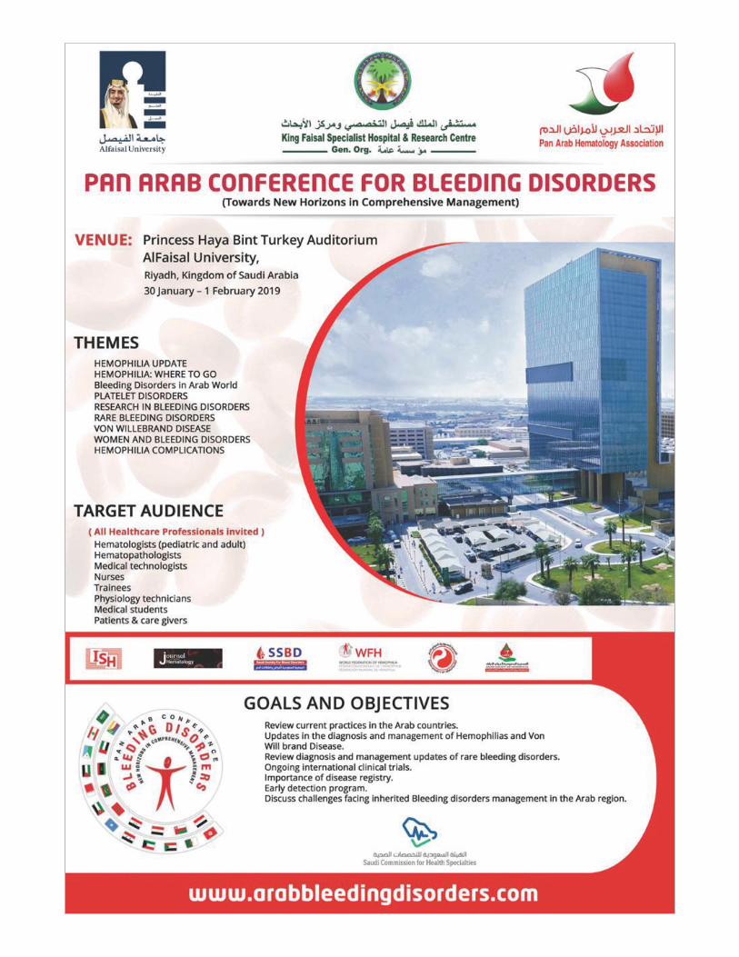
ABSTRACTPan Arab Conference for Bleeding Disorders ........................................................................................................................... S1

Journal of Applied Hematology - Volume 10, Supplement 1, 2019 v
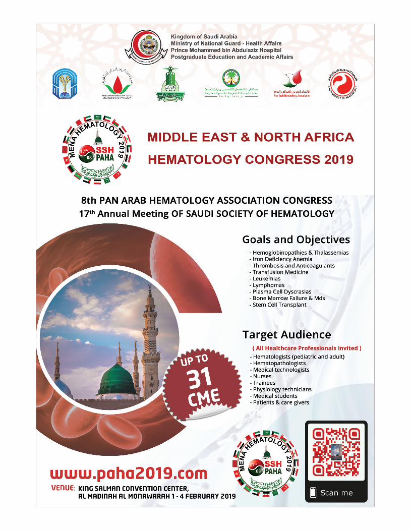
On behalf of organizing, scientific and abstract review committees’ members, we cordially welcome and invite you to the Pan Arab Conference for Bleeding Disorders and The Middle East and North Africa Hematology Congress 2019, & 8th Pan Arab Hematology Association (PAHA) Congress and 17th Annual Meeting of Saudi Society of Hematology (SSH).
The scientific committee has come up with a very interesting scientific program that represents the interests of Hematology professionals’ and promotes the multidisciplinary of the field. All multimodality approaches in the management of hematological disorders are addressed.
We deeply thank the abstracts submitters. Where we have reached a new record this year with sum of 152 abstracts submitted to both congresses, 102 abstracts have been accepted. All abstracts have gone over multi-steps review and scoring. We hope this year hematology knowledge take the up-coming congresses to a new era of research-oriented congresses.
We wish everyone a fruitful and fulfilling time during this conference. Your active participation will give an important boost to the success of this event.
Dr. Ahmad Tarwah, MDChairman, Scientific and abstract review Committee
Dr. Turki Alwasaidi, MDPresident, Middle East and North Africa Hematology Congress 2019, 8th Pan Arab Hematology Association Congress, 17th
Annual Meeting of Saudi Society of Hematology
Prof. Tarek Owaidah, MDPresident, Pan Arab Conference for Bleeding Disorders
Welcome message

vi Journal of Applied Hematology - Volume 10, Supplement 1, 2019
The Indian Journal of Applied Hematology now accepts articles electronically. It is easy, convenient and fast. Check following steps:
Journal of Applied Hematology on Web
Facilities
•Submission of new articles with images•Submission of revised articles•Checking of proofs•Track the progress of article until published
Advantages
•Any-time, any-where access•Faster review•Cost saving on postage•No need for hard-copy submission•Ability to track the progress•Ease of contacting the journal
Requirements for usage
•Computer and internet connection•Web-browser (Latest versions - IE,
Chrome, Safari, FireFox, Opera)•Cookies and javascript to be enabled in
web-browser
Online submission checklist
•First Page File (rtf/doc/docx file) with title page, covering letter, acknowledgement, etc.
•Article File (rtf/doc/docx file) - text of the article, beginning from Title, Abstract till References (including tables).File size limit 4 MB. Do not include images in this file.
•Images (jpg/jpeg/png/gif/tif/tiff): Submit good quality colour images. Each image should be less than 10 MB) in size
•Upload copyright form in .doc / .docx / .pdf / .jpg / .png / .gif format, duly signed by all authors, during the time mentioned in the instructions.
Help
•Check Frequently Asked Questions (FAQs) on the site
•In case of any difficulty contact the editor
1 Registration•Register from http://www.journalonweb.com/joah as a new
author (Signup as author)•Two-step self-explanatory process
2 New article submission•Read instructions on the journal website or download the same
from manuscript management site•Prepare your files (Article file, First page file and Images,
Copyright form & Other forms, if any)•Login as an author•Click on ‘Submit new article’ under ‘Submissions’•Follow the steps (guidelines provided while submitting the
article)•On successful submission you will receive an acknowledge-
ment quoting the manuscript ID
3 Tracking the progress•Login as an author•The report on the main page gives status of the articles and its
due date to move to next phase•More details can be obtained by clicking on the ManuscriptID•Comments sent by the editor and reviewer will be available
from these pages
4 Submitting a revised article•Login as an author•On the main pageclick on ‘Articles for Revision’•Click on the link "Click here to revise your article" against the
required manuscript ID•Follow the steps (guidelines provided while revising the article)•Include the reviewers’ comments along with the point to point
clarifications at the beginning of the revised article file. •Do not include authors’ name in the article file. •Upload the revised article file against New Article File -
Browse, choose your file and then click “Upload” OR Click “Finish”
•On completion of revision process you will be able to check the latest file uploaded from Article Cycle (In Review Articles-> Click on manuscript id -> Latest file will have a number with ‘R’, for example XXXX_100_15R3.docx)
http://www.journalonweb.com/joah

© 2019 Journal of Applied Hematology | Published by Wolters Kluwer - Medknow S1
A-001: UK haemophilia teams’ knowledgeof risk assessment for prolonged bleedingassociated with dental procedures
Aza Rahman, Najla Nizarali1, Alison Dougall2, Blánaid Daly2
Ministry of Health, Kurdistan Region of Iraq, 1Guys and St Thomas’ NHS Foundation Trust, UK, 2Division of Child and Public Dental Health, Dublin Dental University Hospital, School of Dental Science, Trinity College Dublin, Lincoln Place Dublin, Ireland
Introduction: Optimal delivery of dental care for adults with congenital bleeding disorders (CBD) requires close collaboration between haemophilia multi-disciplinary teams (MDT) and dentists. Aim: To explore UK haemophilia MDTs’ knowledge of dental procedures and associated haemostatic management in adults with CBD. Methods: Staff (N=180) from haemophilia centres in the UK were invited to participate in a questionnaire based study using a web-based tool. The questionnaire assessed participants’ knowledge, adherence and appropriateness of application of UK guidance on haemostatic management of common dental procedures. Results: The response rate was 24% (n= 41). While most responders (87%; n=34) reported they adhered to guidelines, participants’ knowledge of guidance was poor. Only 36% (n=15) of the sample applied guidance appropriately in three common dental scenarios. There was a tendency for responders to assign bleeding risk based on a patient’s previous history of prolonged bleeding (for any reason) rather than to the bleeding risk associated with the proposed dental procedure. Conclusion and Recommendations: While haemophilia MDTs were aware of current guidelines, their knowledge of the guidelines and ability to risk assess dental procedures was poor. There was a tendency to overprescribe systemic haemostatic measures for dental procedures. Education initiatives to aid decision making are needed.
Keywords: Bleeding, dental, hemophilia
A-002: Almadinah join hemophilia clinicstrategy and benefits
Mousa Mohammad, Thalath AlhaosawiMadinah Maternity Hospital, Madinah, Saudi Arabia
The comprehensive care provided at hemophilia treatment centres (HTCs) significantly reduces medical complications for people with hemophilia. There is a substantially lower incidence of ill health and early death in people who use HTCs than for those who do not. It also showed a decrease in bleeding-related hospitalizations. In Almadinah joint hemophila clinic we provides state-of-the-art medical care, we offer emotional support, school and work support, insurance consultation, outdoor support and education programs
for our hemophilia patients. Our expert team also works closely with healthcare providers in our local community to meet our hemophilia patients specific needs and to improve their quality of life. Our focus is not only treating issues when they occur, we are keenly aware how to prevent many complications that can occur related to bleeding disorder. This combination of expert treatment and proactive prevention lead to improved health for all of our hemophila patients in Almadinah region.
Keywords: Almadinah, benefits, hemophilia
A-003: Case series of four-factor prothrombincomplex concentrate for anticoagulantreversal at emergency department of anacademic hospital of Saudi Arabia
Waad H. Al-Kathiri1,2, Anas A. Khan3
1Department of Clinical Pharmacy Services, King Saud University Medical City, King Saud University, 2Department of Clinical Pharmacy, College of Pharmacy, King Saud University, 3Department of Emergency Medicine, College of Medicine, King Saud University, Riyadh, Saudi Arabia
Background: Recent international guidelines recommend the use of 4-factor prothrombin complex concentrate (PCC4) over fresh frozen plasma (FFP) for reversal of oral anticoagulant in life-threatening bleeds. The purpose of this study is to describe the effectiveness of low dose (25 mg/kg) PCC4 in controlling bleeds event caused by oral anticoagulant. Methods: Retrospective case series included nine patients who visit Emergency department with acute bleeding events controlled with low dose 4-factor prothrombin complex concentrate. Chart review was conducted between January 2017 and June 2018. International Normalized Ratio (INR), thromboembolic events and hypersensitivity reaction were documented. Results: Eight patients were taking anticoagulant for the treatment of Atrial fibrillation and one patient was taken anticoagulant for Behcet’s disease. Baseline mean [±SD] INR ([±5.3] 6.4), Anticoagulant caused Intracerebral hemorrhage in two patients, and Gastrointestinal bleeding was the most complication caused by anticoagulant. PCC4 was given in dosing range 25-30 unit/kg based on estimated patient weight, after 60 minutes the post-PCC4 mean [±SD] INR ([±0.95] 1.6), PCC4 contributed significant reduction in INR (p=0.02). Six patients reached INR < 1.5, two patients INR <2, and only one patient with INR 4.3. No addition PCC4 doses were needed to control the bleeding event. None of patients experienced a thromboembolic events or hypersensitivity reaction 14 days post PCC4. Conclusion: Low dose (25 unit/kg) PCC4 contributed to efficient reduction of INR in patients with lifethreatening bleeding with low risk of thromboembolism event. We recommend a larger study to evaluate INR rebound and re-bleeding for post PCC4 along with thromboembolism event beyond the 14 days.
Keywords: Anticoagulant, emergency, prothrombin
Abstract
Pan Arab Conference for Bleeding Disorders
Abstract
S2 Journal of Applied Hematology - Volume 10, Supplement 1, 2019
biosensor was successfully performed and showed high sensitivity and selectivity. This work represents novel work and clinically available. With further improvement of the assay and optimization, these aptamers are useful in developing Dabigatran Etexilate detection and analytical applications and with unique potentials of clinical uses in the near future.
Keywords: Anticoagulant, etexilate, nanobiosensor
A-005: DNA methyltransferases 3A −448 G/A and 3B −149C/T single nucleotide polymorphisms in primary immune thrombocytopenia
Eman NasrEldin, Zeinab A. Abd-Elhafez, Tarek T. H. ElMelegy, Alaa S. Abd-ElkaderDepartment of Clinical Pathology, Faculty of Medicine, Assiut University, Assiut, Egypt
Background: DNA methylation is a major epigenetic modification of DNA; it has a golden role in gene expression and chromatin stabilization. It is mediated by a group of enzymes called DNA methyltransferases (DNMTs). Primary immune thrombocytopenia (ITP) is a common hematological disorder of unknown etiology. The promoter of DNMT3B gene contains some single nucleotide polymorphisms (SNPs) including that at position -149 (C/T) which is supposed to be implicated in the genetic susceptibility to ITP. The DNMT3A-448 G/A SNP in the gene promoter had been investigated in many diseases.Our aim is to investigate the association between SNPs located in DNA methyltransferases gene promoters; DNMT3A -448 G/A (rs1550117) and DNMT3B -149 C/T (rs2424913), and ITP and to evaluate the response to therapy in these patients in relation to the studied SNPs. Methods: This study was conducted on 60 patients with primary immune thrombocytopenia and 30 healthy age and sex matched controls. Genotype analysis of DNMT3A -448G/A and DNMT3B -149C/T was done using restriction fragment length polymorphism (PCR-RFLP).Results: The frequency DNMT3A -448G/A SNP variant A-allele was significantly decreased in primary ITP patients compared to controls and had a protective role (OR = 0.829, 95%CI = 0.097 - 0.264). Also, there was statistically significant decrease in heterozygous genotype in ITP patients (21.7%) versus controls (43.3%). DNMT3B -149 C/T SNP variant T-allele was significantly higher in ITP patients and conferred almost double fold increase in the risk of ITP in comparison to controls (OR = 1.731, 95%CI = 1.121-2.582). There was no statistically significant difference in the genotypic and allelic frequency for each polymorphism between different disease phases (acute, persistent and chronic phases). Conclusion: DNMT3A -448 SNP variant A allele might have a protective effect against ITP. Also, DNMT3B -149 SNP variant T-allele could be considered as a molecular risk factor for ITP.
Keywords: Methyltransferases, nucleotide, thrombocytopenia
A-006: Frequency of haemophilia and bleeding parameters of person’s with bleeding disorders in south east Nigeria
A-004: Development of novel nanobiosensor for direct measurement of the oral anticoagulant agent: Dabigatran etexilate
Maher M. Al Johani1,2, Raja Chinnappana3, Shimaa Eissaa4, Tarek Owaidah4,5, Dana Cialla-Mayc6, Jürgen Poppc2, Mohammed Zourob4,7
1Department of Pathology, College of Medicine, Taibah University, Madinah, 3Department of Chemistry, Alfaisal University, 4Alfaisal University, 5Department of Pathology and Laboratory Medicine, King Faisal Specialist Hospital and Research Centre, 7King Faisal Specialist Hospital and Research Center, Riyadh, Saudi Arabia, 2Friedrich Schiller University, 6Institute of Physical Chemistry and Abbe Center of Photonics, Friedrich Schiller University, Jena, Germany
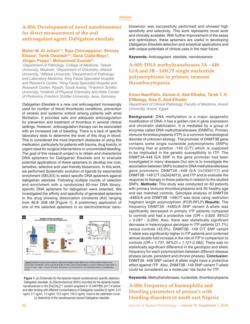
Dabigatran Etexilate is a new oral anticoagulant increasingly used for number of blood thrombosis conditions, prevention of strokes and systemic emboli among patients with atrial fibrillation. It provides safe and adequate anticoagulation for prevention and treatment of thrombus in several clinical settings. However, anticoagulation therapy can be associated with an increased risk of bleeding. There is a lack of specific laboratory tests to determine the level of this drug in blood. This is considered the most important obstacles of using this medication, particularly for patients with trauma, drug toxicity, in urgent need for surgical interventions or uncontrolled bleeding. The goal of this research project is to obtain and characterize DNA aptamers for Dabigatran Etexilate and to evaluate potential applications of these aptamers to develop low cost, sensitive, selective and user-friendly biosensors. In this work, we performed Systematic evolution of ligands by exponential enrichment (SELEX) to select specific DNA aptamers against dabigatran etexilate. Following multiple rounds of selection and enrichment with a randomized 60-mer DNA library, specific DNA aptamers for dabigatran were selected. We investigated the affinity and specificity of generated aptamers to the drug showing dissociation constants (Kd) ranging from 46.8- 208 nM [Figure 1]. A preliminary application of one of the selected aptamers in an electrochemical nano-
Figure 1: (a) Schematic for the Aptamer-based nanobiosensor specific detection Dabigatran etexilate. (b) Electrochemical (SWV) recorded for the Aptamer-based
nanobiosensor in Sm [Fe(CN)5]3-/4- solution prepared in 10 mM PBS, pH 7.4 before and after binding with different concentrations of Dabigatran exexilite (0 ng/ml, 0.01 ng/ml, 0.1 ng/ml, 1.0 ng/ml, 10.0 ng/ml, 100.0 ng/ml). Inset is the calibration curve.
(c) Selectivity of the nanobiosensor toward Dabigatran etexilate
c
ba

Abstract
Journal of Applied Hematology - Volume 10, Supplement 1, 2019 S3
Osuagwu Ugochukwu, Oyeyinka Ahmed BelloAbuja Hospital, Abuja, Nigeria
A significant number of people living with haemophilia are reported to be either under-diagnosed or mismanaged in most developing countries. This may results in increase in disease related morbidity and mortality in childhood. The aim of this study is to evaluate the frequency of haemophilia and bleeding parameters of those with bleeding disorders in South East Nigeria. Fifty consecutive consenting persons with bleeding disorder that met inclusion criteria were recruited from the four tertiary hospitals in South East Nigeria. Blood samples were collected for full blood count, coagulation screening test and Factor VIII assays. Data was analyzed using the graph pad prism version 6. Results obtained from this study showed that 2% of subjects with bleeding disorder has haemophilia and they are within mild range, the most common bleeding symptoms is gastrointestinal bleeding (23.4%) and the most common bleeding score was four (32.4%) was obtained from subjects with bleeding disorders in South East Nigeria. Furthermore, this study also showed that thrombocytopaenia (68%) was found in subjects with bleeding disorders. These results showed that haemophilia may not be under-diagnosed as earlier suggested by other authors. However, it may be necessary to screen for Factor VIII in cases of bleeding when indicated. Further studies may be needed to determine the likely causes of thrombocytopaenia in bleeding disorders in our environment.
Keywords: Haemophilia, Nigeria, parameters
A-007: Molecular and clinical characterization of hereditary factor V deficiency in Saudi Arabia: Report of 4 novel mutations
Nouf S. Al-Numair, Khushnooda Ramzan, Mahasen Saleh, Hazzaa Alzahrani, Ahmed Tarawah, Lina Elbaik, Faiqa Imtiaz, Tarek M. OwaidahKing Faisal Specialist Hospital and Research Centre, Riyadh, Saudi Arabia
Background: Coagulation factor V (FV) plays an important role in the blood coagulation cascade as part of the prothrombinase complex. Factor V deficiency (FVD) is a rare autosomal recessive bleeding disorder with variable phenotypic expression which varies from being asymptomatic to severe bleeding episodes. Objectives: The aim of this study was to perform molecular and clinical characterization of FVD in Saudi Arabia. Patients and Methods: Eleven patients (2 males and 9 females) of Arab ethnicity with confirmed FVD were recruited in the study with ages ranging between 5 and 53 years. A next-generation sequencing-based assay - Hematology panel that encompasses of 393 known genes was used. Results: A total of 6 sequence variations in F5 gene, comprising of 4 missense mutations (p.Pro189Leu, p.Trp2004Arg, p.Met2148Thr, p. Arg2202Cys), a deletion (p.Arg872Lysfs*12) and a splicing variant (c.1118+5G>T) were identified. Four of these variants were identified for the first time in this study. Three patients were homozygous for their respective mutations and 7 patients were heterozygous.
We were not able to identify FV mutation in one patient. In-silico and 3D structural analyses were performed to predict the possible impact and functional consequences of the identified variants. Conclusions: To our knowledge, this is the first study addressing FV mutations in Arabs. The results helped in providing a definitive diagnosis to the patients, carrier detection in extended family members. Overall, the HEME panel assay was efficient, demonstrating good approach for molecular diagnosis of other suspected bleeding disorders.
Keywords: Deficiency, hereditary, molecular
A-009: Regulatory T cell CD4 and CD25 expression and chemokine C-X-C ligand 13 level before and after corticosteroid therapy in pediatric idiopathic thrombocytopenic purpura patients
Heba A Ahmed, Eman M. Nagiub Abdelsalam1, Eman Mohamed Fahmy2
Departments of Clinical Pathology and 2Pediatrics, Faculty of Medicine, Sohag University, Sohag, 1Department of Clinical Pathology, Faculty of Medicine, Assiut University, Assiut, Egypt
Childhood Idiopathic thrombocytopenic purpura (ITP) is one of the most common autoimmune bleeding disorders characterized by isolated, immune-mediated low platelet count, the T-follicular helper (Tfh) cells are a subset of effector CD4 (+) T cells, that plays a pivotal role in maintaining self-tolerance, deregulation of Tfh activities has a key role in immune process taking place in ITP in which the production of platelet autoantibodies might be caused by cytokine network dysregulation, the objective of our study was analyzing the relationship of Tfh cells CD4&CD25 and C-X-C ligand 13 (CXCL13) expression before and after steroid thereby in pediatric ITP [Figure 1]. Materials and Methods: A total of 45 newly diagnosed pediatric ITP and 20 healthy controls were enrolled in the study; we used flow cytometry to assess
0
20
40
60
80
100
CXCL3
0 20 40 60 80 100100-Specificity
Sen
sitiv
ity
Figure 1: Receiver operating characteristic curve of C-X-C ligand 13 for optimum cut off point in predicting idiopathic thrombocytopenic purpura

Abstract
S4 Journal of Applied Hematology - Volume 10, Supplement 1, 2019
percentages of CD4 and CD25 cells as markers of regulatory T cells, also, serum level of interleukin- CXCL13 was measured by ELISA at diagnosis and after 4 weeks receiving corticosteroid. Results: The expression of CD4 & CD25 markers were significantly reduced in ITP patients, the markers level was correlated to platelet count, MPV and ESR. The CXCL13 serum level was elevated in ITP patients versus controls, its level before treatment was correlated with WBCS and platelet count in ITP patients, however, the level declined after treatment, and CXCL13 level after treatment was only correlated with age. A ROC curve analysis demonstrated the CXCL13 optimum cut off point for predicting ITP response to therapy to be >90 pg/ml with AUC 0.976, Sensitivity 88.89% and Specificity 100 % P-value < 0.00. Conclusion: Serum CXCL13 level could be used as a significant predictor of response to therapy in ITP patients, CD4&CD25 expression has a role in the pathogenesis of childhood acute ITP principally linked to the level of platelet count drop.
Keywords: Chemokine, corticosteroid, expression
A-010: Targeted sequencing for blood disorders
Nada Al TassanKing Faisal Specialist Hospital and Research Center, Riyadh, Saudi Arabia
Developing a comprehensive robust accurate diagnostic tool requires and in-depth knowledge of population variants to distinguish disease related mutations from rare variants. The advancement in Next Generation Sequencing (NGS) platforms has enabled reliable, high throughput and quick interrogation of genetic variants causative of inherited diseases. Targeted sequencing of clinically relevant gene panels (containing hundreds of genes) is increasingly used clinically; multiple gene panels are available for research and diagnostic purposes.The Saudi Human Genome Project (SHGP) heme panel is a targeted NGS panel of 393 genes implicated in hematological anomalies. We used this panel as a fast, cost effective and accurate tool for blind screening to identify carriers of mutations/variants in different common blood disorders. Our screening of ~600 Saudi individuals identified disease related variants in different blood and bleeding disorders.
Keywords: Blood, sequencing, targeted
A-011: A companion protein, click chemistry approach to extended half life FVIII
James KellyCell Machines Inc., Houston, TX, USA
Introduction: Standard methods for half life prolongation have been disappointing in FVIII because of its association with von Willebrand’s factor (vWF) which imposes an approximately 18 hour half life on the molecule. We have developed a synthetic protein consisting of the D’ D3 fragment of vWF, a linker and a full length human albumin. This molecule, CM110, can be tethered to FVIII using click chemistry. The resulting complex should then be divorced from the endogenous vWF and can use the albumin for half life extension. Methods: CM110 was produced by transient transfection into Expi 293 cells and the protein was purified using a CaptureSelect HSA affinity column. CM110 was treated with 1 mM NHS-P EG12- transcyclooctene (NPT) for 1 hour at room temperature in 10 mM HEPES, pH7.4, 300 mM NaCl, 4 mM CaCl 2 then separated from unreacted NPT by Superdex 200 (S200) chromatography. B-d eleted FVIII was treated with 1 mM maleimide-P EG4- methyl tetrazine for 1 hour at room temperature in the same buffer, then similarly purified by S200 chromatography. A 3 mg/ml solution of CM110 and 1 mg/ml derivatized FVIII was incubated at room temperature for 2 hours in 20 mM HEPES, pH 7.4, 150 mM NaCl, 4 mM CaCl 2 followed by S200 chromatography to isolate the CM110/FVIII complex (S8). Results: FVIII was quantitatively converted into the S8 complex with a molecular weight of 540,000, a dimer of CM110 containing 2 molecules of FVIII. S8 retains activity in a chromogenic assay and corrects the APTT in FVIII deficient plasma. Preliminary experiments in mice expressing the human neonatal Fc receptor with an albumin knock out suggest a significantly improved half life. Conclusion: Divorcing FVIII from the endogenous vWF can be effected by tethering D’ D3 to the FVIII, enabling use of traditional half life extension techniques, such as albumin ligation.
Keywords: Chemistry, FVIII, protein
A-012: Acute myeloid leukemia immunophynotyping by flowcytometric analysis
Table 1: Correlation between age, laboratory parameters and the studied markers among cases (n=45) and control group (n=20)Parameter P
CD4 CD25 CXCL13 before treatment CXCL13 after treatmentControl Patient Control Patient Control Patient
Age (years) 0.476 0.963 0.771 0.967 0.057 0.148 0.001WBCS (×1000/mm3) 0.936 0.522 0.727 0.021 0.301 0.010 0.737Hb (g/dl) 0.522 0.389 0.367 0.530 0.167 0.031 0.542Platelets (×1000/mm3) 0.726 0.043 0.574 0.021 0.021 0.041 0.563MPV 0.081 0.049 0.164 0.026 0.962 0. 704 0.189Total lymphocytic count 0.081 0.635 0.103 0.790 0.747 0.288 0.716ESR 0.503 0.035 0.166 0.049 0.961 0.503 0.096CXCL13=C-X-C ligand 13; Hb=Hemoglobin; WBCS=White blood cells; MPV=Mean platelet volume; ESR=Erythrocyte sedimentation rate

Abstract
Journal of Applied Hematology - Volume 10, Supplement 1, 2019 S5
Ihsan M. Osman, Amira A. K. Humeida1, Osama Eltayeb2, Inaam Abdelrhman1, Taghreed A. Elhadi2
Faculty of Medicine, Alzaiem Alazhari University, 1Faculty of Medicine, Alneelain University, 2Laboratory Department, Khartoum Oncology Centre (RICK), Khartoum, Sudan
Introduction: Acute Myeloid Leukaemia (AML) accounts for approximately 20% of acute leukemia in children and 80% of acute leukemia in adults. Immunophenotyping has become extremely important not only in diagnosis and subclassification of AML but also in the detection of the minimal residual disease. Immunophynotypic pattern of AML in Sudanese patients have not been addressed before. This study was conducted to characterize immunophenotypic patterns of AML in Sudanese patients. Multiparameter flow cytometry and CD45/SSC gating were used to analyze the surface and cytoplasmic antigen expressions in 106 cases of AML during the period mid2010 to mid2011 at Radioisotope Centre Khartoum (RICK). The following antigens: CD45,HLA-DR,CD34,CD117,CD13,CD33, CD19,CD7,cytoplasmic markers (CD3,CD79a,MPO), CD11c,CD14,CD64,CD42a,CD41 and CD61 were used. Results: Almost all AML blasts were expressing CD45 with no significant differences between the subtypes. CD34 have different expressions in AML subtypes. CD13 and CD33 were also studied among the blast population having mean positivity of 51.5% and 49.8% respectively in all AML subtypes collectively. CD33 was found to have higher positivity among AML-M4 and AML-M5 with mean positivity of 75.9% and 76.6% respectively. CD13 and CD33 had no correlation for all AML subtypes except for AML-M5 with very strong negative correlation (r=- 0.913). Apparent expression of CD7 and CD19 were expressed in 45.1%and 13.6% of all cases respectively. CD7 was mostly expressed in AML-M2 and AML-M3 (75%) and least in AML- M5,while CD19 was only expressed in cases of AML-M0 and AML-M7. Conclusion: Flowcytometric analysis of acute leukemia by combining the patterns and intensity of antigen expression improved the diagnosis of AML in our center. Immunophenotyping results and FAB classification of our AML patients were comparable to international published studies.
Keywords: Flowcytometric, leukemia, myeloid
A-013: Acute promyelocytic leukemia: An experience from a tertiary care centre in Pakistan
Shahtaj Khan, Nadia Altaf, Khizer Abdullah, LihazDepartment of Pathology, Hayatabad Medical Complex, Peshawar, Pakistan
Objective: Acute promyelocytic leukemia (APL) is a unique subtype of AML. There are very limited data about APL from Pakistan. The aim of the present study is to evaluate the clinico-demographic profile along with risk stratification of APL at a tertiary care hospital in Pakistan. Materials and Methods: Between June 2014 and July 2018, 28 patients with APL were enrolled in this descriptive cross-sectional study. All data were documented and statistical analysis was performed by SPSS-20 software. Results: Median age was 21 (range 2-65) years. Male to female ratio was3:1. Hypergranular variant
(92.8%) was more common as compared to microgranular type (7.14%). Majority of patients had complaints of fever (71.4%), bleeding (53.5%) and generalized weakness (14.2%). Pallor (64.2%) was the predominant finding on physical examination followed by petechial and purpural rashes (46.4%). Mean Hemoglobin was 8.3 (range 5.3-12.2) g/dl. The mean total leukocytes count was 39.6 (range 1.3-121) x 109/L and mean platelet count was 40 (range 7-78) x 109/L. Most patients fall into high riskgroup (60.7%) on risk stratification followed by intermediate risk (32.1%) and low risk (7.1%). Conclusion: Demographic, clinical features and risk stratification results of our study are comparable to published data. In the present study pallor is the most commonpresentation. Risk stratification shows predominance of high risk score.
Keywords: Pakistan, promyelocytic, tertiary
A-014: Associated inosine triphosphate pyrophosphatasegene polymorphisms and interferon/ribavirin-induced anemia in Egyptian hepatitis C virus patients
Olfat M. Hendy, Rawhia H. El-Adel1, Enas Said Essa1, Maha M. El-Sabawy2, Heba Mohamed AbdullahDepartments of Clinical Pathology and 2Hepatology, National Liver Institute, Menoufia University, 1Department of Clinical Pathology, Faculty of Medicine, Menoufia University, Menoufia, Egypt
Objective: Evaluate the association of Inosine Triphosphate Pyrophosphatase (ITPA) gene polymorphism rs1127354 and rs7270101 with the development of anemia in chronic hepatitis C (CHC) Egyptian patients during treatment with pegylated-interferon (PEG-IFN) plus ribavirin (RBV). Background: It has been found that ITPase deficiency is caused by ITPA gene polymorphisms. It was observed that ITPA polymorphisms have impact on hematological changes, including hemoglobin (Hb)-decline and platelet decline during treatment of chronic hepatitis C (CHC) patients with pegylated-interferon (PEG-IFN) plus ribavirin (RBV). Methods: The current study included 100 selected Egyptian CHC patients treated with PEG-IFN/RBV, 55 patients developed anemia (Hb decline >2 g\dl), and other45 would not developed anemia (Hb decline ≤2 g\dl) at week 12 throughout the treatment course. Routine laboratory investigations were done for all participates (HCV-Abs, HBs Ag, HCV-RNA levels, complete blood picture, Liver and kidney function tests, AFP and TSH). Single nucleotide polymorphism (SNP) was done using Real time PCR, ABI TaqMan allelic discrimination kit for ITPA polymorphisms (rs1127354 and rs7270101). Results: CC and AA were the most prevalent genotypes of SNPs rs1127354 and rs7270101 respectively among two studied groups. In univariate analysis, we found that rs1127354 polymorphism was associated with Hb-decline at week 12 of treatment, this demonstrated the protective benefit of the minor allele A of rs1127354 against RBV-induced anemia at the week 12 of therapy. Genotyping of ITPA rs1127354 and rs7270101 polymorphism would be beneficial for predicting Platelet decline during treatment. Patients with CC rs1127354 and AA rs7270101 were found to have a lower level of Platelet decline. Conclusion: It is concluded that minor allele A of

Abstract
S6 Journal of Applied Hematology - Volume 10, Supplement 1, 2019
mass and persistence of autoreactive cells. Patients usually present with non malignant enlargement of the lymphoid organs and features of an autoimmune disorder. However, some cases have symptoms and signs that resemble those of ALPS, but the specific pattern or the genetic cause may be different. Hereby, we present a case of an 8 years old boy, born to consanguineous parents, with generalized lymphadopathy, hepatosplenomegaly and oral ulcers. Cervical lymph node biopsy revealed reactive hyperplasia and lymphadenitis. Fundus examination showed a picture suggestive of optic nerve head drusen (ONHD), to which brain MRI was done and revealed bilateral cerebral white matter patchy areas of altered signal, likely representing dysmyelinating disease. Complete blood picture showed pancytopenia and the bone marrow biopsy revealed hypercellular marrow with atypical infiltrate and depressed myelopoiesis. Direct Coomb’s test was positive while screening for autoantibodies (ANA, Anti-dsDNA, ASMA) and virology were negative. Serum IgG was elevated 2,200mg/dl with normal serum IgM 100mg/dl. Flow cytometric analysis of peripheral blood lymphocytes revealed increased percentages of CD3+TCRα/β+CD4-CD8- double negative T (DNT) cells (16% of T cells) with normal B and NK cells and marked reduction of Treg (CD4+CD25+Foxp3+) cells. Molecular testing revealed a mutation in UNC13D gene which usually presents as Hemophagocytic lymphohistiocytosis (HLH).
Keywords: Autoimmune, boy, lymphoproliferation
A-017: BEAM versus single agent high dose melphalan conditioning regimen for autologous hematopoietic stem cell transplant: A retrospective matched analysis in relapse/refractory Hodgkin lymphoma
Hani Al Hashmi, Kaloyannidis Panayotis, Muhamad Rauf1, Irfan Maghfoor1, Salman Harbi, Solaf Kafnar, Eshrak Al Shaibani, Nihad Mokhtar, Mohamed Darweesh, Enas Mutahar, Khalid Bakhit, Rawan Omari, Ann Estanislao, Ioannis Apostolidis, Tusneem Elhassan1, Khalid Al Anezi, Sayed Akhtar1
Department of Adult Hematology and Stem Cell Transplantation, King Fahad Specialist Hospital, Dammam, 1King Faisal Hospital and Research Center, Oncology Center Riyadh, Saudi Arabia
Background: The ideal conditioning regimen still remains a challenge in the autologous stem cell transplantation (ASCT) setting for relapsed/refractory Hodgkin Lymphoma (RR-HL). BEAM is the most popular preparative regimen but single agent high dose Melphalan (HDM) has also been used. However, the experience and data comparing BEAM vs. HDM are limited. Methods: After the Institutional Review Board approval, we retrospectively evaluated the clinical course of 112 RR-HL patients, autografted from November 2008 till May 2017 in two different institutions. Twenty eight conditioned with HDM and compared in a matched paired analysis (1:3) with a cohort of 84 patients who received BEAM. The study groups had similar median age (30ys) and sex (M:F 1.7:1 vs. 1.8:1) and were matched for disease status before salvage (late relapse: 36 vs. 12, early relapse/primary refractory: 48 vs. 16 and disease
rs1127354 plays a crucial role in protection against RBV-induced anemia. Genotyping of ITPA rs1127354 and rs7270101 polymorphism would be beneficial for predicting Platelet decline during treatment with PEG-IFN plus RBV in Egyptian patients with chronic hepatitis C.
Keywords: Chronic hepatitis C, inosine triphosphate pyrophosphatase, pegylated interferon/ribavirin
A-015: Association between genotype and disease complications in Egyptian patients with beta thalassemia
Tamer Hassan, Marwa Zakaria, Manar Fathy, Mohamed Arafa, Sherif El Gebaly, Ahmed Emam, Attia Abdel Wahab, Mohamed Shehab, Hosam Salah, Mai Malek, Khaled El GerbyZagazig University, Department of Pediatrics, Egypt
In beta thalassemia, the degree of globin chain imbalance is determined by the nature of the mutation of the β-gene. β° refers to the complete absence of production of β-globin on the affected allele. β+refers to alleles with some residual production of β-globin. The homozygous state results in severe anemia that necessitates regular blood transfusion. On the other hand, frequent blood transfusion can lead to iron overload resulting in progressive dysfunction of the heart, Liver as well as multiple endocrinopathies. We studied the impact of genotype on the development of disease complications in patients with β thalassemia. A Cross sectional study was carried on 73 patients with beta thalassemia. Genotyping was determined by DNA sequencing technique. Routine investigations as well as MRI liver and heart were performed to assess iron overload. We found that β+β+was the most common genotype in our patients followed by β°β° and β°β+. Mean Liver iron content (LIC) was significantly higher in β°β° compared to β°β+and β+β+genotypes and mean cardiac T2* was significantly lower in β°β° compared to β°β+and β+β+genotypes. Hepatic complications, hepatitis C, cardiac complications and some endocrinopathies were significantly higher in patients with β°β° genotype compared to other genotypes which explain the role of the underlying genetic defect in thalassemia patients in development of disease complications.
Keywords: Egyptian, genotype, thalassemia
A-016: Autoimmune lymphoproliferation in an 8-year-old boy
Safa Meshaal, Rabab El Hawary, Sohilla Lotfy1, Nermeen Galal1, Aisha Elmarsafy1
Departments of Clinical and Chemical Pathology and 1Pediatrics, Faculty of Medicine, Cairo University, Cairo, Egypt
Autoimmune Lymphoproliferative Syndrome (ALPS) is an inherited rare genetic disorder which results from mutations in molecules involved in the Fas-Fas ligand pathway. ALPS represents a failure of apoptotic mechanisms to maintain lymphocyte homeostasis, permitting accumulation of lymphoid

Abstract
Journal of Applied Hematology - Volume 10, Supplement 1, 2019 S7
status pre ASCT [complete remission (CR): 39 vs. 13 and partial remission (PR): 45 vs. 15]. BEAM regimen was given in the standard doses over 6 days, while HDM (200mg/m2) was given in a single day infusion. All patients received prophylaxis against microbial, fungal and viral infections; GCSF was routinely administered at the dose of 5mcg/kg at +1 day (BEAM group) and at +5 day (HDM group). The T-test and Kaplan-Meier method were used for the statistical analyses. Results: The engraftment was successful; the median day for neutrophils >1000/mm3 was +11 for both groups while for platelets >20000/mm3 a faster recovery was noticed for HDM group: +13 vs. +22 days (p<0.001). The median follow up for both groups is 2, 5 years. In the BEAM group 64/84 patients are alive (49 disease free), and 23/28 from the HDM group are alive (20 disease free). In the whole cohort of patients the survival rates were superior in the HDM group thought not statistically significant; 5 years overall survival 65% vs. 80% and progression free survival 52% vs. 70% for the BEAM and HDM group respectively. The HDM regimen associated with better survival rates either for patients in CR or for those in PR before ASCT. The 100 days non relapse mortality was acceptable for both groups: 2/84 (2.3%) in the BEAM group vs. 1/28 (3.5%) in the HDM group. Conclusion: In this study, though retrospective, demonstrated that for RR-HL patients, the conditioning regimen consisting of HDM, offered at least comparable efficacy to the BEAM regimen. The earlier platelets recovery, and the shorter duration of chemotherapy administration (6 days for BEAM vs. 1 day for HDM), resulted in less hospitalization days, which along with the shorter period of GCSF administration post ASCT, may contribute to a better cost effectiveness for the HDM regimen. Nevertheless, prospective studies with larger series of patients and longer follow-up, including also a meticulous cost analysis, are warranted to determine the accurate role of single agent HDM as preparative regimen for ASCT in HL patients.
Keywords: Almadinah, hemophilia, benefits
A-018: Blood group negativity and awareness toward anti-D immunoglobulin among pregnant women at King Abdulla Hospital, Bisha, Saudi Arabia 2018
Amar Ibrahim Omer Yahia, Elhadi Ibrahim Miskeen1
Departments of Hematopathology and 1Obstetrics and Gynecology, College of Medicine, University of Bisha, Bisha, Saudi Arabia
Background: The RhD is a highly immunogenic antigen significant for obstetric medicine due to the ability of anti-D to cause hemolytic disease of the newborn. Hence, awareness toward blood group negativity is important for the prevention of fetal morbidity and mortality. Objectives: The aim of this study is to assess the awareness of pregnant women about the blood group negativity and the importance of anti-D immunoglobulin at King Abdulla Hospital in Bisha province Southern of Saudi Arabia. Methods: This is a prospective cross-sectional study carried out at King Abdulla Hospital in Bisha province Southern of Saudi Arabia in 2018. The study population were pregnant women presented for antenatal care. The blood group was screened. Their awareness toward blood group negativity and the importance of anti-D immunoglobulin was assessed
through direct interview. Data analysis was performed using Statistical Package for the Social Sciences. P-value of less than 0.05 was considered statistically significant. Consent was obtained. Main Results: Total number of pregnant women assessed were 108. Of them 45/108 (41.7%) were aware about the clinical significance of blood group negativity and anti-D immunoglobulin. Statistically significant correlations were found between awareness and age (value 0.035), education (value 0.001), previous experience of vaginal bleeding (value 0.000), blood group (0.001) and previous anti-D immunoglobulin administration (0.00). Conclusion: Although the clinical sequences of blood group negativity and anti-D immunoglobulin, the awareness is so not satisfactory. Structured health education program was recommended specially within the package of antenatal care services.
Keywords: Anti-D immunoglobulin, Bisha, blood group negativity, Saudi Arabia
A-019: Cardiac structural and functional changes evaluated by transthoracic and tissue Doppler echocardiography in Egyptian adult patients with sickle cell disease
Noha M. El Husseiny, Nermein Sayed, Hesham Yehia1, Dahlia El Ramesy1, Alia Abd El Aziz, Doaa El Demerdash, Mona GamilDepartments of Internal Medicine and 1Cardiology, Faculty of Medicine, Cairo University, Cairo, Egypt
Background: Major cardiac abnormalities in patients with sickle cell disease are related to volume overload effects of chronic anemia and ferritin level. Patients with SCA have reduced LV global longitudinal strain. In the detection of subclinical LV dysfunction in patients presenting with sickle cell disease, 2D speckle tracking echocardiography appears to be useful to assess systolic dysfunction and tissue Doppler to assess diastolic dysfunction. Aim: The aim of our study was to evaluate cardiac performance in adult Egyptian sickle cell patient. Methods: The study was conducted on 30 SCD patients who were recruited from the hematology clinic of Kasr El-Ainy Hospital- Cairo University and fifteen healthy control who were age and sex matched. Two-dimensional speckle tracking echocardioghraphy (STE), M-mode, Doppler and tissue Doppler echocardiography (TDE) were are used to assess systolic and diastolic dysfunction in both groups. We used parameters (Global strain, AP3L, AP2L, AP4L, EF, LVIDS and LVIDd) for systolic dysfunction. And for diastolic dysfunction we used medial and lateral E/e’, E/A ratio, Dt and S velocity. Results: Our results revealed significant increase in LVIDS and LVIDd dimensions in cases compared to control group (P<0.001). None had an ejection fraction below 55%. Diastolic dysfunction was elicited in 8 patients (26%). There was a statistically significant correlation between the serum ferritin level and lateral E/E’ ratio. There was a significant reduction in GLS (p value.026) and also apical segments longitudinal strain (p value.002) compared to control. There was statistically significant negative correlation between number of sickle cell crises per year and GLS (p =0.040). There was a statistical significant inverse

Abstract
S8 Journal of Applied Hematology - Volume 10, Supplement 1, 2019
correlation between Global LV systolic function and hemoglobin level (p=0.04). Conclusion: Cardiac performance should include assessment of both systolic and diastolic parameters as there could be alteration in LV diastolic filling indices even in absence of systolic dysfunction. Hence these indices can be used as an early marker for cardiac affection. Speckle tracking echocardiography can recognize the cardiac involvement in really early stages and initiation of treatment and decreasing rate of painful crises to prevent irreversible myocardial dysfunction.
Keywords: Cardiac, Doppler, sickle
A-020: CD27 and CD44 expression pattern in pediatric precursor B-acute lymphoblastic leukemia: Clinical and prognostic implications
Randa A. Osman, Nema R. Hamad1, Mahmoud M. Kamel, Maha M. Aqle2
Department of Clinical Pathology, NCI, Cairo University, Cairo, 1Department of Clinical Pathology, National Institute of Urology and Nephrology,2Department of Clinical Pathology, Faculty of Medicine (For girls), Al-Azhar University, Cairo, Egypt
Background: Leukemia is the most common type of childhood cancer. Acute lymphoblastic leukemia (ALL) is the most common subtype, accounting for 85% of cases. The expression of CD27 and CD44 were claimed to be among new prognostic parameters. Aim of the Work: To shed new light on the biological significance of CD27 and CD44 expression in patients with B cell ALL. Patients and Methods: This study was performed on 60 newly diagnosed pediatric ALL patients, 39 males and 21 females. Age ranged from 6 months to 18 years. CD27 and CD44 were tested by Flow Cytometry. Results: CD44 was positive in 55 patients (91.6%), CD27 in 30 patients (50%), and co- expression in 27 patients (45%). Patients were classified into two groups:1. CD27 positive group: either expressing CD27 alone [SP
(single positive)] or in co-expression with CD44 [DP+ (double positive)]
2. CD27 negative group: either negative for both CD27 and CD44 [DN (double negative)] or CD27-ve, CD44+ve (CD44 SP).
SP CD27 was found in one patient (1.7%), 29 patients (48.3%) co-expressing CD44 and CD27 and 4 patients (6.7%) are DN. CD27 expression (SP or DP) was associated with a higher frequency of TEL/AML1 [11/30 (33.7%) in the CD27 +ve group vs. 2/30 (6.7%) in the 27 –ve group; p= 0.005]. It was also associated with achieving negative MRD at the end of induction [27/30 (90%) in the CD27 +ve group vs. 17/25 (68%); p=0.042]. Higher overall survival was encountered in patients with CD27 positive expression with 100% cumulative overall survival at 26 months versus 77.4% in CD27 negative cases (p=0.084). Conclusion: CD27 expression was more frequent in low-risk patients, those who achieved negative MRD and correlated with the favorable prognostic parameters indicating a role for CD27 in anti-leukemic defense in ALL patients.
Keywords: Acute lymphoblastic leukemia, CD27, CD44, minimal residual disease, TEL/AML
A-021: CD30 expression versus serum soluble CD30 level: Role in prognosis and treatment of acute myeloid leukemia
Dalia A. Nigm, Zeinab A. Abd Elhameed, Mohamed Z. Abd ElrahmanDepartment of Clinical Pathology, Faculty of Medicine, Assiut University, Assiut, Egypt
Objectives: As we noted that CD30 is a valuable molecule in regulation of growth and death of lymphocytes in malignant lymphomas, we analyzed CD30 expression and serum soluble CD30 (sCD30) molecule level in patients with acute myeloid leukemia (AML) to assess their role as a prognostic markers and to examine the possibility of anti-CD30 to be a targeted therapy in these patients. Methods: We studied CD30 expression by Multicolor flow cytometry immunophenotypic analysis on bone marrow aspirates of 50 AML patients. Serum sCD30 level was measured by Enzyme Linked Immunosrbent Assay (ELSA). We correlate CD30 and sCD30 values with all of white blood cell counts, Hemoglobin, platelets, bone marrow blasts and cytogenetics. The Fisher’s exact test or chi-square was used for comparison of categorical variables and the t test or one-way analysis of variance (ANOVA) was applied for numerical comparisons using SPSS version 20. A p-value of <0.05 was considered to be statistically significant. Results: Our study conducted on 50 AML patients, the mean patients’ age was 47.4±18.1 years (range, 17–77), 11 (22%) were males and 39 (78%) were females. 16 (32%) patients have high CD30-expression and 11 (22%) have elevated serum sCD30. We found that there was a significant correlation between both CD30 expression and sCD30 level with WBCs count, BM blasts, adverse risk cytogenetics, FLT3/ITD and with relapse for CD30 expression, complete remission failure with elevated serum sCD30 level. Conclusions: CD30 is expressed by myeloblasts in AML patients. We found that high CD30 expression and elevated sCD30 level can be used as prognostic markers for relapse and complete remission failure respectively. Furthermore, these patients with adverse risk cytogenetics have not too many treatment options, so the use anti-CD30 targeted therapy may be a possible alternative for this patient group which need further studies.
Keywords: CD30, myeloid, serum
A-022: CD34+CD38–CD123+ leukemia stem cells in acute myeloid leukaemia; a promising phenotype for minimal residual disease detection
Marwa M. Hanafi, Azza M. Kamel, Nahla M. El-Sharkawy, Eman Z. Kandeel, Mohammed A. SamraDepartment of Clinical Pathology and Medical Oncology, NCI, Cairo University, Cairo, Egypt
Background: Acute myeloid leukemia (AML) is a heterogeneous disorder with treatment response much inferior to acute lymphoblastic leukemia. Treatment failure is largely attributed to the persistence of leukemia stem cells (LSCs) which are less

Abstract
Journal of Applied Hematology - Volume 10, Supplement 1, 2019 S9
accessible and hence less responsive to chemo-therapeutics. The classical LSCs phenotype is CD34+/CD38-; however LSCs express other markers especially CD123 which may be even earlier than CD34. Aim of this Study: We hypothesized that CD123 may be better marker of LSCs and that the more the number of LSCs at diagnosis and/or at follow up periods, the more the case would be resistant to therapy. Methods: The study was performed on 84 newly diagnosed AML patients. 4 color panels of monoclonal antibodies were used: CD38FITC/CD123PE/CD34ECD/CD45PE-PC5 analyzed on Navios Flow cytometer. Cell populations with different surface markers were calculated using the prism function of the software. The study was performed according to Helsinki declaration for studies on human subjects and approved by the Institution Review Board (IRB) of the National Cancer Institute, Cairo University. Results: Among the studied; A higher CD 123 % at diagnosis (P=<0.001) and at day (d) 14 (p=0.004 & p=<0.001 respectively) had an adverse impact on OS and DFS, A higher [CD34+/CD38-/CD123+] % at diagnosis (P=0.005 & P=<0.001) and follow up periods (at d14 and d28) (P=<0.001 & P=<0.001 and P=0.002 & P=<0.001 respectively) was significantly associated with adverse impact on OS and DFS. Conclusion: CD123 has been shown to be a unique marker of LSC within the CD34+CD38– compartment. It may be used as a unique single phenotype for MRD detection in AML patients.
Keywords: Acute myeloid leukemia, CD123, leukemia stem cells, minimal residual disease
A-023: CD56 and CD11b positivity with low Smac/DIABLO expression as predictors of chemoresistance in acute myeloid leukaemia: Flow cytometric analysis
Abeer Ibrahim, Asmaa M. Zahran1, Sanaa Shaker Aly2, Ahmed Refaat, Mohammed H. Hassan3
Departments of Medical Oncology and Haematological Malignancies and 1Clinical Pathology, South Egypt Cancer Institute, Assiut University, Assiut, 2Department of Clinical Pathology, South Valley University, 3Department of Medical Biochemistry, Faculty of Medicine, South Valley University, Qena, Egypt
Background: Resistance to chemotherapy is a major obstacle to curing acute myeloid leukaemia (AML), and several antigens are claimed to play primary roles in this resistance. Aim of the Work: The aim of this study was to evaluate the roles of CD56, CD11b and Smac/DIABLO gene expression levels as prognostic markers of the clinical outcome, response to chemotherapy and survival of AML patients. Materials and Methods: A cross-sectional observational study was conducted on 60 naïve-AML patients who received induction therapy with mitoxantrone and cytarabine combined with a high dose of cytarabine. The CD56, CD11b and Smac/DIABLO expression levels were assessed using flow cytometry at diagnosis and were analysed for correlation with the possible associated risk factors, response to chemotherapy, and median duration of disease-free survival (DFS) and overall survival (OS). Results: The overall results revealed that AML patients who exhibited positive expression for CD56 and CD11b had short median durations of DFS and OS.(P = 0.019, 0.006, 0.029 and 0.024, respectively). Additionally, low Smac/DIABLO
expression had a negative impact on treatment outcome in terms of CR rate (p=0.012) and reduced DFS (p=0.000) and OS (p=0.000) values. Conclusions: CD56 and CD11b positivity and low Smac/DIABLO expression are important predictive factors for the occurrence of chemoresistance, in addition to other risk factors, among AML patients.
Keywords: Acute myeloid leukaemia, CD11b, CD56, chemoresistance, Smac/DIABLO
A-024: Childhood cancer epidemiology and outcome with highlight on acute lymphoblastic leukemia: Single institutional experience over 12 years at King Salman Armed Forces Hospital, Tabuk, KSA
Mohammed Alpakra, Taha Khattab1, Ehab Hanafy2, Gihan Mahmoud2, Nazim Faisal2
Military Hospital, Khamis Mushait, 1Dr. Soliman Fakeeh Hospital, Jeddah, 2King Salman Armed Forces Hospital, Tabuk, Saudi Arabia
Background: Childhood cancer represent a diverse group of diagnoses that have distinctive age incidence pattern. Even within a single diagnosis, the biologic characteristics of tumor cells may vary between younger and older children. Cure rate of childhood cancer reach 85% for acute lymphoblastic leukemia ALL, 70% for solid tumors. Progress resulted from using risk directed therapy, multiple drugs of chemotherapy and local control with either or both surgery and radiotherapy. Objectives: To find out incidence and survival of solid tumor and hematological malignancies. To improve outcome, we need to know causes of death, disease related or toxic causes.Patients and Methods: From January 2005 to December 2016 all patient treated at KSAFH were collected. Solid tumors types and survival will be determined. Cases of ALL will be reviewed in terms of bone marrow BM immunophenotyping, minimal residual disease (MRD) post induction and cytogenetics. Relapse time and site are determined as well as EFS and OS. Results: Total 140 patients, 66 ALL. Male 70, age range (1.5 – 11) year, median 5 years. Other cancer with their number and survival; AML 4/9 (45%), HL 9/9 (100%), NHL 6/7 (86%), NBL 4/9 (45%), WT 7/7 (100%), MBL 7/9 (78%), Glioma 3/4 (75%), ES 2/3 (66%), OS 2/2 (100%), RMS 1/3 (33%), GCT 3/3 (100%), LCH 3/3 (100%), HLH 1/2 (50%), CML 1/1 (100%), other rare tumor; Pleuropulmonary blastoma and metastatic papillary thyroid carcinoma both survived and AT/RT case died. OS 55/74 (74%). Cases of ALL 53/66 (80%). Immunophenotyping 56/66 B-lineage, 5/66 T-cell, 3 unknowns, 1 pro-B and 1 mixed lineage ALL. CSF positive 7/66 (11%). BM cytogenetics; normal 12, hyperdiploidy 4, t (9;22) 2 and one for each MLL, TEL/AML, trisomy 21 and Runx. Response to therapy available in 22/66 (33%), RER 19/22 and SER 3/22. Positive MRD post induction 3/66 and negative 6/66. Chemotherapy was based on CCG regimens; low risk (1991, 1891) 31/66 (47%), intermediate risk (1961 arm C, 1882) 16/66 (24%), and high risk (1961 arm D) 18/66 (29%). Relapse incidence 19/66 (29%); very early 3/19 died (2 combined & 1 BM), early relapse 7/19 (3 BM, 2 combined, 2 CNS) 6/7

Abstract
S10 Journal of Applied Hematology - Volume 10, Supplement 1, 2019
survived 86% and late relapse 9/19 (4 BM, 4 combined, 1 CNS), 5/9 survived 56%. 11/19 58% survived relapse; 7/11 post allogeneic SCT 64% and 4/11 36% responded to second line chemotherapy (R2 and BFM 2002). Causes of death 7 refractory leukemia, 4 toxic deaths, 1 RTA and 1 secondary neoplasm with Glioblastoma multiform. EFS 41/66 (62%) and OS 53/66 (80%). Conclusions: Further establishment of cytogenetics/molecular studies for hematological malignancies and solid tumor need to be locally available as well as applying risk directed therapy for all malignancies would have its impact for improving outcome.
Keywords: Cancer, leukemia, lymphoblastic, Tabuk
A-025: Cholilithasis in pediatric sickle cell anemia patients in northwestern region of Saudi Arabia (single center study)
Amna M. Alshenqeti, Amal M. Alqrafi, Leema K. Alhussayen, Zakaria M. Alhawsawi1, Abeer Abd Elmoneim1, Waheed A. Turkistani2
College of Medicine, Taibah University, 1Department of Pediatric, Taibah University, 2Pediatric Hematology/Oncology Unite, MCH Hospital, Al Madinah, Saudi Arabia
Background: Chronic hemolysis predisposes to bilirubinate cholelithiasis that can be asymptomatic or result in cholecystitis, choledocholithiasis, cholangitis, and gallstone pancreatitis. The frequency of cholelithiasis in sickle cell patients range from 5%-55%. This study aimed to determine the prevalence of cholelithiasis among pediatric sickle cell disease patients in Al-Madinah and the risk factors associated with the development of gallstones. Methods: All Sickle cell disease (SCD) children aged between 2 and 18 years were enrolled in a retrospective cohort study conducted in Maternity and Children’s hospital, a tertiary level hospital at Al-Madinah Governorate, during the period of March 2017 to Sep 2017. Medical records of these patients were reviewed. Simple T-test and Chi-square test were used to assess the risk factors that possibly associated with cholelithiasis. A multinomial logistic regression analysis was done to identify the factors predictive of cholelithiasis occurrence. Results: One-quarter of the participants developed cholelithiasis at a mean age of 6.9 years. The frequency of cholelithiasis was significantly increased with age (40.8% in 12 years and older), high Hb S and MCV. Its incidence increased in male sex, Saudi nation and those with SCD than those with Sickle-Thalassemia. However, these differences were not statistically significant. Conclusion: The prevalence of cholelithiasis in pediatric SCD patients is high in Al-Madinah. Old age and increased MCV and HbS level are the significant factors that related to cholelithiasis.
Keywords: Anemia, cholilithasis, northwestern
A-026: Clinical and laboratory workup of a patient with whim syndrome
Aisha ElMarsafy, Rabab El Hawary1, Safa Meshaal1, Marwa El-Sharkawy1, Radwa ELKady, Dalia AbdelAziz, Nermeen Galal, Janette Boutrous, Azza Mostafa1
Departments of Clinical and Chemical Pathology and 1Pediatrics, Faculty of Medicine, Cairo University, Cairo, Egypt
Background: WHIM syndrome is a rare congenital immunodeficiency disorder, it is an acronym for some of the characteristic symptoms of the disorder (W) arts, (H) ypogammaglobulinemia, (I)nfections, and (M)yelokathexis. (Liu et al. 2012) (Myelokathexis refers to neutropenia resulting from retention of mature neutrophils and increased neutrophil apoptosis in the BM). Aim of the Study: To describe the clinical and laboratory workup of a patient with suspected WHIM syndrome. A male patient from a non-consanguineous marriage presented with diarrhea following Rota virus vaccination, followed by perianal lesions and recurrent otitis. He had no organomegaly and no lymphadenopathy. Blood picture showed persistent neutropenia and lymphopenia. His serum immunoglobulins levels were all low. Flow cytometric analysis of his blood cells showed CD3 lymphopenia and marked decrease in the memory CD19 (CD19+CD27+ cells were 0.8%). Work up for differential diagnosis for other causes of neutropenia was started. Bone marrow aspiration showed marked hyperplasia, with many cells having signs of apoptosis (hypercondensation of chromatin, hyper segmentation of nucleus and cytoplasmic vaculation). Fluorescence in situ hybridization (FISH) for XY chromosomes showed no maternal engraftment causing graft versus host disease. A diagnosis of WHIM syndrome became very likely; however genetic confirmation by sequencing the chemokine CXC4 receptor gene is mandatory.
Keywords: Laboratory, WHIM syndrome
A-027: Clinical differences in chronic myeloid leukemia presentation in patients of Khyber Pukhtoon Khwa Province, Pakistan
Shahtaj Khan, Muhammad Fawad Khan, Hira UroojDepartment of Pathology, Hayatabad Medical Complex, Hayatabad, Pakistan
Objective: Chronic myeloid leukemia accounts for 15% of all leukemias worldwide and has always known to be primarily a disease of adults. The median age at diagnosis of CML is 60-65 years in western registries and is believed to be rare among children and adolescents. The incidence of CML in our localities is rather lower yet tends to afflict younger population and has more aggressive clinical presentation. Materials and Methods: A retrospective study was carried out, a total of 100 newly diagnosed CML patient. Charts were reviewed (August 2015-August 2018). Results showed the median age at presentation of 39 years, youngest being 8 years old. Patients presented with a variety of clinical findings, ranging from crop up of massive spleen to markedly increased white blood cell counts. The median baseline WBC counts are taken as 80,000/uL – 150,000/uL, whereas our study shows a median WBC count of 366,000/uL with counts as high as 12,44,000/uL. Conclusion: The aim is to determine the clinical differences in CML presentation in our part of the world that may have an impact on disease progression, treatment options and response to the treatment.
Keywords: Leukemia, myeloid, Pakistan

Abstract
Journal of Applied Hematology - Volume 10, Supplement 1, 2019 S11
A-028: Clinical phenotypes and immunological characteristics of 26 Egyptian patients with common variable immunodeficiency
Nancy El Guindy, Safa Meshaal, Rabab El Hawary, Rana Adel, Nermeen Galal1, Aisha Elmarsafy1
Departments of Clinical and Chemical Pathology and 1Pediatrics, Faculty of Medicine, Cairo University, Cairo, Egypt
Background: Common variable immunodeficiency (CVID) is the most frequent symptomatic primary immune deficiency worldwide. The prevalence varies widely worldwide, ranging from 1:50,000 to 1:10,000 of the general population. CVID comprises a heterogeneous group of relatively rare disorders characterized by remarkable hypogammaglobulinemia that involves two or three immunoglobulin isotypes (IgG, IgA, and IgM). The decrease in serum immunoglobulins levels is often associated with defects in cell-mediated immunity. Several studies of large cohorts of CVID patients have demonstrated complex phenotypic and functional immunological abnormalities as defective T-cell signaling and cytokine production, accelerated T cell apoptosis, defect of regulatory T cells (Tregs), defective interactions between T and B lymphocytes and antigen-presenting cells. Methods: The study included 26 CVID patients diagnosed as per the old and revised criteria issued by the European Society for immunodeficiency and 24 sex and age matched healthy subjects to serve as control group. Different clinical symptoms were reviewed from the patients’ records. Memory/naïve T and B cells as well as Tregs were evaluated using flow cytometry for all patients and compared to the control. Results: Most common manifestation was recurrent infections in 48% of patients with chronic lung disease recorded in 40%. Immune dysregulation was documented in 38%. B cells were significantly lower in the patients when compared to the control (p=0.001) while the memory CD27+ B cells didn’t differ between our CVID cohort and the healthy subjects. However the memory helper cells (CD4+45RO+ T cells) were significantly higher in CVID patients (p=0.0001) on the expense of the naïve CD4 Cells (CD4+45RA+) explaining the autoimmunity in those patients.
Keywords: Immunodeficiency, immunological, phenotypes
A-029: Compatibility Studies with phenotyping testat blood bank of maternity and children hospital, Almadinah, Saudi Arabia
Jasem Salem, Souad Abdullah, Abdul Aziz Basher, Ahmad Tarawah1
Blood Bank, King Abdullah Medical City, Maternity and Children Hospital, 1Madinah Hereditary Blood Disorders Center, King Abdullah Medical City, Maternity and Children Hospital, Madinah, Saudi Arabia
Background: A compatibility test is a blood bank procedure to be determined before transfusion if there is serologic
compatibility between a blood donor and an intended recipient. Each compatibility test is a unique experiment in which unknown serums are tested with unknown RBC for in vitro detection of antibodies. Negative results indicate compatibility. Positive results indicate incompatibility. The purposes of compatibility testing are to detect: irregular antibodies; errors in ABO grouping or other systems, and clerical errors in patient identification and result recording. Any degree of agglutination or hemolysis in the crossmatch tubes during any phase indicates incompatibility and the blood should not be given. If agglutination or hemolysis occurs in the screen cells tubes, this indicates the presence of an atypical antibody and further testing must be done. Further tests must be considered in chronic blood recipients like sickle cell disease and thalassemia. The most important are RH antigen systems and Kell antigen system. The Rh blood group system is the second most important blood group system after the ABO system. D, C, c, E, and e are the most important antigens. The Kell blood group system contains many antigens that are highly immunogenic. These antigens are the third most potent, after those of the ABO and Rh blood groups, at triggering an immune reaction. RBCs phenotyping test is a determination of RBCs antigen receptors such as RH system and Kell system. Matching the RBCs phenotype (antigens profile!) for the donor and recipient will decrease the risk of alloimmunization after the transfusion. Objectives: To emphasis the importance of Patient phenotyping for RH and Kell antigens in order to reduce the incompatibility especially in chronic blood recipients who have high risk of become alloimmunized and complicate the transfusion therapy. Methods: Retrospective Review of Blood Bank records at Maternity and Children Hospital, King Abdullah Medical City – AlMadinah. Maternity and Children Hospital is a tertiary level teaching hospital with a capacity of 500 beds. Data collection compare two-years period 2016 and 2018. RBCs Phenotyping test including RH system and Kell system. The most prevalent RH phenotype is CcDee K –ve with 50%, and the least are, ccDEE K –ve with 1%. Results: From 1-1-2016 until 31-12-2016,the total number of crossmatchtests was1104 tests, and from 1-1-2018 until 31-12-2018 the total number of crossmatch tests was 983 tests. The least number of tests was carried onJune 2018 with 64 tests and the largest number of tests was carried onJanuary 2016 with 108 tests.The lowest incompatible tests was on June and November 2018 with zero cases and the highest incompatible tests was on January 2016 with 6 cases. Conclusion: Incompatible blood tests at maternity and children hospital during 2018was decreases to 1.3% comparing to 2016 3.6% after use RBCs phenotyping testing routinely with blood compatibility procedure. With improvement of 36% in compatibility procedure.
Keywords: Children, maternity, phenotyping
A-030: Concordance between conventional and molecular cytogenetic techniques in identification of genetic abnormalities among newly diagnosed multiple myeloma patients

Abstract
S12 Journal of Applied Hematology - Volume 10, Supplement 1, 2019
Ahmad Alharbi, Mohammed Almajed1, Osamah Khojah1, Mohammed Albalwi2, Adnan Alkhayat3 Abdul Rahman Al-Maeen4, Ahmed Alsuhaibani2
Department of Pathology, Majmaah University, Al Majmaah, 1Department of Pathology, King Saud University, 2Department of Pathology and Laboratory Medicine, King Abdulaziz Medical City, Riyadh, 3Department of Pathology and Laboratory Medicine, King Fahad Central Hospital, Abu Arish, 4Department of Pathology, Al-Jouf University, Sakakah, Saudi Arabia
Background/Purpose: Multiple myeloma (MM), also known as a plasma cell myeloma, is a clonal hematological malignancy accompanying by an abnormal proliferation of plasma cell that related to an underlining genetic alteration. It is a relatively a common neoplasm in elderly with a deleterious end organ damage if not treated. The prognosis and monitoring of the disease progression are obtained largely by identification of the genetic basis of the disease. Unfortunately, there is no published data about the genetic abnormalities in MM Saudi patients. Previously, genetic abnormalities were detected in 50-60% of newly diagnosed patients. However, administration of the enrichment plasma cell techniques resulted in better detection rate reaching over 90%. This study reviewed the genetic abnormalities of newly diagnosed patients in our center by two methods and comparing the results with a well-established international data. Methodology: This retrospective study identified 69 newly diagnosed patients with MM in King Abdulaziz Medical City, National Guard Hospital, Riyadh, between 2012-2015. However, only 58 patients had a complete molecular cytogenetic investigation. All the 58 patients underwent for a MM panel through a florescence in situ hybridization (FISH) technique and classical cytogenetic analysis by karyotyping. The validated MM-FISH panel included
five different probes; trisomy 12q15, 13q14/13q34 deletion, 17p13.1 deletion, translocation (11;14) and immunoglobulin heavy chain (IGH) in chromosome (Ch) 14 rearrangement. Results: In this cohort, the median diagnosis age for the 58 MM patients is 64.5 years, ranging from 39-91 years with two cases below age of 40 years. Male cases were predominant with 44 cases and M:F ratio is 3.1:1. Karyotype analysis was done on 54 cases showing positive detection for structural or numerical chromosomal abnormalities in 11 cases (20.4%) and failure or very low yield in 10 cases (18.5%). However, FISH studies revealed a clear better advantage comparing to classical cytogenetic by positive detection of 41out of 58 cases (70.7%). The overall concordance between both methods were only 46.3% [Table 1]. Only a single case showed genetic abnormality by karyotype but cannot be detected by MM FISH panel. Hyperdiploidy (including trisomy), IGH rearrangements, deletion Ch13 and Ch 17 and hypodiploidy were detected by FISH as 43.1%, 20.7%, 24.1%, 3.5% and 10.3%, respectively [Table 2]. Conclusion: This study unveiled some characteristics of genetic alterations among multiple myeloma patients in Saudi Arabia. It also supported the use of FISH technique along with a purified plasma cells medium or other enrichment plasma cell techniques to improve our detection quality leading to enhance patient care. Finally, correlation of these findings with the clinical behavior of each individual patient will refine our standing about the implication of a specific or combined genetic abnormality in our population.
Keywords: Cytogenetic, genetic, myeloma
A-031: CRISPR-mediated modification of IVS1-110 mutation in beta thalassemia hemopoeitic stem cells
H. Gabr, W. Abo Elkheir, M. Elghamrawy, A. Abdel Hafiz,M. Elsisy, A. OsamaFaculty of Medicine, Cairo University, Cairo, Egypt
Background: Homozygous beta thalassemia is one of the commonest causes of hereditary anemia in Egypt, with lifelong transfusion dependency and iron overload problems. The only available effective therapy for thalassemia is stem cell transplantation. Gene therapy trials foe correction of thalassemia mutations remains theoretical due to a number of obstacles. CRISPR-mediated gene modification represents a new tool that might solve many of these problems. Study Design: This study was designed to correct genetic mutation in
Table 1: The concordance between karyotype and florescence in situ hybridization studies in 58 newly diagnosed multiple myeloma patientsResult/test Karyotype (%) FISH (%)Tested samples 54 (100) 58 (100)Failure samples 5 (9.3) 0Inconclusive samples 5 (9.3) 0Negative cases 33 (61.1) 17 (29.3)Positive cases 11 (20.4) 41 (70.7)Overall concordance 25 cases (10 positive and 15 negative),
46.3%FISH=Florescence in situ hybridization
Table 2: Detailed genetic abnormalities for 58 newly diagnosed multiple myeloma patients by florescence in situ hybridization panelAbnormality Cases with unique abnormality (%) Cases with other abnormality (%) Total (%)Negative cases 17 (29.31) NA 17 (29.31)Hyperdiploidy/trisomy 15 (25.86) 10 (17.24) 25 (43.10)Hypodiploidy (not del 13 or 17) 0 (NA) 6 (10.34) 6 (10.34)Deletion 13/13q 5 (8.62) 9 (15.52) 14 (24.14)Deletion 17/TP53 0 (NA) 2 (3.45) 2 (3.45)IGH rearrangement (not t[11;14]) 1 (1.72) 5 (8.62) 6 (10.34)t(11;14) alone 5 (8.62) 1 (1.72) 6 (10.34)Ch 1q gain 0 (NA) 1 (1.72) 1 (1.72)Other abnormalities 0 (NA) 4 (6.90) 4 (6.90)Ch=Chromosome; IGH=Immunoglobulin heavy chain; NA=Not available

Abstract
Journal of Applied Hematology - Volume 10, Supplement 1, 2019 S13
beta thalassemia patients with known homozygous IVS 1-110 mutation. Sequencing was done to exclude any other mutations either in the beta or gamma globin genes. Two approaches were sed: knock-out as a mode for correction of globin chain imbalance depending on HbF synthesis to compensate for the defective beta globin chain and knock-in to insert a correct sequence for the gene and allow for normal globin chain synthesis.Materials and Methods: (1) Design of CRISPR and gRNA. (2) Selection of CD34+ve hemopoeitic stem cells from patients using immunomagnetic beads. (3) Electroporation of CRISPR/as9 and gRNA complexes into the HSCs. (4) Selection of transfected cells through antibiotic resistance. (5) Erythroid differentiation of selected cells in both liquid and semisolid cultures in erythroid inductive medium. (6) After 2 weeks, RNA extraction was done and quantitative globin chain analysis was done for alpha, beta and gamma globin chains using multiplex quantitative real time reverse transcription polymerase chain reaction. Results and Conclusions: Both knock-out and knock-in methods resulted in improvement of globin chain imbalance in patients with IVS 1-110 mutation. However, one of the cases of knock-in resulted in the appearance of de novo mutation.
Keywords: CRISPR, hemopoeitic, thalassemia
A-032: Early postoperative bleeding: From common to rare causes
Soha AbuelelaDepartment of Clinical Pathology, Faculty of Medicine, Ain Shams University, Cairo, Egypt
Introduction: Acquired hemophilia A (AHA) is a rare bleeding disorder caused by autoantibodies against factor VIII (FVIII). The first sign of this disease is often new-onset life-threatening bleeding in patients following surgical treatment. Isolated unexplained prolonged activated partial thromboplastin time (APTT) together with the presence of severe hemorrhage are the diagnostic hallmarks of AHA. Case Description: We describe an 85-year-old man presented with severe bleeding from the tongue, with the presence of multiple clots in the oral cavity after partial hemiglossectomy for tongue squameous cell carcinoma (SCC) that was severe enough to necessitate intubation. The patient was operated on, and no source of bleeding was discovered. Prolonged aPTT was observed only postoperatively. FVIII activity was 20%, FVIII inhibitor screen came out positive and acquired Hemophilia A was diagnosed. Eventually, bleeding was controlled by combined forceful hemostatic and immunosuppressive therapy. Conclusion: Prompt diagnosis of AHA can be challenging, since the patient had no personal or family history of bleeding disorders and only slightly prolonged aPTT preoperatively. This syndrome is remarkable for its abrupt onset within days of surgery, severe bleeding but potential successful outcome with combined hemostatic control and immunosuppression.
Keywords: Bleeding, common, rare
A-033: Effect of NVX-412 on overgrowth of childhood acute leukemia cell lines
Abeer Abd Elmoneim1,2,3, Elizabeth Heuston2, Robert J. Arceci2
1Department of Pediatric, Taibah University, Medina, Saudi Arabia, 2Sidney Kimmel Comprehensive Cancer Center,
Johns Hopkins University, MD, USA, 3Department of Pediatric, Sohag University, Sohag, Egypt
NVX-412 is a novel co=crystal of N’- (7-flouropyrrolo [1,2a] quinoxalin-4-yl) pyrazine-2-carbohydrazide with oxalic acid. This oxalic acid co-crystal NVX-412 was generated from the NVX-144 free base. NVX-144 HCl was first synthesized by adding pyrazinoic acid hydrazide to a 4-chloro-7 fluoropyrrolo quinoxaline solution. The isolated hydrochloride salt NVX-144 HCl was then neutralized with aqueous sodium hydroxide to generate NVX-144 free base. The isolated free base NVX-144 was then added to an oxalic acid solution to generate NVX-412.[1] Aakeröy et al., discussed the advantages of co-crystals of nitrogen-containing heterocycles with carboxylic acids over the corresponding salts.[2] They concluded that the resultant simplification of structure prediction and targeted supra-molecular synthesis should allow increased diversity of accessible solid forms of drug substances that exhibit desirable properties. They prove that there was no significant difference in oral absorption between NVX-412 and NVX-144 when dosed as a solution, but there was a slight improvement in oral absorption with NVX-412 over NVX-144 free-base when dosed as a suspension.[2] Neamati et al. reported nitrogen-containing heterocycles for the treatment of cancer and disorders associated with angiogenesis function.[3] NVX-412 inhibits the growth of cancer cells. Positive results for treatment with NVX-412 was shown in the following human cancer cell lines: leukemia, lung cancer, colon cancer, CNS cancer, melanoma, ovarian cancer, cervical cancer, renal cancer, prostate cancer, breast cancer and Ewing’s sarcoma. In conclusion, this co-crystal demonstrates cancer cell growth inhibition and is more bioavailable than the corresponding free base when administered as a suspension. Childhood leukemia is biologically different from adult leukemia. There are no definite previous results about the effect of this agent on the overgrowth of children leukemia cells. We aim in our study to test the effect of this new agent on children leukemia cell lines. Inhibition of children leukemia cells proliferation with NVX-412 may give hope for treatment of this cancer without exposure to the devastating side effects of other chemotherapeutic drugs.
Keywords: Childhood, leukemia, overgrowth
References1. Grande F, Aiello F, Grazia OD, Brizzi A, Garofalo A, Neamati
N, et al. Synthesis and antitumor activities of a series of novel quinoxalinhydrazides. Bioorg Med Chem 2007;15:288-94.
2. Aakeröy CB, Fasulo ME, Desper J. Cocrystal or salt: Does it really matter? Mol Pharm 2007;4:317-22.
3. Neamati, Neamati N. Nitrogen-Containing Heterocycles for the Treatment of Cancer and Disorders Associated with Angiogenesis Function. U.S. Patent Application Ser. No. 11/265,593; 2004.
A-034: Effects of phoenix dactylifera as antioxidant on disease complications, safety and survival among pediatric cancer patients in King Abdulaziz University Hospital: Controlled study over 9 years

Abstract
S14 Journal of Applied Hematology - Volume 10, Supplement 1, 2019
Soad K. Al Jaouni1,2, Abear Hussein1, Nora Alghamdi1, Mohammed Qari1,2, Dalia El Hossary3,4, Mohammed S. Al Muhayawi3, Duaa Olwi5, Rajaa Al-Raddadi5, Steve Harakeh2,6, Shaker A. Mousa7
1Department of Hematology/Pediatric Oncology, King Abdulaziz University Hospital, Faculty of Medicine, King Abdulaziz University, 2Yousef Abdul Latif Jameel Scientific Chair of Prophetic Medicine Application, Faculty of Medicine, King Abdulaziz University, 3Department of Medical Microbiology/Partasitology and Molecular Microbiology Laboratory, King Abdulaziz University Hospital, King Abdulaziz University, 5Department of Family and Community Medicine, Faculty of Medicine, King Abdulaziz University, 6Department of Special Infectious Disease, King Fahd Medical Research Center, King Abdulaziz University, Jeddah, Saudi Arabia, 7Pharmaceutical Research Institute, Albany College of Pharmacy and Health Sciences, Rensselaer, NY, USA, 4Department of Medical Microbiology and Immunology, Faculty of Medicine, Zagazig University, Egypt
Background: Phoenix dactylifera, (Ajwa) has been known for a long time for its nutritive and medicinal properties. To evaluate the efficacy and safety of Ajwa on Pediatric Oncology patients at King Abdulaziz University Hospital, Faculty of Medicine. Methods: This is a cohort non-randomized controlled trial of pediatric cancer patients between 2008 and 2017. A total of 200 patients were screened. Out of the 200 patients, 56 patients were included and 144 were excluded. Out of those 56, 26 (46.43%) agreed to take Ajwa and 30 served as control (53.57%). Both groups were assessed based on the following parameters: infection rates, frequency of hospital admissions for fever neutropenia, safety and mortality rate. Results: Out of the 56 patients included, twenty six patients opted to be on Ajwa and the rest served as control. The supplementation of Ajwa significantly reduced hospital admissions (for fever associated neutropenia) and infections (P=0.009 and P<0.001 respectively). Ajwa group had a better survival rate in comparison to those in the non-Ajwa group (stratified log-rank P=0.005), where the main cause of death of patients on the non-Ajwa group was disease progression associated with infections (77%). Ajwa showed some sort of cardiac protection in cardiac patients. Conclusions: This controlled study showed the regular intake of Phoenix dactylifera is safe in pediatric cancer patients. Regular intake of Phoenix dactylifera (Ajwa) showed a significant decreases in the number of infections, number of hospitalization per year, decrease of mortality rate and improved survival among pediatric cancer patients. We recommend regular Ajwa intake with standard treatment as integrative approach.
Keywords: Ajwa fruits, anti oxidant safety, cardiac protection and survival, disease complication, integrative medicine, pediatric cancer, phoenix dactylifera
A-035: Eltrombopag is a safe and effective treatment approach for poor graft function postallogeneic stem cell transplantation
Eshrak Al Shaibani, Solaf Kanfar, Panayotis Kaloyannidis, Deia Awami, Khalid Bakhit, Ashraf Suhebeh, Belal Blowi, Reem Khalili, Ioannis Apostolidis, Hani Al Hasmi, Khalid Al Anezi
Department of Adults Hematology and Stem Cell Transplantation, King Fahad Specialist Hospital, Dammam, Saudi Arabia
Introduction: Poor graft function (PGF) post allogeneic hematopoietic stem cell transplantation (alloHSCT) is a considerable cause for alloHSCT failure. The pathogenesis of post alloHSCT-PGF remains unclear. Traditional treatment approaches are growth factors administration, boost of selected CD34+ cells, or second alloHSCT, however are commonly ineffective, and not unlikely, are associated with unacceptable mortality either due to induced Graft vs. Host Disease (GvHD) or to infections. The oral thrombopoietin receptor agonist eltrombopag, which has been approved for the treatment of immune thrombocytopenia and aplastic anemia, might be a reasonable and reliable choice for patients with post alloHSCT-PGF, but so far limited data exist regarding its effectiveness and safety in this field. Here we describe a case of a patient who received eltrombopag for post alloHSCT-PFG. Case: A 42-year-old male allografted for Paroxysmal Nocturnal Hemoglobinuria (PNH) in aplastic phase following a myeloablative conditioning regimen consisted of Busulfan/Cyclophosphamide/antithymocyte globulin. He received marrow graft from his full HLA-matched and ABO-compatible related donor. Though the initial plan was to use the standard combination of Cyclosporine (CSP) plus sort term Methotrexate (MTX) as GvHD prophylaxis, because of severe liver toxicity after the 2nd dose of MTX, finally short term of steroids plus CSP were given. The engraftment was prompt and successfull; neutrophils >1,000/µL and platelets >25000/µl reached at the + 12 and +14day respectively. At the +30 day bone marrow evaluation showed adequate trilineage hematopoiesis, 100% doror’s originated cells, while the PNH clone was intact. Later, 2 months post allografting he experienced Transplant-Associated-Microangiopathy and the GvHD prophylaxis was shifted from CSP to Mycophenolate Mofetil; though the syndrome initially responded, soon after, he developed thrombocytopenia, neutropenia, and he became red cell transfusions dependent. Patient had neither evidence of GvHD nor bacterial, fungal or viral infection. Serial bone marrow evaluations revealed hypocellular marrow with 100% of hematopoietic cells to be doror’s originated, while the PNH continued to be intact. Patient diagnosed to have secondary PFG. Initially growth factors plus platelets and red blood cell transfusions were given but without any evidence of improvement and finally patient put on Eltrombopag at low dose of 25 mg. Fifteen days later no response was noticed, therefore the dose was increased to 50 mg daily. After 2weeks, all 3 cell lines responded: ANCs reached >1.300/µl, platelets reached >235000/µl and became totally red blood cells transfusion independent. The treatment was extremely well tolerated and no side effects were observed. Eltrombopag was given at the same dose for a total of 6 months and subsequently discontinued after slow tapering. Currently the patient is 13 months post alloSCT in complete remission, off immunosuppression and with stable normal blood counts. Conclusion: In our patient, Eltrombopag showed a safe profile, proved to be effective, offering an early and persistence response. However, its role in the post alloHSCT field will be defined only through well design clinical trials with large series of patients and long term follow up.
Keywords: Allogeneic, stem, transplantation

Abstract
Journal of Applied Hematology - Volume 10, Supplement 1, 2019 S15
A-036: Evaluation of CD160 and CD200 expression as differentiating markers between chronic lymphocytic leukemia and other mature B-cell neoplasms
Wafaa Ahmed El-Neanaey, Rania ShafikSwelem, Omar Mohamed Ghallab1, Sara Mohamed Zain El-Abdein Abu-ShelouDepartments of Clinical and Chemical Pathology and 1Internal Medicine, Faculty of Medicine, University of Alexandria, Alexandria, Egypt
Background: Chronic lymphocytic leukemia (CLL) is the most common type of leukemia in western countries in adult and definitive diagnosis reliedonthe typical criteria of lymphocytes morphology&immunophenotype, while some CLL cases still atypical resulting in diagnostic confusion through its resemblance to the more heterogeneous phenotype of mantle cell lymphoma (MCL) & also other subtypes of B-cell lymphomas which may show atypical expression. Objectives: The present work aimed to investigate the expression of CD160 and CD200 in CLL and other MBN patients and their use as an additional diagnostic tool for differentiating CLL from other MBN. Methods: Using Flow cytometric immunophenotyping, we detected the expression of CD160 &CD200 on B-cells from 60 patients (30 patients with CLL & 30 patients with other B-cell neoplasms) in addition to the standard chronic panel of chronic lymphoproliferative disease diagnosis and in 20 control subjects. CDs160/200 measurements were determined as a percentage expression (≥20% was considered positive). Alternately, CDs160/200 measurements were determined as a ratio of the mean fluorescence intensities (MFI)of leukemic cells/controls and were considered positive when the ratios were ≥2. Results: 90% and 100% of the CLL group expressed CDs160/200 in comparison to 60% and 63.3% of other B-cell neoplasms (MBN), (p=0.009, p<0.001) respectively. By mean fluorescence intensity ratios (MFIR), 96.7% and 50% of our CLL group expressed CDs160/200 in comparison to 76.7% and 30% of other B-cell neoplasms, respectively. CDs160/200 were not expressed on the controls. Positive co-expression of CD160 & CD200 was found in 90% of the CLL cases, 60% of hairy cell leukemia (HCL) patients & only in 40% of B-cell non Hodgkin lymphoma (B-NHL). However, double negative expression of both markers was found only in 24% of the B-NHL patients. Conclusion: Flow cytometric expression of CD160 in combination with CD200 can be used as an additional diagnostic markers to the available routine panel to differentiate between B-CLL & other non-specified B-NHL patients.
Keywords: CD160, CD200, chronic lymphocytic leukemia, flowcytometry, MBN
A-037: Evolution of accelerated and blastic phases of chronic myeloid leukemia: Molecular, cytogenetic, flowcytometric and electron microscopic studies
Fayek Ghaleb
Department of Clinical Pathology, Clinical Pathology Laboratories, Research Institute of Ophthalmology, National Cancer Institute, Cairo University, Egypt
Background: Chronic myeloid Leukemia is a clonal disease that results from acquired genetic change in a pleuripotent hematopoietic stem cell. Molecular abnormalities and mutations usually accompany the accelerated and blastic crisis phases of CML. Aim of the Work: This study was conducted to explore the possible ultrastructural, molecular cytogenetic, apoptotic and morphological abnormalities that may contribute to the progression of chronic phase to accelerated and blastic crisis phases in CML patients, for focusing on high risk patients to justify accurate lines of treatment. Subjects and Methods: The study included thirty CML patients newly diagnosed and under treatment presenting to the medical oncology department of the National Cancer Institute, Cairo University, and ten age-matched subjects as a control group. CD95 (FAS) and p53 were assessed by flowcytometry, BCR/ABL gene was studied at the cytogenetic and molecular level by RT-PCR and ultrastructural apoptotic changes were studied by EM in PB samples. Also FISH was performed on few selected cases where conventional cytogenetics was not informative. Results: Mean level of p53% positivity was highly increased in the accelerated and/or blastic crisis phase and the chronic phase compared with controls (p=0.04). Mean level of CD95% expression was higher when measured on the whole cell population in the (accelerated and/or blastic crisis) compared with chronic phase and controls (p=0.14%). By selecting CD34+ve cells, lower levels of CD95% expression were found in the (accelerated and/or blastic crisis phase) compared with the levels expressed on the whole cell population in the same phase. Cases were divided according to follow up into Group 1: 16/30 (53.3%) chronic phase cases that remained chronic during treatment. Group 2: 12/40 (40%) chronic phase cases that developed an accelerated or blastic crisis during treatment then returned to chronic phase. Group 3: 2/30 (6.6%) chronic phase cases that developed an accelerated or blastic crisis phase then died. Mean p53% levels showed no statistically significant difference between the groups of CML (p=0.85). Higher levels of CD95% in CD34 +ve cells were expressed in Group1compared with group 2 and group 3 (p= 0.85). Mean levels of CD95% expression were higher when measured on the whole cell population in Group 2 then Group 1 and Group 3 (p=0.45). Mean level of p53% in the treated cases was higher compared to newly diagnosed cases (before treatment) showing a statistically significant difference (p0.01). Higher mean levels of CD95% on whole cell population and on CD34 +ve selected cells were detected after treatment (p=0.30. p=0.83). The mean levels of p53% and CD95% were higher in BCR/ABL fusion gene negative cases but didn’t reach significant levels respectively (p=0.021, p=0.62). Conclusion: (a) p53% and CD95% levels expression in the accelerated and blastic crisis phases of CML patients were higher than those in the chronic phase. (b) Comparative studies for the apoptotic markers with cytogenetic analysis using an RT-PCR technique revealed higher levels of p53 and CD95 in BCR/ABL positive cases than BCR/ABL negative cases. (c) p53 and CD95 levels were higher in treated cases than newly diagnosed cases.
Keywords: Apoptosis, BCR/ABL, CD34, CD95 (Fas), chronic myeloid leukemia, electron microscopy, florescence in situ hybridization, flow cytometry, p53, philadelphia chromosome

Abstract
S16 Journal of Applied Hematology - Volume 10, Supplement 1, 2019
A-038: Excellent outcome of nodular lymphocyte predominant Hodgkin lymphoma in the Eastern Province of Saudi Arabia: A real-world case series of 49 consecutive patients treated at a referral center from 2006 to 2017
John Apostolidis, Nihad Mokhtar, Mohammed Darweesh, Enas Mutahar, Salman Harbi, Eshrak Shaibani, Mohammed Kawari, Ayman Abulhassan, Jenifer Bacal, Analie Estanislao, Heba Raslan1, Afra Dawood1, Asif Moinuddin2, Ahmed Buali2, Eman Debawy3, Taghreed Hindi3, Ayed Garni4, Panayotis Kaloyannidis, Khalid Anezi, Hani HashmiDepartments of Adult Hematology and Stem Cell Transplantation, 1Hematopathology, 2Nuclear Medicine, 3Radiation Oncology and 4Pathology, King Fahad Specialist Hospital, Dammam, Saudi Arabia
Nodular lymphocyte predominant Hodgkin lymphoma (NLPHL) is a rare HL-subtype and its optimal treatment is controversial. In this study we sought to evaluate and explore the contributing factors on the long-term outcome for patients with NLPHL. From 1/2006 to 12/2017, 49 consecutive NLPHL-patients treated in our institution. Their median age was 29 (16-56) years. At diagnosis 42 (86%) patients had typical histology, 4 (8%) variant, and 3 (6%) transformed type. Thirty-one (63%) were staged as I/II, 18 (37%) as III/IV while approximately 10% of patients presented with B-symptoms, extranodal disease or splenic involvement. Bulky disease (≥5 cm) had 12%, and bone marrow involvement 4% of patients. The German Hodgkin Study Group (GHSG) risk-score was intermediate in 26 (53%) and high in 12 (24%) patients. Only radiotherapy [±Rituximab) (RT)] was given in 8 (16%), chemotherapy [±Rituximab, (CT)] in 21 (43%), combined modality [±Rituximab, (CMT)] in 13 (27%), Rituximab monotherapy in 3, (6%), and active surveillance (AS) in 4 (8%). For the 45 treated patients the overall response rate was 91% (complete responses: 69%) and 98% in the non-transformed histology cases at diagnosis. In a median F/U of 3 years all patients are alive; the 3- and 5-years progression-free survival (PFS) estimates of 80% and 75% while the current PFS is 98%. Two patients experienced transformation at relapse/progression and remain disease-free for 24 and 35months following auto-SCT. The 5-years cumulative risk of transformation (excluding the transformed cases at diagnosis) was 6%. In univariate analysis the: high GHLG risk-score, bulky disease, splenic involvement, transformed histology at diagnosis, and non-RT treatment, identified as unfavorable factors for prolonged PFS (p<0.02), however in the multivariate analysis only the GHLG-score and non-RT approach retained their significance. None factor had any impact on overall survival. In our study, the overall outcome in NLPHL was excellent. Large prospective trials are warranted to determine the optimal treatment approach for NLPHL-patients.
Keywords: Hodgkin, lymphocyte, lymphoma
A-039: Expression level of pro-apoptotic genes determine disease severity of HbE/beta thalassemia
Heba Alsaleh, Nur Natasya Rasidi, Sarina Sulong1, Saovaros Svasti2, Alisa Tubsuwan2, Thongperm Munkongdee2, Rosline HassanDepartments of Hematology and 1Human Genome, School of Medical Science, Universiti Sains Malaysia, Kubang Kerian, Kelantan, Malaysia, 2Thalassemia Research Centre, Institute of Molecular Biosciences, Mahidol University, Thailand
Introduction: Beta thalassemia syndromes are inherited disorders that occur as a result of abnormal synthesis of β-globin. As a result, the life span of erythrocytes is shortened. Imbalance of pro-apoptotic and anti-apoptotic genes could result in imbalance of cell survival. BAX and BAK1 are pro-apoptotic genes induced Cyt c release and caspase activation that will lead to cell apoptosis. Ineffective erythropoiesis has been studied in thalassemia, however the underlying apoptotic mechanism is unclear. Objectives: To determine the expression of pro-apoptotic genes namely in HbE/B thalassemia with different clinical severity. Methods: Hbe/Beta thalassemia were classified into different clinical severity based on scoring system adapted from Sripichai, Makarasara et al. (2008). Two ml of blood was collected. Reticulocyte were isolated using Ficoll-Paque followed by filtration using cellulose column to remove the remaining WBCs. Reticulocyte RNA then was extracted by Trizol isolation reagent. RT2 profiler PCR array kit from Qiagen was used to evaluate the expression of genes. The pro-apoptotic genes analysis such as Cytochrome C (Cyt c) and the related BAX and BAK1 genes were done using Qiagen Data analysis center online software. Results: These three genes were upregulated in severe form of HbE/Beta thalassemia patients. BAX, BAK1 and Cyt c gene expression showed ranged of 2-2.7-fold upregulated. While in mild type, they were downregulated by 0.31. Conclusion: This preliminary result showed increased pro-apoptotic genes expression in reticulocytes have a role to the underlying mechanism of ineffective erythropoiesis and determine disease severity of HbE/Beta thalassemia. Further evaluation and validation using more samples is needed.
Keywords: Genes, pro-apoptotic, thalassemia
A-040: FLT3 receptor/CD135 expression by flow cytometry in acute myeloid leukemia: Relation to FLT3 gene mutations and mRNA transcripts
Mohamed Amin Mekawy, Deena Samir Eissa, Mohamed Tarif Hamza, Gehan Mostafa Hamed, Mariam Karam YoussefDepartment of Clinical Pathology, Faculty of Medicine, Ain Shams University, Cairo, Egypt
Background: Alterations of the FLT3 gene are the most frequent molecular aberrations seen at diagnosis of acute myeloid

Abstract
Journal of Applied Hematology - Volume 10, Supplement 1, 2019 S17
leukemia (AML). Two main types of FLT3 mutations have been the most commonly detected; internal tandem duplication (ITD) in the juxtamembrane domain and point mutation D835Y in the tyrosine kinase domain (TKD). Both classes of mutations result in constitutive activation of FLT3 receptor/CD135. Aim: To assess the frequency of FLT3 gene mutations (ITD and TKD D835Y), FLT3 mRNA transcript level, and the flow cytometric expression of FLT3 receptor/CD135 among AML patients to define the role for FLT3 receptor expression in predicting FLT3 gene mutational status and mRNA transcript level. Subjects and Methods: Eighty AML patients at diagnosis and 20 control subjects were enrolled. FLT3 receptor/CD135 expression, FLT3 gene mutations, and FLT3 transcript level were evaluated by flow cytometry, conventional polymerase chain reaction (PCR), and quantitative real-time reverse-transcription PCR, respectively. Fluorescence in situ hybridization was done to stratify patients into favorable, intermediate, and poor cytogenetic risk groups. Results: FLT3-ITD was detected in 22.5% AML patients, while none had FLT3-TKD D835Y mutation. A cut-off value of >17% was assigned to define FLT3 receptor/CD135+ cases. FLT3 receptor/CD135 and FLT3 transcripts were overexpressed in 100% AML patients; higher levels were found among AML-M5 subtype and poor cytogenetic group. AML patients harboring FLT3-ITD showed a trend for higher FLT3 receptor/CD135 expression and FLT3 transcript level than those with wild-type FLT3. FLT3 receptor/CD135 >49% was predictive for FLT3-ITD. A positive correlation was found between FLT3 receptor/CD135 expression and FLT3 transcript level. Conclusion: Evaluation of FLT3 receptor/CD135 expression by flow cytometry at diagnosis of AML could constitute a predictor for the FLT3-ITD mutational status and FLT3 transcript level.
Keywords: Acute myeloid leukemia, CD135, FLT3 mRNA, FLT3 receptor, FLT3-ITD, FLT3-TKD
A-041: Frequencies of polymorphisms of glutathione s-transferase (Ile105Val) polymorphism and its association with acute lymphoblastic leukemia in Yemeni patients
Radfan Saleh1,2, Hassan Hussein Musa1, Gamal Abdul Hamid3, Muzamil M. Abdel Hamid4
1Faculty of Medical Laboratory Science, University of Khartoum, Khartoum, Sudan, 2Faculty of Medical Laboratory Science, Taiz University, Taiz, 3Faculty of Medicine, Aden University, Aden, Yemen
Background: Glutathione S-transferases (GSTs) are enzymes best known for their ability in detoxification of toxic substance. Previous studies reported the association in the polymorphisms of GSTs with the acute lymphoblastic leukemia (ALL). The results varied between studies and population. Objectives: To analyze the relation between polymorphisms of glutathione s-transferase (GSTP1) Ile105Val genes and susceptibility to acute lymphoblastic leukemia (ALL). Methods: A total of 115 patients with ALL attended oncology centers in Yemen and 140 unrelated apparently healthy individual as control group were involved in a case-control study. DNA was extracted from collected EDTA venous blood samples and analyzed by
PCR-restriction fragment length polymorphism for detection the mutation of GSTP1 gene. Results: The GSTP1 Ile105Val polymorphism were increase the risk of acute lymphoblastic leukemia (p value = 0.005, OR = 1.972, 95%CI=1.194–3.259). The combined effects of GSTT1null, GSTM1null and GSTP1IIe105Val polymorphism were associated with the susceptibility to acute lymphoblastic leukemia (OR 4.125, 95% CI 1.768-9.62) (P.value=0.000). Conclusion: The GSTP1 Ile105Val polymorphisms were represent significant associated with ALL development in Yemen (alone or combined with other GSTs).
Keywords: Acute lymphoblastic leukemia, genetic polymorphism, Glutathione S-transferases (GSTP1 Ile105Val), Yemen
A-042: Gene expression of vanin-1 in Egyptian adults with immune thrombocytopenia: A single center experience
Doaa El Demerdash, Alia Abd El Aziz Ayad, Rania Elsayed Hussein1, Nashwa Abo Elsoad2, Mohamed Talaat El KholyDepartment of Internal Medicine, Clinical Hematology Unit, Faculty of Medicine, Kasr Al-Ainy Hospital, Cairo University, 1Department of Biochemistry, Faculty of Medicine, Kasr Al-Ainy Hospital, Cairo University, Cairo, 2Department of Clinical and Chemical Pathology, Tanta University, Tanta, Egypt
Background: The Distinguishing of self-limited acute immune thrombocytopenia (ITP) from chronic ITP identify potential targets for therapeutic interventions in the group at risk for chronic ITP. Overproduction free radical in the absence of adequate antioxidant defense may cause irreversible changes to biomolecules and the generation of neo-antigenic determinants. This may ultimately contribute to epitope spreading and the abrogation of self-tolerance. Vanin-1 (VNN1) has been proven to be an oxidative stress sensor, which regulates endogenous glutathione levels. Objectives: to find an association between overexpressed VNN1 in blood and progression to chronic ITP. Methods: We investigated 40 Egyptian patients with immune thrombocytopenia as well as 10 healthy controls (HC) through history, physical examination, laboratory tests including CBC, reticulocyte ounts, ESR, PTT, PT, virology markers; CMV IgM, EBV IgM, HCVAb, HBsAg and HBcAb, ANA, Lupus anticoagulant, anticardiolipine, H pylori antigen in stool and TSH and response to therapy. Vanin 1 gene expression by RT-PCR done for all cases as well as HC. Results: We studied 40 Egyptian ITP patients with mean age of34.233± 13.06 years. 6 of them were males and 34 females. With mean PLT count at presentation of 18.633± 7.748 x10 3 cells/cmm at presentation. We had 20 acute ITP patients and 20 patients with chronic disease. We found that vanine1 gene expression was significantly higher in ITP cases (8.115 ±5.348) transcripts/1µg total RNA as compared with HC (1.08 ± 0.171) transcripts/1µg total RNA (P value < 0.001), there was no difference between chronic and acute ITP patients as well as responder and non-responder ITP patients. Also, no significant correlation was found between vanine 1 expression and other studied parameters. Conclusion: Our observations suggest

Abstract
S18 Journal of Applied Hematology - Volume 10, Supplement 1, 2019
that VNN1 gene expression levels in Egyptian patients with ITP could related to altered Vanin 1 expression However, these results should be interpreted with caution and further studies with larger samples sizes are required to confirm these findings.
Keywords: Immune thrombocytopenia, oxidants, reverse transcriptase polymerase chain reaction, Vanin 1
A-043: Genetic characteristic of 123 newly diagnosed plasma cell myeloma patients: A 5 years’ experience of a single oncology center
O. Khojah, M. Al-Majed, N. Al-Mozain, A. Al-Rajeh, F. Al-Sharif, S. KhalilDepartment of Pathology and Laboratory Medicine and Adult Hematology, King Faisal Specialist Hospital and Research Center, Riyadh, Saudi Arabia
Background/Purpose: Plasma cell myeloma (PCM) is a hematological neoplasm arising from clonal plasma cells due to acquired genetic alterations especially in advanced age. It is relatively common accounting for an approximately 1% of all malignancies and 15% of all hematological neoplasms with a slightly male predominance. The initial prognostication and monitoring of the disease progression can be obtained by identification of plasma cell genetic abnormalities. In previous studies, analysis of newly diagnosed patients with PCM revealed a genetic abnormality rate in about 20-30% and 50-60% by convictional karyotyping and fluorescence in situ hybridization (FISH), respectively. More recent studies showed a substantial improvement in detection power of genetic alterations reaching 97% if a modernistic molecular cytogenetic technique used. Furthermore, neoteric reports displayed a racial difference in genetic alteration among PCM patients. This retrospective study evaluated the molecular cytogenetic aberrations at our institution in a cohort of newly diagnosed patients with comparison to internationally published data. Methodology: All adult bone marrow reports
done between 2012 and 2016 were reviewed to identify the newly diagnosed PCM patients. The total number of newly diagnosed patients is 123 patients in the study period. However, 109 patients had a completed molecular cytogenetic study in our institute. The molecular cytogenetic study used FISH interphase technique examining at least 200 cells from bone marrow aspiration sample by two different skillful clinical cytogeneticists. The current validated PCM-FISH panel included five different probes; trisomy 12q15, deletion 13q14/13q34, deletion 17p13.1, translocation (11;14) and translocation (4;14). Results: There are 123 newly diagnosed PCM patients with a median age of 56.5 years (Range: 25-97 years) and 13% of all patients <40 years. Seventy-two (59%) patients were males and 51 (41%) were females with male to female ratio of 1.4. Moreover, 98 (80%) in-house and 25 (20%) referral cases were investigated. The MM FISH-panel was successfully done on 103 patients that detected genetic abnormalities in 82 (75%) of analyzed bone marrow samples with an average of 1.8 abnormalities per positive sample. The hyperdiploidy/trisomy, hypodiploidy/monosomy and immunoglobulin heavy chain (IGH) rearrangement were detected in 47 (43%), 38 (35%) and 30 (28%) cases, respectively, with 21 (20%) cases showing mixing abnormalities [Table 1]. Trisomy of chromosome (Ch) 11 was positive in 46 (42%) cases and monosomy of Ch 13 was detected in 38 (35%) cases. Moreover, deletion of Ch 17 was reported in 10 (9%) of all investigated samples [Table 2]. Classical karyotype study was done in only 13 samples that revealed 62%, 23% and 15% as negative and positive results and failure rate. However, all the negative result cases by karyotyping showed at least one abnormality by FISH method. Other diagnostic laboratory investigations were also incorporated in this study [Table 3]. Conclusion: This is the first reported molecular genetic study using FISH method from Saudi Arabia for newly diagnosed PCM patients showing an early age of onset and a significant difference of genetic abnormality comparing with international reports, namely IGH rearrangements. These observations support a recently published data demonstrated racial differences in PCM characteristics. Furthermore, this study was valuable in implementing a new biphasic PCM-panel approach including two phases; screening and comprehensive [Figure 1].
Keywords: Genetic, oncology, plasma
Table 1: Detailed classification of all detected genetic abnormalities by fluorescence in situ hybridization technique in the 109 newly diagnosed plasma cell myeloma patientsChromosome Trisomy/hyperdiploidy
cases n (%)
Monosomy/hypodiploidy cases
n (%)4 6 (6) 2 (2)11 46 (42) 4 (4)12 11 (10) 4 (4)13 3 (3) 38 (35)14 9 (8) 8 (7)17 10 (9) 10 (9)Total 85 (NA) 66 (NA)IGH rearrangement
IGH/CCND1 fusion, t(11;14)
19 (17)
IGH/FGFR3 fusion, t(4;14) 5 (5)Other IHG rearrangements 6 (6)Total IGH rearrangement 30 (28)
Ch=Chrosome; IGH=Immunoglobulin heavy chain; NA=Not available
Any positive probe exceptIGH rearrangement
Might do the additional 3numerical probes
IGH probe positive forrearrangement
Do the 3 specific IGHpartener probes
All 5 probes bein negative
Do the additional 3numerical probes S
tart
with
uni
fied
scre
enin
gpa
nel i
nclu
ding
5 p
robe
s
Figure 1: The proposed algorithm for genetic evaluation of newly diagnosed plasma cell myeloma patients (the screening phase including; 1P deletion/1q AMP, MDM2 Ch 12, 13q14/13q34 deletion, IgH breakapart and 17p (P53) deletion, the
comprehensive phase as followed: additional numerical panel including; 7/8/9 centromere, 3/11/15 centromere and 5q31/5p15.2 probe and the specific IgH
partner panel including; IgH/FGFR3 dual Fusion, IgH/CCND1 dual Fusion and IgH/MAF dual Fusion). IgH=Immunoglobulin heavy chain

Abstract
Journal of Applied Hematology - Volume 10, Supplement 1, 2019 S19
A-044: Genetic variations in tumor necrosis factor-related apoptosis-inducing ligand-1 and the susceptibility to B cell non-Hodgkin lymphoma in Egypt
Mervat KhorshiedKasr Al Ainy School of Medicine, Cairo, Egypt
B-cell non-Hodgkin lymphomas (B-NHLs) represent a heterogeneous group of disorders characterized in most cases by genetic alterations and chromosomal translocations. As lymphoma is a multi-hit phenomenon, other genetic abnormalities including concurrent deregulation of other dominant oncogenes and/or inactivation of tumor suppressor genes (TSGs) are necessary for lymphomagenesis. Tumor necrosis factor-related apoptosis-inducing ligand-1 (TRAIL1) and its receptor (TRAIL-R) engage the suicide machinery of cells and activates the apoptotic proteases to mediate apoptosis. Dysregulation of their function due to genetic alterations has been reported to play a crucial role in the pathogenesis of different cancers. To explore the possible association between TRAIL1-C626G, -A683C and -A1322G single nucleotide polymorphisms (SNPs) and the susceptibility
to B-NHL in a cohort of Egyptians, we conducted a case-control study. The study included 100 B-NHL patients and 150 healthy controls. Genotyping of TRAIL1 (rs20575, rs20576 and rs2230229) SNPs were done by polymerase chain reaction technique. Results: The frequency of polymorphic alleles of TRAIL1-C626G and A1322G SNPs was higher in B-NHL cases compared to controls and conferred twofold increased risk of B- NHL in Egyptians (OR= 1.76, 95%CI=1.01-3.07 and OR=2.04, 95%CI=1.02-4.07 respectively). There was no statistical difference in the distribution of TRAIL1-A683C genotypes between B-NHL patients and controls (OR=1, 95%CI=0.5-2). Combined genotypes analysis revealed that coinheritance of TRAIL1-C626G and A1322G conferred fivefold increased risk of B-NHL (OR=5.02, 95%CI=2.35-10.73), while coinheritance of A683C and A1322G was associated with almost threefold increased risk of B-NHL (OR=2.6, 95%CI=1/05-6.73). Co-existence of the variant genotypes of the three SNPs conferred fourfold increased risk of BNHL (OR=4.1, 95%CI=1.01-17.63). In conclusion, genetic variations in TRAIL1 gene could be considered as molecular risk factor for B-NHL among Egyptians. Deeper insight into the contribution of TRAIL1 genetic polymorphism in lymphomagenesis is recommended. Furthermore, TRAIL is a novel promising treatment target for hematological malignancies. Ultimately, functional studies concerning the
Table 2: Comparison between King Faisal Specialist Hospital and Research Center study including 109 plasma cell myeloma patients and Mayo Clinic published data included 484 cases (plasma cell myeloma patient with poor prognosis genetic abnormality showed better outcome when a detection of additional trisomy abnormality seen)FISH abnormality Frequency in
KFSHRC, n (%)Trisomy present in KFSHRC, n (%)
Frequency in Mayo (%)
Trisomy present in Mayo (%)
All abnormalities 82 (75) 47 (57) 469 (97) N/AAny IGH rearrangements 30 (28) 9 (30) 220 (46) 74 (34)IGH/CCND1 fusion, t(11;14) 19 (17) 3 (16) 86 (18) 12 (14)IGH/FGFR3 fusion, t(4;14) 5 (5) 3 (60) 47 (10) 19 (40)Other IGH rearrangements 6 (6) 3 (50) 87 (18) 43 (49)Any trisomy 47 (43) 47 (100) 275 (57) N/ATrisomy 11 46 (42) 46 (98) N/A N/ATrisomy alone 33 (30) 33 (30) N/A N/AMonosomy 38 (35) 10 (26) 236 (49) N/AMonosomy 13 38 (35) 10 (26) 228 (47) 98 (43)Del 17/P53 10 (9) 5 (50) 62 (13) 31 (50)Normal result 27 (25) N/A 15 (3) N/AKFSHRC=King Faisal Specialist Hospital and Research Center; FISH=Fluorescence in situ hybridization; IGH=Immunoglobulin heavy chain; N/A=Not available
Table 3: Initial investigation results for the 109 newly diagnosed plasma cell myeloma patientsTest name Number of tested
samples, n (%)Positive/
increased, n (%)Negative/
normal, n (%)Failed/
decreased, n (%)Not done,
n (%)Karyotype 13 (NA) 3 (23) 8 (62) 2 (15) 96 (NA)FISH 109 (NA) 82 (75) 27 (25) 0 0 (NA)CD56 99 (NA) 82 (83) 16 (16) 1 (1) 10 (NA)Bence Jones protein 64 (NA) 37 (80) 13 (20) 0 45 (NA)Protein serum level 85 (NA) 42 (49) 20 (23) 23 (27) 25 (NA)β2-microglobin 86 (NA) 82 (95) 4 (5) 0 23 (NA)Ig type IgG 61 (68) Free 12 (13) 19 (NA)
IgA 9 (10) IgM 3 (3) NAIgD 4 (4) IgG + IgA 1 (1) NA
Ig light chain 71 (65) Lambda 32 (29) 6 (NA)FISH=Fluorescence in situ hybridization; NA=Not available; Ig=Immunoglobulin; IgG=Immunoglobulin G; IgA=Immunoglobulin A; IgD=Immunoglobulin D; IgM=Immunoglobulin M

Abstract
S20 Journal of Applied Hematology - Volume 10, Supplement 1, 2019
role of these polymorphisms may allow the identification of potential therapeutic targets.
Keywords: B-cell non-Hodgkin lymphomas, Egypt, polymorphism, tumor necrosis factor-related apoptosis-inducing ligand-1
Biography
Meravt Khorshied, has completed her M.D at the age of 34 years from Kasr Al Ainy School of Medicine, Cairo University and postdoctoral studies from CairoUniversity School of Medicine. She is the director of Teaching module of master of Clinical and Chemical pathology, Faculty of Medicine, Cairo University. Former member of the committe responsible for directing and improving of blood banks as well as conduction of point of care system, Kasr Al Ainy Teaching Hospitals, Cairo University. She has published more than 35 papers in reputed journals and has been serving as an editorial board member of repute. Hematology Course director of Master degree of Clinical and Chemical Pathology, Faculty of Medicine, Cairo University.
A-045: Glucose homeostasis in beta -thalassemia: Possible risk factors
Ilham Youssry, Fayza Abd El-Hamed1, Soha Abdeldiem2, Amany Ibrahim, Mona HafezDepartments Pediatrics and 1Clinical Pathology, Faculty of Medicine, Cairo University, 2Department of Pediatrics, NRC, Cairo, Egypt
Background: In beta-thalassemia major, impaired glucose homeostasis is a significant and prevalent complication. There are many factors that lead to the disturbance in glucose balance in thalassemic patients. Aim: To assess the risk factors that affect the glycometabolic status in transfusion-dependent Egyptian beta-thalassemia patients and to evaluate the relation between insulin secretion & insulin resistant indicators and abnormal glucose tolerance (AGT). Patients and Methods: Oral glucose tolerance test (OGTT) was done to 54 multi-transfused thalassemic patients and 28 age-matched normal controls with measuring serum insulin level at 0, 120 minutes. Insulin sensitivity and insulin release index were calculated. Insulin autoantibodies (IAA), Islet cell antibody (ISA) and C-peptide were assessed. Iron overload and hepatitis status were assessed. Results: Thirteen patients (24.1%) were diagnosed to have AGT; either diabetes in 6 cases (11.1%) or impaired GT (IGT) in 7 cases (13%). Cases with AGT had significant higher mean postprandial insulin, fasting insulin resistance index (FIRI) and HOMA insulin resistance (IR), p =0.0001 for all, and significant lower mean HOMA β-cell, p =0.007 if compared to the cases with normal GT (NGT). Insulin Autoantibody (IAA), Islet cell Antibody (IA2), C- peptide were significantly higher in the thalassemic patients (NGT, IGT, DM) if compared to the control, p<0.05 for all. By stepwise logistic regression analysis, the total blood taken per year was the independent risk factor for AGT, odds ratio (OR) =0.49, 95% CI=0.2-0.9, p=0.03. Serum ferritin was significantly higher in cases with AGT compared to cases with NGT, p= 0.04 and it had a significant positive correlation with fasting insulin level, FIRI and HOMA IR, p=0.003, 0.001, 0.001 and r=0.4, 0.5, 0.5 respectively. Hepatitis-C positive cases have significantly higher
fasting insulin resistance index, p-value=0.03. Thalassemic patients had significantly higher IAA, ISA and C-peptide than control cases, P-value= 0.04, 0.001,0.0001 respectively. Conclusion: Abnormal glucose tolerance is common in multi-transfused beta-thalassemia patients. Many risk factors could be attributed to early impaired insulin secretion, along with increasing insulin resistance.
Keywords: Glucose, homeostasis, thalassemia
A-046: Glutathione S transferase gene polymorphisms and the risk of central nervous system complications of sickle cell disease in Egyptian patients
Hend N. Ellithy, Gehan Hamed Shaheen1, Sherif Yousry Mohamed1
Department of Internal Medicine, Clinical Hematology Unit, Kasr Al-Ainy School of Medicine, Cairo University, 1Department of Clinical Pathology, Kasr Al-Ainy School of Medicine, Cairo University, Cairo, Egypt
Background: Sickle cell disease (SCD) is characterized by multisystem complications with marked variability in disease severity between individuals. Clinical manifestations of SCD are mainly resulted from sickling of Hb S, which is augmented by oxidative stress. The glutathione system plays an important role in scavenging the products of intracellular oxidation, so protects Hb S from oxidation, minimizing manifestations of SCD. SCD patients with genetically defective glutathione system due to glutathione S transferases (GSTs) gene polymorphisms, are expected to develop sever forms of SCD. Aim of the Work: Exploring the possible association of the GST gene polymorphisms (GSTM1, GSTT1 and GSTP1) and the severity of manifestations of SCD in Egyptian patients, as the GST gene polymorphisms may be considered as a critical genetic modifier in SCD patients. Methods: We studied the frequency of the GSTs (GSTM1, GSTT1 and GSTP1) genes polymorphisms in 100 Egyptian adult SCD patients and 80 age-and sex-matched controls, GSTM1 and GSTT1 gene polymorphisms were examined by multiplex polymerase chain reaction (PCR) with using a housekeeping B globin gene as internal control. A polymerase chain reaction-restriction fragment length polymorphism assay [PCR-RFLP] was used to detect GSTP1 polymorphism. We studied the association between the GSTs gene polymorphisms in the SCD patients, and the risk of developing severe clinical manifestations, including CNS complications in the form of transient ischemic attacks (TIAs) and strokes, avascular necrosis (AVN) of the head of femur (diagnosed by MRI of the hip), and renal injury by estimation of Albumin/creatinine ratio (A/C ratio). Results: Among our study population, GSTM1, GSTT1 and GSTP1 genotypes distribution was similar between SCD patients and controls. CNS complications were observed in 66.7% of the SCD patients, 33.3% of the patients had no history of CNS complication. AVN of the head of femur was observed in 61.5% of the patients, while 38.5% of the patients had no previous AVN of the head of femur. The A/C ratio was ranging from 0.01 to 4, mean= 0.5± 0.6 and median =0.3. The GSTM1 null genotype was significantly associated with CNS complications (P value = 0.03), it was also associated with AVN of the head

Abstract
Journal of Applied Hematology - Volume 10, Supplement 1, 2019 S21
of the femur (Odd ratio= 7) and raised A/C ratio (mean= 0.6± 0.82 vs. 0.49 ± 0.42 for null genotype and non-null genotype respectively), however it was not of statistical significance (P-Value = 0.2 and 0.8 respectively). GSTT1 null genotype was also associated with CNS complication (Odd ratio= 5.1), AVN of the head of femur (Odd ratio= 4.2), and raised A/C ratio (mean = 0.55 ± 0.65 vs. 0.49 ±0.44 for null genotype and non-null genotype respectively), however it was not statistically significant (P-Value = 0.4, 0.5 and 0.9 respectively). Non wild GSTP1 polymorphisms (Homozygous and heterozygous) were not associated with sever clinical manifestations of SCD in the form of CNS complications, AVN of the head of the femur and A/C ratio (P value= 0.5, 0.6 and 0.8 respectively). Conclusion: As GSTM1 and GSTP1 null genotypes were associated with unfavorable clinical outcomes, GST gene polymorphisms may be used as a genetic marker, of a predictive value for defining patients at risk of developing sever SCD complications, due to impaired anti-oxidative defense mechanism. Patients with GSTM1 and GSTP1 null genotypes may get benefits of using prophylactic treatments aimed at reduction of oxidative stress to protect them against severe complications. However, further studies on a large scale including different ethnicities are required to prove our observation.
Keywords: Avascular necrosis, glutathione S-transferase genes polymorphisms, nephropathy, sickle cell disease, stroke
A-047: Hematogones as a prognostic indicator in allogeneic hematopoietic stem cell transplantation in severe aplastic anemia, single center experience
Mai Aly1,2, Douaa Sayed3, Esam ElBeih2,4, Raafat Abdel Fattah5, Refat Naseer4, Reem Elagooz1,2, Almetwaly M. Sultan6, Nahla El-Sharkawy7, Reda Z. Mahfouz1,8
1Department of Translational Hematology and Oncology Research, Cleveland Clinic, Taussig Cancer Institute, Cleveland, OH, USA, 2Faculty of Medicine, Assiut University, 3South Egypt Cancer Institute, Assiut University, 4Department of Internal Medicine, Faculty of Medicine, Assiut University, Assiut, 5Department of Hematology and BMT, Cairo University, 6Nasser Institute for Research and Treatment, 7Department of Clinical Pathology, NCI, Cairo University, Cairo, 8Department of Clinical Pathology, Menofia University, Shebin Elkom, Egypt
Background: Allogeneic hematopoietic stem cell transplantation (allo-HSCT) is the treatment of choice for severe aplastic anemia (SAA) in young age. Hematogones (HG), reflect regeneration of bone marrow post-chemotherapy as well as post-HSCT. Aim: This study is to examine the impact of HGs in comparison to other biomarkers (pre-transplant ferritin and the count of infused CD34 cells) on allo-HSCT outcome in SAA. Patients and Methods: We studied 21 SAA patients, treated with allo-HSCT from MSD (matched sibling donor). HG were measured at engraftment (n=18) by using CD34, CD38, CD10, CD19 antibodies. Cyclosporine with either Methotrexate (CSA+MTX, n=12) or Methylprednisolone (CSA+MP; n=9) used for graft versus host (GVHD) prophylaxis. All patients were followed
up for GVHD, infections, counts and overall survival (OS). Results: We used Receive-Operator Characteristics curve (ROC) to determine HG cut off; 0.37% of total mononuclear cells (MNC), CD34 cut off; 6.8cell x106cells/Kg and ferritin cut off; 1337ng/dl. Patients with high HG (≥ 0.37%) had better total white cell count (WBC); P=.03, absolute neutrophil count (ANC); P=.04, platelet; P=.02 at engraftment time in addition to better OS on the other side CD34 and ferritin didn’t impact OS; P=.005. Patients received CSA+MTX had better OS; P=.04. Conclusion: The study showed that HG at engraftment directly affect the outcome of patients with SAA treated with allo-HSCT.
Keywords: Allogeneic, aplastic, hematogones
A-048: Hematological cellular alterations in plateletpheresis donors
Hanan Mahmoud Fayed, Sanaa Shaker Ali, Eman Salah Eldin1
Department of Clinical and Chemical Pathology, Faculty of Medicine, South Valley University, Qena, 1Department of Clinical and Chemical Pathology, Faculty of Medicine, Assiut University, Assiut, Egypt
Aims: Blood contact with foreign surfaces in the apheresis systems may activate many cell types that return back to the donor. However, the safety issue regarding post-procedure platelet activation is not well assessed. We aimed to evaluate the alterations in hematological parameters and to explore the formation of platelets leukocytes aggregates (PLAs) and/or complexes in healthy donors who underwent first-time plateletpheresis procedure. Methods: Blood from 100 healthy donors were assessed by BD FACS Calibur flow cytometer for: a) detection of PLAs using (CD41, CD42b, CD61) antibodies against platelets surface molecules to detect its expression on neutrophils, monocytes, lymphocytes, and b) evaluation of red cell mechanical fragility (RBC-MF). Results: After donation a significant decrement of donor blood cell counts; the percent (%) reduction in hemoglobin (Hb) 7.9 (5.1–9.2) (p = 0.017), hematocrit (Hct) 6 (2.72-7.81) (p = 0.043) %, residual red cells (p = 0.016), platelet count (PLT) 22.7 (9.5–32) (p = 0.031) together with a significant increase in the MPV (p = 0.001), the absolute neutrophil (p = 0.026) and lymphocyte count (p = 0.041) with an insignificant increase in absolute monocyte (p = 0.103), lymphocyte subsets count, CD4:CD8 ratio, and WBC count. However, there was significant increase in the median platelets complexed with neutrophil, lymphocytes, and monocytes. Conclusion: All donors had a significant drop in all blood counts; none of them manifested features of thrombocytopenia or anemia. However, an increase in PLAs formation provides an evidence of ongoing platelet activation, a platelet-leucocyte interaction that may induce a pre-thrombotic risk. This result is important to consider as it might have potential therapeutic implications. Nevertheless, more prospective studies are essential to establish guidelines for donor safety.
Keywords: Donor safety, flow cytometry, platelet-leukocyte aggregates, single donor platelet
A-049: Hemophilia and childhood acute lymphoblastic leukemia

Abstract
S22 Journal of Applied Hematology - Volume 10, Supplement 1, 2019
Nesreen Ali, Iman Sidhom, Sonia Soliman, Sherine SalemNational Cancer Institute, Children Cancer Hospital, Cairo, Egypt
Background: Acute leukemia is the commonest malignancy in childhood. The coincidental occurrence of leukemia with hemophilia is extremely rare. Hemophilia is a congenital rare X linked bleeding disorder, the main complication of the two diseases is bleeding diathesis which may be life threatening due to many factors, deficiency of coagulation factors in hemophilic patients, thrombocytopenia from disease and chemotherapy in leukemic patients, certain cytotoxic drugs such as asparginase which may result in coagulation disorders and infection which may lead to disseminated intravascular coagulation. Objective: Reporting such a case is imperative to set up treatment guidelines for prevention of bleeding and to optimize the therapeutic approach for these patients.Design and Methods: Seventeen years old boy, presented to children cancer hospital Egypt in June 2015 with pallor and multiple ecchymoses, he was diagnosed as Acute lymphoblastic leukemia, Precursor B ALL, cereprospinal fluid (CSF) was free, Chromosomal analysis revealed hypodiploidy 36, XY, diagnosed with moderate Hemophilia A since birth, factor VIII level was 1.5 % at time of diagnosis, coagulation profile revealed prolonged partial thromboplastin time 89 (normal 26-45), factor VIII was low 1%, prothrombin concentration and prothrombin time were normal100% and 13 seconds, virology screening for hepatitis B core IgG/IgM, HBS Ag, HIV, HC IgG/IgM were negative, The patient started Induction Total XV SJCRH protocol, factor VIII 40 unit/kg was given at presentation before doing BMA, CSF and as a prophylactic before intramuscular asparginase injection, intrathecal and BMA, it was given immediately within 2 hours before the procedures, platelets transfusion was given regularly to maintain platelets count about 50X 103, end of induction MRD was 0.11%, he was matched with his sibling with hemophilia A. Results: Our patient received his induction and re intensification chemotherapy without any major bleeding event which reveals the successful of our guidelines in prevention of bleeding. He developed very early relapse at W7 maintenance by the same clone, he received salvage chemotherapy but didn’t achieve remission and died out of disease and resistant clone. Conclusion: The development of leukemia on top of hemophilia is a major problem, bleeding complication during chemotherapy may be minimized by prophylactic regular factor VIII transfusion, and platelets concentrate with good supportive care. Life threating bleeding complication may be correlated with the severity of hemophilia, We need to collect data about these cases, the biology of leukemic cells, response to treatment, complications to understand the prognostic risk factors for malignancy and for optimizing patient care.
Keywords: Acute lymphoblastic leukemia, children, hemophilia, X linked
A-050: Higher mamilian target of rapamycin expression in acute myeloid leukemia: Bad news
Nahla Osman, Mohamed A. Soliman, Reeham Elzayat, Yaser Elnahas1, Iman Eltounsi
Department of Clinical Pathology, Menoufia University, Menoufia, 1Department of Clinical Pathology, National Cancer Institute, Cairo, Egypt
Background: Acute myeloid leukemia (AML) is increasingly recognized as a heterogeneous group of disorders which is attributed mainly to the diversity of the underlying molecular abnormality. Aberrant expression of genes involved in cell proliferation and survival is implicated in pathogenesis of AML. Mamilian Target of rapamycin (mTOR) is involved in cell growth, proliferation as well as regulation of autophagy. It responds to a wide range of extra and intracellular stimuli. mTOR is a downstream effector of PI3K/AKT pathway which play critical roles in homeostasis of hematopoietic cells. Giving its critical roles, mTOR pathway is tightly controlled at different levels and its deregulation is implicated in a number of malignancies. This Work Aimed: To assess mTOR expression as a prognostic marker in AML. Subjects and Methods: The study included 45 non- APML AML patients and 10 normal controls. mTOR expression was carried out by quantitative RT-PCR. Results: mTOR expression was significantly higher in patients compared to control and in the subgroup with FLT3 mutation compared to those who carried the wild allele (15.06 ± 15.07 Vs 7.91 ± 11.93 respectively, p = 0.023). This difference was not seen among the groups with good, intermediate and poor risk disease, p = 0.244. We demonstrated significantly higher mTOR mRNA levels in those who failed to achieve complete response compared to the responders (17.03 ± 16.44 Vs 3.91 ± 2.55, p < 0.001). Similarly, significantly higher mTOR expression was seen in non- responders compared to responders in each of the groups with FLT3 mutation (18.40 ± 15.76 Vs 4.02 ± 2.66 respectively, p = 0.008) and in those who carried the normal allele (13.56 ± 16.73 Vs 3.66 ± 2.47 respectively, p = 0.003). In addition, higher mTOR expression levels were associated with failure of response both groups with poor risk (17.16 ± 12.99 Vs 4.11 ± 3.01, p = 0.011) and intermediate risk disease (13.29 ± 14.09 Vs 3.67 ± 2.33, p 0.002) but this association was not seen in the group with good risk disease. Higher mTOR expression correlated with poor overall survival in both CR and non-CR groups, r = -0.437 and -0.531, p = 0.016 and 0.019 respectively. Conclusion: Our data showed higher mTOR expression as a marker of poor outcome and can be a useful tool for refining risk stratification of AML patients. In addition, it can be a target for therapy in a significant sector of patients adding an attractive therapeutic option for those patients.
Keywords: Acute myeloid leukemia, expression, mamilian target of rapamycin
A-051: Interleukin-4Rα gene polymorphism in chronic immune thrombocytopenia patients and its relation to disease susubilty, severity and response to treatment
Mohamed Abdelkader Morad, Nehad Tawfeek, Heba Mostafa, Taha Azoz1, Noha A. Doudar2
Clinical Hematology Unit, Faculty of Medicine, Cairo University Hospital, Cairo University, 1Department of Biochemistry, Faculty of Medicine, Cairo University, Cairo, 2Department Clinical and Chemical Pathology, Faculty of Medicine, Beni-Suef University, Beni Suef, Egypt

Abstract
Journal of Applied Hematology - Volume 10, Supplement 1, 2019 S23
Background: Chronic immune thrombocytopenia (cITP) is an autoimmune disease in which disturbed balance of T-helper (Th) cell subsets (Th1/Th2) has been reported. Investigating gene polymorphisms of the cytokine profile of different (Th) cell subsets may give a clue of the pathogenesis of the disease. Patients and Methods: 36 adulthood onset cITP patient, 46 childhood onset, and 60 healthy controls were subjected to IL-4Rα (rs1801275) A>G single nucleotide polymorphism (SNP) genotyping using polymerase chain reaction (PCR), followed by restriction fragment length polymorphism (RFLP) method. Results: In IL-4Rα A>G polymorphism analysis, homozygous mutant GG genotype was significantly higher in control females than female patients of cITP (14.6% vs 1.6 respectively, OR = 6.968), (p value 0.033). cITP patients had a lower frequency of the IL-4Rα mutant GG genotype in comparison to healthy controls, however this difference was not statistically significant (p>0.05). AA genotype carriers had significantly higher bleeding score than carriers of non AA genotype (P value =0.02) in adulthood onset group. In childhood onset group of cITP, significant association with disease severity and response to treatment was detected as carriers of (AA) genotype are more susceptible to mucous membrane bleeding (p=0.040), had less incidence of achieving CR (p=0.017) and had a higher incidence of receiving second-line treatment (p=0.028) than those with non- AA genotypes. Conclusions: IL-4Rα (rs1801275) A>G polymorphism may be associated with clinical severity of cITP and treatment response in Egyptian population. Mutant G allele is protective against susceptibility to the disease in the Egyptian females.
Keywords: Immune thrombocytopenia, interleukin-4Rα, polymorphism, severity, treatment
A-052: Impact of DNMT3A gene mutation on response of acute myeloid leukemia patients to induction therapy
Dina Gamal, Nevene Ramsis, Hanaa Fahmy, Noha KamelDepartment of Clinical Pathology, Faculty of Medicine, Suez Canal University, Ismailia, Egypt
Aim: To assess the relation between the DNA methyltransferase 3A (DNMT3A) gene mutation and response to induction therapy in newly diagnosed acute myeloid leukemia patients. Study Design: Cross-sectional descriptive study.Place and Duration: Hematology units of Suez Canal and Ain Shams schools of Medicine, Egypt. Between September 2016 and July 2017. Methodology: The study enrolled forty patients (male: female ratio was 1; mean age was 52.4 ± 19.4 years) with newly diagnosed de novo AML patients before starting induction therapy. DNMT3A mutations were detected using dye terminator sequencing technique for the second part of DNMT3A, encompassing the PHD and methyltransferase domains representing exons 11 till the last exon 23. Hematological, cytogenetic studies and DNMT3A mutation results were compared to the patients’ hematological response to induction therapy. Results: Fourteen patients (35%) of the study participants had DNMT3A mutations while 65% had the wild type. Approximately 49.5% of mutations occurred in exon 23, The most common mutations were (R882C and R882H
mutations; 28.5% and 21%, respectively). Out of 14 patients with DNMT3A mutation, 9 patients had incomplete remission and only 5 achieved complete remission with no statically significant association. Odds ratio of the response to induction therapy according to DNMT3A mutation status was 1.32 times higher to show incomplete remission than in wild-DNMT3A patients. Conclusion: DNMT3A mutation has high prevalence in AML Egyptian patients with non-statistically significant difference between mutated DNMT3A and wild type when related to the impact on remission rates after induction therapy.
Keywords: DNA methyltransferase 3A, mutation, myeloid
A-053: Impact of genotype of beta globin gene on hepatic and myocardial iron content in Egyptian patients with beta thalassemia
Marwa Zakaria, Tamer H. Hassan, Mohamed M. Abdel Salam, Mohamed Shehab, Dina T. Sarhan El Sayed H. Zidan, Khaled M. El GerbyDepartment of Pediatrics, Faculty of Medicine, Zagazig University, Zagazig, Egypt
Iron overload causes most of the mortality and morbidity associated with thalassemia. Excess iron deposits primarily in the liver, but once a threshold level is reached, iron loading may occur in other tissues such as the heart. Magnetic resonance imaging is a well established technique to noninvasively quantify myocardial and liver iron content. More than 300 disease-causing mutations have been identified. We aimed to determine the impact of genotype on liver iron content in patients with beta thalassemia. Cross sectional study was carried on 73 patients with beta thalassemia. MRI liver and heart was performed to determine hepatic and myocardial iron overload. Genotyping was determined by DNA sequencing technique. The mean liver iron content was 17.4 mg/g dw and mean cardiac T2* was 25.5 ms in our patients. Patients with β0β0 were associated with significantly higher liver and myocardial iron content compared to those with β0β+ and β+β+ genotypes. There was a clear association between genotype and both hepatic and myocardial iron overload. Patients with β0β0 had significantly higher liver and heart iron content compared to those with β0β+ and β+β+ genotypes. Liver iron content was strongly correlated to serum ferritin levels and myocardial iron overload.
Keywords: Genotype, globin, hepatic
A-054: Impact of genotype on endocrinal complications in βthalassemia patients
Mohamed Badr, Usama El Safy, Tamer Hassan, Marwa Zakaria, Elisabeth Kohne1, Joaquin Brintrup1
Pediatric Hematology Unit, Zagazig University, Zagazig, Egypt, 1Hemoglobinopathy Laboratory, Ulm University, Ulm, Germany
In β-thalassemia, certain mutations cause a complete absence of β-globin chain synthesis, termed β(0)-thalassemia, while

Abstract
S24 Journal of Applied Hematology - Volume 10, Supplement 1, 2019
others may allow certain β-globin production and are termed β(+)- or β(++)-thalassemia. The homozygous state results in severe anemia, which requires regular blood transfusion. By contrast, frequent blood transfusion can in turn lead to iron overload, which may result in several endocrinal complications. The present study aimed to investigate the impact of genotype on the development of endocrine complications in β-thalassemia patients. A cross-sectional study was conducted on 100 thalassemia patients >10 years. A data abstraction form was designed to capture the appropriate information from the individual medical records, including full clinical, laboratory, transfusion and chelation data. The genotype of the patients was identified by the DNA sequencing technique. Growth retardation and hypogonadism were the most prominent endocrinal complications (70 and 67%, respectively) followed by hypothyroidism, diabetes mellitus and hypoparathyrodism (8, 8 and 7%, respectively). The most common mutations identified were IVS-1-110, IVS-1-1 and IVS-1-6 (63, 47 and 41%, respectively). Patients with the β(0)β(0) genotype had a significantly higher prevalence of growth retardation, hypogonadism, hypothyroidism and hypoparathyrodism compared to those with the β(0)β(+) and β(+)β(+) genotypes (P<0.001, P<0.001, P<0.001 and P=0.037, respectively). Patients with the homozygous IVS-11-745 mutation had a significantly higher prevalence of diabetes (P=0.001). The underlying genetic defect in thalassemia patients is a contributing factor for the development of endocrinal complications, as patients with the more severe defects have a greater rate of iron loading through higher red cell consumption.
Keywords: βthalassemia, endocrinal, genotype
A-055: Impact of iron deficiency anemia on the function of the immune system in children
Tamer Hassan, Mohamed Badr, Marwa Zakaria, Asmaa Esh1
Pediatric Hematology Unit, Zagazig University, 1Department of Clinical Pathology, Zagazig University, Zagazig, Egypt
The importance of iron deficiency as a public health problem is based ultimately on the seriousness of its consequences on health. The most extensively investigated consequences of iron deficiency involve work performance and immune function. The significance of the effects on work performance is generally accepted. In contrast, data on the influence of iron deficiency on immune function are often perceived as being confusing and contradictory. We aimed to evaluate the effect of iron deficiency anemia on humoral, cellular, nonspecific immunity, and also the effect on the cytokines that are the key factors of many immunologic steps. Forty children with iron deficiency anemia and 20 age and sex-matched healthy children were included. All children were subjected to full medical history, thorough clinical examination, complete blood count, iron indices (serum iron, serum total iron-binding capacity, serum ferritin, and transferrin saturation), immunoglobulin assay (IgA, IgG, and IgM), interleukin (IL)-6 serum level, study of T-lymphocyte subsets, and evaluation of phagocytic function of macrophages and oxidative burst activity of neutrophils. Patients had significantly lower IgG levels, IL-6, phagocytic
activity, and oxidative burst of neutrophils than controls, although there was no significant difference between patients and controls with regard to other immunoglobulins and CD4/CD8 ratio. There was significantly positive correlation between serum iron and IL-6 serum level. We concluded that humoral, nonspecific immunity (phagocytic activity and oxidative burst), and the IL-6 are influenced in patients with iron deficiency anemia. Study of these abnormalities after correction of iron deficiency is strongly needed.
Keywords: Children, immune, iron
A-056: Impact of matrix metalloproteinases 2, 9 and tissue inhibitor of matrix metalloproteinases on clinical course of pediatric acute lymphoblastic leukemia
Maha Saleh, Eman Kandeel, Mohamed KhalilNational Cancer Institute, Cairo, Egypt
Introduction: Acute lymphoblastic leukemia (ALL) is the most frequent acute leukemia affecting pediatric patients. Gelatinase B/matrix metalloproteinase -9 (MMP-9) as well as MMP-2 (gelatinase-A) play an important role in invasion to the basement membrane, initiation, early progression, angiogenesis, dissemination, invasion, motility, formation of the cancer stem cell niche, regulation of tumor immunological surveillance, metastatic site preparation and promotion of metastatic growth. Tissue inhibitor of matrix metalloproteinases (TIMP)-1 is the natural inhibitor of MMPs activity, in addition, it inhibits erythroid-potentiating activity and cell growth–promoting activities. Several studies demonstrated the relation between MMPs and their inhibitor TIMP-1 as a cardinal role in leukemia progression. Aim of this Work: Is to study the impact of presence pf MMPs, TIMP-1and ratios of MMP-2/TIMP-1 & MMP- 9/TIMP-1 on different facets of disease progression in pediatric ALL in terms of laboratory and clinical parameters having prognostic significance, disease-free and overall survival. Methods: Flow cytometric detection of intracytoplasmic MMP-2, MMP-9 and TIMP-1 was done on 53 Egyptian pediatric ALL patients. FCM analysis was done within 24 hours of sampling using multicolor flow cytometry (Coulter Epics XL Flow Cytometer, Hialeah, USA). Gating strategy was applied using dim CD45/side scatter. Data analysis was done on Winlist 6 (Verity Software House, Topsham, ME). Results: The study included 53 pediatric ALL patients, 34 males and 19 females with male: female ratio of 1.8:1.0, their age ranged from 1-18 year with a median of 6 years. There was a significantly higher total leukocyte count (TLC) among TIMP-1 positive ALL cases and a borderline higher TLC among MMP-9 positive ones (P=0.03) & (P=0.06); respectively. Within the MMP-2 negative group, CD34 was positive in 11/31 (35.5 %) as compared to 14/22 (63.3%) among MMP-2 positive group showing a statistically significant higher incidence of CD34 expression in MMP-2 positive group (p = 0.04). As regards clinical parameters, hepatomegaly was higher among MMP-9 positive cases (P=0.03)). No significant association found between MMP-2, MMP-9 or TIMP-1 and DNA status, immunphenotyping, CNS infiltration, OS or DFS. Regarding the impact of MMP-2 and MMP-9 in relation to their inhibitor: MMP-2/TIMP-1 ratio was

Abstract
Journal of Applied Hematology - Volume 10, Supplement 1, 2019 S25
significantly correlated to MMP-9/TIMP-1 ratio (p< 0.001). A low MMP-2/TIMP-1 ratio correlated significantly with presence of splenomegaly (p< 0.02). Patients with MMP-2/TIMP-1 ratio < 2.0% had shorter overall survival with mean 48± 3.4 months as compared to those with ratios ≥2.0% who were all alive till the end of study (p=0.04). Conclusions: MMP-2, MMP-9, TIMP-1 expression as well as, MMP-2/TIMP-1 and MMP-9/TIMP-1 ratios are major players that influence clinical course of pediatric ALL patients.
Keywords: Lymphoblastic, metalloproteinases, pediatric
A-057: Impact of thrombophilia on the risk of hypoxic ischemic encephalopathy in term neonates
Hanan G. AbdelAzeem, Tahra Sherif, Nafisa H. R. AbdelAziz1, Eman M. M. Monazea2 Department of Pediatrics, Assiut University Children Hospital, Departments of 1Clinical Pathology and 2Public Health, Assiut University, Assiut, Egypt
Background: The incidence of neonatal hypoxic–ischemic encephalopathy (HIE) is reportedly high in countries with limited resources. Its pathogenesis is multifactorial. A role for thrombophilia has been described in different patterns of preterm and fullterm perinatal brain injury. Aim of the Work: This study aims to identify risk factors associated with neonatal HIE and also to determine the contributions of genetic thrombophilia in the development of neonatal HIE. Patients and Methods: Sixty-seven neonates with HIE and 67 controls were enrolled in the study. Clinical history and examination were undertaken. Patients and controls were tested for the presence of factor V G1691A and prothrombin G20210A mutations. In addition, protein S, protein C, and antithrombin III levels were assessed. Results: Parental consanguinity and performing emergency cesarean section (CS) were significant risk factors for neonatal HIE (odds ratio [OR] 6.5, 95% confidence interval [CI] 2.6-15.3, P < .001, OR 12.6, 95% CI 2.52-63.3, P ¼ .002, respectively). No significant difference was found regarding maternal age and parity. About 33% of cases and 6% of controls were found to have at least 1 thrombophilic factor (P < .001). Factor V G1691A mutation significantly increased the risk of neonatal HIE (OR 4.5, 95% CI 1.4-14.5, P ¼ .012), while prothrombin G 20210A mutation and protein C deficiency were not. Conclusion: Parental consanguinity, emergency CS, and factor V mutation may contribute to the higher risk of developing neonatal HIE.
Keywords: Neonatal hypoxic–ischemic encephalopathy, risk factors, thrombophilia
A-058: Impact of thymoquinone on genomic and metabolomic in acute leukemia cells
Asma Ahmed Saeed AlGhamdi1,2, Mohammad Razeeth Shait Mohammad1,2, Abdulrahman Labeed AlMalki1,2, Mohammad Hasan Mohammed Qari3, Mohammad Imran Khan1,2, Hani Choudhry1,2
1Cancer and Mutagenesis Research Unit, King Fahad Medical Research Centre, King Abdulaziz University, 2Department of Biochemistry, Faculty of Science, King Abdulaziz University, 3Department of Hematology, Faculty of Medicine, King Abdulaziz University Hospital, King Abdulaziz University, Jeddah, Kingdom of Saudi Arabia
Background: Thymoquinone (TQ), an anticancer compound extracted from nigella sativa oil. Our group has extensively showed anti-cancer properties on TQ in variety of cancer types like breast and hematological cancers. We also identified molecular targets of TQ and suggested TQ can be used as an adjuvant with classical chemotherapies. Metabolism of leukemia cancer cells are extremely important for their survival. However, TQ alters metabolism of hematological cancer cells remains unanswered. In the current work, we explored the metabolic effect of TQ treatment on leukemia cells. Methods: Jurkat and HL-60 cell line were treated with various doses TQ using WST-1 assay. Metabolites were extracted using Acetone: Methanol solvent in ration of 3:2 and metabolites were analyzed using LC-MS/MS. Cell proliferation and apoptosis was analyzed using and Annexin V assay. Results: Untargeted metabolomics showed alteration in 335 metabolites in both HL-60 and Jurkat cells. 302 metabolites show p-value ≤ 0.01 showed alteration in many pathways. Primarily, we found a dramatic increase in Thymine Glycol, a metabolite known to induce DNA damage. Simultaneously, we observed a sharp decrease in cellular guanine levels, clearly suggesting alternation in nucleotide metabolism. We also found decreasedα-ketoglutarate is in both cell lines. Further, we found reduced accumulation of metabolites like fumarate and palmitic acid involved in in cell survival. Discussion: Our study for the first time showed the metabolic landscape of cancer cells during TQ treatment in suppressing human leukemia. TQ alters primarily nucleotide metabolism and promotes metabolites involved in genomic damage and ultimately cell death.
Keywords: Genomic, leukemia, thymoquinone
A-059: Observational comparative study of children and adults with immune thrombocytopenia
Mohsen ElalfyAin Shams University, Cairo, Egypt
Comparative clinical studies of children and adults with immune thrombocytopenia (ITP) are poorly covered in the literature. However, the accepted classification of ITP-childhood ITP and adult ITP-results in considerable differences in treatment protocols and practice guidelines the present report of 2-year follow-up data supports the hypothesis that there are common aspects of childhood and adult ITP. Data of 336 children and 120 adults were collected during the time of 2008 until 2016 at initial diagnosis. Follow-up information was available for 51% and 36% of children and 66% and 51% of adults at 12- and 24-months, respectively. Similarities were found in unexpected areas of ITP, such as the rate of late remission at 12 and 24 months, reported bleeding sites, platelet count in bleeders, and the frequency of treated patients with persistent or chronic ITP. Intracranial hemorrhage is more in adults;

Abstract
S26 Journal of Applied Hematology - Volume 10, Supplement 1, 2019
while other severe bleeding is more common in pediatric age group. Differences were confirmed for the overall rate of remission and treatment modalities. Unexpected differences were found in the percentage of no-bleeders, with more adults in the non-bleeder group. More studies are needed to investigate different age groups with the aim to optimize their management.
Keywords: Children, immune, thrombocytopenia
A-060: klf10 gene as a secondary modifier and a pharmacogenomic biomarker of hydroxyurea treatment among patients with hemoglobinopathies
Mohsen S. Elalfy, Nayera H. K. El Sherif, Tarek M. Kamal1, Nihal H. AlyThalassemia Center, Faculty of Medicine, Ain Shams University, 1Department of Molecular Genetics, Faculty of Medicine, Ain Shams University, Cairo, Egypt
Background: klf10 gene could indirectly modify - globin chain production and hence level of HbF ameliorating the phenotype of beta hemoglobinopathies as well as the response to hydroxycarbamide (HU) therapy. Aim: We aimed to evaluate the frequency of different genotypes for klf10 gene in beta thalassemia major (β-TM), thalassemia intermedia (B-TI) and sickle cell disease (SCD) patients via PCR and to assess its relation to disease phenotypes & HU response. Methods: This cross sectional study included 75 patients; 50 β-TM, 12 SCD and 13 β-TI patients (on stable HU dose). The relation between klf10 gene polymorphism (TIEG, TIEG1, EGRα) (rs3191333: c*.141C>T) to phenotype was studied through assessment of baseline MCV, HbF, transfusion history, while evaluation of response to HU therapy was done clinically and laboratory. Results: The frequency of mutant KLF10 genotype (TT) and that of mutant allele (T) was significantly higher among β-TM patients compared to those with β-TI and SCD patients. Only homozygous SCD patients for wild type allele within klf10 gene had significantly lower transfusion frequency. The percentage of HU responders and non responders between different klf10 polymorphic genotypes among β-TI, or SCD patients was comparable Conclusion: Although klf10 gene has not a standalone role as an HbF modifier, yet our data supports its importance in ameliorating phenotype among beta hemoglobinopathies.
Keywords: Gene, hemoglobinopathies, pharmacogenomic
A-061: Landscape of conventional and molecular cytogenetic abnormalities among newly diagnosed Saudi acute lymphoblastic leukemia patients
Osamah Khojah, Mohammed Almajed, Shahad Fareed Barri, Ahmed Alfaran, Mohammed Alsharif, Nasser Alsaif1, Abdulrahman Al-Maeen2, Abdulaziz Alrajeh, Suleimman Al-Sweedan3, Salem Khalil1
Department of Pathology, Hematology Unit, King Saud University, 1Department of Pathology and Laboratory Medicine, King Faisal Specialist Hospital and Research Center, General Organization, 3Department of Pediatric Hematology/Oncology, King Faisal Specialist Hospital and Research Center, General Organization, Riyadh, 2Department of Pathology, Al-Jouf University, Al-Jouf, Saudi Arabia
Background/Purpose: Acute lymphoblastic leukemia (ALL) is a heterogeneous hematological neoplasm arising from B and T lymphocyte precursors with diverse genetic alterations. Identifying genetic abnormalities is essential for classification, risk stratification, minimal residual disease monitoring and targeted therapy administration. This extensive study provides details of ALL genetic aberrations in our community and compares these findings with international reference data. Methodology: Analysis for bone marrow aspiration of 568 newly diagnosed ALL patients at King Faisal Specialist Hospital and Research Center (KFSH&RC) between Jan 2012 - Jan 2017 was carried out through karyotyping and a specific fluorescence in situ hybridization (FISH) panel. Depending on the type and age of newly diagnosed ALL patient, the specific FISH ALL panel was selected out of three ALL panels including; pediatric or adult panels for B-cell ALL or T-cell ALL panel. However, an adolescent age-group (15-19 years) is separated as individual entity. This lead results in a better understanding and concordance with different conflicted epidemiological studies in which a childhood ALL case might be considered with (0-14 years) or (0-19 years). Finally, the collected results were also paralleled to the reference data acquiring from the current World Health Organization (WHO) classification for hematological and lymphoid neoplasms, 2008, and its update 2016. Results: The median diagnosis age was 8 years (range 0.08-89 years) with a male to female ratio 1.5:1 in 568 newly diagnosed ALL patients. There were 118 (21%) cases referred from outside hospital to be diagnosed or in the process of transferring the patient. Cytogenetic and FISH abnormalities were evident in 431 samples (76%) of all cases. B-ALL and T-ALL constituted 489 (86%) and 79 (14%) of all cases. The pediatric ALL cases, excluding the adolescent group, represented 402 (71%) of all cases, of which B-ALL being the clear majority by 360 cases (90%). Cases in age between 15 and 19 years of age were account for only 56 cases (10%) of all cases. The adult-group consisted of 110 patients (19%) of ALL cases with B and T-ALL representing 77% and 23%, respectively [Table 1]. In the B-ALL group, Philadelphia-positive t(9;21) ALL, KMT2A (MLL) rearrangement, t(12;21) ETV6/RUNX1 translocation, hyperdiploidy, hypodiploidy, t(1;19), intrachromosomal amplification (iamp) 21 and complex groups were detected by classical cytogenetic, FISH or both in 8% (3% in pediatric-ALL, 7% in adolescent-ALL and 31% in adult-ALL), 4% (5%, 2% & 2%), 11% (15%, 0% & 0%), 40% (49%, 19% & 13%), 3% (3%, 7% & 0%), 2% (1%, 9% & 5%), 4% (5%, 5% & 0%) and 4% (3%, 7% & 6%), respectively [Table 2]. Investigation of genetic abnormalities among newly diagnosed T-ALL revealed only 46 positive cases (58%) with two third of these cases (31 cases) harboring a deletion of chromosome 9 short arm. Conclusion: These enormous data supported the value of applying different diagnostic methods to detect genetic alterations among newly diagnosed ALL patients. In addition, our ALL population show a different pattern of

Abstract
Journal of Applied Hematology - Volume 10, Supplement 1, 2019 S27
genetic abnormality rates which is evident by a higher rate of hyperdiploidy and lower high risk genetics frequencies. These could have an impact on the relapse and overall survival. Further regional collaborative study between multicenter and correlation with the clinical outcomes are demanded to strengthen these observations and might have a positive effect on our ALL patients.
Keywords: Abnormalities, cytogenetic, lymphoblastic
A-062: Learn the secrets of Vitamin D in B thalassemia
Galila MokhtarAin Shams University, Cairo, Egypt
Vitamin D deficiency is a pandemic. It is known as a forgotten hormone important for health. The gene of vitamin D receptors is encoded on chromosome 12. The variants of receptors explain the diversity of actions of vitamin D as for bone development, susceptibility to infections, control of blood sugar and blood pressure, cardiac health, prevention of cancer and other health issues. In B thalassemia clinical researches all over the world found that 12.5 up to 80% of patients suffer from significantly severe deficiency of vitamin D. The high risk to develop vitamin D deficiency in those patients comes
from several reasons as genetic and ethnocultural factors, dark skin, concealing clothes, decreased outdoors activity, hypoparathyroidism, a blunting of parathormone response to vitamin D, suboptimal blood transfusion with iron overload, focal osteomalacia, deceased bone formation, liver cirrhosis and decreased synthesis of 25 OHD. A special regimen to control vitamin D deficiency should be followed beside good supplementation of vitamin D, including prompt management of anemia, blood transfusion, calcium and zinc supplements, and other measures to decrease bone resorption, the importance of vitamin D on cardiac health needs special concern. We have to alert the significance of vitamin D on cancer prevention, resistance to infection. Management of hypertension, muscle weakness and diabetes control in such patients.
Keywords: Thalassemia, Vitamin B, Vitamin D
A-063: Lymphocytes subsets and their expression of PD1 in B-cell chronic lymphocytic leukemia: Their role in disease progression and response to treatment
Asmaa M. Zahran, Marwa M. Thabet1, Amal Rayan2, Shimaa Abdallah Ahmed3
Table 1: Distribution of the 568 acute lymphoblastic leukemia cases per cell of origin (B or T cells), age group (pediatric <15 years and adolescent 15-19 years) and genetic analysis resultsAge group B-ALL cases (%) Percentage B-ALL with age T-ALL cases (%) Percentage T-ALL with age Total ALL cases (%)Pediatric group 360 (74) 90 42 (53) 10 402 (71)Adolescent group 44 (9) 79 12 (15) 21 56 (10)Adult group 85 (17) 77 25 (32) 23 110 (19)Total cases 489 (100) 86 79 (100) 14 568 (100)No genetic test 21 (4) N/A 9 (11) N/A 30 (5)Normal result 83 (17) N/A 24 (30) N/A 107 (19)Abnormal result 385 (79) N/A 46 (58) N/A 431 (76)Total cases 489 (N/A) N/A 79 (N/A) N/A 568 (100)ALL=Acute lymphoblastic leukemia; N/A=Not available
Table 2: The sub‑classification of the 489 B‑acute lymphoblastic leukemia cases per the updated 2016 World Health Organization classification system of recurrent genetic abnormalitiesAbnormality Pediatric cases (%) WHO % Adolescent cases (%) Adult cases (%) WHO % Total cases (%)Philadelphia positive, t(9;22) 10 (3) 2-4 3 (7) 26 (31) 25 39 (8)KMT2A (MLL) rearrangement
18 (5) In infant 1 (2) 2 (2) Rare 21 (4)
t(12;21), ETV6-RUNX1 55 (15) 25 0 0 Rare 55 (11)Hyperdiploidy 177 (49) 25 7 (16) 11 (13) Rare 195 (40)Hypodiploidy 10 (3) 5 3 (7) 0 Like Ped 13 (3)t (5;14), IL3-IGH 0 <1 0 0 Like Ped 0t (1;19), TCF3-PBX1 2 (1) 6 4 (9) 4 (5) Less than Ped 10 (2)iamp 21, amp RUNX1 19 (5) 2 2 (5) 0 Rare 21 (4)Other abnormalities 52 (14) N/A 5 (11) 19 (22) N/A 76 (16)Complex abnormality 12 (3) N/A 3 (7) 5 (6) N/A 20 (4)Combined abnormalities 33 (9) N/A 0 4 (5) N/A 37 (8)Normal genetic 52 (14) N/A 14 (32) 17 (20) N/A 83 (17)No genetic result 12 (3) N/A 3 (7) 6 (7) N/A 21 (4)Total cases 360 (100) N/A 44 (100) 85 (100) N/A 489 (100)WHO=World Health Organization; amp=amplification; iamp=Intrachromosomal amplification; IGH=Immunoglobulin heavy chain; N/A=Not available

Abstract
S28 Journal of Applied Hematology - Volume 10, Supplement 1, 2019
Department of Clinical Pathology, South Egypt Cancer Institute, 1Department of Clinical Pathology, Assiut University Hospital, 2Department of Clinical Oncology, Assiut University Hospital, Assiut University, Assiut, 3Department of Internal Medicine, Qena University Hospital, South Valley University, Qena, Egypt
Background: The clinical behavior of patients with chronic lymphocytic leukemia (CLL) is heterogeneous. Imunostatus is one of the important factors affecting the clinical course of those patients. The Aim of this Study: Is to assess the effect of lymphocytes subsets and their expression of PD1 on the outcome of CLL patients. Methods: 20 patients previously diagnosed as CLL were compared with a control group regarding their immune status, by assessment levels of (CD 3, CD4, CD8, CD4/CD8 ratio, NK cells, NK T cells, PD1 CD4, PD1 CD8). Moreover, the immune status in those patients was correlated to CD 38 and ZAP 70 levels as indicators of prognosis and to response to treatment. Results: Regarding immune profile, patients had significantly low CD3, CD4, CD4/CD8 ratio, NK, NKT levels, compared to the control group, patients had significantly higher levels of CD8, PD1 CD8 and PD1 CD 4 than those in the control group. Regarding the disease progression, our study confirmed that there is a significant negative correlation between ZAP 70 and CD4, CD4/CD8. Moreover, there was a significant positive correlation between ZAP 70 and CD8, PD1. CD8, PD1 CD4 and CD38. However, the levels of CD3+/CD16+CD56+cell population were not significantly correlated to the disease prognosis. In addition, patients resistant to treatment had significantly higher levels of CD8 and PD1 CD8, but lower levels of CD4/CD8. Also the response to treatment was not correlated to CD 3, CD 4, PD1 CD4, NK and NKT levels. The 3-year overall survival and progression-free survival were significantly lower in patients with treatment resistance compared to those who responded to treatment. ZAP 70 was negatively correlated to OS and PFS. Conclusions: Cellular immunity and their expression of PD1 seems to play a significant role in the disease progression as well as in the response to treatment and follow up of patients.
Keywords: Leukemia, lymphocytes, lymphocytic
A-064: Malignancy and primary immunodeficiency diseases
Sohilla Lotfy, Safa Meshaal1, Rabab El Hawary1, Dalia AbdelAziz, Radwa ELKady, Alia Eldash1, Nermeen Galal, Jeannette Boutros, Aisha ElmarsafyDepartment of Pediatrics, Faculty of Medicine, Cairo University, 1Department of Clinical and Chemical Pathology, Faculty of Medicine, Cairo University, Cairo, Egypt
Patients with primary immunodeficiency are at an increased risk of malignancy and it is the second most common cause of death, after infections. The overall risk for developing cancer in PID patients is estimated to range from 4 to 25 percent. Cancer risk associated with PID is not well characterized in most studies due to various limitations, including small sample size and limited follow-up. Eleven patients, ten males and one female diagnosed with different primary immunodeficiency
diseases developed malignancies along the course of their disease at Cairo University Children Hospital. Lymphoma (Hodgkin, non- Hodgkin, Burkitt’s, skin T cell lymphoma) and leukemia were seen in patients with ataxia telangiectasia (3 patients), hypogammaglobulinemia (4 patients). Suprarenal spindle cell sarcoma was also seen in one patient with ataxia telangiectasia, gonadoblastoma and invasive squamous cell carcinoma were identified in 2 Hyper IgE patients. Also, skin spindle cell carcinoma complicated a post-transplant SCID patient. Not only cancers were found in PID patients, but also in their family members; lymphomas were the most commonly identified (6 cases), leukemia (1 case) in families of Hyper IgE, Wiskott-Aldrich syndrome, patients with autoimmune lymphoproliferation and hypogammaglobulinemia. Lymphomas are the most common type of malignancies seen among PID patients and their families, however, other types of cancers are identified. A greater understanding of the pathways responsible for this increased risk is highly needed.
Keywords: Diseases, immunodeficiency, malignancy
A-065: Management of adult acute lymphoblastic leukemia with a pediatric-based regimen: A single center experienced
Nihad Mokhtar, Ayman Abulhasan, Eshrak Al Shaibani, Khalid Bakhit, Salman Harbi, Rawan Omeri, Deia Awami, Mohamed Kawari, Solaf Kanfar, Ashraf Suhebeh, Ioannis Apostolidis, Khalid Al Anezi, Kaloyannidis Panayotis, Hani Al HasmiDepartment of Adult Hematology and Stem Cell Transplant, King Fahad Specialist Hospital, Dammam, Saudi Arabia
Background: Adult patients (pts) diagnosed with acute lymphoblastic leukemia (ALL), are considered to have dismal outcome compare to pediatric/adolescent pts. The last two decades the incorporation of pediatric therapeutic protocols in adults–ALL treatment, resulted in promising response and survival rates, however their use still remains an experimental approach, not representing the standard of care for adult-ALL pts and there are still major concerns regarding the long-term efficacy and toxicity of this treatment approach. Aim: In the present study we evaluate retrospectively the outcome in terms of toxicity, complete remission (CR) achievement and overall (OS) and progression free survival (PFS), in 52 adult-ALL pts adult pts who treated with a pediatric-ALL protocol. Methods: From January 2008 to December 2018, the Children Cancer Group-1961 (CCG-1961) protocol which includes Doxorubicin, Vincristin, Asparaginase, Methotrexate and Cytarabine in induction remission (InRe) and consolidation phases, was applied in 34 males and 18 females, with a median age of 21 (16-54) years. Patients with concurrent malignancies or severe co-morbitities were excluded. In 42 the malignant cells were B- and in 10 T-origin. Two pts had CNS involvement, 8 had >50000/mm3 WBCs in the peripheral blood, while 11 found to have poor risk cytogenetic or molecular abnormalities. As per protocol instructions, candidates for allogeneic stem cell transplantation (alloSCT) considered only pts with either minimal residual disease (MRD) post InRe phase or relapsed disease. All patients received antibacterial, antiviral, anti-PCP

Abstract
Journal of Applied Hematology - Volume 10, Supplement 1, 2019 S29
and antifungal prophylaxis Disease response was assessed at day +28 after treatment initiation. The Kaplan-Meir and log-rank tests were used for the statistical analysis. Results: Currently, 52 pts completed the InRe and consolidation arm and 11 are in ongoing treatment. Two pts discontinued early the treatment (during the consolidation phase) because of either severe liver toxicity or intolerable mucositis (grade4). The grade 3 observed toxicities, which did not compromise the treatment plan, were febrile neutropenia in 40 (75%), liver dysfunction (elevation of liver enzymes by 3-fold) in 32 (60%), peripheral neuropathy in 4 (7%), mucositis in 7 (14%), thrombosis in 6 (11%) which was mostly venous catheter related, cardiac toxicity in 3 (6%) while 7 (14%) required admission in intensive care unit. No pt experienced mortality related to the treatment protocol. In a total 46 pts were evaluated for disease response while in 6 the assessment was not avaiable; 43 (85%) were estimated to be in CR. Three (6%) had refractory disease while 11 (25%) relapsed during or after protocol completion; 5/14 succumbed early due to disease refractoriness and 9 were finally able to undergo alloSCT and currently 4/9 (45%) are alive and disease free. The 2 pts who experienced severe toxicity and intolerability during consolidation treatment, underwent early alloSCT and currently are alive and well. The 11 pts with unfavorable cytogenetic/molecular abnormalities, based on protocol instructions, were not allografted in CR1 and continued with the as per protocol scheduled treatment. Five (45%) are currently alive without disease evidence. The 10-years OS and PFS (including the alloSCT treatment) are 60% and 40% respectively. Conclusion: Our study showed that the application of the CCG-1961 pediatric protocol in adult pts is feasible, however, its long-term efficacy and toxicity seems to be considerable. Prospective well organized trials with large series of patients are needed to define the role of intensive pediatric protocol in the treatment of adults-ALL.
Keywords: Leukemia, lymphoblastic, pediatric
A-066: Minimal residual disease detection in acute leukemia patients: Comparative study of flow cytometry and quantitative real time quantitative polymerase chain reaction of fusion gene transcripts
Eman R. Radwan, Eman O. Rasekh1, Eman Z. Kandeel2, Alaa M. El-Haddad3, Mohammed Ghareeb4, Nelly H. Alieldin5, Azza M. Kamel2, Sheila A. Shurtleff6
Department of Clinical and Chemical Pathology, Faculty of Medicine, Cairo University, Departments of 2Clinical Pathology, 3Pediatric Oncology, 4Medical Oncology and 5Biostatistics and Cancer Epidemiology, NCI, Cairo University, Cairo, Egypt, 1???, 6Molecular Pathology Laboratory, SJCRH, Memphis, Tennessee, USA
Minimal residual disease (MRD) detection is an indispensable prognostic parameter in acute leukemia. Two techniques commonly used are Flow cytometry and real time quantitative polymerase chain reaction (RQ-PCR). RQ-PCR for immune receptor gene rearrangement is not practical with only few centers capable of applying it routinely. RQ-PCR for fusion
gene transcripts is applicable for only a fraction of cases. In this work, we applied both flow cytometry and RQ-PCR for fusion gene transcripts. We aimed to use the more accurate fusion gene quantification to test the accuracy and reliability of the more practical flow cytometry method. A total of 108 Bone marrow (BM) follow up samples were obtained, 75 from 43 pediatric precursor-B acute lymphoblastic leukemia (ALL) and 33 from 18 adult acute myeloid leukemia (AML). Four color combinations were used for Flow cytometry. RQ-PCR was performed for t(9;22) BCR/ABL1, t(4;11)KMT2A/AFF1, t(12;21)ETV6/RUNX1, t(1;19)TCF3/PBX1 in ALL and for t(8;21) RUNX1/RUNX1T1 in AML cases. Serial dilutions of positive cell lines in a negative cell line were used to construct standard curves to calculate MRD in clinical samples. A sensitivity of 10-4 was achieved by Flow cytometry and 10-6 by RQ-PCR. Taking 10-4 as the clinically relevant level, concordance rate between both methods was 37/43 (86%) in ALL samples at day 15 (kappa=0.75, p<0.001) and 26/27 (96.3%) at day 42 ((kappa = 0.95, p<0.001). In AML cases 100% concordance was achieved at both day 15 and 28 (Kappa ratio=1, p<0.001). MRD negative patients by either method had significantly better survival (P<0.001). In conclusion, Flow cytometry is a practical simple rapid method for MRD detection in both ALL and AML; it is equally reliable to quantitative evaluation of fusion gene transcript level with the advantage of being applicable to the majority of cases.
Keywords: Acute lymphoblastic leukemia, acute myeloid leukemia, flow cytometry, minimal residual disease, real time quantitative polymerase chain reaction
A-067: Role of minimal residual disease in pediatric T acute lymphoblastic leukemia
Eman Kandeel, Youssef Medney1, Maha SalehDepartments of Clinical Pathology and 1Pediatric Oncology, National Cancer Institute, Cairo University, Cairo, Egypt
Background: Minimal residual disease (MRD) is the most important prognostic parameter in pediatric precursor-B ALL. However, the role of flow cytometry in detection of minimal residual disease (FCM-MRD) in T acute lymphoblastic leukemia (T-ALL) is not well defined. Objectives: In this study we aimed to investigate the prognostic impact of FCM- MRD in T-ALL measured at different time points post- induction. Patients and Methods: In the current study, fifty-eight newly diagnosed pediatric T-ALL cases were evaluated for FCM-MRD at days 15, 28, and 42 post induction. The impact of MRD was studied in relation to other clinical and hematological parameters as well as disease free and overall survival. Results: Patients with FCM-MRD level ≥0.1% at day 15, 28 and 42 post induction had a statistically significant inferior disease free survival (DFS) as compared to patients with lower MRD levels (p=0.007, p=0.0148 and p=0.0004; respectively). No association was detected between FCM-MRD status at the different time points studied and the different clinical and laboratory parameters. Conclusion: Post induction FCM-MRD detection is sensitively reflecting the disease progression in pediatric T-ALL and its measurement is most evident at day 42 followed by day 15.
Keywords: Lymphoblastic, minimal, residual

Abstract
S30 Journal of Applied Hematology - Volume 10, Supplement 1, 2019
A-068: Neuro-cognitive and psychiatric impairment of children with sickle cell anemia
Mona El TaguiDepartment of Pediatric Hematology, Cairo University, Cairo, Egypt
There is increasing evidence that SCD children are at increasing risk of behavioral problems and neuro-cognitive impairment. The overall cerebro- vascular disease with the resultant neuro-cognitive compromise & subtle IQ lowering will eventually affect their quality of life. A study carried out in the pediatric hematology department, Cairo University with the main objective of assessing the cognitive functions and associated psychiatric aspects among Egyptian SCD patients and correlation of these findings with different disease parameters. 25 SCD children (6-12 years) and 25 sex& age matched healthy children (control group) were included in the study. The following had been carried out: Full history & clinical examination. Psychometric assessment by a case control blind-end psychology specialist of the following: Cognitive functions using the (WISC-III). Patient psychopathology using: (CBCL), (CDI) and Anxiety Scale. Sociodemographic Questioner and Psychometric assessment of parents of both groups using (EPQ). Results of the study showed the following: Statistically significant lower IQ IN SCD patients and statistically significant severe anxiety & higher depression score compared to control group. EPQ showed that psychoticism and neuroticism were higher among parents of SCD patients. A statistically significant negative correlation of patients IQ with their age, duration of disease, HbS level, and positive correlation with frequency of blood transfusion, HbF level, treatment with hydroxy urea and social standard of their families. Anxiety and depression were statistically correlated to higher frequency of VOC which affected negatively scholastic achievement. In conclusion SCD children had marked deterioration of their cognitive functions that affected their psychiatric aspects as low self esteem, coping & hope. We recommend a more scientific educational program and psychiatric specialists for rehabilitation and training of these children to cope with their disease and to keep them in hemodynamically stable condition.
Keywords: Neuro-cognitive, psychiatric, sickle cell anemia
A-069: Neurocognitive dysfunction in children with β thalassemia major: Psychometric, neurophysiologic and radiologic evaluation
M. S. Elalfy, R. H. AlyThalassemia Center, Faculty of Medicine, Ain Shams University, Cairo, Egypt
To evaluate the impact of iron chelating drugs and serum ferritin on the neurocognitive functions of patients with βthalassemia major (β-TM), using psychometric, neurophysiologic and radiologic tests. Methods: One-Hundred and Twenty children with β- Thalassemia Major ™ were enrolled into the study and were compared to 80 Sickle cell Disease (SCD) and 80 healthy controls. All participants were evaluated by measuring serum
ferritin, neurocognitive assessment by Benton Visual Retention Test, Wechsler Intelligence Scale for Children, Wisconsin Card Sort Test, P300 and magnetic resonance spectroscopy (MRS). Results: WISC in our study showed that 40% of cases were borderline mental function as regards total IQ. Neurophysiologic tests were significantly impaired in patients compared to control group, with significant impairment in those receiving desferrioxamine (DFO). P300 amplitude was significantly lower in cases compared to controls (2.24 and 4.66 uv, respectively), recording the shortest amplitude in patients receiving DFO. Altered metabolic markers in the brain were detected by MRS in the form of reduced N-acetylaspartate to creatinine ratio in 78.3% of our cases. There were significant correlations between psychometric tests and both neurophysiologic (P300) and radiologic (MRS) tests. Conclusion: β-TM and SCD were associated with neurocognitive impairment that can be assessed by psychometric, neurophysiologic and radiologic tests. The role of hemosiderosis and iron chelation therapy on cognitive functioning still need more research.
Keywords: Dysfunction, neurocognitive, psychometric
A-070: Neurocognitive effects of central nervous system-directed chemotherapy in non-Hodgkin lymphoma diseased children
Abeer Abd Elmoneim1,2, Ahlam Eladawy3, Mona Abu Elkasem4, Soumaya S. Hadhood1
Departments of 1Pediatric and 3Phoniatrics, Sohag Faculty of Medicine, Sohag University, 4Department of Pediatric Oncology, National Cancer Institute, Sohag, Egypt, 2Department of Pediatric, Taibah University, Almadina Almounourah, KSA
Background and Purpose: Because the blood-brain barrier serves as a pharmacological barrier, malignant cells can remain in the central nervous system (CNS) despite systemic chemotherapy. Consequently, CNS-directed treatment is an essential part of therapy for Non-Hodgkin lymphoma (NHL). We aimed in this analysis to determine the neurocognitive effects of CNS-directed chemotherapy in children with NHL. Methods: In a cross-sectional study 20 NHL diseased children and 10 healthy controls were tested for neurocognitive functions. The selection criteria were: 3 years or more after the end of NHL chemotherapy only regimen, no anaemia, no CNS or systemic diseases. Both patients and healthy controls were examined for global intelligence quotient (IQ), verbal IQ, performance IQ, attention measures, auditory tests, long and short memory, math skills and academic achievement. Results: No decline in global IQ in NHL children compared to healthy controls but there was a significant decline of attention, verbal comprehension and performance IQ. Among 20 NHL diseased children 18 had poor recent and immediate memories while remote memory was normal in 16 children. Young age at diagnosis and treatment intensity, were the commonest risk factors. Conclusion: In absence of cranial irradiation, childhood survivors of NHL experience long-term neurocognitive deficits after chemotherapy treatment. Global IQ is not a sufficiently sensitive measure to detect specific CNS deficits in NHL diseased children. The deficits are mainly present in basic neurophysiologic processes of attention and executive functioning.
Keywords: Chemotherapy, lymphoma, neurocognitive

Abstract
Journal of Applied Hematology - Volume 10, Supplement 1, 2019 S31
A-071: Noninvasive estimation of hepatic iron concentration by Fibroscan in transfusion dependent Egyptian patients with chronic hemolytic anemia
Amal El-Beshlawy, Dalia Omran1, Hala M. Abdullatif, Niveen M. Salama, Mohamed A. Abd El Naeem, Mona El-Ghamrawy, Amina Abdel-SalamDepartment of Pediatrics, Pediatric Hematology and BMT Unit, Faculty of Medicine, Cairo University, 1Department of Tropical Medicine, Cairo University, Cairo, Egypt
Background: Transient elastography (Fibroscan®) is a type of ultrasound used to assess liver fibrosis. As Fibroscan® measures liver stiffness (LS), it can be used to predict severity in other conditions that would increase LS such as amyloidosis and may be iron overload. Objectives: To assess the frequency of liver fibrosis in chronic hemolytic anemia patients using Transient elastography (Fibroscan®), and to determine the reliability of this tool as non-invasive method to predict hepatic iron content as compared to liver iron concentration (LIC), measured by MRI. Patients and Methods: Seventy-five transfusion dependent patients (50 β-thalassemia major and 25 sickle cell disease) with a mean age of 13.4±5.2 years and 75 -age and sex matched- healthy children were recruited. All subjects underwent assessment of LS in kilopascals (kPa), by Transient elastography measurement using FibroScan (Echosens, Paris, France І). Steady state serum ferritin (SF), HBsAg and antiHB core antibodies were assessed by enzyme linked immunoassay (ELISA). LIC values, within 6 months’ duration, as identified by quantitative MRI of hepatic iron stores as a signal intensity ratio method based on T1 and T2* contrast imaging without gadolinium were retrieved. Informed consent was obtained from patients’ legal guardians prior to enrollment in the study. Results: The median SF was 2280 ng/ml, with 84% had values exceeding 1000 ng/ml. Their median LIC was 13.86 mg/g dw with 78.7% patients showed LIC above 7 mg/g dw. Their median cardiac T2* was 30.8 ms with 3 patients had values below 20. Fifty-two (69.3%) patients were categorized as F0-1 and 21 (28%) were stage F2, 2 (1.3%) were stage F3, and 2 patients had severe fibrosis. The mean fibroscan (FS) value was 6.19 ±1.76 kPa with a median 5.9 kPa (range 3 to 14.1). Patients had higher mean FS compared to control group (p <0.001). Patients with no or mild fibrosis (F0-1) had lower FS values (5.3kPa) compared to patients with fibrosis grades 2-4 (p <0.001). FS values were not affected by disease type (thalassemia or sickle cell disease), age above 12 years, or HCV sero-positivity. FS values correlated with SF (r=0.410, p< 0.001). Simple regression analysis of the two variables suggested that changes in SF were associated with minimal but significant changes in FS values (p=0.04) with good agreement (kappa =0.324, p=0.003). LIC did not differ in relation to grade of fibrosis (p>0.05), did not correlate with FS values (r= 0.014, p=0.908), and no changes in FS were expected with LIC changes on regression analysis (p=0.466) with low agreement between LIC and FS at cutoff value 5.3 kPa (kappa = 0.015, p=0.9). Sensitivity and specificity of FS values to predict liver iron concentration were high at
cutoff values ranging between 3.2 to 3.75 kPa but decreased markedly at higher cutoff values. On comparing sensitivity and specificity of FS values in prediction of iron overload at different cutoff values by ROC curve, it could not predict iron overload significantly (p=0.7). No correlations were found between LIC and other variables including SF (r=0.2) and changes in SF were not significantly associated with changes in LIC values (p =0.089). However, sensitivity and specificity of SF in predicting LIC were good at cutoff 1003.85 ng/ml but decreased markedly at higher cutoff values. Comparing sensitivity and specificity of SF in the prediction of iron overload at different cutoff values by ROC curve, it could not predict iron overload accurately (p=0.9) and the degree of agreement between the two variables as indicators of iron overload was low (kappa=0.063, p=0.478). Conclusion: Fibro Scan could be a valuable tool to assess liver status in patients with elevated SF, but it is not a reliable tool to predict liver iron concentration in such group of patients especially when severely overloaded. FS values were not affected by disease type, age above 12 years, or HCV sero-positivity.
Keywords: Chronic hemolytic anemia, fibroscan, iron overload, magnetic resonance imaging
A-072: Oral health-related quality of life among Egyptian children with β-thalassemia major: A case-control study
Fatma Soliman Elsayed Ebeid, Nisreen Ibrahim Hussain Khan1, Noha Samir Kabil2
1Department of Paediatric, Faculty of Medicine, Ain Shams University, Cairo, Egypt, 2Department of Pediatric Dentistry and Dental Public Health, Faculty of Dentistry, Aden University, Aden, Yemen, 3Department of Pediatric Dentistry and Dental Public Health, Faculty of Dentistry, Aden, Yeman
Background: Beta-thalassemia major (BTM) is one of the most common genetic disorders around the world, offering a major public health and social problem in the high incidence areas. The improved survival of children with βTM necessitate focus on their oral health related quality of life (OHRQoL). Objective: To assess dental and oral health status of Egyptian children with βTM and to explore the impact of oral health status on OHRQoL in comparison to healthy counterparts. Methods: 53 children with βTM and 52 age-and sex-matched healthy control were recruited and subjected to thorough clinical assessment, dental history, intraoral examination (DMFT, deft, dmft, oral hygiene index simplified (OHI-S), Angle classification). They filled-out OHRQoL questionnaires using Early Childhood Oral Health Impact Scale (ECOHIS) for those aged 2-7 years, Child Perception Questionnaire (CPQ8–10) for those aged 8-10 years and Child Perception Questionnaire-short form16 (CPQ11–14) SF 16 for those aged 11-14 years. Results: The majority of children, both thalassemia and control, showed dental problems which was mainly dental caries. Most of the participants never visited the dental office although they are convinced that they needed dental treatment. VAS scores of self-rating oral health status were significantly worse in BTM. Class II malocclusion, the deft index, and mean of

Abstract
S32 Journal of Applied Hematology - Volume 10, Supplement 1, 2019
debris index (DI), calculus index and OHI-S were significantly higher in BTM than controls. Adolescent with BTM with poor oral hygiene grade had a significantly poor OHRQoL than those with fair or good oral hygiene level and there was a negative impact of oral health on their emotional wellbeing aspect Conclusion: BTM had a negative impact on oral health, most noticeable on the emotional wellbeing aspect of the adolescent with thalassemia. Children with BTM may be able to have better QoL by improving their oral hygiene level and regularly visiting the dental office.
Keywords: Children, dental status, Egyptian, oral health-related quality of life, β-thalassemia major
A-073: Osteonecrosis in children with acute lymphoblastic leukaemia: A report from children’s cancer hospital Egypt
Nesreen Ali, Seham Gohar, Iman Zaky, Ahmed Elghoneimy, Sara Youssef, Gehad Sameer, Dina Yassin, Sherine Salem, Hadeel Magdi, Iman SidhomNational Cancer Institute, Children Cancer Hospital, Cairo, Egypt
Background: As survival rates for children with acute lymphoblastic leukaemia (ALL) improve, awareness of treatment complications becomes important. Osteonecrosis (ON) is a serious disabling complication in treated ALL patients. The aim of the study was to define the frequency of ON identified by magnetic resonance imaging (MRI) and to study the risk factors for ON. Patients and Methods: The frequency of ON was evaluated retrospectively in 858 patients with ALL who were diagnosed at Children’s Cancer Hospital of Egypt from January 2009 to December 2012. Patients were treated with St Jude Total Therapy Study XV. Results: Out of 858 patients evaluated, 665 were eligible for the study, and 65 (9.7%) developed ON. The cumulative 5-year incidence of ON was 11.96% (SE, 0.131%). Out of 154 patients aged ten years and older, 40 (26%) developed ON. The mean age of patients with ON was 10.7 years. The prognostic factors with a significant relationship with ON were age ten years and older (P=0.0001) and intermediate/high-risk group (P=0.0001). However, gender did not have a significant relationship. At the onset of ON, the mean cumulative dexamethasone dose was 796 mg/m2, and the mean total corticosteroid dose, calculated as prednisolone equivalence, was 6,431 mg/m2. Out of 43 patients who developed ON while on corticosteroid therapy, 36 (84%) required dexamethasone dose modification and/or discontinuation. Fourteen patients developed severe ON grade 4-6 within the first year of treatment and underwent steroid dose reduction or cessation. This result may indicate that patients who develop ON early in the course of treatment have worse outcomes and may benefit from steroid dose reduction or cessation. Conclusion: The frequency of ON among the studied patients was 9.7%. Risk factors with a significant association with ON were older age and more intensive corticosteroid therapy.
Keywords: Acute lymphoblastic leukaemia, childhood, dexamethasone, Egypt, osteonecrosis
A-074: Outcome of age-adapted approach to HLA-identical related hematopoietic stem cell transplantation in severe sickle cell disease: Saudi experience
Mohsen Alzahrani1,2,3, Moussab Damlaj1,2,3, Mohammed Essa2,3,4, Bader Alahmari1,2,3, Ahmed Alaskar1,2,3, Ayman Hejazi1,2,3, Enas Basher4, Rodaina Abujoub4, Samer Ghazi1,3, Khadega Abuelgasim1,2,3, Hind Salama1,2,3, Giamal Gmati1,2,3, Abdulrahman Alsultan4,5
1Department of Oncology, King Abdulaziz Medical City, 2King Saud Bin Abdulaziz University For Health Sciences, 3King Abdullah International Medical Research Center, 4Department of Pediatric Hematology/Oncology, King Abdullah Specialist Children’s Hospital, 5Department of Pediatrics, College of Medicine, King Saud University, Riyadh, Saudi Arabia
Background: Sickle cell disease (SCD) is one of the most common inherited disorders in Saudi Arabia with nearly 60,000 affected individuals (Alsultan et al. Ped. Transplant. 2016). Allogeneic hematopoietic stem cell transplantation (HSCT) is a curative treatment in SCD. A recent international survey showed excellent long-term survival of SCD patients after HLA-identical sibling HSCT; however, patients aged 16 years or older had higher risk of GVHD and mortality (Gluckman et al. Blood 2017). HLA-identical related HSCT in adult SCD patients using non-myeloablative (NMA) conditioning consisting of alemtuzumab, 300 cGy total body irradiation (TBI), and sirolimus was successful in establishing long term stable donor chimerism with minimal toxicity and GVHD (Hsieh et al. JAMA 2014). In this study, we reviewed the outcome of SCD patients who underwent transplant at our institution using standard protocols (NMA regimen in patients ≥14 years and myeloablative regimen in <14 years) to address whether age remains a risk factor that influences HSCT outcome in SCD.Methods: Children (<14 years) with severe SCD received myeloablative conditioning using one of two regimens: first regimen was cyclophosphamide (Cy) 200 mg/kg, busulfan (Bu) 16 mg/kg, and thymoglobulin (ATG) 10 mg/kg and more recently we use thiotepa 8 mg/kg, Bu 16 mg/kg, and fludarabine (Flu) 160 mg/m2. Bu pharmacokinetics was performed to target AUC of 900-1350 µmol/L.min. GVHD prophylaxis included cyclosporine and methotrexate 10 mg/m2 on day+1, +3, and +6. Bone marrow was the source of stem cells in all pediatric patients. Cyclosporine was continued during the first-year post HSCT and tapered slowly afterward based on donor chimerism. Adult patients (≥14 years) received hydroxyurea at maximum tolerated dose and hypertransfusion for 2-3 months with iron chelation if indicated prior to HSCT followed by NMA conditioning consisting of alemtuzumab (1mg/kg divided over 5 days on day -7 to -3) and TBI 300 cGy with testicular shielding in males on day -1. Sirolimus was used as GVHD prophylaxis starting day -2 and continued for at least one-year post HSCT with subsequent taper based on donor chimerism. Stem cells source was GCSF mobilized peripheral blood stem cells in all adult patients targeting cell dose of CD34 10X106/kg based on recipient weight. All SCD patients underwent exchange transfusion prior to HSCT to achieve HbS <30%.

Abstract
Journal of Applied Hematology - Volume 10, Supplement 1, 2019 S33
Hemoglobin was maintained between 9-11 g/dL and platelet count > 50,000/uL during HSCT. Supportive care included penicillin V, acyclovir, antifungal, and Pneumocystis jiroveci pneumonia prophylaxis. Levetiracetam was used as seizure prophylaxis. Strict monitoring of blood pressure and magnesium was done to prevent hypertension and hypomagnesemia. Primary or secondary graft failure and death from any cause were considered as events. Results: A total of 51 patients with severe SCD were transplanted at our center, 17 children and 34 adults. Patient and transplant characteristics are summarized in Table 1. Indications for HSCT included recurrent severe pain crisis (n=33), stroke (n=14), avascular necrosis (n=2), sickle cell hepatopathy (n=1), and priapism (n=1). All patients engrafted successfully for both neutrophil and platelets. There was no acute or chronic GVHD among adult patients. Two pediatric patient had mild grade I acute GVHD in the skin that was controlled with topical treatment and none of pediatric patients had chronic GVHD. Three adult patients developed secondary graft failure on day 61, 170, and 225 post transplant with aplastic marrow in two patients despite full donor chimerism and autologous recovery in the third patient. Among the two patients who had aplastic anemia, one died secondary to sepsis and one underwent successful second transplant using cyclophosphamide, Flu, and ATG. Thirty-one adults have stable donor chimerism. Event free survival (EFS) at 2 years was 90% in adult patients and 100% in children (P=0.3). Overall survival (OS) at 2 years was 97% in adults and 100% in children (P=0.4). Median follow up duration was 416 days (range, 61-1256) in adults and 203 (range, 30-2505) days in children [Figure 1]. Conclusion: We demonstrated that age-adapted approach in guiding the choice of conditioning regimen intensity in severe SCD is associated with excellent outcome and minimal risk of graft rejection, GVHD, and transplant-related mortality. This real world data is important to encourage patients and transplant
physicians to consider this curative treatment. Longer follow up duration is needed to confirm our findings.
Keywords: Hematopoietic, sickle, stem cell, transplantation
A-075: Outcomes of chronic lymphocytic leukemia associated with hepatitis C virus infections
Shimaa Abdallah Ahmed, Essam Abdelmohsen1, Rania Bakry2, Yousreyia Abdel Rahman Ahmad3
Department of Internal Medicine, Qena University Hospital, South Valley University, Qena, 1Department of Internal Medicine, Assiut University Hospital, 2Department of Clinical Pathology, South Egypt Cancer Institute, 3Department of Internal Medicine, Clinical Hematology Unit, Assiut University Hospital, Assiut, Egypt
Background: A high prevalence of hepatitis C virus (HCV) infection among patients with B-cell lymphoma was reported. Sustained and persistent stimulation of the immune system by viral components promotes B-cells proliferation and lymphoma pathogenesis.[1] Regression of HCV (+) lymphoma with antiviral therapy supports an etiological link between lymphoma and HCV infection.[2] But, studies that detected HCV prevalence and on patients with chronic lymphocytic leukemia (CLL) was limited and had variable results. Aims: Detection of the prevalence of HCV among CLL patients and its impact on their treatment-related hepatic toxicity and outcomes. Methods: Patients who had de novo CLL during January 2011 and August 2017 in Assiut University hospital And South Egypt Cancer Institute were included in our study. Baseline clinical characteristics, liver function tests, treatment-related hepatic toxicity, first line therapy, treatment outcomes and survival analysis was recorded. Results: HCV antibody and RNA were identified in 19 CLL patients (21.1%) out of 90 patients [Table 1]. There were no significant differences between the two subgroups of patients as regard gender, age, Ria staging and number of deaths. No significant difference between HCV-positive and negative CLL patients as regard first line therapy and response to treatment. Based on liver function tests no significant difference between the two groups. HCV positive CLL patients had higher pre-treatment and post treatment hepatic toxicity than HCV negative CLL patients (p= 0.001, 0.014, respectively) [Table 2]. Severe hepatic toxicity was detected in (10.5%) of HCV positive CLL patients and (2.8%) of HCV negative CLL patients who treated by anthracycine containing regimens (CHOP) and led to dose reduction of hepatotoxic drug and post pone treatment in those patients. The mean follow-up duration was 44 months. There was no difference in 5-year OS of CLL patients with or without HCV infection (83.3% vs.88.8%, P-value=0.779). Patients with or without HCV had no significant difference in 5-year PFS (43.5% vs.58.5%, P value=0.628) respectively [Figure 1 and 2]. Conclusions: HCV infection associated with a higher degree of hepatic toxicity, dose modification and treatment interruption without prognostic impact on OS or PFS of CLL patients.
Keywords: Hepatitis C virus, lymphocytic, leukemia
Figure 1: Event free survival and overall survival of hematopoietic stem cell transplantation in children and adult sickle cell disease patients
Table 1: Patient and transplant characteristics
Cy=Cyclophosphamide; Bu=Busulfan; ATG=Thymoglobulin; TBI=Total body irradiation

Abstract
S34 Journal of Applied Hematology - Volume 10, Supplement 1, 2019
References1. Vannata B, Arcaini L, Zucca E. Hepatitis C virus-associated
B-cell non-Hodgkin’s lymphomas: What do we know? Ther Adv Hematol 2016;7:94-107.
2. MihăilăRG.HepatitisCvirus–AssociatedBcellnon-Hodgkin’slymphoma. World J Gastroenterol 2016;22:6214-23.
A-076: Oxidant–antioxidant status in sickle cell disease patients in relation to trans-cranial Doppler velocities
Khaled Salama, Mona El-Ghamrawy, Rodina Sobhy, Rania Hamdy, Rasha RadyDepartment of Pediatric Hematology, Pediatric Hematology and BMT Unit, Cairo University, Cairo, Egypt
Background: Sickle cell disease (SCD) has been identified as a chronic pro- inflammatory state, even in clinically
asymptomatic patients. The imbalance between production of reactive oxygen species and the countering effect of antioxidants results into a state of chronic oxidative stress which constitutes a critical factor in endothelial dysfunction, inflammation, and multiple organ damage in SCD patients. The relation between this chronic pro-inflammatory state and oxidative stress is bidirectional. Aim of Work: This study aimed to investigate the oxidant –anti oxidant status in SCD patients and its possible relation to transcranial Doppler (TCD) velocities. Patients and Methods: This cross-sectional study included 78 steady state SCD patients (31 SS, 47 S/β thalassemia) and 40 age and sex matched healthy controls. Serum nitrite, malondialdehyde (MDA), paraoxonase (PON), catalase and total antioxidant capacity (TAO) were measured calorimetrically. TCD was performed via transtemporal approach according to STOP trial protocol. Results: Mean serum nitrite and MDA levels were significantly higher in patients than controls (p <0.01 for both) while serum catalase, PON and TAO were significantly lower in patients than controls (p<0.03, <0.01 and <0.01 respectively). Serum nitrite showed significant negative correlation with TAO and serum catalase (p=0.006 and 0.03 respectively). Serum PON showed significant negative correlation with patients’ age and age of onset of hydroxyurea (HU) among studied patients (p<0.05 for both). TAO significantly correlated to TCD velocities in left middle cerebral artery (MCA) and right distal internal carotid artery (dICA) among SCD patients (p<0.01) while serum nitrite significantly correlated to HbF (p<0.05). Conclusion: We demonstrated a chronic oxidative stress in SCD even in
Figure 1: Overall survival for patients with chronic lymphocytic leukemia according to hepatitis C virus status. The Kaplan–Meier estimated no significant difference
between hepatitis C virus positive and hepatitis C virus negative patients as regard 5 years overall survival
Figure 2: Progression-free survival for patients with chronic lymphocytic leukemia according to hepatitis C virus status. there is no significant difference between hepatitis C virus positive and hepatitis C virus negative patients as regard the
estimated 5 years progression-free survival (43.5% vs. 58.5%, P = 0.628)
Table 1: Clinical features of chronic lymphocytic leukemia patients stratified by hepatitis C virus infection statusPatients characters HCV (positive) (n=19; 21.1%), n (%) HCV (negative) (n=71; 78.9%), n (%) PMales 14 (73.7) 35 (49.3) 0.058Age, mean±SD 62±9 57±12 0.163Age >60 years 10 (52.6) 29 (40.8) 0.357Ria stage (III-IV) 11 (57.9) 46 (64.8) 0.580Over all response rate 13 (68.4) 47 (67.1) 0.916Deaths 2 (10.5) 5 (7) 0.615HCV=Hepatitis C virus; SD=Standard deviation
Table 2: First line treatment of chronic lymphocytic leukemia patients stratified by hepatitis C virus infection statusRegimens HCV (positive) (%) HCV (negative) (%)Chemotherapy±R 7 (36.8) 33 (46.5)Alkylating agent 9 (47.4) 21 (29.6)purine analogue±R 3 (15.8) 15 (21.1)wait and see 0 2 (2.8)*Statistically significant. R=Rituximab; HCV=Hepatitis C virus
Abstract
Journal of Applied Hematology - Volume 10, Supplement 1, 2019 S35
steady state and have shown a prognostic value of several oxidants and antioxidants for further studies to confirm our interpretations in children with great emphasis on the effect of HU on different antioxidants.
Keywords: Anti-oxidants, oxidants, sickle cell disease, transcranial Doppler
A-077: Oxidative stress and it’s neuro-cognitive effect in -thalassemia
Mona El TagyDepartment of Pediatric Hematology, Cairo University, Cairo, Egypt
Neuro-cognitive complications of β-thalassemia, though previously studied, were always under-estimated as they can only be detected by neuro-physiological & imaging techniques. Thalassemic erythrocytes are more prone to oxidative injury, anti-oxidant system non compensation and endogenous free radical damage. These multifactorial oxidative stress patho-physiological complications of β-thalassemia have been accused for the neuro-cognitive deficits. A study carried out in the hematology department of Cairo University with the main objective of studying the oxidative stress in β-thalassemia patients and it’s relation to the detected neurocognitive deficits and different disease parameters. Sixty β-thalassemia patients and 30 healthy control subjects were subjected to assessment of their cognitive and extended neurological evaluation as well as study of their oxidant and anti-oxidant status by measuring the S.NO, S.Paraoxinase, S.Total antioxidant capacity and plasma malondialdehyde. The results of the study showed that 47% of our patients have cognitive deficits (slow learner) and extended neurological evaluation score was defective in 31% of them. Oxidative stress was statistically higher in the patient group compared to control group. Antioxidants on the other hand were statistically lower in patients compared to control subjects. In conclusion, neuro-cognitive deficits in B thalassemia, though usually subclinical, they are very common, and increase by age regardless the disease severity. We must be more aware of these complications if we are looking for a better quality of life of our patients.
Keywords: Neuro-cognitive, oxidative, β-thalassemia
A-078: Pediatric nonmalignant blood disorders registry: A robust model to report regional incidence and outcomes
Mahasen Alsaleh, Tarek Owaidah, Abdullah Aljefri, Bouchra Safi, Silgy Jose, Sohaib Zulfiqar, Haneen AlOmar Viqaruddin Mohammed, Hassan AltrabolsiKing Faisal Specialist Hospital and Research Centre, Riyadh, Saudi Arabia
Background: Several National regional cancer registries are functional reporting incidence of different type of cancers but not a single comprehensive registry reports incidence of Non-malignant Blood Disorders (NMBDs). Saudi Arabia like other PAN-ARAB countries is known to have high rates of consanguineous marriages. Prevalence of X-linked and
autosomal recessive disorders is well documented and widely reported however, an exact incidence and its correlation with consanguinity is unknown. Methods: REDCap (Vanderbilt University) web based data management module was used to develop registry database collecting demographic, consanguinity, diagnostic and survival data after approval by Institutional Review Boards (IRBs) at King Faisal Specialist Hospital and Research Centre (KFSH&RC) - Riyadh and Jeddah. Results: Hematology services at KFSH&RC- Riyadh and Jeddah are the National tertiary referral centers for pediatric (age below 14 yrs) patients with NMBDs for disease management and/or possible Bone Marrow Transplant (BMT). Pediatric NMBDs registry established in September 2015 with a total of 641 patients were registered as of December 15th,2018 ; out of which 10% (65) patients were screened for inherited NMBD’s due to a positive family history, 17% (112) patients are still under work-up as they persistently continue to show symptoms (anemia,34%; bleeding, 19%; thrombocytopenia, 18%; others 29%) that may manifest into NMBD’s, while a total 73% (464) patients registered with a confirmatory diagnosis. Analysis was performed on the 464 patients with confirmed diagnosis and a median age at diagnosis of 4.4 years (7 days – 13.9 years) was observed with 58% (269) male and 42% (195 female patients. In terms of geographic distribution of cases, majority came from the Western region 34% (157), followed by Southern 24.5% (113),Central 24% (112), Eastern 9.5% (108) and Northern 8% (38) regions. Diagnostic statistics revealed, RBC disorders formed the major diagnosis 61% (285), followed by Coagulation disorders 26% (120) while Platelet disorders observed in 11% (49) and WBC disorders in 2% (10) of the cases. Positive consanguinity was observed in 26.5% (123) of the cases, however this number may change as consanguinity status could not be established in majority (63%) of the cases. Conclusion: Preliminary analysis establishes the registry’s functionality and ability to capture and report incidence of NNMDs as an effective operative epidemiological method and at this stage of the registry, we would like to invite regional and international hematologists to join and adapt our “one-of-a-kind” cost-effective registry model. We speculate data obtained from this registry will provide strong foundation for a pre-marital screening program aimed at disease prevention. Further, financial estimate on national healthcare system could be estimated for better awareness initiatives.
Keywords: Blood disorders, pediatric, regional
A-079: Pediatric advanced stage Hodgkin lymphoma treated in developing countries; does radiotherapy impacts survival?
Reham Khedr, Asmaa Hamouda, Hanaa Rashad, Engy Mohamed1, Mohamed Khalaf2, Sally Fikry3, Amr El Sayed4, Hala Taha5, Mohamed Saad Zaghloul6, Iman AttiaDepartments of Pediatric Oncology, 2Radiodiagnosis, 4Nuclear Medicine, 5Surgical Pathology and 6Radiation Therapy, National Cancer Institute, Children Cancer Hospital, Cairo University, Departments of 1Clinical Research and 3Clinical Pharmacy, Children Cancer Hospital, Cairo, Egypt

Abstract
S36 Journal of Applied Hematology - Volume 10, Supplement 1, 2019
Background and Aim of the Study: The major challenge in advanced stage Hodgkin Lymphoma is to optimize the balance between overall survival and treatment related toxicity. The aim of the study was to describe the pediatric population with advanced Hodgkin lymphoma (HL) in our country and their treatment outcome comparing those who received Involved Field Radiotherapy (IFR) with those who did not. Patients and Methods: This is a retrospective single center study done at the children’s cancer Hospital Egypt 57357. Data analysis for children with advanced stage HL (IIB or IIIB with bulk disease, or stage IV) as done. Demographic data, staging, number of cycles (doxorubicin, bleomycin, vinblastine and dacarbazine (ABVD)) received and whether or not consolidation radiotherapy (RTH) was administered. Positron emission tomography (PET) was performed baseline and after the second cycle to detect early response without any change to treatment plan. Results: Three hundred and eighty-one patients with newly diagnosed Hodgkin lymphoma were enrolled in the data analysis. Male (283) to female (98) ratio 3:1. B-symptoms was present in 60% of patients. Ann Arbor staging distribution was as follows; 68 (17.8%) IIB, 97 (25.5%) IIIA, 96 (25.2%) IIIB, 55 (14.4%) IVA, 65 (17.1%) IVB. One hundred sixty-five patients with early unfavorable and 216 with advanced-stage disease were treated with ABVD ± RTH. One hundred twenty- nine (34%) patient did not receive RTH. The Five-years Overall survival (OS) and the Event free survival (EFS) in these patients was 90.7 (95% CI: 85.4-95.9 and 71.9% (95% CI: 63.6-76.1) (p value 0.006) respectively [Figure 1]. The OS and EFS of patient with advanced stage HL in the whole study population was 94.1 (95% CI: 91.5-96.6) and79.4 (95% CI 74.3-84.4) respectively. Conclusion: More than 90% of patients are cured with risk-based combined-modality therapy, yet these therapies are frequently associated with risks for significant long term toxicities; innovative approaches are needed for those patients who have a high risk of failure with current therapies.
Keywords: Hodgkin, pediatric, radiotherapy
A-080: Polymorphisms of xeroderma pigmentosum genes (XPC, XPD and XPG) and susceptibility to acute leukemia among a sample of Egyptian patients
Iman Rifaat ElMahgoub, Heba Mahmoud Gouda, Mohamed Abdelmooti Samra1, Iman Abdelmohsen Shaheen, Aya Hassan ElMaraashlyDepartment of Clinical and Chemical Pathology, Faculty of Medicine, Cairo University, 1Department of Clinical Oncology, National Cancer Institute, Cairo University, Cairo, Egypt
DNA repair systems play a key role in protecting the DNA from damage caused by different endogenous and environmental factors. Genetic polymorphisms in DNA repair genes may lead to increased cancer susceptibility including leukemia. Due to different environmental genetic interaction among each population, the aim of the current study was to assess the association between three genetic polymorphisms of xeroderma pigmentosum complementation group: XPD (rs13181), XPC (rs2228001), XPG (rs17655) and the susceptibility to Acute Leukemia in Egypt. The present study included 50 patients with acute leukemia, in addition to 100 normal volunteers as control group. Genotyping for the genes was done by PCR- RFLP technique. The study revealed that patients homovariant for XPD had four fold increased risk of developing AML (OR=4.4, p=0.025) either alone or with variant genotypes of XPC and XPG. No statistically significant association was found between neither individual nor combined polymorphisms and disease risk of ALL in this study. Determination of XPD polymorphism could be considered as molecular markers associated with susceptibility to develop AML.
Keywords: Acute leukemia, genetic polymorphisms, xeroderma pigmentosum complementation group
A-081: Predictors and risk factors of chronic pulmonary disease in pediatric sickle cell disease
Azza Abdel Gawad Tantawy, Amira Abdel Moneam Adly, Fatma Soliman ElSayed Ebeid, Eman Abdel Rahman Ismail1, Mahitab Morsy Hussein, Randa Mohamed Shaarawy, Reham Ahmed KamelDepartments of Pediatrics and 1Clinical Pathology, Faculty of Medicine, Ain Shams University, Cairo, Egypt
The two major forms of clinical lung involvement in SCD are acute chest syndrome and sickle cell chronic lung disease (SCCLD). SCCLD is presumably related to recurring episodes of infarction and infection and is characterized by a decrease in radiolucency of the lungs and moderate to severe impairment of pulmonary function. Pulmonary manifestations of SCD remain under-diagnosed by physicians. Pneumoproteins (proteins synthesized predominantly in the lungs) are promising blood biomarkers because they have high specificity for lung disease. One of them is surfactant protein-D (SP-D), which is a macromolecular lipoprotein complex synthesized by type II pneumocytes and Clara cells. Its level in serum has been suggested as a potential biomarker for the epithelial integrity in idiopathic pulmonary fibrosis, cystic fibrosis, COPD, and for infectious pulmonary diseases. The aim of this study was to assess the prevalence of SCCLD in children and adolescents with SCD and the utility of HRCT chest, pulmonary function tests and SP-D assessment in serum as a biomarker of pulmonary disease and the possible risk factors. This cross-sectional study included 50 patients with SCD (mean age 13.9 ± 3.4
Figure 1: The five-years overall survival (OS)
Abstract
Journal of Applied Hematology - Volume 10, Supplement 1, 2019 S37
years) and 30 age- and sex-matched healthy controls. Patients were examined in the steady state and out of infection. Results revealed that interstitial pulmonary fibrosis grade 3 was present in 14% of patients, abnormal pulmonary functions in 50%, being restrictive in 26%, obstructive in 10% and mixed in 14%. SP-D was significantly higher in SCD patients than controls, particularly patients with homozygous SS disease compared to sickle β-thalassemia. SP-D levels were significantly associated with increasing severity of interstitial pulmonary fibrosis. The highest SP-D levels were observed among patients with restrictive lung disease. SP-D was positively correlated to HbS and serum ferritin while negatively correlated to duration of hydroxyurea therapy and parameters of pulmonary functions (FEV1, FVC, FEF25-75%). Logistic regression analysis showed that SP-D is an independent factor related to abnormal pulmonary function in SCD. ROC curve analysis revealed that SP-D cutoff value 720 ng/mL could significantly detect the presence of abnormal pulmonary function among SCD patients with 82% sensitivity and 88% specificity. Conclusions: Children and adolescents with SCD are at risk of interstitial pulmonary fibrosis, they need to be followed by pulmonary function tests. Serum SP-D assessment may be considered a promising biomarker for screening of SCD patients at risk of pulmonary complications, for early intervention.
Keywords: Predictors, pulmonary, sickle cell
A-082: Presepsin as a diagnostic marker of bacterial infections in febrile neutropenic pediatric patients with hematological malignancies
Marwa Zakaria, Ahmed Baraka1
Pediatric Hematology Unit, Zagazig University, 1Department of Clinical Pathology, Zagazig University, Zagazig, Egypt
Febrile neutropenia (FN) is often observed in hematological malignancies (HEM). Presepsin is also known as soluble CD14 subtype; it is a glycoprotein fragment derived from monocytes and macrophages. We aimed to evaluate the significance of presepsin and other biomarkers for diagnosis of bacteremia in children with HEM. Sixty pediatric patients with different HEM (acute lymphoblastic leukemia 36, acute myeloid leukemia 12, non-Hodgkin lymphoma 10, and Hodgkin disease 2). Thirty age and sex-matched healthy children serving as control were enrolled in this study. Estimation of presepsin, procalcitonin (PCT), and C-reactive protein (CRP) during episode of FN in addition to absolute neutrophils count (ANC) and blood culture was performed for all the participants. Presepsin levels were higher in the patients than in control with a higher increments in the positive blood cultures than the sterile cases. Presepsin concentration was significantly higher in bacteremia than clinically proved infection and fever of unknown origin. A statistically significant positive correlation between presepsin and CRP plus PCT levels but negative correlation with ANC were observed in the patients subgroups. Presepsin is a useful marker for detection of bacteremia with sensitivity and specificity (100 and 85.7%). This finding supported that presepsin was superior to PCT and CRP in identifying bacterial infection in FN.
Keywords: Bacterial, neutropenic, presepsin
A-083: Prevalence of anemia among children aged 6 months - 12 years attending emergency room in princess rahma teaching hospital for children, North of Jordan
Khaled Shalby1,2
1Department of Pediatric Hematology/Oncology, Princess Rahma Teaching Hospital for Children, Irbid, 2Ministry of Health of Jordan, Jordan
Objectives: The purpose of this study was to use the computerized database of princess Rahma teaching hospital for children to analyze the prevalence of anemia among children aged 6 months - 12 years attending emergency room of the hospital. Methods: This was a cross-sectional retrospective study from May to August 2014 using the computerized database of princess Rahma teaching hospital for children for 1728 children aged 6 months to 12 years attending the emergency room. Children with abnormal white blood counts at the time of the hemoglobin test and with chronic diseases were excluded. The data were analyzed for age, gender, hemoglobin level and severity of anemia. Anemia was defined as hemoglobin level < 11 g/dL in children aged 6-59 months and <11.5 g/dL in children aged 5-12 years, according to cut-off levels of hemoglobin suggested by the World Health Organization. Results: The overall prevalence of anemia in children aged 6 months-12 years was 24.9% (N= 431). The overall prevalence of anemia in children aged 6 months to 5 years was 32% (N=351); with children below 2 years were at highest risk of anemia 39% (N=241). The majority of anemic cases in children from 6 months to 5 years 67.5% (237/351) were of the mild type followed by 31.3% (110) cases of moderate anemia and 1.1% (4) severe anemia. Mean hemoglobin value for children from 6 months to 5 years was 11.4 g/dl. The overall prevalence of anemia in children age 5 -12 years was 12.7 % (N=80). The majority of anemic cases in children age 5-12years 57.5% were of moderate type followed by 40% cases of mild type and 2.5% severe anemia. Mean hemoglobin value for children from 5 years to 12 years was 12.8 g/dl. Conclusion: Given the high prevalence of childhood anemia observed in north of Jordan, particularly among those less than 5 years old, and given the negative consequences on their cognitive and behavioral development even in later years, there is an urgent need for effective and efficient public health interventions. In April 2002, Jordan began a wheat flour fortification program that included iron and folic acid, but despite this national fortification program there was no statistically significant change in the prevalence of anemia, indicating that other causes (in addition to iron deficiency) are responsible for anemia.
Keywords: Anemia, children, Jordan population family health survey, Ministry of Health of Jordan, The world Health Organization
A-084: Prognostic significance of lymphocyte subsets in acute myeloid leukemia
Abstract
S38 Journal of Applied Hematology - Volume 10, Supplement 1, 2019
Hoda M. El Gendi, Noha Bassiouny H. Mostafa, Marina M. William LabibDepartment of Clinical Pathology, Faculty of Medicine, Ain Shams University, Cairo, Egypt
Background: Many researches in acute myelo id leukemia (AML) have been focused on understanding the immunophenotypic and genetic aberrations of neoplastic cells, however the surrounding microenvironment of non-neoplastic immune system cells are less understood. Immune responses act as a surveillance and protective system against malignant cells for eradication of any transformed cells. Thus, understanding the different lymphocyte subsets at the beginning of AML diagnosis is critical for the development of new immunotherapeutic strategies. The aim of this work was to study different bone marrow lymphocyte subsets in newly diagnosed acute myeloid leukemia patients and to identify their prognostic significance. Methods: This study was conducted on 33 newly diagnosed AML patients who attended the hematology department of Ain-Shams University Hospital from July 2017 till March 2018. Bone marrow (BM) lymphocyte subsets were detected by flowcytometry. Results: The median total BM lymphocytes percentage was 4.95% in all AML patients. A mean value of 12.4%, 20%, 34.6% and 39.8% for Natural killer (NK), NK-T, T-helper and cytotoxic cells respectively and a median of 2% for B cells was found. Natural killer cells were relatively elevated in patients with t (15:17), while the percentage of T-cytotoxic, T-helper and NK-T cells were relatively higher in patients with t (8:21). AML with myelodysplatic related changes showed the lowest percentage of B-cells (1%), and the highest median percentages in T- helper (40%) and cytotoxic cells (50.3%). T-helper, T-cytotoxic and B-cells were significantly higher (p=0.026) in responders to induction therapy. A numeric cutoff of 5% and 48% for both total BM lymphocytes and T-cytotoxic cells, respectively were associated with good response to induction therapy. Conclusion: Total BM lymphocytes and their subsets in newly diagnosed AML patients were different from normal values. High total BM lymphocytes, T-helper, T-cytotoxic and B-cells were associated with good response to induction therapy and achievement of complete remission. These findings may potentially impact immunomodulatory therapies.
Keywords: Acute myeloid leukemia, bone marrow, fluorescence in situ hybridization, natural killer
A-085: Quality of life in transfusion dependent thalassemia adults on iron chelating therapy: An Egyptian single center experience
Doaa El Demerdash, Eman Osama, Marwa Rashad Mohamed1, Alia Abd El Aziz Ayad, Mohamed Talaat El KholyDepartment of Internal Medicine, Clinical Hematology Unit, Kasr Al-Ainy Hospital, Faculty of Medicine, Cairo University, 1Department of Community Medicine, Faculty of Medicine, Kasr Al-Ainy Hospital, Cairo University, Cairo, Egypt
Background: Thalassemia is the commonest type of chronic hemolytic anemia in Egypt. Despite recent advances in the management of thalassemia, people do not receive satisfactory treatment. For such chronic conditions, not only is patients’ survival important but their quality of life (QOL) is also important, which is primarily driven by psychological and social constraints. This study explores various factors that affect QOL in transfusion-dependent adult thalassemia patients hence the authorities who provide services to patients, be aware of related mental and social consequences in addition to the burden of the disease to prepare a better living environment for those patients. Methods: We investigated 85 Egyptian adults with transfusion Dependent thalassemia, quality of life (QOL) was assessed using the following questionnaires: short form 36-item questionnaire (SF-36), EuroQol Study (EQ-5D-3L) questionnaire, visual analogue scale (VAS) questionnaire along with through complete history taking and physical examination with special emphasis on socio-demographic characters, management details including frequency of transfusion, iron chelation therapy and adequacy, Hepatitis status and does patient had splenectomy or not. This study was conducted at clinical hematology outpatient clinic of internal medicine department, Kasr Alainy school of medicine. Results: we recruited 85 TD adult thalassemia patients (Female were 54%) with median age of 24 years (IQR 20-28), 20% of them had HCV infection, 90.6% were compliant on iron chelating therapy; 23.4% were on deferoxamine (DFO), 35.1% were on deferasirox (DFX) and 41.6% were on deferiprone (DFP). SF-36 shows intermediate median score of 52.8 (IQR 43.4-61.7), better scores in males than females were observed (P value .037), all aspects of QOL were impaired in our studied patients not only physical problems but also emotional problems with significance decrease in their energy level (IQR 30-45) and general health (IQR 37.5-54.1). It was affected by serum ferritin level with lower physical functions with higher serum ferritin level (P value 0.043) was observed. EQ-5D-3L questionnaire assessment showed markedly affected mental health domains (anxiety and depression mainly) more than physical health domains, it was observed that type of iron chelators affect healthy utility subscales as mobility domain which was better in patients receiving deferiprone in comparison to other chelators, mobility domain was affected by the age with better scores in older patients (P value 0.002), also VAS questionnaire showed intermediate median score of 50 (IQR 40-60) among our studied patients and was affected by patients hemoglobin level (P value 0.039) and serum ferritin (P value 0.005). Conclusion: QOL in our studied TD thalassemia patients was markedly compromised and was affected by gender, age, anemia severity, serum ferritin as well as iron chelator therapies, QOL of assessment should implemented in thalassemia patients care and regularly evaluated, finally our patients and their families require all health utility supports.
Keywords: Iron chelators, quality of life, ß-thalassemia
A-086: Recent insight in the trends of chronic myeloid leukemia: A 5 years follow-up in a tertiary referral hospital in Kingdom of Saudi Arabia

Abstract
Journal of Applied Hematology - Volume 10, Supplement 1, 2019 S39
Farjah H. Al Qahtani, Fatmah Said Al Qahtani1
Departments of Hematology and 1Pathology, College of Medicine and King Khalid University Hospital, King Saud University, Riyadh, Kingdom of Saudi Arabia
Background: Chronic myeloid leukemia (CML) is a clonal BCR–ABL1-positive myelo-proliferative disorder that results from an acquired genetic change in a single pluripotent haemo-poietic stem cell. Associated with a significant increase in granulocytes in bone marrow and peripheral blood. The hallmark of CML is the presence of a balanced translocation between the long arms of chromosomes 9 and 22, t(9;22)(q34;q11), known as the Philadelphia (Ph.) chromosome. Exploration of CML in Saudi Arabia populations may provide deeper knowledge and more treatment opportunities. Methods: A retrospective study to examine CML patients presentation and characteristics in the central province of Kingdom of Saudi Arabia. Total 58 consecutive patients were evaluated who attended the King Khalid University Hospital Riyadh KSA, from JAN 2012 to JAN 2018. Diagnosis and stage of CML was made on the basis of World Health Organization criteria, including bone marrow aspiration and reverse-transcriptase polymerase chain reaction (RT-PCR). Results: Total 56 cases of CML. Males and females were almost equally affected 31 (55.4%) Vs.25 (44.6%). Mean age for both sexes was 43.3+ 18.1 years. 5 (8.9%) patients were under 20 years of age. A 20 (35.7%) were between 20-40 yrs. of age; 21 (37.5%) were in the age group of 41-60 yrs. while 10 (17.8%) patients were more than 60 yrs. old. most predominant symptom was fatigue and bone pain present in 9 (16%) patients and the clinical sign was hepato-splenomegaly in 15 (26.7%) CML patients. 4 (7.1%) presented with remarkable weight loss. 3 (5.3%) presented with bleeding episode like epistaxis of the patients were diagnosed accidently due to increased WBC count. 10 (17.8%) had HTN while 6 (10.6%) had DM. The Phase distribution of CML was, chronic phase 48 (85.71%), 1 (1.78%). In accelerated phase while 2 (3.57%)patients were in the blast phase. 28 (50%)patients had White blood cell count between 100-400× 109/L Majority had Hemoglobin between 8-12 and platelets from 100-400. Conclusion: This study concluded that females are equally vulnerable as males. most CML patients were in middle age group. In addition; females were afflicted with this disease at a younger age. Most patients were in chronic phase, while 5.3% were in blast phase. Abdominal pain, bone pain and severe splenomegaly were the commonest associates. Cardiovascular comorbidities were frequent. Further studies and reports are needed to identify the risk factors for CML in Saudi Arabia.
Keywords: Chronic myeloid leukemia, myeloid, Saudi
A-087: Renal iron deposition by magnetic resonance imaging in pediatric β-thalassemia major patients: Relation to renal biomarkers, total body iron and chelation therapy
Mohsen Saleh ElAlfy, Nayera Hazaa El-Sherif, Fatma Soliman El-Sayed Ebeid, Eman Abdel Rahman Ismail1, Khaleid Abo Elfotouh2, Yasser Wagih Darwish1, Ahmed S. Ibrahim2, Islam Reda Farag Elghamry, Nermeen Adel Shokrey, Dunia Naser Alajeil
Departments of Pediatrics, 1Clinical Pathology and 2Radiology, Faculty of Medicine, Ain Shams University, Cairo, Egypt
Background: The reciprocal of multiecho gradient-echo (ME–GRE) T2* magnetic resonance imaging (MRI) R2*, rises linearly with tissue iron concentration in both heart and liver. Little is known about renal iron deposition in β-thalassemia major (β-TM). Aim: To assess renal iron overload by MRI and its relation to total body iron and renal function among 50 pediatric patients with β-TM. Methods: Serum ferritin, serum cystatin C, urinary albumin creatinine ratio (UACR), and urinary β2-microglobulin (β2M) were measured with calculation of β2M/albumin ratio. Quantification of liver, heart and kidney iron overload was done by MRI. Results: Serum cystatin C, UACR and urinary β2 microglobulin as well as urinary β2m/albumin were significantly higher in β-TM patients than the control group. No significant difference was found as regards renal R2* between Patients with mean serum ferritin above 2500 µg/L and those with lower serum cutoff. Renal R2* was higher in patients with poor compliance to chelation therapy and positively correlated to indirect bilirubin, LDH, cystatin C and LIC but inversely correlated to cardiac T2*. Conclusion: Kidney iron deposition impairs renal glomerular and tubular functions in pediatric patients with β-TM and is related to hemolysis, total body iron overload and poor compliance to chelation.
Keywords: Biomarkers, renal, β-thalassemia
A-088: Risk of thrombosis in beta thalassemia patients
Ilham Youssry, Nohair Soliman1, Mona ElGhamrawy, Rania Mohamed Samy1, Amal Nasr1, Mohamed Abdel Mohsen, Mohamed ElShahaat1
Departments of Pediatrics and 1Clinical Pathology, Faculty of Medicine, Cairo University, Cairo, Egypt
Background: In thalassemia, the life expectancy markedly improved over the last decades. However, patients who live longer suffer from many complications as the thromboembolic events (TEE) that lead to major morbidity and mortality. The elevated numbers of microparticles (MPs) has been hypothesized to be responsible for the thrombotic risk. Aim: We evaluated the presence and the risk of the TEE in thalassemia patients and determined the difference between β-thalassemia major (TM) and β-thalassemia intermedia (TI) regarding the TEE and the presence of MPs. Patients and Methods: In 87 thalassemia patients (39 TM & 48 TI), we measured the percentage of 1.0µm MPs by Flow Cytometry to detect the dual expression of annexin and CD 41 for platelet-derived MPs, annexin and CD235a for erythrocyte-derived MPs, annexin and CD146 for endothelial-derived MPs. By multiple regression analysis, we assessed the various clinical & laboratory characteristics of the studied patients and the percentage of the MPs as independent risk factors for the occurrence of TEE. Results: Thalassemic patients, who experienced TEE, had significantly higher platelet count; higher percentage of annexin labelled MPs and higher percentage of platelets derived MPs (p-value= 0.014 & 0.003& 0.014 respectively). Also, the thalassemic patients with TEE had higher erythrocyte derived MPs, but did not reach significant values. There was no significant difference between TM and TI patients in the

Abstract
S40 Journal of Applied Hematology - Volume 10, Supplement 1, 2019
level of any of the MPs. The predictive risk factors for TEE in thalassemic patients were splenectomy, total and direct bilirubin, the platelet-derived MPs, the erythrocyte-derived MPs and the endothelial derived MPs (OR= 10.07 (CI=3.7 to 27.1), 4.3 (CI=2.1- 8.7), 1.4 (CI=1.5- 6.2), 1.3 (CI=1.0-1.6), 1.6 (CI=1.1-2.2), 3.0 (CI=1.9-4.9) respectively). Conclusion: In thalassemia patients, the elevated circulating microparticles is a risk factor for the thromboembolic events (TEE).
Keywords: Beta, Egypt, thalassemia, thrombosis
A-089: Role of immunostaining in detecting extra-pattern and subtle lymphomatous infiltration in bone marrow biopsies of non-Hodgkin lymphoma patients
Noha Bassiouny Hassan, Yasmin N. El Sakhawy, Gehan M. HamedDepartment of Clinical Pathology, Faculty of Medicine, Ain Shams University Hospital, Cairo, Egypt
Introduction: Immunohistochemistry (IHC) enables the examination of a greater number of trephine biopsy levels and is helpful in determining additional scattered malignant cells. The aim of this study was to detect extra- pattern and subtle lymphomatous infiltration in bone marrow biopsies of Non Hodgkin Lymphoma (NHL) patients using CD20 and CD3 immunostaining. Patients and Methods: This study was conducted on 100 newly diagnosed NHL patients. Their bone marrow trephine biopsies were assessed on routine histology [Hematoxylin and Eosin (H & E)], and were further subjected to IHC using CD20 and CD3. Results: Pattern of involvement by H & E was highlighted by IHC. It showed additional interstitial pattern in 9 cases, parasinusoidal streaks in one case and highlighted a patchy pattern in another case with interstitial involvement on H & E. IHC also detected subtle infiltrations on additional 5.5% cases compared with histology alone. It helped in differentiating reactive (12 cases) and malignant lymphoid infiltration (33 cases). Conclusion: CD20 and CD3 immunostaining performed routinely on bone marrow trephine biopsies has the ability to reveal extra-pattern of infiltration and improve detection of subtle lymphoid involvement. A combined procedure identifying several distinctive features, in particular histotopography and IHC, provides a promising way of discriminating reactive from neoplastic lymphoid infiltrates in bone marrow trephine biopsies.
Keywords: Hematoxylin and eosin, immunohistochemistry, non-Hodgkin lymphoma
A-090: Role of microRNA in the pathogenesis of pediatric acute lymphocytic leukemia
Eman A. El-maadawy, Rania M. Bakry1, Mohamed Mousa2, Soby Hasab-El-Naby3, Roba M. TalaatDepartment of Molecular Biology, Genetic Engineering and Biotechnology Research Institute, Sadat City University, Sadat, 1South Egypt Cancer Institute, Assiute University, Assiut, 2Department of Clinical Pathology, Faculty of Science, Ain-Shams University, Cairo, 3Department of Zoology, Faculty of Science, Menoufiya University, Menoufiya, Egypt
Background: MicroRNAs (miRNAs) are a novel class of small, non-coding RNAs that regulate gene expression at the post-transcriptional level. Abnormal expression of miRNA has been recorded to associate with various types of disease, including cancer. Acute Lymphocytic Leukemia (ALL), a malignancy of B or T lymphoblasts, is the most common form of pediatric malignancy. miRNAs play a significant role in the pathogenesis and progression of acute leukemia and are increasingly recognized to be promising diagnostic and therapeutic targets. Materials and Methods: This work is designed to investigate the clinical significance of miR-21, miR-24, miR-26, miR-148a, miR-155 and miR-133b expression in a group of 43 pediatric ALL patients compared to 42 healthy controls. The expressions of miRNAs were determined by quantitative reverse transcriptase polymerase chain reaction (qRT-PCR). The expression levels of miRNA differed between the two groups. Results: Our results pointed that miR-21, miR-148a, miR-133b and miR-24 and were found to be significantly (p<0.05, p<0.01, p<0.05, p<0.05; respectively) up-regulated in ALL patients compared to controls. On the other hand, miR-155 was found to be significantly (p<0.05) down-regulated in ALL group compared to control group. There was no statistical significance in miR-26a expression level in both groups. ROC analysis showed a cutoff value of 2.064 as the sensitivity of 100 % and specificity of 72% for miR-24. In miR-148a, a cutoff value of 1.914, as the sensitivity of 86 % and specificity of 72%. Conclusion: Our data shed some light on the fundamental role of miR-21, miR-155, miR-148a, miR-133b and miR-24, expression in children with ALL, and their great potential value as new novel noninvasive biomarkers for ALL detection. miR-24 and miR-148a upregulation represent an unfavorable prognostic marker in Childhood ALL. Further investigations may reveal the function of these microRNAs and may provide potential targets for novel therapeutic strategies.
Keywords: Lymphocytic, microRNA, pathogenesis
A-091: Role of serum hepcidin levels in the diagnosis of iron deficiency anemia in children in Saudi Arabia
Faisal Abdulaah AloufiKing Abdulaziz Medical City, Riyadh, Saudi Arabia
Introduction: Hepcidin, produced in the liver, is a key regulator of systemic iron metabolism. This study aimed to estimate the significance of hepcidin for diagnosis of iron deficiency anemia (IDA) among children and to determine whether routine hematological parameters and iron profile correlate with hepcidin levels in children in Saudi Arabia. Methods: Blood samples from children were analyzed for hematology parameters, iron profile and hepcidin-25. A total of 128 children were classified according to hemoglobin level and iron parameters as: IDA (N=97; mean age 5 ±3.5 years) and normal control (N=31; mean age 5.5 ±3.2 years). Results: Significantly lower levels of hemoglobin (p=0.003), serum iron (p=0.001), serum ferritin (p=0.001), transferrin saturation (p=0.002), and hepcidin-25 (p= 0.002) were obtained in children with IDA as compared to normal control group. Significantly high levels of total

Abstract
Journal of Applied Hematology - Volume 10, Supplement 1, 2019 S41
iron binding capacity (p=0.002) were noted in IDA group. Positive correlation was observed between hepcidin-25 and serum ferritin (r=0.660) [Figure 1a], serum iron (r=0.374) [Figure 1b], transferrin saturation (r=0.317) [Figure 1c] and hemoglobin levels (r=0.246) [Figure 1d]. Conclusion: Low levels of serum hepcidin are significantly associated with lower iron parameters in children, and could be useful indicator of IDA.
Keywords: Children, iron, serum
A-092: Safety and efficacy of early start of iron chelation therapy with deferiprone in young children newly diagnosed with transfusion-dependent thalassemia
M. S. Elalfy, A. Adly Ain Shams University, Cairo, Egypt
Iron overload is inevitable in patients who are transfusion dependent. In young children with transfusion-dependent thalassemia (TDT), current practice is to delay the start of iron chelation therapy due to concerns over toxicities, which have been observed when deferoxamine was started too early. However, doing so may increase the risk of iron accumulation that will be manifested as toxicities later in life. This study investigated whether deferiprone, a chelator with a lower affinity for iron than deferoxamine, could postpone transfusional iron overload while maintaining a good safety profile. Recently diagnosed TDT infants (N=64 their age ranged from 10 to 18 (median 12) months, 54.7% males; receiving ≤6 transfusions; serum ferittin (SF) >400 to<1000 ng/mL were randomized to “early start deferiprone” (.ES-DFP) at a low dose (50 mg/kg/day) or to “delay chelation” (DC), and remained in the study until their serum ferritin (SF) level reached ≥1000 µg/L. 61
patients continued the study Levels of transferrin saturation (TSAT) and labile plasma iron (LPI) were measured as well. By approximately 6 months post-randomization, 100% of the subjects in DC group had achieved SF>1000 µg/L and TSAT>70% compared with none in the ES-DFP group. LPI level>0.6 µM was observed in 97% vs. 40% of the DS and ES groups, respectively, (P<0.001). The time to reach SF>1000 µg/L was delayed by 6 months in the ES-DFP group (P<0.001) without escalating DFP dose. No unexpected, serious, or severe adverse events were seen in the ES-DFP group.
Keywords: Iron, safety, thalassemia
A-093: Salvage treatment with Brentuximab-vedotin in combination with Bendamustine followed by hematopoietic stem cell transplantation, results in high survival rates for patients with relapsed or refractory Hodgkin lymphoma
Eshrak Al Shaibani, Deia Awami, Mohamed Al Darweesh, Nihat Mokhtar, Ayman Albulhassan, Rawan Omari, Naeema Musailem, Enas Mutahar, Solaf Kanfar, Belal Blowi, Apostolidis John, Khalid Al Anezi, Panayotis Kaloyannidis, Hani Al HashmiDepartment of Adult Hematology and Stem Cell Transplantation, King Fahad Specialist Hospital, Dammam, Saudi Arabia
Introduction: High dose chemotherapy followed by autologous stem cell transplantation (autoSCT) is the most reliable approach that can offers the probability of cure in patients (pts) with refractory/relapsed Hodgkin’s lymphoma (RR-HL) after induction therapy. Nevertheless, pts who failed to first line salvage therapy have extremely poor outcome and only a minority of them can achieve long term survival and the optimal salvage regimen still remains a challenge. Therefore there is an unmet need for more effective and less toxic therapeutic approaches. Brentuximab-vedotin and Bendamustin have been used in combination (BvB) in RR-HL and demonstrated significant activity, safety profile and promising results. Patients and Methods: In the present study we evaluated the efficacy and safety of the BvB combination in 22 pts, 13, 9 male to female respectively with classical type of HL (CHL), median age of 34 (16-69)ys. Five (23%) received BvB as 1st while 17 (77%) as ≥2nd salvage, Advanced stage (≥IIB) had 17 (77%) patients while 5 had previously undergone autoSCT. Following a median of 1.5 (0.2-3.5) years since initial diagnosis, the BvB-treatment was administered in an outpatient basis by i.v. infusion of 1.8 mg/kg brentuximab on day-1 and 90 mg/m2 bendamustine on days-1 and 2, in 3-week cycles. Results: Twenty-one patients completed the treatment. One patient received therapy only the 1st day due to allergic reaction to Bendamustin, and was the only who required 2-days admission; another patient developed transient neuropathy. No other toxicities WHO≥3 were observed. After a median of 3 (1-6) cycles, the overall response rate was 80%; 7/22 (30%) evaluated in complete remission while 8/22 (35%) achieved >75% response. In 4 (20%) patients the disease progressed. The stem cells collection was successful for all the responded-
Figure 1: (a) Serum hepcidin and serum ferritin (r=0.660). (b) Serum hepcidin and serum iron (r=0.374). (c) Serum hepcidin and transferrin saturation (r=0.317). (d)
Serum hepcidin and hemoglobin levels (r=0.246)

Abstract
S42 Journal of Applied Hematology - Volume 10, Supplement 1, 2019
patients. Eventually, the 15 good-responders underwent transplantation after a median of 72 (53-136) days post BvB; 4/5 previously autografted patients underwent alloSCT. Nineteen (85%) patients are alive; 2 succumbed to disease progression and 1 to treatment-related cause post alloSCT. The 5-years overall survival for the whole cohort of patients is 75% while for the transplanted patients reaches 90%. Conclusion: These promising results in our poor-risk cohort of patients support the evidence that the BvB combination is an efficient and safe approach and merits further investigation to clarify its role as a potential “salvage-bridge” to a successful transplant for patients with RR-HL.
Keywords: Brentuximab-vedotin, hematopoietic, Hodgkin, transplantation
A-094: Screening for inhibitors development and it’s risk factors in patients with severe haemophilia in Oman
Laila AL Khanbashi, Adawiya Al Jamei, Hanan Nazir1, Yasser Wali1, Maryam Al Kamzari2, Noof Al Belushi2
Child Health, Oman Medical Speciality Board, 1Child Health, Sultan Qaboos University Hospital, 2Child Health, Ministry of Health, Muscat, Oman
Introduction: A major challenging complication of factor VIII replacement therapy is the development of neutralizing antibodies, rendering therapy ineffective. There are recognised genetic and non-genetic risk factors for inhibitors development. This study aims to screen for the prevalence of inhibitor development among Omani patients with severe haemophilia A and to define its non-genetic risk factors that might help to develop prevention protocols. Methods: A Retrospective cohort study that includes all patients with haemophilia A registered in Oman. Data were collected using hospital information system computerized data. Patient’s demographic data: MRN, age at diagnosis, age at first treatment, date of the first documented inhibitor development, & data on non -genetic risk factors such as, mode of treatment (on demand Vs prophylaxis) type and dose of the concentrate, time interval of exposure to factor VIII till inhibitor development, previous surgeries or major bleeding events or blood transfusion; were included. Missing data were collected through phone interviews whenever possible. Results: Out of the 156 patients registered in Oman, 78 patients had complete data. The age of inhibitor development ranged between 16 months and 21 years (109.4 + 79.9 months). The prevalence of inhibitors among our patients was 35% and mainly of low titer 87%. There was a significant correlation between the intensity of first treatment and inhibitor development (p < 0.05). Inhibitor development was not associated with type of the factor used, time interval of exposure to factor VIII, use of on-demand or prophylaxis regimen and history of major bleed or previous surgery. Discussion and Conclusion: Inhibitors development is a major complication in patients with hemophilia A in Oman. The reported prevalence matches the international reported figures. A larger scale study that includes all the registered patients and their genetic mutation is needed.
Keywords: Haemophilia, inhibitors, Oman, risk factors
A-095: Screening of GATA-1 mutation in infants with Down syndrome
Mohsen S. Elalfy, Iman A. Ragab, Heba S. Abdelkhalek1, Manal H. Fayek2, Tarek M. Kamal1, Nada H. Abdel-FattahPediatric Hematology Oncology Unit, Ain Shams University, 1Genetics Unit, Ain Shams University, 2Department of Clinical Pathology, Ain Shams University, Cairo, Egypt
Background: Infants with Down syndrome are more prone to transient abnormal myelopoiesis. A small percentage of this condition can be fatal, yet they are more prone to develop acute myeloid leukemia if harboring GATA1 mutation. Aim of the work was to screen infants with Down syndrome for peripheral GATA1 mutation by sequencing of exon2 and to correlate it with clinical and laboratory parameters. Patients and Methods: The study included 79 patients with Down syndrome aged 1-90 days from the outpatient Genetics Clinic, Children’s Hospital, Ain Shams University. Complete blood count with manual differential leukocytes count and blast cells count was assessed to all patients. By peripheral blast count patients were divided into three groups, Group A with peripheral blast count more than 10%, group B with blast count >0-10% and group C with no detected blasts in peripheral blood. Mutation analysis of exon2 of GATA1 gene was performed by Sanger sequencing to 15 cases based on results of blast cells count. Results: The median age of patients was 23.5 days. Prematurity was detected in only 6.3% of patients. Eighteen percent of DS patients (15 of 78) had blasts on blood smears (range 1%-60%). %). Group A included 4 patients, group B 11 patients and Group C 63 patients. The patients in group A were younger in age than patients in group B and C. Those 15 patients were divided into two groups: Mutation positive group: 4 patients, all of them had blast cells count >10%. and Mutation negative group: 11 patients. The peripheral blast cells% was significantly higher in the mutation positive than mutation negative group. The only patient presented with splenomegaly was positive for the mutation of exon 2 of GATA-1 gene.There was higher leucocytic count in patients with positive mutations compared to negative ones. Conclusion: All patients with positive GATA1 mutations had blasts>10% in their peripheral blood. One of the mutation positive patients developed acute myeloid leukemia. We conclude that blast cells count in peripheral blood combined with peripheral GATA1 mutation analysis can be used for close follow up of patients for early detection of acute megakaryocytic leukemia in infants with Down syndrome.
Keywords: Down syndrome, GATA-1, infants
A-096: Significance of CD99 expression in T-lineage acute lymphoblastic leukemia
Nahla El SharkawyDepartment of Clinical Pathology, National Cancer Institute, Cairo University, Cairo, Egypt
Background: CD99 was first isolated as an antigen on the T acute lymphoblastic leukemia cells. It has been shown to participate in T cell adhesion and is widely expressed on a

Abstract
Journal of Applied Hematology - Volume 10, Supplement 1, 2019 S43
variety of hematopoietic and non-hematopoietic cell types. Aim of Work: Detection of the expression pattern of CD99 on leukemic and normal T cells and assessing the possibility of its use as a tool for the diagnosis and monitoring of T-ALL cases. Methodology: Flow cytometry technique was used to determine the expression level of CD99 in 62 newly diagnosed TALL patients. Patients were followed up for the presence of minimal residual disease on day 15 and day 42 post-therapy. 20 age and sex matched healthy controls were enrolled in our study. Results: CD99 was expressed in all T-ALL patients, with a higher median expression level when compared to controls (58.5% versus 1.38%, p < 0.001). On day 42 post-therapy, 100% of follow up patients who had initial CD99 expression ≤ 50% had no minimal residual disease, while only 45.5% of those who had initial CD99 expression > 50% had no minimal residual disease (P = 0.03). There was no significant influence of CD99 expression on the 1-year overall survival probability (P = 0.82). Conclusion: CD99 could be used to complement current strategy relying on TdT for diagnosis and monitoring of minimal residual disease during the post-therapy follow up of T-ALL patients. Further studies are needed to confirm these findings.
Keywords: CD99, disease free survival, T acute lymphoblastic leukemia
A-097: Soluble carcinoembryonic antigen cell adhesion molecule 1, 6 and 8 in acute myeloid leukemia: Their relation to survival and prognosis
Nahla A. B. Abdulateef1,2, Amal Zaghloul3, Heba Kamal4, Manar M. Ismail2,4
1Department of Laboratory and Blood Bank, KAMC, 3Department of Hematology and Immunology, Faculty of Medicine, Umm Al Qura University, 4Department of Laboratory Medicine, Faculty of Applied Medical Science, Umm Al Qura University, Makkah, KSA, 2Department of Clinical Pathology, NCI, Cairo University, Cairo, Egypt
Background: The carcinoembryonic antigen cell adhesion molecules (CEACAM) play important roles in cell adhesion as well as cancer cell invasion and metastasis. Objectives: to study the soluble CEACAM 1,6 and 8 in acute myeloid leukemia (AML) and to determine if they had an impact on the survival and prognosis. Methods: 102 subjects were included. They were 53 with AML and 49 healthy persons. All were subjected to the measurement of soluble CEACAM 1,6 and 8 by ELISA. The patients were divided into the high and low group by using median of each parameter in patients as a cut off value. Results: Significant increase of sCEACAM1,6 and 8 was found in their high group when compared to the control group. No significant difference was found in the low group of both sCEACAM1 and 6 when compared to the control. In contrast, a significant increase of sCEACAM8 was found. There were significantly positive and negative correlation of the high sCEACAM1 with lactic dehydrogenase and each of the surface CD66a, sCEACAM6 and 8 respectively. Significant positive correlations were found between sCEACAM6 and 8. There was a significant increase of the relapse-free survival (RFS) in the highest group of sCEACAM6. Also, it was associated
with increased overall survival (OS) 6.2 times when compared to the low group. Soluble CEACAM8 had a significant good impact on induction remission. Conclusion: The high group of sCEACAM6 and sCEACAM8 are independent good prognostic factors for overall survival and induction remission. sCEACAM1 is a poor prognostic factor.
Keywords: Acute myeloid leukemia, carcinoembryonic antigen cell adhesion molecule 1, carcinogenisis
A-098: The clinical significance of immature platelet fraction in thrombocytopenic patients
Suhair AhmedNational Blood Centre, Ministry of Health, Jalan Tun Razzak, Kuala Lumpur, Malaysia
Introduction: Immature platelet fraction (IPF) is a new test parameter which can be used to reflect bone marrow thrombopoiesis rate. Many studies had been performed to evaluate the clinical use of IPF especially in thrombocytopenic patients. Using IPF parameter can help to differentiate between thrombocytopenia caused by peripheral destruction (eg: immune thrombocytopenia purpura) and thrombocytopenia due to bone marrow pathology. Objectives: The general aim of this study was to evaluate the use of IPF in thrombocytopenic patients and specifically to investigate the role of IPF value in the assessment of severity and types of thrombocytopenia. Patients and Methods: A cross sectional study was conducted at Hospital Universiti Sains Malaysia for a period of 1 year. The study included 156 adult patients who had thrombocytopenia (platelet count < 150 x 109/L). Patients were categorized into three groups, comprising of bone marrow pathology group, ITP group and the third group was the other causes of thrombocytopenia group. IPF value were measured by using Sysmex Hematology Analyzer XE5000 (Kobe, Japan) after exclusion of pseudo thrombocytopenia. Thrombocytopenia was grouped into mild (70 x 109 /L to 150 x 109 /L), moderate (20 x 109 /L to 69 x 109/L) and severe thrombocytopenia (<19 x 109/L). IPF values were categorized into high group (6.7%) and normal/ low IPF group (≤ 6.7%). Simple (SLR) and multiple logistic regression (MLR) were used for statistical analysis and p-value of < 0.05 was considered as significant. Results: Majority of patients were Malays (93.6%) with median age of 45 years old. Male and female patients constituted 46.8% and 53.2% of the study subjects respectively. Patients from ITP group were 19.2% (n = 30), those from bone marrow pathology group were 32.1% (n = 50) and those from other causes constituted 48.7% (n= 76). The mean and median of IPF value was 8.6% and 6.4% respectively. The mean IPF value in bone marrow pathology group was 8.18%, in ITP group was 11.15% and in other group was 7.9%. From multiple logistic regression analysis, moderate thrombocytopenia had 3.30 adjusted odds for high IPF compared to mild thrombocytopenia, and severe thrombocytopenia had 23.1 time adjusted odds for high IPF compared to mild thrombocytopenia after adjusting the disease category. ITP and other causes had 2.3 adjusted time odds for high IPF compared to bone marrow pathology after adjusting the number of platelets. Conclusion: This study showed an increased possibility of getting a high IPF value with decreasing

Abstract
S44 Journal of Applied Hematology - Volume 10, Supplement 1, 2019
platelet count after adjusting the disease category. Combining the ITP with other causes of thrombocytopenia with the IPF value and the platelet count will yield significant results for the IPF. So IPF value can be used as a guide in decision making of bone marrow examination procedures and also in platelets transfusion in thrombocytopenia patients.
Keywords: Immature, platelet, thrombocytopenic
A-099: The relation between protein Z polymorphism and the risk of thrombosis in Egyptian patients with antiphospholipid syndrome
Hend N. Ellithy, Yomna K. Eissa, Sherif M. yousry1, Zakaria IsmailDepartment of Internal Medicine, Clinical Hematology Unit, Kasr Al Ainy Faculty of Medicine, Cairo University, 1Department of Clinical Pathology, Kasr Al Ainy Faculty of Medicine, Cairo University, Cairo, Egypt
Background: The genetic background plays an important role in thrombosis and pregnancy morbidities. Low levels of protein Z is associated with increased risk of thrombosis. The G79A polymorphism in the protein Z gene may be a genetic risk factor for thrombosis. Aim of the Study: To investigate the prevalence and clinical significance of the protein Z-79 G/A gene polymorphism in Egyptian patients with antiphospholipid syndrome (APS). Methods: We genotyped 60 APS patients and 41 controls, for protein Z-79 G/A gene polymorphism using the PCR-restriction fragment length. The polymorphism was then analyzed in relation to thrombosis and pregnancy morbidities in APS patients. Results: We observed a higher prevalence of the A allele in the controls when compared to the APS patients (P Value= <0.001).In our studied sample, the G79A polymorphism, as well as its minor A allele, were not associated with an increased risk of thrombosis or pregnancy morbidities in APS. Conclusion: Protein Z-79 G/A gene polymorphism may be of a protective value against thrombosis in APS. The G79A polymorphism of protein Z was not found to be an independent risk factor of thrombosis in APS.
Keywords: Antiphospholipid, polymorphism, thrombosis
A-100: The role of type 1 insulin like growth factor receptor in adult and childhood acute lymphoblastic leukemia
Mahmoud Mohamed Kamel, Amira Ahmed Hammam1, Shereen Mohamed Elhoseiny1, Abeer Mokhles2, Enas Mohsen2
Department of Clinical Pathology, National Cancer Institute, Cairo University, Departments of 1Clinical Pathology and 2Medical Oncology, Faculty of Medicine, Children Cancer Hospital of Egypt, Beni Suef University, Beni Suef, Egypt
Background: Type 1 insulin like growth factor receptor (IGF-IR) is over expressed in many tumors including hematological cancers. It is a critical signaling molecule for tumor cell
proliferation and survival. Data suggest that IGF-IR antibodies can effectively and specifically inhibit cancer cell growth in vitro and in vivo. Blockage of IGF-IR expression could be a promising therapeutic approach for the management of cancer patients. Aim of Work: To characterize the expression pattern of IGF-IR gene in malignant lymphoblasts of children and adults suffering from ALL in relation to clinical features at diagnosis. Patients and Methods: The expression of IGF-IR was analyzed in 60 patients with ALL, 30 childhood ALL (16 newly diagnosed and 14 in complete remission) and 30 adulthood ALL (15 newly diagnosed and 15 in complete remission) together with 20 normal age and sex matched healthy controls using a Real-Time Quantitative Reverse-Transcriptase Polymerase Chain Reaction (RTQ-PCR) to assess the possible relation, association or correlation between IGF-IR expression and ALL clinical and laboratory features at diagnosis. Results: IGF-IR was expressed in all 60 patients with ALL; the expression levels of IGF-IR were significantly higher in newly diagnosed patients than in patients in complete remission (CR) and controls (p<0.001). There were no statistically significant differences in the expression of IGF-IR between patients with different clinical and laboratory features. Conclusion: IGF-1R seems to play a crucial role in patients with ALL since it is expressed in all ALL cases (adulthood and childhood). Therefore, new therapeutic agents targeting IGF-1R may provide a better chance for those patients.
Keywords: Adult ALL, childhood ALL, insulin like growth factor receptor, RTPCRT
A-101: Thrombocytopenia, anemia and myelofibrosis, case report
Hasan AlnomaniKing Khalid Hospital, King Saud University, Riyadh, Saudi Arabia
We herein report a very rare condition, with just one previous case series worldwide which even showed different point mutation, the patient is 8 years old Saudi girl who was suffering from epistaxis and thrombocytopenia since early infancy, no bleeding from any other site, no other symptoms, Clinical Examination reveled mild pallor in otherwise well child, no jaundice, no limbs/skeletal defect, splenomegaly developed at age of 4 years with hypersplinism, no lymphadenopathy. CBC showed thrombocytopenia with normal MPV, mild anemia, normal WBC, PBS showed some giant platelet and few tear drops RBC no blasts, normal WBC. Bone marrow aspirations repeated many times was diluted while biopsies showed reduced megakaryocytes with some features of myelofibrosis.
Patient initially diagnosed as Congenital a Megakaryocytic Thrombocytopenia, she was supported with frequent platelet transfusion and in occasionally PRBC transfusion as needed, with development of Hypersplenism needs of platelet transfusion increased, HSCT sought but no matched related or un related donor. Genetic work up for MDS/MPL, fanconi anemia was not conclusive. Finally Whole exome sequence showed MPIG6B gene mutation at Chr6 ( GRCh37 ): g.31692386CT NM_138272.2:c.523CT p.(Arg175* ) Exon 4, a similar mutation on the gene, but on different Locus was reported on one study of Melhem et al 2016 ( OMIN 617441 ) a

Abstract
Journal of Applied Hematology - Volume 10, Supplement 1, 2019 S45
condition called Thrombocytopenia, anemia and myelofibrosis ( THAMY ), clinical / bone marrow features mimicking our case, those 4 siblings reaching almost 30s/40s of age with just supportive care, our patient clinical condition meanwhile improved with no requirement of platelet transfusions post splenectomy for last month.
Keywords: Anemia, myelofibrosis, thrombocytopenia
A-102: Verification of aberrant expression of CD7 in acute myeloid leukemia
Heba AhmedCairo University, Cairo, Egypt
Introduction: AML is an aggressive disorder characterized by accumulation of blast cells in BM. The heterogeneous phenotype of AML is based on cytogenetic mutations and molecular aberrations. Immunophenotyping is a convenient method for quick and reproducible diagnosis of the majority of hematological malignancies. The higher frequency of aberrant expression of lymphoid markers in AML may be related to environmental changes and accumulation of
biological defects. The CD7, a T-cell antigen, is expressed in a minority of patients with AML and it is the most common aberrant marker found in AML in most studies. Aim: We aimed to determine the frequency of CD7 expression in AML and to verify if this aberrant expression is true or false. Patients and Methods: The study included 32 newly diagnosed AML patients. Detection of CD7 in AML was done by using an independent method to check the gene expression, namely reverse transcriptase-polymerase chain reaction (RT-PCR) beside the usually used monoclonal antibody-based flow cytometric measurements. Results: The study revealed that the frequency of CD7 expression in newly diagnosed AML cases to be 18.1% by flow cytometry. The AML cases with positive CD7 expression by flow cytometry were subjected to RT-PCR to determine gene expression. All cases with positive CD7 by flow cytometry were found negative for CD7 gene expression by PCR. Conclusion: This study raises a reasonable possibility of a false positive detection of aberrant CD7 expression in AML using immunophenotyping by flow cytometry.
Keywords: Acute myeloid leukemia, CD7, ectopic expression, flow cytometry, reverse transcriptase-polymerase chain reaction
S46 Journal of Applied Hematology - Volume 10, Supplement 1, 2019