00-81008-28A1 Fall HL '01.qxd
52
1 CCR7 & Lymphocyte Trafficking 4 News from the CBA Program 8 ImmunoSpot Series 1 Analyzer 10 NEW IFN-γ Elispot Assays 14 Dendritic Cells and Antigen Processing and Presentation 22 NEW Products for NHP Research 24 BD OptEIA™ Monkey ELISA Sets 26 BD OptEIA™ CL Chemiluminescent ELISA Kits 28 BD Biosciences Proteomics 33 BD Biosciences & Dyax partnership 34 BD BaculoGold™ Max-XP 35 Modern Tools for Apoptosis 38 Swine Natural Killer (NK) Cells 41 BD PowerBlot™ Western Array 43 NEW 2001/2002 Catalog Available 44 NEW Products from BD Biosciences Pharmingen 48 Where We’ll Be in Fall 2001 IN THIS ISSUE: VOL 6 NO 2 FALL 2001 Hot Lines BD Biosciences Clontech Discovery Labware Immunocytometry Systems Pharmingen CCR7 and Lymphocyte Trafficking By Jing Ping Shih, Ph.D. Chemokines are a group of small (8 to 14 kDa), structurally related, mostly basic and heparin-binding cytokines. Over 45 chemokines have been identified in humans. Based on the arrangement of the first two amino-terminal cysteine residues, chemokines can be subdivided into four families: CC (CCL1 - CCL28), CXC (CXCL1 – CXCL16), C (XCL1) and CX3C (CX3CL1) 1 . All chemokines exert their biological function via a group of Continued on page 2 1
-
Upload
khangminh22 -
Category
Documents
-
view
0 -
download
0
Transcript of 00-81008-28A1 Fall HL '01.qxd

1 CCR7 & Lymphocyte Trafficking
4 News from the CBA Program
8 ImmunoSpot Series 1 Analyzer
10 NEW IFN-γ Elispot Assays
14 Dendritic Cells and AntigenProcessing and Presentation
22 NEW Products for NHP Research
24 BD OptEIA™ Monkey ELISA Sets
26 BD OptEIA™ CLChemiluminescent ELISA Kits
28 BD Biosciences Proteomics
33 BD Biosciences & Dyax partnership
34 BD BaculoGold™ Max-XP
35 Modern Tools for Apoptosis
38 Swine Natural Killer (NK) Cells
41 BD PowerBlot™ Western Array
43 NEW 2001/2002 Catalog Available
44 NEW Products fromBD Biosciences Pharmingen
48 Where We’ll Be in Fall 2001
IN THIS ISSUE:
V O L 6 N O 2 F A L L 2 0 0 1
HotLines
BD BiosciencesClontechDiscovery LabwareImmunocytometry SystemsPharmingen
CCR7 and Lymphocyte TraffickingBy Jing Ping Shih, Ph.D.
Chemokines are a group of small (8 to
14 kDa), structurally related, mostly basic
and heparin-binding cytokines. Over 45
chemokines have been identified in
humans. Based on the arrangement of the
first two amino-terminal cysteine residues,
chemokines can be subdivided into four
families: CC (CCL1 - CCL28), CXC
(CXCL1 – CXCL16), C (XCL1) and
CX3C (CX3CL1)1. All chemokines exert
their biological function via a group of
Continued on page 2
1

H o t L i n e s F A L L 2 0 0 12
seven-transmembrane, G protein-coupled receptors(GPCRs). Like chemokines, their receptors can also bedivided into 4 families based on the ligands they bind to:CC chemokine receptors (CCR1 – CCR11), CXCchemokine receptors (CXCR1 – CXCR6), C chemokinereceptor (XCR1) and CX3C chemokine receptor(CX3CR1)2.
Chemokines were originally thought to attract granulo-cytes and monocytes and to be involved in acute andchronic inflammatory responses3. Recently, newly emerg-ing chemokines have been shown to be involved in con-trolling leukocyte trafficking. These new chemokines arefunctionally and genetically distinct from the classical“inflammatory chemokines” and may be classified as“lymphoid chemokines” or “homeostatic chemokines”4.
The interaction of CCR7 and its ligands SLC (CCL21)and ELC (CCL19) demonstrates the critical role of thechemokine system in the transmigration of peripheralT lymphocytes to secondary lymphoid tissues5,6. When thecirculating naïve T cells enter the lymph nodes, they haveto make contact with high endothelial venules (HEV) viaCD62L-PNAd interaction. This allows the CCR7 to inter-act with its ligand SLC which is constitutively producedby HEV. The binding of SLC to CCR7 activates the inte-grin system that induces the cell adhesion and transmigra-tion process into the lymph nodes7. The T cells thenmigrate toward the T zones via gradients of anotherCCR7 ligand, ELC, which is expressed within theT zones. Dendritic cells enter the secondary lymphoidtissues through the afferent lymphatics and migratetoward the T zones via a gradient of SLC and ELC8.
The critical roles of CCR7 and its ligand in theseprocesses were further demonstrated in CCR7-deficientmice9 and plt mice10-12, respectively. In CCR7-deficientmice, there were significantly reduced number of T cellspresent in the secondary lymphoid tissues and the struc-ture of their lymph nodes was disorganized9. These micealso revealed an impairment in T cell-dependent immuneresponses such as delayed-type hypersensitivity and anti-body production9. The plt mice are deficient in SLCexpression in HEV11,12 and have phenotypes similar toCCR7-deficient mice. By bringing together the T cells,B cells and dendritic cells within secondary lymphoid
tissues, the CCR7-SLC/ELC interaction illustrates the vitalrole of the chemokine system in leukocyte trafficking andimmune responses.
References:
1. Zlotnik, A., and O. Yoshie. 2000. Chemokines: a new classifica-tion system and their role in immunity. Immunity 12:121-7.
2. Murphy, P. M., M. Baggiolini, I. F. Charo, C. A. Hebert, R. Horuk,K. Matsushima, L. H. Miller, J. J. Oppenheim, and C. A. Power.2000. International union of pharmacology. XXII. Nomenclaturefor chemokine receptors. Pharmacol. Rev. 52:145-76.
3. Campbell, J. J., and E. C. Butcher. 2000. Chemokines in tissue-specific and microenvironment-specific lymphocyte homing.Curr. Opin. Immunol. 12:336-41.
4. Moser, B., and P. Loetscher. 2001. Lymphocyte traffic control bychemokines. Nat. Immunol. 2:123-8.
5. Sallusto, F., D. Lenig, R. Forster, M. Lipp, and A. Lanzavecchia.1999. Two subsets of memory T lymphocytes with distincthoming potentials and effector functions. Nature 401:708-12.
6. Campbell, J. J., K. E. Murphy, E. J. Kunkel, C. E. Brightling, D.Soler, Z. Shen, J. Boisvert, H. B. Greenberg, M. A. Vierra, S. B.Goodman, M. C. Genovese, A. J. Wardlaw, E. C. Butcher, andL. Wu. 2001. CCR7 Expression and Memory T Cell Diversity inHumans. J. Immunol. 166:877-884.
7. Campbell, J. J., E. P. Bowman, K. Murphy, K. R. Youngman, M. A.Siani, D. A. Thompson, L. Wu, A. Zlotnik, and E. C. Butcher. 1998.6-C-kine (SLC), a Lymphocyte Adhesion-triggering ChemokineExpressed by High Endothelium, Is an Agonist for the MIP-3betaReceptor CCR7. J. Cell Biol. 141:1053-9.
8. Cyster, J. G. 1999. Chemokines and the Homing of Dendritic Cellsto the T Cell Areas of Lymphoid Organs. J. Exp. Med. 189:447-450.
9. Förster, R., A. Schubel, D. Breitfeld, E. Kremmer, I. Renner-Muller,E. Wolf, and M. Lipp. 1999. CCR7 coordinates the primaryimmune response by establishing functional microenvironmentsin secondary lymphoid organs. Cell 99:23-33.
10. Nakano, H., T. Tamura, T. Yoshimoto, H. Yagita, M. Miyasaka, E.C. Butcher, H. Nariuchi, T. Kakiuchi, and A. Matsuzawa. 1997.Genetic defect in T lymphocyte-specific homing into peripherallymph nodes. Eur. J. Immunol. 27:215-21.
11. Gunn, M. D., S. Kyuwa, C. Tam, T. Kakiuchi, A. Matsuzawa, L. T.Williams, and H. Nakano. 1999. Mice Lacking Expression ofSecondary Lymphoid Organ Chemokine Have Defects inLymphocyte Homing and Dendritic Cell Localization. J. Exp. Med.189:451-460.
12. Vassileva, G., H. Soto, A. Zlotnik, H. Nakano, T. Kakiuchi, J. A.Hedrick, and S. A. Lira. 1999. The reduced expression of 6Ckinein the plt mouse results from the deletion of one of two 6Ckinegenes. J. Exp. Med. 190:1183-8.
CCR7 and Lymphocyte Trafficking (continued from cover)
BD Biosc iences
H o t L i n e s F A L L 2 0 0 13
CCR7 Reagents and Other New Chemokine Receptor Antibodies
New Cat. No. Cat. No. Product Specificity Clone Format Size
550937 24431C Human CCR7 2H4 Purified 0.25 mg551852 25561A Mouse CXCR4 2B11/CXCR4 Purified 0.1 mg NEW551854 25562X Mouse CXCR4 2B11/CXCR4 Biotin 100 tests NEW551856 25564X Mouse CXCR4 2B11/CXCR4 FITC 100 tests NEW551855 25565X Mouse CXCR4 2B11/CXCR4 PE 100 tests NEW551413 25321A Human CXCR4 (Fusin) 1D9 Purified 0.1 mg NEW551504 25322D Human CXCR4 (Fusin) 1D9 Biotin 0.5 mg NEW551510 25325B Human CXCR4 (Fusin) 1D9 PE 0.2 mg NEW551121 25141A Human CCR4 1G1.1 Purified 0.1 mg551266 25142D Human CCR4 1G1 Biotin 0.5 mg551120 25145B Human CCR4 1G1.1 PE 0.2 mg559560 23531D Human CCR6 11A9 Purified 0.5 mg559561 23532D Human CCR6 11A9 Biotin 0.5 mg559562 23535B Human CCR6 11A9 PE 0.2 mg551773 71975L Human CCR6 11A9 PE 50 test
Pharmingen • Immunocytometry Systems • Discovery Labware • Clontech
CD45RA (FITC)
CC
R7
(PE
)
B.
CD45RA (FITC)
CC
R7
(PE
)
A.
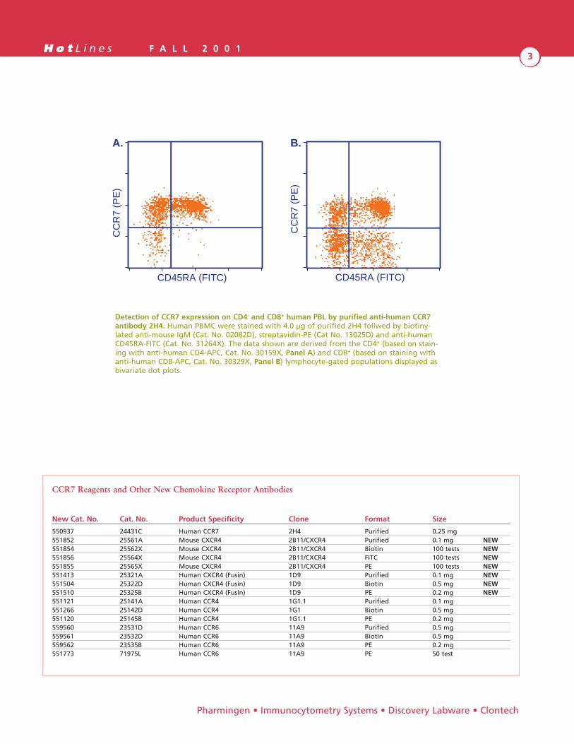
Detection of CCR7 expression on CD4- and CD8+ human PBL by purified anti-human CCR7antibody 2H4. Human PBMC were stained with 4.0 µg of purified 2H4 follwed by biotiny-lated anti-mouse IgM (Cat. No. 02082D), streptavidin-PE (Cat No. 13025D) and anti-humanCD45RA-FITC (Cat. No. 31264X). The data shown are derived from the CD4+ (based on stain-ing with anti-human CD4-APC, Cat. No. 30159X, Panel A) and CD8+ (based on staining withanti-human CD8-APC, Cat. No. 30329X, Panel B) lymphocyte-gated populations displayed asbivariate dot plots.
H o t L i n e s F A L L 2 0 0 14
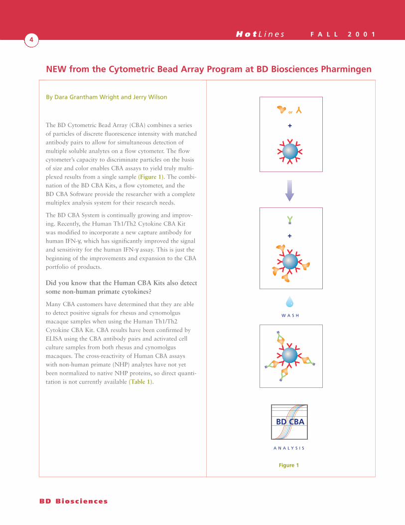
The BD Cytometric Bead Array (CBA) combines a seriesof particles of discrete fluorescence intensity with matchedantibody pairs to allow for simultaneous detection ofmultiple soluble analytes on a flow cytometer. The flowcytometer’s capacity to discriminate particles on the basisof size and color enables CBA assays to yield truly multi-plexed results from a single sample (Figure 1). The combi-nation of the BD CBA Kits, a flow cytometer, and theBD CBA Software provide the researcher with a completemultiplex analysis system for their research needs.
The BD CBA System is continually growing and improv-ing. Recently, the Human Th1/Th2 Cytokine CBA Kitwas modified to incorporate a new capture antibody forhuman IFN-γ, which has significantly improved the signaland sensitivity for the human IFN-γ assay. This is just thebeginning of the improvements and expansion to the CBAportfolio of products.
Did you know that the Human CBA Kits also detectsome non-human primate cytokines?
Many CBA customers have determined that they are ableto detect positive signals for rhesus and cynomolgusmacaque samples when using the Human Th1/Th2Cytokine CBA Kit. CBA results have been confirmed byELISA using the CBA antibody pairs and activated cellculture samples from both rhesus and cynomolgusmacaques. The cross-reactivity of Human CBA assayswith non-human primate (NHP) analytes have not yetbeen normalized to native NHP proteins, so direct quanti-tation is not currently available (Table 1).
By Dara Grantham Wright and Jerry Wilson
BD Biosc iences
NEW from the Cytometric Bead Array Program at BD Biosciences Pharmingen
+
or
+
W A S H
A N A L Y S I S
BD CBA
Figure 1

H o t L i n e s F A L L 2 0 0 15
Instrument setup is even easier on a dual-laserBD FACSCalibur™
While the fluorescently labeled particles in the BD CBAassays are designed to be excited by the 488 nm lasercommon to all BD flow cytometers, they can also beexcited by the red diode laser on dual-laserBD FACSCalibur instruments. Use of the red diode laserfor exciting the CBA particles and detection of particleemission on the FL4 channel simplifies the instrument setup procedure and reduces the need for fluorescence com-pensation. An instrument set up protocol and templatefor dual-laser FACSCalibur instruments, along with manyother updated files for CBA, can be found on CBA pageat the following URL:http://www.bdbiosciences.com/pharmingen/cba/
New versions of the BD CBA Software addcompatibility with all current versions of MS Excel®
The BD CBA Software has been upgraded to version1.1 and version 1.2 to add increased compatibility withmultiple operating systems and MS Excel versions(Table 2).
Cat. No. Description Rhesus and Cynomolgus Cross-reactivity
550749 Human Th1/Th2 Cytokine CBA Kit Interleukin (IL)-4, IL-5, TNF-α, IFN-γ551809 Human Th1/Th2 Cytokine CBA Kit – II Interleukin (IL)-4, IL-6, TNF-α, IFN-γ551811 Human Inflammation CBA Kit – I Interleukin (IL)-8, IL-6, TNF-α
(IL-1ß and IL-12p70 not yet tested)
Pharmingen • Immunocytometry Systems • Discovery Labware • Clontech
Table 1. BD CBA cross-reactivity with non-human primate analytes.
Version 1.0 Version 1.1 Version 1.2
Excel 5 x xExcel 98 x xExcel 2000 (PC only) xExcel 2001 xOS 8.1 to OS 9 x xWindows 98 xWindows NT 4.10 xMac PowerPC ≥ 7000 x xMacintosh G3/G4 x xPC xInt’l Mac OS versions x
Table 2. BD CBA Software compatibility

H o t L i n e s F A L L 2 0 0 16
New BD CBA Assay Kits and Products
Mouse Th1/Th2 Cytokine CBA Kit
50 tests Cat. No. 551287
Simultaneous detection of Mouse IL-2, IL-4, IL-5, IFN-γ,and TNF-α from serum or supernatant samples. Completekit containing sufficient reagents to run 50 samples.
Now Available
Human Th1/Th2 Cytokine CBA Kit – II (Now with IL-6)
50 tests Cat. No. 551809
Simultaneous detection of Human IL-2, IL-4, IL-6, IL-10,TNF-α, and IFN-γ from serum, plasma, or supernatantsamples. Complete kit containing sufficient reagents torun 50 samples.
Now Available
Human Inflammatory Cytokine CBA Kit – I
50 tests Cat. No. 551811
Simultaneous detection of Human IL-8, IL-1ß, IL-6, IL-10, TNF-α, and IL-12p70 from serum, plasma, or super-natant samples. Complete kit containing sufficientreagents to run 50 samples.
Available October 2001
Human Th1/Th2 Cytokine Standards
1 vial Cat. No. 2428KC
A single-use vial containing lyophilized recombinanthuman IL-2, IL-4, IL-5, IL-6, IL-10, TNF-α, and IFN-γfor use as a standard in the Human Th1/Th2 CytokineCBA Kits (Cat. Nos. 550749 and 551809).
Available October 2001
Human Th1/Th2 Cytokine CBA
50 tests Cat. No. 550749
Simultaneous detection IL-2, IL-4, IL-5, IL-10, TNF-α,and IFN-γ from serum, plasma, or supernatant samples.Complete kit containing sufficient reagents to run50 samples.
Now Available
Mouse Ig Isotyping CBA
100 tests Cat. No. 550026
Simultaneous immunoglobulin isotype profile, includinglight chain analysis, of mouse IgG1, IgG2a, IgG2b, IgG3,IgA, IgM, and IgE.
Now Available
BD CBA Software
1 CD (v 1.1 and 1.2) Cat. No. 550065
The BD CBA Software enables rapid analysis of CBA datafiles for generation of standard curves and sample valuereporting.
Now Available
(continued from page 5)
BD Biosc iences
NEW from the Cytometric Bead Array Program at BD Biosciences Pharmingen

H o t L i n e s F A L L 2 0 0 17
Visit the CBA homepage for the most current CBA
system updates:
http://www.bdbiosciences.com/pharmingen/cba/
Did you know you could increase throughput anddecrease hands-on time by combining the BD CBAwith the New BD Multiwell™ AutoSampler System?
Add walk-away automation to your CBA assays withthe new Multiwell™ AutoSampler for use withBD Biosciences flow cytometers.
· Provides walk-away sample introduction from avariety of multiwell plates
· Includes Multiwell Plate Manager software foracquisition and data analysis
· Equipped with the FACSFlow™ Supply Systemfor hours of hands-free operation
· Compatible with instruments with the FACS™Loader option
· Installs easily beneath the cytometer with noadditional bench space required
Features:
· Flexible acquisition from 96- or 384-well plates,both standard and deep-well
· User-definable sample volume, mixing, and washingfor optimal performance
· Innovative Cytometer Interface Unit (CIU) providesconsistent sample throughput
· New graphical user interface for test setup,acquisition control, and data retrieval
· Colorful analysis software shows results at a glance
Specifications:
· Mixing: selectable for 0–3 mix repetitions,definable from 10–250 mL
· Washing: selectable for 0–3 wash repetitions
· Sample volume: definable from 25–250 mL
· Sample carry over: < 1% with one wash cyclebetween cell samples
· Bench space: no additional bench space required;FACScan™ cytometers require lift kits (included).
Pharmingen • Immunocytometry Systems • Discovery Labware • Clontech
H o t L i n e s F A L L 2 0 0 18
The ImmunoSpot™ Analyzer was developed specificallyfor performing complex ELISPOT analyses, and hasmatured over a five-year testing period in a leadingimmunology research laboratory. The ImmunoSpot™Analyzer has been validated in more than seventeen publi-cations and has been selected by the NIH as the referencetool for ELISPOT image analysis. The stand alone systemis equipped with a camera, a lens, a light source, a highprecision X-Y stage, a PC with monitor and keyboard,and the ImmunoSpot™ application program. Satellitesystems are available for the analysis of raw data images.
The ImmunoSpot™ Analyzer has been optimized to meetthe needs of researchers who demand sophistication andflexibility from a highly efficient, user-friendly system forthe acquisition and analysis of ELISPOT data.
By Alexey Karulin, Ph.D., Tameem Ansari, M.Sc,David Sehy, M.Sc, and Paul Lehmann, M.D., Ph.D.
The ImmunoSpot™ Series 1 Analyzer
BD Biosc iences
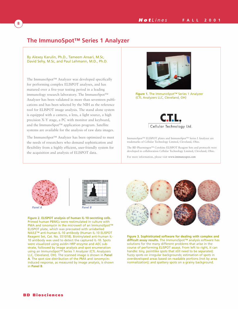
Figure 2. ELISPOT analysis of human IL-10-secreting cells.Primed human PBMCs were restimulated in culture withPMA and ionomycin in the microwell of an ImmunoSpot™ELISPOT plate, which was precoated with unlabelledNA/LE™ anti-human IL-10 antibody (Human IL-10 ELISPOTReagent Set, Cat. No. 551018). Biotinylated anti-human IL-10 antibody was used to detect the captured IL-10. Spotswere visualized using avidin-HRP enzyme and AEC sub-strate, followed by image analysis and spot enumerationusing an ImmunoSpot™ Series 1 Analyzer (CTL AnalyzersLLC, Cleveland, OH). The scanned image is shown in PanelA. The spot size distribution of the PMA and ionomycin-induced response, as measured by image analysis, is shownin Panel B.
Panel BPanel A
Figure 1. The ImmunoSpot™ Series 1 Analyzer(CTL Analyzers LLC, Cleveland, OH)
Figure 3. Sophisticated software for dealing with complex anddifficult assay results. The ImmunoSpot™ analysis software hassolutions for the many different problems that arise in thecourse of performing ELISPOT assays. From left to right, it canhandle: tiny, pointlike spots that still need to be separated;fuzzy spots on irregular backgrounds; estimation of spots inoverdeveloped areas based on readable portions (not by areanormalization); and spattery spots on a grainy background.
ImmunoSpot™ ELISPOT plates and ImmunoSpot™ Series I Analyzer aretrademarks of Cellular Technology Limited, Cleveland, Ohio.
The BD Pharmingen™ Cytokine ELISPOT Reagent Sets and protocols weredeveloped in collaboration Cellular Technology Limited, Cleveland, Ohio.
For more information, please visit www.immunospot.com

H o t L i n e s F A L L 2 0 0 19
ELISPOT Instrumentation and Software
For further information about the ImmunoSpot™ Series 1Analyzer or Satellite and to arrange for a demonstration,please contact your local BD Biosciences reagent salesconsultant.
ImmunoSpot™ Series 1 Analyzer
for scanning of plates and fully automated high resolutionELISPOT image analysis
• Acquisition Unit
• PC System with Windows
• 4x/16/CD-R, frame grabber
• 17” Monitor
• ImmunoSpot™ Software
• Uninterruptible Power Supply
• User’s Manual
ImmunoSpot™ Series 1 Satellite
for fully automated high resolution ELISPOTimage analysis
• PC System with Windows
• 17” Monitor
• ImmunoSpot™ Software
• User’s Manual
ImmunoSpot™ software
Unique features of the ImmunoSpot™Series 1 Analyzer
Fast, fully automated acquisition and storage ofhigh-resolution images
The ImmunoSpot™ Analyzer captures images of all stan-dard ELISPOT 96-well microtiter plates in fully auto-mated mode, and saves those images directly to CD.The images may be sent to an ImmunoSpot™ Satellitewhen the acquisition unit of the system is busy capturingimages, to permit a high throughput of data analysis.The Analyzer provides imaging capability to single-cellresolution.
Fully automatic well centering
The ImmunoSpot™ Analyzer centers each well under thecamera to accommodate variation from plate to plate andautomatically adjust the light intensity ensuring that theimage of each and every well is captured in its entirety.
Software functions for coping with the complexitiesof everyday use
The ImmunoSpot™ Analyzer offers a range of user-defin-able parameters for dealing with real world analysis ofplates: Background Balance, Overdeveloped AreaProcessing, Sensitivity Adjustment, Spot Size, SpotSeparation Tolerance, Detail, and Diffuse Spot Counting— all adjustable to help the researcher customize analysis.
Custom imaging
The ImmunoSpot™ Analyzer permits compilation ofstored images in a variety of ways. During automaticimage acquisition of images the Analyzer stores the imageof each well in its own file and then makes a compositeimage of the whole plate. The storage of well images indiscrete files enables the researcher to retrieve any numberof files and manipulate them into a custom array.
Automated analysis of data
Once the raw images are acquired and the parameters areset, the analysis of images is fully automatic, yielding botha spot count and a histogram. Working in virtual mode,one may re-analyze raw images according to differentparameters, again automatically. The accuracy of auto-matic analysis has been validated to 1%, demonstratingthat objectivity and reproducibility are assured. Data andanalyses are exportable to spreadsheets for generatingfigures and tables.
Versatility
The ImmunoSpot™ Analyzer may be used alone ornetworked with any number of Satellites. Furthermore,the ImmunoSpot™ Analyzer may be coupled with arobotic plate loader/stacker for high volume applications.
Pharmingen • Immunocytometry Systems • Discovery Labware • Clontech
H o t L i n e s F A L L 2 0 0 110
The enzyme-linked immunospot (ELISPOT) assay is apowerful tool for detecting and analyzing individual cellsthat secrete a particular protein in vitro1 . Although origi-nally developed for analyzing specific antibody-secretingcells2,3, the assay has been adapted for measuring thefrequencies of cells that produce and secrete a varietyof other effector molecules such as cytokines4,5,6. TheELISPOT assay has the advantage of being capable ofdetecting cytokine-producing cells from both activatednaive and memory T cell populations, as well as a varietyof other cell types. Cytokine release can be detected at thesingle cell level, allowing direct determination of cytokine-producing cell frequencies5. Furthermore, this assay hasbeen found to be more sensitive than ELISA and intracel-lular staining1,7,8. The sensitivity of the assay enablesmeasurement of even very low frequencies of cytokine-producing cells (e.g., 1/300,000)1. Recent developments inassay plate design and in ELISPOT plate-reader instru-mentation have significantly improved the utility of theELISPOT method for objective and rapid analysis ofcytokine-producing cells1.
The ELISPOT derives its specificity and sensitivity byemploying high affinity capture and detection antibodiesand enzyme-amplification. Since 1989, Pharmingen hasproduced and supplied high quality antibody pairs forcytokine analysis. Numerous publications cite BD Pharmingen™ cytokine ELISA pairs for ELISPOTassay, including Current Protocols in Immunology (JohnWiley & Sons, Inc., NY). Recent collaborations with CTLAnalyzers LLC (www.immunospot.com; Cleveland, OH),the developers of state-of-the-art ImmunoSpot™ platereader instrumentation and software, have led to the iden-tification of novel, optimal antibody clone pairings forELISPOT assays. In the course of screening many anti-body clones for the ELISPOT application, we ascertainedthat some antibody pairs which perform very well forELISA are not necessarily effective for ELISPOT assay.ELISPOT-compatible antibody clones were selected basedon sensitivity and ability to produce discrete and densely-colored spots. In some cases, best ELISPOTs were found
to result from cocktails made from multiple capture anti-body clones combined. To avoid the effects of sodiumazide and endotoxin on cultured cells, theBD Pharmingen™ ELISPOT capture antibodies are specialformulations which contain no sodium azide and minimalendotoxin (NA/LE™).
The IFN-γ ELISPOT assay is one important tool for char-acterization of T cell effector function. To identify the bestIFN-γ ELISPOT reagents, we have screened a panel ofnew anti-mouse and anti-human IFN-γ antibody clones inELISPOT assays with a variety of target cell preparations,including naïve T cells, memory T cells, and cell lines.We are proud to announce that BD BiosciencesPharmingen now has available specially screened andformulated high sensitivity antibody pairs for humanIFN-γ and mouse IFN-γ ELISPOT assay applications.These ELISPOT-compatible antibody pairs have alsoproven to be very effective for conventional sandwichELISA.
The IFN-γ ELISPOT reagents are to be available in threedifferent formats, which provide varying degrees of flexi-bility and time saving convenience: Pairs, Sets, and Kits.
• ELISPOT Reagent Pairs: Sufficient antibody for 5plates of assays. These reagents are tested with, andrecommended for use with, high sensitivity/low back-ground ImmunoSpot™ ELISPOT plates availablefrom CTL Analyzers LLC (Cleveland, OH).www.immunospot.com.
• ELISPOT Reagent Sets: 10 ImmunoSpot™ ELISPOTplates (not pre-coated with antibody), along withsufficient antibody for 10 plates of assays.
• ELISPOT Kits: Designed for maximal time savingsand convenience, supplying 2 plates, precoated withantibody, along with all buffers, substrates, enzymes,and detection reagents needed for those 2 platesof assays.
By Qi Guan, David Sehy, M.S. and David Ernst, Ph.D.
NEW Interferon-γ ELISPOT Assays
BD Biosc iences

H o t L i n e s F A L L 2 0 0 111
BD Pharmingen™ ELISPOT Products
ELISPOT Reagent Pairs
• Unlabelled anti-cytokine capture antibody(NA/LE™ format); sufficient reagent for coating5 plates
• Biotinylated anti-cytokine detection antibody;sufficient reagent for 5 plates
• Certificate of Analysis, providing lot-specificoptimal reagent concentrations
ELISPOT Reagent Sets
• 10 ImmunoSpot™ ELISPOT plates
• Unlabelled anti-cytokine capture antibody(NA/LE™ format); sufficient reagent for coating10 plates
• Biotinylated anti-cytokine detection antibody;sufficient reagent for 10 plates
• Avidin horseradish peroxidase; sufficient for10 plates
• Certificate of Analysis, providing lot-specificoptimal reagent concentrations
ELISPOT Kits
• 2 pre-coated ImmunoSpot™ ELISPOT plates
• Biotinylated anti-cytokine detection antibody;sufficient reagent for 2 plates
• Avidin horseradish peroxidase; sufficient for 2 plates
• Certificate of Analysis, providing lot-specificoptimal reagent concentrations
• Assay diluent
• Wash Buffer
• AEC substrate
Pharmingen • Immunocytometry Systems • Discovery Labware • Clontech
Figure 1. Superior Human IFN-γ ELISPOTs Are Obtained Using a novel anti-human IFN-γ Antibody for Capture. Primed humanPBMC were restimulated (overnight) with PMA (5 ng/ml; Sigma, Cat. No. P-8139) and ionomycin (500 ng/ml; Sigma, Cat. No.I-0634) in the microwell of an ImmunoSpot™ ELISPOT plate that was pre-coated with the NA/LE anti-human IFN-γ. Biotinylatedanti-human IFN-γ antibody was used to detect the captured IFN-γ. Spots were visualized using avidin-HRP enzyme and AECsubstrate. Image analysis and spot enumeration were carried out using the ImmunoSpot™ Series I Analyzer (CTL Analyzers LLC,Cleveland, OH). Panels A and B were derived from experiments conducted using the same activated cells in the sameImmunoSpot™ plate.
Panel A: Human IFN-γ ELISPOT Set (BD Biosciences Pharmingen, Cat. No. 2554KI).
Panel B: Alternative human IFN-γ ELISPOT reagent pairing.
Panel A Panel B
H o t L i n e s F A L L 2 0 0 112
References:
1. Helms, T., B. Boehm, R. Asaad, R. Trezza, P. Lehmann, and M.Tary-Lehmann. 2000. Direct visuali-zation of cytokine-producingrecall antigen-specific CD4 memory T cells in healthy individualsand HIV patients. J. Immunol. 164: 3723-3732.
2. Sedgwick, J., and P. Holt. 1983. A solid-phase immunoenzymatictechnique for the enumeration of specific antibody-secretingcells. J. Immunol. Meth. 57: 301.
3. Czerkinsky, C.C., L.A. Nilsson, H. Nygren, O. Ouchterlony, and A.Tarkowski. 1983. A solid-phase enzyme-linked immunospot(ELISPOT) assay for enumeration of specific antibody-secretingcells. J. Immunol. Meth. 65: 109.
4. Ronnblom, L., B. Cederblad, K. Sandberg, and G. Alm. 1988.Determination of herpes simplex virus-induced alpha interferon-secreting human blood lymphocytes by a filter immuno-plaqueassay. Scand. J. Immunol. 2: 165-171.
5. Czerkinsky, C., G. Andersson, H. Ekre, L. Nilsson, L. Klareskog,and O. Ouchterlony. 1988. Reverse ELISPOT assay for clonalanalysis of cytokine production. J. Immunol. Meth. 110: 29-36.
6. Fujihashi, K., J. McGhee, K. Beagley, D. McPherson, S.McPherson, C.-M. Huang, and H. Kiyono. 1993. Cytokine-specificELISPOT assay: single cell analysis of IL-2, IL-4, and IL-6 producingcells. J. Immunol. Meth. 160: 181-189.
7. Tanguay,S., and J.J. Killion. 1994. Direct comparison of ELISPOTand ELISA-based assay for detection of individual cytokine-secreting cells. Lymphokine Cytokine Res. 13, 259.
BD Biosc iences
NEW Interferon-γ ELISPOT Assays (continued from page 11)
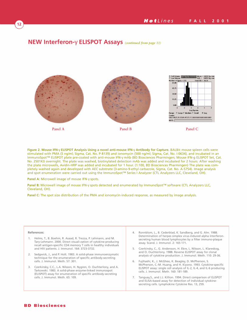
Figure 2. Mouse IFN-γ ELISPOT Analysis Using a novel anti-mouse IFN-γ Antibody for Capture. BALB/c mouse spleen cells werestimulated with PMA (5 ng/ml; Sigma, Cat. No. P-8139) and ionomycin (500 ng/ml; Sigma, Cat. No. I-0634), and incubated in anImmunoSpot™ ELISPOT plate pre-coated with anti-mouse IFN-γ mAb (BD Biosciences Pharmingen; Mouse IFN-g ELISPOT Set, Cat.No. 2501KI) overnight. The plate was washed, biotinylated detection mAb was added and incubated for 2 hours. After washingthe plate microwells, Avidin-HRP was added and incubated for 1 hour. (1:100, BD Biosciences Pharmingen) The plate was com-pletely washed again and developed with AEC substrate (3-amino-9-ethyl carbazole, Sigma, Cat. No. A-5754). Image analysis and spot enumeration were carried out using the ImmunoSpot™ Series I Analyzer (CTL Analyzers LLC, Cleveland, OH).
Panel A: Microwell image of mouse IFN-γ spots.
Panel B: Microwell image of mouse IFN-γ spots detected and enumerated by ImmunoSpot™ software (CTL Analyzers LLC,Cleveland, OH).
Panel C: The spot size distribution of the PMA and ionomycin-induced response, as measured by image analysis.
Panel A Panel B Panel C
H o t L i n e s F A L L 2 0 0 113
Pharmingen • Immunocytometry Systems • Discovery Labware • Clontech
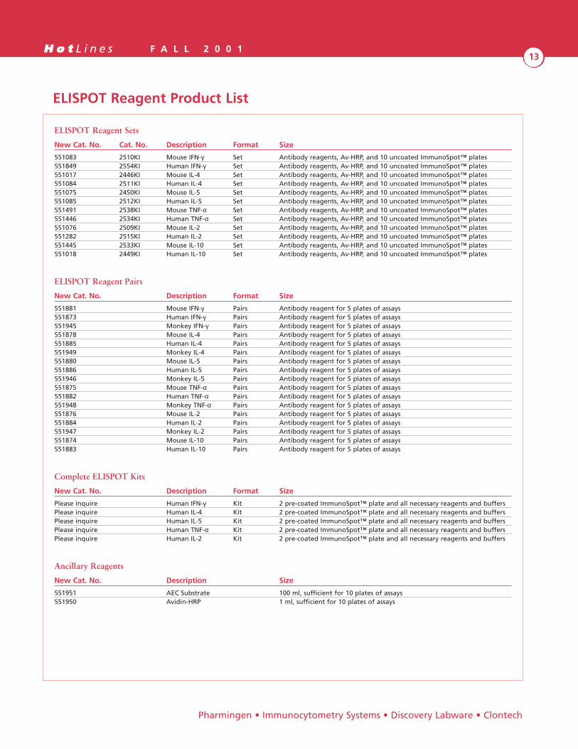
ELISPOT Reagent Product List
ELISPOT Reagent Sets
New Cat. No. Cat. No. Description Format Size
551083 2510KI Mouse IFN-γ Set Antibody reagents, Av-HRP, and 10 uncoated ImmunoSpot™ plates551849 2554KI Human IFN-γ Set Antibody reagents, Av-HRP, and 10 uncoated ImmunoSpot™ plates551017 2446KI Mouse IL-4 Set Antibody reagents, Av-HRP, and 10 uncoated ImmunoSpot™ plates551084 2511KI Human IL-4 Set Antibody reagents, Av-HRP, and 10 uncoated ImmunoSpot™ plates551075 2450KI Mouse IL-5 Set Antibody reagents, Av-HRP, and 10 uncoated ImmunoSpot™ plates551085 2512KI Human IL-5 Set Antibody reagents, Av-HRP, and 10 uncoated ImmunoSpot™ plates551491 2538KI Mouse TNF-α Set Antibody reagents, Av-HRP, and 10 uncoated ImmunoSpot™ plates551446 2534KI Human TNF-α Set Antibody reagents, Av-HRP, and 10 uncoated ImmunoSpot™ plates551076 2509KI Mouse IL-2 Set Antibody reagents, Av-HRP, and 10 uncoated ImmunoSpot™ plates551282 2515KI Human IL-2 Set Antibody reagents, Av-HRP, and 10 uncoated ImmunoSpot™ plates551445 2533KI Mouse IL-10 Set Antibody reagents, Av-HRP, and 10 uncoated ImmunoSpot™ plates551018 2449KI Human IL-10 Set Antibody reagents, Av-HRP, and 10 uncoated ImmunoSpot™ plates
ELISPOT Reagent Pairs
New Cat. No. Description Format Size
551881 Mouse IFN-γ Pairs Antibody reagent for 5 plates of assays551873 Human IFN-γ Pairs Antibody reagent for 5 plates of assays551945 Monkey IFN-γ Pairs Antibody reagent for 5 plates of assays551878 Mouse IL-4 Pairs Antibody reagent for 5 plates of assays551885 Human IL-4 Pairs Antibody reagent for 5 plates of assays551949 Monkey IL-4 Pairs Antibody reagent for 5 plates of assays551880 Mouse IL-5 Pairs Antibody reagent for 5 plates of assays551886 Human IL-5 Pairs Antibody reagent for 5 plates of assays551946 Monkey IL-5 Pairs Antibody reagent for 5 plates of assays551875 Mouse TNF-α Pairs Antibody reagent for 5 plates of assays551882 Human TNF-α Pairs Antibody reagent for 5 plates of assays551948 Monkey TNF-α Pairs Antibody reagent for 5 plates of assays551876 Mouse IL-2 Pairs Antibody reagent for 5 plates of assays551884 Human IL-2 Pairs Antibody reagent for 5 plates of assays551947 Monkey IL-2 Pairs Antibody reagent for 5 plates of assays551874 Mouse IL-10 Pairs Antibody reagent for 5 plates of assays551883 Human IL-10 Pairs Antibody reagent for 5 plates of assays
Complete ELISPOT Kits
New Cat. No. Description Format Size
Please inquire Human IFN-γ Kit 2 pre-coated ImmunoSpot™ plate and all necessary reagents and buffersPlease inquire Human IL-4 Kit 2 pre-coated ImmunoSpot™ plate and all necessary reagents and buffersPlease inquire Human IL-5 Kit 2 pre-coated ImmunoSpot™ plate and all necessary reagents and buffersPlease inquire Human TNF-α Kit 2 pre-coated ImmunoSpot™ plate and all necessary reagents and buffersPlease inquire Human IL-2 Kit 2 pre-coated ImmunoSpot™ plate and all necessary reagents and buffers
Ancillary Reagents
New Cat. No. Description Size
551951 AEC Substrate 100 ml, sufficient for 10 plates of assays551950 Avidin-HRP 1 ml, sufficient for 10 plates of assays

H o t L i n e s F A L L 2 0 0 114
One of the most important functions of the adaptiveimmune response is antigen recognition. Without theability of cells to recognize antigens in a specific fashion,the immune system would be unable to distinguish selffrom non-self. Special cells are capable of processingextracellular pathogens and displaying them on their cellsurface to be recognized by T cells. Infectious bacteria andother extracellular microorganisms are captured bymacrophages, dendritic cells or B cells, internalized forfurther digestion, then presented as an MHC class II:peptide complex recognized by T-helper CD4+ cells.Intracellular parasites like viruses, on the other hand, canbe presented by all nucleated cells and are processedthrough a different cellular compartment. These cellsmostly present these pathogens as a MHC class I:peptide complex recognized by T-suppressor/cytotoxicCD8+ cells1,2.
Dendritic Cells
Among the cells that capture and process antigens, den-dritic cells have been described in the literature as beingthe most efficient2. Dendritic cells (DC) are referred to asthe professional antigen presenting cells (APC). Dendriticcells are a heterogeneous cell population that continuouslyderive from bone marrow stem cells. Following matura-tion, they reside in both lymphoid and non-lymphoidtissues. Liu et al3 described two human dendritic celllineages. Pre -DC1 myeloid monocytes give rise tomyeloid DC1 that produce a significant amount of IL-12and thus induce Th1 responses in humans. Pre-DC2 plas-macytoid DC, which become lymphoid DC2, inducehuman Th2 responses. Both lymphoid and myeloid pre-DCs express toll-like receptors (TLR) and lectin mole-cules, but lymphoid DCs express TLR7 and TLR9 whilemyeloid DCs express only TLR2 and 4. Myeloid DC1sexpress large amount of mannose receptor, but DC2sdo not4.
The development of bone marrow-derived DCs is highlydependent on their microenvironment. Several cytokinesand growth factors have been identified as being partici-pants or inducers of DC differentiation. During theirmaturation process, DC can migrate to various tissues.Some other dendritic cells are found in the thymus, spleen,lymph nodes and even in areas of the central nervoussystem. They reside in tissues in an immature stage andrespond to various chemoattractants from inflammationsites. At the same time, they produce chemokines thatrecruit and activate macrophages, granulocytes, naturalkiller cells, and other immature DC. Once activated bymicroorganisms and inflammatory stimuli, DC undergofurther maturation and migrate to the lymph nodes insearch of antigen specific naïve T cells. During this migra-tion, DC process the antigens they captured in the tissueareas, express costimulatory molecules, and becomepotent stimulators of T cells.
Antigen Processing
Intracellular Antigens
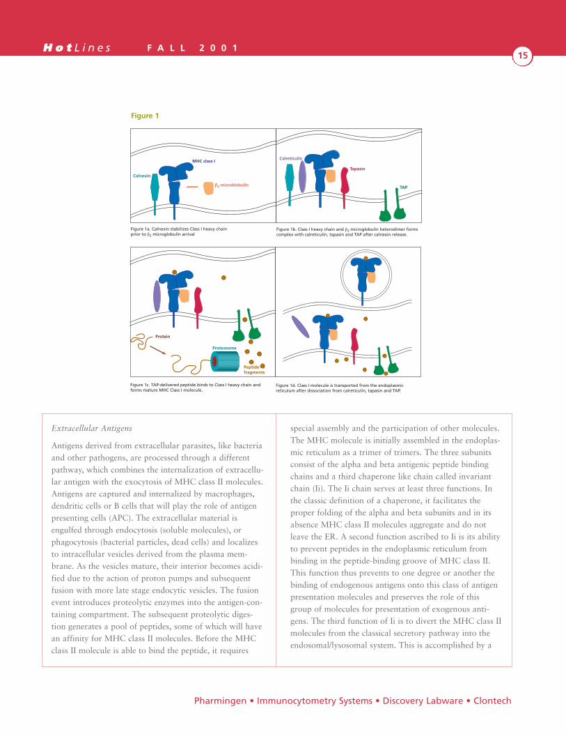
Viral proteins, or other components from intracellularpathogens in the cytoplasm, are reduced to peptides byproteasomes. Proteasomes are special protein complexespresent in the cellular cytosol that have proteolytic func-tion5,6. Once the parasite proteins have been digested tosmall peptides, they are transported into the lumen of theendoplasmic reticulum via a heterodimeric integral mem-brane complex termed TAP (transporter associated withantigen processing). As the MHC class I heavy chain mol-ecule is being folded in the ER, it is stabilized by a mem-brane-bound protein calnexin until the β2-microglobulin(β2m) subunit associates. When β2m binds, calnexin isreleased and the MHC molecule associates with TAP byinteracting with a TAP-associated protein tapasin and achaperone protein calreticulin. The peptide brought inby TAP binds to the MHC molecule and completes theassembly of the complex. The MHC class I:peptidecomplex is released from tapasin and calreticulin, leavesthe ER and is transported to the surface via the classicalsecretory pathway7-10 (Figure 1, a, b).
By Enoc J. Hollemweguer, Ph.D., Bruce Koppelman, Ph.D.,Li Li, Ph.D., Jay Dong, M.D., M.S.
Dendritic Cells and Antigen Processing and Presentation
BD Biosc iences
H o t L i n e s F A L L 2 0 0 115
Extracellular Antigens
Antigens derived from extracellular parasites, like bacteriaand other pathogens, are processed through a differentpathway, which combines the internalization of extracellu-lar antigen with the exocytosis of MHC class II molecules.Antigens are captured and internalized by macrophages,dendritic cells or B cells that will play the role of antigenpresenting cells (APC). The extracellular material isengulfed through endocytosis (soluble molecules), orphagocytosis (bacterial particles, dead cells) and localizesto intracellular vesicles derived from the plasma mem-brane. As the vesicles mature, their interior becomes acidi-fied due to the action of proton pumps and subsequentfusion with more late stage endocytic vesicles. The fusionevent introduces proteolytic enzymes into the antigen-con-taining compartment. The subsequent proteolytic diges-tion generates a pool of peptides, some of which will havean affinity for MHC class II molecules. Before the MHCclass II molecule is able to bind the peptide, it requires
special assembly and the participation of other molecules.The MHC molecule is initially assembled in the endoplas-mic reticulum as a trimer of trimers. The three subunitsconsist of the alpha and beta antigenic peptide bindingchains and a third chaperone like chain called invariantchain (Ii). The Ii chain serves at least three functions. Inthe classic definition of a chaperone, it facilitates theproper folding of the alpha and beta subunits and in itsabsence MHC class II molecules aggregate and do notleave the ER. A second function ascribed to Ii is its abilityto prevent peptides in the endoplasmic reticulum frombinding in the peptide-binding groove of MHC class II.This function thus prevents to one degree or another thebinding of endogenous antigens onto this class of antigenpresentation molecules and preserves the role of thisgroup of molecules for presentation of exogenous anti-gens. The third function of Ii is to divert the MHC class IImolecules from the classical secretory pathway into theendosomal/lysosomal system. This is accomplished by a
Calnexin
MHC class I
b2 microblobulin
Calreticulin
Tapasin
TAP
Figure 1a. Calnexin stabilizes Class I heavy chainprior to b2 microglobulin arrival
Figure 1b. Class I heavy chain and b2 microglobulin heterodimer forms complex with calreticulin, tapasin and TAP after calnexin release.
Protein
Proteasome
Peptidefragments
Figure 1c. TAP-delivered peptide binds to Class I heavy chain and forms mature MHC Class I molecule.
Figure 1d. Class I molecule is transported from the endoplasmic reticulum after dissociation from calreticulin, tapasin and TAP.
Pharmingen • Immunocytometry Systems • Discovery Labware • Clontech
Figure 1
H o t L i n e s F A L L 2 0 0 116
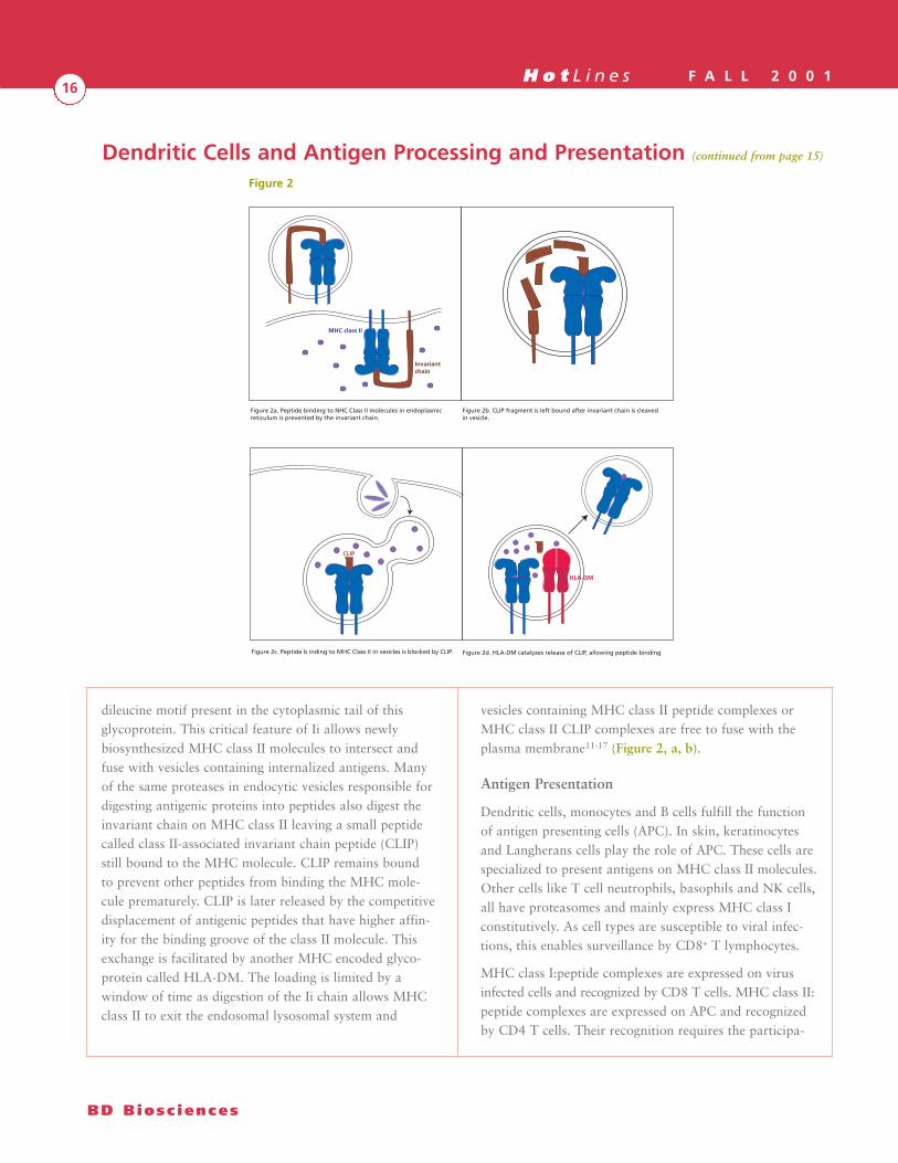
dileucine motif present in the cytoplasmic tail of thisglycoprotein. This critical feature of Ii allows newlybiosynthesized MHC class II molecules to intersect andfuse with vesicles containing internalized antigens. Manyof the same proteases in endocytic vesicles responsible fordigesting antigenic proteins into peptides also digest theinvariant chain on MHC class II leaving a small peptidecalled class II-associated invariant chain peptide (CLIP)still bound to the MHC molecule. CLIP remains boundto prevent other peptides from binding the MHC mole-cule prematurely. CLIP is later released by the competitivedisplacement of antigenic peptides that have higher affin-ity for the binding groove of the class II molecule. Thisexchange is facilitated by another MHC encoded glyco-protein called HLA-DM. The loading is limited by awindow of time as digestion of the Ii chain allows MHCclass II to exit the endosomal lysosomal system and
vesicles containing MHC class II peptide complexes orMHC class II CLIP complexes are free to fuse with theplasma membrane11-17 (Figure 2, a, b).
Antigen Presentation
Dendritic cells, monocytes and B cells fulfill the functionof antigen presenting cells (APC). In skin, keratinocytesand Langherans cells play the role of APC. These cells arespecialized to present antigens on MHC class II molecules.Other cells like T cell neutrophils, basophils and NK cells,all have proteasomes and mainly express MHC class Iconstitutively. As cell types are susceptible to viral infec-tions, this enables surveillance by CD8+ T lymphocytes.
MHC class I:peptide complexes are expressed on virusinfected cells and recognized by CD8 T cells. MHC class II:peptide complexes are expressed on APC and recognizedby CD4 T cells. Their recognition requires the participa-
MHC class II
Invariantchain
CLIP
HLA-DM
Figure 2a. Peptide binding to NHC Class II molecules in endoplasmic reticulum is prevented by the invariant chain.
Figure 2b. CLIP fragment is left bound after invariant chain is cleaved in vesicle.
Figure 2c. Peptide b inding to MHC Class II in vesicles is blocked by CLIP. Figure 2d. HLA-DM catalyzes release of CLIP, allowing peptide binding
BD Biosc iences
Figure 2
Dendritic Cells and Antigen Processing and Presentation (continued from page 15)

H o t L i n e s F A L L 2 0 0 117
tion of other molecules that trigger costimulatory oraccessory signals. The participation of the CD4 or CD8molecules in the recognition of the MHC:peptide complexis essential in this process. CD4 helps recognize the MHCclass II: peptide complex and CD8 helps recognize pep-tides presented in the context of MHC class I18.
APCs express adhesion molecules (either constitutively orupon activation), which can strengthen the bonds betweenT cells and APC and lead to induction of accessorysignals19,20. A partial list of the adhesion molecules foundon APC includes LFA-1 (CD11a/CD18), ICAM-1(CD54), VCAM-1 (CD106) and LFA-3 (CD58).APC can also express MHC Class Ior Class II surface molecules thathave an affinity for processed anti-genic peptides and serve to presentthem to T cells. In addition to theadhesion molecules and MHCcomponents, APC display cos-timulatory molecules, B7-1(CD80), B7-2 (CD86), and prob-ably a B7-X (still to be defined).Costimulatory molecules play acrucial role in determining T-cellresponses to presented antigen21-24.
The first phase (phase I) in theprocess of antigen presentation is theinteraction of APC and lymphocytes viaadhesion molecules. The second phase (phase II) involvesthe recognition of the antigen by the T-cell receptor(TCR). As the T cell recognizes an antigen through itsTCR, it initiates signals which upregulate the expressionof costimulatory molecules. Receipt of secondary signalsprovided by bound costimulatory molecules results in theproduction and secretion of cytokines, and expression ofcytokine receptors on T cells. For example, T-helper cellsproduce interleukin-2 (IL-2) and upregulate IL-2 receptorfollowing signaling by CD28 binding. On the APC side,this is driven by IFN-γ secretion by T cells then subse-quent IL-12 production by APC’s36. Cytokines induce theupregulation of CD28 on T cells and CD86 on APC. AsCD28 and CD86 (Figure 3) connect during phase III,other signals are triggered which induce the expression of
additional costimulatory molecules: CD80 on APC andCTLA-4 on T cells. The process is completed by prolifera-tion of T cells (phase IV). As CD4+ T-helper cells undergoproliferation, they can evolve into either memory T cellsor, in response to additional cytokines, into effector Tcells of either T-helper type 1 (Th1) or T-helper type 2(Th2). Alternatively, CD4+ T-helper cells can undergoactivation induced cell death (AICD)25-29.
DC-SIGN and CMRF-56
BD Biosciences Pharmingen offers two new monoclonalantibodies specific for dendritic cells, CMRF-56, and
DC-SIGN (Figures 4). CMRF-56 reacts withan early activation/differentiation antigen
expressed on dendritic cells.Circulating blood leukocytes andDC do not express the CMRF-56antigen, but following eitherin vitro culture or activation ofPBMC’s, CMRF-56 antigen isexpressed on DC and a subpopu-
lation of CD19+ lymphocytes30.DC- SIGN, a type II membrane
protein of approximately 44 kDa, witha mannose-binding, C-type lectin domain is
highly expressed on dendritic cells in mucosaltissues and binds to ICAM-3 (CD50). Its
sequence is identical to the HIV-1 envelope glyco-protein gp-120-binding C-type lectin and was renamedDC-SIGN31. Reports demonstrate that DC-SIGN bindsto HIV-1 gp-120. DC-SIGN does not function as a viralreceptor, but it allows dendritic cells to efficiently infectresting T cells expressing CD4 and chemokine receptors32.Reports also suggest that DC-SIGN enables the highlyefficient migration of dendritic cells from blood into thetissues33. It can interact with ICAM-2, which has a similarsequence as ICAM-3, and is abundantly expressed on vas-cular and lymphoid endothelium. Thus, DC-SIGN medi-ated dendritic cell rolling, transendothelial migration, andthe ICAM-2 interaction is essential the specific migratoryfunctions of dendritic cells.
Pharmingen • Immunocytometry Systems • Discovery Labware • Clontech
APC
T Cell
CD58 (LFA-3)
CD2
CD4
TCR
CD54 (ICAM-1)
CLASS II MHC
CD80 CD86CD28 CD152
CD11A (LFA-1)
Figure 3

H o t L i n e s F A L L 2 0 0 118
BD Biosc iences
Antigen processing and dendritic cell research reagents from BD Biosciences Pharmingen.*
Human ReagentsCat. No. Description Clone Isotype Format Size
555980 CLIP CerCLIP m IgG1 Purified 0.1 mg555981 CLIP CerCLIP m IgG1 FITC 100 tests551264 DC-SIGN DCN46 m IgG1 FITC 100 tests555982 HLA-DM Map.DM1 m IgG1 Purified 0.1 mg555983 HLA-DM Map.DM1 m IgG1 PE 100 tests559895 HLA-DO DOB.L1 m IgG2b Purified 0.1 mg550020 HLA-DO DOB.L1 m IgG2b FITC Set 100 tests550021 HLA-DO DOB.L1 m IgG2b PE Set 100 tests551295 TAP1 TAP1.28 m IgG1 Purified 0.1 mg551292 TAP2 TAP2.17 m IgG1 Purified 0.1 mg550910 IP30 Map.IP30 m IgG1 Purified 0.1 mg550915 IP30 Map.IP30 m IgG1 FITC Set 100 testsComing Soon TAP2 TAP2.17 m IgG1 FITC Set 100 testsComing Soon TAP2 TAP2.17 m IgG1 PE Set 100 testsComing Soon Antibody Kit for Cultured Human Dendritic Cells† 34224N CD1a-FITC HI149 mIgG1 0.05 mg 33075N CD40-PE 5C3 mIgG1 0.05 mg34235N HLA-DR-PE G46-6 mIgG2a 0.05 mg36794N CD80-FITC L307.4 mIgG1 0.05 mg33404N CD86-FITC FUN-1 mIgG1 0.05 mg36935N CD83-PE HB15e mIgG1 0.05 mg30545N CD14-PE M5E2 mIgG2a 0.05 mg33815N Isotype Contr.-PE MOPC-21 mIgG1 0.05 mg33814N Isotype Contr.-FITC MOPC-21 mIgG1 0.05 mg33035N Isotype Contr.-PE G155-178 mIgG2a 0.05 mg
Mouse Reagents
Expression of cell surface markers on myeloid and lymphoid derived mouse spleen dendritic cells.
Cat. No. Surface marker Myleoid DC (spl.) Lymphoid DC (spl.)
553843, 559871 CD1d No Yes553646, 553042, 553052 CD4 Yes No553027, 553829 CD8a No Yes553118 CD9 Yes Yes553118, 557453 CD11a Yes Yes553308 CD11b Yes dim/neg553799 CD11c Yes Yes558743 CD13 Yes Yes553136 CD23 No Yes558777 CD24 Yes/low Yes553131, 558739 CD44 Yes Yes553250 CD54 Yes Yes553368, 553766 CD80 Low Low553689, 558784 CD86 Yes Low553326 CD102 Yes Yessee Catalog for individual haplotype MHC Class I Yes Yessee Catalog for individual haplotype MHC Class II Yes Yes
† Panel of antibodies useful for detecting dendritic cells as well as an optimized protocol to generate cultured dendritic cells fromperipheral blood mononuclear cells.
Dendritic Cells and Antigen Processing and Presentation (continued from page 17)
*All the BD Biosciences reagents highlighted in this article are “For research use only. Not for use in diagnostic or therapeutic procedures”.
H o t L i n e s F A L L 2 0 0 119
Pharmingen • Immunocytometry Systems • Discovery Labware • Clontech
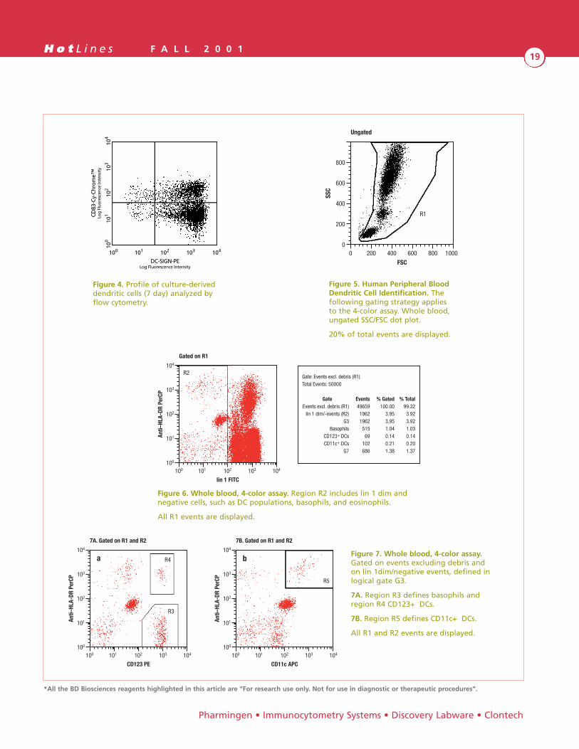
Figure 4. Profile of culture-deriveddendritic cells (7 day) analyzed byflow cytometry.
DC-SIGN-PELog Fluorescence Intensity
101 102 103 104100
101
102
103
104
100
CD83
-Cy-
Chro
me™
Log
Fluo
resc
ence
Inte
nsit
y
*All the BD Biosciences reagents highlighted in this article are “For research use only. Not for use in diagnostic or therapeutic procedures”.
Figure 6. Whole blood, 4-color assay. Region R2 includes lin 1 dim andnegative cells, such as DC populations, basophils, and eosinophils.
All R1 events are displayed.
0 200 400 600 800 1000
FSC
800
600
400
200
0
SSC
Ungated
R1R1
Figure 7. Whole blood, 4-color assay.Gated on events excluding debris andon lin 1dim/negative events, defined inlogical gate G3.
7A. Region R3 defines basophils andregion R4 CD123+ DCs.
7B. Region R5 defines CD11c+ DCs.
All R1 and R2 events are displayed.
100 101 102 103 104
CD123 PE
104
103
102
101
100
Anti–
HLA-
DR P
erCP
7A. Gated on R1 and R2
R4
R3
a
100 101 102 103 104
CD11c APC
104
103
102
101
100
Anti–
HLA-
DR P
erCP
7B. Gated on R1 and R2
R5
b
Gate: Events excl. debris (R1)Total Events: 50000
Gate Events % Gated % TotalEvents excl. debris (R1) 49659 100.00 99.32
lin 1 dim/-events (R2) 1962 3.95 3.92G3 1962 3.95 3.92
Basophils 515 1.04 1.03CD123+ DCs 69 0.14 0.14CD11c+ DCs 102 0.21 0.20
G7 686 1.38 1.37
100 101 102 103 104
lin 1 FITC
104
103
102
101
100
Anti–
HLA-
DR P
erCP
Gated on R1
R2
Figure 5. Human Peripheral BloodDendritic Cell Identification. Thefollowing gating strategy appliesto the 4-color assay. Whole blood,ungated SSC/FSC dot plot.
20% of total events are displayed.
H o t L i n e s F A L L 2 0 0 120
BD Biosc iences
Human Peripheral Blood Dendritic Cell Reagents from BD Biosciences Immunocytometry Systems*
CD123+ dendritic cells, CD11c+ dendritic cells, and basophils can be identified directly from peripheral blood. Based on awhole-blood flow cytometric assay, this system saves time and can identify multiple dendritic cell subsets in a single tube.With the first commercialy available lineage cocktail, these rare cellular subsets can be identified without altering theirfunction or phenotype34,35. Learn more about these dendritic cell subsets and the advantages of dendritic cell identificationusing flow cytometry from our application note (Figure 5, 6, 7).
Peripheral Blood Dendritic Cells Revealed by Flow Cytometry (Please call Technical Services for your copy)34,35.
Dendritic Cell Single Vial Reagents
Cat. No. Antibody
340546 Lineage Cocktail 1 (lin 1) FITC(CD3,CD14,CD16,CD19,CD20,CD56)
340545 CD123 (Anti-IL-3Rα) PE347637 CD11c PE340544 CD11c APC347364 Anti-HLA-DR PerCP340549 Anti-HLA-DR APC349043 Mouse IgG1 PE349053 Mouse IgG2a PE340473 Mouse IgG2a APC
3-Color Dendritic Value Bundle
Cat. No. Antibody
340566 Lineage Cocktail 1 (lin 1) FITCCD123 PECD11c PEAnti-HLA-DR PerCPMouse IgG1 PEMouse IgG2a PE
4-Color Dendritic Value Bundle
Cat. No. Antibody
340565 Lineage Cocktail 1 (lin 1) FITCCD123 PECD11c APCAnti-HLA-DR PerCPMouse IgG1 PEMouse IgG2a APC
BD Biosciences recognizes the need in the scientific community for tools to accelerate dendritic cell research. We havecontinued, over the years, to search for antibodies that provide our customers with products to study the mechanisms ofantigen processing and presentation. Please visit our website for new product announcements: www.bdbiosciences.com.
Dendritic Cells and Antigen Processing and Presentation (continued from page 19)
*All the BD Biosciences reagents highlighted in this article are “For research use only. Not for use in diagnostic or therapeutic procedures”.
H o t L i n e s F A L L 2 0 0 121
References:
1. Bjorkman, P.J., M.A. Saper, B. Samraoui, et al. 1987. The foreignantigen binding site and T cell recognition regions of class I his-tocompatibility antigens. Nature 329: 512
2. Reid, S.D., G. Penna, and L. Adorini. 2000. The control of T cellresponses by dendritic cell subsets. Curr. Opin. Immunol 12: 114
3. Liu, Y-J, H. Kanzler, V. Soumelis, et al. 2001. Dendritic celllineage, plasticity and cross-regulation. Nature Immunology2: 585.
4. Pulendran B. et. al. 1997. Developmental pathways of dendriticcells in vivo: distinct function, phenotype, and localization ofdendritic cell subsets in FLT3 ligand-treated mice. J. Immunol.159: 2222.
5. Goldberg, A.L., and K. L. Rock. 1992. Proteolysis, proteasomesand antigen presentation. Nature 4: 375.
6. Rock, K.L., and A.L. Goldberg. 1999. Degradation of cell proteinsand the generation of MHC class I-presented peptides. Annu.Rev. Immunol 17: 739.
7. Hunt, D.F., R.A. Henderson, J. Shabanowitz, et al. 1992.Characterization of peptides bound to the class I MHC moleculeHLA-A2.1 by mass spectrometry. Science 255: 1261.
8. Shepherd, J.C., T.N.M. Schumacher, P.G. Ashton-Rickardt, et al.1993. TAP-1-dependent peptide translocation in vitro is ATP-dependent and peptide-selective. Cell 74: 577.
9. Ortman, B., M.J. Androlewics, and P. Cresswell. 1994. MHC class IIsol b2-microglobulin complexes associate with TAP transportersbefore peptide binding. Nature 368: 864.
10. Williams, D.B., and T. H. Watts. 1995. Molecular chaperones inantigen presentation. Curr. Op. Immunol 7: 77.
11. Cresswell, P. 1995. Assembly, transport and function of MHC classII molecules. Ann. Rev. Immunol. 12: 259.
12. Roche, P.A., and P. Cresswell. 1990. Invariant chain associationwith HLA-DR molecules inhibits immunogenic peptide binding.Nature. 345: 615.
13. Roche, P.A., M.S. Marks, and P. Cresswell. 1991. Formation ofa nine subunit complex by HLA class II glycoproteins and theinvariant chain. Nature 354: 392.
14. Peters, P.J., J.J. Neefjes, V. Orschot, et al. 1991. Segregation ofMHC class II molecules from MHC class I molecules in the golgicomplex for transport to lysosomal compartments. Nature349: 669.
15. Davidson, H.W., P.A. Reid, A. Lanzavecchia, et al. 1991. Processedantigen to newly synthesized MHC class II molecules in antigenspecific B lymphocytes. Cell 67: 105.
16. Denzin, L.K., N.F. Robbins, C. Carboy-Newcomb, et al. 1994.Assembly and intracellular transport of HLA-DM and correctionof the class II antigen processing defect in T2 cells. Immunity595: 606.
17. Morris, M., J. Shaman, M. Attaya, et al. 1994. An essential rolefor HLA-DM in antigen presentation by class II major histocom-patibility molecules. Nature 368: 551.
18. Norment, A.M., R.D. Salter, P. Parham, et al. 1988. Cell-cell adhe-sion mediated by CD8 and MHC class I molecules. Nature. 3: 79.
19. Dustin, M.L. 2001. Role of adhesion molecules in activationsignaling in T lymphocytes. J. Clin. Immunol. 21: 258.
20. Dustin, M.L., P.M. Allen, and A.S. Shaw. 2001. Environmentalcontrol of immunological synapse formation and duration.Trends Immunol. 22: 192.
21. Moser, M., and K.M. Murphy. 2000. Dendritic cell regulationof Th1-Th2 development. Nat. Immunol. 1: 199.
22. Guinan, E. C., J. G. Gribben, V. A. Boussiotis, et al. 1994. Pivotalrole of the B7: CD28 pathway in transplantation tolerance andtumor immunity. Blood. 84: 3261.
23. Berke, G. 1994. The binding analysis of target cells by cytotoxiclymphocytes: molecular and cellular aspects. Annual Rev.Immunol. 12: 735.
24. Gajewski, T. F., F. Fallarino, C. Uyttenhove, et al. 1996. Tumorrejection requires a CTLA-4 ligand provided by the host orexpressed on the tumor. Superiority of B7-1 over B7-2 foractive tumor immunization. J. Immunol. 156: 2909.
25. Ellis, J. H., M. N. Burden, D. V. Vinogradov, et al. 1996.Interactions of CD80 and CD86 with CD28 and CTLA-4.J. Immunol. 156: 2700.
26. Dallman, M. J. 1995. Cytokines and transplantation: Th1/Th2regulation of the immune response to solid organ transplantsin the adult. Current Opinion in Immunology 7: 632.
27. Allison, J. P., A. A. Hurwitz, and D. R. Leach. 1995. Manipulationof costimulatory signals to enhance antitumor T-cell responses.Current Opinion in Immunology 7: 682.
28. De Waal, M. R., S. Verma, M. T. Bejarano, et al 1993. CD2/LFA-3or LFA-1/ICAM-1 but not CD28/B7 interactions can augmentcytotoxicity by virus-specific CD8+ cytotoxic T lymphocytes.Eur. J. Immunol. 23: 418
29. Haffar, O. K., M. D. Smithgall, J. Bradshaw, et al. 1993.Costimulation of T-cell activation and virus production by B7antigen on activated CD4+ T cells from human immunodefi-ciency virus type 1-infected donors. Proc. Natl. Acad. Sci.USA 90:11094.
30. Hock, B.D., D.B. Fearnley, A. Boyce, et al. 1999. Human dendriticcells express a 95 kDa activation/differentiation antigen definedby CMRF-56. Tissue Antigens 53: 320.
31. Geijtenbeek, T.B.H., R. Torensma, S.J. van Vliet, et al. 2000.Identification of DC-SIGN, a novel dendritic cell-specific ICAM-3receptor that supports primary immune responses. Cell 100: 575.
32. Geijtenbeek, T.B.H., D.S. Kwon, R. Torensma, et al. 2000.DC-SIGN, a dendritic cell-specific HIV-1-binding protein thatenhances trans-infection of T cells. Cell 100: 587.
33. Geijtenbeek, T.B.H., D.E.J.B. Krooshoop, D.A. Bleijs, et al. 2000.DC-SIGN-ICAM-2 interaction mediates dendritic cell trafficking.Nature Immunology 1: 4.
34. Olweus J, BitMansour A, Warnke R, et al. Dendritic cellontogeny: a human dendritic cell lineage of myeloid origin.Proc Natl Acad Sci USA. 1997; 94:12551.
35. Willmann K and Dunne J F. 2000 A flow cytometric immunefunction assay for human peripheral blood dendritic cells.J Leuk Bio. 2000; 67: 536-544.
36. Ma X. et al 1996. The Interleukin 12 p40 Gene Promoter isPrimed by Interferon γ in Monocyte Cells. J. Exp. Med. 183: 147.
Pharmingen • Immunocytometry Systems • Discovery Labware • Clontech
H o t L i n e s F A L L 2 0 0 122
To continue to provide the most complete line of reagentsfor Non-human Primate (NHP) research, BD BiosciencesPharmingen has developed new methodologies andimproved on established protocols to detect cytokineresponses in this animal model.
New NHP Reagents for Cytokine Flow Cytometry
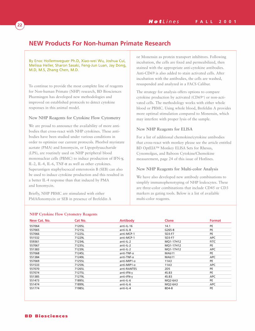
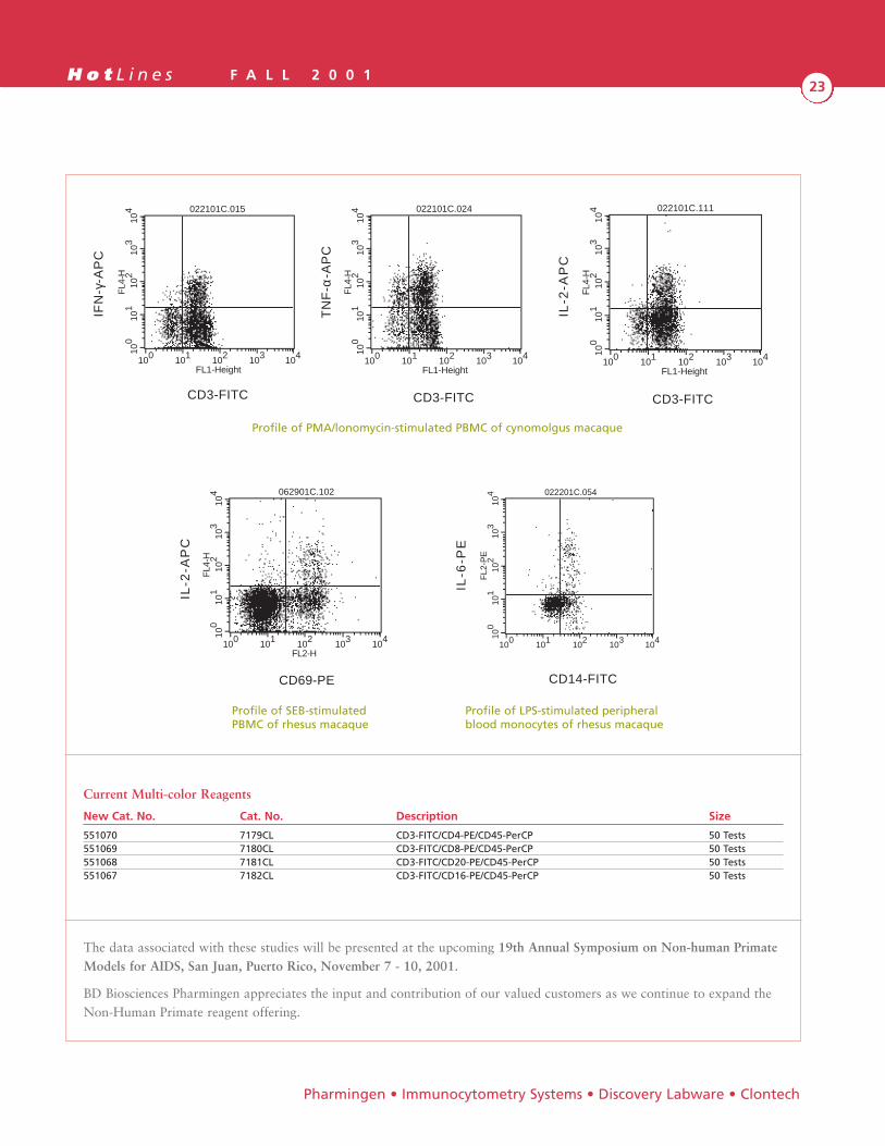
We are proud to announce the availability of more anti-bodies that cross-react with NHP cytokines. These anti-bodies have been studied under various conditions inorder to optimize our current protocols. Phorbol myristateacetate (PMA) and Ionomycin, or Lipopolysaccharide(LPS), are routinely used on NHP peripheral bloodmononuclear cells (PBMC) to induce production of IFN-γ,IL-2, IL-4, IL-6, TNF-α as well as other cytokines.Superantigen staphylococcal enterotoxin B (SEB) can alsobe used to induce cytokine production and this resulted ina better IL-4 response than that induced by PMAand Ionomycin.
Briefly, NHP PBMC are stimulated with eitherPMA/Ionomycin or SEB in presence of Brefeldin A
or Monensin as protein transport inhibitors. Followingincubation, the cells are fixed and permeabilized, thenstained with the appropriate anti-cytokine antibodies.Anti-CD69 is also added to stain activated cells. Afterincubation with the antibodies, the cells are washed,resuspended and analyzed in a FACS Calibur.
The strategy for analysis offers options to comparecytokine production by activated (CD69+) or non-acti-vated cells. The methodology works with either wholeblood or PBMC. Using whole blood, Brefeldin A providesmore optimal stimulation compared to Monensin, whichmay interfere with proper lysis of the sample.
New NHP Reagents for ELISA
For a list of additional chemokine/cytokine antibodiesthat cross-react with monkey please see the article entitledBD OptEIA™ Monkey ELISA Sets for Rhesus,Cynomolgus, and Baboon Cytokine/Chemokinemeasurement, page 24 of this issue of Hotlines.
New NHP Reagents for Multi-color Analysis
We have also developed new antibody combinations tosimplify immunophenotyping of NHP leukocytes. Theseare three-color combinations that include CD45 or CD3markers as gating tools. Below is a list of availablemulti-color reagents.
By Enoc Hollemweguer Ph.D, Xiao-wei Wu, Joshua Cui,Melissa Heller, Sharon Sasaki, Feng-Jun Luan, Jay Dong,M.D, M.S, Zhang Chen, M.D.
NEW Products For Non-human Primate Research
BD Biosc iences
NHP Cytokine Flow Cytometry Reagents
New Cat. No. Cat No. Antibody Clone Format
557064 71205L anti-IL-16 14.1 PE557065 71215L anti-IL-8 G265-8 PE557066 71225L anti-MCP-1 5D3-F7 PE551532 71229L anti-MCP-1 5D3-F7 APC559361 71234L anti-IL-2 MQ1-17H12 FITC557067 71235L anti-IL-2 MQ1-17H12 PE551383 71239L anti-IL-2 MQ1-17H12 APC557068 71245L anti-TNF-α MAb11 PE551384 71249L anti-TNF-α MAb11 APC557069 71255L anti-MIP1-α 11A3 PE551533 71259L anti-MIP1-α 11A3 APC557070 71265L anti-RANTES 2D5 PE557074 71275L anti-IFN-γ 4S.B3 PE551385 71279L anti-IFN-γ 4S.B3 APC551473 71895L anti-IL-6 MQ2-6A3 PE551474 71899L anti-IL-6 MQ2-6A3 APC551774 71985L anti-IL-4 8D4-8 PE
H o t L i n e s F A L L 2 0 0 123
The data associated with these studies will be presented at the upcoming 19th Annual Symposium on Non-human PrimateModels for AIDS, San Juan, Puerto Rico, November 7 - 10, 2001.
BD Biosciences Pharmingen appreciates the input and contribution of our valued customers as we continue to expand theNon-Human Primate reagent offering.
Pharmingen • Immunocytometry Systems • Discovery Labware • Clontech
100
101
102
103
104
FL4
-H
100 101 102 103 104
FL1-Height
022101C.015
CD3-FITC
IFN
-γ-A
PC
Profile of PMA/lonomycin-stimulated PBMC of cynomolgus macaque
Profile of SEB-stimulatedPBMC of rhesus macaque
100
101
102
103
104
FL4
-H
100 101 102 103 104
FL1-Height
022101C.024
CD3-FITC
TN
F-α
-AP
C
Profile of LPS-stimulated peripheralblood monocytes of rhesus macaque
100
101
102
103
104
FL4
-H
100 101 102 103 104
FL1-Height
022101C.111
CD3-FITC
IL-2
-AP
C
100
101
102
103
104
FL4
-H
100 101 102 103 104
FL2-H
062901C.102
IL-2
-AP
C
CD69-PE
100
101
102
103
104
FL2
-PE
100 101 102 103 104
022201C.054
IL-6
-PE
CD14-FITC
Current Multi-color Reagents
New Cat. No. Cat. No. Description Size
551070 7179CL CD3-FITC/CD4-PE/CD45-PerCP 50 Tests551069 7180CL CD3-FITC/CD8-PE/CD45-PerCP 50 Tests551068 7181CL CD3-FITC/CD20-PE/CD45-PerCP 50 Tests551067 7182CL CD3-FITC/CD16-PE/CD45-PerCP 50 Tests
Profile of PMA/lonomycin-stimulated PBMC of cynomolgus macaque
Profile of SEB-stimulatedPBMC of rhesus macaque
Profile of LPS-stimulated peripheralblood monocytes of rhesus macaque
H o t L i n e s F A L L 2 0 0 124
The study of non-human primate (NHP) immune functionis critical for the development of therapeutic and diagnos-tic reagents for human disease. The lack of antibody-based reagents specific for NHP has historically beenproblematic for researchers. BD Biosciences Pharmingen isproud to introduce a new panel of BD OptEIA™ ELISASets that can be used for cytokine/chemokine measure-ment in Rhesus, Cynomolgus, and Baboon serum, plasma,and cell culture supernatant samples.
Cross-Reactivity
We have analyzed multiple (n=28) anti-human specificantibody pair combinations (cytokines/chemokines) fortheir cross-reactivity with NHP. Eight anti-humancytokine ELISA pairs and one chemokine ELISA pair werefound to cross-react with NHP at significant levels (Table1). These results indicate that the utilization of cross-reac-tive antibodies is important for the measurement ofcytokines/chemokines in NHP culture supernatants,serum, and plasma. Thus, cytokine/chemokine productionin NHP disease models may parallel human levels, andrelative values can be obtained in an ELISA-based system.
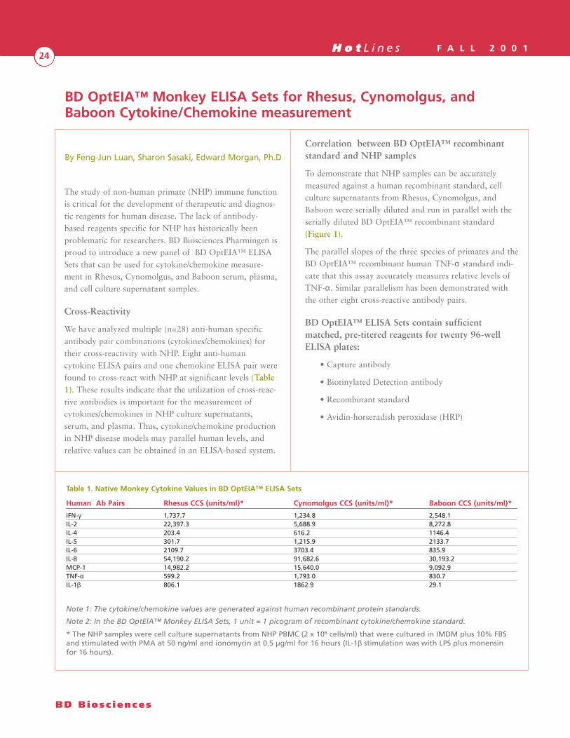
Correlation between BD OptEIA™ recombinantstandard and NHP samples
To demonstrate that NHP samples can be accuratelymeasured against a human recombinant standard, cellculture supernatants from Rhesus, Cynomolgus, andBaboon were serially diluted and run in parallel with theserially diluted BD OptEIA™ recombinant standard(Figure 1).
The parallel slopes of the three species of primates and theBD OptEIA™ recombinant human TNF-α standard indi-cate that this assay accurately measures relative levels ofTNF-α. Similar parallelism has been demonstrated withthe other eight cross-reactive antibody pairs.
BD OptEIA™ ELISA Sets contain sufficientmatched, pre-titered reagents for twenty 96-wellELISA plates:
• Capture antibody
• Biotinylated Detection antibody
• Recombinant standard
• Avidin-horseradish peroxidase (HRP)
By Feng-Jun Luan, Sharon Sasaki, Edward Morgan, Ph.D
BD OptEIA™ Monkey ELISA Sets for Rhesus, Cynomolgus, andBaboon Cytokine/Chemokine measurement
BD Biosc iences
Table 1. Native Monkey Cytokine Values in BD OptEIA™ ELISA Sets
Human Ab Pairs Rhesus CCS (units/ml)* Cynomolgus CCS (units/ml)* Baboon CCS (units/ml)*
IFN-γ 1,737.7 1,234.8 2,548.1IL-2 22,397.3 5,688.9 8,272.8IL-4 203.4 616.2 1146.4IL-5 301.7 1,215.9 2133.7IL-6 2109.7 3703.4 835.9IL-8 54,190.2 91,682.6 30,193.2MCP-1 14,982.2 15,640.0 9,092.9TNF-α 599.2 1,793.0 830.7IL-1β 806.1 1862.9 29.1
Note 1: The cytokine/chemokine values are generated against human recombinant protein standards.
Note 2: In the BD OptEIA™ Monkey ELISA Sets, 1 unit = 1 picogram of recombinant cytokine/chemokine standard.
* The NHP samples were cell culture supernatants from NHP PBMC (2 x 10⁶ cells/ml) that were cultured in IMDM plus 10% FBSand stimulated with PMA at 50 ng/ml and ionomycin at 0.5 µg/ml for 16 hours (IL-1β stimulation was with LPS plus monensinfor 16 hours).
H o t L i n e s F A L L 2 0 0 125
Pharmingen • Immunocytometry Systems • Discovery Labware • Clontech
Product listing
New Cat. No. Cat. No. Description
551492 2750KI Monkey IFN-γ BD OptEIA™ ELISA Set 551493 2751KI Monkey TNF-α BD OptEIA™ ELISA Set 551494 2752KI Monkey IL-2 BD OptEIA™ ELISA Set 551495 2753KI Monkey IL-4 BD OptEIA™ ELISA Set 551496 2754KI Monkey IL-6 BD OptEIA™ ELISA Set Coming soon. Please inquire Monkey IL-5 BD OptEIA™ ELISA Set Coming soon. Please inquire Monkey IL-8 BD OptEIA™ ELISA SetComing soon. Please inquire Monkey MCP-1 BD OptEIA™ ELISA Set
0.1
1
Opt
ical
Den
sity
(45
0 nm
)
10 100 1000
TNF-α (pg/ml)
Baboon CCS
Cynomolgus CCS
Rhesus CCS
BD OptEIAHuman Standard
Note: These curves depict the optical densities of cell culturesupernatant samples when diluted in the range of theBD OptEIA™ recombinant human TNF-α standard curve. Thequantitation of these samples is not reflected in this figure.
Figure 1. Parallelism of Rhesus, Cynomolgus, Baboon CellCulture Supernatant in Monkey TNF-α OptEIA ELISA Set
H o t L i n e s F A L L 2 0 0 126
Broader Assay Range, Superior Low-endSensitivity
Chemiluminescence ELISA systems provide a broaderdynamic assay range, superior low-end sensitivity, anda faster protocol than the conventional colorimetricmethods. The new BD OptEIA™ CL ChemiluminescentELISA Kits utilize a luminol-based chemiluminescentsubstrate and enhancer that result in rapid kinetic lightoutput and high signal intensity. The specificallyfragmented antibodies and diluents used in theBD OptEIA™ CL Kits enable quantitation of cytokinesand other soluble proteins in serum, plasma, or cellculture supernatant samples.
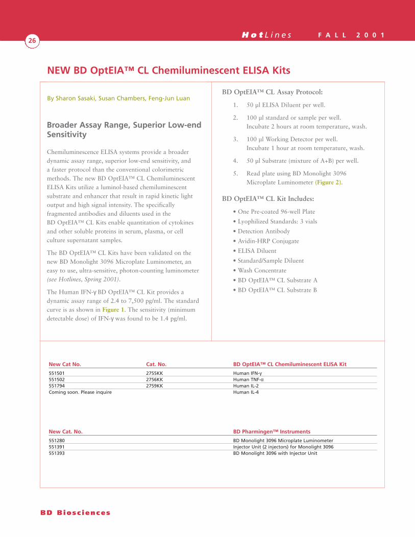
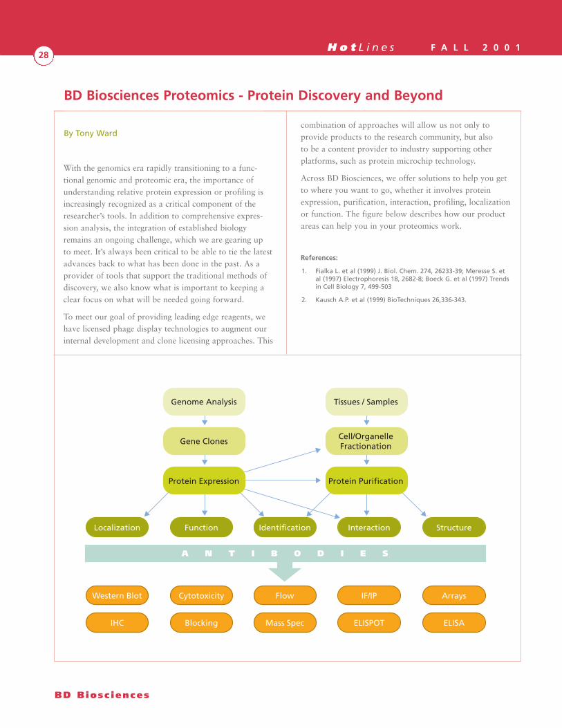
The BD OptEIA™ CL Kits have been validated on thenew BD Monolight 3096 Microplate Luminometer, aneasy to use, ultra-sensitive, photon-counting luminometer(see Hotlines, Spring 2001).
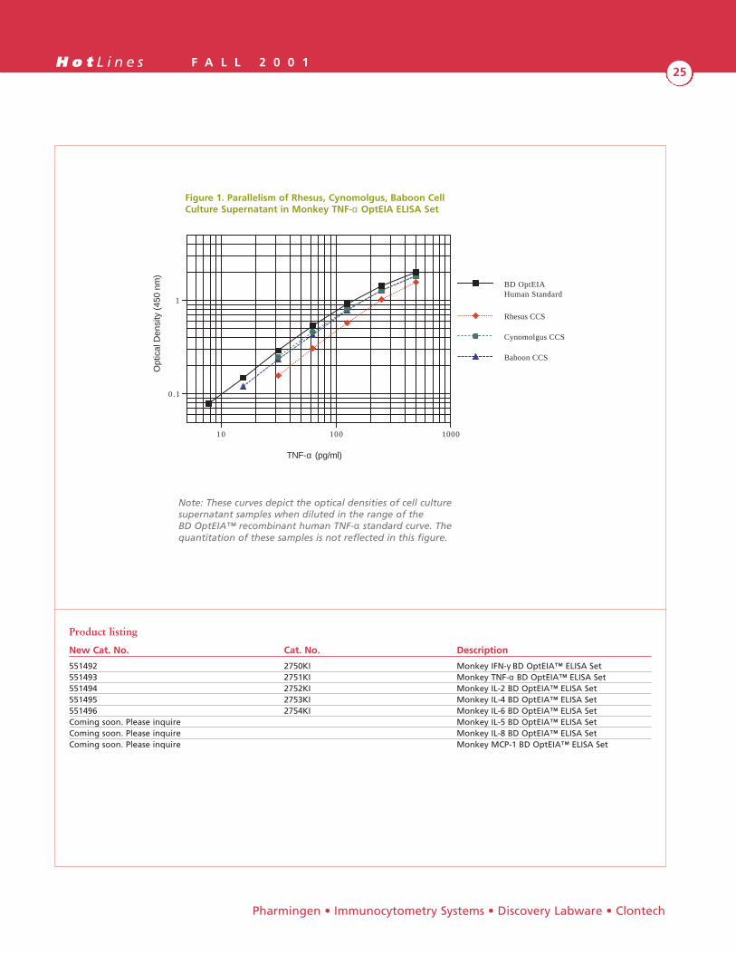
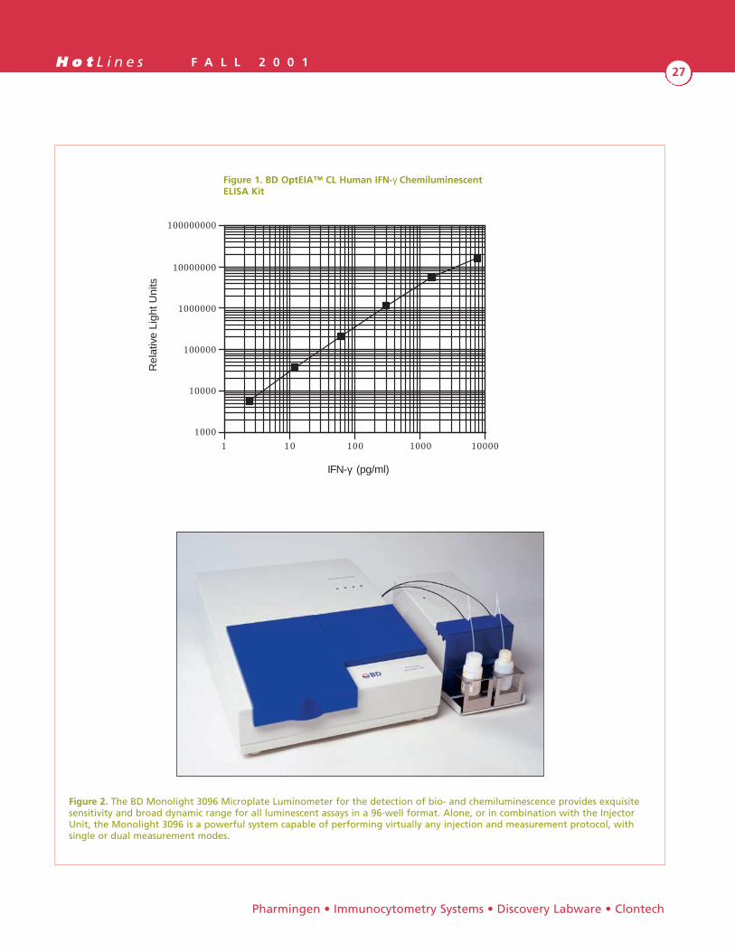
The Human IFN-γ BD OptEIA™ CL Kit provides adynamic assay range of 2.4 to 7,500 pg/ml. The standardcurve is as shown in Figure 1. The sensitivity (minimumdetectable dose) of IFN-γ was found to be 1.4 pg/ml.
BD OptEIA™ CL Assay Protocol:
1. 50 µl ELISA Diluent per well.
2. 100 µl standard or sample per well.Incubate 2 hours at room temperature, wash.
3. 100 µl Working Detector per well.Incubate 1 hour at room temperature, wash.
4. 50 µl Substrate (mixture of A+B) per well.
5. Read plate using BD Monolight 3096Microplate Luminometer (Figure 2).
BD OptEIA™ CL Kit Includes:
• One Pre-coated 96-well Plate
• Lyophilized Standards: 3 vials
• Detection Antibody
• Avidin-HRP Conjugate
• ELISA Diluent
• Standard/Sample Diluent
• Wash Concentrate
• BD OptEIA™ CL Substrate A
• BD OptEIA™ CL Substrate B
By Sharon Sasaki, Susan Chambers, Feng-Jun Luan
NEW BD OptEIA™ CL Chemiluminescent ELISA Kits
BD Biosc iences
New Cat No. Cat. No. BD OptEIA™ CL Chemiluminescent ELISA Kit
551501 2755KK Human IFN-γ551502 2756KK Human TNF-α551794 2759KK Human IL-2 Coming soon. Please inquire Human IL-4
New Cat. No. BD Pharmingen™ Instruments
551280 BD Monolight 3096 Microplate Luminometer551391 Injector Unit (2 injectors) for Monolight 3096551393 BD Monolight 3096 with Injector Unit
H o t L i n e s F A L L 2 0 0 127
1000
10000
100000
1000000
10000000
100000000
Rel
ativ
e Li
ght U
nits
1 10 100 1000 10000
IFN-γ (pg/ml)
Pharmingen • Immunocytometry Systems • Discovery Labware • Clontech
Figure 2. The BD Monolight 3096 Microplate Luminometer for the detection of bio- and chemiluminescence provides exquisitesensitivity and broad dynamic range for all luminescent assays in a 96-well format. Alone, or in combination with the InjectorUnit, the Monolight 3096 is a powerful system capable of performing virtually any injection and measurement protocol, withsingle or dual measurement modes.
Figure 1. BD OptEIA™ CL Human IFN-γ ChemiluminescentELISA Kit
H o t L i n e s F A L L 2 0 0 128
With the genomics era rapidly transitioning to a func-tional genomic and proteomic era, the importance ofunderstanding relative protein expression or profiling isincreasingly recognized as a critical component of theresearcher’s tools. In addition to comprehensive expres-sion analysis, the integration of established biologyremains an ongoing challenge, which we are gearing upto meet. It’s always been critical to be able to tie the latestadvances back to what has been done in the past. As aprovider of tools that support the traditional methods ofdiscovery, we also know what is important to keeping aclear focus on what will be needed going forward.
To meet our goal of providing leading edge reagents, wehave licensed phage display technologies to augment ourinternal development and clone licensing approaches. This
combination of approaches will allow us not only toprovide products to the research community, but alsoto be a content provider to industry supporting otherplatforms, such as protein microchip technology.
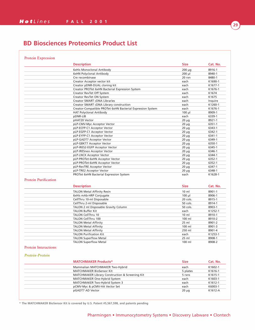
Across BD Biosciences, we offer solutions to help you getto where you want to go, whether it involves proteinexpression, purification, interaction, profiling, localizationor function. The figure below describes how our productareas can help you in your proteomics work.
References:
1. Fialka L. et al (1999) J. Biol. Chem. 274, 26233-39; Meresse S. etal (1997) Electrophoresis 18, 2682-8; Boeck G. et al (1997) Trendsin Cell Biology 7, 499-503
2. Kausch A.P. et al (1999) BioTechniques 26,336-343.
By Tony Ward
BD Biosc iences
Genome Analysis Tissues / Samples
Gene ClonesCell/Organelle Fractionation
Protein Expression
Localization
Protein Purification
Function Identification
A N T I B O D I E S
Interaction Structure
Western Blot Cytotoxicity Flow IF/IP Arrays
IHC Blocking Mass Spec ELISPOT ELISA
BD Biosciences Proteomics - Protein Discovery and Beyond
H o t L i n e s F A L L 2 0 0 129
Pharmingen • Immunocytometry Systems • Discovery Labware • Clontech
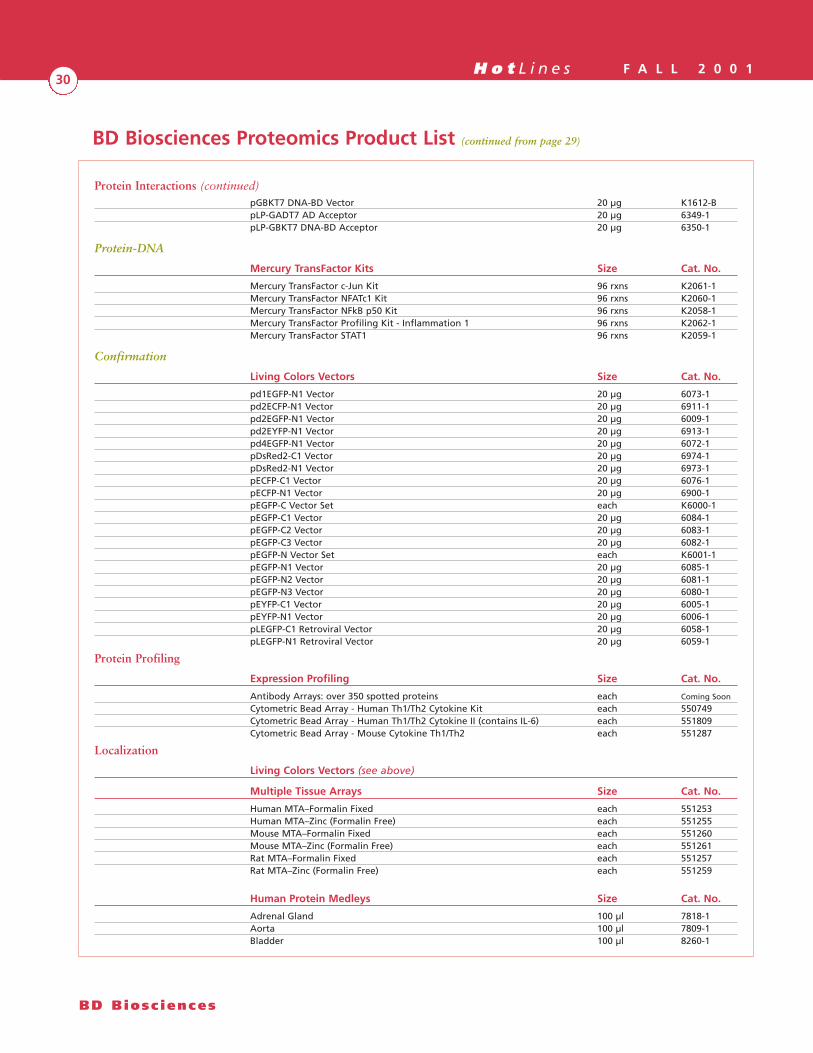
BD Biosciences Proteomics Product List
Protein Expression
Description Size Cat. No.
6xHis Monoclonal Antibody 200 µg 8916-16xHN Polyclonal Antibody 200 µl 8940-1Cre recombinase 20 rxn 8480-1Creator Acceptor vector kit each K1690-1Creator pDNR-DUAL cloning kit each K1677-1Creator PROTet 6xHN Bacterial Expression System each K1676-1Creator RevTet Off System each K1674Creator RevTet ON System each K1675Creator SMART cDNA Libraries each InquireCreator SMART cDNA Library construction each K1260-1Creator-Compatible PROTet 6xHN Bacterial Expression System each K1676-1HAT Polyclonal Antibody 100 µl 8909-1pDNR-LIB each 6339-1pHAT20 Vector 20 µg 8921-1pLP-CMV-Myc Acceptor Vector 20 µg 6351-1 pLP-ECFP-C1 Acceptor Vector 20 µg 6343-1pLP-EGFP-C1 Acceptor Vector 20 µg 6342-1pLP-EYFP-C1 Acceptor Vector 20 µg 6341-1pLP-GADT7 Acceptor Vector 20 µg 6349-1pLP-GBKT7 Acceptor Vector 20 µg 6350-1pLP-IRES2-EGFP Acceptor Vector 20 µg 6345-1pLP-IRESneo Acceptor Vector 20 µg 6346-1pLP-LNCX Acceptor Vector 20 µg 6344-1pLP-PROTet-6xHN Acceptor Vector 20 µg 6352-1pLP-PROTet-6xHN Acceptor Vector 20 µg 6352-1pLP-RevTRE Acceptor Vector 20 µg 6347-1pLP-TRE2 Acceptor Vector 20 µg 6348-1PROTet 6xHN Bacterial Expression System each K1628-1
Protein Purification
Description Size Cat. No.
TALON Metal Affinity Resin 10 ml 8901-16xHis mAb-HRP Conjugate 100 µl 8906-1CellThru 10-ml Disposable 20 cols. 8915-1CellThru 2-ml Disposable 50 cols. 8914-1TALON 2 ml Disposable Gravity Column 50 cols. 8903-1TALON Buffer Kit each K1252-1TALON CellThru 10 10 ml 8910-1TALON CellThru 100 100 ml 8910-2TALON Metal Affinity 25 ml 8901-2TALON Metal Affinity 100 ml 8901-3TALON Metal Affinity 250 ml 8901-4TALON Purification Kit each K1253-1TALON Superflow Metal 25 ml 8908-1TALON Superflow Metal 100 ml 8908-2
Protein Interactions
Protein-Protein
MATCHMAKER Products* Size Cat. No.
Mammalian MATCHMAKER Two-Hybrid each K1602-1MATCHMAKER BioSensor Kit 5 plates K1616-1MATCHMAKER Library Construction & Screening Kit 5 rxns K1615-1MATCHMAKER One-Hybrid System each K1603-1MATCHMAKER Two-Hybrid System 3 each K1612-1pCMV-Myc & pCMV-HA Vector Set each K6003-1pGADT7 AD Vector 20 µg K1612-A
* The MATCHMAKER BioSensor Kit is covered by U.S. Patent #5,567,598, and patents pending
H o t L i n e s F A L L 2 0 0 130
BD Biosc iences
Protein Interactions (continued)pGBKT7 DNA-BD Vector 20 µg K1612-BpLP-GADT7 AD Acceptor 20 µg 6349-1pLP-GBKT7 DNA-BD Acceptor 20 µg 6350-1
Protein-DNA
Mercury TransFactor Kits Size Cat. No.
Mercury TransFactor c-Jun Kit 96 rxns K2061-1Mercury TransFactor NFATc1 Kit 96 rxns K2060-1Mercury TransFactor NFkB p50 Kit 96 rxns K2058-1Mercury TransFactor Profiling Kit - Inflammation 1 96 rxns K2062-1Mercury TransFactor STAT1 96 rxns K2059-1
Confirmation
Living Colors Vectors Size Cat. No.
pd1EGFP-N1 Vector 20 µg 6073-1pd2ECFP-N1 Vector 20 µg 6911-1pd2EGFP-N1 Vector 20 µg 6009-1pd2EYFP-N1 Vector 20 µg 6913-1pd4EGFP-N1 Vector 20 µg 6072-1pDsRed2-C1 Vector 20 µg 6974-1pDsRed2-N1 Vector 20 µg 6973-1pECFP-C1 Vector 20 µg 6076-1pECFP-N1 Vector 20 µg 6900-1pEGFP-C Vector Set each K6000-1pEGFP-C1 Vector 20 µg 6084-1pEGFP-C2 Vector 20 µg 6083-1pEGFP-C3 Vector 20 µg 6082-1pEGFP-N Vector Set each K6001-1pEGFP-N1 Vector 20 µg 6085-1pEGFP-N2 Vector 20 µg 6081-1pEGFP-N3 Vector 20 µg 6080-1pEYFP-C1 Vector 20 µg 6005-1pEYFP-N1 Vector 20 µg 6006-1pLEGFP-C1 Retroviral Vector 20 µg 6058-1pLEGFP-N1 Retroviral Vector 20 µg 6059-1
Protein Profiling
Expression Profiling Size Cat. No.
Antibody Arrays: over 350 spotted proteins each Coming Soon
Cytometric Bead Array - Human Th1/Th2 Cytokine Kit each 550749Cytometric Bead Array - Human Th1/Th2 Cytokine II (contains IL-6) each 551809Cytometric Bead Array - Mouse Cytokine Th1/Th2 each 551287
Localization
Living Colors Vectors (see above)
Multiple Tissue Arrays Size Cat. No.
Human MTA–Formalin Fixed each 551253Human MTA–Zinc (Formalin Free) each 551255Mouse MTA–Formalin Fixed each 551260Mouse MTA–Zinc (Formalin Free) each 551261Rat MTA–Formalin Fixed each 551257Rat MTA–Zinc (Formalin Free) each 551259
Human Protein Medleys Size Cat. No.
Adrenal Gland 100 µl 7818-1Aorta 100 µl 7809-1Bladder 100 µl 8260-1
BD Biosciences Proteomics Product List (continued from page 29)

H o t L i n e s F A L L 2 0 0 131
Pharmingen • Immunocytometry Systems • Discovery Labware • Clontech
Localization (continued)
Human Protein Medleys Size Cat. No.
Brain 100 µl 7800-1Brain, amygdala 100 µl 7821-1Brain, cerebellum 100 µl 8251-1Brain, cerebral cortex 100 µl 7827-1Brain, frontal lobe 100 µl 7822-1Brain, hippocampus 100 µl 7823-1Brain, hypothalamus 100 µl 7824-1Brain, temporal 100 µl 7825-1Brain, thalamus 100 µl 7826-1Brainstem 100 µl 8250-1Cecum 100 µl 8259-Duodenum 100 µl 8255-1Esophagus 100 µl 8254-1Fetal Brain 100 µl 8265-1Fetal Kidney 100 µl 8266-1Fetal Liver 100 µl 8267-1Fetal Lung 100 µl 8268-1Heart 100 µl 7801-1Ileum 100 µl 8257-1Jejunum 100 µl 8256-1Kidney 100 µl 7802-1Liver 100 µl 7805-1Lung 100 µl 7803-1Lymph Node 100 µl 7819-1Mammary Gland 100 µl 8269-1Ovary 100 µl 7807-1Placenta 100 µl 7806-1Prostate 100 µl 8261-1Skeletal Muscle 100 µl 7804-1Small Intestine 100 µl 7812-1Smooth Muscle 100 µl 8258-1Spinal Cord 100 µl 7828-1Spleen 100 µl 7813-1Stomach 100 µl 7814-1Testis 100 µl 7808-1Thyroid 100 µl 7817-1Tongue 100 µl 8253-1Trachea 100 µl 8252-1Uterus (no endometrium) 100 µl 8263-1Uterus 100 µl 8262-1Vagina 100 µl 8264-1
Function/State
Status-Specific Antibodies Size Cat. No.
Over 1,000 antibodies to measure and stimulate cellular pathways See catalogincluding phospho-specific mAbs. for details
Sample Preparation for Organelles
Try our antibodies to purify organelles for protein identification by 2D gel electrophoresis, mass spectrometry, or to assess thepurity of your organelle preparations. Organelle specific antibodies and organelle reactive dyes have been used to assess thepurity of organelle preparations and for further purification by FACS sorting1 and magnetic particle based selection2. Theselected antibodies have been shown to work in immunofluorescence assays, and may be suitable for such applications.
Caveolae Size Cat. No.
Caveolin 50 or 150 ug C13630Caveolin 1 50 or 150 ug C13620

H o t L i n e s F A L L 2 0 0 132
BD Biosc iences
Sample Preparation for Organelles (continued)
Caveolae Size Cat. No.
Caveolin 1 50 or 150 ug C36220Caveolin 1 50 or 150 ug C37120Caveolin 1 50 or 150 ug C43420Caveolin 2 50 or 150 ug C57820Caveolin 3 50 or 150 ug C38320Flotillin-1 50 or 150 ug F65020Flotillin-2/ESA 50 or 150 ug E35820Phospho-Caveolin (Y14) 50 or 150 ug C91520
Endosomes Size Cat. No.
EEA1 50 or 150 ug E41120
Endoplasmic Reticulum Size Cat. No.
BiP/GRP78 50 or 150 ug G73320ERp72 50 or 150 ug E72920SQS 50 or 150 ug S10120SRP54 50 or 150 ug S71420
Golgi Size Cat. No.
Bet1 50 or 150 ug B32320Cab45 50 or 150 ug C77720GM130 50 or 150 ug G65120GMAP-210/Trip230 50 or 150 ug G10020Golgin-84 50 or 150 ug G93720GS15 50 or 150 ug G72420GS27 50 or 150 ug G76220GS28 50 or 150 ug G83820p115 50 or 150 ug P67420p230 trans Golgi 50 or 150 ug G88620TGN38 50 or 150 ug T69020
Lysosomes Size Cat. No.
Lamp-1 50 or 150 ug L76620SNX1 50 or 150 ug S95620SNX2 50 or 150 ug S90020Cathepsin L 50 or 150 ug C78820
Mitochondria Size Cat. No.
Aralar 50 or 150 ug A78520ASS 50 or 150 ug A10020Hsp60 50 or 150 ug H99020Mcl-1 50 or 150 ug M54020MCM 50 or 150 ug M17520Metaxin 50 or 150 ug M16020Tim23 50 or 150 ug T85720
Nucleus Size Cat. No.
CHD3 50 or 150 ug C25020Coilin 50 or 150 ug C28020Exportin-1/CRM1 50 or 150 ug E11620Exportin-t 50 or 150 ug E95720Karyopherin alpha/Rch-1 50 or 150 ug R43020Karyopherin beta 50 or 150 ug K48020Ki-67 50 or 150 ug K72820
BD Biosciences Proteomics Product List (continued from page 31)
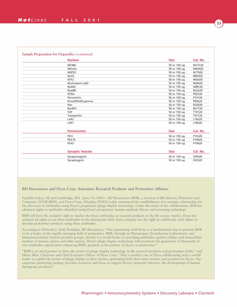
H o t L i n e s F A L L 2 0 0 133
Pharmingen • Immunocytometry Systems • Discovery Labware • Clontech
Sample Preparation for Organelles (continued)
Nucleus Size Cat. No.
MCM6 50 or 150 ug M13120Mitosin 50 or 150 ug M62420NDP52 50 or 150 ug N77920No55 50 or 150 ug N83420NTF2 50 or 150 ug N42420Nucleoporin p62 50 or 150 ug N43620NuMA 50 or 150 ug N48120Nup88 50 or 150 ug N22220PCNA 50 or 150 ug P56720Pericentrin 50 or 150 ug P14120Psme3/PA28-gamma 50 or 150 ug P83620Ran 50 or 150 ug R32620RanBP1 50 or 150 ug R61720TAP 50 or 150 ug T10120Transportin 50 or 150 ug T57720LAP2 50 or 150 ug L74520LAP1 50 or 150 ug L78220
Peroxisomes Size Cat. No.
PECI 50 or 150 ug P15320PEX19 50 or 150 ug P10620PEX5 50 or 150 ug P10420
Synaptic Vesicles Size Cat. No.
Synaptotagmin 50 or 150 ug S39520Synaptogyrin 50 or 150 ug S52520
BD Biosciences and Dyax Corp. Announce Research Products and Proteomics Alliance
Franklin Lakes, NJ and Cambridge, MA (June 13, 2001) – BD Biosciences (BDB), a division of BD (Becton, Dickinson andCompany) (NYSE:BDX), and Dyax Corp. (Nasdaq: DYAX) today announced the establishment of a strategic relationship forthe discovery of antibodies using Dyax’s proprietary phage display technology. Under the terms of the collaboration, BDB hasobtained rights to antibodies identified using Dyax’s proprietary human antibody library and screening technology.
BDB will have the exclusive right to market the Dyax antibodies as research products to the life science market. Dyax hasretained all rights to use these antibodies in the therapeutic field. Each company has the right to collaborate with others todevelop proteomics products using these antibodies.
According to Deborah J. Neff, President, BD Biosciences, “Our partnership with Dyax is a fundamental step to position BDBto be a leader in the rapidly emerging field of proteomics. BDB, through its Pharmingen, Transduction Laboratories andImmunocytometry Systems product groups, already is a world leader in providing antibodies against cellular and intracellularmarkers in human, mouse and other species. Dyax’s phage display technology will accelerate the generation of thousands ofnew antibodies significantly enhancing BDB’s position as the partner of choice in proteomics.”
“BDB is an ideal partner to drive the power of phage display technology in the research products and proteomics fields,” saidHenry Blair, Chairman and Chief Executive Officer of Dyax Corp. “This is another case of Dyax collaborating with a worldleader to exploit the power of phage display in their market, generating both short term revenue and royalties for Dyax. Ourcorporate partnering strategy provides resources and focus to support Dyax’s principal objective, the development of humantherapeutic products.”
H o t L i n e s F A L L 2 0 0 134
New Product Highlight
The genomic era of biomedical research has given rise tothe birth of proteomics as a scientific discipline. Vital tothe advancement of proteomic research are the ability toefficiently clone a gene into an expression system and theability to maximize the expression of the target recombi-nant protein. For over twelve years, BD BiosciencesPharmingen has set the industry standard by supplyingthe highest quality reagents for analyzing gene expression,protein expression, and cellular response and interaction.We strive to maintain this position and are proud to bethe leading supplier of premium reagents for generatingrecombinant proteins using the BD BaculoGold™Baculovirus Expression Vector System. Our latestinnovation comes in the form of our new BDBaculoGold™ Max-XP Serum-Free Insect CellMedium (cat. no. 551411).
BD BaculoGold™ Max-XP is produced using a propri-etary system, resulting in a metabolically-enhancedmedium that far outperforms the competition.Experiments indicate that the production of functionalrecombinant protein in insect cell lines grown inBD BaculoGold™ Max-XP increases up to three-foldcompared with insect cells grown in complete TNM-FHand other serum-free media. In addition, BD BaculoGold™Max-XP is designed to be used in multiple cell lines andhas been shown to optimize cell growth and recombinantprotein production in Drosophila melanogaster,
Tricholplusia ni, Heliothis zea, the Spodoptera frugiperda
cell lines Sf9 and Sf21, and more.
BD BaculoGold™ Max-XP is ideal for quick, direct adap-tation of your insect cells cultured in serum-supplementedmedia or in serum-free media. If you’re currently usingeither our BD BaculoGold™ TMN-FH Insect CellMedium or BD BaculoGold™ Protein-Free Medium,minimal adaptation is necessary. Typical results are 4 to 8passages for complete adaptation. It must be noted,however, that some cell lines are more sensitive to mediachanges and will require more than 8 passages. (Pleasecontact our Technical Service Department for assistancewith either direct or sequential adaptation techniques.)
BD BaculoGold™ Max-XP promotes high cell density andoptimum protein production, making it ideal for bothlarge-scale industrial use and research-scale production.In addition, BD BaculoGold™ Max-XP’s composition isprecisely defined, which facilitates the purification ofrecombinant proteins and isolation of virus.
In summary, this metabolically-designed serum-freemedium enhances the growth of many types of insectcells, amplifies recombinant protein production, andaugments baculovirus yield.
BD BaculoGold™ Max-XP Serum-Free Insect Cell Medium
BD Biosc iences

H o t L i n e s F A L L 2 0 0 135
The impact of apoptosis on various cellular processesunderlies the basis for the tools that are available fordetecting and analyzing apoptotic cells. This article focuseson applications for the assays most commonly used andaccepted by the scientific community to determine whethercells are undergoing apoptosis. We will show data gener-ated by scientists and research associates in our CellBiology/Cell Signaling laboratory using BD BiosciencesPharmingen reagents.
Apoptosis signaling can be generally divided into receptor-and mitochondrial-mediated pathways. These pathwaysconverge at a number of downstream points including themitochondria, caspase activation, and substrate cleavage.Apoptosis is a highly conserved process; ergo, the majorityof apoptosis assays can be used for a wide variety ofmodel systems. In the examples presented here, apoptosiswas induced either through the Fas receptor-mediatedpathway using agonistic anti-Fas monoclonal antibodies,or through the mitochondrial-mediated pathway usingstaurosporine (a protein kinase inhibitor) or camptothecin(a topoisomerase I inhibitor).
When apoptosis is induced, procaspases are proteolyticallycleaved and reassemble to form active heterotetramericenzymes (caspases). Active caspase-3 has emerged as apowerful marker of cells undergoing apoptosis; its pres-ence is considered to be diagnostic for apoptosis.Antibodies recognizing caspase-3 are useful tools forassessing its activation status (Figure 1). In this experi-ment, proteins from Jurkat T cell lysates were immunopre-cipitated with monoclonal antibodies specific for either theactive form of caspase-3 [lanes 1 and 3: clone C92-605,Cat. No. 68651G (recognizes the 17-22 kDa large subunitof active caspase-3)] or for both procaspase-3 (31 kDa)and active caspase-3 (17-22 kDa) (lanes 2 and 4: clone 19,Cat. No. C31720). This was followed by western blot withpolyclonal antibodies recognizing both pro and activecaspase-3 (Cat. No. 65906E). The results show that activecaspase-3 is present in campthothecin-treated cell lysates(lanes 3 and 4), but not in control cell lysates (lanes 1 and 2),
which contain procaspase-3 only (lane 2). Therefore, wedetermine that only the camptothecin-treated lysate isapoptotic. Furthermore, these data show that procaspase-3is also present in the apoptotic population (lane 4). Thisindicates that not all cells in the population were undergo-ing measurable apoptosis when the lysate was made. Thisis consistent with the dogma that cells in a population donot generally undergo apoptosis at the same time.
Apoptosis signaling results in the cleavage of multipleprotein substrates en masse by active caspases, which leadsto the loss of cellular structure and function, and ultimatelyresults in cell death. Cleaved proteins are thus markers ofapoptosis, and cleavage site-specific antibodies are emergingas particularly valuable tools for identifying apoptotic cells.PARP [Poly(ADP-ribose)polymerase], a 116 kDa nuclearchromatin-associated enzyme involved in DNA repair, is aclassical example. It was the first molecule discovered to becleaved by active caspases during apoptosis, and cleavageresults in 85 kDa and 25 kDa fragments. In this experi-ment, HeLa cells were induced to undergo apoptosis withstaurosporine. The fluorescent micrographs shown in Figure2 indicate that cleavage site-specific PARP-FITC antibodies(polyclonal, Cat. No. 81114E) stain the staurosporine-treated (B, page 36), but not the control cells (A, page 36).A' and B' (page 36) represent phase contrast correlates ofA and B respectively.
By Lisa S. Stein, Ph.D, Michael J. Boyer, Ph.D,Anissa Agadir, Hai Le, Dana Radman, Andy Castellanos,and David George
Modern Tools for Detecting & Analyzing Apoptosis
Pharmingen • Immunocytometry Systems • Discovery Labware • Clontech
Active
Pro/A
ctive
Active
Pro/A
ctive
kDa
97
66
55
36
31
21
14
Pro-
Ig
Active
Caspase-3
Control Apoptotic(Camptothecin, 5hr)
1 2 3 4
Figure 1. Caspase-3 Activation as a Marker of Apoptosis
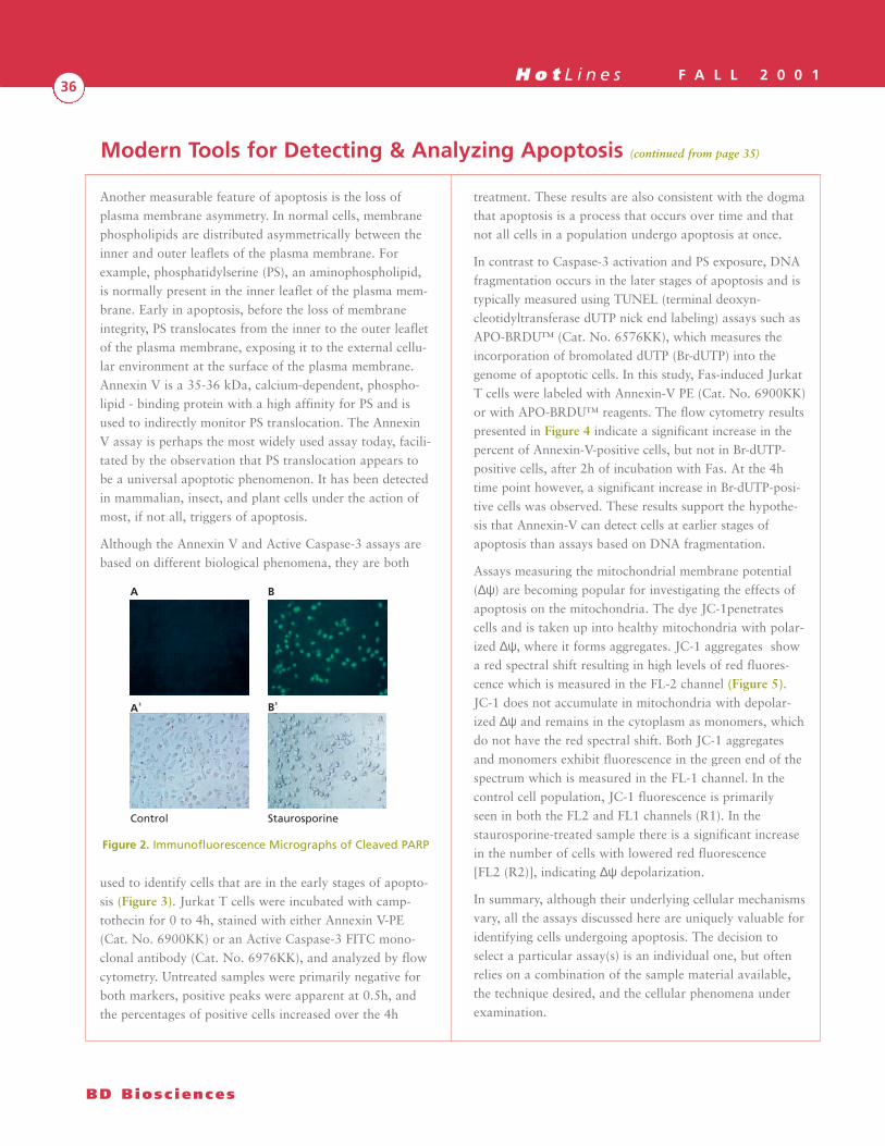
H o t L i n e s F A L L 2 0 0 136
Another measurable feature of apoptosis is the loss ofplasma membrane asymmetry. In normal cells, membranephospholipids are distributed asymmetrically between theinner and outer leaflets of the plasma membrane. Forexample, phosphatidylserine (PS), an aminophospholipid,is normally present in the inner leaflet of the plasma mem-brane. Early in apoptosis, before the loss of membraneintegrity, PS translocates from the inner to the outer leafletof the plasma membrane, exposing it to the external cellu-lar environment at the surface of the plasma membrane.Annexin V is a 35-36 kDa, calcium-dependent, phospho-lipid - binding protein with a high affinity for PS and isused to indirectly monitor PS translocation. The AnnexinV assay is perhaps the most widely used assay today, facili-tated by the observation that PS translocation appears tobe a universal apoptotic phenomenon. It has been detectedin mammalian, insect, and plant cells under the action ofmost, if not all, triggers of apoptosis.
Although the Annexin V and Active Caspase-3 assays arebased on different biological phenomena, they are both
used to identify cells that are in the early stages of apopto-sis (Figure 3). Jurkat T cells were incubated with camp-tothecin for 0 to 4h, stained with either Annexin V-PE(Cat. No. 6900KK) or an Active Caspase-3 FITC mono-clonal antibody (Cat. No. 6976KK), and analyzed by flowcytometry. Untreated samples were primarily negative forboth markers, positive peaks were apparent at 0.5h, andthe percentages of positive cells increased over the 4h
treatment. These results are also consistent with the dogmathat apoptosis is a process that occurs over time and thatnot all cells in a population undergo apoptosis at once.
In contrast to Caspase-3 activation and PS exposure, DNAfragmentation occurs in the later stages of apoptosis and istypically measured using TUNEL (terminal deoxyn-cleotidyltransferase dUTP nick end labeling) assays such asAPO-BRDU™ (Cat. No. 6576KK), which measures theincorporation of bromolated dUTP (Br-dUTP) into thegenome of apoptotic cells. In this study, Fas-induced JurkatT cells were labeled with Annexin-V PE (Cat. No. 6900KK)or with APO-BRDU™ reagents. The flow cytometry resultspresented in Figure 4 indicate a significant increase in thepercent of Annexin-V-positive cells, but not in Br-dUTP-positive cells, after 2h of incubation with Fas. At the 4htime point however, a significant increase in Br-dUTP-posi-tive cells was observed. These results support the hypothe-sis that Annexin-V can detect cells at earlier stages ofapoptosis than assays based on DNA fragmentation.
Assays measuring the mitochondrial membrane potential(∆ψ) are becoming popular for investigating the effects ofapoptosis on the mitochondria. The dye JC-1penetratescells and is taken up into healthy mitochondria with polar-ized ∆ψ, where it forms aggregates. JC-1 aggregates showa red spectral shift resulting in high levels of red fluores-cence which is measured in the FL-2 channel (Figure 5).JC-1 does not accumulate in mitochondria with depolar-ized ∆ψ and remains in the cytoplasm as monomers, whichdo not have the red spectral shift. Both JC-1 aggregatesand monomers exhibit fluorescence in the green end of thespectrum which is measured in the FL-1 channel. In thecontrol cell population, JC-1 fluorescence is primarilyseen in both the FL2 and FL1 channels (R1). In thestaurosporine-treated sample there is a significant increasein the number of cells with lowered red fluorescence[FL2 (R2)], indicating ∆ψ depolarization.
In summary, although their underlying cellular mechanismsvary, all the assays discussed here are uniquely valuable foridentifying cells undergoing apoptosis. The decision toselect a particular assay(s) is an individual one, but oftenrelies on a combination of the sample material available,the technique desired, and the cellular phenomena underexamination.
Modern Tools for Detecting & Analyzing Apoptosis (continued from page 35)
BD Biosc iences
Figure 2. Immunofluorescence Micrographs of Cleaved PARP
A B
A' B'
Control Staurosporine
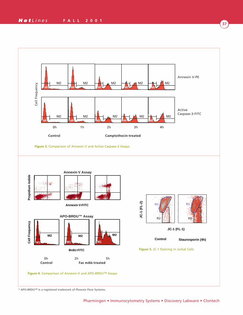
H o t L i n e s F A L L 2 0 0 137
Pharmingen • Immunocytometry Systems • Discovery Labware • Clontech
Cel
l Fr
equ
ency
Annexin V-PE
Control Camptothecin-treated
ActiveCaspase-3-FITC
0h
M2M1
M2M1
M2M1
M2M1
M2M1
M2M1
M2M1
M2M1
M2M1
M2M1
1h 2h 3h 4h
Figure 3. Comparison of Annexin-V and Active Caspase-3 Assays
Annexin V-FITC
Pro
pid
ium
Iod
ide
BrdU-FITC
Annexin V Assay
APO-BRDU™ Assay
M1 M1 M1
M2 M2 M2
Cel
l Fre
qu
ency
0h 2h 5hControl Fas mAb-treated
Figure 4. Comparison of Annexin-V and APO-BRDU™ Assays
R1
R2
R1
R2JC-1
(F
L-2
)
JC-1 (FL-1)
Control Staurosporin (4h)
Figure 5. JC-1 Staining in Jurkat Cells
* APO-BRDU™ is a registered trademark of Phoenix Flow Systems.
H o t L i n e s F A L L 2 0 0 138
Natural Killer (NK) cells are large granular lymphocyteswith non-adaptive non-MHC restricted cytotoxicity. Theyplay an important role in resistance to tumor cells, virallyinfected cells and allogeneic grafts. NK cells also regulatethe immune response through secretion of cytokines suchas IL-2 and IFN-γ. Cells with NK activity have been iden-tified in many different animal models, including swine.The swine immune system differs from other species inthe existence of four peripheral lymphocyte populationsdefined by the expression of the CD4 and CD8 antigens(CD4+CD8lo, CD4-CD8lo, CD4- CD8hi, CD4+CD8+)(Figure 1). The CD4-CD8lo cells are involved in non-MHC-restricted lymphocyte functions associated with NKcells such as spontaneous cytolytic activity against tumorcells, generation of lymphokine activate killer (LAK) activ-ity, and proliferative response against syngeneic leukocytesin mixed leukocyte reactions1.
As the availability of various monoclonal antibodiesraised against swine antigens has increased, a more com-prehensive phenotypic characterization of swine NK cellshas been performed. The swine NK cell population hasbeen phenotypcially characterized as CD2+ CD3- CD4-
CD5- CD6- CD8dim CD16+. Below, we briefly describe thephenotypic and functional characteristics of the swine NKcell population.
Swine CD16 is present on NK cells (CD4-CD8lo)(Figure 2). CD16 is a low-affinity receptor for IgG and isalso found on macrophages, monocytes, and granulocytes.The antigen recognized by the FcG7 monoclonal antibodyhas been identified as the swine FcγRIIIa (CD16) homo-logue2. It interacts at the cell surface with a cytolytictrigger molecule on swine NK cells and neutrophils. TheFcG7 monoclonal antibody induces and enhances cytotox-icity of NK cells and neutrophils against nucleated FcR+
target cells2,3,4. Analysis of swine CD5 and CD6 antigensenables a further characterization of NK cells. The non-MHC-restricted cell-mediated cytotoxicity is associatedwith NK cells, which are CD4- CD8lo CD5- CD6- lympho-cytes (Figure 3). T lymphocytes with MHC-restricted
cytotoxicity are found in the CD5+ and CD6+
populations1,5. Swine CD2 is a membrane glycoproteinexpressed on peripheral T lymphocytes and thymocytes.Swine CD2 is also present on NK cells (data not shown).It has been shown that depletion of CD2+ cells from PBLabolishes the NK activity6.
The swine is an important large animal model for avariety of immunological studies. Recently, the potentialfor swine to be xenogeneic organ donors to man hasincreased the need for further examination of the swineimmune system. BD Biosciences Pharmingen is committedto expanding the swine product line to support yourresearch. For new and upcoming pig reagents, pleasecontact BD Biosciences Pharmingen Technical Services.
References:
1. Saalmuller. 1996. Differentiation between MHC-restricted andnon-MHC-restricted porcine cytolytic T lymphocytes.Immunology; 88:238-246.
2. Kim, Y. 1994. Molecular cloning and identification of the porcinecytolytic trigger molecule G7 as a FcγRIIIa (CD16) homologue.The Journal of Immunology; 153: 2631-2641.
3. Kim, Y. 1995. Characterization of the cytolytic trigger moleculesG7/PNK-E as a molecular complex on the surface of porcinephagocytes. Cellular Immunology; 161: 270-278.
4. Kim, Y. 1996. Identification of a unique porcine FcγRIIIAa molec-ular complex. Cellular Immunology;172: 92-99.
5. Saalmuller. 1994. Analyses of monoclonal antibodies reactivewith porcine CD5. Veterinary Immunology andImmunopathology; 43: 237-242.
6. Yang, H. 1996. Phenotypic classification of porcine lymphocytessubpopulations in blood and lymphoid tissues. Immunology;89: 76-83.
By Jennifer Bickel and Belen Ybarrondo
Swine Natural Killer Cells
BD Biosc iences

H o t L i n e s F A L L 2 0 0 139
Pharmingen • Immunocytometry Systems • Discovery Labware • Clontech
100 101 102 103 104
CD8a-FITC
100
101
102
103
104
CD4a-PE
100 101 102 103 104
RelativeCellNumber
CD16-APC
A
100 101 102 103 104
CD8a-FITC
100
101
102
103
104
CD4a-PE
B (CD16- lymphocytes)
CD16+
CD16-
100 101 102 103 104
CD8a-FITC
100
101
102
103
104
CD4a-PE
C (CD16+ lymphocytes)
Figure 1. Swine peripheral lymphocytes were stainedwith anti- pig CD4a PE mAb 74-12-4 (Cat. No. 559586)and anti-CD8a FITC mAb 76-2-11 (Cat. No. 551303).For data analysis, a gate was set to include onlyviable lymphocytes.
Figure 2. Swine peripheral blood was simultaneouslystained with anti-pig CD4 PE, anti-pig CD8 FITC, andbiotinylated anti-pig CD16 mAb FcG7 (Cat.No. 551395)followed by Sav-APC (Cat. No. 554067). For analysis, agate was set to include only viable lymphocytes. In A,the histogram shows staining with anti-pig CD16 mAbFcG7. Expression of CD4a and CD8a on CD16
_(B) and
CD16+ (C) lymphocytes is shown.

H o t L i n e s F A L L 2 0 0 140
BD Biosc iences
100 101 102 103 104
RelativeCellNumber
CD5-APC
A
100 101 102 103 104
CD8a-FITC
100
101
102
103
104
CD4a-PE
B (CD5- lymphocytes)
CD5+
CD5-
100 101 102 103 104
CD8a-FITC
100
101
102
103
104
CD4a-PE
C
100 101 102 103 104
CD8a-FITC
100
101
102
103
104
CD4a-PE
D (CD5+ lymphocytes)
100 101 102 103 104
RelativeCellNumber
CD6-APC
E
100 101 102 103 104
CD8a-FITC
100
101
102
103
104
CD4a-PE
F (CD6- lymphocytes)
CD6+
CD6-
100 101 102 103 104
CD8a-FITC
100
101
102
103
104
CD4a-PE
G (CD6+ lymphocytes)
CD5Int
Jennifer Bickel Fall 2001 Hotlines swine article9/7/01Figure 3revised colors of data submitted 8/10/01
Figure 3. Swine peripheral blood was simultaneouslystained with anti-pig CD4 PE, anti-pig CD8, purified anti-pig CD5 mAb b53b7 (Cat. No. 551541) or purified anti-pig CD6 mAb a38b2 (Cat. No. 551507) followed bybiotinylated Rat anti-mouse IgG1 mAb A85-1 (Cat. No.553441) then SAv-APC (Cat. No. 554067). For analysis,a gate was set to include only viable lymphocytes. In Aand E, the histograms show staining with anti-pig CD5mAb b53b7 and anti-pig CD6 mAb a38b2, respectively.Expression of CD4 and CD8 on CD5
_(B) and CD5+ (D) is
shown. Expression of CD4 and CD8 on CD6_
(F) and CD6+
(G) is also shown.
Swine Natural Killer Cells (continued from page 39)

H o t L i n e s F A L L 2 0 0 141
BD Biosciences Transduction Laboratories offers theBD PowerBlot™ Western Array Screening Service.Utilizing a panel of over 800 high quality monoclonalantibodies targeted to a wide range of cellular signalingproteins, we screen your control and experimentalsamples for changes in the levels of protein expression.
BD PowerBlot™ adds to the arsenal of techniques cur-rently available to elucidate dynamic protein changes andoffers the opportunity to rapidly advance research in thelife sciences.
Simultaneously Analyze Hundreds of SignalingProteins with BD PowerBlot™
Powerful. Carefully prepared monoclonal antibody combinations used in parallel analysis of signal transduc-tion proteins without the need for classical chemicalprotein analysis.
Specific. High quality monoclonal antibodies capable of recognizing unique targets among thousands of proteinsexpressed in cells and tissues.
Time Saving. Unlike conventional 2D analysis, protein identification is immediate and reagents needed forfollow-up studies are well characterized and readily avail-able for the researcher. This allows the researcher to focuson interpretation of the results and planning offollow-up studies.
Affordable. This service eliminates the cost of purchasingour library of over 800 monoclonal antibodies, as well asthe time spent on sample processing and data analysis.
Sensitive. Ability to detect proteins at the nanogram levelfacilitates indentification of subtle protein changes in cellsor tissues.
Unique. With such a large panel of monoclonal antibodies, only BD Biosciences TransductionLaboratories can offer this one-of-a-kind service.
Features
• A unique advancement in proteomics thatsimultaneously examines relative changes inprotein expression
• Complements current nucleic acid analysisapproaches, such as BD Riboscreen™ MembraneArray System and Clontech Atlas™ Arrays
• Avoids limitations of protein chemistry tecniques, such as 2D-PAGE, Mass Spectrometry,and SELDI
• Utilizes highly specific (sensitive to the nanogramlevel) monoclonal antibodies that are producedvia a rigorous in- house development process and subjected to extensive quality assurance analysis
• Carefully formulated antibody combinationsenable reproducible, semi-quantifiable resultsfrom analysis of total cell or tissue lysates
• Distinguishes closely related members of manyimportant signaling families
BD PowerBlot™ Western Array Screening Service
Pharmingen • Immunocytometry Systems • Discovery Labware • Clontech
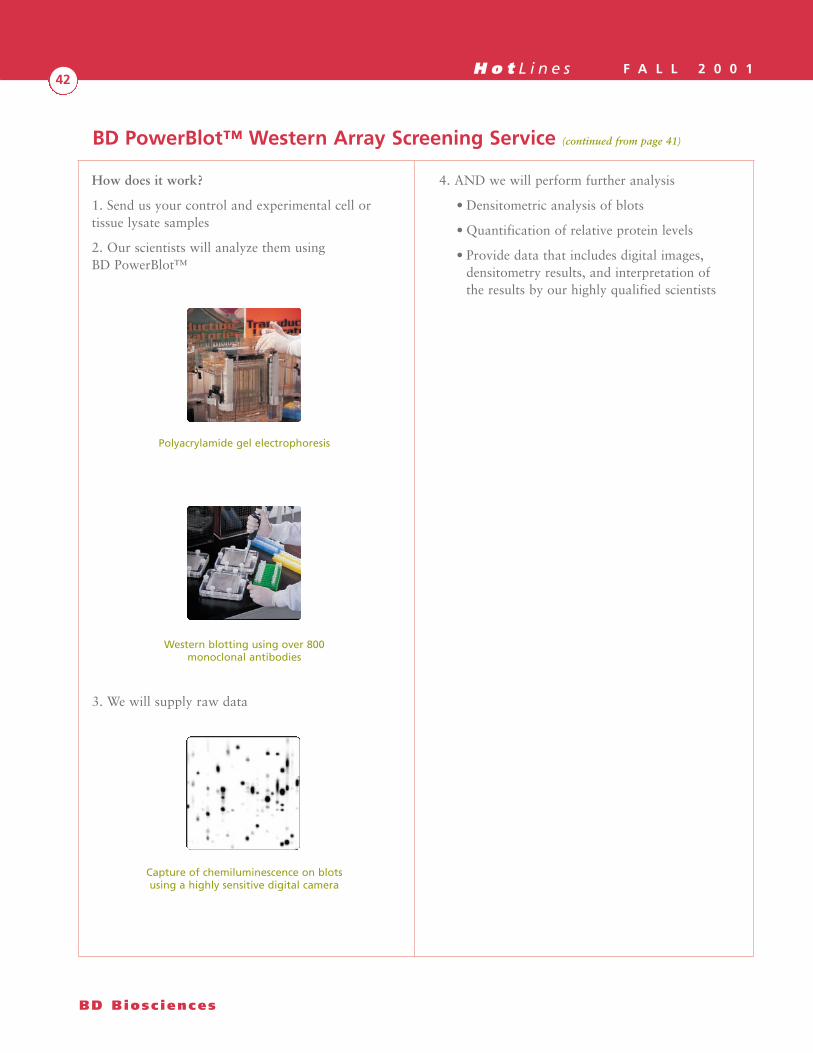
H o t L i n e s F A L L 2 0 0 142
How does it work?
1. Send us your control and experimental cell ortissue lysate samples
2. Our scientists will analyze them usingBD PowerBlot™
Polyacrylamide gel electrophoresis
Western blotting using over 800monoclonal antibodies
3. We will supply raw data
Capture of chemiluminescence on blotsusing a highly sensitive digital camera
4. AND we will perform further analysis
• Densitometric analysis of blots
• Quantification of relative protein levels
• Provide data that includes digital images,densitometry results, and interpretation ofthe results by our highly qualified scientists
BD Biosc iences
BD PowerBlot™ Western Array Screening Service (continued from page 41)

H o t L i n e s F A L L 2 0 0 143
The New BD Biosciences 2001/2002 catalogfeatures products from
Immunocytometry Systems, Pharmingen,and Transduction Laboratories.
Please visit www.bdbiosciences.com to requesta NEW catalog and CDROM
Pharmingen • Immunocytometry Systems • Discovery Labware • Clontech
The New
BD Biosciences 2001/2002Catalog & CDROM
is NOW available …
Order YourCopy Today!

H o t L i n e s F A L L 2 0 0 144
BD Biosc iences
Cell Biology Reagents551456 8108HE Acinus Rabbit pAb Purified 100 ul551429 8103HE AIF (Apoptosis-Inducing Factor) Rabbit pAb Purified 100 ul550651 80101F Aquaporin 1 Rabbit pAb Purified 50 ul550650 80101M Aquaporin 1 Rabbit pAb Purified 200 ul550649 80091F Aquaporin 2 Rabbit pAb Purified 50 ul550648 80091M Aquaporin 2 Rabbit pAb Purified 200 ul550647 80081F Aquaporin 3 Rabbit pAb Purified 50 ul550646 80081M Aquaporin 3 Rabbit pAb Purified 200 ul612072 A34020-050 ASAP1 19 Purified 50 ug612073 A34020-150 ASAP1 19 Purified 150 ug551432 8106HE BACE (β-site APP Cleaving Enzyme) Rabbit pAb Purified 100 ul612110 B33720-050 BAF47 25 Purified 50 ug612111 B33720-150 BAF47 25 Purified 150 ug612112 B35220-050 Beclin 20 Purified 50 ug612113 B35220-150 Beclin 20 Purified 150 ug612130 C20824-050 E-Cadherin 36 FITC 50 ug612131 C20824-150 E-Cadherin 36 FITC 150 ug551241 80831F Caspase-7 11-1-56.1 Purified 150 ug551240 80831N Caspase-7 11-1-56.1 Purified 50 ug551430 8104HE Caspase-12 Rabbit pAb Purified 100 ul551443 8107HE Caspase-13 Rabbit pAb Purified 100 ul550873 80641F Caspase-14 70A1426 Purified 150 ug551526 81096N Cdk1/Cdk2 AN21.2 Purified 50 ug551527 81096F Cdk1/Cdk2 AN21.2 Purified 150 ug612074 C28020-050 Coilin 56 Purified 50 ug612075 C28020-150 Coilin 56 Purified 150 ug612076 C34120-050 Collybistin 3 Purified 50 ug612077 C34120-150 Collybistin 3 Purified 150 ug612078 C34520-050 CRP2 1 Purified 50 ug612079 C34520-150 CRP2 1 Purified 150 ug611911 611911 Daudi Cell Lysate Lysate 500 ug550945 80446E DRAK2 Purified 200 ul612090 D34020-050 β-Dystroglycan 56 Purified 50 ug612091 D34020-150 β-Dystroglycan 56 Purified 150 ug612114 E35520-050 EPLIN 20 Purified 50 ug612115 E35520-150 EPLIN 20 Purified 150 ug551527 81106E Fractin (Cleaved Actin) Rabbit pAb Purified 100 ul612092 G34320-050 GRASP55 21 Purified 50 ug612093 G34320-150 GRASP55 21 Purified 150 ug611872 H31020-050 HDJ-2 30 Purified 50 ug611873 H31020-150 HDJ-2 30 Purified 150 ug611886 H11320-050 Headpin 31 Purified 50 ug611887 H11320-150 Headpin 31 Purified 150 ug611888 H11620-050 Headpin 49 Purified 50 ug611889 H11620-150 Headpin 49 Purified 150 ug612066 612066 HepG2+ IL-6 Ctrl Lysate Lysate 500 ug612067 612067 HepG2+IL-6 (15') Lysate Lysate 500 ug612118 H35920-050 Hip1R 44 Purified 50 ug612119 H35920-150 Hip1R 44 Purified 150 ug611786 611786 Human Carcinoma I Lysate Kit Lysate 1 Kit551818 81171N IKBα, Phospho-Specific 39A1413 Purified 50 ug612120 J36020-050 JAM-1 43 Purified 50 ug612121 J36020-150 JAM-1 43 Purified 150 ug550959 8076KK Jurkat Apoptotic Lysate Set I Set Lysate 1 mg550687 80271M K+ Channel Kv1.2 Rabbit pAb Purified 200 ul550685 80261M K+ Channel Kv1.3 Rabbit pAb Purified 200 ul550680 80231F K+ Channel Kv1.6 Rabbit pAb Purified 50 ul550679 80231M K+ Channel Kv1.6 Rabbit pAb Purified 200 ul
New Cat No. Cat. No. Description Clone Format Size Price($)
NEW Products Available Since the 2001/2002 BD Biosciences Catalog

H o t L i n e s F A L L 2 0 0 145
Pharmingen • Immunocytometry Systems • Discovery Labware • Clontech
Cell Biology Reagents (continued)550678 80221F K+ Channel Kv2.1 Rabbit pAb Purified 50 ul550677 80221M K+ Channel Kv2.1 Rabbit pAb Purified 200 ul550675 80211F K+ Channel Kv3.1b Rabbit pAb Purified 50 ul550673 80211M K+ Channel Kv3.1b Rabbit pAb Purified 200 ul550655 80121F K+ Channel SK3 Rabbit pAb Purified 50 ul550654 80121M K+ Channel SK3 Rabbit pAb Purified 200 ul612094 K34220-050 KIF1A 16 Purified 50 ug612096 K35620-050 KIF2 7 Purified 50 ug612097 K35620-150 KIF2 7 Purified 150 ug612080 M32220-050 MAPKAPK-5 50 Purified 50 ug612081 M32220-150 MAPKAPK-5 50 Purified 150 ug612082 M35020-050 α-Methylacyl-CoA Racemase 15 Purified 50 ug612083 M35020-150 α-Methylacyl-CoA Racemase 15 Purified 150 ug612100 N41320-050 Neurotensin Receptor 3 48 Purified 50 ug612101 N41320-150 Neurotensin Receptor 3 48 Purified 150 ug612098 N34920-050 NSP1 6 Purified 50 ug612099 N34920-150 NSP1 6 Purified 150 ug550871 80631F Nucleosome 6E5 Purified 150 ug551358 8102HN PAK4 E440-883 Purified 50 ug551528 81114E PARP (Cleavage Site-Specific) Rabbit pAb Purified 200 ul612084 P34620-050 Peroxiredoxin V 44 Purified 50 ug612085 P34620-150 Peroxiredoxin V 44 Purified 150 ug612102 P35820-050 Phosphatase Methylesterase-1 8 Purified 50 ug612103 P35820-150 Phosphatase Methylesterase-1 8 Purified 150 ug550694 80301F Purinergic Receptor p2X7 Rabbit pAb Purified 50 ul550693 80301M Purinergic Receptor p2X7 Rabbit pAb Purified 200 ul551431 8105HE RAIDD/CRADD Rabbit pAb Purified 100 ul612086 S34220-050 SCAMP1 22 Purified 50 ug612087 S34220-150 SCAMP1 22 Purified 150 ug612088 S34420-050 Selenocysteine Lyase 32 Purified 50 ug612089 S34420-150 Selenocysteine Lyase 32 Purified 150 ug612104 S57320-050 SGT1 29 Purified 50 ug612105 S57320-150 SGT1 29 Purified 150 ug551821 8119HE Smac/DIABLO Rabbit pAb Serum 100 ul511816 8116GN Syk 4D10 Purified 50 ug511817 8116GF Syk 4D10 Purified 150 ug612122 T36120-050 TIP120 48 Purified 50 ug612123 T36120-150 TIP120 48 Purified 150 ug551357 8101HE TOAD-64 Rabbit pAb Serum 100 ul551316 80951A TROP-1 162-21 Purified 0.1 mg551317 80961A TROP-2 162-46 Purified 0.1 mg612106 T34120-050 Tubby 40 Purified 50 ug612107 T34120-150 Tubby 40 Purified 150 ug612126 X37320-050 XPF 26 Purified 50 ug612127 X37320-150 XPF 26 Purified 150 ug612108 Z34720-050 ZIP Kinase 17 Purified 50 ug612109 Z34720-150 ZIP Kinase 17 Purified 150 ug612128 Z37520-050 ZPR1 8 Purified 50 ug612129 Z37520-150 ZPR1 8 Purified 150 ug
New Cat No. Cat. No. Description Clone Format Size Price($)
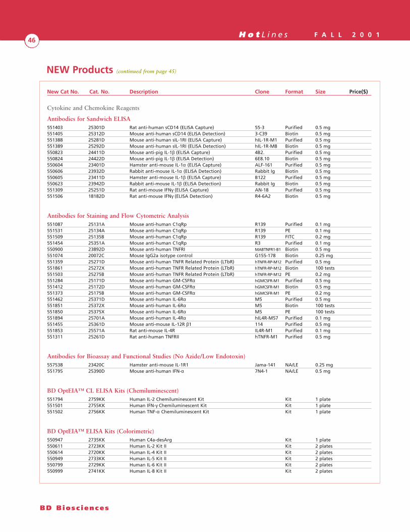
H o t L i n e s F A L L 2 0 0 146
BD Biosc iences
NEW Products (continued from page 45)
Cytokine and Chemokine Reagents
Antibodies for Sandwich ELISA551403 25301D Rat anti-human sCD14 (ELISA Capture) 55-3 Purified 0.5 mg551405 25312D Mouse anti-human sCD14 (ELISA Detection) 3-C39 Biotin 0.5 mg551388 25281D Mouse anti-human sIL-1RI (ELISA Capture) hIL-1R-M1 Purified 0.5 mg551389 25292D Mouse anti-human sIL-1RI (ELISA Detection) hIL-1R-M8 Biotin 0.5 mg550823 24411D Mouse anti-pig IL-1β (ELISA Capture) 4B2. Purified 0.5 mg550824 24422D Mouse anti-pig IL-1β (ELISA Detection) 6E8.10 Biotin 0.5 mg550604 23401D Hamster anti-mouse IL-1α (ELISA Capture) ALF-161 Purified 0.5 mg550606 23932D Rabbit anti-mouse IL-1α (ELISA Detection) Rabbit Ig Biotin 0.5 mg550605 23411D Hamster anti-mouse IL-1β (ELISA Capture) B122 Purified 0.5 mg550623 23942D Rabbit anti-mouse IL-1β (ELISA Detection) Rabbit Ig Biotin 0.5 mg551309 25251D Rat anti-mouse IFNγ (ELISA Capture) AN-18 Purified 0.5 mg551506 18182D Rat anti-mouse IFNγ (ELISA Detection) R4-6A2 Biotin 0.5 mg
Antibodies for Staining and Flow Cytometric Analysis551087 25131A Mouse anti-human C1qRp R139 Purified 0.1 mg551531 25134A Mouse anti-human C1qRp R139 PE 0.1 mg551509 25135B Mouse anti-human C1qRp R139 FITC 0.2 mg551454 25351A Mouse anti-human C1qRp R3 Purified 0.1 mg550900 23892D Mouse anti-human TNFRI MABTNFR1-B1 Biotin 0.5 mg551074 20072C Mouse IgG2a isotype control G155-178 Biotin 0.25 mg551359 25271D Mouse anti-human TNFR Related Protein (LTbR) hTNFR-RP-M12 Purified 0.5 mg551861 25272X Mouse anti-human TNFR Related Protein (LTbR) hTNFR-RP-M12 Biotin 100 tests551503 25275B Mouse anti-human TNFR Related Protein (LTbR) hTNFR-RP-M12 PE 0.2 mg551284 25171D Mouse anti-human GM-CSFRα hGMCSFR-M1 Purified 0.5 mg551412 25172D Mouse anti-human GM-CSFRα hGMCSFR-M1 Biotin 0.5 mg551373 25175B Mouse anti-human GM-CSFRα hGMCSFR-M1 PE 0.2 mg551462 25371D Mouse anti-human IL-6Rα M5 Purified 0.5 mg551851 25372X Mouse anti-human IL-6Rα M5 Biotin 100 tests551850 25375X Mouse anti-human IL-6Rα M5 PE 100 tests551894 25701A Mouse anti-human IL-4Rα hIL4R-M57 Purified 0.1 mg551455 25361D Mouse anti-mouse IL-12R β1 114 Purified 0.5 mg551853 25571A Rat anti-mouse IL-4R IL4R-M1 Purified 0.1 mg551311 25261D Rat anti-human TNFRII hTNFR-M1 Purified 0.5 mg
Antibodies for Bioassay and Functional Studies (No Azide/Low Endotoxin)557538 23420C Hamster anti-mouse IL-1R1 Jama-141 NA/LE 0.25 mg551795 25390D Mouse anti-human IFN-α 7N4-1 NA/LE 0.5 mg
BD OptEIA™ CL ELISA Kits (Chemiluminescent)551794 2759KK Human IL-2 Chemiluminescent Kit Kit 1 plate551501 2755KK Human IFN-γ Chemiluminescent Kit Kit 1 plate551502 2756KK Human TNF-α Chemiluminescent Kit Kit 1 plate
BD OptEIA™ ELISA Kits (Colorimetric)550947 2735KK Human C4a-desArg Kit 1 plate550611 2723KK Human IL-2 Kit II Kit 2 plates550614 2720KK Human IL-4 Kit II Kit 2 plates550949 2733KK Human IL-5 Kit II Kit 2 plates550799 2729KK Human IL-6 Kit II Kit 2 plates550999 2741KK Human IL-8 Kit II Kit 2 plates
New Cat No. Cat. No. Description Clone Format Size Price($)

H o t L i n e s F A L L 2 0 0 147
Pharmingen • Immunocytometry Systems • Discovery Labware • Clontech
Cytokine and Chemokine Reagents
BD OptEIA™ ELISA Kits (Colorimetric) (continued)550613 2721KK Human IL-10 Kit Kit 2 plates550801 2731KK Mouse IL-2 Kit Kit 2 plates550997 2739KK Mouse IL-5 Kit Kit 2 plates550950 2734KK Mouse IL-6 Kit Kit 2 plates551423 2746KK Mouse IL-12 p40 Kit Kit 2 plates
BD OptEIA™ Sets (Colorimetric)550926 2732KI Human IP-10 Set Set Rgts for 20 plates551424 2747KI Human sICAM-1 Set Set Rgts for 20 plates550995 2737KI Human TNF-β Set Set Rgts for 20 plates550996 2738KI Human TNFRI Set Set Rgts for 20 plates550948 2736KI Human TRAIL Set Set Rgts for 20 plates551492 2750KI Monkey IFN-γ Set Set Rgts for 20 plates551494 2752KI Monkey IL-2 Set Set Rgts for 20 plates551495 2753KI Monkey IL-4 Set Set Rgts for 20 plates551496 2754KI Monkey IL-6 Set Set Rgts for 20 plates551493 2751KI Monkey TNF-α Set Set Rgts for 20 plates551866 2760KI Mouse IFN-γ Set (AN-18) Set Rgts for 20 plates559753 2668KI Mouse MIP-1β Set Set Rgts for 20 plates550319 2705KI Rat IL-6 Set Set Rgts for 20 plates
Human Cell Surface Reagents551059 35818X Mouse anti-human β7 integrin FIB504 Cy-Chrome 100 tests551082 35819X Mouse anti- human β7 integrin FIB504 APC 100 tests551340 38381A Mouse anti-human Bcl-10 151 Purified 0.1 mg550935 38241A Mouse anti-human µ-Calpain B27D8 Purified 0.1 mg550936 38245X Mouse anti-human µ-Calpain B27D8 PE 100 tests551312 38371A Mouse anti-human c-Mpl BAH-1 Purified 0.1 mg551313 38375X Mouse anti-human c-Mpl BAH-1 PE 100 tests551314 38379X Mouse anti-human c-Mpl BAH-1 APC 100 tests550796 3010TA Mouse anti-human CD3 UCHT1 PerCP-Cy5.5 0.1 mg550788 3015TA Mouse anti-human CD4 RPA-T4 PerCP-Cy5.5 0.1 mg550791 3032TA Mouse anti-human CD8 RPA-T8 PerCP-Cy5.5 0.1 mg551131 30448X Mouse anti-human CD11a/LFA-1 HI111 Cy-Chrome 100 tests550787 3054TA Mouse anti-human CD14 M5E2 PerCP-Cy5.5 0.1 mg551060 35659X Mouse anti-human CD18 6.7 APC 100 tests550789 3065TA Mouse anti-human CD19 HIB19 PerCP-Cy5.5 0.1 mg551064 30698X Mouse anti-human CD21 B-ly4 Cy-Chrome 100 tests551141 31138X Mouse anti-human CD42b HIP1 Cy-Chrome 100 tests550988 38271A Mouse anti-human CD44 515 Purified 0.1 mg551063 33508X Mouse anti-human CD79b CB3-1 Cy-Chrome 100 tests550813 38094X Mouse anti-human Cw60 UM-4D4 FITC 100 tests551137 36008X Mouse anti-human CDw137 4B4-1 Cy-Chrome 100 tests551138 36888X Mouse anti-human CD161 (NKR-P1A) DX12 Cy-Chrome 100 tests550951 38251A Mouse anti-human Cytokeratin 14,15,16 and 19 KA4 Purified 0.1 mg551264 38304X Mouse anti-human DC-SIGN DCN46 FITC 100 tests551285 38314A Mouse anti-human HLA-A2 BB7.2 FITC 0.1 mg551127 35749X Mouse anti-human IL-8 RB 6C6 APC 100 tests551056 38291A Mouse anti-human PRR2 R2.525 Purified 0.1 mg551292 38331A Mouse anti-human TAP2 TAP2.17 Purified 0.1 mg550940 08271A Mouse anti-human TIMP-1 102D1 Purified 0.1 mg550795 3381TA Mouse IgG1, kappa isotype control MOPC-21 PerCP-Cy5.5 0.1 mg550915 38174K IP30 FITC Set FITC set
New Cat No. Cat. No. Description Clone Format Size Price($)
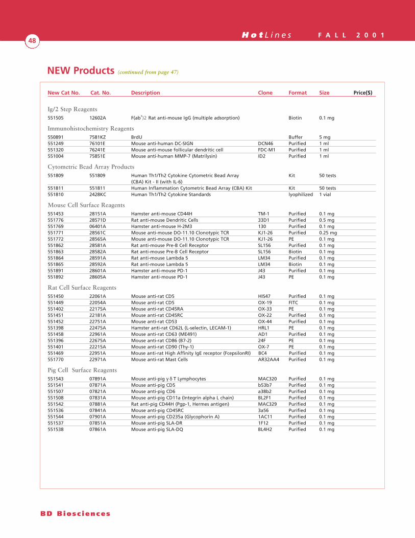
H o t L i n e s F A L L 2 0 0 148
BD Biosc iences
Ig/2 Step Reagents551505 12602A F(ab')2 Rat anti-mouse IgG (multiple adsorption) Biotin 0.1 mg
Immunohistochemistry Reagents550891 7581KZ BrdU Buffer 5 mg551249 76101E Mouse anti-human DC-SIGN DCN46 Purified 1 ml551320 76241E Mouse anti-mouse follicular dendritic cell FDC-M1 Purified 1 ml551004 75851E Mouse anti-human MMP-7 (Matrilysin) ID2 Purified 1 ml
Cytometric Bead Array Products551809 551809 Human Th1/Th2 Cytokine Cytometric Bead Array Kit 50 tests
(CBA) Kit - II (with IL-6)551811 551811 Human Inflammation Cytometric Bead Array (CBA) Kit Kit 50 tests551810 2428KC Human Th1/Th2 Cytokine Standards lyophilized 1 vial
Mouse Cell Surface Reagents551453 28151A Hamster anti-mouse CD44H TM-1 Purified 0.1 mg551776 28571D Rat anti-mouse Dendritic Cells 33D1 Purified 0.5 mg551769 06401A Hamster anti-mouse H-2M3 130 Purified 0.1 mg551771 28561C Mouse anti-mouse DO-11.10 Clonotypic TCR KJ1-26 Purified 0.25 mg551772 28565A Mouse anti-mouse DO-11.10 Clonotypic TCR KJ1-26 PE 0.1 mg551862 28581A Rat anti-mouse Pre-B Cell Receptor SL156 Purified 0.1 mg551863 28582A Rat anti-mouse Pre-B Cell Receptor SL156 Biotin 0.1 mg551864 28591A Rat anti-mouse Lambda 5 LM34 Purified 0.1 mg551865 28592A Rat anti-mouse Lambda 5 LM34 Biotin 0.1 mg551891 28601A Hamster anti-mouse PD-1 J43 Purified 0.1 mg551892 28605A Hamster anti-mouse PD-1 J43 PE 0.1 mg
Rat Cell Surface Reagents551450 22061A Mouse anti-rat CD5 HIS47 Purified 0.1 mg551449 22054A Mouse anti-rat CD5 OX-19 FITC 0.1 mg551402 22175A Mouse anti-rat CD45RA OX-33 PE 0.1 mg551451 22181A Mouse anti-rat CD45RC OX-22 Purified 0.1 mg551452 22751A Mouse anti-rat CD53 OX-44 Purified 0.1 mg551398 22475A Hamster anti-rat CD62L (L-selectin, LECAM-1) HRL1 PE 0.1 mg551458 22961A Mouse anti-rat CD63 (ME491) AD1 Purified 0.1 mg551396 22675A Mouse anti-rat CD86 (B7-2) 24F PE 0.1 mg551401 22215A Mouse anti-rat CD90 (Thy-1) OX-7 PE 0.1 mg551469 22951A Mouse anti-rat High Affinity IgE receptor (FcepsilonRI) BC4 Purified 0.1 mg551770 22971A Mouse anti-rat Mast Cells AR32AA4 Purified 0.1 mg
Pig Cell Surface Reagents551543 07891A Mouse anti-pig γ δ T Lymphocytes MAC320 Purified 0.1 mg551541 07871A Mouse anti-pig CD5 b53b7 Purified 0.1 mg551507 07821A Mouse anti-pig CD6 a38b2 Purified 0.1 mg551508 07831A Mouse anti-pig CD11a (Integrin alpha L chain) BL2F1 Purified 0.1 mg551542 07881A Rat anti-pig CD44H (Pgp-1, Hermes antigen) MAC329 Purified 0.1 mg551536 07841A Mouse anti-pig CD45RC 3a56 Purified 0.1 mg551544 07901A Mouse anti-pig CD235a (Glycophorin A) 1AC11 Purified 0.1 mg551537 07851A Mouse anti-pig SLA-DR 1F12 Purified 0.1 mg551538 07861A Mouse anti-pig SLA-DQ BL4H2 Purified 0.1 mg
NEW Products (continued from page 47)
New Cat No. Cat. No. Description Clone Format Size Price($)

H o t L i n e s F A L L 2 0 0 149
Pharmingen • Immunocytometry Systems • Discovery Labware • Clontech
Molecular Biology Reagents - BD RiboQuant™ Multi-Probe Template Sets
Mouse Cell Cycle Regulator/Complement551490 45751P mComplement RiboQuant Multi-Probe Template Set 10 reactions
Mouse Angiogenesis551418 45765P mAngio-1 RiboQuant Multi-Probe Template Set 10 reactions
Mouse Matrix Metalloproteinase551276 45760P mMMP-1 RiboQuant Multi-Probe Template Set 10 reactions550249 45761P mMMP-2 RiboQuant Multi-Probe Template Set 10 reactions
Mouse Developmental Gene550925 45719P mWnt-1 RiboQuant Multi-Probe Template Set 10 reactions550249 45720P mWnt-2 RiboQuant Multi-Probe Template Set 10 reactions
Human Cytokines and Chemokines551787 45666P hCK-8 RiboQuant Multi-Probe Template Set 10 reactions551488 45665P hCK-9 RiboQuant Multi-Probe Template Set 10 reactions
Human DNA Repair Pathway550251 45730P hDisR RiboQuant Multi-Probe Template Set 10 reactions
Human Matrix Metalloproteinase551274 45760P hMMP-1 RiboQuant Multi-Probe Template Set 10 reactions551275 45761P hMMP-2 RiboQuant Multi-Probe Template Set 10 reactions
Human Sulfotransferase550793 45735P hTOX-1b RiboQuant Multi-Probe Template Set 10 reactions550794 45736P hTOX-1 RiboQuant Multi-Probe Template Set 10 reactions
Applicable Patents:PE and APC: US Patent No. 4,520,110; 4,859,582; 5,055,556. European Patent No. 76,695; and Canadian Patent No. 1,179,942
PerCP: US Patent No. 4,876,190
Cy5.5 and Cy7: US Patent No. 5,268,486; 5,486,616; 5,569,587; 5,569,766; and 5,627,027
APC-Cy7: US Patent No. 5,714,386
New Cat No. Cat. No. Description Clone Format Size Price($)
Tools for Neuroscience Research
BD Biosciences:Your Network for Neuroscience ResearchGene ExpressionBD RiboQuant™ Multi-Probe RNaseProtection AssaysMulti-probe template sets for Apoptosis,Neurotrophins, and Ephrin Receptors.
BD Clontech Systems & Kits• ApoAlert™ Apoptosis Products• Atlas™ Arrays• Mercury™ Signal Transduction Products
Protein Expression - ProteomicsBD PowerBlot™Simultaneously evaluate over 800 proteinsfor changes in expression.
Cell Function & AnalysisApoptosis-Related Kits & ReagentsHundreds of Neuroscience Abs: Apoptosis,Calcium Signaling, NeurotransmitterReceptors, Ion Channels & Transporters,Synaptic Vesicles & Proteins, and More!
Cell CultureIn addition to BD Falcon™ cultureware,BD Biosciences Discovery Labware offersBD BioCoat™ cellware pre-coated with avariety of ECMs and attachment factors foroptimal neuronal growth and differentia-tion.
BD BiosciencesClontechDiscovery LabwareImmunocytometry SystemsPharmingen
BD BiosciencesVisit www.bdbiosciences.com. Call 877.232.8995 (in the US) to order.
For research use only. Not for use in diagnostic or therapeutic procedures. Not for Resale.BD, BD Logo and all other trademarks are the property of Becton, Dickinson and Company. ©2001 BD

BD Pharmingen™ Apoptosis Kits & Reagents
The Apoptosis Signaling Cascade: Powered by BD Biosciences Pharmingen
BD BiosciencesClontechDiscovery LabwareImmunocytometry SystemsPharmingen
BD Biosciences PharmingenVisit www.bdbiosciences.com. Call 877.232.8995 (in the US) to order.
For research use only. Not for use in diagnostic or therapeutic procedures. Not for Resale.BD, BD Logo and all other trademarks are the property of Becton, Dickinson and Company. ©2001 BD
Apoptotic signaling events are delicateand precise, yet powerful; their studyrequires both insight and sophistication.Only BD Biosciences offers you thecomprehensive tool set you need toinvestigate every aspect of apoptosis:From Genes to Proteins to Cells.
Gene ExpressionBD RiboQuant™ Multi-Probe RNaseProtection AssaysMulti-probe template sets for human,mouse, and rat apoptotic gene expression.
Protein Expression - ProteomicsBD PowerBlot™
Simultaneously evaluate over 800proteins for changes in expression.
Cell Function & AnalysisApoptosis-Related Kits & ReagentsHundreds of Apoptosis-Related Abs,Apoptosis Antibody Sampler Kits,Annexin V Conjugates, CaspaseActivity Detector (SR-VAD-FMK),MitoScreen (JC-1).
Visit our website for details.
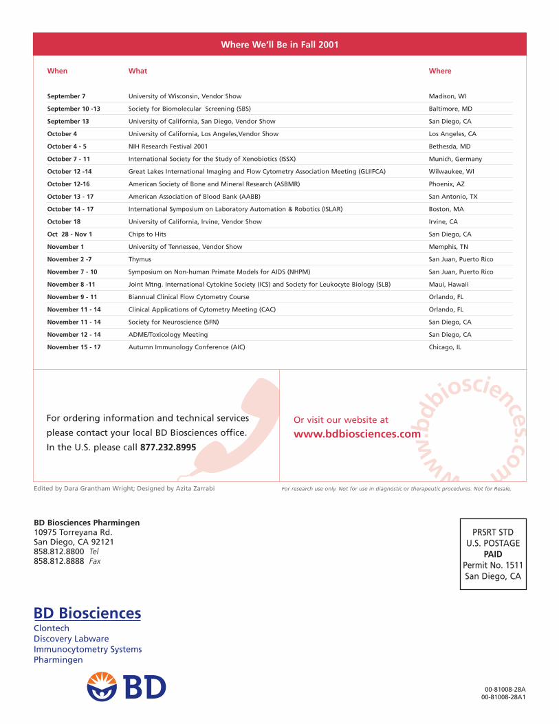
www
.bdb
iosciences.com
PRSRT STDU.S. POSTAGE
PAIDPermit No. 1511San Diego, CA
BD Biosciences Pharmingen10975 Torreyana Rd.San Diego, CA 92121858.812.8800 Tel858.812.8888 Fax
00-81008-28A00-81008-28A1
BD BiosciencesClontechDiscovery LabwareImmunocytometry SystemsPharmingen
Where We’ll Be in Fall 2001
When What Where
September 7 University of Wisconsin, Vendor Show Madison, WI
September 10 -13 Society for Biomolecular Screening (SBS) Baltimore, MD
September 13 University of California, San Diego, Vendor Show San Diego, CA
October 4 University of California, Los Angeles,Vendor Show Los Angeles, CA
October 4 - 5 NIH Research Festival 2001 Bethesda, MD
October 7 - 11 International Society for the Study of Xenobiotics (ISSX) Munich, Germany
October 12 -14 Great Lakes International Imaging and Flow Cytometry Association Meeting (GLIIFCA) Wilwaukee, WI
October 12-16 American Society of Bone and Mineral Research (ASBMR) Phoenix, AZ
October 13 - 17 American Association of Blood Bank (AABB) San Antonio, TX
October 14 - 17 International Symposium on Laboratory Automation & Robotics (ISLAR) Boston, MA
October 18 University of California, Irvine, Vendor Show Irvine, CA
Oct 28 - Nov 1 Chips to Hits San Diego, CA
November 1 University of Tennessee, Vendor Show Memphis, TN
November 2 -7 Thymus San Juan, Puerto Rico
November 7 - 10 Symposium on Non-human Primate Models for AIDS (NHPM) San Juan, Puerto Rico
November 8 -11 Joint Mtng. International Cytokine Society (ICS) and Society for Leukocyte Biology (SLB) Maui, Hawaii
November 9 - 11 Biannual Clinical Flow Cytometry Course Orlando, FL
November 11 - 14 Clinical Applications of Cytometry Meeting (CAC) Orlando, FL
November 11 - 14 Society for Neuroscience (SFN) San Diego, CA
November 12 - 14 ADME/Toxicology Meeting San Diego, CA
November 15 - 17 Autumn Immunology Conference (AIC) Chicago, IL
For ordering information and technical services
please contact your local BD Biosciences office.
In the U.S. please call 877.232.8995
Or visit our website at
www.bdbiosciences.com
Edited by Dara Grantham Wright; Designed by Azita Zarrabi For research use only. Not for use in diagnostic or therapeutic procedures. Not for Resale.