3jHCOP = 15.3cos2* - 6.1coso + 1.6 - NCBI
16
Carbon-13 NMR in conformational analysis of nucleic acid fragments. 4. The torsion angle distribution about the C3'-03' bond in DNA constituents P.P.Lankhorst, C.A.G.Haasnoot*, C.Erkelens, H.P.Westerink, G.A.van der Marel, J.H.van Boom and C.Altona Gorlaeus Laboratories, State University of Leiden, P.O. Box 9502, 2300 RA Leiden, The Netherlands, and *Department of Biophysical Chemistry, Toemooiveld, Faculty of Sciences, University of Nijmegen, 6525 Nijmegen, The Netherlands Received 6 December 1984; Accepted 14 January 1985 ABSTRACT Carbon-13 and proton NMR spectra of a series of oligodeoxynucleotides (d(CT), d(CC), d(TA), d(AT), d(CG), d(GC), d(AG), d(AAA), d(TATA) and d(GGTAAT)) were measured at various temperatures. The three coupling con- stants that are related to the magnitude of backbone angle c (J(C4'-P), J(C2'-P) and J(H3'-P)) are analyzed in terms of a three-state equilibrium about this bond. Two e(trans) angles occur, which differ in magnitude depending on the conformation (N or S) of the adjoining deoxyribose ring. The S-type deoxyri- bose ring is associated with a smaller £(trans) angle: E(t,S) = 192°. The N-type deoxyribose ring is associated with a larger £(trans) angle E(t,N) = 212°. The third rotamer participating in the conformational equilibrium, is a gauche(-) (E(-)) conformer and occurs exclusively in combination with the S-type sugar ring (E(-,S) = 2660). Within the limits of experimental error, the magnitude of these three angles appears to be independent of the particular base sequence, except in the case of d(CG) where a slightly larger E(t,S) angle (1970) is indi- cated. A simple equation is proposed which may be used to calculate the popu- lation of e(t,S) conformer in cases where only J(H3'-P) is known. INTRODUCTION 1,2 Carbon-13 NMR spectroscopy is rapidly becoming an indispensible supple- ment to 1H NMR studies aimed at understanding fine details of conformational behaviour of oligoribonucleotides. In previous papers from our laboratories it was shown, first of all3, that heteronuclear '3C-1H chemical shift correlation spectroscopy affords an unambiguous assignment of 13C resonances of ribonu- cleic acid dimers and trimers. That study also revealed a good correlation be- tween the chemical shifts of the 13C atoms of the ribose ring and the sugar conformational equilibrium. Subsequently, a reparametrization of the Karplus equations for vicinal NMR coupling constants in CCOP (eq. 1) and HCOP (eq. 2) fragments was introduced": 3J CCOP= 6.9cos2¢ - 3.4coso + 0.7 (1) 3jHCOP = 15.3cos2* - 6.1coso + 1.6 (2) © I RL Press Limited, Oxford, England. Nucleic Acids Research Volume 13 Number 3 1985 927
-
Upload
khangminh22 -
Category
Documents
-
view
6 -
download
0
Transcript of 3jHCOP = 15.3cos2* - 6.1coso + 1.6 - NCBI

Carbon-13 NMR in conformational analysis of nucleic acid fragments. 4. The torsion angledistribution about the C3'-03' bond in DNA constituents
P.P.Lankhorst, C.A.G.Haasnoot*, C.Erkelens, H.P.Westerink, G.A.van der Marel, J.H.van Boomand C.Altona
Gorlaeus Laboratories, State University of Leiden, P.O. Box 9502, 2300 RA Leiden, TheNetherlands, and *Department of Biophysical Chemistry, Toemooiveld, Faculty of Sciences,University of Nijmegen, 6525 Nijmegen, The Netherlands
Received 6 December 1984; Accepted 14 January 1985
ABSTRACTCarbon-13 and proton NMR spectra of a series of oligodeoxynucleotides
(d(CT), d(CC), d(TA), d(AT), d(CG), d(GC), d(AG), d(AAA), d(TATA) andd(GGTAAT)) were measured at various temperatures. The three coupling con-stants that are related to the magnitude of backbone angle c (J(C4'-P),J(C2'-P) and J(H3'-P)) are analyzed in terms of a three-state equilibrium aboutthis bond.
Two e(trans) angles occur, which differ in magnitude depending on theconformation (N or S) of the adjoining deoxyribose ring. The S-type deoxyri-bose ring is associated with a smaller £(trans) angle: E(t,S) = 192°. The N-typedeoxyribose ring is associated with a larger £(trans) angle E(t,N) = 212°. Thethird rotamer participating in the conformational equilibrium, is a gauche(-)(E(-)) conformer and occurs exclusively in combination with the S-type sugarring (E(-,S) = 2660). Within the limits of experimental error, the magnitude ofthese three angles appears to be independent of the particular base sequence,except in the case of d(CG) where a slightly larger E(t,S) angle (1970) is indi-cated.
A simple equation is proposed which may be used to calculate the popu-lation of e(t,S) conformer in cases where only J(H3'-P) is known.
INTRODUCTION 1,2
Carbon-13 NMR spectroscopy is rapidly becoming an indispensible supple-ment to 1H NMR studies aimed at understanding fine details of conformationalbehaviour of oligoribonucleotides. In previous papers from our laboratories it
was shown, first of all3, that heteronuclear '3C-1H chemical shift correlationspectroscopy affords an unambiguous assignment of 13C resonances of ribonu-cleic acid dimers and trimers. That study also revealed a good correlation be-tween the chemical shifts of the 13C atoms of the ribose ring and the sugarconformational equilibrium. Subsequently, a reparametrization of the Karplusequations for vicinal NMR coupling constants in CCOP (eq. 1) and HCOP (eq. 2)fragments was introduced":
3JCCOP= 6.9cos2¢ - 3.4coso + 0.7 (1)
3jHCOP = 15.3cos2* - 6.1coso + 1.6 (2)
© I RL Press Limited, Oxford, England.
Nucleic Acids ResearchVolume 13 Number 3 1985
927
Nucleic Acids Research
The parameters for equations (1) and (2) were derived simultaneously from adata set of coupling constants 'JCCOP and 'JHCOP of RNA fragments. Specialattention was paid to pinpointing the magnitude of the backbone angle c
(C4'-C3'-03'-P) in the Et and e conformational ranges. At this point of our in-vestigations the question arose whether or not the parameters of eqns. (1) and(2) were applicable to DNA fragments. From a comparison of 3',5'-cyclic AMPwith 3',5'-cyclic dAMP, and from a detailed analysis of couplings obtained fromd(TpA) at a number of temperatures, it could be concluded5 that a 2' oxygensubstituent does not affect the C2'-P coupling to a measurable degree.
From many previous studies 1-2 on RNA nucleotide fragments it is knownthat the conformational preferences about e are closely correlated to the actualconformation of the 3'-O attached ribose ring: when the sugar adopts theN-type form, the torsion angle e is strictly confined to the t domain. From 1Hand "C NMR experiments on RNA model compounds' the exact magnitude of Etwas found to vary with the base-sequence involved: the C-C sequence displayeda relatively large value of Et (226') whereas for the A-U sequence a relativelysmall value of Et (2140) was derived. An average value of Et = 219° was deter-mined, in good agreement with the value of Ft = 2180, which is the average val-ue from available X-ray data on RNA dimers'. When the ribose ring reverts to
the S-type conformation, the 3'-phosphate is able to swing over to the otherside of the C3'-H3' bond, resulting in a blend of £ (E - 2770) and Et rotamers.
The situation is different for DNA constituents. In a DNA helix the deoxy-ribose conformational equilibrium is biased strongly toward the S-type form.Nevertheless, the preferred rotamer about the C3'-03' bond is E . At this pointit is of interest to note that the magnitude of the angle E(t,S) in deoxynucleo-tides (for example ca. 195° in d(TpA) 5 or 1910 in the dodecamerCGCGAATTCGCG13) appears to be rather smaller than the value of E(t,N) (-219') in RNA constituents. Now the question arises as to the origin of this dif-ference. Two possibilities come to mind:(1) E(t,N) in RNA is primarily affected by the 2-hydroxyl group. If this weretrue, then one would expect that in DNA E(t,N) s(t,S) and the coupling con-
stant data can be interpreted in terms of a simple two-state equilibrium Et/E .
In fact, this line of thought was followed in our previous communication' con-cerning the case of d(TpA).(2) The magnitude of Et in DNA is determined by the conformation adopted bythe sugar ring. In other words, one would expect that N-type deoxyribose con-formers are associated with high Et values (W(t,N) > 210'), whereas the S-typedeoxysugars might display £ magnitudes in a lower range (for example E(t,S) <
928

Nucleic Acids Research
200°). Because a third conformer (E) makes its appearance in the equilibriumblend when a stacked species reverts to a random coil form, a three state con-
formational model (in terms of e(t,S), c(t,N) and F.) is called for.
The present study was undertaken in order to shed more light upon theconformational characteristics of the backbone angle c of DNA oligonucleotides in
aqueous solution. "3C-31P and 1H-31P vicinal coupling constants were measuredfor a series of oligodeoxynucleotides d(AAA), d(AG), d(AT), d(TA), d(CC),d(CT), d(CG) and d(GC) at three or more temperatures each. Care was takento include a minimum of two examples of each possible sequence of purines andpyrimidines: d(pur-pur), d(pur-pyr), d(pyr-pur) and d(pyr-pyr).
MATERIALS AND METHODSNMR samples
The compounds studied in this work were synthesized via a modified phos-photriester approach"'. D20 solutions of samples were prepared with nucleotideconcentrations ranging from 20-50 mM. A 5 mm NMR tube containing 0.4 ml ofsolution was used for d(TA) and d(CG). 10 mm tubes containing 1.2 ml of sol-ution were used for the remaining samples. A trace of EDTA was added in orderto neutralize paramagnetic contaminations, tetramethylammonium chloride servedas an internal reference. The pH of the samples was adjusted to 7.5 ± 0.5 (me-ter reading).One-dimensional NMR spectra
13C NMR spectra were recorded on a Bruker WM-300 spectrometer operat-ing at 75.5 MHz and on a Bruker WM-200 spectrometer operating at 50.3 MHz. Inboth cases a 13C probe with 31P decoupling facilities was used. 13C spectrawere acquired on 8K datapoints using a two-level decoupling sequence with isrelaxation delay between observation pulses. During acquisition 0.5-1.0 W de-coupling power was employed. FIDs were multiplied by a Gaussian window andthe Fourier transformation was carried out on 128K datapoints.
300 MHz 1H-NMR spectra were recorded on a selective 1H probe with 31Pdecoupling facilities. 'H-31P coupling constants were determined from the dif-ference between 1H spectra and 31P-decoupled 1H spectra. Special care wastaken to adjust the 31P decoupling power and decoupling frequency in order tobring about complete decoupling and at the same time avoid sample heating.
During 'IC acquisition the true sample temperature was determined fromthe chemical shift difference 6HDO86TMA" by recording a 1H spectrum on the
decouple coil immediately after each 13C run. This internal temperature cali-bration is essential as it was found previously that a small temperature increase'
(0-5'C) of the sample may occur as a result of 1H decoupling3.
929

Nucleic Acids Research
BASE RING
Ci'O0 C2'
0 -,P(i) -E-Os -+ CsCC-_-c4-- Ci--Oi -+P(i+1)-O| a(i) p(i) y (i) Noi Eli 5cil
(i-l) ith nucleotide unit (i+)
W chain direction
Figure 1: Conformational nomenclature.
Two-dimensional NMR spectroscopyThe spectra of d(TpA), d(ApG) ,d(CpC), d(TATA) and d(GGTAAT) were
assigned by means of two-dimensional heteronuclear chemical shift correlationspectroscopy'' 7, using the pulse sequence and phase cycling proposed byBax". Time domain spectra consisted of 64 datapoints (t1-dimension) and 2Kdatapoints (t2-dimension). Before Fourier transformation a phase shifted (1/61r)sine square window was applied and FIDs were zero-filled to 512 and 4K data-
points, respectively.Nomenclatu re
The proposed IUPAC-IUB nomenclature" is used in this work, i.e. back-
bone torsion angles are labelled a-; starting with a at the 5' terminal P-05' bond
(see Figure 1). Carbon atoms are numbered according to Figure 2.
RESULTS AND DISCUSSIONAssignment of Carbon-13 spectra
The assignment of carbon NMR spectra of deoxydinucleoside monophosp-hates is relatively straightforward. A number of carbon resonances is split dueto coupling to phosphorus. In a dinucleoside monophosphate these resonances
are C4'(1), C3'(1), C2'(1), C5'(2) and. C4'(2). C3'(1) and C5'(2) have geminalcouplings to phosphorus which are invariant with temperature (-5.3-5.6 Hz) andas such are of least interest. However, C4'(1) is a sensitive probe for the con-
formational situation around £ and therefore this signal should be discriminatedfrom C4'(2). This can be achieved by considering the magnitude of 3J C4'-P athigh temperature: it was pointed out in a previous paper' that the signal withthe largest coupling to phosphorus at high temperature should be assigned toC4' of the 3' terminus, whereas the doublet with the smaller splitting should beassigned to the C4' of the 5' terminus. The C2' signals, finally, resonate at highfield and can be recognized easily. In a few selected cases, notably d(AG),
930

Nucleic Acids Research
H Figure 2: Structure of d(TA).5' T
0
0
POP°2
0A
4'(2)
OH
d(CC), d(TA), d(TATA) and d(GGTAAT), the correctness of the assignmentwas substantiated by means of two-dimensional heteronuclear chemical shift cor-relation spectroscopy.The determination of J(HCOP) and J(CCOP)
Proton decoupled 13C spectra of each compound were recorded at three ormore temperatures. 3JCP values were directly determined from the spectra. Inaddition, 1H spectra and 3iP decoupled 1H spectra were recorded at temper-atures corresponding as close as possible to those selected for the 13C spectra.31P-1H coupling constants were determined by comparing the width of the H3'multiplet in the coupled and in the 31P decoupled spectra. In some cases (d(TA)and d(AAA), d(TATA) and d(GGTAAT)) 31P-1H coupling constants were avail-able from computer simulations carried out previously2 -23. In some instances itwas not feasible to measure 31P-1H coupling constants at the same temperatureas the 13C couplings, because of overlapping multiplets or overlap of the resi-dual HDO peak. In those cases 1H-31P couplings were determined by interpo-lation between bracketing values: one measured at higher temperature and oneat lower temperature. All relevant couplings are listed in Table 1. In some cas-es, notably d(AAA), d(TATA) and d(GGTAAT), the measurements were limitedto the couplings displayed by the 5' terminal residue 1 because the C4' signalsof the remaining residues 2, 3 ...... etc. appear as pseudotriplets and this pre-cludes a straightforward analysis. Moreover, signal overlap and/or small split-tings prevented the measurement of JC2' -p in these three compounds.Conformation around C3'-03' (£)
At first sight three classical rotameric ranges around the C3'-03' bond(g, t, g ) should be taken into consideration in the conformational analysis of e
However, the wealth of information extracted from X-ray diffraction studies2',as well as from a lanthanide-induced chemical shift investigation', shows that
the 3'-phosphate group strictly avoids to adopt the 9 rotamer (E - 600). In
931

Nucleic Acids Research
Table 1: J(C4'P), J(C2'P) and J(H3'P) (in Hz) as a function of temperature ('C)of the 5' terminal residues of all DNA constituents used in this work.
d(AG) d(CG) d(AT)
T C4'P C2'P H3'P T C4'P C2'P H3'P T C4'P C2'P H3'P
31 9.1 1.8 5.2 29 7.3 2.7 6.9 23 8.5 2.2 6.258 8.0 2.4 5.9 43 6.9 3.3 7.0 35 8.0 2.4 6.377 7.4 2.7 6.1 72 6.6 3.6 7.1 47 7.6 2.8 6.3
82 6.5 3.7 7.0 58 7.4 2.8 6.423 7.2 3.1 6.8
d(CT) d(CC) d(GC)
T C4'P C2'P H3'P T C4'P C2'P H3'P T C4'P C2'P H3'P
14 7.7 2.7 7.0 8 7.1 2.8 7.1 21 8.0 2.3 6.320 7.6 2.8 7.0 28 6.8 3.4 6.9 30 7.7 2.7 6.527 7.3 3.1 7.0 46 6.8 3.5 7.2 47 7.3 3.1 6.733 7.1 2.9 6.9 55 6.4 3.5 7.2 56 7.1 3.1 6.544 7.0 3.1 6.9 87 6.1 3.4 7.1 65 7.1 3.3 6.856 6.9 3.1 7.0 78 6.9 3.5 6.977 6.8 3.3 7.0 56
d(TA) d(TA) d(AAA)
T C4'P C2'P H3'P T C4'P C2'P H3'P T C4'P C2'P H3'P
2 8.8 - 5.8 38 7.7 2.6 6.3 5 10.3 - 4.55 8.5 - 5.8 46 7.5 2.9 6.4 11 10.0 - 4.87 8.5 - 5.8 51 7.1 3.1 6.4 21 9.9 - 4.8
15 8.3 - 5.9 56 7.2 3.1 6.5 43 8.9 - 5.420 8.0 - 6.0 60 6.9 3.4 6.5 76 7.6 - 6.530 7.9 - 6.1 92 6.6 3.6 6.8
d(TATA) d(GGTAAT)
T C4'P C2'P H3'P T C4'P C2'P H3'P
7 8.3 - 5.9 7 8.6 - 5.712 8.1 - 6.4 35 8.0 - 5.947 7.2 - 6.3 57 7.3 - -
fact, the e conformer never has been observed in the solid state and maytherefore be excluded safely from consideration (Figure 3).
In the introductory part of the present communication it is explained thatsome care should be exercised in the interpretation of the coupling constants ofDNA oligonucleotides measured along the C3'-03' bond, viz. 2J(C4'-P),2J(C2'-P) and 3J(H3'-P). In particular, one should be aware of the possibilitythat the usual two-state conformational approach - in terms of an t/e equilib-rium blend - may turn out to be too simple when the value of at changes withchanging conformation of the adjacent deoxyribose ring.
932

Nucleic Acids Research
H3' pe 0 H3X Figure 3: The trans and gauche(-) rotamer for c
C2' C41 C 2
E: trans E:gauche-
In the following, two strategies will be employed for the determination of E
values and corresponding conformational populations. It will be shown that thetwo-state approach, although applicable to each individual compound, fails toyield an overall consistent picture of the rotational preferences about theC3'-03' bond in DNA constituents.
Two-state analysis of e rotamers
Taking three measured coupling constants into account, the two-state
equations can be written:
J(H3'P,obs) = ptJ(H3'P)t + (1-pt)J(H3'P)- (3)J (C4'P, obs) = ptJ (C4' P)t + (1_pt)J(C4'P)- (4)J(C2'P,obs) = ptJ(C2'P)t + (1-pt)J(C2'P)- (5)
where pt stands for the mol fraction of et rotamer and the superscripts t and -
refer to the corresponding pure t and c states, respectively. Under the usu-
al assumption of trigonal projection symmetry (Figure 3) the following relationsare valid:
torsion angle C2'-C3'-03'-P = e - 120° (6)torsion angle C4'-C3'-03'-P = e (7)torsion angle H3'-C3'-03'-P = 2400 - e (8)
From these relations, in combination with eqs. (1), (2) and (3)-(5), the magni-tude of et and £ as well as the position of the et/e equilibrium at all exper-imental temperatures can be calculated in an iterative least-squares procedure,given a set of 3J values. The calculations were carried out for each compoundseparately; the resulting (apparent) et and c angles are listed in Table 2, to-
gether with the calculated Et population at the highest and lowest experimentaltemperature used in each case. For reasons explained below we also show thecorresponding population of S-type sugar pS conformation as determined from
the width of the H2" signal according to the sum rule25, eq. (9):
Ps = (17--8 - J1'21 - J2113)/10.9 (9)
Scrutiny of Table 2 reveals some peculiar features. The purine-purine oli-gonucleotide sequences d(AAA) and d(AG) appear to display smaller et angles(- 191°) than do the pyrimidine-pyrimidine sequences (- 2030), whereas themixed pyr-pur and pur-pyr compounds show intermediate values. Although a
933

Nucleic Acids Research
Table 2: Results of the two-state analyses of the vicinal C-P and H-P couplingsof 8 different DNA dimers. Apparent(*) backbone torsion angles £(t) and E(-) areshown as well as calculated trans populations lOOxp(t) at two extreme temper-atures. The corresponding populations of S-type sugar conformation are also giv-en, p(S)xlOO.
compound e(t) E -) T(°C) p(t) p(S)
d(AAA) 191 253 5 97 10076 63 84
d(AG) 190 256 31 80 9977 63 82
d(TA) 195 261 2 82 9092 58 76
d(AT) 196 262 23 77 7276 64 72
d(GC) 197 264 21 75 7378 60 72
d(CG) 201 267 29 71 8682 61 78
d(CC) 203 269 8 71 7187 62 70
d(CT) 204 272 14 77 6577 68 68
(*) The numbers given should be regarded as artefacts because the two-state ap-proach is too simple, see text.
sequence-dependent ct angle would not come as a surprise, attention should be
paid to the following. Concomitant with the increase of et angle in going from
pur-pur to pyr-pyr sequences, the t angle also increases from -255° to -270°.Since the e rotamer is associated with the unstacked or random-coil forms, this
result appears highly unlikely. In the first place one cannot imagine a likely me-
chanism whereby the base sequence would affect £ to such a large extent.
Secondly, it is noted (Table 2) that the lower range of t values (d(AAA),d(AG), d(TA)) appears to be associated with high populations of S-type sugar
conformations (even approaching 100% purity at low temperatures) and vice ver-
sa: high P. angles, as in d(CG), d(CC) and d(CT), apparently correlate withdecreased S-type populations and hence with an increase of N-type species.Most likely, the latter correlations should be considered as artefacts introducedby the incorrect limitations inherent to the two-state approach. In the followingsection it will be shown that these apparent anomalies disappear when the con-formational analysis is carried out in terms of a three-state equilibrium.Three-state analysis of c rotamers
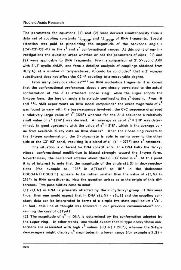
A two-state equilibrium is characterized by the feature that a plot of twoobservables, for example 'JC4'P vs. 3JH3'P' yields a straight line when thetemperature is varied, whereas in the case of a three-state situation the plottedpoints will lie within the confines of a triangle2. A plot of a selection of thedata of Table 1 is given as an example (Figure 4). Figure 4 as well as similar
934

Nucleic Acids Research
266* 212
z _~ +4+ +
S~~~~~~~~~~~~~
'I~~~~ ~ ~~~~~~~~,4 I
0 X 2 3 S 6 7 S 9 10 I1J (Cm 'P)
Figure 4: Plot of J(H3'P) vs. J(C4'P) for the following compounds:d(AAA): x,d(TA) + ,d(CC):o ,d(CT):A. The corners of the trian-gle represent the values of J(C4'P) and J(H3'P) in pure £(t,N),c(t,S) and e(-,S) conformers as derived below (see text).
plots (not shown) for other compounds indeed strongly suggest the existence ofa three-state equilibrium. The question as to which particular states participatein the equilibrium blend can be answered by the observation mentioned in theprevious section: the two-state calculations reveal that, as the N-type sugarconformational populations increase in going from pur-pur to pyr-pyr se-quences, the resulting Ft values appear to increase concomitantly. Thus, twodifferent ct angles are indicated: one e(t,S) in the lower range, associated withan S-type deoxyribose ring as found in B-DNA and another one (E(t,N)) in thehigher range, associated with an N-type sugar conformation as occurs in A-DNAand A-RNA.
From this point onwards use was made of a computer program (written inAPL'5) which allows one to carry out an iterative least-squares minimization ofthe quantity Jobs - Jcalc' A large set of experimental coupling constants can beused as input data and the program searches for the "best" combination of theangles E(t,S), E(t,N) and E(-,S). The corresponding populations p(t,S),p(t,N) and p(-,S) are also computed. It should be remembered that, sincep(t,S) + p(t,N) + p(-,S) = 1, only two of these represent independent vari-ables. In order to avoid possible pitfalls due to undetected minima, a number ofpilot calculations was performed first. In these calculations angles c(t,S) andc(t,N) were in turn (and stepwise) constrained to adopt a series of predeter-mined values. In this way conformational space was scanned for c(t,N) varyingfrom 200' to 240° and a(-,S) varying from 250° to 270'. Only a single -but fair-ly shallow- minimum was found. A remarkable feature was readily apparent in all
935

Nucleic Acids Research
Table 3: Results of the three-state analysis of C-P and H-P couplings of 7 dif-ferent DNA dimers. Observed and calculated couplings are shown. E(t,N) = 2120and e(t,S) = 1920 and e(-,S) = 2660, except for d(CpG), see text.A = J(obs) - J(calc) (in Hz), the overall rms: 0.14 Hz.
compound T J(H3'P) J(C4'P) J(C2'P)obs calc A obs calc A obs calc A
d(ApG) 31 5.2 5.2 0.0 9.1 9.0 0.1 1.8 1.6 0.256 5.9 5.8 0.1 8.0 8.0 0.0 2.4 2.4 0.077 6.1 6.3 -0.2 7.4 7.4 0.0 2.7 2.7 0.0
d(TpA) 38 6.3 6.1 0.2 7.7 7.6 0.1 2.6 2.6 0.056 6.5 6.4 0.1 7.2 7.1 0.1 3.1 3.0 0.192 6.8 6.9 -0.1 6.6 6.5 0.1 3.6 3.4 0.2
d(ApT) 58 6.4 6.6 -0.2 7.4 7.4 0.0 2.8 2.6 0.076 6.8 6.7 0.1 7.2 7.1 0.1 3.1 2.9 0.2
d(GpC) 21 6.3 6.3 0.0 8.0 8.0 0.0 2.3 2.2 0.147 6.7 6.7 0.0 7.3 7.1 0.2 3.1 2.9 0.278 6.9 6.9 0.0 6.9 6.6 0.3 3.5 3.2 0.3
d(CpG) 29 6.9 6.7 0.2 7.3 7.2 0.1 2.7 2.7 0.0(*) 43 7.0 6.9 0.1 6.9 6.8 0.1 3.3 3.1 0.2
72 7.1 7.1 0.0 6.6 6.4 0.2 3.6 3.4 0.282 7.0 7.2 -0.2 6.5 6.3 0.2 3.7 3.4 0.3
d(CpC) 8 7.1 6.8 0.3 7.1 7.0 0.1 2.8 2.9 -0.146 7.2 7.0 0.2 6.8 6.6 0.2 3.5 3.3 0.287 7.1 7.1 0.0 6.1 6.3 -0.1 3.4 3.5 -0.1
d(CpT) 14 7.0 6.8 0.2 7.7 7.4 0.3 2.7 2.5 0.244 6.9 6.9 0.0 7.0 6.9 0.1 3.1 3.0 0.177 7.0 7.0 0.0 6.8 6.7 0.1 3.3 3.2 0.1
(*) e(t,S) = 1970, rms = 0.16 Hz.
calculations that were carried out
root-mean-square (rms) deviations: theusing input values that gave lowrotamer population p(t,N), produced by
the least-squares calculation, was - within the limits of error - invariably equalto the population of N-type conformation (PN) of the attached deoxyribose ring.Note that PN was determined independently from the H2" signal (eq. 9), PN +
Ps = 1. Therefore, in the final step the number of independent variables was
drastically reduced by fixing each p(t,N) to the value of each correspondingpercentage of N-type sugar ring, i.e. determined for the same sequence at thesame temperature, viz. Table 2. It was also clear that the computed couplingconstants of d(CG) fitted less well to the experimental ones. For these reasonsthe final calculations were carried out as follows: a data set, consisting of 51coupling constants of the six dimers d(AG), d(TA), d(AT), d(GC), d(CC) andd(CT) was used as input (see Table 3) and the populations p(t,S) as well as thevalues of the three c angle were refined (20 independent variables vs. 51 ob-servables). The iteration rapidly converged and yielded e(t,S) = 192 ±2',
936

Nucleic Acids Research
Table 4: C-P and H-P coupling constants (Hz) calculated for the three pure ro-tamers t(t,N) = 211.70 and E(t,S) = 192.30 and E(-,S) = 266.00(*)
angle J(P-H3') J(P-C4') J(P-C2')
E(t,S) = 192.3 4.45 10.60 0.30ce(t,N) = 211.7 8.11 8.58 0.81(-, S) = 266.0 8.50 0.97 8.26
(*) In order to avoid rounding-off errors, angles are shown to 0.10. and coupl-ing constants to 0.01 Hz. The last digit is not significant.
e(t,N) = 212 +4° and e(-,S) = 266 t3°, overall rms deviation 0.14 Hz, maximum
deviation 0.3 Hz. The excellent agreement between Jobs and J calc is shown in
Table 3.The 12 available couplings of d(CG) were treated in a separate
least-squares minimization. In this run E(t,N) and c(-,S) were kept fixed at thepreviously calculated values and E(t,S) was refined, together with the popu-lation p(t,S). For E(t,S) = 1970 an excellent fit between Jobs and Jcalc was ob-tained (rms = 0. 16 Hz). These results are also displayed in Table 3
In the foregoing derivation of limiting couplings (see Table 4) the data onthree compounds, for which 3Jc2'P could not be determined experimentally,were necessarily left out of consideration. These data concern the 5' terminalresidues in the higher olgomers d(AAA), d(TATA) and d(GGTAAT). Now wemay revert the procedure and test whether or not a good fit between observedand calculated 3JC4'P and 3jH3'P can be obtained using the previously estab-lished limiting couplings as input and p(t,S) as the only independent variable.As before, p(t,N) was set equal to the population of N-type sugar conformationestablished earlier20-23 at various temperatures. It turns out that thethree-state analysis indeed yields an excellent fit (rms 0.11-0.19 Hz). The re-sulting rotamer populations are also collected in Table 5
The results of the trimer d(AAA) are of special interest because this com-pound is the only one in the present series for which the thermodynamic param-eters of the stack/unstack equilibrium have been measured fromtemperature-dependent CD spectra20 and a comparison of the population p(t,S)with the known population of base-stacked single-helical species (Table 7 inref20) can be made. In the case of d(AAA) p(t,S) drops from 0.98 to 0.55 onraising the temperature from 5 IC to 76 'C. At the same time the population of
base-base stack drops from 0.87 to 0.33. It is seen that at all temperatures thepopulation of stacked species is significantly less than p(t,S). This finding maybe taken to mean that the random-coil form permits E to adopt both the E(t,S)and E(-,SY rotamers. Hence, one should be careful to equate neither p(t,S),nor the sum p(t,S) + p(t,N), to the population of stacked species. From the
937

Nucleic Acids Research
Table 5: Results of the three-state analysis of C-P and H-P couplings of all DNAconstituents represented in Table 1, showing the calculated rotameric popu-lations (x 100) p(t,S), p(t,N) and p(-,S) at various temperatures(*).
compound T(0C) p(t,S) p(t,N) Ep(t) p(-,S)
d(AAA) 5 98 0 98 211 93 0 93 721 91 2 93 743 75 9 84 1676 55 16 71 29
d(AG) 31 82 1 83 1758 65 10 75 2577 53 18 72 28
d(TA) 2 72 10 82 185 70 10 80 207 70 10 80 20
15 66 12 78 2220 62 13 75 2530 60 14 74 2638 57 15 72 2846 53 17 70 3051 50 17 67 3356 51 17 68 3260 46 20 66 3492 40 24 64 36
d(AT) 23 57 23 80 2035 54 23 77 2347 54 17 71 2958 43 29 72 2876 41 28 69 31
d(GC) 21 51 27 78 2230 48 26 74 2647 43 27 70 3056 41 28 69 3165 40 27 67 3378 37 28 65 35
d(CG) 29 56 14 70 30(**) 43 48 18 66 34
72 42 20 62 3882 40 22 62 38
d(CC) 8 40 29 69 3128 37 29 66 3436 35 29 64 3655 33 29 62 3887 31 30 61 39
d(CT) 14 40 35 75 2520 39 34 73 2727 37 33 70 3033 38 31 69 3144 37 31 68 3256 36 31 67 3377 _ 34 32 66 34
d(TATA) 7 65 14 79 2112 59 17 76 2447 48 21 69 31
d(GGTAAT) 7 76 2 78 2237 65 10 75 25
(*) As explained in the text, the populationto the N-type sugar conformational population,(**) s(t,S) = 197'
p(t,N) was constrained to be equal
938

Nucleic Acids Research
Table 6: Comparison of solid state data with solution data. Mean c values arelisted for a number of DNA oligomers, with N-type ribose rings (first column)and for a number of DNA oligomers with have S-type sugar rings (second column).
N sugars S sugars
compound E ref compound c ref
(I)CGCG/(I)CGCG 199 27 CGCGAATTCGCG 194 31(16K)
GGTATACC 208 28 CGCGAATTCGCG 191 31(293K)
GGCCGGCC 194 29 CGCGAATTCGCG 197 31+ cisplatin
GCGTATACGC 209 30 CGCGAATTCGCG 187 319-Br analogue (20°C)
Arnott A80 fiber 205 31 CGCGAATTCGCG 191 319-Br analogue (7°C)
Arnott A82 fiber 214 31 CG 204 33pATAT, residue 1 213 32 pATAT, residue 2 204 32pATAT, residue 3 212 32
mean 207 mean 194
solution data 212 (*) solution data 192 (*)
(*) this work
-admittedly meagre- available data one gains the impression that roughly 40% ofthe total random coil population occurs as e
Comparison of E angles in solution with solid state dataA comparison of the presently derived e angles with those obtained earlier
from X-ray diffraction data is now in order. Unfortunately, the number of
available X-ray structures is relatively small. Table 6 gives the mean values oftorsion angle E for a number of solid state structures. The E angles are divided
into three classes, corresponding to those derived in this work, i.e. E(t) angleswhich are associated with N-type sugar ring are listed in the left-hand column(A-type helices) and E(t) angles which are associated with S-type sugar ringsare listed in the right-hand column (B-helices). To the best of our knowledgeonly in one case the E(-) conformation has been observed in crystal structuresof oligodeoxynucleotides: viz. in the unstacked molecule d(pTpT)" (e(-) =
2550. This value compares favourably with the value of 2660 derived in the pre-sent work. Returning to Table 6, one notes that the solid state data and the so-lution data display similar trends, i.e. N-type sugar rings are associated withlarger e(t) values than S-type sugar rings. Moreover, the mean values of the
solid state data agree surprisingly well with those obtained in the present work.Finally, it should be pointed out that most of the solid state data refer to doublehelices, whereas the present work was based upon a blend of single-helices and
random coil forms. It is thus indicated that the duplex structure does not im-
939

Nucleic Acids Research
pose any important additional constraint upon the backbone angle E.
Information from J(H3'-P)At present a far greater number of proton spectra of DNA constituents
have been analyzed with respect to J(H3'-P) than is the case for 13C spectra
with respect to J(C4'-P) and J(C2'-P). Is it possible to utilize the present re-
sults for a semiquantitative interpretation of J(H3'-P) in terms of C(t,S) whenthe carbon-13 spectrum has not been measured? The answer is affirmative, pro-
vided a rough estimate of the S-type deoxyribose population is available, e.g.from the width of the H2" signal (eqn. 13) or from the coupling pattern dis-
played by the Hi' resonance25. With the aid of the limiting couplings of Table 4
the following relation between p(t,S) and the observed coupling J(H3'-P) can be
deduced, eq. (10)p(t,S) = (C - J(H3'-P))/4.05 (10)
where C is a parameter that varies linearly with the time-average sugar confor-mational population distribution: from 8.48 Hz for a pure S-type sugar to 8.09Hz for a pure N-type sugar conformation. A value of C = 8.4 would suffice formost practical applications (60 - 100 % S-type sugar). It stands to reason that
maximum accuracy requires the use of both proton-phosphorus and car-
bon-phosphorus coupling constant data.
SUMMARY AND CONCLUSIONSThe three observable vicinal couplings along the C3'-03' backbone angle E,
viz. 3J(C4'-P), 3J(C2'-P) and 3J(H3'-P), of six different deoxydinucleosidemonophosphates were measured at several temperatures each. The total set of 51
couplings was used to test a model with three well-defined E angles: c(t,S),E(t,N) and e(-,S), where S and N indicate the conformation of the adjoining de-
oxyribose ring. The overall agreement (rms difference 0.14 Hz) lends strongsupport to the correctness of the three-state model: E(t,S) = 1920, E(t,N) =
212°, E(-,S) = 266°. The model also applies to 5' terminal nucleotidyl residues in
several higher oligomers. Only one compound, d(CG), appears to require aslightly (50) different backbone angle E(t,S).
The N-type deoxyribose ring conformer is associated with the C(t,N)(2120) rotamer; the combination E(-,N) appears forbidden. In the case ofd(AAA) the population of base-stacked species is definitely less than the E(t,S)population. The randomly coiled (destacked) state appears to permit the exist-ence of both E(t) and E(-) rotamers in significant amounts.
A simple equation is proposed by means of which the population E(t,S) canbe deduced from 3JH3'- p provided that a rough estimate of the sugar conforma-tional equilibrium population is available.
940

Nucleic Acids Research
As was mentioned in the Introduction, RNA model-compounds show a dis-tinct base-sequence dependency of the magnitude of E . In the present modelderived for deoxy oligonucleotides such a base sequence dependency was implic-itly ignored. For the time being this approach is justified since the present da-taset does not allow for the inclusion of such an effect on £(t,S). Neverthelessthe aberrant Et value found for the C-G dimer may indicate that a smallbase-sequence dependency of e(t,S) occurs. Further investigations on this sub-ject are currently being undertaken in this laboratory.
ACKNOWLEDG EMENTSThis research was supported by the Netherlands Foundation for Chemical
Research (S.O.N.) with financial aid from the Netherlands Organization for theAdvancement of Pure Research (Z.W.O.).
Spectra were recorded at the Dutch National 500-200 MHz hf NMR Facilityat Nijmegen and on the 300 MHz spectrometer in the Department of Chemistry atLeiden. We wish to thank Ing. P.A.W. van Dael and Ing. W. Guijt for technicalassistance, and Mr. H. van der Elst for synthesis of d(CG).
REFERENCES1. This paper is part 40 in the series "Nucleic Acid Constituents" from this
laboratory. For part 39 see Lankhorst, P.P., Haasnoot, C.A.G., Erkel-ens, C. and Altona, C. (1984) Nucleic Acids Res. 12, 5419-5428.
2. Abbreviations used in this study: NMR, nuclear magnetic resonance;EDTA, ethylenediaminetetraacetic acid.
3. Lankhorst, P.P., Erkelens, C., Haasnoot, C.A.G. and Altona, C. (1983)Nucleic Acids Res. 11, 7215-7230.
4. Lankhorst, P.P., Haasnoot, C.A.G., Erkelens, C. and Altona, C. (1984)J. Biomol. Struct. Dyns. 1, 1387-1405.
5. Lankhorst, P.P., Haasnoot, C.A.G., Erkelens, C. and Altona, C.(1984)'Nucleic Acids Res. 12, 5419-5428.
6. Davies, D.B. and Sadikot, H. (1983) Biopolymers 22, 1843-1847.7. Yokoyama, S., Inagaki, F. and Miyazawa, T. (1981) Biochemistry 20,
2981-2988.8. Blonski, W.J.P., Hruska, F.E., Sadana, K.L. and Loewen, P.C. (1983)
Biopolyrners 22, 605-616.9. Jack, A., Ladner, J.E. and Klug, A. (1976) J. Mol. Biol. 108, 619-649.
10. Ezra, F.S., Lee, C.-H., Kondo, N.S., Danyluk, S.S. and Sarma, R.H.(1977) Biochemistry 16, 1977-1987.
11. Niemczura, W.P. and Hruska, F.E. (1980) Can. J. Chem. 58, 472-478.12. Dhingra, M.M. and Saran, A. (1982) Biopolymers 21, 859-872.13. Dickerson, R.E. (1983) J. Mol. Biol. 166, 183-201.14. van der Marel, G.A., van Boeckel, C.A.A., Wille, G. and van Boom,
J.H. (1983) Tetrahedron Letters 22, 3887-3890.15. Hartel, A.J., Lankhorst, P. P. and Altona, C. (1982) Eur. J. Biochem.
129, 343-357.16. Maudsley, A.A.. Muller, L., Ernst, R.R. (1977) J. Magn. Reson. 28,
463-469.17. Bodenhausen, G. and Freeman, R. (1977) J. Magn. Reson. 28, 471-476.18. Bax, A. thesis Delft (1982).
941

Nucleic Acids Research
19. IUPAC-IUB Nomenclature Commission (1983) Eur. J. Biochem. 131 9-15.20. Olsthoorn, C.S.M., Bostelaar, L.J., van Boom, J.H. and Altona, C.
(1980) Eur. J. Biochem. 112, 95-110.21. Haasnoot, C.A.G., Westerink, H.P. and Altona, C., unpublished results.22. Mellema, J.-R. et al., to be published.23. Mellema, J.-R., Pieters, J.M.L., van der Marel, G.A., van Boom, J.H.,
Haasnoot, C.A.G. and Altona, C. (1984) Eur. J. Biochem. 143, 285-301.24. Sundaralingam, M. (1969) Biopolymers 7, 821-860.25. Altona, C. (1982) Recl. Trav. Chim. Pays-Bas 101, 413-433.26. Altona, C., Buys, H.R., Hageman, H.J. and Havinga, E. (1967) Tetra-
hedron 23, 2265-2279.27. Conner, B.J., Yoon, C., Dickerson, J.L. and Dickerson, R.E. (1984) J.
Mol. Biol. 174, 663-695.28. Shakked, Z., Rabinovich, D., Kennard, O., Cruse, W.B.T., Salisbury,
S.A. and Viswamitra, M.A. (1983) J. Mol. Biol. 166, 183-201.29. Wang, A.H.-J., Fujii, S., van Boom, J.H. and Rich, A. (1982) Proc.
Nat. Acad. Sci. U.S.A. 79, 3968-3972.30. Wang, A.H.-J., Fujii, S., van Boom, J.H., van der Marel, G.A., van
Boeckel, C.A.A. and Rich A. (1982) Nature 299, 601-604.31. Data taken from ref. 27.32. Viswamitra, M.A., Shakked, Z., Jones, P.G., Sheldrick, G.M., Salis-
bury, S.A. and Kennard, 0. (1982) Biopolymers 21, 513-533.33. Cruse, W.B.T., Egert,E., Kennard, O., Sala, G.B., Salisbury, S.A.
and Viswamitra, M.A. (1983) Biochemistry 22, 1833-1839.34. Camerman, N., Fawcett, J.K. and Camerman, A. (1976) J. Mol. Biol. 107,
601-621.
942