
2U12/U21554 A l
152
(12) INTERNATIONAL APPLICATION PUBLISHED UNDER THE PATENT COOPERATION TREATY (PCT) (19) World Intellectual Property Organization International Bureau (10) International Publication Number (43) International Publication Date « 16 February 2012 (16.02.2012) 2U12/U21554 Al (51) International Patent Classification: (72) Inventor; and C12N 15/11 (2006.01) A61K 31/708 (2006.01) (75) Inventor/Applicant (for US only): BREAKER, Ronald C12N 15/113 (2010.01) A61K 31/7084 (2006.01) [US/US]; 13 1 Sugar Loaf Road, Guilford, CT 06437 C12N 15/115 (2010.01) C12N 15/09 (2006.01) (US). CI 2Q 1/68 (2006.01) (74) Agents: PABST, Patrea, L. et al; PABST PATENT (21) International Application Number: GROUP LLP, 1545 Peachtree Street, N.E. Suite 320, At PCT/US201 1/047143 lanta, GA 30309 (US). (22) International Filing Date: (81) Designated States (unless otherwise indicated, for every ' August 201 1 (09.08.201 1) kind of national protection available): AE, AG, AL, AM, AO, AT, AU, AZ, BA, BB, BG, BH, BR, BW, BY, BZ, (25) Filing Language: English CA, CH, CL, CN, CO, CR, CU, CZ, DE, DK, DM, DO, (26) Publication Language: English DZ, EC, EE, EG, ES, FI, GB, GD, GE, GH, GM, GT, HN, HR, HU, ID, IL, IN, IS, JP, KE, KG, KM, KN, KP, (30) Priority Data: KR, KZ, LA, LC, LK, LR, LS, LT, LU, LY, MA, MD, 61/371,9 15 ' August 2010 (09.08.2010) US ME, MG, MK, MN, MW, MX, MY, MZ, NA, NG, NI, (71) Applicant (for all designated States except US): YALE NO, NZ, OM, PE, PG, PH, PL, PT, QA, RO, RS, RU, UNIVERSITY [US/US]; 2 Whitney Avenue, New SC, SD, SE, SG, SK, SL, SM, ST, SV, SY, TH, TJ, TM, Haven, CT 065 10 (US). TN, TR, TT, TZ, UA, UG, US, UZ, VC, VN, ZA, ZM, ZW. (84) Designated States (unless otherwise indicated, for every kind of regional protection available): ARIPO (BW, GH, GM, KE, LR, LS, MW, MZ, NA, SD, SL, SZ, TZ, UG, [Continued on next page] (54) Title: CYCLIC DI-GMP-II RIBOSWITCHES, MOTIFS, AND COMPOUNDS, AND METHODS FOR THEIR USE c-di-GMP-ll (57) Abstract: Disclosed are compositions and methods optamer consensus involing cyclic di-GMP -responsive riboswitches and cyclic di-GMP-II motifs. FIG.1 A 5'—
-
Upload
khangminh22 -
Category
Documents
-
view
5 -
download
0
Transcript of 2U12/U21554 A l
(12) INTERNATIONAL APPLICATION PUBLISHED UNDER THE PATENT COOPERATION TREATY (PCT)
(19) World Intellectual Property OrganizationInternational Bureau
(10) International Publication Number(43) International Publication Date «16 February 2012 (16.02.2012) 2U12/U21554 A l
(51) International Patent Classification: (72) Inventor; andC12N 15/11 (2006.01) A61K 31/708 (2006.01) (75) Inventor/Applicant (for US only): BREAKER, RonaldC12N 15/113 (2010.01) A61K 31/7084 (2006.01) [US/US]; 13 1 Sugar Loaf Road, Guilford, CT 06437C12N 15/115 (2010.01) C12N 15/09 (2006.01) (US).CI2Q 1/68 (2006.01)
(74) Agents: PABST, Patrea, L. et al; PABST PATENT(21) International Application Number: GROUP LLP, 1545 Peachtree Street, N.E. Suite 320, A t
PCT/US201 1/047143 lanta, GA 30309 (US).
(22) International Filing Date: (81) Designated States (unless otherwise indicated, for every' August 201 1 (09.08.201 1) kind of national protection available): AE, AG, AL, AM,
AO, AT, AU, AZ, BA, BB, BG, BH, BR, BW, BY, BZ,(25) Filing Language: English CA, CH, CL, CN, CO, CR, CU, CZ, DE, DK, DM, DO,
(26) Publication Language: English DZ, EC, EE, EG, ES, FI, GB, GD, GE, GH, GM, GT,HN, HR, HU, ID, IL, IN, IS, JP, KE, KG, KM, KN, KP,
(30) Priority Data: KR, KZ, LA, LC, LK, LR, LS, LT, LU, LY, MA, MD,61/371,9 15 ' August 2010 (09.08.2010) US ME, MG, MK, MN, MW, MX, MY, MZ, NA, NG, NI,
(71) Applicant (for all designated States except US): YALE NO, NZ, OM, PE, PG, PH, PL, PT, QA, RO, RS, RU,
UNIVERSITY [US/US]; 2 Whitney Avenue, New SC, SD, SE, SG, SK, SL, SM, ST, SV, SY, TH, TJ, TM,
Haven, CT 065 10 (US). TN, TR, TT, TZ, UA, UG, US, UZ, VC, VN, ZA, ZM,ZW.
(84) Designated States (unless otherwise indicated, for everykind of regional protection available): ARIPO (BW, GH,GM, KE, LR, LS, MW, MZ, NA, SD, SL, SZ, TZ, UG,
[Continued on next page]
(54) Title: CYCLIC DI-GMP-II RIBOSWITCHES, MOTIFS, AND COMPOUNDS, AND METHODS FOR THEIR USE
c-di-GMP-ll (57) Abstract: Disclosed are compositions and methodsoptamer consensus involing cyclic di-GMP -responsive riboswitches and
cyclic di-GMP-II motifs.
FIG.1 A 5'—

w o 2012/021554 Ai III III II II III II I lll ll lllll Hill II I II
ZM, ZW), Eurasian (AM, AZ, BY, KG, KZ, MD, RU, TJ, Published:TM), European (AL, AT, BE, BG, CH, CY, CZ, DE, DK,
— with international search report (Art. 21(3))EE, ES, FI, FR, GB, GR, HR, HU, IE, IS, IT, LT, LU,LV, MC, MK, MT, NL, NO, PL, PT, RO, RS, SE, SI, SK, — with sequence listing part of description (Rule 5.2(a))SM, TR), OAPI (BF, BJ, CF, CG, CI, CM, GA, GN, GQ,GW, ML, MR, NE, SN, TD, TG).
CYCLIC DI-GMP-II RIBOSWITCHES, MOTIFS, AND COMPOUNDS,
AND METHODS FOR THEIR USE
CROSS-REFERENCE TO RELATED APPLICATIONS
This application claims benefit of U.S. Provisional Application No. 61/371,915,
filed August 9, 2010. U.S. Provisional Application No. 61/371,915, filed August 9, 2010,
is hereby incorporated herein by reference in its entirety.
STATEMENT REGARDING FEDERALLY SPONSORED RESEARCH
This invention was made with government support under Grant No. GM022778
awarded by the National Institutes of Health (NIH). The government has certain rights in
the invention.
REFERENCE TO SEQUENCE LISTING
The Sequence Listing submitted August 9, 201 1 as a text file named
"YU_5362_PCT_AMD_AFD_Sequence_Listing.txt," created on August 9, 201 1, and
having a size of 57,880 bytes is hereby incorporated by reference pursuant to 37 C.F.R. §
1.52(e)(5).
FIELD OF THE INVENTION
The disclosed invention is generally in the fields of gene expression and
antimicrobial compounds, and specifically in the area of regulation of gene expression and
targeting of gene expression with antimicrobial compounds.
BACKGROUND OF THE INVENTION
Precision genetic control is an essential feature of living systems, as cells must
respond to a multitude of biochemical signals and environmental cues by varying genetic
expression patterns. Most known mechanisms of genetic control involve the use of
protein factors that sense chemical or physical stimuli and then modulate gene expression
by selectively interacting with the relevant DNA or messenger RNA sequence. Proteins
can adopt complex shapes and carry out a variety of functions that permit living systems to
sense accurately their chemical and physical environments. Protein factors that respond to
metabolites typically act by binding DNA to modulate transcription initiation (e.g. the lac
repressor protein; Matthews, K.S., and Nichols, J.C., 1998, Prog. Nucleic Acids Res. Mol.
Biol. 58, 127-164) or by binding RNA to control either transcription termination (e.g. the
PyrR protein; Switzer, R.L., et al, 1999, Prog. Nucleic Acids Res. Mol. Biol. 62, 329-367)
or translation (e.g. the TRAP protein; Babitzke, P., and Gollnick, P., 2001, J . Bacteriol.
183, 5795-5802). Protein factors respond to environmental stimuli by various mechanisms
45127589 1
such as allosteric modulation or post-translational modification, and are adept at exploiting
these mechanisms to serve as highly responsive genetic switches (e.g. see Ptashne, M., and
Gann, A. (2002). Genes and Signals. Cold Spring Harbor Laboratory Press, Cold Spring
Harbor, NY).
In addition to the widespread participation of protein factors in genetic control, it is
also known that RNA can take an active role in genetic regulation. Recent studies have
begun to reveal the substantial role that small non-coding RNAs play in selectively
targeting mRNAs for destruction, which results in down-regulation of gene expression
(e.g. see Hannon, G.J. 2002, Nature 418, 244-251 and references therein). This process of
RNA interference takes advantage of the ability of short RNAs to recognize the intended
mRNA target selectively via Watson-Crick base complementation, after which the bound
mRNAs are destroyed by the action of proteins. RNAs are ideal agents for molecular
recognition in this system because it is far easier to generate new target-specific RNA
factors through evolutionary processes than it would be to generate protein factors with
novel but highly specific RNA binding sites.
Although proteins fulfill most requirements that biology has for enzyme, receptor
and structural functions, RNA also can serve in these capacities. For example, RNA has
sufficient structural plasticity to form numerous ribozyme domains (Cech & Golden,
Building a catalytic active site using only RNA. In: The RNA World R. F. Gesteland, T. R.
Cech, J . F. Atkins, eds., pp.321-350 (1998); Breaker, In vitro selection of catalytic
polynucleotides. Chem. Rev. 97, 371-390 (1997)) and receptor domains (Osborne &
Ellington, Nucleic acid selection and the challenge of combinatorial chemistry. Chem.
Rev. 97, 349-370 (1997); Hermann & Patel, Adaptive recognition by nucleic acid
aptamers. Science 287, 820-825 (2000)) that exhibit considerable enzymatic power and
precise molecular recognition. Furthermore, these activities can be combined to create
allosteric ribozymes (Soukup & Breaker, Engineering precision RNA molecular switches.
Proc. Natl. Acad. Sci. USA 96, 3584-3589 (1999); Seetharaman et al, Immobilized
riboswitches for the analysis of complex chemical and biological mixtures. Nature
Biotechnol. 19, 336-341 (2001)) that are selectively modulated by effector molecules.
Riboswitches are genetic control elements embodied in RNA transcripts that
regulate expression of the transcripts in which they are located. Riboswitches are often
located within the 5'-untranslated region (5 '-UTR) of the main coding region of a
particular mRNA. Riboswitches are genetic regulatory elements composed solely of RNA
that bind metabolites and control gene expression commonly without the involvement of
45127589
protein factors (Breaker RR. Riboswitches: from ancient gene-control systems to modern
drug targets. Future Microbiol 2009; 4:771-773).
BRIEF SUMMARY OF THE INVENTION
Disclosed are methods and compositions for altering gene expression of genes by
affecting cyclic di-GMP-responsive riboswitches operably linked to the genes, where the
riboswitch comprises a cyclic di-GMP-II motif. For example, the methods can comprise
bringing into contact a compound and a cell, where the compound affects the riboswitch.
The cell can comprise a gene encoding an RNA comprising a cyclic di-GMP-responsive
riboswitch. The riboswitch can comprise a cyclic di-GMP-II motif.
In some forms, the cell can have been identified as being in need of altered gene
expression. In some forms, the cell can be a bacterial cell. In some forms, the cell can be
a Clostridium, Deinococcus, or Bacillus cell. In some forms, the compound kills or
inhibits the growth of the bacterial cell. In some forms, the compound and the cell can be
brought into contact by administering the compound to a subject. In some forms, the cell
is a bacterial cell in the subject and the compound kills or inhibits the growth of the
bacterial cell. In some forms, the subject has a bacterial infection. In some forms, the
compound can be administered in combination with another antimicrobial compound. In
some forms, the compound inhibits bacterial growth in a biofilm.
Also disclosed are regulatable gene expression constructs comprising cyclic di-
GMP-responsive riboswitches operably linked to coding regions, where the riboswitch
comprises a cyclic di-GMP-II motif. For example, the disclosed constructs can comprise a
nucleic acid molecule encoding an RNA comprising a riboswitch operably linked to a
coding region, where the riboswitch regulates expression of the RNA, where the
riboswitch and coding region are heterologous, and where the riboswitch is a cyclic di-
GMP-responsive riboswitch, where the riboswitch comprises a cyclic di-GMP-II motif.
Also disclosed are riboswitches where the riboswitch is a non-natural derivative of a
naturally-occurring a cyclic di-GMP-responsive riboswitch, where the naturally-occurring
riboswitch comprises a cyclic di-GMP-II motif. Also disclosed are riboswitch ribozymes
comprising a riboswitch aptamer domain operably linked to a self-splicing ribozyme,
where the aptamer is comprised of the cyclic di-GMP-II motif.
In some forms, the riboswitch can comprise an aptamer domain and an expression
platform domain, where the aptamer domain and the expression platform domain are
heterologous, where the aptamer is comprised of the cyclic di-GMP-II motif. In some
forms, the riboswitch can comprise two or more aptamer domains and an expression
45127589 3
platform domain, where at least one of the aptamer domains and the expression platform
domain are heterologous, where at least one of the aptamer domains is comprised of the
cyclic di-GMP-II motif. In some forms, at least two of the aptamer domains can exhibit
cooperative binding. In some forms, the riboswitch can comprise the consensus structure
of Figure 1A or 5 .
In some forms, the riboswitch can comprise an aptamer domain and an expression
platform domain, where the aptamer domain is derived from a naturally-occurring cyclic
di-GMP-responsive riboswitch. In some forms, the aptamer domain can be the aptamer
domain of a naturally-occurring cyclic di-GMP-responsive riboswitch. In some forms, the
aptamer domain can have the consensus structure of an aptamer domain of the naturally-
occurring riboswitch. In some forms, the aptamer domain can consist of only base pair
conservative changes of the naturally-occurring riboswitch.
In some forms, the aptamer domain can comprise a PI stem, where the PI stem
comprises an aptamer strand and a control strand, where the expression platform domain
comprises a regulated strand, and where the regulated strand, the control strand, or both
have been designed to form a stem structure. In some forms, the aptamer domain can
comprise a control stem, where the control stem comprises an aptamer strand and a control
strand, where the expression platform domain comprises a regulated strand, and where the
regulated strand, the control strand, or both have been designed to form a stem structure.
In some forms, the riboswitch can comprise an aptamer domain and an expression
platform domain, where the aptamer domain and the expression platform domain are
heterologous, and where the aptamer is comprised of the cyclic di-GMP-II motif. In some
forms, the riboswitch can be activated by a trigger molecule, where the riboswitch
produces a signal when activated by the trigger molecule.
In some forms, the aptamer domain can comprise a control stem, where the control
stem comprises an aptamer strand and a control strand, where the ribozyme comprises a
regulated strand, and where the regulated strand, the control strand, or both have been
designed to form a stem structure. In some forms, the aptamer domain and the ribozyme
can be heterologous. In some forms, the riboswitch ribozyme can be operatively linked to
a coding region, where the riboswitch ribozyme and the coding region are heterologous.
Also disclosed are methods and compositions for detecting a compound of interest.
The method can comprise bringing into contact a sample and a riboswitch, where the
riboswitch produces a signal when the sample contains the compound of interest. The
riboswitch can be activated by the compound of interest and the riboswitch produces a
45127589
signal when activated by the compound of interest. The riboswitch is a cyclic di-GMP-
responsive riboswitch, where the riboswitch comprises a cyclic di-GMP-II motif.
In some forms, the riboswitch can change conformation when activated by the
compound of interest, where the change in conformation produces a signal via a
conformation dependent label. In some forms, the riboswitch can change conformation
when activated by the compound of interest, where the change in conformation causes a
change in expression of an R A linked to the riboswitch, and where the change in
expression produces a signal. In some forms, the signal can be produced by a reporter
protein expressed from the RNA linked to the riboswitch.
Also disclosed are methods comprising (a) testing a compound for altering gene
expression of a gene encoding an RNA comprising a riboswitch, and (b) altering gene
expression by bringing into contact a cell and a compound that altered gene expression in
step (a). The alteration can be via the riboswitch. The riboswitch is a cyclic di-GMP-
responsive riboswitch, where the riboswitch comprises a cyclic di-GMP-II motif. The cell
can comprise a gene encoding an RNA comprising a riboswitch, where the compound
inhibits expression of the gene by binding to the riboswitch.
Also disclosed are methods and compositions for identifying riboswitches. The
method can comprise assessing in-line spontaneous cleavage of an RNA molecule in the
presence and absence of a compound, where the RNA molecule is encoded by a gene
regulated by the compound, where a change in the pattern of in-line spontaneous cleavage
of the RNA molecule indicates a riboswitch, where the RNA comprises a cyclic di-GMP-
responsive riboswitch or a derivative of a cyclic di-GMP-responsive riboswitch. The
riboswitch can comprise a cyclic di-GMP-II motif and the compound can be cyclic di-
GMP.
Additional advantages of the disclosed method and compositions will be set forth
in part in the description which follows, and in part will be understood from the
description, or can be learned by practice of the disclosed method and compositions. The
advantages of the disclosed method and compositions will be realized and attained by
means of the elements and combinations particularly pointed out in the appended claims.
It is to be understood that both the foregoing general description and the following
detailed description are exemplary and explanatory only and are not restrictive of the
invention as claimed.
45127589 5
BRIEF DESCRIPTION OF THE DRAWINGS
The accompanying drawings, which are incorporated in and constitute a part of this
specification, illustrate several embodiments of the disclosed method and compositions
and together with the description, serve to explain the principles of the disclosed method
and compositions.
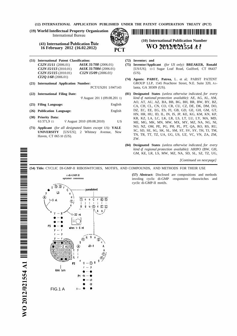
Figures 1A, IB, 1C, ID, and IE show c-di-GMP -II riboswitches. (A) Consensus
sequence and secondary structure model for c-di-GMP-II riboswitch aptamers (SEQ ID
NOs:49, 50, and 52). Nucleotides in circles are conserved in >97% of the representatives.
Other annotations are described in Figure 5 . (B) Wild-type (WT) 84 Cd RNA
encompassing the motif upstream of a possible virulence gene (SEQ ID NO:52).
Disruptive (M2) and restorative (M3) mutations are depicted, and sites of spontaneous
cleavage are derived from C. Asterisks identify nucleotides added to facilitate in vitro
transcription. (C) Denaturing polyacrylamide gel electrophoresis (PAGE) of WT 84 Cd
RNA in-line probing cleavage products. No reaction (NR); partial digestion with
ribonuclease (RNase) T l (Tl, cleaves after G residues); alkali ( OH, cleaves at all
linkages); incubation in the absence (-) or presence (+) of 10 mM c-di-GMP. Selected
RNase Tl digestion product bands are identified. (D) Plot of the fraction of RNA
remaining unbound versus the logarithm of the concentration of c-di-GMP present during
in-line probing. (E) KD values for 84 Cd binding to c-di-GMP and various di- and
mononucleotide analogs. A, adenosine; G, guanosine.
Figures 2A, 2B, 2C, and 2D show gene control by c-di-GMP-II riboswitches from
B. halodurans and C. difficile. (A) Wild-type (WT; SEQ ID NO:53), Jl/2 mutant (Ml) or
stem P3 mutants (M2 and M3) of the riboswitch from the B. halodurans BH063 1 locus
were fused to a β-galactosidase reporter gene and introduced into Bacillus subtilis for
analysis. (B) Plot of the normalized level of β-galactosidase activity from cells carrying
the WT, Ml, M2 or M3 reporter constructs depicted in A. A value of 1 equals 157 Miller
units. (C) Wild-type (WT; SEQ ID NO:54) or stem P3 mutants (Ml and M2) of the
riboswitch from the ompRlbaeS operon from C. difficile. Brackets identify the 146-
nucleotide marker transcript (M) in D. (D) Second messenger-dependent transcription
termination assays using WT and various mutant riboswitches as noted in C. FL, Term,
and M identify the full-length, terminated, and marker transcripts, respectively.
Figures 3A, 3B, 3C, 3D, and 3E show the architecture, mechanism, and activity of
a tandem riboswitch-ribozyme. (A) Structural features of the conjoined c-di-GMP-II
aptamer and group I ribozyme from C. difficile, and the importance of aptamer function in
45127589
R A self-splicing (SEQ ID NO:55). Two alternative base pair interactions proposed to be
important for allosteric function are outlined with alternating dashes and dots. (B) PAGE
separation of products generated by self-splicing assays. NR, no reaction; C-IVS and L-
IVS, circular and linear intervening sequences, respectively; Pre, 864-nucleotide precursor
RNA (Figure 6); 5Έ -3Έ , spliced exons; 3Έ *, 3 ' fragment generated by GTP attack at an
alternative site; Fragments designate additional RNA products presumably created by IVS
circularization. Data for all mutants is not shown. (C) Tandem c-di-GMP-II aptamer and
group I ribozyme arrangement. (D) Key features of the aptamer-ribozyme system,
including validated splice and GTP attack sites (SEQ ID NOs:55, 56, and 57). Alternative
base-pair interactions guiding allosteric function are enclosed in a dashed line oval and a
alternating dash dot line oval. (E) PAGE separation of products generated by self-splicing
assays. NR, no reaction; C-IVS and L-IVS, circular and linear intervening sequences,
respectively; Pre, 864-nucleotide precursor RNA (Figure 6); 5Έ -3Έ , spliced exons; 3Έ *,
3 ' fragment generated by GTP attack at an alternative site; Fragments designate additional
RNA products presumably created by IVS circularization.
Figures 4A, 4B, 4C, 4D and 4E show the rate constant modulation by c-di-GMP.
(A) Time course of [a- P]GTP attack at sites GTP or GTP in the absence or presence of
c-di-GMP. (B) Plot of the natural logarithm of the fraction of unprocessed or differently
processed RNA (pre-dp RNA) versus time for the reaction in (A). Values for fraction
processed were corrected for -50% of the precursor remaining after exhaustive incubation.
(C) Time course of the production of spliced exons (5Έ -3Έ ) or alternative GTP site
fragment (3Έ *) in the absence or presence of c-di-GMP. Annotations are as described for
Figure 2C. (D) Plot of the natural logarithm of the fraction of unprocessed or differently
processed RNA versus time for the reaction in (C), corrected as described in (B). (E)
Changes in splice product yields on introduction of c-di-GMP. (Left) Ratio of the number
of 3Έ * molecules versus the number of 5Έ -3Έ spliced exon molecules in the absence or
presence of c-di-GMP, respectively. (Right) Ratio of the numbers of 3Έ * molecules and
the numbers of 5Έ -3Έ spliced exon molecules in the absence or presence of c-di-GMP,
respectively.
Figure 5 shows comparison of the consensus sequences and structural models for
class I (left; SEQ ID NOs:58 and 59) and class II (right; SEQ ID NOs:49, 50, and 51) c-di-
GMP aptamers. Consensus model for c-di-GMP-I is as reported in Smith et al. 2009. The
internal bulge between P2 and P3 of c-di-GMP-II aptamers conforms to a kink-turn motif
(Klein et al. 2001; Winkler et al. 2001).
45127589 7
Figure 6 shows the sequence and secondary structure model for the 5 ' UTR and a
portion of the ORF containing the 84 Cd aptamer and group I intron upstream of the gene
at the CD3246 locus (SEQ ID NOs:122). The ribozyme has all the hallmarks of a typical
group I ribozyme, including stems PI through P10, and a U-G wobble base pair that
defines a typical 5 ' splice site (5 ' SS). Nucleotides comprising the putative atypical start
codon (UUG) for the associated ORF are depicted in circles. Other annotations are as
described in and for Figure 3D in the main text. This 864-nucleotide tandem construct was
used for most in vitro splicing assays.
Figure 7 is a c-di-GMP-dependent dose-response curve showing fraction of RNA
fully spliced (5Έ -3Έ ) vs log c (cyclic-di-GMP, M). The curve is for generation of ligated
exons (5Έ -3Έ ) by 5 ' P-labeled 864 Cd Tandem RNA incubated for 120 minutes in the
presence of various concentrations of the second messenger. The Sso value is the
concentration of c-di-GMP required to half-maximally stimulate production of ligated
exons. The line represents an expected curve for a 1-to-l interaction between ligand and
allosteric ribozyme.
Figures 8A, 8B, 8C, and 8D show in-line probing analysis of alternative base-
paired structures that may be responsible for allosteric control. (A) Sequence and
secondary structure model (SEQ ID NO:60). The structure depicted is the expected c-di-
GMP ligand-bound state, while the nucleotides enclosed in dashed line ovals identify the
alternative base-paired structure that would compete with formation of both the aptamer
and ribozyme PI stems. Sites of spontaneous cleavage, including sites of structure
modulation, are noted with circles, diamonds, and squared enclosing nucleotides.
Annotations are as described in the brief description to Figures 1A and 5 . (B, C, D)
Similar analyses of mutant RNAs as indicated (SEQ ID NOs:61, 62, and 63, respectively).
Figures 9A and 9B show the junctions of ribozyme self-circularization products.
(A) Products due to circularization after GTP attack at the 5 ' SS (GTPi), followed by 5'
exon attack at the 3 ' SS (nucleotide 667) or one nucleotide downstream (nucleotide 668).
SEQ ID NOs:64, 65, and 66. (B) Products due to circularization after GTP attack at
nucleotide 670, which corresponds to the alternative attack site (GTP2) . SEQ ID NOs:67,
68, 69, and 70. Nucleotides at the junctions are numbered, and the number of
representatives of each circularization product observed among 19 clones sequenced are
noted in parentheses.
Figures 10A, 10B, and IOC show the proposed mechanism for allosteric ribozyme-
mediated gene control. (A) Precursor mRNA with the start codon sequestered by the
451 2 7589
ribozyme P10 stem (SEQ ID NOs:72 and 73). (B) RNA processed in the presence of GTP
and c-di-GMP unmasks the start codon and creates a perfect ribosome binding site (SEQ
ID NO:74). (C) RNA processed in the presence of GTP alone lacks a ribosome binding
site (SEQ ID NO:75).
DETAILED DESCRIPTION OF THE INVENTION
The disclosed methods, compounds, and compositions can be understood more
readily by reference to the following detailed description of particular embodiments and
the Examples included therein and to the Figures and their previous and following
description.
Described herein are the interactions of two small RNA molecules and two larger
RNA molecules that together influence the function of a self-splicing ribozyme, a structure
many biologists had believed had no role other than to reproduce itself. However, in the
pathogenic stomach bacterium Clostridium difficile, this RNA structure acts as a sort of
sensor to help regulate the expression of genes, probably to help the bacterium manipulate
human cells. The disclosed compositions and methods relate to a new class of
riboswitches that sense the bacterial second messenger cyclic di-GMP. In the pathogen
Clostridium difficile, three of these riboswitches are present. One resides adjacent to a
group I self-splicing ribozyme, and the biochemical data described herein prove this RNA
architecture represents the first natural example of an allosteric ribozyme. Ligand binding
controls splicing of an mRNA for a virulence gene. Therefore, this new riboswitch class is
a useful drug target for novel antibiotics.
Messenger RNAs are typically thought of as passive carriers of genetic information
that are acted upon by protein- or small RNA-regulatory factors and by ribosomes during
the process of translation. It was discovered that certain mRNAs carry natural aptamer
domains and that binding of specific metabolites directly to these RNA domains leads to
modulation of gene expression. In particular, it as been discovered certain cyclic di-GMP-
responsive riboswitches comprise a cyclic di-GMP-II motif. Natural riboswitches exhibit
two surprising functions that are not typically associated with natural RNAs. First, the
mRNA element can adopt distinct structural states wherein one structure serves as a
precise binding pocket for its target metabolite. Second, the metabolite-induced allosteric
interconversion between structural states causes a change in the level of gene expression
by one of several distinct mechanisms. Riboswitches typically can be dissected into two
separate domains: one that selectively binds the target (aptamer domain) and another that
45127589 9
influences genetic control (expression platform). It is the dynamic interplay between these
two domains that results in metabolite-dependent allosteric control of gene expression.
Distinct classes of riboswitches have been identified and are shown to selectively
recognize activating compounds (referred to herein as trigger molecules). For example,
coenzyme Bi2, glycine, thiamine pyrophosphate (TPP), and flavin mononucleotide (FMN)
activate riboswitches present in genes encoding key enzymes in metabolic or transport
pathways of these compounds. The aptamer domain of each riboswitch class conforms to a
highly conserved consensus sequence and structure. Thus, sequence homology searches
can be used to identify related riboswitch domains. Riboswitch domains have been
discovered in various organisms from bacteria, archaea, and eukarya.
Riboswitches are genetic regulatory elements composed solely of RNA that bind
metabolites and control gene expression commonly without the involvement of protein
factors (Breaker RR. Riboswitches: from ancient gene-control systems to modern drug
targets. Future Microbiol 2009; 4:771-773). Most simple riboswitches are composed of an
aptamer domain and an expression platform, where the aptamer functions as a receptor for
a specific metabolite and the expression platform modulates the expression of one or more
genes in a ligand-dependent fashion (Barrick et al. The distributions, mechanisms, and
structures of metabolite-binding riboswitches. Genome Biol 2007; 8:R239; Dambach et al.
Expanding roles for metabolite-sensing regulatory RNAs. Curr Opin Microbiol 2009;
12:161-1 69). Riboswitches are usually found in the 5' untranslated regions (UTRs) of
bacterial mRNAs and often control gene expression in cis either at the level of
transcription or translation, although other regulatory mechanisms are also known (Roth et
al. The structural and functional diversity of metabolite-binding riboswitches. Annu Rev
Biochem 2009; 78:305-334). In most cases, metabolite binding triggers a structural
rearrangement that affects the formation of either a terminator stem or a base-paired
element that occludes the ribosome binding site. In addition, there is a known example of a
trans-acting riboswitch (Loh et al. A trans-acting riboswitch controls expression of the
virulence regulator PrfA in listeria monocytogenes. Cell 2009; 139:770-779) as well as
eukaryotic riboswitches (Wachter A . Riboswitch-mediated control of gene expression in
eukaryotes. RNA Biol 2010; 7:67-76) that modulate expression by controlling alternative
mRNA spicing in algae (Croft et al. Thiamine biosynthesis in algae is regulated by
riboswitches. Proc Natl Acad Sci USA 2007; 104:20770-20775), plants (Wachter et al.
Riboswitch control of gene expression in plants by splicing and alternative 3' end
processing of mRNAs. Plant Cell 2007; 19:3437-3450), and fungi (Cheah et al. Control of
4512 7589 f
alternative RNA splicing and gene expression by eukaryotic riboswitches. Nature 2007;
447:497-500).
Comparative sequence analysis methods have been developed for novel riboswitch
class discovery (Rodionov et al. Regulation of lysine biosynthesis and transport genes in
bacteria: yet another RNA riboswitch? Nucleic Acids Res 2003; 31:6748-6757; Barrick et
al. New RNA motifs suggest an expanded scope for riboswitches in bacterial genetic
control. Proc Natl Acad Sci USA 2004; 101:6421-6426; Weinberg et al, 2007). These
techniques involve computational searches through genomic and metagenomic databases
for sequences that are conserved both in their primary and secondary structures (Yao et al.
A computational pipeline for high-throughput discovery of cis-regulatory noncoding RNA
in prokaryotes. PLoS Comput Biol 2007; 3:el26; Tseng et al. Finding non-coding RNAs
through genome-scale clustering. J Bioinform Comput Biol 2009; 7:373-388). Through
one of these searches, the glnA motif and the Downstream-peptide motif (Figure 29) were
discovered in cyanobacteria and marine metagenomic sequences (Weinberg et al.,
Genome Biol 2010; 11:R31).
Structured noncoding RNAs perform many functions that are essential for protein
synthesis, RNA processing, and gene regulation. Structured RNAs can be detected by
comparative genomics, in which homologous sequences are identified and inspected for
mutations that conserve RNA secondary structure. By applying a comparative genomics-
based approach to genome and metagenome sequences from bacteria and archaea, 104
structured RNA motifs were identified. Three metabolite-binding RNA motifs were
validated, including one that binds the coenzyme S-adenosylmethionine, and a further nine
metabolite-binding RNA motifs were identified. New-found czs-regulatory RNA motifs
are implicated in photosynthesis or nitrogen regulation in cyanobacteria, purine and one-
carbon metabolism, stomach infection by Helicobacter, and many other physiological
processes. A riboswitch termed crcB is represented in both bacteria and archaea. Another
RNA motif controls gene expression from 3' untranslated regions (UTRs) of mRNAs,
which is unusual for bacteria. Many noncoding RNAs that act in trans are also revealed,
and several of the noncoding RNA motifs are found mostly or exclusively in metagenome
DNA sequences. This work greatly expands the variety of highly-structured noncoding
RNAs known to exist in bacteria and archaea.
Following the discovery of complex riboswitch arrangements (Mandal et al.
Science 306, 275 (2004); Welz and Breaker, RNA 13, 573 (2007); Sudarsan, et al. Science
13, 300 (2006); and E. Poiata, et al. RNA 15, 2046 (2009)) and examples of riboswitches
45127589 \ \
that control alternative splicing in eukaryotes (Cheah et al. Nature 447, 497 (2007);
Wachter et al. Plant Cell 19, 3437 (2007); Bocobza, et al. Genes Dev. 21, 2874 (2007);
Croft et al. Proc. Natl. Acad. Sci. USA 104, 20770 (2007)) it was discovered that some of
the many thousands of known group I or group II self-splicing ribozymes participate with
other RNA motifs to purposefully regulate gene expression. The findings disclosed herein
demonstrate that an allosteric ribozyme architecture exists naturally, and that an all-RNA
component network (riboswitch, c-di-GMP, ribozyme, GTP) can be used to perform
complex sensory and enzymatic functions.
It was realized that some organisms can productively harness group I ribozymes by
thinking about the observation that bacteriophage ribozyme splicing is diminished in a
bacterial host when ribosome function is inhibited (Sandergren and Sjoberg, J . Bacteriol.
189, 980-990 (2007)). Also, it was realized that the genomic locations and gene
associations of some group I ribozymes are conserved in evolutionarily distant species
(Nielsen and Johansen, RNA Biol. 6, 375 (2009)) strongly implies useful rather than
purely selfish functions. Interestingly, of ten group I ribozymes present in C. difficile 630,
nine are associated with a transposase gene (Table 6 in Example 1), which facilitates
mobility of selfish genetic elements. Only the allosteric ribozyme described herein lacks a
transposase gene, which led to the realization that this representative is not a selfish RNA
element, but has a location and a function that benefits the host.
RNA engineering methods exist to couple aptamer and ribozyme domains to create
allosteric ribozymes (Breaker, Curr. Opin. Biotechnol. 13, 3 1 (2002); Silverman, RNA 9,
377 (2003)). Interestingly, these methods were previously used to generate an allosteric
group I ribozyme construct that controls gene expression when theophylline is added to
cell culture (Thompson et al. BCM Biotechnol. 2, 2 1 (2002)). In these engineered allosteric
ribozymes, theophylline aptamers were grafted to internal stems (P5 and P6) such that the
presence of ligand would stabilize these important substructures of the ribozyme. In
contrast, the natural allosteric group I ribozyme positions the c-di-GMP aptamer to
influence folding most directly at the 5 ' SS, and indirectly near the 3 ' SS. However, the
structural complexity of large ribozymes provides numerous additional locations wherein
aptamer function can allosterically control catalysis.
Bacteria naturally exploit tandem riboswitch architectures to create more complex
gene control elements (Mandal et al. Science 306, 275, 2004; Welz et al. RNA 13, 573,
2007; Sudarsan et al, Science 314, 300, 2006; Poiata et al. RNA 15, 2046, 2009). These
findings add to this complexity by demonstrating how two ligand responsive RNAs, a self-
45127589
splicing ribozyme and a riboswitch aptamer, collaborate to function as an allosteric RNA
requiring two RNA compounds (GTP and c-di-GMP) to promote splicing. This conjoined
riboswitch-ribozyme system validates a prediction made more than 20 years ago (Shub et
al. Cold Spring Harb. Symp. Quant. Biol. 52, 193, 1987) that some group I ribozymes
could be controlled by nucleotide-derived alarmones. Also, this RNA is a natural mimic of
an engineered ribozyme that controls splicing and gene expression in response to
theophylline binding (Thompson et al. BMC Biotechnol., 2, 21, 2002).
Some group I ribozymes can independently function as riboswitches that sense
guanosine or one of its phosphorylated derivatives, because sufficient levels of the
attacking nucleophile must be present for efficient splicing (Ames and Breaker, In: The
Chemical Biology of Nucleic Acids, G. Meyer, ed., Wiley-VCH; Breaker, In: The RNA
World, 4th ed., Gesteland, Cech, Atkins, eds., Cold Spring Harbor Laboratory Press). The
tandem riboswitch-ribozyme examined in this study could constitute a two-input gene
control system that naturally reads the concentration of GTP and c-di-GMP, and trigger
splicing accordingly. It is interesting to note that GTP is used as the immediate precursor
for the synthesis of c-di-GMP (Hengge, Nat. Rev. Microbiol. 7, 263 (2009)), and
overproduction of this second messenger causes substantial decreases in GTP
concentrations in vivo (Simm et al. Mol. Microbiol. 53, 1123 (2004)). The biological
utility of this tandem riboswitch-ribozyme may be used to ensure expression of the
associated gene only when both compounds are present in sufficient quantities.
As the number of riboswitch discoveries expands, there are increasing
opportunities to identify rare combinations of RNA structures that arrange aptamers in
tandem or with other functional domains such as ribozymes. The allosteric self-splicing
system described herein is one representative of many sophisticated RNA devices that are
used by cells to carry out specialized sensory and catalytic functions.
A. General Organization of Riboswitch RNAs
Bacterial riboswitch RNAs are genetic control elements that are located primarily
within the 5 '-untranslated region (5 '-UTR) of the main coding region of a particular
mRNA. Structural probing studies (discussed further below) reveal that riboswitch
elements are generally composed of two domains: a natural aptamer (T. Hermann, D. J .
Patel, Science 2000, 287, 820; L. Gold, et al., Annual Review of Biochemistry 1995, 64,
763) that serves as the ligand-binding domain, and an 'expression platform' that interfaces
with RNA elements that are involved in gene expression {e.g. Shine-Dalgarno (SD)
elements; transcription terminator stems). These conclusions are drawn from the
45127589
observation that aptamer domains synthesized in vitro bind the appropriate ligand in the
absence of the expression platform (see Examples 2, 3 and 6 of U.S. Application
Publication No. 2005-0053951). Moreover, structural probing investigations indicate that
the aptamer domain of most riboswitches adopts a particular secondary- and tertiary-
structure fold when examined independently, that is essentially identical to the aptamer
structure when examined in the context of the entire 5 ' leader RNA. This indicates that, in
many cases, the aptamer domain is a modular unit that folds independently of the
expression platform (see Examples 2, 3 and 6 of U.S. Application Publication No. 2005-
0053951).
Ultimately, the ligand-bound or unbound status of the aptamer domain is
interpreted through the expression platform, which is responsible for exerting an influence
upon gene expression. The view of a riboswitch as a modular element is further supported
by the fact that aptamer domains are highly conserved amongst various organisms (and
even between kingdoms as is observed for the TPP riboswitch), (N. Sudarsan, et al, RNA
2003, 9, 644) whereas the expression platform varies in sequence, structure, and in the
mechanism by which expression of the appended open reading frame is controlled. For
example, ligand binding to the TPP riboswitch of the tenA mRNA of B. subtilis causes
transcription termination (A. S. Mironov, et al., Cell 2002, 111, 747). This expression
platform is distinct in sequence and structure compared to the expression platform of the
TPP riboswitch in the thiM mRNA from E. coli, wherein TPP binding causes inhibition of
translation by a SD blocking mechanism (see Example 2 of U.S. Application Publication
No. 2005-0053951). The TPP aptamer domain is easily recognizable and of near identical
functional character between these two transcriptional units, but the genetic control
mechanisms and the expression platforms that carry them out are very different.
Aptamer domains for riboswitch RNAs typically range from ~70 to 170 nt in
length (Figure 11 of U.S. Application Publication No. 2005-0053951). This observation
was somewhat unexpected given that in vitro evolution experiments identified a wide
variety of small molecule-binding aptamers, which are considerably shorter in length and
structural intricacy (T. Hermann, D. J . Patel, Science 2000, 287, 820; L. Gold, et al,
Annual Review of Biochemistry 1995, 64, 763; M. Famulok, Current Opinion in Structural
Biology 1999, 9, 324). Although the reasons for the substantial increase in complexity and
information content of the natural aptamer sequences relative to artificial aptamers
remains to be proven, this complexity is believed required to form RNA receptors that
function with high affinity and selectivity. Apparent Κ , values for the ligand-riboswitch
45127589
complexes range from low nanomolar to low micromolar. It is also worth noting that some
aptamer domains, when isolated from the appended expression platform, exhibit improved
affinity for the target ligand over that of the intact riboswitch. (~10 to 100-fold) (see
Example 2 of U.S. Application Publication No. 2005-0053951). Presumably, there is an
energetic cost in sampling the multiple distinct RNA conformations required by a fully
intact riboswitch RNA, which is reflected by a loss in ligand affinity. Since the aptamer
domain must serve as a molecular switch, this might also add to the functional demands on
natural aptamers that might help rationalize their more sophisticated structures.
Riboswitches must be capable of discriminating against compounds related to their
natural ligands to prevent undesirable regulation of metabolic genes. However, it is
possible to generate analogs that trigger riboswitch function and inhibit bacterial growth,
as has been demonstrated for riboswitches that normally respond to lysine (Sudarsan
2003) and thiamine pyrophosphate (Sudarsan 2006).
B. Riboswitch Regulation of Gene Expression
Riboswitches control expression, effect, and function of RNA molecules in a
variety of ways. For example, riboswitches can regulate transcription (or full
transcription) of RNA molecules by, for example, causing premature termination of
transcription, such as by forming a terminator signal in the RNA molecule or altering the
coordination of transcription and translation of the RNA molecule. Riboswithes can also
regulate tranlslation of RNA molecules by, for example, blocking or affecting binding of
translation enzymes, proteins, or factors and/or altering the coordination of transcription
and translation of the RNA molecule. Riboswitches can also affect expression of RNA
molecules by altering processing of the RNA molecule. For example, riboswitches can
modulate cleavage of the RNA molecule, splicing of the RNA molecules, latering stability
of the RNA molecules (through regulation of addition or effect of RNA stability
sequences, for example) and/or processing of the RNA molecules. Natural and engineered
examples of such rregulation by riboswitches are known and can be adapted for use with
the disclosed riboswitches.
Bacteria primarily make use of two methods for termination of transcription.
Certain genes incorporate a termination signal that is dependent upon the Rho protein, (J.
P. Richardson, Biochimica et Biophysica Acta 2002, 1577, 251). while others make use of
Rho-independent terminators (intrinsic terminators) to destabilize the transcription
elongation complex (I. Gusarov, E. Nudler, Molecular Cell 1999, 3, 495; E. Nudler, M. E.
Gottesman, Genes to Cells 2002, 7, 755). The latter RNA elements are composed of a GC-
45127589 ζ
rich stem-loop followed by a stretch of 6-9 uridyl residues. Intrinsic terminators are
widespread throughout bacterial genomes (F. Lillo, et al, 2002, 18, 971), and are typically
located at the 3 '-termini of genes or operons. Interestingly, an increasing number of
examples are being observed for intrinsic terminators located within 5 ' -UTRs.
Among the wide variety of genetic regulatory strategies employed by bacteria there
is a growing class of examples wherein RNA polymerase responds to a termination signal
within the 5 ' -UTR in a regulated fashion (T. M. Henkin, Current Opinion in Microbiology
2000, 3, 149). During certain conditions the RNA polymerase complex is directed by
external signals either to perceive or to ignore the termination signal. Although
transcription initiation might occur without regulation, control over mRNA synthesis (and
of gene expression) is ultimately dictated by regulation of the intrinsic terminator.
Presumably, one of at least two mutually exclusive mRNA conformations results in the
formation or disruption of the RNA structure that signals transcription termination. A
trans-acting factor, which in some instances is a RNA (F. J . Grundy, et al, Proceedings of
the National Academy of Sciences of the United States of America 2002, 99, 11121; T. M.
Henkin, C. Yanofsky, Bioessays 2002, 24, 700) and in others is a protein (J. Stulke,
Archives of Microbiology 2002, 177, 433), is generally required for receiving a particular
intracellular signal and subsequently stabilizing one of the RNA conformations.
Riboswitches offer a direct link between RNA structure modulation and the metabolite
signals that are interpreted by the genetic control machinery.
Riboswitches can affect or regulate expression of RNA molecules by affecting
processing of the RNA molecules. For example, regulation of splicing can affect
processing of an RNA in which splicing is regulated. For example, a self-spicing
ribozyme regulated by a riboswitch (the combination can be referred to as a riboswitch
ribozyme) can be used to regulate formation of a functional (or non-functional) RNA
through self-splicing.
As another example example, an intron in the RNA can include an RNA
processing signal or site. Splicing of the RNA can result in elimination of the processing
signal or site. For example, a transcription termination signal or RNA cleavage site in the
3' UTR of a mRNA can be deleted from the RNA if it resides in an intron that is spliced
out of the RNA. Regulation of the splicing of that intron by a riboswitch as described
herein can thus affect the processing of the RNA. As another example, an RNA
processing signal or site can be created via splicing of an intron or different elements of an
RNA processing system, signal or site can be brought into or taken out of an operable
45127589
arrangement by splicing of an intron. As another example, an RNA processing signal or
site can be brought into or taken out of an operable proximity with other elements of the
RNA.
RNA processing can also be affected directly by a riboswitch without mediation by
regulation of splicing. For example, an RNA processing signal or site can be in the
expression platform domain of a riboswitch. In this way, the alteration in the structural
relationship of the expression platform (and thus of the RNA processing signal or site) by
activation of the riboswitch can affect processing by affecting the ability of the RNA
processing signal or site to operate.
The riboswitch can affect RNA processing. By "affect RNA processing" is meant
that the riboswitch can either directly or indirectly (via regulation of splicing, for example)
act upon RNA to allow, stimulate, reduce or prevent RNA processing to take place. This
can include, for example, allowing any processing to take place. This can increase or
decrease processing fully or partially to any degree compared to the number of processing
events that would have taken place without the riboswitch.
RNA processing can include, for example, transcription termination, formation of
the 3' terminus of the RNA, polyadenylation, and degradation or turnover of the RNA. As
used herein, and RNA processing signal or site is a sequence, structure or location in an
RNA that mediates, signals or is required for an RNA processing event or condition. For
example, certain sequences or structures can signal transcription termination, RNA
cleavage or polyadenylation.
The riboswitch can activate or repress splicing. By "activate splicing" is meant that
the riboswitch can either directly or indirectly act upon RNA to allow splicing to take
place. This can include, for example, allowing any splicing to take place (such as a single
splice versus no splice) or allowing alternative splicing to take place. This can increase
splicing fully or partially to any degree compared to the number of splicing events that
would have taken place without the riboswitch.
By "repress splicing" is meant that the riboswitch can either directly or indirectly
act upon RNA to suppress splicing. This can include, for example, preventing any splicing
or reducing splicing from taking place (such as no splice versus a single splice) or
preventing or reducing alternative splicing from taking place. This can decrease
alternative splicing fully or partially to any degree compared to the number of alternative
splicing events that would have taken place without the riboswitch.
45127589 17
The riboswitch can activate or repress alternative splicing. By "activate alternative
splicing" is meant that the riboswitch can either directly or indirectly act upon RNA to
allow alternative splicing to take place. This can increase alternative splicing fully or
partially to any degree compared to the number of alternative splicing events that would
have taken place without the riboswitch.
By "repress alternative splicing" is meant that the riboswitch can either directly or
indirectly act upon RNA to suppress alternative splicing. This can decrease alternative
splicing fully or partially to any degree compared to the number of alternative splicing
events that would have taken place without the riboswitch.
The riboswitch can affect expression of a protein encoded by the RNA. For
example, regulation of splicing or alternative splicing can affect the ability of the RNA to
be translated, alter the coding region, or alter the translation initiation or termination.
Alternative splicing can, for example, cause a start or stop codon (or both) to appear in the
processed transcript that is not present in normally processed transcripts. As another
example, alternative splicing can cause the normal start or stop codon to be removed from
the processed transcript. A useful mode for using riboswitch-regulated splicing to regulate
expression of a protein encoded by an RNA is to introduce a riboswitch in an intron in the
5' untranslated region of the RNA and include or make use of a start codon in the intron
such that the start codon in the intron will be the first start codon in the alternatively
spliced RNA. Another useful mode for using riboswitch-regulated splicing to regulate
expression of a protein encoded by an RNA is to introduce a riboswitch in an intron in the
5' untranslated region of the RNA and include or make use of a short open reading frame
in the intron such that the reading frame will appear first in the alternatively spliced RNA.
The RNA molecule can have a branched structure. For example, in the fungal TPP
riboswitch (Cheah 2007), when TPP concentration is low, the newly transcribed mRNA
adopts a structure that occludes the second 5 ' splice site, while leaving the branch site
available for splicing. Pre-mRNA splicing from the first 5 ' splice site leads to production
of the 1-3 form of mRNA and expression of the NMT1 protein. When TPP concentration
is high, ligand binding to the TPP aptamer causes allosteric changes in RNA folding to
increase the structural flexibility near the second 5 ' splice site and to occlude nucleotides
near the branch site.
Translation of RNA molecules can be regulated by riboswitches in a variety of
ways. For example, a functionally significant sequence in an RNA molecule can be
blocked or made accessible through action of a riboswitch. For example, the sequence of
45127589
the aptamer and control strands of an aptamer domain can be adapted so that the control
strand is complementary to a functionally significant sequence in an expression platform.
For example, the control strand can be adapted to be complementary to the Shine-
Dalgarno sequence of an RNA such that, upon formation of a stem structure between the
control strand and the SD sequence, the SD sequence becomes inaccessible to ribosomes,
thus reducing or preventing translation initiation. An example of this for of regulation
where activation of a riboswitch causes inhibition of translation by a SD blocking
mechanism is described in Example 2 of U.S. Application Publication No. 2005-0053951.
As another example, the control strand can be adapted to be complementary to the
initiation codon (or the region of the initiation codon) of an RNA such that, upon
formation of a stem structure between the control strand and the initiation codon, the
initiation codon becomes inaccessible to ribosomes, thus reducing or preventing
translation initiation. As another example, the control strand can be adapted to be
complementary to the binding site of a translation factor such that, upon formation of a
stem structure between the control strand and the binding site, the binding site becomes
inaccessible to the translation factor, thus reducing or preventing translation initiation.
C. Features of and Methods of Using Cyclic di-GMP-responsive Riboswitches
Disclosed are methods and compositions for altering gene expression of genes by
affecting cyclic di-GMP-responsive riboswitches operably linked to the genes, where the
riboswitch comprises a cyclic di-GMP-II motif. For example, the methods can comprise
bringing into contact a compound and a cell, where the compound affects the riboswitch.
The cell can comprise a gene encoding an RNA comprising a cyclic di-GMP-responsive
riboswitch. The riboswitch can comprise a cyclic di-GMP-II motif.
In some forms, the cell can have been identified as being in need of altered gene
expression. In some forms, the cell can be a bacterial cell. In some forms, the cell can be
a Clostridium, Deinococcus, or Bacillus cell. In some forms, the compound kills or
inhibits the growth of the bacterial cell. In some forms, the compound and the cell can be
brought into contact by administering the compound to a subject. In some forms, the cell
is a bacterial cell in the subject and the compound kills or inhibits the growth of the
bacterial cell. In some forms, the subject has a bacterial infection. In some forms, the
compound can be administered in combination with another antimicrobial compound. In
some forms, the compound inhibits bacterial growth in a biofilm.
Also disclosed are regulatable gene expression constructs comprising cyclic di-
GMP-responsive riboswitches operably linked to coding regions, where the riboswitch
45127589 (
comprises a cyclic di-GMP-II motif. For example, the disclosed constructs can comprise a
nucleic acid molecule encoding an R A comprising a riboswitch operably linked to a
coding region, where the riboswitch regulates expression of the RNA, where the
riboswitch and coding region are heterologous, where the riboswitch is a cyclic di-GMP-
responsive riboswitch, and where the riboswitch comprises a cyclic di-GMP-II motif. As
used herein, a riboswitch and coding region can be said to be heterologous if they are not
operably linked in nature. For example, riboswitches and coding regions from different
sources, such as different genes, different chromosomes, different organisms, and the like,
can be said to be heterologous. Also disclosed are riboswitches where the riboswitch is a
non-natural derivative of a naturally-occurring a cyclic di-GMP-responsive riboswitch,
where the naturally-occurring riboswitch comprises a cyclic di-GMP-II motif. Also
disclosed are riboswitch ribozymes comprising a riboswitch aptamer domain operably
linked to a self-splicing ribozyme, where the aptamer is comprised of the cyclic di-GMP-II
motif.
In some forms, the riboswitch can comprise an aptamer domain and an expression
platform domain, where the aptamer domain and the expression platform domain are
heterologous, where the aptamer is comprised of the cyclic di-GMP-II motif. In some
forms, the riboswitch can comprise two or more aptamer domains and an expression
platform domain, where at least one of the aptamer domains and the expression platform
domain are heterologous, where at least one of the aptamer domains is comprised of the
cyclic di-GMP-II motif. In some forms, at least two of the aptamer domains can exhibit
cooperative binding. In some forms, the riboswitch can comprise the consensus structure
of Figure 1A or 5 . As used herein, an aptamer domain and an expression platform domain
can be said to be heterologous if they are not operably linked in nature. For example,
aptamer domains and expression platform domains from different sources, such as
different riboswitches, different genes, different chromosomes, different organisms, and
the like, can be said to be heterologous.
In some forms, the riboswitch can comprise an aptamer domain and an expression
platform domain, where the aptamer domain is derived from a naturally-occurring cyclic
di-GMP-responsive riboswitch. In some forms, the aptamer domain can be the aptamer
domain of a naturally-occurring cyclic di-GMP-responsive riboswitch. In some forms, the
aptamer domain can have the consensus structure of an aptamer domain of the naturally-
occurring riboswitch. In some forms, the aptamer domain can consist of only base pair
conservative changes of the naturally-occurring riboswitch.
45127589 0
In some forms, the aptamer domain can comprise a PI stem, where the PI stem
comprises an aptamer strand and a control strand, where the expression platform domain
comprises a regulated strand, and where the regulated strand, the control strand, or both
have been designed to form a stem structure. In some forms, the aptamer domain can
comprise a control stem, where the control stem comprises an aptamer strand and a control
strand, where the expression platform domain comprises a regulated strand, and where the
regulated strand, the control strand, or both have been designed to form a stem structure.
In some forms, the riboswitch can comprise an aptamer domain and an expression
platform domain, where the aptamer domain and the expression platform domain are
heterologous, and where the aptamer is comprised of the cyclic di-GMP-II motif. In some
forms, the riboswitch can be activated by a trigger molecule, where the riboswitch
produces a signal when activated by the trigger molecule.
In some forms, the aptamer domain can comprise a control stem, where the control
stem comprises an aptamer strand and a control strand, where the ribozyme comprises a
regulated strand, and where the regulated strand, the control strand, or both have been
designed to form a stem structure. In some forms, the aptamer domain and the ribozyme
can be heterologous. In some forms, the riboswitch ribozyme can be operatively linked to
a coding region, where the riboswitch ribozyme and the coding region are heterologous.
As used herein, a riboswitch ribozyme and coding region can be said to be heterologous if
they are not operably linked in nature. For example, riboswitch ribozymes and coding
regions from different sources, such as different genes, different chromosomes, different
organisms, and the like, can be said to be heterologous.
Also disclosed are methods and compositions for detecting a compound of interest.
The method can comprise bringing into contact a sample and a riboswitch, where the
riboswitch produces a signal when the sample contains the compound of interest. The
riboswitch can be activated by the compound of interest and the riboswitch produces a
signal when activated by the compound of interest. The riboswitch is a cyclic di-GMP-
responsive riboswitch, where the riboswitch comprises a cyclic di-GMP-II motif.
In some forms, the riboswitch can change conformation when activated by the
compound of interest, where the change in conformation produces a signal via a
conformation dependent label. In some forms, the riboswitch can change conformation
when activated by the compound of interest, where the change in conformation causes a
change in expression of an R A linked to the riboswitch, and where the change in
45127589 2 1
expression produces a signal. In some forms, the signal can be produced by a reporter
protein expressed from the RNA linked to the riboswitch.
Also disclosed are methods comprising (a) testing a compound for altering gene
expression of a gene encoding an RNA comprising a riboswitch, and (b) altering gene
expression by bringing into contact a cell and a compound that altered gene expression in
step (a). The alteration can be via the riboswitch. The riboswitch is a cyclic di-GMP-
responsive riboswitch, where the riboswitch comprises a cyclic di-GMP-II motif. The cell
can comprise a gene encoding an RNA comprising a riboswitch, where the compound
inhibits expression of the gene by binding to the riboswitch.
Also disclosed are methods and compositions for identifying riboswitches. The
method can comprise assessing in-line spontaneous cleavage of an RNA molecule in the
presence and absence of a compound, where the RNA molecule is encoded by a gene
regulated by the compound, where a change in the pattern of in-line spontaneous cleavage
of the RNA molecule indicates a riboswitch, where the RNA comprises a cyclic di-GMP-
responsive riboswitch or a derivative of a cyclic di-GMP-responsive riboswitch. The
riboswitch can comprise a cyclic di-GMP-II motif and the compound can be cyclic di
GMP.
Further disclosed are methods of killing or inhibiting the growth of bacteria. The
method can comprise, for example, contacting the bacteria with a compound identified
and/or confirmed by any of the methods disclosed herein. Further disclosed are methods
of killing bacteria. The method can comprise, for example, contacting the bacteria with a
compound identified and/or confirmed by any of the methods disclosed herein. The
disclosed methods can be performed in a variety of ways and using different options or
combinations of features and components. As an example, a gel-based assay or a chip-
based assay can be used to determine if the test compound interacts with, modulates,
inhibits, blocks, deactivates, and/or activates the riboswitch, such as a cyclic diGMP
riboswitch. The test compound can interact in any manner, such as, for example, via van
der Waals interactions, hydrogen bonds, electrostatic interactions, hydrophobic
interactions, or a combination. The riboswitch, such as a cyclic diGMP riboswitch, can
comprise an RNA cleaving ribozyme, for example. A fluorescent signal can be generated
when a nucleic acid comprising a quenching moiety is cleaved. Molecular beacon
technology can be employed to generate the fluorescent signal. The methods disclosed
herein can be carried out using a high throughput screen.
45127589 22
Also disclosed are compositions and methods for selecting and identifying
compounds that can activate, deactivate or block a riboswitch, such as a cyclic diGMP
riboswitch. Activation of a riboswitch, such as a cyclic di-GMP riboswitch, refers to the
change in state of the riboswitch upon binding of a trigger molecule. A riboswitch, such
as a cyclic di-GMP riboswitch, can be activated by compounds other than the trigger
molecule and in ways other than binding of a trigger molecule. The term trigger molecule
is used herein to refer to molecules and compounds that can activate a riboswitch. This
includes the natural or normal trigger molecule for the riboswitch and other compounds
that can activate the riboswitch. Natural or normal trigger molecules are the trigger
molecule for a given riboswitch in nature or, in the case of some non-natural riboswitches,
the trigger molecule for which the riboswitch was designed or with which the riboswitch
was selected (as in, for example, in vitro selection or in vitro evolution techniques). Non-
natural trigger molecules can be referred to as non-natural trigger molecules.
Deactivation of a riboswitch refers to the change in state of the riboswitch, such as
a cyclic di-GMP riboswitch, when the trigger molecule is not bound. A riboswitch, such
as a cyclic di-GMP riboswitch, can be deactivated by binding of compounds other than the
trigger molecule and in ways other than removal of the trigger molecule. Blocking of a
riboswitch, such as a cyclic di-GMP riboswitch, refers to a condition or state of the
riboswitch where the presence of the trigger molecule does not activate the riboswitch.
Activation of a riboswitch, such as a cyclic di-GMP riboswitch, can be assessed in any
suitable manner. For example, the riboswitch, such as a cyclic di-GMP riboswitch, can be
linked to a reporter RNA and expression, expression level, or change in expression level
of the reporter RNA can be measured in the presence and absence of the test compound.
As another example, the riboswitch, such as a cyclic di-GMP riboswitch, can include a
conformation dependent label, the signal from which changes depending on the activation
state of the riboswitch, such as a cyclic di-GMP riboswitch. Such a riboswitch preferably
uses an aptamer domain from or derived from a naturally occurring riboswitch. As can be
seen, assessment of activation of a riboswitch can be performed with the use of a control
assay or measurement or without the use of a control assay or measurement. Methods for
identifying compounds that deactivate a riboswitch can be performed in analogous ways.
Also disclosed are method of inhibiting growth of a cell, such as a bacterial cell,
that is in a subject. The method can comprise administering to the subject an effective
amount of a compound identified and/or confirmed in any of the methods described
herein. This can result in the compound being brought into contact with the cell. The
45127589 3
subject can have, for example, a bacterial infection, and the bacterial cells can be the cells
to be inhibited by the compound. The bacteria can be any bacteria, such as bacteria from
the genus Clostridium, Deinococcus, or Bacillus, for example. Bacterial growth can also
be inhibited in any context in which bacteria are found. For example, bacterial growth in
fluids, biofilms, and on surfaces can be inhibited. The compounds disclosed herein can be
administered or used in combination with any other compound or composition. For
example, the disclosed compounds can be administered or used in combination with
another antimicrobial compound.
It is to be understood that the disclosed methods and compositions are not limited
to specific examples unless otherwise specified, and, as such, can vary. It is also to be
understood that the terminology used herein is for the purpose of describing particular
embodiments only and is not intended to be limiting.
Materials
Disclosed are materials, compositions, and components that can be used for, can be
used in conjunction with, can be used in preparation for, or are products of the disclosed
methods and compositions. These and other materials are disclosed herein, and it is
understood that when combinations, subsets, interactions, groups, etc. of these materials
are disclosed that while specific reference to each of various individual and collective
combinations and permutation of these compounds can not be explicitly disclosed, each is
specifically contemplated and described herein. For example, if a riboswitch or aptamer
domain is disclosed and discussed and a number of modifications that can be made to a
number of molecules including the riboswitch or aptamer domain are discussed, each and
every combination and permutation of riboswitch or aptamer domain and the
modifications that are possible are specifically contemplated unless specifically indicated
to the contrary. Thus, if a class of molecules A, B, and C are disclosed as well as a class
of molecules D, E, and F and an example of a combination molecule, A-D is disclosed,
then even if each is not individually recited, each is individually and collectively
contemplated. Thus, in this example, each of the combinations A-E, A-F, B-D, B-E, B-F,
C-D, C-E, and C-F are specifically contemplated and should be considered disclosed from
disclosure of A, B, and C; D, E, and F; and the example combination A-D. Likewise, any
subset or combination of these is also specifically contemplated and disclosed. Thus, for
example, the sub-group of A-E, B-F, and C-E are specifically contemplated and should be
considered disclosed from disclosure of A, B, and C; D, E, and F; and the example
combination A-D. This concept applies to all aspects of this application including, but not
45127589 24
limited to, steps in methods of making and using the disclosed compositions. Thus, if
there are a variety of additional steps that can be performed it is understood that each of
these additional steps can be performed with any specific embodiment or combination of
embodiments of the disclosed methods, and that each such combination is specifically
contemplated and should be considered disclosed.
A. Riboswitches
Riboswitches are expression control elements that are part of an RNA molecule to
be expressed and that change state when bound by a trigger molecule. Riboswitches
typically can be dissected into two separate domains: one that selectively binds the target
(aptamer domain) and another that influences genetic control (expression platform
domain). It is the dynamic interplay between these two domains that results in metabolite-
dependent allosteric control of gene expression. Disclosed are isolated and recombinant
riboswitches, recombinant constructs containing such riboswitches, heterologous
sequences operably linked to such riboswitches, and cells and transgenic organisms
harboring such riboswitches, riboswitch recombinant constructs, and riboswitches
operably linked to heterologous sequences. The heterologous sequences can be, for
example, sequences encoding proteins or peptides of interest, including reporter proteins
or peptides. Preferred riboswitches are, or are derived from, naturally occurring
riboswitches. As used herein, a riboswitch and a sequence can be said to be heterologous if
they are not operably linked in nature. For example, riboswitches and sequences from
different sources, such as different genes, different chromosomes, different organisms, and
the like, can be said to be heterologous.
The disclosed riboswitches, including the derivatives and recombinant forms
thereof, generally can be from any source, including naturally occurring riboswitches and
riboswitches designed de novo. Any such riboswitches can be used in or with the
disclosed methods. However, different types of riboswitches can be defined and some
such sub-types can be useful in or with particular methods (generally as described
elsewhere herein). Types of riboswitches include, for example, naturally occurring
riboswitches, derivatives and modified forms of naturally occurring riboswitches, chimeric
riboswitches, and recombinant riboswitches. A naturally occurring riboswitch is a
riboswitch having the sequence of a riboswitch as found in nature. Such a naturally
occurring riboswitch can be an isolated or recombinant form of the naturally occurring
riboswitch as it occurs in nature. That is, the riboswitch has the same primary structure
but has been isolated or engineered in a new genetic or nucleic acid context. Chimeric
45127589 5
riboswitches can be made up of, for example, part of a riboswitch of any or of a particular
class or type of riboswitch and part of a different riboswitch of the same or of any different
class or type of riboswitch; part of a riboswitch of any or of a particular class or type of
riboswitch and any non-riboswitch sequence or component. Recombinant riboswitches
are riboswitches that have been isolated or engineered in a new genetic or nucleic acid
context.
Riboswitches can have single or multiple aptamer domains. Aptamer domains in
riboswitches having multiple aptamer domains can exhibit cooperative binding of trigger
molecules or can not exhibit cooperative binding of trigger molecules (that is, the
aptamers need not exhibit cooperative binding). In the latter case, the aptamer domains
can be said to be independent binders. Riboswitches having multiple aptamers can have
one or multiple expression platform domains. For example, a riboswitch having two
aptamer domains that exhibit cooperative binding of their trigger molecules can be linked
to a single expression platform domain that is regulated by both aptamer domains.
Riboswitches having multiple aptamers can have one or more of the aptamers joined via a
linker. Where such aptamers exhibit cooperative binding of trigger molecules, the linker
can be a cooperative linker.
Aptamer domains can be said to exhibit cooperative binding if they have a Hill
coefficient n between x and x-1, where x is the number of aptamer domains (or the number
of binding sites on the aptamer domains) that are being analyzed for cooperative binding.
Thus, for example, a riboswitch having two aptamer domains (such as glycine-responsive
riboswitches) can be said to exhibit cooperative binding if the riboswitch has Hill
coefficient between 2 and 1. It should be understood that the value of x used depends on
the number of aptamer domains being analyzed for cooperative binding, not necessarily
the number of aptamer domains present in the riboswitch. This makes sense because a
riboswitch can have multiple aptamer domains where only some exhibit cooperative
binding.
Disclosed are chimeric riboswitches containing heterologous aptamer domains and
expression platform domains. That is, chimeric riboswitches are made up an aptamer
domain from one source and an expression platform domain from another source. The
heterologous sources can be from, for example, different specific riboswitches, different
types of riboswitches, or different classes of riboswitches. The heterologous aptamers can
also come from non-riboswitch aptamers. The heterologous expression platform domains
can also come from non-riboswitch sources.
45127589 6
Modified or derivative riboswitches can be produced using in vitro selection and
evolution techniques. In general, in vitro evolution techniques as applied to riboswitches
involve producing a set of variant riboswitches where part(s) of the riboswitch sequence is
varied while other parts of the riboswitch are held constant. Activation, deactivation or
blocking (or other functional or structural criteria) of the set of variant riboswitches can
then be assessed and those variant riboswitches meeting the criteria of interest are selected
for use or further rounds of evolution. Useful base riboswitches for generation of variants
are the specific and consensus riboswitches disclosed herein. Consensus riboswitches can
be used to inform which part(s) of a riboswitch to vary for in vitro selection and evolution.
Also disclosed are modified riboswitches with altered regulation. The regulation
of a riboswitch can be altered by operably linking an aptamer domain to the expression
platform domain of the riboswitch (which is a chimeric riboswitch). The aptamer domain
can then mediate regulation of the riboswitch through the action of, for example, a trigger
molecule for the aptamer domain. Aptamer domains can be operably linked to expression
platform domains of riboswitches in any suitable manner, including, for example, by
replacing the normal or natural aptamer domain of the riboswitch with the new aptamer
domain. Generally, any compound or condition that can activate, deactivate or block the
riboswitch from which the aptamer domain is derived can be used to activate, deactivate or
block the chimeric riboswitch.
Also disclosed are inactivated riboswitches. Riboswitches can be inactivated by
covalently altering the riboswitch (by, for example, crosslinking parts of the riboswitch or
coupling a compound to the riboswitch). Inactivation of a riboswitch in this manner can
result from, for example, an alteration that prevents the trigger molecule for the riboswitch
from binding, that prevents the change in state of the riboswitch upon binding of the
trigger molecule, or that prevents the expression platform domain of the riboswitch from
affecting expression upon binding of the trigger molecule.
Also disclosed are biosensor riboswitches. Biosensor riboswitches are engineered
riboswitches that produce a detectable signal in the presence of their cognate trigger
molecule. Useful biosensor riboswitches can be triggered at or above threshold levels of
the trigger molecules. Biosensor riboswitches can be designed for use in vivo or in vitro.
For example, biosensor riboswitches operably linked to a reporter RNA that encodes a
protein that serves as or is involved in producing a signal can be used in vivo by
engineering a cell or organism to harbor a nucleic acid construct encoding the
riboswitch/reporter RNA. An example of a biosensor riboswitch for use in vitro is a
45127589
riboswitch that includes a conformation dependent label, the signal from which changes
depending on the activation state of the riboswitch. Such a biosensor riboswitch
preferably uses an aptamer domain from or derived from a naturally occurring riboswitch.
Biosensor riboswitches can be used in various situations and platforms. For example,
biosensor riboswitches can be used with solid supports, such as plates, chips, strips and
wells.
Also disclosed are modified or derivative riboswitches that recognize new trigger
molecules. New riboswitches and/or new aptamers that recognize new trigger molecules
can be selected for, designed or derived from known riboswitches. This can be
accomplished by, for example, producing a set of aptamer variants in a riboswitch,
assessing the activation of the variant riboswitches in the presence of a compound of
interest, selecting variant riboswitches that were activated (or, for example, the
riboswitches that were the most highly or the most selectively activated), and repeating
these steps until a variant riboswitch of a desired activity, specificity, combination of
activity and specificity, or other combination of properties results.
In general, any aptamer domain can be adapted for use with any expression
platform domain by designing or adapting a regulated strand in the expression platform
domain to be complementary to the control strand of the aptamer domain. Alternatively,
the sequence of the aptamer and control strands of an aptamer domain can be adapted so
that the control strand is complementary to a functionally significant sequence in an
expression platform. For example, the control strand can be adapted to be complementary
to the Shine-Dalgarno sequence of an RNA such that, upon formation of a stem structure
between the control strand and the SD sequence, the SD sequence becomes inaccessible to
ribosomes, thus reducing or preventing translation initiation. Note that the aptamer strand
would have corresponding changes in sequence to allow formation of a PI stem in the
aptamer domain. In the case of riboswitches having multiple aptamers exhibiting
cooperative binding, one the PI stem of the activating aptamer (the aptamer that interacts
with the expression platform domain) need be designed to form a stem structure with the
SD sequence.
As used herein, a control stem is a stem structure that can form in an aptamer
domain of a riboswitch, where the control stem is formed from an aptamer strand and a
control strand. As used herein, a regulated stem is a stem structure that can form in a
riboswitch, where the regulated stem is formed from a control strand and a regulated
strand. Control strands are part of aptamer domains of riboswitches that can form a stem
45127589 8
structure with the aptamer strand of the riboswitch. Aptamer strands are part of aptamer
domains of riboswitches that can form a stem structure with the control strand of the
riboswitch. Regulated strands are part of expression platform domains of riboswitches
that can form a stem structure with the control strand of the riboswitch. Thus, the control
strand can form alternative stem structures in a riboswitch, generally based on whether the
riboswitch is bound by a trigger molecule or not. One of the alterantive stem structures is
the control stem, where the control strand forms a stem with the aptamer strand. The other
alternative stem structure is the regulated stem, where the control strand forms a stem with
the regulated strand. Control stems, control strands, and aptamer strands can be referred to
as being of or belonging to or being comprised in a riboswitch or aptamer domain.
Regulated strands can be referred to as being of or belonging to or being comprised in a
riboswitch or expression platform domain. Regulated stems can be referred to as being of
or belonging to or being comprised in a riboswitch. Because regulated stems are
comprised of one strand of the aptamer domain and one strand of the expression platform
domain, regulated stems can also be referred to as being of or belonging to or being
comprised in the aptamer domain or the expression platform domain.
In some forms, a transcription terminator can be added to an RNA molecule (most
conveniently in an untranslated region of the RNA) where part of the sequence of the
transcription terminator is complementary to the control strand of an aptamer domain (the
sequence will be the regulated strand). This will allow the control strand of the aptamer
domain to form alternative stem structures with the aptamer strand and the regulated
strand, thus either forming or disrupting a transcription terminator stem upon activation or
deactivation of the riboswitch. Any other expression element can be brought under the
control of a riboswitch by similar design of alternative stem structures.
For transcription terminators controlled by riboswitches, the speed of transcription
and spacing of the riboswitch and expression platform elements can be important for
proper control. Transcription speed can be adjusted by, for example, including
polymerase pausing elements (e.g., a series of uridine residues) to pause transcription and
allow the riboswitch to form and sense trigger molecules.
Disclosed are regulatable gene expression constructs comprising a nucleic acid
molecule encoding an RNA comprising a riboswitch operably linked to a coding region,
where the riboswitch regulates expression of the RNA, where the riboswitch and coding
region are heterologous, where the riboswitch is a cyclic di-GMP-responsive riboswitch,
and where the riboswitch comprises a cyclic di-GMP-II motif. The riboswitch can
45127589 9
comprise an aptamer domain and an expression platform domain, where the aptamer
domain and the expression platform domain are heterologous. The riboswitch can
comprise an aptamer domain and an expression platform domain, where the aptamer
domain comprises a PI stem, where the PI stem comprises an aptamer strand and a
control strand, where the expression platform domain comprises a regulated strand, where
the regulated strand, the control strand, or both have been designed to form a stem
structure. The riboswitch can comprise two or more aptamer domains and an expression
platform domain, where at least one of the aptamer domains and the expression platform
domain are heterologous. The riboswitch can comprise two or more aptamer domains and
an expression platform domain, where at least one of the aptamer domains comprises a PI
stem, where the PI stem comprises an aptamer strand and a control strand, where the
expression platform domain comprises a regulated strand, where the regulated strand, the
control strand, or both have been designed to form a stem structure.
Riboswitches can be referred to in different ways. For example, riboswitches can
be identified by their trigger molecule (or main or natural trigger molecule): cyclic di-
GMP riboswitch or SAM/SAH riboswitch, for example. Riboswitches can be identified
by their responsiveness to a trigger molecule: cyclic di-GMP-responsive riboswitch or
SAH-responsive riboswitch, for example. Riboswitches can be identified by the aptamer
in the riboswitch: cyclic di-GMP-II, Downstream-peptide riboswitch, or crcB riboswitch,
for example. Examples of riboswitches include cyclic di-GMP riboswitches and cyclic di-
GMP-II riboswitches.
1. Aptamer Domains
Aptamers are nucleic acid segments and structures that can bind selectively to
particular compounds and classes of compounds. Riboswitches have aptamer domains
that, upon binding of a trigger molecule result in a change in the state or structure of the
riboswitch. In functional riboswitches, the state or structure of the expression platform
domain linked to the aptamer domain changes when the trigger molecule binds to the
aptamer domain. Aptamer domains of riboswitches can be derived from any source,
including, for example, natural aptamer domains of riboswitches, artificial aptamers,
engineered, selected, evolved or derived aptamers or aptamer domains. Aptamers in
riboswitches generally have at least one portion that can interact, such as by forming a
stem structure, with a portion of the linked expression platform domain. This stem
structure will either form or be disrupted upon binding of the trigger molecule. In the
disclosed riboswitches, the aptamer domains can be or can be derived from cyclic di-GMP
45127589 3 Q
riboswitches and cyclic di-GMP-II riboswitches and can contain ob be comprised of cyclic
di-GMP-II mtifs.
Consensus and specific aptamer domains of cyclic di-GMP riboswitches and cyclic
di-GMP-II riboswitches a variety of natural riboswitches are shown in Figures 1A, IB,
2A, 2C, 3A, 3D, 5, and 8A and elsewhere herein. These aptamer domains (including all of
the direct variants embodied therein) can be used in riboswitches. The consensus
sequences and structures indicate variations in sequence and structure. Aptamer domains
that are within the indicated variations are referred to herein as direct variants. These
aptamer domains can be modified to produce modified or variant aptamer domains.
Conservative modifications include any change in base paired nucleotides such that the
nucleotides in the pair remain complementary. Moderate modifications include changes in
the length of stems or of loops (for which a length or length range is indicated) of less than
or equal to 20% of the length range indicated. Loop and stem lengths are considered to be
"indicated" where the consensus structure shows a stem or loop of a particular length or
where a range of lengths is listed or depicted. Moderate modifications include changes in
the length of stems or of loops (for which a length or length range is not indicated) of less
than or equal to 40% of the length range indicated. Moderate modifications also include
and functional variants of unspecified portions of the aptamer domain. Consensus aptamer
domains of a variety of other natural riboswitches are shown in Figure 11 of U.S.
Application Publication No. 2005-005395 1. These aptamer domains (including all of the
direct variants embodied therein) can be used in riboswitches.
The PI stem (or control stem) and its constituent strands can be modified in
adapting aptamer domains for use with expression platforms and RNA molecules. Such
modifications, which can be extensive, are referred to herein as PI modifications or
control stem modifications. PI modifications include changes to the sequence and/or
length of the PI stem of an aptamer domain.
Aptamer domains of the disclosed riboswitches can also be used for any other
purpose, and in any other context, as aptamers. For example, aptamers can be used to
control ribozymes, other molecular switches, and any RNA molecule where a change in
structure can affect function of the RNA.
2. Expression Platform Domains
Expression platform domains are a part of riboswitches that affect expression of
the RNA molecule that contains the riboswitch. Expression platform domains generally
have at least one portion that can interact, such as by forming a stem structure, with a
45127589 3\
portion of the linked aptamer domain. This stem structure, which can be referred to as a
regulated stem, will either form or be disrupted upon binding of the trigger molecule. The
stem structure generally either is, or prevents formation of, or prevents access to, an
expression regulatory structure. An expression regulatory structure is a structure that
allows, prevents, enhances or inhibits expression of an RNA molecule containing the
structure. Examples include Shine-Dalgarno sequences, initiation codons, expression
factor binding sites, transcription terminators, splicing signals, stability signals, and
processing signals.
B. Trigger Molecules
Trigger molecules are molecules and compounds that can activate a riboswitch.
This includes the natural or normal trigger molecule for the riboswitch and other
compounds that can activate the riboswitch. Natural or normal trigger molecules are the
trigger molecule for a given riboswitch in nature or, in the case of some non-natural
riboswitches, the trigger molecule for which the riboswitch was designed or with which
the riboswitch was selected (as in, for example, in vitro selection or in vitro evolution
techniques). For the disclosed riboswitches, preferred trigger molecules are or are
structurally related to cyclic diGMP.
C. Cyclic di-GMP Riboswitches (Cyclic diGMP-II Motifs)
Disclosed is a new riboswithc motif class, termed cyclic di-GMP-II motif class.
The cyclic di-GMP-II motif forms two internal bulges using three base-paired regions (PI,
P2 and P3), and an imperfect pseudoknot structure between nucleotides of the P3 loop
(L3) and the junction linking stems P2 and PI (J2-1) (Figure 1A). The internal bulge
between P2 and P3 conforms to a kink-turn motif (Klein et al. EMBOJ. 20, 4214 (2001);
Winkler et al. RNA 7, 1165 (2001)). Although, 24 of the 28 bacterial species that carry this
riboswitch also carry c-di-GMP-I riboswitches, it was realized that representatives of the
new RNA motif serve as aptamers for a distinct class of c-di-GMP riboswitches, termed c-
di-GMP-II.
A representative RNA from C. difficile was subjected to in-line probing (Soukup
and Breaker, RNA 5, 1308 (1999); E. E . Regulski, R. R. Breaker, Methods Mol. Biol. 419,
53 (2008)). An 84-nucleotide 5 ' P-labeled RNA, corresponding to the conserved motif
located upstream of a putative virulence gene in C. difficile (Figure IB), was incubated in
the absence or presence of 10 µΜ c-di-GMP under in-line probing conditions.
Spontaneous RNA cleavage products were separated by denaturing polyacrylamide gel
electrophoresis (PAGE), and band locations and intensities (Figure 1C) were used to map
45127589 3
regions of structural change. The observed pattern of cleavage products produced in the
absence of c-di-GMP is consistent with the structural model generated by comparative
sequence analysis. Furthermore, 11 internucleotide linkages in four spans within the L3
and J2-1 regions exhibit reduced strand scission when c-di-GMP is present (Figures IB
and 1C), which indicates that the RNA structure is stabilized by binding the second
messenger.
In-line probing reactions using a range of c-di-GMP concentrations resulted in a
binding curve with an apparent dissociation constant (K ) of -200 pM (Figure ID). The
concentrations of c-di-GMP range from a minimum of 260 fM to a maximum of 1.2 µΜ ,
with increments of 3-fold increases in concentration. This apparent K∑, approaches the
limit of sensitivity of the assay (Regulski and Breaker, Methods Mol. Biol. 419, 53
(2008)), and therefore the true affinity may be even better. Similarly, the affinities were
determined for various analogs of c-di-GMP, revealing that the RNA discriminates against
the linear form of the second messenger and other analogs by more than three orders of
magnitude (Figure IE). The molecular recognition characteristics displayed by this RNA
are similar to those observed for c-di-GMP-I aptamers (Sudarsan et al. Science 321, 4 11
(2008); Smith et al. Nat. Struct. Mol. Biol. 16, 1218 (2009)), and therefore it is concluded
that the RNA represents a new class of aptamers for this second messenger termed c-di-
GMP-II.
The example c-di-GMP-II riboswitch studied here is operably linked to and
regulates activity of a self-splicing ribozyme. Two alternative base pairing structures were
noticed that could explain how c-di-GMP binding controls splicing in the studied example
of c-di-GMP-II riboswitch. The first alternative stem, called anti-5 ' SS (Figure 3A,
enclosed in dased line oval), includes the left shoulder of the aptamer PI (nucleotides 8
through 17), which are complementary to nucleotides (90 through 99) that link the
aptamer and ribozyme domains. The second alternative stem, called alternative ribozyme
PI (Figure 3A, enclosed in alternating dash dot oval), includes the right shoulder of
ribozyme PI (nucleotides 186 through 189), which are complementary to nucleotides (667
through 670) near the 3 ' end of the ribozyme.
Formation of the anti-5 ' SS stem disrupts two base pairs of the ribozyme PI stem.
Since c-di-GMP is expected to stabilize the proposed aptamer structure, the absence of c-
di-GMP will weaken the aptamer PI stem, and favor formation of the anti-5' SS stem.
Therefore, low concentrations of c-di-GMP can inhibit formation of the stem carrying the
5' SS and prevent GTP attack at the 5' SS. As predicted, c-di-GMP addition yields a
45127589 33
spontaneous cleavage pattern consistent with aptamer PI stem formation, while the
absence of the second messenger permits the RNA to form the anti-5 ' SS alternative
pairing (Figure 8A). The changes in the pattern of in-line probing data upon addition of c-
di-GMP are consistent with formation of the alternative base-paired structure in the
absence of ligand. Concentrations of c-di-GMP range from a minimum of 260 fM to a
maximum of 1.2 µΜ , with increments of 9-fold increases in concentration.
Mutants M l through M3 were prepared to examine the role of c-di-GMP binding
on ribozyme splicing. Mutants M4 through Mi l were prepared to assess other structures
proposed to be important for allosteric ribozyme function, and mutant 12 was prepared to
examine this group I ribozyme 's usage of the typical guanosine binding pocket. The
effects on self-splicing regulation of mutant M4, which retains aptamer PI formation but
disrupts the first alternative pairing, were also examined. M4 retains c-di-GMP binding
activity (Figure 8B), but splicing is no longer responsive to the second messenger,
indicating that the ability to form the anti-5 ' SS stem is necessary for allosteric control of
ribozyme activity. Moreover, although M4 is no longer responsive to c-di-GMP, the yield
of spliced exons is higher in the absence of the second messenger comparted to WT,
which is expected if the aptamer PI stem is not effectively competing with anti-5' SS stem
formation.
Construct M5, which carries mutations in the linker between the aptamer and the
ribozyme, was expected to have similar activity as M4. This mutant loses splicing control
as expected, but the RNA is defective in ligand binding and does not produce fully spliced
products. Although the latter results are unexpected, in-line probing reveals that this
mutant RNA is misfolded (Figure 8C), which accounts for its lack of function. In contrast,
placing M4 and M5 mutations in the same construct (M6) restores the potential for anti-5 '
SS stem formation (Figure 8D), and likewise restores allosteric activity of the construct.
Collectively, the characteristics of these mutants strongly support the allosteric control
mechanism involving mutually exclusive formation of aptamer PI and anti-5' SS stems.
Similar experiments using additional mutants yield results that also support the formation
of the alternative ribozyme PI stem.
D. Self-splicing Ribozymes
Ribozymes can act on other RNA molecules or can act upon themselves. A self-
splicing ribozyme is a ribonucleic acid which is defined by its capability to excise itself
from a transcript in which it is located. The presence of this self- splicing ability is a
preferable feature of the methods of the invention because this removes the need for
45127589 3
additional components (e.g. the spliceosome) for a successful splicing reaction to occur.
Such ribozymes are observed as introns in transcribed nucleic acids in a wide range of
organisms, including prokaryotic and eukaryotic species, and also n viruses (e.g. Ko et ah
2002, J . Bact. 184: 3917-3922; Bonocora and Shub, 2004, J . Ba 186: 8153-
8155;Yamada et al., 1994 Nucleic Acids Research 22: 2532-2537).
In the transcribed nucleic acid, the functional structure of the ribozyme forms. The
ribozyme then proceeds, through a series of reactions, to catalyse the cleavage and
rejoining of the transcript such that the ribozyme is excised. Self-splicing ribozymes can in
theory be incorporated anywhere within an encoding nucleic acid, but will only be
removed from a transcribed region. Ribozymes only fold into their active form in any
single-stranded RNA. The methods of this invention are equally applicable to the
mutagenesis of messenger RNA (mRNA), transfer RNA (tRNA) and ribosomal RNA
(rRNA). Use of self-splicing ribozymes is particularly advantageous as it permits the
insertion of a nucleic acid fragment into a target nucleic acid, which will be excised when
the sequence is present in a single-stranded ribonucleic acid, for example where the
sequence to be removed is incorporated into the ribozyme.
E. Constructs, Vectors and Expression Systems
The disclosed riboswitches, such as cyclic diGMP riboswitches, can be used with
any suitable expression system. Recombinant expression is usefully accomplished using a
vector, such as a plasmid. The vector can include a promoter operably linked to
riboswitch-encoding sequence and RNA to be expression (e.g., RNA encoding a protein).
The vector can also include other elements required for transcription and translation. As
used herein, vector refers to any carrier containing exogenous DNA. Thus, vectors are
agents that transport the exogenous nucleic acid into a cell without degradation and
include a promoter yielding expression of the nucleic acid in the cells into which it is
delivered. Vectors include but are not limited to plasmids, viral nucleic acids, viruses,
phage nucleic acids, phages, cosmids, and artificial chromosomes. A variety of
prokaryotic and eukaryotic expression vectors suitable for carrying riboswitch-regulated
constructs can be produced. Such expression vectors include, for example, pET, pET3d,
pCR2.1, pBAD, pUC, and yeast vectors. The vectors can be used, for example, in a
variety of in vivo and in vitro situation.
Viral vectors include adenovirus, adeno-associated virus, herpes virus, vaccinia
virus, polio virus, AIDS virus, neuronal trophic virus, Sindbis and other RNA viruses,
including these viruses with the HIV backbone. Also useful are any viral families which
45127589 3
share the properties of these viruses which make them suitable for use as vectors.
Retroviral vectors, which are described in Verma (1985), include Murine Maloney
Leukemia virus, MMLV, and retroviruses that express the desirable properties of MMLV
as a vector. Typically, viral vectors contain, nonstructural early genes, structural late
genes, an RNA polymerase III transcript, inverted terminal repeats necessary for
replication and encapsidation, and promoters to control the transcription and replication of
the viral genome. When engineered as vectors, viruses typically have one or more of the
early genes removed and a gene or gene/promoter cassette is inserted into the viral
genome in place of the removed viral DNA.
A "promoter" is generally a sequence or sequences of DNA that function when in a
relatively fixed location in regard to the transcription start site. A "promoter" contains
core elements required for basic interaction of RNA polymerase and transcription factors
and can contain upstream elements and response elements.
"Enhancer" generally refers to a sequence of DNA that functions at no fixed
distance from the transcription start site and can be either 5' (Laimins, 1981) or 3' (Lusky
et al., 1983) to the transcription unit. Furthermore, enhancers can be within an intron
(Banerji et al, 1983) as well as within the coding sequence itself (Osborne et al, 1984).
They are usually between 10 and 300 bp in length, and they function in cis. Enhancers
function to increase transcription from nearby promoters. Enhancers, like promoters, also
often contain response elements that mediate the regulation of transcription. Enhancers
often determine the regulation of expression.
Expression vectors used in eukaryotic host cells (yeast, fungi, insect, plant, animal,
human or nucleated cells) can also contain sequences necessary for the termination of
transcription which can affect mRNA expression. These regions are transcribed as
polyadenylated segments in the untranslated portion of the mRNA encoding tissue factor
protein. The 3' untranslated regions also include transcription termination sites. It is
preferred that the transcription unit also contain a polyadenylation region. One benefit of
this region is that it increases the likelihood that the transcribed unit will be processed and
transported like mRNA. The identification and use of polyadenylation signals in
expression constructs is well established. It is preferred that homologous polyadenylation
signals be used in the transgene constructs.
The vector can include nucleic acid sequence encoding a marker product. This
marker product is used to determine if the gene has been delivered to the cell and once
45127589 36
delivered is being expressed. Preferred marker genes are the E. Coli lacZ gene which
encodes β-galactosidase and green fluorescent protein.
In some embodiments the marker can be a selectable marker. When such
selectable markers are successfully transferred into a host cell, the transformed host cell
can survive if placed under selective pressure. There are two widely used distinct
categories of selective regimes. The first category is based on a cell's metabolism and the
use of a mutant cell line which lacks the ability to grow independent of a supplemented
media. The second category is dominant selection which refers to a selection scheme used
in any cell type and does not require the use of a mutant cell line. These schemes typically
use a drug to arrest growth of a host cell. Those cells which have a novel gene would
express a protein conveying drug resistance and would survive the selection. Examples of
such dominant selection use the drugs neomycin, (Southern and Berg, 1982),
mycophenolic acid, (Mulligan and Berg, 1980) or hygromycin (Sugden et al, 1985).
Gene transfer can be obtained using direct transfer of genetic material, in but not
limited to, plasmids, viral vectors, viral nucleic acids, phage nucleic acids, phages,
cosmids, and artificial chromosomes, or via transfer of genetic material in cells or carriers
such as cationic liposomes. Such methods are well known in the art and readily adaptable
for use in the method described herein. Transfer vectors can be any nucleotide
construction used to deliver genes into cells (e.g., a plasmid), or as part of a general
strategy to deliver genes, e.g., as part of recombinant retrovirus or adenovirus (Ram et al.
Cancer Res. 53:83-88, (1993)). Appropriate means for transfection, including viral
vectors, chemical transfectants, or physico-mechanical methods such as electroporation
and direct diffusion of DNA, are described by, for example, Wolff, J . A., et al, Science,
247, 1465-1468, (1990); and Wolff, J . A. Nature, 352, 815-818, (1991).
1. Viral Vectors
Preferred viral vectors are Adenovirus, Adeno-associated virus, Herpes virus,
Vaccinia virus, Polio virus, AIDS virus, neuronal trophic virus, Sindbis and other RNA
viruses, including these viruses with the HIV backbone. Also preferred are any viral
families which share the properties of these viruses which make them suitable for use as
vectors. Preferred retroviruses include Murine Maloney Leukemia virus, MMLV, and
retroviruses that express the desirable properties of MMLV as a vector. Retroviral
vectors are able to carry a larger genetic payload, i.e., a transgene or marker gene, than
other viral vectors, and for this reason are a commonly used vector. However, they are
not useful in non-proliferating cells. Adenovirus vectors are relatively stable and easy to
45127589 37
work with, have high titers, and can be delivered in aerosol formulation, and can transfect
non-dividing cells. Pox viral vectors are large and have several sites for inserting genes,
they are thermostable and can be stored at room temperature. A preferred embodiment is
a viral vector which has been engineered so as to suppress the immune response of the
host organism, elicited by the viral antigens. Preferred vectors of this type will carry
coding regions for Interleukin 8 or 10.
Viral vectors have higher transaction (ability to introduce genes) abilities than do
most chemical or physical methods to introduce genes into cells. Typically, viral vectors
contain, nonstructural early genes, structural late genes, an R A polymerase III transcript,
inverted terminal repeats necessary for replication and encapsidation, and promoters to
control the transcription and replication of the viral genome. When engineered as vectors,
viruses typically have one or more of the early genes removed and a gene or
gene/promoter cassette is inserted into the viral genome in place of the removed viral
DNA. Constructs of this type can carry up to about 8 kb of foreign genetic material. The
necessary functions of the removed early genes are typically supplied by cell lines which
have been engineered to express the gene products of the early genes in trans
i. Retroviral Vectors
A retrovirus is an animal virus belonging to the virus family of Retroviridae,
including any types, subfamilies, genus, or tropisms. Retroviral vectors, in general, are
described by Verma, I.M., Retroviral vectors for gene transfer. In Microbiology- 1985,
American Society for Microbiology, pp. 229-232, Washington, (1985), which is
incorporated by reference herein. Examples of methods for using retroviral vectors for
gene therapy are described in U.S. Patent Nos. 4,868,1 16 and 4,980,286; PCT applications
WO 90/02806 and WO 89/07136; and Mulligan, (Science 260:926-932 (1993)); the
teachings of which are incorporated herein by reference.
A retrovirus is essentially a package which has packed into it nucleic acid cargo.
The nucleic acid cargo carries with it a packaging signal, which ensures that the replicated
daughter molecules will be efficiently packaged within the package coat. In addition to
the package signal, there are a number of molecules which are needed in cis, for the
replication, and packaging of the replicated virus. Typically a retroviral genome, contains
the gag, pol, and env genes which are involved in the making of the protein coat. It is the
gag, pol, and env genes which are typically replaced by the foreign DNA that it is to be
transferred to the target cell. Retrovirus vectors typically contain a packaging signal for
incorporation into the package coat, a sequence which signals the start of the gag
45127589 3
transcription unit, elements necessary for reverse transcription, including a primer binding
site to bind the tRNA primer of reverse transcription, terminal repeat sequences that guide
the switch of RNA strands during DNA synthesis, a purine rich sequence 5' to the 3' LTR
that serve as the priming site for the synthesis of the second strand of DNA synthesis, and
specific sequences near the ends of the LTRs that enable the insertion of the DNA state of
the retrovirus to insert into the host genome. The removal of the gag, pol, and env genes
allows for about 8 kb of foreign sequence to be inserted into the viral genome, become
reverse transcribed , and upon replication be packaged into a new retroviral particle. This
amount of nucleic acid is sufficient for the delivery of a one to many genes depending on
the size of each transcript. It is preferable to include either positive or negative selectable
markers along with other genes in the insert.
Since the replication machinery and packaging proteins in most retroviral vectors
have been removed (gag, pol, and env), the vectors are typically generated by placing
them into a packaging cell line. A packaging cell line is a cell line which has been
transfected or transformed with a retrovirus that contains the replication and packaging
machinery, but lacks any packaging signal. When the vector carrying the DNA of choice
is transfected into these cell lines, the vector containing the gene of interest is replicated
and packaged into new retroviral particles, by the machinery provided in cis by the helper
cell. The genomes for the machinery are not packaged because they lack the necessary
signals.
ii. Adenoviral Vectors
The construction of replication-defective adenoviruses has been described
(Berkner et al, J . Virology 61:1213-1220 (1987); Massie et al, Mol. Cell. Biol. 6:2872-
2883 (1986); Haj-Ahmad et al, J . Virology 57:267-274 (1986); Davidson et al, J .
Virology 6 1:1226-1239 (1987); Zhang "Generation and identification of recombinant
adenovirus by liposome-mediated transfection and PCR analysis" BioTechniques 15:868-
872 (1993)). The benefit of the use of these viruses as vectors is that they are limited in
the extent to which they can spread to other cell types, since they can replicate within an
initial infected cell, but are unable to form new infectious viral particles. Recombinant
adenoviruses have been shown to achieve high efficiency gene transfer after direct, in vivo
delivery to airway epithelium, hepatocytes, vascular endothelium, CNS parenchyma and a
number of other tissue sites (Morsy, J . Clin. Invest. 92:1580-1586 (1993); Kirshenbaum,
J . Clin. Invest. 92:381-387 (1993); Roessler, J . Clin. Invest. 92:1085-1092 (1993);
Moullier, Nature Genetics 4:154-159 (1993); La Salle, Science 259:988-990 (1993);
45127589 3
Gomez-Foix, J . Biol. Chem. 267:25129-25134 (1992); Rich, Human Gene Therapy
4:461-476 (1993); Zabner, Nature Genetics 6:75-83 (1994); Guzman, Circulation
Research 73:1201-1207 (1993); Bout, Human Gene Therapy 5:3-10 (1994); Zabner, Cell
75:207-216 (1993); Caillaud, Eur. J . Neuroscience 5:1287-1291 (1993); and Ragot, J .
Gen. Virology 74:501-507 (1993)). Recombinant adenoviruses achieve gene transduction
by binding to specific cell surface receptors, after which the virus is internalized by
receptor-mediated endocytosis, in the same manner as wild type or replication-defective
adenovirus (Chardonnet and Dales, Virology 40:462-477 (1970); Brown and
Burlingham, J . Virology 12:386-396 (1973); Svensson and Persson, J . Virology 55:442-
449 (1985); Seth, et al, J . Virol. 51:650-655 (1984); Seth, et al, Mol. Cell. Biol. 4:1528-
1533 (1984); Varga et al, J . Virology 65:6061-6070 (1991); Wickham et al, Cell
73:309-319 (1993)).
A preferred viral vector is one based on an adenovirus which has had the El gene
removed and these virons are generated in a cell line such as the human 293 cell line. In
another preferred embodiment both the El and E3 genes are removed from the adenovirus
genome.
Another type of viral vector is based on an adeno-associated virus (AAV). This
defective parvovirus is a preferred vector because it can infect many cell types and is
nonpathogenic to humans. AAV type vectors can transport about 4 to 5 kb and wild type
AAV is known to stably insert into chromosome 19. Vectors which contain this site
specific integration property are preferred. An especially preferred embodiment of this
type of vector is the P4.1 C vector produced by Avigen, San Francisco, CA, which can
contain the herpes simplex virus thymidine kinase gene, HSV-tk, and/or a marker gene,
such as the gene encoding the green fluorescent protein, GFP.
The inserted genes in viral and retroviral usually contain promoters, and/or
enhancers to help control the expression of the desired gene product. A promoter is
generally a sequence or sequences of DNA that function when in a relatively fixed
location in regard to the transcription start site. A promoter contains core elements
required for basic interaction of RNA polymerase and transcription factors, and can
contain upstream elements and response elements.
2. Viral Promoters and Enhancers
Preferred promoters controlling transcription from vectors in mammalian host cells
can be obtained from various sources, for example, the genomes of viruses such as:
polyoma, Simian Virus 40 (SV40), adenovirus, retroviruses, hepatitis-B virus and most
45127589 4 Q
preferably cytomegalovirus, or from heterologous mammalian promoters, e.g. beta actin
promoter. The early and late promoters of the SV40 virus are conveniently obtained as an
SV40 restriction fragment which also contains the SV40 viral origin of replication (Fiers
et al., Nature, 273: 113 (1978)). The immediate early promoter of the human
cytomegalovirus is conveniently obtained as a Hindlll E restriction fragment (Greenway,
P.J. et al, Gene 18: 355-360 (1982)). Of course, promoters from the host cell or related
species also are useful herein.
Enhancer generally refers to a sequence of DNA that functions at no fixed distance
from the transcription start site and can be either 5' (Laimins, L. et al., Proc. Natl. Acad.
Sci. 78: 993 (1981)) or 3 (Lusky, M.L., et al, Mol. Cell Bio. 3 : 1108 (1983)) to the
transcription unit. Furthermore, enhancers can be within an intron (Banerji, J.L. et al.,
Cell 33: 729 (1983)) as well as within the coding sequence itself (Osborne, T.F., et al,
Mol. Cell Bio. 4 : 1293 (1984)). They are usually between 10 and 300 bp in length, and
they function in cis. Enhancers function to increase transcription from nearby promoters.
Enhancers also often contain response elements that mediate the regulation of
transcription. Promoters can also contain response elements that mediate the regulation of
transcription. Enhancers often determine the regulation of expression of a gene. While
many enhancer sequences are now known from mammalian genes (globin, elastase,
albumin, a-fetoprotein and insulin), typically one will use an enhancer from a eukaryotic
cell virus. Preferred examples are the SV40 enhancer on the late side of the replication
origin (bp 100-270), the cytomegalovirus early promoter enhancer, the polyoma enhancer
on the late side of the replication origin, and adenovirus enhancers.
The promoter and/or enhancer can be specifically activated either by light or
specific chemical events which trigger their function. Systems can be regulated by
reagents such as tetracycline and dexamethasone. There are also ways to enhance viral
vector gene expression by exposure to irradiation, such as gamma irradiation, or alkylating
chemotherapy drugs.
It is preferred that the promoter and/or enhancer region be active in all eukaryotic
cell types. A preferred promoter of this type is the CMV promoter (650 bases). Other
preferred promoters are SV40 promoters, cytomegalovirus (full length promoter), and
retroviral vector LTF.
It has been shown that all specific regulatory elements can be cloned and used to
construct expression vectors that are selectively expressed in specific cell types such as
45127589 4 1
melanoma cells. The glial fibrillary acetic protein (GFAP) promoter has been used to
selectively express genes in cells of glial origin.
Expression vectors used in eukaryotic host cells (yeast, fungi, insect, plant, animal,
human or nucleated cells, for example) can also contain sequences necessary for the
termination of transcription which can affect mRNA expression. These regions are
transcribed as polyadenylated segments in the untranslated portion of the mRNA encoding
tissue factor protein. The 3' untranslated regions also include transcription termination
sites. It is preferred that the transcription unit also contain a polyadenylation region. One
benefit of this region is that it increases the likelihood that the transcribed unit will be
processed and transported like mRNA. The identification and use of polyadenylation
signals in expression constructs is well established. It is preferred that homologous
polyadenylation signals be used in the transgene constructs. In a preferred embodiment of
the transcription unit, the polyadenylation region is derived from the SV40 early
polyadenylation signal and consists of about 400 bases. It is also preferred that the
transcribed units contain other standard sequences alone or in combination with the above
sequences improve expression from, or stability of, the construct.
3. Markers
The vectors can include nucleic acid sequence encoding a marker product. This
marker product is used to determine if the gene has been delivered to the cell and once
delivered is being expressed. Preferred marker genes are the E. Coli lacZ gene, which
encodes β-galactosidase, and green fluorescent protein.
In some embodiments the marker can be a selectable marker. Examples of
suitable selectable markers for mammalian cells are dihydrofolate reductase (DHFR),
thymidine kinase, neomycin, neomycin analog G418, hydromycin, and puromycin. When
such selectable markers are successfully transferred into a mammalian host cell, the
transformed mammalian host cell can survive if placed under selective pressure. There are
two widely used distinct categories of selective regimes. The first category is based on a
cell's metabolism and the use of a mutant cell line which lacks the ability to grow
independent of a supplemented media. Two examples are: CHO DHFR cells and mouse
LTK cells. These cells lack the ability to grow without the addition of such nutrients as
thymidine or hypoxanthine. Because these cells lack certain genes necessary for a
complete nucleotide synthesis pathway, they cannot survive unless the missing nucleotides
are provided in a supplemented media. An alternative to supplementing the media is to
45127589 42
introduce an intact DHFR or TK gene into cells lacking the respective genes, thus altering
their growth requirements. Individual cells which were not transformed with the DHFR or
TK gene will not be capable of survival in non-supplemented media.
The second category is dominant selection which refers to a selection scheme used
in any cell type and does not require the use of a mutant cell line. These schemes typically
use a drug to arrest growth of a host cell. Those cells which would express a protein
conveying drug resistance and would survive the selection. Examples of such dominant
selection use the drugs neomycin, (Southern P. and Berg, P., J . Molec. Appl. Genet. 1:
327 (1982)), mycophenolic acid, (Mulligan, R.C. and Berg, P. Science 209: 1422 (1980))
or hygromycin, (Sugden, B. et al, Mol. Cell. Biol. 5 : 410-413 (1985)). The three
examples employ bacterial genes under eukaryotic control to convey resistance to the
appropriate drug G418 or neomycin (geneticin), xgpt (mycophenolic acid) or hygromycin,
respectively. Others include the neomycin analog G418 and puramycin.
F. Biosensor Riboswitches
Also disclosed are biosensor riboswitches. Biosensor riboswitches are engineered
riboswitches that produce a detectable signal in the presence of their cognate trigger
molecule. Useful biosensor riboswitches can be triggered at or above threshold levels of
the trigger molecules. Biosensor riboswitches can be designed for use in vivo or in vitro.
For example, biosensor riboswitches operably linked to a reporter RNA that encodes a
protein that serves as or is involved in producing a signal can be used in vivo by
engineering a cell or organism to harbor a nucleic acid construct encoding the
riboswitch/reporter RNA. An example of a biosensor riboswitch for use in vitro is a
riboswitch that includes a conformation dependent label, the signal from which changes
depending on the activation state of the riboswitch. Such a biosensor riboswitch
preferably uses an aptamer domain from or derived from a naturally occurring riboswitch,
such as from a cyclic diGMP riboswitch.
G. Reporter Proteins and Peptides
For assessing activation of a riboswitch, or for biosensor riboswitches, a reporter
protein or peptide can be used. The reporter protein or peptide can be encoded by the
RNA the expression of which is regulated by the riboswitch. The examples describe the
use of some specific reporter proteins. The use of reporter proteins and peptides is well
known and can be adapted easily for use with riboswitches. The reporter proteins can be
any protein or peptide that can be detected or that produces a detectable signal. Preferably,
the presence of the protein or peptide can be detected using standard techniques (e.g.,
45127589 3
radioimmunoassay, radio-labeling, immunoassay, assay for enzymatic activity,
absorbance, fluorescence, luminescence, and Western blot). More preferably, the level of
the reporter protein is easily quantifiable using standard techniques even at low levels.
Useful reporter proteins include luciferases, green fluorescent proteins and their
derivatives, such as firefly luciferase (FL) from Photinus pyralis, and Renilla luciferase
(RL) from Renilla reniformis.
H. Conformation Dependent Labels
Conformation dependent labels refer to all labels that produce a change in
fluorescence intensity or wavelength based on a change in the form or conformation of the
molecule or compound (such as a riboswitch) with which the label is associated.
Examples of conformation dependent labels used in the context of probes and primers
include molecular beacons, Amplifluors, FRET probes, cleavable FRET probes, TaqMan
probes, scorpion primers, fluorescent triplex oligos including but not limited to triplex
molecular beacons or triplex FRET probes, fluorescent water-soluble conjugated
polymers, PNA probes and QPNA probes. Such labels, and, in particular, the principles of
their function, can be adapted for use with riboswitches. Several types of conformation
dependent labels are reviewed in Schweitzer and Kingsmore, Curr. Opin. Biotech. 12:21-
27 (2001).
Stem quenched labels, a form of conformation dependent labels, are fluorescent
labels positioned on a nucleic acid such that when a stem structure forms a quenching
moiety is brought into proximity such that fluorescence from the label is quenched. When
the stem is disrupted (such as when a riboswitch containing the label is activated), the
quenching moiety is no longer in proximity to the fluorescent label and fluorescence
increases. Examples of this effect can be found in molecular beacons, fluorescent triplex
oligos, triplex molecular beacons, triplex FRET probes, and QPNA probes, the operational
principles of which can be adapted for use with riboswitches.
Stem activated labels, a form of conformation dependent labels, are labels or pairs
of labels where fluorescence is increased or altered by formation of a stem structure. Stem
activated labels can include an acceptor fluorescent label and a donor moiety such that,
when the acceptor and donor are in proximity (when the nucleic acid strands containing
the labels form a stem structure), fluorescence resonance energy transfer from the donor to
the acceptor causes the acceptor to fluoresce. Stem activated labels are typically pairs of
labels positioned on nucleic acid molecules (such as riboswitches) such that the acceptor
and donor are brought into proximity when a stem structure is formed in the nucleic acid
45127589
molecule. If the donor moiety of a stem activated label is itself a fluorescent label, it can
release energy as fluorescence (typically at a different wavelength than the fluorescence of
the acceptor) when not in proximity to an acceptor (that is, when a stem structure is not
formed). When the stem structure forms, the overall effect would then be a reduction of
donor fluorescence and an increase in acceptor fluorescence. FRET probes are an
example of the use of stem activated labels, the operational principles of which can be
adapted for use with riboswitches.
I. Detection Labels
To aid in detection and quantitation of riboswitch activation, deactivation or
blocking, or expression of nucleic acids or protein produced upon activation, deactivation
or blocking of riboswitches, detection labels can be incorporated into detection probes or
detection molecules or directly incorporated into expressed nucleic acids or proteins. As
used herein, a detection label is any molecule that can be associated with nucleic acid or
protein, directly or indirectly, and which results in a measurable, detectable signal, either
directly or indirectly. Many such labels are known to those of skill in the art. Examples
of detection labels suitable for use in the disclosed method are radioactive isotopes,
fluorescent molecules, phosphorescent molecules, enzymes, antibodies, and ligands.
Examples of suitable fluorescent labels include fluorescein isothiocyanate (FITC),
5,6-carboxymethyl fluorescein, Texas red, nitrobenz-2-oxa-l,3-diazol-4-yl (NBD),
coumarin, dansyl chloride, rhodamine, amino-methyl coumarin (AMCA), Eosin,
Erythrosin, BODIPY®, Cascade Blue®, Oregon Green®, pyrene, lissamine, xanthenes,
acridines, oxazines, phycoerythrin, macrocyclic chelates of lanthanide ions such as
quantum dye™, fluorescent energy transfer dyes, such as thiazole orange-ethidium
heterodimer, and the cyanine dyes Cy3, Cy3.5, Cy5, Cy5.5 and Cy7. Examples of other
specific fluorescent labels include 3-Hydroxypyrene 5,8,10-Tri Sulfonic acid, 5-Hydroxy
Tryptamine (5-HT), Acid Fuchsin, Alizarin Complexon, Alizarin Red, Allophycocyanin,
Aminocoumarin, Anthroyl Stearate, Astrazon Brilliant Red 4G, Astrazon Orange R,
Astrazon Red 6B, Astrazon Yellow 7 GLL, Atabrine, Auramine, Aurophosphine,
Aurophosphine G, BAO 9 (Bisaminophenyloxadiazole), BCECF, Berberine Sulphate,
Bisbenzamide, Blancophor FFG Solution, Blancophor SV, Bodipy Fl, Brilliant
Sulphoflavin FF, Calcien Blue, Calcium Green, Calcofluor RW Solution, Calcofiuor
White, Calcophor White ABT Solution, Calcophor White Standard Solution, Carbostyryl,
Cascade Yellow, Catecholamine, Chinacrine, Coriphosphine O, Coumarin-Phalloidin,
CY3.1 8, CY5.1 8, CY7, Dans ( 1-Dimethyl Amino Naphaline 5 Sulphonic Acid), Dansa
45127589
(Diamino Naphtyl Sulphonic Acid), Dansyl NH-CH3, Diamino Phenyl Oxydiazole
(DAO), Dimethylamino-5-Sulphonic acid, Dipyrrometheneboron Difluoride, Diphenyl
Brilliant Flavine 7GFF, Dopamine, Erythrosin ITC, Euchrysin, FIF (Formaldehyde
Induced Fluorescence), Flazo Orange, Fluo 3, Fluorescamine, Fura-2, Genacryl Brilliant
Red B, Genacryl Brilliant Yellow 1OGF, Genacryl Pink 3G, Genacryl Yellow 5GF,
Gloxalic Acid, Granular Blue, Haematoporphyrin, Indo-1, Intrawhite Cf Liquid,
Leucophor PAF, Leucophor SF, Leucophor WS, Lissamine Rhodamine B200 (RD200),
Lucifer Yellow CH, Lucifer Yellow VS, Magdala Red, Marina Blue, Maxilon Brilliant
Flavin 10 GFF, Maxilon Brilliant Flavin 8 GFF, MPS (Methyl Green Pyronine Stilbene),
Mithramycin, NBD Amine, Nitrobenzoxadidole, Noradrenaline, Nuclear Fast Red,
Nuclear Yellow, Nylosan Brilliant Flavin E8G, Oxadiazole, Pacific Blue, Pararosaniline
(Feulgen), Phorwite AR Solution, Phorwite BKL, Phorwite Rev, Phorwite RPA,
Phosphine 3R, Phthalocyanine, Phycoerythrin R, Polyazaindacene Pontochrome Blue
Black, Porphyrin, Primuline, Procion Yellow, Pyronine, Pyronine B, Pyrozal Brilliant
Flavin 7GF, Quinacrine Mustard, Rhodamine 123, Rhodamine 5 GLD, Rhodamine 6G,
Rhodamine B, Rhodamine B 200, Rhodamine B Extra, Rhodamine BB, Rhodamine BG,
Rhodamine WT, Serotonin, Sevron Brilliant Red 2B, Sevron Brilliant Red 4G, Sevron
Brilliant Red B, Sevron Orange, Sevron Yellow L, SITS (Primuline), SITS (Stilbene
Isothiosulphonic acid), Stilbene, Snarf 1, sulpho Rhodamine B Can C, Sulpho Rhodamine
G Extra, Tetracycline, Thiazine Red R, Thioflavin S, Thioflavin TCN, Thioflavin 5,
Thiolyte, Thiozol Orange, Tinopol CBS, True Blue, Ultralite, Uranine B, Uvitex SFC,
Xylene Orange, and XRITC.
Useful fluorescent labels are fluorescein (5-carboxyfiuorescein-N-
hydroxysuccinimide ester), rhodamine (5,6-tetramethyl rhodamine), and the cyanine dyes
Cy3, Cy3.5, Cy5, Cy5.5 and Cy7. The absorption and emission maxima, respectively, for
these fluors are: FITC (490 nm; 520 nm), Cy3 (554 nm; 568 nm), Cy3.5 (581 nm; 588
nm), Cy5 (652 nm: 672 nm), Cy5.5 (682 nm; 703 nm) and Cy7 (755 nm; 778 nm), thus
allowing their simultaneous detection. Other examples of fluorescein dyes include 6-
carboxyfluorescein (6-FAM), 2',4',1,4,-tetrachlorofiuorescein (TET), 2',4',5',7',1,4-
hexachlorofluorescem (HEX), 2',7'-dimethoxy-4', 5'-dichloro-6-carboxyrhodamine (JOE),
2'-chloro-5'-fluoro-7',8'-fused phenyl- l,4-dichloro-6-carboxyfiuorescein (NED), and 2'-
chloro-7'-phenyl-l,4-dichloro-6-carboxyfluorescein (VIC). Fluorescent labels can be
obtained from a variety of commercial sources, including Amersham Pharmacia Biotech,
Piscataway, NJ; Molecular Probes, Eugene, OR; and Research Organics, Cleveland, Ohio.
45127589
Additional labels of interest include those that provide for signal only when the
probe with which they are associated is specifically bound to a target molecule, where
such labels include: "molecular beacons" as described in Tyagi & Kramer, Nature
Biotechnology (1996) 14:303 and EP 0 070 685 Bl. Other labels of interest include those
described in U.S. Pat. No. 5,563,037; WO 97/17471 and WO 97/17076.
Labeled nucleotides are a useful form of detection label for direct incorporation
into expressed nucleic acids during synthesis. Examples of detection labels that can be
incorporated into nucleic acids include nucleotide analogs such as BrdUrd (5-
bromodeoxyuridine, Hoy and Schimke, Mutation Research 290:217-230 (1993)),
aminoallyldeoxyuridine (Henegariu et al, Nature Biotechnology 18:345-348 (2000)), 5-
methylcytosine (Sano et al, Biochim. Biophys. Acta 951:157-165 (1988)), bromouridine
(Wansick et al, J. Cell Biology 122:283-293 (1993)) and nucleotides modified with biotin
(Langer et al, Proc. Natl. Acad. Sci. USA 78:6633 (1981)) or with suitable haptens such as
digoxygenin (Kerkhof, Anal. Biochem. 205:359-364 (1992)). Suitable fluorescence-
labeled nucleotides are Fluorescein-isothiocyanate-dUTP, Cyanine-3-dUTP and Cyanine-
5-dUTP (Yu et al, Nucleic Acids Res., 22:3226-3232 (1994)). A preferred nucleotide
analog detection label for DNA is BrdUrd (bromodeoxyuridine, BrdUrd, BrdU, BUdR,
Sigma-Aldrich Co). Other useful nucleotide analogs for incorporation of detection label
into DNA are AA-dUTP (aminoallyl-deoxyuridine triphosphate, Sigma-Aldrich Co.), and
5-methyl-dCTP (Roche Molecular Biochemicals). A useful nucleotide analog for
incorporation of detection label into RNA is biotin- 16-UTP (biotin- 16-uridine-5'-
triphosphate, Roche Molecular Biochemicals). Fluorescein, Cy3, and Cy5 can be linked
to dUTP for direct labeling. Cy3.5 and Cy7 are available as avidin or anti-digoxygenin
conjugates for secondary detection of biotin- or digoxygenin-labeled probes.
Detection labels that are incorporated into nucleic acid, such as biotin, can be
subsequently detected using sensitive methods well-known in the art. For example, biotin
can be detected using streptavidin-alkaline phosphatase conjugate (Tropix, Inc.), which is
bound to the biotin and subsequently detected by chemiluminescence of suitable substrates
(for example, chemiluminescent substrate CSPD: disodium, 3-(4-methoxyspiro-[l,2,-
dioxetane-3-2'-(5'-chloro)tricyclo [3.3.1.1 ' ]decane]-4-yl) phenyl phosphate; Tropix, Inc.).
Labels can also be enzymes, such as alkaline phosphatase, soybean peroxidase,
horseradish peroxidase and polymerases, that can be detected, for example, with chemical
signal amplification or by using a substrate to the enzyme which produces light (for
example, a chemiluminescent 1,2-dioxetane substrate) or fluorescent signal.
45127589 7
Molecules that combine two or more of these detection labels are also considered
detection labels. Any of the known detection labels can be used with the disclosed probes,
tags, molecules and methods to label and detect activated or deactivated riboswitches or
nucleic acid or protein produced in the disclosed methods. Methods for detecting and
measuring signals generated by detection labels are also known to those of skill in the art.
For example, radioactive isotopes can be detected by scintillation counting or direct
visualization; fluorescent molecules can be detected with fluorescent spectrophotometers;
phosphorescent molecules can be detected with a spectrophotometer or directly visualized
with a camera; enzymes can be detected by detection or visualization of the product of a
reaction catalyzed by the enzyme; antibodies can be detected by detecting a secondary
detection label coupled to the antibody. As used herein, detection molecules are
molecules which interact with a compound or composition to be detected and to which one
or more detection labels are coupled.
J . Sequence Similarities
It is understood that as discussed herein the use of the terms homology and identity
mean the same thing as similarity. Thus, for example, if the use of the word homology is
used between two sequences (non-natural sequences, for example) it is understood that
this is not necessarily indicating an evolutionary relationship between these two
sequences, but rather is looking at the similarity or relatedness between their nucleic acid
sequences. Many of the methods for determining homology or identity between two
evolutionarily related molecules are routinely applied to any two or more nucleic acids or
proteins for the purpose of measuring sequence similarity or identity regardless of whether
they are evolutionarily related or not.
In general, it is understood that one way to define any known variants and
derivatives or those that might arise, of the disclosed riboswitches, aptamers, expression
platforms, genes and proteins herein, is through defining the variants and derivatives in
terms of homology or identity to specific known sequences. This identity of particular
sequences disclosed herein is also discussed elsewhere herein. In general, variants of
riboswitches, aptamers, expression platforms, genes and proteins herein disclosed
typically have at least, about 70, 71, 72, 73, 74, 75, 76, 77, 78, 79, 80, 81, 82, 83, 84, 85,
86, 87, 88, 89, 90, 91, 92, 93, 94, 95, 96, 97, 98, or 99 percent homology or identity to a
stated sequence or a native sequence. Those of skill in the art readily understand how to
determine the homology or identity of two proteins or nucleic acids, such as genes. For
45127589 48
example, the homology or identity can be calculated after aligning the two sequences so
that the homology or identity is at its highest level.
Another way of calculating homology or identity can be performed by published
algorithms. Optimal alignment of sequences for comparison can be conducted by the local
homology algorithm of Smith and Waterman Adv. Appl. Math. 2 : 482 (1981), by the
homology alignment algorithm of Needleman and Wunsch, J . MoL Biol. 48: 443 (1970),
by the search for similarity method of Pearson and Lipman, Proc. Natl. Acad. Sci. U.S.A.
85: 2444 (1988), by computerized implementations of these algorithms (GAP, BESTFIT,
FASTA, and TFASTA in the Wisconsin Genetics Software Package, Genetics Computer
Group, 575 Science Dr., Madison, WI), or by inspection.
The same types of homology or identity can be obtained for nucleic acids by for
example the algorithms disclosed in Zuker, M. Science 244:48-52, 1989, Jaeger et al.
Proc. Natl. Acad. Sci. USA 86:7706-7710, 1989, Jaeger et al. Methods Enzymol. 183:281-
306, 1989 which are herein incorporated by reference for at least material related to
nucleic acid alignment. It is understood that any of the methods typically can be used and
that in certain instances the results of these various methods can differ, but the skilled
artisan understands if identity is found with at least one of these methods, the sequences
would be said to have the stated identity.
For example, as used herein, a sequence recited as having a particular percent
homology or identity to another sequence refers to sequences that have the recited
homology or identity as calculated by any one or more of the calculation methods
described above. For example, a first sequence has 80 percent homology or identity, as
defined herein, to a second sequence if the first sequence is calculated to have 80 percent
homology or identity to the second sequence using the Zuker calculation method even if
the first sequence does not have 80 percent homology or identity to the second sequence as
calculated by any of the other calculation methods. As another example, a first sequence
has 80 percent homology or identity, as defined herein, to a second sequence if the first
sequence is calculated to have 80 percent homology or identity to the second sequence
using both the Zuker calculation method and the Pearson and Lipman calculation method
even if the first sequence does not have 80 percent homology or identity to the second
sequence as calculated by the Smith and Waterman calculation method, the Needleman
and Wunsch calculation method, the Jaeger calculation methods, or any of the other
calculation methods. As yet another example, a first sequence has 80 percent homology or
identity, as defined herein, to a second sequence if the first sequence is calculated to have
45127589
80 percent homology or identity to the second sequence using each of calculation methods
(although, in practice, the different calculation methods will often result in different
calculated homology or identity percentages).
K. Hybridization and Selective Hybridization
The term hybridization typically means a sequence driven interaction between at
least two nucleic acid molecules, such as a primer or a probe and a riboswitch or a gene.
Sequence driven interaction means an interaction that occurs between two nucleotides or
nucleotide analogs or nucleotide derivatives in a nucleotide specific manner. For example,
G interacting with C or A interacting with T are sequence driven interactions. Typically
sequence driven interactions occur on the Watson-Crick face or Hoogsteen face of the
nucleotide. The hybridization of two nucleic acids is affected by a number of conditions
and parameters known to those of skill in the art. For example, the salt concentrations,
pH, and temperature of the reaction all affect whether two nucleic acid molecules will
hybridize.
Parameters for selective hybridization between two nucleic acid molecules are well
known to those of skill in the art. For example, in some embodiments selective
hybridization conditions can be defined as stringent hybridization conditions. For
example, stringency of hybridization is controlled by both temperature and salt
concentration of either or both of the hybridization and washing steps. For example, the
conditions of hybridization to achieve selective hybridization can involve hybridization in
high ionic strength solution (6X SSC or 6X SSPE) at a temperature that is about 12-25°C
below the Tm (the melting temperature at which half of the molecules dissociate from
their hybridization partners) followed by washing at a combination of temperature and salt
concentration chosen so that the washing temperature is about 5°C to 20°C below the Tm.
The temperature and salt conditions are readily determined empirically in preliminary
experiments in which samples of reference DNA immobilized on filters are hybridized to a
labeled nucleic acid of interest and then washed under conditions of different stringencies.
Hybridization temperatures are typically higher for DNA-RNA and R A-R A
hybridizations. The conditions can be used as described above to achieve stringency, or as
is known in the art (Sambrook et al., Molecular Cloning: A Laboratory Manual, 2nd Ed.,
Cold Spring Harbor Laboratory, Cold Spring Harbor, New York, 1989; Kunkel et al.
Methods Enzymol. 1987:154:367, 1987 which is herein incorporated by reference for
material at least related to hybridization of nucleic acids). A preferable stringent
hybridization condition for a DNA:DNA hybridization can be at about 68°C (in aqueous
45127589 Q
solution) in 6X SSC or 6X SSPE followed by washing at 68°C. Stringency of
hybridization and washing, if desired, can be reduced accordingly as the degree of
complementarity desired is decreased, and further, depending upon the G-C or A-T
richness of any area wherein variability is searched for. Likewise, stringency of
hybridization and washing, if desired, can be increased accordingly as homology desired is
increased, and further, depending upon the G-C or A-T richness of any area wherein high
homology is desired, all as known in the art.
Another way to define selective hybridization is by looking at the amount
(percentage) of one of the nucleic acids bound to the other nucleic acid. For example, in
some embodiments selective hybridization conditions would be when at least about, 60,
65, 70, 71, 72, 73, 74, 75, 76, 77, 78, 79, 80, 81, 82, 83, 84, 85, 86, 87, 88, 89, 90, 91, 92,
93, 94, 95, 96, 97, 98, 99, 100 percent of the limiting nucleic acid is bound to the non-
limiting nucleic acid. Typically, the non-limiting nucleic acid is in for example, 10 or 100
or 1000 fold excess. This type of assay can be performed at under conditions where both
the limiting and non-limiting nucleic acids are for example, 10 fold or 100 fold or 1000
fold below their kd, or where only one of the nucleic acid molecules is 10 fold or 100 fold
or 1000 fold or where one or both nucleic acid molecules are above their kd.
Another way to define selective hybridization is by looking at the percentage of
nucleic acid that gets enzymatically manipulated under conditions where hybridization is
required to promote the desired enzymatic manipulation. For example, in some
embodiments selective hybridization conditions would be when at least about, 60, 65, 70,
71, 72, 73, 74, 75, 76, 77, 78, 79, 80, 81, 82, 83, 84, 85, 86, 87, 88, 89, 90, 91, 92, 93, 94,
95, 96, 97, 98, 99, 100 percent of the nucleic acid is enzymatically manipulated under
conditions which promote the enzymatic manipulation, for example if the enzymatic
manipulation is DNA extension, then selective hybridization conditions would be when at
least about 60, 65, 70, 71, 72, 73, 74, 75, 76, 77, 78, 79, 80, 81, 82, 83, 84, 85, 86, 87, 88,
89, 90, 91, 92, 93, 94, 95, 96, 97, 98, 99, 100 percent of the nucleic acid molecules are
extended. Preferred conditions also include those suggested by the manufacturer or
indicated in the art as being appropriate for the enzyme performing the manipulation.
Just as with homology and identity, it is understood that there are a variety of
methods herein disclosed for determining the level of hybridization between two nucleic
acid molecules. It is understood that these methods and conditions can provide different
percentages of hybridization between two nucleic acid molecules, but unless otherwise
indicated meeting the parameters of any of the methods would be sufficient. For example
45127589 ζ
if 80% hybridization was required and as long as hybridization occurs within the required
parameters in any one of these methods it is considered disclosed herein.
It is understood that those of skill in the art understand that if a composition or
method meets any one of these criteria for determining hybridization either collectively or
singly it is a composition or method that is disclosed herein.
L. Nucleic Acids
There are a variety of molecules disclosed herein that are nucleic acid based,
including, for example, riboswitches, aptamers, and nucleic acids that encode riboswitches
and aptamers. The disclosed nucleic acids can be made up of for example, nucleotides,
nucleotide analogs, or nucleotide substitutes. Non-limiting examples of these and other
molecules are discussed herein. It is understood that for example, when a vector is
expressed in a cell, that the expressed mRNA will typically be made up of A, C, G, and U.
Likewise, it is understood that if a nucleic acid molecule is introduced into a cell or cell
environment through for example exogenous delivery, it is advantageous that the nucleic
acid molecule be made up of nucleotide analogs that reduce the degradation of the nucleic
acid molecule in the cellular environment.
So long as their relevant function is maintained, riboswitches, aptamers, expression
platforms and any other oligonucleotides and nucleic acids can be made up of or include
modified nucleotides (nucleotide analogs). Many modified nucleotides are known and can
be used in oligonucleotides and nucleic acids. A nucleotide analog is a nucleotide which
contains some type of modification to either the base, sugar, or phosphate moieties.
Modifications to the base moiety would include natural and synthetic modifications of A,
C, G, and T/U as well as different purine or pyrimidine bases, such as uracil-5-yl,
hypoxanthin-9-yl (I), and 2-aminoadenin-9-yl. A modified base includes but is not limited
to 5-methylcytosine (5-me-C), 5-hydroxymethyl cytosine, xanthine, hypoxanthine,
2-aminoadenine, 6-methyl and other alkyl derivatives of adenine and guanine, 2-propyl
and other alkyl derivatives of adenine and guanine, 2-thiouracil, 2-thiothymine and
2-thiocytosine, 5-halouracil and cytosine, 5-propynyl uracil and cytosine, 6-azo uracil,
cytosine and thymine, 5-uracil (pseudouracil), 4-thiouracil, 8-halo, 8-amino, 8-thiol,
8-thioalkyl, 8-hydroxyl and other 8-substituted adenines and guanines, 5-halo particularly
5-bromo, 5-trifluoromethyl and other 5-substituted uracils and cytosines, 7-methylguanine
and 7-methyladenine, 8-azaguanine and 8-azaadenine, 7-deazaguanine and 7-deazaadenine
and 3-deazaguanine and 3-deazaadenine. Additional base modifications can be found for
example in U.S. Pat. No. 3,687,808, Englisch et al., Angewandte Chemie, International
45127589
Edition, 1991, 30, 613, and Sanghvi, Y. S., Chapter 15, Antisense Research and
Applications, pages 289-302, Crooke, S. T. and Lebleu, B. ed., CRC Press, 1993. Certain
nucleotide analogs, such as 5-substituted pyrimidines, 6-azapyrimidines and N-2, N-6 and
0-6 substituted purines, including 2-aminopropyladenine, 5-propynyluracil and
5-propynylcytosine. 5-methylcytosine can increase the stability of duplex formation.
Other modified bases are those that function as universal bases. Universal bases include
3-nitropyrrole and 5-nitroindole. Universal bases substitute for the normal bases but have
no bias in base pairing. That is, universal bases can base pair with any other base. Base
modifications often can be combined with for example a sugar modification, such as 2'-0-
methoxyethyl, to achieve unique properties such as increased duplex stability. There are
numerous United States patents such as 4,845,205; 5,130,302; 5,134,066; 5,175,273;
5,367,066; 5,432,272; 5,457,187; 5,459,255; 5,484,908; 5,502,177; 5,525,71 1; 5,552,540;
5,587,469; 5,594,121, 5,596,091; 5,614,617; and 5,681,941, which detail and describe a
range of base modifications. Each of these patents is herein incorporated by reference in
its entirety, and specifically for their description of base modifications, their synthesis,
their use, and their incorporation into oligonucleotides and nucleic acids.
Nucleotide analogs can also include modifications of the sugar moiety.
Modifications to the sugar moiety would include natural modifications of the ribose and
deoxyribose as well as synthetic modifications. Sugar modifications include but are not
limited to the following modifications at the 2' position: OH; F; 0-, S-, or N-alkyl; 0-, S-,
or N-alkenyl; 0-, S- or N-alkynyl; or O-alkyl-O-alkyl, wherein the alkyl, alkenyl and
alkynyl can be substituted or unsubstituted CI to CIO, alkyl or C2 to CIO alkenyl and
alkynyl. 2' sugar modifications also include but are not limited to -0[(CH 2)n 0]m CH3, -
0(CH 2)n OCH3, -0(CH 2)n NH2, -0(CH 2)n CH3, -0(CH 2)n -ONH2, and -
0(CH 2)nON[(CH2)n CH3)]2, where n and m are from 1 to about 10.
Other modifications at the 2' position include but are not limited to: CI to CIO
lower alkyl, substituted lower alkyl, alkaryl, aralkyl, O-alkaryl or O-aralkyl, SH, SCH3,
OCN, CI, Br, CN, CF3, OCF3, SOCH3, S0 2 CH3, ON0 2, N0 2, N3, NH2, heterocycloalkyl,
heterocycloalkaryl, aminoalkylamino, polyalkylamino, substituted silyl, an RNA cleaving
group, a reporter group, an intercalator, a group for improving the pharmacokinetic
properties of an oligonucleotide, or a group for improving the pharmacodynamic
properties of an oligonucleotide, and other substituents having similar properties. Similar
modifications can also be made at other positions on the sugar, particularly the 3' position
of the sugar on the 3' terminal nucleotide or in 2'-5' linked oligonucleotides and the 5'
451 2 7589 3
position of 5' terminal nucleotide. Modified sugars would also include those that contain
modifications at the bridging ring oxygen, such as CH2 and S. Nucleotide sugar analogs
can also have sugar mimetics such as cyclobutyl moieties in place of the pentofuranosyl
sugar. There are numerous United States patents that teach the preparation of such
modified sugar structures such as 4,981,957; 5,1 18,800; 5,319,080; 5,359,044; 5,393,878;
5,446,137; 5,466,786; 5,514,785; 5,519,134; 5,567,81 1; 5,576,427; 5,591,722; 5,597,909;
5,610,300; 5,627,053; 5,639,873; 5,646,265; 5,658,873; 5,670,633; and 5,700,920, each of
which is herein incorporated by reference in its entirety, and specifically for their
description of modified sugar structures, their synthesis, their use, and their incorporation
into nucleotides, oligonucleotides and nucleic acids.
Nucleotide analogs can also be modified at the phosphate moiety. Modified
phosphate moieties include but are not limited to those that can be modified so that the
linkage between two nucleotides contains a phosphorothioate, chiral phosphorothioate,
phosphorodithioate, phosphotriester, aminoalkylphosphotriester, methyl and other alkyl
phosphonates including 3'-alkylene phosphonate and chiral phosphonates, phosphinates,
phosphoramidates including 3'-amino phosphoramidate and aminoalkylphosphoramidates,
thionophosphoramidates, thionoalkylphosphonates, thionoalkylphosphotriesters, and
boranophosphates. It is understood that these phosphate or modified phosphate linkages
between two nucleotides can be through a 3'-5' linkage or a 2'-5' linkage, and the linkage
can contain inverted polarity such as 3 -5' to 5'-3' or 2'-5' to 5'-2'. Various salts, mixed
salts and free acid forms are also included. Numerous United States patents teach how to
make and use nucleotides containing modified phosphates and include but are not limited
to, 3,687,808; 4,469,863; 4,476,301; 5,023,243; 5,177,196; 5,188,897; 5,264,423;
5,276,019; 5,278,302; 5,286,717; 5,321,131; 5,399,676; 5,405,939; 5,453,496; 5,455,233;
5,466,677; 5,476,925; 5,519,126; 5,536,821; 5,541,306; 5,550,1 11; 5,563,253; 5,571,799;
5,587,361; and 5,625,050, each of which is herein incorporated by reference its entirety,
and specifically for their description of modified phosphates, their synthesis, their use, and
their incorporation into nucleotides, oligonucleotides and nucleic acids.
It is understood that nucleotide analogs need only contain a single modification,
but can also contain multiple modifications within one of the moieties or between different
moieties.
Nucleotide substitutes are molecules having similar functional properties to
nucleotides, but which do not contain a phosphate moiety, such as peptide nucleic acid
(PNA). Nucleotide substitutes are molecules that will recognize and hybridize to (base
4512 7589
pair to) complementary nucleic acids in a Watson-Crick or Hoogsteen manner, but which
are linked together through a moiety other than a phosphate moiety. Nucleotide
substitutes are able to conform to a double helix type structure when interacting with the
appropriate target nucleic acid.
Nucleotide substitutes are nucleotides or nucleotide analogs that have had the
phosphate moiety and/or sugar moieties replaced. Nucleotide substitutes do not contain a
standard phosphorus atom. Substitutes for the phosphate can be for example, short chain
alkyl or cycloalkyl internucleoside linkages, mixed heteroatom and alkyl or cycloalkyl
internucleoside linkages, or one or more short chain heteroatomic or heterocyclic
internucleoside linkages. These include those having morpholino linkages (formed in part
from the sugar portion of a nucleoside); siloxane backbones; sulfide, sulfoxide and sulfone
backbones; formacetyl and thioformacetyl backbones; methylene formacetyl and
thioformacetyl backbones; alkene containing backbones; sulfamate backbones;
methyleneimino and methylenehydrazino backbones; sulfonate and sulfonamide
backbones; amide backbones; and others having mixed N, O, S and CH2 component parts.
Numerous United States patents disclose how to make and use these types of phosphate
replacements and include but are not limited to 5,034,506; 5,166,315; 5,185,444;
5,214,134; 5,216,141; 5,235,033; 5,264,562; 5,264,564; 5,405,938; 5,434,257; 5,466,677;
5,470,967; 5,489,677; 5,541,307; 5,561,225; 5,596,086; 5,602,240; 5,610,289; 5,602,240;
5,608,046; 5,610,289; 5,618,704; 5,623,070; 5,663,312; 5,633,360; 5,677,437; and
5,677,439, each of which is herein incorporated by reference its entirety, and specifically
for their description of phosphate replacements, their synthesis, their use, and their
incorporation into nucleotides, oligonucleotides and nucleic acids.
It is also understood in a nucleotide substitute that both the sugar and the
phosphate moieties of the nucleotide can be replaced, by for example an amide type
linkage (aminoethylglycine) (PNA). United States patents 5,539,082; 5,714,331; and
5,719,262 teach how to make and use PNA molecules, each of which is herein
incorporated by reference (see also Nielsen et al., Science 254:1497-1500 (1991)).
Oligonucleotides and nucleic acids can be comprised of nucleotides and can be
made up of different types of nucleotides or the same type of nucleotides. For example,
one or more of the nucleotides in an oligonucleotide can be ribonucleotides, 2'-0-methyl
ribonucleotides, or a mixture of ribonucleotides and 2'-0-methyl ribonucleotides; about
10% to about 50% of the nucleotides can be ribonucleotides, 2'-0-methyl ribonucleotides,
or a mixture of ribonucleotides and 2'-0-methyl ribonucleotides; about 50% or more of the
45127589
nucleotides can be ribonucleotides, 2'-0-methyl ribonucleotides, or a mixture of
ribonucleotides and 2'-0-methyl ribonucleotides; or all of the nucleotides are
ribonucleotides, 2'-0-methyl ribonucleotides, or a mixture of ribonucleotides and 2'-0-
methyl ribonucleotides. Such oligonucleotides and nucleic acids can be referred to as
chimeric oligonucleotides and chimeric nucleic acids.
M. Solid Supports
Solid supports are solid-state substrates or supports with which molecules (such as
trigger molecules) and riboswitches (or other components used in, or produced by, the
disclosed methods) can be associated. Riboswitches and other molecules can be
associated with solid supports directly or indirectly. For example, analytes (e.g., trigger
molecules, test compounds) can be bound to the surface of a solid support or associated
with capture agents (e.g., compounds or molecules that bind an analyte) immobilized on
solid supports. As another example, riboswitches can be bound to the surface of a solid
support or associated with probes immobilized on solid supports. An array is a solid
support to which multiple riboswitches, probes or other molecules have been associated in
an array, grid, or other organized pattern.
Solid-state substrates for use in solid supports can include any solid material with
which components can be associated, directly or indirectly. This includes materials such
as acrylamide, agarose, cellulose, nitrocellulose, glass, gold, polystyrene, polyethylene
vinyl acetate, polypropylene, polymethacrylate, polyethylene, polyethylene oxide,
polysilicates, polycarbonates, teflon, fluorocarbons, nylon, silicon rubber, polyanhydrides,
polyglycolic acid, polylactic acid, polyorthoesters, functionalized silane,
polypropylfumerate, collagen, glycosaminoglycans, and polyamino acids. Solid-state
substrates can have any useful form including thin film, membrane, bottles, dishes, fibers,
woven fibers, shaped polymers, particles, beads, microparticles, or a combination. Solid-
state substrates and solid supports can be porous or non-porous. A chip is a rectangular or
square small piece of material. Preferred forms for solid-state substrates are thin films,
beads, or chips. A useful form for a solid-state substrate is a microtiter dish. In some
embodiments, a multiwell glass slide can be employed.
An array can include a plurality of riboswitches, trigger molecules, other
molecules, compounds or probes immobilized at identified or predefined locations on the
solid support. Each predefined location on the solid support generally has one type of
component (that is, all the components at that location are the same). Alternatively,
multiple types of components can be immobilized in the same predefined location on a
45127589 ζ
solid support. Each location will have multiple copies of the given components. The
spatial separation of different components on the solid support allows separate detection
and identification.
Although useful, it is not required that the solid support be a single unit or
structure. A set of riboswitches, trigger molecules, other molecules, compounds and/or
probes can be distributed over any number of solid supports. For example, at one extreme,
each component can be immobilized in a separate reaction tube or container, or on
separate beads or microparticles.
Methods for immobilization of oligonucleotides to solid-state substrates are well
established. Oligonucleotides, including address probes and detection probes, can be
coupled to substrates using established coupling methods. For example, suitable
attachment methods are described by Pease et al., Proc. Natl. Acad. Sci. USA 91(1 1):5022-
5026 (1994), and Khrapko et al, Mol Biol (Mosk) (USSR) 25:718-730 (1991). A method
for immobilization of 3'-amine oligonucleotides on casein-coated slides is described by
Stimpson et al, Proc. Natl. Acad. Sci. USA 92:6379-6383 (1995). A useful method of
attaching oligonucleotides to solid-state substrates is described by Guo et al., Nucleic
Acids Res. 22:5456-5465 (1994).
Each of the components (for example, riboswitches, trigger molecules, or other
molecules) immobilized on the solid support can be located in a different predefined
region of the solid support. The different locations can be different reaction chambers.
Each of the different predefined regions can be physically separated from each other of the
different regions. The distance between the different predefined regions of the solid
support can be either fixed or variable. For example, in an array, each of the components
can be arranged at fixed distances from each other, while components associated with
beads will not be in a fixed spatial relationship. In particular, the use of multiple solid
support units (for example, multiple beads) will result in variable distances.
Components can be associated or immobilized on a solid support at any density.
Components can be immobilized to the solid support at a density exceeding 400 different
components per cubic centimeter. Arrays of components can have any number of
components. For example, an array can have at least 1,000 different components
immobilized on the solid support, at least 10,000 different components immobilized on the
solid support, at least 100,000 different components immobilized on the solid support, or
at least 1,000,000 different components immobilized on the solid support.
45127589 57
N. Kits
The materials described above as well as other materials can be packaged together
in any suitable combination as a kit useful for performing, or aiding in the performance of,
the disclosed method. It is useful if the kit components in a given kit are designed and
adapted for use together in the disclosed method. For example disclosed are kits for
detecting compounds, the kit comprising one or more biosensor riboswitches. The kits
also can contain reagents and labels for detecting activation of the riboswitches.
O. Mixtures
Disclosed are mixtures formed by performing or preparing to perform the disclosed
method. For example, disclosed are mixtures comprising riboswitches and trigger
molecules.
Whenever the method involves mixing or bringing into contact compositions or
components or reagents, performing the method creates a number of different mixtures.
For example, if the method includes 3 mixing steps, after each one of these steps a unique
mixture is formed if the steps are performed separately. In addition, a mixture is formed at
the completion of all of the steps regardless of how the steps were performed. The present
disclosure contemplates these mixtures, obtained by the performance of the disclosed
methods as well as mixtures containing any disclosed reagent, composition, or component,
for example, disclosed herein.
P. Systems
Disclosed are systems useful for performing, or aiding in the performance of, the
disclosed method. Systems generally comprise combinations of articles of manufacture
such as structures, machines, devices, and the like, and compositions, compounds,
materials, and the like. Such combinations that are disclosed or that are apparent from the
disclosure are contemplated. For example, disclosed and contemplated are systems
comprising biosensor riboswitches, a solid support and a signal-reading device.
Q. Data Structures and Computer Control
Disclosed are data structures used in, generated by, or generated from, the
disclosed method. Data structures generally are any form of data, information, and/or
objects collected, organized, stored, and/or embodied in a composition or medium.
Riboswitch structures and activation measurements stored in electronic form, such as in
RAM or on a storage disk, is a type of data structure.
The disclosed method, or any part thereof or preparation therefor, can be
controlled, managed, or otherwise assisted by computer control. Such computer control
45127589
can be accomplished by a computer controlled process or method, can use and/or generate
data structures, and can use a computer program. Such computer control, computer
controlled processes, data structures, and computer programs are contemplated and should
be understood to be disclosed herein.
Methods
Disclosed are methods of identifying compounds that activate, deactivate or block
a riboswitch. For example, compounds that activate a riboswitch can be identified by
bringing into contact a test compound and a riboswitch and assessing activation of the
riboswitch. If the riboswitch is activated, the test compound is identified as a compound
that activates the riboswitch. Activation of a riboswitch can be assessed in any suitable
manner. For example, the riboswitch can be linked to a reporter R A and expression,
expression level, or change in expression level of the reporter RNA can be measured in the
presence and absence of the test compound. As another example, the riboswitch can
include a conformation dependent label, the signal from which changes depending on the
activation state of the riboswitch. Such a riboswitch preferably uses an aptamer domain
from or derived from a naturally occurring riboswitch. As can be seen, assessment of
activation of a riboswitch can be performed with the use of a control assay or
measurement or without the use of a control assay or measurement. Methods for
identifying compounds that deactivate a riboswitch can be performed in analogous ways.
Identification of compounds that block a riboswitch can be accomplished in any
suitable manner. For example, an assay can be performed for assessing activation or
deactivation of a riboswitch in the presence of a compound known to activate or deactivate
the riboswitch and in the presence of a test compound. If activation or deactivation is not
observed as would be observed in the absence of the test compound, then the test
compound is identified as a compound that blocks activation or deactivation of the
riboswitch.
Multiple different approaches can be used to detect binding RNAs, including, for
example, allosteric ribozyme assays using gel-based and chip-based detection methods,
and in-line probing assays. High throughput testing can also be accomplished by using,
for example, fluorescent detection methods. For example, the natural catalytic activity of
a glucosamine-6-phosphate sensing riboswitch that controls gene expression by activating
RNA-cleaving ribozyme can be used. This ribozyme can be reconfigured to cleave
separate substrate molecules with multiple turnover kinetics. Therefore, a fluorescent
group held in proximity to a quenching group can be uncoupled (and therefore become
45127589
more fluorescent) if a compound triggers ribozyme function. Second, molecular beacon
technology can be employed. This creates a system that suppresses fluorescence if a
compound prevents the beacon from docking to the riboswitch R A. Either approach can
be applied to any of the riboswitch classes by using RNA engineering strategies described
herein.
High-throughput screening can also be used to reveal entirely new chemical
scaffolds that also bind to riboswitch RNAs either with standard or non- standard modes
of molecular recognition. Multiple different approaches can be used to detect metabolite
binding RNAs, including allosteric ribozyme assays using gel-based and chip-based
detection methods, and in-line probing assays. Also disclosed are compounds made by
identifying a compound that activates, deactivates or blocks a riboswitch and
manufacturing the identified compound. This can be accomplished by, for example,
combining compound identification methods as disclosed elsewhere herein with methods
for manufacturing the identified compounds. For example, compounds can be made by
bringing into contact a test compound and a riboswitch, assessing activation of the
riboswitch, and, if the riboswitch is activated by the test compound, manufacturing the test
compound that activates the riboswitch as the compound.
Also disclosed are compounds made by checking activation, deactivation or
blocking of a riboswitch by a compound and manufacturing the checked compound. This
can be accomplished by, for example, combining compound activation, deactivation or
blocking assessment methods as disclosed elsewhere herein with methods for
manufacturing the checked compounds. For example, compounds can be made by
bringing into contact a test compound and a riboswitch, assessing activation of the
riboswitch, and, if the riboswitch is activated by the test compound, manufacturing the test
compound that activates the riboswitch as the compound. Checking compounds for their
ability to activate, deactivate or block a riboswitch refers to both identification of
compounds previously unknown to activate, deactivate or block a riboswitch and to
assessing the ability of a compound to activate, deactivate or block a riboswitch where the
compound was already known to activate, deactivate or block the riboswitch.
Certain materials, compounds, compositions, and components disclosed herein can
be obtained commercially or readily synthesized using techniques generally known to
those of skill in the art. For example, the starting materials and reagents used in preparing
the disclosed compounds and compositions are either available from commercial suppliers
such as Aldrich Chemical Co., (Milwaukee, Wis.), Acros Organics (Morris Plains, N.J.),
45127589 Q
Fisher Scientific (Pittsburgh, Pa.), or Sigma (St. Louis, Mo.) or are prepared by methods
known to those skilled in the art following procedures set forth in references such as Fieser
and Fieser's Reagents for Organic Synthesis, Volumes 1-17 (John Wiley and Sons, 1991);
Rodd's Chemistry of Carbon Compounds, Volumes 1-5 and Supplemental (Elsevier
Science Publishers, 1989); Organic Reactions, Volumes 1-40 (John Wiley and Sons,
1991); March's Advanced Organic Chemistry, (John Wiley and Sons, 4th Edition); and
Larock's Comprehensive Organic Transformations (VCH Publishers Inc., 1989).
It should be understood that particular contacts and interactions (such as hydrogen
bond donation or acceptance) described herein for compounds interacting with
riboswitches are preferred but are not essential for interaction of a compound with a
riboswitch. For example, compounds can interact with riboswitches with less affinity
and/or specificity than compounds having the disclosed contacts and interactions. Further,
different or additional functional groups on the compounds can introduce new, different
and/or compensating contacts with the riboswitches. For example, for cyclic diGMP
riboswitches, large or small functional groups can be used. Such functional groups can
have, and can be designed to have, contacts and interactions with other part of the
riboswitch. Such contacts and interactions can compensate for contacts and interactions of
the trigger molecules and core structure. Useful functional groups can be attached, for
example, to the alpha-carbon of cyclic diGMP. Modifications to the side chain, carboxy
group, primary amino group, or a combination, of cyclic diGMP can be used or avoided.
Also disclosed are methods of killing or inhibiting the growth of bacteria. The
method can comprise contacting the bacteria with a compound identified by any of the
methods disclosed herein. The method can comprise selecting a compound identified by
any of the methods disclosed herein and contacting the bacteria with the selected
compound. Also disclosed are methods of inhibiting gene expression. The method can
comprise bringing into contact a compound and a cell, where the compound is identified
by any of the disclosed methods. Also disclosed are methods of inhibiting gene
expression. The method can comprise bringing into contact a compound and a cell, where
the compound is identified by any of the disclosed methods. The method can comprise
selecting a compound identified by any of the methods disclosed herein and bringing into
contact the compound and a cell. Also disclosed are methods of promoting gene
expression. The method can comprise bringing into contact a compound and a cell, where
the compound is identified by any of the disclosed methods. The method can comprise
45127589 6 1
selecting a compound identified by any of the methods disclosed herein and bringing into
contact the compound and a cell.
Also disclosed are methods comprising: (a) testing a compound identified by any
of the disclosed methods for inhibition of gene expression of a gene encoding an RNA
comprising a cyclic diGMP riboswitch, where the inhibition is via the riboswitch; and (b)
inhibiting gene expression by bringing into contact a cell and a compound that inhibited
gene expression in step (a). The cell can comprise a gene encoding an RNA comprising a
target riboswitch, where the target riboswitch is a cyclic diGMP riboswitch, where the
compound inhibits expression of the gene by binding to the target riboswitch. The
riboswitch can comprise a cyclic di-GMP-II motif.
Also disclosed are methods for activating, deactivating or blocking a riboswitch.
Such methods can involve, for example, bringing into contact a riboswitch and a
compound or trigger molecule that can activate, deactivate or block the riboswitch.
Riboswitches function to control gene expression through the binding or removal of a
trigger molecule. Compounds can be used to activate, deactivate or block a riboswitch.
The trigger molecule for a riboswitch (as well as other activating compounds) can be used
to activate a riboswitch. Compounds other than the trigger molecule generally can be used
to deactivate or block a riboswitch. Riboswitches can also be deactivated by, for example,
removing trigger molecules from the presence of the riboswitch. Thus, the disclosed
method of deactivating a riboswitch can involve, for example, removing a trigger
molecule (or other activating compound) from the presence or contact with the riboswitch.
A riboswitch can be blocked by, for example, binding of an analog of the trigger molecule
that does not activate the riboswitch. The method can comprise selecting a compound or
trigger molecule that can activate, deactivate or block a riboswitch and bringing into
contact the riboswitch and the selected compound or trigger molecule. The method can
comprise selecting a compound identified by any of the disclosed methods that can
activate, deactivate or block a riboswitch and bringing into contact the riboswitch and the
selected compound.
Also disclosed are methods for altering expression of an RNA molecule, or of a
gene encoding an RNA molecule, where the RNA molecule includes a riboswitch, by
bringing a compound into contact with the RNA molecule. Riboswitches function to
control gene expression through the binding or removal of a trigger molecule. Thus,
subjecting an RNA molecule of interest that includes a riboswitch to conditions that
activate, deactivate or block the riboswitch can be used to alter expression of the RNA.
45127589
Expression can be altered as a result of, for example, termination of transcription or
blocking of ribosome binding to the RNA. Binding of a trigger molecule can, depending
on the nature of the riboswitch, reduce or prevent expression of the RNA molecule or
promote or increase expression of the RNA molecule. The method can comprise selecting
a compound that can activate, deactivate or block a riboswitch and bringing into contact an
RNA molecule comprising the riboswitch and the selected compound. The method can
comprise selecting a compound identified by any of the disclosed methods that can
activate, deactivate or block a riboswitch and bringing into contact an RNA molecule
comprising the riboswitch and the selected compound.
Also disclosed are methods for regulating expression of a naturally occurring gene
or RNA that contains a riboswitch by activating, deactivating or blocking the riboswitch.
If the gene is essential for survival of a cell or organism that harbors it, activating,
deactivating or blocking the riboswitch can result in death, stasis or debilitation of the cell
or organism. For example, activating a naturally occurring riboswitch in a naturally
occurring gene that is essential to survival of a microorganism can result in death of the
microorganism (if activation of the riboswitch turns off or represses expression). This is
one basis for the use of the disclosed compounds and methods for antimicrobial and
antibiotic effects. The compounds that have these antimicrobial effects are considered to
be bacteriostatic or bacteriocidal. The method can comprise selecting a compound that
can activate, deactivate or block a riboswitch and bringing into contact a gene or RNA that
contains the riboswitch and the selected compound. The method can comprise selecting a
compound identified by any of the disclosed methods that can activate, deactivate or block
a riboswitch and bringing into contact a gene or RNA that contains the riboswitch and the
selected compound.
Also disclosed are methods for selecting and identifying compounds that can
activate, deactivate or block a riboswitch. Activation of a riboswitch refers to the change
in state of the riboswitch upon binding of a trigger molecule. A riboswitch can be
activated by compounds other than the trigger molecule and in ways other than binding of
a trigger molecule. The term trigger molecule is used herein to refer to molecules and
compounds that can activate a riboswitch. This includes the natural or normal trigger
molecule for the riboswitch and other compounds that can activate the riboswitch. Natural
or normal trigger molecules are the trigger molecule for a given riboswitch in nature or, in
the case of some non-natural riboswitches, the trigger molecule for which the riboswitch
was designed or with which the riboswitch was selected (as in, for example, in vitro45127589 3
selection or in vitro evolution techniques). Non-natural trigger molecules can be referred
to as non-natural trigger molecules.
Also disclosed are methods of killing or inhibiting bacteria or microorganisms,
comprising contacting the bacteria or microorganisms with a compound disclosed herein
or identified by the methods disclosed herein. The method can comprise selecting a
compound identified by any of the methods disclosed herein and bringing into contact
bacteria or microorganisms and the selected compound. The method can comprise
selecting a compound identified by any of the methods disclosed herein and bringing into
contact bacteria or microorganisms and the selected compound. The method can comprise
selecting a compound that can activate, deactivate or block a riboswitch and bringing into
contact bacteria or microorganisms and the selected compound. The method can comprise
selecting a compound identified by any of the disclosed methods that can activate,
deactivate or block a riboswitch and bringing into contact bacteria or microorganisms and
the selected compound. The method can comprise selecting a compound that can activate,
deactivate or block a riboswitch and bringing into contact bacteria or microorganisms that
contain the riboswitch and the selected compound. The method can comprise selecting a
compound identified by any of the disclosed methods that can activate, deactivate or block
a riboswitch and bringing into contact bacteria or microorganisms that contain the
riboswitch and the selected compound.
Also disclosed are methods of identifying compounds that activate, deactivate or
block a riboswitch. For examples, compounds that activate a riboswitch can be identified
by bringing into contact a test compound and a riboswitch and assessing activation of the
riboswitch. If the riboswitch is activated, the test compound is identified as a compound
that activates the riboswitch. Activation of a riboswitch can be assessed in any suitable
manner. For example, the riboswitch can be linked to a reporter RNA and expression,
expression level, or change in expression level of the reporter RNA can be measured in the
presence and absence of the test compound. As another example, the riboswitch can
include a conformation dependent label, the signal from which changes depending on the
activation state of the riboswitch. Such a riboswitch preferably uses an aptamer domain
from or derived from a naturally occurring riboswitch. As can be seen, assessment of
activation of a riboswitch can be performed with the use of a control assay or
measurement or without the use of a control assay or measurement. Methods for
identifying compounds that deactivate a riboswitch can be performed in analogous ways.
45127589 64
In addition to the methods disclosed elsewhere herein, identification of compounds
that block a riboswitch can be accomplished in any suitable manner. For example, an
assay can be performed for assessing activation or deactivation of a riboswitch in the
presence of a compound known to activate or deactivate the riboswitch and in the presence
of a test compound. If activation or deactivation is not observed as would be observed in
the absence of the test compound, then the test compound is identified as a compound that
blocks activation or deactivation of the riboswitch.
Also disclosed are methods of detecting compounds using biosensor riboswitches.
The method can include bringing into contact a test sample and a biosensor riboswitch and
assessing the activation of the biosensor riboswitch. Activation of the biosensor
riboswitch indicates the presence of the trigger molecule for the biosensor riboswitch in
the test sample. Biosensor riboswitches are engineered riboswitches that produce a
detectable signal in the presence of their cognate trigger molecule. Useful biosensor
riboswitches can be triggered at or above threshold levels of the trigger molecules.
Biosensor riboswitches can be designed for use in vivo or in vitro. For example, biosensor
riboswitches operably linked to a reporter R A that encodes a protein that serves as or is
involved in producing a signal can be used in vivo by engineering a cell or organism to
harbor a nucleic acid construct encoding the riboswitch/reporter RNA. An example of a
biosensor riboswitch for use in vitro is a cyclic diGMP riboswitch that includes a
conformation dependent label, the signal from which changes depending on the activation
state of the riboswitch. Such a biosensor riboswitch preferably uses an aptamer domain
from or derived from a naturally occurring cyclic diGMP riboswitch.
Also disclosed are compounds made by identifying a compound that activates,
deactivates or blocks a riboswitch and manufacturing the identified compound. This can
be accomplished by, for example, combining compound identification methods as
disclosed elsewhere herein with methods for manufacturing the identified compounds.
For example, compounds can be made by bringing into contact a test compound and a
riboswitch, assessing activation of the riboswitch, and, if the riboswitch is activated by the
test compound, manufacturing the test compound that activates the riboswitch as the
compound.
Also disclosed are compounds made by checking activation, deactivation or
blocking of a riboswitch by a compound and manufacturing the checked compound. This
can be accomplished by, for example, combining compound activation, deactivation or
blocking assessment methods as disclosed elsewhere herein with methods for
45127589 ζ
manufacturing the checked compounds. For example, compounds can be made by
bringing into contact a test compound and a riboswitch, assessing activation of the
riboswitch, and, if the riboswitch is activated by the test compound, manufacturing the test
compound that activates the riboswitch as the compound. Checking compounds for their
ability to activate, deactivate or block a riboswitch refers to both identification of
compounds previously unknown to activate, deactivate or block a riboswitch and to
assessing the ability of a compound to activate, deactivate or block a riboswitch where the
compound was already known to activate, deactivate or block the riboswitch.
Disclosed is a method of detecting a compound of interest, the method comprising
bringing into contact a sample and a cyclic diGMP riboswitch, where the riboswitch is
activated by the compound of interest, where the riboswitch produces a signal when
activated by the compound of interest, where the riboswitch produces a signal when the
sample contains the compound of interest. The riboswitch can change conformation when
activated by the compound of interest, where the change in conformation produces a
signal via a conformation dependent label. The riboswitch can change conformation when
activated by the compound of interest, where the change in conformation causes a change
in expression of an RNA linked to the riboswitch, where the change in expression
produces a signal. The signal can be produced by a reporter protein expressed from the
RNA linked to the riboswitch.
Disclosed is a method comprising (a) testing a compound for inhibition of gene
expression of a gene encoding an RNA comprising a riboswitch, where the inhibition is
via the riboswitch, and (b) inhibiting gene expression by bringing into contact a cell and a
compound that inhibited gene expression in step (a), where the cell comprises a gene
encoding an RNA comprising a riboswitch, where the compound inhibits expression of the
gene by binding to the riboswitch.
A. Identification of Antimicrobial Compounds
Riboswitches are a class of structured RNAs that have evolved for the purpose of
binding small organic molecules. The natural binding pocket of riboswitches can be
targeted with metabolite analogs or by compounds that mimic the shape-space of the
natural metabolite. The small molecule ligands of riboswitches provide useful sites for
derivitization to produce drug candidates. Distribution of some riboswitches is shown in
Table 1 of U.S. Application Publication No. 2005-0053951. Once a class of riboswitch has
been identified and its potential as a drug target assessed, such as the cyclic diGMP
riboswitch, candidate molecules can be identified.
45127589
The emergence of drug-resistant stains of bacteria highlights the need for the
identification of new classes of antibiotics. Anti-riboswitch drugs represent a mode of anti
bacterial action that is of considerable interest for the following reasons. Riboswitches
control the expression of genes that are critical for fundamental metabolic processes.
Therefore manipulation of these gene control elements with drugs yields new antibiotics.
These antimicrobial agents can be considered to be bacteriostatic, or bacteriocidal.
Riboswitches also carry RNA structures that have evolved to selectively bind metabolites,
and therefore these RNA receptors make good drug targets as do protein enzymes and
receptors. Furthermore, it has been shown that two antimicrobial compounds (discussed
above) kill bacteria by deactivating the antibiotics resistance to emerge through mutation
of the RNA target.
B. Methods of Using Antimicrobial Compounds
Disclosed herein are in vivo and in vitro anti-bacterial methods. By "anti-bacterial"
is meant inhibiting or preventing bacterial growth, killing bacteria, or reducing the number
of bacteria. Thus, disclosed is a method of inhibiting or preventing bacterial growth
comprising contacting a bacterium with an effective amount of one or more compounds
disclosed herein. Additional structures for the disclosed compounds are provided herein.
Disclosed herein is also a method of inhibiting growth of a cell, such as a bacterial
cell or a microbial cell, that is in a subject, the method comprising administering an
effective amount of a compound as disclosed herein to the subject. This can result in the
compound being brought into contact with the cell. The subject can have, for example, a
bacterial infection, and the bacterial cells can be inhibited by the compound. The bacteria
can be any bacteria, such as cyanobacteria or bacteria from the genus Bacillus or
Staphylococcus, for example. Bacterial growth can also be inhibited in any context in
which bacteria are found. For example, bacterial growth in fluids, biofilms, and on
surfaces can be inhibited. The compounds disclosed herein can be administered or used in
combination with any other compound or composition. For example, the disclosed
compounds can be administered or used in combination with another antimicrobial
compound.
The bacteria can be any bacteria, such as bacteria from the genus Bacillus,
Acinetobacter, Actinobacillus, Alkaliphilus, Clostridium, Dehalococcides, Deinococcus,
Desulfitobacterium, Enterococcus, Erwinia, Escherichia, Exiguobacterium,
Fusobacterium, Geobacillus, Haemophilus, Halothermothrix, Klebsiella, Idiomarina,
Lactobacillus, Lactococcus, Leuconostoc, Listeria, Moorella, Mycobacterium,
45127589 j
Oceanobacillus, Oenococcus, Pasteurella, Pediococcus, Pelotomaculum, Pseudomonas,
Shewanella, Shigella, Solibacter, Staphylococcus, Streptococcus, Thermoanaerobacter,
Thermosinus, Thermotoga, and Vibrio, for example. The bacteria can be, for example,
Actinobacillus pleuropneumoniae, Alkaliphilus metalliredigens, Bacillus anthracis,
Bacillus cereus, Bacillus clausii, Bacillus halodurans, Bacillus licheniformis, Bacillus
subtilis, Bacillus thuringiensis, Clostridium acetobutylicum, Clostridiumbeijerinckii,
Clostridiumbutyricum, Clostridium dificile, Clostridium kluyveri, Clostridium novyi,
Clostridium perfringens, Clostridium tetani, Clostridium thermocellum, Deinococcus
geothermalis, Deinococcus radiodurans, Desulfitobacterium hafniense, Enterococcus
faecalis, Erwinia carotovora, Escherichia coli, Exiguobacterium sp., Fusobacterium
nucleatum, Geobacillus kaustophilus, Haemophilus ducreyi, Haemophilus influenzae,
Haemophilus somnus, Halothermothrix orenii, Idiomarina loihiensis, Lactobacillus
acidophilus, Lactobacillus casei, Lactobacillus delbrueckii, Lactobacillus gasseri,
Lactobacillus johnsonii, Lactobacillus plantarum, Lactococcus lactis, Leuconostoc
mesenteroides, Listeria innocua, Listeria monocytogenes, Moorella thermoacetica,
Oceanobacillus iheyensis, Oenococcus oeni, Pasteurella multocida, Pediococcus
pentosaceus, Pelotomaculum thermopropionicum, Shewanella oneidensis, Shigella
flexneri, Solibacter usitatus, Staphylococcus aureus, Staphylococcus epidermidis,
Thermoanaerobacter ethanolicus, Thermoanaerobacter tengcongensis, Thermosinus
carboxydivorans, Thermotoga maritima, Vibrio cholerae, Vibriofischeri, Vibrio
parahaemolyticus, or Vibrio vulnificus.
Bacterial growth can also be inhibited in any context in which bacteria are found.
For example, bacterial growth in fluids, biofilms, and on surfaces can be inhibited. The
compounds disclosed herein can be administered or used in combination with any other
compound or composition. For example, the disclosed compounds can be administered or
used in combination with another antimicrobial compound.
"Inhibiting bacterial growth" is defined as reducing the ability of a single
bacterium to divide into daughter cells, or reducing the ability of a population of bacteria
to form daughter cells. The ability of the bacteria to reproduce can be reduced by about
10%, about 20%, about 30%, about 40%, about 50%, about 60%, about 70%, about 80%,
about 90%, or 100% or more.
Also provided is a method of killing a bacterium or population of bacteria
comprising contacting the bacterium with one or more of the compounds disclosed and
described herein.
45127589
"Killing a bacterium" is defined as causing the death of a single bacterium, or
reducing the number of a plurality of bacteria, such as those in a colony. When the
bacteria are referred to in the plural form, the "killing of bacteria" is defined as cell death
of a given population of bacteria at the rate of 10% of the population, 20% of the
population, 30%> of the population, 40%> of the population, 50%> of the population, 60%> of
the population, 70%> of the population, 80%> of the population, 90%> of the population, or
less than or equal to 100% of the population.
The compounds and compositions disclosed herein have anti-bacterial activity in
vitro or in vivo, and can be used in conjunction with other compounds or compositions,
which can be bacteriocidal as well.
By the term "therapeutically effective amount" of a compound as provided herein
is meant a nontoxic but sufficient amount of the compound to provide the desired
reduction in one or more symptoms. As will be pointed out below, the exact amount of
the compound required will vary from subject to subject, depending on the species, age,
and general condition of the subject, the severity of the disease that is being treated, the
particular compound used, its mode of administration, and the like. Thus, it is not possible
to specify an exact "effective amount." However, an appropriate effective amount may be
determined by one of ordinary skill in the art using only routine experimentation.
The compositions and compounds disclosed herein can be administered in vivo in a
pharmaceutically acceptable carrier. By "pharmaceutically acceptable" is meant a material
that is not biologically or otherwise undesirable, i.e., the material may be administered to a
subject without causing any undesirable biological effects or interacting in a deleterious
manner with any of the other components of the pharmaceutical composition in which it is
contained. The carrier would naturally be selected to minimize any degradation of the
active ingredient and to minimize any adverse side effects in the subject, as would be well
known to one of skill in the art.
The compositions or compounds disclosed herein can be administered orally,
parenterally (e.g., intravenously), by intramuscular injection, by intraperitoneal injection,
transdermally, extracorporeally, topically or the like, including topical intranasal
administration or administration by inhalant. As used herein, "topical intranasal
administration" means delivery of the compositions into the nose and nasal passages
through one or both of the nares and can comprise delivery by a spraying mechanism or
droplet mechanism, or through aerosolization of the nucleic acid or vector.
Administration of the compositions by inhalant can be through the nose or mouth via
45127589
delivery by a spraying or droplet mechanism. Delivery can also be directly to any area of
the respiratory system (e.g., lungs) via intubation. The exact amount of the compositions
required will vary from subject to subject, depending on the species, age, weight and
general condition of the subject, the severity of the allergic disorder being treated, the
particular nucleic acid or vector used, its mode of administration and the like. Thus, it is
not possible to specify an exact amount for every composition. However, an appropriate
amount can be determined by one of ordinary skill in the art using only routine
experimentation given the teachings herein.
Parenteral administration of the composition or compounds, if used, is generally
characterized by injection. Injectables can be prepared in conventional forms, either as
liquid solutions or suspensions, solid forms suitable for solution of suspension in liquid
prior to injection, or as emulsions. A more recently revised approach for parenteral
administration involves use of a slow release or sustained release system such that a
constant dosage is maintained. See, e.g., U.S. Patent No. 3,610,795, which is incorporated
by reference herein.
The compositions and compounds disclosed herein can be used therapeutically in
combination with a pharmaceutically acceptable carrier. Suitable carriers and their
formulations are described in Remington: The Science and Practice of Pharmacy (19th
ed.) ed. A.R. Gennaro, Mack Publishing Company, Easton, PA 1995. Typically, an
appropriate amount of a pharmaceutically-acceptable salt is used in the formulation to
render the formulation isotonic. Examples of the pharmaceutically-acceptable carrier
include, but are not limited to, saline, Ringer's solution and dextrose solution. The pH of
the solution is preferably from about 5 to about 8, and more preferably from about 7 to
about 7.5. Further carriers include sustained release preparations such as semipermeable
matrices of solid hydrophobic polymers containing the antibody, which matrices are in the
form of shaped articles, e.g., films, liposomes or microparticles. It will be apparent to
those persons skilled in the art that certain carriers may be more preferable depending
upon, for instance, the route of administration and concentration of composition being
administered.
Pharmaceutical carriers are known to those skilled in the art. These most typically
would be standard carriers for administration of drugs to humans, including solutions such
as sterile water, saline, and buffered solutions at physiological pH. The compositions can
be administered intramuscularly or subcutaneously. Other compounds will be
administered according to standard procedures used by those skilled in the art.
45127589 7 Q
Pharmaceutical compositions may include carriers, thickeners, diluents, buffers,
preservatives, surface active agents and the like in addition to the molecule of choice.
Pharmaceutical compositions may also include one or more active ingredients such as
antimicrobial agents, antiinflammatory agents, anesthetics, and the like.
The pharmaceutical composition may be administered in a number of ways
depending on whether local or systemic treatment is desired, and on the area to be treated.
Administration may be topically (including ophthalmically, vaginally, rectally, intranasally),
orally, by inhalation, or parenterally, for example by intravenous drip, subcutaneous,
intraperitoneal or intramuscular injection. The disclosed antibodies can be administered
intravenously, intraperitoneally, intramuscularly, subcutaneously, intracavity, or
transdermally.
Preparations for parenteral administration include sterile aqueous or non-aqueous
solutions, suspensions, and emulsions. Examples of non-aqueous solvents are propylene
glycol, polyethylene glycol, vegetable oils such as olive oil, and injectable organic esters
such as ethyl oleate. Aqueous carriers include water, alcoholic/aqueous solutions,
emulsions or suspensions, including saline and buffered media. Parenteral vehicles
include sodium chloride solution, Ringer's dextrose, dextrose and sodium chloride,
lactated Ringer's, or fixed oils. Intravenous vehicles include fluid and nutrient
replenishers, electrolyte replenishers (such as those based on Ringer's dextrose), and the
like. Preservatives and other additives may also be present such as, for example,
antimicrobials, anti-oxidants, chelating agents, and inert gases and the like.
Formulations for topical administration may include ointments, lotions, creams, gels,
drops, suppositories, sprays, liquids and powders. Conventional pharmaceutical carriers,
aqueous, powder or oily bases, thickeners and the like may be necessary or desirable.
Compositions for oral administration include powders or granules, suspensions or
solutions in water or non-aqueous media, capsules, sachets, or tablets. Thickeners,
flavorings, diluents, emulsifiers, dispersing aids or binders may be desirable.
Some of the compositions may potentially be administered as a pharmaceutically
acceptable acid- or base- addition salt, formed by reaction with inorganic acids such as
hydrochloric acid, hydrobromic acid, perchloric acid, nitric acid, thiocyanic acid, sulfuric
acid, and phosphoric acid, and organic acids such as formic acid, acetic acid, propionic
acid, glycolic acid, lactic acid, pyruvic acid, oxalic acid, malonic acid, succinic acid,
maleic acid, and fumaric acid, or by reaction with an inorganic base such as sodium
45127589 7 1
hydroxide, ammonium hydroxide, potassium hydroxide, and organic bases such as mono-,
di-, trialkyl and aryl amines and substituted ethanolamines.
Therapeutic compositions as disclosed herein may also be delivered by the use of
monoclonal antibodies as individual carriers to which the compound molecules are
coupled. The therapeutic compositions of the present disclosure may also be coupled with
soluble polymers as targetable drug carriers. Such polymers can include, but are not
limited to, polyvinyl-pyrrolidone, pyran copolymer, polyhydroxypropylmethacryl-
amidephenol, polyhydroxyethylaspartamidephenol, or polyethyl-eneoxidepolylysine
substituted with palmitoyl residues. Furthermore, the therapeutic compositions of the
present disclosure may be coupled to a class of biodegradable polymers useful in
achieving controlled release of a drug, for example, polylactic acid, polyepsilon
caprolactone, polyhydroxy butyric acid, polyorthoesters, polyacetals, polydihydro-pyrans,
polycyanoacrylates and cross-linked or amphipathic block copolymers of hydrogels.
Preferably at least about 3%, more preferably about 10%, more preferably about
20%, more preferably about 30%>, more preferably about 50%>, more preferably 75% and
even more preferably about 100% of the bacterial infection is reduced due to the
administration of the compound. A reduction in the infection is determined by such
parameters as reduced white blood cell count, reduced fever, reduced inflammation,
reduced number of bacteria, or reduction in other indicators of bacterial infection. To
increase the percentage of bacterial infection reduction, the dosage can increase to the
most effective level that remains non-toxic to the subject.
As used throughout, "subject" refers to an individual. Preferably, the subject is a
mammal such as a non-human mammal or a primate, and, more preferably, a human.
"Subjects" can include domesticated animals (such as cats, dogs, etc.), livestock (e.g.,
cattle, horses, pigs, sheep, goats, etc.), laboratory animals (e.g., mouse, rabbit, rat, guinea
pig, etc.) and fish.
A "bacterial infection" is defined as the presence of bacteria in a subject or sample.
Such bacteria can be an outgrowth of naturally occurring bacteria in or on the subject or
sample, or can be due to the invasion of a foreign organism.
The compounds disclosed herein can be used in the same manner as antibiotics.
Uses of antibiotics are well established in the art. One example of their use includes
treatment of animals. When needed, the disclosed compounds can be administered to the
animal via injection or through feed or water, usually with the professional guidance of a
veterinarian or nutritionist. They are delivered to animals either individually or in groups,
45127589
depending on the circumstances such as disease severity and animal species. Treatment
and care of the entire herd or flock may be necessary if all animals are of similar immune
status and all are exposed to the same disease-causing microorganism.
Another example of a use for the compounds includes reducing a microbial
infection of an aquatic animal, comprising the steps of selecting an aquatic animal having
a microbial infection, providing an antimicrobial solution comprising a compound as
disclosed, chelating agents such as EDTA, TRIENE, adding a pH buffering agent to the
solution and adjusting the pH thereof to a value of between about 7.0 and about 9.0,
immersing the aquatic animal in the solution and leaving the aquatic animal therein for a
period that is effective to reduce the microbial burden of the animal, removing the aquatic
animal from the solution and returning the animal to water not containing the solution. The
immersion of the aquatic animal in the solution containing the EDTA, a compound as
disclosed, and TRIENE and pH buffering agent may be repeated until the microbial
burden of the animal is eliminated. (US Patent 6,518,252).
Other uses of the compounds disclosed herein include, but are not limited to, dental
treatments and purification of water (this can include municipal water, sewage treatment
systems, potable and non-potable water supplies, and hatcheries, for example).
Examples
A. Example 1: Class II cyclic di-GMP riboswitches and natural allosteric ribozymes
1. Materials and Methods
i. Reagents, oligonucleotides, plasmids and bacteria
c-di-GMP, pGpG and 3 ' ,5 ' -cyclic GMP were obtained from the BioLog Life
Science Institute. ApG, GpG, GpA, pGpA, and pApA were purchased from Oligos Etc.
and 3'-guanosine monophosphate was purchased from MP Biomedicals. All other
chemicals were purchased from Sigma-Aldrich. Synthetic oligonucleotides were obtained
from either Sigma-Genosys or the W.M. Keck Foundation Biotechnology Resource
Laboratory at Yale University.
pCR2.1 was purchased as a component of the TOPO TA cloning kit from
Invitrogen. pDG1661 and B. subtilis strain 1A1 were obtained from the Bacillus Genetic
Stock Center (BGSC; The Ohio State University). C. difficile 630 genomic DNA was
obtained from ATCC (ATCC number: BAA-1382D-5). Competent TOP 10 E. coli were
obtained from Invitrogen. C. difficile 630 spores used to initiate cultures were obtained as
a gift from Line Sonenshein (Tufts University).
45127589 73
ii. Bioinformatics
Computer aided searches for new riboswitch candidates and the subsequent
analysis of consensus sequence and structural models were conducted as reported
previously (Weiberg et al, Nature 463:656 (2009); Weinbrg et al, Genome Biol. 11:R3 1
(2010)).
iii. Preparation of RNAs
Wild-type DNA for the preparation of tandem riboswitch-ribozyme RNAs was
amplified by PCR from the genomic DNA of Clostridium difficile 630. Primers were
designed to include the promoter sequence for T7 RNA polymerase (T7 RNAP) in
addition to two guanine residues at the start site of transcription to increase RNA yields.
DNA was cloned into pCR2.1 using a TOPO TA cloning kit and TOP 10 E. coli cells
(Invitrogen) following the manufacturer's protocol. Mutants were obtained by using
appropriately-mutated primers during PCR, and the resulting PCR products were cloned as
described above. Nucleotide sequences of the resulting constructs were verified by DNA
sequencing (W.M. Keck Foundation DNA sequencing facility). All primers used to
generate wild-type and mutant tandem DNAs are listed in Table 4 .
DNA templates for in vitro RNA transcription were obtained by PCR amplification
of wild-type or mutant pCR2.1 plasmids prepared as described above. The full-length
templates were transcribed in vitro using bacteriophage T7 RNA polymerase (T7 RNAP)
in 80 mM N-(2- hydroxyethyl)piperazine-N ' -(2-ethanesulfonic acid) (HEPES) (pH 7.5 at
25°C), 40 mM dithiothreitol (DTT), 6 mM MgCl2, 2 mM spermidine, and 2 mM of each
nucleoside 5 ' -triphosphate (NTP). Note that Mg + concentrations are reduced compared to
typical transcription reactions to restrict ribozyme splicing during production. For
preparation of internally P-labeled RNAs, transcription reaction mixtures were
supplemented with 0.5 µ /µ [a- P]UTP. Shorter templates (smaller than 250
nucleotides and lacking ribozyme activity) were transcribed in vitro with T7 RNAP under
the same conditions as described above, except that 24 mM MgCl2 was used. RNA was
purified using denaturing (8 M urea) 6% polyacrylamide gel electrophoresis (PAGE).
Desired RNAs were isolated by excising and crushing the appropriate product band, and
eluting the RNA with 10 mM Tris-HCl (pH 7.5 at 23°C), 200 mM NaCl, and 1 mM EDTA
(pH 8.0), followed by precipitation with ethanol.
5 ' P-labeled RNAs were prepared by first removing the 5 ' -terminal phosphates
using alkaline phosphatase (Roche Diagnostics) following the manufacturer's protocol.
RNAs were then radiolabeled using [γ - Ρ]ΑΤΡ and T4 polynucleotide kinase (New
4512 7589
England Biolabs). 5 ' P-labeled RNAs were gel purified and precipitated as described
above.
iv. In-line probing
In-line probing exploits the instability of R A phosphodiester linkages in
unstructured portions of an RNA polymer to yield ligand-binding and strucrural
information. In-line probing reactions were carried out largely as described previously
(Figure 2). Briefly, 5 ' P-labeled RNA (-100 pM) was incubated at room temperature
(~23°C) for approximately 40 hours in 100 mM KC1, 50 mM Tris-HCl (pH 8.3 at 23°C),
and 20 mM MgCl 2. Cleavage products were separated by denaturing 10% PAGE. Gels
were dried and imaged using a Phosphorlmager (Molecular Dynamics). Analyses of band
intensities were carried out using SAFA vl.l software (Figure 6). This data was corrected
for differences in radioactive sample loading per lane. Areas of band-intensity modulation
were identified (designated as 1, 2, and 3 in Figure 1C and data not shown) and the
intensities were normalized and plotted against the logarithm of the concentration of
ligand (M), as shown in Figure ID. Concentrations of c-di-GMP used for in-line probing
of the 84 Cd RNA ranged from 1.2 µΜ to 260 fM with 1:3 dilutions. The apparent ΚΌof
200 pM for 84 Cd RNA approaches the limit of sensitivity of the assay, and therefore the
true affinity may be even better. In-line probing with various analogs used concentrations
of linear pGpG, GpG, and ApG ranging from 100 µΜ to 2.3 pM with 1:3 dilutions.
Concentrations of c-di-GMP used for in-line probing of the 132 Cd RNA ranged from 1.2
µΜ to 260 fM with 1:9 dilutions. Analysis of mutant 132 Cd RNAs used c-di-GMP
concentrations from 1 µΜ to 100 pM with 1:10 dilutions. Dissociation constant (ΚΌ)
values were determined by a best fit of the data to a standard 1:1 binding curve equation.
The standard binding curve is represented by the equation F = [L]/([L] + ΚΌ), where F and
[L] represent the normalized fraction of RNA modulated and ligand concentration (M),
respectively.
v. B. halodurans reporter constructs
DNAs corresponding to the 5 ' UTR from the BH0361 locus of B. halodurans
encompassing the c-di-GMP aptamer and the corresponding expression platform were
created by PCR with two overlapping synthetic oligonucleotides that span the entire
region. The native promoter was replaced with the promoter from the B. subtilis lysC gene
to separate the effects of the promoter from the aptamer function. To do this, a synthetic
oligonucleotide containing sequences corresponding to the lysC promoter and part of the
c-di-GMP-II aptamer was used in a subsequent PCR amplification. EcoRI and BamHI
45127589
restriction sites were included at the 5 ' ends of the oligonucleotides to facilitate ligation
using quick ligase (NEB) into pDG1661 after restriction digestion (NEB) and PCR
purification (Qiagen) according to the manufacturers' protocols. Ligated plasmid was
transformed into TOP 10 cells (Invitrogen) and plated on Luria-Bertani (LB) agar plates
containing 100 µg/mL carbenicillin. Plasmid DNA was collected from single-colony
transformed cells grown in liquid LB medium supplemented with 100 µg/mL carbenicillin
using a QIAprep Spin Miniprep kit (Qiagen) according to the manufacturer's protocol.
The wild-type construct was verified by DNA sequencing (W.M. Keck Foundation DNA
sequencing facility) and then used as a PCR template for the preparation of mutants as
described above.
Wild-type and mutant Bha-pDG1661 plasmids were transformed into the genome
of B. subtilis by double-crossover homologous recombination at the amyE locus. B.
subtilis strain 1A l was grown to an OD600 of between 0.4 and 0.8. Approximately 0.5 µg
of each plasmid was added to 1 mL of the culture and grown with shaking for 40 minutes
at 37°C. 2XYT medium was added and the cultures were grown as before for 45 minutes
before plating 200 µΐ on TBAB plates containing 5 µg/mL chloramphenicol. Single
colonies were tested for double-crossover events by verifying that they did not grow on
100 µg/mL spectinomycin. The spectinomycin-resistance gene in pDG1661 will only
remain if a single crossover event occurs.
vi. β-galactosidase assays
Strains of B. subtilis containing wild-type or mutant B. halodurans BH0361 locus
aptamers fused to an E. coli lacZ gene were grown overnight in 2XYT medium containing
5 µg/mL chloramphenicol at 37°C with shaking. Cultures were diluted to an OD600 of
approximately 0.2 and grown for approximately 3 hours before performing β-galactosidase
assays using the standard protocol.
vii. Transcription termination assays
Single round transcription assays were conducted using a method (Figure 7)
adapted as described previously (data not shown) with the following modifications. The
elongation mixture contained 50 µΜ UTP, and 150 µΜ each of ATP, GTP and CTP.
Heparin was added at 0.1 mg/mL at initiation of elongation. For the reactions containing
the second messenger, c-di-GMP was added to a final concentration of 50 µΜ .
A template DNA harboring the 5 ' UTR of the ompR gene of C. difficile and its
native promoter was prepared by PCR (see Table 4). The predicted intrinsic transcription
termination site is 40 nucleotides shorter than the terminus of the template DNA. The
45127589 7
ompR riboswitch is predicted to be an ON' switch based on its architecture. Therefore the
full length transcript was expected to undergo decreased termination in the presence of c-
di-GMP. Mutant templates M2 and M3 were constructed by PCR and confirmed by
sequencing (see Table 4). A template DNA for production of a simulated intrinsic
transcription termination product was generated by PCR and was used as a marker during
PAGE analysis of transcription products.
viii. Splicing assays of wild-type and mutant RNAs
Initial ribozyme self-splicing reaction conditions were based on those previously
reported for use with a group I ribozyme from Tetrahymena thermophila and consisted of
incubations at 30°C in the presence of 50 mM HEPES (pH 7.5 at 23°C), 100 mM
(NH4)2S0 4, 5 mM MgCl2, and 0.1 mM GTP. At concentrations of MgCl2 above 10 mM
and temperatures at 37°C or above, the difference between the ligated exons and alternate
GTP attack site products became less pronounced. A range of ligand concentrations were
tested for both GTP (5 µΜ to 1 mM) and cyclic di-GMP (200 pM to 100 µΜ) to determine
the optimal amounts for splicing activity, based on probable biological relevance and
degree of allosteric control.
Splicing assays for wild-type and mutant RNAs were carried out for 30 minutes
(unless otherwise noted) under the conditions described above, with the addition of 50 µΜ
GTP and/or 10 µΜ c-di-GMP when stated. Final RNA concentrations were lower than 1
µΜ for all reactions. Reaction mixtures containing unlabeled RNA were supplemented
with 1 µ /µ [a- P]GTP. Products were separated by denaturing 6% PAGE. Gels were
dried and products were subsequently imaged using a Phosphorlmager (Molecular
Dynamics).
ix. Splicing assays with [a- P]GTP to find the 5 ' splice site
Unlabeled RNA (~5 µΜ) was subjected to the splicing reaction conditions
described above. The reaction mixture was supplemented with 2.5 µ /µ [a- P]GTP to
radiolabel cleavage products derived from GTP attack. The reaction mixtures were
incubated at 30°C for 1 to 2 hours. Products were separated by denaturing 6% PAGE and
RNA products were recovered from the gel as described above. The purified products
carried [a- P]GTP at their 5 ' terminus and therefore could be directly analyzed using
partial digests with RNase Tl and alkali as described for the marker lanes for in-line
probing assays (Figure 2). Products of the partial digestion reactions were separated by
denaturing 20% PAGE and the resulting gels were imaged using a Phosphorlmager
(Molecular Dynamics).
4 5127589
x. Splicing assays to determine rate constants and the Sso
Aliquots from splicing reactions conducted with either 5 ' or internally P-labeled
864 Cd Tandem RNAs were removed at various incubation times and the reactions were
terminated by the addition of stop buffer (see below). Similarly, time course experiments
were conducted with unlabeled 864 Cd Tandem RNAs supplemented with -50 nCi/|jL [α-
P]GTP. Reactions were initiated by combining a solution containing RNA only (-100
pM 5 ' P-labeled RNA, - 1 µΜ internally P-labeled RNA, or -10 nM unlabeled RNA)
with a solution containing the buffer and any ligands to yield a final concentration of
contents as described above. The two mixtures were briefly pre-heated at 30°C and the
reaction was initiated by the transfer of buffer/ligand solution to the RNA solution. The
zero time point was removed prior to initiation of the reaction and immediately placed into
an equivalent volume of stop buffer (8 M urea, 20% w/v sucrose, 0.1 % SDS, 0.05%>
bromophenol blue, 0.05% xylene cyanol FF, 0.09 M Tris base, 0.09 M borate, 1 mM
EDTA [pH 8.0]). Likewise, subsequent aliquots were removed at each time point and
quenched by placement in stop buffer. Reaction products were separated as described
above and dried gels were imaged using a Phosphorlmager (Molecular Dynamics).
For precursor RNAs that were initially P-labeled, rate constants for product
formation were established by determining the yield of specific products relative to the
unprocessed or misprocessed RNAs. First, an average background for each lane was
calculated and subtracted from intensities of individual bands or lanes. Second, product
band intensities were compared to the total intensity of their corresponding lane, which
corrected for any differences due to sample loading. Third, the band intensity at time zero
was subtracted from the corresponding bands of all other time points. Yields of products
from assays conducted with internally P-labeled RNAs were also corrected for the
number of uridine nucleotides in the product RNA versus the total number of uridine
nucleotides in the precursor RNA (252 nucleotides). This correction allows for direct
comparison of the number of molecules present in each product band. The total fraction of
RNA processed at 120 minutes is ~40%. However, the yields do not increase substantively
at these longer incubation times, and therefore we speculate that approximately half of the
precursor RNAs are unable to react (perhaps due to misfolding problems). This extent of
RNA misfolding, particularly in the absense of possible protein factors, is also typical of
other large functional RNAs. Thus, at 120 minutes, approximately 80% of the total
reactable RNA has undergone processing. The fractions of RNA processed for the
internally P-labeled RNA was adjusted to reflect this value of 80% at 120 minutes.
45127589 7
For splicing assays conducted using unlabeled precursor RNAs, determination of
rate constants were conducted differently. To account for differences in lane loading, a
loading correction factor was calculated. This factor was BN/BI , where B i and B Nrepresent
the background intensities for the first lane (time zero minutes) and the Nth lane,
respectively. Lane intensities are likely proportional to the amount of [a- P]GTP loaded
into each lane. The loading correction factor was multiplied by the intensity of the product
band at time zero. This value was subtracted from the band intensities obtained for
subsequent time points. The total fraction of the two products processed at 120 minutes
was assumed to be 80% of the active RNA population, based on the estimations described
above.
Rate constants (Table 5) were calculated by plotting the natural logarithm of the
fraction of unreacted RNA versus time for the internally P-labeled RNA and the
unlabeled RNA assays. The rate constants were estimated by determining the negative
slope of the resulting best fit line through the data points from the first 15 minutes of each
assay. The resulting plots are presented in Figure 4B and 4D.
The S50, defined as the concentration of c-di-GMP at which the 5Έ -3Έ is half-
maximally processed, was determined by conducting assays using 5 ' P-labeled 864 Cd
Tandem RNA. The fraction of the 5Έ -3Έ processed at 120 minutes was plotted versus
the logarithm of the concentration of cyclic di-GMP, ranging from 300 pM to 3 µΜ . The
Ssois ~30 nM.
xi. Growth of Clostridium difficile 630
Growth of C. difficile strain 630 cells were initiated from spore stock (gift of Dr.
Line Sonenshein, Tufts University), were maintained at 37° C in a anaerobic chamber
(Coy) on BHIS (Brain Heart Infusion Medium; obtained from BD Biosciences) agar plates
supplemented with 0 .1% cysteine, or grown in BHIS medium as described previously
(Sorg and Dineen, Curr. Protoc. Microbiol. Ch. 9, Unit 9A.1 (2009)). For total RNA
isolation, C. difficile cells were grown overnight in BHIS medium and were subcultured
into fresh medium the next day. Aliquots of cells were drawn at different time points and
were frozen at -80°C.
RNA was isolated using the Trizol LS reagent (Invitrogen) following the
manufacturer's instructions.
xii. RT-PCR analysis of Clostridium difficile 630 splicing
In vivo splicing was examined by conducting reverse transcription and PCR of
RNA samples isolated from C. difficile cells as described above. The total RNA isolated
45127589 7
was treated with RNase-free DNase RQ1 (Promega) at 37°C for two hours. After
inactivation of DNase (65°C, 10 min), reverse transcription was carried out using ~ 1.25
µg of RNA at 55°C with Superscript III reverse transcriptase (Invitrogen) using primer 47
that anneals near the 5 ' end within the ORF of CD3246 and downstream of the predicted
group I intron 3 ' splice site. To rule out the possibility of amplification from genomic
DNA, identical control reactions were carried out without the addition of reverse
transcriptase. Primer 48 anneals in the 5 ' region of the c-di-GMP-II aptamer located in the
5 ' UTR of the CD3246 gene, and was used with primer 47 for PCR.
The PCR product corresponding in size to the spliced exons was purified by
agarose gel electrophoresis, cloned using a TA cloning kit (Invitrogen), and the splice
product was confirmed by DNA sequencing.
xiii. RT-PCR analysis of in vitro splice products
Products of RNA processing were evaluated by conducting RT-PCR reactions with
appropriate DNA primers and sequencing the resulting products. For example, RNAs
corresponding to the putative spliced exons (5Έ -3Έ ) were isolated by denaturing PAGE,
and approximately 1 ng of RNA was used to make cDNA with SSII reverse transcriptase
(Invitrogen) according to the manufacturer's protocol. The resulting cDNA was amplified
by PCR using primers 3 and 37 (Table 4) and cloned into the TOPO pCR2. 1 vector as
described above. The sequence of the ligated exons was determined by DNA sequencing
(W.M. Keck Foundation DNA sequencing facility).
Similarly, RT-PCR products were prepared and examined to establish sites of
ribozyme circularization. Briefly, splicing reactions were carried out as described above
and products were separated by denaturing 6% PAGE. This gel was run long enough to
visualize three or four circularization products via autoradiography. An attempt was made
to excise individual circularization product bands from the gel. RT-PCR experiments and
cloning were carried out as described above on the excised product bands (GTP only lane).
Approximately five single colonies were chosen from the colonies on each plate and their
DNA was sequenced as noted above. Identical sequences were found on multiple plates,
suggesting that separation of different circular RNA products was incomplete. Although
there may be a bias toward certain products, our results verify the existence of the GTP2
attack site and the 3 ' SS.
Table 4 : Sequences of DNA primers used tin this study. The T7 RNAP promoter is
shown in italics, lysine promoter is shown in bold, and the restriction sites are underlined.
45127589 80
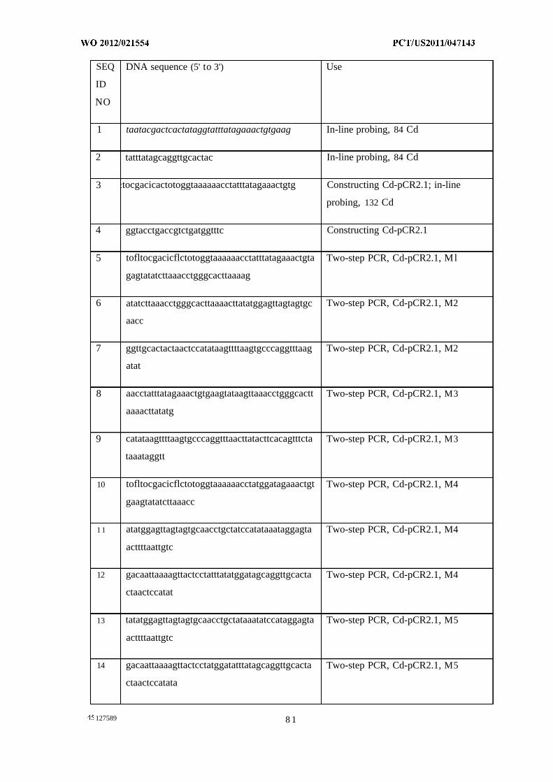
SEQ DNA sequence (5' to 3') Use
ID
NO
1 taatacgactcactataggtatttatagaaactgtgaag In-line probing, 84 Cd
2 tatttatagcaggttgcactac In-line probing, 84 Cd
3 tocgacicactotoggtaaaaaacctatttatagaaactgtg Constructing Cd-pCR2.1; in-line
probing, 132 Cd
4 ggtacctgaccgtctgatggtttc Constructing Cd-pCR2.1
5 tofltocgacicflctotoggtaaaaaacctatttatagaaactgta Two-step PCR, Cd-pCR2.1, Ml
gagtatatcttaaacctgggcacttaaaag
6 atatcttaaacctgggcacttaaaacttatatggagttagtagtgc Two-step PCR, Cd-pCR2.1, M2
aacc
7 ggttgcactactaactccatataagttttaagtgcccaggtttaag Two-step PCR, Cd-pCR2.1, M2
atat
8 aacctatttatagaaactgtgaagtataagttaaacctgggcactt Two-step PCR, Cd-pCR2.1, M3
aaaacttatatg
9 catataagttttaagtgcccaggtttaacttatacttcacagtttcta Two-step PCR, Cd-pCR2.1, M3
taaataggtt
10 tofltocgacicflctotoggtaaaaaacctatggatagaaactgt Two-step PCR, Cd-pCR2.1, M4
gaagtatatcttaaacc
1 1 atatggagttagtagtgcaacctgctatccatataaataggagta Two-step PCR, Cd-pCR2.1, M4
acttttaattgtc
12 gacaattaaaagttactcctatttatatggatagcaggttgcacta Two-step PCR, Cd-pCR2.1, M4
ctaactccatat
13 tatatggagttagtagtgcaacctgctataaatatccataggagta Two-step PCR, Cd-pCR2.1, M5
acttttaattgtc
14 gacaattaaaagttactcctatggatatttatagcaggttgcacta Two-step PCR, Cd-pCR2.1, M5
ctaactccatata
127589 8 1
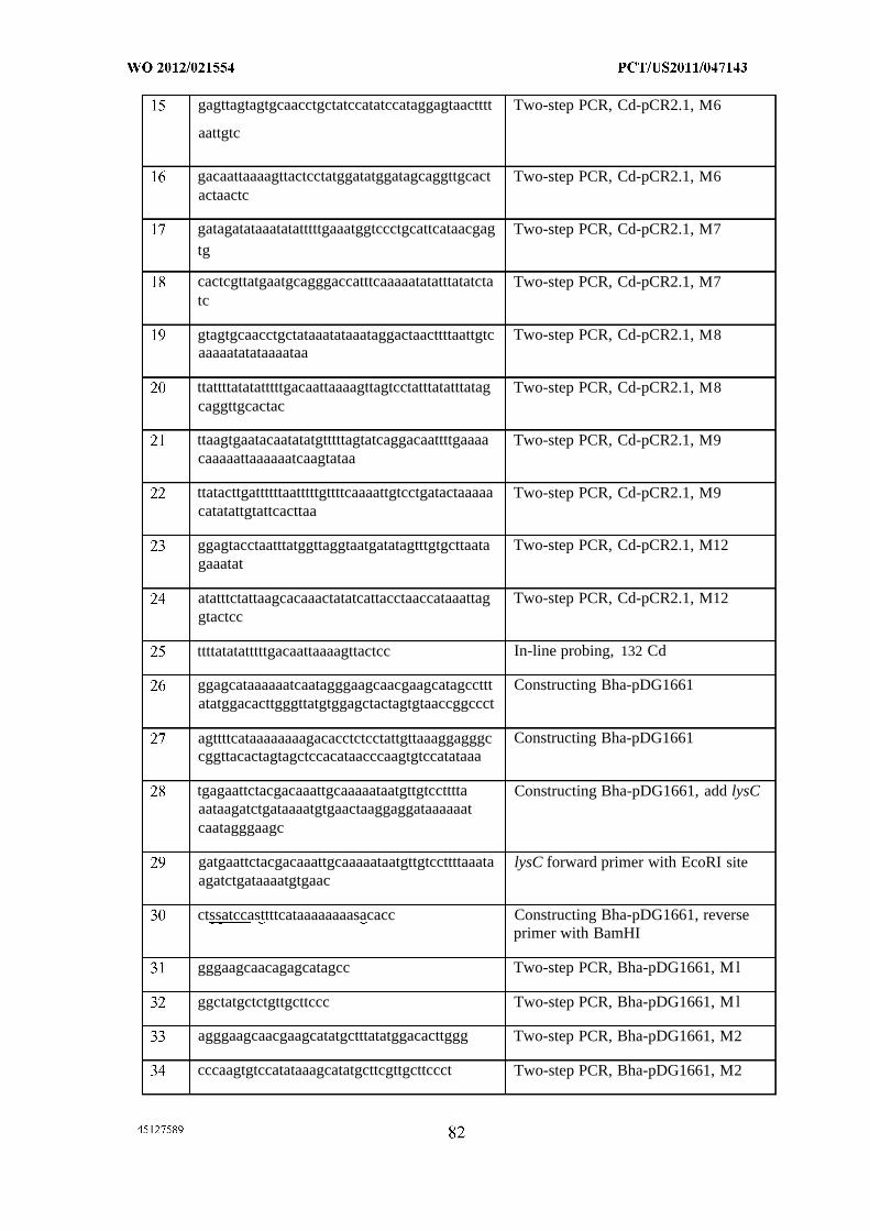
gagttagtagtgcaacctgctatccatatccataggagtaactttt Two-step PCR, Cd-pCR2.1, M6
aattgtc
gacaattaaaagttactcctatggatatggatagcaggttgcact Two-step PCR, Cd-pCR2.1, M6actaactc
gatagatataaatatatttttgaaatggtccctgcattcataacgag Two-step PCR, Cd-pCR2.1, M7tg
cactcgttatgaatgcagggaccatttcaaaaatatatttatatcta Two-step PCR, Cd-pCR2.1, M7tc
gtagtgcaacctgctataaatataaataggactaacttttaattgtc Two-step PCR, Cd-pCR2.1, M8aaaaatatataaaataa
ttattttatatatttttgacaattaaaagttagtcctatttatatttatag Two-step PCR, Cd-pCR2.1, M8caggttgcactac
ttaagtgaatacaatatatgtttttagtatcaggacaattttgaaaa Two-step PCR, Cd-pCR2.1, M9caaaaattaaaaaatcaagtataa
ttatacttgattttttaatttttgttttcaaaattgtcctgatactaaaaa Two-step PCR, Cd-pCR2.1, M9catatattgtattcacttaa
ggagtacctaatttatggttaggtaatgatatagtttgtgcttaata Two-step PCR, Cd-pCR2.1, M12gaaatat
atatttctattaagcacaaactatatcattacctaaccataaattag Two-step PCR, Cd-pCR2.1, M12gtactcc
ttttatatatttttgacaattaaaagttactcc In-line probing, 132 Cd
ggagcataaaaaatcaatagggaagcaacgaagcatagccttt Constructing Bha-pDG1661atatggacacttgggttatgtggagctactagtgtaaccggccct
agttttcataaaaaaaagacacctctcctattgttaaaggagggc Constructing Bha-pDG1661cggttacactagtagctccacataacccaagtgtccatataaa
tgagaattctacgacaaattgcaaaaataatgttgtcctttta Constructing Bha-pDG1661, add lysCaataagatctgataaaatgtgaactaaggaggataaaaaatcaatagggaagc
gatgaattctacgacaaattgcaaaaataatgttgtccttttaaata lysC forward primer with EcoRI siteagatctgataaaatgtgaac
ctssatccasttttcataaaaaaaasacacc Constructing Bha-pDG1661, reverseprimer with BamHI
gggaagcaacagagcatagcc Two-step PCR, Bha-pDG1661, Ml
ggctatgctctgttgcttccc Two-step PCR, Bha-pDG1661, Ml
agggaagcaacgaagcatatgctttatatggacacttggg Two-step PCR, Bha-pDG1661, M2
cccaagtgtccatataaagcatatgcttcgttgcttccct Two-step PCR, Bha-pDG1661, M2
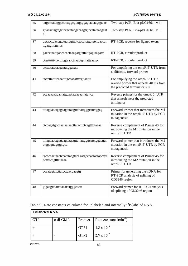
35 tatgctttatatggacacttggcgtatgtggagctactagtgtaac Two-step PCR, Bha-pDG1661, M3
36 gttacactagtagctccacatacgccaagtgtccatataaagcat Two-step PCR, Bha-pDG1661, M3a
37 ggtacctgaccgtctgatggtttctctacatctggtgtctgaccat RT-PCR, reverse for ligated exonsttgattgttcttctcc
38 gaccctaattgaacacactaaagatgtattattgagtaagattc RT-PCR, circular product
39 ctaattttttctactttcgtaacctcaagtgcttattaaatgc RT-PCR, circular product
40 atcttatatctaagaatatggaaata For amplifying the ompR 5' UTR fromC.difficile, forward primer
4 1 tactcttattttcaaattttgcaacatttttgttaatttt For amplifying the ompR 5' UTR,reverse primer that anneals 40 nts fromthe predicted terminator site
42 acaaaaaaagactatgcaatataaaaattatattcat Reverse primer for the ompR 5' UTRthat anneals near the predictedterminator
43 ttttagaaactgagaagtataagttattattgggcatctggag Forward Primer that introduces the Mlmutation in the ompR 5' UTR by PCRmutagenesis
44 ctccagatgcccaataataacttatacttctcagtttctaaaa Reverse complement of Primer 43 forintroducing the Ml mutation in theompR 5' UTR
45 ttttagaaactgagaagtataagttattattgggcatctggacttat Forward primer that introduces the M2atggagttagtggtgca mutation in the ompR 5' UTR by PCR
mutagenesis
46 tgcaccactaactccatataagtccagatgcccaataataacttat Reverse complement of Primer 45 foracttctcagtttctaaaa introducing the M2 mutation in the
ompR 5' UTR
47 ccaatagtatcttatgctgacgaagtg Primer for generating the cDNA forRT-PCR analysis of splicing ofCD3246 region
48 gtgaagtatatcttaaacctgggcactt Forward primer for RT-PCR analysisof splicing of CD3246 region
Table 5: Rate constants calculated for unlabeled and internally P-labeled RNA.
45127589 83
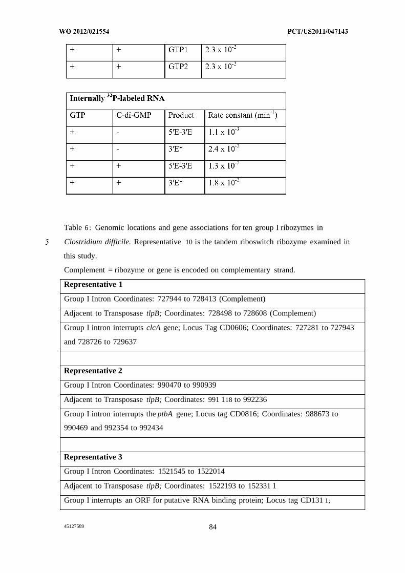
Table 6 : Genomic locations and gene associations for ten group I ribozymes in
Clostridium difficile. Representative 10 is the tandem riboswitch ribozyme examined in
this study.
Complement = ribozyme or gene is encoded on complementary strand.
Representative 1
Group I Intron Coordinates: 727944 to 728413 (Complement)
Adjacent to Transposase tlpB; Coordinates: 728498 to 728608 (Complement)
Group I intron interrupts clcA gene; Locus Tag CD0606; Coordinates: 727281 to 727943
and 728726 to 729637
Representative 2
Group I Intron Coordinates: 990470 to 990939
Adjacent to Transposase tlpB; Coordinates: 991 118 to 992236
Group I intron interrupts the ptbA gene; Locus tag CD0816; Coordinates: 988673 to
990469 and 992354 to 992434
Representative 3
Group I Intron Coordinates: 1521545 to 1522014
Adjacent to Transposase tlpB; Coordinates: 1522193 to 152331 1
Group I interrupts an ORF for putative RNA binding protein; Locus tag CD131 1;
45127589 84
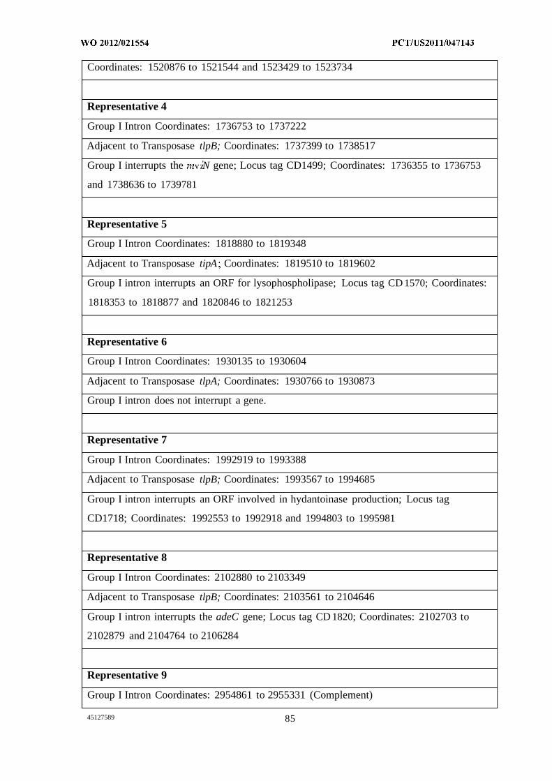
Coordinates: 1520876 to 1521544 and 1523429 to 1523734
Representative 4
Group I Intron Coordinates: 1736753 to 1737222
Adjacent to Transposase tlpB; Coordinates: 1737399 to 1738517
Group I interrupts the vz'N gene; Locus tag CD1499; Coordinates: 1736355 to 1736753
and 1738636 to 1739781
Representative 5
Group I Intron Coordinates: 1818880 to 1819348
Adjacent to Transposase tipA Coordinates: 1819510 to 1819602
Group I intron interrupts an ORF for lysophospholipase; Locus tag CD 1570; Coordinates:
1818353 to 1818877 and 1820846 to 1821253
Representative 6
Group I Intron Coordinates: 1930135 to 1930604
Adjacent to Transposase tlpA; Coordinates: 1930766 to 1930873
Group I intron does not interrupt a gene.
Representative 7
Group I Intron Coordinates: 1992919 to 1993388
Adjacent to Transposase tlpB; Coordinates: 1993567 to 1994685
Group I intron interrupts an ORF involved in hydantoinase production; Locus tag
CD1718; Coordinates: 1992553 to 1992918 and 1994803 to 1995981
Representative 8
Group I Intron Coordinates: 2102880 to 2103349
Adjacent to Transposase tlpB; Coordinates: 2103561 to 2104646
Group I intron interrupts the adeC gene; Locus tag CD 1820; Coordinates: 2102703 to
2102879 and 2104764 to 2106284
Representative 9
Group I Intron Coordinates: 2954861 to 2955331 (Complement)
45127589 85
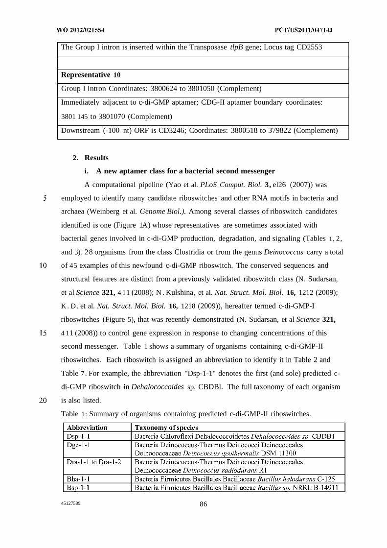
The Group I intron is inserted within the Transposase tlpB gene; Locus tag CD2553
Representative 10
Group I Intron Coordinates: 3800624 to 3801050 (Complement)
Immediately adjacent to c-di-GMP aptamer; CDG-II aptamer boundary coordinates:
3801 145 to 3801070 (Complement)
Downstream (-100 nt) ORF is CD3246; Coordinates: 3800518 to 379822 (Complement)
2. Results
i. A new aptamer class for a bacterial second messenger
A computational pipeline (Yao et al. PLoS Comput. Biol. 3, el26 (2007)) was
employed to identify many candidate riboswitches and other RNA motifs in bacteria and
archaea (Weinberg et al. Genome Biol.). Among several classes of riboswitch candidates
identified is one (Figure 1A) whose representatives are sometimes associated with
bacterial genes involved in c-di-GMP production, degradation, and signaling (Tables 1, 2,
and 3). 28 organisms from the class Clostridia or from the genus Deinococcus carry a total
of 45 examples of this newfound c-di-GMP riboswitch. The conserved sequences and
structural features are distinct from a previously validated riboswitch class (N. Sudarsan,
et al Science 321, 4 11 (2008); N . Kulshina, et al. Nat. Struct. Mol. Biol. 16, 1212 (2009);
K. D. et al. Nat. Struct. Mol. Biol. 16, 1218 (2009)), hereafter termed c-di-GMP-I
riboswitches (Figure 5), that was recently demonstrated (N. Sudarsan, et al Science 321,
4 11 (2008)) to control gene expression in response to changing concentrations of this
second messenger. Table 1 shows a summary of organisms containing c-di-GMP-II
riboswitches. Each riboswitch is assigned an abbreviation to identify it in Table 2 and
Table 7 . For example, the abbreviation "Dsp-1-1" denotes the first (and sole) predicted c-
di-GMP riboswitch in Dehalococcoides sp. CBDBl. The full taxonomy of each organism
is also listed.
Table 1 : Summary of organisms containing predicted c-di-GMP-II riboswitches.
45127589 86
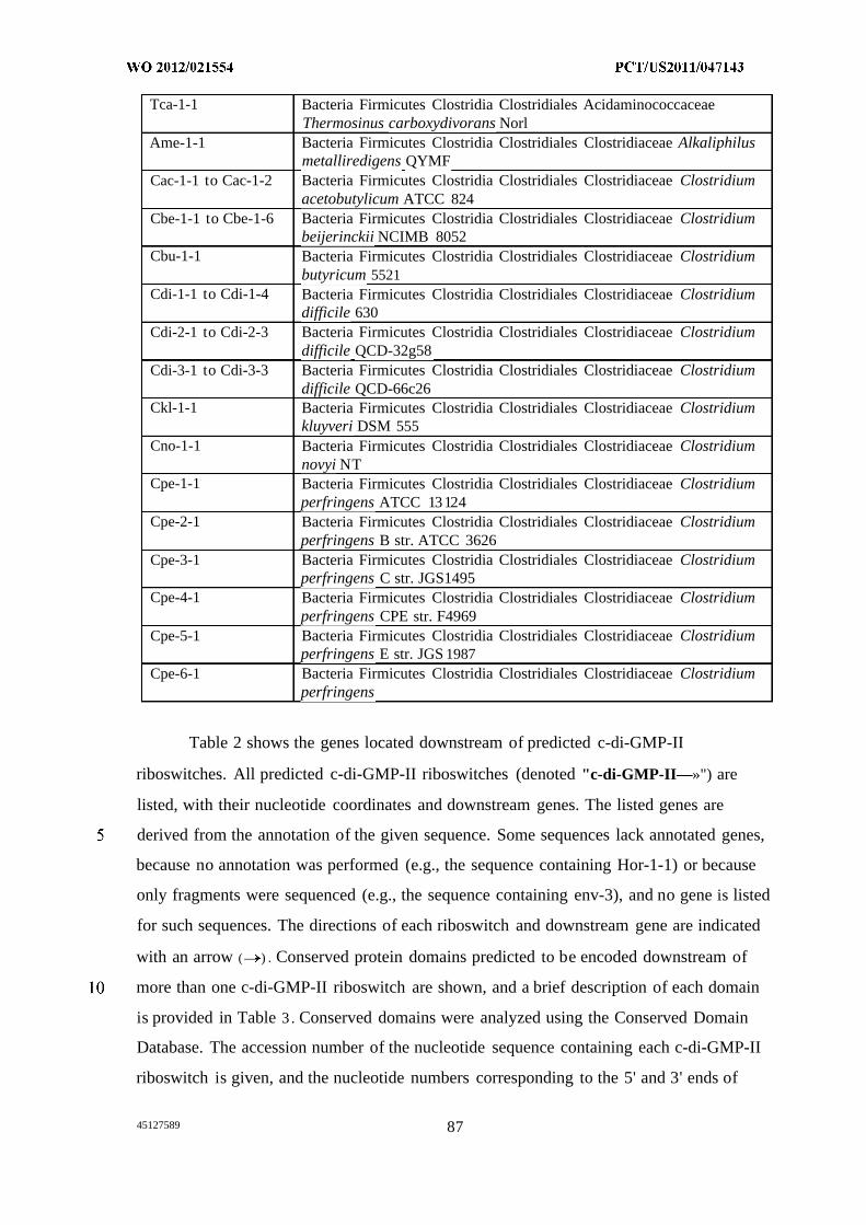
Tca-1-1 Bacteria Firmicutes Clostridia Clostridiales AcidaminococcaceaeThermosinus carboxydivorans Norl
Ame-1-1 Bacteria Firmicutes Clostridia Clostridiales Clostridiaceae Alkaliphilusmetalliredigens QYMF
Cac-1-1 to Cac-1-2 Bacteria Firmicutes Clostridia Clostridiales Clostridiaceae Clostridiumacetobutylicum ATCC 824
Cbe-1-1 to Cbe-1-6 Bacteria Firmicutes Clostridia Clostridiales Clostridiaceae Clostridiumbeijerinckii NCIMB 8052
Cbu-1-1 Bacteria Firmicutes Clostridia Clostridiales Clostridiaceae Clostridiumbutyricum 5521
Cdi-1-1 to Cdi-1-4 Bacteria Firmicutes Clostridia Clostridiales Clostridiaceae Clostridiumdifficile 630
Cdi-2-1 to Cdi-2-3 Bacteria Firmicutes Clostridia Clostridiales Clostridiaceae Clostridiumdifficile QCD-32g58
Cdi-3-1 to Cdi-3-3 Bacteria Firmicutes Clostridia Clostridiales Clostridiaceae Clostridiumdifficile QCD-66c26
Ckl-1-1 Bacteria Firmicutes Clostridia Clostridiales Clostridiaceae Clostridiumkluyveri DSM 555
Cno-1-1 Bacteria Firmicutes Clostridia Clostridiales Clostridiaceae Clostridiumnovyi NT
Cpe-1-1 Bacteria Firmicutes Clostridia Clostridiales Clostridiaceae Clostridiumperfringens ATCC 13124
Cpe-2-1 Bacteria Firmicutes Clostridia Clostridiales Clostridiaceae Clostridiumperfringens B str. ATCC 3626
Cpe-3-1 Bacteria Firmicutes Clostridia Clostridiales Clostridiaceae Clostridiumperfringens C str. JGS1495
Cpe-4-1 Bacteria Firmicutes Clostridia Clostridiales Clostridiaceae Clostridiumperfringens CPE str. F4969
Cpe-5-1 Bacteria Firmicutes Clostridia Clostridiales Clostridiaceae Clostridiumperfringens E str. JGS 1987
Cpe-6-1 Bacteria Firmicutes Clostridia Clostridiales Clostridiaceae Clostridiumperfringens
Table 2 shows the genes located downstream of predicted c-di-GMP-II
riboswitches. All predicted c-di-GMP-II riboswitches (denoted "c-di-GMP-II—»") are
listed, with their nucleotide coordinates and downstream genes. The listed genes are
derived from the annotation of the given sequence. Some sequences lack annotated genes,
because no annotation was performed (e.g., the sequence containing Hor-1-1) or because
only fragments were sequenced (e.g., the sequence containing env-3), and no gene is listed
for such sequences. The directions of each riboswitch and downstream gene are indicated
with an arrow ( — ) . Conserved protein domains predicted to be encoded downstream of
more than one c-di-GMP-II riboswitch are shown, and a brief description of each domain
is provided in Table 3 . Conserved domains were analyzed using the Conserved Domain
Database. The accession number of the nucleotide sequence containing each c-di-GMP-II
riboswitch is given, and the nucleotide numbers corresponding to the 5' and 3' ends of
45127589 87
each predicted riboswitch aptamer is provided. The riboswitches env-4 and env-8 are
adjacent to one another, and are depicted on the same line.
Conserved domains that were referenced in Table 2 are briefly described in Table
3, using text from the Conserved Domain Database.
Table 2 Genes located downstream of predicted c-di-GMP-II riboswitches
45127589 88
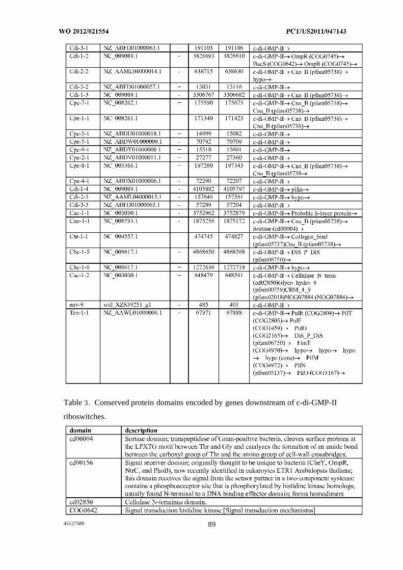
Table 3 . Conserved protein domains encoded by genes downstream of c-di-GMP-II
riboswitches.
45127589 89
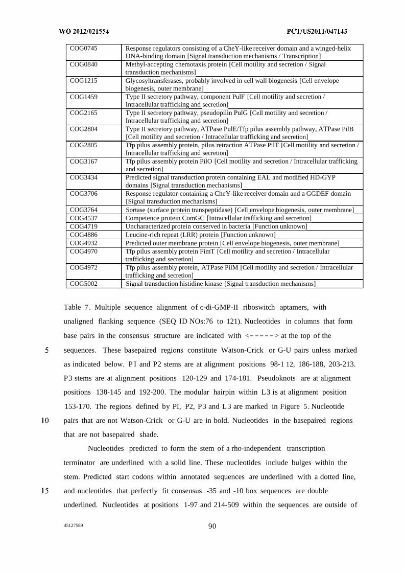
COG0745 Response regulators consisting of a CheY-like receiver domain and a winged-helixDNA-binding domain [Signal transduction mechanisms / Transcription]
COG0840 Methyl-accepting chemotaxis protein [Cell motility and secretion / Signaltransduction mechanisms]
COG1215 Glycosyltransferases, probably involved in cell wall biogenesis [Cell envelopebiogenesis, outer membrane]
COG1459 Type II secretory pathway, component PulF [Cell motility and secretion /Intracellular trafficking and secretion]
COG2165 Type II secretory pathway, pseudopilin PulG [Cell motility and secretion /Intracellular trafficking and secretion]
COG2804 Type II secretory pathway, ATPase PulE/Tfp pilus assembly pathway, ATPase PilB[Cell motility and secretion / Intracellular trafficking and secretion]
COG2805 Tfp pilus assembly protein, pilus retraction ATPase PilT [Cell motility and secretion /Intracellular trafficking and secretion]
COG3167 Tfp pilus assembly protein PilO [Cell motility and secretion / Intracellular traffickingand secretion]
COG3434 Predicted signal transduction protein containing EAL and modified HD-GYPdomains [Signal transduction mechanisms]
COG3706 Response regulator containing a CheY-like receiver domain and a GGDEF domain[Signal transduction mechanisms]
COG3764 Sortase (surface protein transpeptidase) [Cell envelope biogenesis, outer membrane]COG4537 Competence protein ComGC [Intracellular trafficking and secretion]COG4719 Uncharacterized protein conserved in bacteria [Function unknown]COG4886 Leucine-rich repeat (LRR) protein [Function unknown]COG4932 Predicted outer membrane protein [Cell envelope biogenesis, outer membrane]COG4970 Tfp pilus assembly protein FimT [Cell motility and secretion / Intracellular
trafficking and secretion]COG4972 Tfp pilus assembly protein, ATPase PilM [Cell motility and secretion / Intracellular
trafficking and secretion]COG5002 Signal transduction histidine kinase [Signal transduction mechanisms]
Table 7 . Multiple sequence alignment of c-di-GMP-II riboswitch aptamers, with
unaligned flanking sequence (SEQ ID NOs:76 to 121). Nucleotides in columns that form
base pairs in the consensus structure are indicated with < > at the top of the
sequences. These basepaired regions constitute Watson-Crick or G-U pairs unless marked
as indicated below. P I and P2 stems are at alignment positions 98-1 12, 186-188, 203-213.
P3 stems are at alignment positions 120-129 and 174-181. Pseudoknots are at alignment
positions 138-145 and 192-200. The modular hairpin within L3 is at alignment position
153-170. The regions defined by PI, P2, P3 and L3 are marked in Figure 5 . Nucleotide
pairs that are not Watson-Crick or G-U are in bold. Nucleotides in the basepaired regions
that are not basepaired shade.
Nucleotides predicted to form the stem of a rho-independent transcription
terminator are underlined with a solid line. These nucleotides include bulges within the
stem. Predicted start codons within annotated sequences are underlined with a dotted line,
and nucleotides that perfectly fit consensus -35 and -10 box sequences are double
underlined. Nucleotides at positions 1-97 and 214-509 within the sequences are outside of
45127589 90
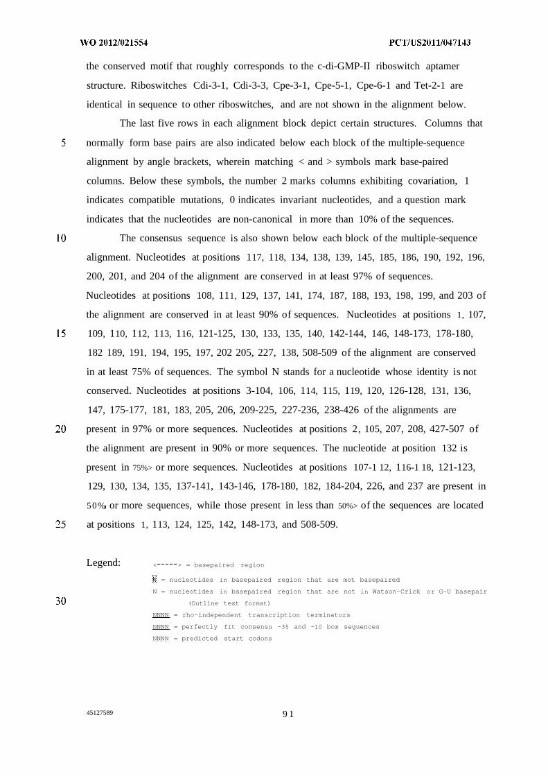
the conserved motif that roughly corresponds to the c-di-GMP-II riboswitch aptamer
structure. Riboswitches Cdi-3-1, Cdi-3-3, Cpe-3-1, Cpe-5-1, Cpe-6-1 and Tet-2-1 are
identical in sequence to other riboswitches, and are not shown in the alignment below.
The last five rows in each alignment block depict certain structures. Columns that
normally form base pairs are also indicated below each block of the multiple-sequence
alignment by angle brackets, wherein matching < and > symbols mark base-paired
columns. Below these symbols, the number 2 marks columns exhibiting covariation, 1
indicates compatible mutations, 0 indicates invariant nucleotides, and a question mark
indicates that the nucleotides are non-canonical in more than 10% of the sequences.
The consensus sequence is also shown below each block of the multiple-sequence
alignment. Nucleotides at positions 117, 118, 134, 138, 139, 145, 185, 186, 190, 192, 196,
200, 201, and 204 of the alignment are conserved in at least 97% of sequences.
Nucleotides at positions 108, 111, 129, 137, 141, 174, 187, 188, 193, 198, 199, and 203 of
the alignment are conserved in at least 90% of sequences. Nucleotides at positions 1, 107,
109, 110, 112, 113, 116, 121-125, 130, 133, 135, 140, 142-144, 146, 148-173, 178-180,
182 189, 191, 194, 195, 197, 202 205, 227, 138, 508-509 of the alignment are conserved
in at least 75% of sequences. The symbol N stands for a nucleotide whose identity is not
conserved. Nucleotides at positions 3-104, 106, 114, 115, 119, 120, 126-128, 131, 136,
147, 175-177, 181, 183, 205, 206, 209-225, 227-236, 238-426 of the alignments are
present in 97% or more sequences. Nucleotides at positions 2, 105, 207, 208, 427-507 of
the alignment are present in 90% or more sequences. The nucleotide at position 132 is
present in 75%> or more sequences. Nucleotides at positions 107-1 12, 116-1 18, 121-123,
129, 130, 134, 135, 137-141, 143-146, 178-180, 182, 184-204, 226, and 237 are present in
50% or more sequences, while those present in less than 50%> of the sequences are located
at positions 1, 113, 124, 125, 142, 148-173, and 508-509.
Legend: < > = basepaired region
= nucleotides in basepaired region that are mot basepaired
N = nucleotides in basepaired region that are not in Watson-Crick or G-U basepair
(Outline text format)
NNNN = rho-independent transcription terminators
NNNN = perfectly fit consensu -35 and -10 box seguences
NNNN = predicted start codons
45127589 9 1
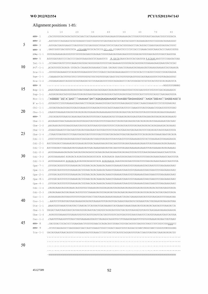
Alignment positions 1-85:
1 10 20 30 40 50 60 70 80
env- 1 .CACUUUGGUACAUACAUUCACAACCACUAAAAUAGAUUAAGAAAGUUAAAAAGACCUUAUUUGUUAUCAAUAACGGUCUGGACA
env- 2 .AUCUGCCCAGGAGCGCACGUAGGCCGGCCGGUUCUCGAUUAGCACGCCAUCACGGUCCAGGAAGAUUGCCGGGUACAUAAGGGC
env- 3 .AUUGACGAGUGAAGCCUAGUUGCCGCGAUGGGCUUGACUUCUCGAGCACGGGUAGCCUACAGUACCGAAUGAGGUAGUACUUUC
env- 4 GAUCGGUCGACGUUCUCA CG GAUUCGCACUCCG CC C CCGAUCUCCCCUCCACCCGAAACGGUCAAGCACCCAAUCGUUG
A e-1-1 GUGUAGAAAAGAGGGUUUCGUUGUAUAAUGUAGAACUGUGUAAUAGUAAUAAGUGUUGGAUAUUGUUAAUAGUUUGAUCAAGUGA
env- 5 AGUUGAUUAUCCCCACCCCGAGUUAAAUAGCUCAAAUUCG GACAGAAGCAUUCACGAGUUA ACAA AAGCUCCUAAGACUAA
Tet- 1-1 .GCUAAUUAUUCUUUCAAAUUAUGGACAGGGGGAUUUUUCGACAAAAAUUUGGUAUACAGGUAUUUAAAAGAAGGAAUUACGCAU
Pt -1-1 .ACAUUUGGCAUAUA UUGACA CAAAAUAUAAAAACCGAA UAUAAUUAACGUAAAAUAUAAAAUAUUAAAAUAAAAGCAUUAAAUA
Dra- 1-1 .GUUGUGAGAAAUCUCAUAUUUGGAAGUGCUUCCUGAGCCAUUAUAAAGAAGUCCCCGCGCACCCCAUGCCUGGCCGUAUGAGGA
Dge- 1-1 .CAAAAGUCACUUUGCUUCCUUUUUGUGCUGCUGUUGUGACGAGCUGCUGUUGUGAGUUGGCAGUAAGGGUUCUGUUAGAGGUGC
Dra- 1-2 .UGGAAAGAAUCAGGGCGCAUUAAGACGCCUUAAGAGUUCUGUCAGAGAUCCCACUUUAGACUCGCUUCACUUCGCACAGAUUCA
env- 6 NGGUUUUNNNG
Bsp- 1-1 .AAAUGAAUAAAAUAUAUAUUGACUUAAUAUGACAGGAAUCAUAUAUUUUAAUUGGCUUGCAAUUGUCUGUUUCGACAGAAAAUG
Dsp- 1-1 .AGGAUAUAGCAGUGGUAAUGUAUAGUAAUGAUAAUAUAGUAUCAGUUAUAUUGACUUUGCAUCUUUUGCAGGCCAAUAAUGGUA
Hor- 1-1 Αϋϋϋϋϋ ϋϋ Α ϋΑϋ ΑΑΑΑΑΑ ϋΑ ΑϋΑϋΑΑϋΑΑΑΑϋϋ ΑΑΑϋϋϋ ϋΑϋΑΑϋϋΑϋ ΑΑϋΑ ϋϋϋΑΑ ϋΑϋϋϋΑϋϋ ϋB a-1-1 .GUUAUUCCUGUUAAAAGUAAUAACCUUAGACAAAAUUGUAUCAUUUUUAAGAAGUCGGACCAAAUUAAAUGCCUCGUGUAAUAU
Ckl- 1-1 .GUUAUUAGAGUGAUUUGAGUGAAAGUUUUAAUAUUUGGCAGUUAAAUUAUUUUCCAAAGUUUAUUGAAACUGGAGUGUUUUUAU
Cbu- 1-1 .UAAGUUAAAAAUGAUAUUAUAUAUAUGGAAAUAGAAAAAAAUGGUAUAUAAUUACAUUAUGUUAGGUUAUAUAUAAGUAAAAAU
env- 7 .UGCAUAUUUUAAUGCAAAUAAUUAUAUUUUUUAUCAAAAUAUGCUUGAAUAUAUGGAGUUAUUAGAAUAUUAUAUAUAUAAAUU
Cbe- 1-1 .AGGAAAUUAACAAAAAGAUAGUUAGAUUAUUGAUUUUUUAAUAUCUAUAUGAUAAUAUGACAUAUAUAUUAAAAAAAUGACAAU
Cbe- 1-2 .AAUAAAUAUUGUAAGGUAAUUAUUGCAUUUAAUGUUGAUUUUUCUUAUAUUAUUGUUAUAAUAUAUACAUAGAAAGACAUAAAG
Cbe- 1-3 .GGAAUGAAAUUCGACAAUUUAUAUUAAGAAUCAUUGAUUUUCUUACAUGAAUAUGAUAAUAUUUCUAUAUAUUAGGUAAGUGUA
Cbe- 1-4 .CUAAUUUAGUAUUCUUAAUGAUAUUAUUUGUUGAUUUUAUUAUAGAUUGAUGAUAAUAUUUCAUAUAUAUGAAAUAAGUACAUA
Cdi- 1-1 .UGUCUUUUAUUAGAUAAGUAUCUAGGAUACUAAUUUAUAUUUUAUUGAGUUGUUAAAAAAGUGCACUUAAAAAGUUAAAAAAGA
Cdi- 2-1 AUCUUAUAUCUAAAAAUAUGGAAAUAUUGACAAAAUAAUAUUACAAUGUUAGAAUAAAAAAGAAAUUUAUGAAAAUAUAUAAAAG
Cdi- 1-2 AUCUUAUAUCUAAGAAUAUGGAAAUAUUGACAAAAUAAUAUUACAAUGUUAGAAUAAAAAAGAAAUUUAUGAAAAUAUAUAAAAG
Cdi- 2-2 .AUGUAAAAAAGAUAGACAAUAGUAUAGAGGCAUAAUAUAAUAGAAGUAUAUAAUUGUAUUUUUAAUAUAAAUAAAGCAAUUUUA
Cdi- 3-2 .AUGUAAAAAAG AUAGACAAUAGUAUAGAGGCAUA AUAUAAUA GAAGUAUAUAAUUGUAUUUUUAAUAUAAAUAAAGCAAUUUUA
Cdi- 1-3 .AUGUAAAAAGG AUAGACAAUAGUAUAGAGGCAUA AUAUAAUA GAAGUAUAUAAUUGUAUUUUUAAUAUAAAUAAAGCAAUUUUA
Cpe- 7-1 .UUUGACAGGUUUUUUAAAAUACUGUAACAUACAAAUACAAAUUGAAAAUUAAGUGUUAAAAAGUAGUAAUUUUUAAGAAAUAAU
Cpe- 1-1 .UUUGACAGUUUUUUUAAAAUACUGUAACAUACAAAUACAAAUUGAAAAUUAAGUGUUAAAAAGUAAUGAAUUUUAAGAAAUAAU
Cpe- 2-1 .UUUGACAGUUUUUUUAAAAUACUGUAACAUACAAAUACAAAUUGAAAAUUAAGUGUUAAAAAGUAAUGAAUUUUAAGAAAUAAU
Cpe- 8-1 .UUUGACAGUUUUUUUAAAAUACUGUAACAUACAAAUACAAAUUGAAAAUUAAGUGUUAAAAAGUAAUGAAUUUUAAGAAAUAAU
Cpe- 4-1 .UUUGACAGUUUUUUUAAAAUACUGUAACAUACAAAUACAAAUUGAAAAUUAAGUGUUAAAAAGUAAUGAAUUUUAAGAAAUAAU
Cdi- 1-4 .UAUAUAAAUAGUAUAAACAGUGUUGCUAAAAUAUGGUAGAUAUUAUAUAAUAUAAGUGGAUAUGUAUAUACAUUAUGAGUUAUA
Cdi- 2-3 .UAUAUAAAUAGUAUAAACAGUGUUGCUAAAAUAUGGUAGAUAUUAUAUAAUAUAAGUGGAUAUGUAUAUACAUUAUGAGUUAUA
Cac- 1-1 .AUAAAAAAUAUUAAUUUUUUUUGAUUUACCUAUUAAAGAAAAUAAAAACUAUACGAAAAUAAUAUUGUUAAGAGUUUUAAAUAA
Cno- 1-1 ..AAUUCUUUAUUAUUAAUAAAAUAUAUAUUAAAAUUUUAUAUUGACAAAGUAAUAUGCAUAAAUUACUAUAAAUAUAAGAUUAA
Cte- 1-1 .AAAUGGUUAAAUUUAUUACCUAAUACUCAUUAUUGAUAAAAUCAUGAAAUUAAAGUAAUAUGUAUAUACAAUAGUUUAAUACCA
Cbe- 1-5 UAGACCAAUUAAUGAUCAUUAUGGUAUAAUUACUAUGGCAUAUAUUGGCUACAUUUAAUAUGUUAUGCAAGAAAUAAAAGAAAUA
Cbe- 1-6 .AUAUGUUAAAAUUUGAAAUUGUUUCAUUUUAUUCACUAUUUGUUCAUUGAUUUGUAAUUAAUUCCAUGUUAAAAUGAUCAUUAA
Cac- 1-2 .CAAUUUUAAGGUUUUAGCUAUGAAAAAGUAGUCUAGAGGCAGGUUGCUUUAAAUAUAAUUUUGUGGUAAAAUAUAACUAUUAAU
env- 8 .UACGUACCGCACGCCUGAAGAACUUGUUGUAACGCAGACAUCGUAACUGUGACAGUGCGAGUGCUAGCCUUCGGUCGUGGGUCC
env- 9 .UCUUCAAGAGGCCGAGGGAGCGACCAGCUGAAGCUGGCCGGACCAAGCGUUCAGGACGCANCUNGGCANCCGGGUUUNUGGGNG
Tca- 1-1 UACAUGAAUUAACAGUUCUUUGAAAUAUUGUAAACCCGUCUACUUCAUUGCAGGAUUUUUACCAAUUUAUUACGAAUAUAUACGU
-NNNNNNNNNNNNNNNNNNNNNNNNNNNNNNNNNNNNNNNNNNNNNNNNNNNNNNNNNNNNNNNNNNNNNNNNNNNNNNNNNNNN
45127589 92
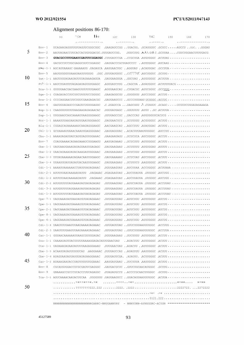
Alignment positions 86-170:
90 l i 12C 130 150 160 170
< - -> < - -> < - -> < - ->
Env- 1 UCAGAAUAGGUUGUUAGUUCGGGCGGC .GAAGAUCCGG ..UGACUG. .GCAUGGUC .GCUCC AUCCU ..UUC. ..GGGAU
Env- 2 AAUUAUAACCUUCACCACUGUGGACGC.UUUGAUCCGG. .UGUCUUG . GG C .GCUUAA ....CGUCUGGAACUUUUGACG
Env- 3 GUACGCCUUUGAAUCGAUUUCGGAUGC .CUUGAUCCGA ..CCGCUGA .AUGUGGGC .ACUUAG
Env- 4 GACGCCUCCUGCGAGGCGUCCGGAGGC .GAUGACCCGCUGAUCCCC ..AUUUGGGC .AUCAAG
A e-1-1 AGCAUUAAAGC GAAAAAUG .UAGAGCA .AAUGAACUGC ..AGGUAU ..ACAUGGAC .GCCUUA
Env- 5 AAUGUGGGUUAAAUAAUUUGGUG .GGC .GUUGAUGUGC ..CC G .AUCUGGUC .GCUUG
Tet- 1-1 AAUUUGUAGAAUAUUUUAUAAAGAGUA .GAUGAAGUGA ..GGCUAU ..AUGUGGGC .ACUUUA
Pt -1-1 AAUCUGAUUUUAGAGAUAGUGUGAAGC .AGUGAUCUUG ..CAGCUA ..AUAUGGUC .ACUUUUUACG
Dra- 1-1 GUGUGAACGACGAAGUGGUUUUGAAGC .AGGGAAGCAU ..CUGACGC .AUUCGGGC .GCCUUG
Dge- 1-1 CUAGAUACCUUCUUCUUGUUCCGGGGC .GAAGAGGCGU ..GUGGUGU .AUCCGGGC .ACCUG
Dra- 1-2 CCCCAGGAACUUCUGUCGAAGAGACGC .GAUGAAUCCC ..GCCCUGUAAU UCGGGC ACCUC
Env- 6 GAUUGUAUAGCCCGAUUCUGUGGAGGC .C .UGAUCCA ...UAGCGGU . .CUGGUC .GCAGC UCUUUCUGGAUAGAAAGA
Bsp- 1-1 CAAGGGUGUUUUAAGAAUAUAGAACAC .UGUGAUGAGC ..GGUUUUU .AUUU ..GC .ACUUUA
Dsp- 1-1 UUGGAAUCAGCAAAAUUAAGGGGAAGC .GUUGAGCCGC ..UACCCAU .AUGUGGUUCACUCG
Hor- 1-1 AAAAUUUAAUAAUAUUGAAUGGGAAGC .UAUGAACUCU ..UCUUUGU .ACGUGGUC .ACUUG
B a-1-1 CGGAGCAUAAAAAAUCAAUAGGGAAGC .AACGAAGCAU ..AGCCUUU .AUAUGGAC .ACUUG
Ckl- 1-1 GCUAAAAUUAAACAAAUUGAGGGGAAC .GAUGAGGUAC ..ACACUUUAAUUUGGGC .AUCCUU
Cbu- 1-1 AAAAUAGAUUAUCAUGUAGUUGGAAAC .GAAGAAUAGU ..UCUCUCA .AUCUGGGC .ACCUU
Env- 7 CUAUGAAAACAUAAUAAAUCUGGAAGU .AAUGAUAAAU ..UCUCUUU .AUUUGGGC .ACUUU
Cbe- 1-1 UAUUAAUGAAAUAUAGUAAUUGAGAGC .UAUGAAGAAU ..UCUUUUU .AUGUGGAU .AUUUU
Cbe- 1-2 UAACAUAAAUAUACAAAGAUUGGGAAC .UAUGAAGAAU ..UCUCUGU .AUUUGGGC .AUCUU
Cbe- 1-3 UUUAUAAAAAAUAGAACAAUUGGAAGC .GAUGAAAAAU ..UCUCUAU .AUCUGGGC .ACUUU
Cbe- 1-4 UUAAUGUAUUAUAUUACAAUUGGAAGC .UAUGAAGAAU ..UCUUUCU .AAGUGGGC .ACUUU
Cdi- 1-1 AAAAUUAAAAAACCUAUUUAUAGAAAC .UGUGAAGUAU ..AUCUUAA .ACCUGGGC .ACUUAAA
Cdi- 2-1 AUUUUUAAUAAAAAUAUUU .UAGAAAC .UGAGAAGUAU ..AUCUUAUUA .UUGGGC .AUCUGG
Cdi- 1-2 AUUUUUAAUAAAAAUAUUU .UAGAAAC .UGAGAAGUAU ..AUCUUAUUA .UUGGGC .AUCUGG
Cdi- 2-2 AUUGUUUUUAUGAAAUAUUAUAGAGAU .GUUGAAGUAU ..AUUCUAUUA .UUGGGC .ACCUUAU
Cdi- 3-2 AUUGUUUUUAUGAAAUAUUAUAGAGAU .GUUGAAGUAU ..AUUCUAUUA .UUGGGC .ACCUUAU
Cdi- 1-3 AUUGUUUUUAUGAAAUAUUAUAGAGAU .GUUGAAGUAU ..AUUCUAUUA .UUGGGC .ACCUUAU
Cpe- 7-1 UAGUAAAUAUUAAUAUGUUAUAGAAAC .GUUGAUGUAU ..AUUCUGU .AUUUGGGC .AUCUG
Cpe- 1-1 UAGUAAAUAUUAAAAUGUUAUAGAAAC .GUUGAUGUAU ..AUUCUGU .AUUUGGGC .AUCUG
Cpe- 2-1 UAGUAAAUAUUAAAAUGUUAUAGAAAC .GUUGAUGUAU ..AUUCUGU .AUUUGGGC .AUCUG
Cpe- 8-1 UAGUAAAUAUUAAAAUGUUAUAGAAAC .GUUGAUGUAU ..AUUCUGU .AUUUGGGC .AUCUG
Cpe- 4-1 UAGUAAAUAUUAAAAUGUUAUAGAAAC .GUUGAUGUAU ..AUUCUGU .AUUUGGGC .AUCUG
Cdi- 1-4 UAAUUUUGAAUUUAAUAAAAUAGAAAC .GUUGAUUUAU ..GUUCUGUAAUGUGGGC .ACCUUG
Cdi- 2-3 UAAUUUUGAAUUUAAUAAAAUAGAAAC .GUUGAUUUAU ..GUUCUGUAAUGUGGGC .ACCUUA
Cac- 1-1 GGUAACAAAAAAUUAAGCGUUGGAGAC .UGUGAAGAAU ..UUCUUGU .AUUUGGGC .ACCUU
Cno- 1-1 CAAAAUAUUUACUUUUUAAAAGGAGACAUUUGAACUAU ..AUACUUU .AUUUGGGC .ACUUU
Cte- 1-1 UAUAAAUAUAAUAUUUUAAGAGGAAAU .UUUGAACUAU ..AUACUU ..AUUUGGGC .ACUUU
Cbe- 1-5 ACAAGUAUAUUUUGUCAU .AAGGAAAC .UUUGAUCCAU ..AUAUUUU .AAUUGGGC .ACUUU
Cbe- 1-6 AUAGUAAUAUUAUGUAUAUAAGGAAAC .UGUGAUUCGA. .AUAUUU. .ACUUGGGC .ACUUU
Cac- 1-2 AUAAAGAAUACCUAUUUGUUUGGAAAC .AAUGAUGAAU ..UUCUUUA .AAUUGGGC .ACUUG
Env- 8 CGCAUGUGGACCUCGCGAUUCGAGGGC .GAUGACGCUC ..GUUCUGCAACAUGGGC .GCUUG
Env- 9 GNAAAGCCGCCCUCACCCUUCAGAGGC .UUAGAUGCCU ..ACCCCGCAACUUGGGC .GCUUG
Tca- 1-1 AGUCAAAACAAUACUGCAA .UGGGUGU .GAUGAAGUCC ..GGACAGUAAUGUGGGC .ACUUA
<«<<«<<«.<« <<<<..<«< «<««.... »>»»
????????222.222 2222. .2222 2222722. ...2272222
<«< .<«
0121.222
NNNNNNNNNNNNNNNNNNNNNRGARRC-NNUGANNYRY — NNNYUNN-AYNUGGRC-ACYUN
45127589 93
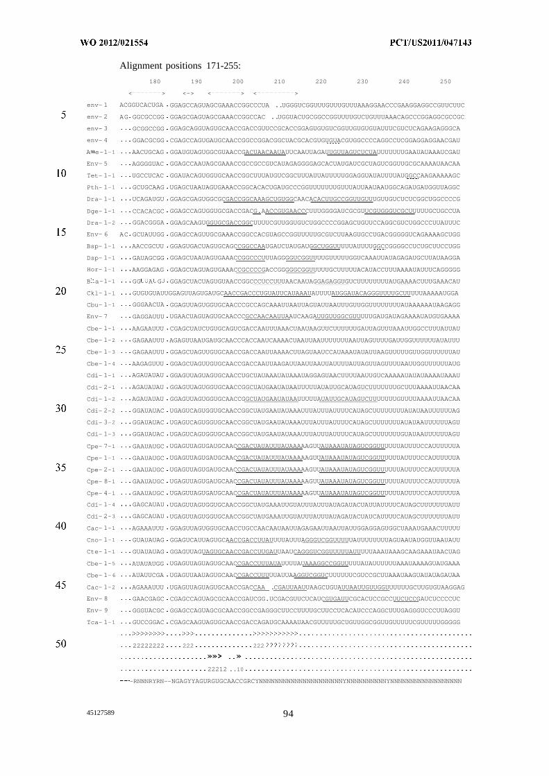
Alignment positions 171-255:
180 190 200 210 220 230 240 250
< > <-> < > < >
env- 1 ACGGUCACUGA GGAGCCAGUAGCGAAACCGGCCCUA ..UGGGUCGGUUUGUUUGUUUAAAGGAACCCGAAGGAGGCCGUUCUUC
env- 2 AG GGCGCCGG GGAGCGAGUAGCGAAACCGGCCAC ..UGGUACUGCGGCCGGUUUUGUCUGUUUAAACAGCCCGGAGGCGCCGC
env- 3 GCGGCCGG GGAGCAGGUAGUGCAACCGACCGUUCCGCACCGGAGUGUGUCGGUUGUGUGUAUUUCGUCUCAGAAGAGGGCA
env- 4 GGACGCGG CGAGCCAGUGAUGCAACCGGCCGGACGGCUACGCACGUGUGUACGUGGCCCCAGGCCUCGGAGGAGGAACGAU
A e-1-1 AACUGCAG GGAUGUAGUGGCGUAACCGACUAACAAUAUUCAAUUAGAUUGUUAGUCUCUAUUUUUUUGAAUAUAAAUCGAU
Env- 5 AGGGGUAC GGAGCCAAUAGCGAAACCGCCGCCGUCAUAGAGGGGAGCACUAUGAUCGCUAGUCGGUUGCGCAAAAUAACAA
Tet- 1-1 UGCCUCAC GGAUACAGUGGUGCAACCGGCUUUAUGUCGGCUUUAUUAUUUUUGGAGGUAUAUUUUAUGGCCAAGAAAAAGC
Pth- 1-1 GCUGCAAG UGAGCUAAUAGUGAAACCGGCACACUGAUGCCCGGUUUUUUUGUUUAUUAAUAAUGGCAGAUGAUGGUUAGGC
Dra- 1-1 UCAGAUGU GGAGCGAGUGGCGCGACCGGCAAAGCUGUGGCAACACACUUGCCGGUUGUUUGUUGUCUCUCGGCUGGCCCCG
Dge- 1-1 CCACACGC GGAGCCAGUGGUGCGACCGACG.AACCGUGAACCCUUUGGGGAUCGCGUUCGUGGGUCGCUUUUUGCUGCCUA
Dra- 1-2 GGACGGGA GGAGCAAGUGGUGCGACCGGCUUUUCGUUGGUGUCUGGCCCCGGAGCUGUUCCAGGCGUCUGGCCCUUAUUUC
Env- 6 AC GCUAUUGG GGAGCCAGUUGCGAAACCGGCCACGUAGCCGGUUUUUGCGUCUUAAGUGCCUGACGGGGGUCAGAAAAGCUGG
Bsp- 1-1 AACCGCUU GGAGUGACUAGUGCAGCCGGCCAAUGAUCUAUGAUGGCUGGUUUUUAUUUUGGCCGGGGCCUCUGCUUCCUGG
Dsp- 1-1 GAUAGCGG GGAGCUAAUAGUGAAACCGGCCCUUUAGGGGUCGGUUUUGUUUUUGGUCAAAUUAUAGAGAUGCUUAUAAGGA
Hor- 1-1 AAGGAGAG GGAGCUAGUAGUGAAACCGCCCCGACCGGGGGCGGUUUUUGCUUUUUACAUACCUUUAAAAUAUUUCAGGGGG
B a-1-1 GG A G GGAGCUACUAGUGUAACCGGCCCUCCUUUAACAAUAGGAGAGGUGUCUUUUUUUUAUGAAAACUUUGAAACAU
Ckl- 1-1 GUGUGUAUUGGAGUUAGUGAUGCAACCGACCCUGUAUUCAUAAAUAUUUUAUGGAUACAGGGUUUUGCUUUUUAAAAAUGGA
Cbu- 1-1 GGGAACUA GGAGUUAGUGGUGCAACCCGCCAGCAAAUUAAUUAGUAUUAAUUUGUUGGUUUUUUUUAUAAAAAAUAAGAGG
Env- 7 GAGGAUUU UGAACUAGUAGUGCAACCCGCCAACAAUUAAUCAAGAUUGUUGGCGUUUUUGAUGAUAGAAAAUAUGUGAAAA
Cbe- 1-1 AAGAAUUU CGAGCUAUCUGUGCAGUCGACCAAUUUAAACUAAUAAGUUCUUUUUUGAUUAGUUUAAAUUGGCCUUUAUUAU
Cbe- 1-2 GAGAAUUU AGAGUUAAUGAUGCAACCCACCAAUCAAAACUAAUUAAUUUUUUUAAUUAGUUUUGAUUGGUUUUUUAUAUUU
Cbe- 1-3 GAGAAUUU GGAGCUAGUUGUGCAACCGACCAAUUAAAACUUAGUAAUCCAUAAAUAUAUUAAGUUUUUGUUGGUUUUUUAU
Cbe- 1-4 AAGAGUUU GGAGCUAGUUGUGCAACCGACCAAUUAAGAUUAAUUAAUUAUUUUAUUAGUUAGUUUUAAUUGGUUUUUUAUG
Cdi- 1-1 AGAUAUAU GGAGUUAGUAGUGCAACCUGCUAUAAAUAUAAAUAGGAGUAACUUUUAAUUGUCAAAAAUAUAUAAAAUAAAU
Cdi- 2-1 AGAUAUAU GGAGUUAGUGGUGCAACCGGCUAUGAAUAUAAUUUUUAUAUUGCAUAGUCUUUUUUUGCUUUAAAAUUAACAA
Cdi- 1-2 AGAUAUAU GGAGUUAGUGGUGCAACCGGCUAUGAAUAUAAUUUUUAUAUUGCAUAGUCUUUUUUUGUUUUAAAAUUAACAA
Cdi- 2-2 GGAUAUAC UGAGUCAGUGGUGCAACCGGCUAUGAAUAUAAAUUUAUUUAUUUUCAUAGCUUUUUUUUAUAUAAUUUUUUAG
Cdi- 3-2 GGAUAUAC UGAGUCAGUGGUGCAACCGGCUAUGAAUAUAAAUUUAUUUAUUUUCAUAGCUUUUUUUAUAUAAUUUUUUAGU
Cdi- 1-3 GGAUAUAC UGAGUCAGUGGUGCAACCGGCUAUGAAUAUAAAUUUAUUUAUUUUCAUAGCUUUUUUUGUAUAAUUUUUUAGU
Cpe- 7-1 GAAUAUGC UGAGUUAGUGAUGCAACCGACUAUAUUUAUAAAAAGUUAUAAAUAUAGUCGGUUUUUUAUUUUCCAUUUUUUA
Cpe- 1-1 GAAUAUGC UGAGUUAGUGAUGCAACCGACUAUAUUUAUAAAAAGUUAUAAAUAUAGUCGGUUUUUUAUUUUCCAUUUUUUA
Cpe- 2-1 GAAUAUGC UGAGUUAGUGAUGCAACCGACUAUAUUUAUAAAAAGUUAUAAAUAUAGUCGGUUUUUUAUUUUCCAUUUUUUA
Cpe- 8-1 GAAUAUGC UGAGUUAGUGAUGCAACCGACUAUAUUUAUAAAAAGUUAUAAAUAUAGUCGGUUUUUUAUUUUCCAUUUUUUA
Cpe- 4-1 GAAUAUGC UGAGUUAGUGAUGCAACCGACUAUAUUUAUAAAAAGUUAUAAAUAUAGUCGGUUUUUUAUUUUCCAUUUUUUA
Cdi- 1-4 GAGCAUAU UGAGUUAGUGGUGCAACCGGCUAUGAAAUUGUAUUUAUUUAUAGAUACUAUUAUUUUCAUAGCUUUUUUUAUU
Cdi- 2-3 GAGCAUAU UGAGUUAGUGGUGCAACCGGCUAUGAAAUUGUAUUUAUUUAUAGAUACUAUCAUUUUCAUAGCUUUUUUUAUU
Cac- 1-1 AGAAAUUU GGAGUUAGUGGUGCAACCUGCCAACAAUAAUUAGAGAAUUAAUUAUUGGAGGAGUGGCUAAAUGAAACUUUUU
Cno- 1-1 GUAUAUAG GGAGUCAUUAGUGCAACCGACCUUAUUUUAUUUAGGGUCGGUUUUUAUUUUUUUUAGUAAUAUGGUUAAUAUU
Cte- 1-1 GUAUAUAG GGAGUUAGUAGUGCAACCGACCUUGAUUAAUCAGGGUCGGUUUUUAUUUUUAAAUAAAGCAAGAAAUAACUAG
Cbe- 1-5 AUAUAUGG UGAGUUAGUAGUGCAACCGACCUUUAUAUUUUAUAAAGGCCGGUUUUUAUAUUUUUUAAAUAAAAGUAUGAAA
Cbe- 1-6 AUAUUCGA UGAGUUAAUAGUGCAACCGACCUUUUUAUUAAGGUCGGUCUUUUUUCGUCCGCUUAAAUAAGUAUAUAGAUAA
Cac- 1-2 AGAAAUUU UGAGUUAGUAGUGCAACCGACCAA .CGAUUAAUUAAGCUGUAUUAAUUGUUGGUUUUUUGCUUGUGUAAGGAG
Env- 8 GAACGAGC CGAGCCAGUAGCGCAACCGAUCGG.UCGACGUUCUCAUCGUGAUUCGCACUCCGCCUUCUCCGAUCUCCCCUC
Env- 9 GGGUACGC GGAGCCAGUAGCGCAACCGGCCGAGGGCUUCCUUUUGCUUCCUCACAUCCCAGGCUUUGAGGGUCCCUUAGGU
Tca- 1-1 GUCCGGAC CGAGCAAGUAGUGCAACCGACCAGAUGCAAAAUAACGUUUUUGCUGUUGGCGGUUGUUUUUCGUUUUUGGGGG
22222222 222 222
»»> »
22212 ..10
-RNNNRYRN--NGAGYYAGURGUGCAACCGRCYNNNNNNNNNNNNNNNNNNNNNYNNNNNNNNNNYNNNNNNNNNNNNNNNNNN
45127589 94
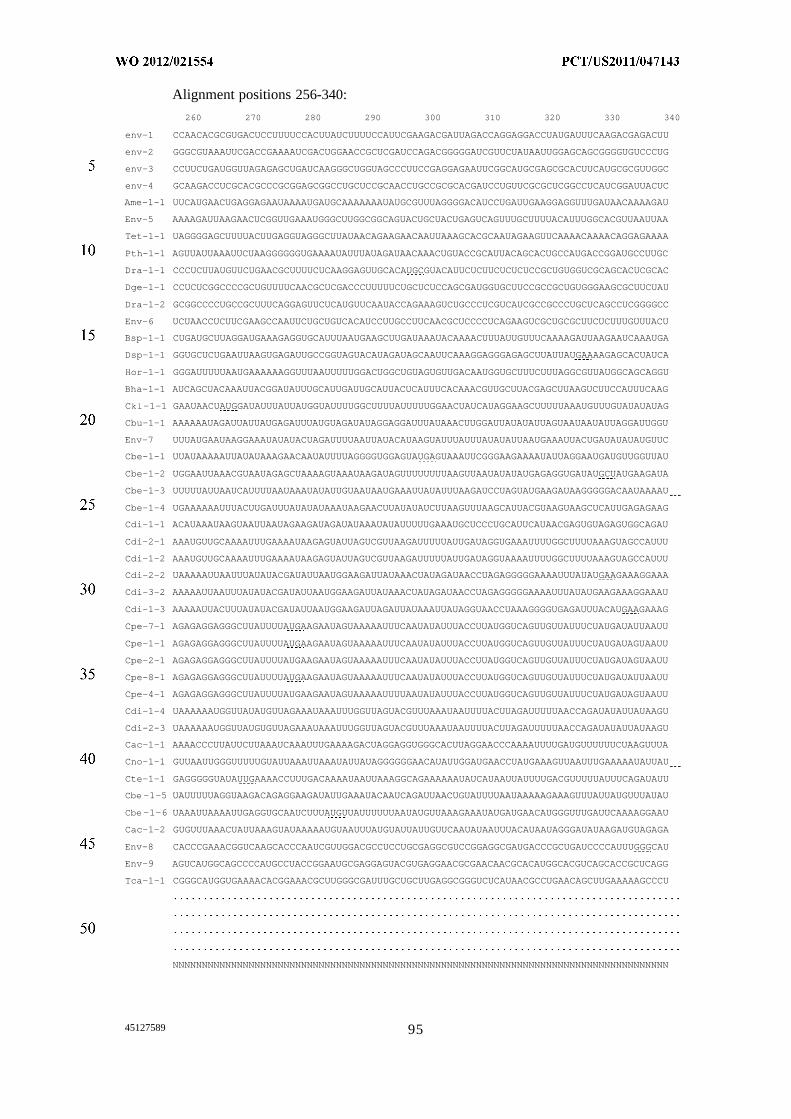
Alignment positions 256-340:
260 270 280 290 300 310 320 330 340
env-1 CCAACACGCGUGACUCCUUUUCCACUUAUCUUUUCCAUUCGAAGACGAUUAGACCAGGAGGACCUAUGAUUUCAAGACGAGACUU
env-2 GGGCGUAAAUUCGACCGAAAAUCGACUGGAACCGCUCGAUCCAGACGGGGGAUCGUUCUAUAAUUGGAGCAGCGGGGUGUCCCUG
env-3 CCUUCUGAUGGUUAGAGAGCUGAUCAAGGGCUGGUAGCCCUUCCGAGGAGAAUUCGGCAUGCGAGCGCACUUCAUGCGCGUUGGC
env-4 GCAAGACCUCGCACGCCCGCGGAGCGGCCUGCUCCGCAACCUGCCGCGCACGAUCCUGUUCGCGCUCGGCCUCAUCGGAUUACUC
Ame-1-1 UUCAUGAACUGAGGAGAAUAAAAUGAUGCAAAAAAAUAUGCGUUUAGGGGACAUCCUGAUUGAAGGAGGUUUGAUAACAAAAGAU
Env-5 AAAAGAUUAAGAACUCGGUUGAAAUGGGCUUGGCGGCAGUACUGCUACUGAGUCAGUUUGCUUUUACAUUUGGCACGUUAAUUAA
Tet-1-1 UAGGGGAGCUUUUACUUGAGGUAGGGCUUAUAACAGAAGAACAAUUAAAGCACGCAAUAGAAGUUCAAAACAAAACAGGAGAAAA
Pth-1-1 AGUUAUUAAAUUCUAAGGGGGGUGAAAAUAUUUAUAGAUAACAAACUGUACCGCAUUACAGCACUGCCAUGACCGGAUGCCUUGC
Dra-1-1 CCCUCUUAUGUUCUGAACGCUUUUCUCAAGGAGUUGCACAUGCGUACAUUCUCUUCUCUCUCCGCUGUGGUCGCAGCACUCGCAC
Dge-1-1 CCUCUCGGCCCCGCUGUUUUCAACGCUCGACCCUUUUUCUGCUCUCCAGCGAUGGUGCUUCCGCCGCUGUGGGAAGCGCUUCUAU
Dra-1-2 GCGGCCCCUGCCGCUUUCAGGAGUUCUCAUGUUCAAUACCAGAAAGUCUGCCCUCGUCAUCGCCGCCCUGCUCAGCCUCGGGGCC
Env-6 UCUAACCUCUUCGAAGCCAAUUCUGCUGUCACAUCCUUGCCUUCAACGCUCCCCUCAGAAGUCGCUGCGCUUCUCUUUGUUUACU
Bsp-1-1 CUGAUGCUUAGGAUGAAAGAGGUGCAUUUAAUGAAGCUUGAUAAAUACAAAACUUUAUUGUUUCAAAAGAUUAAGAAUCAAAUGA
Dsp-1-1 GGUGCUCUGAAUUAAGUGAGAUUGCCGGUAGUACAUAGAUAGCAAUUCAAAGGAGGGAGAGCUUAUUAUGAAAAGAGCACUAUCA
Hor-1-1 GGGAUUUUUAAUGAAAAAAGGUUUAAUUUUUGGACUGGCUGUAGUGUUGACAAUGGUGCUUUCUUUAGGCGUUAUGGCAGCAGGU
Bha-1-1 AUCAGCUACAAAUUACGGAUAUUUGCAUUGAUUGCAUUACUCAUUUCACAAACGUUGCUUACGAGCUUAAGUCUUCCAUUUCAAG
Ckl-1-1 GAAUAACUAUGGAUAUUUAUUAUGGUAUUUUGGCUUUUAUUUUUGGAACUAUCAUAGGAAGCUUUUUAAAUGUUUGUAUAUAUAG
Cbu-1-1 AAAAAAUAGAUUAUUAUGAGAUUUAUGUAGAUAUAGGAGGAUUUAUAAACUUGGAUUAUAUAUUAGUAAUAAUAUUAGGAUUGGU
Env-7 UUUAUGAAUAAGGAAAUAUAUACUAGAUUUUAAUUAUACAUAAGUAUUUAUUUAUAUAUUAAUGAAAUUACUGAUAUAUAUGUUC
Cbe-1-1 UUAUAAAAAUUAUAUAAAGAACAAUAUUUUAGGGGUGGAGUAUGAGUAAAUUCGGGAAGAAAAUAUUAGGAAUGAUGUUGGUUAU
Cbe-1-2 UGGAAUUAAACGUAAUAGAGCUAAAAGUAAAUAAGAUAGUUUUUUUUAAGUUAAUAUAUAUGAGAGGUGAUAUGCUAUGAAGAUA
Cbe-1-3 UUUUUAUUAAUCAUUUUAAUAAAUAUAUUGUAAUAAUGAAAUUAUAUUUAAGAUCCUAGUAUGAAGAUAAGGGGGACAAUAAAAU
Cbe-1-4 UGAAAAAAUUUACUUGAUUUAUAUAUAAAUAAGAACUUAUAUAUCUUAAGUUUAAGCAUUACGUAAGUAAGCUCAUUGAGAGAAG
Cdi-1-1 ACAUAAAUAAGUAAUUAAUAGAAGAUAGAUAUAAAUAUAUUUUUGAAAUGCUCCCUGCAUUCAUAACGAGUGUAGAGUGGCAGAU
Cdi-2-1 AAAUGUUGCAAAAUUUGAAAAUAAGAGUAUUAGUCGUUAAGAUUUUUAUUGAUAGGUGAAAUUUUGGCUUUUAAAGUAGCCAUUU
Cdi-1-2 AAAUGUUGCAAAAUUUGAAAAUAAGAGUAUUAGUCGUUAAGAUUUUUAUUGAUAGGUAAAAUUUUGGCUUUUAAAGUAGCCAUUU
Cdi-2-2 UAAAAAUUAAUUUAUAUACGAUAUUAAUGGAAGAUUAUAAACUAUAGAUAACCUAGAGGGGGAAAAUUUAUAUGAAGAAAGGAAA
Cdi-3-2 AAAAAUUAAUUUAUAUACGAUAUUAAUGGAAGAUUAUAAACUAUAGAUAACCUAGAGGGGGAAAAUUUAUAUGAAGAAAGGAAAU
Cdi-1-3 AAAAAUUACUUUAUAUACGAUAUUAAUGGAAGAUUAGAUUAUAAAUUAUAGGUAACCUAAAGGGGUGAGAUUUACAUGAAGAAAG
Cpe-7-1 AGAGAGGAGGGCUUAUUUUAUGAAGAAUAGUAAAAAUUUCAAUAUAUUUACCUUAUGGUCAGUUGUUAUUUCUAUGAUAUUAAUU
Cpe-1-1 AGAGAGGAGGGCUUAUUUUAUGAAGAAUAGUAAAAAUUUCAAUAUAUUUACCUUAUGGUCAGUUGUUAUUUCUAUGAUAGUAAUU
Cpe-2-1 AGAGAGGAGGGCUUAUUUUAUGAAGAAUAGUAAAAAUUUCAAUAUAUUUACCUUAUGGUCAGUUGUUAUUUCUAUGAUAGUAAUU
Cpe-8-1 AGAGAGGAGGGCUUAUUUUAUGAAGAAUAGUAAAAAUUUCAAUAUAUUUACCUUAUGGUCAGUUGUUAUUUCUAUGAUAUUAAUU
Cpe-4-1 AGAGAGGAGGGCUUAUUUUAUGAAGAAUAGUAAAAAUUUUAAUAUAUUUACCUUAUGGUCAGUUGUUAUUUCUAUGAUAGUAAUU
Cdi-1-4 UAAAAAAUGGUUAUAUGUUAGAAAUAAAUUUGGUUAGUACGUUUAAAUAAUUUUACUUAGAUUUUUAACCAGAUAUAUUAUAAGU
Cdi-2-3 UAAAAAAUGGUUAUGUGUUAGAAAUAAAUUUGGUUAGUACGUUUAAAUAAUUUUACUUAGAUUUUUAACCAGAUAUAUUAUAAGU
Cac-1-1 AAAACCCUUAUUCUUAAAUCAAAUUUGAAAAGACUAGGAGGUGGGCACUUAGGAACCCAAAAUUUUGAUGUUUUUUCUAAGUUUA
Cno-1-1 GUUAAUUGGGUUUUUGUAUUAAAUUAAAUAUUAUAGGGGGGAACAUAUUGGAUGAACCUAUGAAAGUUAAUUUGAAAAAUAUUAU
Cte-1-1 GAGGGGGUAUAUUGAAAACCUUUGACAAAAUAAUUAAAGGCAGAAAAAAUAUCAUAAUUAUUUUGACGUUUUUAUUUCAGAUAUU
Cbe-1-5 UAUUUUUAGGUAAGACAGAGGAAGAUAUUGAAAUACAAUCAGAUUAACUGUAUUUUAAUAAAAAGAAAGUUUAUUAUGUUUAUAU
Cbe-1-6 UAAAUUAAAAUUGAGGUGCAAUCUUUAUGUUAUUUUUUAAUAUGUUAAAGAAAUAUGAUGAACAUGGGUUUGAUUCAAAAGGAAU
Cac-1-2 GUGUUUAAACUAUUAAAGUAUAAAAAUGUAAUUUAUGUAUUAUUGUUCAAUAUAAUUUACAUAAUAGGGAUAUAAGAUGUAGAGA
Env-8 CACCCGAAACGGUCAAGCACCCAAUCGUUGGACGCCUCCUGCGAGGCGUCCGGAGGCGAUGACCCGCUGAUCCCCAUUUGGGCAU
Env-9 AGUCAUGGCAGCCCCAUGCCUACCGGAAUGCGAGGAGUACGUGAGGAACGCGAACAACGCACAUGGCACGUCAGCACCGCUCAGG
Tca-1-1 CGGGCAUGGUGAAAACACGGAAACGCUUGGGCGAUUUGCUGCUUGAGGCGGGUCUCAUAACGCCUGAACAGCUUGAAAAAGCCCU
NNNNNNNNNNNNNNNNNNNNNNNNNNNNNNNNNNNNNNNNNNNNNNNNNNNNNNNNNNNNNNNNNNNNNNNNNNNNNNNNNNNNN
45127589 95
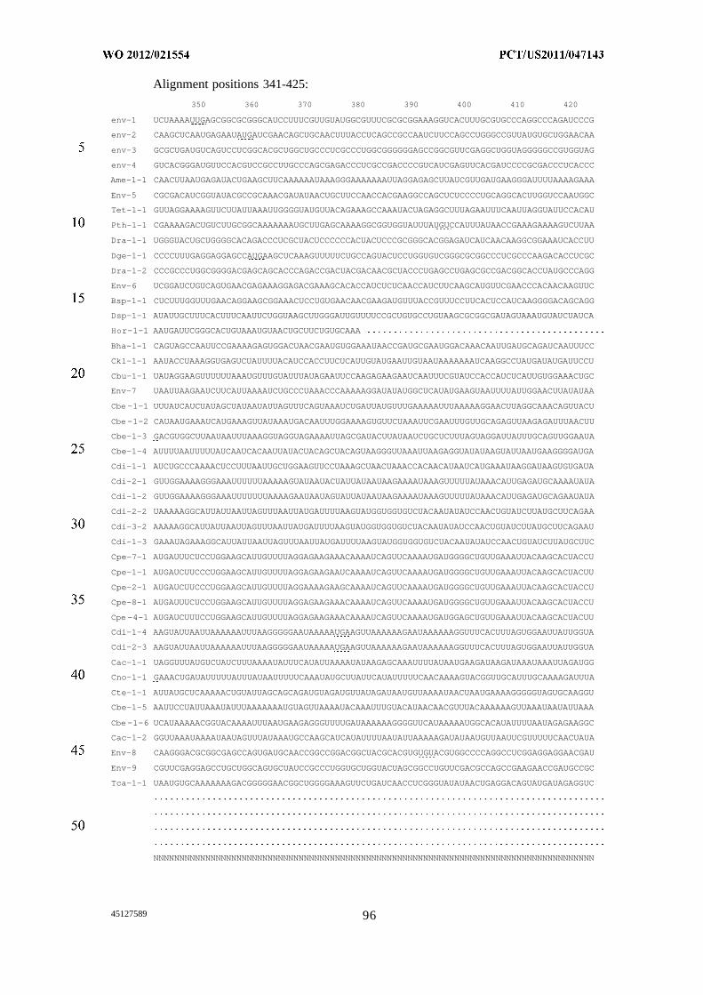
Alignment positions 341-425:
350 360 370 380 390 400 410 420
env-1 UCUAAAAUUGAGCGGCGCGGGCAUCCUUUCGUUGUAUGGCGUUUCGCGCGGAAAGGUCACUUUGCGUGCCCAGGCCCAGAUCCCG
env-2 CAAGCUCAAUGAGAAUAUGAUCGAACAGCUGCAACUUUACCUCAGCCGCCAAUCUUCCAGCCUGGGCCGUUAUGUGCUGGAACAA
env-3 GCGCUGAUGUCAGUCCUCGGCACGCUGGCUGCCCUCGCCCUGGCGGGGGGAGCCGGCGUUCGAGGCUGGUAGGGGGCCGUGGUAG
env-4 GUCACGGGAUGUUCCACGUCCGCCUUGCCCAGCGAGACCCUCGCCGACCCCGUCAUCGAGUUCACGAUCCCCGCGACCCUCACCC
Ame-1-1 CAACUUAAUGAGAUACUGAAGCUUCAAAAAAUAAAGGGAAAAAAAUUAGGAGAGCUUAUCGUUGAUGAAGGGAUUUUAAAAGAAA
Env-5 CGCGACAUCGGUAUACGCCGCAAACGAUAUAACUGCUUCCAACCACGAAGGCCAGCUCUCCCCUGCAGGCACUUGGUCCAAUGGC
Tet-1-1 GUUAGGAAAAGUUCUUAUUAAAUUGGGGUAUGUUACAGAAAGCCAAAUACUAGAGGCUUUAGAAUUUCAAUUAGGUAUUCCACAU
Pth-1-1 CGAAAAGACUGUCUUGCGGCAAAAAAAUGCUUGAGCAAAAGGCGGUGGUAUUUAUGUCCAUUUAUAACCGAAAGAAAAGUCUUAA
Dra-1-1 UGGGUACUGCUGGGGCACAGACCCUCGCUACUCCCCCCACUACUCCCGCGGGCACGGAGAUCAUCAACAAGGCGGAAAUCACCUU
Dge-1-1 CCCCUUUGAGGAGGAGCCAUGAAGCUCAAAGUUUUUCUGCCAGUACUCCUGGUGUCGGGCGCGGCCCUCGCCCAAGACACCUCGC
Dra-1-2 CCCGCCCUGGCGGGGACGAGCAGCACCCAGACCGACUACGACAACGCUACCCUGAGCCUGAGCGCCGACGGCACCUAUGCCCAGG
Env-6 UCGGAUCUGUCAGUGAACGAGAAAGGAGACGAAAGCACACCAUCUCUCAACCAUCUUCAAGCAUGUUCGAACCCACAACAAGUUC
Bsp-1-1 CUCUUUGGUUUGAACAGGAAGCGGAAACUCCUGUGAACAACGAAGAUGUUUACCGUUUCCUUCACUCCAUCAAGGGGACAGCAGG
Dsp-1-1 AUAUUGCUUUCACUUUCAAUUCUGGUAAGCUUGGGAUUGUUUUCCGCUGUGCCUGUAAGCGCGGCGAUAGUAAAUGUAUCUAUCA
Hor-1-1 AAUGAUUCGGGCACUGUAAAUGUAACUGCUUCUGUGCAAA
Bha-1-1 CAGUAGCCAAUUCCGAAAAGAGUGGACUAACGAAUGUGGAAAUAACCGAUGCGAAUGGACAAACAAUUGAUGCAGAUCAAUUUCC
Ckl-1-1 AAUACCUAAAGGUGAGUCUAUUUUACAUCCACCUUCUCAUUGUAUGAAUUGUAAUAAAAAAAUCAAGGCCUAUGAUAUGAUUCCU
Cbu-1-1 UAUAGGAAGUUUUUUAAAUGUUUGUAUUUAUAGAAUUCCAAGAGAAGAAUCAAUUUCGUAUCCACCAUCUCAUUGUGGAAACUGC
Env-7 UAAUUAAGAAUCUUCAUUAAAAUCUGCCCUAAACCCAAAAAGGAUAUAUGGCUCAUAUGAAGUAAUUUUAUUGGAACUUAUAUAA
Cbe-1-1 UUUAUCAUCUAUAGCUAUAAUAUUAGUUUCAGUAAAUCUGAUUAUGUUUGAAAAAUUUAAAAAGGAACUUAGGCAAACAGUUACU
Cbe-1-2 CAUAAUGAAAUCAUGAAAGUUAUAAAUGACAAUUUGGAAAAGUGUUCUAAAUUCGAAUUUGUUGCAGAGUUAAGAGAUUUAACUU
Cbe-1-3 GACGUGGCUUAAUAAUUUAAAGGUAGGUAGAAAAUUAGCGAUACUUAUAAUCUGCUCUUUAGUAGGAUUAUUUGCAGUUGGAAUA
Cbe-1-4 AUUUUAAUUUUUAUCAAUCACAAUUAUACUACAGCUACAGUAAGGGUUAAAUUAAGAGGUAUAUAAGUAUUAAUGAAGGGGAUGA
Cdi-1-1 AUCUGCCCAAAACUCCUUUAAUUGCUGGAAGUUCCUAAAGCUAACUAAACCACAACAUAAUCAUGAAAUAAGGAUAAGUGUGAUA
Cdi-2-1 GUUGGAAAAGGGAAAUUUUUUAAAAAGUAUAAUACUAUUAUAAUAAGAAAAUAAAGUUUUUAUAAACAUUGAGAUGCAAAAUAUA
Cdi-1-2 GUUGGAAAAGGGAAAUUUUUUUAAAAGAAUAAUAGUAUUAUAAUAAGAAAAUAAAGUUUUUAUAAACAUUGAGAUGCAGAAUAUA
Cdi-2-2 UAAAAAGGCAUUAUUAAUUAGUUUAAUUAUGAUUUUAAGUAUGGUGGUGUCUACAAUAUAUCCAACUGUAUCUUAUGCUUCAGAA
Cdi-3-2 AAAAAGGCAUUAUUAAUUAGUUUAAUUAUGAUUUUAAGUAUGGUGGUGUCUACAAUAUAUCCAACUGUAUCUUAUGCUUCAGAAU
Cdi-1-3 GAAAUAGAAAGGCAUUAUUAAUUAGUUUAAUUAUGAUUUUAAGUAUGGUGGUGUCUACAAUAUAUCCAACUGUAUCUUAUGCUUC
Cpe-7-1 AUGAUUUCUCCUGGAAGCAUUGUUUUAGGAGAAGAAACAAAAUCAGUUCAAAAUGAUGGGGCUGUUGAAAUUACAAGCACUACCU
Cpe-1-1 AUGAUCUUCCCUGGAAGCAUUGUUUUAGGAGAAGAAUCAAAAUCAGUUCAAAAUGAUGGGGCUGUUGAAAUUACAAGCACUACUU
Cpe-2-1 AUGAUCUUCCCUGGAAGCAUUGUUUUAGGAAAAGAAGCAAAAUCAGUUCAAAAUGAUGGGGCUGUUGAAAUUACAAGCACUACCU
Cpe-8-1 AUGAUUUCUCCUGGAAGCAUUGUUUUAGGAGAAGAAACAAAAUCAGUUCAAAAUGAUGGGGCUGUUGAAAUUACAAGCACUACCU
Cpe-4-1 AUGAUCUUUCCUGGAAGCAUUGUUUUAGGAGAAGAAACAAAAUCAGUUCAAAAUGAUGGAGCUGUUGAAAUUACAAGCACUACUU
Cdi-1-4 AAGUAUUAAUUAAAAAAUUUAAGGGGGAAUAAAAAUGAAGUUAAAAAAGAAUAAAAAAGGUUUCACUUUAGUGGAAUUAUUGGUA
Cdi-2-3 AAGUAUUAAUUAAAAAAUUUAAGGGGGAAUAAAAAUGAAGUUAAAAAAGAAUAAAAAAGGUUUCACUUUAGUGGAAUUAUUGGUA
Cac-1-1 UAGGUUUAUGUCUAUCUUUAAAAUAUUUCAUAUUAAAAUAUAAGAGCAAAUUUUAUAAUGAAGAUAAGAUAAAUAAAUUAGAUGG
Cno-1-1 GAAACUGAUAUUUUUAUUUAUAAUUUUUCAAAUAUGCUUAUUCAUAUUUUUCAACAAAAGUACGGUUGCAUUUGCAAAAGAUUUA
Cte-1-1 AUUAUGCUCAAAAACUGUAUUAGCAGCAGAUGUAGAUGUUAUAGAUAAUGUUAAAAUAACUAAUGAAAAGGGGGUAGUGCAAGGU
Cbe-1-5 AAUUCCUAUUAAAUAUUUAAAAAAAUGUAGUUAAAAUACAAAUUUGUACAUAACAACGUUUACAAAAAAGUUAAAUAAUAUUAAA
Cbe-1-6 UCAUAAAAACGGUACAAAAUUUAAUGAAGAGGGUUUUGAUAAAAAAGGGGUUCAUAAAAAUGGCACAUAUUUUAAUAGAGAAGGC
Cac-1-2 GGUUAAAUAAAAUAAUAGUUUAUAAAUGCCAAGCAUCAUAUUUUAAUAUUAAAAAGAUAUAAUGUUAAUUCGUUUUUCAACUAUA
Env-8 CAAGGGACGCGGCGAGCCAGUGAUGCAACCGGCCGGACGGCUACGCACGUGUGUACGUGGCCCCAGGCCUCGGAGGAGGAACGAU
Env-9 CGUUCGAGGAGCCUGCUGGCAGUGCUAUCCGCCCUGGUGCUGGUACUAGCGGCCUGUUCGACGCCAGCCGAAGAACCGAUGCCGC
Tca-1-1 UAAUGUGCAAAAAAAGACGGGGGAACGGCUGGGGAAAGUUCUGAUCAACCUCGGGUAUAUAACUGAGGACAGUAUGAUAGAGGUC
NNNNNNNNNNNNNNNNNNNNNNNNNNNNNNNNNNNNNNNNNNNNNNNNNNNNNNNNNNNNNNNNNNNNNNNNNNNNNNNNNNNNN
45127589 96
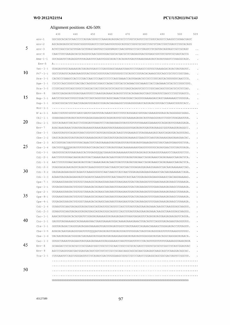
Alignment positions 426-509:
430 440 450 460 470 480 490 500 509
env-1 GGCGGCACACUCAACCCCCAUGACGUGCCCAAAUAUAUGACGCCCCUGCUCAUUCCGCCGGUCAUGCCCAAGGCCGGAACGAUC
env-2 AGCAUAUAUGCGCUGGCGGGUUGGAUCCCGUCAAUUGUUGGCAUUGCCGUGCGCGGCCUGCUCUACCGUCUGAUCCUGCACAUG
env-3 ACUCCGGCCGCGCUUGACGCUUAGCGAUUGCCGGUGNGUCCAACGUUGCCCGCCUAGUCCUCAUUACAGUAGCCGCCGCAGU ..
env-4 CAACCUUCGAAGACACGCAGGUGCAACGGUUGGACGGCACGACGCUCGAGAGUGACGUGAGCGUCUUCGUUCGCGAUCACCG ..
Ame-1-1 GUCAGAUUCUAGAGGUUUUAGAAUUUCAAUUGGGCAUACCACAUAUUGAUUUAAAUAAAUAUCAUAUUAAUCCUAAGGCAGU. .
Env-5 A
Tet- 1-1 GUAGAUUUGCAAAAAUACUAUAUUGAUCCCGAUGUAGCAAAAUUAAUUCCUGAAGCCGUUGCUAAAAGACAUACUAUUAUUC. .
Pt -1-1 GGCCUUAUUCAGAAUAAGUUUACUUGCUGUUUUACUGUUGUGCCUCAUUCCGUUACACAAAGCGUCAGCCGCUUCCGGCGAA. .
Dra- 1-1 CACGCCCGAAUCCACCCCGACCAACCCCAACCCCCCCAGCGAAACCAUUGAGACGCCGCCCGUCAUCACGGUGGUCAACCCG. .
Dge- 1-1 CUCUCCAGCUGGUCUACGACCAGGUGCUGAUCCAGACCGUCACGCAGAACGGCAAAGCCACCGAGAAACGCACGCCGGGCGUG.
Dra- 1-2 CCGUCAGCUUCAGCGUGCCCAGCACCACCGUCACGCUCAGCGCCGAGCAGAUUCGCCCCGGCAACAGCUUCACGCUCUCCAU. .
Env- 6 UAUUCGAGAUAUUGGAUGAAUUUGCCUAAGGAGAAACAGAUGCUUCACAGAAUAUCGAUCGUAUUUCGAGCCCUGCUGAUCG. .
Bsp- 1-1 AACUCUGCAGCUUGUCGGACUCCAUCAGGUGGCAGGCAAACUGAUGGACCAGGUGGAAAAGAGCAGCGAAAAAAUCUGGGGA. .
Dsp- 1-1 GCAGCGGCACUUCAACUGAGAUUGUAGGCGUAUACAAUAAGGCUGGAGGGGGAUCAGUAUACGUUGACCUAAGCGGUUCACC. .
Hor- 1-1
B a-1-1 AGACCGUUUCGUUUCGAUCGAUUCGAUUUUACAAAUCAUCCUUUCAUGGAGCGUUGACGAAAGUGUAGCACAGGGGGCGGAG.
Ckl- 1-1 GUAAUAAGUUAUAUCAUUUUGAGAGGAAGAUGCAGAUAUUGCGGCAAAAAGAUACAUUUAAGGUAUCCUGUCUUAGAAUUUA.
Cbu- 1-1 GGUCAUAAUCUACAGCCUGUAGAUUUAAUUCCUAUAAUAAGUUAUGUGUUUUUAAAAGGAAAAUGCAGAUAUUGUAAGGAGA.
Env- 7 AUACAAAUAAACUUAUUAUAAAAAUUAAAUAAAUUAUUAAAAGGGGUUGAUAUAUUGAUUAUAAGGCGGUUAAGUAUAGGCC.
Cbe- 1-1 CAAUGUAUUUCAGAUUUAACUGUUUCCAUUGAUGGAGACAAGCUUGAAAAGCUUAUAAAAGAGCAAUCAGAUGACAGUGUAG.
Cbe- 1-2 UAGCAGAUAUGUAUUAUAUUGAAAAGAUAUCGAGUAUUGAUAGUAUAAAAGCGAAGUUCAAUUAUAAAAUAAUAAAUAACAC.
Cbe- 1-3 ACCGGUUACUAUUUUUUACAAACGUCUAGUAAAAGUAUGGAGGUUAUGUAUAGUGAAAGAUUGCUAUCAAGUGAAUGGCUUA.
Cbe- 1-4 GACUGUUAUGGAUAUUUUUGUAGCUAGACAGCCUAUAUUUAACAAAAAAAAUGAGGUUGUUGCAUAUGAACUUCUAUUUAGA.
Cdi- 1-1 GAGUGUGCAUUUAAUAAGCACUUGAGGUUACGAAAGUAGAAAAAAUUAGUUAGAUAGCAUAAGGUUAAAUCCUAAGUGCCUU.
Cdi- 2-1 AACCUUUUGUAACAAUAUAUUUACCAAAAUAAUACAAUUUAUUUUAUAUUAUAACCAUAUAAAUCAUAUAAAUCAAUACUCA.
Cdi- 1-2 AACCUUUUGUAACAAUAUAUUUACCAAAAUAAUACAAUUUAUUUUAUAUUAUAACCAUAUAAAUCAUAUAAAUCAAUACUCA.
Cdi- 2-2 UUAGGAGAGAAUAGUCAGAUUCAAAGUGGUUCAACUAAUUCAUCAACUGGAGAGGAGAAGGAAAGCGACAAUAAAAAACCAG.
Cdi- 3-2 UAGGAGAGAAUAGUCAGAUUCAAAGUGGUUCAACUAAUUCAUCAACUGGAGAGGAGAAGGAAAGCGACAAUAAAAAACCAGA.
Cdi- 1-3 AGAAUUAGGAGAGAAUAGUCAGAUUCAAAGUGGUUCAACUAAUUCAUCAACUGGAGAGGAGAAGGAAAGCGACAAUAAAAAG.
Cpe- 7-1 UUGAAAGUAAUACUGUUGCUAAAGGCAUAAGUAAUAAUUUAAGAAUUGACUAUAAGAUUUUGAAUAAAGAUAAGCUUAAAGA.
Cpe- 1-1 UUGAGAGUAAUACUGUGGCUAAAGACAUAAGCAAUAAUUUAAGAAUUGACUAUAAGAUUUUGAAUAAAGAUAAGCUUAAAGA.
Cpe- 2-1 UUGAAAGUAAUACUGUUGCUAAAGACAUAAGCAAUAAUUUAAGAAUUGAUUAUAAGAUUUUGAAUAAAGAUAAGCUUAAAGA.
Cpe- 8-1 UUGAAAGUAAUACUGUUGCUAAAGGCAUAAGCAAUAAUUUAAGAAUUGACUAUAAGAUUUUGAAUAAAGAUAAGCUUAAAGA.
Cpe- 4-1 UUGAGAGUAAUACUGUGGCUAAAGACAUAAGCAAUAAUUUAAGAAUUGACUAUAAGAUUUUGAAUAAAGAUAAGCUUAAAGA.
Cdi- 1-4 GUAAUUGCAAUUAUAGGUAUAUUAGCAGUAGUGGCAGUUCCAGCUUUAUUUAGUAAUAUAAACAAGGCUAAGGUAGCAAGUG.
Cdi- 2-3 GUAAUUGCAAUUAUAGGUAUAUUAGCAGUAGUGGCAGUUCCAGCUUUAUUUAGUAAUAUAAACAAGGCUAAGGUAGCAAGUG.
Cac- 1-1 AAACAUUGAUACACAUGAUUCGGAUAUAAAAAGUAUAAGAGAAUUUAAUGAGAGUUCAGAUAUAGUAAGAGAAGAUUCAGUA.
Cno- 1-1 GAUGUUAUAAAAAGCAUAAAAAUAACUAAUGAAAAUGGACAAAAUAAAGAAACUUACAUUCCAGGUGAUAGAAUUAGGGUUG.
Cte- 1-1 GGUUAUAGACCAGGUGAUAGAAUAAAAAUUGAUGUAGAUUGGUCUAUUAAAUCAGAAACAAAAGCUGGAGAUACCUUUAGUU.
Cbe- 1-5 AUAUACAAUGAGAGGUGAUUUUUUGGAUUAUAUAUUUAUAUGUAUUUUGGGACUAGUUAUAGGUAGUUUUUUAAAUGUUUGU.
Cbe- 1-6 UACAAUAUAGACGGGUACGAUAAAUAUGGAUAUGAUAAAGAAGGAUAUAAUAGUGGAGGAUAUGACAGGCAAGGAUAUAACA.
Cac- 1-2 GUGGCUAAAUUGAGAAUUAUGAAGAUAAAUGUAGGAAAAUCAAUUUGAUUUUCCUACAUUUGUUUUUUGAAAAGGUAAAUAUA
Env- 8 GCAAGACCUCGCACGCCCGCGGAGCGGCCUGCUCCGCAACCUGCCGCGCACGAUCCUGUUCGCGCUCGGCCUCAUCGGAUUAC
Env- 9 AGCCCGAGUUGACGACGGAUGACAUCGUCUUCUCCGCCGCAACAAGCAGCACAACGGAGAUCAAGCAGCUCAAGGACAGCAC.
Tca- 1-1 CUUGAAUUCCAGCUGGGAGUUCCUCAUAUCGACUUGGGAAGCGUGCCGCCCGAUCCGGAGGCAGCGGCGACUAUUCCGGCUU.
NNNNNNNNNNNNNNNNNNNNNNNNNNNNNNNNNNNNNNNNNNNNNNNNNNNNNNNNNNNNNNNNNNNNNNNNNNNNNNNNNN
45127589 97
The RNAs reported herein are predicted to form two internal bulges using three
base-paired regions (PI, P2 and P3), and an imperfect pseudoknot structure between
nucleotides of the P3 loop (L3) and the junction linking stems P2 and PI (J2-1) (Figure
1A). The internal bulge between P2 and P3 conforms to a kink-turn motif (Klein et al.
EMBOJ. 20, 4214 (2001); Winkler et al. RNA 7, 1165 (2001)). Although, 24 of the 28
bacterial species that carry this riboswitch also carry c-di-GMP -I riboswitches, it was
realized that representatives of the new RNA motif serve as aptamers for a distinct class of
c-di-GMP riboswitches, termed c-di-GMP-II.
A representative RNA from C. difficile was subjected to in-line probing (Soukup
and Breaker, RNA 5, 1308 (1999); E. E . Regulski, R. R. Breaker, Methods Mol. Biol. 419,
53 (2008)). This method exploits the instability of RNA phosphodiester linkages in
unstructured portions of an RNA polymer to yield information on ligand-mediated
structural changes and ligand binding affinities. An 84-nucleotide 5 ' P-labeled RNA
(termed 84 Cd), corresponding to the conserved motif located upstream of a putative
virulence gene (CD3246) in C. difficile strain 630 (Figure IB), was incubated in the
absence or presence of 10 µΜ c-di-GMP under in-line probing conditions. Spontaneous
RNA cleavage products were separated by denaturing polyacrylamide gel electrophoresis
(PAGE), and band locations and intensities (Figure 1C) were used to map regions of
structural change. The observed pattern of cleavage products produced in the absence of c-
di-GMP is consistent with the structural model generated by comparative sequence
analysis. Furthermore, 11 internucleotide linkages in four spans within the L3 and J2-1
regions exhibit reduced strand scission when c-di-GMP is present (Figures IB and 1C),
which indicates that the RNA structure is stabilized by binding the second messenger.
In-line probing reactions using a range of c-di-GMP concentrations resulted in a
binding curve with an apparent dissociation constant (K ) of -200 pM (Figure ID). The
concentrations of c-di-GMP range from a minimum of 260 fM to a maximum of 1.2 µΜ ,
with increments of 3-fold increases in concentration. This apparent K∑, approaches the
limit of sensitivity of the assay (Regulski and Breaker, Methods Mol. Biol. 419, 53
(2008)), and therefore the true affinity may be even better. Similarly, the affinities were
determined for various analogs of c-di-GMP, revealing that the RNA discriminates against
the linear form of the second messenger and other analogs by more than three orders of
magnitude (Figure IE). The molecular recognition characteristics displayed by this RNA
are similar to those observed for c-di-GMP-I aptamers (Sudarsan et al. Science 321, 4 11
(2008); K. D. Smith, et al. Nat. Struct. Mol. Biol. 16, 1218 (2009)), and therefore it is
45127589
concluded that the RNA represents a new class of aptamers for this second messenger
termed c-di-GMP-II.
i. c-di-GMP-II aptamers are components of riboswitches
The 84 Cd aptamer from the CD3246 locus is not associated with any commonly-
observed expression platform. Therefore, gene control function was confirmed for this
aptamer class by using a representative RNA located upstream of the gene at the BH0361
locus from Bacillus halodurans (Figure 2A). The BH0361 RNA is predicted to regulate
gene expression by controlling the formation of an intrinsic transcription terminator stem
(Soukup and Breaker, RNA 5, 1308 (1999); Regulski and Breaker, Methods Mol. Biol.
419, 53 (2008)). The ligand-bound state of this aptamer was predicted to preclude
formation of an intrinsic transcription terminator (strong stem followed by a run of U
residues) (Yarnell and Roberts, Science 284, 6 11 (1999); Gusarov and Nudler, Mol. Cell 3,
495 (1999)), and therefore c-di-GMP binding was expected to activate gene expression.
A transcriptional fusion was created of the predicted riboswitch element or its
corresponding Ml, M2 and M3 mutants to a β-galactosidase reporter gene and
transformed the construct into a surrogate organism, Bacillus subtilis. High levels of
reporter gene expression were observed with the wild-type (WT) and compensatory (M3)
constructs, but reduced expression resulted when two conserved bases in Jl/2 were
exchanged (Ml), or when the P3 stem was disrupted (M2) (Figure 2B). These constructs
represent constructs with heterologous aptamer domains and expression platform domains
and with heterologous riboswitch and coding region. Reporter gene fusions with WT or
mutant motifs transformed into Bacillus subtilis confirmed the BH0361 locus RNA is a
genetic ON' switch. These results are consistent with the proposed function of the B.
halodurans BH0361 locus RNA as a genetic ON' switch.
Further evidence of aptamer-controlled transcription termination was observed by
conducting single-round transcription assays in vitro using another C. difficile c-di-GMP-
II construct from the ompR mRNA (Figure 2C). As predicted from the expression platform
architecture, the WT ompR construct also performs as an ON' switch and exhibits robust
production of full-length RNA transcript only when c-di-GMP is added to the reaction
(Figure 2D). Mutations that disrupt P3 stem formation (M2) substantially reduce
termination control, whereas additional mutations that restore P3 formation (M3) also
restore function to near WT level.
45127589 99
ii. A tandem aptamer-ribozyme arrangement
The c-di-GMP-II aptamer from the CD3246 locus is very unusual because it lacks
a typical riboswitch expression platform and it is located more than 600 nucleotides
upstream of the putative AUG start codon for its associated open reading frame (ORF).
The nucleotide sequence and structure of this unusually large gap correspond to a group I
self-splicing ribozyme (Kruger et al. Cell 31, 147 (1982); Nielsen and Johansen, RNA Biol.
6, 375 (2009)) (Figure 3A; Figure 6). The predicted ribozyme has all the hallmarks of a
typical group I ribozyme, including stems PI through P10, and a U-G wobble base pair
that defines a typical 5 ' splice site (5 ' SS).
The first step of group I ribozyme function normally is triggered solely by
guanosine or one of its phosphorylated derivatives such as GTP. The 3 ' oxygen atom of
GTP functions as the nucleophile to initiate a phosphoester transfer reaction at the 5 '
splice site (5 ' SS). The second step involves a nucleophilic attack of the newly liberated 3 '
oxygen atom of the 5' exon to initiate another phosphoester transfer reaction at the 3'
splice site (3 ' SS) to yield spliced 5 ' and 3 ' exons (5Έ -3Έ ) and a linear intron or
intervening sequence (IVS). The IVS can subsequently undergo one or more
transesterification and hydrolysis reactions to create various circular and linear intron
fragments (Yarnell and Roberts, Science 284, 6 11 (1999)).
Some bacteria exploit tandem riboswitch architectures to create sophisticated gene
control systems that yield more digital gene control (Mandal et al. Science 306, 275
(2004); Welz and Breaker, RNA 13, 573 (2007)), two-input logic gates (Sudarsan et al.
Science 13, 300 (2006)), or dual-mechanism switches (Poiata et al. RNA 15, 2046 (2009)).
Furthermore, RNA engineering studies have demonstrated that the fusion of aptamer and
ribozyme domains can produce allosteric constructs wherein ligand binding to the aptamer
results in regulation of ribozyme activity (Breaker, Curr. Opin. Biotechnol. 13, 3 1 (2002);
Silverman, RNA 9, 377 (2003)). Taking note of the existence of these more complex
natural and engineered RNA switches, and of the fact that eukaryotic riboswitches that
sense thiamin pyrophosphate (TPP) control nuclear splicing (Cheah et al. Nature 447, 497
(2007); Wachter et al. Plant Cell 19, 3437 (2007); Bocobza et al. Genes Dev. 21, 2874
(2007); Croft et al. Proc. Natl. Acad. Sci. USA 104, 20770 (2007)), it was realized and
discovered that C. difficile 630 carries the first example of a natural allosteric ribozyme
wherein c-di-GMP binding controls self-splicing. Only 10 nucleotides separate the 3'
terminus of the 84 Cd aptamer domain and the typical group I ribozyme 5 ' splice site (5 '
45127589 100
SS) predicted for this R A. Thus the aptamer is well positioned to regulate ribozyme
access to one of its splice site junctions.
iii. An allosteric self-splicing ribozyme
Normally, group I ribozyme splicing is triggered exclusively by guanosine or one
of its phosphorylated derivatives. The 3 ' oxygen atom of the nucleoside functions as the
attacking nucleophile to initiate a phosphoester transfer reaction at the 5' splice site (5 '
SS), which constitutes the first step of splicing. The second step involves a nucleophilic
attack of the newly liberated 3 ' oxygen atom of the 5 ' exon to initiate another
phosphoester transfer reaction at the 3 ' splice site (3 ' SS), which yields spliced 5 ' and 3 '
exons (5Έ -3Έ ) and a linear intron or intervening sequence (IVS). The IVS can
subsequently undergo one or more transesterification and hydrolysis reactions to create
various circular and linear fragments of the intron (Kruger et al. Cell 31, 147 (1982)).
Allosteric control of the tandem aptamer-ribozyme arrangement was established in
vitro by using 864-nucleotide internally P-labeled RNAs (termed 864 Cd Tandem;
Figure 3A, Figure 6) that carry both the aptamer and ribozyme domains, as well as
additional nucleotides that form part of the ORF. Incubation of the wild-type 864 Cd
Tandem RNA with GTP triggers splicing to yield several products that are characteristic
of group I ribozyme activity (Figure 3B). Importantly, the yield of spliced exons increases
substantially when both GTP and c-di-GMP are present. The dose-response curve for c-di-
GMP induction of splicing fits a 1-to-l binding curve with half-maximal production of
spliced exons occurring at a second messenger concentration of 30 nM (Figure 7).
In addition, the yield of another RNA product (3Έ *), subsequently determined to
be a fragment of the 3' exon caused by GTP attack far from the normal 5' SS, is reduced
in the presence of c-di-GMP (Figure 3B). Aptamer mutants Ml and M2 that disrupt c-di-
GMP binding no longer modulate product yields when the second messenger is included
in the splicing reaction. In contrast, mutant M3 carries nucleotide changes to M2 that
restore aptamer P3 stem formation and also recovers c-di-GMP-mediated splicing control
(Figure 3B). These results demonstrate that c-di-GMP addition and proper function of the
adjoining aptamer trigger increased production of spliced exons by the ribozyme.
A comparison of ribozyme reaction products detected by using differently-
radiolabeled RNAs and GTP was performed. The were products generated by using either
(1) internally P-labeled 864 Cd Tandem RNA and unlabeled GTP, or (2) unlabeled 864
Cd Tandem RNA and [a- P]GTP. Products were also generated by using either (1)
internally P-labeled 864 Cd Tandem RNA and unlabeled GTP, or (2) 5 ' P-labeled 864
45127589 Q
Cd Tandem RNA and unlabeled GTP. Additional annotations are as described in the
description of Figure 3B. Kinetic assays conducted under similar conditions (see Figure
4) reveal rate constants for unlabeled 864 Cd Tandem RNA and [a- P]GTP of 2.3 x 10 2
min 1 in the presence of both GTP and c-di-GMP versus 1.8 x 10 min 1 when only GTP is
present. There is only a small reduction in the initial rate constant for GTP attack at the
alternative GTP2 site (2.7 x 10 2 min 1 versus 2.3 x 10 2 min 1) . Internally-labeled 864 Cd
Tandem RNAs incubated with GTP exhibited kobs values of 1.1 x 10 3 min 1 and 1.3 x 10
min-1 in the absense and presence of c-di-GMP, respectively. Again, there is only a
modest reduction in the initial kobs for production of the alternative GTP attack site
product in the absence (2.4 x 10 2 min 1) versus the presence (1.8 x 10 2 min 1) of the
second messenger. See also Table 5 .
iv. Mechanism of allosteric splicing control
Two alternative base pairing structures were noticed that could explain how c-di-
GMP binding controls splicing. The first alternative stem, called anti-5 ' SS (Figure 3A,
enclosed in dased line oval), includes the left shoulder of the aptamer PI (nucleotides 8
through 17), which are complementary to nucleotides (90 through 99) that link the
aptamer and ribozyme domains. The second alternative stem, called alternative ribozyme
PI (Figure 3A, enclosed in alternating dash dot oval), includes the right shoulder of
ribozyme PI (nucleotides 186 through 189), which are complementary to nucleotides (667
through 670) near the 3 ' end of the ribozyme.
Formation of the anti-5 ' SS stem disrupts two base pairs of the ribozyme PI stem.
Since c-di-GMP is expected to stabilize the proposed aptamer structure, the absence of c-
di-GMP will weaken the aptamer PI stem, and favor formation of the anti-5 'SS stem.
Therefore, low concentrations of c-di-GMP can inhibit formation of the stem carrying the
5' SS and prevent GTP attack at the 5' SS. This was examined by conducting in-line
probing on a 132-nucleotide construct encompassing the aptamer through nucleotide 130
of the tandem RNA. This construct isolates nucleotides predicted to be involved in
forming the anti-5 ' SS stem, and PI stems of the aptamer and ribozyme. As predicted, c-
di-GMP addition yields a spontaneous cleavage pattern consistent with aptamer PI stem
formation, while the absence of the second messenger permits the RNA to form the anti-5 '
SS alternative pairing (Figure 8A). The changes in the pattern of in-line probing data
upon addition of c-di-GMP are consistent with formation of the alternative base-paired
structure in the absence of ligand. Concentrations of c-di-GMP range from a minimum of
260 fM to a maximum of 1.2 µΜ , with increments of 9-fold increases in concentration.
451 2 7589 ()2
Mutants M l through M3 were prepared to examine the role of c-di-GMP binding
on ribozyme splicing. Mutants M4 through Mi l were prepared to assess other structures
proposed to be important for allosteric ribozyme function, and mutant 1 was prepared to
examine this group I ribozyme 's usage of the typical guanosine binding pocket. The
effects on self-splicing regulation of mutant M4, which retains aptamer PI formation but
disrupts the first alternative pairing, were also examined. M4 retains c-di-GMP binding
activity (Figure 8B), but splicing is no longer responsive to the second messenger,
indicating that the ability to form the anti-5 ' SS stem is necessary for allosteric control of
ribozyme activity. Moreover, although M4 is no longer responsive to c-di-GMP, the yield
of spliced exons is higher in the absence of the second messenger comparted to WT,
which is expected if the aptamer PI stem is not effectively competing with anti-5 ' SS stem
formation.
Construct M5, which carries mutations in the linker between the aptamer and the
ribozyme, was expected to have similar activity as M4. This mutant loses splicing control
as expected, but the RNA is defective in ligand binding and does not produce fully spliced
products. Although the latter results are unexpected, in-line probing reveals that this
mutant RNA is misfolded (Figure 8C), which accounts for its lack of function. In contrast,
placing M4 and M5 mutations in the same construct (M6) restores the potential for anti-5 '
SS stem formation (Figure 8D), and likewise restores allosteric activity of the construct.
Collectively, the characteristics of these mutants strongly support the allosteric control
mechanism involving mutually exclusive formation of aptamer PI and anti-5' SS stems.
Similar experiments using additional mutants yield results that also support the formation
of the alternative ribozyme PI stem.
v . Abnormal splice site junctions
Several lines of evidence reveal that this group I ribozyme uses splice and GTP
attack sites that are distinct from most other group I ribozymes previously examined. This
distinction is independent of allosteric regulation, but clarifies the origin of several
reaction products. Sequence analysis of the 5Έ -3Έ splice product purified from in vitro
assays revealed the exon junction sequence AGGAgGAGA, wherein nucleotides left of
the lowercase letter correspond to 5 ' exon nucleotides 97 through 100, while those on the
right correspond to nucleotides 668 through 671. The lowercase nucleotide is derived from
position 101, and not from 667.
More importantly, the splice junction lacks a U residue that is normally derived
from the last nucleotide of the 5 ' exon. This observation is striking because the ribozyme
45127589 Q3
PI stem carries a U-G wobble pair that group I ribozymes typically use to recognize 5 '
splice sites (Strobel et al. Nat. Struct. Biol. 5, 60 (1998)). If GTP attack occurred as normal
at the RNA linkage immediately following U 102 in this wobble pair, then the junction
between the spliced exons should carry this nucleotide at the terminus of the 5 ' exon.
However, the sequence of the splice junction indicates that the first step of splicing
ignores this U-G wobble, and directs GTP attack at the preceding internucleotide linkage.
Unusual GTP attack and splice site use was also identified by sequencing the
products of ribozyme circularization (Figure 9), which the intervening sequence self-
catalyzes after its excision from the precursor RNA. Of 19 clones examined, two exhibit
circularization with nucleotide 667 and one with nucleotide 668, which are liberated for
subsequent circularization by exon ligation at or near the established 3' SS. The remaining
clones involve circularization using nucleotide 670, which corresponds to the nucleotide
liberated by GTP attack at an alternative site.
To assess whether the unusual 5' splice site is biologically relevant, total RNA
from cultured C. difficile 630 cells was isolated and subjected to RT-PCR using primers
specific to the aptamer and the coding region of the tandem riboswitch-ribozyme system.
The dominant PCR product isolated corresponds in size to that expected for a spliced 5 '
UTR. Sequence analysis of this major product confirmed that this DNA represents spliced
exons with a splice junction matching that found in the biochemical assays, thus indicating
that this group I ribozyme naturally employs an abnormal 5 ' SS. Furthermore, the extent
of splicing increases with the age of the culture. This latter finding is consistent with
allosteric activation of splicing by c-di-GMP, whose concentrations should increase with
increasing cell density.
vi. Control of GTP attack by c-di-GMP
To provide independent confirmation that the 5' SS is atypical, in vitro splicing
reactions were conducted using unlabeled 864 Cd Tandem RNA and P-labeled GTP.
Two radiolabeled products (short and long) were generated when [a- P]GTP was added
to initiate the splicing reaction (Figure 4A, Figure 7). Interestingly, the inclusion of both
[a- P]GTP and c-di-GMP results in substantially greater amounts of the long product,
indicating that c-di-GMP binding regulates the first step of splicing by controlling the
efficiency of GTP attack at one of two sites.
Precise GTP attack locations were mapped by purifying the radiolabeled reaction
products and subjecting them to partial digestion with alkali and with RNase Tl. The data
reveals that the long RNA product results from GTP attack at the phosphorus center after
45127589 Q
nucleotide G101. Partial digests of long R A products that carry a 5 ' -terminal P-labeled
GTP residue due to attack by [a- P]GTP were examined. The product band corresponding
to cleavage after U 102 is identified, which represents a dinucleotide wherein the source of
the other nucleotide is [a- P]GTP added during the first step of splicing. The lowest band
should correspond to [a- P]GTP>p, where >p represents 2 ' ,3 ' -cyclic phosphate generated
either by R ase Tl or alkali. Other annotations are as described in the description of
Figure 1C. This GTP attack site (GTPi; Figure 3A) corresponds to that required to produce
the unusual splice site junction observed by sequencing the spliced products (5Έ -3Έ ) .
Moreover, the short product is generated by GTP attack at the phosphorus center after
nucleotide G670 (GTP2; Figure 3A), which is nearly 570 nucleotides downstream from the
expected attack site. This data is consistent with the mechanism for c-di-GMP-mediated
splicing control, wherein c-di-GMP controls ribozyme PI formation and increases the
probability of GTP attack at the 5' SS. In the absence of c-di-GMP, GTP attack after
nucleotide 670 within the alternative ribozyme PI stem yields the product 3Έ * (Figure
3B).
vii. Mutations support a mechanism for allosteric ribozyme function
Additional mutations were made to the 864 Cd Tandem RNA to more fully assess
the mechanism of allosteric control (Figure 3A). RNA splicing is eliminated by a C186G
mutation (mutant M7), which is predicted to disrupt the base pair interactions immediately
upstream of both the GTPi and GTP2 attack sites in ribozyme PI and alternative ribozyme
PI stems (Figure 3A). The M8 construct, which carries the compensatory G101C mutation
to M7, only restores normal GTP attack and splicing. Mutant M9 carries a G670C
mutation that disrupts pairing of the alternative ribozyme PI stem. As expected, M9
exhibits c-di-GMP-dependent allosteric regulation of splicing without the alternative GTP
attack reaction occurring at GTP2.
The double mutant M10, which combines the mutations from M7 and M9, yields
only the GTP2 attack product. Moreover, the triple mutation present in Ml 1 should restore
the potential for ribozyme PI and alternative PI stem formation, and indeed this construct
mimics WT function. Mutant M12 carries a single C51 1U mutation that disrupts binding
of the nucleophile GTP, rendering the ribozyme inactive. This latter observation confirms
that group I ribozyme activity is required for any RNA processing to occur. Moreover, all
these results are consistent with the involvement of the ribozyme PI and alternative
ribozyme PI stems in directing ribozyme-catalyzed GTPi and GTP2 attack, respectively.
45127589 105
viii. Rate constants for c-di-GMP-controlled splicing
If competition exists between formation of the various (aptamer, ribozyme, and
alternative ribozyme) PI stems, then the rate constants for GTP attack and for the
production of fully spliced exons will be altered by c-di-GMP binding. Rate constants for
GTP attack were estimated by conducting splicing reactions with unlabeled 864 Cd
Tandem RNA and [a- P]GTP (Figure 4A). The yields of labeled RNA products due to
attack at sites GTPi and GTP2 (Figure 3A) were quantitated, plotted (Figure 4B), and used
to derive rate constants. A near 13-fold increase in initial rate constant (first 15 minutes)
was observed for GTP attack at the 5' SS when both GTP and c-di-GMP are present (2.3 x
10 2 min 1) versus when only GTP is present ( 1.8 x 10 3 min 1) . In contrast, there is only a
modest reduction in the initial rate constant in GTP attack at the alternative site (2.7 x 10 2
min 1 versus 2.3 x 10 2 min 1) .
This same pattern of ribozyme modulation is observed when internally labeled
RNAs are incubated under similar conditions (Figure 4C). Plots of the yields of spliced
exons (5Έ -3Έ ) and of the product of alternative GTP attack (3Έ *) (Figure 4D) again
reflect a 12-fold increase in the initial rate constant for spliced product formation in the
absence (1.1 x 10 min 1) and presence (1.3 x 10 2 min 1) of c-di-GMP. Likewise, there is
only a modest reduction in the initial rate constant for production of the alternative GTP
attack site product in the absence (2.4 x 10 2 min 1) versus the presence (1.8 x 10 2 min 1)
of the second messenger.
Similar results are obtained by evaluating the yields of spliced exon and alternative
attack products after incubating for 120 minutes (Figure 4C, 4E). In the absence of c-di-
GMP, the ribozyme produces more than 20 fold more alternative attack product than
spliced exons (Figure 4E, left). In contrast, near equal amounts of these two products are
produced when c-di-GMP is present. This change is largely due to an ~8-fold increase in
the production of spliced exons, as the ratio of alternative attack product in the absence
versus the presence of c-di-GMP drops only modestly (Figure 4E, right). These assays
reveal that c-di-GMP enhances the production of fully spliced exons largely by favoring
formation of the ribozyme PI, but can gain a small increase in yield of spliced products by
disfavoring formation of the alternative ribozyme PI (Figure 4E). Thus, c-di-GMP
enhances the production of fully spliced exons largely by favoring formation of the
ribozyme PI.
The sequences and structures of the precursor mRNA and processed RNAs suggest
a mechanism for translation control of the CD3246 ORF (Figure 10). Although the UUG
4512 7589
start codon predicted in annotated C. difficile genomes is atypical, this translation start site
and initial polypeptide sequence is consistent with similar genes in related organisms. In
the precursor RNA, the start codon resides in the right shoulder of the P10 stem (Figure
10A). This arrangement should restrict ribosome access and preclude translation, which is
a common mechanism for translation control by riboswitches (Barrick et al, Genome Biol.
8:R239, 2007). With sufficient c-di-GMP and GTP, ribozyme action yields a processed
mR A wherein the spliced junction resides in a perfect ribosome binding site (AGGAGG)
located an optimal distance upstream of the start codon (Figure 10B). Thus, allosteric
activation of ribozyme self-splicing by c-di-GMP should favor translation. In contrast,
ribozyme action in the absence of c-di-GMP favors GTP attack only four nucleotides
upstream of the start codon. This product is trimmed of nucleotides that otherwise could
serve as a ribosome binding site (Figure IOC), which should disfavor translation.
It is understood that the disclosed method and compositions are not limited to the
particular methodology, protocols, and reagents described as these may vary. It is also to
be understood that the terminology used herein is for the purpose of describing particular
embodiments only, and is not intended to limit the scope of the present invention which
will be limited only by the appended claims.
It must be noted that as used herein and in the appended claims, the singular forms
"a ", "an", and "the" include plural reference unless the context clearly dictates otherwise.
Thus, for example, reference to "a riboswitch" includes a plurality of such riboswitches,
reference to "the riboswitch" is a reference to one or more riboswitches and equivalents
thereof known to those skilled in the art, and so forth.
"Optional" or "optionally" means that the subsequently described event,
circumstance, or material may or may not occur or be present, and that the description
includes instances where the event, circumstance, or material occurs or is present and
instances where it does not occur or is not present.
Ranges may be expressed herein as from "about" one particular value, and/or to
"about" another particular value. When such a range is expressed, also specifically
contemplated and considered disclosed is the range from the one particular value and/or to
the other particular value unless the context specifically indicates otherwise. Similarly,
when values are expressed as approximations, by use of the antecedent "about," it will be
understood that the particular value forms another, specifically contemplated embodiment
that should be considered disclosed unless the context specifically indicates otherwise. It
45127589 JQ
will be further understood that the endpoints of each of the ranges are significant both in
relation to the other endpoint, and independently of the other endpoint unless the context
specifically indicates otherwise. Finally, it should be understood that all of the individual
values and sub-ranges of values contained within an explicitly disclosed range are also
specifically contemplated and should be considered disclosed unless the context
specifically indicates otherwise. The foregoing applies regardless of whether in particular
cases some or all of these embodiments are explicitly disclosed.
Unless defined otherwise, all technical and scientific terms used herein have the
same meanings as commonly understood by one of skill in the art to which the disclosed
method and compositions belong. Although any methods and materials similar or
equivalent to those described herein can be used in the practice or testing of the present
method and compositions, the particularly useful methods, devices, and materials are as
described. Publications cited herein and the material for which they are cited are hereby
specifically incorporated by reference. Nothing herein is to be construed as an admission
that the present invention is not entitled to antedate such disclosure by virtue of prior
invention. No admission is made that any reference constitutes prior art. The discussion
of references states what their authors assert, and applicants reserve the right to challenge
the accuracy and pertinency of the cited documents. It will be clearly understood that,
although a number of publications are referred to herein, such reference does not
constitute an admission that any of these documents forms part of the common general
knowledge in the art.
Throughout the description and claims of this specification, the word "comprise"
and variations of the word, such as "comprising" and "comprises," means "including but
not limited to," and is not intended to exclude, for example, other additives, components,
integers or steps.
Those skilled in the art will recognize, or be able to ascertain using no more than
routine experimentation, many equivalents to the specific embodiments of the method and
compositions described herein. Such equivalents are intended to be encompassed by the
following claims.
45127589 108
CLAIMS
We claim:
1. A method of altering gene expression, the method comprising
bringing into contact a compound and a cell, wherein the cell comprises a gene
encoding an RNA comprising a cyclic di-GMP-responsive riboswitch, wherein the riboswitch
comprises a cyclic di-GMP-II motif.
2 . The method of any of claim 1, wherein the cell has been identified as being in need
of altered gene expression.
3 . The method of claim 1 or 2, wherein the cell is a bacterial cell.
4 . The method of any one of claims 1-3, wherein the cell is a Clostridium cell.
5 . The method claim 3 or 4, wherein the compound kills or inhibits the growth of the
bacterial cell.
6 . The method of any one of claims 1-5, wherein the compound and the cell are
brought into contact by administering the compound to a subject.
7 . The method of claim 6, wherein the cell is a bacterial cell in the subject, wherein the
compound kills or inhibits the growth of the bacterial cell.
8. The method of claim 7, wherein the subject has a bacterial infection.
9 . The method of any one of claims 6-8, wherein the compound is administered in
combination with another antimicrobial compound.
10. The method of claim 1, wherein the compound inhibits bacterial growth in a
biofilm.
11. A regulatable gene expression construct comprising
a nucleic acid molecule encoding an RNA comprising a riboswitch operably linked to a
coding region, wherein the riboswitch regulates expression of the RNA, wherein the riboswitch
and coding region are heterologous, wherein the riboswitch is a cyclic di-GMP-responsive
riboswitch, wherein the riboswitch comprises a cyclic di-GMP-II motif.
12. The construct of claim 11, wherein the riboswitch comprises an aptamer domain
and an expression platform domain, wherein the aptamer domain and the expression platform
domain are heterologous, wherein the aptamer is comprised of the cyclic di-GMP-II motif.
12. The construct of claim 11, wherein the riboswitch comprises two or more aptamer
domains and an expression platform domain, wherein at least one of the aptamer domains and
the expression platform domain are heterologous, wherein at least one of the aptamer domains
is comprised of the cyclic di-GMP-II motif.
45127589 109
13. The construct of claim 12, wherein at least two of the aptamer domains exhibit
cooperative binding.
14. The construct of any one of claims 11-13, wherein the riboswitch comprises the
consensus structure of Figure 1A or 5 .
15. The construct of claim 11, wherein the riboswitch comprises an aptamer domain
and an expression platform domain, wherein the aptamer domain is derived from a naturally-
occurring cyclic di-GMP-responsive riboswitch.
16. The construct of claim 15, wherein the aptamer domain is the aptamer domain of a
naturally-occurring cyclic di-GMP-responsive riboswitch.
17. The construct of claim 15, wherein the aptamer domain has the consensus structure
of an aptamer domain of the naturally-occurring riboswitch.
18. The construct of claim 15, wherein the aptamer domain consists of only base pair
conservative changes of the naturally-occurring riboswitch.
19. The construct of any one of claims 11-18, wherein the aptamer domain comprises a
PI stem, wherein the PI stem comprises an aptamer strand and a control strand, wherein the
expression platform domain comprises a regulated strand, wherein the regulated strand, the
control strand, or both have been designed to form a stem structure.
20. The construct of any one of claims 11-18, wherein the aptamer domain comprises a
control stem, wherein the control stem comprises an aptamer strand and a control strand,
wherein the expression platform domain comprises a regulated strand, wherein the regulated
strand, the control strand, or both have been designed to form a stem structure.
2 1. A riboswitch, wherein the riboswitch is a non-natural derivative of a naturally-
occurring a cyclic di-GMP-responsive riboswitch.
22. The riboswitch of claim 21, wherein the riboswitch comprises an aptamer domain
and an expression platform domain, wherein the aptamer domain and the expression platform
domain are heterologous, wherein the aptamer is comprised of the cyclic di-GMP-II motif.
23. The riboswitch of claim 2 1 or 22, wherein the riboswitch is activated by a trigger
molecule, wherein the riboswitch produces a signal when activated by the trigger molecule.
24. A riboswitch ribozyme comprising a riboswitch aptamer domain operably linked to
a self-splicing ribozyme, wherein the aptamer is comprised of the cyclic di-GMP-II motif.
25. The riboswitch ribozyme of claim 24, wherein the aptamer domain comprises a
control stem, wherein the control stem comprises an aptamer strand and a control strand,
wherein the ribozyme comprises a regulated strand, wherein the regulated strand, the control
strand, or both have been designed to form a stem structure.
45127589 110
26. The riboswitch ribozyme of claim 24, wherein the aptamer domain and the
ribozyme are heterologous.
27. The riboswitch ribozyme of claim 25 or 26, wherein the riboswitch ribozyme is
operatively linked to a coding region, wherein the riboswitch ribozyme and the coding region
are heterologous.
28. A method of detecting a compound of interest, the method comprising
bringing into contact a sample and a riboswitch, wherein the riboswitch is activated by
the compound of interest, wherein the riboswitch produces a signal when activated by the
compound of interest, wherein the riboswitch produces a signal when the sample contains the
compound of interest, wherein the riboswitch is a cyclic di-GMP-responsive riboswitch,
wherein the riboswitch comprises a cyclic di-GMP-II motif.
29. The method of claim 28, wherein the riboswitch changes conformation when
activated by the compound of interest, wherein the change in conformation produces a signal
via a conformation dependent label.
30. The method of claim 28, wherein the riboswitch changes conformation when
activated by the compound of interest, wherein the change in conformation causes a change in
expression of an RNA linked to the riboswitch, wherein the change in expression produces a
signal.
31. The method of claim 30, wherein the signal is produced by a reporter protein
expressed from the RNA linked to the riboswitch.
32. A method comprising
(a) testing a compound for altering gene expression of a gene encoding an RNA
comprising a riboswitch, wherein the alteration is via the riboswitch, wherein the riboswitch is
a cyclic di-GMP-responsive riboswitch, wherein the riboswitch comprises a cyclic di-GMP-II
motif,
(b) altering gene expression by bringing into contact a cell and a compound that altered
gene expression in step (a),
wherein the cell comprises a gene encoding an RNA comprising a riboswitch, wherein
the compound inhibits expression of the gene by binding to the riboswitch.
33. A method of identifying riboswitches, the method comprising
assessing in-line spontaneous cleavage of an RNA molecule in the presence and
absence of a compound, wherein the RNA molecule is encoded by a gene regulated by the
compound,
45127589 111

wherein a change in the pattern of in-line spontaneous cleavage of the RNA molecule
indicates a riboswitch, wherein the RNA comprises a cyclic di-GMP-responsive riboswitch or
a derivative of a cyclic di-GMP-responsive riboswitch, wherein the riboswitch comprises a
cyclic di-GMP-II motif, wherein the compound is cyclic di-GMP.
45127589 112
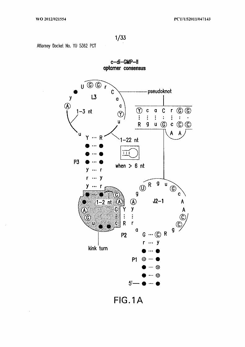
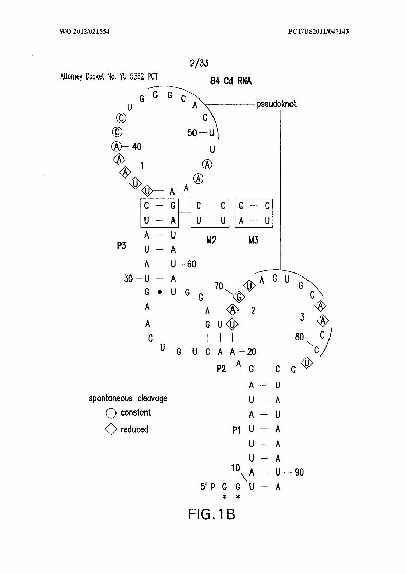
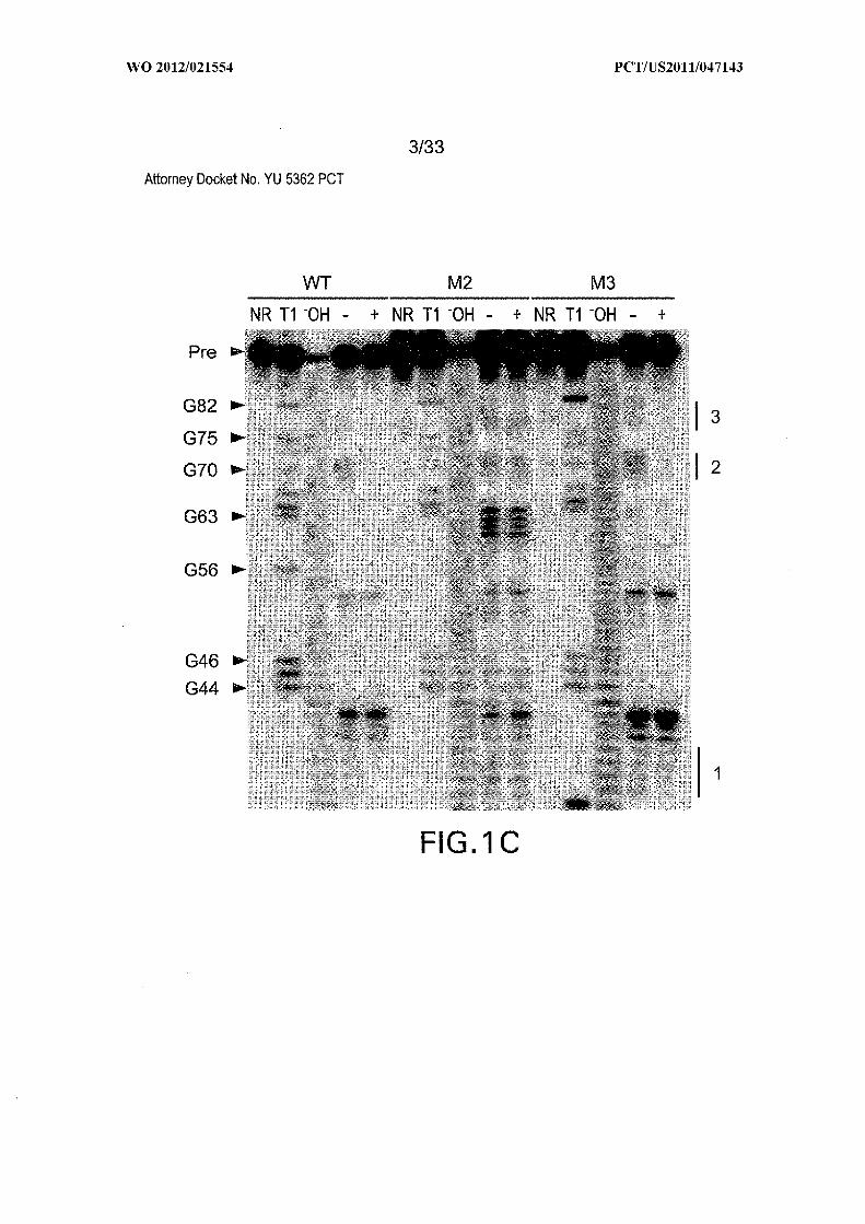
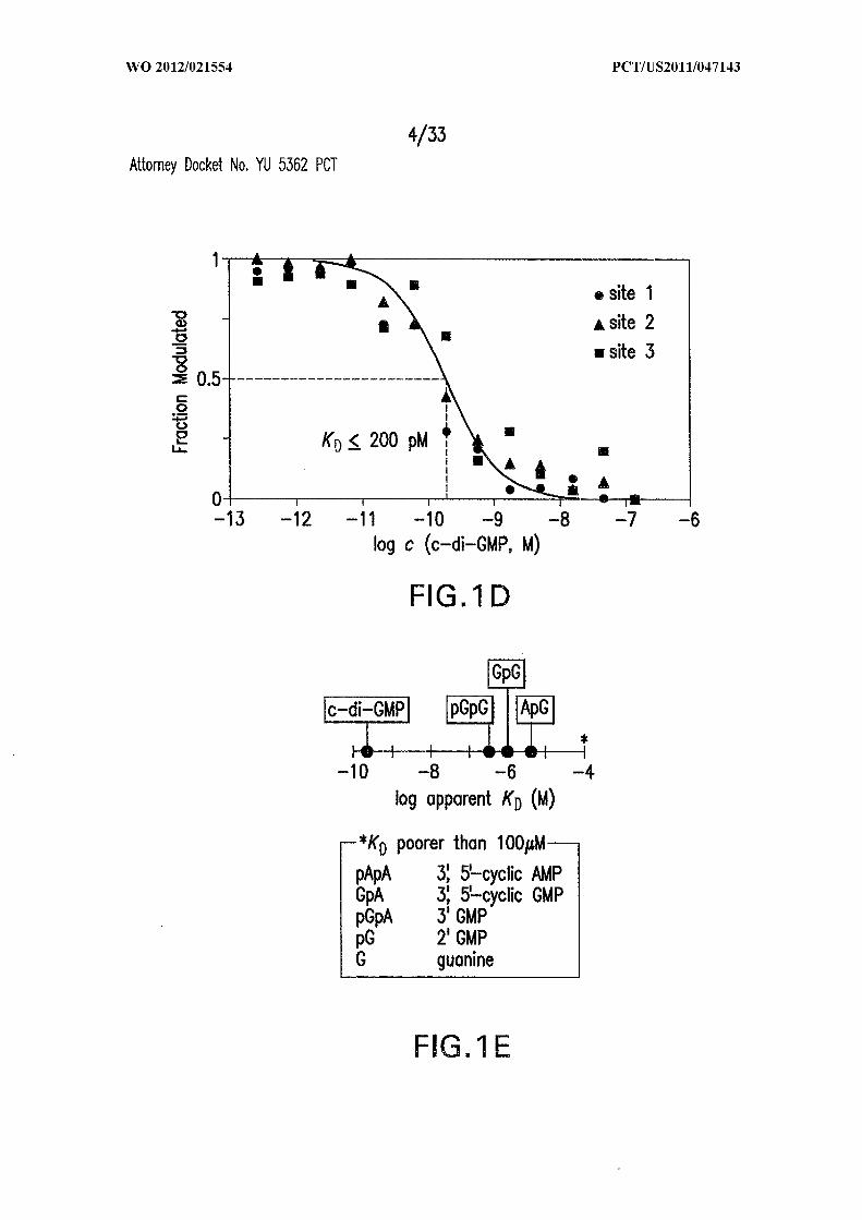
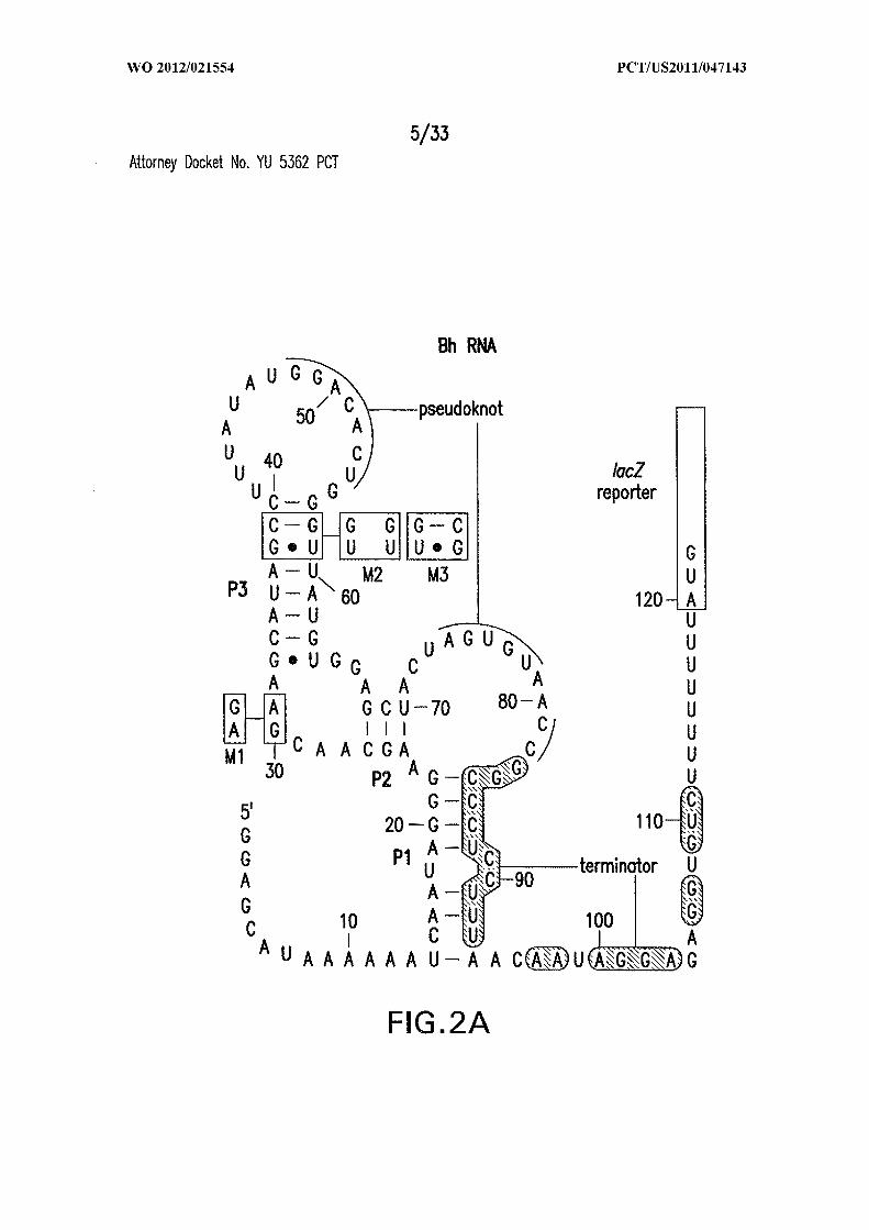
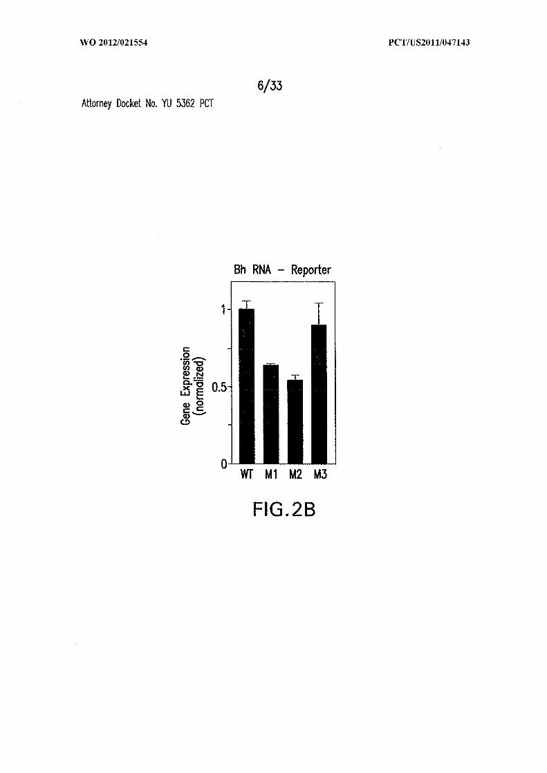

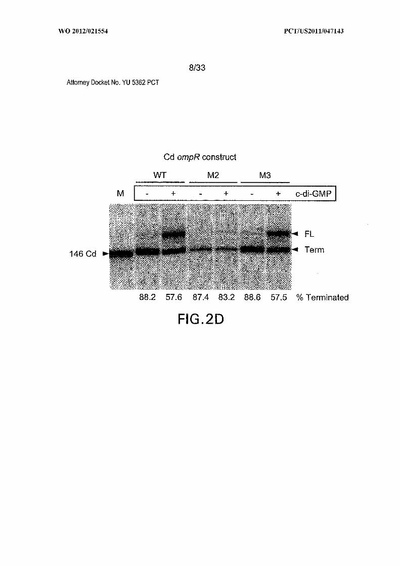
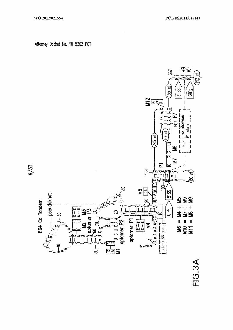
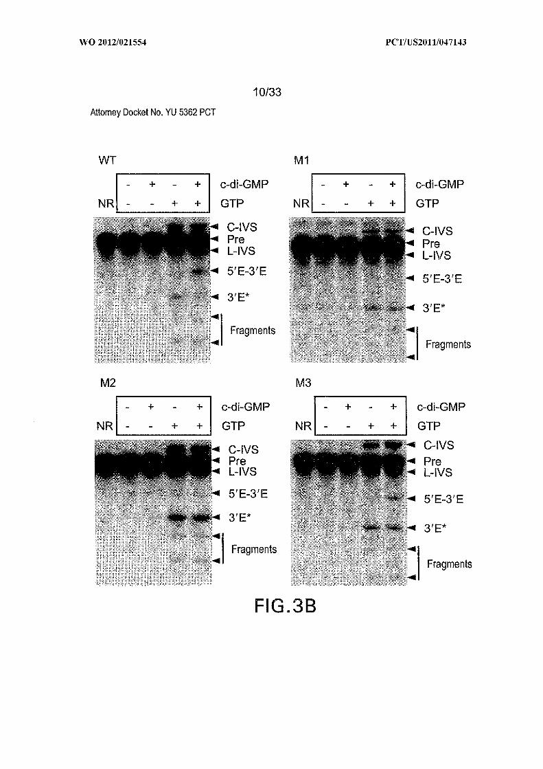
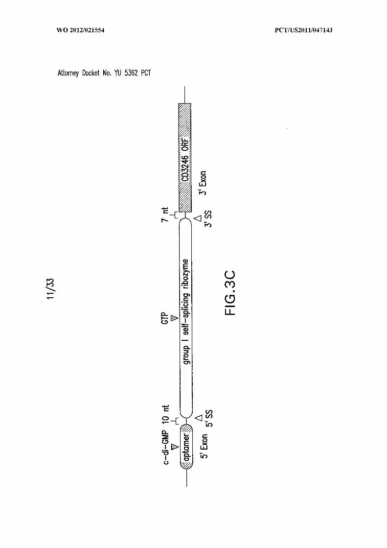
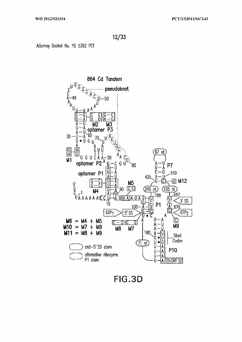
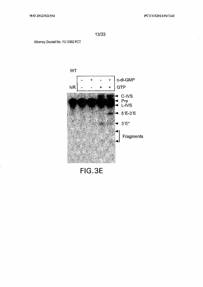
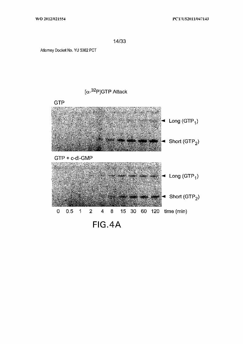
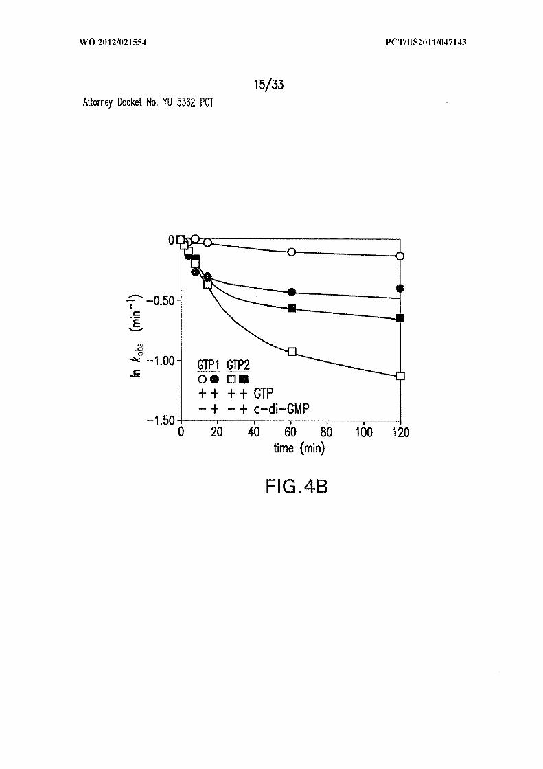
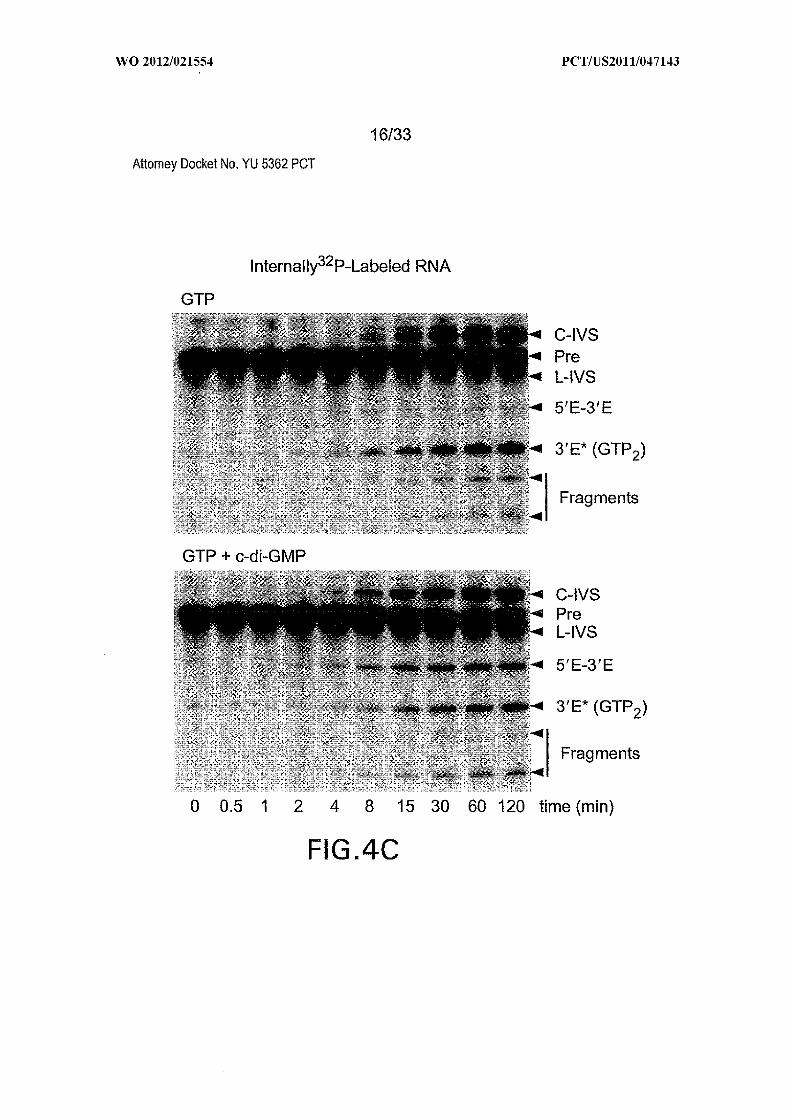
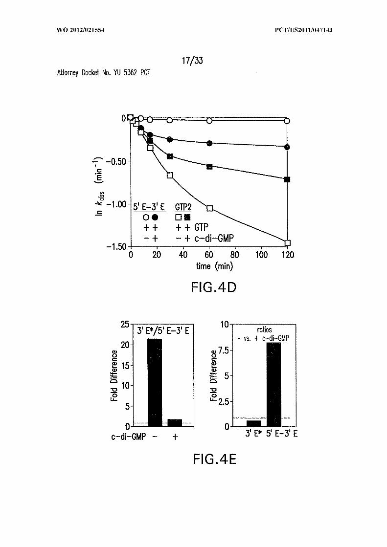
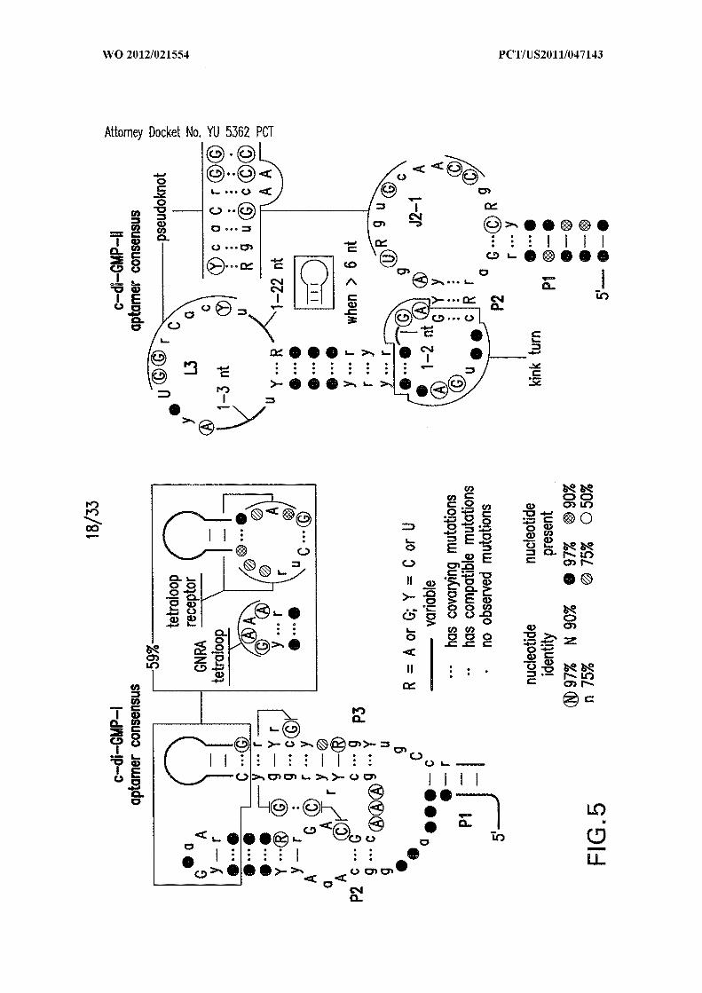

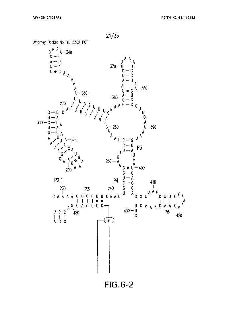
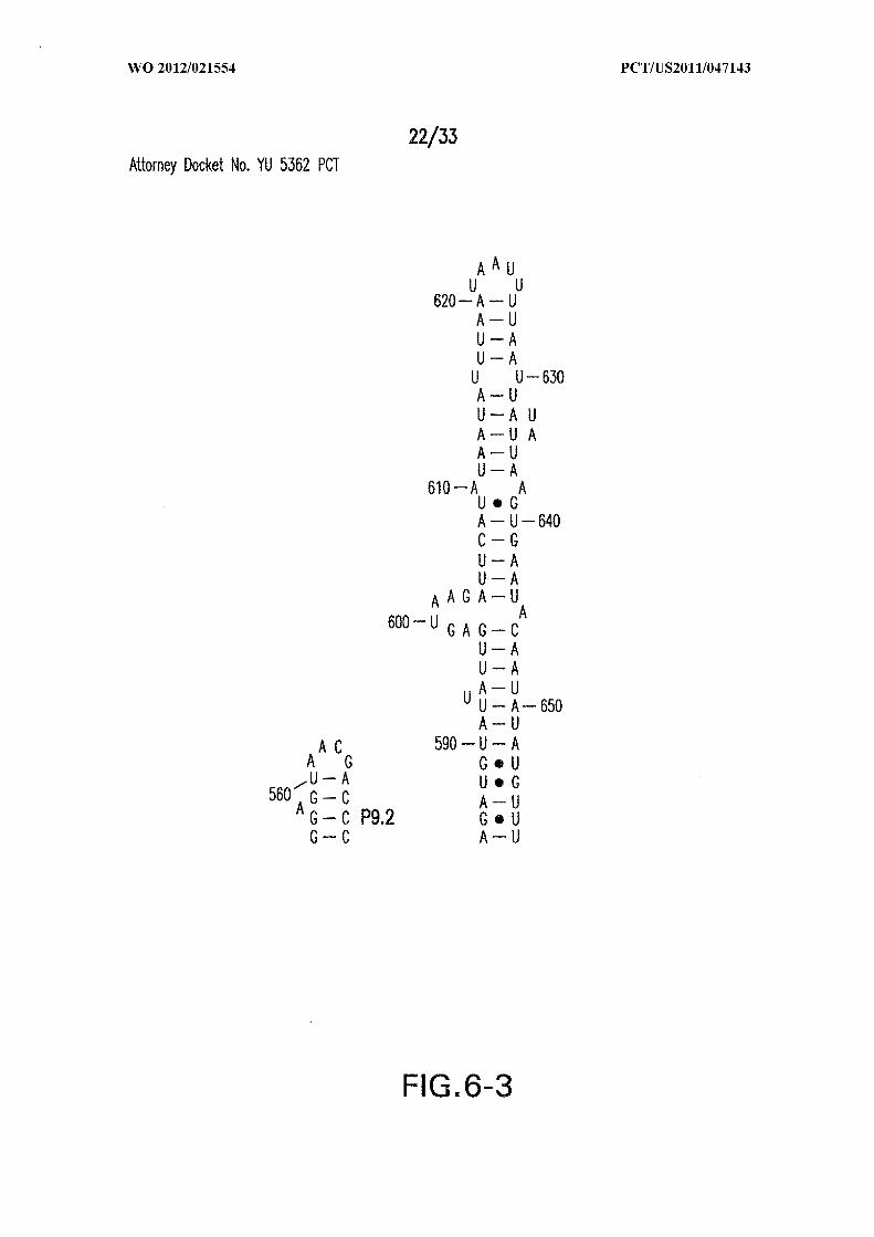
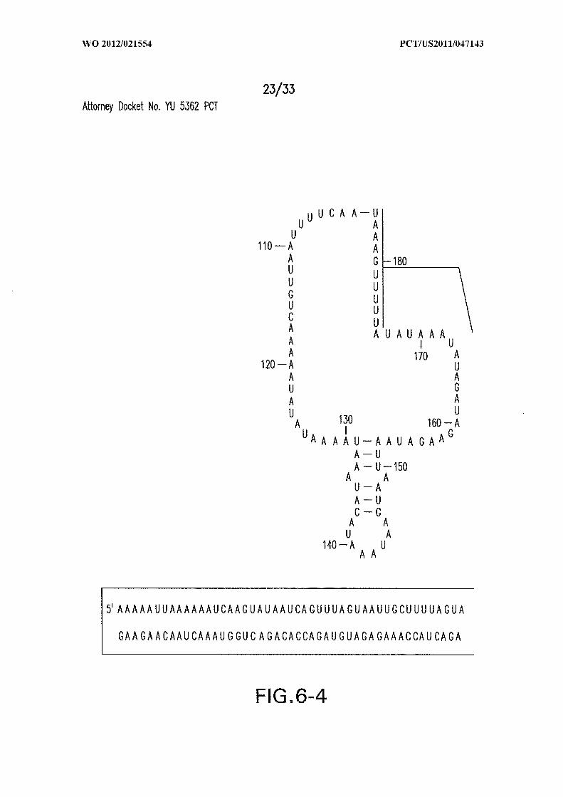
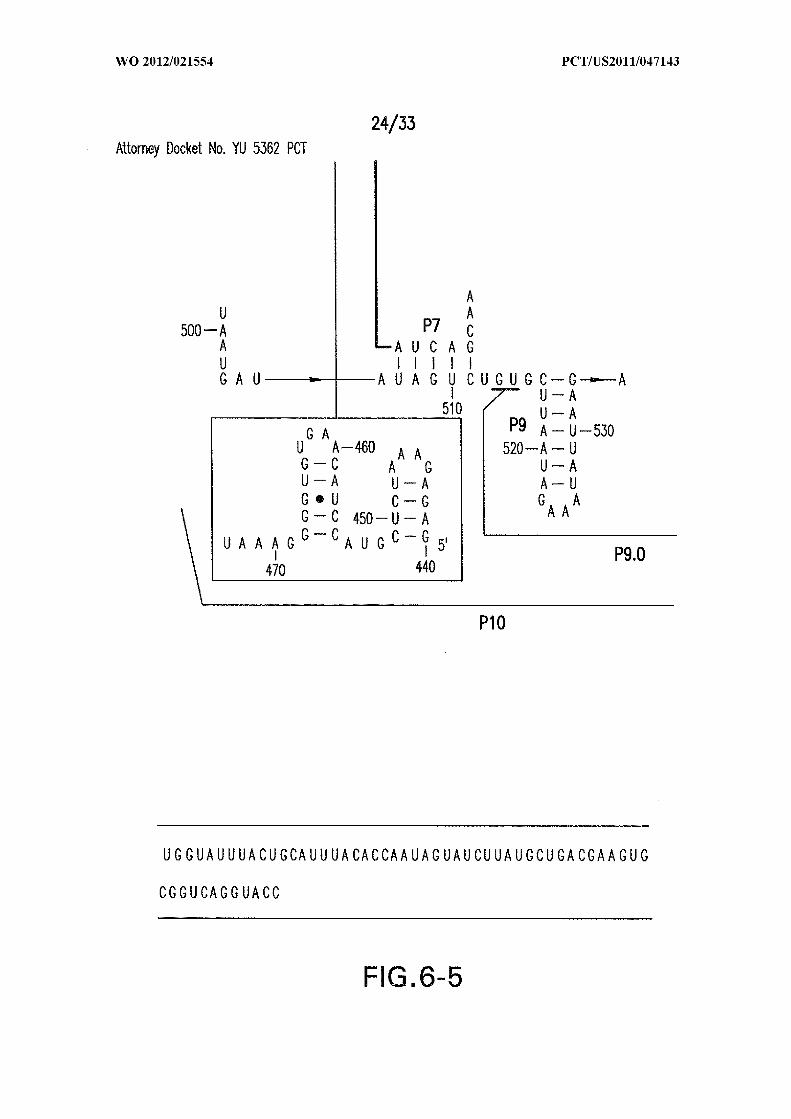
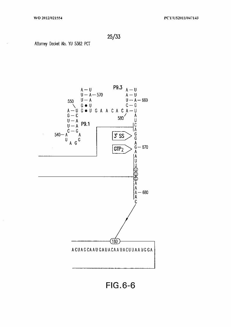
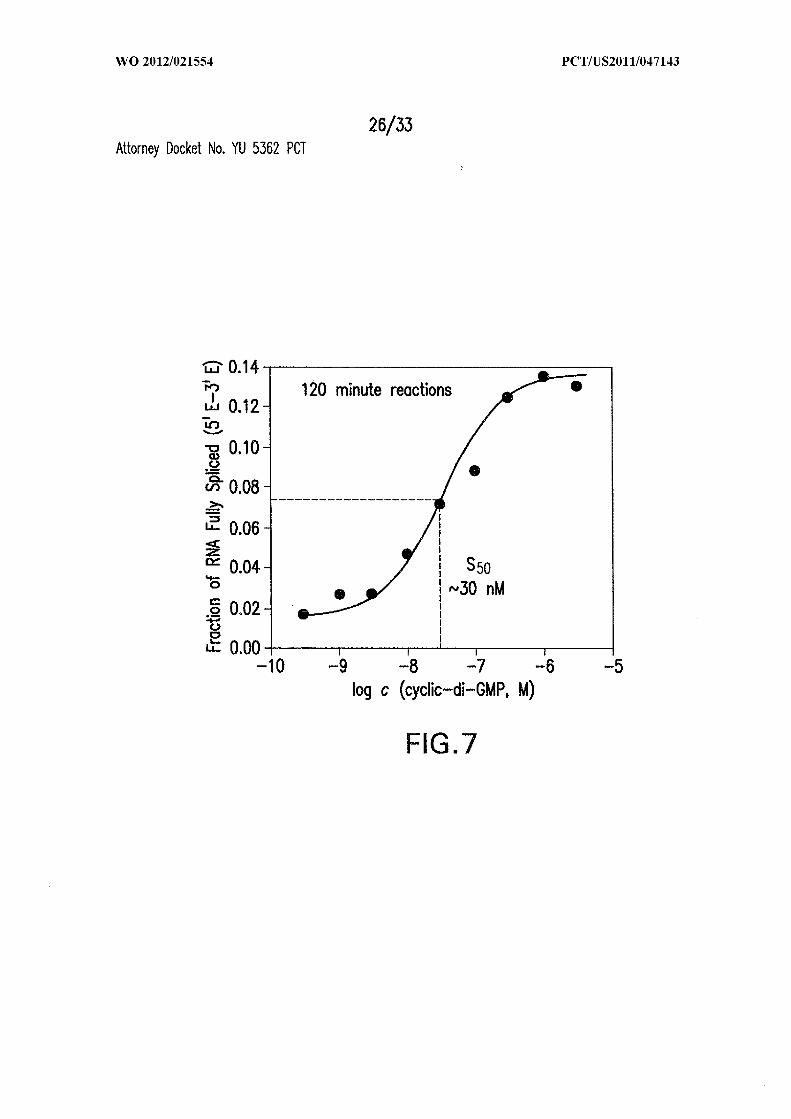
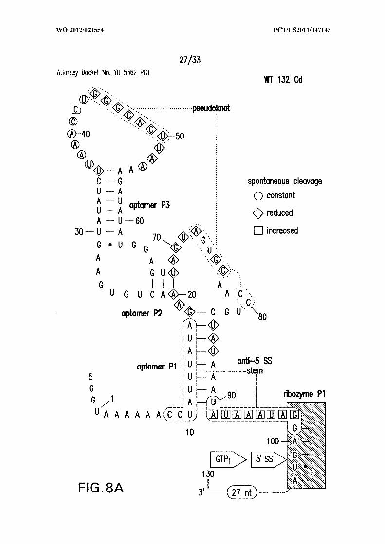
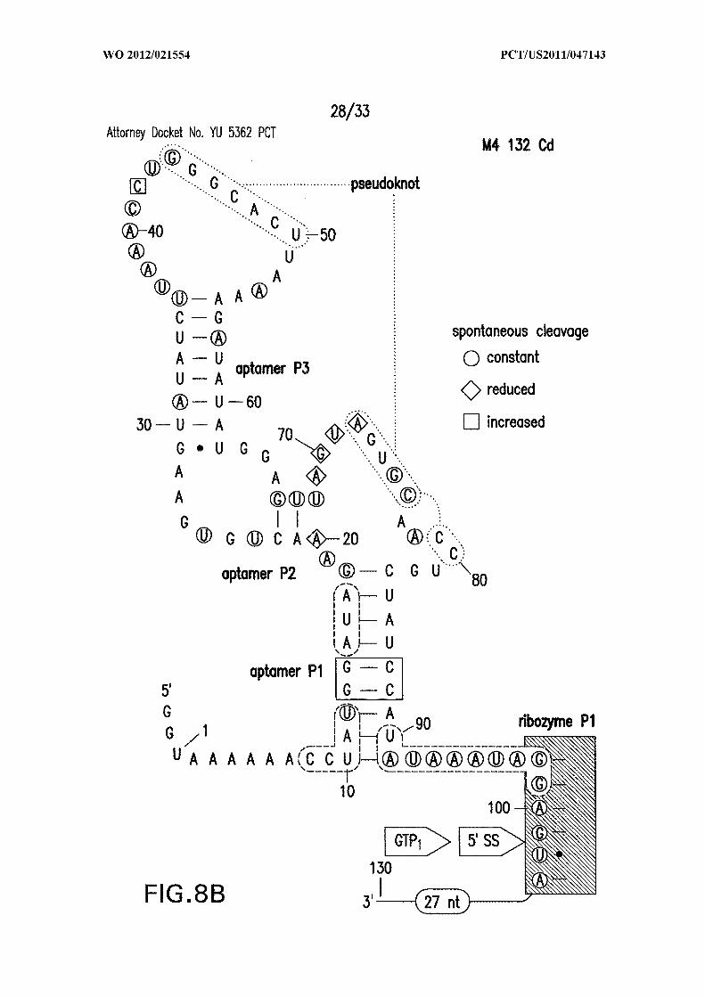
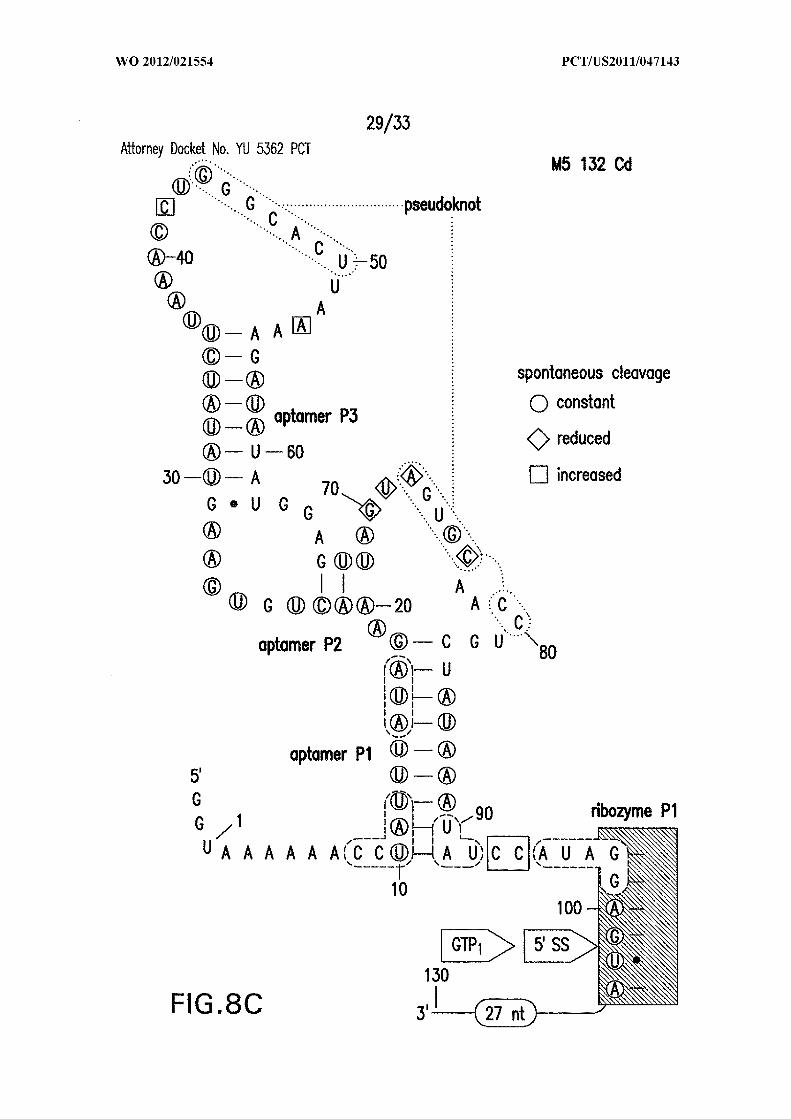
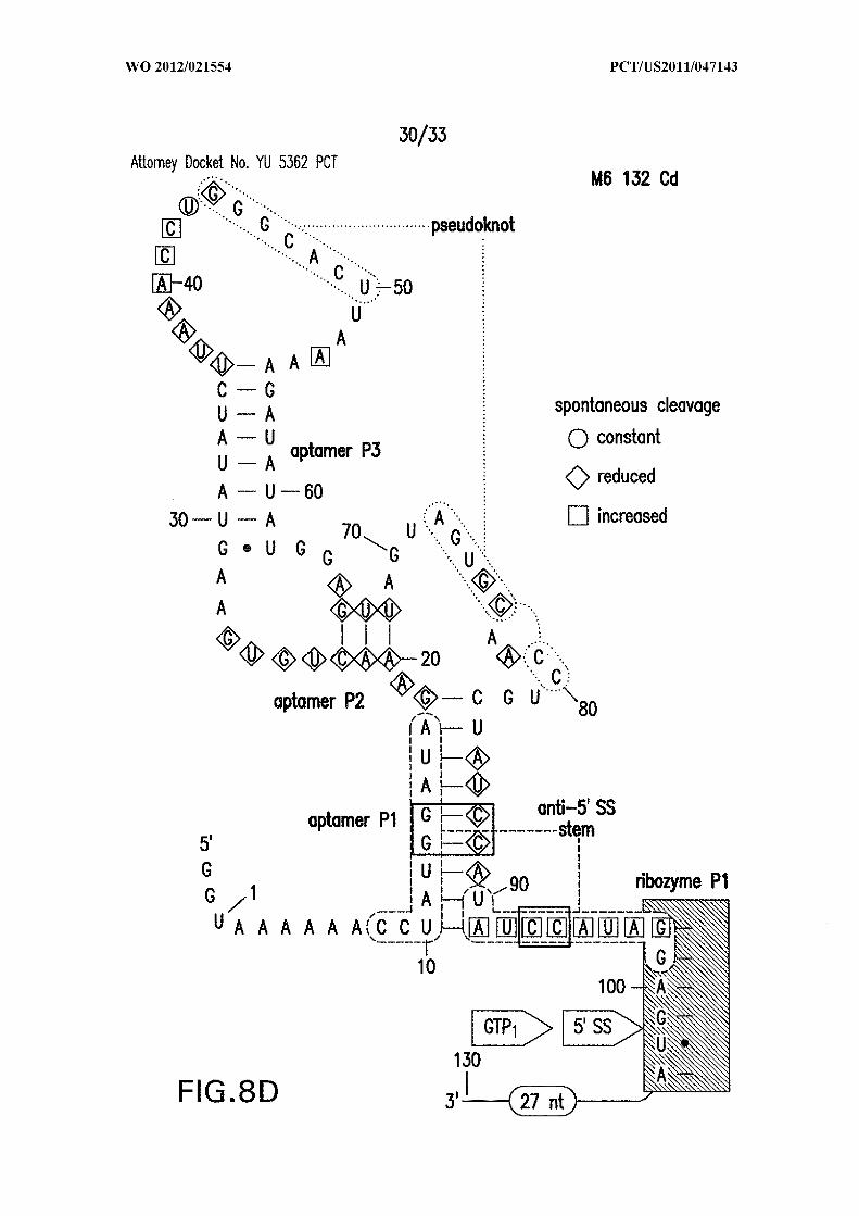
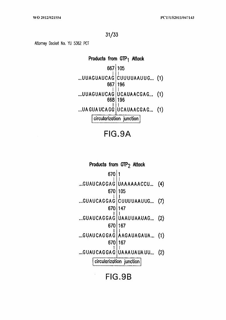
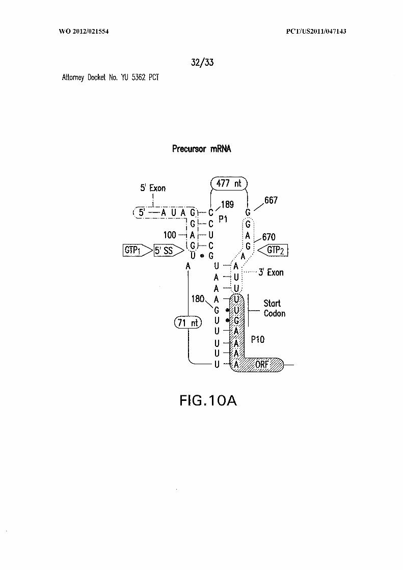
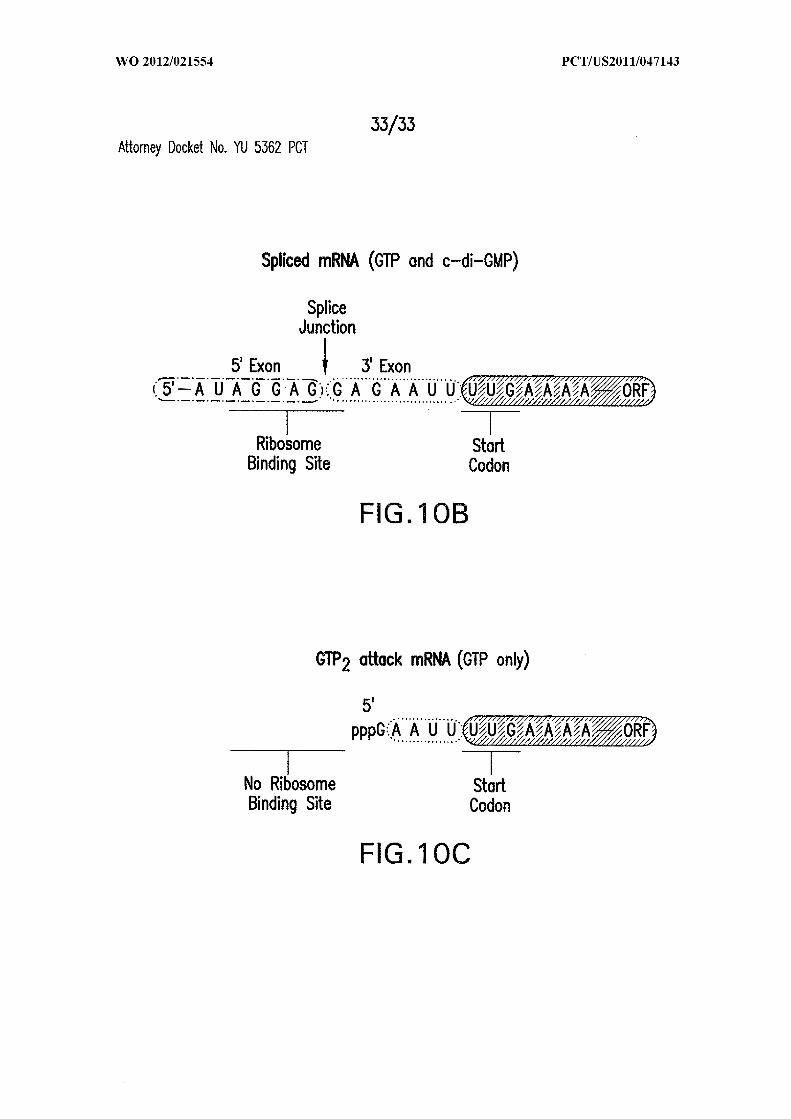
INTERNATIONAL SEARCH REPORTInternational application No
PCT/US2011/047143
A. CLASSIFICATION O F SUBJECT MATTERINV. C12N15/11 C12N15/113 C12N15/115 C12Q1/68 A61K31/708
A61K31/7084 C12N15/09ADD.According to International Patent Classification (IPC) or to both national classification and IPC
B. FIELDS SEARCHED
Minimum documentation searched (classification system followed by classification symbols)
C12N C12Q A61K
Documentation searched other than minimum documentation to the extent that such documents are included in the fields searched
Electronic data base consulted during the international search (name of data base and, where practical, search terms used)
EPO-Internal , BIOSIS, MEDLINE, Sequence Search , WPI Data
C. DOCUMENTS CONSIDERED TO BE RELEVANT
Category* Citation of document, with indication, where appropriate, of the relevant passages Relevant to claim No.
WO 2008/156987 A2 (UNIV YALE [US] ; BREAKER 1-13,RONALD R [US] ; WEINBERG ZASHA; SUDARSAN 15-33NARASI ) 24 December 2008 (2008-12-24)the whole document 14
WO 2005/030186 A2 (UNIV MARYLAND [US] ; 1-10KA RAO LIS DAVID K R [US] )7 Apri l 2005 (2005-04-07)the whole document
/ -
Further documents are listed in the continuation of Box C. See patent family annex.
* Special categories of cited documents :"T" later document published after the international filing date
or priority date and not in conflict with the application but"A" document defining the general state of the art which is not cited to understand the principle or theory underlying the
considered to be of particular relevance invention"E" earlier document but published on or after the international "X" document of particular relevance; the claimed invention
filing date cannot be considered novel or cannot be considered to"L" document which may throw doubts on priority claim(s) or involve an inventive step when the document is taken alone
which is cited to establish the publication date of another " document of particular relevance; the claimed inventioncitation or other special reason (as specified) cannot be considered to involve an inventive step when the
"O" document referring to an oral disclosure, use, exhibition or document is combined with one or more other such docu¬other means ments, such combination being obvious to a person skilled
"P" document published prior to the international filing date but in the art.
later than the priority date claimed "&" document member of the same patent family
Date of the actual completion of the international search Date of mailing of the international search report
1 November 2011 09/11/2011
Name and mailing address of the ISA/ Authorized officer
European Patent Office, P.B. 5818 Patentlaan 2NL - 2280 HV Rijswijk
Tel. (+31-70) 340-2040,Fax: (+31-70) 340-3016 Spi ndler, Mark-Peter
INTERNATIONAL SEARCH REPORTInternational application No
PCT/US2011/047143
C(Continuation). DOCUMENTS CONSIDERED TO BE RELEVANT
Category* Citation of document, with indication, where appropriate, of the relevant passages Relevant to claim No.
X KA RAO LIS D K R ET AL: "C-DI -GMP 1-10(3 ' - 5 -CYCLIC DIGUANYLIC ACID) INHIBITSSTAPHYLOCOCCUS AUREUSCELL-CELL-INTERACTIONS AND BIOFI L
FORMATION" ,ANTIMICROBIAL AGENTS AND CHEMOTHERAPY,AMERICAN SOCI ETY FOR MICROBIOLOGY,WASHINGTON, DC, US,vol . 49, no. 3 , 1 March 2005 (2005-03-01) ,pages 1029-1038, XP009052927 ,ISSN : 0066-4804, D0I :10. 1128/AAC. 49. 3 . 1029-1038.2005the whole document
X SUDARSAN N ET AL: "Ri boswi tches i n 1-4, 10,eubacteri a sense the second messenger 2 1 ,23cycl i c d i -GMP" ,SCI ENCE, AMERICAN ASSOCIATION FOR THEADVANCEMENT OF SCI ENCE, WASHINGTON, DC;US,vol . 321 , no. 5887,18 July 2008 (2008-07-18) , pages 411-413 ,XP002584282,ISSN : 0036-8075the whole document
X KATHRYN D SMITH ET AL: "Structural basi s 1-4, 10,of l i gand binding by a c-di -GMP 2 1 ,23r i boswi tch" ,NATURE STRUCTURAL & MOLECULAR BIOLOGY,vol . 16, no. 12,1 December 2009 (2009-12-01) , pages1218-1223 , XP55010839 ,ISSN : 1545-9993, DOI : 10. 1038/nsmb. 1702the whole document
A BLOUNT KENNETH F ET AL: "Riboswi tches a s 1-33antibacterial drug targets" ,NATURE BIOTECHNOLOGY, NATURE PUBLISHINGGROUP, NEW YORK, NY, US,vol . 24, no. 12,1 December 2006 (2006-12-01) , pages1558-1564, XP002584284,ISSN : 1087-0156, DOI : 10. 1038/NBT1268[retri eved on 2006-12-11]the whole document
-/--

INTERNATIONAL SEARCH REPORTInternational application No
PCT/US2011/047143
C(Continuation). DOCUMENTS CONSIDERED TO BE RELEVANT
Category* Citation of document, with indication, where appropriate, of the relevant passages Relevant to claim No.
X, P E. P.. LEE ET AL: "An Al l osteri c 1-33Sel f -Spl i cing Ribozyme Tri ggered by aBacterial Second Messenger" ,SCI ENCE,vol . 329 , no. 5993,13 August 2010 (2010-08-13) , pages845-848, XP55010727,ISSN : 0036-8075, D0I :10. 1126/science. 1190713the whole document
International application No.
INTERNATIONAL SEARCH REPORTPCT/US 0 1 1 / 0 4 143
Box No. I Nucleotide and/or amino acid sequence(s) (Continuation of item 1.c of the first sheet)
1. With regard to any nucleotide and/or amino acid sequence disclosed in the international application and necessary to the claimedinvention, the international search was carried out on the basis of:
a . (means)
I I on paper
IX I in electronic form
b. (time)
I I in the international application as filed
I I together with the international application in electronic form
subsequently to this Authority for the purpose of search
□ In addition, in the case that more than one version or copy of a sequence listing and/or table relating thereto has been filedor furnished, the required statements that the information in the subsequent or additional copies is identical to that in theapplication as filed or does not go beyond the application as filed, as appropriate, were furnished.
3 . Additional comments:
Form PCT/ISA/21 0 (continuation of first sheet (1)) (July 2009)
INTERNATIONAL SEARCH REPORTInternational application No
Information on patent family membersPCT/US2011/047143
Patent document Publication Patent family Publicationcited in search report date member(s) date
WO 2008156987 A2 24-12-2008 AU 2008266684 Al 24-12-2008CA 2688575 Al 24-12-2008CN 101849020 A 29-09-2010EP 2164996 A2 24-03-2010J P 2010528617 A 26-08-2010KR 20100017905 A 16-02-2010US 2010286082 Al 11-11-2010
W0 2005030186 A2 07-04-2005 AU 2004275696 Al 07-04-2005CA 2533873 Al 07-04-2005EP 1651242 A2 03-05-2006I L 173065 A 31-05-2011J P 2007500697 A 18-01-2007