2013 J Mol Catal B
10
This article appeared in a journal published by Elsevier. The attached copy is furnished to the author for internal non-commercial research and education use, including for instruction at the authors institution and sharing with colleagues. Other uses, including reproduction and distribution, or selling or licensing copies, or posting to personal, institutional or third party websites are prohibited. In most cases authors are permitted to post their version of the article (e.g. in Word or Tex form) to their personal website or institutional repository. Authors requiring further information regarding Elsevier’s archiving and manuscript policies are encouraged to visit: http://www.elsevier.com/authorsrights
Transcript of 2013 J Mol Catal B

This article appeared in a journal published by Elsevier. The attachedcopy is furnished to the author for internal non-commercial researchand education use, including for instruction at the authors institution
and sharing with colleagues.
Other uses, including reproduction and distribution, or selling orlicensing copies, or posting to personal, institutional or third party
websites are prohibited.
In most cases authors are permitted to post their version of thearticle (e.g. in Word or Tex form) to their personal website orinstitutional repository. Authors requiring further information
regarding Elsevier’s archiving and manuscript policies areencouraged to visit:
http://www.elsevier.com/authorsrights
Author's personal copy
Journal of Molecular Catalysis B: Enzymatic 99 (2014) 34– 42
Contents lists available at ScienceDirect
Journal of Molecular Catalysis B: Enzymatic
jo u r n al homep age: www.elsev ier .com/ locate /molcatb
Kinetic role of a histidine residue in the T1 copper site of the laccasefrom Rigidoporus lignosus
Fabio Vianelloa,b,∗, Giovanni Miottoc,d, Maria Teresa Cambriae, Giuseppina P.P. Limaf,Paola Vanzanic,g, Maria Luisa Di Paoloc,g
a Department of Comparative Biomedicine and Food Science, University of Padova, Italyb Regional Centre of Advanced Technologies and Materials, Department of Physical Chemistry, Palacky University in Olomouc, Czech Republicc Department of Molecular Medicine, University of Padova, Italyd Proteomic Center of Padova University, VIMM and Padova University Hospital, Padova, Italye Section of Biochemistry, Department of Chemical Sciences, University of Catania, Italyf Department of Chemistry and Biochemistry, Institute of Biosciences, Universidade Estadual Paulista, (UNESP), Botucatu, SP, Brazilg Consorzio Interuniversitario “Istituto Nazionale Biostrutture e Biosistemi”, Rome, Italy
a r t i c l e i n f o
Article history:Received 12 July 2013Received in revised form 11 October 2013Accepted 21 October 2013Available online 29 October 2013
Keywords:Blu-copper laccaseRigidoporus lignosuspH profileIonic strength effectsHistidine modificationT1 copper site
a b s t r a c t
Laccases (benzendiol:oxygen oxidoreductases; EC 1.10.3.2) catalyze the oxidation of a broad range of sub-strates, such as polyphenols, dyes and pollutants, and thus these enzymes are widely applied in industrial,biotechnological and environmental fields. In order to improve their biotechnological applications, a deepknowledge of structural factors involved in controlling their activity, in various experimental conditionsand on different substrates, is required. In the present study, a laccase from the mushroom Rigidoporuslignosus was kinetically characterized. In particular, the stability, the effects of pH, ionic strength andfluoride ion concentration on the kinetic parameters were investigated, using three di-hydroxy-benzeneisomers (1,2-dihydroxy-benzene, 1,3-dihydroxy-benzene and 1,4-dihydroxy-benzene) as substrates. Thecatalytic constant values of the laccase showed a bell-shaped pH profile, with the same optimum pH andpKa values for all tested substrates. This behavior appears to be due to the presence of an ionizableresidue in the enzyme active site. To identify this residue, the enzyme was derivatized with diethyl-pyrocarbonate to modify accessible histidine residues, which, according to structural data, are present inthe active site of this enzyme. The kinetic behavior of the derivatized laccase was compared with that ofthe native enzyme and the derivatized residues were identified by mass spectrometry. Mass spectrom-etry and kinetic results suggest the main role of His-457 in the control of the catalytic activity of laccasefrom R. lignosus.
© 2013 Elsevier B.V. All rights reserved.
1. Introduction
Laccases (benzendiol:oxygen oxidoreductases; EC 1.10.3.2) aremulticopper oxidases widely distributed in plant and fungal species[1,2]. Their physiological roles are numerous and various, fromlignification and delignification, to fungal morphogenesis and vir-ulence [3], according to type of organism. Among fungal laccases,great variability has been observed in induction mechanism, degreeof polymorphism, physico-chemical properties and enzymatic
Abbreviations: DEPC, diethylpyrocarbonate; NHE, normal hydrogen electrode;H2Q, dihydroxy-benzene derivative; Q, quinone derivative; RlL, Rigidoporus lignosuslaccase; SEM, standard error of the mean; TvL, Trametes versicolor laccase.
∗ Corresponding author at: Department of Comparative Biomedicine and FoodScience, University of Padova, Agripolis – Viale dell’Università 16, Legnaro, 35020(PD), Italy. Tel.: +39 49 8276863; fax: +39 49 8073310.
E-mail address: [email protected] (F. Vianello).
activity [4–7]. Laccases contain four copper ions distributed in threecopper sites, defined according to their spectroscopic properties(T1, T2, T3). Site T1 contains a type 1 blue copper, which is tightlycoordinated to two histidines, and one cysteine residue, responsi-ble for a visible absorption band at about 600 nm. Site T2 containsa type 2 copper with a characteristic electron paramagnetic reso-nance spectrum. In site T3, a pair of strongly antiferromagneticallycoupled type 3 copper ions is EPR-silent. According to the acceptedcatalytic mechanism, the mononuclear copper site T1 extractselectrons from the reducing substrate and, through the highly con-served His-Cys-His tripeptide, transfers them to the tri-nuclearT2/T3 center, where molecular oxygen is reduced [1,8]. The reduc-tion of T1 copper is the rate-limiting step of the catalytic reactionand occurs according to the Marcus “outer-sphere” mechanism [9],according to which the electron transfer rate is determined by the�E◦ between site T1 and the reducing substrate. The E◦ value ofsite T1 differs among the various laccases, in the range 420–790 mV(vs NHE), depending on the structural properties of the copper site
1381-1177/$ – see front matter © 2013 Elsevier B.V. All rights reserved.http://dx.doi.org/10.1016/j.molcatb.2013.10.017

Author's personal copy
F. Vianello et al. / Journal of Molecular Catalysis B: Enzymatic 99 (2014) 34– 42 35
[10]. Recently, also steric factors have been found to affect substratespecificity of some laccases [11].
Laccases can oxidize phenols, anilines, benzenthiols and phe-nothiazines [12], and generally present rather broad reducingsubstrate specificity, molecular oxygen being the terminal elec-tron acceptor in a four-electron process in which water is the finalproduct [13]. It is believed that the first reaction product of laccaseactivity is an organic radical, due to monoelectronic substrate oxi-dation (a semiquinone radical in the case of di-hydroxy-benzenesubstrates). In the case of phenolic substrates, generally, phenoxyradicals dismutate, leading to a bi-electronically oxidized productand the starting reduced substrate.
Laccases, particularly “high-potential” enzymes, are widelyapplied in industrial and biotechnological fields, such as woodfiber modifications [14], biosensor development [15–17] andwastewater bioremediation [18]. In order to improve the biotech-nological applications of laccases, knowledge of the structuralfactors involved in controlling their activity, in various experimen-tal conditions, on different substrates, is required [19–21].
Few papers have been published on the effect of pH on the cat-alytic activity of this class of fungal enzymes. The most systematicstudies were carried out by F. Xu [10,12,22,23] on laccases from var-ious sources and with different substrates. Xu et al. observed that,in all the studied enzymes, with phenol substances as substrates,catalytic activity had a bell-shaped curve. According to the aboveauthors, the kinetic behavior of laccases as a function of pH is theresult of the balance of two opposing effects: the former effect isresponsible for the decrease in enzymatic activity observed at pHlower than the optimum, and is due to the decrease in the redoxpotential difference between the T1 copper site of the enzyme andthe phenol substrate. The latter effect, responsible for the des-cending part of the bell-shaped pH profile, observed at pH valuesabove optimum, is explained by the inhibitory effect of OH− onlaccase activity, probably at the T2/T3 copper site. In addition, site-directed mutagenesis studies on laccase from Trametes versicolorand CotA have recently demonstrated that carboxylated residuesare involved in the control of catalytic properties and in the pHdependence of laccase activity on pH values lower than optimum[20,24].
No information about the factors controlling the pH dependenceof the enzymatic activity of laccase from Rigidoporus lignosus (RlL),another “high-potential enzyme”, has been reported so far.
The aim of this work was therefore to perform a kinetic studyon the catalytic activity behavior of laccase from R. lignosus as afunction of pH, ionic strength and F− concentration. In order toinvestigate the possible involvement of histidine residues in thecontrol of enzyme activity and taking into account the impossibil-ity to perform site directed mutagenesis experiments on essentialaminoacids involved in copper coordination in the enzyme activesite, RIL was derivatized with diethylpyrocarbonate. Kinetic param-eters of derivatized laccase were compared with those of thenative enzyme. The identification of the derivatized residues wasperformed by mass spectrometry. Results suggest a novel interpre-tation of pH dependence of the kinetic parameters of RlL and themain role of His-457 in the control of the catalytic activity of laccasefrom R. lignosus.
2. 2. Materials and methods
2.1. Materials
All reagents were of the highest purity available and used with-out further purification. Coomassie Brilliant Blue was from Serva(Heidelberg, Germany). All other reagents and laccase from T. ver-sicolor (TvL) were from Fluka (Buchs, Switzerland). Doubly distilledwater was used.
Table 1Physico-chemical characteristics of dihydroxy-benzene derivatives.
Substrate pKa1a pKa2
a pKa*b E◦
pH 5c (V)
1,2-Dihydroxy-benzene 9.4 12.8 5.0 0.681,3-Dihydroxy-benzene 9.8 11.3 7.0 0.921,4-Dihydroxy-benzene 9.8 11.4 4.1 0.65
Physico-chemical characteristics of three substrates from refs. [26–28].a pKa values of two hydroxyl groups of dihydroxy-benzene derivatives.b pKa of semiquinone radical, i.e., product of enzymatic oxidation of dihydroxy-
benzene derivatives.c Potentials, calculated at pH 5.0, are given as reduction of semiquinone radicals
to corresponding dihydroxy-benzene derivative (vs NHE), according to Eq. (6).
Laccase from R. lignosus (RlL) was purified according to Cambria[25]. Enzyme purity was checked by Maldi-TOF, showing a singlemass peak at 55.2 kDa.
Britton-Robinson buffer was prepared by mixing 0.1 M boricacid, 0.1 M acetic acid and 0.1 M phosphoric acid to give 33 mMas final concentration for each buffering substance. KOH (1 M)was added to the desired pH. Unless otherwise specified, allexperiments were carried out in air-saturated solutions at roomtemperature (22 ± 1 ◦C) in the presence of 0.1 mM EDTA.
Spectrophotometric measurements were carried out on a VarianCary 50 instrument, oxygraphic determinations with a mod. 641Metrohm instrument connected to a Linseis E250L recorder, usinga Yellow Springs oxygen electrode.
2.2. Enzyme activity measurements
The catalytic activity of RlL was studied with 1,2-dihydroxy-benzene, 1,3-dihydroxy-benzene and 1,4-dihydroxy-benzene assubstrates. Table 1 lists the pKa values of the three substrates,extracted from literature data [26–28].
Enzyme activity measurements were carried out in 33 mMBritton Robinson buffer, pH 2.5–8.5 [29], and the rate offormation of 1,4-benzoquinone from 1,4-dihydroxy-benzene(�ε at 245 nm = 2.2 × 10⁴ M−1 cm−1) and the disappearancethe substrate in the case of 1,2-dihydroxy-benzene (�ε at275 nm = 2.4 × 10³ M−1 cm−1) were monitored. The rate of for-mation of phenoxyl radicals from phenol and 4-methoxyphenolwere also monitored by spectrophotometry at �max = 400 ± 2 nmfor phenol (�ε400nm = 1300 M−1 cm−1), and at �max = 245 ± 2 nm for4-methoxyphenol (�ε245nm = 4900 M−1 cm−1).
Enzyme activity was also determined by measuring oxygenconsumption during substrate oxidation, with a Clark electrode.Measurements were carried out in a 4.5-mL gas-tight cell undercontinuous stirring at room temperature. The stoichiometric ratio,�[S]/�[O2], was calculated by injection of a known amount of sub-strates (�[S]) into the oxygraphic cell and measurement of thedecrease of O2 concentration (�[O2]), in conditions of [S] « [O2].Enzyme activity toward 1,3-dihydroxy-benzene was measuredonly by oxygraphy. Enzyme catalytic parameters were obtainedfrom the initial rate of substrate oxidation measured within thefirst 5 min of substrate addition to the reaction solution (underthese conditions, the amount of consumed oxygen was alwayslower than 18% of the amount of initial oxygen in the oxygraphicgas-tight cell). Kinetic parameters were calculated according to theMichaelis–Menten equation.
The inhibition of laccase by F− was assayed with 1,4-dihydroxy-benzene as substrate, in Britton-Robinson buffer at pH 5.0. Enzymeactivity data were acquired as a function of KF concentration (in therange 0.1–2.0 mM) and inhibition constants (Ki) were determinedaccording to Fersht [30].
The ionic strength of buffer solutions was calculated consideringthe concentration of each ionized species at the specified pH value,according to the Henderson–Hasselbach relationship. The ionic

Author's personal copy
36 F. Vianello et al. / Journal of Molecular Catalysis B: Enzymatic 99 (2014) 34– 42
strength of Britton–Robinson buffer at various values of pH wasmodified by suitable addition of 2 M NaClO4. No effect of NaClO4 ondihydroxy-benzene isomers were observed. Control experimentswere carried out in the presence of NaNO3 as substitute of NaClO4.
2.3. Kinetic analysis
The pKa values of the groups controlling the dependence ofkcat on pH were calculated by an equation for two dissociationconstants. In particular, the Tipton and Dixon equation [31], mod-ified according to Koudelka et al. [32], was applied:
kcat = kcat,o
1 + ˛ Ka1[H+]
+ ˇ [H+]Ka2
1 + [H+]Ka1
+ Ka2[H+]
(1)
where kcat,o is the pH-independent catalytic constant, ̨ and ̌ arecorrection factors to account for the non-zero activity of the enzymeat low and high pH values, and Ka1 and Ka2 are the acid dissociationconstants of the groups controlling the ascending and descendingparts of the pH profile, respectively.
The pKa values of the groups controlling the dependenceof KM on (pH) were calculated according to the followingHenderson–Hasselbach equation for a single dissociation constant(Ka):
KM = a × 10−pH + b × 10−pKa
10−pKa + 10−pH(2)
where “a” and “b” represent the limiting KM value at acid and basicpH, respectively.
The pKa values of the groups controlling the dependence ofkcat/KM on pH were calculated according to the following equationfor two dissociation constants [31]:
Log(kcat/KM) = Log(kcat/KM)0 − Log(1 + 10pKa1−pH + 10pH−pKa2 )
(3)
where (kcat/KM)0 is the pH-independent catalytic efficiency and Ka1and Ka2 are the acid dissociation constants of the groups controllingthe ascending and descending parts of the pH profile, respectively.
The dependence of enzyme kinetic parameters on ionic strength(I) was studied in the I range 10–300 mM. The kinetic data wereanalyzed according to the Debye–Hückel equation [33]:
log k = log(k0) + 2 · C · Za · Zb · (I)1/2 (4)
where k is the kinetic parameter (kcat or KM), Za and Zb are thecharges of the interacting species, k0is the value of the kineticparameter at I = 0, and constant C is about 0.5 M−1/2 at 22 ◦C, inwater [34].
Non-linear least-squares analysis was performed with commer-cial graphic software (SigmaPlot 10.0 program, Jandel, Scientific).The values of the best fit parameters and the standard error of themean value (SEM) are reported. All determinations were performedat least in triplicate.
2.4. Protein determination
Laccase concentration was determined according to the Brad-ford procedure [35], with bovine serum albumin as standard.
2.5. Enzyme derivatization by diethyl-pyrocarbonate (DEPC)
RlL was derivatized with DEPC following the proceduredescribed by Miles [36]. Briefly, 1 mM of freshly prepared DEPCin ethanol was added to 20 �M laccase (a molecular weight of55 kDa was considered) in 0.1 M potassium phosphate buffer, pH7.0, and the solution was incubated at 0 ◦C for 30 min. In these
conditions, DEPC reacts primarily with histidine residues [37].Enzyme derivatization was monitored by spectrophotometry fol-lowing the formation of N-carbethoxy-imidazole at 240 nm, witha molar extinction coefficient of 3000 M−1 cm−1 [37]. At the end ofthe reaction, the enzyme was extensively dialyzed against 20 mMpotassium phosphate buffer, pH 7.0, at 4 ◦C. At the end of the pro-cedure, enzyme activity and concentration were determined, asalready mentioned.
2.6. Mass spectrometry analyses
MALDI-TOF analyses were carried out as follows: RlL sam-ples, native or DEPC derivatized, were extensively dialyzed againstmilliQ grade water (3 × 2 h buffer changes with a 1000:1 vol-ume ratio), then dried under vacuum and finally resuspended(100 pmol/�l) in 10% hexafluoro-2-propanol/0.1% trifluoroaceticacid (TFA). Samples (1 �L) were mixed with the matrix solution(1 �L of 12 mg/mL sinapic acid in 30% acetonitrile/0.1% TFA) and thefinal mixtures (0.8 �L) were spotted onto a stainless steel MALDItarget plate. Samples were then analyzed using a MALDI-TOF-TOF4800 mass spectrometer (Applied Biosystems, Toronto, Canada)operating in linear mode with positive polarity. Data were obtainedby averaging 2000 laser shots with power set at 5000.
The identification of DEPC modified histidine residues wasperformed by MS/MS analysis of digested proteins. Briefly, Lac-case samples, native or DEPC treated, were extensively dialyzedagainst milliQ grade water (3×2 h buffer changes with a 1000:1volume ratio), dried under vacuum and re-suspended in 25 mMammonium bicarbonate (100 �L). Native RlL and DEPC-RlL sam-ples were incubated overnight at 37 ◦C with sequencing gradetrypsin or Asp-N (Promega, Madison, WI, USA) and with an enzymeto substrate ratio of 120 (w/w). Samples were then dried by aSpeedVac (ThermoFisher Scientific) and resuspended in 0.1% TFA(15�L) for MS analysis on a LTQ-Orbitrap XL mass spectrome-ter (ThermoFisher Scientific), coupled to an on-line Ultimate 3000nano-HPLC (Dionex). Samples (0.5 pmol) were loaded onto a 10 cmlong homemade pico-frit column packed with C18 (Reprosil-PurC18-AQ, 3 �m, Dr. Maisch Cmb) and separated using a 45 min lineargradient of acetonitrile/0.1% formic acid (from 0% to 40% acetoni-trile in 25 min), at a flow rate of 250 nL/min. Capillary voltage wasset at 1.3–1.5 kV and source temperature at 200 ◦C. MS2 acqui-sition method was based on a full-scan mode on the Orbitrapwith a 60,000 resolution, followed by the MS/MS scans on the4 most intense ions, performed in the linear ion-trap. Raw datafiles were converted in mzData (.xml) files and analyzed againstthe UniRef100 database (May 2011 version, 12831896 sequences)interfaced to a Mascot search engine (Matrix Science, London, UK,version 2.3). Enzyme specificity was set to trypsin or Asp-N with2 missed cleavages. Mass tolerance window was set to 5 ppm forparent mass and to 0.6 Da for fragment ions. After laccase identifi-cation, the set was searched again with no enzyme specificity anderror tolerant mode to maximize the sequence coverage.
3. Results
3.1. Stability of RlL as a function of pH
In order to verify whether variations of enzyme activity as afunction of pH could be ascribed to the inactivation or denatur-ation of RlL during measurements, laccase stability was checked byincubation in 33 mM Britton-Robinson buffer, pH range 2.0–8.5, atroom temperature. Aliquots of enzyme solutions were withdrawnat various times and their activity was measured at pH 5.0 in thepresence of 1 mM 1,4-dihydroxy-benzene. According to these mea-surements, laccase appears relatively stable in the pH range 6.0–9.0.

Author's personal copy
F. Vianello et al. / Journal of Molecular Catalysis B: Enzymatic 99 (2014) 34– 42 37
In particular, at pH values above 6.0, slow exponential decay(t½ ≥ 12.5 days) was found, whereas in the pH range 6.0–5.0, t½ fellby two orders of magnitude, reaching a t½ ≈ 1 h at pH 2 (Figure S1).In addition, the UV–vis spectra of RlL showed a broad band centeredat 598 ± 2 nm (extinction coefficient of 5200 ± 200 M−1 cm−1), seeFigure S2. It should be noted that our spectroscopic characterizationdid not show any variation of RlL spectral parameters with H+ con-centration, indicating that, if present, distortion of the T1 coppersite of the enzyme was minimal. We therefore presume that thelaccase we studied was one of the two possible isoforms alreadyfound by other authors [38,39].
As the activity measurements, carried out by spectrophoto-metry and oxygraphy, were conducted within 5 min, the decay ofenzyme activity due to inactivation during the activity assay wasconsidered negligible, even at pH values below 6.0.
3.2. Dependence of RlL activity on pH
The dependence of catalytic parameters kcat, KM and kcat/KM ofRlL in the pH range 2.0–8.5 were studied with dihydroxy-benzenederivatives as substrates. Previously, the optical behavior of 1,2-dihydroxy-benzene 1,4-dihydroxy-benzene and their enzymaticoxidation products, 1,2-benzoquinone and 1,4-benzoquinone,were studied in the same pH range. No changes in theirextinction coefficients were observed. Laccase activity wasdetermined by measuring the disappearance of 1,2-dihydroxy-benzene (�max = 275 ± 2 nm, ε275nm = 2400 ± 300 M−1 cm−1)and the formation of 1,4-benzoquinone (�max = 245 ± 2 nm,ε245nm = 22,000 ± 1000 M−1 cm−1). As 1,3-dihydroxy-benzene hasa low extinction coefficient and its oxidation product does not leadto the formation of quinone, measurements on this substrate werecarried out only by oxygraphy. Control experiments were carriedout with 1,2-dihydroxy-benzene and 1,4-dihydroxy-benzene.Comparisons of spectrophotometric and oxygraphic measure-ments showed that the �[S]/�[O2] ratio was 2.01 ± 0.15. Thismeans that two substrate molecules are oxidized by one moleculeof O2, according to the following equation:
2H2Q + O2laccase−→ 2Q + 2H2O (5)
where H2Q represents the dihydroxy-benzene derivative com-pound and Q the oxidation product. Additionally, no significantdifferences in the KM and kcat values, calculated taking intoaccount of the �[S]/�[O2] ratio, were found comparing the valuesobtained using the two different methods (oxygraphic and spec-trophotometric) on 1,2-dihydroxy- and 1,4-dihydroxy-benzene.
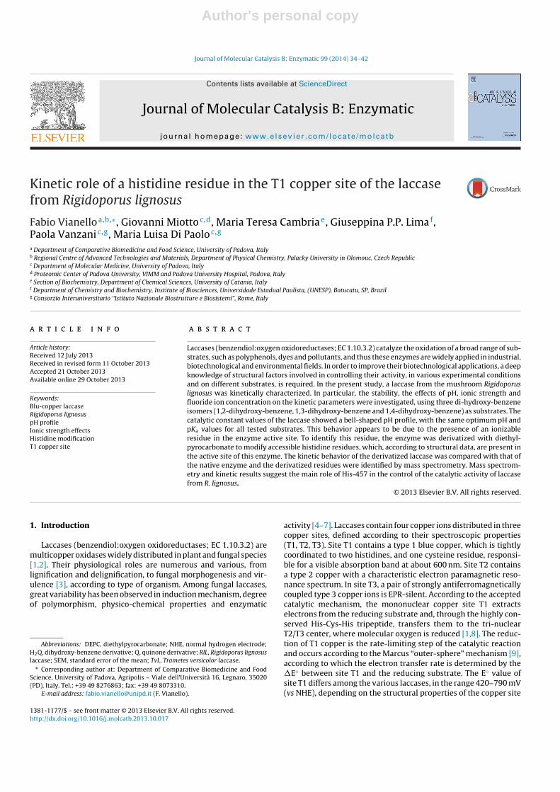
The catalytic constant (kcat) values of RlL vs pH showeda bell-shaped profile centered at pH 5.0 for all three sub-strates. At optimum pH, kcat values for 1,2-dihydroxy-benzene,1,4-dihydroxy-benzene and 1,3-dihydroxy-benzene were about650 s−1, 256 s−1 and 172 s−1, respectively, i.e., kcat,1,2-H2Q >kcat,1,4-H2Q > kcat,1,3-H2Q (Fig. 1, panel A). Interestingly, at pH 5.0,the one-electron redox potential of the HQ·/H2Q couple of thesecompounds does not follow the same order, that is, E◦
1,3-H2Q >E◦
1,2-H2Q > E◦1,4-H2Q (E◦ values listed in Table 1 [26,27]).
The experimental data of kcat vs pH were treated as originatingfrom the involvement of two ionizable groups, and equation 1 wasapplied to determine their pKa values. The best fits of Eq. (1) to theexperimental data gave the continuous lines shown in Fig. 1, panelA, and the pKa1 and pKa2 values in Table 2. Table 2 shows that, forRlL, the pKa values of ionizable groups 1 and 2 are independent,within experimental error, of the dihydroxy-benzene isomers usedas substrate.
Conversely, the KM values of RlL have sigmoidal behavioras a function of pH for all substrates (Fig. 1B). From the bestfits of Eq. (2) to the experimental data of Fig. 1, panel B, the
pH1 2 3 4 5 6 7 8 9
k cat
(s-1
)
0
100
200
300
400
500
600
700
800A
pH1 2 3 4 5 6 7 8 9
KM
(M)
0.00
0.05
0.10
0.15
0.20
0.25
0.30
0.35
B
pH2 3 4 5 6 7 8 9
log(
k cat/K
M)
0
2
4
6 C
Fig. 1. pH profiles of catalytic parameters, catalytic constant kcat (panel A), Michaelisconstant, KM (panel B), and catalytic efficiency, kcat/KM (panel C) of laccase fromR. lignosus. Kinetic parameters calculated from spectrophotometric or oxymet-ric measurements. Measurements carried out at room temperature, in 33 mMBritton-Robinson buffer, equilibrated with air, at various pH values, in presence ofdifferent substrates. (�) 1,2-dihydroxy-benzene; (�) 1,3-dihydroxy-benzene; (�))1,4-dihydroxy-benzene. Solid lines: results of best fitting of Eqs. (1)–(3) to experi-mental data of panels A, B and C, respectively.
Table 2pKa values calculated from the pH profiles of catalytic constants (kcat) of laccase fromR. lignosus.
Substrate pKa1 pKa2
1,2-Dihydroxy-benzene 4.3 ± 0.2 5.7 ± 0.21,3-Dihydroxy-benzene 4.5 ± 0.4 5.8 ± 0.41,4-Dihydroxy-benzene 4.3 ± 0.3 5.8 ± 0.1
Data obtained from the best fitting reported in Fig. 1A.
Author's personal copy
38 F. Vianello et al. / Journal of Molecular Catalysis B: Enzymatic 99 (2014) 34– 42
following pKa values were obtained: pKa = 4.6 ± 0.3, 4.7 ± 0.3 and4.7 ± 0.1 for 1,2-dihydroxy-benzene, 1,3-dihydroxy-benzene and1,4-dihydroxy-benzene, respectively. In the case of 1,4-dihydroxy-benzene, the substrate with the highest affinity (lowest KM value)among the tested substrates, its KM value was about 30 mM at pHvalues below 4.0, but fell to about 10 �M at pH 7.0.
Analysis of the plots of log(kcat/KM) of RlL as a function of pHshows profiles with different optimum pH, depending on type ofsubstrate. The highest kcat/KM values were found at pH ≈ 5.5 with1,2-dihydroxy-benzene (kcat/KM ≈ 1 × 10⁵ M−1 s−1), at pH ≈ 6.5with 1,3-dihydroxy-benzene (kcat/KM ≈ 8 × 10³ M−1 s−1) and abovepH 7.0 with 1,4-dihydroxy-benzene (kcat/KM ≈ 1 × 10⁵ M−1 s−1)(Fig. 1, panel C). At pH lower then optimum pH, in the pH range3.0–5.5, a linear decrease of log(kcat/KM) was observed usingthe three dihydroxy-benzene isomers, for which the same slopelog(kcat/KM)/pH unit ≈ 1, was found. Above optimum pH values,the descending part of the pH profile was found to be substratedependent and, in the case of 1,4-dihydroxy-benzene, a plateauregion was found in the pH range 6.5–8.0. Applying Eq. (3) to theexperimental data of log(kcat/KM) vs pH, a pKa1 value of about5.8 ± 0.2, independently of type of substrate, was found. Differ-ently, pKa2 values appeared substrate-dependent (Fig. 1, panel C):they were not well determinable and, in the case of 1,4-dihydroxy-benzene, a pKa2 > 8, higher than those of the two other substrates,can be estimated. This behavior indicates that pKa1 may beattributed to a residue present in the active site of the free enzyme,deprotonation of which favors free enzyme-substrate interac-tion; pKa2 may be attributed to a group affected by the specificsubstrate.
To test the possible effect of oxygen on the kinetic parameters ofRlL, measurements were carried out at various pH in the presenceof 25 and 1200 �M oxygen. The substrate for which RlL shows thehighest affinity and catalytic efficiency, 1,4-dihydroxy-benzene,was used as substrate for this and the following kinetic stud-ies reported here. Various oxygen concentrations were obtainedby bubbling the test solution with 2% and 100% oxygen atmo-sphere for 10 min before addition of the enzyme and substrate. Novariation in catalytic parameters (kcat and KM), within the exper-imental error, were found at the various oxygen concentrations,demonstrating that RlL is always in saturating conditions in theoxygen concentration range explored or that the rate-limiting stepswhich determine catalytic parameters kcat and KM are not affectedby oxygen reduction, when 1,4-dihydroxy-benzene is used assubstrate.
3.3. Catalytic activity of RlL and TvL vs redox potential ofsubstrate
In order to compare the correlation of log(kcat) and log(kcat/KM)with �E between the redox potential of laccase (T1 copper site)and the substrate, the three dihydroxy-benzene isomers, and twosubstrates presenting no “acid protons”, namely phenol and 4-methoxyphenol, were tested as substrates on RlL and on laccasefrom T. versicolor (TvL) (see Table S1 in Supplementary material).The redox potentials of the selected molecules were calculatedaccording to the following equation [26]:
EpH = E0 + 0.059 log(Ka + 10−pH) (6)
Results (Figure S3) indicate that, at optimum pH, log(kcat) val-ues of TvL and RlL are almost linearly dependent on the redoxpotential difference between enzyme and substrates (�E). It isto note that, in the case of RlL, the relative maximum activ-ity (kcat) for the three dihydroxy-benzene isomers follows theorder: 1,2-dihydroxy- > 1,4-dihydroxy- > 1,3-dihydroxy-benzene,while, for TvL, 1,4-dihydroxy- > 1,2-dihydroxy- > 1,3-dihydroxy-benzene (Figure S3).
These results suggest that in addition to the redox potential dif-ference between enzyme and phenol substrate, other factors, suchas structural features inside the enzyme active site may finely tunethe RlL activity.
3.4. Modification of RlL with diethyl-pyrocarbonate
Crystal structures of laccases, from RlL and other sources [38,39],revealed that various histidine residues are present in the enzymeactive site, where they are involved in the docking of 2,3-xylidine,coordination of copper atoms and electron transfer to oxygen.According to these data, and to obtain further information on thenature of the residues involved in controlling the catalytic activ-ity of RlL, this enzyme was derivatized with diethyl-pyrocarbonate(DEPC-RlL), as described in Methods. The reaction was followed byspectrophotometry and, during the reaction with DEPC, the solu-tion showed increasing absorbance at 240 nm, as described in theliterature [37]. According to a calibration plot built with histidine,2.4 DEPC molecules/protein were calculated to react.
Upon enzyme derivatization, a decrease in the enzyme molarextinction coefficient at 598 nm, from 5200 to 4400 M−1 cm−1, wasobserved, indicating an alteration of the T1 copper site of the pro-tein (Figure S2).
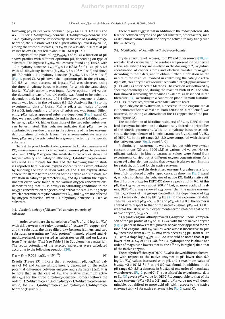
The modification of histidine residue(s) of RlL by DEPC did notlead to enzyme inactivation but dramatically altered the pH profilesof the kinetic parameters. With 1,4-dihydroxy-benzene as sub-strate, the dependences of kinetic parameters kcat, KM and kcat/KMof DEPC-RlL in the pH range 2.5–8.0 were compared with those ofthe native enzyme (Fig. 2, panel A–C).
Preliminary measurements were carried out with two oxygenconcentrations (25 and 1200 �M) at various pH values. No sig-nificant variation in kinetic parameter values were found fromexperiments carried out at different oxygen concentrations for agiven pH value, demonstrating that oxygen is always non-limitingfor catalysis, as found for the native enzyme.
Also in the case of the derivatized enzyme, kcat values as a func-tion of pH produced a bell-shaped curve, as shown in Fig. 2, panelA, which also shows the behavior of native RlL. Unlike native RlL,the pH profile of kcat for DEPC-RlL shows a peak at pH ≈ 6.0. At thispH, the kcat value was about 200 s−1 but, at more acidic pH val-ues, DEPC-RlL always showed kcat lower than the native enzyme.The pKa values of the groups controlling the dependence of kcat
on pH were calculated by fitting Eq. (2) to the experimental data.Their values were pKa1 = 5.3 ± 0.3 and pKa2 = 6.1 ± 0.3: the former isshifted with respect to that of the native enzyme, pKa1 = 4.3 ± 0.3,whereas the latter, within experimental error, matches that of thenative enzyme, pKa2 = 5.8 ± 0.1.
As regards enzyme affinity toward 1,4-hydroquinone, compari-son of the pH profile of KM of DEPC-RlL with that of native enzyme(Fig. 2, panel B) shows that sigmoidal behavior disappeared for themodified enzyme, and KM values were almost insensitive to pH:KM increased from 0.2 to 1.7 mM with decreasing pH, from 8.0 to3.0, with a slope log(KM)/pH = −0.22. It should be noted that, at pHlower than 4, KM of DEPC-RlL for 1,4-hydroquinone is about oneorder of magnitude lower (that is, the affinity is higher) than thatof the native enzyme.
The catalytic efficiency of DEPC-RlL also showed different behav-ior with respect to the native enzyme: at pH lower than 6.0,log(kcat/KM) values increased with pH, and a maximum value ofkcat/KM = 2 × 10⁴ M−1 s−1 at pH 6.0 was found. In addition, in thepH range 6.0–8.5, a decrease in kcat/KM of one order of magnitudewas observed (Fig. 2, panel C). The best fits of the experimental datato Eq. (3) gave a pKa1 value for DEPC-RlL comparable to that of thenative enzyme (pKa1 = 5.6 ± 0.2) and a pKa2 value not well deter-minable, but shifted to more acid pH with respect to the nativeenzyme (pKa2 > 8 for native enzyme) (See Fig. 2, panel C).

Author's personal copy
F. Vianello et al. / Journal of Molecular Catalysis B: Enzymatic 99 (2014) 34– 42 39
A
pH1 2 3 4 5 6 7 8 9
k cat (
s-1)
0
100
200
300
pH2 3 4 5 6 7 8 9
KM
(M)
-0.01
0.00
0.01
0.02
0.03
0.04
B
pH2 3 4 5 6 7 8 9
Log
(kca
t/KM
)
0
1
2
3
4
5
6
C
Fig. 2. pH profiles of catalytic parameters, catalytic constant kcat (panel A), Michaelisconstant, KM (panel B), and catalytic efficiency, kcat/KM (panel C) of laccase from R.lignosus native and derivatized with diethylpyrocarbonate. Measurements carriedout in 33 mM Britton–Robinson buffer, equilibrated with air, at various pH values,in presence of 1,4-dihydroxy-benzene as substrate. (�) native RlL; (©) DEPC deriva-tized RlL. Solid lines: results of best fitting of Eqs. (1)–(3) to experimental data ofpanels A, B and C, respectively.
3.5. Effect of ionic strength on RlL catalytic activity
The above results indicate that histidine residues are involved inthe catalytic steps of RlL. For information on any electrostatic inter-actions involved in enzyme activity, the dependence of the kineticparameters of RlL and DEPC-RlL on ionic strength (I) was studied,with 1,4-dihydroxy-benzene as substrate. The plots of log(kcat) vsI1/2, obtained at varying pH values (pH range 3.5–7.5), show lineardependences (r > 0.98) and the values of the product of interacting
Fig. 3. Values of product of interacting charges (Za·Zb), calculated from slope valuesof plots of log(kcat) vs I 1/2, at various pH values. Measurements carried out in 33 mMBritton–Robinson buffer, equilibrated with air, at various pH values, in presence of1,4-dihydroxy-benzene as substrate. Ionic strength modified by NaClO4 additions(I = 10 – 300 mM); (�) native RlL; (©) DEPC derivatized RlL. Solid line: results of bestfitting of Henderson–Hasselbach equation to experimental data.
charges (Za·Zb) were calculated from their slope values, determinedaccording to Eq. (4).
As regards native RlL, from the dependence of kcat on ionicstrength, a value of Za·Zb close to zero was found in the pH range5.0–7.5. At pH below 5.0, the product (Za·Zb) controlling kcat fell toabout −1 (Fig. 3), suggesting the involvement of charged groups ofopposite charges. Instead, in the case of DEPC-RlL, product (Za·Zb)was close to zero throughout the pH range explored (pH 3.5–7.5).
The same difference between RlL and DEPC-RlL was found fromthe dependence of (Za·Zb) values on the pH of KM values, that is:(Za·Zb) ≈ 0 in the pH range 7.5–5.5 and (Za·Zb) ≈ −1 at pH lowerthan 4.5 were found for the native enzyme, but no dependence ofKM on ionic strength was found for DEPC-RlL (data not shown).
Interestingly, no effect of ionic strength was found on kcat/KM,in the pH range 4.0–6.0, for either form of the enzymes, indicatingthat there were no electrostatic interactions in the first recogni-tion steps between the free native or DEPC-derivatized enzymeand 1,4-dihydroxy-benzene, which was uncharged in the pH rangeexplored.
As the same substrate, 1,4-dihydroxy-benzene, was used forboth forms of RlL, we deduce that DEPC treatment of RlL modifiedionizable groups of the enzyme, probably histidines, which wereresponsible for the pH profile of native enzyme activity and forionic interactions during the catalytic cycle.
Control experiments on the effect of ionic strength on RlL activ-ity were performed with phenol and 4-methoxyphenol. Even withthese non-ionizable substrates, enzyme activity was affected byionic strength, ruling out the involvement of charged species ofsubstrates and reaction products.
3.6. Effect of fluoride ion on catalytic activity of RlL, native andderivatized with diethyl-pyrocarbonate
The effect of fluoride ion on the activity of RlL and DEPC-RlL wasstudied, in order to obtain information about the effect of accessiblehistidine residues on the oxygen binding site of RlL.
Measurements of the enzymatic activity of RlL and DEPC-RlL, inthe presence of fluoride ion in the concentration range 0–2 mM,showed that F− does not influence KM values of either form ofthe enzyme, with 1,4-hydroquinone as substrate. Instead, fluo-ride ion decreased catalytic constants kcat of RlL and DEPC-RlL ina concentration-dependent manner. This behavior indicates thatfluoride acts as a non-competitive inhibitor. The same inhibitionconstant values were calculated for RlL and DEPC-RlL, i.e. 0.68 mM

Author's personal copy
40 F. Vianello et al. / Journal of Molecular Catalysis B: Enzymatic 99 (2014) 34– 42
Fig. 4. Sequence coverage of RlL and DEPC-RlL obtained by separate digestions of samples with Trypsin and Asp-C. Sequenced aminoacids are indicate as bold letter, underscoreindicates aminoacids involved in copper coordination, gray shadowed histidines are supposed to be DEPC modified.
and 0.71 mM, respectively. These results indicate that the bindingsite of fluoride in RIL is not affected by derivatization with DEPC,that is, the modified histidine residues did not affect the oxygen-involving steps.
3.7. Quantification and identification of DEPC modified histidineby mass spectrometry
Mass spectrometry was used to quantify and localize the DEPCmodified histidine residues. In particular, Maldi-TOF was usedto analyze both native and derivatized laccase, as described inMethods. Mass spectra showed the high purity of the enzymepreparations. The comparison between RlL and DEPC-RlL spec-tra yields a difference of 224.9 Da, corresponding to 3.12 DEPCmolecules/protein, M.W. 72, in good agreement with optical spec-troscopy measurements (Figure A4).
Moreover, modified histidines on RlL were identified by trypticand Asp-N digestion and MS/MS analysis on a Orbitrap mass spec-trometer, followed by fragment identification by Mascot searchengine (v 2.3). The combination of the results obtained by sepa-rate samples digestion with Trypsin and Asp-C led to an extensivesequence coverage (82%) (see Fig. 4), showing the remarkableexceptions of His-396, His-457 and His-64, belonging to the cop-per coordination center 1 and 2, respectively [38]. As the MS/MSsearch for DEPC modified histidines failed to return any peptide,these histidine residues were identified only in native RlL, butnot in DEPC-RlL, strongly suggesting their specific involvement inDEPC modification. It is supposed that the digestion and chromato-graphic conditions, following the DEPC modification of histidineshave induced their degradation to an adduct of unknown mass. Thisconclusion is supported by the already reported low stability ofDEPC modified histidines [36], under the experimental conditionsused during sample preparation for mass analysis (see Section 2).
4. Discussion
In the present experimental work, we carried out the kineticcharacterization of the laccase isolated from R. lignosus, for bet-ter understanding the factors controlling the pH dependence ofenzyme activity. In particular, the dependences of the kineticparameters of both native and DEPC derivatized enzymes werestudied as function of pH, ionic strength and fluoride ion.
Results obtained by comparing the correlation of the catalyticactivity (log(kcat)) of RlL and TvL with the redox potential dif-ference between the T1 copper site of enzyme (0.79 V for TvL,[23]; and 0.73 V for RlL, [39,40]), and substrates, particularly usingdihydroxy-benzene isomers, showed that, unlike TvL, in the case ofRlL, other factors, in addition to the �E, could be involved in thecontrol of enzyme activity [10,12,22,23].
We suppose that, in the laccase from R. lignosus, the ascendingpart of the pH profile of kcat may be, at least partly, attributed totitration of specific aminoacid residues in the enzyme active site. Inparticular, the most acidic residue (pKa of about 4.6) responsible forthe ascending part of the pH profile, may be a histidine. This sug-gestion is supported by experiments on derivatizing RlL with DEPC.After DEPC treatment, this pKa was shifted to 5.3 and an alterationof the T1 copper site was found. This alteration should be respon-sible of the changes in the kinetic parameters (decrease in kcat andincrease in the affinity) of RIL for the tested substrate.
The possible role of histidine residues was also supported bythe results obtained by studying the effect of ionic strength on kcat
and KM. In the native enzyme, the product of interacting chargesis ZaZb ≈ −1 in the pH range where histidine should be protonated,i.e., pH < 4.5. The derivatization of RlL with DEPC led to a loss of theresponse of kcat and KM toward ionic strength (ZaZb ≈ 0). At pH > 4.5,no effect of ionic interactions was observed for either form of theenzyme and the two kinetic parameters. Interestingly, modificationof histidine residues by DEPC led also to an increase in the affinityof the enzyme toward the substrate, at pH values lower than 4.5(lower KM values).
These results indicate that histidine residues play a role in con-trolling the catalytic constant and the enzyme-substrate/productdissociation constant values (KM) as a function of pH.
Experiments with phenol and 4-methoxyphenol, as well,demonstrated the influence of ionic strength on RlL activity. Thesetwo neutral molecules, upon oxidation by laccases, give neutral rad-ical products, ruling out the participation of the substrate in theeffect of ionic strength on RlL activity. The effect of ionic strengthon enzyme activity should be attributed to RlL structural factors,and in particular to the importance of a charged His residue on theenzyme catalytic site.
Interestingly, the slight variation of kcat and KM observed, afterRlL derivatization, at pH values above optimum pH, suggests thatthese histidine residues are not involved in controlling the descen-ding part of the pH profile of these kinetic parameters, or that this

Author's personal copy
F. Vianello et al. / Journal of Molecular Catalysis B: Enzymatic 99 (2014) 34– 42 41
Fig. 5. View of the active site of R. lignosus laccase, elaborated with PyMol from thecrystallographic structure, PDB code 1KYA [43].
behavior is ascribed to other factors independent of the titration ofionizable groups, such as the increasing inhibition of laccase activ-ity, probably at the T2/T3 copper site, by OH− ion, as suggested byXu [10,12,22,23].
The pKa value of histidine residues (pKa ≈ 4.6) we calculatedby a kinetic approach (kcat and KM dependence on pH) is verylow in comparison with that of the free residue in water solution(pKa = 6.5), indicating that the environment in which histidines areembedded tends to stabilize the neutral unprotonated form. Thatis, these histidines may be in an apolar microenvironment, as foundfor the substrate binding site of RlL by crystal structure studies anddocking simulations [42]. In order to identify the histine residue(s)responsible of the kinetic behavior of RlL, we carried out massspectrometry analyses of native and derivatized enzymes. Threeresidues appeared to be modified upon DEPC treatment: His-64,His-396 and His-457. The first one (His-64) belongs to the trinuclearT2/T3 copper site [38], which is presumed to bind molecular oxygenand where the reductive steps of the overall catalytic reaction occur[1]. As it was found that F− behaves as a non-competitive inhibitor,with comparable inhibition constants for both native and DEPCenzymes, the involvement of His-64 in the control of RlL kineticbehavior as a function of pH, is ruled out.
Consequently, we believe that one of the other two histidineresidues (His-396 and His-457) is the main responsible of the pHdependence of RlL catalytic activity at pH lower than the optimum.In fact, at low pH value, these residues are protonated and couldbe involved in slowing the catalytic reaction. In agreement withreported docking simulations [42], we believe that His-457 shouldbe the most probable candidate.
In summary, experimental evidences here presented highlightthe role of an accessible histidine in RlL, namely His-457 (Fig. 5),controlling its activity at pH lower than the optimum.
It should be stressed that previous studies on TvL have shownthat Asp206 probably plays a role in determining the pH depend-ence of TvL activity and interacts with the reducing substrateduring the catalytic cycle [24,41]. In addition, in mutagenesis stud-ies Kallio et al. [44] suggested that a crucial role is played bycarboxylic acid residue in the oxidation of phenol compoundscatalyzed by Melanocarpus albomyces laccase. Owing to the substi-tution of Asp206 (in TvL) by Phe211 in RlL, it is reasonable to assumethat RlL has a different mechanism of interaction with substrates,and different residues may play an important role in affecting thedependence of its enzyme activity on pH. Taking into account theimpossibility to perform site directed mutagenesis experiments onessential histidines involved in copper coordination in the enzymeactive site, these results provide new information about factorscontrolling the activity of this “high-potential” enzyme and may beuseful in the development of novel biotechnological applications ofRIL.
Acknowledgements
Our deep and sincere gratitude go to our mentor, Prof. AdelioRigo, recently retired, for his knowledge and logical way of thinking,which were of great value for this work. His support, encourage-ment and personal guidance provided an excellent basis for thepresent work.
The Foundation for Advanced Biomedical Research and VIMMare grateful to the “Veneto Banca” Holding for funding the acqui-sition of the MALDI-TOF/TOF mass spectrometer. The University ofPadova is grateful to the “Cassa di Risparmio di Padova e Rovigo”Holding for funding the acquisition of the LTQ-Orbitrap XL massspectrometer. We are very grateful to Dr. Giorgio Arrigoni for excel-lent assistance in MALDI-TOF/TOF operations.
This work was supported by institutional grants of the Univer-sity of Padova-Italy (Ex 60%, codes 60A06-7411 and 60A06–8055).
Appendix A. Supplementary data
Supplementary material related to this article can befound, in the online version, at http://dx.doi.org/10.1016/j.plantsci.2004.08.011.
References
[1] G.J. Davies, V. Ducros, Laccases, in: A. Messerschmidt, R. Huber, T. Poulos, K.Wieghardt (Eds.), Handbook of Metalloproteins, Vol. II, Wiley, Chichester, UK,2001, pp. 1359–1368.
[2] O.V. Morozova, G.P. Shumakovich, M.A. Gorbacheva, S.V. Shleev, A.I. Yaropolov,Biochemistry (Moscow) 72 (2007) 1136–1150.
[3] H. Claus, Micron 35 (2004) 93–96.[4] N. Duran, M.A. Rosa, A. D’Annibale, L. Gianfreda, Enzyme Microb. Technol. 31
(2002) 907–931.[5] J.M. Bollag, A. Leonowicz, Appl. Environ. Microbiol. 48 (1984) 849–854.[6] D.S. Arora, P.K. Gill, Bioresour. Technol. 73 (2000) 283–285.[7] A.C. Mot, R. Silaghi-Dumitrescu, Biochemistry (Moscow) 77 (2012) 1395–1407.[8] L. Quintanar, C. Stoj, A.B. Taylor, P.J. Hart, D.J. Kosman, E.I. Solomon, Acc. Chem.
Res. 40 (2007) 445–452.[9] R.A. Marcus, N. Sutin, Biochim. Biophys. Acta 811 (1985) 265–322.
[10] F. Xu, W. Shin, S.H. Brown, J.A. Wahleithner, U.M. Sundaram, E.I. Solomon,Biochim. Biophys. Acta 1292 (1996) 303–311.
[11] M.A. Tadesse, A. D’Annibale, C. Galli, P. Gentili, F. Sergi, Org. Biomol. Chem. 6(2008) 868–878.
[12] F. Xu, Biochemistry 35 (1996) 7608–7614.[13] E.I. Solomon, U.M. Sundaram, T.E. Machonkin, Chem. Rev. 96 (1996) 2563–2605.[14] P. Bajpai, A. Anand, P.K. Bajpai, Biotechnol. Ann. Rev. 12 (2006) 349–378.[15] R.S. Freire, N. Duran, L.T. Kubota, Anal. Chim. Acta 463 (2002) 229–238.[16] F. Vianello, A. Cambria, S. Ragusa, M.T. Cambria, A. Rigo, Biosens. Bioelectron.
20 (2004) 315–321.[17] F. Vianello, S. Ragusa, M.T. Cambria, A. Rigo, Biosens. Bioelectron. 21 (2006)
2155–2160.[18] S.R. Couto, J.L.T. Herrera, Biotechnol. Adv. 24 (2006) 500–513.[19] C.J. Rodgers, C.F. Blanford, S.R. Giddens, P. Skamnioti, F.A. Armstrong, S.J. Gurr,
Trends in Biotechnol. 28 (2009) 63–72.[20] V. Brissos, Z. Chen, L.O. Martins, Dalton Trans. 41 (2012) 6247–6255.[21] A. Gupta, I. Nederlof, S. Sottini, A.W.J.W. Tepper, E.J.J. Groenen, E.A.J. Thomassen,
G.W. Canters, J. Am. Chem. Soc. 134 (2012) 18213–18216.[22] F. Xu, J. Biol. Chem. 272 (1997) 924–928.[23] F. Xu, R.M. Berka, J.A. Wahleithner, B.A. Nelson, J.R. Shuster, S.H. Brown, A.E.
Palmer, E.I. Solomon, Biochem. J. 334 (1998) 63–70.[24] C. Madzak, M.C. Mimmi, E. Caminade, A. Brault, S. Baumberger, P. Briozzo, C.
Mougin, C. Jolivalt, Protein Eng. Des. Sel. 19 (2006) 77–84.[25] M.T. Cambria, A. Cambria, S. Ragusa, S. Rizzarelli, Prot. Expr. Purif. 18 (2000)
141–147.[26] S. Steenken, P. Neta, J. Phys. Chem. 86 (1982) 3661–3667.[27] S.V. Jovanovic, M. Tosic, M.G. Simic, J. Phys. Chem. 95 (1991) 10824–10827.[28] M.D. Liptak, K.C. Gross, P.G. Seybold, S. Feldgus, G.C. Shields, J. Am. Chem. Soc.
124 (2002), 6421-2427.[29] V.S. Stoll, J.S. Blanchard, Meth. Enzymol. 182 (1990) 24–37.[30] A. Fersht, Enzyme Structure and Mechanism, 2nd ed, W.H. Freeman & Co, New
York, 1985.[31] K.F. Tipton, H.B. Dixon, Meth. Enzymol. 63 (1979) 183–219.[32] G.B. Koudelka, F.B. Hansen, M.J. Ettinger, J. Biol. Chem. 260 (1985) 15561–15565.[33] P.W. Atkins, Physical Chemistry, 3rd ed, Oxford University Press, Oxford, 1986,
pp. 242–243.[34] K. Leidler, P. Bunting, The Chemical Kinetics of Enzyme Action, 2nd ed.,
Clarendon Press, Oxford, 1973, pp. 52–53.[35] M.M. Bradford, Anal. Biochem. 72 (1976) 248–254.

Author's personal copy
42 F. Vianello et al. / Journal of Molecular Catalysis B: Enzymatic 99 (2014) 34– 42
[36] E.W. Miles, Meth. Enzymol. 47 (1997) 431–442.[37] W.B. Melchior, D. Fahrney, Biochemistry 9 (1970) 251–258.[38] S. Garavaglia, M.T. Cambria, M. Miglio, S. Ragusa, V. Iacobazzi, F. Palmieri, C.
D’Ambrosio, A. Scaloni, M. Rizzi, J. Mol. Biol. 342 (2004) 1519–1531.[39] R.P. Bonomo, A.M. Boudet, R. Cozzolino, E. Rizzarelli, A.M. Santoro, R. Sterjiades,
R. Zappalà, J. Inorg. Biochem. 71 (1988) 205–211.[40] A.M. Garzillo, M.C. Colao, V. Buonocore, R. Oliva, L. Falcigno, M. Saviano, A.M.
Santoro, R. Zappalà, R.P. Bonomo, C. Bianco, P. Giardina, G. Palmieri, G. Sannia,J. Protein Chem. 20 (2001) 191–201.
[41] T. Bertrand, C. Jolivalt, P. Briozzo, E. Caminade, N. Joly, C. Madzak, C. Mougin,Biochemistry 41 (2002) 7325–7333.
[42] M.T. Cambria, D. Di Marino, M. Falconi, S. Garavaglia, A. Cambria, J. Biomol.Struct. Dyn. 27 (2010) 501–510.
[43] W. DeLano, The PyMOL molecular graphics system, DeLano Scientific, Palo Alto,2002, http://www.pymol.org
[44] J.P. Kallio, S. Auer, J. Jänis, M. Andberg, K. Kruss, J. Roivinen, A. Koivula, N.Hakulinen, J. Mol. Biol. 392 (2009) 895–909.