B., Collin, J., Coyle, M., Hughes, C., & Thomas, S. (2017). A
20
Main, B., Collin, J., Coyle, M., Hughes, C., & Thomas, S. (2017). A guide to deep neck space fascial infections for the dental team. Dental Update, 43(8), 745-752. https://doi.org/10.12968/denu.2016.43.8.745 Peer reviewed version Link to published version (if available): 10.12968/denu.2016.43.8.745 Link to publication record in Explore Bristol Research PDF-document This is the accepted author manuscript (AAM). The final published version (version of record) is available online via George Warman Publications at http://www.dental-update.co.uk/articleMatchListArticle.asp?aKey=1577. Please refer to any applicable terms of use of the publisher. University of Bristol - Explore Bristol Research General rights This document is made available in accordance with publisher policies. Please cite only the published version using the reference above. Full terms of use are available: http://www.bristol.ac.uk/red/research-policy/pure/user-guides/ebr-terms/
-
Upload
khangminh22 -
Category
Documents
-
view
1 -
download
0
Transcript of B., Collin, J., Coyle, M., Hughes, C., & Thomas, S. (2017). A

Main, B., Collin, J., Coyle, M., Hughes, C., & Thomas, S. (2017). Aguide to deep neck space fascial infections for the dental team. DentalUpdate, 43(8), 745-752. https://doi.org/10.12968/denu.2016.43.8.745
Peer reviewed version
Link to published version (if available):10.12968/denu.2016.43.8.745
Link to publication record in Explore Bristol ResearchPDF-document
This is the accepted author manuscript (AAM). The final published version (version of record) is available onlinevia George Warman Publications at http://www.dental-update.co.uk/articleMatchListArticle.asp?aKey=1577.Please refer to any applicable terms of use of the publisher.
University of Bristol - Explore Bristol ResearchGeneral rights
This document is made available in accordance with publisher policies. Please cite only thepublished version using the reference above. Full terms of use are available:http://www.bristol.ac.uk/red/research-policy/pure/user-guides/ebr-terms/

Oral Surgery
A guide to deep neck space fascial infections for the dental team
Authors:
Mr Barry Main MRCS (Ed), MFDS (Ed), MB ChB (Hons), BDS (Hons), BMSc (Hons)
Doctoral Research Fellow and Honorary Specialty Registrar in Oral and Maxillofacial Surgery, School of Oral and Dental Science, University of Bristol, Lower Maudlin Street, Bristol BS1 2LY
Mr John Collin BSc, MB ChB, MRCS, BDS
Specialty Registrar in Oral and Maxillofacial Surgery, Division of Oral and Maxillofacial Surgery, School of Oral and Dental Science, University of Bristol, Lower Maudline Street, Bristol BS1 2LY
Ms Margaret Coyle BA, BDentSc, MFDSRCSI, MB BCh BAO, FRCS (OMFS)
Specialty Registrar in Oral and Maxillofacial Surgery, Gloucestershire Royal Hospital, Gloucester, GL1 3NN Mr Ceri Hughes BDS, FDRSCR, MB ChB, FRCS (OMFS), FRACCDS (OMS)
Consultant Oral and Maxillofacial Surgeon, University Hospitals Bristol NHS Foundation Trust, Bristol, BS1 2LY Professor Steven Thomas BDS, MB ChB, PhD, FDSRCS, FRCS (OMFS) Professor and Consultant in Oral and Maxillofacial Surgery, Division of Oral and Maxillofacial Surgery, School of Oral and Dental Science, University of Bristol, Lower Maudlin Street, Bristol, BS1 2LY

1
A guide to deep neck space fascial infections for the dental team
Abstract
Most serious dental infections can be prevented by early treatment of the local
pathology. Patients with potentially life-threatening neck space infections arising
from the oral cavity may, however, still present in dental practice. This paper
outlines the pertinent surgical anatomy and pathophysiology; signs and
symptoms; and key early-stage management of these severe infections.
Clinical relevance
The dental team should be able to assess patients presenting with potential neck
space involvement from a dental or oral infection. They should be able to
instigate appropriate early treatment, and identify those requiring prompt
referral for assessment and management.
Objectives statement
After reading this paper, the reader should understand the basic surgical
anatomy of the fascial planes of the head and neck, and how an infection arising
in the dental tissues may spread into them. The reader should be able to
recognise the cardinal signs and symptoms of potentially-serious systemic
infection in those patients who require further assessment and management by a
maxillofacial team.

2
Introduction
Despite overall improvements in the oral health of the population1, reports in the
literature suggest an increase in the incidence of patients being managed for the
complications of dental abscess by maxillofacial teams in the United Kingdom.2
An analysis of in-patient episodes for NHS Trusts in England showed a doubling
in admissions and bed days required for the operative management of dental
abscess in the period 1998-9 and 2005-6.2 The reasons for this are not entirely
clear but it may, in part, reflect changes in the delivery of and access to dental
services in the UK.3 There is also evidence that the incidence of dental abscess is
related to socio-economic deprivation.4 These statistics reveal that, in many
cases, dental infection is not being addressed at an early stage and, therefore,
patients are presenting late with potentially serious sequelae.
One of the most serious and potentially life-threatening complications of dental
sepsis is the spread of infection into the fascial spaces of the neck. In adults,
odontogenic infections are the most common cause of such a presentation, while
in children the tonsils account for the majority of cases.5 Other aetiologies
include salivary gland infection, trauma, post-operative infection, and
complications related to congenital abnormalities of the neck such as an infected
thyroglossal duct cyst.5
Whatever the cause, spread of infection through the potential spaces in the neck
poses a risk to the airway. It may also result in systemic compromise and
cardiovascular collapse. Furthermore, infection can spread inferiorly into the

3
mediastinal or pleural cavities; or superiorly to the peri-orbital or orbital tissues,
and via the facial vein to the cavernous sinus. Patients at risk of these
complications may present initially to the dental team. It is important, therefore,
that dental practitioners are able to assess these cases and arrange for urgent
referral to a maxillofacial team for further management when required.
This paper provides the dental team with the knowledge required to recognise
patients with actual or impending neck space infection. The basic surgical
anatomy of the fascial spaces is reviewed to provide an illustration of how
infection arising in the dental tissues may spread through them. The cardinal
clinical signs and symptoms of deep neck space infection are described, with a
particular emphasis on those features that merit the prompt input of a
maxillofacial team. The principles of the management of deep neck space
infections are also described. Table 1 provides a glossary of commonly used
terminology.
Surgical anatomy of the cervical fascia6
The neck is a complex anatomical region. It serves musculoskeletal functions and
as a conduit for blood vessels, nerves, the airway, and upper gastrointestinal
tract. Fascia is the loose connective tissue that lies beneath the skin, enevelopes
the muscles, and invests the internal organs.6 The vital structures of the neck are,
therefore, invested in cervical fascia. This is made up of the superficial (lying just
deep to the subcutaneous tissue to surround the whole neck and invest the
platysma muscle) and deep layers. The deep cervical fascia consists of three
layers:
4
1. The superficial (or investing) layer of deep cervical fascia surrounds the
entire neck and envelopes the sternocleidomastoid and trapezius
muscles. This layer divides to form the capsules of the submandibular and
parotid salivary glands
2. The middle layer extends from the base of the skull into the thoracic cavity
to become continuous with the pericardium. This layer invests the
pharynx, larynx, trachea, oesophagus, thyroid gland, and strap muscles
3. The deep layer surrounds the spine and paravertebral muscles
Between these layers of deep fascia are potential spaces containing loose
connective tissue. It is within these layers that infection arising in the dental
tissues can spread. Table 2 summarises the fascial spaces of the neck and Figure
1 demonstrates the potential paths of spread of infection of odontogenic origin. If
infection spreads inferiorly into the neck, the anatomy of the deep fascial tissues
will limit spread to a certain extent, but the complex arrangement of connection
between some of the spaces means that, if infection is not controlled, more
distant spread is possible.

5
Clinical features of deep neck space infection
Symptoms
In addition to pain around the causative tooth or teeth, patients with neck space
infection will feel generally unwell. They may complain of fever and rigors.
Particularly worrying symptoms are trismus, dysphagia, dyspnoea, and change
in voice. An example of the latter is the ‘hot potato’ voice resulting from elevation
of the floor of mouth and tongue in the oral cavity. These all indicate actual or
impending airway compromise and patients presenting in this way should be
referred for urgent assessment in hospital.
Signs
Vital signs
Septic patients may be tachycardic (pulse rate >90 beats per minute) and
pyrexial. If there is an abscess, a swinging pyrexia may be seen. The respiratory
rate is a sensitive sign that may increase before an abnormality is seen in other
vital signs. A rate of > 20 breaths per minute is abnormal in an adult.7
The combination of a high respiratory rate, tachycardia, very high or very low
temperature, with a very high or very low white blood cell count is the systemic
inflammatory response syndrome (SIRS). Sepsis is defined as the presence of
SIRS in addition to a confirmed infective process. Septic shock occurs when a
septic patient remains hypotensive despite aggressive attempts at restoring the
blood pressure. Importantly, it is well recognised that the prognosis of septic
patients is improved when appropriate treatment is delivered promptly.8

6
Surgical signs
In addition to these general signs of sepsis, there are specific clinical features of
neck space infection that reflect the regional anatomy. These features are
summarised in Table 3. It is important to recognise that more than one space can
be involved. Occasionally, a patient may be systemically very unwell (showing
signs of septic shock) without any overt neck swelling or other loco-regional
symptoms or signs.
Ludwig’s angina
Ludwig’s angina is a specific diagnosis and is defined as a bilateral cellulitis of
the submandibular and sublingual spaces, most often arising from a lower molar
tooth. The floor of mouth contains the sublingual, submandibular, and submental
spaces with ready communication across the midline. Infection can, therefore,
spread to involve all spaces in the floor of mouth. Clinically, there is a firm
swelling of the floor of mouth and resultant elevation of the tongue. The
submandibular and sublingual spaces become tense and tender. There may be
accompanying trismus, dysphagia, and respiratory embarrassment. The cellulitis
may spread to involve the lateral pharyngeal space. Figure 2 shows the typical
clinical appearance in Ludwig’s angina. The importance of recognising this
diagnosis is that these patients require immediate referral to hospital for urgent
antibiotic therapy and surgical drainage, with or without additional airway
support.

7
Investigation of deep neck space infection
Investigation of the patient with a neck space infection will be dictated by the
individual clinical scenario and urgency with which hospital management is
required.
Investigation in the dental setting
The assessment of the patient in the dental setting should include checking the
temperature and basic vital signs (pulse and respiratory rates). A plain
radiograph should be obtained if the patient is well enough to tolerate it. In
patients with severe odontogenic infection there is often some degree of trismus
that makes obtaining intra-oral views difficult. A dental panoramic tomogram
can, therefore, provide a good overview of the state of the entire dentition and is
likely to reveal the source of any infection. Where possible, the dental
practitioner referring a patient into hospital should send the imaging with the
patient. This helps save time in the assessment and management of the patient
while also avoiding unnecessary repeated exposure to radiation.
Investigation in the hospital setting
Patients admitted to hospital with neck space infection will undergo blood tests,
possible further imaging including ultrasound and/or CT scan, and eventual
microbiological investigation. Where possible, a sample of pus is obtained for
microbiological culture and antibiotic sensitivity testing. Most often, pus is
obtained at the time of operation. The acute odontogenic abscess is usually
polymicrobial in nature, comprising facultative anaerobes (for example, viridans

8
streptococci and the Streptococcus anginosus group), and strict anaerobes like
Prevotella and Fusobacterium species.9,10
Principles of the surgical management of deep neck space infection
Figure 3 provides a guide to the initial assessment and management of patients
with suspected neck space infection. The patient presenting with suspected neck
space infection should be assessed immediately for Airway, Breathing, and
Circulation (ABC). This gauges the urgency of the need for referral to hospital. A
stable patient with localised swelling and minimal soft tissue involvement is
likely to be suitable for early, local treatment including pulp extirpation or
extraction of the tooth, with or without systemic antibiotics.
For those patients referred to hospital, the principles of establishing surgical
drainage, removal of the source of infection, and systemic antibiotics also apply.
Septic patients will be treated aggressively with fluid resuscitation and early,
empirical administration of antibiotics. Drainage of neck space infections will
usually take place in the operating theatre under general anaesthetic. Severe
airway compromise may necessitate placement of a tracheostomy tube and post-
operative admission to intensive care. In Ludwig’s angina, there is often no
collection of pus, but surgical exploration of the affected spaces is performed to
‘decompress’ the neck. Surgical drains are placed until resolution of the infection.
Corticosteroids (e.g. dexamethasone) may be given to help reduce the oedema
associated with these infections.

9
Summary
It is important to remember that the vast majority of odontogenic infections can
be managed using local measures such as extraction of the tooth, extirpation of
the pulp, or intra-oral incision and drainage of a buccal space abscess. Antibiotic
therapy is indicated where there are signs of systemic infection, but they are not
a substitute for removing the source of infection.
Occasionally, infection arising in the oro-dental tissues can spread into the fascial
spaces of the neck, leading to potentially life-threatening airway compromise
and/or sepsis. This paper has discussed the pertinent surgical anatomy and
described the cardinal signs and symptoms of spreading infection that should
alert the dental practitioner to a patient who required specialist in-patient
assessment and management.

10
Captions for figures
Table 1. Glossary of terms used
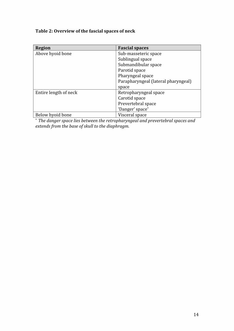
Table 2. Overview of the fascial spaces of the neck
Table 3. Summary of the clinical signs and symptoms associated with neck space
infection
Figure 1. Potential routes of spread of infection of odontogenic origin
Figure 2. Typical clinical appearance in Ludwig’s angina
Figure 3. Guide to the initial assessment and management in suspected neck
space infection

11
References
1. Steele JG, Treasure ET, O’Sullivan I, Morris J, Murray JJ. Adult dental health
survery 2009: transformation in British oral health 1968-2009. Br Dent J
2012; 213: 523-527
2. Thomas SJ, Highes C, Atkinson C, Ness AR, Revington PJ. Is there an
epidemic of admissions for surgical treatment of dental abscess in the UK?
BMJ 2008; 336: 1219-1220
3. Freeman R. Reforming NHS dentistry. Equitable distribution of affordable
dental services is still possible. BMJ 2008; 336: 1202-1203
4. Moles DR, Frost C, Grundy C. Inequalities in availability of National Health
Service general dental practitioners in England and Wales. Br Dent J 2001;
190: 548-553
5. Vieira F, Allen SM, Stocks RM, Thompson JW. Deep neck infection.
Otolarngol Clin North Am 2008; 41: 459-483
6. Ellis H. Clinical anatomy – applied anatomy for students and junior doctors.
11th ed. pp262-264. Blackwell: Oxford, 2006
7. Handley T, Devlin M, Koppel D, McCaul J. The sepsis syndrome in
odontogenic infection. J Intensive Care Med 2009; 10: 21-25
8. Dellinger RP, Levy MM, Carlet JM et al. Surviving sepsis campaign:
international guidelines for management of severe sepsis and septic
shock. Intensive Care Med 2008; 34: 17-60
9. Robertson D, Smith AJ. The microbiology of the acute dental abscess. J
Med Microbiol 2009; 58: 155-162

12
10. Al-Qamachi LH, Aga H, McMahon J, Leonard A. Microbiology of
odontogenic infections in deep neck spaces: a retrospective study. Br J
Oral Maxillofac Surg 2010; 48: 37-39

13
Table 1. Glossary of terms used
Term Definition Abscess Localised collection of pus Cellulitis Inflammation of the subcutaneous
tissues with no significant localisation of pus (may later organise to form an abscess)
Sepsis The presence of the systemic inflammatory response syndrome (temperature <36° or >38°; pulse rate >90 per minute; respiratory rate >20 per minute; and white cell count <4 or >12 X 109/L) plus confirmed infection
Dysphagia The subjective sensation of difficulty swallowing
Dyspnoea The subjective sensation of difficulty breathing
14
Table 2: Overview of the fascial spaces of neck Region Fascial spaces Above hyoid bone Sub-masseteric space
Sublingual space Submandibular space Parotid space Pharyngeal space Parapharyngeal (lateral pharyngeal) space
Entire length of neck Retropharyngeal space Carotid space Prevertebral space ‘Danger’ space*
Below hyoid bone Visceral space * The danger space lies between the retropharyngeal and prevertebral spaces and extends from the base of skull to the diaphragm.

15
Table 3. Summary of the clinical signs and symptoms associated with neck space infection Anatomical space Description of space Signs and symptoms of infection in space
Sublingual space Bounded by the mucosa of the floor of mouth superiorly
and the mylohyoid muscle inferiorly Swollen, red floor of mouth. Little or no extra-oral sign of swelling. Elevation of the tongue may result in dysarthria, dysphagia and/or dyspnoea
Submandibular space Lies between the mylohyoid muscle, superficial fascia, platysma and skin. Contains the submandibular gland and lymph nodes
Painful, red swelling of the neck immediately below the lower border of the mandible. Involvement of the muscles of mastication results in trismus
Submental space Lies between the mylohyoid muscles and the skin, just beneath the chin. Contains submental lymph nodes.
Swelling and erythema of chin
Submasseteric space Bounded by the lateral border of the mandible and the medial aspect of the masseter
Pain and swelling over the angle of the mandible plus trismus
Parapharyngeal (lateral pharyngeal) space
Extends from the base of skull to the level of the hyoid bone. It contains the internal carotid artery, the internal jugular vein, cranial nerves IX to XII and the sympathetic chain.
Examination of the oro-pharynx reveals deviation of the uvula to the opposite side, accompanied by displaced tonsil and lateral pharyngeal surface. There may be trimsus and swelling of the lateral neck. Involvement of vital structures may include internal jugular vein thrombosis, Horner syndrome* and meningitis
Retropharyngeal space This space lies behind the pharynx and oesophagus between the skull base and mediastinum.
The patient will be unwell and complain of severe sore throat. There will be dysphagia and limitation in neck movement.
* Horner syndrome is caused by involvement of the sympathetic chain and is characterised by the triad of ptosis (drooping eyelid), miosis (constricted pupil) and anhydrosis (lack of sweating on affected side of fac

16
Figure 1. Potential routes of spread of infection of odontogenic origin

17
Figure 2. Typical clinical appearance in Ludwig’s angina

18
Figure 3. Guide to the initial assessment and management in suspected
neck space infection
Patient with suspected neck space infection
Initial assessment: ABCD
A. Can the patient speak normally with no drooling of saliva? B. Is the breathing rate less than 20 per minute and effortless? C. Is the pulse rate less than 90 beats per minute? D. Is the patient fully conscious and coherent?
YES NO
Examine patient Obtain DPT if possible Can local treatment be performed (e.g. extraction, extirpation, intra-oral incision and drainage) Call local maxillofacial team for advice and arrange onward referral if necessary Send patient to the hospital along with X-rays and referral letter
Call local maxillofacial team to arrange urgent assessment of the patient in hospital Send the patient to hospital urgently. If necessary (e.g. suspected immediate airway loss or collapse) call 999 for an ambulance