2010 Plos ONE Deep Sequencing of Human Nuclear and Cytoplasmic Small RNAs Reveals an Unexpectedly...
14
Deep Sequencing of Human Nuclear and Cytoplasmic Small RNAs Reveals an Unexpectedly Complex Subcellular Distribution of miRNAs and tRNA 39 Trailers Jian-You Liao, Li-Ming Ma, Yan-Hua Guo, Yu-Chan Zhang, Hui Zhou, Peng Shao, Yue-Qin Chen, Liang-Hu Qu* State Key Laboratory of Biocontrol, Key Laboratory of Gene Engineering of the Ministry of Education, Sun Yat-sen University, Guangzhou, People’s Republic of China Abstract Background: MicroRNAs (miRNAs) are ,22-nt small non-coding regulatory RNAs that have generally been considered to regulate gene expression at the post-transcriptional level in the cytoplasm. However, recent studies have reported that some miRNAs localize to and function in the nucleus. Methodology/Principal Findings: To determine the number of miRNAs localized to the nucleus, we systematically investigated the subcellular distribution of small RNAs (sRNAs) by independent deep sequencing sequenced of the nuclear and cytoplasmic pools of 18- to 30-nucleotide sRNAs from human cells. We identified 339 nuclear and 324 cytoplasmic known miRNAs, 300 of which overlap, suggesting that the majority of miRNAs are imported into the nucleus. With the exception of a few miRNAs evidently enriched in the nuclear pool, such as the mir-29b, the ratio of miRNA abundances in the nuclear fraction versus in the cytoplasmic fraction vary to some extent. Moreover, our results revealed that a large number of tRNA 39trailers are exported from the nucleus and accumulate in the cytoplasm. These tRNA 39 trailers accumulate in a variety of cell types, implying that the biogenesis of tRNA 39 trailers is conserved and that they have a potential functional role in vertebrate cells. Conclusion/Significance: Our results provide the first comprehensive view of the subcellular distribution of diverse sRNAs and new insights into the roles of miRNAs and tRNA 39 trailers in the cell. Citation: Liao J-Y, Ma L-M, Guo Y-H, Zhang Y-C, Zhou H, et al. (2010) Deep Sequencing of Human Nuclear and Cytoplasmic Small RNAs Reveals an Unexpectedly Complex Subcellular Distribution of miRNAs and tRNA 39 Trailers. PLoS ONE 5(5): e10563. doi:10.1371/journal.pone.0010563 Editor: Shuang-yong Xu, New England Biolabs, Inc, United States of America Received February 3, 2010; Accepted April 19, 2010; Published May 14, 2010 Copyright: ß 2010 Liao et al. This is an open-access article distributed under the terms of the Creative Commons Attribution License, which permits unrestricted use, distribution, and reproduction in any medium, provided the original author and source are credited. Funding: This research was supported by the National Natural Science Foundation of China (No. 30830066, 30771151 and 30870530), funds from National High- Tech Program (No. 2008AA02Z106), the Young Teacher Training Program of Sun Yat-sen University (2009-33000-3161051) and the National Basic Research Program (No.2005CB724600) from the Ministry of Science and Technology of China. The funders had no role in study design, data collection and analysis, decision to publish, or preparation of the manuscript. Competing Interests: The authors have declared that no competing interests exist. * E-mail: [email protected] Introduction MicroRNAs (miRNAs) are ,22-nt long non-coding regulatory RNAs that are widely expressed in metazoans and regulate many important biological processes, including differentiation, apoptosis and cellular transformation [1]. Most miRNA genes are transcribed by RNA polymerase II into primary miRNA transcripts, which are further processed into hairpin-structured miRNA precursors (pre-miRNAs) in the nucleus by Drosha and its partner DGCR8/Pasha [2]. Pre-miRNAs are then exported to the cytoplasm by Exportin5 [3] and converted into ,22-nt mature miRNAs by Dicer, after which one strand of the newly formed duplex is incorporated into the Ago protein complex [4,5]. miRNAs are generally believed to inhibit mRNA translation post- transcriptionally by binding partially complementary target sites in the 39 untranslated regions (UTRs) of target mRNAs in the cytoplasm [6]. However, recent studies have shown that some miRNAs are localized to the nucleus. For instance, several rat miRNAs localize to the nucleolus [7,8] and human miR-29b contains a nuclear import element at its 39 end that can direct nuclear enrichment of this miRNA [9]. Moreover, miRNAs can inhibit or activate gene expression at the transcriptional level in the nucleus of human and plant cells [10–12]. Although only a few miRNAs have been identified in the nucleus thus far, it is very likely that many more miRNAs localize to and function in the nucleus. Identification of nuclear miRNAs may provide new insights into the regulatory roles played by miRNAs in the nucleus. Humans express four Ago proteins (Ago1-Ago4) in numerous tissues and cell types [13]. All four of these Ago proteins associate with miRNAs and other small RNAs (sRNAs) [14–16] and they all contribute to the process of miRNA-mediated gene silencing [17]. The Ago2 protein, a key component of the RISC complex, can be imported into the nucleus from the cytoplasm [18,19]. To date, the exact nuclear function of the Ago2 protein remains unclear but treatment with exogenous siRNAs (which associate with the Ago2 protein after the introduction of double-stranded RNAs (dsRNAs) into the cell) directed against nuclear RNAs such as the 7SK RNA efficiently reduce 7SK RNA levels in the nuclear fraction [20]. These findings suggest that the Ago2 protein can mediate the cleavage of target RNAs in the nucleus. The Ago1 protein is also known to PLoS ONE | www.plosone.org 1 May 2010 | Volume 5 | Issue 5 | e10563
Transcript of 2010 Plos ONE Deep Sequencing of Human Nuclear and Cytoplasmic Small RNAs Reveals an Unexpectedly...
Deep Sequencing of Human Nuclear and CytoplasmicSmall RNAs Reveals an Unexpectedly ComplexSubcellular Distribution of miRNAs and tRNA 39 TrailersJian-You Liao, Li-Ming Ma, Yan-Hua Guo, Yu-Chan Zhang, Hui Zhou, Peng Shao, Yue-Qin Chen, Liang-Hu
Qu*
State Key Laboratory of Biocontrol, Key Laboratory of Gene Engineering of the Ministry of Education, Sun Yat-sen University, Guangzhou, People’s Republic of China
Abstract
Background: MicroRNAs (miRNAs) are ,22-nt small non-coding regulatory RNAs that have generally been considered toregulate gene expression at the post-transcriptional level in the cytoplasm. However, recent studies have reported thatsome miRNAs localize to and function in the nucleus.
Methodology/Principal Findings: To determine the number of miRNAs localized to the nucleus, we systematicallyinvestigated the subcellular distribution of small RNAs (sRNAs) by independent deep sequencing sequenced of the nuclearand cytoplasmic pools of 18- to 30-nucleotide sRNAs from human cells. We identified 339 nuclear and 324 cytoplasmicknown miRNAs, 300 of which overlap, suggesting that the majority of miRNAs are imported into the nucleus. With theexception of a few miRNAs evidently enriched in the nuclear pool, such as the mir-29b, the ratio of miRNA abundances inthe nuclear fraction versus in the cytoplasmic fraction vary to some extent. Moreover, our results revealed that a largenumber of tRNA 39trailers are exported from the nucleus and accumulate in the cytoplasm. These tRNA 39 trailersaccumulate in a variety of cell types, implying that the biogenesis of tRNA 39 trailers is conserved and that they have apotential functional role in vertebrate cells.
Conclusion/Significance: Our results provide the first comprehensive view of the subcellular distribution of diverse sRNAsand new insights into the roles of miRNAs and tRNA 39 trailers in the cell.
Citation: Liao J-Y, Ma L-M, Guo Y-H, Zhang Y-C, Zhou H, et al. (2010) Deep Sequencing of Human Nuclear and Cytoplasmic Small RNAs Reveals an UnexpectedlyComplex Subcellular Distribution of miRNAs and tRNA 39 Trailers. PLoS ONE 5(5): e10563. doi:10.1371/journal.pone.0010563
Editor: Shuang-yong Xu, New England Biolabs, Inc, United States of America
Received February 3, 2010; Accepted April 19, 2010; Published May 14, 2010
Copyright: � 2010 Liao et al. This is an open-access article distributed under the terms of the Creative Commons Attribution License, which permits unrestricteduse, distribution, and reproduction in any medium, provided the original author and source are credited.
Funding: This research was supported by the National Natural Science Foundation of China (No. 30830066, 30771151 and 30870530), funds from National High-Tech Program (No. 2008AA02Z106), the Young Teacher Training Program of Sun Yat-sen University (2009-33000-3161051) and the National Basic ResearchProgram (No.2005CB724600) from the Ministry of Science and Technology of China. The funders had no role in study design, data collection and analysis, decisionto publish, or preparation of the manuscript.
Competing Interests: The authors have declared that no competing interests exist.
* E-mail: [email protected]
Introduction
MicroRNAs (miRNAs) are ,22-nt long non-coding regulatory
RNAs that are widely expressed in metazoans and regulate many
important biological processes, including differentiation, apoptosis
and cellular transformation [1]. Most miRNA genes are
transcribed by RNA polymerase II into primary miRNA
transcripts, which are further processed into hairpin-structured
miRNA precursors (pre-miRNAs) in the nucleus by Drosha and its
partner DGCR8/Pasha [2]. Pre-miRNAs are then exported to the
cytoplasm by Exportin5 [3] and converted into ,22-nt mature
miRNAs by Dicer, after which one strand of the newly formed
duplex is incorporated into the Ago protein complex [4,5].
miRNAs are generally believed to inhibit mRNA translation post-
transcriptionally by binding partially complementary target sites in
the 39 untranslated regions (UTRs) of target mRNAs in the
cytoplasm [6]. However, recent studies have shown that some
miRNAs are localized to the nucleus. For instance, several rat
miRNAs localize to the nucleolus [7,8] and human miR-29b
contains a nuclear import element at its 39 end that can direct
nuclear enrichment of this miRNA [9]. Moreover, miRNAs can
inhibit or activate gene expression at the transcriptional level in
the nucleus of human and plant cells [10–12]. Although only a few
miRNAs have been identified in the nucleus thus far, it is very
likely that many more miRNAs localize to and function in the
nucleus. Identification of nuclear miRNAs may provide new
insights into the regulatory roles played by miRNAs in the nucleus.
Humans express four Ago proteins (Ago1-Ago4) in numerous
tissues and cell types [13]. All four of these Ago proteins associate with
miRNAs and other small RNAs (sRNAs) [14–16] and they all
contribute to the process of miRNA-mediated gene silencing [17].
The Ago2 protein, a key component of the RISC complex, can be
imported into the nucleus from the cytoplasm [18,19]. To date, the
exact nuclear function of the Ago2 protein remains unclear but
treatment with exogenous siRNAs (which associate with the Ago2
protein after the introduction of double-stranded RNAs (dsRNAs)
into the cell) directed against nuclear RNAs such as the 7SK RNA
efficiently reduce 7SK RNA levels in the nuclear fraction [20]. These
findings suggest that the Ago2 protein can mediate the cleavage of
target RNAs in the nucleus. The Ago1 protein is also known to
PLoS ONE | www.plosone.org 1 May 2010 | Volume 5 | Issue 5 | e10563
localize to and function in the nucleus [21,22]. It is unclear whether
the Ago proteins can enter the nucleus while bound to cytoplasmic
sRNAs. Notably, the nuclear localization of NRDE-3, an Ago protein
of Caenorhabditis elegans, requires sRNA binding [23].
Recently, deep sequencing of 18- to 30-nt fractionated RNA has
become the most common and widely used approach for the
discovery of sRNAs. This is a transcriptome-wide approach and is
highly effective even for the identification of very low-abundance
sRNAs. However, this approach does not provide information on
the subcellular localization of the sRNAs. This additional
information is often very useful in characterizing the functions of
the sRNAs. In fact, many small regulatory RNAs, including
endogenous siRNAs in worms [23], heterochromatin-related
siRNAs in fungi [24] and piRNAs (piwi-associated RNAs) in
mammals [25], are believed to function at least partially within the
nucleus. In contrast, miRNAs are generally believed to function
predominantly in the cytoplasm based on their subcellular
localization. Next-generation sequencing technology has been
used extensively for the discovery and profiling of miRNAs in cells
[26–28]. This technology could also provide a powerful approach
for investigating the subcellular localization and function of sRNAs
by combining it with subcellular fractionation techniques. In this
study, we performed a deep sequencing analysis of sRNAs isolated
from both the cytoplasm and the nucleus of human cells. Our
results provide the first evidence of an unexpectedly complex
subcellular distribution of diverse sRNAs. Specifically, we provide
a comprehensive examination of the patterns of distribution of
miRNAs and tRNA 39 trailers in human cells.
Results
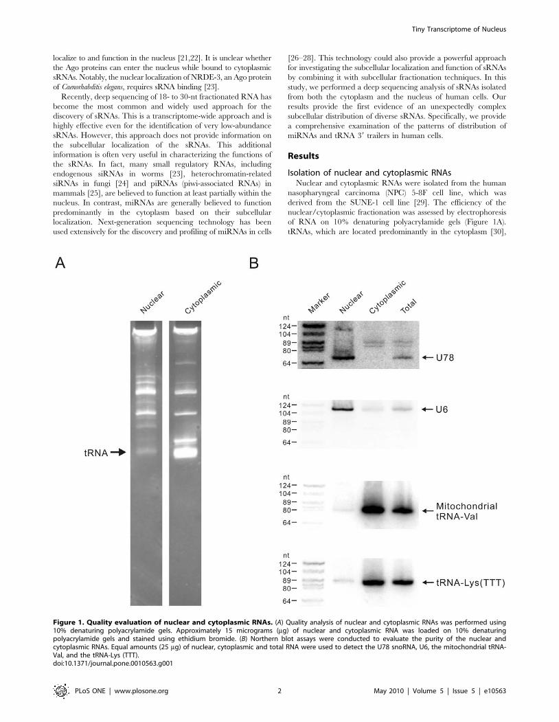
Isolation of nuclear and cytoplasmic RNAsNuclear and cytoplasmic RNAs were isolated from the human
nasopharyngeal carcinoma (NPC) 5-8F cell line, which was
derived from the SUNE-1 cell line [29]. The efficiency of the
nuclear/cytoplasmic fractionation was assessed by electrophoresis
of RNA on 10% denaturing polyacrylamide gels (Figure 1A).
tRNAs, which are located predominantly in the cytoplasm [30],
Figure 1. Quality evaluation of nuclear and cytoplasmic RNAs. (A) Quality analysis of nuclear and cytoplasmic RNAs was performed using10% denaturing polyacrylamide gels. Approximately 15 micrograms (mg) of nuclear and cytoplasmic RNA was loaded on 10% denaturingpolyacrylamide gels and stained using ethidium bromide. (B) Northern blot assays were conducted to evaluate the purity of the nuclear andcytoplasmic RNAs. Equal amounts (25 mg) of nuclear, cytoplasmic and total RNA were used to detect the U78 snoRNA, U6, the mitochondrial tRNA-Val, and the tRNA-Lys (TTT).doi:10.1371/journal.pone.0010563.g001
Tiny Transcriptome of Nucleus
PLoS ONE | www.plosone.org 2 May 2010 | Volume 5 | Issue 5 | e10563
were almost entirely depleted in the nuclear RNA fraction and
abundant in the cytoplasmic fraction, indicating that nuclear and
cytoplasmic RNAs were successfully separated. Northern blot
analysis of three different compartment-specific RNAs was further
used to evaluate the purity of the nuclear and cytoplasmic RNA
fractions (Figure 1B). The U78 snoRNA (small nucleolar RNAs)
and U6 snRNA were present exclusively in the nuclear RNA
fraction, whereas the nuclear-encoded tRNA and mitochondrial-
encoded tRNA were enriched in the cytoplasmic RNA. Together,
these results indicated that the nuclear and cytoplasmic RNAs
were highly purified. Northern blot analysis of U78 snoRNA and
tRNA-Lys (TTT) indicated that the proportions of nuclear RNA
in the 5-8F cell total RNA were ,20%, the value that is in
agreement with that estimated previously for HeLa cells [16].
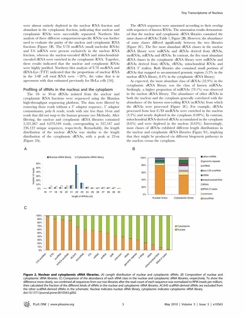
Profiling of sRNAs in the nucleus and the cytoplasmThe 18- to 30-nt sRNAs isolated from the nuclear and
cytoplasmic RNA fractions were sequenced using the Illumina
high-throughput sequencing platform. The data were filtered by
removing those reads without a 39 adaptor sequence, 59 adaptor
contaminants, poly-A reads, reads with size less than 16-nt and
reads that did not map to the human genome (see Methods). After
filtering, the nuclear and cytoplasmic sRNA libraries contained
5,321,867 and 4,079,549 reads, corresponding to 337,547 and
236,122 unique sequences, respectively. Remarkably, the length
distribution of the nuclear sRNAs was similar to the length
distribution of the cytoplasmic sRNAs, with a peak at 22-nt
(Figure 2A).
The sRNA sequences were annotated according to their overlap
with sequences of known RNAs. The annotation results demonstrat-
ed that the nuclear and cytoplasmic sRNA libraries contained the
same classes of sRNAs (Table 1, Figure 2B). However, the abundance
of some classes differed significantly between the two libraries
(Figure 2C). The five most abundant sRNA classes in the nuclear
sRNA library were miRNAs and sRNAs derived from rRNAs,
snoRNAs, mRNAs and tRNAs. In contrast, the five most abundant
sRNA classes in the cytoplasmic sRNA library were miRNAs and
sRNAs derived from tRNAs, rRNAs, mitochondrial RNAs and
tRNA 39 trailers. Both libraries also contained small portions of
sRNAs that mapped to un-annotated genomic regions (5.3% in the
nuclear sRNA library, 8.4% in the cytoplasmic sRNA library).
As expected, the most abundant class of sRNAs (52.9%) in the
cytoplasmic sRNA library was the class of known miRNAs.
Strikingly, a higher proportion of miRNAs (79.1%) was observed
in the nuclear sRNA library. The abundance of other sRNAs in
both the nucleus and the cytoplasm generally correlated with the
abundance of the known non-coding RNA (ncRNAs) from which
the sRNAs were processed (Figure 2C). For example, sRNAs
processed from box C/D snoRNAs were enriched in the nucleus
(3.3%) and nearly depleted in the cytoplasm (0.08%). In contrast,
mitochondrial RNA-derived sRNAs accumulated in the cytoplasm
(8.6%) and were depleted in the nucleus (0.63%). Interestingly,
most classes of sRNAs exhibited different length distributions in
the nuclear and cytoplasmic sRNA libraries (Figure S1), implying
that they might be produced via different biogenesis pathways in
the nucleus versus the cytoplasm.
Figure 2. Nuclear and cytoplasmic sRNA libraries. (A) Length distribution of nuclear and cytoplasmic sRNAs. (B) Composition of nuclear andcytoplasmic sRNA libraries. (C) Comparison of the abundance of each sRNA class in the nuclear and cytoplasmic sRNA libraries, respectively. To show thedifference more clearly, we combined all sequences from our two libraries after the read count of each sequence was normalized to RPM (reads per million),then calculated the fraction of the different kinds of sRNAs in the nuclear and cytoplasmic sRNA libraries. ACA45 scaRNA-derived sRNAs are excluded fromthe other scaRNA-derived sRNAs in the schematic. Nuclear indicates nuclear sRNA library, cytoplasmic indicates cytoplasmic sRNA library.doi:10.1371/journal.pone.0010563.g002
Tiny Transcriptome of Nucleus
PLoS ONE | www.plosone.org 3 May 2010 | Volume 5 | Issue 5 | e10563
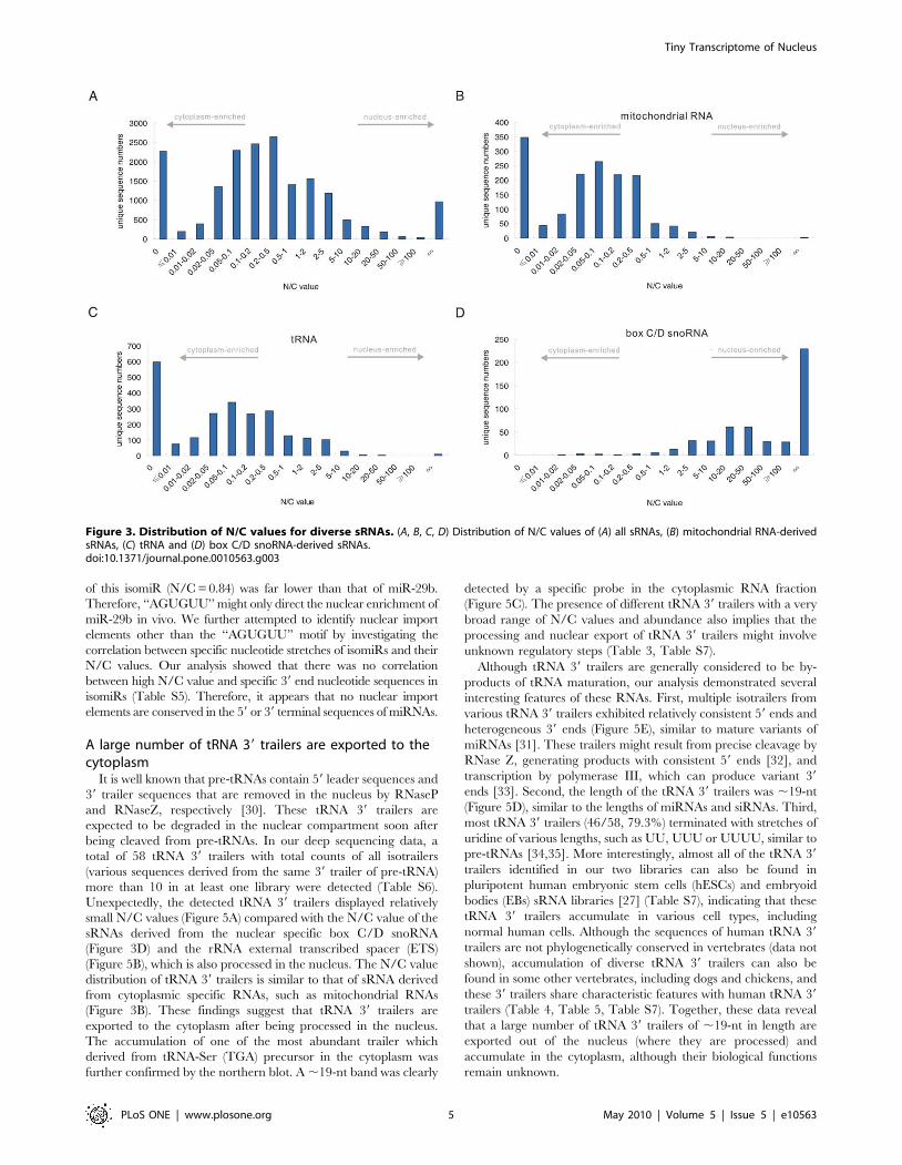
A total of 17,839 unique sequences presented raw read counts
(non-normalized) greater than 10 in at least one library,
representing the majority of sRNAs in the 5-8F cell line. We
calculated the ratio of nuclear sRNA library counts to cytoplasmic
sRNA library counts (N/C) of these unique sequences. The N/C
values of the majority of sRNAs (72.4%) ranged from 0.02 to 5
(Figure 3A). Some sRNAs were identified exclusively in the
nuclear (5.4%) or cytoplasmic (12.8%) sRNA libraries, but the
majority of these were present at a relatively low abundance. The
N/C values of the unique sequences could reflect their subcellular
distribution. Each of the sRNA class had its own characteristic
distribution pattern of N/C value and the sRNAs had a very broad
range of N/C values (Table S1, Figure 3A, Figure S2), suggesting a
complex subcellular distribution of the sRNAs. Larger N/C values
indicate greater enrichment in the nucleus, whereas smaller values
indicate greater enrichment in the cytoplasm. As expected, the
sRNAs derived from cytoplasmic-localized RNAs, such as
mitochondrial RNAs and tRNAs, displayed relatively low N/C
values (Figure 3B, 3C, Figure S2) and the sRNAs derived from
nuclear-localized RNAs, such as box C/D snoRNAs and rRNA
external transcribed spacer (ETS), displayed relative high N/C
values (Figure 3D, 5B, Figure S2).
It is important to note that some sRNAs derived from
compartment-specific RNA families, such as mitochondrial RNAs
and snoRNAs, displayed significantly higher or lower N/C values
than other family members (Figure 3B, 3D, Figure S2). For
example, the N/C values of a few sRNAs derived from
mitochondrial RNAs were larger than 1, much higher than most
of the other mitochondrial RNA-derived sRNAs, which presented
N/C values lower than 0.1. This suggests that these few sRNAs do
not display the same subcellular localization as their precursor or
host RNAs.
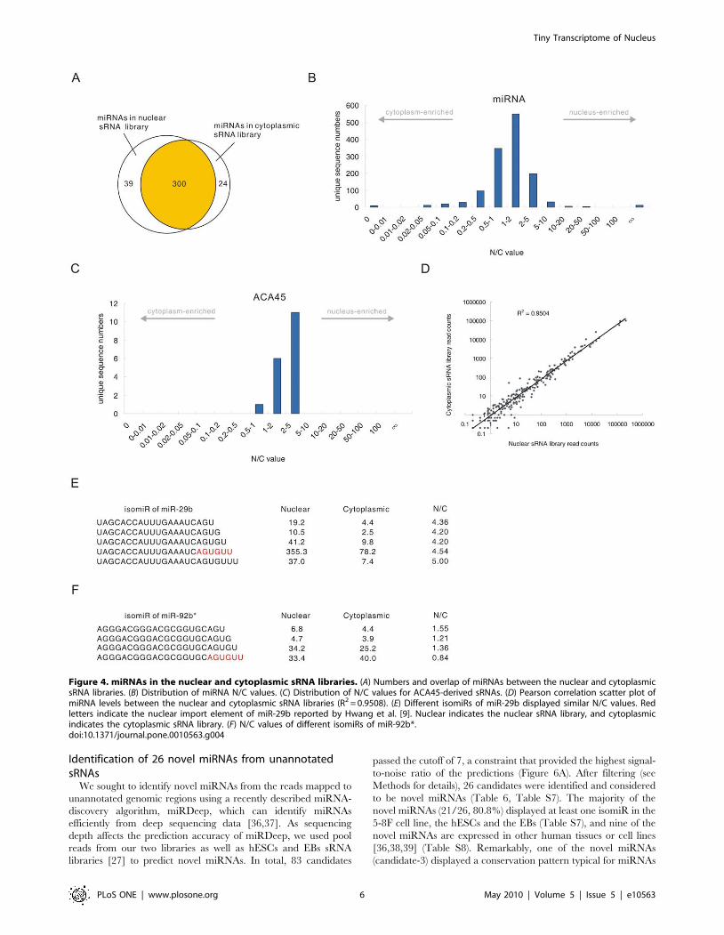
Most miRNAs are imported into the nucleusAdditional investigations were carried out to explore the
miRNA profiles in further detail. In this study, a total of 339
and 324 miRNAs, corresponding to 361 and 334 distinct miRNA
genes, were identified in the nuclear and cytoplasmic sRNA
libraries, respectively (Table S2). We identified a large overlap of
miRNAs between the two libraries (Figure 4A), and those miRNAs
present exclusively in either the nuclear or the cytoplasmic sRNA
libraries were generally present at very low abundance. Each
miRNA had multiple mature variants, called isomiRs [27].
Although miRNAs have been generally considered to localize
and function only in the cytoplasm, most isomiRs with raw read
count more than 10 in at least one library (98.5%) were found to
present in both the nuclear and cytoplasmic sRNA libraries and
microRNAs (Figure 4B) displayed very different N/C value
distribution from the sRNAs derived from mitochondrial RNAs
(Figure 3B) and box C/D snoRNAs (Figure 3D), indicating that
most miRNAs, regardless of their sequence, are imported into the
nucleus. Remarkably, the N/C values and length distribution of
the sRNAs processed from the ACA45 scaRNA (small cajal body
specific RNA) (Figure 4C, Figure S1B) were similar to the length
and N/C values of the miRNAs (Figure 4B, Figure S1A), but
different from other scaRNA-derived sRNAs (Figure S2A, S1C).
This result is consistent with the recent finding that ACA45 can be
processed into miRNA-like functional sRNAs [15] and provides
support for the import of miRNAs into the nucleus, regardless of
their processing pathway.
We employed the read count of the most abundant isomiR to
represent the expression level of each miRNA in the two libraries,
as previous reports have indicated that the most abundant isomiR
is the most useful for identifying differentially expressed miRNAs
[27]. A small subset of miRNAs presented different most-abundant
isomiRs in the two libraries (Table S3). Since these miRNAs
account for only a small portion of the total miRNAs found in the
5-8F cell line and the majority of them were expressed at very low
levels (,10 in both libraries), they were not used in further
analyses. Therefore, a total of 281 and 266 different mature
miRNAs were analyzed from the nuclear and cytoplasmic sRNA
libraries, respectively. We found that the expression level of each
miRNA in the nuclear miRNA pool correlated with the expression
level in the cytoplasmic miRNA pool (R2 = 0.9508) (Figure 4D).
A previous report has shown that miR-29b is enriched in the
nucleus via a process directed by the 39 end hexanucleotide
AGUGUU [9]. Consistent with this report, our results showed that
miR-29b was highly enriched in the nucleus (N/C = 4.54). Several
other miRNAs, such as miR-32 (N/C = 6.24), miR-148a (N/
C = 4.87) and miR-148b (N/C = 3.72) (Table 2) were also
enriched in the nucleus to a similar extent as miR-29b. However,
we did not observe conserved 39 end hexanucleotides or any other
conserved elements between the miRNAs with high N/C values,
implying that a mechanism other than that directed by
hexanucleotides is involved in miRNA import into the nucleus.
It is worth noting that some miR-29b isomiRs contained shortened
or extended versions of the 39 end motif ‘‘AGUGUU’’, i.e. ‘‘AGU’’,
‘‘AGUG’’, ‘‘AGUGU’’, and ‘‘AGUGUUU’’. Interestingly, these
isomiRs all presented similar N/C values as the miR-29b containing
a complete ‘‘AGUGUU’’ hexanucleotide sequence at its 39 end
(Figure 4E). However, we found that the other miRNAs with these
motifs at their 39 end did not have high N/C value (Table S4),
indicating that these motifs could not directed the nuclear enrichment
of other miRNAs. We next asked whether the ‘‘AGUGUU’’ motif
could direct the nuclear enrichment of other miRNAs. Only one
isomiR, an isomiR of miR-92b*, contained the ‘‘AGUGUU’’
sequence at its 39 end (Figure 4F). However, the nuclear enrichment
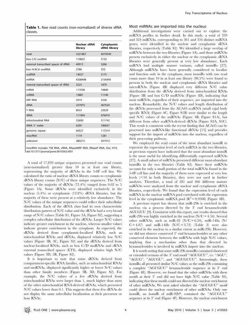
Table 1. Raw read counts (non-normalized) of diverse sRNAclasses.
Nuclear sRNAlibrary
CytoplasmicsRNA library
total 5321867 4079549
box C/D snoRNA 174833 3132
external transcribed spacer of rRNA 40913 3266
box H/ACA snoRNA 2780 353
scaRNA 14027 5175
miRNA 4208498 2156495
internal transcribed spacer of rRNA 3225 1879
mRNA 117038 74840
snRNA 13881 11140
SRP RNA 2919 3256
rRNA 237973 367524
Y RNA 8033 22629
tRNA 111904 476974
mitochondrial RNA 33690 311742
tRNA 39 trailer 6295 180657
genomic repeat 64527 117314
miscRNA 1119 1281
unknown 280215 341913
miscRNA includes 7SK RNA, vRNA, RNaseMRP RNA, RNaseP RNA, Xist, H19.doi:10.1371/journal.pone.0010563.t001
Tiny Transcriptome of Nucleus
PLoS ONE | www.plosone.org 4 May 2010 | Volume 5 | Issue 5 | e10563
of this isomiR (N/C = 0.84) was far lower than that of miR-29b.
Therefore, ‘‘AGUGUU’’ might only direct the nuclear enrichment of
miR-29b in vivo. We further attempted to identify nuclear import
elements other than the ‘‘AGUGUU’’ motif by investigating the
correlation between specific nucleotide stretches of isomiRs and their
N/C values. Our analysis showed that there was no correlation
between high N/C value and specific 39 end nucleotide sequences in
isomiRs (Table S5). Therefore, it appears that no nuclear import
elements are conserved in the 59 or 39 terminal sequences of miRNAs.
A large number of tRNA 39 trailers are exported to thecytoplasm
It is well known that pre-tRNAs contain 59 leader sequences and
39 trailer sequences that are removed in the nucleus by RNaseP
and RNaseZ, respectively [30]. These tRNA 39 trailers are
expected to be degraded in the nuclear compartment soon after
being cleaved from pre-tRNAs. In our deep sequencing data, a
total of 58 tRNA 39 trailers with total counts of all isotrailers
(various sequences derived from the same 39 trailer of pre-tRNA)
more than 10 in at least one library were detected (Table S6).
Unexpectedly, the detected tRNA 39 trailers displayed relatively
small N/C values (Figure 5A) compared with the N/C value of the
sRNAs derived from the nuclear specific box C/D snoRNA
(Figure 3D) and the rRNA external transcribed spacer (ETS)
(Figure 5B), which is also processed in the nucleus. The N/C value
distribution of tRNA 39 trailers is similar to that of sRNA derived
from cytoplasmic specific RNAs, such as mitochondrial RNAs
(Figure 3B). These findings suggest that tRNA 39 trailers are
exported to the cytoplasm after being processed in the nucleus.
The accumulation of one of the most abundant trailer which
derived from tRNA-Ser (TGA) precursor in the cytoplasm was
further confirmed by the northern blot. A ,19-nt band was clearly
detected by a specific probe in the cytoplasmic RNA fraction
(Figure 5C). The presence of different tRNA 39 trailers with a very
broad range of N/C values and abundance also implies that the
processing and nuclear export of tRNA 39 trailers might involve
unknown regulatory steps (Table 3, Table S7).
Although tRNA 39 trailers are generally considered to be by-
products of tRNA maturation, our analysis demonstrated several
interesting features of these RNAs. First, multiple isotrailers from
various tRNA 39 trailers exhibited relatively consistent 59 ends and
heterogeneous 39 ends (Figure 5E), similar to mature variants of
miRNAs [31]. These trailers might result from precise cleavage by
RNase Z, generating products with consistent 59 ends [32], and
transcription by polymerase III, which can produce variant 39
ends [33]. Second, the length of the tRNA 39 trailers was ,19-nt
(Figure 5D), similar to the lengths of miRNAs and siRNAs. Third,
most tRNA 39 trailers (46/58, 79.3%) terminated with stretches of
uridine of various lengths, such as UU, UUU or UUUU, similar to
pre-tRNAs [34,35]. More interestingly, almost all of the tRNA 39
trailers identified in our two libraries can also be found in
pluripotent human embryonic stem cells (hESCs) and embryoid
bodies (EBs) sRNA libraries [27] (Table S7), indicating that these
tRNA 39 trailers accumulate in various cell types, including
normal human cells. Although the sequences of human tRNA 39
trailers are not phylogenetically conserved in vertebrates (data not
shown), accumulation of diverse tRNA 39 trailers can also be
found in some other vertebrates, including dogs and chickens, and
these 39 trailers share characteristic features with human tRNA 39
trailers (Table 4, Table 5, Table S7). Together, these data reveal
that a large number of tRNA 39 trailers of ,19-nt in length are
exported out of the nucleus (where they are processed) and
accumulate in the cytoplasm, although their biological functions
remain unknown.
Figure 3. Distribution of N/C values for diverse sRNAs. (A, B, C, D) Distribution of N/C values of (A) all sRNAs, (B) mitochondrial RNA-derivedsRNAs, (C) tRNA and (D) box C/D snoRNA-derived sRNAs.doi:10.1371/journal.pone.0010563.g003
Tiny Transcriptome of Nucleus
PLoS ONE | www.plosone.org 5 May 2010 | Volume 5 | Issue 5 | e10563
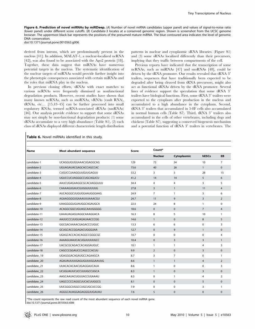
Identification of 26 novel miRNAs from unannotatedsRNAs
We sought to identify novel miRNAs from the reads mapped to
unannotated genomic regions using a recently described miRNA-
discovery algorithm, miRDeep, which can identify miRNAs
efficiently from deep sequencing data [36,37]. As sequencing
depth affects the prediction accuracy of miRDeep, we used pool
reads from our two libraries as well as hESCs and EBs sRNA
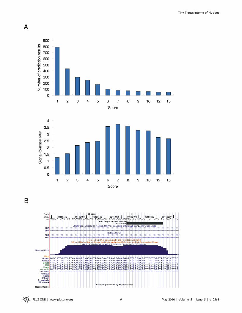
libraries [27] to predict novel miRNAs. In total, 83 candidates
passed the cutoff of 7, a constraint that provided the highest signal-
to-noise ratio of the predictions (Figure 6A). After filtering (see
Methods for details), 26 candidates were identified and considered
to be novel miRNAs (Table 6, Table S7). The majority of the
novel miRNAs (21/26, 80.8%) displayed at least one isomiR in the
5-8F cell line, the hESCs and the EBs (Table S7), and nine of the
novel miRNAs are expressed in other human tissues or cell lines
[36,38,39] (Table S8). Remarkably, one of the novel miRNAs
(candidate-3) displayed a conservation pattern typical for miRNAs
Figure 4. miRNAs in the nuclear and cytoplasmic sRNA libraries. (A) Numbers and overlap of miRNAs between the nuclear and cytoplasmicsRNA libraries. (B) Distribution of miRNA N/C values. (C) Distribution of N/C values for ACA45-derived sRNAs. (D) Pearson correlation scatter plot ofmiRNA levels between the nuclear and cytoplasmic sRNA libraries (R2 = 0.9508). (E) Different isomiRs of miR-29b displayed similar N/C values. Redletters indicate the nuclear import element of miR-29b reported by Hwang et al. [9]. Nuclear indicates the nuclear sRNA library, and cytoplasmicindicates the cytoplasmic sRNA library. (F) N/C values of different isomiRs of miR-92b*.doi:10.1371/journal.pone.0010563.g004
Tiny Transcriptome of Nucleus
PLoS ONE | www.plosone.org 6 May 2010 | Volume 5 | Issue 5 | e10563
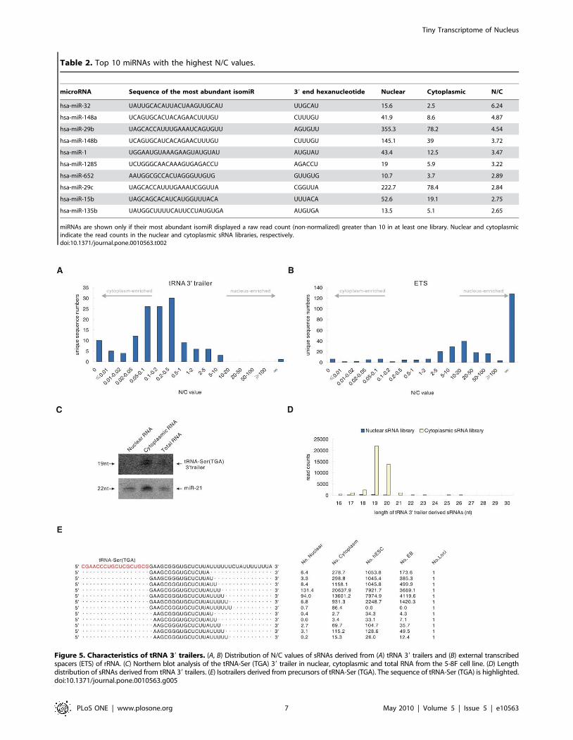
Table 2. Top 10 miRNAs with the highest N/C values.
microRNA Sequence of the most abundant isomiR 39 end hexanucleotide Nuclear Cytoplasmic N/C
hsa-miR-32 UAUUGCACAUUACUAAGUUGCAU UUGCAU 15.6 2.5 6.24
hsa-miR-148a UCAGUGCACUACAGAACUUUGU CUUUGU 41.9 8.6 4.87
hsa-miR-29b UAGCACCAUUUGAAAUCAGUGUU AGUGUU 355.3 78.2 4.54
hsa-miR-148b UCAGUGCAUCACAGAACUUUGU CUUUGU 145.1 39 3.72
hsa-miR-1 UGGAAUGUAAAGAAGUAUGUAU AUGUAU 43.4 12.5 3.47
hsa-miR-1285 UCUGGGCAACAAAGUGAGACCU AGACCU 19 5.9 3.22
hsa-miR-652 AAUGGCGCCACUAGGGUUGUG GUUGUG 10.7 3.7 2.89
hsa-miR-29c UAGCACCAUUUGAAAUCGGUUA CGGUUA 222.7 78.4 2.84
hsa-miR-15b UAGCAGCACAUCAUGGUUUACA UUUACA 52.6 19.1 2.75
hsa-miR-135b UAUGGCUUUUCAUUCCUAUGUGA AUGUGA 13.5 5.1 2.65
miRNAs are shown only if their most abundant isomiR displayed a raw read count (non-normalized) greater than 10 in at least one library. Nuclear and cytoplasmicindicate the read counts in the nuclear and cytoplasmic sRNA libraries, respectively.doi:10.1371/journal.pone.0010563.t002
Figure 5. Characteristics of tRNA 39 trailers. (A, B) Distribution of N/C values of sRNAs derived from (A) tRNA 39 trailers and (B) external transcribedspacers (ETS) of rRNA. (C) Northern blot analysis of the tRNA-Ser (TGA) 39 trailer in nuclear, cytoplasmic and total RNA from the 5-8F cell line. (D) Lengthdistribution of sRNAs derived from tRNA 39 trailers. (E) Isotrailers derived from precursors of tRNA-Ser (TGA). The sequence of tRNA-Ser (TGA) is highlighted.doi:10.1371/journal.pone.0010563.g005
Tiny Transcriptome of Nucleus
PLoS ONE | www.plosone.org 7 May 2010 | Volume 5 | Issue 5 | e10563
in mammals (Figure 6B). Furthermore, the presumed mature form
of this candidate was found in the deep sequencing dataset of
sRNAs extracted from Ago proteins [15]. Together, these findings
provide further support for the hypothesis that the sRNA
candidates are bona fide miRNAs.
Discussion
miRNAs have generally been considered to be cytoplasmic-
localized small regulatory RNAs that regulate gene expression
predominantly at the post-transcriptional level in the cytoplasm.
However, recent studies have reported that a few miRNAs, such as
miR-320, miR-373 and miR-29b, localize to or function in the
nucleus [9–11]. Consistent with these findings, our results revealed
that most miRNAs found in the cytoplasm might also localize to
the nucleus in the 5-8F cell line. We have not compared the sRNA
levels in the transformed cell line and its parental cell line which
might have a different pattern, however, we measured the level of
expression of miRNAs in other human cells (i.e., 293T cells) using
microRNA arrays. Although the miRNA profiling in 293T cells
revealed a very different expression pattern from that in 5-8F cells,
almost all miRNAs found in the cytoplasm were also detected in
the purified nuclear RNA fraction (Table S9). Furthermore, the
abundance of most miRNAs in nuclear RNA generally correlated
with the abundance in cytoplasmic RNA (Figure 7, Table S9).
This indicates that the import of most miRNAs into the nucleus
may be a general phenomenon that occurs in a variety of human
cells.
Pre-miRNAs are cleaved by Dicer in the cytoplasm to generate
a short dsRNA. One strand of this dsRNA is then incorporated
into the Ago proteins, while the other is rapidly degraded [40].
Studies have reported that Ago proteins can be imported into the
nucleus [18,19,21,22]. Our findings that most miRNAs are
imported into the nucleus and that the nuclear and cytoplasmic
abundances of miRNAs are correlated imply that miRNAs might
be co-imported with the Ago proteins into the nucleus. This
hypothesis is further supported by evidence that the majority of
isomiRs processed from ACA45 are incorporated into Ago protein
complexes [15] and that they are also imported into the nucleus, as
we have shown in this study. Although Ago proteins might be
imported into the nucleus while complexed with miRNAs, we
found that the nuclear entry ability of different miRNAs was
highly diverse based on their N/C values. (Table S2). It is likely
that the nuclear import of Ago proteins is affected by other
regulatory factors, such as the sequence of the associated mature
miRNAs.
The fact that most miRNAs are imported into the nucleus
suggests that numerous, instead of a few, miRNAs might play
regulatory roles in the nucleus although the nuclear targets of
miRNAs are largely unknown. To date, only two miRNAs, miR-
320a and miR-373, have been demonstrated to regulate gene
expression in the nucleus through targeting the promoter regions
of protein-coding genes [10][11]. However, many protein-coding
genes were found to contain sites of near-perfect complementarity
to many mature miRNAs in their promoter regions [10].
Moreover, studies of miRNA target sequences interacting with
the Ago proteins identified a substantial number of sequences
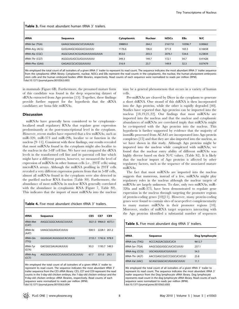
Table 3. Five most abundant human tRNA 39 trailers.
tRNA Sequence Cytoplasmic Nuclear hESCs EBs N/C
tRNA-Ser (TGA) GAAGCGGGUGCUCUUAUUU 39889.0 264.2 21617.0 10396.7 0.00662
tRNA-Arg (ACG) GUGUAAGCAGGGUCGUUUU 1176.6 196.0 371.9 165.5 0.16658
tRNA-Ala (CGC) AGGCGAUCACGUAGAUUUUGUUUA 853.0 203.3 2076.1 534.6 0.23834
tRNA-Thr (CGT) AGGGUGUGCGUGUUUUUUU 349.3 144.7 172.1 54.7 0.41426
tRNA-Phe (GAA) GAGAGCGCUCGGUUUUU 316.9 23.7 149.9 32.3 0.07479
We employed the total count of all isotrailers of a given tRNA 39 trailer to represent its read count. The sequence indicates the most abundant tRNA 39 trailer sequencefrom the cytoplasmic sRNA library. Cytoplasmic, nuclear, hESCs and EBs represent the read counts in the cytoplasmic, the nuclear, the human pluripotent embryonicstem cells and the human embryoid bodies sRNA libraries, respectively. Read counts of each sequence were normalized to reads per million (RPM).doi:10.1371/journal.pone.0010563.t003
Table 4. Five most abundant chicken tRNA 39 trailers.
tRNA Sequence CE5 CE7 CE9
tRNA-Met(CAT)
AAGGCGGGCAAUGCUUUUC 3021.0 9964.0 4073.1
tRNA-Ile(AAT)
UAAGCGGUAGUCUUUU 500.5 2228.1 261.2
tRNA-Gln(TTG)
GGUGGACAGGGGUCACUUUU 2153.1 1742.6 978.9
tRNA-Tyr(GTA)
GACGGCGAUAUAUUUU 93.3 1193.7 149.5
tRNA-Arg(CCT)
AGCGGAUUGCCCUUUUCUCGCUUUU 67.7 331.0 29.3
We employed the total count of all isotrailers of a given tRNA 39 trailer torepresent its read count. The sequence indicates the most abundant tRNA 39
trailer sequence from the CE5 sRNA library. CE5, CE7 and CE9 represent the readcounts in the 5-day-old chicken embryo, the 7-day-old chicken embryo and the9-day-old chicken embryo sRNA libraries, respectively. Read counts of eachsequence were normalized to reads per million (RPM).doi:10.1371/journal.pone.0010563.t004
Table 5. Five most abundant dog tRNA 39 trailers.
tRNA Sequence Dog lymphocyte
tRNA-Leu (TAG) ACCCAGGACGGUCUCA 9612.7
tRNA-Ser (TGA) AAGCGGGUGGCUUCUCUUUU 237.1
tRNA-Arg (CCG) UGCAGUGGUCGUUUUU 54.3
tRNA-Thr (AGT) AACCGAGCGUCCCGGCUCUCUU 25.8
tRNA-Val (AAC) GCAGCGGGCACUGUUGCUUUU 11.1
We employed the total count of all isotrailers of a given tRNA 39 trailer torepresent its read count. The sequence indicates the most abundant tRNA 39
trailer sequence from the Dog lymphocyte sRNA library. Dog lymphocyterepresents read count in the dog lymphocyte sRNA library. Read counts of eachsequence were normalized to reads per million (RPM).doi:10.1371/journal.pone.0010563.t005
Tiny Transcriptome of Nucleus
PLoS ONE | www.plosone.org 8 May 2010 | Volume 5 | Issue 5 | e10563
Tiny Transcriptome of Nucleus
PLoS ONE | www.plosone.org 9 May 2010 | Volume 5 | Issue 5 | e10563
derived from introns, which are predominantly present in the
nucleus [41]. In addition, MALAT-1, a nuclear-localized ncRNA
[42], was also found to be associated with the Ago2 protein [18].
Together, these data suggest that miRNAs have numerous
potential targets in the nucleus. The systematic identification of
the nuclear targets of miRNAs would provide further insight into
the phenotypic consequences associated with certain miRNAs and
the roles that miRNA play in the nucleus.
In previous cloning efforts, sRNAs with exact matches to
various ncRNAs were frequently dismissed as nonfunctional
degradation products. However, recent studies have shown that
many known ncRNAs, such as snoRNAs, vRNAs (vault RNA),
tRNAs, etc… [15,43–45] can be further processed into small
regulatory RNAs, termed ncRNA-associated sRNAs (nasRNAs)
[46]. Our analysis provide evidence to support that some sRNAs
may not simply be non-functional degradation products: (1) some
sRNAs accumulate to a very high abundance (Table S1), (2) each
class of sRNAs displayed different characteristic length distribution
patterns in nuclear and cytoplasmic sRNA libraries. (Figure S1)
and (3) some sRNAs localized differently than their precursors,
implying that they traffic between compartments of the cell.
Previous reports have indicated that the transcription of some
ncRNAs, such as miRNAs [47] and snoRNAs [48], could be
driven by the tRNA promoter. Our results revealed that tRNA 39
trailers, sequences that have traditionally been expected to be
degraded after being cleaved from tRNA precursors, might also
act as functional sRNAs driven by the tRNA promoter. Several
lines of evidence support the speculation that some tRNA 39
trailers have biological functions. First, some tRNA 39 trailers were
exported to the cytoplasm after production in the nucleus and
accumulated to a high abundance in the cytoplasm. Second,
tRNA 39 trailers that accumulated in 5-8F cells also accumulated
in normal human cells (Table S7). Third, tRNA 39 trailers also
accumulated in the cells of other vertebrates, including dogs and
chickens (Table S7), suggesting a conserved biogenesis mechanism
and a potential function of tRNA 39 trailers in vertebrates. The
Figure 6. Prediction of novel miRNAs by miRDeep. (A) Number of novel miRNA candidates (upper panel) and values of signal-to-noise ratio(lower panel) under different score cutoffs. (B) Candidate-3 locates at a conserved genomic region. Shown is screenshot from the UCSC genomebrowser. The uppermost black bar represents the positions of the presumed mature miRNA. The blue contoured area indicates the level of genomicDNA conservation.doi:10.1371/journal.pone.0010563.g006
Table 6. Novel miRNAs identified in this study.
Name Most abundant sequence Score Count*
Nuclear Cytoplasmic hESCs EB
candidate-1 UCAGGUGUGGAAACUGAGGCAG 129 73 34 10 7
candidate-2 UGUAGAUACGAGCACCAGCCAC 73.6 40 26 1 0
candidate-3 CUGUCCUAAGGUUGUUGAGUU 53.2 3 5 28 15
candidate-4 UGUCCUCUAGGGCCUGCAGUCU 41.2 14 14 5 6
candidate-5 AAUCUGAGAAGGCGCACAAGGUUU 34.4 4 4 3 5
candidate-6 CAAAAGUGAUCGUGGUUUUUG 27.8 3 1 11 4
candidate-7 AUCAGGGCUUGUGGAAUGGGAAG 24.9 7 1 3 6
candidate-8 AGAAGGGGUGAAAUUUAAACGU 24.7 11 9 3 2
candidate-9 UAAGGGGUGUAUGGCAGAUGCA 22.5 24 8 1 0
candidate-10 ACAGGCGGCUGUAGCAAUGGGGG 18.6 0 0 7 4
candidate-11 UAAAUAGAGUAGGCAAAGGACA 16.3 8 5 10 1
candidate-12 AAUUCCCUUGUAGAUAACCCGG 14.6 1 0 8 5
candidate-13 GGCGACAAAACGAGACCCUGUC 13.3 6 6 0 3
candidate-14 GCUGCACCGGAGACUGGGUAA 12.7 0 9 1 0
candidate-15 UGAGCACCACACAGGCCGGGCGC 10.7 0 0 0 4
candidate-16 AAAAGUAAUCACUGUUUUUGCC 10.4 0 3 3 1
candidate-17 UACGCGCAGACCACAGGAUGUC 10.1 1 1 4 3
candidate-18 CAGCCCGGAUCCCAGCCCACUU 9.9 2 0 3 0
candidate_19 UGAGGGACAGAUGCCAGAAGCA 8.7 3 7 0 1
candidate_20 AGAUAUUUUGAGUGUUUGGAAUUG 8.6 1 1 4 2
candidate_21 UUACACACAACUGAGGAUCAUA 8.6 1 0 0 3
candidate_22 UCUGUAUUCUCCUUUGCCUGCA 8.3 1 0 3 0
candidate_23 AAGCAAUACUGUUACCUGAAAU 8.3 0 1 4 2
candidate_24 UAGCCCCCAGGCUUCACUUGGCG 8.1 0 0 5 0
candidate_25 UUCGGGCUGGCCUGCUGCUCCGG 7.9 0 0 3 1
candidate_26 AGGGCAUAGGAGAGGGUUGAUAU 7.6 5 0 0 0
*The count represents the raw read count of the most abundant sequence of each novel miRNA gene.doi:10.1371/journal.pone.0010563.t006
Tiny Transcriptome of Nucleus
PLoS ONE | www.plosone.org 10 May 2010 | Volume 5 | Issue 5 | e10563

exact transport process of the tRNA trailers in cells is still
unknown, but the nuclear export of tRNA 39 trailers might be
mediated by the La protein which binds to tRNA 39 trailers during
processing and modification of pre-tRNAs [49], since the RNA
recognition motif (RRM) of the La protein displays nuclear export
activity [50].
Advances in high-throughput next-generation sequencing
technology have greatly transformed the transcriptomic research
landscape. Using this technology, we identified an unexpectedly
complex subcellular distribution of miRNAs and tRNA 39 trailers
and also identified numerous unannotated sRNAs, including novel
miRNA-producing regions of the human genome. As recently
demonstrated, there remain many unknown sRNAs in human
cells [38,46], and the characterization of the subcellular distribu-
tions of human sRNAs might provide hints toward the
identification of novel sRNA structures and their functions.
Materials and Methods
Preparation of nuclear, cytoplasmic and total RNAHuman Nasopharyngeal carcinoma cell lines 5-8F was
purchased from the Cancer Research Institute of Sun Yat-sen
University (Guangzhou, China). Cells was maintained at 37uC in
DMEM containing 10% FBS. Cells were collected by centrifuga-
tion and washed with PBS (pH 7.4). Total RNA was isolated from
cells using the method of guanidine thiocyanate/phenol –
chloroform [51]. Nuclear and cytoplasmic fractions were isolated
according to the method of Greenberg et al. [52] and Qiagen
(Valencia, CA) RNeasy Minikit protocol with some modifications.
Briefly, the cell pellet was resuspended in 20 pellet volumes of
RLN buffer (50 mM Tris-HCl, pH 7.4, 0.14 M NaCl, 1.5 mM
MgCl2, 0.5% IGEPAL CA-630 (Sigma), 1 U/ul RNase Inhibitor
(TaKaRa), 1 mM DTT) and incubated for 5 min on ice. The
nuclei were collected by centrifugation at 300 g for 3 min at 4uC.
The supernatant was used to isolate cytoplasmic RNA using the
same method that used to isolate total RNA. Nuclei pellet was
resuspended in 20 pellet volumes of RSB buffer (0.25 M sucrose,
10 mM Tris-HCl, pH 7.4, 10 mM NaCl, 3 mM MgCl2, 1 mM
DTT), then nuclei were collected by ultracentrifugation through a
sucrose cushion. Nuclear RNA was isolated from sucrose gradient
centrifugation purified nuclei using the same method used to
isolate cytoplasmic RNA. The integrity of the RNA was assessed
using denaturing agarose gel electrophoresis. The nuclear and
cytoplasmic RNAs were run on a 10% denaturing polyacryamide
gels to assess whether the nuclear and cytoplasmic RNAs were
successfully separated.
sRNA library preparation and sequencingsRNA library preparation and Solexa sequencing was performed
by Beijing Genomics Institute (BGI) at ShenZhen according to the
manufacturer’s instructions. Briefly, sRNAs ranging from 18 to 30-nt
were gel-purified and ligated to the 39 adaptor (59-pUCGUAUGCC-
GUCUUCUGCUUGidT-39; p, phosphate; idT, inverted deoxythy-
midine) and 59 adaptor (59-GUUCAGAGUUCUACAGUCCGAC-
GAUC-39). Ligation products were gel-purified, reverse transcribed,
and amplified using Illumina’s sRNA primer set (59-CAAGCAGAA-
GACG GCATACGA-39; 59-AATGATACGGCGACCACCGA-
39). Samples were sequenced on an Illumina 1G Genome Analyzer.
Bioinformatic analysis of sRNA librariesThe 39 adaptor sequences were removed from the Illumina-
generated reads at BGI Shenzhen using a dynamic programming
algorithm that required at least 5-nt overlap between 35-nt reads
and the 39 adaptor sequence. After removing the reads without the
adaptor sequences, poly-A reads and 59 adaptor contaminants, the
remaining 16- to 30-nt reads were mapped to the UCSC hg18
assembly of the human genome [53] using bowtie [54] with the
following options: –f –n 0 –a. All reads that could be aligned to the
human genome were moved to the ‘‘mapped’’ data set. The
remaining reads constituted the ‘‘unmapped’’ data set. We next
moved the reads from the ‘‘unmapped’’ data set that could be
aligned to rRNA genes downloaded from the NCBI (Accession
number: U13369), the exon-exon junction of the tRNA and
mRNA, or the tRNA 39 end that contained the post-transcrip-
tionally-added CCA into the ‘‘mapped’’ data set. Each sequence in
‘‘mapped’’ data set was annotated by simply aligning to known
RNA sequences which were downloaded from Genebank (http://
www.ncbi.nih.gov/Genbank/index.html, rRNA), snoRNA-LBME-
db http://wwwsnorna.biotoul.fr/index.php, snoRNA), UCSC
(http://genome.ucsc.edu, various RNAs and repeat sequences),
miRBase (http://microrna.sanger.ac.uk/sequences/index.shtml, re-
lease version 12.0, miRNA). The annotation order was miRNA,
mitochondrial RNA, rRNA, internal transcribed spacer (ITS) of
rRNA, external transcribed spacer (ETS) of rRNA, Box H/ACA
snoRNA, Box C/D snoRNA, scaRNA, tRNA, snRNA, RNaseP,
SRP RNA, Xist RNA, 7SK RNA, H19 RNA, vRNA, hY RNA,
RNaseMRP, mRNA and 39 trailer sequence of tRNA. Considering
that some reads could be aligned to multiple kinds of RNA, we
removed them from the ‘‘mapped’’ data set when they were first
annotated to one kind of RNA. For instance, if some reads were
annotated as miRNA, they were removed from the ‘‘mapped’’ data
set before the remnant reads were aligned to mitochondrial RNA. We
found that many sequences in the ‘‘unmapped’’ data set aligned with
the known tRNA sequences with a single nucleotide mismatch, which
might be caused by extensive modification of mature tRNA
sequences. These sequences were moved to the ‘‘mapped’’ data set
and annotated as tRNA-derived sRNAs. The imperfect mapping to
the human genome of many reads in the ‘‘unmapped’’ data set might
be caused by 39 untemplated nucleotides [55–58]. We removed the 39
untemplated nucleotides from reads if they possessed an exact match
to the human genome starting at the 59 end and maintained a
contiguous match of 17 or more nucleotides. These trimmed reads
were then moved to the ‘‘mapped’’ data set. The remnant sequences
in the ‘‘unmapped’’ data set were discarded. All reads in the
‘‘mapped’’ data set were compiled into a set of unique sequences, with
Figure 7. Pearson correlation scatter plot of miRNA expressionlevels between the nuclear and cytoplasmic sRNAs of 293T cellline (R2 = 0.9508).doi:10.1371/journal.pone.0010563.g007
Tiny Transcriptome of Nucleus
PLoS ONE | www.plosone.org 11 May 2010 | Volume 5 | Issue 5 | e10563
the number of reads for each sequence reflecting relative abundance
[59]. The read count of each unique sequence was normalized to
reads per million (RPM), according to the total read count of the
‘‘mapped’’ data set.
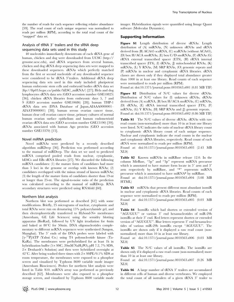
Analysis of tRNA 39 trailers and the sRNA deepsequencing data sets used in this study
40 nucleotides immediately downstream of each tRNA gene of
human, chicken and dog were downloaded from UCSC (http://
genome.ucsc.edu), and sRNA sequences from several human,
chicken and dog sRNA deep sequencing data sets were mapped to
these downloaded sequences. sRNAs which perfectly matched
from the first or second nucleotide of any downloaded sequence
were considered to be tRNA 39trailers. Additional sRNA deep
sequencing data sets used in this study included: pluripotent
human embryonic stem cells and embryoid bodies sRNA data set
(ftp://ftp03.bcgsc.ca/public/hESC_miRNA/) [27]; Hela and dog
lymphocytes sRNA data sets (GEO accession number GSE10825)
[36]; chicken sRNA libraries from embryonic days 5, 7 and
9 (GEO accession number GSE10686) [28]; human THP-1
sRNA data sets (DNA Database of Japan,AIAAA0000001–
AIAAT0000001) [38]; human serous ovarian cancer tissue,
human clear cell ovarian cancer tissue, primary cultures of normal
human ovarian surface epithelium and human endometrioid
ovarian sRNA data sets (GEO accession number GSE15190) [39];
sRNAs associated with human Ago proteins (GEO accession
number GSE13370) [15].
Novel miRNA predictionNovel miRNAs were predicted by a recently described
algorithm miRDeep [36]. Prediction was performed according
to the manual of miRDeep. The data set we used to predicted
miRNA comprised pooled reads from nuclear, cytoplasmic,
hESCs and EBs sRNA libraries [27]. We discarded the following
miRNA candidates: (1) the mature form of candidates had more
than 5 loci in the genome or overlap with known RNAs; (2)
candidates overlapped with the minus strand of known miRNAs;
(3) the length of the mature form of candidates shorter than 19-nt
or longer than 25-nt. The signal-to-noise ratio of the prediction
was calculated according to the manual of miRDeep. RNA
secondary structures were predicted using RNAfold [60].
Northern blot analysisNorthern blot was performed as described [61] with some
modifications. Briefly, 25 micrograms of nuclear, cytoplasmic and
total RNAs were run on denaturing 15% polyacrylamide gel, and
then electrophoretically transferred to Hybond-N+ membranes
(Amersham, GE Life Sciences) using the semidry blotting
apparatus (BioRad), followed by UV light irradiation for 4min
and baked at 80uC for 50min. DNA oligonucleotides comple-
mentary to different ncRNA sequences were synthesized (Sangon,
Shanghai). The 59 ends of the DNA probes were labeled with
[c-32P]ATP (Yahui Co.) using T4 polynucleotide kinase (Ta-
KaRa). The membranes were prehybridized for at least 1h in
hybridization buffer (56SSC, 20mM NaH2PO4 pH 7.2, 7% SDS,
26 Denhardt’s Solution) and then were hybridized overnight at
42uC. After being washed three times with 26SSPE/0.1% SDS at
room temperature, the membranes were exposed to a phosphor
screen and visualized by Typhoon 8600 variable mode imager
(Amersham Biosciences). Probes for northern blot analysis were
listed in Table S10. miRNA array was performed as previously
described [62]. Membranes were also exposed to a phosphor
storage screen, and visualized by Typhoon 8600 variable mode
imager. Hybridization signals were quantified using Image Quant
software (Molecular Dynamics).
Supporting Information
Figure S1 Length distribution of diverse sRNAs. Length
distribution of (A) miRNAs, (N) unknown sRNAs and sRNA
derived from (B) ACA45 scaRNA, (C) scaRNAs (without ACA45),
(D) box H/ACA snoRNAs, (E) box C/D snoRNAs, (F) rRNAs (G)
rRNA external transcribed spacer (ETS), (H) rRNA internal
transcribed spacer (ITS), (I) tRNAs, (J) mitochondrial RNAs, (K)
snRNAs, (L) Y RNAs, (M) SRP RNAs, (O) genomic repeats and
(P) mRNAs in nuclear and cytoplasmic sRNA libraries. sRNA
classes are shown only if they displayed total abundance greater
than 1000 in at least one library. Read counts of each sequence
were normalized to reads per million (RPM).
Found at: doi:10.1371/journal.pone.0010563.s001 (0.81 MB TIF)
Figure S2 Distribution of N/C values for diverse sRNAs.
Distribution of N/C values for (I) unknown sRNAs, sRNAs
derived from (A) scaRNA, (B) box H/ACA snoRNAs, (C) mRNAs,
(D) rRNAs, (E) rRNA internal transcribed spacer (ITS), (F)
snRNAs, (G) Y RNAs, (H) SRP RNAs and (J) genomic repeats.
Found at: doi:10.1371/journal.pone.0010563.s002 (0.86 MB TIF)
Table S1 The N/C values of diverse sRNAs. sRNAs with raw
read counts (non-normalized) more than 10 in at least one library
were listed. N/C indicates the ratio of nuclear sRNA library count
to cytoplasmic sRNA library count of each unique sequence.
Nuclear and cytoplasmic indicate the read counts in the nuclear
and cytoplasmic sRNA libraries, respectively. Read count of each
sRNA were normalized to reads per million (RPM).
Found at: doi:10.1371/journal.pone.0010563.s003 (2.43 MB
XLS)
Table S2 Known miRNAs in miRBase release 12.0. In the
column MirBase, ‘‘5p’’ and ‘‘3p’’ represent miRNA precursor
which is annotated to have mature form locate in 59 arm and 39
arm respectively by miRBase, and ‘‘*’’ represents miRNA
precursor which is annotated to have miRNA* by miRBase.
Found at: doi:10.1371/journal.pone.0010563.s004 (3.08 MB
HTML)
Table S3 miRNAs that present different most abundant isomiR
in nuclear and cytoplasmic sRNA libraries. Read counts of each
sequence were normalized to reads per million (RPM).
Found at: doi:10.1371/journal.pone.0010563.s005 (0.03 MB
XLS)
Table S4 IsomiRs which had shorten or extended version of
‘‘AGUGUU’’ or various 39 end hexanucleotides of miR-29b
isomiRs at their 39 end. Red letters represent shorten or extended
version of ‘‘AGUGUU’’. Blue letters represent 39 end hexanucleo-
tides of various miR-29b isomiRs except ‘‘AGUGUU’’. The
isomiRs are shown only if it displayed a raw read count (non-
normalized) more than 10 in at least one library.
Found at: doi:10.1371/journal.pone.0010563.s006 (0.03 MB
XLS)
Table S5 The N/C values of all isomiRs. The isomiRs are
shown only if it displayed a raw read count (non-normalized) more
than 10 in at least one library.
Found at: doi:10.1371/journal.pone.0010563.s007 (0.26 MB
XLS)
Table S6 A large number of tRNA 39 trailers are accumulated
in different cells of human and diverse vertebrates. We employed
the total count of all isotrailers of a given tRNA 39 trailer to
Tiny Transcriptome of Nucleus
PLoS ONE | www.plosone.org 12 May 2010 | Volume 5 | Issue 5 | e10563
represent its read count. tRNA 39 trailers of human and chicken
with raw read counts more than 10 in at least one library were
listed. The sequence indicates the most abundant tRNA 39trailer
sequence from cytoplasmic sRNA library in human, CE5 sRNA
library in chicken. Cytoplasmic, nuclear, hESCs, EBs, CE5, CE7,
CE9 and dog lymphocyte represent the read counts in
cytoplasmic, nuclear, human pluripotent human embryonic stem
cells, human embryoid bodies, 5-day-old chicken embryo, 7-day-
old chicken embryo, 9-day-old chicken embryo and dog
lymphocyte sRNA libraries, respectively. C/N indicates the ratio
of cytoplasmic sRNA library count to nuclear sRNA library count
of each unique sequence. Read counts of each sequence were
normalized to reads per million (RPM).
Found at: doi:10.1371/journal.pone.0010563.s008 (0.03 MB
XLS)
Table S7 Novel miRNAs identified in this study.
Found at: doi:10.1371/journal.pone.0010563.s009 (0.14 MB
HTML)
Table S8 Novel miRNAs in other tissues or cell lines. ‘‘!’’
indicates that at least one isomiR of novel miRNA gene presented
in corresponding sRNA library. Osc, Occ, Opc and Oec represent
sRNAs from serous ovarian cancer tissue, clear cell ovarian cancer
tissue, primary cultures of normal human ovarian surface
epithelium (HOSE) and endometrioid ovarian cancer tissue,
respectively.
Found at: doi:10.1371/journal.pone.0010563.s010 (0.04 MB
DOC)
Table S9 The expression level of all microRNAs that detected in
the nuclear and cytoplasmic 18–30nt sRNA of 293T cell line.
Found at: doi:10.1371/journal.pone.0010563.s011 (0.03 MB
XLS)
Table S10 Probes for Northern blot analysis. All probes were
synthesized and purified by Sangon Co. (Shanghai, China).
Found at: doi:10.1371/journal.pone.0010563.s012 (0.03 MB
DOC)
Acknowledgments
We would like to thank Jie-Hua He, Zhen-Dong Xiao for their technical
assistance.
Author Contributions
Conceived and designed the experiments: JYL HZ LHQ. Performed the
experiments: JYL LMM YHG YCZ. Analyzed the data: JYL YCZ PS
LHQ. Contributed reagents/materials/analysis tools: HZ YQC LHQ.
Wrote the paper: JYL HZ PS YQC LHQ.
References
1. Bartel DP (2004) MicroRNAs: genomics, biogenesis, mechanism, and function.
Cell 116: 281–297.
2. Bushati N, Cohen SM (2007) microRNA functions. Annu Rev Cell Dev Biol 23:
175–205.
3. Yi R, Qin Y, Macara IG, Cullen BR (2003) Exportin-5 mediates the nuclear
export of pre-microRNAs and short hairpin RNAs. Genes Dev 17: 3011–3016.
4. Gregory RI, Chendrimada TP, Cooch N, Shiekhattar R (2005) Human RISC
couples microRNA biogenesis and posttranscriptional gene silencing. Cell 123:
631–640.
5. Maniataki E, Mourelatos Z (2005) A human, ATP-independent, RISC assembly
machine fueled by pre-miRNA. Genes Dev 19: 2979–2990.
6. Filipowicz W, Bhattacharyya SN, Sonenberg N (2008) Mechanisms of post-
transcriptional regulation by microRNAs: are the answers in sight? Nat Rev
Genet 9: 102–114.
7. Politz JC, Hogan EM, Pederson T (2009) MicroRNAs with a nucleolar location.
RNA 15: 1705–1715.
8. Politz JC, Zhang F, Pederson T (2006) MicroRNA-206 colocalizes with
ribosome-rich regions in both the nucleolus and cytoplasm of rat myogenic cells.
Proc Natl Acad Sci U S A 103: 18957–18962.
9. Hwang HW, Wentzel EA, Mendell JT (2007) A hexanucleotide element directs
microRNA nuclear import. Science 315: 97–100.
10. Kim DH, Saetrom P, Snove O, Jr., Rossi JJ (2008) MicroRNA-directed
transcriptional gene silencing in mammalian cells. Proc Natl Acad Sci U S A
105: 16230–16235.
11. Place RF, Li LC, Pookot D, Noonan EJ, Dahiya R (2008) MicroRNA-373
induces expression of genes with complementary promoter sequences. Proc Natl
Acad Sci U S A 105: 1608–1613.
12. Khraiwesh B, Arif MA, Seumel GI, Ossowski S, Weigel D, et al. (2010)
Transcriptional control of gene expression by microRNAs. Cell 140: 111–122.
13. Peters L, Meister G (2007) Argonaute proteins: mediators of RNA silencing. Mol
Cell 26: 611–623.
14. Azuma-Mukai A, Oguri H, Mituyama T, Qian ZR, Asai K, et al. (2008)
Characterization of endogenous human Argonautes and their miRNA partners
in RNA silencing. Proc Natl Acad Sci U S A 105: 7964–7969.
15. Ender C, Krek A, Friedlander MR, Beitzinger M, Weinmann L, et al. (2008) A
human snoRNA with microRNA-like functions. Mol Cell 32: 519–528.
16. Meister G, Landthaler M, Patkaniowska A, Dorsett Y, Teng G, et al. (2004)
Human Argonaute2 mediates RNA cleavage targeted by miRNAs and siRNAs.
Mol Cell 15: 185–197.
17. Su H, Trombly MI, Chen J, Wang X (2009) Essential and overlapping functions
for mammalian Argonautes in microRNA silencing. Genes Dev 23: 304–317.
18. Weinmann L, Hock J, Ivacevic T, Ohrt T, Mutze J, et al. (2009) Importin 8 is a
gene silencing factor that targets argonaute proteins to distinct mRNAs. Cell
136: 496–507.
19. Ohrt T, Mutze J, Staroske W, Weinmann L, Hock J, et al. (2008) Fluorescence
correlation spectroscopy and fluorescence cross-correlation spectroscopy reveal
the cytoplasmic origination of loaded nuclear RISC in vivo in human cells.
Nucleic Acids Res 36: 6439–6449.
20. Robb GB, Brown KM, Khurana J, Rana TM (2005) Specific and potent RNAi
in the nucleus of human cells. Nat Struct Mol Biol 12: 133–137.
21. Janowski BA, Huffman KE, Schwartz JC, Ram R, Nordsell R, et al. (2006)Involvement of AGO1 and AGO2 in mammalian transcriptional silencing. Nat
Struct Mol Biol 13: 787–792.
22. Kim DH, Villeneuve LM, Morris KV, Rossi JJ (2006) Argonaute-1 directs
siRNA-mediated transcriptional gene silencing in human cells. Nat Struct MolBiol 13: 793–797.
23. Guang S, Bochner AF, Pavelec DM, Burkhart KB, Harding S, et al. (2008) An
Argonaute transports siRNAs from the cytoplasm to the nucleus. Science 321:
537–541.
24. Volpe TA, Kidner C, Hall IM, Teng G, Grewal SI, et al. (2002) Regulation ofheterochromatic silencing and histone H3 lysine-9 methylation by RNAi.
Science 297: 1833–1837.
25. Carmell MA, Girard A, van de Kant HJ, Bourc’his D, Bestor TH, et al. (2007)
MIWI2 is essential for spermatogenesis and repression of transposons in themouse male germline. Dev Cell 12: 503–514.
26. Creighton CJ, Reid JG, Gunaratne PH (2009) Expression profiling ofmicroRNAs by deep sequencing. Brief Bioinform 10: 490–497.
27. Morin RD, O’Connor MD, Griffith M, Kuchenbauer F, Delaney A, et al. (2008)
Application of massively parallel sequencing to microRNA profiling and
discovery in human embryonic stem cells. Genome Res 18: 610–621.
28. Glazov EA, Cottee PA, Barris WC, Moore RJ, Dalrymple BP, et al. (2008) AmicroRNA catalog of the developing chicken embryo identified by a deep
sequencing approach. Genome Res 18: 957–964.
29. Li J, Fan Y, Chen J, Yao KT, Huang ZX (2010) Microarray analysis of
differentially expressed genes between nasopharyngeal carcinoma cell lines 5-8Fand 6-10B. Cancer Genet Cytogenet 196: 23–30.
30. Hopper AK, Shaheen HH (2008) A decade of surprises for tRNA nuclear-cytoplasmic dynamics. Trends Cell Biol 18: 98–104.
31. Seitz H, Ghildiyal M, Zamore PD (2008) Argonaute loading improves the 59
precision of both MicroRNAs and their miRNA strands in flies. Curr Biol 18:147–151.
32. Morl M, Marchfelder A (2001) The final cut. The importance of tRNA 39-processing. EMBO Rep 2: 17–20.
33. Dieci G, Fiorino G, Castelnuovo M, Teichmann M, Pagano A (2007) The
expanding RNA polymerase III transcriptome. Trends Genet 23: 614–622.
34. Hagenbuchle O, Larson D, Hall GI, Sprague KU (1979) The primary
transcription product of a silkworm alanine tRNA gene: identification of in vitrosites of initiation, termination and processing. Cell 18: 1217–1229.
35. Koski RA, Clarkson SG (1982) Synthesis and maturation of Xenopus laevismethionine tRNA gene transcripts in homologous cell-free extracts. J Biol Chem
257: 4514–4521.
36. Friedlander MR, Chen W, Adamidi C, Maaskola J, Einspanier R, et al. (2008)
Discovering microRNAs from deep sequencing data using miRDeep. NatBiotechnol 26: 407–415.
37. Friedlander MR, Adamidi C, Han T, Lebedeva S, Isenbarger TA, et al. (2009)
High-resolution profiling and discovery of planarian small RNAs. Proc Natl
Acad Sci U S A 106: 11546–11551.
Tiny Transcriptome of Nucleus
PLoS ONE | www.plosone.org 13 May 2010 | Volume 5 | Issue 5 | e10563
38. Taft RJ, Glazov EA, Cloonan N, Simons C, Stephen S, et al. (2009) Tiny RNAs
associated with transcription start sites in animals. Nat Genet 41: 572–578.39. Wyman SK, Parkin RK, Mitchell PS, Fritz BR, O’Briant K, et al. (2009)
Repertoire of microRNAs in epithelial ovarian cancer as determined by next
generation sequencing of small RNA cDNA libraries. PLoS ONE 4: e5311.40. O’Toole AS, Miller S, Haines N, Zink MC, Serra MJ (2006) Comprehensive
thermodynamic analysis of 39 double-nucleotide overhangs neighboring Watson-Crick terminal base pairs. Nucleic Acids Res 34: 3338–3344.
41. Chi SW, Zang JB, Mele A, Darnell RB (2009) Argonaute HITS-CLIP decodes
microRNA-mRNA interaction maps. Nature 460: 479–486.42. Hutchinson JN, Ensminger AW, Clemson CM, Lynch CR, Lawrence JB, et al.
(2007) A screen for nuclear transcripts identifies two linked noncoding RNAsassociated with SC35 splicing domains. BMC Genomics 8: 39.
43. Persson H, Kvist A, Vallon-Christersson J, Medstrand P, Borg A, et al. (2009)The non-coding RNA of the multidrug resistance-linked vault particle encodes
multiple regulatory small RNAs. Nat Cell Biol 11: 1268–1271.
44. Li Y, Luo J, Zhou H, Liao JY, Ma LM, et al. (2008) Stress-induced tRNA-derived RNAs: a novel class of small RNAs in the primitive eukaryote Giardia
lamblia. Nucleic Acids Res 36: 6048–6055.45. Yamasaki S, Ivanov P, Hu GF, Anderson P (2009) Angiogenin cleaves tRNA
and promotes stress-induced translational repression. J Cell Biol 185: 35–42.
46. Yang JH, Shao P, Zhou H, Chen YQ, Qu LH (2009) deepBase: a database fordeeply annotating and mining deep sequencing data. Nucleic Acids Res 38:
D123–130.47. Pfeffer S, Sewer A, Lagos-Quintana M, Sheridan R, Sander C, et al. (2005)
Identification of microRNAs of the herpesvirus family. Nat Methods 2: 269–276.48. Kruszka K, Barneche F, Guyot R, Ailhas J, Meneau I, et al. (2003) Plant
dicistronic tRNA-snoRNA genes: a new mode of expression of the small
nucleolar RNAs processed by RNase Z. EMBO J 22: 621–632.49. Wolin SL, Cedervall T (2002) The La protein. Annu Rev Biochem 71: 375–403.
50. Bayfield MA, Kaiser TE, Intine RV, Maraia RJ (2007) Conservation of amasked nuclear export activity of La proteins and its effects on tRNA
maturation. Mol Cell Biol 27: 3303–3312.
51. Chomczynski P, Sacchi N (1987) Single-step method of RNA isolation by acid
guanidinium thiocyanate-phenol-chloroform extraction. Anal Biochem 162:
156–159.
52. Greenberg ME, Bender TP (2007) Identification of newly transcribed RNA.
Curr Protoc Mol Biol Chapter 4: Unit 4 10.
53. Karolchik D, Baertsch R, Diekhans M, Furey TS, Hinrichs A, et al. (2003) The
UCSC Genome Browser Database. Nucleic Acids Res 31: 51–54.
54. Langmead B, Trapnell C, Pop M, Salzberg SL (2009) Ultrafast and memory-
efficient alignment of short DNA sequences to the human genome. Genome Biol
10: R25.
55. Bar M, Wyman SK, Fritz BR, Qi J, Garg KS, et al. (2008) MicroRNA discovery
and profiling in human embryonic stem cells by deep sequencing of small RNA
libraries. Stem Cells 26: 2496–2505.
56. Ruby JG, Jan C, Player C, Axtell MJ, Lee W, et al. (2006) Large-scale
sequencing reveals 21U-RNAs and additional microRNAs and endogenous
siRNAs in C. elegans. Cell 127: 1193–1207.
57. Ruby JG, Stark A, Johnston WK, Kellis M, Bartel DP, et al. (2007) Evolution,
biogenesis, expression, and target predictions of a substantially expanded set of
Drosophila microRNAs. Genome Res 17: 1850–1864.
58. Berezikov E, Cuppen E, Plasterk RH (2006) Approaches to microRNA
discovery. Nat Genet 38: S2–S7.
59. Landgraf P, Rusu M, Sheridan R, Sewer A, Iovino N, et al. (2007) A
mammalian microRNA expression atlas based on small RNA library
sequencing. Cell 129: 1401–1414.
60. Hofacker IL, Fontana W, Stadler PF, Bonhoeffer LS, Tacker M, et al. (1994)
Fast folding and comparison of RNA secondary structures. Monatsh Chem 125:
167–188.
61. Wang JF, Zhou H, Chen YQ, Luo QJ, Qu LH (2004) Identification of 20
microRNAs from Oryza sativa. Nucleic Acids Res 32: 1688–1695.
62. Tang X, Gal J, Zhuang X, Wang W, Zhu H, et al. (2007) A simple array
platform for microRNA analysis and its application in mouse tissues. RNA 13:
1803–1822.
Tiny Transcriptome of Nucleus
PLoS ONE | www.plosone.org 14 May 2010 | Volume 5 | Issue 5 | e10563