Regulation of Glycolysis by Non-coding RNAs in Cancer
22
Review Regulation of Glycolysis by Non-coding RNAs in Cancer: Switching on the Warburg Effect Hamed Mirzaei 1 and Michael R. Hamblin 2 1 Research Center for Biochemistry and Nutrition in Metabolic Diseases, Institute for Basic Sciences, Kashan University of Medical Sciences, Kashan, I.R. Iran; 2 Laser Research Centre, Faculty of Health Science, University of Johannesburg, Doornfontein, Johannesburg 2028, South Africa The “Warburg effect” describes the reprogramming of glucose metabolism away from oxidative phosphorylation toward aero- bic glycolysis, and it is one of the hallmarks of cancer cells. Several factors can be involved in this process, but in this review, the roles of non-coding RNAs (ncRNAs) are highlighted in several types of human cancer. ncRNAs, including microRNAs, long non-coding RNAs, and circular RNAs, can all affect meta- bolic enzymes and transcription factors to promote glycolysis and modulate glucose metabolism to enhance the progression of tumors. In particular, the 5 0 -AMP-activated protein kinase (AMPK) and the phosphatidylinositol 3-kinase (PI3K)/AKT/ mammalian target of rapamycin (mTOR) pathways are associ- ated with alterations in ncRNAs. A better understanding of the roles of ncRNAs in the Warburg effect could ultimately lead to new therapeutic approaches for suppressing cancer. Otto Heinrich Warburg first discovered that tumors tended to show a specific metabolic phenotype in the 1920s. Cancer cells, even when they have sufficient oxygen, tend to make ATP using glycolysis, rather than the oxidative phosphorylation (OXPHOS) that occurs in normal cells and generates more ATP per glucose molecule. This phenome- non of aerobic glycolysis became known as the “Warburg effect.” In glycolysis, the glucose uptake by the cancer cells is higher, and lactic acid is produced from pyruvate, thus reducing the pH of the tumor. 1,2 Soon after the discovery, Warburg proposed that the mitochondrial function was fundamentally altered in tumor cells, and he even pro- posed that this could be a cause of cancer. 3 However, he subsequently stated that mitochondrial function was not damaged in most kinds of cancer cells. 4 Moreover, Warburg suggested that the increase in glucose/glutamine consumption by cancer cells could led to them becoming “glucose ad- dicted” or “glutamine addicted,” which may induce cell death. Meta- bolic intermediates that are produced by cells through glucose/gluta- mine catabolism act as building blocks and reducing agents for the production of macromolecules and the generation of ATP. 5 In addition to ATP, rapidly proliferating cancer cells have a high requirement for proteins, membrane phospholipids, nucleic acids, and fatty acids. Glycolysis can provide intermediates and substrates for producing many biological macromolecules. It also produces small molecules or precursors, such as non-essential amino acids, acetyl-coenzyme A (CoA), and ribose for the biosynthesis of nucleo- tides needed for the rapid replication of DNA. 3,4,6 The increased metabolism of tumors tends to create high quantities of reactive ox- ygen species (ROS). In tumor cells, these ROS can trigger senescence and apoptosis caused by oxidative stress. 7 The “reverse Warburg effect” defines a situation when cancer-associ- ated stromal cells could metabolically support adjacent cancer cells by carrying out glycolysis. The transfer of metabolites can induce stro- mal cell-cancer cell coupling, which could allow cancer cells to generate more ATP in order to support proliferation. 8 The switch to glycolysis away from OXPHOS therefore tends to reduce the levels of ROS generated in the mitochondria. The glyco- lytic intermediates provide the required sources of carbon for rapid cell proliferation, but less ATP is formed compared to OXPHOS. 9 The lactate produced by glycolysis reduces the pH levels in the extracellular matrix (ECM). 10 The acidic microenvironment can enhance tumor invasion and metastasis, while also increasing the resistance to treatment with ionizing radiation. 11,12 Therefore, the Warburg effect can be regarded as a pathway in which tumor cells harness cellular stress to progress. 13,14 In the Warburg effect, it is proposed that the cellular metabolism is adapted to increase the incorporation of molecules into the biomass, such as nucleotides, amino acids, and lipids, which are needed for production of new cells. In support of this idea, numerous signaling pathways involved in cell proliferation can also regulate the metabolic pathways that incorporate nutrients into the biomass. Moreover, specific cancer- associated mutations could enable cancer cells to take up and metabolize nutrients in a way that is conducive to proliferation rather than ATP production. 3 Additionally, it has been found that glycolysis in cancer cells is regu- lated by N 6 -methyladenosine modifications in several mRNAs. Pyru- vate dehydrogenase kinase 4 is involved in N 6 -methyladenosine-regu- lated glycolysis. When the 5 0 untranslated region (UTR) of pyruvate dehydrogenase kinase 4 is modified with N 6 -methyladenosine, trans- lation elongation is improved by binding to the YTHDF1/eEF-2 https://doi.org/10.1016/j.omto.2020.10.003. Correspondence: Hamed Mirzaei, Research Center for Biochemistry and Nutrition in Metabolic Diseases, Institute for Basic Sciences, Kashan University of Medical Sciences, Kashan, I.R. Iran. E-mail: [email protected]; [email protected] 218 Molecular Therapy: Oncolytics Vol. 19 December 2020 ª 2020 The Author(s). This is an open access article under the CC BY-NC-ND license (http://creativecommons.org/licenses/by-nc-nd/4.0/).
-
Upload
khangminh22 -
Category
Documents
-
view
4 -
download
0
Transcript of Regulation of Glycolysis by Non-coding RNAs in Cancer

Review
Regulation of Glycolysis by Non-coding RNAsin Cancer: Switching on the Warburg EffectHamed Mirzaei1 and Michael R. Hamblin2
1Research Center for Biochemistry and Nutrition in Metabolic Diseases, Institute for Basic Sciences, Kashan University of Medical Sciences, Kashan, I.R. Iran; 2Laser
Research Centre, Faculty of Health Science, University of Johannesburg, Doornfontein, Johannesburg 2028, South Africa
https://doi.org/10.1016/j.omto.2020.10.003.
Correspondence:HamedMirzaei, Research Center for Biochemistry and Nutritionin Metabolic Diseases, Institute for Basic Sciences, Kashan University of MedicalSciences, Kashan, I.R. Iran.E-mail: [email protected]; [email protected]
The “Warburg effect” describes the reprogramming of glucosemetabolism away from oxidative phosphorylation toward aero-bic glycolysis, and it is one of the hallmarks of cancer cells.Several factors can be involved in this process, but in this review,the roles of non-coding RNAs (ncRNAs) are highlighted inseveral types of human cancer. ncRNAs, including microRNAs,long non-coding RNAs, and circular RNAs, can all affect meta-bolic enzymes and transcription factors to promote glycolysisand modulate glucose metabolism to enhance the progressionof tumors. In particular, the 50-AMP-activated protein kinase(AMPK) and the phosphatidylinositol 3-kinase (PI3K)/AKT/mammalian target of rapamycin (mTOR) pathways are associ-ated with alterations in ncRNAs. A better understanding ofthe roles of ncRNAs in the Warburg effect could ultimatelylead to new therapeutic approaches for suppressing cancer.
Otto HeinrichWarburg first discovered that tumors tended to show aspecific metabolic phenotype in the 1920s. Cancer cells, even whenthey have sufficient oxygen, tend to make ATP using glycolysis, ratherthan the oxidative phosphorylation (OXPHOS) that occurs in normalcells and generates more ATP per glucose molecule. This phenome-non of aerobic glycolysis became known as the “Warburg effect.” Inglycolysis, the glucose uptake by the cancer cells is higher, and lacticacid is produced from pyruvate, thus reducing the pH of the tumor.1,2
Soon after the discovery, Warburg proposed that the mitochondrialfunction was fundamentally altered in tumor cells, and he even pro-posed that this could be a cause of cancer.3 However, he subsequentlystated that mitochondrial function was not damaged in most kinds ofcancer cells.4
Moreover, Warburg suggested that the increase in glucose/glutamineconsumption by cancer cells could led to them becoming “glucose ad-dicted” or “glutamine addicted,” which may induce cell death. Meta-bolic intermediates that are produced by cells through glucose/gluta-mine catabolism act as building blocks and reducing agents for theproduction of macromolecules and the generation of ATP.5
In addition to ATP, rapidly proliferating cancer cells have a highrequirement for proteins, membrane phospholipids, nucleic acids,and fatty acids. Glycolysis can provide intermediates and substratesfor producing many biological macromolecules. It also producessmall molecules or precursors, such as non-essential amino acids,acetyl-coenzyme A (CoA), and ribose for the biosynthesis of nucleo-
218 Molecular Therapy: Oncolytics Vol. 19 December 2020 ª 2020 TheThis is an open access article under the CC BY-NC-ND license (http
tides needed for the rapid replication of DNA.3,4,6 The increasedmetabolism of tumors tends to create high quantities of reactive ox-ygen species (ROS). In tumor cells, these ROS can trigger senescenceand apoptosis caused by oxidative stress.7
The “reverse Warburg effect” defines a situation when cancer-associ-ated stromal cells could metabolically support adjacent cancer cells bycarrying out glycolysis. The transfer of metabolites can induce stro-mal cell-cancer cell coupling, which could allow cancer cells togenerate more ATP in order to support proliferation.8
The switch to glycolysis away from OXPHOS therefore tends toreduce the levels of ROS generated in the mitochondria. The glyco-lytic intermediates provide the required sources of carbon for rapidcell proliferation, but less ATP is formed compared to OXPHOS.9
The lactate produced by glycolysis reduces the pH levels in theextracellular matrix (ECM).10 The acidic microenvironment canenhance tumor invasion and metastasis, while also increasing theresistance to treatment with ionizing radiation.11,12 Therefore, theWarburg effect can be regarded as a pathway in which tumor cellsharness cellular stress to progress.13,14 In the Warburg effect, it isproposed that the cellular metabolism is adapted to increase theincorporation of molecules into the biomass, such as nucleotides,amino acids, and lipids, which are needed for production of newcells. In support of this idea, numerous signaling pathways involvedin cell proliferation can also regulate the metabolic pathways thatincorporate nutrients into the biomass. Moreover, specific cancer-associated mutations could enable cancer cells to take up andmetabolize nutrients in a way that is conducive to proliferationrather than ATP production.3
Additionally, it has been found that glycolysis in cancer cells is regu-lated by N6-methyladenosine modifications in several mRNAs. Pyru-vate dehydrogenase kinase 4 is involved inN6-methyladenosine-regu-lated glycolysis. When the 50 untranslated region (UTR) of pyruvatedehydrogenase kinase 4 is modified with N6-methyladenosine, trans-lation elongation is improved by binding to the YTHDF1/eEF-2
Author(s).://creativecommons.org/licenses/by-nc-nd/4.0/).

www.moleculartherapy.org
Review
complex. Moreover, mRNA stability can also be improved by inter-acting with IGF2BP3.15 Although most N6-methyladenosine modifi-cations take place on mRNAs, it has been shown that N6-methylade-nosine modifications can also occur with noncoding RNAs, especiallymiRNAs and circular RNAs (circRNAs).16
Many proteomic and genomic pathways are involved in the initiationand progression of cancer.17–19 Genome-wide surveys of cancer cellshave revealed that there are frequent changes or variations in copynumber located within non-coding regions of the DNA.17,20 In thehuman genome, it has been reported that 95% of the total sequencedo not actually code for proteins. This non-coding DNA (ncRNA)is transcribed to produce tens of thousands of functional ncRNAs.These ncRNAs include small interfering RNAs (siRNAs), microRNAs(miRNAs), long ncRNAs (lncRNAs), and antisense RNAs (asR-NAs).20–23 Recently, new types of ncRNAs, namely circRNAs, havebeen reported.24–26 A large portion of the circRNAs are producedfrom the exons of coding genes, but many of them do not encodeany protein.24–26
Glycolysis in Cancer
As reviewed by Zhang and Yang,27 cancer cells frequently tend todisplay the glycolytic phenotype. Bonnet et al.28 suggested that byswitching the glycolytic phenotype toward OXPHOS, this could causethe cancer cells to die. In addition, the restoration of the mitochon-drial-K+ channel function was enough to enhance apoptosis becausethe mitochondrial-K+ channel axis of cancer cells was suppressed.This study provided two important hypotheses: (1) glycolysis facili-tates the growth of tumor notwithstanding a suppressed mitochon-drial-K+ channel axis, and (2) reversal of the glycolytic phenotypeback to OXPHOS may enhance cancer cell death.
The lack of a sufficient blood supply, which is characteristic of rapidlygrowing tumors, causes areas of hypoxia in which OXPHOS is inac-tive, and a metabolic switch frommitochondrial respiration to glycol-ysis is accompanied by mitochondrial dysfunction.29,30 In function-ally active mitochondria, the occurrence of aerobic glycolysis undernormoxic conditions is intriguing. It is noteworthy that mutationsin mitochondrial DNA (mtDNA) can affect the enzymes ofOXPHOS, in particular three enzymes, that is, isocitrate dehydroge-nase (IDH), succinate dehydrogenase (SDH), and fumarate dehydro-genase (FDH) (reviewed by Wallace31). Similarly, mutations in thenuclear DNA (nDNA) can also affect the bioenergetics of cancercells.31 These changes in enzyme activity are related to many cellularpathways, and the metabolic machinery of cancer cells can be reprog-rammed; for example, SDH mutations lead to the accumulation ofsuccinate, which inhibits prolyl hydroxylase dehydrogenase (PHD)and finally causes hypoxia-inducible factor (HIF)-1a stabilization.This mechanism links HIF-1a and angiogenesis to aerobic glycolysisand lactate production.
However, the laboratory depletion of mtDNA in cancer cells usingethidium bromide (rho zero cells) led to a decreased growth rateand colony formation and inhibited tumorigenicity.32 These rho
zero cells showed an increased sensitivity to cytotoxic drugs, thusrefuting the argument that drug resistance was based on functionalmitochondria. Warburg’s original statement that dysfunctional mito-chondria were a “common cause of cancer growth”must now be seento bemore complex. Transfer of mitochondria from adjoining normalhealthy cells can compensate for the function of OXPHOS.33 Howev-er, although the exact relationship between alterations of mitochon-drial function and the cancer phenotype remains controversial, theenergy metabolism of cancer cells, in particular glycolysis and lactateproduction, is considered a valid target for therapeutic intervention.
Despite the fact that glycolysis produces a lower amount of ATPcompared to mitochondrial OXPHOS, tumors can still derivesome benefits from the switch.34 First, the rate of turnover in theglycolytic enzymes is much higher (up to 100 times faster) comparedto OXPHOS.35 The low yield of ATP but the high rate of produc-tion36 means that there are selective benefits in the case of shared en-ergy sources, proposing an evolutionary advantage to glycolysis.37
Rapidly dividing cells such as microorganisms with a doublingtime in the range of a few minutes to several hours require ATPfor proliferation, while cancer cells with a relatively longer doublingtime (�24 h) require ATP more for cell maintenance, although theirproliferation is still rapid compared to normal cells. Hence, for thegrowth of cancer, the ATP made by glycolysis is sufficient, but therapid rate provides a selective growth benefit.38,39 In addition toATP, cancer cells need a more abundant supply of metabolic precur-sors and intermediates, which are essential for the biosynthesis ofmacromolecules and for increasing the tumor mass.40 The accumu-lation of glycolytic intermediates has been found to be important forthe pentose phosphate pathway (PPP), leading to the generation ofribulose-5-phosphate and NADPH. Both molecules are necessaryfor the biosynthesis of nucleic acids and lipids. Finally, the increasedNADPH levels allows the cancer cells to maintain adequate levels ofreduced glutathione (GSH), which is an important non-enzymaticantioxidant. GSH plays a fundamental role in the protection of can-cer cells from their elevated levels of ROS, and from damage causedby anti-neoplastic agents and chemotherapeutic drugs.41,42 In thisregard, Zhou et al.37 showed that chemoresistant cell lines hadelevated levels of aerobic glycolysis, demonstrating a biochemicalconnection between glycolysis and chemoresistance. Additionally,aerobic glycolysis has been implicated in the resistance of cancercells to radiotherapy.43
KRAS can act as an oncogene by altering the metabolism of cancercells in many ways, such as increasing the uptake of glucose, and trig-gering glycolysis even in the presence of oxygen (the Warburg ef-fect).44 These effects of oncogenic KRAS are due to the upregulationof the expression of glucose transporters as well as glycolytic en-zymes.45 Amendola et al.46 found that hexokinase 1 (HK1) caninteract with KRAS4A, which can change the activity of the kinase,and suggested that HK1 could be considered as an effector ofKRAS4A. The palmitoylation-depalmitoylation cycle that KRAS4Aundergoes enables co-localization with HK1 at the outer membraneof mitochondria. Therefore, KRAS4A expression in different cancer
Molecular Therapy: Oncolytics Vol. 19 December 2020 219

Figure 1. Mechanism of miRNA Biogenesis
miRNAs are initially transcribed as a long 50-capped and 30-polyadenylated pri-miRNA. The Drosha complex transforms the pri-miRNA into a hairpin-shaped pre-miRNA.
Exportin-5 exports the pre-miRNA into the cytoplasm, where it is further processed by Dicer. The double-stranded miRNA is dissociated, and the mature miRNA strand is
combined in the RISC where it carries out gene silencing by increasing the degradation of target mRNAs or by translational inhibition.
www.moleculartherapy.org
Review
cells may drive specific metabolic alterations that could be used astherapeutic targets.
The pyruvate kinase isoform (pyruvate kinase M2 [PKM2]), analternatively spliced product of the PKM2 gene, can alterglucose metabolism in cancer cells and increase tumorigenesis.This function is due not to its known enzyme activity, but to adirect interaction with the HIF-1 subunit HIF-1a, leading to thetransactivation of HIF-1 target genes through increasing the bind-ing of HIF-1 and recruitment of p300 to the hypoxia responseelements.47
The PPP plays an important role in the biosynthesis of macromol-ecules, and recent studies have associated it with antioxidant pro-tection and resistance to radiation and chemotherapy.48 The trans-ketolase (TKTL1) enzyme is particularly involved in cell survivalunder starvation and stress conditions.49–51 Other reports haveindicated that TKTL1 can influence the chemosensitivity of cancercells to cetuximab52 and imatinib.53 Therefore, it is clear that aer-obic glycolysis together with the pentose shunt pathway can pro-vide multiple advantages to cancer cells, including for tumor pro-gression and development of resistance to treatment. Therefore,
220 Molecular Therapy: Oncolytics Vol. 19 December 2020
tumor glycolysis could present a promising target for therapeuticintervention.
miRNA Biogenesis
The biosynthesis mechanism of miRNA is evolutionarily conserved.The pathway involves successive endonuclease cleavage steps medi-ated by two ribonuclease (RNase) III enzymes called Dicer and Dro-sha (see Figure 1).54 The primary miRNA transcript (pri-miRNA) isproduced in the nucleus by RNA polymerase II (RNA Pol II). The pri-miRNA is transformed by Drosha into a hairpin structure of �60–100 nt, called the precursor-miRNA (pre-miRNA) (see Figure 1).55–58 The pre-miRNA is carried out of the nucleus by the interaction be-tween Ran-GTP and exportin-5. The pre-miRNA then undergoesfurther processing catalyzed by Dicer (see Figure 1)59,60 to producean �22-nt double-stranded RNA (dsRNA) construct composed ofthe passenger (miRNA*) strand and the mature miRNA guide strand(see Figure 1). The mature miRNA strand binds to the 30 UTR of itstarget genes. This binding triggers the assembly of a large proteincomplex called the RNA-induced silencing complex (RISC), whichreduces gene expression, either by the degradation of the mRNA orby inhibiting translation.61

www.moleculartherapy.org
Review
The binding of the miRNA to the miRNA-recognition site in the 30
UTR of the target mRNA can accommodate incomplete base pairingwithin the sequence. The incomplete nature of the binding betweenmiRNA and mRNA means that any single miRNA could potentiallytarget tens to hundreds of different mRNAs.62–64 The miRNA-RISCcomplex increases mRNA degradation and translational inhibition,causing silencing of the particular gene.65–69 These effects are heavilyinvolved in cell differentiation, embryonic development, cell death,metabolism, and proliferation.70–72
Regulation of Glycolysis by miRNAs in Cancer
Many different oncogenes and tumor suppressor genes are regulatedby miRNAs.73 The effects of miRNAs on gene regulation have beenreported in the pathogenesis of cancers originating in multiple originsand composed of different cell types.74–76
Among other cellular functions, miRNA can regulate metabolic path-ways, many of which are altered in cancer. The changes in tumormetabolism have been associated directly or indirectly with the down-stream targets of many different miRNAs.77 Table 1 lists the glycolyticpathways involved in cancer that have been reported to be regulatedby miRNAs, either directly or at the level of oncogenes.
The glucose transporter (GLUT) family of receptors (GLUT1–GLUT4) mediates transport of glucose through the plasma mem-brane of eukaryotic cells. GLUT3 as well as GLUT1 are mostly un-modified in many cancer types.157 However, GLUT4 activity is regu-lated by insulin or insulin-like growth factor (IGF) signaling, whereasthe function of GLUT3 and GLUT1 is independent of insulin.158–160
Many miRNAs have been found to regulate the expression levels ofGLUT3 and GLUT1, although none of these has been experimentallyshown to target the mRNA levels of GLUT3 or GLUT1. However, theexpression of GLUT4 was reduced by miRNA (miR)-133 and miR-223 in cardiomyocytes.161,162 Whether the expression levels ofGLUT4 are involved in the metabolic phenotype in cancer remainsto be investigated.
The targets of miRNAs include some glycolytic enzymes. The firststep in the glycolytic pathway is the action of hexokinases (HKs)that are responsible for a transferring a phosphoryl group fromATP to the 6-hydroxyl of glucose for synthesizing glucose-6-phos-phate (G6P). In this manner, glucose phosphorylation directs theglucose into the cells by maintaining the gradient essential for facili-tative GLUTs. There is no evidence yet suggesting the role of miRNAsin regulating HKs.163 The miR-200 family (miR-200a, miR-200b, andmiR-200c) regulates phosphoglucose isomerase that plays roles in theinvasion andmetastasis of cancer cells.164Mimetics of miR-122 actingin human and rat liver cells regulate aldolase A (ALDOA).165 Phos-phoglycerate kinase 1 (PGK1), enolase 1 (ENO1), ENO2, and triose-phosphate isomerase 1 (TPI1) are all regulated by themiR-17-92 clus-ter.166 However, there are no target sites for miRNAs (miR-17-92)within these genes. The regulation of HIFs by the miR-17-92 clusteraffects some glycolytic genes.166 One of the targets for miR-195 isTPI1, which was found to be downregulated in bladder cancer.167
However, a direct relationship has still to be confirmed. TPI1 and AL-DOA were detected in a proteomic screen carried out in MEG-01leukemic cells to be regulated through the miR-15a/16-1 cluster,and they were downregulated or eliminated in chronic lymphocyticleukemia (CLL) B cells.168 TPI1 was also predicted to be a direct targetof the miR-15a/16-1 cluster. ALDH6A1 (aldehyde dehydrogenase 6family member A1) was detected in a microarray screen in CLL tobe regulated by the miR-15a/16-1 cluster.168 The expression levelsof the target genes of phosphoglucomutase 1 (PGM1), ENO1, andPGK1 were detected using proteomic analysis in the invasive lungcancer cell line DLKP-A and found to be modulated by miR-29a.169
The mechanism involved miRNA downregulating ENO2d in theHepG2 hepatocarcinoma cell line, and this was confirmed becauseENO2 levels were significantly upregulated by Drosha knockdown.170
Nevertheless, the details of ENO2-targeting miRNAs are so far un-clear. Some cancer cells significantly overexpress PKM2, which is adirect target of miR-326.170 In solid tumors, the gene for PKM2 isalso targeted by miR-133a and miR-133b.171
A large amount of pyruvate is converted into lactate, instead of acetyl-CoA embracing the mitochondrial Krebs cycle, within the glycolysisin cancer cells. The produced cytosol lactate leaves the cells withthe help of monocarboxylate transporters (MCTs). In a study on hu-man pancreatic cancer, the molecular mechanisms of miR-124 toregulate glycolysis were evaluated by Wu et al.,78 who found higherexpression of MCT1 in PDAC tissue when comparing with normaltissue. The metabolism of lactate is influenced by the MCT1 inhibi-tion, and the results are increased intracellular pH and decreasedPANC-1 cell proliferation. The target gene of miR-124 is MCT1.The results of an in vitro study showed that the glycolytic activityof PANC-1 cells is impeded by miR-124 through targeting MCT1,which causes a decrease in the tumor phenotype by raising the intra-cellular pH via LDH-A and HIF-1a. The tumor growth was signifi-cantly blocked by miR-124 overexpression and MCT1 silencingwithin an in vivo study. In the lactate metabolic pathway, thePANC-1 progression is inhibited by miR-124 through MCT1 target-ing. The results from the present study reported new findingsregarding further functional studies of miR-124 that predispose po-tential therapeutic strategies to PDAC.78
According to Pullen et al.,172 DNA methylation at the Mct1 pro-moter is probably associated with cell type-specific transcriptionalrepression (three miRNAs, i.e., miR-29a, miR-29b, and miR-124)selectively targeting MCT1 30 UTRs in humans andmice. The effectsof the miRNAs of mutated miR-29 or miR-124 binding sites areblocked, meaning a direct function of these miRNAs on theMCT1 message. Although it is expressed in the mouse b cell lineMIN6, no expression of miR-124 was evident in mature mouse is-lets, whereas the three miR-29 isoforms are expressed and enrichedin mouse islets in large amounts. According to our results, Mct1mRNA levels are elevated due to miR-29a inhibition in primarymouse islets, so that the isoforms of miR-29 cause b cell-specificsilencing of the MCT1 transporter and influence the secretion ofinsulin.172
Molecular Therapy: Oncolytics Vol. 19 December 2020 221

Table 1. Selected MicroRNAs Involved in the Regulation of Glycolysis Processes in Various Cancers
Cancer MicroRNAsExpression inCancer Target Model Samples References
Pancreatic cancer
miR-124 downregulation MCT1, PANC-1 in vitro 78
miR-7 downregulation LKB1-AMPK-mTOR in vitro 79
miR-135 upregulation phosphofructokinase-1 in vitro, in vivo 80
Glioma
miR-218 upregulation HK2, Bim1 in vitro, human 21 81
miR-150 upregulation HIF-1a in vitro 82
miR-451 downregulation GLUT1 in vitro 83
miR-200b downregulation LDHA in vitro 84
Hepatocellular carcinoma
miR-491-5p downregulation PKM2 in vitro 85
miR-142-3p downregulation LDHA in vitro 86
miR-125b downregulation HK2 in vitro, in vivo 87
miR-125a downregulation HK2 in vitro, in vivo 88
miR-199a-5p downregulation HIF-1a in vitro 89
miR-505 downregulation IGF-1R in vitro, human 60 90
Liver cancermiR-34a downregulation LDHA in vitro, human 22 91
miR-139-5p downregulation HK1, PFKFB3 in vitro, in vivo 92
Gastric cancer
miR-21-5p upregulation PDHA1 human 46 93
miR-34a downregulation LDHA human 73 94
miR-214 upregulation A2AR, PRDM16 in vitro 95
miR-139-5p downregulation PRKAA1 in vitro 96
miR-148b downregulation GLUT1 in vitro 97
miR-181b downregulation HK2 in vitro 98
Renal cell carcinoma
miR-409-3p downregulation PDK1 in vitro 99
miR-143 downregulation K-RASin vitro, in vivo,human
100
Osteosarcoma
miR-15b-5p downregulation in vitro 101
miR-323a-3p downregulation LDHA in vitro 102
miR-33b downregulation LDHA in vitro 103
miR-125b downregulation HK2 in vitro 104
miR-186 downregulation PTTG1 and HIF-1 in vitro 105
miR-150 downregulation Glut1 in vitro 106
miR-185 downregulation hexokinase 2 in vitro, human 30 107
Testicular tumors miR-199a-3p downregulation LDHA, Sp1 in vitro 108
Tongue squamous cell carcinoma mitomiR-2392 upregulation HK2 and PKM2 in vitro, in vivo 109
Breast cancer
miR-27b upregulation PDHX in vitro 110
miR-31 downregulation DNMT3 in vitro 111
miR-155 upregulationPIK3R1-PDK/AKT-FOXO3a-cMYC
in vitro 112
miR-340 downregulation MCU in vitro 113
miR-30a-5p downregulation LDHA in vitro 114
miR-342-3p downregulation MCT1in vitro, in vivo,human
146 115
Lung cancer
miR-31-5p upregulation HIF-1a inhibitor in vitro, in vivo 116
miR-214 upregulation HK2, PKM2, PTEN/Akt/mTOR in vitro 117
miR-449a downregulation LDHA in vitro 118
(Continued on next page)
222 Molecular Therapy: Oncolytics Vol. 19 December 2020
www.moleculartherapy.org
Review
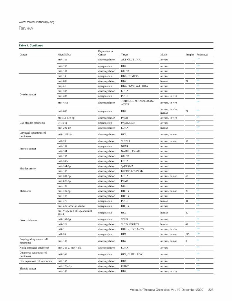
Table 1. Continued
Cancer MicroRNAsExpression inCancer Target Model Samples References
miR-124 downregulation AKT-GLUT1/HK2 in vitro 119
miR-155 upregulation HK2 in vitro 120
miR-144 downregulation GLUT1 in vitro 121
Ovarian cancer
miR-14 upregulation HK2, DNMT3A in vitro 122
miR-603 downregulation HK2 human 21 123
miR-21 upregulation HK2, PKM2, and LDHA in vitro 124
miR-383 downregulation LDHA in vitro 125
miR-203 upregulation PDHB in vitro, in vivo 126
miR-450a downregulationTIMMDC1, MT-ND2, ACO2,ATP5B
in vitro, in vivo 127
miR-603 upregulation HK2in vitro, in vivo,human
21 123
Gall bladder carcinoma
miRNA-139-5p downregulation PKM2 in vitro, in vivo 128
let-7a-5p upregulation PKM2, Stat3 in vitro 129
miR-30d-5p downregulation LDHA human 130
Laryngeal squamous cellcarcinoma
miR-125b-5p downregulation HK2 in vitro, human 131
Prostate cancer
miR-29c downregulation SLC2A3 in vitro, human 57 132
miR-137 upregulation NOX4 in vitro 133
miR-101 downregulation NADPH, TIGAR in vitro 134
miR-132 downregulation GLUT1 in vitro 135
Bladder cancer
miR-200c downregulation LDHA in vitro 136
miR-361-5p downregulation Sp1/PKM2 in vitro 137
miR-145 downregulation KLF4/PTBP1/PKMs in vitro 138
miR-204-3p downregulation LDHA in vitro, human 60 139
Melanoma
miR-625-5p downregulation PKM2 in vitro 140
miR-137 downregulation GLO1 in vitro 141
miR-33a-5p downregulation HIF-1a in vitro, human 20 142
miR-33b downregulation HIF-1a in vitro 143
miR-370 upregulation PDHB human 41 144
Colorectal cancer
miR-23a�27a�24 cluster upregulation HIF-1a in vitro 145
miR-9-5p, miR-98-5p, and miR-199-5p
upregulation HK2 human 40 146
miR-142-5p upregulation SDHB in vitro 147
miR-328 downregulation SLC2A1/GLUT1 human 47 148
miR-1 downregulation HIF-1a, HK2, MCT4 in vitro, in vivo 149
miR-98 upregulation HK2 in vitro, human 215 150
Esophageal squamous cellcarcinoma
miR-143 downregulation HK2 in vitro, human 8 151
Nasopharyngeal carcinoma miR-34b-3, miR-449a downregulation LDHA in vitro 152
Cutaneous squamous cellcarcinoma
miR-365 upregulation HK2, GLUT1, PDK1 in vitro 153
Oral squamous cell carcinoma miR-143 downregulation HK2 in vitro 154
Thyroid cancermiR-125a-5p downregulation CD147 in vitro 155
miR-143 downregulation HK2 in vitro, in vivo 156
Molecular Therapy: Oncolytics Vol. 19 December 2020 223
www.moleculartherapy.org
Review

www.moleculartherapy.org
Review
Aberrant miR-1 expression in aerobic glycolysis (the Warburg effect)was shown by Xu et al.149 in cancer cells. The aerobic glycolysis andproliferation of tumor cells were blocked by miR-1 because ofSmad3 inactivation and HIF-1a targeting. Therefore, the expressionof HK2 and MCT4 was decreased, indicating a new pathway that me-diates aerobic glycolysis in cancer cells. The tumor glycolysis wasreduced significantly following the overexpression miR-1 mimics,including the generation of lactate and the absorption of glucose, aswell as cell proliferation, and ectopic Smad3 expression reversed theseeffects. HIF-1a is regulated by and interacts with endogenous Smad3;this activates Smad3, which is significantly suppressed by miR-1 addi-tion. Based on their results, Smad3 has a pivotal role in the effects ofmiR-1 in colorectal cancer cells. This presents a mechanism for thefirst time so that the Warburg effect is provided easily by the miR-1/Smad3/HIF-1a axis in enhancing cancer progression, in vitro andin vivo. miR-1 is associated with tumor suppression, and thus it canact as a suppressor in molecular therapy of patients suffering fromadvanced colorectal cancer.149
The tumor cells frequently overexpress lactate dehydrogenase A(LDHA). Wang et al. observed higher LDHA levels in patients withcolorectal cancer when comparing samples with adjacent normal tis-sue.173 The production of lactate and ATP and the adsorption ofglucose were reduced following LDHA knockdown. A much slowergrowth rate was seen for the colorectal cancer cells with LDHAknockdown compared to the control cells. Their results showedthat LDHA is targeted by miR-34a, miR-34c, miR-369-3p, miR-374a, and miR-4524a/b, thereby regulating glycolysis in cancer cells.In colorectal cancer tissues, an inverse correlation has been found be-tween these miRNAs and LDHA expression. They detected a geneticloci related to the high progression of colorectal cancer, that is,rs18407893 at 11p15.4 (in the 30 UTR of LDHA), which maps tothe seed sequence identified by miR-374a. The LDHA level is lowerin cancer cells overexpressing miR-374a than in miR-374a-mutant(MUT) (rs18407893 at 11p15.4). These new results can present prom-ising therapeutic strategies to the Warburg effect and therapeutic tar-gets of cancer energy metabolism.173
Yuan et al.136 in their studies evaluated the expression and functionof LDHA in bladder cancer and reported the upregulation of LDHAin bladder cancer cells, enhancing proliferation, invasion, andglycolysis. According to their results, LDHA is targeted directly bymiR-200c in bladder cancer cells. As well, LDHA-induced glycol-ysis, cell proliferation, and invasion were inhibited by ectopicexpression of miR-200c. In fact, one of the possible therapeutic ap-proaches can be LDHA targeting via miR-200c in bladder cancer.Mammalian cells extracted an array of cellular processes for main-taining their survival by improving glycolysis and considering lowoxygen hypoxia or tension. Under these conditions, the use of geneswas operated frequently by HIF-1a, which is fundamental for thehelix-loop-helix transcription element of the PAS family.174 Thefactor of transcription act as a function of a heterodimer of HIF-1b and HIF-1a. Note that HIF-1a protein can be degraded due toexisting oxygen (normoxia) by the ubiquitin-proteasomal proced-
224 Molecular Therapy: Oncolytics Vol. 19 December 2020
ure, whereas HIF-1b and HIF-1a are basically transcribed andtranslated.
Kelly et al.175 specified enzyme glycerol-3-phosphate dehydrogenase1-like (GPD1L) as a new regulator of a direct target of miR-210and HIF-1a stability. In response to the expression of miR-210, thelevels of HIF-1a were stabilized by reduced levels of GPD1L, leadingto enhancing HIF-1a target genes. The enhancement of HIF-1a pro-tein levels and hypoxia-induced miR-210 suppresses GPD1L by sup-pression of PHD activity. Different influences of HIF-1a onto thetranscription of metabolic genes were determined to be straight;nevertheless, HIF-1a additionally adjusts a number of miRNAs,including miR-210, that can regulate an excess of genes in variousphysiological functions. In multiple cancers, miR-210 can be overex-pressed dramatically.176–179 In addition, it suppresses ISCU1/2 andhence reduces the activity of prototypical iron-sulfur proteins con-trolling mitochondrial metabolism, consisting of complex I as wellas aconitase, by taking into account hypoxic conditions.180 However,miR-210 suppresses mitochondrial respiration and can be a reasonfor more aerobic glycolysis in cancer, indirectly. By apparatuses inde-pendent of aerobic glycolysis, many miRNAs may overall affect thehomeostasis of glucose. miR-375, known as one such miRNA, canadjust glucose homeostasis in the body with the modulation of insulinsecretion. In b cells, the mentioned overexpression prevents insulinexocytosis with related mechanisms, which are independent of varia-tions in the intracellular Ca2+ signaling and transmembrane Ca2+
fluxes.181 Many previous investigations have demonstrated thatglucose homeostasis regulation using miR-375 can be mediated bytaking into consideration modulation of the phosphatidylinositol 3-kinase (PI3K) signaling procedure in pancreatic b cells.182 In pancre-atic adenocarcinoma cells, expression levels of miR-375 were dramat-ically downregulated.183,184 Hence, the accurate influence of thedownregulation on the metabolism of glucose as well as the potentialobjectives have not been determined in the case of the tumor cells.
Biogenesis of lncRNAs
lncRNAs play important roles in several human diseases and also innormal development.185,186 An understanding of the biogenesis oflncRNAs has been very helpful for differentiating them from othertypes of ncRNAs, and also for deciphering their functional impor-tance. The biogenesis of lncRNAs is cell stage- and cell type-depen-dent.187,188 Many lncRNAs have been transcribed from DNA se-quences, such as promoters, intergenic regions, and enhancerregions in eukaryotic genomes.189 The first step involved in lncRNAbiogenesis is cleavage by RNase P for generating mature ends. MostlncRNAs undergo 50 capping and 30 polyadenylation in the sameway as mRNAs, but others have small nucleolar RNA (snoRNA)caps at the ends.190,191 In recent years, sub-nuclear structures called“paraspeckles” about 200–1,000 nm in size have been identified be-tween the chromatin structures. Paraspeckles consist of several pro-teins wrapped around a lncRNA core.192 RNA interference (RNAi)evaluations of 40 paraspeckle proteins (PSPs) led to the identificationof 4 PSPs that were essential for the formation of paraspeckles.193,194
The mechanisms for formation and regulation of many lncRNAs are

Figure 2. lncRNAs Regulate Some Molecules Involved in Glucose Metabolism in Cancer
lncRNAs regulate glucose uptake and glycolytic flux by modulating GLUTs and glycolytic enzymes. This figure was adapted from Fan et al.200
www.moleculartherapy.org
Review
not entirely understood. New techniques, such as chromatin isolationby RNA purification sequencing (ChIRP-seq), ribosome profiling,cross-linking immunoprecipitation (CLIP), phylogenetic lineagetracing, and RNA structure mapping using genetic screens andCRISPR, will provide much more information on the structure andfunction of lncRNAs.195–197
lncRNAs carry out regulatory functions because of their modularstructure and RNA-DNA or RNA-RNA base pairing ability.20
lncRNAs regulate gene function in several ways, including (1) recruit-ment of the chromatin remodeling complex to alter the chromatinorganizational pattern; (2) acting as “sponges” by base pairing withmiRNAs to reduce their effects; (3) providing docking sites for pro-teins that are located within the same pathway; (4) guiding certaintranscription factors (TFs) to bind to the promoters; and (5) seques-tering transcription factors and keeping them away from thepromoters.196
Regulation of Glycolysis by lncRNAs
The lncRNA NBR2 regulates 50-AMP-activated protein kinase(AMPK) activity under energy stress induced by glucose starvation.Liu and Gan198 demonstrated that the knockout of NBR2 did not in-fluence the activity of phenformin-induced AMPK, but it did decrease
the expression of GLUT1, thereby decreasing glucose utilization. ThelncRNA called CRNDE (colorectal neoplasia differentially expressed)promotes colorectal cancer progression and drug resistance. Theintron 4 of the gene contains a highly conserved sequence (gVC-In4). Ellis et al.199 showed that in HT29 colorectal cancer cells, treat-ment with insulin or IGF inhibited transcription of CRNDE andgVC-In4. Knockout of gVC-In4 in vitro decreased the expression ofgenes involved in aerobic glycolysis and pyruvate conversion toacetyl-CoA, and it reduced the production of lactic acid. In addition,they found that the GLUT4 expression was decreased, showing thatCRNDE positively regulates the GLUT4 level199 (Figure 2).
Yiya is an lncRNA that was found to be upregulated in several can-cers, and it is located on 1q41.201 The expression of Yiya is associatedwith cell proliferation and cell cycle regulation.201 Cyclin-dependentkinase 6 (CDK6) is considered a major regulator of the cell cycle andmodulates several cell functions, although this is not completely un-derstood.202 When CDK6 was deleted in mice, they found disordersrelated to hematopoiesis.203 According to some studies, CDK6 con-trols the PPP.204 Small molecule inhibitors of CDK6 impaired PPPactivity and lactate synthesis.204 In one study, Xing et al.205 assessedthe effects of Yiya expression on glycolysis in breast cancer.Yiya was associated with cytosolic CDK6 and modulated CDK6-
Molecular Therapy: Oncolytics Vol. 19 December 2020 225

www.moleculartherapy.org
Review
dependent phosphorylation of the fructose bisphosphatase PFK2(PFKFB3) enzyme, which mediates the conversion of glucose 6-phos-phate to fructose-2,6-bisphosphate/fructose-1,6-bisphosphate. CDK6silencing and CRISPR-Cas9-mediated suppression of Yiya inhibitedglycolysis and tumor development. In 40% of breast cancer cases,the expression of Yiya was related to CKD6 and a poor prognosis.205
NORAD (ncRNA activated by DNA damage) is also known asLOC647979 or LINC00657.206 NORAD is known to be an oncogeneand is implicated in breast cancer207 and pancreatic cancer.208 It isknown that some lncRNAs can interact with miRNAs and act asRNA sponges that reduce their effect on the target mRNAs.209,210
In pancreatic cancer, NORAD functions as a sponge for miR125a3pin order to modulate the Ras homolog family member A.208 NORADinteracts with miR-202 and can promote the epithelial-mesenchymaltransition, leading to metastasis and a poor prognosis in colorectalcancer.211 NORAD is also highly expressed in non-small-cell lungcancer (NSCLC) and could be related to glycolysis and increasedcell viability in lung cancer as reported by Gao et al.212 These workersused quantitative reverse transcriptase-polymerase chain reaction(qRT-PCR) to investigate the expression of NORAD, miR136-5p,and markers of glycolysis and cell proliferation. The direct interactionof miR136-5p with NORAD was investigated using RNA immuno-precipitation and a luciferase reporter assay. NORAD was upregu-lated in NSCLC cell lines, and glycolysis and proliferation were alsoincreased. They proposed that NORAD acted as a competing endog-enous RNA for miR136-5p. Gain- and loss-of-function assays showedthat miR136-5p reversed the effects of NORAD in lung cancer.212
MALAT1 (metastasis associated lung adenocarcinoma transcript 1)plays an oncogenic role in some cancers. For instance, Zhanget al.213 showed that MALAT1 participates in renal cell carcinomaby modulation of the miR-203/BIRC5 axis. Sun et al.214 also showedthat MALAT1 was upregulated in ovarian cancer, and it played a rolein apoptosis and proliferation by targeting miR503. Si et al.215 re-ported that MALAT1 could suppress apoptosis and promote auto-phagy by sponging miR-101 in colorectal cancer. MALAT1 is highlyexpressed in multiple myeloma (MM),216 and Liu et al.217 reportedthat downregulation of MALAT1 by RNAi activated apoptosis andsuppressed proliferation.
SRY-box 13 (SOX13) is a member of the Sry-related high-mobilitygroup box (Sox) transcription factor family, and it has been impli-cated in some cancers, such as glioma218 and gastric carcinoma.219
Xu et al.220 showed SOX13 was upregulated in MM.
Liu et al.221 investigated the effects of MALAT1 on glycolysis in MMby measuring the expression levels of MALAT1, SOX13, and miR-1271-5p using quantitative real-time PCR. Flow cytometry, a trans-well assay, andMTT (3-(4,5-dimethylthiazol-2-yl)-2,5-diphenyltetra-zolium bromide) were used to examine apoptosis, invasion, and cellviability, respectively. The lactate production, ATP/ADP ratio, relatedenzyme activities, and glucose consumption were examined to quan-tify glycolysis, plus western blotting for the related proteins. A dual-
226 Molecular Therapy: Oncolytics Vol. 19 December 2020
luciferase reporter assay was used for understanding target interac-tions. An MM xenograft tumor model was carried out in mice. MA-LAT1 was overexpressed and miR-1271-5p was downregulated inMM cells. Deletion of MALAT1 inhibited invasion, glycolysis, andreduced cell viability. MALAT1 targeted miR-1271-5p, and SOX13was shown to be a target of miR-1271-5p. MALAT1 regulatedSOX13 by targeting miR-1271-5p. By downregulation of MALAT1,MM development and glycolysis were suppressed via the miR-1271-5p/SOX13 axis.221
Ftx (five prime to the X-inactivation center [XIST]) is located nearXIST on the X chromosome.222 Ftx encodes a transcript of 2,300 ntwith nine introns, the second and seventh of which encode two clus-ters of miRNAs, that is, miR-545/miR-374a and miR-421/miR-374b.The other introns encode Ftx itself, so there are no overlaps betweenmiRNAs and Ftx. lncRNA Ftx/miR-545 participates in tumorigenesisin hepatocellular carcinoma (HCC) by affecting the PI3K/RAC-aserine/threonine-protein kinase axis via targeting the DExD/H-boxhelicase 58.223 The relationship between glycolysis and Ftx is notyet clear.
Peroxisome proliferator-activated receptor g (PPARg) combineswith PPAR elements to modulate the transcription of target genes,and it heterodimerizes with the retinoid X receptor. PPARg has arole in steatosis-associated hepatic tumorigenesis,224 and it is involvedin cellular sensitivity to insulin and insulin resistance.225 It has a rolein the regulation of some enzymes in carbohydrate metabolism. Forinstance, it promotes GLUT4 expression226 and suppresses PDK1expression.227 PPARgwas reported to reduce the production of leptinand tumor necrosis factor (TNF)-a and improve cellular sensitivity toinsulin and facilitate glucose utilization.228
Li et al.229 studied the role of Ftx in glycolysis in HCC. High expres-sion levels of Ftx enhanced invasion, migration, and proliferation ofHCC, while low expression of Ftx had the opposite effects. Ftx playeda role in glycolysis, the expression of enzymes related to carbohydratemetabolism, glucose transporter expression, glucose consumption,and lactate production. PPARg expression was related to the expres-sion of Ftx in HCC. The inhibition of PPARg using GW9662 in Huh7HCC cells that overexpressed Ftx partially abrogated the increases inglucose uptake, lactate production, and relative glycolytic enzymeexpression induced by Ftx. Conversely, in Bel-7402 HCC cells, activa-tion of PPARg rescued Ftx-mediated suppression of lactate produc-tion, glycolytic enzyme expression, and glucose uptake induced byFtx.229
Proteins, lipids, and nucleotides derived from glycolytic metabolismare used for cell division and proliferation.38 LDHA catalyzes thelast step of the glycolysis pathway. Abnormal expression levels ofLDHA have been found in some cancers such as pancreatic cancer,230
HCC,231 and breast cancer.232 Suppression of LDHA decreases malig-nant transformation and delays tumor progression.4 Studies haveinvestigated themechanisms of the suppression of tumor growth afterLDHA inhibition.233,234 Reduction of LDHA activity either by siRNA
www.moleculartherapy.org
Review
or by a small molecule inhibitor altered energy metabolism andcaused oxidative stress and cell death in lymphoma both in vitroand in vivo.233 LDHA deletion inhibited tumorigenicity via stimula-tion of oxidative stress-mediated mitochondrial apoptosis in breastcancer.234 In one study, Chen et al.235 assessed the role of the lncRNACRYBG3 on glycolysis in lung cancer. Overexpression of CRYBG3stimulated glycolysis and enhanced glucose uptake and lactate pro-duction, while its deletion had the opposite effect.235
GLUT1 (also known as SLC2A1) is a transporter for glucose uptake,and it is expressed by cancer cells for rapid proliferation and energyproduction by glycolysis. SLC2A1 is highly expressed in lung can-cer,236 colon cancer,237 and gastric cancer.97 Chen et al.238 showedthat CREB (cyclic AMP [cAMP]-responsive element binding protein)affected glucose transport via modulating the expression of SLC2A1and modulated the metabolism and progression of glioma. Shiet al.239 investigated the effect of LINC00174 on glycolysis in glioma.In situ hybridization and quantitative RT-PCR were used to examinethe expression of LINC00174 in glioma cell lines and samples. ELISAassays, a transwell invasion assay, scratch wound healing, TUNEL(terminal deoxynucleotidyltransferase-mediated deoxyuridinetriphosphate nick end labeling), and a Cell Counting Kit-8 (CCK-8)assay were used to examine the effects on glioma cells. Western blot-ting, RNA pull-down, dual-luciferase reporter, and RNA immuno-precipitation assays were performed to understand the mechanism.A nude mouse xenograft model was used to understand the role ofLINC00174 in glioma development. LINC00174 was overexpressedin glioma tissues and cell lines. LINC00174 deletion suppressedglycolysis, proliferation, migration, and invasion and also affected gli-oma tumorigenesis. Suppression of miR-152-3p or overexpression ofSLC2A1 could reverse the anti-tumor effects of LINC00174 deletionon glioma. Downregulation of LINC00174 reduced the tumor volumeand delayed tumor development.239
Phosphatase and tensin homolog (PTEN) is known to function as atumor inhibitor in some cancers, such as endometrial carcinoma,kidney cancer, and melanoma.240–242 In esophageal squamous cellcarcinoma (ESCC), epigenetic silencing of PTEN by promoter hy-permethylation was shown to be a mechanism of PTEN reduc-tion.243 Li et al.244 investigated the effects of LINC00184 on glycol-ysis in esophageal cancer. Quantitative RT-PCR was used toexamine LINC00184, and it was found to be upregulated in ESCC,and at the same time PTEN was downregulated. Gain and loss offunction was used to evaluate cellular processes in vivo andin vitro. Deletion of LINC00184 was found to suppress colony for-mation, proliferation, migration, and invasion, while glycolysis wasreduced and mitochondrial OXPHOS was restored. LINC00184promoted promoter methylation of PTEN by recruiting DNMT1.Suppression of PTEN promoter methylation increased mitochon-drial OXPHOS while it inhibited glycolysis. LINC00184 was pro-posed to regulate glycolysis and mitochondrial OXPHOS in ESCCvia Akt phosphorylation, and when Akt was inhibited byLY294002 the effects of LINC00184 on mitochondrial OXPHOSand glycolysis were reversed.244
The dysregulation of the oncogeneMYC has been found to be a com-mon event in tumorigenesis. MYC encodes a transcription factor, c-Myc, that stimulates proliferation and cell growth. Kim et al.245
showed that HIF-1 could act cooperatively with dysregulated c-Mycto promote glycolysis in the P493-6 Burkitt’s lymphoma modelwith an inducible MYC. The expression of the glycolysis enzymehexokinase 2 was induced, and also pyruvate dehydrogenase kinase1 that can inactivate pyruvate dehydrogenase and decrease mitochon-drial respiration. The lncRNA PCGEM1 (prostate cancer geneexpression marker 1) can affect many metabolic processes at the tran-scriptional level, such as the PPP, glucose metabolism, fatty acidbiosynthesis, nucleic acid, and the tricarboxylic acid cycle. PCGEM1was shown to directly interact with c-Myc, promote c-Myc chromatinrecruitment, and bind to the promoters of target genes.246 The effectsof c-Myc on glycolytic gene expression can increase glucose meta-bolism under normal oxygen conditions. The c-Myc protein levelcan be decreased by the lncRNAMIF (c-Myc inhibitory factor), hencesuppressing glycolysis. It was proposed that lncRNA-MIF works as anendogenous competitive RNA to sequester miR-586, therebydecreasing the inhibitory effect of miR-586 against Fbxw7 (F-box/WD repeat-containing protein 7). Fbxw7 is an E3 ubiquitin ligasethat can reduce the stability of c-Myc protein. Therefore, lncRNA-MIF can increase the expression of Fbxw7 and reduce the level ofc-Myc protein. As shown in Figure 3, there is a feedback loop betweenlncRNA-MIF and c-Myc that can regulate glucose metabolism andglycolysis.247 Alternatively, the metabolic signaling pathways shownin Figure 3 do not completely reflect the current understanding ofthese complex networks. For example, the insulin receptor (IR) acti-vates the PI3K/Akt signaling pathway, which is responsible for mostof the metabolic activity of insulin. The IR has two splice isoforms,both of which can phosphorylate at least six known IR substrate(IRS) proteins. These IRSs are capable of interacting with eight knownforms of the PI3K regulatory subunit. PI3K regulatory subunits, inturn, can associate with three forms of the PI3K catalytic subunit,and the products of PI3K enzyme activity can then activate three iso-forms of Akt. The combinatorial possibilities of the IR/IRS/PI3K/Aktpart of the insulin-signaling pathway alone exceed 1,000 combina-tions. When differential compartmentalization, stoichiometry, andthe kinetics of the various downstream signaling components aretaken into account, this number increases dramatically248 (Table 2).
circRNA Biogenesis
In eukaryotic cells, the processing of the exons of pre-mRNAs by alter-native splicing can lead to linear mRNA sequences. circRNAs are pro-duced by a different kind or aberrant RNA splicing in contrast to linearRNAs.274 circRNAs are more resistant to degradation by RNase en-zymes, and they have been found to be more stable compared to linearRNAs due to the presence of a covalent closed-loop structure.275 Voet al.26 characterized circRNAs in more than 2,000 different cancersamples. They found a general reduction in the global abundance ofcircRNAs compared to the adjoining normal tissues. Many subtypesof circRNA have recently been characterized using next-generationtechnologies. There are now four recognized subtypes of circRNAs:(1) exonic circRNAs (ecircRNAs) derived from several or many single
Molecular Therapy: Oncolytics Vol. 19 December 2020 227

Figure 3. Role of PI3K/AKT/mTOR, LKB1-AMPK, and lncRNA-mediated HIF in Glucose Metabolism in Tumor Cells
The stability and synthesis of HIF-1a protein can be affected by lncRNAs, thus modulating HIF-1-mediated metabolic reprogramming. In cancer cells, the translation of HIF-
1a mRNA is dependent on the activity of the mammalian target of rapamycin (mTOR) governed by the activity of upstream tumor suppressor proteins and oncoproteins. HIF-
1a plays a key role in blocking mitochondrial activity and stimulating glycolic enzymes. lncRNAs can also regulate the Akt and AMPK pathways. Akt may increase oxidative
phosphorylation by enhancing metabolic coupling between glycolysis and oxidative phosphorylation, through facilitating the association of mitochondrial hexokinase with
VDAC in the mitochondria. Akt enhances glycolytic flux via several mechanisms. First, it increases glucose uptake and flux. Second, hyperactivated Akt activates mTORC1,
which promotes HIF-1a accumulation under normoxic conditions and increases levels of GLUT1, HKII, and lactate dehydrogenase (LDH). Finally, Akt-increased cellular ATP
levels help to maintain low AMPK activity, which is required for full activation of mTORC1. This figure was adapted from Fan et al.200
www.moleculartherapy.org
Review
exons; (2) circular intronic RNAs (ciRNAs) derived only from introns;(3) exonic-intronic circRNAs (EIciRNAs), including both exons andintrons; and (4) tRNA intronic circRNAs (tricRNAs) created by thesplicing of pre-tRNA introns. Recently, most of the sequenced circR-NAs have been determined to be ecircRNAs. The biogenesis mecha-nisms of circRNAs depend on the particular sub-type. ecircRNAs areproduced by back splicing and lariat-driven circularization. In thelariat, the existing introns are removed, and the exons are linkedtogether by a 50–30 phosphodiester bond. The downstream donorand the upstream splice acceptor sites are then closed in a circle tocreate a lariat, which includes the exons.276 In the downstream and up-stream introns, interactions occur with RNA-binding proteins (RBPs)to form a “bridge,” followed by back splicing for the formation of theecircRNAs.277,278 In base pairing-driven circularization of ecircRNAs,the downstream splicing donor site can be linked to the upstreamsplicing receptor site based on ALU complementary sequences. The in-trons can either be removed or maintained to produce both EIciRNAsor ecircRNAs.275 The biosynthesis of ciRNAs is mainly based on an 11-nt C-rich sequence and a 7-nt GU-rich element for directing the sites ofexonucleolytic cleavage and debranching degradation.279 EIciRNAs
228 Molecular Therapy: Oncolytics Vol. 19 December 2020
and ciRNAs are located within the nucleus where they play role in con-trolling parental gene transcription, in contrast to ecircRNAs, whichare localized within the cytoplasm. tricRNA is a particular introniccircRNA found in Archaea and Drosophila species.280–282 The forma-tion of tricRNA requires the activity of tRNA splicing enzymes todivide the pre-tRNA into two different sections. One of these sectionsproduces tRNAs, while tricRNAs are produced from the other sectionby formation of a 30–50 phosphodiester bond.281 It is accepted that thebinding of RNA proteins and the presence of particular repetitive se-quences surrounding the introns and the circularized exons governthe formation of circRNAs.
The mechanisms of how circRNAs regulate gene function have beeninvestigated in several studies, leading to three main possibilities.277,283
(1) circRNAsoftenhavemiRNAbinding sites that allow themtobind tomiRNAs, acting as a “miRNA sponge” in order to inhibit miRNA bind-ing to the target mRNAs. (2) circRNAs can also interact with RBPs,which leads to the depletion of RBPs and thus reduces their interactionwith RNA targets. (3) It has been suggested that the circRNAs that aregenerated by circularization of exonsmight competewith the splicing of

Table 2. Selected lncRNAs Involved in the Regulation of Glycolysis in Various Cancers
Cancer lncRNAs Expression in Cancer Target Model Samples References
Pancreatic ductal adenocarcinoma PVT1 upregulation HIF-1a in vitro, in vivo, human 249
Hepatocellular carcinoma
SLC2A1-AS1 downregulation STAT3/FOXM1/GLUT1 pathway in vitro, in vivo, human 33 250
LINC01554 downregulation PKM2, Akt/mTOR signaling pathway in vitro, in vivo, human 167 251
HOTAIR upregulation HIF-1a in vitro, human 38 252
Colorectal cancer
HNF1A-AS1 upregulation MYO6 in vitro, human 40 253
LINRIS upregulation IGF2BP2 in vitro, in vivo, human 254
MAFG-AS1 upregulation NDUFA4 in vitro, in vivo, human 52 255
GLCC1 upregulation LDHA in vitro, in vivo, human 40 256
Non-small-cell lung cancer
BCYRN1 upregulation PKM2 in vitro, human 20 257
LINK-A upregulation HKII in vitro, in vivo, human 113 258
LINC00243 upregulation PDK4 in vitro, human 70 259
HOTTIP upregulation HMGB3 in vitro 260
Multiple myeloma MALAT1 upregulation SOX13 in vitro, in vivo, human 30 261
Endometrial carcinoma SNHG16 upregulation HK2 in vitro, in vivo, human 262
GliomaLINC00174 upregulation SLC2A1 in vitro, in vivo, human 45 239
LINC00689 upregulation PKM2 in vitro, in vivo, human 56 263
Esophageal cancer LINC00184 upregulation PTEN in vitro, human 84 244
Cervical cancer UCA1 upregulation HK2 in vitro, human 264
Breast cancerYIYA upregulation CDK6 in vitro, human 205
MEG3 downregulation PI3K/Akt pathway in vitro, in vivo, human 20 265
Ovarian cancer, CAFs LINC00092 upregulation CXCL14 in vitro, in vivo, human 58 266
Nasopharyngeal carcinoma ANRIL upregulation mTOR signaling pathway in vitro, human 88 267
Lung cancer IGFBP4-1 upregulation HK2, PDK1, LDHA in vitro, in vivo, human 159 268
Papillary thyroid carcinoma ASMTL-AS1 downregulation FOXO1 in vitro, in vivo, human 95 269
Ovarian cancer LINC00504 upregulation PKM2, HK2, PDK1 in vitro, human 45 270
Oral squamous cell carcinoma p23154 upregulation GLUT1 in vitro, in vivo, human 49 271
Pediatric acute myeloid leukemia UCA1 upregulation hexokinase 2 pathway in vitro, human 31 272
Osteosarcoma PVT1 upregulation HK2 in vitro, human 46 273
CAF, cancer-associated fibroblast.
www.moleculartherapy.org
Review
pre-mRNA into linear RNA, because they act on the same splice sites.Therefore, the mRNAs might lack the exons that were present in thecircRNA, thus altering the composition of the mRNAs.284
Regulation of Glycolysis by circRNAs
As mentioned above, circRNAs have been found to be widespreadand involved in many developmental steps as well as normal physio-logical conditions using high-throughput deep RNA sequencing aswell as bioinformatics technology.275,285 Naturally occurring circR-NAs play an important role in the interaction network of RNA. circR-NAs are relatively stable, diverse, extremely abundant, and highlyconserved.275 circRNAs are involved in processes such as controllingthe expression of parental genes, acting as endogenous RNAs tosponge miRNAs, regulating RNA–protein interactions and func-tioning as scaffolds for the assembly of protein complexes, as wellas modulating alternative splicing.286,287
It has also been discovered that circRNAs are involved in the patho-genesis of many human diseases, including cardiovascular disor-ders,288 nervous system disorders,289 osteoarthritis,290 diabetes,291
Alzheimer’s disease, silicosis,292 and cancer.293,294 circRNAs haveimportant roles in the growth of cancer, stemness of the malignantcells, metastasis, and treatment resistance.295,296
There is one circRNA, named circMYC (hsa_circ_0085533), that hasbeen found in HepG2 cancer cells, HeLa S3 cells, normal humanepidermal keratinocytes, and human lung fibroblasts. It is locatedon chromosome 8 with a 555-base genomic length.285 circMYC ishighly expressed in melanoma and is related to tumorigenesis.LDHA changes pyruvate into lactate and is one of the hallmarks ofcancers.297 LDHA is considered a valid therapeutic target in manycancers, such as pancreatic cancer, lymphoma, and lung, liver, andbreast cancers. Increased LDHA activity and lactate production are
Molecular Therapy: Oncolytics Vol. 19 December 2020 229

Table 3. Selected Circular RNAs Involved in the Regulation of Glycolysis in Various Cancers
Cancer Circular RNAs Expression in Cancer Target Model Samples Reference
Melanoma circMYC upregulation 30 UTR of LDHA in vitro, human 25 311
Non-small-cell lung carcinoma circACACA upregulation PI3K/PKB pathway in vitro, in vivo, human 312
Lung cancer circAKT3 upregulation STAT3 in vitro, in vivo, human 28 313
www.moleculartherapy.org
Review
associated with poor prognosis and resistance to chemotherapy andradiotherapy.231,234,298,299 LDHA was found to be overexpressed innevic melanocytes and primary and metastatic melanomas.300 Inone study, Jin et al.301 assessed the effect of circMYC on glycolysisin melanoma. circMYC increased the proliferation in Mel-CV cellsand human melanoma cells. Overexpression of circMYC led to inhi-bition of LDHA activity and lower glycolysis. circMYC targeted miR-1236 and bound to it as a molecular sponge.301
The PI3K/protein kinase B (PKB) signaling pathway is critical for cellapoptosis and survival.302 It has been proposed that some cancers arerelated to the incorrect activation of that pathway.303 The PI3K/PKBsignaling pathway was implicated in the initiation of endometrial can-cer304 and cisplatin (DDP) resistance in lung cancer.305 Understand-ing of the PI3K/PKB pathway could help to improve therapies forlung cancer.
Wu et al.306 studied the effects of circACACA on glycolysis inNSCLC. qPCR was used to measure the expression of miR-1183and the circACACA Transwell assay, and CCK-8 assays were usedto measure migration and proliferation. The Seahorse XFe96 analyzerwas used to study mitochondrial metabolism, and western blottingwas used to quantify the expression of GLUT1, matrix metalloprotei-nase 9, c-Myc protein, PTEN, phosphorylated (p-)PI3K, PKB, p-PKB,and PI3K. An RNA pull-down assay, RNA immunoprecipitationassay, and dual-luciferase reporter assays were used to study the cor-relation between circACACA and miR-1183. A xenograft tumormodel was used to understand the biological role of circACACA. cir-cACACA was highly expressed in NSCLC samples and cell lines, andthe pattern was the opposite of miR-1183 expression. Deletion of cir-cACACA suppressed glycolysis, proliferation, and migration inNSCLC. miR-1183 was targeted by circACACA, and circACACAmodulated the PI3K/PKB signaling pathway by interacting withmiR-1183. Reducing the expression of circACACA inhibited tumordevelopment.306
Signal transducer and activator of transcription 3 (STAT3) has a rolein ischemic cardiac hypertrophy and tumor-associated macrophagedifferentiation.307,308 Dong et al.309 showed that circ_0076305 regu-lated DDP resistance in NSCLC by positively modulating STAT3via sponging of miR-296–5p. Xu et al.310 investigated the effects of cir-cAKT3 on glycolysis in NSCLC. Western blotting and quantitativereal-time PCR were used to examine the expression of miR-516b-5p, circAKT3, and STAT3 in NSCLC. MTT was used to measurethe sensitivity to DDP. For measuring the lactate production andglycolysis in cells treated with different plasmids or 2-deoxy-glucose
230 Molecular Therapy: Oncolytics Vol. 19 December 2020
(2-DG), a lactate assay and glucose assay kits were used. The expres-sion of HIF-1a in H1299 and A549 cells was detected by western blot-ting. Relationships between circAKT3, miR-516b-5p, and STAT3were studied using a dual-luciferase reporter assay. circAKT3 andSTAT3 were highly expressed in lung cancer while miR-516b-5pshowed low expression. Deletion of circAKT3 inhibited glycolysisand increased the sensitivity to DDP. In A549 cells, the inhibitionof HIF-1a-induced glycolysis abrogated the circAKT3-induced che-moresistance. miR-516b-5p was found to bind to circAKT3. Suppres-sion of miR-516b-5p reversed the effects of circAKT3 deletion onglycolysis, as well as DDP sensitivity. Deletion of circAKT3 inhibiteddevelopment of tumors in vivo by the miR-516b-5p/STAT3 axis310
(Table 3).
Conclusions
Cancer is a complex disease at the cellular level, whose causative fac-tors include environmentally induced mutations, heritable geneticconditions, and epigenetic alterations. Cancer cells are furtherrequired to modulate their metabolism in accord with the tumormicroenvironment in order to survive and proliferate under the con-ditions imposed on them by the unrestricted growth of the tumor.The metabolic reprogramming that occurs in tumors to supportcell expansion and proliferation is facilitated by the loss of tumor sup-pressor proteins or the activation of oncogenes. In recent times, alarge number of different ncRNAs have been discovered, and theseare now accepted as major mechanisms of epigenetic control ofgene expression. Many ncRNAs have been implicated in the initia-tion, progression, and metastatic spread of different types of cancer.The dysregulated metabolism in cancer cells (called the Warburg ef-fect) relies on switching energy production away from OXPHOS to-ward aerobic glycolysis. Several different sub-types of ncRNAs,including miRNAs, lncRNAs, and circRNAs, have been found toplay regulatory roles in cancer metabolism, particularly in mitochon-drial functions, and in the metabolism of lipids, glucose, and gluta-mine. Many ncRNAs have been implicated in cancer metabolic re-programming and the switch to glycolysis, thus increasing theoverall aggressiveness of the tumor. Not only could further investiga-tions into the mechanisms and functions of ncRNAs allow the discov-ery of additional prognostic biomarkers, but they could also be bene-ficial for the development of novel therapeutic approaches.
AUTHOR CONTRIBUTIONSH.M. and M.R.H. contributed to the conception, design, data collec-tion, and drafting of the manuscript. Both authors approved the finalversion for submission.

www.moleculartherapy.org
Review
CONFLICTS OF INTERESTM.R.H. declares the following potential conflicts of interest: ScientificAdvisory Boards: Transdermal Cap, Cleveland, OH, USA; BeWellGlobal, Wan Chai, Hong Kong; Hologenix, Santa Monica, CA,USA; LumiThera, Poulsbo, WA, USA; Vielight, Toronto, ON, Can-ada; Bright Photomedicine, Sao Paulo, Brazil; Quantum Dynamics,Cambridge, MA, USA; Global Photon, Bee Cave, TX, USA; MedicalCoherence, Boston MA, USA; NeuroThera, Newark, DE, USA;JOOVV, Minneapolis-St. Paul, MN, USA; AIRx Medical, Pleasanton,CA, USA; FIR Industries, Ramsey, NJ, USA; UVLRx Therapeutics,Oldsmar, FL, USA; Ultralux UV, Lansing, MI, USA; Illumiheal & Pet-thera, Shoreline, WA, USA; MB Lasertherapy, Houston, TX, USA;ARRC LED, San Clemente, CA, USA; Varuna Biomedical, InclineVillage, NV, USA; and Niraxx Light Therapeutics, Boston, MA,USA. Consulting: Lexington International, Boca Raton, FL, USA;USHIO, Japan; Merck KGaA, Darmstadt, Germany; Philips Elec-tronics Nederland BV, Eindhoven, the Netherlands; Johnson & John-son, Philadelphia, PA, USA; and Sanofi-Aventis Deutschland GmbH,Frankfurt am Main, Germany. Stock holdings: Global Photon, BeeCave, TX, USA; and Mitonix, Newark, DE, USA. H.M. declares nocompeting interests.
REFERENCES1. Warburg, O. (1956). On the origin of cancer cells. Science 123, 309–314.
2. Warburg, O. (1928). The chemical constitution of respiration ferment. Science 68,437–443.
3. Vander Heiden, M.G., Cantley, L.C., and Thompson, C.B. (2009). Understandingthe Warburg effect: the metabolic requirements of cell proliferation. Science 324,1029–1033.
4. Fantin, V.R., St-Pierre, J., and Leder, P. (2006). Attenuation of LDH-A expressionuncovers a link between glycolysis, mitochondrial physiology, and tumor mainte-nance. Cancer Cell 9, 425–434.
5. Nguyen, T.-L., and Durán, R.V. (2018). Glutamine metabolism in cancer therapy.Cancer Drug Resist. 1, 126–138.
6. DeBerardinis, R.J., Mancuso, A., Daikhin, E., Nissim, I., Yudkoff, M., Wehrli, S., andThompson, C.B. (2007). Beyond aerobic glycolysis: transformed cells can engage inglutamine metabolism that exceeds the requirement for protein and nucleotide syn-thesis. Proc. Natl. Acad. Sci. USA 104, 19345–19350.
7. Wang, Y., Wu, Y., Wang, Y., Fu, A., Gong, L., Li, W., and Li, Y. (2017). Bacillus amy-loliquefaciens SC06 alleviates the oxidative stress of IPEC-1 via modulating Nrf2/Keap1 signaling pathway and decreasing ROS production. Appl. Microbiol.Biotechnol. 101, 3015–3026.
8. Wilde, L., Roche, M., Domingo-Vidal, M., Tanson, K., Philp, N., Curry, J., andMartinez-Outschoorn, U. (2017). Metabolic coupling and the reverse Warburg ef-fect in cancer: implications for novel biomarker and anticancer agent development.Semin. Oncol. 44, 198–203.
9. Brooks, G.A. (2009). Cell-cell and intracellular lactate shuttles. J. Physiol. 587, 5591–5600.
10. Held-Warmkessel, J., and Dell, D.D. (2014). Lactic acidosis in patients with cancer.Clin. J. Oncol. Nurs. 18, 592–594.
11. Peppicelli, S., Bianchini, F., and Calorini, L. (2014). Extracellular acidity, a “reappre-ciated” trait of tumor environment driving malignancy: perspectives in diagnosisand therapy. Cancer Metastasis Rev. 33, 823–832.
12. Shiraishi, T., Verdone, J.E., Huang, J., Kahlert, U.D., Hernandez, J.R., Torga, G.,Zarif, J.-C., Epstein, T., Gatenby, R., McCartney, A., et al. (2015). Glycolysis is theprimary bioenergetic pathway for cell motility and cytoskeletal remodeling in hu-man prostate and breast cancer cells. Oncotarget 6, 130–143.
13. Zhao, M., Fan, J., Liu, Y., Yu, Y., Xu, J., Wen, Q., Zhang, J., Fu, S., Wang, B., Xiang, L.,et al. (2016). Oncogenic role of the TP53-induced glycolysis and apoptosis regulatorin nasopharyngeal carcinoma through NF-kB pathwaymodulation. Int. J. Oncol. 48,756–764.
14. Faubert, B., Vincent, E.E., Griss, T., Samborska, B., Izreig, S., Svensson, R.U., Mamer,O.A., Avizonis, C., Shackelford, D.B., Shaw, R.J., and Jones, R.G. (2014). Loss of thetumor suppressor LKB1 promotes metabolic reprogramming of cancer cells viaHIF-1a. Proc. Natl. Acad. Sci. USA 111, 2554–2559.
15. Li, Z., Peng, Y., Li, J., Chen, Z., Chen, F., Tu, J., Lin, S., and Wang, H. (2020). N6-methyladenosine regulates glycolysis of cancer cells through PDK4. Nat.Commun. 11, 2578.
16. Wang, T., Kong, S., Tao, M., and Ju, S. (2020). The potential role of RNA N6-meth-yladenosine in cancer progression. Mol. Cancer 19, 88.
17. Yu, A.-M., Jian, C., Yu, A.H., and Tu, M.-J. (2019). RNA therapy: are we using theright molecules? Pharmacol. Ther. 196, 91–104.
18. Hayes, J., Peruzzi, P.P., and Lawler, S. (2014). MicroRNAs in cancer: biomarkers,functions and therapy. Trends Mol. Med. 20, 460–469.
19. Li, Y., Xu, Q., Lv, N., Wang, L., Zhao, H., Wang, X., Guo, J., Chen, C., Li, Y., and Yu,L. (2017). Clinical implications of genome-wide DNA methylation studies in acutemyeloid leukemia. J. Hematol. Oncol. 10, 41.
20. Guttman, M., and Rinn, J.L. (2012). Modular regulatory principles of large non-cod-ing RNAs. Nature 482, 339–346.
21. Esteller, M. (2011). Non-coding RNAs in human disease. Nat. Rev. Genet. 12,861–874.
22. Matsui, M., and Corey, D.R. (2017). Non-coding RNAs as drug targets. Nat. Rev.Drug Discov. 16, 167–179.
23. Yoon, S., and Rossi, J.J. (2018). Therapeutic potential of small activating RNAs(saRNAs) in human cancers. Curr. Pharm. Biotechnol. 19, 604–610.
24. Dong, Y., He, D., Peng, Z., Peng, W., Shi, W., Wang, J., Li, B., Zhang, C., and Duan,C. (2017). Circular RNAs in cancer: an emerging key player. J. Hematol. Oncol. 10,2.
25. Li, X., Yang, L., and Chen, L.-L. (2018). The biogenesis, functions, and challenges ofcircular RNAs. Mol. Cell 71, 428–442.
26. Vo, J.N., Cieslik, M., Zhang, Y., Shukla, S., Xiao, L., Zhang, Y., Wu, Y.M.,Dhanasekaran, S.M., Engelke, C.G., Cao, X., et al. (2019). The landscape of circularRNA in cancer. Cell 176, 869–881.e13.
27. Zhang, Y., and Yang, J.-M. (2013). Altered energy metabolism in cancer: a uniqueopportunity for therapeutic intervention. Cancer Biol. Ther. 14, 81–89.
28. Bonnet, S., Archer, S.L., Allalunis-Turner, J., Haromy, A., Beaulieu, C., Thompson,R., Lee, C.T., Lopaschuk, G.D., Puttagunta, L., Bonnet, S., et al. (2007). A mitochon-dria-K+ channel axis is suppressed in cancer and its normalization promotesapoptosis and inhibits cancer growth. Cancer Cell 11, 37–51.
29. Hu, Y., Lu, W., Chen, G., Wang, P., Chen, Z., Zhou, Y., Ogasawara, M.,Trachootham, D., Feng, L., Pelicano, H., et al. (2012). K-rasG12V transformationleads to mitochondrial dysfunction and a metabolic switch from oxidative phos-phorylation to glycolysis. Cell Res. 22, 399–412.
30. Lu, W., Hu, Y., Chen, G., Chen, Z., Zhang, H., Wang, F., Feng, L., Pelicano, H.,Wang, H., Keating, M.J., et al. (2012). Novel role of NOX in supporting aerobicglycolysis in cancer cells with mitochondrial dysfunction and as a potential targetfor cancer therapy. PLoS Biol. 10, e1001326.
31. Wallace, D.C. (2012). Mitochondria and cancer. Nat. Rev. Cancer 12, 685–698.
32. Cavalli, L.R., Varella-Garcia, M., and Liang, B.C. (1997). Diminished tumorigenicphenotype after depletion of mitochondrial DNA. Cell Growth Differ. 8, 1189–1198.
33. King, M.P., and Attardi, G. (1989). Human cells lacking mtDNA: repopulation withexogenous mitochondria by complementation. Science 246, 500–503.
34. de Souza, A.C.S., Justo, G.Z., de Araújo, D.R., and Cavagis, A.D.M. (2011). Definingthe molecular basis of tumor metabolism: a continuing challenge since Warburg’sdiscovery. Cell. Physiol. Biochem. 28, 771–792.
35. Locasale, J.W., and Cantley, L.C. (2010). Altered metabolism in cancer. BMC Biol. 8,88.
Molecular Therapy: Oncolytics Vol. 19 December 2020 231

www.moleculartherapy.org
Review
36. Pfeiffer, T., Schuster, S., and Bonhoeffer, S. (2001). Cooperation and competition inthe evolution of ATP-producing pathways. Science 292, 504–507.
37. Zhou, Y., Tozzi, F., Chen, J., Fan, F., Xia, L., Wang, J., Gao, G., Zhang, A., Xia, X.,Brasher, H., et al. (2012). Intracellular ATP levels are a pivotal determinant of che-moresistance in colon cancer cells. Cancer Res. 72, 304–314.
38. Gatenby, R.A., and Gillies, R.J. (2004). Why do cancers have high aerobic glycolysis?Nat. Rev. Cancer 4, 891–899.
39. Lunt, S.Y., and Vander Heiden, M.G. (2011). Aerobic glycolysis: meeting the meta-bolic requirements of cell proliferation. Annu. Rev. Cell Dev. Biol. 27, 441–464.
40. Deberardinis, R.J., Sayed, N., Ditsworth, D., and Thompson, C.B. (2008). Brick bybrick: metabolism and tumor cell growth. Curr. Opin. Genet. Dev. 18, 54–61.
41. Backos, D.S., Franklin, C.C., and Reigan, P. (2012). The role of glutathione in braintumor drug resistance. Biochem. Pharmacol. 83, 1005–1012.
42. Traverso, N., Ricciarelli, R., Nitti, M., Marengo, B., Furfaro, A.L., Pronzato, M.A.,Marinari, U.M., and Domenicotti, C. (2013). Role of glutathione in cancer progres-sion and chemoresistance. Oxid. Med. Cell. Longev. 2013, 972913.
43. Pitroda, S.P., Wakim, B.T., Sood, R.F., Beveridge, M.G., Beckett, M.A., MacDermed,D.M., Weichselbaum, R.R., and Khodarev, N.N. (2009). STAT1-dependent expres-sion of energy metabolic pathways links tumour growth and radioresistance to theWarburg effect. BMC Med. 7, 68.
44. Tsai, F.D., Lopes, M.S., Zhou, M., Court, H., Ponce, O., Fiordalisi, J.J., Gierut, J.J.,Cox, A.D., Haigis, K.M., and Philips, M.R. (2015). K-Ras4A splice variant is widelyexpressed in cancer and uses a hybrid membrane-targeting motif. Proc. Natl. Acad.Sci. USA 112, 779–784.
45. Kerr, E.M., Gaude, E., Turrell, F.K., Frezza, C., and Martins, C.P. (2016). MutantKras copy number defines metabolic reprogramming and therapeutic susceptibil-ities. Nature 531, 110–113.
46. Amendola, C.R., Mahaffey, J.P., Parker, S.J., Ahearn, I.M., Chen, W.-C., Zhou, M.,Court, H., Shi, J., Mendoza, S.L., Morten, M.J., et al. (2019). KRAS4A directly reg-ulates hexokinase 1. Nature 576, 482–486.
47. Luo, W., Hu, H., Chang, R., Zhong, J., Knabel, M., O’Meally, R., Cole, R.N., Pandey,A., and Semenza, G.L. (2011). Pyruvate kinase M2 is a PHD3-stimulated coactivatorfor hypoxia-inducible factor 1. Cell 145, 732–744.
48. Riganti, C., Gazzano, E., Polimeni, M., Aldieri, E., and Ghigo, D. (2012). The pentosephosphate pathway: an antioxidant defense and a crossroad in tumor cell fate. FreeRadic. Biol. Med. 53, 421–436.
49. Xu, X., Zur Hausen, A., Coy, J.F., and Löchelt, M. (2009). Transketolase-like protein1 (TKTL1) is required for rapid cell growth and full viability of human tumor cells.Int. J. Cancer 124, 1330–1337.
50. Sun, W., Liu, Y., Glazer, C.A., Shao, C., Bhan, S., Demokan, S., Zhao, M., Rudek,M.A., Ha, P.K., and Califano, J.A. (2010). TKTL1 is activated by promoter hypome-thylation and contributes to head and neck squamous cell carcinoma carcinogenesisthrough increased aerobic glycolysis and HIF1a stabilization. Clin. Cancer Res. 16,857–866.
51. Wanka, C., Steinbach, J.P., and Rieger, J. (2012). Tp53-induced glycolysis andapoptosis regulator (TIGAR) protects glioma cells from starvation-induced celldeath by up-regulating respiration and improving cellular redox homeostasis.J. Biol. Chem. 287, 33436–33446.
52. Monteleone, F., Rosa, R., Vitale, M., D’Ambrosio, C., Succoio, M., Formisano, L.,Nappi, L., Romano, M.F., Scaloni, A., Tortora, G., et al. (2013). Increased anaerobicmetabolism is a distinctive signature in a colorectal cancer cellular model of resis-tance to antiepidermal growth factor receptor antibody. Proteomics 13, 866–877.
53. Zhao, F., Mancuso, A., Bui, T.V., Tong, X., Gruber, J.J., Swider, C.R., Sanchez, P.V.,Lum, J.J., Sayed, N., Melo, J.V., et al. (2010). Imatinib resistance associated withBCR-ABL upregulation is dependent on HIF-1a-induced metabolic reprograming.Oncogene 29, 2962–2972.
54. Kim, V.N. (2005). MicroRNA biogenesis: coordinated cropping and dicing. Nat.Rev. Mol. Cell Biol. 6, 376–385.
55. Lee, Y., Kim, M., Han, J., Yeom, K.H., Lee, S., Baek, S.H., and Kim, V.N. (2004).MicroRNA genes are transcribed by RNA polymerase II. EMBO J. 23, 4051–4060.
232 Molecular Therapy: Oncolytics Vol. 19 December 2020
56. Lee, Y., Ahn, C., Han, J., Choi, H., Kim, J., Yim, J., Lee, J., Provost, P., Rådmark, O.,Kim, S., and Kim, V.N. (2003). The nuclear RNase III Drosha initiates microRNAprocessing. Nature 425, 415–419.
57. Gregory, R.I., Yan, K.P., Amuthan, G., Chendrimada, T., Doratotaj, B., Cooch, N.,and Shiekhattar, R. (2004). The microprocessor complex mediates the genesis ofmicroRNAs. Nature 432, 235–240.
58. Denli, A.M., Tops, B.B., Plasterk, R.H., Ketting, R.F., and Hannon, G.J. (2004).Processing of primary microRNAs by the microprocessor complex. Nature 432,231–235.
59. Bohnsack, M.T., Czaplinski, K., and Gorlich, D. (2004). Exportin 5 is a RanGTP-dependent dsRNA-binding protein that mediates nuclear export of pre-miRNAs.RNA 10, 185–191.
60. Lee, Y., Jeon, K., Lee, J.T., Kim, S., and Kim, V.N. (2002). MicroRNA maturation:stepwise processing and subcellular localization. EMBO J. 21, 4663–4670.
61. Flynt, A.S., and Lai, E.C. (2008). Biological principles of microRNA-mediated regu-lation: shared themes amid diversity. Nat. Rev. Genet. 9, 831–842.
62. Selbach, M., Schwanhäusser, B., Thierfelder, N., Fang, Z., Khanin, R., and Rajewsky,N. (2008). Widespread changes in protein synthesis induced by microRNAs. Nature455, 58–63.
63. Friedman, R.C., Farh, K.K.-H., Burge, C.B., and Bartel, D.P. (2009). Most mamma-lian mRNAs are conserved targets of microRNAs. Genome Res. 19, 92–105.
64. Baek, D., Villén, J., Shin, C., Camargo, F.D., Gygi, S.P., and Bartel, D.P. (2008). Theimpact of microRNAs on protein output. Nature 455, 64–71.
65. Filipowicz, W., Bhattacharyya, S.N., and Sonenberg, N. (2008). Mechanisms of post-transcriptional regulation by microRNAs: are the answers in sight? Nat. Rev. Genet.9, 102–114.
66. Pillai, R.S., Bhattacharyya, S.N., Artus, C.G., Zoller, T., Cougot, N., Basyuk, E.,Bertrand, E., and Filipowicz, W. (2005). Inhibition of translational initiation byLet-7 microRNA in human cells. Science 309, 1573–1576.
67. Humphreys, D.T., Westman, B.J., Martin, D.I., and Preiss, T. (2005). MicroRNAscontrol translation initiation by inhibiting eukaryotic initiation factor 4E/cap andpoly(A) tail function. Proc. Natl. Acad. Sci. USA 102, 16961–16966.
68. Petersen, C.P., Bordeleau, M.-E., Pelletier, J., and Sharp, P.A. (2006). Short RNAsrepress translation after initiation in mammalian cells. Mol. Cell 21, 533–542.
69. Nottrott, S., Simard, M.J., and Richter, J.D. (2006). Human let-7a miRNA blocksprotein production on actively translating polyribosomes. Nat. Struct. Mol. Biol.13, 1108–1114.
70. Kloosterman, W.P., and Plasterk, R.H. (2006). The diverse functions of microRNAsin animal development and disease. Dev. Cell 11, 441–450.
71. Gangaraju, V.K., and Lin, H. (2009). MicroRNAs: key regulators of stem cells. Nat.Rev. Mol. Cell Biol. 10, 116–125.
72. Bushati, N., and Cohen, S.M. (2007). microRNA functions. Annu. Rev. Cell Dev.Biol. 23, 175–205.
73. Krol, J., Loedige, I., and Filipowicz, W. (2010). The widespread regulation ofmicroRNA biogenesis, function and decay. Nat. Rev. Genet. 11, 597–610.
74. Fathullahzadeh, S., Mirzaei, H., Honardoost, M.A., Sahebkar, A., and Salehi, M.(2016). Circulating microRNA-192 as a diagnostic biomarker in human chroniclymphocytic leukemia. Cancer Gene Ther. 23, 327–332.
75. Croce, C.M. (2009). Causes and consequences of microRNA dysregulation in can-cer. Nat. Rev. Genet. 10, 704–714.
76. Pourhanifeh, M.H., Vosough, M., Mahjoubin-Tehran, M., Hashemipour, M., Nejati,M., Abbasi-Kolli, M., Sahebkar, A., and Mirzaei, H. (2020). Autophagy-relatedmicroRNAs: possible regulatory roles and therapeutic potential in and gastrointes-tinal cancers. Pharmacol. Res. 161, 105133.
77. Yousefpouran, S., Mostafaei, S., Manesh, P.V., Iranifar, E., Bokharaei-Salim, F.,Nahand, J.S., Mirzaei, H., Taran, M., Babaei, F., Sayad, B., and Moghoofei, M.(2020). The assessment of selected miRNAs profile in HIV, HBV, HCV, HIV/HCV, HIV/HBV co-infection and elite controllers for determination of biomarker.Microb. Pathog. 147, 104355.
78. Wu, D.H., Liang, H., Lu, S.N., Wang, H., Su, Z.L., Zhang, L., Ma, J.-Q., Guo, M., Tai,S., and Yu, S. (2018). miR-124 suppresses pancreatic ductal adenocarcinoma growth

www.moleculartherapy.org
Review
by regulating monocarboxylate transporter 1-mediated cancer lactate metabolism.Cell. Physiol. Biochem. 50, 924–935.
79. Liu, Y., Tang, Z.-G., Lin, Y., Qu, X.-G., Lv, W., Wang, G.-B., and Li, C.L. (2017).Effects of quercetin on proliferation and migration of human glioblastoma U251cells. Biomed. Pharmacother. 92, 33–38.
80. Yang, Y., Ishak Gabra, M.B., Hanse, E.A., Lowman, X.H., Tran, T.Q., Li, H., Milman,N., Liu, J., Reid, M.A., Locasale, J.W., et al. (2019). miR-135 suppresses glycolysisand promotes pancreatic cancer cell adaptation to metabolic stress by targetingphosphofructokinase-1. Nat. Commun. 10, 809.
81. Chen, H., He, J., Lanzafame, R., Stadler, I., Hamidi, H.E., Liu, H., Celli, J., Hamblin,M.R., Huang, Y., Oakley, E., et al. (2017). Quantum dot light emitting devices forphotomedical applications. J. Soc. Inf. Disp. 25, 177–184.
82. Li, S.J., Liu, H.L., Tang, S.L., Li, X.J., and Wang, X.Y. (2017). MicroRNA-150 regu-lates glycolysis by targeting von Hippel-Lindau in glioma cells. Am. J. Transl. Res. 9,1058–1066.
83. Guo, H., Nan, Y., Zhen, Y., Zhang, Y., Guo, L., Yu, K., Huang, Q., and Zhong, Y.(2016). miRNA-451 inhibits glioma cell proliferation and invasion by downregulat-ing glucose transporter 1. Tumour Biol. 37, 13751–13761.
84. Yu, F., Lu, Z., Huang, K., Wang, X., Xu, Z., Chen, B., Dong, P., and Zheng, J. (2016).MicroRNA-17-5p-activated Wnt/b-catenin pathway contributes to the progressionof liver fibrosis. Oncotarget 7, 81–93.
85. Xu, Q., Dou, C., Liu, X., Yang, L., Ni, C., Wang, J., Guo, Y., Yang, W., Tong, X., andHuang, D. (2018). Oviductus ranae protein hydrolysate (ORPH) inhibits thegrowth, metastasis and glycolysis of HCC by targeting miR-491-5p/PKM2 axis.Biomed. Pharmacother 107, 1692–1704.
86. Hua, S., Liu, C., Liu, L., and Wu, D. (2018). miR-142-3p inhibits aerobic glycolysisand cell proliferation in hepatocellular carcinoma via targeting LDHA. Biochem.Biophys. Res. Commun. 496, 947–954.
87. Li,W., Hao, J., Zhang, L., Cheng, Z., Deng, X., and Shu, G. (2017). Astragalin reduceshexokinase 2 through increasing miR-125b to inhibit the proliferation of hepatocel-lular carcinoma cells in vitro and in vivo. J. Agric. Food Chem 65, 5961–5972.
88. Jin, F., Wang, Y., Zhu, Y., Li, S., Liu, Y., Chen, C.,Wang, X., Zen, K., and Li, L. (2017).The miR-125a/HK2 axis regulates cancer cell energy metabolism reprogramming inhepatocellular carcinoma. Sci. Rep. 7, 3089.
89. Kheimar, A., Previdelli, R.L., Wight, D.J., and Kaufer, B.B. (2017). Telomeres andtelomerase: role in Marek’s disease virus pathogenesis, integration and tumorigen-esis. Viruses 9, 173.
90. Ren, L., Yao, Y., Wang, Y., and Wang, S. (2019). miR-505 suppressed the growth ofhepatocellular carcinoma cells via targeting IGF-1R. Biosci. Rep 39, BSR20182442.
91. Zhang, H.F., Wang, Y.C., and Han, Y.D. (2018). MicroRNA-34a inhibits liver cancercell growth by reprogramming glucose metabolism. Mol. Med. Rep. 17, 4483–4489.
92. Hua, S., Lei, L., Deng, L., Weng, X., Liu, C., Qi, X., Wang, S., Zhang, D., Zou, X., Cao,C., et al. (2018). miR-139-5p inhibits aerobic glycolysis, cell proliferation, migration,and invasion in hepatocellular carcinoma via a reciprocal regulatory interaction withETS1. Oncogene 37, 1624–1636.
93. Liu, Z., Yu, M., Fei, B., Fang, X., Ma, T., and Wang, D. (2018). miR-21-5p targetsPDHA1 to regulate glycolysis and cancer progression in gastric cancer. Oncol.Rep. 40, 2955–2963.
94. Ping, W., Senyan, H., Li, G., Yan, C., and Long, L. (2018). Increased lactate in gastriccancer tumor-infiltrating lymphocytes is related to impaired T cell function due tomiR-34a deregulated lactate dehydrogenase A. Cell. Physiol. Biochem 49, 828–836.
95. Yang, L., Zhang,W.,Wang, Y., Zou, T., Zhang, B., Xu, Y., Pang, T., Hu, Q., Chen,M.,Wang, L., et al. (2018). Hypoxia-induced miR-214 expression promotes tumour cellproliferation and migration by enhancing the Warburg effect in gastric carcinomacells. Cancer Lett. 414, 44–56.
96. Sun, K., Hu, P., and Xu, F. (2018). LINC00152/miR-139-5p regulates gastric cancercell aerobic glycolysis by targeting PRKAA1. Biomed. Pharmacother 97, 1296–1302.
97. Ding, X., Liu, J., Liu, T., Ma, Z., Wen, D., and Zhu, J. (2017). miR-148b inhibitsglycolysis in gastric cancer through targeting SLC2A1. Cancer Med. 6, 1301–1310.
98. Li, L.Q., Yang, Y., Chen, H., Zhang, L., Pan, D., and Xie, W.J. (2016). MicroRNA-181b inhibits glycolysis in gastric cancer cells via targeting hexokinase 2 gene.Cancer Biomark 17, 75–81.
99. Wang, Y., He, Y., Bai, H., Dang, Y., Gao, J., and Lv, P. (2019). Phosphoinositide-dependent kinase 1-associated glycolysis is regulated by miR-409-3p in clear cellrenal cell carcinoma. J. Cell. Biochem 120, 126–134.
100. Takai, T., Tsujino, T., Yoshikawa, Y., Inamoto, T., Sugito, N., Kuranaga, Y.,Heishima, K., Soga, T., Hayashi, K., Miyata, K., et al. (2019). Synthetic miR-143 ex-hibited an anti-cancer effect via the downregulation of K-RAS networks of renal cellcancer cells in vitro and in vivo. Mol. Ther. 27, 1017–1027.
101. Weng, Y., Shen, Y., He, Y., Pan, X., Xu, J., Jiang, Y., Zhang, Q., Wang, S., Kong, F.,Zhao, S., et al. (2018). The miR-15b-5p/PDK4 axis regulates osteosarcoma prolifer-ation through modulation of the Warburg effect. Biochem. Biophys. Res. Commun.503, 2749–2757.
102. Chen, H., Gao, S., and Cheng, C. (2018). miR-323a-3p suppressed the glycolysis ofosteosarcoma via targeting LDHA. Hum. Cell 31, 300–309.
103. Zheng, X.M., Xu, C.W., and Wang, F. (2018). miR-33b inhibits osteosarcoma cellproliferation through suppression of glycolysis by targeting lactate dehydrogenaseA (LDHA). Cell. Mol. Biol. 64, 31–35.
104. Wu, Y., He, H., Wu, B., Wen, J., Guo, Z., Luo, Y., and Cao, G. (2017). [miR-125bsuppresses the aerobic glycolysis of osteosarcoma HOS cells by downregulatingthe expression of hexokinase-2]. Xi Bao Yu Fen Zi Mian Yi Xue Za Zhi 33, 1365–1370.
105. Xiao, Q., Wei, Z., Li, Y., Zhou, X., Chen, J., Wang, T., Shao, G., Zhang, M., andZhang, Z. (2018). miR-186 functions as a tumor suppressor in osteosarcoma cellsby suppressing the malignant phenotype and aerobic glycolysis. Oncol. Rep. 39,2703–2710.
106. Yuan, G., Zhao, Y., Wu, D., and Gao, Ch. (2017). miR-150 up-regulates Glut1 andincreases glycolysis in osteosarcoma cells. Asian Pac. J. Cancer Prev. 18, 1127–1131.
107. Liu, C., Cai, L., and Li, H. (2019). miR-185 regulates the growth of osteosarcoma cellsvia targeting hexokinase 2. Mol. Med. Rep. 20, 2774–2782.
108. Zhou, S., Min, Z., Sun, K., Qu, S., Zhou, J., Duan, H., Liu, H., Liu, X., Gong, Z., and Li,D. (2018). miR-199a-3p/Sp1/LDHA axis controls aerobic glycolysis in testicular tu-mor cells. Int. J. Mol. Med. 42, 2163–2174.
109. Fan, S., Tian, T., Chen, W., Lv, X., Lei, X., Zhang, H., Sun, S., Cai, L., Pan, G., He, L.,et al. (2019). Mitochondrial miRNA determines chemoresistance by reprogram-ming metabolism and regulating mitochondrial transcription. Cancer Res 79,1069–1084.
110. Eastlack, S.C., Dong, S., Ivan, C., and Alahari, S.K. (2018). Suppression of PDHX bymicroRNA-27b deregulates cell metabolism and promotes growth in breast cancer.Mol. Cancer 17, 100.
111. Zhao, Y., He, J., Yang, L., Luo, Q., and Liu, Z. (2018). Histone deacetylase-3 modi-fication of microRNA-31 promotes cell proliferation and aerobic glycolysis in breastcancer and is predictive of poor prognosis. J. Breast Cancer 21, 112–123.
112. Cao, K., Li, J., Chen, J., Qian, L., Wang, A., Chen, X., Xiong, W., Tang, J., Tang, S.,Chen, Y., et al. (2017). microRNA-33a-5p increases radiosensitivity by inhibitingglycolysis in melanoma. Oncotarget 8, 83660–83672.
113. Wang, Y., Huang, Y.Y., Wang, Y., Lyu, P., and Hamblin, M.R. (2017). Red (660 nm)or near-infrared (810 nm) photobiomodulation stimulates, while blue (415 nm),green (540 nm) light inhibits proliferation in human adipose-derived stem cells.Sci. Rep. 7, 7781.
114. Li, L., Kang, L., Zhao, W., Feng, Y., Liu, W., Wang, T., Mai, H., Huang, J., Chen, S.,Liang, Y., et al. (2017). miR-30a-5p suppresses breast tumor growth and metastasisthrough inhibition of LDHA-mediated Warburg effect. Cancer Lett. 400, 89–98.
115. Romero-Cordoba, S.L., Rodriguez-Cuevas, S., Bautista-Pina, V., Maffuz-Aziz, A.,D’Ippolito, E., Cosentino, G., Baroni, S., Iorio, M.V., and Hidalgo-Miranda, A.(2018). Loss of function of miR-342-3p results in MCT1 over-expression and con-tributes to oncogenic metabolic reprogramming in triple negative breast cancer. Sci.Rep. 8, 12252.
116. Zhu, B., Cao, X., Zhang, W., Pan, G., Yi, Q., Zhong, W., and Yan, D. (2019).MicroRNA-31-5p enhances the Warburg effect via targeting FIH. FASEB J. 33,545–556.
117. Zhang, K., Zhang, M., Jiang, H., Liu, F., Liu, H., and Li, Y. (2018). Down-regulationof miR-214 inhibits proliferation and glycolysis in non-small-cell lung cancer cells
Molecular Therapy: Oncolytics Vol. 19 December 2020 233

www.moleculartherapy.org
Review
via down-regulating the expression of hexokinase 2 and pyruvate kinase isozymeM2. Biomed. Pharmacother 105, 545–552.
118. Ferguson, S.W., Wang, J., Lee, C.J., Liu, M., Neelamegham, S., Canty, J.M., andNguyen, J. (2018). The microRNA regulatory landscape of MSC-derived exosomes:a systems view. Sci. Rep. 8, 1419.
119. Zhao, X., Lu, C., Chu, W., Zhang, B., Zhen, Q., Wang, R., Zhang, Y., Li, Z., Lv, B., Li,H., and Liu, J. (2017). MicroRNA-124 suppresses proliferation and glycolysis innon-small cell lung cancer cells by targeting AKT-GLUT1/HKII. Tumour Biol.39, 1010428317706215.
120. Lv, X., Yao, L., Zhang, J., Han, P., and Li, C. (2016). Inhibition of microRNA-155sensitizes lung cancer cells to irradiation via suppression of HK2-modulated glucosemetabolism. Mol. Med. Rep. 14, 1332–1338.
121. Liu, M., Gao, J., Huang, Q., Jin, Y., andWei, Z. (2016). Downregulating microRNA-144 mediates a metabolic shift in lung cancer cells by regulating GLUT1 expression.Oncol. Lett. 11, 3772–3776.
122. Zhang, S., Pei, M., Li, Z., Li, H., Liu, Y., and Li, J. (2018). Double-negative feedbackinteraction between DNAmethyltransferase 3A andmicroRNA-145 in theWarburgeffect of ovarian cancer cells. Cancer Sci 109, 2734–2745.
123. Lu, J., Wang, L., Chen, W., Wang, Y., Zhen, S., Chen, H., Cheng, J., Zhou, Y., Li, X.,and Zhao, L. (2019). miR-603 targeted hexokinase-2 to inhibit the malignancy ofovarian cancer cells. Arch. Biochem. Biophys. 661, 1–9.
124. Guo, N.L., Zhang, J.X., Wu, J.P., and Xu, Y.H. (2017). Isoflurane promotes glucosemetabolism through up-regulation of miR-21 and suppresses mitochondrial oxida-tive phosphorylation in ovarian cancer cells. Biosci. Rep. 37, BSR20170818.
125. Han, R.L., Wang, F.P., Zhang, P.A., Zhou, X.Y., and Li, Y. (2017). miR-383 inhibitsovarian cancer cell proliferation, invasion and aerobic glycolysis by targeting LDHA.Neoplasma 64, 244–252.
126. Xiaohong, Z., Lichun, F., Na, X., Kejian, Z., Xiaolan, X., and Shaosheng, W. (2016).MiR-203 promotes the growth and migration of ovarian cancer cells by enhancingglycolytic pathway. Tumour Biol. 37, 14989–14997.
127. Muys, B.R., Sousa, J.F., Placa, J.R., de Araujo, L.F., Sarshad, A.A., Anastasakis, D.G.,Wang, X., Li, X.L., de Molfetta, G.A., Ramão, A., et al. (2019). miR-450a acts as atumor suppressor in ovarian cancer by regulating energy metabolism. Cancer Res79, 3294–3305.
128. Chen, J., Yu, Y., Chen, X., He, Y., Hu, Q., Li, H., Han, Q., Ren, F., Li, J., Li, C., et al.(2018). miR-139-5p is associated with poor prognosis and regulates glycolysis by re-pressing PKM2 in gallbladder carcinoma. Cell Prolif. 51, e12510.
129. Yao, A., Xiang, Y., Si, Y.R., Fan, L.J., Li, J.P., Guo, W., He, H.-X., Liang, X.-J., Tan, Y.,et al. (2019). PKM2 promotes glucose metabolism through a let-7a-5p/Stat3/hnRNP-A1 regulatory feedback loop in breast cancer cells. J. Cell. Biochem 120,6542–6554.
130. He, Y., Chen, X., Yu, Y., Li, J., Hu, Q., Xue, C., Chen, J., Shen, S., Luo, Y., Ren, F., et al.(2018). LDHA is a direct target of miR-30d-5p and contributes to aggressive pro-gression of gallbladder carcinoma. Mol. Carcinog. 57, 772–783.
131. Hui, L., Zhang, J., and Guo, X. (2018). miR-125b-5p suppressed the glycolysis oflaryngeal squamous cell carcinoma by down-regulating hexokinase-2. Biomed.Pharmacother 103, 1194–1201.
132. Ghasemi, A., Rabiee, N., Ahmadi, S., Hashemzadeh, S., Lolasi, F., Bozorgomid, M.,Kalbasi, A., Nasseri, B., Shiralizadeh Dezfuli, A., Aref, A.R., et al. (2018). Optical as-says based on colloidal inorganic nanoparticles. Analyst (Lond.) 143, 3249–3283.
133. Wu, Q.Q., Zheng, B., and Weng, G.B. (2019). Downregulated NOX4 underlies anovel inhibitory role of microRNA-137 in prostate cancer. J. Cell. Biochem 120,10215–10227.
134. Huang, S., Yang, Z., Ma, Y., Yang, Y., and Wang, S. (2017). miR-101 enhancescisplatin-induced DNA damage through decreasing nicotinamide adenine dinucle-otide phosphate levels by directly repressing Tp53-induced glycolysis and apoptosisregulator expression in prostate cancer Cells. DNA Cell Biol. 36, 303–310.
135. Qu, W., Ding, S.M., Cao, G., Wang, S.J., Zheng, X.H., and Li, G.H. (2016). miR-132mediates a metabolic shift in prostate cancer cells by targeting Glut1. FEBS Open Bio6, 735–741.
234 Molecular Therapy: Oncolytics Vol. 19 December 2020
136. Yuan, D., Zheng, S., Wang, L., Li, J., Yang, J., Wang, B., Chen, X., and Zhang, X.(2017). miR-200c inhibits bladder cancer progression by targeting lactate dehydro-genase A. Oncotarget 8, 67663–67669.
137. Ling, Z., Liu, D., Zhang, G., Liang, Q., Xiang, P., Xu, Y., Han, C., and Tao, T. (2017).miR-361-5p modulates metabolism and autophagy via the Sp1-mediated regulationof PKM2 in prostate cancer. Oncol. Rep. 38, 1621–1628.
138. Minami, K., Taniguchi, K., Sugito, N., Kuranaga, Y., Inamoto, T., Takahara, K.,Takai, T., Yoshikawa, Y., Kiyama, S., Akao, Y., and Azuma, H. (2017). miR-145negatively regulates Warburg effect by silencing KLF4 and PTBP1 in bladder cancercells. Oncotarget 8, 33064–33077.
139. Guo, J., Zhao, P., Liu, Z., Li, Z., Yuan, Y., Zhang, X., Yu, Z., Fang, J., and Xiao, K.(2019). miR-204-3p inhibited the proliferation of bladder cancer cells via modu-lating lactate dehydrogenase-mediated glycolysis. Front. Oncol. 9, 1242.
140. Zhang, H., Feng, C., Zhang, M., Zeng, A., Si, L., Yu, N., and Bai, M. (2019). miR-625-5p/PKM2 negatively regulates melanoma glycolysis state. J. Cell. Biochem. 120,2964–2972.
141. Lv, N., Hao, S., Luo, C., Abukiwan, A., Hao, Y., Gai, F., Huang, W., Huang, L., Xiao,X., Eichmüller, S.B., and He, D. (2018). miR-137 inhibits melanoma cell prolifera-tion through downregulation of GLO1. Sci. China Life Sci. 61, 541–549.
142. Cao, K., Li, J., Chen, J., Qian, L., Wang, A., Chen, X., Xiong, W., Tang, J., Tang, S.,Chen, Y., et al. (2017). MicroRNA-33a-5p increases radiosensitivity by inhibitingglycolysis in melanoma. Oncotarget 8, 83660–83672.
143. Zhao, Y., Wu, C., and Li, L. (2017). MicroRNA-33b inhibits cell proliferation andglycolysis by targeting hypoxia-inducible factor-1a in malignant melanoma. Exp.Ther. Med. 14, 1299–1306.
144. Wei, S., and Ma, W. (2017). miR-370 functions as oncogene in melanoma by directtargeting pyruvate dehydrogenase B. Biomed. Pharmacother 90, 278–286.
145. Jin, F., Yang, R., Wei, Y., Wang, D., Zhu, Y., Wang, X., Lu, Y., Wang, Y., Zen, K., andLi, L. (2019). HIF-1a-induced miR-23a�27a�24 cluster promotes colorectal cancerprogression via reprogramming metabolism. Cancer Lett. 440–441, 211–222.
146. Snezhkina, A.V., Krasnov, G.S., Zhikrivetskaya, S.O., Karpova, I.Y., Fedorova, M.S.,Nyushko, K.M., Belyakov, M.M., Gnuchev, N.V., Sidorov, D.V., Alekseev, B.Y., et al.(2018). [Overexpression of microRNAs miR-9, -98, and -199 Correlates with theDownregulation of HK2 Expression in Colorectal Cancer]. Molekuliarnaia biologiia52, 220–230.
147. Liu, S., Xiao, Z., Ai, F., Liu, F., Chen, X., Cao, K., Ren, W., Zhang, X., Shu, P., Zhang,D., et al. (2017). miR-142-5p promotes development of colorectal cancer throughtargeting SDHB and facilitating generation of aerobic glycolysis. Biomed.Pharmacother 92, 1119–1127.
148. Santasusagna, S., Moreno, I., Navarro, A., Muñoz, C., Martinez, F., Hernández, R.,Castellano, J.J., and Monzo, M. (2018). miR-328 mediates a metabolic shift in coloncancer cells by targeting SLC2A1/GLUT1. Clin. Transl. Oncol. 20, 1161–1167.
149. Xu, W., Zhang, Z., Zou, K., Cheng, Y., Yang, M., Chen, H., Wang, H., Zhao, J., Chen,P., He, L., et al. (2017). miR-1 suppresses tumor cell proliferation in colorectal cancerby inhibition of Smad3-mediated tumor glycolysis. Cell Death Dis. 8, e2761.
150. Zhu, W., Huang, Y., Pan, Q., Xiang, P., Xie, N., and Yu, H. (2017). MicroRNA-98suppress Warburg effect by targeting HK2 in colon cancer cells. Dig. Dis. Sci. 62,660–668.
151. Fong, L.Y., Jing, R., Smalley, K.J., Taccioli, C., Fahrmann, J., Barupal, D.K., Alder, H.,Farber, J.L., Fiehn, O., and Croce, C.M. (2017). Integration of metabolomics, tran-scriptomics, and microRNA expression profiling reveals a miR-143-HK2-glucosenetwork underlying zinc-deficiency-associated esophageal neoplasia. Oncotarget8, 81910–81925.
152. Li, H., Li, X., Ge, X., Jia, L., Zhang, Z., Fang, R., Yang, J., Liu, J., Peng, S., Zhou, M.,et al. (2016). miR-34b-3 and miR-449a inhibit malignant progression of nasopha-ryngeal carcinoma by targeting lactate dehydrogenase A. Oncotarget 7, 54838–54851.
153. Zhou, L., Wang, Y., Zhou, M., Zhang, Y., Wang, P., Li, X., Yang, J., Wang, H., andDing, Z. (2018). HOXA9 inhibits HIF-1a-mediated glycolysis through interactingwith CRIP2 to repress cutaneous squamous cell carcinoma development. Nat.Commun. 9, 1480.

www.moleculartherapy.org
Review
154. Sun, X., and Zhang, L. (2017). MicroRNA-143 suppresses oral squamous cell carci-noma cell growth, invasion and glucose metabolism through targeting hexokinase 2.Biosci. Rep. 37, BSR20160404.
155. Huang, L., El-Hussein, A., Xuan,W., and Hamblin, M.R. (2018). Potentiation by po-tassium iodide reveals that the anionic porphyrin TPPS4 is a surprisingly effectivephotosensitizer for antimicrobial photodynamic inactivation. J. Photochem.Photobiol. B 178, 277–286.
156. Miao, Y., Zhang, L.F., Zhang, M., Guo, R., Liu, M.F., and Li, B. (2019). Therapeuticdelivery of miR-143 targeting tumor metabolism in poorly differentiated thyroidcancer xenografts and efficacy evaluation using 18F-FDG microPET-CT. Hum.Gene Ther. 30, 882–892.
157. Macheda, M.L., Rogers, S., and Best, J.D. (2005). Molecular and cellular regulation ofglucose transporter (GLUT) proteins in cancer. J. Cell. Physiol. 202, 654–662.
158. Ogura, K., Sakata, M., Yamaguchi, M., Kurachi, H., and Murata, Y. (1999). Highconcentration of glucose decreases glucose transporter-1 expression in mouseplacenta in vitro and in vivo. J. Endocrinol. 160, 443–452.
159. Hoffman, N.J., and Elmendorf, J.S. (2011). Signaling, cytoskeletal and membranemechanisms regulating GLUT4 exocytosis. Trends Endocrinol. Metab. 22, 110–116.
160. He, A., Liu, X., Liu, L., Chang, Y., and Fang, F. (2007). Howmany signals impinge onGLUT4 activation by insulin? Cell. Signal. 19, 1–7.
161. Lu, H., Buchan, R.J., and Cook, S.A. (2010). MicroRNA-223 regulates Glut4 expres-sion and cardiomyocyte glucose metabolism. Cardiovasc. Res. 86, 410–420.
162. Horie, T., Ono, K., Nishi, H., Iwanaga, Y., Nagao, K., Kinoshita, M., Kuwabara, Y.,Takanabe, R., Hasegawa, K., Kita, T., and Kimura, T. (2009). MicroRNA-133 regu-lates the expression of GLUT4 by targeting KLF15 and is involved in metabolic con-trol in cardiac myocytes. Biochem. Biophys. Res. Commun. 389, 315–320.
163. Singh, P.K., Mehla, K., Hollingsworth, M.A., and Johnson, K.R. (2011). Regulationof aerobic glycolysis by microRNAs in cancer. Mol. Cell. Pharmacol. 3, 125–134.
164. Ahmad, A., Aboukameel, A., Kong, D., Wang, Z., Sethi, S., Chen, W., Sarkar, F.H.,and Raz, A. (2011). Phosphoglucose isomerase/autocrine motility factor mediatesepithelial-mesenchymal transition regulated by miR-200 in breast cancer cells.Cancer Res. 71, 3400–3409.
165. Fabani, M.M., and Gait, M.J. (2008). miR-122 targeting with LNA/20-O-methyloligonucleotide mixmers, peptide nucleic acids (PNA), and PNA-peptide conju-gates. RNA 14, 336–346.
166. Taguchi, A., Yanagisawa, K., Tanaka, M., Cao, K., Matsuyama, Y., Goto, H., andTakahashi, T. (2008). Identification of hypoxia-inducible factor-1a as a novel targetfor miR-17-92 microRNA cluster. Cancer Res. 68, 5540–5545.
167. Ichimi, T., Enokida, H., Okuno, Y., Kunimoto, R., Chiyomaru, T., Kawamoto, K.,Kawahara, K., Toki, K., Kawakami, K., Nishiyama, K., et al. (2009). Identificationof novel microRNA targets based on microRNA signatures in bladder cancer. Int.J. Cancer 125, 345–352.
168. Calin, G.A., Cimmino, A., Fabbri, M., Ferracin, M., Wojcik, S.E., Shimizu, M.,Taccioli, C., Zanesi, N., Garzon, R., Aqeilan, R.I., et al. (2008). miR-15a and miR-16-1 cluster functions in human leukemia. Proc. Natl. Acad. Sci. USA 105, 5166–5171.
169. Muniyamppa, M.K., Dowling, P., Henry, M., Meleady, P., Doolan, P., Gammell, P.,Clynes, M., and Barron, N. (2009). iRNA-29a regulates the expression of numerousproteins and reduces the invasiveness and proliferation of human carcinoma celllines. Eur. J. Cancer 45, 3104–3118.
170. Liu, Q., Fu, H., Sun, F., Zhang, H., Tie, Y., Zhu, J., Xing, R., Sun, Z., and Zheng, X.(2008). miR-16 family induces cell cycle arrest by regulating multiple cell cyclegenes. Nucleic Acids Res. 36, 5391–5404.
171. Wong, T.S., Liu, X.B., Chung-Wai Ho, A., Po-Wing Yuen, A., Wai-Man Ng, R., andIgnace Wei, W. (2008). Identification of pyruvate kinase type M2 as potential onco-protein in squamous cell carcinoma of tongue through microRNA profiling. Int. J.Cancer 123, 251–257.
172. Pullen, T.J., da Silva Xavier, G., Kelsey, G., and Rutter, G.A. (2011). miR-29a andmiR-29b contribute to pancreatic b-cell-specific silencing of monocarboxylatetransporter 1 (Mct1). Mol. Cell. Biol. 31, 3182–3194.
173. Wang, J., Wang, H., Liu, A., Fang, C., Hao, J., and Wang, Z. (2015). Lactate dehy-drogenase A negatively regulated by miRNAs promotes aerobic glycolysis and isincreased in colorectal cancer. Oncotarget 6, 19456–19468.
174. Semenza, G.L. (2010). Defining the role of hypoxia-inducible factor 1 in cancerbiology and therapeutics. Oncogene 29, 625–634.
175. Kelly, T.J., Souza, A.L., Clish, C.B., and Puigserver, P. (2011). A hypoxia-inducedpositive feedback loop promotes hypoxia-inducible factor 1a stability throughmiR-210 suppression of glycerol-3-phosphate dehydrogenase 1-like. Mol. Cell.Biol. 31, 2696–2706.
176. Greither, T., Würl, P., Grochola, L., Bond, G., Bache, M., Kappler, M.,Lautenschläger, C., Holzhausen, H.J., Wach, S., Eckert, A.W., and Taubert, H.(2012). Expression of microRNA 210 associates with poor survival and age of tumoronset of soft-tissue sarcoma patients. Int. J. Cancer 130, 1230–1235.
177. Devlin, C., Greco, S., Martelli, F., and Ivan, M. (2011). miR-210: more than a silentplayer in hypoxia. IUBMB Life 63, 94–100.
178. Radojicic, J., Zaravinos, A., Vrekoussis, T., Kafousi, M., Spandidos, D.A., andStathopoulos, E.N. (2011). MicroRNA expression analysis in triple-negative (ER,PR and Her2/neu) breast cancer. Cell Cycle 10, 507–517.
179. Puisségur, M.P., Mazure, N.M., Bertero, T., Pradelli, L., Grosso, S., Robbe-Sermesant, K., Maurin, T., Lebrigand, K., Cardinaud, B., Hofman, V., et al.(2011). miR-210 is overexpressed in late stages of lung cancer and mediates mito-chondrial alterations associated with modulation of HIF-1 activity. Cell DeathDiffer. 18, 465–478.
180. Chan, S.Y., Zhang, Y.-Y., Hemann, C., Mahoney, C.E., Zweier, J.L., and Loscalzo, J.(2009). MicroRNA-210 controls mitochondrial metabolism during hypoxia by re-pressing the iron-sulfur cluster assembly proteins ISCU1/2. Cell Metab. 10, 273–284.
181. Poy, M.N., Eliasson, L., Krutzfeldt, J., Kuwajima, S., Ma, X., Macdonald, P.E., Pfeffer,S., Tuschl, T., Rajewsky, N., Rorsman, P., and Stoffel, M. (2004). A pancreatic islet-specific microRNA regulates insulin secretion. Nature 432, 226–230.
182. El Ouaamari, A., Baroukh, N., Martens, G.A., Lebrun, P., Pipeleers, D., and vanObberghen, E. (2008). miR-375 targets 30-phosphoinositide-dependent protein ki-nase-1 and regulates glucose-induced biological responses in pancreatic b-cells.Diabetes 57, 2708–2717.
183. Bloomston, M., Frankel, W.L., Petrocca, F., Volinia, S., Alder, H., Hagan, J.P., Liu,C.-G., Bhatt, D., Taccioli, C., and Croce, C.M. (2007). MicroRNA expression pat-terns to differentiate pancreatic adenocarcinoma from normal pancreas and chronicpancreatitis. JAMA 297, 1901–1908.
184. Lee, E.J., Gusev, Y., Jiang, J., Nuovo, G.J., Lerner, M.R., Frankel, W.L., Morgan, D.L.,Postier, R.G., Brackett, D.J., and Schmittgen, T.D. (2007). Expression profiling iden-tifies microRNA signature in pancreatic cancer. Int. J. Cancer 120, 1046–1054.
185. Schmitz, S.U., Grote, P., and Herrmann, B.G. (2016). Mechanisms of long noncod-ing RNA function in development and disease. Cell. Mol. Life Sci. 73, 2491–2509.
186. Delás, M.J., and Hannon, G.J. (2017). lncRNAs in development and disease: fromfunctions to mechanisms. Open Biol. 7, 170121.
187. Akerman, I., Tu, Z., Beucher, A., Rolando, D.M.Y., Sauty-Colace, C., Benazra, M.,Nakic, N., Yang, J., Wang, H., Pasquali, L., et al. (2017). Human pancreatic b celllncRNAs control cell-specific regulatory networks. Cell Metab. 25, 400–411.
188. Morán, I., Akerman, I., van de Bunt, M., Xie, R., Benazra, M., Nammo, T., Arnes, L.,Naki�c, N., García-Hurtado, J., Rodríguez-Seguí, S., et al. (2012). Human b cell tran-scriptome analysis uncovers lncRNAs that are tissue-specific, dynamically regulated,and abnormally expressed in type 2 diabetes. Cell Metab. 16, 435–448.
189. Wu, H., Yang, L., and Chen, L.-L. (2017). The diversity of long noncoding RNAs andtheir generation. Trends Genet. 33, 540–552.
190. Vicens, Q., and Westhof, E. (2014). Biogenesis of circular RNAs. Cell 159, 13–14.
191. Chen, L.-L. (2016). The biogenesis and emerging roles of circular RNAs. Nat. Rev.Mol. Cell Biol. 17, 205–211.
192. Naganuma, T., and Hirose, T. (2013). Paraspeckle formation during the biogenesisof long non-coding RNAs. RNA Biol. 10, 456–461.
193. Yamazaki, T., and Hirose, T. (2015). The building process of the functional para-speckle with long non-coding RNAs. Front. Biosci. (Elite Ed.) 7, 1–41.
Molecular Therapy: Oncolytics Vol. 19 December 2020 235
www.moleculartherapy.org
Review
194. Krol, J. (2017). Paraspeckles: nuclear nests helping to raise mature miRNAs. Nat.Struct. Mol. Biol. 24, 783–784.
195. Yoon, J.-H., and Gorospe, M. (2016). Cross-linking immunoprecipitation and qPCR(CLIP-qPCR) analysis to map interactions between long noncoding RNAs andRNA-binding proteins. Methods Mol. Biol 1402, 11–17.
196. Salehi, S., Taheri, M.N., Azarpira, N., Zare, A., and Behzad-Behbahani, A. (2017).State of the art technologies to explore long non-coding RNAs in cancer. J. Cell.Mol. Med. 21, 3120–3140.
197. Li, J.-H., Liu, S., Zheng, L.-L., Wu, J., Sun, W.-J., Wang, Z.-L., Zhou, H., Qu, L.-H.,and Yang, J.-H. (2015). Discovery of protein-lncRNA interactions by integratinglarge-scale CLIP-seq and RNA-seq datasets. Front. Bioeng. Biotechnol. 2, 88.
198. Liu, X., and Gan, B. (2016). lncRNANBR2modulates cancer cell sensitivity to phen-formin through GLUT1. Cell Cycle 15, 3471–3481.
199. Ellis, B.C., Graham, L.D., and Molloy, P.L. (2014). CRNDE, a long non-coding RNAresponsive to insulin/IGF signaling, regulates genes involved in central metabolism.Molecular Cell Research 1843, 372–386.
200. Fan, C., Tang, Y., Wang, J., Xiong, F., Guo, C., Wang, Y., Zhang, S., Gong, Z., Wei, F.,Yang, L., et al. (2017). Role of long non-coding RNAs in glucose metabolism in can-cer. Mol. Cancer 16, 130.
201. Yang, F., Yi, F., Zheng, Z., Ling, Z., Ding, J., Guo, J., Mao, W., Wang, X., Wang, X.,Ding, X., et al. (2012). Characterization of a carcinogenesis-associated long non-coding RNA. RNA Biol. 9, 110–116.
202. Lim, S., and Kaldis, P. (2013). Cdks, cyclins and CKIs: roles beyond cell cycle regu-lation. Development 140, 3079–3093.
203. Scheicher, R., Hoelbl-Kovacic, A., Bellutti, F., Tigan, A.-S., Prchal-Murphy, M.,Heller, G., Schneckenleithner, C., Salazar-Roa, M., Zöchbauer-Müller, S., Zuber,J., et al. (2015). CDK6 as a key regulator of hematopoietic and leukemic stem cellactivation. Blood 125, 90–101.
204. Zanuy, M., Ramos-Montoya, A., Villacañas, O., Canela, N., Miranda, A., Aguilar, E.,Agell, N., Bachs, O., Rubio-Martinez, J., Pujol, M.D., et al. (2012). Cyclin-dependentkinases 4 and 6 control tumor progression and direct glucose oxidation in thepentose cycle. Metabolomics 8, 454–464.
205. Xing, Z., Zhang, Y., Liang, K., Yan, L., Xiang, Y., Li, C., Hu, Q., Jin, F., Putluri, V.,Putluri, N., et al. (2018). Expression of long noncoding RNA YIYA promotes glycol-ysis in breast cancer. Cancer Res. 78, 4524–4532.
206. Lee, S., Kopp, F., Chang, T.-C., Sataluri, A., Chen, B., Sivakumar, S., Yu, H., Xie, Y.,and Mendell, J.T. (2016). Noncoding RNA NORAD regulates genomic stability bysequestering PUMILIO proteins. Cell 164, 69–80.
207. Liu, H., Li, J., Koirala, P., Ding, X., Chen, B., Wang, Y., Wang, Z., Wang, C., Zhang,X., and Mo, Y.Y. (2016). Long non-coding RNAs as prognostic markers in humanbreast cancer. Oncotarget 7, 20584–20596.
208. Li, H., Wang, X., Wen, C., Huo, Z., Wang, W., Zhan, Q., Cheng, D., Chen, H., Deng,X., Peng, C., and Shen, B. (2017). Long noncoding RNANORAD, a novel competingendogenous RNA, enhances the hypoxia-induced epithelial-mesenchymal transi-tion to promote metastasis in pancreatic cancer. Mol. Cancer 16, 169.
209. Cesana, M., Cacchiarelli, D., Legnini, I., Santini, T., Sthandier, O., Chinappi, M.,Tramontano, A., and Bozzoni, I. (2011). A long noncoding RNA controls muscledifferentiation by functioning as a competing endogenous RNA. Cell 147, 358–369.
210. Tay, Y., Rinn, J., and Pandolfi, P.P. (2014). The multilayered complexity of ceRNAcrosstalk and competition. Nature 505, 344–352.
211. Zhang, J., Li, X.-Y., Hu, P., and Ding, Y.-S. (2018). lncRNA NORAD contributes tocolorectal cancer progression by inhibition of miR-202-5p. Oncology Res. 26, 1411–1418.
212. Gao, W., Weng, T., Wang, L., Shi, B., Meng, W., Wang, X., Wu, Y., Jin, L., and Fei, L.(2019). Long non-coding RNA NORAD promotes cell proliferation and glycolysisin non-small cell lung cancer by acting as a sponge for miR-136-5p. Mol. Med.Rep. 19, 5397–5405.
213. Zhang, H., Li, W., Gu, W., Yan, Y., Yao, X., and Zheng, J. (2019). MALAT1 accel-erates the development and progression of renal cell carcinoma by decreasing theexpression of miR-203 and promoting the expression of BIRC5. Cell Prolif. 52,e12640.
236 Molecular Therapy: Oncolytics Vol. 19 December 2020
214. Sun, Q., Li, Q., and Xie, F. (2019). lncRNA-MALAT1 regulates proliferation andapoptosis of ovarian cancer cells by targeting miR-503-5p. OncoTargets Ther. 12,6297–6307.
215. Si, Y., Yang, Z., Ge, Q., Yu, L., Yao, M., Sun, X., Ren, Z., and Ding, C. (2019). Longnon-coding RNA Malat1 activated autophagy, hence promoting cell proliferationand inhibiting apoptosis by sponging miR-101 in colorectal cancer. Cell. Mol.Biol. Lett. 24, 50.
216. Cho, S.-F., Chang, Y.C., Chang, C.-S., Lin, S.-F., Liu, Y.-C., Hsiao, H.-H., Chang, J.G.,and Liu, T.-C. (2014).MALAT1 long non-coding RNA is overexpressed in multiplemyeloma and may serve as a marker to predict disease progression. BMC Cancer 14,809.
217. Liu, H., Wang, H., Wu, B., Yao, K., Liao, A., Miao, M., Li, Y., and Yang, W. (2017).Down-regulation of long non-coding RNA MALAT1 by RNA interference inhibitsproliferation and induces apoptosis in multiple myeloma. Clin. Exp. Pharmacol.Physiol. 44, 1032–1041.
218. He, Z., Ruan, X., Liu, X., Zheng, J., Liu, Y., Liu, L., Ma, J., Shao, L., Wang, D., Shen, S.,et al. (2019). FUS/circ_002136/miR-138-5p/SOX13 feedback loop regulates angio-genesis in glioma. J. Exp. Clin. Cancer Res. 38, 65.
219. Bie, L.-Y., Li, D., Wei, Y., Li, N., Chen, X.-B., and Luo, S.-X. (2019). SOX13 depen-dent PAX8 expression promotes the proliferation of gastric carcinoma cells. Artif.Cells Nanomed. Biotechnol. 47, 3180–3187.
220. Xu, H., Li, J., and Zhou, Z.G. (2018). NEAT1 promotes cell proliferation in multiplemyeloma by activating PI3K/AKT pathway. Eur. Rev. Med. Pharmacol. Sci. 22,6403–6411.
221. Liu, N., Feng, S., Li, H., Chen, X., Bai, S., and Liu, Y. (2020). Long non-coding RNAMALAT1 facilitates the tumorigenesis, invasion and glycolysis of multiple myelomavia miR-1271-5p/SOX13 axis. J. Cancer Res. Clin. Oncol. 146, 367–379.
222. Chureau, C., Chantalat, S., Romito, A., Galvani, A., Duret, L., Avner, P., andRougeulle, C. (2011). Ftx is a non-coding RNA which affects Xist expression andchromatin structure within the X-inactivation center region. Hum. Mol. Genet.20, 705–718.
223. Liu, Z., Dou, C., Yao, B., Xu, M., Ding, L., Wang, Y., Jia, Y., Li, Q., Zhang, H., Tu, K.,et al. (2016). Ftx non coding RNA-derived miR-545 promotes cell proliferation bytargeting RIG-I in hepatocellular carcinoma. Oncotarget 7, 25350–25365.
224. Patitucci, C., Couchy, G., Bagattin, A., Cañeque, T., de Reyniès, A., Scoazec, J.-Y.,Rodriguez, R., Pontoglio, M., Zucman-Rossi, J., Pende, M., and Panasyuk, G.(2017). Hepatocyte nuclear factor 1a suppresses steatosis-associated liver cancerby inhibiting PPARg transcription. J. Clin. Invest. 127, 1873–1888.
225. Janani, C., and Ranjitha Kumari, B.D. (2015). PPAR gamma gene—a review.Diabetes Metab. Syndr. 9, 46–50.
226. Wu, Z., Xie, Y., Morrison, R.F., Bucher, N.L., and Farmer, S.R. (1998). PPARgammainduces the insulin-dependent glucose transporter GLUT4 in the absence of C/EBPalpha during the conversion of 3T3 fibroblasts into adipocytes. J. Clin. Invest.101, 22–32.
227. Lecarpentier, Y., Claes, V., Vallée, A., and Hébert, J.-L. (2017). Thermodynamics incancers: opposing interactions between PPAR gamma and the canonical WNT/beta-catenin pathway. Clin. Transl. Med. 6, 14.
228. Rangwala, S.M., and Lazar, M.A. (2004). Peroxisome proliferator-activated receptorg in diabetes and metabolism. Trends Pharmacol. Sci. 25, 331–336.
229. Li, X., Zhao, Q., Qi, J., Wang, W., Zhang, D., Li, Z., and Qin, C. (2018). lncRNA Ftxpromotes aerobic glycolysis and tumor progression through the PPARg pathway inhepatocellular carcinoma. Int. J. Oncol. 53, 551–566.
230. Shi, M., Cui, J., Du, J., Wei, D., Jia, Z., Zhang, J., Zhu, Z., Gao, Y., and Xie, K. (2014).A novel KLF4/LDHA signaling pathway regulates aerobic glycolysis in and progres-sion of pancreatic cancer. Clin. Cancer Res. 20, 4370–4380.
231. Sheng, S.L., Liu, J.J., Dai, Y.H., Sun, X.G., Xiong, X.P., and Huang, G. (2012).Knockdown of lactate dehydrogenase A suppresses tumor growth and metastasisof human hepatocellular carcinoma. FEBS J. 279, 3898–3910.
232. Zhao, Y.H., Zhou, M., Liu, H., Ding, Y., Khong, H.T., Yu, D., Fodstad, O., and Tan,M. (2009). Upregulation of lactate dehydrogenase A by ErbB2 through heat shockfactor 1 promotes breast cancer cell glycolysis and growth. Oncogene 28, 3689–3701.
www.moleculartherapy.org
Review
233. Le, A., Cooper, C.R., Gouw, A.M., Dinavahi, R., Maitra, A., Deck, L.M., Royer, R.E.,Vander Jagt, D.L., Semenza, G.L., and Dang, C.V. (2010). Inhibition of lactate dehy-drogenase A induces oxidative stress and inhibits tumor progression. Proc. Natl.Acad. Sci. USA 107, 2037–2042.
234. Wang, Z.-Y., Loo, T.Y., Shen, J.-G., Wang, N., Wang, D.-M., Yang, D.-P., Mo, S.-L.,Guan, X.-Y., and Chen, J.-P. (2012). LDH-A silencing suppresses breast cancertumorigenicity through induction of oxidative stress mediated mitochondrialpathway apoptosis. Breast Cancer Res. Treat. 131, 791–800.
235. Chen, H., Pei, H., Hu, W., Ma, J., Zhang, J., Mao, W., Nie, J., Xu, C., Li, B., Hei, T.K.,et al. (2018). Long non-coding RNA CRYBG3 regulates glycolysis of lung cancercells by interacting with lactate dehydrogenase A. J. Cancer 9, 2580–2588.
236. Kim, S.-J., Hwang, S.-H., Kim, I.J., Lee, M.K., Lee, C.H., Lee, S.-Y., and Lee, E.Y.(2010). The association of 18F-deoxyglucose (FDG) uptake of PET with polymor-phisms in the glucose transporter gene (SLC2A1) and hypoxia-related genes(HIF1A, VEGFA, APEX1) in non-small cell lung cancer. SLC2A1 polymorphismsand FDG-PET in NSCLC patients. J. Exp. Clin. Cancer Res. 29, 69.
237. Goos, J.A., de Cuba, E.M., Coupé, V.M., Diosdado, B., Delis-Van Diemen, P.M.,Karga, C., Beliën, J.A., Menke-Van der Houven van Oordt, C.W., Geldof, A.A.,Meijer, G.A., et al.; DeCoDe PET Group (2016). Glucose transporter 1 (SLC2A1)and vascular endothelial growth factor A (VEGFA) predict survival after resectionof colorectal cancer liver metastasis. Ann. Surg. 263, 138–145.
238. Chen, J., Zhang, C., Mi, Y., Chen, F., and Du, D. (2017). CREB1 regulates glucosetransport of glioma cell line U87 by targeting GLUT1. Mol. Cell. Biochem. 436,79–86.
239. Shi, J., Zhang, Y., Qin, B., Wang, Y., and Zhu, X. (2019). Long non-coding RNALINC00174 promotes glycolysis and tumor progression by regulating miR-152-3p/SLC2A1 axis in glioma. J. Exp. Clin. Cancer Res. 38, 395.
240. Que, W.C., Qiu, H.Q., Cheng, Y., Liu, M.B., and Wu, C.Y. (2018). PTEN in kidneycancer: a review and meta-analysis. Clin. Chim. Acta 480, 92–98.
241. Dong, Y., Richards, J.-A., Gupta, R., Aung, P.P., Emley, A., Kluger, Y., Dogra, S.K.,Mahalingam, M., and Wajapeyee, N. (2014). PTEN functions as a melanoma tumorsuppressor by promoting host immune response. Oncogene 33, 4632–4642.
242. Feng, Z.-Z., Chen, J.-W., Yang, Z.-R., Lu, G.-Z., and Cai, Z.-G. (2012). Expression ofPTTG1 and PTEN in endometrial carcinoma: correlation with tumorigenesis andprogression. Med. Oncol. 29, 304–310.
243. Sun, Z., Ji, N., Bi, M., Wang, S., Liu, X., and Wang, Z. (2015). PTEN gene is infre-quently hypermethylated in human esophageal squamous cell carcinoma.Tumour Biol. 36, 5849–5857.
244. Li, W., Huang, K., Wen, F., Cui, G., Guo, H., He, Z., and Zhao, S. (2019). LINC00184silencing inhibits glycolysis and restores mitochondrial oxidative phosphorylationin esophageal cancer through demethylation of PTEN. EBioMedicine 44, 298–310.
245. Kim, J.W., Gao, P., Liu, Y.-C., Semenza, G.L., and Dang, C.V. (2007). Hypoxia-inducible factor 1 and dysregulated c-Myc cooperatively induce vascular endothelialgrowth factor and metabolic switches hexokinase 2 and pyruvate dehydrogenase ki-nase 1. Mol. Cell. Biol. 27, 7381–7393.
246. Hung, C.-L., Wang, L.-Y., Yu, Y.-L., Chen, H.-W., Srivastava, S., Petrovics, G., andKung, H.J. (2014). A long noncoding RNA connects c-Myc to tumor metabolism.Proc. Natl. Acad. Sci. USA 111, 18697–18702.
247. Zhang, P., Cao, L., Fan, P., Mei, Y., and Wu, M. (2016). lncRNA-MIF, a c-Myc-acti-vated long non-coding RNA, suppresses glycolysis by promoting Fbxw7-mediatedc-Myc degradation. EMBO Rep. 17, 1204–1220.
248. Taniguchi, C.M., Emanuelli, B., and Kahn, C.R. (2006). Critical nodes in signallingpathways: insights into insulin action. Nat. Rev. Mol. Cell Biol. 7, 85–96.
249. Sun, J., Zhang, P., Yin, T., Zhang, F., andWang, W. (2020). Upregulation of lncRNAPVT1 facilitates pancreatic ductal adenocarcinoma cell progression and glycolysisby regulating miR-519d-3p and HIF-1A. J. Cancer 11, 2572–2579.
250. Shang, R., Wang, M., Dai, B., Du, J., Wang, J., Liu, Z., Qu, S., Yang, X., Liu, J., Xia, C.,et al. (2020). Long noncoding RNA SLC2A1-AS1 regulates aerobic glycolysis andprogression in hepatocellular carcinoma via inhibiting the STAT3/FOXM1/GLUT1 pathway. Mol. Oncol 14, 1381–1396.
251. Zheng, Y.-L., Li, L., Jia, Y.-X., Zhang, B.-Z., Li, J.-C., Zhu, Y.-H., Li, M.Q., He, J.Z.,Zeng, T.T., Ban, X.J., et al. (2019). LINC01554-mediated glucose metabolism re-
programming suppresses tumorigenicity in hepatocellular carcinoma via downregu-lating PKM2 expression and inhibiting Akt/mTOR signaling pathway. Theranostics9, 796–810.
252. Hu, M., Fu, Q., Jing, C., Zhang, X., Qin, T., and Pan, Y. (2020). lncRNA HOTAIRknockdown inhibits glycolysis by regulating miR-130a-3p/HIF1A in hepatocellularcarcinoma under hypoxia. Biomed. Pharmacother. 125, 109703.
253. Guo, X., Zhang, Y., Liu, L., Yang, W., and Zhang, Q. (2020). HNF1A-AS1 regulatescell migration, invasion and glycolysis via modulating miR-124/MYO6 in colorectalcancer cells. Onco. Targets Ther 13, 1507–1518.
254. Wang, Y., Lu, J.-H., Wu, Q.-N., Jin, Y., Wang, D.-S., Chen, Y.-X., Liu, J., Luo, X.J.,Meng, Q., Pu, H.Y., et al. (2019). lncRNA LINRIS stabilizes IGF2BP2 and promotesthe aerobic glycolysis in colorectal cancer. Mol. Cancer 18, 174.
255. Cui, S., Yang, X., Zhang, L., Zhao, Y., and Yan, W. (2018). lncRNAMAFG-AS1 pro-motes the progression of colorectal cancer by sponging miR-147b and activation ofNDUFA4. Biochem. Biophys. Res. Commun. 506, 251–258.
256. Tang, J., Yan, T., Bao, Y., Shen, C., Yu, C., Zhu, X., Tian, X., Guo, F., Liang, Q., Liu,Q., et al. (2019). lncRNA GLCC1 promotes colorectal carcinogenesis and glucosemetabolism by stabilizing c-Myc. Nat. Commun. 10, 3499.
257. Lang, N., Wang, C., Zhao, J., Shi, F., Wu, T., and Cao, H. (2020). Long non-codingRNA BCYRN1 promotes glycolysis and tumor progression by regulating the miR-149/PKM2 axis in non-small-cell lung cancer. Mol. Med. Rep. 21, 1509–1516.
258. Zhao, W., Li, W., Dai, W., Huang, N., and Qiu, J. (2018). LINK-A promotes cell pro-liferation through the regulation of aerobic glycolysis in non-small-cell lung cancer.OncoTargets Ther. 11, 6071–6080.
259. Feng, X., and Yang, S. (2020). Long non-coding RNA LINC00243 promotes prolif-eration and glycolysis in non-small cell lung cancer cells by positively regulatingPDK4 through sponging miR-507. Mol. Cell. Biochem. 463, 127–136.
260. Shi, J., Wang, H., Feng, W., Huang, S., An, J., Qiu, Y., andWu, K. (2019). Long non-coding RNAHOTTIP promotes hypoxia-induced glycolysis through targeting miR-615-3p/HMGB3 axis in non-small cell lung cancer cells. Eur. J. Pharmacol. 862,172615.
261. Liu, N., Feng, S., Li, H., Chen, X., Bai, S., and Liu, Y. (2020). Long non-coding RNAMALAT1 facilitates the tumorigenesis, invasion and glycolysis of multiple myelomavia miR-1271-5p/SOX13 axis. J. Cancer Res. Clin. Oncol 146, 367–379.
262. Zhang, G., Ma, A., Jin, Y., Pan, G., and Wang, C. (2019). lncRNA SNHG16 inducedby TFAP2A modulates glycolysis and proliferation of endometrial carcinomathrough miR-490-3p/HK2 axis. Am. J. Transl. Res. 11, 7137–7145.
263. Liu, X., Zhu, Q., Guo, Y., Xiao, Z., Hu, L., and Xu, Q. (2019). lncRNA LINC00689promotes the growth, metastasis and glycolysis of glioma cells by targeting miR-338-3p/PKM2 axis. Biomed. Pharmacother. 117, 109069.
264. Wu, F., Zhou, D., Cui, Y., Shen, G., Li, Y., andWei, F. (2018). Long non-coding RNAUCA1 modulates the glycolysis of cervical cancer cells by miR-493-5p/HK2. Int. J.Clin. Exp. Pathol. 11, 3943–3951.
265. Zhu, M., Wang, X., Gu, Y., Wang, F., Li, L., and Qiu, X. (2019). MEG3 overexpres-sion inhibits the tumorigenesis of breast cancer by downregulating miR-21 throughthe PI3K/Akt pathway. Arch. Biochem. Biophys. 661, 22–30.
266. Zhao, L., Ji, G., Le, X., Wang, C., Xu, L., Feng, M., Zhang, Y., Yang, H., Xuan, Y.,Yang, Y., et al. (2017). Long noncoding RNA LINC00092 acts in cancer-associatedfibroblasts to drive glycolysis and progression of ovarian cancer. Cancer Res. 77,1369–1382.
267. Zou, Z.W., Ma, C., Medoro, L., Chen, L., Wang, B., Gupta, R., Liu, T., Yang, X.Z.,Chen, T.T., Wang, R.Z., et al. (2016). lncRNA ANRIL is up-regulated in nasopha-ryngeal carcinoma and promotes the cancer progression via increasing proliferation,reprograming cell glucose metabolism and inducing side-population stem-like can-cer cells. Oncotarget 7, 61741–61754.
268. Yang, B., Zhang, L., Cao, Y., Chen, S., Cao, J., Wu, D., Chen, J., Xiong, H., Pan, Z.,Qiu, F., et al. (2017). Overexpression of lncRNA IGFBP4-1 reprograms energy meta-bolism to promote lung cancer progression. Mol. Cancer 16, 154.
269. Feng, Z., Chen, R., Huang, N., and Luo, C. (2020). Long non-coding RNA ASMTL-AS1 inhibits tumor growth and glycolysis by regulating the miR-93-3p/miR-660/FOXO1 axis in papillary thyroid carcinoma. Life Sci. 244, 117298.
Molecular Therapy: Oncolytics Vol. 19 December 2020 237

www.moleculartherapy.org
Review
270. Liu, Y., He, X., Chen, Y., and Cao, D. (2020). Long non-coding RNA LINC00504regulates the Warburg effect in ovarian cancer through inhibition of miR-1244.Mol. Cell. Biochem. 464, 39–50.
271. Wang, Y., Zhang, X., Wang, Z., Hu, Q., Wu, J., Li, Y., Ren, X., Wu, T., Tao, X., Chen,X., et al. (2018). lncRNA-p23154 promotes the invasion-metastasis potential of oralsquamous cell carcinoma by regulating Glut1-mediated glycolysis. Cancer Lett. 434,172–183.
272. Zhang, Y., Liu, Y., and Xu, X. (2018). Knockdown of lncRNA-UCA1 suppresses che-moresistance of pediatric AML by inhibiting glycolysis through the microRNA-125a/hexokinase 2 pathway. J. Cell. Biochem. 119, 6296–6308.
273. Song, J., Wu, X., Liu, F., Li, M., Sun, Y., Wang, Y., Wang, C., Zhu, K., Jia, X., Wang,B., and Ma, X. (2017). Long non-coding RNA PVT1 promotes glycolysis and tumorprogression by regulating miR-497/HK2 axis in osteosarcoma. Biochem. Biophys.Res. Commun. 490, 217–224.
274. Cocquerelle, C., Mascrez, B., Hétuin, D., and Bailleul, B. (1993). Mis-splicing yieldscircular RNA molecules. FASEB J. 7, 155–160.
275. Jeck, W.R., Sorrentino, J.A., Wang, K., Slevin, M.K., Burd, C.E., Liu, J., Marzluff,W.F., and Sharpless, N.E. (2013). Circular RNAs are abundant, conserved, and asso-ciated with ALU repeats. RNA 19, 141–157.
276. Zaphiropoulos, P.G. (1996). Circular RNAs from transcripts of the rat cytochromeP450 2C24 gene: correlation with exon skipping. Proc. Natl. Acad. Sci. USA 93,6536–6541.
277. Ashwal-Fluss, R., Meyer, M., Pamudurti, N.R., Ivanov, A., Bartok, O., Hanan, M.,Evantal, N., Memczak, S., Rajewsky, N., and Kadener, S. (2014). circRNA biogenesiscompetes with pre-mRNA splicing. Mol. Cell 56, 55–66.
278. Conn, S.J., Pillman, K.A., Toubia, J., Conn, V.M., Salmanidis, M., Phillips, C.A.,Roslan, S., Schreiber, A.W., Gregory, P.A., and Goodall, G.J. (2015). The RNA bind-ing protein quaking regulates formation of circRNAs. Cell 160, 1125–1134.
279. Zhang, Y., Zhang, X.-O., Chen, T., Xiang, J.-F., Yin, Q.-F., Xing, Y.-H., Zhu, S., Yang,L., and Chen, L.L. (2013). Circular intronic long noncoding RNAs. Mol. Cell 51,792–806.
280. Abelson, J., Trotta, C.R., and Li, H. (1998). tRNA splicing. J. Biol. Chem. 273, 12685–12688.
281. Salgia, S.R., Singh, S.K., Gurha, P., and Gupta, R. (2003). Two reactions ofHaloferaxvolcanii RNA splicing enzymes: joining of exons and circularization of introns. RNA9, 319–330.
282. Lu, Z., Filonov, G.S., Noto, J.J., Schmidt, C.A., Hatkevich, T.L., Wen, Y., Jaffrey, S.R.,and Matera, A.G. (2015). Metazoan tRNA introns generate stable circular RNAsin vivo. RNA 21, 1554–1565.
283. Du, W.W., Yang, W., Liu, E., Yang, Z., Dhaliwal, P., and Yang, B.B. (2016). Foxo3circular RNA retards cell cycle progression via forming ternary complexes withp21 and CDK2. Nucleic Acids Res. 44, 2846–2858.
284. Huang, S., Yang, B., Chen, B.J., Bliim, N., Ueberham, U., Arendt, T., and Janitz, M.(2017). The emerging role of circular RNAs in transcriptome regulation. Genomics109, 401–407.
285. Salzman, J., Chen, R.E., Olsen, M.N., Wang, P.L., and Brown, P.O. (2013). Cell-typespecific features of circular RNA expression. PLoS Genet. 9, e1003777.
286. Zhang, Y., Liang, W., Zhang, P., Chen, J., Qian, H., Zhang, X., and Xu, W. (2017).Circular RNAs: emerging cancer biomarkers and targets. J. Exp. Clin. Cancer Res.36, 152.
287. Zhang, M., and Xin, Y. (2018). Circular RNAs: a new frontier for cancer diagnosisand therapy. J. Hematol. Oncol. 11, 21.
288. Holdt, L.M., Stahringer, A., Sass, K., Pichler, G., Kulak, N.A.,Wilfert, W., Kohlmaier,A., Herbst, A., Northoff, B.H., Nicolaou, A., et al. (2016). Circular non-coding RNAANRIL modulates ribosomal RNA maturation and atherosclerosis in humans. Nat.Commun. 7, 12429.
289. Bai, Y., Zhang, Y., Han, B., Yang, L., Chen, X., Huang, R., Wu, F., Chao, J., Liu, P.,Hu, G., et al. (2018). Circular RNA DLGAP4 ameliorates ischemic stroke outcomesby targeting miR-143 to regulate endothelial-mesenchymal transition associatedwith blood–brain barrier integrity. J. Neurosci. 38, 32–50.
238 Molecular Therapy: Oncolytics Vol. 19 December 2020
290. Liu, Q., Zhang, X., Hu, X., Dai, L., Fu, X., Zhang, J., and Ao, Y. (2016). Circular RNArelated to the chondrocyte ECM regulates MMP13 expression by functioning as amiR-136 “sponge” in human cartilage degradation. Sci. Rep. 6, 22572.
291. Zhao, Z., Li, X., Jian, D., Hao, P., Rao, L., and Li, M. (2017). hsa_circ_0054633 inperipheral blood can be used as a diagnostic biomarker of pre-diabetes and type 2diabetes mellitus. Acta Diabetol. 54, 237–245.
292. Zhou, Z., Jiang, R., Yang, X., Guo, H., Fang, S., Zhang, Y., Cheng, Y., Wang, J., Yao,H., and Chao, J. (2018). circRNAmediates silica-induced macrophage activation viaHECTD1/ZC3H12A-dependent ubiquitination. Theranostics 8, 575–592.
293. Geng, Y., Jiang, J., andWu, C. (2018). Function and clinical significance of circRNAsin solid tumors. J. Hematol. Oncol. 11, 98.
294. Chen, L., Zhang, S., Wu, J., Cui, J., Zhong, L., Zeng, L., and Ge, S. (2019). Retracted.Oncogene 38, 5750.
295. Guarnerio, J., Bezzi, M., Jeong, J.C., Paffenholz, S.V., Berry, K., Naldini, M.M., Lo-Coco, F., Tay, Y., Beck, A.H., and Pandolfi, P.P. (2016). Oncogenic role of fusion-circRNAs derived from cancer-associated chromosomal translocations. Cell 165,289–302.
296. Liang, H.-F., Zhang, X.-Z., Liu, B.-G., Jia, G.-T., and Li, W.-L. (2017). Circular RNAcirc-ABCB10 promotes breast cancer proliferation and progression throughsponging miR-1271. Am. J. Cancer Res. 7, 1566–1576.
297. �Zdralevi�c, M., Brand, A., Di Ianni, L., Dettmer, K., Reinders, J., Singer, K., Peter, K.,Schnell, A., Bruss, C., Decking, S.M., et al. (2018). Double genetic disruption oflactate dehydrogenases A and B is required to ablate the “Warburg effect” restrictingtumor growth to oxidative metabolism. J. Biol. Chem. 293, 15947–15961.
298. Augoff, K., Hryniewicz-Jankowska, A., and Tabola, R. (2015). Lactate dehydroge-nase 5: an old friend and a new hope in the war on cancer. Cancer Lett. 358, 1–7.
299. Xie, H., Hanai, J., Ren, J.-G., Kats, L., Burgess, K., Bhargava, P., Signoretti, S., Billiard,J., Duffy, K.J., Grant, A., et al. (2014). Targeting lactate dehydrogenase—a inhibitstumorigenesis and tumor progression in mouse models of lung cancer and impactstumor-initiating cells. Cell Metab. 19, 795–809.
300. Ho, J., de Moura, M.B., Lin, Y., Vincent, G., Thorne, S., Duncan, L.M., Hui-Min, L.,Kirkwood, J.M., Becker, D., Van Houten, B., and Moschos, S.J. (2012). Importanceof glycolysis and oxidative phosphorylation in advanced melanoma. Mol. Cancer 11,76.
301. Jin, C., Dong, D., Yang, Z., Xia, R., Tao, S., and Piao, M. (2020). circMYC regulatesglycolysis and cell proliferation in melanoma. Cell Biochem. Biophys. 78, 77–88.
302. Nicholson, K.M., and Anderson, N.G. (2002). The protein kinase B/Akt signallingpathway in human malignancy. Cell. Signal. 14, 381–395.
303. Mahajan, K., and Mahajan, N.P. (2012). PI3K-independent AKT activation in can-cers: a treasure trove for novel therapeutics. J. Cell. Physiol. 227, 3178–3184.
304. Dong, P., Konno, Y., Watari, H., Hosaka, M., Noguchi, M., and Sakuragi, N. (2014).The impact of microRNA-mediated PI3K/AKT signaling on epithelial-mesen-chymal transition and cancer stemness in endometrial cancer. J. Transl. Med. 12,231.
305. Chen, K., Abuduwufuer, A., Zhang, H., Luo, L., Suotesiyali, M., and Zou, Y. (2019).SNHG7 mediates cisplatin-resistance in non-small cell lung cancer by activatingPI3K/AKT pathway. Eur. Rev. Med. Pharmacol. Sci. 23, 6935–6943.
306. Wu, W., Xi, W., Li, H., Yang, M., and Yao, X. (2020). Circular RNA circ-ACACAregulates proliferation, migration and glycolysis in non-small-cell lung carcinomavia miR-1183 and PI3K/PKB pathway. Int. J. Mol. Med. 45, 1814–1824.
307. Yuan, L., Qiu, L., Ye, Y., Wu, J., Wang, S., Wang, X., Zhou, N., and Zou, Y. (2018).Heat-shock transcription factor 1 is critically involved in the ischaemia-induced car-diac hypertrophy via JAK2/STAT3 pathway. J. Cell. Mol. Med. 22, 4292–4303.
308. Kumar, V., Cheng, P., Condamine, T., Mony, S., Languino, L.R., McCaffrey, J.C.,Hockstein, N., Guarino, M., Masters, G., Penman, E., et al. (2016). CD45 phospha-tase inhibits STAT3 transcription factor activity in myeloid cells and promotes tu-mor-associated macrophage differentiation. Immunity 44, 303–315.
309. Dong, Y., Xu, T., Zhong, S., Wang, B., Zhang, H., Wang, X., Wang, P., Li, G., andYang, S. (2019). circ_0076305 regulates cisplatin resistance of non-small cell lungcancer via positively modulating STAT3 by sponging miR-296-5p. Life Sci. 239,116984.

www.moleculartherapy.org
Review
310. Xu, Y., Jiang, T., Wu, C., and Zhang, Y. (2020). circAKT3 inhibits glycolysis balancein lung cancer cells by regulating miR-516b-5p/STAT3 to inhibit cisplatin sensi-tivity. Biotechnol. Lett. 42, 1123–1135.
311. Jin, C., Dong, D., Yang, Z., Xia, R., Tao, S., and Piao, M. (2020). circMYC reg-ulates glycolysis and cell proliferation in melanoma. Cell Biochem. Biophys 78,77–88.
312. Wu, W., Xi, W., Li, H., Yang, M., and Yao, X. (2020). Circular RNA circACACAregulates proliferation, migration and glycolysis in nonsmallcell lung carcinomavia miR1183 and PI3K/PKB pathway. Int. J. Mol. Med 45, 1814–1824.
313. Xu, Y., Jiang, T., Wu, C., and Zhang, Y. (2020). circAKT3 inhibits glycolysis balancein lung cancer cells by regulating miR-516b-5p/STAT3 to inhibit cisplatin sensi-tivity. Biotechnol. Lett 42, 1123–1135.
Molecular Therapy: Oncolytics Vol. 19 December 2020 239