Attachment and spreading of fibroblasts on an RGD peptide ...
Upload
independentCategory
view
1download
0
2008;68:918-926. Cancer Res Rosa F. Hwang, Todd Moore, Thiruvengadam Arumugam, et al. Tumor ProgressionCancer-Associated Stromal Fibroblasts Promote Pancreatic
Updated version
http://cancerres.aacrjournals.org/content/68/3/918
Access the most recent version of this article at:
Cited Articles
http://cancerres.aacrjournals.org/content/68/3/918.full.html#ref-list-1
This article cites by 43 articles, 18 of which you can access for free at:
Citing articles
http://cancerres.aacrjournals.org/content/68/3/918.full.html#related-urls
This article has been cited by 48 HighWire-hosted articles. Access the articles at:
E-mail alerts related to this article or journal.Sign up to receive free email-alerts
Subscriptions
Reprints and
To order reprints of this article or to subscribe to the journal, contact the AACR Publications
Permissions
To request permission to re-use all or part of this article, contact the AACR Publications
Research. on March 19, 2014. © 2008 American Association for Cancercancerres.aacrjournals.org Downloaded from
Research. on March 19, 2014. © 2008 American Association for Cancercancerres.aacrjournals.org Downloaded from
Cancer-Associated Stromal Fibroblasts Promote Pancreatic
Tumor Progression
Rosa F. Hwang,1Todd Moore,
1Thiruvengadam Arumugam,
2Vijaya Ramachandran,
2
Keith D. Amos,1Armando Rivera,
1Baoan Ji,
2Douglas B. Evans,
1
and Craig D. Logsdon2
Departments of 1Surgical Oncology and 2Cancer Biology, The University of Texas M. D. Anderson Cancer Center, Houston, Texas
Abstract
Pancreatic adenocarcinoma is characterized by a dense back-ground of tumor associated stroma originating from abun-dant pancreatic stellate cells. The aim of this study was todetermine the effect of human pancreatic stellate cells (HPSC)on pancreatic tumor progression. HPSCs were isolated fromresected pancreatic adenocarcinoma samples and immortal-ized with telomerase and SV40 large T antigen. Effects of HPSCconditioned medium (HPSC-CM) on in vitro proliferation,migration, invasion, soft-agar colony formation, and survivalin the presence of gemcitabine or radiation therapy weremeasured in two pancreatic cancer cell lines. The effects ofHPSCs on tumors were examined in an orthotopic murinemodel of pancreatic cancer by co-injecting them withcancer cells and analyzing growth and metastasis. HPSC-CMdose-dependently increased BxPC3 and Panc1 tumor cellproliferation, migration, invasion, and colony formation.Furthermore, gemcitabine and radiation therapy were lesseffective in tumor cells treated with HPSC-CM. HPSC-CM acti-vated the mitogen-activated protein kinase and Akt pathwaysin tumor cells. Co-injection of tumor cells with HPSCs in anorthotopic model resulted in increased primary tumorincidence, size, and metastasis, which corresponded with theproportion of HPSCs. HPSCs produce soluble factors thatstimulate signaling pathways related to proliferationand survival of pancreatic cancer cells, and the presenceof HPSCs in tumors increases the growth and metastasis ofthese cells. These data indicate that stellate cells have animportant role in supporting and promoting pancreatic can-cer. Identification of HPSC-derived factors may lead to novelstroma-targeted therapies for pancreatic cancer. [Cancer Res2008;68(3):918–26]
Introduction
Pancreatic ductal adenocarcinoma is characterized by a highlymalignant phenotype resistant to currently available systemictherapies; incidence rates are nearly equal to mortality rates (1).The extremely dense desmoplastic infiltration that is characteristicof pancreatic adenocarcinoma is unique among solid tumors, andmay impede effective systemic treatments on a molecular level.Importantly, in many tumor types, the local microenvironment
is thought to be an active participant in the process of cancerinitiation, progression, and metastasis (2, 3). In many solid tumors,the stroma is increasingly recognized to be important in promotingtumor proliferation, invasion, metastasis, and chemoresistance(2, 4). However, despite the abundant desmoplastic reaction thatis a hallmark of pancreatic adenocarcinoma, the role of tumor-associated stroma in pancreatic cancer is not well described.
Pancreatic stellate cells are myofibroblast-like cells that wereoriginally identified as the source of the fibrosis in chronic pan-creatitis (5) and are now thought to be responsible for the densestroma associated with pancreatic adenocarcinoma (6). Pancreaticstellate cells are similar to hepatic stellate cells, which are impor-tant effector cells in hepatic fibrosis (7–9). Both pancreatic andhepatic stellate cells stain positive for vimentin, desmin, and asmooth muscle actin (aSMA) and contain vitamin A–storing lipiddroplets in the cytoplasm, indicating that they are neither fibro-blasts nor smooth muscle cells (5). The precise role of pancreaticstellate cells in tumor-stroma interactions in pancreatic cancerhas not been well defined, in part because of the difficulty in ob-taining primary HPSCs in sufficient quantities for study. Onceisolated, HPSCs can only be passaged for a limited number of timesbefore undergoing senescence. Most studies using pancreaticstellate cells have used cells isolated from rats (10, 11), withrelatively few studies using primary HPSCs from pancreatic cancer(12, 13). Detailed characterization of the role of pancreatic stellatecells in human pancreatic cancer progression would provide aset of potential targets for stromal-directed therapy. In addition,pancreatic cancer–associated stromal factors may also serve astumor biomarkers and possible targets for cancer prevention.
The purpose of this study was to investigate the effects ofprimary HPSCs on pancreatic tumor progression and response totherapy in vitro and in an orthotopic model of pancreatic cancer.For this purpose, we isolated human adenocarcinoma-associatedpancreatic stellate cells from resected surgical specimens. Thesecells were immortalized and examined for their influence onpancreatic cancer cells in vitro and in vivo . We found that thestellate cells had several effects that promoted the progressionof pancreatic adenocarcinoma.
Materials and Methods
Cell culture. BxPC3 and Panc1 pancreatic tumor cells were obtained
from American Type Culture Collection. Tumor cells were cultured in
DMEM (Invitrogen) containing 10% FCS with 2 mmol/L glutamine andpenicillin/streptomycin (both from Invitrogen) at 37jC in a humidified
atmosphere of 5% CO2.
Human pancreatic stellate cells (HPSC) were prepared by the outgrowthmethod (12). Fresh tissue was obtained from residual pancreatic adeno-
carcinoma specimens from patients undergoing primary surgical resection
at The University of Texas M. D. Anderson Cancer Center. All human
Requests for reprints: Rosa F. Hwang, Department of Surgical Oncology, TheUniversity of Texas M. D. Anderson Cancer Center, Unit 444, 1515 HolcombeBoulevard, Houston, TX 77230-1402. Phone: 713-563-1873; Fax: 713-745-1462; E-mail:[email protected].
I2008 American Association for Cancer Research.doi:10.1158/0008-5472.CAN-07-5714
Cancer Res 2008; 68: (3). February 1, 2008 918 www.aacrjournals.org
Research Article
Research. on March 19, 2014. © 2008 American Association for Cancercancerres.aacrjournals.org Downloaded from
samples were obtained in accordance with the policies and practices of theInstitutional Review Board of The University of Texas M. D. Anderson
Cancer Center. Briefly, tumor samples were minced and seeded in six-well
plates containing 15% FCS/DMEM, L-glutamine (2 mmol/L), penicillin/
streptomycin, and amphotericin. After f5 days, cells were able to grow outfrom the tissue clumps. Medium was changed every 3 days. All cells were
maintained at 37jC in a humidified atmosphere of 5% CO2. When HPSCs
grew to confluence, cells were trypsinized and passaged 1:3.
Cell purity was determined by immunohistochemistry for aSMA,vimentin, and desmin, as well as morphology (spindle-shaped cells with
cytoplasmic extensions) and positive staining with Oil Red O (lipid
inclusions were visualized). Antibodies used were antihuman aSMA (clone
1A4), anti-vimentin (clone V9), and antihuman desmin (clone D33; all fromDAKO). For the assays using nonimmortalized primary HPSCs, passage
numbers 5 to 12 were used.
HPSCs were immortalized using a HPSC line derived from the pancreatictumor of a patient who had received no prior therapy before surgery.
Lentiviral vectors with human telomerase (hTERT) or SV40 large T antigen
(TAg) were produced by transfection of 293 cells. Plasmids containing
TAg (pHIV7-CNPO-TAg) and hTERT (pHIV7-CNPO-hTERT) have beendescribed in detail (14) and were kindly provided by Dr. Jiing-Kuan Yee
(City of Hope National Medical Center, Duarte, CA). After 48 h, viral
supernatant was collected and added to HPSCs. HPSCs carrying the hTERT
or SV40-T were selected in 1 to 3 mg/mL G418 for 3 weeks (Invitrogen). Thepresence of SV40-T was confirmed by Western blotting (Santa Cruz
Biotech). Expression of aSMA and vimentin was confirmed as described
above (Fig. 1). To quantify growth rate, cells were grown in six-well platesand counted every 2 days. Doubling time was calculated as (0.301 � Dt) /
(log10 N/N0), where Dt represents the difference in time, measured in hours,
and N0 and N represent the number of cells at the initial and end time
points.Preparation of conditioned media. HPSCs, both primary and
immortalized, were grown to 70% to 80% confluence in 20-cm2 dishes in
DMEM/10% FCS. The media were changed to serum-free DMEM and cellswere cultured for 48 h. Media were collected, centrifuged at 1,000 � g for
10 min, and the supernatant was concentrated with Centricon YM3 filters
(Millipore). Protein content of the concentrated HPSC conditioned media
(HPSC-CM) was determined by Bradford assay (Bio-Rad Laboratories) andaliquots were stored at �80jC until use. For all functional assays using
HPSC-CM, conditioned media from immortalized and nonimmortalized
HPSCs were used and produced similar results.
Proliferation assay. BxPC3 and Panc1 cells were seeded at 3,000 perwell in 96-well plates and cultured in DMEM/10% FCS. Medium was
changed to serum-free DMEM for overnight incubation. Concentrated
HPSC-CM was added to cells at varying concentrations (0.1, 0.25, 0.5,
1.0 Ag/AL); serum-free DMEM was added to control wells. Cell growthwas analyzed at 24, 48, and 72 h with the MTS reagent (Promega) added
1 h before taking the spectrophotometric reading, according to the
manufacturer’s directions.Invasion assays. For studies of cell invasiveness, BioCoat Matrigel-
coated invasion chambers (BD Biosciences) were used. Briefly, 2 � 104
BxPC3 or Panc1 cells in 500-AL serum-free medium were added to the
upper chamber. Medium containing 10% FCS or concentrated HPSC-CM(0.1, 0.5, and 1.0 Ag/AL) was added into the lower chamber. Serum-free
medium was added to the lower chamber of control wells. The cells were
allowed to invade the Matrigel for 48 h at 37jC in a 5% CO2 atmosphere.
The noninvading cells on the upper surface of the membrane were removedwith a cotton swab. Cells were fixed in methanol, stained with hematoxylin,
and air-dried. Invading cells in three adjacent microscope fields for each
membrane were counted at �20 magnification.Soft-agar colony formation. BxPC3 or Panc1 cells were plated in
24-well plates (104 per well) with 0.3% agar on a 0.6% agar (Noble agar,
Becton Dickinson) underlayer containing HPSC-CM, serum-free medium, or
DMEM containing 10% FCS. HPSC-CM was replenished daily on the toplayer. Cells were cultured for 15 days and colonies were counted in 10
random fields per well.
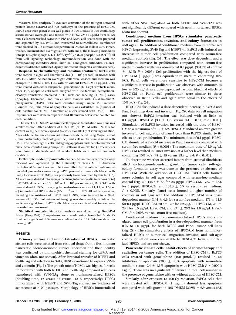
Figure 1. Immortalization of HPSCs.Primary HPSCs were exposed to lentiviralvectors carrying hTERT and SV40-TAg.Immunohistochemical staining of HPSCstransduced with hTERT and SV40-TAg foraSMA (A) and vimentin (B). C , growthcurve for primary HPSCs, HPSCstransduced with SV40-TAg, and HPSCstransduced with both hTERT andSV40-TAg. HPSCs carrying hTERT andTag show no evidence of senescenceat >100 passages.
Stromal Fibroblasts Promote Pancreatic Cancer
www.aacrjournals.org 919 Cancer Res 2008; 68: (3). February 1, 2008
Research. on March 19, 2014. © 2008 American Association for Cancercancerres.aacrjournals.org Downloaded from
Western blot analysis. To evaluate activation of the mitogen-activatedprotein kinase (MAPK) and Akt pathways in the presence of HPSC-CM,
BxPC3 cells were grown in six-well plates in 10% DMEM to 70% confluency,
serum starved overnight, and treated with HPSC-CM (1 Ag/AL) for 0 to 120
min. Cells were washed twice with PBS and lysed. Cell lysates were preparedand separated by SDS-PAGE and transferred to nitrocellulose. Membranes
were blocked for 1 h at room temperature in 5% nonfat milk in 0.1% Tween,
washed, and incubated overnight at 4jC with one of the following antibodies:
total p44/42, phospho p44/42 (Thr202/Tyr204), Akt, or phospho Akt (Ser473), allfrom Cell Signaling Technology. Immunodetection was done with the
corresponding secondary Alexa Fluor 680–conjugated antibodies. Fluores-
cence was detected with the Odyssey fluorescent imager (Li-Cor Biosciences).
Response to chemotherapy and radiation treatment. BxPC3 cellswere seeded in eight-well chamber slides (1 � 105 per well) in DMEM with
10% FCS. After incubation overnight, cells were washed and medium was
changed to DMEM + 10% FCS, with or without HPSC-CM (1 Ag/AL). Cellswere treated with either 100 Amol/L gemcitabine (Eli Lilly) or vehicle alone.
After 48 h, apoptotic cells were analyzed with the terminal deoxyribonu-
cleotidyl transferase–mediated dUTP nick end labeling (TUNEL) assay
(ApopTag, Chemicon) and nuclei were stained with 4¶,6-diamidino-2-phenylindole (DAPI). Cells were counted using Simple PCI software
(Compix, Inc.). The ratio of apoptotic cells was calculated as (number of
cells positive for TUNEL / number of nuclei stained with DAPI) � 100.
Experiments were done in duplicate and 10 random fields were counted foreach condition.
The effect of HPSC-CM on tumor cell response to radiation was done in a
similar fashion. After addition of HPSC-CM (or DMEM with 10% FCS forcontrol wells), cells were exposed to either 0 or 100 Gy of ionizing radiation.
After 24-h incubation, caspase activation was detected using Magic Red kit
(Immunochemistry Technologies, Inc.) and cell nuclei were stained with
DAPI. The percentage of cells undergoing apoptosis and the total number ofnuclei were counted using Simple PCI software (Compix, Inc.). Experiments
were done in duplicate and 10 random fields were counted for each
condition.
Orthotopic model of pancreatic cancer. All animal experiments werereviewed and approved by the University of Texas M. D. Anderson
Institutional Animal Care and Use Committee. An orthotopic nude mouse
model of pancreatic cancer using BxPC3 pancreatic tumor cells labeled withfirefly luciferase (BxPC3-FL) has previously been described by this lab (15).
All mice were divided into groups receiving intrapancreatic injections of (a)
BxPC3-FL alone, either 0.5 � 106 or 1 � 106 per mouse; (b) BxPC3-FL with
immortalized HPSCs, in varying tumor-to-stroma ratios (1:1, 1:1, or 1:5); or(c) immortalized HPSCs alone (0.5 � 106 or 1 � 106). All cell suspensions,
including the mixtures of BxPC3 and HPSCs, were injected in a 50-AL
volume of HBSS. Bioluminescent imaging was done weekly to follow the
luciferase signal from BxPC3 cells. Mice were sacrificed and tumors wereharvested and measured.
Statistical analysis. Statistical analysis was done using GraphPad
Prism (GraphPad). Comparisons were made using two-tailed Student’s
t test and significant difference was defined as P < 0.05. Data are shown asmean F SE.
Results
Primary culture and immortalization of HPSCs. Pancreaticstellate cells were isolated from residual tissue from a fresh humanpancreatic adenocarcinoma surgical specimen and their identitywas confirmed by immunohistochemical staining for aSMA andvimentin (data not shown). After lentiviral transfer of hTERT andSV40-TAg and selection in G418, HPSCs continued to express aSMAand vimentin (Fig. 1). The growth rate of HPSCs was highest for cellsimmortalized with both hTERT and SV40-TAg compared with cellstransduced with SV40-TAg alone or nonimmortalized HPSCs(doubling time, 12 versus 42 versus 113 h, respectively). HPSCsimmortalized with hTERT and SV40-Tag showed no evidence ofsenescence at >100 passages. Morphology of HPSCs immortalized
with either SV40 Tag alone or both hTERT and SV40-TAg wasnot significantly different compared with nonimmortalized HPSCs(data not shown).Conditioned medium from HPSCs stimulates pancreatic
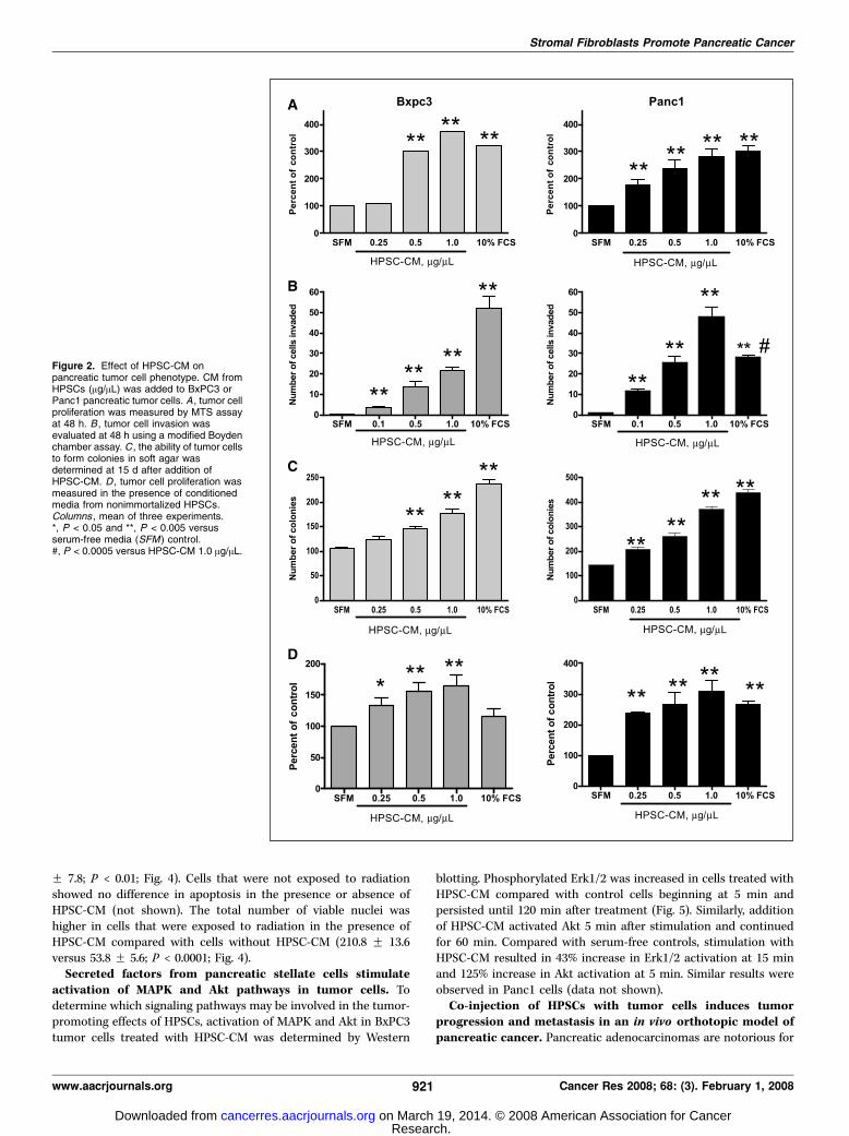
tumor cell proliferation, invasion, and colony formation insoft agar. The addition of conditioned medium from immortalizedHPSCs (expressing SV40 Tag and hTERT) to BxPC3 cells induced anincrease in tumor cell proliferation compared with serum-freemedium controls (Fig. 2A). The effect was dose dependent and asignificant increase in proliferation compared with serum-freemedium control wells was observed at 0.5 Ag/AL (301.7% of controlF 43.1%; P < 0.005). Cell proliferation with the highest dose ofHPSC-CM (1 Ag/AL) was equivalent to medium containing 10%FCS. Panc1 cells were more sensitive to HPSC-CM because asignificant increase in proliferation was observed with amounts aslow as 0.25 Ag/AL in a dose-dependent fashion. Maximal effects ofHPSC-CM on Panc1 cell proliferation were similar to thoseobserved in BxPC3 cells and again were equal to the effects of10% FCS (Fig. 2A).
HPSC-CM also induced a dose-dependent increase in BxPC3 andPanc1 cell migration and invasion (Fig. 2B ; data on cell migrationnot shown). BxPC3 invasion was induced with as little as0.1 Ag/AL HPSC-CM [3.4 F 1.78 versus 0.4 F 0.51; P < 0.0001].Stimulation of BxPC3 invasion increased with the dose of HPSC-CM to a maximum of 21.5 F 6.2. HPSC-CM induced an even greaterincrease in cell migration of Panc1 cells than BxPC3, similar to itseffects on cell proliferation. The minimum dose of 0.1 Ag/AL HPSC-CM stimulated a 19-fold increase in Panc1 invasion compared withserum-free medium (P < 0.0001). The maximum dose of 1.0 Ag/ALHPSC-CM resulted in Panc1 invasion at a higher level than mediumcontaining 10% FCS (48 F 15 versus 28.3 F 3.4; P < 0.001).
To determine whether secreted factors from stromal fibroblastsaffect anchorage-independent growth of tumor cells, soft-agarcolony formation assay was done in the presence or absence ofHPSC-CM. With the addition of HPSC-CM, BxPC3 cells formedmore colonies in soft agar compared with serum-free mediumcontrol (Fig. 2C ; 146.7 F 7.6 for 0.5 Ag/AL HPSC-CM, 178.7 F 13.6for 1 Ag/AL HPSC-CM, and 105.3 F 5.5 for serum-free medium;P < 0.005). Similarly, Panc1 cells formed a higher number ofcolonies in soft agar with the addition of HPSC-CM in a dose-dependent manner (144 F 6.6 for serum-free medium, 171 F 11.5for 0.1 Ag/AL HPSC-CM, 209 F 12.7 for 0.25 Ag/AL HPSC-CM, 261 F23.1 for 0.5 Ag/AL HPSC-CM, and 371 F 20.2 for 1.0 Ag/AL HPSC-CM; P < 0.005, versus serum-free medium).
Conditioned medium from nonimmortalized HPSCs also stim-ulated tumor cell proliferation in a dose-dependent manner, from0.25 to 1.0 Ag/AL for both BxPC3 and Panc1 tumor cell lines(Fig. 2D). The stimulatory effects of HPSC-CM from nonimmor-talized HPSCs on tumor cell migration, invasion, and soft-agarcolony formation were comparable to HPSC-CM from immortal-ized HPSCs and are not shown.Pancreatic stellate cells inhibit effects of chemotherapy and
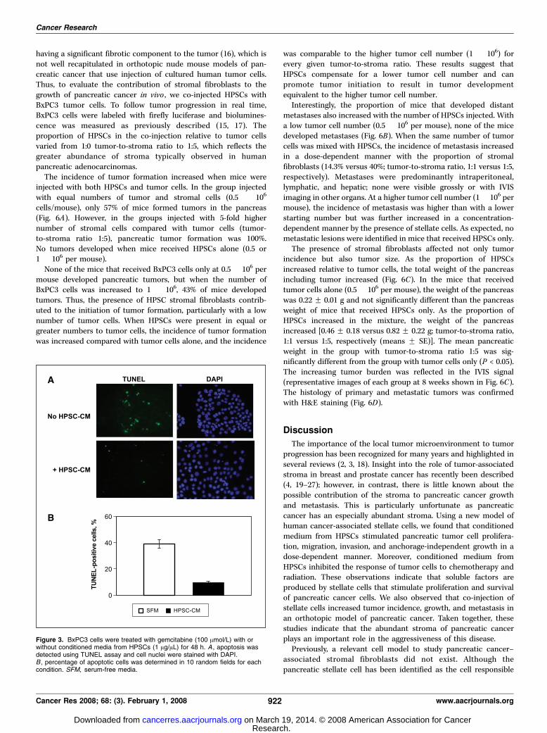
radiation on tumor cells. The addition of HPSC-CM to BxPC3cells treated with gemcitabine (100 Amol/L) resulted in aninhibition of apoptosis (38.9 F 3.1% apoptosis with serum-freemedium versus 9.4 F 1.1% apoptosis with HPSC-CM; P < 0.0001;Fig. 3). There was no significant difference in total cell number inthe presence of gemcitabine with or without addition of HPSC-CM.
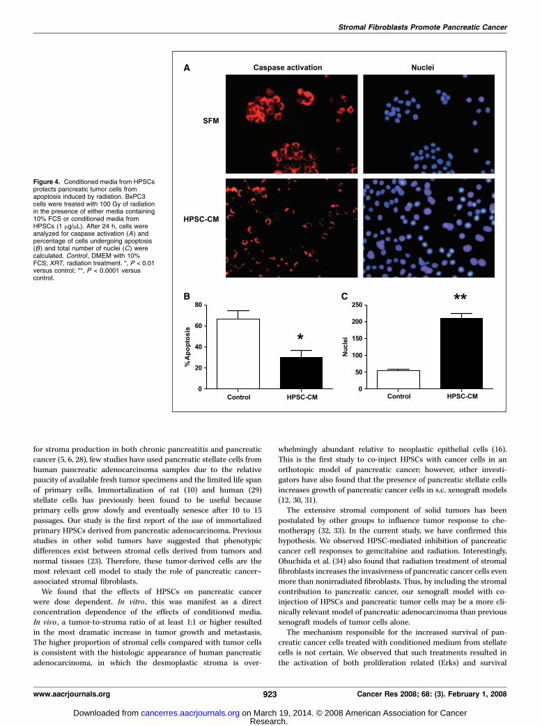
Similarly, after exposure to 100-Gy radiation, BxPC3 cells thatwere treated with HPSC-CM (1 Ag/AL) showed less apoptosiscompared with cells grown in 10% DMEM (29.9% F 6.9 versus 66.8
Cancer Research
Cancer Res 2008; 68: (3). February 1, 2008 920 www.aacrjournals.org
Research. on March 19, 2014. © 2008 American Association for Cancercancerres.aacrjournals.org Downloaded from
F 7.8; P < 0.01; Fig. 4). Cells that were not exposed to radiationshowed no difference in apoptosis in the presence or absence ofHPSC-CM (not shown). The total number of viable nuclei washigher in cells that were exposed to radiation in the presence ofHPSC-CM compared with cells without HPSC-CM (210.8 F 13.6versus 53.8 F 5.6; P < 0.0001; Fig. 4).Secreted factors from pancreatic stellate cells stimulate
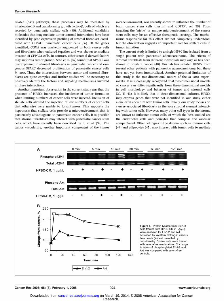
activation of MAPK and Akt pathways in tumor cells. Todetermine which signaling pathways may be involved in the tumor-promoting effects of HPSCs, activation of MAPK and Akt in BxPC3tumor cells treated with HPSC-CM was determined by Western
blotting. Phosphorylated Erk1/2 was increased in cells treated withHPSC-CM compared with control cells beginning at 5 min andpersisted until 120 min after treatment (Fig. 5). Similarly, additionof HPSC-CM activated Akt 5 min after stimulation and continuedfor 60 min. Compared with serum-free controls, stimulation withHPSC-CM resulted in 43% increase in Erk1/2 activation at 15 minand 125% increase in Akt activation at 5 min. Similar results wereobserved in Panc1 cells (data not shown).Co-injection of HPSCs with tumor cells induces tumor
progression and metastasis in an in vivo orthotopic model ofpancreatic cancer. Pancreatic adenocarcinomas are notorious for
Figure 2. Effect of HPSC-CM onpancreatic tumor cell phenotype. CM fromHPSCs (Ag/AL) was added to BxPC3 orPanc1 pancreatic tumor cells. A , tumor cellproliferation was measured by MTS assayat 48 h. B , tumor cell invasion wasevaluated at 48 h using a modified Boydenchamber assay. C , the ability of tumor cellsto form colonies in soft agar wasdetermined at 15 d after addition ofHPSC-CM. D , tumor cell proliferation wasmeasured in the presence of conditionedmedia from nonimmortalized HPSCs.Columns , mean of three experiments.*, P < 0.05 and **, P < 0.005 versusserum-free media (SFM) control.#, P < 0.0005 versus HPSC-CM 1.0 Ag/AL.
Stromal Fibroblasts Promote Pancreatic Cancer
www.aacrjournals.org 921 Cancer Res 2008; 68: (3). February 1, 2008
Research. on March 19, 2014. © 2008 American Association for Cancercancerres.aacrjournals.org Downloaded from
having a significant fibrotic component to the tumor (16), which isnot well recapitulated in orthotopic nude mouse models of pan-creatic cancer that use injection of cultured human tumor cells.Thus, to evaluate the contribution of stromal fibroblasts to thegrowth of pancreatic cancer in vivo , we co-injected HPSCs withBxPC3 tumor cells. To follow tumor progression in real time,BxPC3 cells were labeled with firefly luciferase and biolumines-cence was measured as previously described (15, 17). Theproportion of HPSCs in the co-injection relative to tumor cellsvaried from 1:0 tumor-to-stroma ratio to 1:5, which reflects thegreater abundance of stroma typically observed in humanpancreatic adenocarcinomas.
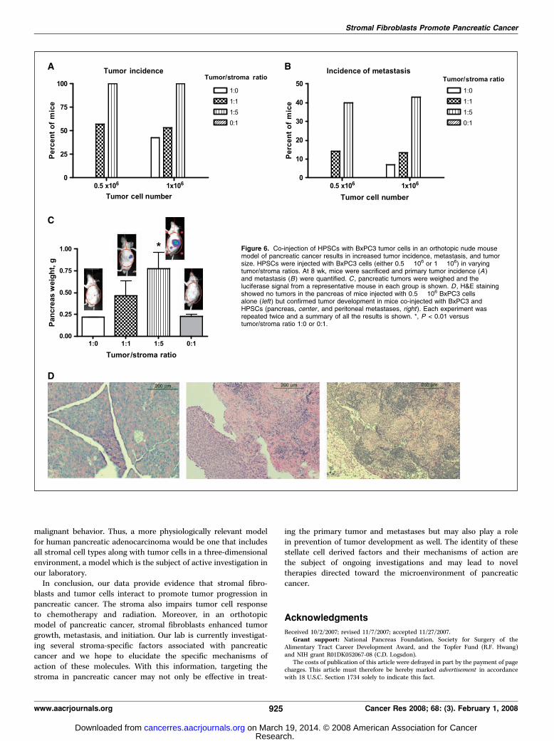
The incidence of tumor formation increased when mice wereinjected with both HPSCs and tumor cells. In the group injectedwith equal numbers of tumor and stromal cells (0.5 � 106
cells/mouse), only 57% of mice formed tumors in the pancreas(Fig. 6A). However, in the groups injected with 5-fold highernumber of stromal cells compared with tumor cells (tumor-to-stroma ratio 1:5), pancreatic tumor formation was 100%.No tumors developed when mice received HPSCs alone (0.5 or1 � 106 per mouse).
None of the mice that received BxPC3 cells only at 0.5 � 106 permouse developed pancreatic tumors, but when the number ofBxPC3 cells was increased to 1 � 106, 43% of mice developedtumors. Thus, the presence of HPSC stromal fibroblasts contrib-uted to the initiation of tumor formation, particularly with a lownumber of tumor cells. When HPSCs were present in equal orgreater numbers to tumor cells, the incidence of tumor formationwas increased compared with tumor cells alone, and the incidence
was comparable to the higher tumor cell number (1 � 106) forevery given tumor-to-stroma ratio. These results suggest thatHPSCs compensate for a lower tumor cell number and canpromote tumor initiation to result in tumor developmentequivalent to the higher tumor cell number.
Interestingly, the proportion of mice that developed distantmetastases also increased with the number of HPSCs injected. Witha low tumor cell number (0.5 � 106 per mouse), none of the micedeveloped metastases (Fig. 6B). When the same number of tumorcells was mixed with HPSCs, the incidence of metastasis increasedin a dose-dependent manner with the proportion of stromalfibroblasts (14.3% versus 40%; tumor-to-stroma ratio, 1:1 versus 1:5,respectively). Metastases were predominantly intraperitoneal,lymphatic, and hepatic; none were visible grossly or with IVISimaging in other organs. At a higher tumor cell number (1 � 106 permouse), the incidence of metastasis was higher than with a lowerstarting number but was further increased in a concentration-dependent manner by the presence of stellate cells. As expected, nometastatic lesions were identified in mice that received HPSCs only.
The presence of stromal fibroblasts affected not only tumorincidence but also tumor size. As the proportion of HPSCsincreased relative to tumor cells, the total weight of the pancreasincluding tumor increased (Fig. 6C). In the mice that receivedtumor cells alone (0.5 � 106 per mouse), the weight of the pancreaswas 0.22 F 0.01 g and not significantly different than the pancreasweight of mice that received HPSCs only. As the proportion ofHPSCs increased in the mixture, the weight of the pancreasincreased [0.46 F 0.18 versus 0.82 F 0.22 g; tumor-to-stroma ratio,1:1 versus 1:5, respectively (means F SE)]. The mean pancreaticweight in the group with tumor-to-stroma ratio 1:5 was sig-nificantly different from the group with tumor cells only (P < 0.05).The increasing tumor burden was reflected in the IVIS signal(representative images of each group at 8 weeks shown in Fig. 6C).The histology of primary and metastatic tumors was confirmedwith H&E staining (Fig. 6D).
Discussion
The importance of the local tumor microenvironment to tumorprogression has been recognized for many years and highlighted inseveral reviews (2, 3, 18). Insight into the role of tumor-associatedstroma in breast and prostate cancer has recently been described(4, 19–27); however, in contrast, there is little known about thepossible contribution of the stroma to pancreatic cancer growthand metastasis. This is particularly unfortunate as pancreaticcancer has an especially abundant stroma. Using a new model ofhuman cancer-associated stellate cells, we found that conditionedmedium from HPSCs stimulated pancreatic tumor cell prolifera-tion, migration, invasion, and anchorage-independent growth in adose-dependent manner. Moreover, conditioned medium fromHPSCs inhibited the response of tumor cells to chemotherapy andradiation. These observations indicate that soluble factors areproduced by stellate cells that stimulate proliferation and survivalof pancreatic cancer cells. We also observed that co-injection ofstellate cells increased tumor incidence, growth, and metastasis inan orthotopic model of pancreatic cancer. Taken together, thesestudies indicate that the abundant stroma of pancreatic cancerplays an important role in the aggressiveness of this disease.
Previously, a relevant cell model to study pancreatic cancer–associated stromal fibroblasts did not exist. Although thepancreatic stellate cell has been identified as the cell responsible
Figure 3. BxPC3 cells were treated with gemcitabine (100 Amol/L) with orwithout conditioned media from HPSCs (1 Ag/AL) for 48 h. A , apoptosis wasdetected using TUNEL assay and cell nuclei were stained with DAPI.B , percentage of apoptotic cells was determined in 10 random fields for eachcondition. SFM, serum-free media.
Cancer Research
Cancer Res 2008; 68: (3). February 1, 2008 922 www.aacrjournals.org
Research. on March 19, 2014. © 2008 American Association for Cancercancerres.aacrjournals.org Downloaded from
for stroma production in both chronic pancreatitis and pancreaticcancer (5, 6, 28), few studies have used pancreatic stellate cells fromhuman pancreatic adenocarcinoma samples due to the relativepaucity of available fresh tumor specimens and the limited life spanof primary cells. Immortalization of rat (10) and human (29)stellate cells has previously been found to be useful becauseprimary cells grow slowly and eventually senesce after 10 to 15passages. Our study is the first report of the use of immortalizedprimary HPSCs derived from pancreatic adenocarcinoma. Previousstudies in other solid tumors have suggested that phenotypicdifferences exist between stromal cells derived from tumors andnormal tissues (23). Therefore, these tumor-derived cells are themost relevant cell model to study the role of pancreatic cancer–associated stromal fibroblasts.
We found that the effects of HPSCs on pancreatic cancerwere dose dependent. In vitro , this was manifest as a directconcentration dependence of the effects of conditioned media.In vivo , a tumor-to-stroma ratio of at least 1:1 or higher resultedin the most dramatic increase in tumor growth and metastasis.The higher proportion of stromal cells compared with tumor cellsis consistent with the histologic appearance of human pancreaticadenocarcinoma, in which the desmoplastic stroma is over-
whelmingly abundant relative to neoplastic epithelial cells (16).This is the first study to co-inject HPSCs with cancer cells in anorthotopic model of pancreatic cancer; however, other investi-gators have also found that the presence of pancreatic stellate cellsincreases growth of pancreatic cancer cells in s.c. xenograft models(12, 30, 31).
The extensive stromal component of solid tumors has beenpostulated by other groups to influence tumor response to che-motherapy (32, 33). In the current study, we have confirmed thishypothesis. We observed HPSC-mediated inhibition of pancreaticcancer cell responses to gemcitabine and radiation. Interestingly,Ohuchida et al. (34) also found that radiation treatment of stromalfibroblasts increases the invasiveness of pancreatic cancer cells evenmore than nonirradiated fibroblasts. Thus, by including the stromalcontribution to pancreatic cancer, our xenograft model with co-injection of HPSCs and pancreatic tumor cells may be a more cli-nically relevant model of pancreatic adenocarcinoma than previousxenograft models of tumor cells alone.
The mechanism responsible for the increased survival of pan-creatic cancer cells treated with conditioned medium from stellatecells is not certain. We observed that such treatments resulted inthe activation of both proliferation related (Erks) and survival
Figure 4. Conditioned media from HPSCsprotects pancreatic tumor cells fromapoptosis induced by radiation. BxPC3cells were treated with 100 Gy of radiationin the presence of either media containing10% FCS or conditioned media fromHPSCs (1 Ag/AL). After 24 h, cells wereanalyzed for caspase activation (A) andpercentage of cells undergoing apoptosis(B) and total number of nuclei (C) werecalculated. Control , DMEM with 10%FCS; XRT, radiation treatment. *, P < 0.01versus control; **, P < 0.0001 versuscontrol.
Stromal Fibroblasts Promote Pancreatic Cancer
www.aacrjournals.org 923 Cancer Res 2008; 68: (3). February 1, 2008
Research. on March 19, 2014. © 2008 American Association for Cancercancerres.aacrjournals.org Downloaded from
related (Akt) pathways; these processes may be mediated byinterleukin-1h and transforming growth factor-h, both of which aresecreted by pancreatic stellate cells (35). Additional candidatemolecules that may mediate tumor-stromal interactions have beenidentified by gene expression profiling of stromal fibroblast cocul-tured with CFPAC1 pancreatic cancer cells (36). Of the genesidentified, COX-2 was markedly augmented in both cancer cellsand fibroblasts when cultured together and was shown to mediateinvasion of CFPAC1 cells. In contrast, other stromal-derived factorsmay suppress tumor growth. Sato et al. (37) found that SPARC wasoverexpressed in stromal fibroblasts in pancreatic cancer and exo-genous SPARC decreased proliferation of pancreatic cancer cellsin vitro . Thus, the interactions between tumor and stromal fibro-blasts are quite complex and further studies will be necessary topositively identify the factors and signaling mechanisms involvedin these interactions.
Another important observation in the current study was that thepresence of HPSCs increased the incidence of tumor formationwhen limiting numbers of cancer cells were injected. Inclusion ofstellate cells allowed the injection of low numbers of cancer cellsthat otherwise were unable to form tumors. This supports thehypothesis that stellate cells provide a microenvironment that isparticularly advantageous to pancreatic cancer cells. It is possiblethat stromal fibroblasts may interact with pancreatic cancer stemcells, which have recently been described by Li et al. (38). Thetumor vasculature, another important component of the tumor
microenvironment, was recently shown to influence the number ofbrain cancer stem cells (nestin+ and CD133+; ref. 39). Thus,targeting the ‘‘niche’’ or unique microenvironment of the cancerstem cells may be an effective therapeutic strategy. The mecha-nisms responsible for this effect are not completely understood,but the observation suggests an important role for stellate cells intumor initiation.
The current study is limited to a single HPSC line isolated from asingle patient with pancreatic adenocarcinoma. The effects ofstromal fibroblasts from different individuals may vary, as has beenshown in prostate cancer (40). Our lab has isolated HPSCs fromseveral other patients with pancreatic adenocarcinoma but thesehave not yet been immortalized. Another potential limitation ofthis study is the two-dimensional nature of the in vitro experi-ments. It is increasingly recognized that two-dimensional modelsof cancer can differ significantly from three-dimensional modelsin cell morphology and behavior of tumor and stromal cells(20, 41–43). It is likely that in three-dimensional cultures, HPSCsmay express genes that were not identified in our study, eitheralone or in coculture with tumor cells. Finally, our study focuses oncancer-associated fibroblasts as the sole stromal element interact-ing with tumor cells. However, many other cell types in the stromaare known to influence tumor cells, of which the best studied arethe endothelial cells and pericytes that compose the vascularcompartment. Other cell types in the stroma, such as immune cells(44) and adipocytes (45), also interact with tumor cells to mediate
Figure 5. Protein lysates from BxPC3cells treated with HPSC-CM (1 Ag/AL)were analyzed for Erk1/2 and Aktactivation by Western blotting at varioustime points (A) and quantifed bydensitometry. Control cells were treatedwith serum-free media alone. B , changein levels of phosphorylated Erk1/2 andAkt was compared with serum-freecontrols.
Cancer Research
Cancer Res 2008; 68: (3). February 1, 2008 924 www.aacrjournals.org
Research. on March 19, 2014. © 2008 American Association for Cancercancerres.aacrjournals.org Downloaded from
malignant behavior. Thus, a more physiologically relevant modelfor human pancreatic adenocarcinoma would be one that includesall stromal cell types along with tumor cells in a three-dimensionalenvironment, a model which is the subject of active investigation inour laboratory.
In conclusion, our data provide evidence that stromal fibro-blasts and tumor cells interact to promote tumor progression inpancreatic cancer. The stroma also impairs tumor cell responseto chemotherapy and radiation. Moreover, in an orthotopicmodel of pancreatic cancer, stromal fibroblasts enhanced tumorgrowth, metastasis, and initiation. Our lab is currently investigat-ing several stroma-specific factors associated with pancreaticcancer and we hope to elucidate the specific mechanisms ofaction of these molecules. With this information, targeting thestroma in pancreatic cancer may not only be effective in treat-
ing the primary tumor and metastases but may also play a rolein prevention of tumor development as well. The identity of thesestellate cell derived factors and their mechanisms of action arethe subject of ongoing investigations and may lead to noveltherapies directed toward the microenvironment of pancreaticcancer.
Acknowledgments
Received 10/2/2007; revised 11/7/2007; accepted 11/27/2007.Grant support: National Pancreas Foundation, Society for Surgery of the
Alimentary Tract Career Development Award, and the Topfer Fund (R.F. Hwang)and NIH grant R01DK052067-08 (C.D. Logsdon).
The costs of publication of this article were defrayed in part by the payment of pagecharges. This article must therefore be hereby marked advertisement in accordancewith 18 U.S.C. Section 1734 solely to indicate this fact.
Figure 6. Co-injection of HPSCs with BxPC3 tumor cells in an orthotopic nude mousemodel of pancreatic cancer results in increased tumor incidence, metastasis, and tumorsize. HPSCs were injected with BxPC3 cells (either 0.5 � 106 or 1 � 106) in varyingtumor/stroma ratios. At 8 wk, mice were sacrificed and primary tumor incidence (A )and metastasis (B ) were quantified. C , pancreatic tumors were weighed and theluciferase signal from a representative mouse in each group is shown. D , H&E stainingshowed no tumors in the pancreas of mice injected with 0.5 � 106 BxPC3 cellsalone (left ) but confirmed tumor development in mice co-injected with BxPC3 andHPSCs (pancreas, center , and peritoneal metastases, right ). Each experiment wasrepeated twice and a summary of all the results is shown. *, P < 0.01 versustumor/stroma ratio 1:0 or 0:1.
Stromal Fibroblasts Promote Pancreatic Cancer
www.aacrjournals.org 925 Cancer Res 2008; 68: (3). February 1, 2008
Research. on March 19, 2014. © 2008 American Association for Cancercancerres.aacrjournals.org Downloaded from
Cancer Research
Cancer Res 2008; 68: (3). February 1, 2008 926 www.aacrjournals.org
References
1. Evans DB, Abbruzzese J, Willett C. Cancer of thepancreas. In: Devita V, Jr., Hellman S, Rosenberg S,editors. Cancer: principles and practice of oncology.Philadelphia: Lippincott; 2001. p. 1126–61.2. Liotta LA, Kohn EC. The microenvironment of the
tumour-host interface. Nature 2001;411:375–9.3. Fidler IJ. The organ microenvironment and cancer
metastasis. Differentiation 2002;70:498–505.4. De Wever O, Mareel M. Role of tissue stroma in cancer
cell invasion. J Pathol 2003;200:429–47.5. Apte MV, Haber PS, Applegate TL, et al. Periacinar
stellate shaped cells in rat pancreas: identification,isolation, and culture. Gut 1998;43:128–33.6. Apte MV, Park S, Phillips PA, et al. Desmoplastic
reaction in pancreatic cancer: role of pancreatic stellatecells. Pancreas 2004;29:179–87.7. Blomhoff R, Wake K. Perisinusoidal stellate cells of the
liver: important roles in retinol metabolism and fibrosis.FASEB J 1991;5:271–7.8. de Leeuw AM, McCarthy SP, Geerts A, Knook DL.
Purified rat liver fat-storing cells in culture divide andcontain collagen. Hepatology 1984;4:392–403.9. Yokoi Y, Namihisa T, Kuroda H, et al. Immunocyto-
chemical detection of desmin in fat-storing cells (Itocells). Hepatology 1984;4:709–14.10. Sparmann G, Hohenadl C, Tornoe J, et al. Generation
and characterization of immortalized rat pancreaticstellate cells. Am J Physiol Gastrointest Liver Physiol2004;287:G211–9.11. Phillips PA, McCarroll JA, Park S, et al. Rat pancreatic
stellate cells secrete matrix metalloproteinases: impli-cations for extracellular matrix turnover. Gut 2003;52:275–82.12. Bachem MG, Schunemann M, Ramadani M, et al.
Pancreatic carcinoma cells induce fibrosis by stimulat-ing proliferation and matrix synthesis of stellate cells.Gastroenterology 2005;128:907–21.13. Erkan M, Kleeff J, Reiser C, et al. Preoperative acute
pancreatitis in periampullary tumors: implications forsurgical management. Digestion 2007;75:165–71.14. Kowolik CM, Liang S, Yu Y, Yee JK. Cre-mediated
reversible immortalization of human renal proximaltubular epithelial cells. Oncogene 2004;23:5950–7.15. Amos K, Moore T, Arumugam T, Logsdon C, Hwang
RF. Pancreatic stellate cells promote malignant poten-tial in pancreatic cancer. In: 97th Annual Meeting,American Association for Cancer Research, Washington,D.C., 2006.16. Seymour AB, Hruban RH, Redston M, et al. Allelotype
of pancreatic adenocarcinoma. Cancer Res 1994;54:2761–4.17. Arumugam T, Simeone DM, Van Golen K, Logsdon
CD. S100P promotes pancreatic cancer growth, survival,and invasion. Clin Cancer Res 2005;11:5356–64.18. Kalluri R, Zeisberg M. Fibroblasts in cancer. Nat Rev
Cancer 2006;6:392–401.19. Park CC, Bissell MJ, Barcellos-Hoff MH. The influence
of the microenvironment on the malignant phenotype.Mol Med Today 2000;6:324–9.20. Weaver VM, Petersen OW, Wang F, et al. Reversion of
the malignant phenotype of human breast cells in three-dimensional culture and in vivo by integrin blockingantibodies. J Cell Biol 1997;137:231–45.21. Shekhar MP, Werdell J, Santner SJ, Pauley RJ, Tait L.
Breast stroma plays a dominant regulatory role in breastepithelial growth and differentiation: implications fortumor development and progression. Cancer Res 2001;61:1320–6.22. Tuxhorn JA, Ayala GE, Smith MJ, Smith VC, Dang TD,
Rowley DR. Reactive stroma in human prostate cancer:induction of myofibroblast phenotype and extracellularmatrix remodeling. Clin Cancer Res 2002;8:2912–23.23. Olumi AF, Grossfeld GD, Hayward SW, Carroll PR,
Tlsty TD, Cunha GR. Carcinoma-associated fibroblastsdirect tumor progression of initiated human prostaticepithelium. Cancer Res 1999;59:5002–11.24. Cunha GR, Hayward SW, Wang YZ. Role of stroma in
carcinogenesis of the prostate. Differentiation 2002;70:473–85.25. Bhowmick NA, Chytil A, Plieth D, et al. TGF-h
signaling in fibroblasts modulates the oncogenic poten-tial of adjacent epithelia. Science 2004;303:848–51.26. Tlsty TD. Stromal cells can contribute oncogenic
signals. Semin Cancer Biol 2001;11:97–104.27. Bissell MJ, Radisky DC, Rizki A, Weaver VM, Petersen
OW. The organizing principle: microenvironmentalinfluences in the normal and malignant breast. Differ-entiation 2002;70:537–46.28. Bachem MG, Schneider E, Gross H, et al. Identification,
culture, and characterization of pancreatic stellate cells inrats and humans. Gastroenterology 1998;115:421–32.29. Jesnowski R, Furst D, Ringel J, et al. Immortalization
of pancreatic stellate cells as an in vitro model ofpancreatic fibrosis: deactivation is induced by Matrigeland N-acetylcysteine. Lab Invest 2005;85:1276–91.30. Neesse A, Wagner M, Ellenrieder V, Bachem M, Gress
TM, Buchholz M. Pancreatic stellate cells potentiateproinvasive effects of SERPINE2 expression in pancreaticcancer xenograft tumors. Pancreatology 2007;7:380–5.31. Schneiderhan W, Diaz F, Fundel M, et al. Pancreatic
stellate cells are an important source of MMP-2 inhuman pancreatic cancer and accelerate tumor pro-gression in a murine xenograft model and CAM assay. JCell Sci 2007;120:512–9.32. Miyamoto H, Murakami T, Tsuchida K, Sugino H,
Miyake H, Tashiro S. Tumor-stroma interaction of
human pancreatic cancer: acquired resistance toanticancer drugs and proliferation regulation is depen-dent on extracellular matrix proteins. Pancreas 2004;28:38–44.33. Muerkoster S, Wegehenkel K, Arlt A, et al. Tumor
stroma interactions induce chemoresistance in pancre-atic ductal carcinoma cells involving increased secretionand paracrine effects of nitric oxide and interleukin-1h.Cancer Res 2004;64:1331–7.34. Ohuchida K, Mizumoto K, Murakami M, et al.
Radiation to stromal fibroblasts increases invasivenessof pancreatic cancer cells through tumor-stromalinteractions. Cancer Res 2004;64:3215–22.35. Aoki H, Ohnishi H, Hama K, et al. Autocrine loop
between TGF-h1 and IL-1h through Smad3- and ERK-dependent pathways in rat pancreatic stellate cells. Am JPhysiol Cell Physiol 2006;290:C1100–8.36. Sato N, Maehara N, Goggins M. Gene expression
profiling of tumor-stromal interactions between pan-creatic cancer cells and stromal fibroblasts. Cancer Res2004;64:6950–6.37. Sato N, Fukushima N, Maehara N, et al. SPARC/
osteonectin is a frequent target for aberrant methylationin pancreatic adenocarcinoma and a mediator of tumor-stromal interactions. Oncogene 2003;22:5021–30.38. Li C, Heidt DG, Dalerba P, et al. Identification of
pancreatic cancer stem cells. Cancer Res 2007;67:1030–7.39. Calabrese C, Poppleton H, Kocak M, et al. A
perivascular niche for brain tumor stem cells. CancerCell 2007;11:69–82.40. Tuxhorn JA, McAlhany SJ, Dang TD, Ayala GE,
Rowley DR. Stromal cells promote angiogenesis andgrowth of human prostate tumors in a differentialreactive stroma (DRS) xenograft model. Cancer Res2002;62:3298–307.41. Yamada KM, Cukierman E. Modeling tissue morpho-
genesis and cancer in 3D. Cell 2007;130:601–10.42. Kenny HA, Krausz T, Yamada SD, Lengyel E. Use of a
novel 3D culture model to elucidate the role ofmesothelial cells, fibroblasts and extra-cellular matriceson adhesion and invasion of ovarian cancer cells to theomentum. Int J Cancer 2007;121:1463–72.43. Nelson CM, Bissell MJ. Modeling dynamic reciprocity:
engineering three-dimensional culture models of breastarchitecture, function, and neoplastic transformation.Semin Cancer Biol 2005;15:342–52.44. Yu H, Kortylewski M, Pardoll D. Crosstalk between
cancer and immune cells: role of STAT3 in the tumourmicroenvironment. Nat Rev Immunol 2007;7:41–51.45. Iyengar P, Espina V, Williams TW, et al. Adipocyte-
derived collagen VI affects early mammary tumorprogression in vivo , demonstrating a critical interactionin the tumor/stroma microenvironment. J Clin Invest2005;115:1163–76.
Research. on March 19, 2014. © 2008 American Association for Cancercancerres.aacrjournals.org Downloaded from
Copyright © 2022 FDOKUMEN