1H-MRS Detected Lipolysis in Diabetic Rat Hearts Requires Neutral Lipase*
12
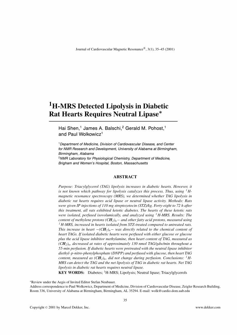
Journal of Cardiovascular Magnetic Resonance , 3(1), 35–45 (2001) 1 H-MRS Detected Lipolysis in Diabetic Rat Hearts Requires Neutral Lipase ∗ Hai Shen, 1 James A. Balschi, 2 Gerald M. Pohost, 1 and Paul Wolkowicz 1 1 Department of Medicine, Division of Cardiovascular Disease, and Center for NMR Research and Development, University of Alabama at Birmingham, Birmingham, Alabama 2 NMR Laboratory for Physiological Chemistry, Department of Medicine, Brigham and Women’s Hospital, Boston, Massachusetts ABSTRACT Purpose: Triacylglycerol (TAG) lipolysis increases in diabetic hearts. However, it is not known which pathway for lipolysis catalyzes this process. Thus, using 1 H- magnetic resonance spectroscopy (MRS), we determined whether TAG lipolysis in diabetic rat hearts requires acid lipase or neutral lipase activity. Methods: Rats were given IP injections of 110 mg streptozotocin (STZ)/kg. Forty-eight to 72 h after this treatment, all rats exhibited ketotic diabetes. The hearts of these ketotic rats were isolated, perfused isovolumically, and analyzed using 1 H-MRS. Results: The content of methylene protons (CH 2 ) n − and other fatty acid protons, measured using 1 H-MRS, increased in hearts isolated from STZ-treated compared to untreated rats. This increase in heart −(CH 2 ) n − was directly related to the chemical content of heart TAGs. If isolated diabetic hearts were perfused with either glucose or glucose plus the acid lipase inhibitor methylamine, then heart content of TAG, measured as (CH 2 ) n , decreased at rates of approximately 130 nmol TAG/gdw/min throughout a 55-min perfusion. If diabetic hearts were pretreated with the neutral lipase inhibitor diethyl-p-nitro-phenylphosphate (DNPP) and perfused with glucose, then heart TAG content, measured as (CH 2 ) n , did not change during perfusion. Conclusions: 1 H- MRS can detect the TAG and the net lipolysis of TAG in diabetic rat hearts. Net TAG lipolysis in diabetic rat hearts requires neutral lipase. KEY WORDS: Diabetes; 1 H-MRS; Lipolysis; Neutral lipase; Triacylglycerols ∗ Review under the Aegis of Invited Editor Stefan Neubauer. Address correspondence to Paul Wolkowicz, Department of Medicine, Division of Cardiovascular Disease, Zeigler Research Building, Room 336, University of Alabama at Birmingham, Birmingham, AL 35294. E-mail: [email protected] 35 Copyright C 2001 by Marcel Dekker, Inc. www.dekker.com
-
Upload
independent -
Category
Documents
-
view
4 -
download
0
Transcript of 1H-MRS Detected Lipolysis in Diabetic Rat Hearts Requires Neutral Lipase*
Journal of Cardiovascular Magnetic Resonance, 3(1), 35–45 (2001)
1H-MRS Detected Lipolysis in DiabeticRat Hearts Requires Neutral Lipase∗
Hai Shen,1 James A. Balschi,2 Gerald M. Pohost,1
and Paul Wolkowicz1
1Department of Medicine, Division of Cardiovascular Disease, and Centerfor NMR Research and Development, University of Alabama at Birmingham,Birmingham, Alabama2NMR Laboratory for Physiological Chemistry, Department of Medicine,Brigham and Women’s Hospital, Boston, Massachusetts
ABSTRACT
Purpose: Triacylglycerol (TAG) lipolysis increases in diabetic hearts. However, itis not known which pathway for lipolysis catalyzes this process. Thus, using 1 H-magnetic resonance spectroscopy (MRS), we determined whether TAG lipolysis indiabetic rat hearts requires acid lipase or neutral lipase activity. Methods: Ratswere given IP injections of 110 mg streptozotocin (STZ)/kg. Forty-eight to 72 h afterthis treatment, all rats exhibited ketotic diabetes. The hearts of these ketotic ratswere isolated, perfused isovolumically, and analyzed using 1 H-MRS. Results: Thecontent of methylene protons (CH2)n− and other fatty acid protons, measured using1 H-MRS, increased in hearts isolated from STZ-treated compared to untreated rats.This increase in heart −(CH2)n− was directly related to the chemical content ofheart TAGs. If isolated diabetic hearts were perfused with either glucose or glucoseplus the acid lipase inhibitor methylamine, then heart content of TAG, measured as(CH2)n, decreased at rates of approximately 130 nmol TAG/gdw/min throughout a55-min perfusion. If diabetic hearts were pretreated with the neutral lipase inhibitordiethyl-p-nitro-phenylphosphate (DNPP) and perfused with glucose, then heart TAGcontent, measured as (CH2)n, did not change during perfusion. Conclusions: 1 H-MRS can detect the TAG and the net lipolysis of TAG in diabetic rat hearts. Net TAGlipolysis in diabetic rat hearts requires neutral lipase.KEY WORDS: Diabetes; 1H-MRS; Lipolysis; Neutral lipase; Triacylglycerols
∗Review under the Aegis of Invited Editor Stefan Neubauer.Address correspondence to Paul Wolkowicz, Department of Medicine, Division of Cardiovascular Disease, Zeigler Research Building,Room 336, University of Alabama at Birmingham, Birmingham, AL 35294. E-mail: [email protected]
35
Copyright C© 2001 by Marcel Dekker, Inc. www.dekker.com
ORDER REPRINTS
36 Shen et al.
INTRODUCTION
Heart content of triacylglycerols increases significantlyin ketotic diabetes (1). These neutral lipids, localizedeither to membrane-bound cytoplasmic inclusions or tolysosomes (2), serve as an endogenous pool of fattyacids that can contribute significantly to heart energymetabolism (3).
Both the steady-state content of triacylglycerols in theheart and the turnover of this pool of lipids reflect theintracellular rates of free fatty acid esterification into tri-acylglycerols and the intracellular rate of triacylglycerollipolysis (2). Two distinct pathways for lipolysis exist inthe heart. The first pathway is present in lysosomes andan acid lipase activity regulates flux through this pathway(4). Lysosomotrophic agents, such as methylamine andchloroquine, inhibit acid lipase-dependent lipolysis (4).The second pathway for lipolysis is associated with the cy-toplasmic triacylglycerol-rich inclusions themselves (2).A hormone-sensitive neutral lipase regulates flux throughthis pathway (5). DNPP inhibits irreversibly the neutral li-pase and, thereby, neutral lipase-dependent lipolysis (6,7).
The acid lipase pathway is the main pathway for triacyl-glycerol lipolysis in the normal rat heart (4). In contrast,the neutral lipase pathway is the predominant pathwayfor lipolysis in hypertriglyceridemic hearts, for example,in hearts isolated from rats fed rapeseed oil (RSO) (4).The contribution of the neutral lipase and the acid li-pase pathways to lipolysis in the hypertriglyceridemichearts of severely diabetic rats, however, remains unre-solved.
Most investigations of triacylglycerol metabolism indiabetic hearts have used radiochemical or biochemicaltechniques. For example, since heart muscle contains lit-tle glycerol kinase (8), release of glycerol either fromperfused hearts or from myocytes can provide one mea-sure of cardiac triacylglycerol lipolysis (6). Likewise, tri-acylglycerol lipolysis can be measured by prelabeling theheart triacylglycerol pool with 14C-labeled fatty acids (9).While providing key pieces of information about triacyl-glycerol lipolysis in the heart, these approaches do notprovide a direct, continual assessment of the net tria-cylglycerol content of the heart. 1H- and 13C-MRS canprovide such information by quantitating the triacylglyc-erol content, rates of net triacylglycerol lipolysis, andrates of fatty acid esterification into triacylglycerols innormal hearts or in hearts isolated from rats fed RSO(10,11,12).
Using 1H-MRS methodology, therefore, we investi-gated whether net lipolysis in diabetic hearts requires
acid lipase activity, neutral lipase activity, or both. Toanswer this question, experiments were performed thatestablish for the first time that 1H-MRS can quantitatethe triacylglycerol content and measure the rate of nettriacylglycerol lipolysis in hearts isolated from diabeticrats. Furthermore, we demonstrate for the first time thatnet lipolysis in diabetic heats requires only neutral lipaseactivity.
METHODS
These investigations conform to the Guide for the Careand Use of Laboratory Animals published by the NationalInstitutes of Health (NIH publication No. 85-23, Revised1985), as enforced by the Institutional Animal Use andCare Central Committee of the University of Alabama atBirmingham.
Induction of Diabetes
Streptozotocin (STZ) was suspended in 0.5 M Na+
citrate (pH 5.4) at a concentration of 70 mg/mL. MaleSprague Dawley rats (n = 30 total) were given a singleIP injection of 110 mg STZ/kg (13). Twenty-four to 72 hfollowing treatment with STZ, one group of rats (n = 10)was sacrificed and used to measure the relationship be-tween the chemical content of heart triacylglycerol andthe heart content of 1H-MRS observable fatty acid pro-tons. The remaining rats (n = 20) were recovered 72 hfollowing treatment with STZ, and the glucose and ketonebody content of their urine was assessed using commer-cially available diagnostic chemstrips. All of these ratswere ketonuric. Their hearts were used to measure ratesof net triacylglycerol lipolysis.
Chemical Measurement of the Heart Contentof Triacylglycerol
Hearts were isolated either from rats treated with STZ(n = 10, total) for 24, 48, or 72 h, or from rats treatedwith citrate vehicle (n = 2). During this range of timesfollowing STZ treatment, a progressive increase has beenreported to occur in the triacylglycerol content of the heart(13). These hearts were perfused in a retrograde fashion(11) with glucose and acetate as exogenous substrates.1H-MRS spectra of these hearts were then acquired andanalyzed for their content of (CH2)n protons.
At the end of these perfusions, each heart was snap-frozen in liquid nitrogen and stored at −20◦C. Frozen

ORDER REPRINTS
1H-MRS Detected Lipolysis in Diabetic Rat Hearts 37
hearts were pulverized, and the total lipids from 200 to400 mg of heart powder were extracted and concentratedunder nitrogen. The triacylglycerols in these samples wereselectively saponified using tetraethyl ammonium hydrox-ide and the glycerol content of these saponified sampleswas assayed (10).
Preparation of Isolated Hearts andPerfusion Protocols
Twenty Ketotic diabetic rats were given a single IPinjection of 300 units heparin/kg. Ten to 15 min later,all rats were given a single IP injection of 0.2 g pento-barbital/kg. Under anesthesia, the hearts were removedfrom these rats, chilled in ice-cold Krebs-Henseliet (KH)buffer (19), their aortas cannulated, and their left ventri-cles vented at the apex. Retrograde perfusion was initiatedusing KH containing 1.75 mM CaCl2, with 10 mM glu-cose and 7.5 mM Na+ acetate as exogenous substrates.Acetate was used to suppress lipolysis during the sta-bilization period prior to the initiation of any specificexperimental protocol (10). All perfusates were aeratedwith 95% O2/5% CO2. All perfusions were carried out at37◦C, a perfusate pH of 7.4, and a perfusion pressure of80 mm Hg.
A latex balloon was placed in the left ventricle of allperfused hearts and sutured in place. This intraventricularballoon was connected via a fluid-filled catheter to apressure transducer. The balloon transducer assemblywas used to measure heart rate, LVEDP, and LVDP. AKeithley System 570 analog-to-digital converter (KeithleyMetrabyte, Taunton, MA) was used to digitize pressuretraces, and these were stored on a personal computer forsubsequent analysis. At the outset of all perfusions, thevolume of the balloon was adjusted so that the LVEDPof each heart reached approximately 9 mm Hg. A 4-mmglass sphere containing 25 µl of a 180-mM solution of3-(trimethylsilyl) tetradeutero sodium propionate (TSP),a 1H standard, then was positioned next to all hearts andattached to the perfusion cannula. All hearts were thenplaced into a 20-mm MRS tube and inserted into thebore of a Bruker AM 360 NMR spectrometer (BrukerInstrument Co., Billerica, MA). Hearts were allowed tostabilize for 15 to 20 min, during which time the probewas tuned and the samples were shimmed.
Four perfusion conditions were studied using heartsisolated from the 20 ketotic diabetic rats. One group of fivediabetic hearts was perfused with KH containing 10 mMglucose and 7.5 mM Na+ acetate during the stabiliza-tion period. Following the acquisition of three baseline
1H-MRS spectra, these hearts were then perfused duringthe experimental period with 10 mM glucose as sole ex-ogenous substrate. A second group of five diabetic heartswas perfused with KH containing 10 mM glucose and7.5 mM Na+ acetate during both the stabilization and theexperimental periods. A third group of five diabetic heartswas perfused with KH containing 10 mM glucose, 7.5 mMNa+ acetate, and 5 mM methylamine during the stabiliza-tion period. These hearts were then perfused with KH con-taining 10 mM glucose and 5 mM methylamine during theexperimental period. The fourth and final group of fivediabetic hearts was perfused with KH containing 10 mMglucose, 7.5 mM Na+ acetate, and 0.04 mM DNPP duringthe stabilization period, followed by perfusion with KHcontaining 10 mM glucose during the experimental period.1H-MRS spectra were acquired throughout all perfusionsin these four groups.
Hearts in the first three experimental groups were al-lowed to beat at their natural rates. Since DNPP inducesbradycardia (14), hearts in the last experimental groupwere paced at 250 beats per min.
The heart rate and the left ventricular developed pres-sure (LVDP) for each heart were multiplied to give heartrate pressure product (RPP). RPP is reported as a measureof heart mechanical performance.
1H-MRS of Perfused Hearts
1H-MRS spectra were acquired using the decouplingcoil of a 20 mm 31P-MRS probe. 1H-MRS FIDs had asweep width of 5000 Hz, 8192 data points, and 64 averagesper spectrum. Water suppressed excitation was achievedusing the 1331 pulse sequence with an interpulse delayof 400 µs, the transmitter frequency set at the water res-onance, and a 5-s delay between acquisitions (10). Com-pensation for the frequency dependent variation in trans-verse magnetization of the 1331 sequence was made usingproduct operators.
The NMR1 software package (Tripos, East Syracuse,NY) was used to process all 1H-MRS spectra. All 1H FIDswere processed using a line broadening of 3 Hz. The ex-ternal TSP standard was used to quantify both the contentof and the changes in the areas of 1H-MRS resonancesduring perfusion.
Statistics
All values are reported as means± standard error. Com-parisons between groups were made using the unpaired
ORDER REPRINTS
38 Shen et al.
Student’s t test (two tailed), and a Bonferroni correctionfor multiple comparisons. Comparison within groups usedrepeated measures ANOVA. A probability value of 0.05or less was considered statistically significant.
Materials
Streptozotocin and DNPP were from Sigma ChemicalCo. (St. Louis, MO). Chemstrips for analysis of glucosuriaand ketonuria were obtained from Boehringer Mannheim(Indianapolis, IN). All other chemicals were of standardlaboratory grade.
RESULTS
Experiments were performed to determine whether thehypertriglyceridemia that occurs in ketotic diabetic rat
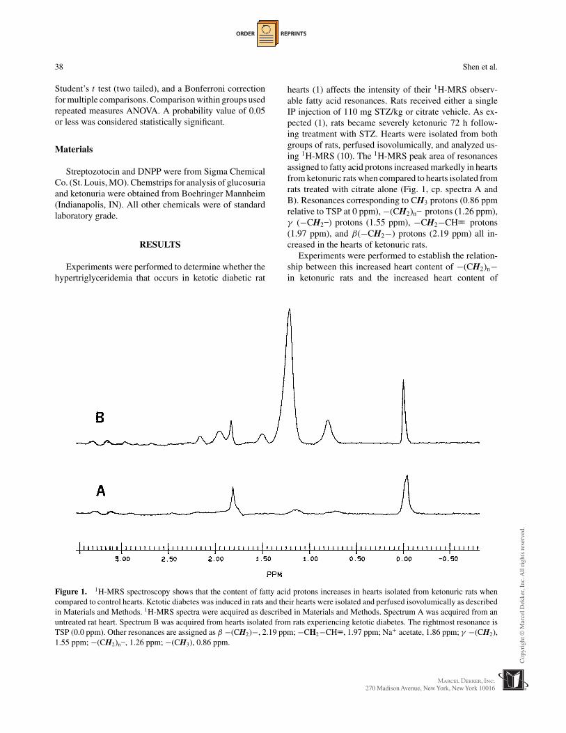
Figure 1. 1H-MRS spectroscopy shows that the content of fatty acid protons increases in hearts isolated from ketonuric rats whencompared to control hearts. Ketotic diabetes was induced in rats and their hearts were isolated and perfused isovolumically as describedin Materials and Methods. 1H-MRS spectra were acquired as described in Materials and Methods. Spectrum A was acquired from anuntreated rat heart. Spectrum B was acquired from hearts isolated from rats experiencing ketotic diabetes. The rightmost resonance isTSP (0.0 ppm). Other resonances are assigned as β −(CH2)−, 2.19 ppm; −CH2−CH==, 1.97 ppm; Na+ acetate, 1.86 ppm; γ −(CH2),1.55 ppm; −(CH2)n–, 1.26 ppm; −(CH3), 0.86 ppm.
hearts (1) affects the intensity of their 1H-MRS observ-able fatty acid resonances. Rats received either a singleIP injection of 110 mg STZ/kg or citrate vehicle. As ex-pected (1), rats became severely ketonuric 72 h follow-ing treatment with STZ. Hearts were isolated from bothgroups of rats, perfused isovolumically, and analyzed us-ing 1H-MRS (10). The 1H-MRS peak area of resonancesassigned to fatty acid protons increased markedly in heartsfrom ketonuric rats when compared to hearts isolated fromrats treated with citrate alone (Fig. 1, cp. spectra A andB). Resonances corresponding to CH3 protons (0.86 ppmrelative to TSP at 0 ppm), −(CH2)n– protons (1.26 ppm),γ (−CH2–) protons (1.55 ppm), −CH2−CH== protons(1.97 ppm), and β(−CH2−) protons (2.19 ppm) all in-creased in the hearts of ketonuric rats.
Experiments were performed to establish the relation-ship between this increased heart content of −(CH2)n−in ketonuric rats and the increased heart content of

ORDER REPRINTS
1H-MRS Detected Lipolysis in Diabetic Rat Hearts 39
triacylglycerols that occurs in ketotic diabetes (1). Ten ratswere treated with STZ and 2 rats were treated with citratealone. Twenty-four to 72 h following their treatment withSTZ or citrate, rats were sacrificed and their hearts wereperfused isovolumically. 1H-MRS spectra were acquiredfrom these hearts, and these spectra were analyzed for theintensity of their −(CH2)n− resonance. On average, thereare 64.3 mol of −(CH2)n− per mol of triacylglycerolsin diabetic hearts (15). This factor was used to converta heart content of −(CH2)n− to a heart content of tri-acylglycerols using 1H-MRS approaches that have beendescribed previously (10). The total lipids were extractedfrom these hearts, and the chemical content of triacyl-glycerols was measured in these extracts. Comparing the1H-MRS measurements of heart triacylglycerols with thechemical measurements of the actual triacylglycerol con-tent of ketonuric hearts gave a direct linear relationship(Fig. 2, m = 0.93, r = 0.92).
Two sets of experiments were performed to establishwhether the 1H-MRS observable triacylglycerols in ke-tonuric rat hearts were metabolically active. First, hearts(n = 5) isolated from ketonuric rats were perfused withglucose as the sole exogenous substrate during the experi-mental period. A time-dependent decrease occurred in thepeak area of the −(CH2)n− resonance of these hearts dur-ing the course of these perfusions (Figs. 3A and 4). Usingthe linear relationship between the heart content of triacyl-glycerols measured using 1H-MRS and the heart contentof triacylglycerols measured chemically (Fig. 2), it wasestimated that hearts isolated from ketonuric rats lipolyzed135.6 ± 8.2 nmol triacylglycerol per gram dry wt-minwhen perfused with glucose as sole exogenous sub-strate.
Second, hearts (n = 5) isolated from ketonuric ratswere perfused with glucose and acetate during both thestabilization and the experimental periods. Acetate is analternate metabolic substrate that suppresses lipolysis(1,10). The peak area of heart −(CH2)n− decreasedslowly throughout the course of these perfusions (Fig. 4).It was estimated that hearts isolated from ketonuric ratslipolyzed 21.0 ± 5.6 nmol of triacylglycerol per gramdry wt-min when perfused with glucose and acetate asexogenous substrates. These changes in heart −(CH2)n−,a direct measure of heart triacylglycerol content, werelinear (Fig. 4) and were significantly different from eachother (p < 0.05).
Two sets of experiments were performed to testwhether the acid lipase or the neutral lipase catalyzedtriacylglycerol lipolysis in hearts from ketonuric rats.First, hearts (n = 5) isolated from ketonuric rats wereperfused with glucose and methylamine during the
Figure 2. The 1H-MRS peak area of (CH2)n in the hearts ofketotic rats correlates with their chemical content of triacylglyc-erols. The content of −(CH2)n− protons in hearts of untreatedrats (�) and hearts from STZ-treated rats (�) was measured us-ing 1H-MRS as described in Materials and Methods. 1H-MRS(CH2)n resonance peak area was converted to a 1H-MRS mea-sured triacylglycerol content (left axis) as described in Meth-ods. The chemical content of triacylglycerol in these hearts wasdetermined as described in Methods. The data from these twomeasurements fit a line with r equal to 0.92, and a slope of 0.93.
experimental period. Methylamine is an inhibitor of acidlipase activity (4). A time-dependent decrease occurred inthe −(CH2)n− peak area of ketonuric hearts throughoutthe course of perfusion with glucose and methylamine(Figs. 3B and 4). It was estimated that ketonuric heartslipolyzed 129.4 ± 11.1 nmol triacylglycerol per gram drywt-min during perfusion with glucose and methylamine.The rate of (CH2)n decrease in hearts perfused withglucose and methylamine was not different from ratesmeasured in hearts perfused with glucose alone.
Second, hearts (n = 5) from ketonuric rats wereperfused with glucose, acetate, and DNPP during thestabilization period, and then with glucose alone duringthe experimental period. DNPP is a potent inhibitor ofesterases such as neutral lipase (7,14). The −(CH2)n−peak area of glucose-perfused, ketonuric hearts that hadbeen pretreated with DNPP decreased much less rapidlycompared to ketonuric hearts perfused with glucose andmethylamine (cp. Figs. 3B, 3C, and 4). It was estimatedthat hearts isolated from ketonuric rats and pretreated with0.04 mM DNPP lipolyzed 2.5 ± 6.6 nmol triacylglycerolper gram dry wt-min when perfused with glucose as soleexogenous substrate. The rate of (CH2)n decrease in theseDNPP-treated, glucose-perfused hearts was significantly
ORDER REPRINTS
40 Shen et al.
different from the rate measured for hearts perfused withglucose and methylamine (p < 0.05).
The mechanical performance of these four groups of di-abetic hearts was measured throughout perfusion. Follow-ing 100 total min of perfusion, that is, at the end of the sta-bilization and the experimental periods, the RPP of heartsperfused with glucose was 18.6 ± 1.8 kmm Hg/min, whilethe RPP of hearts perfused with glucose and acetate was19.7 ± 2.9 kmm Hg/min. These values were not statisti-cally different. The RPP for hearts perfused with glucoseand methylamine was 18.7 ± 1.8 kmm Hg/min, while itwas 18.1 ± 2.1 kmm Hg/min for hearts perfused withglucose and DNPP. These values were not statisticallydifferent. In addition, these values were not significantlydifferent from the RPPs measured in the first 20 min of
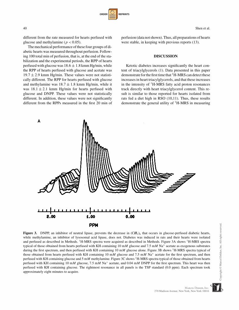
Figure 3. DNPP, an inhibitor of neutral lipase, prevents the decrease in (CH2)n that occurs in glucose-perfused diabetic hearts,while methylamine, an inhibitor of lysosomal acid lipase, does not. Diabetes was induced in rats and their hearts were isolatedand perfused as described in Methods. 1H-MRS spectra were acquired as described in Methods. Figure 3A shows 1H-MRS spectratypical of those obtained from hearts perfused with KH containing 10 mM glucose and 7.5 mM Na+ acetate as exogenous substratesduring the first spectrum, and then perfused with KH containing 10 mM glucose alone. Figure 3B shows 1H-MRS spectra typical ofthose obtained from hearts perfused with KH containing 10 mM glucose and 7.5 mM Na+ acetate for the first spectrum, and thenperfused with KH containing glucose and 5 mM methylamine. Figure 3C shows 1H-MRS spectra typical of those obtained from heartsperfused with KH containing 10 mM glucose, 7.5 mM Na+ acetate, and 0.04 mM DNPP for the first spectrum. This heart was thenperfused with KH containing glucose. The rightmost resonance in all panels is the TSP standard (0.0 ppm). Each spectrum tookapproximately eight minutes to acquire.
perfusion (data not shown). Thus, all preparations of heartswere stable, in keeping with previous reports (13).
DISCUSSION
Ketotic diabetes increases significantly the heart con-tent of triacylglycerols (1). Data presented in this paperdemonstrate for the first time that 1H-MRS can detect theseincreases in heart triacylglycerols, and that these increasesin the intensity of 1H-MRS fatty acid proton resonancestrack directly with heart triacylglycerol content. This re-sult is similar to those reported for hearts isolated fromrats fed a diet high in RSO (10,11). Thus, these resultsdemonstrate the general utility of 1H-MRS in measuring

ORDER REPRINTS
1H-MRS Detected Lipolysis in Diabetic Rat Hearts 41
Figure 3. Continued

ORDER REPRINTS
42 Shen et al.
Figure 4. Decreases in the −(CH2)n− content of perfused diabetic rat hearts under the four groups of experimental conditions.Diabetes was induced in rats and their hearts were isolated and perfused as per Methods. 1H-MRS spectra were acquired as perMethods. Hearts were perfused with KH containing either (i) 10 mM glucose (�, n = 5), (ii) 10 mM glucose and 7.5 mM Na+ acetate(�, n = 5), (iii) 10 mM glucose and 5 mM methylamine (�, n = 5), or (iv) were pre-treated with 0.04 mM DNPP and then perfusedwith 10 mM glucose (�, n = 5) during the experimental period. Values are the mean ± the S.E.M. of the percent decrease in heartcontent of (CH2)n compared to the initial content of (CH2)n in that heart.
the rates of net triacylglycerol lipolysis in hypertriglyceri-demic hearts. 1H-MRS may also find utility in tracking netincreases in heart triacylglycerol under conditions knownto increase the intracellular content of this lipid, for ex-ample in ischemic hearts (16).
Lipolysis of heart triacylglycerols occurs via twodistinct pathways. The first pathway is associated withcardiac lysosomes and requires an acid lipase activity.Lysosomotrophic agents, such as methylamine and chloro-quine, inhibit its activity (2,4). The second pathway forlipolysis is associated with the cytosolic triacylglyceroldroplets themselves and requires a hormone-sensitive neu-tral lipase activity (2,5). Esterase inhibitors, such as DNPP,inhibit neutral lipase (4,6,7). While the activities of theacid and the neutral lipases have been measured in severelydiabetic hearts (17), their relative contribution to net tri-acylglycerol lipolysis in the diabetic heart remains unre-solved. Our results support the conclusion that triacylglyc-erol lipolysis in diabetic hearts requires only neutral lipaseactivity and not acid lipase activity.
Normally, lysosomes play a critical role in myocardiallipolysis (2). For example, despite the presence of roughlyequal levels of neutral lipase and acid lipase activities in
normal hearts, approximately 90% of lipolysis in normalhearts is sensitive to inhibitors of acid lipase (4). This resultindicates that triacylglycerols associated with lysosomesmay be the metabolically active pool in normal hearts. Therelative contribution of the acid and the neutral lipases toflux through cardiac lipolysis, however, is different in ratsfed RSO so as to induce myocardial hypertriglyceridemia.Here, acid lipase activity accounts for roughly 30% of totallipase activity, and approximately 30% of total lipolysis(4).
It is clear both from our results (Fig. 4) and frompreceding reports (18) that in diabetic hearts, despite thepresence of significant levels of acid lipase activity (17),this enzyme is not involved in triacylglycerol lipolysis. Itmay be that the partitioning of triacylglycerols between thepool present in lysosomes and the pool present in cytoplas-mic droplets is markedly altered in the hearts of diabeticrats. If low levels of triacylglycerols were associated withlysosomes in diabetic hearts, then lipolysis in the diabeticheart would be relatively insensitive to inhibitors of acidlipase. Further studies are required to establish whether adecrease occurs in the amount of myocardial triacylglyc-erols associated with lysosomes in diabetic hearts.

ORDER REPRINTS
1H-MRS Detected Lipolysis in Diabetic Rat Hearts 43
It has been proposed that the basal rates of triacyl-glycerol lipolysis in diabetic hearts increase solely be-cause of increases in the intracellular levels of triacyl-glycerol (19,20). This conclusion, however, does not ac-count for several facts. For example, while similar ratesof lipolysis occur in hearts isolated either from rats fedRSO or from diabetic rats, neutral lipase catalyzes 60–70% of overall lipolysis in the former model (14), and100% in the latter model (Fig. 4) (18). Thus, lipolysisin hearts isolated from rats fed RSO originates from twophysically distinct pools of triacylglycerols, one associ-ated with lysosomes and a second associated with cyto-plasmic lipid droplets. Acid lipase and neutral lipase reg-ulate lipolysis from these two pools, respectively (2,4). Itis not clear how lipolysis in RSO hearts could occur atrates similar to those measured in diabetic hearts, wherelipolysis occurs solely from the cytoplasmic droplets andis regulated solely by neutral lipase (Fig. 4) (2,18). Inaddition, five-fold lower levels of neutral lipase activityhave been reported in diabetic hearts compared to heartsisolated from rats fed RSO, at similar absolute levels oftriacylglycerol (17,19). Nonetheless, the maximum ratesof lipolysis are similar in these two models of the hyper-triglyceridemic heart (17,20). Consequently, regulation ofthe activity of neutral lipase itself probably plays a keyrole in setting the rates of net triacylglycerol lipolysis inthese hearts.
Phosphorylation of neutral lipase at two neighboringserine residues regulates both its catalytic activity (5)and its intracellular location (21). Cyclic AMP- andcyclic GMP-dependent protein kinases phosphorylateneutral lipase at a “regulatory” site, serine 563 in therat (5), thereby activating lipase catalytic activity (5)and inducing its translocation onto cytoplasmic tria-cylglycerol droplets (21). This translocation reversiblyassociates neutral lipase with its substrate. Neutral lipasealso contains a “basal” phosphorylation site, serine 565in the rat (5). AMP-activated protein kinase (AMPK)and calmodulin-dependent protein kinase phosphorylatethis site and prevent cyclic nucleotide-dependent proteinkinase activation of neutral lipase (5). Phosphorylatingeither of these two sites hinders the subsequent phospho-rylation of the remaining site (5). Consequently, the netactivity of neutral lipase represents a balance between thedegree of phosphorylation of its regulatory site and itsbasal site.
Treating rats with STZ markedly decreases their seruminsulin (22–24). This change could affect the activity ofneutral lipase in the diabetic heart in two ways. First,
low levels of serum insulin increase heart cyclic AMP(25,26). This change may activate cyclic AMP-dependentprotein kinase that would increase the phosphorylationof the regulatory site of neutral lipase and, thereby, in-crease neutral lipase activity. Second, insulin depressesAMPK activity in myocytes (27,28). In ketotic diabeticrats, serum insulin is low, therefore AMPK activity mayincrease (29). This increase could enhance phosphory-lation of the basal site on neutral lipase (5) and lowerneutral lipase activity. Measuring the phosphorylationstate, activity, and intracellular location of neutral li-pase in diabetic hearts, and the activity of AMPK indiabetic hearts perfused with glucose, would establishwhether activation of neutral lipase in diabetic hearts isresponsible for the enhanced rates of lipolysis in thesehearts.
In conclusion, 1H-MRS demonstrates that neutral li-pase catalyzes triacylglycerol lipolysis in diabetic hearts.Elevated rates of lipolysis occur in glucose-perfusedhearts isolated from diabetic rats, and the mechanismfor this activation of neutral lipase-catalyzed lipolysis re-mains unresolved. Either increased phosphorylation of theneutral lipase regulatory site or decreased phosphoryla-tion of its basal site may enhance lipolysis in diabetichearts.
ABBREVIATIONS
(CH3) triacylglycerol methyl protons(CH2)n triacylglycerol methylene protonsDNPP diethyl-p-nitrophenylphosphateFIDs free induction decaysIP intra-peritonealKH Krebs-HenselietLVDP total left ventricular developed pressureLVEDP left ventricular end diastolic pressureMRS magnetic resonance spectroscopyRPP rate pressure productRSO rapeseed oilSTZ streptozotocinTSP 3-(trimethylsilyl) tetradeutero sodium
propionate
ACKNOWLEDGMENTS
Grants HL 52977 (P.E.W.), HL 57965 (G.M.P.), andHL 46033 (J.A.B.) supported this work.
ORDER REPRINTS
44 Shen et al.
REFERENCES
1. Murthy, V.K.; Shipp, J.C. Accumulation of MyocardialTriglycerides in Ketotic Diabetes. Evidence for IncreasedBiosynthesis. Diabetes 1977, 26, 222–29.
2. Van Der Vusse, G.J.; Glatz, J.F.C.; Stam, H.C.G.;Reneman, R.S. Fatty Acid Homeostasis in the Nor-moxic and Ischemic Heart. Physiol. Rev. 1992, 72, 881–934.
3. Wall, S.R.; Lopaschuk, G.C. Glucose Oxidation Ratesin Fatty Acid Perfused Isolated Working Hearts fromDiabetic Rats. Biochim. Biophys. Acta 1989, 1006,97–103.
4. Hulsmann, W.C.; Stam, H.; Breeman, W.A.P. Acid andNeutral Lipases Involved in Endogenous Lipolysis inSmall Intestine and Heart. Biochem. Biophys. Res. Com-mun. 1981, 102, 440–48.
5. Yeaman, S.J. Hormone Sensitive Lipase: A MultipurposeEnzyme in Lipid Metabolism. Biochim. Biophys. Acta1990, 1052, 128–32.
6. Hulsmann, W.C.; Stam, H. Intracellular Origins of Hor-mone Sensitive Lipolysis in the Rat. Biochem. Biophys.Res. Commun. 1978, 82, 53–59.
7. Moreau, H.; Moulin, A.; Gargouri, Y.; Noel, J.P.; Verger,R. Inactivation of Gastric and Pancreatic Lipases byDiethyl-p-nitrophenyl Phosphate. Biochemistry 1991,30, 1037–41.
8. Robinson, J.; Newsholme, E.A. Glycerol Kinase Activi-ties in Rat Heart and Adipose Tissue. Biochem. J. 1967,104, 2C–4C.
9. Crass, M.F. Regulation of Triglyceride Metabolism inthe Isotopically Pre-Labeled Perfused Heart. FederationProceedings 1977, 36, 1995–99.
10. Madden, M.C.; van Winkle, W.B.; Kirk, K.; Pike, M.M.;Pohost, G.M.; Wolkowicz, P.E. 1H NMR SpectroscopyCan Accurately Quantitate the Lipolysis and Oxidation ofCardiac Triacylglycerols. Biochim. Biophys. Acta 1993,1169, 176–82.
11. Madden, M.C.; van Winkle, W.B.; Vaughn, J.M.; Pohost,G.M.; Wolkowicz, P.E. Morphometric Analysis Demon-strates That Metabolically Active Cardiac Triacylglyc-erols Are 1H NMR Visible. J. Mol. Cell. Cardiol. 1993,25, 587–97.
12. Wolkowicz, P.E.; Balschi, J.A.; Shen, H.; Guo, L.L. 13C-Magnetic Resonance Spectroscopy Observes the Esterifi-cation of Exogenous Palmitate into Triglycerides in Iso-lated Perfused Rat Hearts. Circulation 1997, 96 (suppl I),571–72.
13. Murthy, V.K.; Bauman, M.D.; Shipp, J.C. Regulation ofTriacylglycerol Lipolysis in the Perfused Hearts of Nor-mal and Diabetic Rats. Diabetes 1983, 32, 718–22.
14. Bataillard, A.; Sannajust, F.; Yoccoz, D.; Blanchet,G.; Sentenac-Roumanou, H.; Sassard, J. CardiovascularConsequences of Organophosphorous Poisoning and of
Antidotes on Conscious Unrestrained Rats. Pharmacol.Toxicol. 1990, 67, 27–35.
15. Paulson, D.J.; Crass, M.F. Myocardial TriacylglycerolFatty Acid Composition in Diabetes Mellitus. Life Sci.1980, 27, 2237–43.
16. Straeter-Knowlen, I.M.; Evanochko, W.T.; den Hollander,J.A.; Wolkowicz, P.E.; Balschi, J.A.; Caulfield, J.B.; Ku,D.D.; Pohost, G.M. 1H-NMR Spectroscopic Imaging ofMyocardial Triglycerides in Excised Dog Hearts Sub-jected to 24 Hours of Coronary Occlusion. Circulation1996, 93, 1464–70.
17. Stam, H.; Schoonderwoerd, W.; Breeman, W.;Hulsmann, W.C. Effect of Hormones, Fasting, andDiabetes on Triglyceride Lipase Activities in Rat Heartand Liver. Horm. Metab. Res. 1984, 16, 293–97.
18. Kryski, A.; Larsen, T.S.; Ramirez, I.; Severson, D.L.Methylamine Does Not Inhibit Rates of EndogenousLipolysis in Isolated Myocardial Cells from Rat Heart.Can. J. Physiol. Pharmacol. 1987, 65, 226–29.
19. Stam, H.; Hulsmann, W.C. Neutral Lipase of Rat Heart:An Inducible Enzyme? Biochem. Biophys. Res. Com-mun. 1982, 104, 333–40.
20. Kenno, K.A.; Severson, D.L. Lipolysis in Isolated My-ocardial Cells from Diabetic Rat Hearts. Am. J. Physiol.1985, 249, H1024–30.
21. Egan, J.J.; Greenberg, A.S.; Chang, M.K.; Wek, S.A.;Moos, M.C.; Londos, C. Mechanism of Hormone Stimu-lated Lipolysis in Adipocytes: Translocation of HormoneSensitive Lipase to the Lipid Storage Droplet. Proc. Natl.Acad. Sci. USA 1992, 89, 8537–41.
22. Rupp, H.; Elimban, V.; Dhalla, N.S. Modification ofMyosin Isozymes and SR Ca2+ Pump ATPase of the Di-abetic Rat Heart by Lipid Lowering Interventions. Mol.Cell. Biochem. 1994, 132, 69–80.
23. Gawler, D.; Milligan, G.; Spiegel, A.M.; Unson, C.G.;Houslay, M.D. Abolition of the Expression of InhibitoryGuanine Nucleotide Regulatory Protein Gi Activity in Di-abetes. Nature 1987, 327, 229–32.
24. Del Vila, M.; Lilligan, G.; Standaert, M.L.; Farese, R.V.Insulin Activates Glycerol-3-phosphate Acyltransferase(de novo Phosphatidic Acid) Synthesis Through a Phos-pholipid Derived Mediator: Apparent Involvement of Gαi
and Activation of a Phospholipase C. Biochemistry 1990,29, 8735–40.
25. Ingebretsen, W.R.; Peralta, C.; Monsher, M.; Wagner,L.K.; Ingebretsen, C.G. Diabetes Alters the MyocardialcAMP Protein Kinase Cascade System. Am. J. Physiol.1981, 240, H375–82.
26. Almira, E.C.; Misbin, R.I. Effects of Insulin and Strep-tozotocin Diabetes on Isoproterenol Stimulated CyclicAMP Production in Myocytes Isolated from Rat Heart.Metabolism 1989, 38, 102–103.
27. Witters, L.A.; Kemp, B.E. Insulin Activation of AcetylCoA Carboxylase Accompanied by Inhibition of the 5′

ORDER REPRINTS
1H-MRS Detected Lipolysis in Diabetic Rat Hearts 45
AMP Activated Protein Kinase. J. Biol. Chem. 1992, 267,2864–67.
28. Gamble, J.; Lopaschuk, G.D. Insulin Inhibition of 5′
Adenosine Monophosphate Activated Protein Kinase inthe Heart Results in Activation of Acetyl CoA Carboxy-
lase and Inhibition of Fatty Acid Oxidation. Metabolism1997, 46, 1270–74.
29. Stanley, W.C.; Lopaschuk, G.D.; McCormack, J.G. Reg-ulation of Energy Substrate Metabolism in the DiabeticHeart. Cardiovasc. Res. 1997, 34, 25–33.
Received March 1, 2000Accepted July 11, 2000

Order now!
Reprints of this article can also be ordered at
http://www.dekker.com/servlet/product/DOI/101081JCMR100000141
Request Permission or Order Reprints Instantly!
Interested in copying and sharing this article? In most cases, U.S. Copyright Law requires that you get permission from the article’s rightsholder before using copyrighted content.
All information and materials found in this article, including but not limited to text, trademarks, patents, logos, graphics and images (the "Materials"), are the copyrighted works and other forms of intellectual property of Marcel Dekker, Inc., or its licensors. All rights not expressly granted are reserved.
Get permission to lawfully reproduce and distribute the Materials or order reprints quickly and painlessly. Simply click on the "Request Permission/Reprints Here" link below and follow the instructions. Visit the U.S. Copyright Office for information on Fair Use limitations of U.S. copyright law. Please refer to The Association of American Publishers’ (AAP) website for guidelines on Fair Use in the Classroom.
The Materials are for your personal use only and cannot be reformatted, reposted, resold or distributed by electronic means or otherwise without permission from Marcel Dekker, Inc. Marcel Dekker, Inc. grants you the limited right to display the Materials only on your personal computer or personal wireless device, and to copy and download single copies of such Materials provided that any copyright, trademark or other notice appearing on such Materials is also retained by, displayed, copied or downloaded as part of the Materials and is not removed or obscured, and provided you do not edit, modify, alter or enhance the Materials. Please refer to our Website User Agreement for more details.