- The Journal of Indian Association of Physiotherapists
63
The Indian Association of Physiotherapists ISSN 0973-6549 www.pjiap.org Volume 14 • Issue 1 • January-June 2020 - The Journal of Indian Association of Physiotherapists - The Journal of Indian Association of Physiotherapists - The Journal of Indian Association of Physiotherapists Physiotherapy
-
Upload
khangminh22 -
Category
Documents
-
view
0 -
download
0
Transcript of - The Journal of Indian Association of Physiotherapists
The Indian Association of Physiotherapists
ISSN 0973-6549
www.pjiap.org
Volume 14 • Issue 1 • January-June 2020
- The Journal of Indian Association of Physiotherapists- The Journal of Indian Association of Physiotherapists- The Journal of Indian Association of Physiotherapists
PhysiotherapyPh
ys
ioth
era
py
- Th
e J
ou
rna
l of In
dia
n A
ss
oc
iatio
n o
f Ph
ys
ioth
era
pis
ts • V
olu
me
14
• Issue
1 • J
an
ua
ry-J
un
e 2
02
0 • P
age
s ***-****
Spine 2.5 mm

Physiotherapy - The Journal of Indian Association of Physiotherapists - Volume 14, Issue 1, January-June 2020 i
Editorial BoardEditor in Chief A G K Sinha, BPT, MSPT, PhD [Sports Medicine and Physiotherapy]Professor Department of Physiotherapy & Ex Dean Faculty of Medicine, Punjabi University Patiala, Punjab, India. [email protected]
EditorsC S Ram, PhD Principal ITS College of Physiotherapy, Ghaziabad, UP. [email protected]
Hariohm K, MPT Independent Researcher and Faculty, Centre for Evidence Based Neuro-Rehabilitation -CEBNR, KK Nagar Chennai, Tamil Nadu. [email protected]
Kavitha Raja, PhDPrincipal, JSS College of Physiotherapy, Mysore, Karnataka. [email protected]
P P Mohanty, PhDAssociate Professor & Head Department of Physiotherapy, SV National Institute of Rehabilitation Training And Research, Olatpur Cuttack, Orissa. [email protected]
Rajeev Aggarwal , PhDSenior Physiotherapists & In Charge Neurophysiotherapy Unit, All India Institute Of Medical Sciences, New Delhi. [email protected]
Sanjiv Kumar, PhDProfessor and Principal, KLE Institute of Physiotherapy, KEL University, Nehru Nagar, Belagavi, Karnataka. [email protected]
Associate EditorsAsir John Samuel, MPTAssociate Professor, MM Institute of Physiotherapy & Rehabilitation, MM University, Mullana Ambala, Harayana. [email protected]
Kalpana Jutshi, MPT Associate Professor, Department of Rehabilitation Sciences, Jamia Hamdard, New Delhi. [email protected]
The JournalPhysiotherapy - The Journal of Indian Association of Physiotherapists (ISSN: Print - 0973-6549), a publication of Indian Association of Physiotherapists, is a peer-reviewed online journal with semiannual print on demand compilation of issues published. The journal’s full text is available online at http://www.pjiap.org. The journal allows free access (Open Access) to its contents and permits authors to self-archive final accepted version of the articles on any OAI-compliant institutional / subject-based repository.
Abstracting and Indexing InformationThe journal is registered with the following abstracting partners:Baidu Scholar, CNKI (China National Knowledge Infrastructure), EBSCO Publishing’s Electronic Databases, Ex Libris – Primo Central, Google Scholar, Hinari, Infotrieve, ProQuest, TdNet, Wanfang Data.
Information for AuthorsThere are no page charges for submissions to the journal. Please check http://www.pjiap.org/contributors.asp for details.All manuscripts must be submitted online at www.journalonweb.com/pjiap
Subscription InformationA subscription to Physiotherapy - The Journal of Indian Association of Physiotherapists comprises 2 issues. Prices include postage. Subscription ratesFor Annual subscription ratePrint Individual in India (INR) 1,300; overseas (US$) 75Print Institutional in India (INR) 2,530; overseas (US$) 140
For Single issue subscription ratePrint Individual in India (INR) 813; overseas (US$) 47
For mode of payment and other details, please visit [email protected] for missing issues will be serviced at no charge if received within 60 days of the cover date for domestic subscribers, and 3 months for subscribers outside India. Duplicate copies cannot be sent to replace issues not delivered because of failure to notify publisher of change of address.The journal is published and distributed by Wolters Kluwer India Pvt. Ltd. Copies are sent to subscribers directly from the publisher’s address. It is illegal to acquire copies from any other source. If a copy is received for personal use as a member of the association/society, one cannot resale or give-away the copy for commercial or library use.The copies of the journal to the members of the association are sent by ordinary post only if a member requests and pays the subscription charges. The editorial board, association or publisher will not be responsible for non receipt of copies. If any member/subscriber wishes to receive the copies by registered post or courier, kindly contact the publisher’s office. Providing complete, correct and up-to-date address is the responsibility of the member/subscriber.
Advertising PoliciesThe journal accepts display and classified advertising. Frequency discounts and special positions are available. Inquiries about advertising should be sent to Wolters Kluwer India Pvt. Ltd., [email protected]
The journal reserves the right to reject any advertisement considered unsuitable according to the set policies of the journal.
The appearance of advertising or product information in the various sections in the journal does not constitute an endorsement or approval by the journal and/or its publisher of the quality or value of the said product or of claims made for it by its manufacturer.
CopyrightThe entire contents of the Physiotherapy - The Journal of Indian Association of Physiotherapists are protected under Indian and international copyrights. The Journal, however, grants to all users a free, irrevocable, worldwide, perpetual right of access to, and a license to copy, use, distribute, perform and display the work publicly and to make and distribute derivative works in any digital medium for any reasonable non-commercial purpose, subject to proper attribution of authorship and ownership of the rights. The journal also grants the right to make small numbers of printed copies for their personal non-commercial use.
PermissionsFor information on how to request permissions to reproduce articles/information from this journal, please visit www.pjiap.org
Disclaimer The information and opinions presented in the Journal reflect the views of the authors and not of the Journal or its Editorial Board or the Publisher. Publication does not constitute endorsement by the journal. Neither the Physiotherapy - The Journal of Indian Association of Physiotherapists nor its publishers nor anyone else involved in creating, producing or delivering the Physiotherapy - The Journal of Indian Association of Physiotherapists or the materials contained therein, assumes any liability or responsibility for the accuracy, completeness, or usefulness of any information provided in the Physiotherapy - The Journal of Indian Association of Physiotherapists, nor shall they be liable for any direct, indirect, incidental, special, consequential or punitive damages arising out of the use of the Physiotherapy - The Journal of Indian Association of Physiotherapists. The Physiotherapy - The Journal of Indian Association of Physiotherapists, nor its publishers, nor any other party involved in the preparation of material contained in the Physiotherapy - The Journal of Indian Association of Physiotherapists represents or warrants that the information contained herein is in every respect accurate or complete, and they are not responsible for any errors or omissions or for the results obtained from the use of such material. Readers are encouraged to confirm the information contained herein with other sources.
AddressesEditorial OfficeProf (Dr.) A. G. K. Sinha Department of Physiotherapy, Punjabi University Patiala, Punjab, India.Email: [email protected]: www.pjiap.org
Published byWolters Kluwer India Pvt. Ltd.A-202, 2nd Floor, The Qube, C.T.S. No.1498A/2 Village Marol, Andheri (East), Mumbai - 400 059, India.Phone: 91-22-66491818Website: www.medknow.com
General Information
Physiotherapy - The Journal of Indian Association of Physiotherapists
Official Publication of The Indian Association of PhysiotherapistsPrint ISSN 0973-6549
E-ISSN 2589-5583

ii Physiotherapy - The Journal of Indian Association of Physiotherapists - Volume 14, Issue 1, January-June 2020
ContentsVolume 14 | Issue 1 | January-June 2020
Physiotherapy - The Journal of Indian Association of Physiotherapists
Official Publication of The Indian Association of Physiotherapists
EDITORIALLiving in a different timeAkhoury Gourang Kumar Sinha .....................................................................................................................................................1
REVIEW ARTICLESUnderstanding COVID‑19: origin, symptoms and current treatment guidelinesSandeep Singh, Honey Goel, Sonia Singh, Ashok Kumar Tiwary .................................................................................................5
Should kinesiology taping be used to manage pain in musculoskeletal disorders? An evidence synthesis from systematic reviewsGourav Banerjee, Mark I. Johnson ..............................................................................................................................................17
ORIGINAL ARTICLESComparison of immediate effect of lateral wedge and uniform lift on the symmetry of weight‑bearing during quiet stance and sit‑to‑stand activities among individuals with chronic strokeDrashti Nilesh Rughani, R. Ravindran .........................................................................................................................................26
Factors affecting kinesiophobia in coronary artery disease patientsNatasha Nitin Tungare, Razia K. Nagarwala, Ashok K. Shyam, Parag K. Sancheti .....................................................................32
Development of training manuals for community disability workersKavitha Raja, Saumen Gupta, Jerin Mathew, Pratiksha Rao .......................................................................................................37
Relationship between physical activity and cognition among young adultsSalvi Shah, Suchi Shah, Shivani Chauhan ...................................................................................................................................41
CASE REPORTSChronic headache in a case of lipedematous scalp: Physiotherapy in symptom managementLourembam Surbala Devi, Dhwani Dakoria ................................................................................................................................46
Effectiveness of physiotherapy treatment in a case of diffuse idiopathic skeletal hyperostosis (DISH) in 65 year old maleHemal M. Patel, Damini Vinod Patel ...........................................................................................................................................50
CONTEMPORARY REPORTGuidelines of physiotherapy management in acute care of COVID‑19 at dedicated COVID center in MumbaiChhaya V Verma, Rachna D Arora, Jaimala V Shetye, Niteen D Karnik, Pranali C Patil, Hetal M Mistry, Swati V Kubal, Nandini S Kolwankar, Anushka A Dalvi, Sonal A Vichare, Unnati D Desai, Seema H Kini, Mohan A Joshi ............................................................................................................................................................................55

© 2020 Physiotherapy - The Journal of Indian Association of Physiotherapists | Published by Wolters Kluwer - Medknow 1
Living in a different time
The world is passing through an unprecedented time. The arrival of
a highly infectious disease in the world scenario – ushered in a new era that is threatening to change the world order and giving birth to several new norms. On February 11, 2020, the WHO gave a name for this disease – COVID‑19 – and declared it a Public Health Emergency of International Concern on January 30. 2020. Since then, many words unheard before – lock down, social distancing, quarantine, isolation, hand wash, face mask, personal protective equipments (PPE), ventilator etc. have become part of daily use vocabulary.
On December 31, 2019 – when China first announced the arrival of a pneumonia of unknown cause – no one had imagined the aftermath this disease would be going to unleash on the human civilization. Unknown clinical course, uncertain health consequences, deaths, no effective treatment, no vaccine, and a very high transmissibility rates, compelled governments across the world to impose strange measures of varying degrees for minimizing human‑to‑human contact in a bid to curb the spread of the virus. However, the virus kept spreading its wings and as of now, cases of COVID‑19 have been reported on every continent except Antarctica. It is now one of the most feared infectious diseases in the human history.
The pandemic, of course, is placing significant demands on health‑care resources, but its effects are far reaching and beyond imagination in terms of its economic, political, and sociological fallouts.
The first case of this virus was detected in India on January 30, 2020, On March 12, the first death of a man of 76 years due to COVID‑19 was reported. On March 22, 2020, India observed a 14‑h voluntary public curfew and on March 24, a nationwide lockdown for 21 days, limiting the movement of the entire 1.3 billion population of India,
was imposed. Subsequently, the lockdown was extended four times till May 30. From June onward, the country started the gradual process of unlock down.
The sudden lockdown gave no time to most of the people to prepare for the long‑term confinement. In the initial days of the lockdown, the society struggled to meet the requirement of ration, vegetable, fruit, milk, and medicine. However, within a week’s time, the supply chain of essential items was mostly restored and new order of marketing and sales took effect. Nonetheless, as the period of lockdown got extended – week after week – the concern for livelihood acquired significance. The closure of market, business, and industries has left millions jobless. The absence of transportation and access to markets made the situation critical for informal workers, micro and small enterprises, farmers, and the self‑employed. The condition was worst for daily wage earners – those working in construction sites, factories, and street hawkers who were earning their livelihood in streets by selling petty items. Government and nongovernment organizations distributed rations and cooked food to millions of such individuals. Nevertheless, within weeks, with saving getting subsumed in meeting the daily expenses, difficulty in paying rents for housing, uncertain future, and threat of plausible death looming large, there began the process of reverse migration. Thousands of workers started walking on the highways to cover thousands of miles in a desperate attempt to go home. These events by and large contributed to the spread of disease to the so far unaffected areas. Gradually, the frontline workers – doctors, nurses, paramedics, and police and administrative officers – also started getting affected with COVID‑19.
The impact of COVID‑19 on the health‑care system has been unprecedented. For the first time in its history, the All India
Editorial
Access this article onlineQuick Response Code:
Website:www.pjiap.org
DOI:10.4103/PJIAP.PJIAP_31_20
Submission: 31-05-2020Accepted: 02-06-2020Published: 29-06-2020
Sinha: living in a different time
2 Physiotherapy - The Journal of Indian Association of Physiotherapists - Volume 14, Issue 1, January-June 2020
Institute of Medical Sciences, New Delhi, had shut down all its outpatient departments. The other premier institutions also followed suit. The attention of the entire health machinery was focused on measures to ensure physical distancing, purchase of personal protection kits and arrangement for isolation wards, and separate accommodation for doctors and nurses attending patients and suspects. The hospitals catered mainly to cases that came to the emergency with few outpatients having chronic and serious health issues. All the elective surgeries had also been put on hold.
In later weeks, when the rules were relaxed and hospitals were allowed to function, the footfalls of patients were not as before. The fear of getting infected with COVID‑19 prevented many from visiting health‑care facilities and also made many hospitals in the private sector hesitant to provide services. In India, about 70% of curative health services are offered by the private sector, and closure of the entire chain left many patients in lurch. On the other hand, across the country, health‑care professionals faced the growing stigma and subsequent inconveniences from their neighbors and landlords over the fears of being the carrier of dangerous disease. Many health workers were evicted from their homes.
However, this has not deterred the health‑care professionals to explore the use of innovative and improvised way of providing care. The use of modalities of telemedicine gained prominence. Hospitals, clinics, and practitioners made good usage of internet‑based videoconferencing tools to offer consultation and advice. Work from home became a new norm that encouraged hosting of web‑based seminars (webinars) and expert’s interaction using online video tools. A good number of experts used their time of confinement to house in making educational videos and putting them on the public domain through YouTube. The Government of India (GOI) also used an online platform to impart training for handling the various aspects of COVID care. It is expected that this trend of telecommunication would continue to increase in the coming days.
The effect of the lockdown on physiotherapy practice in India has been variable. Physiotherapy practitioners of India can be broadly grouped under three broad categories – physiotherapists employed in government sector, physiotherapists employed in private sector, and self‑employed physiotherapists. Each group has a different story to tell. From the beginning of the lockdown on March 25 till May 4, clinics of physiotherapy, both private and public sectors, remained closed due to strict lockdown guidelines.
COVID‑19 is primarily a respiratory illness, and there may be a role of physiotherapy in the direct management
of this condition. The guidelines prepared by Thomas et al.[1] is a valuable piece of literature to highlight this point. GOI guidelines for clinical management of COVID‑19 also recommend active mobilization of the patient early in the course of illness for reducing the incidence of intensive care unit‑related weakness.[2] The COVID centers of premier hospitals of the country have utilized the services of physiotherapists in the direct clinical care of COVID patients. The physiotherapists working in government sectors were also deputed in supervisory capacity for looking after the logistics and operational aspects of patient screening, quarantine, and various nonclinical duties of managing the COVID care centers. In response to the government’s call for COVID volunteers, about 2188 physiotherapists have registered themselves.[3] India has witnessed the death of at least two physiotherapists due to the COVID infection.
Worldwide, the WHO and other organizations issued guidelines of preventive measures to be followed during patient handling.[4,5] These guidelines necessitate wearing of PPE of different levels depending on the kind of risk anticipated and sanitization of self and working areas. These guidelines also prescribe no contact until very essential. The care and consultation shifted to video and audio call and other means of electronic communication. Many senior physiotherapists offered consultation and advice through online means.
Some self‑employed physiotherapists and also those working with home care agencies attempted to provide home care services by wearing protective equipment and observing all the social and personal etiquettes required during such epidemic. However, there was a constant fear for contacting the virus among the patients and the treating physiotherapists. As a matter of fact, some physiotherapists did get affected with COVID‑19 and some patients treated by them also got infected. Subsequently, the physiotherapists, the patients, and all those who came in contact with them were quarantined.[6] Most physiotherapists had temporarily stopped visiting clients to ensure their safety as well as follow social distancing.
For self‑employed physiotherapists running small physiotherapy clinics, the pandemic was very challenging. In the initial days, there was complete shutdown of clinics. At later weeks, when rules were relaxed, some clinics were opened, but the footfall of patients was minimal. A lot of physiotherapy treatments require hands‑on approach that makes the implementation of norms of social distancing extremely challenging. The clientage of physiotherapy centers consists mostly of the elderly and children – the most vulnerable population for COVID‑19. Therefore, it was natural for them to avoid visiting clinics unless very
Sinha: living in a different time
Physiotherapy - The Journal of Indian Association of Physiotherapists - Volume 14, Issue 1, January-June 2020 3
available means of preventing the spread of COVID‑19 and are going to stay for longer in the near future. Given the job requirements of physiotherapists, this looks difficult but not impossible. The way of handling patients, and the choice of therapeutic modality, would require a changeover. Minimal manual therapy, exercise therapy with personal exercising equipment, and judicious use of electrotherapy modalities would be the sensible choices. Finishing the history and interviewing part of assessment and guidance over telephone, generation of online prescription, and supervision of exercise would help to minimize the close contact with patients without compromising their care.
At societal level, many customs and practices would go redundant. Namaste has already started taking precedent over hand shake and hugging may become a thing of past.
At national level, the GOI has given call for atmnirvar bharat (self‑reliant India). This would necessitate several changes in the policies and practices. From physiotherapy point of view, the following four important steps need to be taken urgently: (a) encouraging Indian manufactures to make instruments for therapy and research, (b) creating a mechanism for standardization and certification of physiotherapy equipment, (c) creating registries of chronic patients, and, last but the not the least, (d) expediting the formation of a statutory regulatory body for physiotherapy.
Indian companies do manufacture physiotherapy equipment. However, lack of quality assurance that emerges from the absence of a standardization and certifying body compels the users to procure imported equipment. The Quality Council of India and the Bureau of Indian Standards have the mandate of certifying the safety standards of industrial products. However, these organizations have not any set standards for electrotherapy equipment. With standardization and certification, a quality assurance can be obtained from Indian manufacturers. This would go a long way in making India self‑reliant in physiotherapy manufacturing machines. Engagement of Indian companies in manufacturing research‑related equipment is minimal, and most of research organizations import these equipment. It is imperative that these equipment are manufactured in India. After all, why can not a country that has capabilities to send satellite to moon and mars, make quality electromyography, isokinetic and gait analysing equipments? Definitely as a nation, we have technical capabilities. What we lack is the focus and priorities. The focused interaction between technologists, engineers, and clinical users and targeted manufacturing of equipment is the need of hour if we want to make India self‑reliant.
essential. As a result, the income of clinics reduced considerably, but the expenditure remained the same. Most of them had to pay high rents for space taken in commercial establishments, to repay the loans taken from financing agencies for starting the clinic and also to pay the salaries of employees. No income for 3 months and the uncertainty of patient flow in the near future make the task of sustaining the enterprise very difficult. The requirement of maintaining a high level of sanitization and personal protection further added to the running cost. As a matter of fact, most of the physiotherapy clinics are reeling under severe financial crunch.
To provide relief to millions of small businesses reeling under the impact of the COVID‑19 lockdown, the GOI has announced Rs. 20 lakh crore stimulus package to save the lockdown‑battered economy. Economic package for medium, small, and micro enterprise (MSME) sector made provision for collateral‑free automatic loan of 4‑year tenure with a moratorium of 12 months on principal payment with 100% credit guarantee cover.
There is an urgent need of a similar package for physiotherapy service providers because in terms of need and operation, there is a similarity between private physiotherapy establishments and the service sectors of MSME. It is imperative that the GOI announces such relief package for physiotherapy and other small health sector enterprises.
Educational institutes across the country were shut down, and many were converted as quarantine centers. Physiotherapy institutions were no exception. Ongoing examinations were postponed. The research activities came to a grinding halt. Many research projects involving patients got struck in the midway. However, the education institutions adopted alternative strategies using e‑learning platforms to cater to the needs of students. Online interaction, web‑based seminars (webinars), and interviews of senior physiotherapists were put on air. Some institutes also conducted web‑based conferences. However, at the moment, the effectiveness of these measures is difficulty to assess. Logically, online teaching cannot replace the hands‑on training – the essential requirement of physiotherapy practice. It remains to be seen how the teaching and research activities of academic institutions shape up in the coming days.
With no cure and no vaccine coming in the near future, it is obvious that the world has to learn to live with COVID‑19. This calls for long‑term major changes in behavior, lifestyle, and policy. At the personal level, the norms of social distancing, mask wearing, hand hygiene, surface decontamination, limited physical contact, restricted travel, and taking all precautions against the infection should be internalized as they are the only
Sinha: living in a different time
4 Physiotherapy - The Journal of Indian Association of Physiotherapists - Volume 14, Issue 1, January-June 2020
A patient registry is a powerful tool to observe the course of disease and to measure quality of care. Mostly, these registries have surveillance and research objectives. However, at the time of crisis, these registries can be tapped to provide targeted delivery of services while observing all precautions. Currently, only few registries exist for cancer, injury surveillance trauma registry, maturity onset diabetes of the young, and Stroke. It shall be in the interest of the nation to expand the registries. A population‑based registry containing the records of people diagnosed with cerebral palsy, rheumatoid arthritis, cardiac ailments, parkinsonism, multiple sclerosis, etc., is the need of the hour.
The efficiency of a health‑care delivery system do not only depend on the performance and quality of medical doctors but also on the quality and competency of several allied health professionals. The COVID‑19 pandemic has again underscored this point. However, Indian efforts with regard to the regulation and registration of all human resources in health can best be described as dismal. The country does not have a mechanism for recognition, registration, and standardization of a variety of health professionals including physiotherapists. The Allied and Healthcare Professions Bill, 2018, is pending before the parliament. It is important to enact this bill at the earliest so that we can have a registry of professionals. A registry of physiotherapists and physiotherapy clinics across the country would not only help locate these service providers in the hour of need, but would also enable us to offer service and facilities to them in case of need. The need to strengthen the public health‑care system cannot be overemphasized. At the same time, the private health sector should also receive protection. Without establishing a proper statutory framework that includes all and excludes none, it is not possible.
The amount of fear COVID‑19 has generated is little beyond comprehension. It is true that COVID‑19 is extremely transmissible, however a comparison with other pandemics in terms of mortality and morbidity rates makes it a lesser evil. The case fatality rate of COVID‑19 (2%–6%) is very less in comparison to plague (90%–95%), SARS (9.8%), MARS (38%),[7] cholera (50%–60%), and bacterial meningitis (50%). With regard to lack of vaccine, it has to be remembered that a vast majority of infectious diseases do not have vaccines. The world is living with Chikungunya, dengue,
Cytomegalovirus, HIV/AIDS, malaria, leprosy, etc., which, in fact, produce more suffering than COVID‑19. As a matter of fact, COVID‑19 has a less severe clinical picture. Mortality is mainly associated with older age, comorbidities (hypertension, diabetes, cardiovascular disease, chronic lung disease, and cancer), and secondary infections. In more than 80% of patients, COVID‑19 is a self‑limiting disease.[1] Therefore, the keyword should be caution not fear. The extreme responses may prove counterproductive.
Akhoury Gourang Kumar SinhaEditor in Chief, Professor, Department of Physiotherapy,
Punjabi University, Patiala, Punjab, India. E‑mail: [email protected]
References
1. Thomas P, Baldwin C, Bissett B, Boden I, Gosselink R, Granger CL, et al. Physiotherapy management for COVID‑19 in the acute hospital setting: clinical practice recommendations. J Phys 2020;66:73‑82.
2. Avai lable f rom: ht tps ://www.mohfw.gov. in/pdf/R e v i s e d N a t i o n a l C l i n i c a l M a n a g e m e n t G u i d e l i n e f o r COVID1931032020.pdf. [Last accessed on 2020 May 31].
3. Available from: https://covidwarriors.gov.in/Covid_Inner.aspx?OrgId=70. [Last accessed on 2020 May 31].
4. WHO. Coronavirus Disease (COVID‑19) Outbreak: Rights, Roles and Responsibilities of Health Workers, Including key Considerations for Occupational Safety and Health. WHO/2019‑nCov/HCW_advice/2020.
5. Avai lable f rom: ht tps ://www.mohfw.gov. in/pdf/GuidelinesonpreventivemeasurestocontainspreadofCOVID19 inworkplacesettings.pdf. [Last accessed on 2020 May 31].
6. Available from: https://www.deccanherald.com/state/two‑women‑treated‑by‑covid‑19‑infected‑physiotherapist‑in‑isolation‑822559.html. [Last accessed on 2020 May 31].
7. Petrosillo N, Viceconte G, Ergonul O, Ippolito G, Petersen E. COVID‑19, SARS and MERS: Are they closely related? Clin Microbiol Infect 2020;26:729‑34.
This is an open access journal, and articles are distributed under the terms of the Creative Commons Attribution-NonCommercial-ShareAlike 4.0 License, which allows others to remix, tweak, and build upon the work non-commercially, as long as appropriate credit is given and the new creations are licensed under the identical terms.
How to cite this article: Sinha AG. Living in a different time. Physiother - J Indian Assoc Physiother 2020;14:1-4.

© 2020 Physiotherapy - The Journal of Indian Association of Physiotherapists | Published by Wolters Kluwer - Medknow 5
Understanding COVID‑19: origin, symptoms and current treatment guidelinesSandeep Singh1, Honey Goel2, Sonia Singh1, Ashok Kumar Tiwary3
Abstract:2019‑novel corona virus (nCoV) has come as an unexpected health emergency to the world. The highly contagious and unknown virus is still being studied for its origin, molecular structure and virulence as the globe faces numerous deaths every day. The situation is highly challenging because there is currently no vaccine available for 2019‑nCoV as the virus had never infected humans. Every nation is facing multiple challenges of testing, diagnosing treating and containing the spread of COVID (as is 2019‑nCoV infection commonly called). The economies of all nations have been ravaged due to the exigencies arising out of this extraordinary situation. In the midst of this global health emergency, it is essential to learn from the concurrent clinical cases and develop measures to detect, diagnose and treat the patients. This article aims at consolidating the existing knowledge with respect to the different aspects related to the COVID infection.Keywords:COVID‑19, diagnostic testing, physiotherapy, severe acute respiratory syndrome‑coronaviruses 2, treatment
Introduction
Wuhan, the People’s Republic of China, reported the first case of now
known as COVID‑19 on December 31, 2019. Since then, COVID cases have continued increasing unabated, transgressing geographical boundaries, social status, and gender. The ongoing outbreak of novel coronavirus (2019‑nCoV) has generated global socioeconomic concerns. The nCoV spread with such tenacious ferocity that the International Health Regulations Emergency Committee was forced to advise the WHO Director‑General to declare the outbreak of 2019‑nCoV a Public Health Emergency of International Concern on January 30, 2020. Currently, the entire world is experiencing vast devastation of human life and economy through numerous deaths and complete lockdown of almost
all facilities in an attempt to contain the spread of virus.
Coronaviruses (CoVs) are regarded important for human and vertebrates due to their pathogenicity. They can infect respiratory, gastrointestinal, hepatic and central nervous system of human, livestock, birds, bat, mouse and many other wild animals.[1‑3] Severe acute respiratory syndrome (SARS), the first identified in 2002 and diagnosed in Southern China, occurred from a human CoV. Then, exactly 10 years after the SARS‑CoV emergence with mortality rate of 10%, a new emerging CoV named Middle East respiratory syndrome (MERS‑CoV) infected people with a high mortality rate of nearly 37% in the Middle East.[4] Currently, the mortality rate of 2019‑nCoV is estimated to be 2.0%. However, its transmissibility is higher. The mean R0 (R0 is used to estimate the transmissibility of virus) of 2019‑nCoV ranges from 3.3 to 5.5, and it appears (slightly) higher than those of SARS‑CoV (2–5) and
Address for correspondence:
Dr. Sandeep Singh, Department of Physiotherapy,
Punjabi University, Patiala - 147 002,
Punjab, India. E-mail: sandyraina24@
gmail.com
Submission: 21-04-2020Revision: 27-04-2020
Accepted: 13-05-2020Published: 29-06-2020
1Department of Physiotherapy Punjabi
University, Patiala, Punjab, 2Department of
Pharmaceutical Sciences, Baba Farid University of
Health Sciences, Faridkot, Punjab, India, 3Department
of Pharmaceutical Sciences and Drug Research, Punjabi University, Patiala,
Punjab, India
Review Article
Access this article onlineQuick Response Code:
Website:www.pjiap.org
DOI:10.4103/PJIAP.PJIAP_18_20
How to cite this article: Singh S, Goel H, Singh S, Tiwary AK. Understanding COVID-19: origin, symptoms and current treatment guidelines. Physiother - J Indian Assoc Physiother 2020;14:5-16.
This is an open access journal, and articles are distributed under the terms of the Creative Commons Attribution-NonCommercial-ShareAlike 4.0 License, which allows others to remix, tweak, and build upon the work non-commercially, as long as appropriate credit is given and the new creations are licensed under the identical terms.
For reprints contact: [email protected]

Singh, et al.: Understanding COVID-19
6 Physiotherapy - The Journal of Indian Association of Physiotherapists - Volume 14, Issue 1, January-June 2020
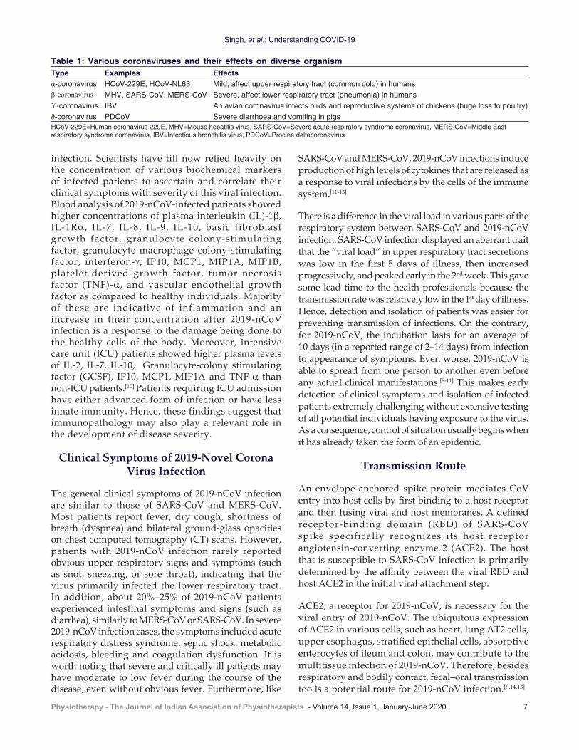
MERS‑CoV (2.7–3.9).[5] The subfamily Coronavirinae includes four genera: alphacoronavirus, betacoronavirus, gammacoronavirus, and deltacoronavirus. The phylogenetic tree of the CoVs displays the evolutional relationships among common ancestors as shown in Figure 1.
Protein sequence analysis shows that the 2019‑nCoV possesses a typical genome structure of CoV and belongs to the cluster of betacoronaviruses. Typically, the nCoV possesses the largest genome among the known viruses. Different CoVs identified so far belong to one of four general (α, β, γ, and ∂) and effect diverse organisms [Table 1].
Before 2019, there were only six CoVs that were known to infect humans and cause respiratory diseases. 2019‑nCoV can also infect the lower respiratory tract and cause pneumonia in humans but it seems that the symptoms are milder than SARS and MERS. This 2019‑nCoV is the seventh member of the family of CoVs that infects humans.
Clinical Findings
GenomeThe length of whole genome of SARS‑CoV‑2 is 29,727 nucleotides and the genome organization exhibits 79.0%
nucleotide identity to SARS‑CoV and 51.8% identity to MERS‑CoV,[6] which belongs to β‑CoV genus. The genome sequence (SARS‑CoV‑2, Urbani strain‑Accession number‑AY278741) is open for public view at Gene Bank information on National Center for Biotechnology Information, National Library of Medicine.[7]
Furthermore, it has been reported that 2019‑nCoV is 96% identical across the entire genome to a bat CoV.[8] This suggests that there is a high probability that this infection causing virus originated from bats. The in vitro tests have shown that its inoculation onto surface layers of human airway epithelial cells causes cytopathic effects and cessation of the cilium beating of the cells.[9]
Morphology and nature of severe acute respiratory syndrome coronavirus‑2It exhibits round or elliptic and often pleomorphic form and has a diameter of ~ 60–140 nm. Like other CoVs, it is sensitive to ultraviolet light and heat. Further, it can be inactivated by lipid solvents including ether (75%), ethanol, chlorine‑containing disinfectant, peroxyacetic acid and chloroform except for chlorhexidine.
Biochemical markersIt is important to note that at present, there is no specific biochemical marker for identifying 2019‑nCoV
Figure 1: Phylogenetic tree of coronaviruses
Singh, et al.: Understanding COVID-19
Physiotherapy - The Journal of Indian Association of Physiotherapists - Volume 14, Issue 1, January-June 2020 7
SARS‑CoV and MERS‑CoV, 2019‑nCoV infections induce production of high levels of cytokines that are released as a response to viral infections by the cells of the immune system.[11‑13]
There is a difference in the viral load in various parts of the respiratory system between SARS‑CoV and 2019‑nCoV infection. SARS‑CoV infection displayed an aberrant trait that the “viral load” in upper respiratory tract secretions was low in the first 5 days of illness, then increased progressively, and peaked early in the 2nd week. This gave some lead time to the health professionals because the transmission rate was relatively low in the 1st day of illness. Hence, detection and isolation of patients was easier for preventing transmission of infections. On the contrary, for 2019‑nCoV, the incubation lasts for an average of 10 days (in a reported range of 2–14 days) from infection to appearance of symptoms. Even worse, 2019‑nCoV is able to spread from one person to another even before any actual clinical manifestations.[8‑11] This makes early detection of clinical symptoms and isolation of infected patients extremely challenging without extensive testing of all potential individuals having exposure to the virus. As a consequence, control of situation usually begins when it has already taken the form of an epidemic.
Transmission Route
An envelope‑anchored spike protein mediates CoV entry into host cells by first binding to a host receptor and then fusing viral and host membranes. A defined receptor‑binding domain (RBD) of SARS‑CoV spike specifically recognizes its host receptor angiotensin‑converting enzyme 2 (ACE2). The host that is susceptible to SARS‑CoV infection is primarily determined by the affinity between the viral RBD and host ACE2 in the initial viral attachment step.
ACE2, a receptor for 2019‑nCoV, is necessary for the viral entry of 2019‑nCoV. The ubiquitous expression of ACE2 in various cells, such as heart, lung AT2 cells, upper esophagus, stratified epithelial cells, absorptive enterocytes of ileum and colon, may contribute to the multitissue infection of 2019‑nCoV. Therefore, besides respiratory and bodily contact, fecal–oral transmission too is a potential route for 2019‑nCoV infection.[8,14,15]
infection. Scientists have till now relied heavily on the concentration of various biochemical markers of infected patients to ascertain and correlate their clinical symptoms with severity of this viral infection. Blood analysis of 2019‑nCoV‑infected patients showed higher concentrations of plasma interleukin (IL)‑1β, IL‑1Rα , IL‑7, IL‑8, IL‑9, IL‑10, basic fibroblast growth factor, granulocyte colony‑stimulating factor, granulocyte macrophage colony‑stimulating factor, interferon‑γ, IP10, MCP1, MIP1A, MIP1B, platelet‑derived growth factor, tumor necrosis factor (TNF)‑α, and vascular endothelial growth factor as compared to healthy individuals. Majority of these are indicative of inflammation and an increase in their concentration after 2019‑nCoV infection is a response to the damage being done to the healthy cells of the body. Moreover, intensive care unit (ICU) patients showed higher plasma levels of IL‑2, IL‑7, IL‑10, Granulocyte‑colony stimulating factor (GCSF), IP10, MCP1, MIP1A and TNF‑α than non‑ICU patients.[10] Patients requiring ICU admission have either advanced form of infection or have less innate immunity. Hence, these findings suggest that immunopathology may also play a relevant role in the development of disease severity.
Clinical Symptoms of 2019‑Novel Corona Virus Infection
The general clinical symptoms of 2019‑nCoV infection are similar to those of SARS‑CoV and MERS‑CoV. Most patients report fever, dry cough, shortness of breath (dyspnea) and bilateral ground‑glass opacities on chest computed tomography (CT) scans. However, patients with 2019‑nCoV infection rarely reported obvious upper respiratory signs and symptoms (such as snot, sneezing, or sore throat), indicating that the virus primarily infected the lower respiratory tract. In addition, about 20%–25% of 2019‑nCoV patients experienced intestinal symptoms and signs (such as diarrhea), similarly to MERS‑CoV or SARS‑CoV. In severe 2019‑nCoV infection cases, the symptoms included acute respiratory distress syndrome, septic shock, metabolic acidosis, bleeding and coagulation dysfunction. It is worth noting that severe and critically ill patients may have moderate to low fever during the course of the disease, even without obvious fever. Furthermore, like
Table 1: Various coronaviruses and their effects on diverse organismType Examples Effectsα‑coronavirus HCoV‑229E, HCoV‑NL63 Mild; affect upper respiratory tract (common cold) in humansβ-coronavirus MHV, SARS‑CoV, MERS‑CoV Severe, affect lower respiratory tract (pneumonia) in humansϒ‑coronavirus IBV An avian coronavirus infects birds and reproductive systems of chickens (huge loss to poultry)∂-coronavirus PDCoV Severe diarrhoea and vomiting in pigsHCoV‑229E=Human coronavirus 229E, MHV=Mouse hepatitis virus, SARS‑CoV=Severe acute respiratory syndrome coronavirus, MERS‑CoV=Middle East respiratory syndrome coronavirus, IBV=Infectious bronchitis virus, PDCoV=Procine deltacoronavirus

Singh, et al.: Understanding COVID-19
8 Physiotherapy - The Journal of Indian Association of Physiotherapists - Volume 14, Issue 1, January-June 2020
Origin of 2019‑Novel Corona Virus
A detailed computer‑aided analysis of interactions between residues on the receptor‑binding motifs of 2019‑nCoV and ACE‑2 analogs of various species has revealed that it uses civet ACE2 as its receptor, although it appears that 2019‑nCoV RBD has not evolved adaptively for civet ACE2 binding. Moreover, 2019‑nCoV likely does not use mouse or rat ACE2 as its receptor due to no significant virus‑receptor interaction as judged by computational analysis. 2019‑nCoV RBD likely recognizes ACE2 from pigs, ferrets, cats, orangutans, monkeys and humans with similar efficiencies, because these ACE2 molecules are identical or similar in the critical virus‑binding residues. The situation involving bat ACE2 is complex because of the diversity of bat species. However, it still likely recognizes bat ACE2 as its receptor for ACE2 from Rhinolophus sinicus bats (which can be recognized by bat SARS‑CoV strain Rs3367).
In the case of SARS‑CoV, some of its critical receptor‑binding motif residues were adapted to human ACE2, while some others were adapted to civet ACE2. This type of partial viral adaptation to two host species promoted virus replication and cross‑species transmission between the two host species. However, in the case of 2019‑nCoV, no strong evidence for adaptive mutations in its critical receptor‑binding motif residues that would specifically promote viral binding to civet ACE2 have been identified. Hence, either palm civets were not intermediate hosts for 2019‑nCoV, or they passed 2019‑nCoV to humans quickly before 2019‑nCoV had any chance to adapt to civet ACE2.[16]
Bats are less likely to have direct contact with human, and thus, direct transmission of the virus from bat to human is unlikely. Although SARS‑CoV and MERS‑CoV originated from bats, they were transmitted to humans via intermediate host civets and camels, respectively. Therefore, 2019‑nCoV could have also originated from bat but was then transmitted to humans via an intermediate host in the market. Recently, 2019‑nCoV virus that was isolated from pangolins was found to have 99% similarity with the genomic sequence of the isolated strain of 2019‑nCoV that had infected humans. Hence, it could be possible that the transmission and evolution path of 2019‑nCoV was from bat‑CoV to pangolins (the intermediate hosts), from where it infected humans.
However, it is a matter of deep investigation to locate the origin and intermediate hosts of 2019‑nCoV before it infected humans. The fact that mutations were not detected in receptor‑binding motif residues on 2019‑nCoV for binding to civet ACE2 and the former’s genomic sequence was found identical to that isolated from pangolins, makes it even more difficult to pin
point the exact source and intermediate host. Most importantly, the computer‑aided structural analysis has predicted that a single mutation may significantly enhance the binding affinity between 2019‑nCoV RBD and human ACE2. Thus, 2019‑nCoV evolution in patients should be closely monitored for the emergence of novel mutations at the 501 position (to a lesser extent, also the 494 position).
Two most important inferences could be logically drawn from limited reports available so for on 2019‑nCoV. Prima facie it seems less convincing that the culinary interests of inhabitants of Wuhan could have contributed to this disaster. This contention arises from the fact that the natives should have acquired immunity against this virus over the period as they would have been consuming civets and pangolins since long. Therefore, it would be appropriate presently to apprehend that the route of infection was bats to pangolins to human. However, whether bats infect pangolins with 2019‑nCoV does require deep contemplation. Second, nonsignificant virus‑receptor interaction between 2019‑nCoV and rat and mouse ACE2 suggests that these cannot be used for developing experimental model for research.
Diagnosis
In wake of global health crisis inflicted by the outbreak of COVID‑19 disease, foremost priority of any nation of world today is to contain spread of this highly contagious disease. Since definitive treatment and vaccine remains unavailable, diagnostic testing plays a pivotal role in this crisis contributing to patient screening, early identification, and diagnosis of COVID‑19 even monitoring treatment, as well as in epidemiologic surveillance.[17] Early diagnosis is key to halt transmission the COVID‑19 as it will assist in early treatment, reducing the mortality, thus decreasing the burden on health‑care systems allowing them to deal effectively with epidemic. Clinical diagnosis of the COVID‑19 can be made taking manifestation into consideration (fever, dry cough, dyspnea, ad other upper respiratory symptoms), epidemiological risk (travel history to COVID‑19‑affected region), and other factors including age and comorbidities.[18]
Since the clinical symptoms and signs of patients infected with SARS‑CoV‑2 are highly atypical and mimic features of respiratory infections caused by other viruses such as parainfluenza virus, adenovirus, respiratory syncytial virus, rhinovirus, and SARS‑CoV,[19,20] COVID‑19 requires confirmatory laboratory diagnosis. Further, keeping in view the present scenario, in order to decrease the pace of progression of pandemic, many health organizations are clamoring for conduction of early laboratory testing to confirm the diagnosis of COVID‑19 suspected cases so

Singh, et al.: Understanding COVID-19
Physiotherapy - The Journal of Indian Association of Physiotherapists - Volume 14, Issue 1, January-June 2020 9
that necessary intervention (isolation/quarantine) could be taken.[17] The important diagnostic tests include (i) nucleic acid amplification test (NAAT), (ii) serological tests, (iii) chest radiographs and CT scans, and (iv) others.i. The basis of NAAT test is to find the virus in
the secretions of patient by detecting presence of genetic material (nucleic acid) of SARS‑CoV2 virus. The most common and effective method recommended by the WHO for nucleic acid detection of SARS‑CoV‑2 is real‑time quantitative polymerase chain reaction (RT‑qPCR).[21] In this test, upper airway specimen (pharyngeal swabs,nasal swabs, nasopharyngeal secretions) as well as lower airway specimen (sputum, bronchoalveolar lavage fluid) and even blood or fecal samples are collected. Extraction of RNA[22] is done. The protocol has been published by the WHO for using RT‑qPCR.[21] The extracted RNA is transcribed into DNA by adding enzymes. This DNA is put into a RT‑qPCR machine that essentially xeroxes the DNA, making thousands of copies of genetic material. Further few specific genes of 2019‑nCoV, namely the open reading frame la/b, nucleocapsid protein (N), envelope protein (E) genes, and RNA dependent RNA polymerase genes, are searched for confirming COVID‑19. Results are positive if two genes are present, not conclusive if one gene is present and negative if no gene is present.[22‑24] Though RT‑qPCR test suffer certain shortcomings such as biological safety hazards due to sample collection or transportation. cumbersome nucleic acid detection operations and long waiting time for results but still RT‑qPCR remains gold standard test for diagnosis of COVID‑19 as it can detect virus at an early stage of infection.[23] Shortage of RT‑qPCR kits is being experienced due to unprecedented rise in infected cases. Thus, it becomes necessary to prioritize who gets tested according to health objectives of the nation and testing is recommended for individual with high index of suspicion. The Indian Council of Medical Research (ICMR), New Delhi, has also developed diagnostic strategy for testing,[25] which is being revised and updated time to time as new information about 2019‑nCoV emerges (https://icmr.nic.in/content/covid‑19)
ii. Serological tests: Serology‑based tests analyze the serum component of whole blood to detect presence of antibodies to know whether person has been exposed to a corona virus. These tests include colloidal gold immunochromatography, enzyme‑linked immunosorbent assay, immunofluorescence assay, and chemiluminescence immunoassay.[26] Two antibodies develop in the body against viral infection, i.e., immunoglobulin (IgM) antibodies and IgG antibodies. Detection of IgM antibodies reflects recent exposure whereas IgG antibodies indicate viral exposure some time ago. Detection of both IgM
and IgG provides information on virus infection time course.[27] IgM becomes detectable around 3–5 days after onset; IgG reaches a titration of at least 4‑fold increase during convalescence compared with the acute phase. During follow‑up monitoring, IgM is detectable 10 days after symptom onset and IgG is detectable 12 days after symptom onset. A positive interpretation of antibody test has been defined as a positive IgM or an increased IgG titer (>4‑fold than that in the acute phase).[28] In cases where NAAT reports have been negative, but there is a strong epidemiological link to COVID‑19 infection, IgM and IgG testing validated serology tests (in the acute and convalescent phase) could support diagnosis.[21] However, few authorities question the usefulness of serological testing in COVID‑19 diagnosis and monitoring as these tests detect infection after 7–10 days of exposure to virus and they also may cross‑react with serologic responses to seasonal CoVs. Overlooking these limitations, serological tests could prove highly valuable in point‑of‑care testing as they are rapid, simple to use, and provide results within 15 min. Therefore, rapid antibody tests with high sensitivity and specificity will quickly identify 2019‑nCoV in infected patients and would give impetus to containment efforts for COVID‑19 disease.[27] The ICMR has made significant progress in this direction and validated five rapid antibody tests (list of tests released on April 2, 2020).[29] Most significant benefit of serological assays would be in determining who developed immunity to COVID‑19. This knowledge would help in identifying individuals who showed strong immunological response to 2019‑nCoV and could then serve as donors for the generation of convalescent serum therapeutics. Additional usefulness of these tests would be for deploying immunologically strong health‑care workers in high viral risk areas to prevent inadvertent spread of the virus[30]
iii. Chest radiographs and CT scans: Chest radiographs are not especially sensitive for COVID‑19 and have little diagnostic value in early stages, whereas CT findings may be present even before symptom onset.[31] CT is significantly more sensitive than RT‑qPCR, but not much specific as many of its imaging features can easily be confused with other disease process such as H1N1, SARS, MERS, and seasonal flu.[32,33] Chest CT or X‑ray is not currently recommend as a diagnostic method. The American College of Radiology recommends not to use CT scan for screening or primary testing for diagnosis of COVID‑19.[33] According to the Center for Disease Control and Prevention, viral testing needs to be conducted for diagnosis confirmation even if a chest CT or X‑ray suggests COVID‑19.[31] Notwithstanding reservations for using CT scan for initial diagnosis
Singh, et al.: Understanding COVID-19
10 Physiotherapy - The Journal of Indian Association of Physiotherapists - Volume 14, Issue 1, January-June 2020
COVID‑19 infection, it is very valuable for monitoring disease progression of severely ill patients and categorization of clinical syndromes
iv. Other laboratory tests: In the early stage of the disease, close check should be kept on absolute value of lymphocytes. If it is <0.8 × 10⁹/L, or the numbers of CD4 and CD8 T cells are significantly decreased, it is generally recommend to recheck the routine blood changes after 3 days.[19,34] More laboratory tests for checking the status of 2019‑nCoV infection include blood gas analysis, function tests of liver and kidney, myocardial enzyme, myoglobin, erythrocyte sedimentation rate, alanine aminotransferase, cardiac troponin, C‑reactive protein, procalcitonin, lactate, D‑dimer, coagulation image, urine routine test, inflammatory factors (IL‑6, IL‑10, TNF‑α), 11 items of tuberculosis subgroup, complement, and anti‑acid staining. Aforementioned in vitro laboratory tests beyond being valuable in etiological diagnosis of COVID‑19 are critical for assessing disease severity and monitoring therapeutic intervention. Many of these tests have been implicated in unfavorable COVID‑19 progression wherein they provide important prognostic information.[34‑36] Emerging evidence suggests that severe COVID‑19 patients are at risk for cytokine storm syndrome which could be major cause of mortality. Cytokine tests, particularly IL‑6, assesses hyperinflammation in severe patients and would be instrumental in checking rise of COVID‑19 mortality.[37,38]
Treatment
Severity of the COVID‑19 disease has been classified into four types:a. Mild cases: Having mild clinical symptoms and
pneumonia manifestations not present in imagingb. Moderate cases: Having symptoms such as fever and
respiratory tract symptoms, etc., and pneumonia manifestations seen in imaging
c. Severe cases: Dyspnea, hypoxia, or >50% lung involvement on imaging
d. Critical cases: Respiratory failure, shock, or multiorgan system dysfunction; about 80% of COVID‑19 patients develop only mild or uncomplicated illness and approximately 14% patients develop severe disease requiring hospitalization and oxygen support, while 5% require admission to an ICU.[39,40] In severe cases of COVID‑19, many complications may develop such as acute respiratory disease syndrome (ARDS), sepsis and septic shock, multiorgan failure, including acute kidney injury and cardiac injury.[41]
Further, clinical course of COVID‑19 disease can progress through six clinical syndromes outlined by the World Health Organization, which include mild illness, pneumonia, severe pneumonia, ARDS, sepsis,
and septic shock.[42] Treatment of patient is mainly based on syndrome differentiation of disease.
Supportive Treatment
Many patients with a mild illness and without underlying risk factors (lung or heart disease, renal failure, or immunocompromising conditions) of developing complications may not be hospitalized owing to limited health‑care resources and care to them can be provided at home that too by family members. The decision to monitor a patient in the inpatient or outpatient requires careful clinical judgment and will depend on whether the residential setting is suitable for providing care and whether patient and the family are capable of adhering to the precautions that will be recommended as part of home care isolation (e.g., hand hygiene, respiratory hygiene, environmental cleaning, and limitations on movement around or from the house). Home management is mainly supportive with proper nutrition, hydration, antipyretics (especially paracetamol recommended), and analgesics. Further, given the possible risk of progression to severe illness in the 2nd week after symptom onset, health‑care workers should monitor the patient closely and provision for immediate hospitalization should be well in place.[43,44] The detail guidelines about home care management of COVID‑19 patients have been developed by the WHO.[43]
Few COVID‑19 patients will require hospitalization for management (inpatient) as the disease develops and complications including pneumonia, hypoxemic respiratory failure/ARDS, sepsis and septic shock, cardiomyopathy and arrhythmia, acute kidney injury, and secondary bacterial infections set in.[10,45] As of now, currently, no specific treatment for COVID‑19 is approved. Inpatient management of COVID‑19 provides supportive management of the most common complications of severe COVID‑19.[44]
The WHO has developed guidelines on the basis of scientific evidence derived from the treatment of previous epidemics from human corona viruses (SARS and MERS). This guideline provides recommendations for the management of adults, pregnant, and children with COVID‑19.[43] Recently, on March 31, 2020, the Government of India also released national guidelines on clinical management of COVID‑19, which aim to provide clinicians with updated interim guidance on timely, effective, and safe supportive management of patients with COVID‑19.[46] Important strategies of these guidelines are discussed as follows:
I. For management of severe COVID‑19a. Provide airway management and oxygen therapy
during resuscitation to target SpO2 ≥94% to patients

Singh, et al.: Understanding COVID-19
Physiotherapy - The Journal of Indian Association of Physiotherapists - Volume 14, Issue 1, January-June 2020 11
with severe acute respiratory infection (SARI) exhibiting emergency signs (obstructed or absent breathing, severe respiratory distress, central cyanosis, shock, coma, or convulsions)
b. In patients with SARI but having no evidence of shock, administer conservative fluid management with intravenous fluid. Aggressive fluid resuscitation needs to be avoided as it may worsen oxygenation.
c. Administer appropriate empiric antimicrobials within 1 h of identification of sepsis to treat all likely pathogens causing SARI
d. Avoid routine corticosteroids for the treatment of viral pneumonia or ARDS unless they are indicated for another reason as the lack of evidence survival benefit and can cause possible harm
e. Patients to be closely monitored for signs of clinical deterioration, such as rapidly progressive respiratory failure and sepsis, and provide supportive care interventions immediately as supportive therapies are the cornerstone of therapy to improve chance of survival of COVID‑19 patient.
II. For management of critical COVID‑19: Acute respiratory distress syndromea. When a patient with respiratory distress is failing
standard oxygen therapy, severe hypoxemic respiratory failure needs to be recognized and preparation to provide advanced oxygen/ventilatory support is done
b. When respiratory distress and/or hypoxemia of the patient cannot be alleviated after receiving standard oxygen therapy, high‑flow nasal cannula oxygen (HFNO) therapy or noninvasive ventilation (NIV) is considered
c. Patients receiving a trial of HFNO or NIV should be in a monitored setting, and in case the patient acutely deteriorates or does not improve in about 1 h, tracheal intubation and invasive mechanical ventilation should be instituted in a timely manner. Patients with hemodynamic instability, multiorgan failure, or abnormal mental status should not receive NIV
d. Mechanical ventilation to be implemented using lower tidal volumes (4–8 ml/kg predicted body weight) and lower inspiratory pressures (plateau pressure <30 cm H2O)
e. In patients with severe ARDS, prone ventilation for >12 h per day is recommended
f. In patients with moderate or severe ARDS, higher positive end‑expiratory pressure (PEEP) instead of lower PEEP is suggested to maintain driving pressure
g. In moderate or severe ARDS (PaO2/FiO2 <150), neuromuscular blockade by continuous infusion should not be routinely used
h. Never disconnect patient from ventilator rather use in‑line catheters for airway suctioning and clamp
endotracheal tube when disconnection is required, as it would result into atelectasis
i. Patients with refractory hypoxemia despite lung protective ventilation should be referred to settings having access to expertise in extracorporeal life support.
III. Management of critical illness: Septic shocka. When infection is suspected or confirmed, monitor
and try to recognize signs of septic shock using values of mean arterial pressure and serum lactate levels. Standard care should start within 1 h of recognition which includes antimicrobial therapy and initiation of fluid bolus and vasopressors for hypotension. Detailed guidelines from the Surviving Sepsis Campaign and WHO are available for the management of septic shock in adults[47]
b. Hemodynamic support is essential for resuscitation of adults from septic shock.[48] In the first 15–30 min, give patient 250–500 mL isotonic crystalloid fluid as rapid bolus and reassess for signs of fluid overload after each bolus. Do not use hypotonic crystalloids, starches, or gelatins for resuscitation
c. Fluid resuscitation may lead to volume overload, including respiratory failure. If there is no response to fluid loading and signs of volume overload appear (for example, jugular venous distension, crackles on lung auscultation, pulmonary edema on imaging, or hepatomegaly in children), reduce or discontinue fluid administration. This step is particularly important where mechanical ventilation is not available
d. Administer vasopressors (norepinephrine, epinephrine, and vasopressin) when shock persists during or after fluid resuscitation. The initial blood pressure target is mean arterial pressure (MAP) ≥65 mmHg in adults
e. If signs of poor perfusion and cardiac dysfunction persist despite achieving MAP target with fluids and vasopressors, consider an inotrope such as dobutamine.
Other therapeutic measuresGlucocorticoids can be administered only for 3–5 days in patients with progressive deterioration of oxygenation indicators, rapid worsening on imaging and excessive activation of the body’s inflammatory response. The dose should not exceed the equivalent of methylprednisolone 1–2 mg/kg/day as larger dose of glucocorticoid will delay the removal of CoV due to immunosuppressive effects. Psychological support through counseling should be provided to patients who suffer from anxiety and fear.
Physiotherapy as supportive treatmentExpertise and knowledge of physiotherapists can be utilized at various levels and settings and they can
Singh, et al.: Understanding COVID-19
12 Physiotherapy - The Journal of Indian Association of Physiotherapists - Volume 14, Issue 1, January-June 2020
contribute significantly in stabilizing a 2019‑nCoV patient. In primary care settings, physiotherapists can manage and share workload and can help in triage and early identification of cases. In community care (i.e., in the home), they can help in educating patients, serve as care givers, and contribute in workforce planning. In acute care (i.e., the hospital setting), the physiotherapy emphasis will be on the management of respiratory symptoms and prevention of complications.[49,50]
Physiotherapy can be beneficial in the respiratory treatment and physical rehabilitation of patients with COVID‑19. Not all COVID‑19 positive patients develop high secretion loads, so respiratory physiotherapy is indicated only for selected patients, however those patients who have pre‑existing respiratory conditions require personalized physiotherapy treatments which may include mechanical airway clearance or use of oscillating devices. In this scenario, it is important to take clearance of critical care consultants after discussing with them the risk and benefit of continuing with the physiotherapy.[51,52]
During the acute phase of COVID 19, physiotherapy interventions that could potentially increase the risk of breathing should be avoided.[53] However, once patient is stable and if respiratory physiotherapy is strongly indicated, the main goal is to mobilize secretions and ease the work of breathing. Interventions may include techniques such as positioning, autogenic drainage, deep breathing exercises, breath stacking, active cycle of breathing mobilization, and manual techniques (e.g., percussion, vibrations, and assisted cough) to aid sputum expectoration. It is necessary for physiotherapist to protect himself/herself from contamination by following recommendation regarding the use personal protective equipment.
In the mechanically ventilated COVID‑19 patients, important physiotherapy methods include positioning with regular turning which are vital to prevent atelectasis, optimize ventilation, and prevent pressure sores. Patients can be positioned in lateral positioning, but prone positioning is well recognized to treat hypoxemic respiratory failure. It is highly recommended to deliver ventilation to patients with ARDS in the prone position. Prone ventilation is found to enhance lung mechanics and gas exchange, thus increasing oxygenation and improving outcomes.[52‑54]
Physiotherapists can play a key role in the prevention of a range of complications including ventilator‑associated pneumonias, secondary infections, contractures, or pressure areas/sores. Further, the main role of the physiotherapist in the management of COVID‑19 patients will be witnessed in recovery phase (rehabilitation phase)
of COVID‑19 patients. Physiotherapy in this phase focusses on early mobilization of patient, returning to functional activities, so that duration of hospital stay is reduced and functional decline is minimized. This phase starts from rehabilitation and exercise within the ICU to ward‑based rehabilitation. Physiotherapist uses diverse methods such as passive, active‑assisted, active, or resisted joint range of motion exercises to maintain or improve joint integrity and range of motion and muscle strength and mobilization exercise programs such as bed mobility, movement transition, tilt table standing, and upper limb or lower limb ergometry.[52‑54] An international team of expert researchers and clinicians within the intensive care and acute cardiorespiratory fields has developed recommendation to provide information to physiotherapists about the potential role of physiotherapy in the management of hospital‑admitted patients with confirmed and/or suspected COVID‑19.[52] For detailed information about physiotherapy role in COVID‑19 patient management, one can refer these guidelines available at: Physiotherapy Management for COVID‑19 in the Acute Hospital Setting: Recommendations to Guide Clinical Practice.
Therapeutic Intervention
Since 2019‑nCoV has not been found before in humans, there is no vaccine or special treatment for it so far. The number of cases is increasing rapidly. The need of the hour is to intensify testing and isolating all diagnosed cases as soon as possible in order to cut off the source of infection. Several drugs are under clinical trial and compassionate use protocols based on in vitro activity (against SARS‑CoV‑2 on limited clinical experience). However, the following line of drug/therapies[55] has been utilized for the treatment of COVID 19, until the approved, efficacious therapy is developed.• Chloroquine – In vitro and limited clinical data
suggest potential benefit• Hydroxychloroquine – In vitro and limited clinical
data suggest potential benefit• Lopinavir – Ritonavir‑role in the treatment of
COVID‑19 is unclear. Preclinical data suggested potential benefit; however, more recent data have failed to confirm
• Remdesivir – Investigational and available only through expanded access and study protocols; several large clinical trials are underway
• Azithromycin – Used in some protocols based on theoretical mechanism and limited preliminary data as adjunct therapy
• Tocilizumab – Immunomodulating agent used in some protocols based on theoretical mechanism and limited preliminary data as adjunct therapy
• COVID‑19 convalescent plasma – Investigational use is being studied

Singh, et al.: Understanding COVID-19
Physiotherapy - The Journal of Indian Association of Physiotherapists - Volume 14, Issue 1, January-June 2020 13
• Corticosteroid therapy is not recommended for viral pneumonia; however, use may be considered for patients with refractory shock or acute respiratory distress syndrome.
Preventive Measures Based on the WHO Guidelines
• Prevent close contact with subjects suffering from acute respiratory infections
• Frequently wash hands especially after contact with infected people or their environment
• Evade unprotected contact with farm or wild animals• People with symptoms of acute airway infection
should keep their distance, cover coughs or sneezes with disposable tissues or clothes, and wash their hands
• Strengthen, in particular, in emergency medicine departments, the application of strict hygiene measures for the prevention and control of infections
• Individuals that are immunocompromised should avoid public gatherings.
Potential Risks and Challenges in the Development of Human Vaccines
The development of safe, effective, and stable vaccines is a lengthy process. In addition, it should also be effective against various mutated strains in order to be useful to the infected patients. This makes the task even more challenging. Traditional drug or vaccine development processes are not viable during such suddenly emerging epidemics.
It is pertinent to note here that animal vaccination against some animal CoVs are available. Live or attenuated virus vaccine is effective against porcine epidemic diarrhea virus and avian infectious bronchitis virus. However, in the development of human vaccines (especially live virus or attenuated CoV), the potential risk would be the recombination of genomes of vaccine strains with wild type CoVs. Hence, killed or subunit vaccines containing spike glycoprotein or along with some other viral proteins might prevent the complications such as lower respiratory tract disease in humans. It has been reported that some vaccines against feline CoVs augmented the severity of the disease rather than reduction, when the vaccinated animals were exposed to wild type/form of CoVs. This challenge could be another obstacle in the smooth translation of vaccine development for humans.
Therefore, the first option available could be to systematically screen existing drugs to determine whether they have activity against the 2019‑nCoV. Such screening practices have found that nelfinavir
has potential antiviral activity against 2019‑nCoV. Based on previous studies, an anti‑HIV drug named Kaletra (composed of two protease inhibitors, ritonavir and lopinavir) can be screened as they had displayed therapeutic efficiency on SARS and MERS. More recently, Kaletra was also recommended to treat Wuhan pneumonia by the National Health Commission of the People’s Republic of China.[56] Patients with SARS or MERS have been treated with several drugs including ribavirin, interferon, lopinavir‑ritonavir, and corticosteroids, but the efficacy of certain drugs is still controversial.
Other antiviral drugs, such as US Food and Drug Administration (FDA)‑approved drugs including ribavirin, penciclovir, nitrazine, nalfamusta, and chloroquine, are being evaluated by measuring the effects of these compounds on cytotoxicity, virus yield, and infection rate of 2019‑nCoV. Recent results have shown that remdesivir and chloroquine are effective in controlling 2019‑nCoV infection in vitro and may be evaluated in human patients with 2019‑nCoV disease. Currently, remdesivir is in clinical research phase for the treatment of Ebola virus infection. Moreover, the fifth edition of infection prevention and control guidance has announced that severe and critically ill patients could be treated with recovery plasma.[57,58]
The drug favilavir (marketed by the name Avigan) developed by Fugifilm Toyama Chemicals, Japan, has become the first ever antiviral medicine to be approved for use as a treatment for Covid‑19 in China. It was earlier used for treating influenza in Japan and China. This approval is based on the reports of patients in Shenzhen where patients receiving favilavir turned negative for CoV after a median of 4 days after becoming positive as compared to 11 patients who did not receive the drug. Furthermore, X‑rays of chest showed improvements in 91% of the patients as compared to 62% of the patients who did not receive the drug. However, the USA has not yet approved this drug for the treatment of CoV. Nevertheless, clinical trials are going on in Japan to see if it can be used for preventing the virus from multiplying in the patients suffering from mild‑to‑moderate symptoms.
Another important development has been reported from the University of Pittsburgh’s Center for Vaccine Research (CVR), USA. They are developing a SARS‑CoV‑2 vaccine using a measles vector (a measles vaccine tailored to express SARS‑CoV‑2 proteins on its surface). This is aimed to be sued for generating immunity to the virus. CVR is a part of an international consortium led by Institut Pasteur (Paris, France) in collaboration with Themis Bioscience GmbH (Vienna, Austria). It is expected that the vaccine shall be ready by April 2020
Singh, et al.: Understanding COVID-19
14 Physiotherapy - The Journal of Indian Association of Physiotherapists - Volume 14, Issue 1, January-June 2020
and will undergo trails in 60–80 human volunteers in Europe by the end of this year. The Coalition for Epidemic Preparedness Innovations, an international intergovernmental organization, has committed about United States Dollar 5 million to the consortium for this purpose.
Working on immediate need awaiting development of new treatments, Roche (Basel, Switzerland) has been given permission by the US‑FDA to initiate randomized, double‑blind, placebo‑controlled Phase III clinical trial in collaboration with the Biomedical Advanced Research and Development Authority to evaluate the safety and efficacy of Actemra/Ro‑Actemra (having tocilizumab) in hospitalized adult patients with severe COVID0‑19 pneumonia. Actemra/Ro‑Actemra was the first approved anti‑IL‑6 receptor available for the treatment of adult patients suffering from moderate‑to‑severe active rheumatoid arthritis.
Future Possible Targets or Interventions
Some of the potential targets for the development of future drugs against SARS‑CoV‑2 could be among protease inhibitors (which prevent processing of the RNA polymerase or cleavage of the viral S glycoprotein), CoV acetylesterase inhibitors (which limit viral replication) likely as neuraminidase inhibitors which inhibit the replication of influenza virus A and B. Further, inhibitors of membrane fusion may be potentially useful in blocking viral entry as do several new drugs against HIV. Therefore, antibodies against viral S glycoprotein or the unidentified receptor for the SARS‑CoV‑2 could also block entry of the virus.
Conclusions
The primary job seems to track the origin of the human pathogen and learn from experiences of various nations. Human activities, including unlimited invasion of natural habitats of animals, consumption of some of these animals, have probably given rise to such emergency situations. Adaption of viruses from natural hosts to humans has been happening and seems to be escalating. Seemingly trivial, invisible viruses can have devastating effects on the entire human race not sparing ones who were not perpetuators of the chain. People who had not visited the Wuhan animal market have also been diagnosed positive for 2019‑nCoV. This was possible through human‑to‑human contact even at very far off countries. These findings indicate the virulence of 2019‑nCoV and its stable nature even after exposure to various environment conditions. Therefore, there is an immediate need to investigate the animal etiology, recognize, and eliminate the chances of high risk pathogens from entering human chain. Complete
ban on consumption of animals posing high risk of such infections, maintaining constant vigil on such highly virulent infective organisms especially from the wild, may help in reducing such episodes.
Financial support and sponsorshipNil.
Conflicts of interestThere are no conflicts of interest.
References
1. Wang LF, Shi Z, Zhang S, Field H, Daszak P, Eaton BT. Review of bats and SARS. Emerg Infect Dis 2006;12:1834‑40.
2. Ge XY, Li JL, Yang XL, Chmura AA, Zhu G, Epstein JH, et al. Isolation and characterization of a bat SARS‐like coronavirus that uses the ACE2 receptor. Nature 2013;503:535‐8.
3. Chen Y, Guo D. Molecular mechanisms of coronavirus RNA capping and methylation. Virol Sin 2016;31:3‑11.
4. Lu R, Zhao X, Li J, Niu P, Yang B, Wu H, et al. Genomic characterisation and epidemiology of 2019 novel coronavirus: Implications for virus origins and receptor binding. Lancet 2020;395:565‑74.
5. Lin Q, Chiu AP, Zhao S, He D. Modeling the spread of middle east respiratory syndrome coronavirus in Saudi Arabia. Stat Methods Med Res 2018;27:1968‑78.
6. Ren LL, Wang YM, Wu ZQ, Xiang ZC, Guo L, Xu T, et al. Identification of a novel coronavirus causing severe pneumonia in humans: A descriptive study. Chin Med J 2020;1:133:1015‑24. [doi: 10.1097/CM9.0000000000000722].
7. SARS‑CoV‑2 (severe acute respiratory syndrome coronavirus 2) sequences. Available from: https://www.ncbi.nlm.nih.gov/gen bank/sars‑cov‑2‑seqs/#nucleotide‑sequences. [Last accessed on 2020 Mar 30].
8. Zhou P, Yang XL, Wang XG, Hu B, Zhang L, Zhang W, et al. A pneumonia outbreak associated with a new coronavirus of probable bat origin. Nature 2020;579:270‑3.
9. Zhu N, Zhang D, Wang W, Li X, Yang B, Song J, et al. A novel coronavirus from patients with pneumonia in China, 2019. N Engl J Med 2020;382:727‑33.
10. Huang C, Wang Y, Li X, Ren L, Zhao J, Hu Y, et al. Clinical features of patients infected with 2019 novel coronavirus in Wuhan, China. Lancet 2020;395:497‑506.
11. Chan JF, Yuan S, Kok KH, To KK, Chu H, Yang J, et al. A familial cluster of pneumonia associated with the 2019 novel coronavirus indicating person‑to‑person transmission: A study of a family cluster. Lancet 2020;395:514‑23.
12. Chen C, Zhang XR, Ju ZY, He WF. Advances in the research of cytokine storm mechanism induced by corona virus disease 2019 and the corresponding immunotherapies. Chin J Burns 2020;1:36.
13. Chen N, Zhou M, Dong X, Qu J, Gong F, Han Y. Epidemiological and clinical characteristics of 99 cases of 2019 novel coronavirus pneumonia in Wuhan, China: A descriptive study. Lancet 2020;395:507‑13.
14. Hoffmann M, Kleine WH, Krüger N, Müller M. The novel coronavirus 2019 (2019‑nCoV) uses the SARS‑coronavirus receptor ACE2 and the cellular protease TMPRSS2 for entry into target cells. bioRxiv 2020. [doi.org/10.1101/2020.01.31.929042].
15. Wu F, Zhao S, Yu B, Chen YM, Wang W, Song ZG, et al. A new coronavirus associated with human respiratory disease in China. Nature 2020;579:265‑9.
16. Letko M, Munster V. Functional assessment of cell entry and receptor usage for lineage B β‑coronaviruses, including
Singh, et al.: Understanding COVID-19
Physiotherapy - The Journal of Indian Association of Physiotherapists - Volume 14, Issue 1, January-June 2020 15
2019‑nCoV. Nat Microbiol 2020;5:562‑9.17. European Centre for Disease Prevention and Control. An
Overview of the Rapid Test Situation for COVID‑19 Diagnosis in the EU/EEA. European Centre for Disease Prevention and Control. Available from: https://www.ecdc.europa.eu/en/publications‑data/overview‑rapid‑test‑situation‑co vid‑19‑diagnosis‑eueea#no‑link. [Last accessed on 2020 Apr 06].
18. Holshue ML, De Bolt C, Lindquist S, Lofy KH, Wiesman J, Bruce H, et al. First case of 2019 novel coronavirus in the United States. N Engl J Med 2020;382:929‑36.
19. Jin YH, Cai L, Cheng ZS, Cheng H, Deng T, Fan YP, et al. A rapid advice guideline for the diagnosis and treatment of 2019 novel coronavirus (2019‑nCoV) infected pneumonia (standard version). Mil Med Res 2020;7:1‑3. [doi.org/10.1186/s40779‑020‑00246‑8].
20. Li X, Geng M, Peng Y, Meng L, Lu S. Molecular immune pathogenesis and diagnosis of COVID‑19. J Pharm Anal 2020; 10:102‑8. [doi.org/10.1016/j.jpha. 2020.03.001].
21. World Health Organization. Laboratory Testing for Coronavirus Disease (COVID‑19) in Suspected Human Cases. World Health Organization. Available from: https://www.who.int/publications‑detail/laboratory‑testing‑for‑2019‑novel‑coronavirus‑insuspected‑human‑cases‑20200117. [Last accessed on 2020 Apr 03].
22. Pappas S. Coronavirus Testing is Ramping up. Here are the New Tests and how they Work. Available from: https://www.livescience.com/coronavirus‑tests‑available.html. [Last accessed on 2020 Apr 03].
23. Fraley L. What to Know About COVID‑19 Diagnosis. Available from: https://www.healthline.com/health/coronavirus‑diagnosis. [Last accessed on 2020 Apr 06].
24. Hongliu C, Yu C, Zuobing C, Qiang F, Wei LI, Shaohua H, et al. Diagnosis and Treatment. Handbook of COVIO‑19 Prevention and Treatment. In: Tingbo L, editor. Handbook of COVIO‑19 Prevention and Treatment. Hangzhou: The First Affiliated Hospital, Zhejiang University School of Medicine (FAHZU); 2020. p. 19‑24.
25. Indian Council of Medical Research. Strategy of Covid‑19 Testing in India. Indian Council of Medical Research. Available from: https://icmr.nic.in/sites/default/files/upload_documents/Strategy_COV ID19_testing_India.pdf. [Last accessed on 2020 Apr 06].
26. John Hopkin’s Center for Health Security. Serology Testing for COVID‑19. John Hopkin’s Center for Health Security. Available from: http://www.centerforhealthsecurity.org/resources/COVID‑19/200228‑Serolo gy‑testing‑COVID.pdf. [Last accessed on 2020 Apr 04].
27. Li Z, Yi Y, Luo X, Xiong N, Liu Y, Li S, et al. Development and clinical application of a rapid IgM‑IgG combined antibody test for SARS‑CoV‑2 infection diagnosis. J Med Virol 2020; [doi: 10.1002/jmv.25727].
28. National Health Commission and State Administration of Traditional Chinese Medicine. Diagnosis and Treatment Protocol for Novel Coronavirus Pneumonia. National Health Commission and State Administration of Traditional Chinese Medicine; 2020. Available from: http://www.kankyokansen.org/upl oads/uploads/files/jsipc/protocol_V7.pdf. [Last accessed on 2020 Apr 04].
29. Indian Council of Medical Research. List of Antibody (IgM, IgG) Based Rapid Tests. Indian Council of Medical Research. Available from: https://icmr.nic.in/sites/default/files/upl oad_documents/Antibody_based_tests_02042020.pdf. [Last accessed on 2020 Apr 5].
30. Amanat F, Nguyen T, Chromikova V, Strohmeier S, Stadlbauer D, Javier A, et al. A serological assay to detect SARS‑CoV‑2 seroconversion in humans. medRxiv; 2020. Available from: https://www.medrxiv.org/content/10.1101/2020.03.17.20037713v1. [Last accessed on 2020 Apr 4].
31. Center for Disease Control and Prevention. Coronavirus Disease 2019 (COVID‑19): Clinical Care. Center for Disease Control and Prevention. Available from: https://www.cdc.gov/coronavirus/2019‑nco v/hcp/clinical‑guidance‑management‑pati ents.htm. [Last accessed on 2020 Apr 04].
32. Ai T, Yang Z, Hou H, Zhan C, Chen C, Lv W, et al. Correlation of chest CT and RT‑PCR testing in coronavirus disease 2019 (COVID‑19) in China: A report of 1014 Cases. Radiology 2020. [doi: 10.1148/radiol. 2020200642].
33. American College of Radiology. Recommendations for the Use of Chest Radiography and Computed Tomography (CT) for Suspected COVID‑19 Infection. American College of o. Available from: https://www.acr.org/Advocacy‑and‑Economics/ACR‑Position‑Statements/Recommendations‑for‑Chest‑Ra diography‑and‑CT‑for‑Suspected‑COVID19‑Infection. [Last accessed on 2020 Apr 04].
34. Li T. Diagnosis and clinical management of severe acute respiratory syndrome coronavirus 2 (SARS‑CoV‑2) infection: An operational recommendation of Peking Union Medical College Hospital (V2. 0) Working Group of 2019 Novel Coronavirus, Peking Union Medical College Hospital. Emerg Microbes Infect 2020;9:582‑5.
35. Lippi G, Plebani M. The critical role of laboratory medicine during coronavirus disease 2019 (COVID‑19) and other viral outbreaks. Clin Chem Lab Med 2020. [doi.org/10.1515/cclm‑2020‑0240].
36. Lippi G, Plebani M. Laboratory abnormalities in patients with COVID‑2019 infection. Clin Chem Lab Med 2020. [doi.org/10.1515/cclm‑2020‑0198].
37. Mehta P, McAuley DF, Brown M, Sanchez E, Tattersall RS, Manson JJ, et al. COVID‑19: Consider cytokine storm syndromes and immunosuppression. Lancet 2020;395:1033‑4.
38. Center for Disease Control and Prevention. Interim Clinical Guidance for Management of Patients with Confirmed Coronavirus Disease (COVID‑19). Center for Disease Control and Prevention. Available from: https://www.cdc.gov/coronavirus/2019‑nco v/hcp/clinical‑guidance‑ma nagement‑patients.html. [Last accessed on 2020 Apr 06].
39. Wu Z, McGoogan JM. Characteristics of and important lessons from the coronavirus disease 2019 (COVID‑19) outbreak in China: Summary of a report of 72314 cases from the Chinese Center for Disease Control and Prevention. JAMA 2020;323:1239‑42.
40. Feng Z, Li Q, Zhang Y, Wu Z, Dong X, Ma H, et al. The novel coronavirus pneumonia emergency response epidemiology team. Vital surveillances: The epidemiological characteristics of an outbreak of 2019 novel coronavirus diseases (COVID‑19) – China, 2020. China CDC Wkly 2020;2:113.
41. Yang X, Yu Y, Xu J, Shu H, Xia J, Liu H, et al. Clinical course and outcomes of critically ill patients with SARS‑CoV‑2 pneumonia in Wuhan, China: A single‑centered, retrospective, observational study. Lancet Respir Med 2020; 8:475‑81. [doi: 10.1016/S2213‑2600 (20) 30079‑5].
42. World Health Organisation. Clinical Management of Severe Acute Respiratory Infection (SARI) when COVID‑19 Disease is Suspected – Interim Guidance. World Health Organisation; 2020. Available from: https://www.who.int/publications‑detail/clinical‑management‑of‑severe‑acute‑respiratory‑infec tion‑when‑novel‑coronavirus‑(ncov)‑inf ection‑is‑suspected. [Last accessed on 2020 Apr 06].
43. World Health Organization. Home care for patients with suspected novel coronavirus (nCoV) infection presenting with mild symptoms and management of their contacts. Interim guidelines. World Health Organization; 2020. Available from: https://www.who.int/publications‑detail/home‑care‑for‑patients‑with‑su spected‑novel‑coronavirus‑(ncov)‑infection‑presenting‑wit h‑mild‑symptoms‑and‑management‑of‑contacts. [Last accessed on 2020 Apr 06].
44. Center for Disease Control and Prevention. Interim Guidance for Implementing Home Care of People Not Requiring
Singh, et al.: Understanding COVID-19
16 Physiotherapy - The Journal of Indian Association of Physiotherapists - Volume 14, Issue 1, January-June 2020
Hospitalization for Coronavirus Disease 2019 (COVID‑19). Center for Disease Control and Prevention. Available from: https://www.cdc.gov/coronavir us/2019‑ncov/hcp/clinical‑gu idance‑management‑patients.html. [Last accessed on 2020 Apr 06].
45. Zhou F, Yu T, Du R, Fan G, Liu Y, Liu Z, et al. Clinical course and risk factors for mortality of adult inpatients with COVID‑19 in Wuhan, China: A retrospective cohort study. Lancet 2020;395:1054‑62.
46. Government of India Ministry of Health and Family Welfare Directorate General of Health Services (EMR Division). Revised Guidelines on Clinical Management of COVID – 19. Available from: https://www.mohfw.g o v . i n / p d f / R e v i s e d N a t i o n a l C l i n i c a l M a n a g e mentGuidelineforCOVID1931032020.pdf. [Last accessed on 2020 Apr 06].
47. Rhodes A, Evans LE, Alhazzani W, Levy MM, Antonelli M, Ferrer R, et al. Surviving sepsis campaign: International guidelines for management of sepsis and septic shock. Intensive Care Med 2017;43:304‑77.
48. Cascella M, Rajnik M, Cuomo A, Dulebohn SC, Di NR. Features, Evaluation and Treatment Coronavirus (COVID‑19). StatPearls Publishing; 2020. Available from: https://www.ncbi.nlm.nih.gov/books/NBK554776/. [Last accessed on 2020 Mar 31].
49. Role of the Physiotherapist in COVID‑19. Available from: https://www.physiopedia.com/Rol e_of_the_Physiotherapist_in_CO VID‑19#cite_note‑1. [Last accessed on 2020 Mar 31].
50. APTA Statement on Patient Care and Practice Management during COVID‑19 Outbreak. Available from: http://www.apta.org/Coronavirus/Statement/. [Last accessed on 2020 Mar 28].
51. Moses R. COVID‑19 and Respiratory Physiotherapy Referral Guideline. Lancashire Teaching Hospitals . NHS Foundat ion Trust . Avai lab le f rom: h t tps ://w w w . a c p r c . o r g . u k / D a t a / R e s o u r c e _ D o w n l o a d s /COVID19andRespiratoryPhysiotherapyReferralGuideli ne.pdf?date=31/03/2020%2004:50:09. [Last accessed on
2020 Apr 06].52. Thomas P, Baldwin C, Bissett B, Boden I, Gosselink R, Granger CL,
et al. Physiotherapy management for COVID‑19 in the acute hospital setting. Recommendations to guide clinical practice. J Physiother 2020;66:73‑82. [doi.org/10.1016/j.jphys. 2020.03.011]. Available from: https://www.sciencedirect.com/science/article/pii/S183695532030028X. [Last accessed on 2020 Apr 05].
53. Lazzeri M, Lanza A, Bellini R, Bellofiore A, Cecchetto S, Colombo A, et al. Respiratory physiotherapy in patients with COVID‑19 infection in acute setting: A position paper of the Italian Association of Respiratory Physiotherapists (ARIR). Monaldi Arch Chest Dis 2020;90:163‑68. [doi.org/10.4081/monaldi. 2020.1285].
54. Respiratory Management of COVID 19. Available from: https://www.physio‑pedia.com/Re spiratory_Managemen t_of_COVID_19#cite_ref‑:8_6‑0. [Last accessed on 2020 Apr 05].
55. Smith T, Bushek J, Prosser T. COVID‑19 Drug Therapy. Clinical Drug Information/Clinical Solutions. Available from. https://www.elsevier.com/__data/assets/pdf_file/0007/988648/COVID‑19‑Drug‑Therapy_Mar‑2020.pdf. [Last accessed on 2020 Apr 05].
56. Wan Y, Shang J, Graham R, Baric RS, Li F. Receptor recognition by the novel coronavirus from Wuhan: An analysis based on decade‑long structural studies of SARS Coronavirus. J Virol 2020;94: 1‑9. [doi: 10.1128/JVI.00127‑20].
57. Arabi YM, Asiri AY, Assiri AM, Jokhdar HA, Alothman A, Balkhy HH, et al. Treatment of middle east respiratory syndrome with a combination of lopinavir/ritonavir and interferon‑β1b (MIRACLE trial): Statistical analysis plan for a recursive two‑stage group sequential randomized controlled trial. Trials 2020;21:1‑8.
58. Wang ML, Cao R, Zhang L, Yang XL, Liu J, Xu MY, Remdesivir and chloroquine effectively inhibit the recently emerged novel coronavirus (2019‑nCoV) in vitro. Cell Res 2020;30:269‑71.

© 2020 Physiotherapy - The Journal of Indian Association of Physiotherapists | Published by Wolters Kluwer - Medknow 17
Should kinesiology taping be used to manage pain in musculoskeletal disorders? An evidence synthesis from systematic reviewsGourav Banerjee, Mark I. Johnson
Abstract:Kinesiology taping has emerged as a relatively new treatment used for the management of pain in musculoskeletal disorders. The purpose of our review was to synthesise up‑to‑date evidence from systematic reviews on the clinical efficacy of kinesiology taping for managing musculoskeletal pain. Electronic databases (MEDLINE/PubMed, CENTRAL, AMED, CINAHL, PEDro, SPORTDiscus, OTseeker, Scopus, Web of Science, ProQuest, Open Thesis, EThOS) were searched for systematic reviews with or without meta-analysis published in English and non-English languages. Search findings were screened against eligibility criteria and systematic review data was extracted, tabulated and descriptively analysed. Our review included 43 systematic reviews (17 meta‑analyses). Systematic reviewers reported a paucity of high‑quality randomised controlled trials and that overall evidence was of “very low” to “moderate” quality. There were 32 systematic reviews published since 2015 and these provided tentative evidence that kinesiology taping was superior to no or minimal treatment, but not superior to conventional physical therapies for reducing pain and improving function in the short‑term in myofascial pain syndrome, shoulder impingement syndrome, chronic low back pain, knee osteoarthritis and patellofemoral pain syndrome. There is insufficient high-quality evidence to determine the clinical efficacy of kinesiology taping for managing musculoskeletal pain with any certainty. We recommend that an enriched enrolment randomised withdrawal trial is needed to increase the trustworthiness of evidence to inform clinical practice. Healthcare professionals in musculoskeletal practice should view kinesiology taping as one of a variety of nonpharmacological approaches with uncertain efficacy that may be used in combination with the core treatment.Keywords:Athletic tape, complementary therapy, Kinesio tape, pain, physical therapy modality
Introduction
Musculoskeletal pain is a common consequence of cumulative trauma,
repetitive strain, or overuse injuries of the soft‑tissues, bones and joints. Chronic musculoskeletal pain that persists or recurs for longer than 3 months is one of the leading causes of suffering and disability worldwide and is associated with large direct (e.g., healthcare costs) and indirect (e.g., sickness benefit and lost productivity) financial
costs to the society.[1‑3] It is estimated that the annual costs of chronic pain in the USA is greater than that for heart disease, cancer and diabetes, and that chronic pain in Europe accounts for approximately 1.5% to 3% of its gross domestic product expenditure.[4,5] Management of chronic musculoskeletal primary and secondary pain is challenging and involves the use of a multidisciplinary biopsychosocial approach. In many cases pain‑relief is inadequate despite optimal evidence‑based treatment.[6,7] The role of pharmacotherapy for managing chronic pain in the long‑term
Address for correspondence:
Dr. Gourav Banerjee, Centre for Pain Research,
School of Clinical and Applied Sciences,
Leeds Beckett University, Leeds, United Kingdom.
E-mail: [email protected]
Submission: 22-10-2019Revision: 20-02-2020
Accepted: 23-03-2020Published: 29-06-2020
Centre for Pain Research, School of Clinical and
Applied Sciences, Leeds Beckett University,
Leeds, United Kingdom
Review Article
Access this article onlineQuick Response Code:
Website:www.pjiap.org
DOI:10.4103/PJIAP.PJIAP_31_19
How to cite this article: Banerjee G, Johnson MI. Should kinesiology taping be used to manage pain in musculoskeletal disorders? An evidence synthesis from systematic reviews. Physiother - J Indian Assoc Physiother 2020;14:17-25.
This is an open access journal, and articles are distributed under the terms of the Creative Commons Attribution-NonCommercial-ShareAlike 4.0 License, which allows others to remix, tweak, and build upon the work non-commercially, as long as appropriate credit is given and the new creations are licensed under the identical terms.
For reprints contact: [email protected]
Banerjee and Johnson: Kinesiology taping for the management of pain in musculoskeletal disorders
18 Physiotherapy - The Journal of Indian Association of Physiotherapists - Volume 14, Issue 1, January-June 2020
is limited given the propensity of analgesic drugs to cause adverse effects. This is especially problematic in the elderly population in whom musculoskeletal pain (e.g., due to arthritis) is more prevalent putting them at risk of developing polypharmacy‑related complications including organ damage and death.[1,2,7‑9] The World Health Organization and the International Association for the Study of Pain recommend using effective, safe, low‑cost nonpharmacological therapies integrated into a self‑management care plan that empowers patients to take control of their pain management leading to better outcomes in the longer term.[1,10]
Kinesiology taping is a low‑cost, non‑pharmacological treatment method commonly used by healthcare professionals in musculoskeletal and sports settings for the prevention and rehabilitation of musculoskeletal injuries including management of pain. Kinesiology taping involves the application of thin, elastic cotton‑based water‑resistant adhesive kinesiology tape to the skin using a variety of techniques. Kinesiology tape can be stretched longitudinally 60% or more of its resting length and worn continuously for 3 days or longer to support soft tissues and joints without restricting movements.[11‑13] This differs from conventional therapeutic tapes that are rigid or minimally elasticated in nature and are used to provide structural support at joints and soft‑tissues.
Kinesiology taping is indicated for the management of chronic pain associated with musculoskeletal conditions highly prevalent in the adult general population such as the lower back, shoulders and knees.[1,2,7] Kinesiology taping has a low‑risk of hazard probability and severity with minor skin‑related irritation occurring in a small proportion of individuals. Kinesiology taping is inexpensive, available without prescription, and can be administered by patient or carer following relatively simple instructions or training. Hence, kinesiology taping aligns with the good practice recommendations and has potential as a treatment option for managing musculoskeletal pain.[1,10] The purpose of our review
was to synthesise up‑to‑date evidence from systematic reviews evaluating the clinical efficacy of kinesiology taping for managing musculoskeletal pain. Our approach was to outline research findings through commentary rather than a comprehensive objective appraisal of systematic reviews.
Methods
Search strategyThe following electronic databases were searched throughout June 2019 for systematic reviews published in English and non‑English languages: MEDLINE/PubMed, CENTRAL, AMED, CINAHL, PEDro, SPORTDiscus, OTseeker, Scopus, Web of Science, ProQuest, Open Thesis, EThOS [Table 1]. In addition, a search of Google Scholar was performed (up to 5 pages of each keyword search). Detailed search strategies were developed separately for each database based on controlled vocabulary and free text terms with the combination of multiple keywords and the Boolean operators AND/OR ([“kinesiology tap*e/ing; kinesio tap*e/ing; elastic therapeutic tap*ing; neuromuscular tap*ing” AND “systematic review OR meta‑analysis” “ALL FIELDS”]). The reference lists of systematic reviews found from the search were also screened for potentially relevant reviews.
Criteria for considering systematic reviews in this reviewFor this review, kinesiology tape was defined as an “elastic adhesive tape” of any colour, shape, size, texture or brand. Rigid nonelastic or minimally elastic adhesive tapes, elastic adhesive and nonadhesive bandages were excluded from the review. The eligibility criteria were decided a priori by the authors. Eligibility for inclusion were full report of systematic reviews with or without meta‑analysis of kinesiology taping (in combination with or without other interventions including nonelastic taping) for pain and related outcome measures including range‑of‑motion, function, disability and
Table 1: PubMed PICO search strategy using MeSH keywordsPICOS Description MeSH keywordsPopulation (P) Adults and/or children with painful
musculoskeletal conditions#1. (“Pain/prevention and control”[MeSH] OR “Pain/rehabilitation”[MeSH] OR “Pain/therapy”[MeSH]) OR (“Musculoskeletal Diseases/prevention and control”[MeSH] OR “Musculoskeletal Diseases/rehabilitation”[MeSH] OR “Musculoskeletal Diseases/therapy”[MeSH]) OR (“Pain Management/therapy”[MeSH])
Intervention (I) Kinesiology taping (elastic adhesive taping) #2. “Athletic Tape” [MeSH] OR “Physical Therapy Modalities”[MeSH]Comparison (C) Placebo, standard of care, or other treatmentOutcome (O) Pain (main outcome) #3. “Pain Measurement”[MeSH] OR “Pain Perception”[MeSH]Study design Systematic reviews with or without meta‑
analyses#4. (“Systematic Reviews as Topic”[MeSH] OR Systematic Review” [Publication Type]) OR (Meta‑Analysis as Topic”[MeSH] OR Meta‑Analysis” [Publication Type])
Search strategy (S)
#1 AND #2 AND #3 AND #4
Banerjee and Johnson: Kinesiology taping for the management of pain in musculoskeletal disorders
Physiotherapy - The Journal of Indian Association of Physiotherapists - Volume 14, Issue 1, January-June 2020 19
for alleviating pain associated with myofascial pain syndrome (MPS),[48,59] and “very low” to “moderate” evidence for sub‑acromial/shoulder impingement syndrome,[32,38,42,49,51] chronic low back pain,[37,54‑56] osteoarthritis of the knee[45‑47] and patellofemoral pain syndrome.[31,39,43,52] It was notable that some systematic reviewers reported nonelastic or minimally elastic/rigid taping was superior to kinesiology taping for reducing knee pain associated with patellofemoral pain syndrome[31,39] and osteoarthritis.[45] Evidence from the systematic reviews for other conditions such as Achilles tendinopathy[33] was lacking due to insufficient RCTs.
Discussion
This review summarises the up‑to‑date evidence synthesised from systematic reviews on the effectiveness of kinesiology taping for managing pain in musculoskeletal disorders. We noted inconsistency in grading of methodological quality of RCTs in similar systematic reviews. For example, Morris et al.[21] used multiple tools including the Cochrane criteria checklist to assess methodological quality of RCTs,[62] and then categorised RCT quality using a method adapted from Clarke et al.[63] to exclude low quality studies from the analysis. Morris et al., then, graded the overall quality of evidence using the Cochrane level of evidence grading system.[61] In contrast, Luz Júnior et al.[55] assessed the methodological quality of RCTs using the PEDro scale[64] and graded the overall quality of evidence using the GRADE classification system.[60] Nevertheless, our review found a large number of systematic reviews yet few high‑quality RCTs from which to judge efficacy with any degree of certainty. In some instances,[55] the quality of evidence from RCTs with higher methodological quality scores on PEDro scale when meta‑analysed was downgraded to “low” or “very low” because of inadequate sample sizes, substantial statistical heterogeneity, imprecision of treatment effects, inconsistency of results between studies and the presence of publication bias.
Overall, we judge there to be tentative evidence from systematic reviews that kinesiology taping is superior to no treatment at short‑term alleviation of musculoskeletal pain associated with chronic low back pain, osteoarthritis of the knee, sub‑acromial/shoulder impingement syndrome and patellofemoral pain syndrome. At present, it is difficult to judge with certainty whether kinesiology taping is superior to conventional physical therapies. To date, the strongest evidence comes from a recent systematic review of 20 RCTs that included a meta‑analysis of 959 patients with MPS and found that kinesiology taping was superior to noninvasive therapies (including manual therapies) for reducing pain and increasing range of motion.[59]
quality‑of‑life (QoL). Systematic reviews evaluating the efficacy of kinesiology taping for nonpain related outcome measures such as muscle strength and proprioception in healthy human population were excluded. Systematic reviews that included primary studies evaluating both pain and nonpain outcomes were included, however, only findings related to pain and associated outcomes such as function, disability and QoL were included for descriptive analysis.
Data collection and analysisTitles and abstracts of reports identified by the search were screened for relevance by Gourav Banerjee (GB) and cross‑checked by Mark I. Johnson (MIJ). Full text reports of potentially relevant systematic reviews articles were obtained and screened independently by GB and MIJ and any disagreements about meeting eligibility criteria resolved by discussion. Information from systematic reviews was extracted and tabulated prior to undertaking a descriptive analysis.
Results
Searches of databases found 49 systematic reviews that evaluated the effects of kinesiology taping on pain and related outcome measures. Three of these 49 systematic reviews were excluded because they had been previously ‘withdrawn’ by the publisher[14] or a full text report was not available or could not be obtained.[15,16] Additionally, three reports were excluded despite being titled as “systematic review” because they were narrative in nature and lacked characteristics associated with systematic review methodology.[17‑19] Thus, 43 systematic reviews were included in our synthesis, and 17 of these systematic reviews included a meta‑analysis [Table 2].
It was notable that 32 out of 43 systematic reviews were published between 2015 and June 2019. Systematic reviews published between 2010 and 2014 tended to be inconclusive because of a paucity of randomised controlled trials (RCTs) on which to base a judgement of clinical efficacy. Inspection of the 32 systematic reviews published after 2014 revealed an increase in the numbers of RCTs being analysed, although reviewers still reported insufficient high‑quality evidence on which to judge the efficacy of kinesiology taping for managing musculoskeletal pain with certainty. In general, reviewers judged the overall quality of evidence as either “very low”, “low” or “moderate” when using GRADE,[60] or “limited”, “conflicting” and “moderate” when using the Cochrane grading system.[61] In general, the effect size was small with reviewers arguing that kinesiology taping may have short‑term benefits as an adjunct to the conventional physical therapies such as exercise and manual therapies. There was “low” quality evidence of clinical efficacy

Banerjee and Johnson: Kinesiology taping for the management of pain in musculoskeletal disorders
20 Physiotherapy - The Journal of Indian Association of Physiotherapists - Volume 14, Issue 1, January-June 2020
Table 2: Summary of systematic reviews with or without meta-analysis included in this reviewReference Clinical condition/outcome, n Summary of findings/conclusionsBassett et al., 2010[20] MSK disorders (SIS, WAD), n=3 Lack of demonstration of clinical significance for pain, disability and ROM,
no substantial evidence to judge the treatment efficacy of KT and support its use
Mostafavifar et al., 2012[11]
MSK disorders (AT, CLBP, PFPS, SIS, WAD), n=6
Insufficient evidence for or against the use of KT to improve pain, function and affect
Williams et al., 2012‡ (MA)[12]
MSK disorders (SIS, WAD), n=3 Small beneficial effect of KT on active ROM of an injured area. Insufficient evidence for improvements in pain
Kalron and Bar‑Sela, 2013‡[13]
MSK disorders (AT, CLBP, PF, PFPS, SIS, WAD), n=9
Moderate‑quality evidence for effectiveness of KT for immediate pain relief but not long‑term. No or inconclusive evidence for improvements in ROM
Morris et al., 2013‡[21] MSK disorders (CLBP, PF, PFPS, SIS, WAD), n=6
Limited to moderate‑quality evidence that KT is clinically more effective than sham or usual treatment for managing pain, ROM, disability and function. Insufficient evidence to support the use of KT over other PT
Montalvo et al., 2014 (MA)[22]
MSK disorders (CLBP, medial epicondylitis, PF, PFPS, SIS, WAD/neck pain), n=13
KT may have limited potential to reduce pain but may not be clinically meaningful. KT is not superior to other treatment modalities for reducing pain and may be used in conjunction with more traditional therapies
Parreira et al., 2014[23] MSK disorders (CLBP, PF, PFPS/knee pain, SIS/shoulder pin, WAD/neck pain), n=12
KT was not superior to active therapies, sham taping or placebo in improving pain, disability, QoL and return to work. In studies where beneficial effects were observed, the effect sizes were too small to have clinical significance. Current evidence does not support its clinical use
Artioli and Bertolini, 2014[24]
MSK disorders (AS, CLBP, medial epicondylitis, PFPS, SIS, WAD/neck pain), n=10
KT produced similar or slightly superior short‑term pain relief compared with other active PT. KT can be considered as an adjunct to other therapies
Aguilera Eguía et al., 2014[25]
SIS, n=2 Low-quality insufficient evidence to suggest KT reduces pain and improves shoulder function at rest and during movement in the short‑term
Méndez-Rebolledo et al., 2014[26]
PFPS, n=6 Insufficient evidence to support the use of KT in PFPS
Batista et al., 2014[27] CLBP, n=3 No evidence for effectiveness of KT in reducing low back painVanti et al., 2015* (MA)[28]
MSK spinal disorders (CLBP, WAD/neck pain), n [NET(2) + KT(6)]=8
The immediate effect of KT on reducing low back pain and disability is insignificant. KT has a positive but small effect on reducing whiplash associated or specific neck pain, which may not be clinically relevant. The paucity of studies does not allow final conclusions
Lim and Tay, 2015 (MA)[29]
MSK disorders (CLBP, knee OA/PFPS, PF, MPS, neck pain, Quervain’s disease, SIS), n=17
KT is superior to minimal intervention for pain relief but is not superior to other treatment approaches for reducing pain and disability
Beatriz and Rafael, 2015[30]
PFPS, n=12 Inconclusive evidence to support the use of KT in PFPS. KT is an inexpensive technique that has no side effects and can be used alongside other therapies
Chang et al., 2015* (MA)[31]
PFPS, n [NET(6) + KT(5)]=11 KT has small effect in reducing pain, however, unlike McConnell taping, KT cannot change patellar alignment. Both techniques of patellar taping can substantially improve QoL in patients with PFPS
Desjardins‑Charbonneau et al., 2015* (MA)[32]
SIS, n [NET(4) + KT(6)]=10 KT significantly improved pain free ROM, however, there is insufficient evidence to formally conclude on the efficacy of KT when used as a standalone or adjunct treatment for SIS
Scott et al., 2015*[33] AT, n [other treatments(10) + KT(2)]=12
Insufficient (no RCTs) and conflicting evidence to support the use of KT in achilles tendinopathy
Grampurohit et al., 2015*[34]
Poststroke pain, n [NET(13) + KT(2)]=15
Inconclusive evidence to support the use of taping for improving pain intensity, ROM or function in patients with stroke
Dong et al., 2015* (MA)[35]
SIS, n [other treatments (32) + KT(1)]=33
Exercise and other therapies like KT are ideal treatments in early stage of SIS
Liddle and Pennick, 2015*[36]
Pregnancy‑related low back and pelvic pain, n=1
Low‑quality evidence that KT is superior to exercise for reducing pain
Nelson 2016[37] CLBP, n=5 Moderate‑quality evidence that KT as a standalone or adjunct therapy is not superior to conventional PT including exercise for reducing pain and disability. Limited evidence that KT improves ROM and global perceived effect in the short‑term. KT is not a substitute, rather, a useful adjunct to conventional PT for managing CLBP
Mclaren et al., 2016*[38] SIS, n [NET(1) + KT(4)]=5 Moderate‑quality evidence that KT is effective for reducing pain and improving function in the short‑term. Taping treatments are useful adjunct to conventional PT
Contd...
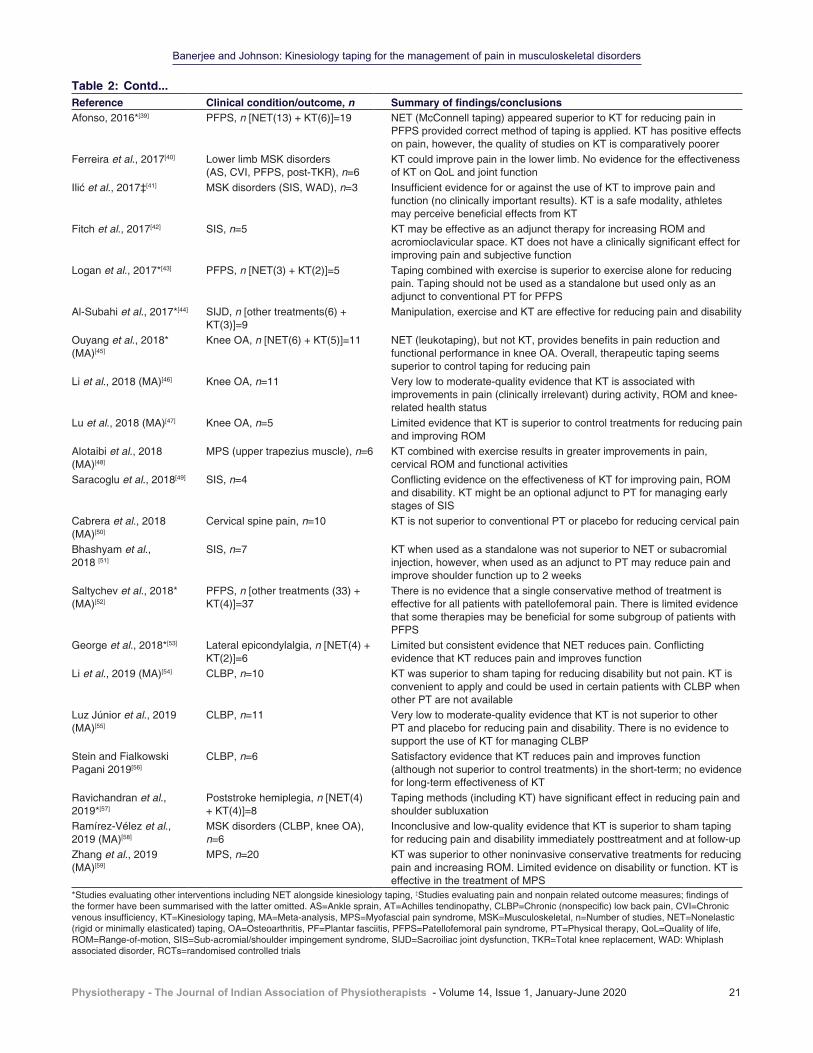
Banerjee and Johnson: Kinesiology taping for the management of pain in musculoskeletal disorders
Physiotherapy - The Journal of Indian Association of Physiotherapists - Volume 14, Issue 1, January-June 2020 21
Table 2: Contd...Reference Clinical condition/outcome, n Summary of findings/conclusionsAfonso, 2016*[39] PFPS, n [NET(13) + KT(6)]=19 NET (McConnell taping) appeared superior to KT for reducing pain in
PFPS provided correct method of taping is applied. KT has positive effects on pain, however, the quality of studies on KT is comparatively poorer
Ferreira et al., 2017[40] Lower limb MSK disorders (AS, CVI, PFPS, post‑TKR), n=6
KT could improve pain in the lower limb. No evidence for the effectiveness of KT on QoL and joint function
Ilić et al., 2017‡[41] MSK disorders (SIS, WAD), n=3 Insufficient evidence for or against the use of KT to improve pain and function (no clinically important results). KT is a safe modality, athletes may perceive beneficial effects from KT
Fitch et al., 2017[42] SIS, n=5 KT may be effective as an adjunct therapy for increasing ROM and acromioclavicular space. KT does not have a clinically significant effect for improving pain and subjective function
Logan et al., 2017*[43] PFPS, n [NET(3) + KT(2)]=5 Taping combined with exercise is superior to exercise alone for reducing pain. Taping should not be used as a standalone but used only as an adjunct to conventional PT for PFPS
Al‑Subahi et al., 2017*[44] SIJD, n [other treatments(6) + KT(3)]=9
Manipulation, exercise and KT are effective for reducing pain and disability
Ouyang et al., 2018* (MA)[45]
Knee OA, n [NET(6) + KT(5)]=11 NET (leukotaping), but not KT, provides benefits in pain reduction and functional performance in knee OA. Overall, therapeutic taping seems superior to control taping for reducing pain
Li et al., 2018 (MA)[46] Knee OA, n=11 Very low to moderate‑quality evidence that KT is associated with improvements in pain (clinically irrelevant) during activity, ROM and knee‑related health status
Lu et al., 2018 (MA)[47] Knee OA, n=5 Limited evidence that KT is superior to control treatments for reducing pain and improving ROM
Alotaibi et al., 2018 (MA)[48]
MPS (upper trapezius muscle), n=6 KT combined with exercise results in greater improvements in pain, cervical ROM and functional activities
Saracoglu et al., 2018[49] SIS, n=4 Conflicting evidence on the effectiveness of KT for improving pain, ROM and disability. KT might be an optional adjunct to PT for managing early stages of SIS
Cabrera et al., 2018 (MA)[50]
Cervical spine pain, n=10 KT is not superior to conventional PT or placebo for reducing cervical pain
Bhashyam et al., 2018 [51]
SIS, n=7 KT when used as a standalone was not superior to NET or subacromial injection, however, when used as an adjunct to PT may reduce pain and improve shoulder function up to 2 weeks
Saltychev et al., 2018* (MA)[52]
PFPS, n [other treatments (33) + KT(4)]=37
There is no evidence that a single conservative method of treatment is effective for all patients with patellofemoral pain. There is limited evidence that some therapies may be beneficial for some subgroup of patients with PFPS
George et al., 2018*[53] Lateral epicondylalgia, n [NET(4) + KT(2)]=6
Limited but consistent evidence that NET reduces pain. Conflicting evidence that KT reduces pain and improves function
Li et al., 2019 (MA)[54] CLBP, n=10 KT was superior to sham taping for reducing disability but not pain. KT is convenient to apply and could be used in certain patients with CLBP when other PT are not available
Luz Júnior et al., 2019 (MA)[55]
CLBP, n=11 Very low to moderate‑quality evidence that KT is not superior to other PT and placebo for reducing pain and disability. There is no evidence to support the use of KT for managing CLBP
Stein and Fialkowski Pagani 2019[56]
CLBP, n=6 Satisfactory evidence that KT reduces pain and improves function (although not superior to control treatments) in the short‑term; no evidence for long‑term effectiveness of KT
Ravichandran et al., 2019*[57]
Poststroke hemiplegia, n [NET(4) + KT(4)]=8
Taping methods (including KT) have significant effect in reducing pain and shoulder subluxation
Ramírez-Vélez et al., 2019 (MA)[58]
MSK disorders (CLBP, knee OA), n=6
Inconclusive and low‑quality evidence that KT is superior to sham taping for reducing pain and disability immediately posttreatment and at follow‑up
Zhang et al., 2019 (MA)[59]
MPS, n=20 KT was superior to other noninvasive conservative treatments for reducing pain and increasing ROM. Limited evidence on disability or function. KT is effective in the treatment of MPS
*Studies evaluating other interventions including NET alongside kinesiology taping, ‡Studies evaluating pain and nonpain related outcome measures; findings of the former have been summarised with the latter omitted. AS=Ankle sprain, AT=Achilles tendinopathy, CLBP=Chronic (nonspecific) low back pain, CVI=Chronic venous insufficiency, KT=Kinesiology taping, MA=Meta-analysis, MPS=Myofascial pain syndrome, MSK=Musculoskeletal, n=Number of studies, NET=Nonelastic (rigid or minimally elasticated) taping, OA=Osteoarthritis, PF=Plantar fasciitis, PFPS=Patellofemoral pain syndrome, PT=Physical therapy, QoL=Quality of life, ROM=Range‑of‑motion, SIS=Sub‑acromial/shoulder impingement syndrome, SIJD=Sacroiliac joint dysfunction, TKR=Total knee replacement, WAD: Whiplash associated disorder, RCTs=randomised controlled trials
Banerjee and Johnson: Kinesiology taping for the management of pain in musculoskeletal disorders
22 Physiotherapy - The Journal of Indian Association of Physiotherapists - Volume 14, Issue 1, January-June 2020
The mechanism (s) of action of kinesiology taping remains largely speculative to date due to the lack of scientific evidence. Conventional taping and bandaging techniques use rigid or minimally elasticated tapes or bandages to provide compression, immobilisation and stabilisation to the injured soft tissues and joints for alleviating pain and promoting recovery. [65] Manufacturers claim that kinesiology taping reduces pain by different mechanisms depending on the location and stretch of kinesiology tape. It is claimed that kinesiology taping techniques for pain relief create traction on the skin to produce convolutions of the skin, i.e., lifting of the epidermis away from the underlying tissues thereby causing decompression in the regions underneath. This is believed to reduce pressure on the subcutaneous nociceptors during inflammation and facilitate increase of the flow of blood and lymph in the microcirculation resulting in the drainage of inflammatory exudates thereby reducing swelling and pain.[11,29] While there is evidence that kinesiology taping of the skin produces mechanical deformation of tissues underneath and an increase in epidermal‑dermal distance,[66‑69] laboratory studies have found that kinesiology taping does not increase cutaneous or skeletal muscle blood flow when measured using laser Doppler imaging technologies under a variety of experimental conditions such as at rest and during exercise.[70‑73]
Perhaps the most plausible mechanism by which kinesiology taping may reduce pain is the notion that kinesiology taping during movement causes stretching and recoil of the skin, which activates low‑threshold mechanoreceptor peripheral afferents causing central inhibition of nociceptive transmission and hence modulation of pain in line with the gate control theory of pain.[29] Findings from laboratory studies that evaluated the effect of kinesiology taping to transient nociceptive‑stimuli interacting with a normally functioning nociceptive system in the presence or absence of sensitisation found conflicting results.[74‑81] Thus, it seems plausible that pain‑relief effects associated with kinesiology taping techniques result from a combination of (a) neurophysiological, i.e., stimulation of low threshold mechanoreceptors, (b) biomechanical, i.e., correction of articular malalignments and elasticated support to the soft‑tissues and joints to unload incumbent forces acting on painful structures[29,82,83] as well as (c) psychological mechanisms, i.e., expectation of benefit from being administered a treatment by a clinician, “laying on of hands”.[84‑86] Notwithstanding, it seems likely that kinesiology tape will have lesser stabilising influence on correcting joint and soft‑tissues malalignment in conditions such as patellofemoral pain syndrome than conventional rigid or minimally elasticated tapes.
Limitations and future directionsA limitation of our review was the omission of assessment of the methodological quality of RCTs or systematic reviews. Not formally appraising the methodological quality of the systematic reviews using tools such as AMSTAR makes our review vulnerable to selection and evaluation biases and hence reduces confidence in our interpretations of evidence. In addition, we performed a simple descriptive analysis and made no attempt to extract and pool RCT data. Our original intention was to overview evidence to generate a ‘one‑stop’ reference source of systematic review information rather than conduct a formal overview of systematic reviews that would include a meta‑analysis of all available RCTs. Thus, we view our review as a precursor to a structured overview of systematic reviews.
Our finding that systematic reviews tended to be inconclusive due to insufficient high‑quality RCT data raises questions about the appropriateness of continuing to undertake systematic reviews in this field. One of the recurring themes is inadequacy of sample sizes in RCTs and meta‑analysis. Inadequate sample sizes are associated with larger treatment effects, and overestimation of clinical efficacy. To create a more trustworthy evidence base, the Cochrane collaboration advise that RCTs assessing pain should have at least 200 participants per treatment arm, and meta‑analyses have >400 pooled data points per treatment arm.
Our review found that most of previous systematic reviews focus on specific medical conditions reducing sample sizes available for data pooling and the statistical power of the meta‑analysis. In future, a review that evaluated the efficacy of kinesiology taping on musculoskeletal pain irrespective of condition would have the potential of increasing statistical power and confidence in findings. Concern about clinical heterogeneity associated with variability in the context and pathology associated with different types of pain can be offset by conducting sub‑group analyses of specific medical conditions (e.g., according to ICD‑11 categories). In addition, systematic reviewers express concern about statistical heterogeneity (“noisy data”) resulting from a plethora of factors including variance of baseline characteristics of participants, variance of kinesiology taping technique and/or dosage, and the appropriateness of control interventions to name but a few. There is certainly a need to develop consensus of what constitutes an adequate dose of kinesiology taping, and a systematic review that extracts, maps and analyses the variability of characteristics kinesiology taping technique used in previous RCTs would be a useful addition to the literature.
Banerjee and Johnson: Kinesiology taping for the management of pain in musculoskeletal disorders
Physiotherapy - The Journal of Indian Association of Physiotherapists - Volume 14, Issue 1, January-June 2020 23
Our review demonstrates the high volume of RCT research that has persistently failed to determine with any degree of certainty the clinical efficacy of kinesiology taping for the management of musculoskeletal pain. There is a significant financial cost associated with generating such large volumes of inconclusive evidence, based on inadequate sample sizes. Conducting RCTs with adequate sample sizes is likely to remain an issue because of funding constraints for large scale studies on kinesiology taping. Thus, we propose that there needs to be refinement of the design of RCTs on kinesiology taping.
We believe that the sensitivity of RCTs on kinesiology taping could be improved by adopting an enriched enrolment randomised withdrawal trial design whereby the sample of participants enrolling into the randomised controlled phase of the trial is enriched so that it includes only those participants that are likely to benefit. This increases the trustworthiness of primary evidence by reducing variance in data and the need for large sample sizes.[87] Enriched enrolment randomised withdrawal trials consist of two phases: firstly, an observational open‑label phase where participants receive only the active treatment (kinesiology taping) and treatment and dosage is titrated and optimised; and secondly a RCT phase that includes participants who obtained befit without adverse events in phase one and who wished to continue with treatment (i.e., an enriched sample). These participants are then randomised to receive either experimental (kinesiology taping) or control interventions. The control intervention could be nonelastic tape, or no treatment, or standard of care or another treatment. To our knowledge, there have not been any enriched enrolment randomised withdrawal‑controlled trials of kinesiology taping for musculoskeletal pain, although the approach has been used to determine the efficacy of drugs for chronic pain conditions.
Conclusion
Our review demonstrates that there is insufficient high‑quality evidence to determine the clinical efficacy of kinesiology taping for managing musculoskeletal pain with any certainty. In view of the available evidence, healthcare professionals in musculoskeletal practice should continue to view kinesiology taping as one of a variety of nonpharmacological approaches with uncertain efficacy. The decision whether to select kinesiology taping from a raft of available treatment options (e.g., manual therapies, massage, electrophysical agents, and acupuncture) remains at the discretion of the individual practitioner and clinic policy.
Financial support and sponsorshipThis project was funded by a PhD student bursary from the Jane Tomlinson Appeal, United Kingdom.
Conflicts of interestMIJ’s institution has received research and consultancy funding for work that he has undertaken for GlaxoSmithKline. GB declares that he has no conflict of interests.
References
1. International Association for the Study of Pain (IASP): Musculoskeletal Pain Fact Sheets. Available from: https://www.iasp‑pain.org/Advocacy/Content.aspx?ItemNumber=1101. [Last accessed on 2019 Jul 10].
2. World Health Organization (WHO): Musculoskeletal Conditions Key Facts. Available from: https://www.who.int/news‑room/fact‑sheets/detail/musculoskeletal‑conditions. [Last accessed on 2019 Jul 10].
3. Treede RD, Rief W, Barke A, Aziz Q, Bennett MI, Benoliel R, et al. Chronic pain as a symptom or a disease: The IASP Classification of Chronic Pain for the International Classification of Diseases (ICD‑11). Pain 2019;160:19‑27.
4. Phillips CJ. Economic burden of chronic pain. Expert Rev Pharmacoecon Outcomes Res 2006;6:591‑601.
5. Gaskin DJ, Richard P. The economic costs of pain in the United States. J Pain 2012;13:715‑24.
6. Lewis J, O’Sullivan P. Is it time to reframe how we care for people with non‑traumatic musculoskeletal pain? Br J Sports Med 2018;52:1543‑4.
7. Briggs AM, Woolf AD, Dreinhöfer K, Homb N, Hoy DG, Kopansky‑Giles D, et al. Reducing the global burden of musculoskeletal conditions. Bull World Health Organ 2018;96:366‑8.
8. Davies EA, O’Mahony MS. Adverse drug reactions in special populations‑the elderly. Br J Clin Pharmacol 2015;80:796‑807.
9. Jones CM, Mack KA, Paulozzi LJ. Pharmaceutical overdose deaths, United States, 2010. JAMA 2013;309:657‑9.
10. Hoffman AJ. Enhancing self‑efficacy for optimized patient outcomes through the theory of symptom self‑management. Cancer Nurs 2013;36:E16‑26.
11. Mostafavifar M, Wertz J, Borchers J. A systematic review of the effectiveness of Kinesio taping for musculoskeletal injury. Phys Sportsmed 2012;40:33‑40.
12. Williams S, Whatman C, Hume PA, Sheerin K. Kinesio taping in treatment and prevention of sports injuries: A meta‑analysis of the evidence for its effectiveness. Sports Med 2012;42:153‑64.
13. Kalron A, Bar‑Sela S. A systematic review of the effectiveness of Kinesio taping‑fact or fashion? Eur J Phys Rehabil Med 2013;49:699‑709.
14. Mine K, Nakayama T, Milanese S, Grimmer K. Effects of Kinesio Tape on pain, muscle strength and functional performance: A systematic review of Japanese‑language literature. Physical Therapy Reviews 2018;23:108‑15.
15. Bailey B. The effects of conservative therapy with Kinesio taping versus conservative therapy alone in adults with chronic non‑specific low back pain on disability and pain: A meta‑analysis. doctor of physical therapy, California State University, Fresno California USA; May, 2017.
16. Gilbert A, Greene O, Kattato A, Kelly M, Liu E, Schoenfelder J. Kinesio tape application for muscle inhibition and its effects on pain, EMG activity, and headaches: A systematic review. New York, USA; 2017.
17. Behbahani HS, Arab AM, Nejad L. Systematic review: Effects of using Kinesio tape on treatment of lateral epicondylitis. Phys Treat Specif Phys Ther J 2014;4:115‑22.
18. Kaur J, Malik M, Rani M. A systematic review on efficacy of Kinesio taping in pain management. Int J Physiother 2016;3:355‑61.
Banerjee and Johnson: Kinesiology taping for the management of pain in musculoskeletal disorders
24 Physiotherapy - The Journal of Indian Association of Physiotherapists - Volume 14, Issue 1, January-June 2020
19. Mishra NJ, Ganvir S. Meta‑analysis on physiotherapy modalities used in patients of total knee replacement for pain relief. Indian journal of physiotherapy & occupational therapy 2018;12:67‑72.
20. Bassett KT, Lingman SA, Ellis RF. The use and treatment of kinaesthetic taping for musculoskeletal conditions: A systematic review. N Z J Physiother 2010;38:56‑62.
21. Morris D, Jones D, Ryan H, Ryan CG. The clinical effects of Kinesio® Tex taping: A systematic review. Physiother Theory Pract 2013;29:259‑70.
22. Montalvo AM, Cara EL, Myer GD. Effect of kinesiology taping on pain in individuals with musculoskeletal injuries: Systematic review and meta‑analysis. Phys Sportsmed 2014;42:48‑57.
23. Parreira Pdo C, Costa Lda C, Hespanhol LC Jr., Lopes AD, Costa LO. Current evidence does not support the use of Kinesio taping in clinical practice: A systematic review. J Physiother 2014;60:31‑9.
24. Artioli DP, Bertolini GR. Kinesio taping: Application and results on pain: Systematic review. Fisioter Pesqui 2014;21:94‑9.
25. Aguilera Eguía RA, Zafra Santos EO, Araya Quintanilla FA, Gómez Carreño LE, Soto Aliaga JX, Vargas Varga A, et al. Uso del taping en el manejo clínico de sujetos con pinzamiento sub‑acromial: Revisión sistemática. Rev Soc Esp Dolor 2014;21:39‑49.
26. Méndez‑Rebolledo G, Gatica‑Rojas V, Cuevas‑Contreras D, Sánchez‑Leyton C. Efectos del kinesio tape en la rehabilitación de pacientes con síndrome de dolor patelofemoral: Una revisión sistemática. Fisioterapia 2014;36:280‑7.
27. Batista CV, Ruaro JA, Ruaro MB, Dubiela A, Kerppers II, Suckow PP, et al. Uso da kinesio taping na dor lombar: Revisão sistemática (Kinesio taping on low back pain: Systematic review). ConScientiae Saúde 2014;13:147‑52.
28. Vanti C, Bertozzi L, Gardenghi I, Turoni F, Guccione AA, Pillastrini P. Effect of taping on spinal pain and disability: Systematic review and meta‑analysis of randomized trials. Phys Ther 2015;95:493‑506.
29. Lim EC, Tay MG. Kinesio taping in musculoskeletal pain and disability that lasts for more than 4 weeks: Is it time to peel off the tape and throw it out with the sweat? A systematic review with meta‑analysis focused on pain and also methods of tape application. Br J Sports Med 2015;49:1558‑66.
30. Beatriz LA, Rafael MM. Kinesio taping and patellofemoral pain syndrome: A systematic review. Cent Eur J Sport Sci Med 2015;9:47‑54.
31. Chang WD, Chen FC, Lee CL, Lin HY, Lai PT. Effects of Kinesio taping versus mcconnell taping for patellofemoral pain syndrome: A systematic review and meta‑analysis. Evid Based Complement Alternat Med 2015;2015:471208.
32. Desjardins‑Charbonneau A, Roy JS, Dionne CE, Desmeules F. The efficacy of taping for rotator cuff tendinopathy: A systematic review and meta‑analysis. Int J Sports Phys Ther 2015;10:420‑33.
33. Scott LA, Munteanu SE, Menz HB. Effectiveness of orthotic devices in the treatment of Achilles tendinopathy: A systematic review. Sports Med 2015;45:95‑110.
34. Grampurohit N, Pradhan S, Kartin D. Efficacy of adhesive taping as an adjunt to physical rehabilitation to influence outcomes post‑stroke: A systematic review. Top Stroke Rehabil 2015;22:72‑82.
35. Dong W, Goost H, Lin XB, Burger C, Paul C, Wang ZL, et al. Treatments for shoulder impingement syndrome: A PRISMA systematic review and network meta‑analysis. Medicine (Baltimore) 2015;94:e510.
36. Liddle SD, Pennick V. Interventions for preventing and treating low‐back and pelvic pain during pregnancy. Cochrane Database of Systematic Reviews 2015;2015:CD001139.
37. Nelson NL. Kinesio taping for chronic low back pain: A systematic review. J Bodyw Mov Ther 2016;20:672‑81.
38. Mclaren C, Colman Z, Rix A, Sullohern C. The effectiveness of scapular taping on pain and function in people with subacromial
impingement syndrome: A systematic review. Int Musculoskelet Med 2016;38:81‑9.
39. Afonso J. Effets du taping McConnell et du Kinesio‑Taping sur la douleur chez les patients souffrant d’un syndrome fémoro‑patellaire: Revue systématique de la littérature. Kinésithér Rev 2016;16:5‑17.
40. Ferreira R, Resende R, Roriz P. The effects of Kinesio taping® in lower limb musculoskeletal disorders: A systematic review. Int J Therap Rehabil Res 2017;6:1.
41. Ilić B, Nikolić A, Ilić D. Efficiency of Kinesio taping in prevention and rehabilitation of sport injuries. Sportlogia 2017;13:53‑65.
42. Fitch C, Frendt T, Lipinski C, Moore C, Donovan L. Efficacy of kinesiology taping as an adjunct treatment of shoulder impingement syndrome: A systematic review. J Athl Train 2017;52:S291.
43. Logan CA, Bhashyam AR, Tisosky AJ, Haber DB, Jorgensen A, Roy A, et al. Systematic review of the effect of taping techniques on patellofemoral pain syndrome. Sports Health 2017;9:456‑61.
44. Al‑Subahi M, Alayat M, Alshehri MA, Helal O, Alhasan H, Alalawi A, et al. The effectiveness of physiotherapy interventions for sacroiliac joint dysfunction: A systematic review. J Phys Ther Sci 2017;29:1689‑94.
45. Ouyang JH, Chang KH, Hsu WY, Cho YT, Liou TH, Lin YN. Non‑elastic taping, but not elastic taping, provides benefits for patients with knee osteoarthritis: Systemic review and meta‑analysis. Clin Rehabil 2018;32:3‑17.
46. Li X, Zhou X, Liu H, Chen N, Liang J, Yang X, et al. Effects of elastic therapeutic taping on knee osteoarthritis: A systematic review and meta‑analysis. Aging Dis 2018;9:296‑308.
47. Lu Z, Li X, Chen R, Guo C. Kinesio taping improves pain and function in patients with knee osteoarthritis: A meta‑analysis of randomized controlled trials. Int J Surg 2018;59:27‑35.
48. Alotaibi M, Ayoub A, King T, Uddin S. The effect of Kinesio taping in reducing myofascial pain syndrome on the upper trapezius muscle: A systematic review and meta‑analysis. Eur Sci J 2018;28:6.
49. Saracoglu I, Emuk Y, Taspinar F. Does taping in addition to physiotherapy improve the outcomes in subacromial impingement syndrome? A systematic review. Physiother Theory Pract 2018;34:251‑63.
50. Cabrera N, Abril K, Méndez Méndez LC, Garcia Rojas E. The effect of Kinesio taping treatment on cervical pain: A meta‑analysis and systematic review. Rev Sanid Mil 2018;72:40‑6.
51. Bhashyam AR, Logan CA, Rider SM, Schurko B, Provenher MT. A systematic review of taping for pain management in shoulder impingement. Orthop J Harvard Med Sch 2018;19:18‑23.
52. Saltychev M, Dutton RA, Laimi K, Beaupré GS, Virolainen P, Fredericson M. Effectiveness of conservative treatment for patellofemoral pain syndrome: A systematic review and meta‑analysis. J Rehabil Med 2018;50:393‑401.
53. George C, Heales L, Stanton R, Wintour S, Kean C. The immediate effects of therapeutic tape on pain and function in lateral epicondylalgia: A systematic review. J Sci Med Sport 2018;21:S90.
54. Li Y, Yin Y, Jia G, Chen H, Yu L, Wu D. Effects of Kinesiotape on pain and disability in individuals with chronic low back pain: A systematic review and meta‑analysis of randomized controlled trials. Clin Rehabil 2019;33:596‑606.
55. Luz Júnior MA, Almeida MO, Santos RS, Civile VT, Costa LO. Effectiveness of Kinesio taping in patients with chronic nonspecific low back pain: A systematic review with meta‑analysis. Spine (Phila Pa 1976) 2019;44:68‑78.
56. Stein E Jr., Fialkowski Pagani N. Ação Da Bandagem Neuromuscular Na Dor Lombar: Uma Revisão Sistemática. Rev Inspirar Mov Saude 2019;19:1‑19.
57. Ravichandran H, Janakiraman B, Sundaram S, Fisseha B, Gebreyesus T, Yitayeh Gelaw A. Systematic review on effectiveness of shoulder taping in hemiplegia. J Stroke
Banerjee and Johnson: Kinesiology taping for the management of pain in musculoskeletal disorders
Physiotherapy - The Journal of Indian Association of Physiotherapists - Volume 14, Issue 1, January-June 2020 25
Cerebrovasc Dis 2019;28:1463‑73.58. Ramírez‑Vélez R, Hormazábal‑Aguayo I, Izquierdo M,
González‑Ruíz K, Correa‑Bautista JE, García‑Hermoso A. Effects of Kinesio taping alone versus sham taping in individuals with musculoskeletal conditions after intervention for at least one week: A systematic review and meta‑analysis. Physiotherapy 2019;105:412‑20.
59. Zhang XF, Liu L, Wang BB, Liu X, Li P. Evidence for Kinesio taping in management of myofascial pain syndrome: A systematic review and meta‑analysis. Clin Rehabil 2019;33:865‑74.
60. Guyatt GH, Oxman AD, Vist GE, Kunz R, Falck‑Ytter Y, Alonso‑Coello P, et al. GRADE: An emerging consensus on rating quality of evidence and strength of recommendations. BMJ 2008;336:924‑6.
61. van Tulder M, Furlan A, Bombardier C, Bouter L, Editorial Board of the Cochrane Collaboration Back Review Group. Updated method guidelines for systematic reviews in the cochrane collaboration back review group. Spine (Phila Pa 1976) 2003;28:1290‑9.
62. Furlan AD, Pennick V, Bombardier C, van Tulder M, Editorial Board, Cochrane Back Review Group. 2009 updated method guidelines for systematic reviews in the Cochrane Back Review Group. Spine (Phila Pa 1976) 2009;34:1929‑41.
63. Clarke CL, Ryan CG, Martin DJ. Pain neurophysiology education for the management of individuals with chronic low back pain: Systematic review and meta‑analysis. Man Ther 2011;16:544‑9.
64. Maher CG, Sherrington C, Herbert RD, Moseley AM, Elkins M. Reliability of the PEDro scale for rating quality of randomized controlled trials. Phys Ther 2003;83:713‑21.
65. Constantinou M, Brown M. Therapeutic taping for Musculoskeletal Conditions. Australia: Elsevier Health Sciences; 2010.
66. Pamuk U, Yucesoy CA. MRI analyses show that Kinesio taping affects much more than just the targeted superficial tissues and causes heterogeneous deformations within the whole limb. J Biomech 2015;48:4262‑70.
67. Cimino SR, Beaudette SM, Brown SHM. Kinesio taping influences the mechanical behaviour of the skin of the low back: A possible pathway for functionally relevant effects. J Biomech 2018;67:150‑6.
68. Shim JY, Lee HR, Lee DC. The use of elastic adhesive tape to promote lymphatic flow in the rabbit hind leg. Yonsei Med J 2003;44:1045‑52.
69. Kafa N, Citaker S, Omeroglu S, Peker T, Coskun N, Diker S. Effects of kinesiologic taping on epidermal‑dermal distance, pain, edema and inflammation after experimentally induced soft tissue trauma. Physiother Theory Pract 2015;31:556‑61.
70. Miller M, Klawon R, Lininger M, Cheatham C, Michael TA. Preliminary Investigation into the effect of Kinesio and athletic tape on skin blood flow changes. J Strength Cond Res 2011;25:S57‑8.
71. Stedge HL, Kroskie RM, Docherty CL. Kinesio taping and the circulation and endurance ratio of the gastrocnemius muscle. J Athl Train 2012;47:635‑42.
72. Woodward KA, Unnithan V, Hopkins ND. Forearm skin blood flow after kinesiology taping in healthy soccer players: An exploratory investigation. J Athl Train 2015;50:1069‑75.
73. Banerjee G, Briggs M, Johnson MI. The immediate effects of kinesiology taping on cutaneous blood flow in healthy humans under resting conditions: A randomised controlled repeated‑measures laboratory study. PLoS One 2020;15:e0229386.
74. Merino‑Marban R, Mayorga‑Vega D, Fernandez‑Rodriguez E. Effect of Kinesio tape application on calf pain and ankle range of motion in duathletes. J Hum Kinet 2013;37:129‑35.
75. Bae SH, Lee YS, Kim GD, Kim KY. The effects of Kinesio‑taping applied to delayed onset muscle on changes in pain. Int J Biosci Biotechnol 2014;6:133‑42.
76. Ozmen T, Aydogmus M, Dogan H, Acar D, Zoroglu T, Willems M. The effect of Kinesio taping on muscle pain, sprint performance, and flexibility in recovery from squat exercise in young adult women. J Sport Rehabil 2016;25:7‑12.
77. Boguszewski D, Oko B, Adamczyk JG, Białoszewski D. Evaluation of the effectiveness of kinesiotaping in reducing delayed onset muscle soreness of the biceps brachii. Biomed Human Kinet 2016;8:88‑94.
78. Kruszyniewicz J, Skonieczna‑Żydecka K, Sroka R, Adler G. The analgesic efficacy of Kinesiology taping in delayed onset muscle soreness (DOMS). Cent Eur J Sport Sci Med 2016;1:73‑9.
79. Krejci A. The Immediate Effects of Therapeutic Taping on Musculoskeletal Pain. Thesis Master of Science, University of Northern Iowa, USA; 2016.
80. Boobphachart D, Manimmanakorn N, Manimmanakorn A, Thuwakum W, Hamlin MJ. Effects of elastic taping, non‑elastic taping and static stretching on recovery after intensive eccentric exercise. Res Sports Med 2017;25:181‑90.
81. Banerjee G, Briggs M, Johnson MI. The effects of kinesiology taping on experimentally‑induced thermal and mechanical pain in otherwise pain‑free healthy humans: A randomised controlled repeated‑measures laboratory study. PLoS One 2019;14:e0226109.
82. Aminaka N, Gribble PA. A systematic review of the effects of therapeutic taping on patellofemoral pain syndrome. J Athl Train 2005;40:341‑51.
83. Franettovich M, Chapman A, Blanch P, Vicenzino B. A physiological and psychological basis for anti‑pronation taping from a critical review of the literature. Sports Med 2008;38:617‑31.
84. Petrovic P, Dietrich T, Fransson P, Andersson J, Carlsson K, Ingvar M. Placebo in emotional processing‑induced expectations of anxiety relief activate a generalized modulatory network. Neuron 2005;46:957‑69.
85. Vercelli S, Ferriero G, Bravini E, Sartorio F. How much is Kinesio taping a psychological crutch? Man Ther 2013;18:e11.
86. Benedetti F. Placebo and the new physiology of the doctor‑patient relationship. Physiol Rev 2013;93:1207‑46.
87. Moore RA, Derry S, Wiffen PJ. Challenges in design and interpretation of chronic pain trials. Br J Anaesth 2013;111:38‑45.

26 © 2020 Physiotherapy - The Journal of Indian Association of Physiotherapists | Published by Wolters Kluwer - Medknow
Comparison of immediate effect of lateral wedge and uniform lift on the symmetry of weight‑bearing during quiet stance and sit‑to‑stand activities among individuals with chronic strokeDrashti Nilesh Rughani, R. Ravindran
Abstract:CONTEXT: Following stroke, weight‑bearing asymmetry during stance and functional activities may arise from the compensatory pattern of learned nonuse.AIMS: The aim of the study was to compare the immediate effect of lateral wedge and uniform lift under the foot of the nonparetic lower limb on the symmetry of weight‑bearing in individuals with chronic stroke during quiet stance (QS) and sit‑to‑stand (STS) activities.SETTINGS AND DESIGN: This study was conducted in a physiotherapy department of a tertiary health‑care setting and this was a cross‑sectional experimental study.SUBJECTS AND METHODS: Convenience sampling method was used for this study. Eighty participants who fulfilled the study criteria were included in this study. The percentage of body weight borne by each leg was assessed using a force platform system during the activities of QS and STS during three conditions: Barefoot, ¼th inch lateral wedge and ¼th inch uniform lift under nonparetic lower limb.STATISTICAL ANALYSIS USED: Repeated‑measures ANOVA and post hoc analysis were used for the statistical analysis.RESULTS: During QS activity, the percentage of weight-bearing improved significantly using lateral wedge and uniform lift (P < 0.0001) as compared to barefoot. During STS activity, no significant difference in the percentage of weight‑bearing was noted during the conditions of barefoot, lateral wedge, and uniform lift (P = 0.0567).CONCLUSIONS: Both lateral wedge and uniform lift were found to be equally effective in improving the symmetry of weight‑bearing during QS activity. However, the introduction of lateral wedge or uniform lift had no significant improvement in weight-bearing symmetry during STS activity when compared with the barefoot.Keywords:Chronic stroke, lateral wedge, symmetry of weight‑bearing, uniform lift
Introduction
Lower extremity functions of standing and sit to stand (STS) are often affected
after a stroke, restricting their functional activities.[1,2]
Lateral wedge[3,4] and uniform lift[1,5,6] are the most common methods used to compel and shift the body weight from the nonparetic lower limb to the paretic lower limb, respectively. However, there is a lack of evidence to prove that any one method is superior over the other, and as per the
Address for correspondence:
Dr. Drashti Rughani, Flat No. 304, C-Tower, Raj Residency-2, Near
Parkland Residency, Vapi - 396 191, Gujarat, India.
E-mail: [email protected]
Submission: 03-12-2019Revision: 14-02-2020
Accepted: 19-03-2020Published: 29-06-2020
Department of Physiotherapy, All
India Institute of Physical Medicine and
Rehabilitation, Mumbai, Maharashtra, India
Original Article
Access this article onlineQuick Response Code:
Website:www.pjiap.org
DOI:10.4103/PJIAP.PJIAP_4_19
How to c i te th is ar t ic le : Rughan i DN, Ravindran R. Comparison of immediate effect of lateral wedge and uniform lift on the symmetry of weight-bearing during quiet stance and sit-to-stand act iv i t ies among indiv iduals wi th chronic stroke. Physiother - J Indian Assoc Physiother 2020;14:26-31.
This is an open access journal, and articles are distributed under the terms of the Creative Commons Attribution-NonCommercial-ShareAlike 4.0 License, which allows others to remix, tweak, and build upon the work non-commercially, as long as appropriate credit is given and the new creations are licensed under the identical terms.
For reprints contact: [email protected]
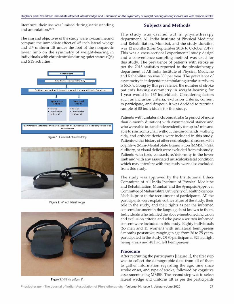
Rughani and Ravindran: Immediate effect of lateral wedge and uniform lift on the symmetry of weight bearing among individuals with chronic stroke
Physiotherapy - The Journal of Indian Association of Physiotherapists - Volume 14, Issue 1, January-June 2020 27
literature, their use was limited during static standing and ambulation.[3,7,8]
The aim and objectives of the study were to examine and compare the immediate effect of ¼th inch lateral wedge and ¼th uniform lift under the foot of the nonparetic lower limb on the symmetry of weight‑bearing in individuals with chronic stroke during quiet stance (QS) and STS activities.
Subjects and Methods
The study was carried out in physiotherapy department, All India Institute of Physical Medicine and Rehabilitation, Mumbai, and the study duration was 12 months (from September 2016 to October 2017). This was a cross‑sectional experimental study design and a convenience sampling method was used for this study. The prevalence of patients with stroke as per the 2015 statistics reported to the physiotherapy department at All India Institute of Physical Medicine and Rehabilitation was 300 per year. The prevalence of asymmetry in independent ambulating stroke survivors is 55.5%. Going by this prevalence, the number of stroke patients having asymmetry in weight‑bearing for 1 year would be 167 individuals. Considering factors such as inclusion criteria, exclusion criteria, consent to participate, and dropout, it was decided to recruit a sample of 80 individuals for this study.
Patients with unilateral chronic stroke (a period of more than 6‑month duration) with asymmetrical stance and who were able to stand independently for up to 5 min and able to rise from a chair without the use of hands, walking aids, and orthotic devices were included in this study. Patients with a history of other neurological diseases, with cognitive (Mini‑Mental State Examination [MMSE] <24), auditory, or visual deficit were excluded from this study. Patients with fixed contracture/deformity in the lower limb and with any associated musculoskeletal condition which may interfere with the study were also excluded from this study.
The study was approved by the Institutional Ethics Committee of All India Institute of Physical Medicine and Rehabilitation, Mumbai and the Synopsis Approval Committee of Maharashtra University of Health Sciences, Nashik, prior to the recruitment of participants. All the participants were explained the nature of the study, their role in the study, and their rights as per the informed consent document in the language best known to them. Individuals who fulfilled the above‑mentioned inclusion and exclusion criteria and who gave a written informed consent were included in this study. Eighty individuals (65 men and 15 women) with unilateral hemiparesis 6 months poststroke, ranging in age from 26 to 75 years, participated in the study. Of 80 participants, 32 had right hemiparesis and 48 had left hemiparesis.
ProcedureAfter recruiting the participants [Figure 1], the first step was to collect the demographic data from all of them to gather information regarding the age, time since stroke onset, and type of stroke, followed by cognitive assessment using MMSE. The second step was to select lateral wedge and uniform lift as per the participants
Figure 1: Flowchart of methodolog
Figure 2: ¼th inch lateral wedge
Figure 3: ¼th inch uniform lift
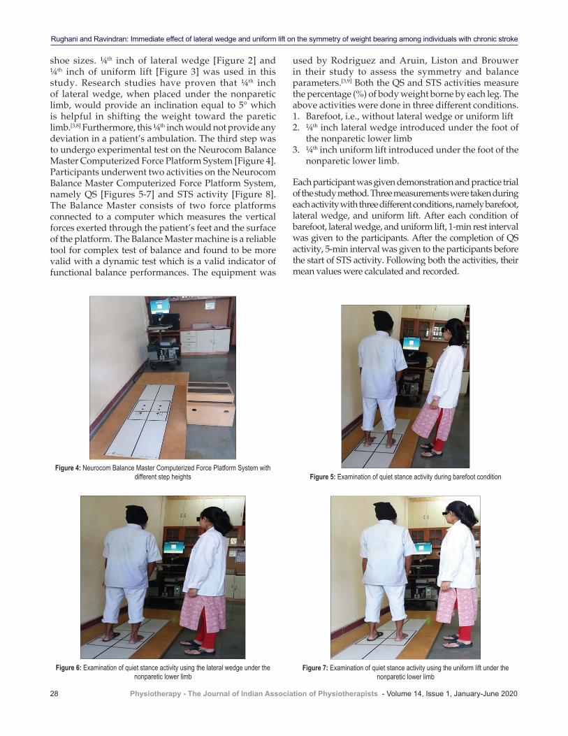
Rughani and Ravindran: Immediate effect of lateral wedge and uniform lift on the symmetry of weight bearing among individuals with chronic stroke
28 Physiotherapy - The Journal of Indian Association of Physiotherapists - Volume 14, Issue 1, January-June 2020
shoe sizes. ¼th inch of lateral wedge [Figure 2] and ¼th inch of uniform lift [Figure 3] was used in this study. Research studies have proven that ¼th inch of lateral wedge, when placed under the nonparetic limb, would provide an inclination equal to 5° which is helpful in shifting the weight toward the paretic limb.[3,8] Furthermore, this ¼th inch would not provide any deviation in a patient’s ambulation. The third step was to undergo experimental test on the Neurocom Balance Master Computerized Force Platform System [Figure 4]. Participants underwent two activities on the Neurocom Balance Master Computerized Force Platform System, namely QS [Figures 5‑7] and STS activity [Figure 8]. The Balance Master consists of two force platforms connected to a computer which measures the vertical forces exerted through the patient’s feet and the surface of the platform. The Balance Master machine is a reliable tool for complex test of balance and found to be more valid with a dynamic test which is a valid indicator of functional balance performances. The equipment was
used by Rodriguez and Aruin, Liston and Brouwer in their study to assess the symmetry and balance parameters.[3,9] Both the QS and STS activities measure the percentage (%) of body weight borne by each leg. The above activities were done in three different conditions.1. Barefoot, i.e., without lateral wedge or uniform lift2. ¼th inch lateral wedge introduced under the foot of
the nonparetic lower limb3. ¼th inch uniform lift introduced under the foot of the
nonparetic lower limb.
Each participant was given demonstration and practice trial of the study method. Three measurements were taken during each activity with three different conditions, namely barefoot, lateral wedge, and uniform lift. After each condition of barefoot, lateral wedge, and uniform lift, 1‑min rest interval was given to the participants. After the completion of QS activity, 5‑min interval was given to the participants before the start of STS activity. Following both the activities, their mean values were calculated and recorded.
Figure 5: Examination of quiet stance activity during barefoot condition
Figure 6: Examination of quiet stance activity using the lateral wedge under the nonparetic lower limb
Figure 7: Examination of quiet stance activity using the uniform lift under the nonparetic lower limb
Figure 4: Neurocom Balance Master Computerized Force Platform System with different step heights

Rughani and Ravindran: Immediate effect of lateral wedge and uniform lift on the symmetry of weight bearing among individuals with chronic stroke
Physiotherapy - The Journal of Indian Association of Physiotherapists - Volume 14, Issue 1, January-June 2020 29
Outcome measurePercentage (%) of weight bearing on the paretic limb (WP) during QS and STS activities was calculated and recorded from Balance Master Computerized Force Platform.
Statistical analysisAnalysis of data for this study was done using software SPSS (SPSS Statistics for Windows, version 18.0, SPSS Inc., Chicago, Ill., USA). The level of significance was kept at P < 0.05 at 95% confidence interval level for all statistical analyses. All the data were assessed for normality using Shapiro–Wilk normality test. The data followed a Gaussian curve of normal distribution during both QS and STS activities, and therefore, parametric tests were used to analyze the data.
Repeated‑measures ANOVA was used to see whether there was a significant difference in the percentage of weight‑bearing in the paretic limb between the three conditions: bare foot, lateral wedge, and uniform lift. This was followed by Tukey’s multiple comparison tests (post hoc analysis) to see which conditions performed better.
Results
The results of QS activity [Tables 1 and 2] showed that the percentage (%) of weight‑bearing on the paretic limb (WP) varied from 38.01% to 44.69%. Participants showed a significant improvement on weight‑bearing symmetry using lateral wedge and uniform lift as compared to the barefoot (P < 0.0001) [Figure 9]. However, no real difference in the percentage of weight‑bearing on the paretic limb when comparing lateral wedge and uniform lift (P = 0.6344) was noted.
The results of STS activity [Table 3] showed that the percentage (%) of weight‑bearing on the paretic limb (WP) varied from 30.02% to 42.04%. During the dynamic activity of STS, the percentage of weight‑bearing did not improve during any of the three conditions (barefoot, wedge, and lift) (P = 0.0567) [Figure 10]. Thus, no statistically significant difference between the three conditions was noted.
Discussion
Asymmetry is commonly seen during standing and functional activities such as STS and walking. About 55.5% independent ambulating stroke survivors show gait asymmetry.[7] According to the American Heart Association, stroke individuals are more prone to falls during transitional activities due to asymmetrical weight‑bearing.[10] Studies have shown that this asymmetry results from poor postural control, muscle weakness, impaired force generation, and sensory‑motor deficit.[2]
As per the literature,[11] there is an underloading of the paretic limb not only during static task of standing but
Figure 9: Percentage of weight bearing improved significantly using lateral wedge and uniform lift compared with barefoot (Tukey’s test, P < 0.05). Data expressed as
mean ± standard deviation. Participants with chronic stroke n = 80
Figure 10: No significant improvements were noted between the conditions of barefoot, lateral wedge, and uniform lift (Tukey’s test, P > 0.05). Data expressed as
mean ± standard deviation. Participants with chronic stroke n = 80
Figure 8: Examination of sit‑to‑stand activity
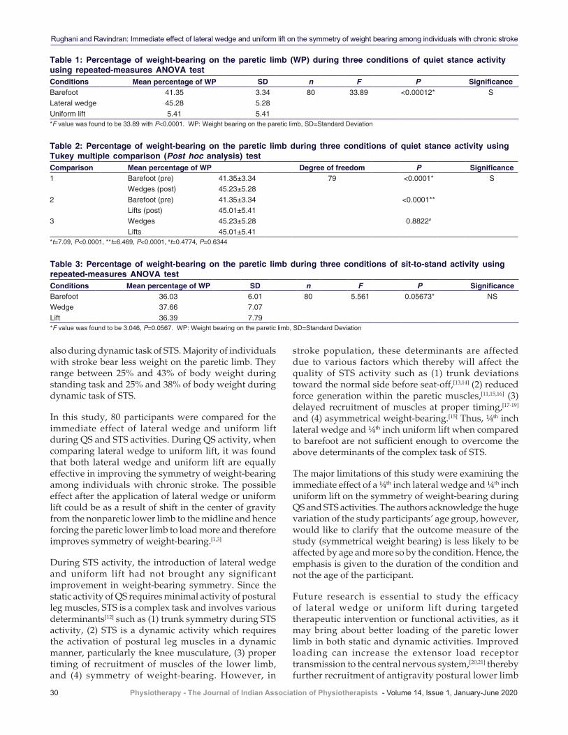
Rughani and Ravindran: Immediate effect of lateral wedge and uniform lift on the symmetry of weight bearing among individuals with chronic stroke
30 Physiotherapy - The Journal of Indian Association of Physiotherapists - Volume 14, Issue 1, January-June 2020
also during dynamic task of STS. Majority of individuals with stroke bear less weight on the paretic limb. They range between 25% and 43% of body weight during standing task and 25% and 38% of body weight during dynamic task of STS.
In this study, 80 participants were compared for the immediate effect of lateral wedge and uniform lift during QS and STS activities. During QS activity, when comparing lateral wedge to uniform lift, it was found that both lateral wedge and uniform lift are equally effective in improving the symmetry of weight‑bearing among individuals with chronic stroke. The possible effect after the application of lateral wedge or uniform lift could be as a result of shift in the center of gravity from the nonparetic lower limb to the midline and hence forcing the paretic lower limb to load more and therefore improves symmetry of weight‑bearing.[1,3]
During STS activity, the introduction of lateral wedge and uniform lift had not brought any significant improvement in weight‑bearing symmetry. Since the static activity of QS requires minimal activity of postural leg muscles, STS is a complex task and involves various determinants[12] such as (1) trunk symmetry during STS activity, (2) STS is a dynamic activity which requires the activation of postural leg muscles in a dynamic manner, particularly the knee musculature, (3) proper timing of recruitment of muscles of the lower limb, and (4) symmetry of weight‑bearing. However, in
stroke population, these determinants are affected due to various factors which thereby will affect the quality of STS activity such as (1) trunk deviations toward the normal side before seat‑off,[13,14] (2) reduced force generation within the paretic muscles,[11,15,16] (3) delayed recruitment of muscles at proper timing,[17‑19] and (4) asymmetrical weight‑bearing.[15] Thus, ¼th inch lateral wedge and ¼th inch uniform lift when compared to barefoot are not sufficient enough to overcome the above determinants of the complex task of STS.
The major limitations of this study were examining the immediate effect of a ¼th inch lateral wedge and ¼th inch uniform lift on the symmetry of weight‑bearing during QS and STS activities. The authors acknowledge the huge variation of the study participants’ age group, however, would like to clarify that the outcome measure of the study (symmetrical weight bearing) is less likely to be affected by age and more so by the condition. Hence, the emphasis is given to the duration of the condition and not the age of the participant.
Future research is essential to study the efficacy of lateral wedge or uniform lift during targeted therapeutic intervention or functional activities, as it may bring about better loading of the paretic lower limb in both static and dynamic activities. Improved loading can increase the extensor load receptor transmission to the central nervous system,[20,21] thereby further recruitment of antigravity postural lower limb
Table 2: Percentage of weight-bearing on the paretic limb during three conditions of quiet stance activity using Tukey multiple comparison (Post hoc analysis) testComparison Mean percentage of WP Degree of freedom P Significance1 Barefoot (pre) 41.35±3.34 79 <0.0001* S
Wedges (post) 45.23±5.282 Barefoot (pre) 41.35±3.34 <0.0001**
Lifts (post) 45.01±5.413 Wedges 45.23±5.28 0.8822#
Lifts 45.01±5.41*t=7.09, P<0.0001, **t=6.469, P<0.0001, #t=0.4774, P=0.6344
Table 1: Percentage of weight-bearing on the paretic limb (WP) during three conditions of quiet stance activity using repeated-measures ANOVA testConditions Mean percentage of WP SD n F P SignificanceBarefoot 41.35 3.34 80 33.89 <0.00012* SLateral wedge 45.28 5.28Uniform lift 5.41 5.41*F value was found to be 33.89 with P<0.0001. WP: Weight bearing on the paretic limb, SD=Standard Deviation
Table 3: Percentage of weight-bearing on the paretic limb during three conditions of sit-to-stand activity using repeated-measures ANOVA testConditions Mean percentage of WP SD n F P SignificanceBarefoot 36.03 6.01 80 5.561 0.05673* NSWedge 37.66 7.07Lift 36.39 7.79*F value was found to be 3.046, P=0.0567. WP: Weight bearing on the paretic limb, SD=Standard Deviation
Rughani and Ravindran: Immediate effect of lateral wedge and uniform lift on the symmetry of weight bearing among individuals with chronic stroke
Physiotherapy - The Journal of Indian Association of Physiotherapists - Volume 14, Issue 1, January-June 2020 31
muscles which can reduce the learned disuse of the paretic limb.
Conclusion
Both lateral wedge and uniform lift were found to be equally effective in improving the symmetry of weight‑bearing during QS activity. However, the introduction of lateral wedge or uniform lift had no significant improvement in the symmetry of weight‑bearing during STS activity when compared with the barefoot.
Declaration of patient consentThe authors certify that they have obtained all appropriate patient consent forms. In the form, the patients have given their consent for their images and other clinical information to be reported in the journal. The patients understand that their names and initials will not be published and due efforts will be made to conceal their identity, but anonymity cannot be guaranteed.
Financial support and sponsorshipNil.
Conflicts of interestThere are no conflicts of interest.
References
1. Aruin AS, Hanke T, Chaudhuri G, Harvey R, Rao N. Compelled weightbearing in persons with hemiparesis following stroke: The effect of a lift insert and goal‑directed balance exercise. J Rehabil Res Dev 2000;37:65‑72.
2. Wall JC, Turnbull GI. Gait asymmetries in residual hemiplegia. Arch Phys Med Rehabil 1986;67:550‑3.
3. Rodriguez GM, Aruin AS. The effect of shoe wedges and lifts on symmetry of stance and weight bearing in hemiparetic individuals. Arch Phys Med Rehabil 2002;83:478‑82.
4. Yu WH, Liu WY, Wong AM, Wang TC, Li YC, Lien HY. Effect of forced use of the lower extremity on gait performance and mobility of post‑acute stroke patients. J Phys Ther Sci 2015;27:421‑5.
5. Aruin AS, Rao N, Sharma A, Chaudhuri G. Compelled body weight shift approach in rehabilitation of individuals with chronic stroke. Top Stroke Rehabil 2012;19:556‑63.
6. Chitra J, Mishra S. A comparative study on the effect of compelled body weight shift therapy (CBWST) and modified constraint induced movement therapy (M CIMT) on weight bearing symmetry and balance in stroke patients. Int J Therapies Rehabilit Res 2015;4:209‑18.
7. Patterson KK, Parafianowicz I, Danells CJ, Closson V, Verrier MC, Staines WR, et al. Gait asymmetry in community‑ambulating stroke survivors. Arch Phys Med Rehabil 2008;89:304‑10.
8. Chen CH, Lin KH, Lu TW, Chai HM, Chen HL, Tang PF, et al. Immediate effect of lateral‑wedged insole on stance and ambulation after stroke. Am J Phys Med Rehabil 2010;89:48‑55.
9. Liston RA, Brouwer BJ. Reliability and validity of measures obtained from stroke patients using the Balance Master. Arch Phys Med Rehabil 1996;77:425‑30.
10. Nyberg L, Gustafson Y. Patient falls in stroke rehabilitation. A challenge to rehabilitation strategies. Stroke 1995;26:838‑42.
11. Eng JJ, Chu KS. Reliability and comparison of weight‑bearing ability during standing tasks for individuals with chronic stroke. Arch Phys Med Rehabil 2002;83:1138‑44.
12. Boukadida A, Piotte F, Dehail P, Nadeau S. Determinants of sit‑to‑stand tasks in individuals with hemiparesis post stroke: A review. Ann Phys Rehabil Med 2015;58:167‑72.
13. Mazza C, Stanhope SJ, Taviani A, Cappozzo A. Biomechanical modeling of sit‑to‑stand to upright posture for mobility assessment of persons witch chronic stroke. Arch Phys Med Rehabil 2006;91:288‑97.
14. Lecours J, Nadeau S, Gravel D, Teixera‑Salmela L. Interactions between foot placement, trunk frontal position, weight‑bearing and knee moment asymmetry at seat‑off during rising from a chair in healthy controls and persons with hemiparesis. J Rehabil Med 2008;40:200‑7.
15. Lomaglio MJ, Eng JJ. Muscle strength and weight‑bearing symmetry relate to sit‑to‑stand performance in individuals with stroke. Gait Posture 2005;22:126‑31.
16. Roebroeck ME, Doorenbosch CA, Harlaar J, Jacobs R, Lankhorst GJ. Biomechanics and muscular activity during sit‑to‑stand transfer. Clin Biomech (Bristol, Avon) 1994;9:235‑44.
17. Prudente C, Rodrigues‑de‑Paula F, Faria CD. Lower limb muscle activation during the sit‑to‑stand task in subjects who have had a stroke. Am J Phys Med Rehabil 2013;92:666‑75.
18. Cheng PT, Chen CL, Wang CM, Hong WH. Leg muscle activation patterns of sit‑to‑stand movement in stroke patients. Am J Phys Med Rehabil 2004;83:10‑6.
19. Silva A, Sousa AS, Pinheiro R, Ferraz J, Tavares JM, Santos R, et al. Activation timing of soleus and tibialis anterior muscles during sit‑to‑stand and stand‑to‑sit in post‑stroke vs. healthy subjects. Somatosens Mot Res 2013;30:48‑55.
20. Dietz V. Evidence for a load receptor contribution to the control of posture and locomotion. Neurosci Biobehav Rev 1998;22:495‑9.
21. Dietz V, Duysens J. Significance of load receptor input during locomotion: A review. Gait Posture 2000;11:102‑10.
32 © 2020 Physiotherapy - The Journal of Indian Association of Physiotherapists | Published by Wolters Kluwer - Medknow
Factors affecting kinesiophobia in coronary artery disease patientsNatasha Nitin Tungare, Razia K. Nagarwala1, Ashok K. Shyam2, Parag K. Sancheti2
Abstract:INTRODUCTION: Kinesiophobia may act as a barrier to physical activities. It is, therefore, an important domain to consider when assessing psychosocial characteristics in coronary artery disease (CAD) patients. Different factors may affect the occurrence of kinesiophobia in CAD patients, and hence it becomes important to study these factors so that proper psychological counseling can be included under patient education to have better rehabilitation.OBJECTIVE: The objective was to find if factors such as socioeconomic status, addictions, age, gender, anxiety, and financial security (medical insurance) affect kinesiophobia in CAD patients so as to know how these factors are significant in predicting kinesiophobia and direct patients to proper psychological counseling in future.METHODOLOGY: A cross‑sectional study was done on 62 patients, diagnosed with CAD at government and private hospitals and clinics were included in the study. Tampa Scale for Kinesiophobia Heart was used to assess the kinesiophobia. Logistic regression analyses and Spearman correlation were done for factors taken with kinesiophobia as a dependent variable and considered factors as independent.RESULTS: About 83.87% of population had high level of kinesiophobia. Gender (odds ratio >1) and anxiety (P < 0.05) were significant in predicting kinesiophobia whereas age (P > 0.05), financial security (odds ratio <1), socioeconomic status (P > 0.05), and presence of addictions (odds ratio <1) did not show any correlation with kinesiophobia.CONCLUSION: Of six factors taken into consideration, gender and anxiety significantly affect kinesiophobia, whereas age, financial security, socioeconomic status, and presence of addictions do not significantly affect kinesiophobia.Keywords:Coronary artery disease, kinesiophobia, Tampa Scale Heart
Introduction
Coronary artery disease (CAD) is blockage of one or more arteries that
supply blood to the heart, usually due to atherosclerosis. Ischemic or chronic heart disease is another term for CAD. Myocardial infarction occurs in later stages (mostly chronic), eventually leading to cardiac failure. These diseases are caused mainly due to thrombosis in coronary arteries causing ischemia. Symptoms of
the disease include pain in chest and arms, breathlessness, nausea, vomiting, syncope and anxiety, and fear of impending death.[1]
Kinesiophobia is defined as “an excessive, irrational, and debilitating fear of physical movement and activity, resulting from a feeling of vulnerability to painful injury or reinjury.”[2] Based on clinical experience, patients with CAD often have doubts that if physical activity can be performed safely due to the disease.[3] Despite strong evidence for the benefits of exercise‑based cardiac rehabilitation, studies show poor
Address for correspondence:
Prof. Dr. Razia Nagarwala, Department of Cardiology, Sancheti Institute College of Physiotherapy, Shivaji
Nagar, Pune - 411 005, Maharashtra, India.
E-mail: [email protected]
Submission: 13-09-2019Revision: 18-10-2019
Accepted: 17-03-2020Published: 29-06-2020
Departments of Cardio‑Respiratory Physiotherapy
and 1Cardiology, Sancheti Institute
College of Physiotherapy, 2Sancheti Institute
for Orthopedics and Rehabilitation, Pune,
Maharashtra, India
Original Article
Access this article onlineQuick Response Code:
Website:www.pjiap.org
DOI:10.4103/PJIAP.PJIAP_29_19
How to c i te th is ar t ic le : Tungare NN, Nagarwala RK, Shyam AK, Sancheti PK. Factors affecting kinesiophobia in coronary artery disease patients. Physiother - J Indian Assoc Physiother 2020;14:32-6.
This is an open access journal, and articles are distributed under the terms of the Creative Commons Attribution-NonCommercial-ShareAlike 4.0 License, which allows others to remix, tweak, and build upon the work non-commercially, as long as appropriate credit is given and the new creations are licensed under the identical terms.
For reprints contact: [email protected]
Tungare, et al.: Kinesiophobia in Cardiac Patients
Physiotherapy - The Journal of Indian Association of Physiotherapists - Volume 14, Issue 1, January-June 2020 33
attendance and adherence. There is a need to identify obstacles that prevent patients from participating in cardiac rehabilitation and increasing their levels of physical activity, including exercise. Daily activities and functional capacity may be reduced to avoid pain, leading to decreased physical activity, disuse, disability, and further chronicity of pain.[4] Epidemiological studies have provided convincing evidence about the importance of physical activity in the cardiac rehabilitation process.[5] Kinesiophobia may act as a barrier to physical activities.[6] Kinesiophobia is, therefore, an important domain to consider when assessing psychosocial characteristics in CAD patients.[7] India is a country with a diverse range of cultures, ethnicities, religions, and languages. Socioeconomic and financial security factors do play a role in Indian beliefs, and hence, may affect the perception toward healthcare. Hence, it becomes important to consider these factors.[8]
Tampa Scale for Kinesiophobia‑Swedish version (TSK‑SV Heart) is for patients with CAD. The validity and reliability of this scale is published. This scale consists of four‑component model. The four components include “Perceived danger of heart problem,” “Avoidance of exercise,” “Fear of injury/reinjury,” and “Dysfunctional self.” Kinesiophobia: the TSK‑SV heart comprises 17 statements that assess the subjective rating of kinesiophobia. The statements are rated from “strongly disagree” (score = 1) to “strongly agree” (score = 4) on a four‑point Likert scale. The total score varies between 17 and 68. The higher the value, the greater is the degree of kinesiophobia.[7,9,10]
Demographic factors such as age, gender, and presence of addictions do affect the prevalence of cardiac disorders, and hence, it becomes necessary to find if these factors affect kinesiophobia also in CAD patients. Anxiety can hinder psychosocial adjustment to the chronicity of cardiovascular disease and physical recovery after an acute event. Higher anxiety is predictive of worse quality of life among patients with cardiovascular disease.[11‑13]
Methodology
EthicsThe procedure followed for the study was in accordance with the ethical standards of the ethical committee. Informed consent was obtained from all the patients before handling them the questionnaires to answer, and we assure the confidentiality of the identity of patients. The institutional ethical committee has approved the study for publication.
Study designIt is a cross‑sectional study done on 62 patients (39 males, 26 females: mean age: 57 ± 10) from government and private hospitals and clinics, diagnosed with CAD was taken. Convenient sampling technique was done. Inclusion criteria was mainly CAD diagnosed patients which consisted of ischemic heart disease (n = 25), myocardial infarction (n = 26), and cardiac failure patients (n = 11). The patients who were already operated for cardiac disease or had any recent injury or undergone any surgery were excluded from the study.
ProcedureAfter the diagnosis of CAD, when patients were in stable condition, the questionnaire (scales) was filled by them. The scales used were also translated into local languages for patients who did not understand English. The face and content validation of these translated scales was done.
MeasurementsPersonal factorsAge (mean age: 57 ± 10), gender, addictions, and financial security (medical insurance): these factors were obtained from the demographic data of CAD patients.
Socioeconomic statusKuppuswamy scale (2018) is an updated version to calculate the socioeconomic class of the Indian citizens. This scale depends on the overall income of family or individual.[14]
AnxietyBeck inventory anxiety scale was used. It is a self‑report measure of anxiety. It have 21‑items with scorings done on the LIKERT scale ranging from 0 to 3.[11]
Statistical analysesData were analyzed using SPSS version 20 software (IBM Corp. Released 2011. IBM SPSS Statistics for Windows, Version 20.0. Armonk, NY: IBM Corp.). Descriptive statistics was used to find out the number of CAD patients having kinesiophobia and classifying them on the basis of gender, having/not having addictions, and having/not having insurance. Significance of prediction of kinesiophobia by gender, insurance, and addictions was found by Logistic regression, and Spearman correlation was done for age, socioeconomic status, and anxiety. The significance level was set at P ≤ 0.05.
Results
Of 62 CAD patients, 52 patients that is 83.87% persists the high level of kinesiophobia [Table 1]. When considering
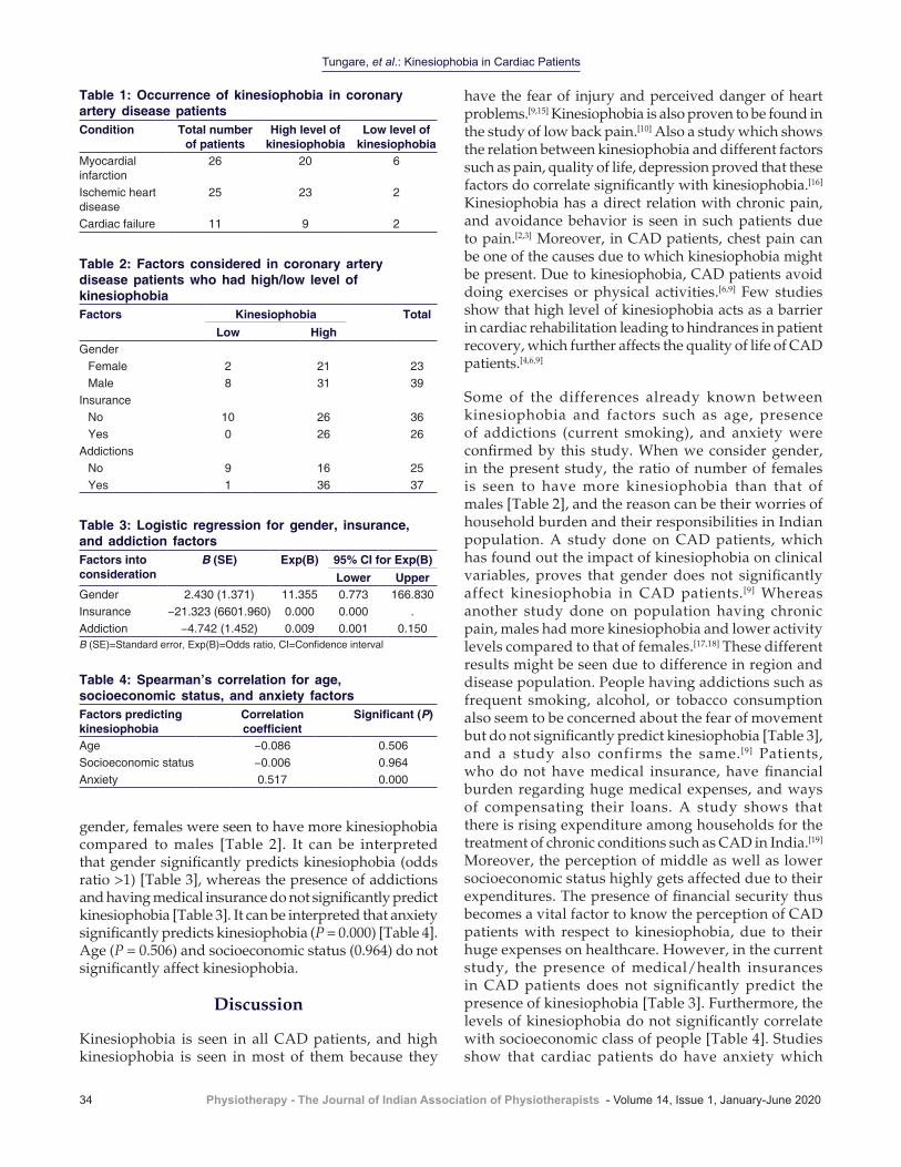
Tungare, et al.: Kinesiophobia in Cardiac Patients
34 Physiotherapy - The Journal of Indian Association of Physiotherapists - Volume 14, Issue 1, January-June 2020
gender, females were seen to have more kinesiophobia compared to males [Table 2]. It can be interpreted that gender significantly predicts kinesiophobia (odds ratio >1) [Table 3], whereas the presence of addictions and having medical insurance do not significantly predict kinesiophobia [Table 3]. It can be interpreted that anxiety significantly predicts kinesiophobia (P = 0.000) [Table 4]. Age (P = 0.506) and socioeconomic status (0.964) do not significantly affect kinesiophobia.
Discussion
Kinesiophobia is seen in all CAD patients, and high kinesiophobia is seen in most of them because they
have the fear of injury and perceived danger of heart problems.[9,15] Kinesiophobia is also proven to be found in the study of low back pain.[10] Also a study which shows the relation between kinesiophobia and different factors such as pain, quality of life, depression proved that these factors do correlate significantly with kinesiophobia.[16] Kinesiophobia has a direct relation with chronic pain, and avoidance behavior is seen in such patients due to pain.[2,3] Moreover, in CAD patients, chest pain can be one of the causes due to which kinesiophobia might be present. Due to kinesiophobia, CAD patients avoid doing exercises or physical activities.[6,9] Few studies show that high level of kinesiophobia acts as a barrier in cardiac rehabilitation leading to hindrances in patient recovery, which further affects the quality of life of CAD patients.[4,6,9]
Some of the differences already known between kinesiophobia and factors such as age, presence of addictions (current smoking), and anxiety were confirmed by this study. When we consider gender, in the present study, the ratio of number of females is seen to have more kinesiophobia than that of males [Table 2], and the reason can be their worries of household burden and their responsibilities in Indian population. A study done on CAD patients, which has found out the impact of kinesiophobia on clinical variables, proves that gender does not significantly affect kinesiophobia in CAD patients.[9] Whereas another study done on population having chronic pain, males had more kinesiophobia and lower activity levels compared to that of females.[17,18] These different results might be seen due to difference in region and disease population. People having addictions such as frequent smoking, alcohol, or tobacco consumption also seem to be concerned about the fear of movement but do not significantly predict kinesiophobia [Table 3], and a study also confirms the same.[9] Patients, who do not have medical insurance, have financial burden regarding huge medical expenses, and ways of compensating their loans. A study shows that there is rising expenditure among households for the treatment of chronic conditions such as CAD in India.[19] Moreover, the perception of middle as well as lower socioeconomic status highly gets affected due to their expenditures. The presence of financial security thus becomes a vital factor to know the perception of CAD patients with respect to kinesiophobia, due to their huge expenses on healthcare. However, in the current study, the presence of medical/health insurances in CAD patients does not significantly predict the presence of kinesiophobia [Table 3]. Furthermore, the levels of kinesiophobia do not significantly correlate with socioeconomic class of people [Table 4]. Studies show that cardiac patients do have anxiety which
Table 1: Occurrence of kinesiophobia in coronary artery disease patientsCondition Total number
of patientsHigh level of
kinesiophobiaLow level of
kinesiophobiaMyocardial infarction
26 20 6
Ischemic heart disease
25 23 2
Cardiac failure 11 9 2
Table 2: Factors considered in coronary artery disease patients who had high/low level of kinesiophobiaFactors Kinesiophobia Total
Low HighGender
Female 2 21 23Male 8 31 39
InsuranceNo 10 26 36Yes 0 26 26
AddictionsNo 9 16 25Yes 1 36 37
Table 4: Spearman’s correlation for age, socioeconomic status, and anxiety factorsFactors predicting kinesiophobia
Correlation coefficient
Significant (P)
Age −0.086 0.506Socioeconomic status −0.006 0.964Anxiety 0.517 0.000
Table 3: Logistic regression for gender, insurance, and addiction factorsFactors into consideration
B (SE) Exp(B) 95% CI for Exp(B)Lower Upper
Gender 2.430 (1.371) 11.355 0.773 166.830Insurance −21.323 (6601.960) 0.000 0.000 .Addiction −4.742 (1.452) 0.009 0.001 0.150B (SE)=Standard error, Exp(B)=Odds ratio, CI=Confidence interval

Tungare, et al.: Kinesiophobia in Cardiac Patients
Physiotherapy - The Journal of Indian Association of Physiotherapists - Volume 14, Issue 1, January-June 2020 35
affects their physical activities, and hence, proves as a causative factor for kinesiophobia.[9,20‑22] Patients having high amount of anxiety, having high level of kinesiophobia due to fear of injury, and perception of the risk factors associated with heart disease. Two studies have shown significant anxiety and depression in patients suffering from myocardial infarction.[12,13] Some studies have also shown kinesiophobia having its impact on psychological factors such as anxiety and depression in CAD patients.[9] Since age is not significantly predicting kinesiophobia [Table 4], CAD patients of any age, younger ones, or older can have a fear of movement. A study done on CAD patients, which has found out the impact of kinesiophobia on clinical variables, proves that age does not have significant affection over kinesiophobia.[9] Some studies do suggest that age has a significant affection over kinesiophobia but in other patient population.[22,23]
Strengths and limitations of studyThis study helps us to know the factors affecting kinesiophobia so that we can direct a proper psychological counseling to prevent kinesiophobia occurring in CAD patients so as to have a better physical rehabilitation. There were no limitations in this study.
Future scope of the studyConsidering these factors, proper interventions can be carried out to treat or prevent kinesiophobia in CAD patients.
Conclusion
Of the six factors taken into consideration, two factors which are gender and anxiety significantly affect kinesiophobia and four factors which are age, financial security, socioeconomic status, and presence of addictions do not significantly affect the kinesiophobia.
AcknowledgementsThe authors would like to thank the patients who participated in this study, the hospitals and clinics who permitted to conduct this study, and the translators who helped in the translation of the scales used in this study.
Financial support and sponsorshipNil.
Conflicts of interestThere are no conflicts of interest.
References
1. Colledge NR, Walker BR, Davidson’s Principles and Practice of Medicine. 21st ed. Churchill Livingstone Elsevier, 2010.
2. Korri SH, Miller RP, Todd DD. Kinesiophobia: A new view of chronic pain behavior. Pain Manag 1990;3:35‑43.
3. Philips HC. Avoidance behaviour and its role in sustaining chronic pain. Behaviour Res Ther 1987;25:273‑9.
4. Vlaeyen JW, Linton SJ. Fear‑avoidance and its consequences in chronic musculoskeletal pain: A state of the art. Pain 2000;85:317‑32.
5. Szmit S, Filipiak KJ. Czy u pacjenta z choroba serca wazna jest aktywnosc fizyczna? Is the physical activity important for the patient with a heart disease? Przewodnik Lekarza 2007;7:32‑8.
6. Kocjan J, Knapik A, Barriers of physical activity (kinesiophobia) in patients subjected to cardiac rehabilitation, Baltic J Health Phys Activ 2014;6:291.
7. Bäck M, Jansson B, Cider Å, Herlitz J, Lundberg M. Validation of a questionnaire to detect Kinesiophobia (fear of movement) in patients with coronary artery disease. J Rehabil Med 2012;44:363‑9.
8. Worthington RP, Gogne A. Cultural aspects of primary healthcare in India: A case‑based analysis. Asia Pacific Fam Med 2011;10:8.
9. Bäck M, Cider Å, Herlitz J, Lundberg M. The impact on kinesiophobia (fear of movement) by clinical variables for patients with coronary artery disease. Int J Cardiol 2013;167:391‑7.
10. Goubert L, Crombez G, van Damme S, Vlaeyen JW, Bijttebier P, Roelofs J. Confirmatory factor analysis of the Tampa Scale for Kinesiophobia: Invariant two‑factor model across low back pain patients and fibromyalgia patients. Clin J Pain 2004;20:103‑10.
11. Beck AT, Epstein N, Brown G, Steer RA. An inventory for measuring clinical anxiety: Psychometric properties. J Consulting Clin Psychol 1988;56:893.
12. Mayou RA, Gill D, Thompson DR, Day A, Hicks N, Volmink J, et al. Depression and anxiety as predictors of outcome after myocardial infarction. Psychosomatic Med 2000;62:212‑9.
13. Lane D, Carroll D, Ring C, Beevers DG, Lip GY. Effects of depression and anxiety on mortality and quality‑of‑life 4 months after myocardial infarction. J Psychosomatic Res 2000;49:229‑38.
14. Saleem MS. Modified kuppuswamy scale updated for year 2018. Paripex Indian J Res 2018;7:2250‑1991.
15. Lundberg MK, Styf J, Carlsson SG. A psychometric evaluation of the Tampa Scale for Kinesiophobia—from a physiotherapeutic perspective. Physiother Theory Pract 2004;20:121‑33.
16. Oskay D, Tuna Z, Düzgün İ, Elbasan B, Yakut Y, Tufan A. Relationship between kinesiophobia and pain, quality of life, functional status, disease activity, mobility, and depression in patients with ankylosing spondylitis. Turkish J Med Sci 2017;47:1340‑7.
17. Silva NS, Abreu SS, Suassuna PD. Kinesiophobia and associated factors in elderly females with chronic musculoskeletal pain: pilot study. Revista Dor 2016;17:188‑91.
18. Rovner GS, Sunnerhagen KS, Björkdahl A, Gerdle B, Borsbo B, Johansson F, et al. Chronic pain and sex‑differences; women accept and move, while men feel blue. PloS One 2017;12:e0175737.
19. Chauhan AS, Mukherjee K. Economic burden of coronary heart disease in North India. Int J Noncommun Dis 2016;1:18.
20. Lüning Bergsten C, Lundberg M, Lindberg P, Elfving B. Change

Tungare, et al.: Kinesiophobia in Cardiac Patients
36 Physiotherapy - The Journal of Indian Association of Physiotherapists - Volume 14, Issue 1, January-June 2020
in kinesiophobia and its relation to activity limitation after multidisciplinary rehabilitation in patients with chronic back pain. Dis Rehabil 2012;34:852‑8.
21. Reneman MF, Jorritsma W, Dijkstra SJ, Dijkstra PU. Relationship between kinesiophobia and performance in a functional capacity evaluation. J Occup Rehabil 2003;13:277‑85.
22. Larsson C, Hansson EE, Sundquist K, Jakobsson U. Kinesiophobia
and its relation to pain characteristics and cognitive affective variables in older adults with chronic pain. BMC Geriatrics 2016;16:128.
23. Larsson C, Hansson EE, Sundquist K, Jakobsson U. Psychometric properties of the Tampa scale of Kinesiophobia (TSK‑11) among older people with chronic pain. Physiother Theory Pract 2014;30:421‑8.
© 2020 Physiotherapy - The Journal of Indian Association of Physiotherapists | Published by Wolters Kluwer - Medknow 37
Development of training manuals for community disability workersKavitha Raja, Saumen Gupta1, Jerin Mathew2, Pratiksha Rao3
Abstract:BACKGROUND: People with disability (PWD) living in the community often require ongoing assistance for self‑care and for continuing rehabilitation maintenance programs. While awareness material is easily available, training for volunteers working with disabled population and family members of PWD is hard to come by.AIM: The aim of this study was to develop resource material for training of disability workers, community-based rehabilitation (CBR) workers, and school teachers in rural areas in specific areas.METHODOLOGY: Using standard guidelines, training manuals were prepared under five specific categories to train CBR volunteers to undertake routine maintenance exercise programs and functional training strategies. These manuals were translated into Kannada, and in stepwise process fieldworkers, school teachers and CBR workers were trained. Revisions were made based on feedback received from stakeholders.RESULTS: Five manuals pertaining to specific areas have been prepared and tested.CONCLUSION: The training manuals developed for CBR workers are available in English and Kannada and were found to be useful by the target population in this preliminary study.Keywords:Community‑based rehabilitation training, community‑based rehabilitation workers, persons with disabilities
Introduction
People with disability (PWD) living in the community often require ongoing
assistance for self‑care and for continuing rehabilitation maintenance programs. While awareness material is easily available, training for volunteers working with disabled population and family members of PWD is hard to come by. Moreover, available resources are often inadequate in scope for the specific requirements of people living with chronic illness or disability in the community in India. The commonly available training material has been developed by the World Health Organization (WHO), United Nations Educational, Scientific and Cultural
Organization (UNESCO), and other arms of the united nations.[1,2]
A review of training manuals from the WHO, community‑based rehabilitation (CBR) g u i d e l i n e s , C h i l d h o o d D i s a b i l i t y Information Kit from UNESCO,[3] and CBR training manual from light of the world[4] revealed that they had limited information and were rarely specific to conditions. Due to the lack of condition‑specific information, the community worker would be required to use reasoning to identify what exercises/techniques were to be taught to the individual patient.[5] This is often beyond the training and understanding of casual community workers. Hence, the need for India‑specific manuals with focus on specific functional deficits was envisioned.
Address for correspondence:
Dr. Kavitha Raja, JSS College of Physiotherapy,
Mysore - 570 004, Karnataka, India.
E-mail: [email protected]
Submission: 27-08-2019Revision: 12-02-2020
Accepted: 26-03-2020Published: 29-06-2020
Department of Community Based Rehabilitation,
JSS College of Physiotherapy, Mysore,
Karnataka, 1Department of Physiotherapy, Sikkim
Manipal University, Gangtok, Sikkim, India,
2Centre for Health, Activity, and Rehabilitation
Research, School of Physiotherapy, University
of Otago, Dunedin, New Zealand,
3Independent Practitioner (PT), Bengaluru, Karnataka, India
Original Article
Access this article onlineQuick Response Code:
Website:www.pjiap.org
DOI:10.4103/PJIAP.PJIAP_19_19
How to cite this article: Raja K, Gupta S, Mathew J, Rao P. Development of training manuals for community disability workers. Physiother - J Indian Assoc Physiother 2020;14:37-40.
This is an open access journal, and articles are distributed under the terms of the Creative Commons Attribution-NonCommercial-ShareAlike 4.0 License, which allows others to remix, tweak, and build upon the work non-commercially, as long as appropriate credit is given and the new creations are licensed under the identical terms.
For reprints contact: [email protected]
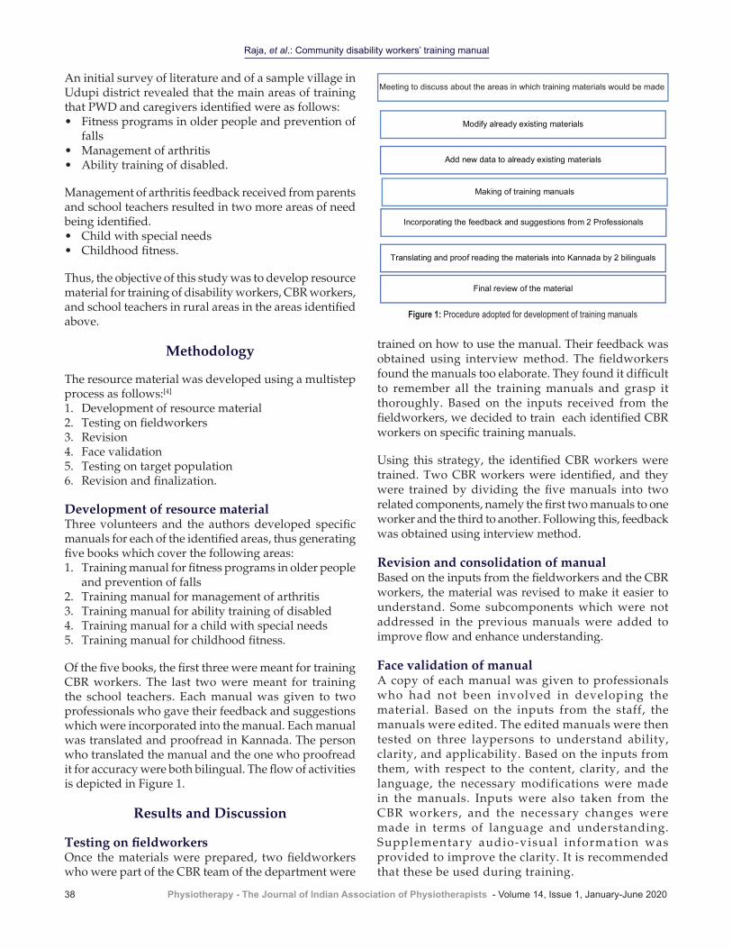
Raja, et al.: Community disability workers’ training manual
38 Physiotherapy - The Journal of Indian Association of Physiotherapists - Volume 14, Issue 1, January-June 2020
An initial survey of literature and of a sample village in Udupi district revealed that the main areas of training that PWD and caregivers identified were as follows:• Fitness programs in older people and prevention of
falls• Management of arthritis• Ability training of disabled.
Management of arthritis feedback received from parents and school teachers resulted in two more areas of need being identified.• Child with special needs• Childhood fitness.
Thus, the objective of this study was to develop resource material for training of disability workers, CBR workers, and school teachers in rural areas in the areas identified above.
Methodology
The resource material was developed using a multistep process as follows:[4]
1. Development of resource material2. Testing on fieldworkers3. Revision4. Face validation5. Testing on target population6. Revision and finalization.
Development of resource materialThree volunteers and the authors developed specific manuals for each of the identified areas, thus generating five books which cover the following areas:1. Training manual for fitness programs in older people
and prevention of falls2. Training manual for management of arthritis3. Training manual for ability training of disabled4. Training manual for a child with special needs5. Training manual for childhood fitness.
Of the five books, the first three were meant for training CBR workers. The last two were meant for training the school teachers. Each manual was given to two professionals who gave their feedback and suggestions which were incorporated into the manual. Each manual was translated and proofread in Kannada. The person who translated the manual and the one who proofread it for accuracy were both bilingual. The flow of activities is depicted in Figure 1.
Results and Discussion
Testing on fieldworkersOnce the materials were prepared, two fieldworkers who were part of the CBR team of the department were
trained on how to use the manual. Their feedback was obtained using interview method. The fieldworkers found the manuals too elaborate. They found it difficult to remember all the training manuals and grasp it thoroughly. Based on the inputs received from the fieldworkers, we decided to train each identified CBR workers on specific training manuals.
Using this strategy, the identified CBR workers were trained. Two CBR workers were identified, and they were trained by dividing the five manuals into two related components, namely the first two manuals to one worker and the third to another. Following this, feedback was obtained using interview method.
Revision and consolidation of manualBased on the inputs from the fieldworkers and the CBR workers, the material was revised to make it easier to understand. Some subcomponents which were not addressed in the previous manuals were added to improve flow and enhance understanding.
Face validation of manualA copy of each manual was given to professionals who had not been involved in developing the material. Based on the inputs from the staff, the manuals were edited. The edited manuals were then tested on three laypersons to understand ability, clarity, and applicability. Based on the inputs from them, with respect to the content, clarity, and the language, the necessary modifications were made in the manuals. Inputs were also taken from the CBR workers, and the necessary changes were made in terms of language and understanding. Supplementary audio‑visual information was provided to improve the clarity. It is recommended that these be used during training.
Add new data to already existing materials
Incorporating the feedback and suggestions from 2 Professionals
Translating and proof reading the materials into Kannada by 2 bilinguals
Final review of the material
Modify already existing materials
Making of training manuals
Meeting to discuss about the areas in which training materials would be made
Figure 1: Procedure adopted for development of training manuals
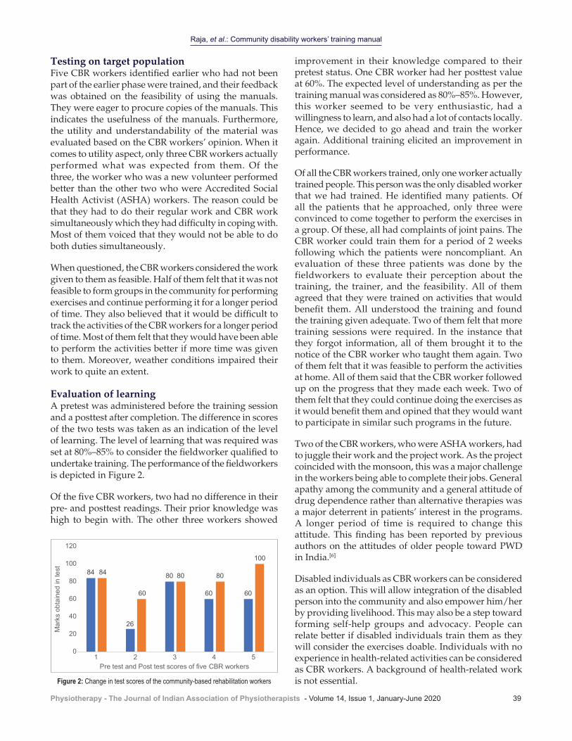
Raja, et al.: Community disability workers’ training manual
Physiotherapy - The Journal of Indian Association of Physiotherapists - Volume 14, Issue 1, January-June 2020 39
Testing on target populationFive CBR workers identified earlier who had not been part of the earlier phase were trained, and their feedback was obtained on the feasibility of using the manuals. They were eager to procure copies of the manuals. This indicates the usefulness of the manuals. Furthermore, the utility and understandability of the material was evaluated based on the CBR workers’ opinion. When it comes to utility aspect, only three CBR workers actually performed what was expected from them. Of the three, the worker who was a new volunteer performed better than the other two who were Accredited Social Health Activist (ASHA) workers. The reason could be that they had to do their regular work and CBR work simultaneously which they had difficulty in coping with. Most of them voiced that they would not be able to do both duties simultaneously.
When questioned, the CBR workers considered the work given to them as feasible. Half of them felt that it was not feasible to form groups in the community for performing exercises and continue performing it for a longer period of time. They also believed that it would be difficult to track the activities of the CBR workers for a longer period of time. Most of them felt that they would have been able to perform the activities better if more time was given to them. Moreover, weather conditions impaired their work to quite an extent.
Evaluation of learningA pretest was administered before the training session and a posttest after completion. The difference in scores of the two tests was taken as an indication of the level of learning. The level of learning that was required was set at 80%–85% to consider the fieldworker qualified to undertake training. The performance of the fieldworkers is depicted in Figure 2.
Of the five CBR workers, two had no difference in their pre‑ and posttest readings. Their prior knowledge was high to begin with. The other three workers showed
improvement in their knowledge compared to their pretest status. One CBR worker had her posttest value at 60%. The expected level of understanding as per the training manual was considered as 80%–85%. However, this worker seemed to be very enthusiastic, had a willingness to learn, and also had a lot of contacts locally. Hence, we decided to go ahead and train the worker again. Additional training elicited an improvement in performance.
Of all the CBR workers trained, only one worker actually trained people. This person was the only disabled worker that we had trained. He identified many patients. Of all the patients that he approached, only three were convinced to come together to perform the exercises in a group. Of these, all had complaints of joint pains. The CBR worker could train them for a period of 2 weeks following which the patients were noncompliant. An evaluation of these three patients was done by the fieldworkers to evaluate their perception about the training, the trainer, and the feasibility. All of them agreed that they were trained on activities that would benefit them. All understood the training and found the training given adequate. Two of them felt that more training sessions were required. In the instance that they forgot information, all of them brought it to the notice of the CBR worker who taught them again. Two of them felt that it was feasible to perform the activities at home. All of them said that the CBR worker followed up on the progress that they made each week. Two of them felt that they could continue doing the exercises as it would benefit them and opined that they would want to participate in similar such programs in the future.
Two of the CBR workers, who were ASHA workers, had to juggle their work and the project work. As the project coincided with the monsoon, this was a major challenge in the workers being able to complete their jobs. General apathy among the community and a general attitude of drug dependence rather than alternative therapies was a major deterrent in patients’ interest in the programs. A longer period of time is required to change this attitude. This finding has been reported by previous authors on the attitudes of older people toward PWD in India.[6]
Disabled individuals as CBR workers can be considered as an option. This will allow integration of the disabled person into the community and also empower him/her by providing livelihood. This may also be a step toward forming self‑help groups and advocacy. People can relate better if disabled individuals train them as they will consider the exercises doable. Individuals with no experience in health‑related activities can be considered as CBR workers. A background of health‑related work is not essential.
84
26
80
60 60
84
60
80 80
100
0
20
40
60
80
100
120
1 2 3 4 5
Mar
ks o
btai
ned
in te
st
Pre test and Post test scores of five CBR workers
Figure 2: Change in test scores of the community‑based rehabilitation workers

Raja, et al.: Community disability workers’ training manual
40 Physiotherapy - The Journal of Indian Association of Physiotherapists - Volume 14, Issue 1, January-June 2020
Regarding the manuals for teachers, teachers in two local schools were trained. The physical education teachers were interested and appreciative of the manuals. Although physical education curriculum is given as a manual to schools following the Central Board of Secondary Education curriculum, such a system is not currently present in the government schools. Hence, the teachers were appreciative about the fitness manual. In the scenario of inclusive education, the manual of special children was also appreciated by teachers.
Community‑based programs to improve childhood health and fitness has been suggested by previous authors in a large study.[7,8] This need is expected to be partly fulfilled by this study. The manuals in English and Kannada are available with the authors on request free of charge.
Conclusion
The training manuals developed for CBR workers are available in English and Kannada and were found to be useful by the target population in this preliminary study.
Financial support and sponsorshipThis study is an offshoot of a project funded by WHO, India WHO Regn 2011/138001‑0, and was partly conducted under the aegis of Department of
Physiotherapy, Manipal College of Allied Health Sciences, Manipal, and partly under JSS College of Physiotherapy, Mysuru.
Conflicts of interestThere are no conflicts of interest.
References
1. Thomas M, Thomas MJ. Manual for CBR planners. Asia Pac Disabil Rehabil J 2003; 1:1‑88.
2. Wirz S. Training of CBR personnel. Asia Pac Disabil Rehabil J 2000; 2:100‑12.
3. Houtrow AJ, Larson K, Olson LM, Newacheck PW, Halfon N. Changing trends of childhood disability, 2001–2011. Pediatrics. 2014;134:530‑8.
4. Robb A. Report on “Technical Meeting on Development of CBR M&E” and the 1st CBR World Congress; 2012. p. 26‑8.
5. Robertson J, Emerson E, Hatton C, Yasamy MT. Efficacy of community‑based rehabilitation for children with or at significant risk of intellectual disabilities in low‑ and middle‑income countries: A review. J Appl Res Intellect Disabil 2012;25:143‑54.
6. World Health Organization. Information, education and communication: A guide for AIDS programme managers. WHO Regional Office for South‑East Asia; 2000.
7. Bakheit AM, Shanmugalingam V. A study of the attitudes of a rural Indian community toward people with physical disabilities. Clin Rehabil 1997;11:329‑34.
8. Ranjani H, Pradeepa R, Mehreen TS, Anjana RM, Anand K, Garg R, et al. Determinants, consequences and prevention of childhood overweight and obesity: An Indian context. Indian J Endocrinol Metab 2014;18 Suppl. S1:S17‑25.

© 2020 Physiotherapy - The Journal of Indian Association of Physiotherapists | Published by Wolters Kluwer - Medknow 41
Relationship between physical activity and cognition among young adultsSalvi Shah, Suchi Shah1, Shivani Chauhan1
Abstract:BACKGROUND AND PURPOSE: Poor cognitive functioning is a predictor of mortality at all ages and as such can be seen as a marker of general health status. Leisure‑time physical activity (PA) has been shown to have a beneficial impact on cognitive function. Existing research has focused on this association in children and the elderly. However, less is focused on young adults. Hence, the purpose of the present study was to find the relationship between PA and cognition among young adults.METHODOLOGY: Two hundred individuals (18–24 years) were selected from different colleges of Surat city after obtaining permission from the college. All necessary instructions to perform cognition tests (Rey Verbal Auditory Learning Test, Stroop test, and Trail Making Test A and B) and to fill up the International PA Questionnaire were given to participants after obtaining the consent form. Spearman’s correlation was used to analyze relationships between PA and cognition among participants.RESULTS: Results showed a significant positive correlation (r = 0.53, 0.44, and 0.68) between PA and all the three cognitive measure tests (P < 0.05).CONCLUSION: PA is an important factor in cognitive functioning among young adults. This finding of young adults suggests that PA may be beneficial to cognition during early and middle periods of the human life span and may continue to protect against age‑related loss of cognitive function during older adulthood.Keywords:Aging, International Physical Activity Questionnaire, physical activity, Rey Verbal Auditory Learning Test, Stroop test, Trail Making Test
Introduction
Regular physical activity (PA) is known to have numerous physical benefits. A
physically active lifestyle is associated with a decreased risk of developing the most prevalent lifestyle diseases: coronary heart disease, stroke, obesity, type II diabetes and cancer (specific types).There is also evidence for enhanced cognitive function and a lower risk of cognitive decline and dementia. Despite these health benefits, physical inactivity has become a global problem.[1] Physical activity might play a central role in ameliorating age‑associated cognitive losses.[2]
Cognition is a term that describes higher brain functions such as decision‑making, calculating, problem solving, producing and using language and memory.[3] Cognition is the mental action or process of acquiring knowledge and understanding through thought, experience, and senses.[4] Superior cognitive function is key to maintaining a high quality of life as it helps individuals carry out day to day activities.[5]
Evidence suggests that physical activity may play a protective role in maintaining cognitive health among older adults, as measured by tests of neurophysiologic structure and function and traditional behavioral assessments of cognition.[6] Inverse relationships between cognitive decline and self‑reported physical
Address for correspondence:
Dr. Salvi Shah (Ph. D Scholar, Gujarat
University), S. P. B. Physiotherapy College, Ugat-Bhesan
Road, Surat - 395 005, Gujarat, India.
E-mail: [email protected]
Received: 27-09-2018Revised: 15-05-2019
Accepted: 07-01-2020Published: 29-06-2020
Assistant Professor (Ph. D Scholar,
Gujarat University), 1Internee, Department of
Physiotherapy, S. P. B. Physiotherapy College,
Surat, Gujarat, India
Original Article
Access this article onlineQuick Response Code:
Website:www.pjiap.org
DOI:10.4103/PJIAP.PJIAP_43_18
How to cite this article: Shah S, Shah S, Chauhan S. Relationship between physical activity and cognition among young adults. Physiother - J Indian Assoc Physiother 2020;14:41-5.
This is an open access journal, and articles are distributed under the terms of the Creative Commons Attribution-NonCommercial-ShareAlike 4.0 License, which allows others to remix, tweak, and build upon the work non-commercially, as long as appropriate credit is given and the new creations are licensed under the identical terms.
For reprints contact: [email protected]
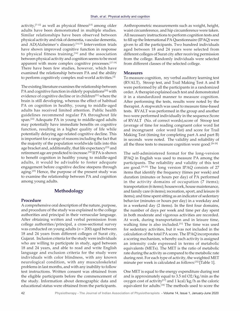
Shah, et al.: Physical activity and cognition
42 Physiotherapy - The Journal of Indian Association of Physiotherapists - Volume 14, Issue 1, January-June 2020
activity,[7‑12] as well as physical fitness[13] among older adults have been demonstrated in multiple studies. Similar relationships have been observed between physical activity and risk of dementia, vascular dementia, and AD(Alzheimer’s disease).[14,15] Intervention trials have shown improved cognitive function in response to physical fitness training,[16] and the association between physical activity and cognition seems to be most apparent with more complex cognitive processes.[17‑19] There have been few studies, however, which have examined the relationship between PA and the ability to perform cognitively complex real‑world activities.[3]
The existing literature examines the relationship between PA and cognitive function in elderly populations[20] with evidence of cognitive decline or in children[21] where the brain is still developing, whereas the effect of habitual PA on cognition in healthy, young to middle‑aged adults has received limited attention. Public health guidelines recommend regular PA throughout life span.[22] Adequate PA in young to middle‑aged adults may potentially have immediate benefits on cognitive function, resulting in a higher quality of life while potentially delaying age‑related cognitive decline. This is important for a range of reasons including the fact that the majority of the population worldwide falls into this age bracket and, additionally, that life expectancy[23] and retirement age are predicted to increase.[24] If PA is shown to benefit cognition in healthy young to middle‑aged adults, it would be advisable to foster adequate engagement before cognitive decline steepens through aging.[25] Hence, the purpose of the present study was to examine the relationship between PA and cognition among young adults.
Methodology
ProcedureA comprehensive oral description of the nature, purpose, and procedure of the study was explained to the college authorities and principal in their vernacular language. After obtaining written and verbal permission from college authorities/principal, a cross‑sectional survey was conducted on young adults (n = 200) aged between 18 and 24 years from different colleges of Surat city, Gujarat. Inclusion criteria for the study were individuals who are willing to participate in study, aged between 18 and 24 years, and able to read and write English language and exclusion criteria for the study were individuals with color blindness, with any known neurological condition, with any musculoskeletal problems in last months, and with any inability to follow test instructions. Written consent was obtained from the eligible participants before the commencement of the study. Information about demographic data and educational status were obtained from the participants.
Anthropometric measurements such as weight, height, waist circumference, and hip circumference were taken. All necessary instructions to perform cognition tests and to fill up the International PA Questionnaire (IPAQ) were given to all the participants. Two hundred individuals aged between 18 and 24 years were selected from different colleges of Surat city after receiving permission from the college. Randomly individuals were selected from different classes of the selected college.
MeasuresTo measure cognition, rey verbal auditory learning test (RVALT), Stroop test, and Trail Making Test A and B were performed by all the participants in a randomized order. A therapist explained each test and demonstrated it in a standardized manner to measure cognition. After performing the tests, results were noted by the therapist. A stopwatch was used to measure time‑based tests. RVALT was performed in the group and another two were performed individually in the sequence.Score of RVALT (No. of correct words),score of Stroop test (average of time for reading congruent color word list and incongruent color word list) and score for Trail Making Test (timing for completing part A and part B) in seconds were noted. The validity and reliability of all the three tests to measure cognition were good.[26‑28]
The self‑administered format for the long‑version IPAQ in English was used to measure PA among the participants. The reliability and validity of this test are good.[29,30] The long version IPAQ consists of 27 items that identify the frequency (times per week) and duration (minutes or hours per day) of PA performed in the activity domains of occupation (7 items); transportation (6 items); housework, house maintenance, and family care (6 items); recreation, sport, and leisure (6 items); and time spent sitting as an indicator of sedentary behavior (minutes or hours per day) in a weekday and in a weekend day (2 items). In the first four domains, the number of days per week and time per day spent in both moderate and vigorous activities are recorded. At work, during transportation and in leisure time, walking time is also included.[31] The time was used for sedentary activities, but it was not included in the calculation of the total PA score. The IPAQ incorporates a scoring mechanism, whereby each activity is assigned an intensity code expressed in terms of metabolic equivalents (METs). The MET is the ratio of metabolic rate during the activity as compared to the metabolic rate during rest. For each type of activity, the weighted MET minute per week is calculated as follows:[32] [Table 1].
One MET is equal to the energy expenditure during rest and is approximately equal to 3.5 ml O2/kg/min as the oxygen cost of activity[17] and 1 kcal/kg/h as the caloric equivalent for adults.[18] The methods used to score the
Shah, et al.: Physical activity and cognition
Physiotherapy - The Journal of Indian Association of Physiotherapists - Volume 14, Issue 1, January-June 2020 43
in mediation of the effects of physical exercise on brain functions has been promoted.[34] Further research is needed and should make the use of newer technologies in molecular biology and imaging to assist in studying the underpinning biological mechanisms. In addition to its role in health promotion, PA could also reduce the economic burden to society related to chronic degenerative diseases (e.g. hypertension, diabetes, and cardiovascular disease) that impair cognitive functioning.[39,50] Evidence
long version IPAQ are presented in detail on the IPAQ website as well (www.ipaq.ki.se). The scoring protocol was followed for cleaning and truncation.[33] The total PA MET minute/week was then computed by summing the walking, moderate, and vigorous MET minute/week scores.[34]
Statistical analyses were done by SPSS 20 (IBM SPSS 20, trial version). Spearman’s correlation was used to analyze the relationships between PA and cognition among the participants. Correlation was calculated and analyzed with the significance threshold set at P < 0.05.
Results
A total number of 200 participants (112 females and 88 males) were included in the present study. The physical characteristics of all the participants are shown in Table 2. The mean value of three different cognitive tests for all the participants is shown in Table 3. The classification of PA among the participants has been shown in Table 4. The correlation between PA and cognition is shown in Table 5. Results of the present study showed that a positive correlation between PA and all three cognitive measures tests.
Discussion
The study was aimed to find the relationship between PA and cognitive functions among young adults. Results of the study showed a positive correlation (P < 0.05) between PA and cognitive function (episodic memory and executive function).
Few studies reported a significant positive association between PA and executive function.[18,35‑39] Two studies noted nonsignificant, positive trends[40,41] and four studies noted null result.[42‑45] One of the reviews suggested that episodic memory was found to be significantly positively associated with PA in two studies[40,46] but not in another.[45]
A range of plausible biological mechanisms[47] may explain how PA influences cognitive functioning. These include structural changes to the central nervous system, neurotransmitter release, modulation of arousal levels, and enhanced cerebral capillary density/blood flow.[48] Exercise increases brain‑derived neurotrophic factor (BDNF) and nerve growth factor which mediate short‑ and long‑term enhancement of synaptic strength and reduce cell death in the hippocampus.[49] These neurotrophic, angiogenic, and neurogenic and synaptogenesis effects may play a role as both an enhancer and a protector of cognitive function and central nervous system integrity. The role of central (BDNF) and peripheral (estrogens, corticosteroids, growth hormone, and insulin‑like growth factor‑1) factors
Table 2: Physical characteristics of participantsMean±SD
n (male:female) 200±112:88Age (years) 20.62±1.47Height (m) 1.63±0.09Weight (kg) 66.03±14.57BMI (kg/m2) 24.99±4.57Waist circumference (cm) 73.22±10.14Hip circumference (cm) 97.01±10.65W: H ratio 0.88±1.46BMI=Body mass index, W: H ratio=Waist: hip ratio, SD=Standard deviation
Table 3: Mean value of cognitive tests Mean±SD
RVALT (number of correct words) (average of 6 trails) 7.023±1.89Stoop test (average of time (s) for reading congruent color words list and incongruent color words list)
49.18±17.73
Trail Making Test (s) (Task A) 28.03±10.60Trail Making Test (s) (Task B) 57.00±22.58RVALT=Rey Verbal Auditory Learning Test, SD=Standard deviation
Table 4: Classification of physical activityMean±SD
Work (MET‑min/week) 893.5±2247Transportation (MET‑min/week) 721.5±108Domestic activities (MET‑min/week) 1790±224Leisure (MET‑min/week) 1408±229Total score (MET‑min/week) 4813±383PA=Physical activity, METS=Metabolic equivalents
Table 5: Correlation between physical activity and cognition Mean±SD r PPA total score (MET‑min/week)RVALT 0.15 0.0325Stoop test 0.38 <0.0001Trail Making Test (Task A) 0.35 0.0492Trail Making Test (Task B) 0.21 0.003RVALT=Rey Verbal Auditory Learning Test, SD=Standard deviation, METS=Metabolic equivalents, PA=Physical activity
Table 1: Weighted MET minute per week for different type of activityWalking MET (min/week) 3.3×walking minutes×walking daysModerate MET (min/week) 4.0×moderate intensity activity
minutes×moderate activity daysVigorous MET (min/week) 8.0×vigorous intensity activity
minutes×vigorous activity days

Shah, et al.: Physical activity and cognition
44 Physiotherapy - The Journal of Indian Association of Physiotherapists - Volume 14, Issue 1, January-June 2020
of a benefit of PA in the elderly provides evidence of neuroplasticity even in old age.[51] It is, therefore, plausible that PA might modify cognitive function in earlier adulthood and that these changes might enhance existing cognitive function or attenuates the decline with aging. Recent reviews indicate that high cardiovascular fitness may be particularly beneficial for brain volume in prefrontal regions,[52,53] consistent with the selective effects of chronic exercise on executive function shown in recent studies.[35,39,43]
There are certain limitations to the study. The sample size of the study was relatively small, thus limiting statistical power and the generalizability of the results. The study used a cross‑sectional design, and therefore, the performance effects attributed to variable amounts of PA may be due to other factors. Another limitation is the use of self‑reported measures of PA rather than objective measures of activity (e.g., pedometer or accelerometer) and fitness (e.g., VO2). Hence, the longitudinal study with large sample size along with objective measurement of PA can be done. A future research with consideration of examining the differential effects on cognition of moderate versus vigorous physical activity, as well as the amount of physical activity along with the use of brain imaging techniques in conjunction with cognitive tests can be done.
Conclusion
Results of the study showed that PA has an important role in cognitive functioning among young adults. This finding of young adults suggests that PA may be beneficial to cognition during early and middle periods of the human life span and may continue to protect against age‑related loss of cognitive function during older adulthood. The results indicate that regular PA has a beneficial effect on the cognitive processes on executive function in young adults.
AcknowledgmentThe authors would like to thank all the participants as well as principals of the college for their kind cooperation and giving permission to collect data from their colleges.
Financial support and sponsorshipNil.
Conflicts of interestThere are no conflicts of interest.
References
1. Cox EP, O’Dwyer N, Cook R, Vetter M, Cheng HL, Rooney K, et al. Relationship between physical activity and cognitive function in apparently healthy young to middle‑aged adults: A systematic review. J Sci Med Sport 2016;19:616‑28.
2. G r a s s m a n n V , S u b r a m a n i a p i l l a i M , D u n c a n M ,
Arbour‑Nicitopoulos K, Faulkner GE. The relationship between moderate‑to‑vigorous physical activity and executive function among individuals with schizophrenia: Differences by illness duration. Braz J Psychiatry 2017;39:309‑15.
3. Haley C. Exploring the Relationship between Physical Activity and Everyday Cognitive Function in Older Adults: Within‑and Between‑Person Variability 2013.
4. Samantha R. Exercise and Cognition in Young Adults. Psychological Sciences Undergraduate Publications. Presentations and Projects; 2015.
5. Small DM. Individual differences in the neurophysiology of reward and the obesity epidemic. Int J Obes (Lond) 2009;33 Suppl 2:S44‑8.
6. Ball K, Brown W, Crawford D. Who does not gain weight? Prevalence and predictors of weight maintenance in young women. Int J Obes Relat Metab Disord 2002;26:1570‑8.
7. McAuley E, Kramer AF, Colcombe SJ. Cardiovascular fitness and neurocognitive function in older adults: A brief review. Brain Behav Immun 2004;18:214‑20.
8. Lindwall M, Rennemark M, Berggren T. Movement in mind: The relationship of exercise with cognitive status for older adults in the Swedish National Study on Aging and Care (SNAC). Aging Ment Health 2008;12:212‑20.
9. Lytle ME, Vander Bilt J, Pandav RS, Dodge HH, Ganguli M. Exercise level and cognitive decline: The MoVIES project. Alzheimer Dis Assoc Disord 2004;18:57‑64.
10. Middleton L, Kirkland S, Rockwood K. Prevention of CIND by physical activity: Different impact on VCI‑ND compared with MCI. J Neurol Sci 2008;269:80‑4.
11. van Gelder BM, Tijhuis MA, Kalmijn S, Giampaoli S, Nissinen A, Kromhout D. Physical activity in relation to cognitive decline in elderly men: The FINE Study. Neurology 2004;63:2316‑21.
12. Yaffe K, Barnes D, Nevitt M, Lui LY, Covinsky K. A prospective study of physical activity and cognitive decline in elderly women: Women who walk. Arch Intern Med 2001;161:1703‑8.
13. Wang L, Larson EB, Bowen JD, van Belle G. Performance‑based physical function and future dementia in older people. Arch Intern Med 2006;166:1115‑20.
14. Podewils LJ, Guallar E, Kuller LH, Fried LP, Lopez OL, Carlson M, et al. Physical activity, APOE genotype, and dementia risk: Findings from the Cardiovascular Health Cognition Study. Am J Epidemiol 2005;161:639‑51.
15. Ravaglia G, Forti P, Lucicesare A, Pisacane N, Rietti E, Bianchin M, et al. Physical activity and dementia risk in the elderly: Findings from a prospective Italian study. Neurology 2008;70:1786‑94.
16. Colcombe S, Kramer AF. Fitness effects on the cognitive function of older adults: A meta‑analytic study. Psychol Sci 2003;14:125‑30.
17. Bixby WR, Spalding TW, Haufler AJ, Deeny SP, Mahlow PT, Zimmerman JB, et al. The unique relation of physical activity to executive function in older men and women. Med Sci Sports Exerc 2007;39:1408‑16.
18. Hillman CH, Kramer AF, Belopolsky AV, Smith DP. A cross‑sectional examination of age and physical activity on performance and event‑related brain potentials in a task switching paradigm. Int J Psychophysiol 2006;59:30‑9.
19. Smiley‑Oyen AL, Lowry KA, Francois SJ, Kohut ML, Ekkekakis P. Exercise, fitness, and neurocognitive function in older adults: The “selective improvement” and “cardiovascular fitness” hypotheses. Ann Behav Med 2008;36:280‑91.
20. Angevaren M, Aufdemkampe G, Verhaar HJ, Aleman A, Vanhees L. Physical activity and enhanced fitness to improve cognitive function in older people without known cognitive impairment. Cochrane database of systematic reviews. 2008(2).
21. Fedewa AL, Ahn S. The effects of physical activity and physical fitness on children’s achievement and cognitive outcomes: A meta‑analysis. Res Q Exerc Sport 2011;82:521‑35.
22. Garber CE, Blissmer B, Deschenes MR, Franklin BA, Lamonte MJ,
Shah, et al.: Physical activity and cognition
Physiotherapy - The Journal of Indian Association of Physiotherapists - Volume 14, Issue 1, January-June 2020 45
Lee IM, et al. American College of Sports Medicine position stand. Quantity and quality of exercise for developing and maintaining cardiorespiratory, musculoskeletal, and neuromotor fitness in apparently healthy adults: Guidance for prescribing exercise. Med Sci Sports Exerc 2011;43:1334‑59.
23. Statistics AB. Life Expectancy Trends‑Australia. Canberra: Australian Bureau of Statistics; 2011.
24. National Institute on Aging. Global Health and Aging. Available from: http://www.nia.nih.gov/research/publication/global‑health‑and‑aging/living‑longer. [Last accessed on 2014 Jun 28].
25. Chang YK, Huang CJ, Chen KF, Hung TM. Physical activity and working memory in healthy older adults: An ERP study. Psychophysiology 2013;50:1174‑82.
26. Paula JJ, Melo LP, Nicolato R, Moraes EN, Bicalho MA, Hamdan AC, et al. Reliability and construct validity of the rey‑auditory verbal learning test in Brazilian elders. Arch Clin Psychiatr (São Paulo) 2012;39:19‑23.
27. Siegrist M. Test‑retest reliability of different versions of the stroop test. J Psychol 1997;131:299‑306.
28. Wagner S, Helmreich I, Dahmen N, Lieb K, Tadic A. Reliability of three alternate forms of the trail making tests a and B. Arch Clin Neuropsychol 2011;26:314‑21.
29. Craig CL, Marshall AL, Sjöström M, Bauman AE, Booth ML, Ainsworth BE, et al. International physical activity questionnaire: 12‑country reliability and validity. Med Sci Sports Exerc 2003;35:1381‑95.
30. Hagströmer M, Oja P, Sjöström M. The International Physical Activity Questionnaire (IPAQ): A study of concurrent and construct validity. Public Health Nutr 2006;9:755‑62.
31. Hagstromer M, Ainsworth BE, Oja P, Sjostrom M. Comparison of a subjective and an objective measure of physical activity in a population sample. J Phys Act Health 2010;7:541‑50.
32. IPAQ Research Committee. Guidelines for Data Processing and Analysis of the International Physical Activity Questionnaire (IPAQ) – Short and Long Form. IPAQ Research Committee; November 2005. Available from: http://www. ipaq. ki.se.
33. Nor Shazwani MN Jr, Suzana S, Hanis Mastura Y, Lim CJ, Teh SC, Mohd Fauzee MZ, et al. Assessment of physical activity level among individuals with type 2 diabetes mellitus at Cheras health clinic, Kuala Lumpur. Malays J Nutr 2010;16:101‑12.
34. Gligoroska JP, Manchevska S. The effect of physical activity on cognition – Physiological mechanisms. Mater Sociomed 2012;24:198‑202.
35. Padilla C, Perez L, Andres P, Parmentier FB. Exercise improves cognitive control: evidence from the stop signal task. Appl Cogn Psychol 2013;27:505‑11.
36. Hillman CH, Motl RW, Pontifex MB, Posthuma D, Stubbe JH, Boomsma DI, et al. Physical activity and cognitive function in a cross‑section of younger and older community‑dwelling individuals. Health Psychol 2006;25:678‑87.
37. Kamijo K, Takeda Y. General physical activity levels influence
positive and negative priming effects in young adults. Clin Neurophysiol 2009;120:511‑9.
38. Kamijo K, Takeda Y. Regular physical activity improves executive function during task switching in young adults. Int J Psychophysiol 2010;75:304‑11.
39. Pérez L, Padilla C, Parmentier FB, Andrés P. The effects of chronic exercise on attentional networks. PLoS One 2014;9:e101478.
40. Winneke AH, Godde B, Reuter EM, Vieluf S, Voelcker‑Rehage C. The association between physical activity and attentional control in younger and older middle‑aged adults. GeroPsych 2012 Nov 29..
41. Woo E, Sharps MJ. Cognitive aging and physical exercise. Educ Gerontol 2003;29:327‑37.
42. Spirduso WW. Reaction and movement time as a function of age and physical activity level. J Gerontol 1975;30:435‑40.
43. Padilla C, Pérez L, Andrés P. Chronic exercise keeps working memory and inhibitory capacities fit. Front Behav Neurosci 2014;8:49.
44. Boucard GK, Albinet CT, Bugaiska A, Bouquet CA, Clarys D, Audiffren M. Impact of physical activity on executive functions in aging: A selective effect on inhibition among old adults. J Sport Exerc Psychol 2012;34:808‑27.
45. Erickson KI, Banducci SE, Weinstein AM, Macdonald AW 3rd, Ferrell RE, Halder I, et al. The brain‑derived neurotrophic factor Val66Met polymorphism moderates an effect of physical activity on working memory performance. Psychol Sci 2013;24:1770‑9.
46. Heisz JJ, Vandermorris S, Wu J, McIntosh AR, Ryan JD. Age differences in the association of physical activity, sociocognitive engagement, and TV viewing on face memory. Health Psychol 2015;34:83‑8.
47. Swain RA, Harris AB, Wiener EC, Dutka MV, Morris HD, Theien BE, et al. Prolonged exercise induces angiogenesis and increases cerebral blood volume in primary motor cortex of the rat. Neuroscience 2003;117:1037‑46.
48. Cotman CW, Berchtold NC. Exercise: A behavioral intervention to enhance brain health and plasticity. Trends Neurosci 2002;25:295‑301.
49. World Health Organization. Global Recommendations on Physical Activity for Health. Public Significance of Physical Activity. Switzerland: World Health Organization; 2010. p. 10.
50. United States Census Bureau. World Population by Age and Sex. https://www.census.gov/popclock/. [Last accessed on 2014 Jul 01].
51. Hötting K, Röder B. Beneficial effects of physical exercise on neuroplasticity and cognition. Neurosci Biobehav Rev 2013;37:2243‑57.
52. Voss MW, Nagamatsu LS, Liu‑Ambrose T, Kramer AF. Exercise, brain, and cognition across the life span. J Appl Physiol (1985) 2011;111:1505‑13.
53. Hillman CH, Erickson KI, Kramer AF. Be smart, exercise your heart: Exercise effects on brain and cognition. Nat Rev Neurosci 2008;9:58‑65.
46 © 2020 Physiotherapy - The Journal of Indian Association of Physiotherapists | Published by Wolters Kluwer - Medknow
Chronic headache in a case of lipedematous scalp: Physiotherapy in symptom managementLourembam Surbala Devi, Dhwani Dakoria1
Abstract:Lipedematous scalp is a rare disorder of unknown etiology and pathogenesis. Since Cornbleet first described it in 1935, there have been better insights into the presentation, prognosis, and the treatment of the disease. The objective of this case report is to outline the physiotherapy findings and symptom management strategies as an adjunct to pharmacotherapy in a 52‑year‑old Indian female who presented with the complaints of chronic continuous headache and spongy scalp. She had no disorder other than hypertension. Chronic cervicogenic headache was diagnosed in our case with lipedematous scalp during physiotherapy examination for headache, which was the patient’s primary complaint. Physiotherapy interventions in adjunct to pharmacotherapy had promising effects in reducing the severity of headache (NPRS score 8/10 to 1/10), neck stiffness and trapezius myalgia.Keywords:Cervicogenic headache, chronic headache, lipedematous scalp, physiotherapy, spongy scalp
Introduction
Lipedematous scalp is a rare disease characterized by a localized accumulation
of fatty tissue in the subcutaneous layer of the scalp, without hair loss. When associated with alopecia, it is known as lipedematous alopecia. Approximately 80 cases of lipedematous scalp with or without alopecia have been reported in the literature since its first report by Cornbleet in 1935.[1] The case is reported predominantly among the African‑American women.[2] However, it has also been reported in Hispanics, Arabic, Turkish, Chinese, and Japanese. Lipedematous scalp may be associated with diabetes mellitus, hyperlipidemia, hypertension, and ovarian cysts. The clinical characteristics include an onset in the mid‑50s, female predominance, and frequent involvement of the scalp vertex. Symptoms include diffuse pain, headache, burning, abnormal sensations (paresthesia),
and thickening of the scalp with localized or generalized sensitivity of the scalp or itching (dysesthesia). Six cases have so far been reported on Indians; where chronic headache was one of the presenting features in two of the cases[3] and scalp dysesthesia in two other cases that was treated with antihistamines.[4] The characteristic finding is a gradual increase in scalp thickness, but the exact etiology is unknown. Most cases reported in the literature are focused on histopathological findings, MRI findings, and other biochemical and serological findings.[5] So far, physical examination and symptom management of chronic headache in LS has not been reported yet despite being the commonest presenting feature. The objective of this case report is to present the findings of physical examination and physiotherapy techniques in symptom management in adjunct to pharmacotherapy as presented in a 52 year old female with lipedematous scalp with the primary complaint of severe chronic headache.
Address for correspondence:
Dr. Lourembam Surbala Devi,
Department of Neurological Sciences,
Ashok and Rita Patel Institute of Physiotherapy,
CHARUSAT, Changa, Anand, Gujarat, India. E-mail: surbalampt@
gmail.com
Submission: 20-07-2019Revision: 08-03-2020
Accepted: 26-03-2020Published: 29-06-2020
Departments of Neurological Sciences and 1Musculoskeletal Sciences, Ashok and
Rita Patel Institute of Physiotherapy,
CHARUSAT, Anand, Gujarat, India
Case Report
Access this article onlineQuick Response Code:
Website:www.pjiap.org
DOI:10.4103/PJIAP.PJIAP_18_19
How to cite this article: Devi LS, Dakoria D. Chronic headache in a case of lipedematous scalp: Physiotherapy in symptom management. Physiother - J Indian Assoc Physiother 2020;14:46-9.
This is an open access journal, and articles are distributed under the terms of the Creative Commons Attribution-NonCommercial-ShareAlike 4.0 License, which allows others to remix, tweak, and build upon the work non-commercially, as long as appropriate credit is given and the new creations are licensed under the identical terms.
For reprints contact: [email protected]

Devi and Dakoria: Chronic headache in lipedematous scalp
Physiotherapy - The Journal of Indian Association of Physiotherapists - Volume 14, Issue 1, January-June 2020 47
Case Report
A 52‑year‑old Indian Gujarati female presented to our outpatient physiotherapy department with the complaints of severe headache which was almost permanent, neck stiffness, and pain extending to the occiput. The headache was severe, which she scored 8/10 on Numerical Pain Rating scale (NPRS) and reported to have had the symptoms for 2 years, mostly diffused or present in the occipital region and sometimes radiating to the frontal region bilaterally. She managed her headache with over‑the‑counter medications and never consulted a physician earlier, to seek medical advice for her headache and other complaints. She denied any similar complains in the family. She was a known case of hypertension and was on antihypertensive medications for 5 years.
Initial observation of the patient revealed a forward stoop neck posture and a frowning forehead. Physical examination revealed features of cervicogenic dysfunction characterized by restriction in the cervical range of motion with symptoms worse in neck extension. Palpation of the neck region and cervical spine revealed a bilateral hyperactive upper trapezius muscle with associated myalgia, and hypomobility of the upper cervical spine. We measured the patient’s baseline status using: Goniometric measurement for Cervical Range of Motion (CROM), Numerical Pain Rating Scale (NPRS) to quantify the severity of headache; and Pressure Algometry for Trapezius myalgia. We also used the Headache Disability Index (HDI) to examine the perceived impact of headaches on emotional functioning and daily activities. Our baseline examination revealed that the patient had severe headache (NPRS: 8/10), significant restriction of the CROM (CROM Extension: 0‑52º; CROM Flexion: 0‑46º; CROM Rotation – Right: 0‑70º, Left: 0‑65º), Trapezius myalgia on palpation (Pressure algometry ‑ Left: 3.54 kg, Right: 3.06 kg)
and affection of emotional functioning and majority of the daily activities (HDI Score: 64/100‑severe disability). A diagnosis of cervicogenic headache was made based on the physical examination according to the Cervicogenic Headache International Study Group Diagnostic Criteria.[6] A soft spongy diffused bogginess and swelling of the occipital scalp region extending to the vertex was noted during palpation, which was associated with tenderness [Figure 1]. There was no visual evidence of scalp inflammation or difference in the density of hair in the occipital region. She reported the swelling to be gradual in onset and denied any history of pruritis, hair loss, head trauma, or any systemic problems. The spongy swelling of the scalp was clinically diagnosed as lipedematous scalp without alopecia by the dermatologist and prescribed medications; Pregabalin 75 Mg (P.O. Bid) and Tryptomer 10 Mg (h.s.) for 10 days. These medications have been proven effective in the management of neuropathic pain and migraine headaches, but its effectiveness in cervicogenic headache is yet unknown.[7] Routine investigation of complete blood cell counts (CBC), random blood sugar (RBS), lipid profile, and her neurological examination did not reveal any remarkable findings.
Symptomatic physiotherapy management for chronic headache, neck pain and stiffness, and trapezius myalgia started on the day of examination as per the treatment recommendations by Biondi.[8] The physiotherapy management included the release of the hyperactive trapezius muscle and suboccipital muscles, Maitland mobilization of the upper cervical spine (C1‑4), deep neck flexor strengthening, and transcutaneous electrical nerve stimulation in the nap of the neck and in the occipital region [Table 1]. The interventions were provided on a daily basis for six sessions and on alternate days in the subsequent days. At the end of the 12th session, the intensity of the headache reduced considerably which she scored 4/10 in the NPRS, along with decreased neck pain and stiffness. After the 12th session of supervised physiotherapy management, she was advised for home exercises which included isometric neck exercises and deep neck flexor strengthening and to follow up after one month. At follow up after 1 month, she had minimal complains of headache (0‑1 in NPRS), neck stiffness or trapezius myalgia and reduced impact on daily activities (HDI Score – 24/100) [Table 2]. However, the bogginess and swelling of the scalp did not change significantly.
Discussion
Lipedematous scalp is an uncommon disease and and chronic headache is one of the most common manifestations of LS. Majority of the cases were reported in African‑American women, but it may not be limited to a
Figure 1: A remarkably thickened superficial tissue in the occipital region extending till the vertex
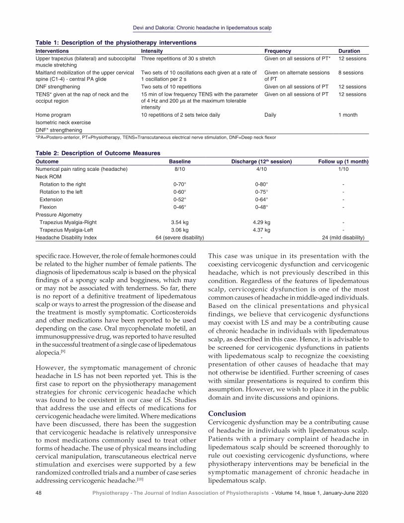
Devi and Dakoria: Chronic headache in lipedematous scalp
48 Physiotherapy - The Journal of Indian Association of Physiotherapists - Volume 14, Issue 1, January-June 2020
This case was unique in its presentation with the coexisting cervicogenic dysfunction and cervicogenic headache, which is not previously described in this condition. Regardless of the features of lipedematous scalp, cervicogenic dysfunction is one of the most common causes of headache in middle‑aged individuals. Based on the clinical presentations and physical findings, we believe that cervicogenic dysfunctions may coexist with LS and may be a contributing cause of chronic headache in individuals with lipedematous scalp, as described in this case. Hence, it is advisable to be screened for cervicogenic dysfunctions in patients with lipedematous scalp to recognize the coexisting presentation of other causes of headache that may not otherwise be identified. Further screening of cases with similar presentations is required to confirm this assumption. However, we wish to place it in the public domain and invite discussions and opinions.
Conclusion Cervicogenic dysfunction may be a contributing cause of headache in individuals with lipedematous scalp. Patients with a primary complaint of headache in lipedematous scalp should be screened thoroughly to rule out coexisting cervicogenic dysfunctions, where physiotherapy interventions may be beneficial in the symptomatic management of chronic headache in lipedematous scalp.
specific race. However, the role of female hormones could be related to the higher number of female patients. The diagnosis of lipedematous scalp is based on the physical findings of a spongy scalp and bogginess, which may or may not be associated with tenderness. So far, there is no report of a definitive treatment of lipedematous scalp or ways to arrest the progression of the disease and the treatment is mostly symptomatic. Corticosteroids and other medications have been reported to be used depending on the case. Oral mycophenolate mofetil, an immunosuppressive drug, was reported to have resulted in the successful treatment of a single case of lipedematous alopecia.[9]
However, the symptomatic management of chronic headache in LS has not been reported yet. This is the first case to report on the physiotherapy management strategies for chronic cervicogenic headache which was found to be coexistent in our case of LS. Studies that address the use and effects of medications for cervicogenic headache were limited. Where medications have been discussed, there has been the suggestion that cervicogenic headache is relatively unresponsive to most medications commonly used to treat other forms of headache. The use of physical means including cervical manipulation, transcutaneous electrical nerve stimulation and exercises were supported by a few randomized controlled trials and a number of case series addressing cervicogenic headache.[10]
Table 1: Description of the physiotherapy interventionsInterventions Intensity Frequency DurationUpper trapezius (bilateral) and suboccipital muscle stretching
Three repetitions of 30 s stretch Given on all sessions of PT* 12 sessions
Maitland mobilization of the upper cervical spine (C1‑4) ‑ central PA glide
Two sets of 10 oscillations each given at a rate of 1 oscillation per 2 s
Given on alternate sessions of PT
8 sessions
DNF strengthening Two sets of 10 repetitions Given on all sessions of PT 12 sessionsTENS* given at the nap of neck and the occiput region
15 min of low frequency TENS with the parameter of 4 Hz and 200 µs at the maximum tolerable intensity
Given on all sessions of PT 12 sessions
Home programIsometric neck exerciseDNF* strengthening
10 repetitions of 2 sets twice daily Daily 1 month
*PA=Postero-anterior, PT=Physiotherapy, TENS=Transcutaneous electrical nerve stimulation, DNF=Deep neck flexor
Table 2: Description of Outcome MeasuresOutcome Baseline Discharge (12th session) Follow up (1 month)Numerical pain rating scale (headache) 8/10 4/10 1/10Neck ROM
Rotation to the right Rotation to the left ExtensionFlexion
0‑70°0‑60°0‑52°0‑46°
0‑80°0‑75°0‑64°0‑48°
‑‑‑‑
Pressure AlgometryTrapezius Myalgia-RightTrapezius Myalgia-Left
3.54 kg3.06 kg
4.29 kg4.37 kg
‑‑
Headache Disability Index 64 (severe disability) ‑ 24 (mild disability)

Devi and Dakoria: Chronic headache in lipedematous scalp
Physiotherapy - The Journal of Indian Association of Physiotherapists - Volume 14, Issue 1, January-June 2020 49
AcknowledgmentWe would like to thank our valuable patient for her consent and cooperation.
Declaration of patient consentThe authors certify that they have obtained all appropriate patient consent forms. In the form the patient(s) has/have given his/her/their consent for his/her/their images and other clinical information to be reported in the journal. The patients understand that their names and initials will not be published and due efforts will be made to conceal their identity, but anonymity cannot be guaranteed.
Financial support and sponsorshipNil.
Conflicts of interestThere are no conflicts of interest.
References
1. Cornbleet T. Cutis verticis gyrata? Lipoma? Arch Derm Syphilol 1935;32:688.
2. Martín JM, Monteagudo C, Montesinos E, Guijarro J, Llombart B, Jordá E. Lipedematous scalp and lipedematous alopecia: A clinical and histologic analysis of 3 cases. J Am Acad Dermatol 2005;52:152‑6.
3. Sahu P, Sangal B, Dayal S, Kumar S. Lipedematous scalp with varied presentations: A case series of four patients. Indian Dermatol Online J 2019;10:571‑3.
4. Peter CV, Jennifer A, Raychaudhury T, Chandrashekhar L, Merilyn S, Gowda S, et al. Lipedematous scalp. Indian J Dermatol Venereol Leprol 2014;80:270‑2.
5. Carrasco‑Zuber JE, Alvarez‑Veliz S, Cataldo‑Cerda K, Gonzalez‑Bombardiere S. Lipedematous scalp: A case report and review of the current literature. J Dtsch Dermatol Ges 2016;14:418‑21.
6. Sjaastad O, Fredriksen TA, Pfaffenrath V. Cervicogenic headache: Diagnostic criteria. The cervicogenic headache international study group. Headache 1998;38:442‑5.
7. Pizzolato R, Villani V, Prosperini L, Ciuffoli A, Sette G. Efficacy and tolerability of pregabalin as preventive treatment for migraine: a 3‑month follow‑up study. J Headache Pain 2011;12:521‑5.
8. Biondi DM. Cervicogenic headache: A review of diagnostic and treatment strategies. J Am Osteopath Assoc 2005;105:16S‑22S.
9. Cabrera R, Larrondo J, Whittle C, Castro A, Gosch M. Successful treatment of lipedematous alopecia using mycophenolate mofetil. Acta Derm Venereol 2015;95:1011‑2.
10. Haldeman S, Dagenais S. Cervicogenic headaches: a critical review. Spine J 2001;1:31‑46.
50 © 2020 Physiotherapy - The Journal of Indian Association of Physiotherapists | Published by Wolters Kluwer - Medknow
Effectiveness of physiotherapy treatment in a case of diffuse idiopathic skeletal hyperostosis (DISH) in 65 year old maleHemal M. Patel, Damini Vinod Patel
Abstract:Diffuse idiopathic skeletal hyperostosis (DISH) is common after 50 years, which is characterized by the ossification of the anterior longitudinal ligament. The purpose of this case report is to describe the physiotherapy intervention strategies and report its effectiveness in the symptomatic management of DISH in 65-year-old male who complains of severe lower back pain and difficulty in changing side while lying on the bed, prolonged standing, and walking for the last 1 month. The condition was managed based on clinical presentations with supervised and unsupervised physiotherapy for 6 weeks. The supervised physiotherapy intervention consisted of patient’s education, postural correction, electrotherapeutic modalities, exercise therapy, and counseling for 45–60 min a day for 5 days/week. The effectiveness of physiotherapy is followed for 6 weeks and symptom-specific outcome measures such as Quebec disability index, Schober’s test, visual analog scale, and lumbar lateral flexion test were used. Physiotherapy interventions are found to be effective in the symptom management of DISH with increased patient satisfaction.Keywords:Diffuse idiopathic skeletal hyperososis, forestiers disease, physical therapy
Introduction
Diffuse idiopathic skeletal hyperostosis (DISH) is also known as Forestier’s
disease. It was first described by Jacques Forestier and his student Jaume Rotes‑Querol in 1950. It is a systemic noninflammatory disease. DISH is characterized by thickening, ossification and calcification of ligament, tendon, and joint capsule.[1] A study was conducted in Japan on the prevalence of DISH. It was 19.5%. The prevalence of disease was 17%–19.5% in older subjects. Age and sex were significantly related to the presence of DISH, suggesting that men and older individuals have a higher probability of developing DISH.[2,3] The most common site of involvement was thoracic (65.1%)
and thoracolumbar (24.2%); other areas commonly involved are as follows: pelvis, patella, calcaneus, and olecranon.[2,4] Certain studies showed that obesity, hypertension, diabetes mellitus Type 2, and dyslipidemia are common risk factors for developing DISH.[5] Usually, the disease is asymptomatic in most clinical cases; symptomatic cases show severe pain, reduced mobility, and stiffness.[6] The diagnosis of DISH is based on the radiological findings, defined by Resnick and Niwayana. According to the definition, the presence of flowing calcification and ossification mainly along the anterior longitudinal ligament (ALL) of at least four contiguous vertebrae with preserved disc height is indicative of the condition. There are three primary highlights for diagnosis:
1. Flowing bony ossification along the anterolateral aspect of at least four
Address for correspondence:
Dr. Hemal M. Patel, Department of
Musculoskeletal Science, Ashok & Rita Patel
Institute of Physiotherapy, CHARUSAT Campus,
Changa, Petlad, Anand - 388 421,
Gujarat, India. E-mail: drhempatel@
gmail.com
Submission: 21-10-2019Revision: 14-12-2019
Accepted: 18-02-2020Published: 29-06-2020
Department of Musculoskeletal Science,
Ashok & Rita Patel Institute of Physiotherapy,
CHARUSAT, Anand, Gujarat, India
Case Report
Access this article onlineQuick Response Code:
Website:www.pjiap.org
DOI:10.4103/PJIAP.PJIAP_30_19
How to cite this article: Patel HM, Patel DV. Effectiveness of physiotherapy treatment in a case of diffuse idiopathic skeletal hyperostosis (DISH) in 65 year old male. Physiother - J Indian Assoc Physiother 2020;14:50-4.
This is an open access journal, and articles are distributed under the terms of the Creative Commons Attribution-NonCommercial-ShareAlike 4.0 License, which allows others to remix, tweak, and build upon the work non-commercially, as long as appropriate credit is given and the new creations are licensed under the identical terms.
For reprints contact: [email protected]
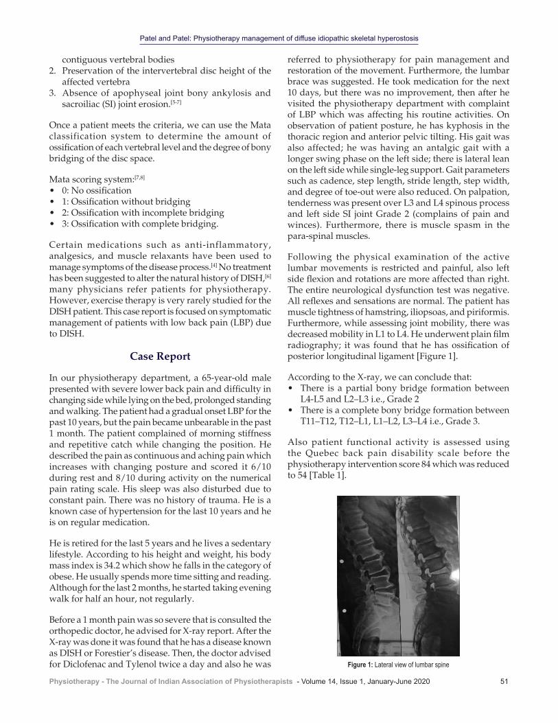
Patel and Patel: Physiotherapy management of diffuse idiopathic skeletal hyperostosis
Physiotherapy - The Journal of Indian Association of Physiotherapists - Volume 14, Issue 1, January-June 2020 51
contiguous vertebral bodies2. Preservation of the intervertebral disc height of the
affected vertebra3. Absence of apophyseal joint bony ankylosis and
sacroiliac (SI) joint erosion.[5‑7]
Once a patient meets the criteria, we can use the Mata classification system to determine the amount of ossification of each vertebral level and the degree of bony bridging of the disc space.
Mata scoring system:[7,8]
• 0: No ossification• 1: Ossification without bridging• 2: Ossification with incomplete bridging• 3: Ossification with complete bridging.
Certain medications such as anti‑inflammatory, analgesics, and muscle relaxants have been used to manage symptoms of the disease process.[4] No treatment has been suggested to alter the natural history of DISH,[6] many physicians refer patients for physiotherapy. However, exercise therapy is very rarely studied for the DISH patient. This case report is focused on symptomatic management of patients with low back pain (LBP) due to DISH.
Case Report
In our physiotherapy department, a 65‑year‑old male presented with severe lower back pain and difficulty in changing side while lying on the bed, prolonged standing and walking. The patient had a gradual onset LBP for the past 10 years, but the pain became unbearable in the past 1 month. The patient complained of morning stiffness and repetitive catch while changing the position. He described the pain as continuous and aching pain which increases with changing posture and scored it 6/10 during rest and 8/10 during activity on the numerical pain rating scale. His sleep was also disturbed due to constant pain. There was no history of trauma. He is a known case of hypertension for the last 10 years and he is on regular medication.
He is retired for the last 5 years and he lives a sedentary lifestyle. According to his height and weight, his body mass index is 34.2 which show he falls in the category of obese. He usually spends more time sitting and reading. Although for the last 2 months, he started taking evening walk for half an hour, not regularly.
Before a 1 month pain was so severe that is consulted the orthopedic doctor, he advised for X‑ray report. After the X‑ray was done it was found that he has a disease known as DISH or Forestier’s disease. Then, the doctor advised for Diclofenac and Tylenol twice a day and also he was
referred to physiotherapy for pain management and restoration of the movement. Furthermore, the lumbar brace was suggested. He took medication for the next 10 days, but there was no improvement, then after he visited the physiotherapy department with complaint of LBP which was affecting his routine activities. On observation of patient posture, he has kyphosis in the thoracic region and anterior pelvic tilting. His gait was also affected; he was having an antalgic gait with a longer swing phase on the left side; there is lateral lean on the left side while single‑leg support. Gait parameters such as cadence, step length, stride length, step width, and degree of toe‑out were also reduced. On palpation, tenderness was present over L3 and L4 spinous process and left side SI joint Grade 2 (complains of pain and winces). Furthermore, there is muscle spasm in the para‑spinal muscles.
Following the physical examination of the active lumbar movements is restricted and painful, also left side flexion and rotations are more affected than right. The entire neurological dysfunction test was negative. All reflexes and sensations are normal. The patient has muscle tightness of hamstring, iliopsoas, and piriformis. Furthermore, while assessing joint mobility, there was decreased mobility in L1 to L4. He underwent plain film radiography; it was found that he has ossification of posterior longitudinal ligament [Figure 1].
According to the X‑ray, we can conclude that:• There is a partial bony bridge formation between
L4‑L5 and L2–L3 i.e., Grade 2• There is a complete bony bridge formation between
T11–T12, T12–L1, L1–L2, L3–L4 i.e., Grade 3.
Also patient functional activity is assessed using the Quebec back pain disability scale before the physiotherapy intervention score 84 which was reduced to 54 [Table 1].
Figure 1: Lateral view of lumbar spine
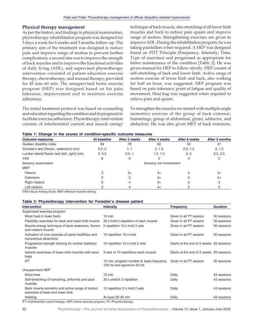
Patel and Patel: Physiotherapy management of diffuse idiopathic skeletal hyperostosis
52 Physiotherapy - The Journal of Indian Association of Physiotherapists - Volume 14, Issue 1, January-June 2020
technique of back muscle, also stretching of all lower limb muscles and back to reduce pain spasm and improve range of motion. Strengthening exercises are given to improve ADL. During the rehabilitation program, he was taking painkillers when required. A HEP was designed based on FITT Principle (Frequency, Intensity, Time, Type of exercises) and progressed as appropriate for better maintenance of the condition [Table 2]. He was given manual for HEP to follow strictly. HEP consist of self‑stretching of back and lower limb. Active range of motion exercise of lower limb and back, also walking for half an hour, was suggested. HEP program was based on pain tolerance, point of fatigue and quality of movement. Heat bag was suggested when required to relieve pain and spasm.
To strengthen the muscles we started with multiple‑angle isometrics exercise of the group of back extensor, hamstrings, group of abdominal, glutei, adductor, and abductors. He was also given MET of back extensors,
Physical therapy managementAs per the history and findings in physical examination, physiotherapy rehabilitation program was designed for 5 days a week for 6 weeks and 3 months follow‑up. The primary aim of the treatment was designed to reduce pain and improve range of motion to prevent further complications, a second aim was to improve the strength of back muscles and to improve the functional activities of daily living (ADL), and supervised physiotherapy intervention consisted of patient education exercise therapy, electrotherapy, and manual therapy, provided for 45 min–60 min. The unsupervised home exercise program (HEP) was designed based on his pain tolerance, improvement and to maintain exercise adherence.
The initial treatment protocol was based on counseling and education regarding the condition and its prognosis to facilitate exercise adherence. Physiotherapy intervention consists of interferential current and muscle energy
Table 2: Physiotherapy intervention for Forestier’s disease patientIntervention Intensity Frequency DurationSupervised exercise program
Moist heat in lower back 10 min Given in all PT session 30 sessionsFlexibility exercises for back and lower limb muscle 30 s hold 3 repetition of each muscle Given in all PT session 30 sessionsMuscle energy technique of back extensors, flexors and rotators muscle
5 repetition 10 s hold 3 sets Given in all PT session 30 sessions
Activation of core muscles of spine multifidus and transversus abdominis
10 repetition 10 s hold Given in all PT session 30 sessions
Progressive strength training for lumbar stabilizer muscles
10 repetition 10 s hold 2 sets Starts at the end of 2 weeks 20 sessions
Isotonic exercises of lower limb muscles with sand bags
3 sets of 10 repetitions each muscle Starts at the end of 2 weeks 20 sessions
IFT 15 min, program number 8, base frequency 100 Hz and spectrum 50 Hz
Given in all PT session 30 sessions
Unsupervised HEPMoist heat 10 min Daily 43 sessionsSelf‑stretching of hamstring, piriformis and back muscles
30 s stretch 3 repetition Daily 43 sessions
Back muscle isometric and active range of motion exercises of back and lower limb
10 repetition 5 s hold 2 sets Daily 43 sessions
Walking At least 20‑30 min Daily 43 sessionsIFT=Interferential current therapy, HEP=Home exercise program, PT=Physical therapy
Table 1: Change in the scores of condition‑specific outcome measuresOutcome measures At baseline After 2 weeks After 4 weeks After 6 weeks After 3 monthsQuebec disability index 84 78 62 54 41Schober’s test (flexion, extension) (cm) 0.5, 0 1, 1 2, 1.5 2.5, 1.5 3, 1.5Lumbar lateral flexion test (left, right) (cm) 0, 0.5 0.5, 1 1.5, 1.5 2, 2 2.5, 2.5VAS 8 6 5 3 2Sensory examination Sensory not involvementMMT
Flexors 3 3+ 3+ 4 4+Extensors 2 3 3+ 4 4+Right rotators 3 4 4+ 5 5Left rotators 3 4 4+ 5 5
VAS=Visual Analog Scale, MMT=Manual muscles testing
Patel and Patel: Physiotherapy management of diffuse idiopathic skeletal hyperostosis
Physiotherapy - The Journal of Indian Association of Physiotherapists - Volume 14, Issue 1, January-June 2020 53
rotators and hamstring and isotonic exercise of lower limb muscles with progressive doses [Table 2].
In 6 weeks, he received 30 supervised physiotherapy sessions approximately 45 min–60 min a day and discharged with advice to resume daily activities and walking for 30 min. On discharge he has minimal pain (3/10 visual analogue scale), he was able to perform his ADL and there is an improvement in the back range of motion also there is a reduction in the disability index. He was advised for follow‑up once in 15 days. He came for 3 follow‑up sessions; at the end of sessions, he was able to do his ADL.
Discussion
This case report was intended to outline DISH with its clinical feature, to provide an insight into physiotherapy management strategies, to highlight its symptomatic management. In our case, the patient was symptomatic for the last 1 month and he was having difficulty in turning, getting in and out of bed, bending and even walking. The entire physiotherapy management was based on symptoms.
Few conditions may mimic DISH based on the presence of bony excrescences similar to those seen in the condition. Spondylosis deformans and Ankylosing spondylitis are, however, the two conditions that are most similar to DISH. Spondylosis deformans is the more common of the two. It can be differentiated from DISH based on the fact that it spares the ALL of the spine. Ankylosing spondylitis, on the other hand, is a relatively rare condition with an incidence of 0.05%–1.4% as against DISH that has an incidence of 2.9%–25% of the population.[9,10] This condition is a chronic inflammatory rheumatoid disorder that characteristically affects young Caucasian males.[9,10] In as, the patients, usually have symptoms and also present with associated conditions such as ulcerative colitis, iritis, and oruveitis. Pathologically, there is the presence of SI and apophyseal joint fusion or sclerosis and SI joint inflammation.[10] The earliest symptoms of these include back pain and stiffness. The inflammation progressively involves the intervertebral joints, leading to spondylitis, as well as the large peripheral joints including the knees, the hips, and the shoulders. The patient in very severe cases may after many years develop characteristic postural abnormalities like “Bechterew scoop”. The cause is attributed to a combination of environmental and genetic factors which are still unknown. Research has however revealed the influence of several genes in the development of the disorder. The implicated genes include HLA‑B27, ERAP1, IL1A, and IL23R.[10]
The frequency, severity, and nature of complaints among the patients with DISH vary depending on the location of
the ossification. Many patients with this condition based on incidental X‑ray findings may, however, be free of any form of symptoms.[11] The most common manifestations of DISH are directly related to the effects on the spine. These include spinal pain, and radicular symptoms such as pain, paraesthesia, numbness, and weakness in the extremities. They can also present with a reduced range of motion in the spine and predisposition to the development of unstable spinal fractures. Airway obstruction may occur due to tracheal compression by large osteophytes. Dysphagia can also occur generally as a result of a combination of factors. The factors include mass effect by osteophytes, injury to the recurrent laryngeal nerve, inflammation, and fibrosis of the esophageal wall due irritation by osteophytes.[11] In our study, we found all these clinical features and based on this clinical symptoms we planned our intervention for this case.
There is limited literature describing the specific physiotherapy management for symptomatic management of DISH. Studies show that there are symptomatic benefits from mild analgesic, local heat, local corticosteroids, bracing, and massage therapy. Only a few articles suggest spinal surgery for DISH with a severe disability.[1,4,5] Physical therapy is used to reduce pain, improve range of motion and function of the person.[4]
Physical therapy can be used as an adjunct therapy along with medications to relieve pain, improve range of motion and to improve ADL.[4,5] In our case, we used electrotherapeutic modalities with exercise therapy and unsupervised home‑based protocol to give satisfactory symptomatic treatment. The patient was benefited with the exercises and symptoms were relieved.
Conclusion
Physiotherapy intervention in the form of electrotherapy, exercise therapy, and unsupervised home‑based exercise program is effective in symptom management as well as in functional activity (ADL) in the case of DISH. Further trials need to be carried before generalizing the findings for other patients.
Declaration of patient consentThe authors certify that they have obtained all appropriate patient consent forms. In the form the patient(s) has/have given his/her/their consent for his/her/their images and other clinical information to be reported in the journal. The patients understand that their names and initials will not be published and due efforts will be made to conceal their identity, but anonymity cannot be guaranteed.
Financial support and sponsorshipNil.
Patel and Patel: Physiotherapy management of diffuse idiopathic skeletal hyperostosis
54 Physiotherapy - The Journal of Indian Association of Physiotherapists - Volume 14, Issue 1, January-June 2020
Conflicts of interestThere are no conflicts of interest.
References
1. Nascimento FA, Gatto LA, Lages RO, Neto HM, Demartini Z, Koppe GL. Diffuse idiopathic skeletal hyperostosis: A review. Surg Neurol Int 2014;5:S122‑5.
2. Hiyama A, Katoh H, Sakai D, Sato M, Tanaka M, Watanabe M. Prevalence of diffuse idiopathic skeletal hyperostosis (DISH) assessed with whole‑spine computed tomography in 1479 subjects. BMC Musculoskelet Disord 2018;19:178.
3. Westerveld LA, van Ufford HM, Verlaan JJ, Oner FC. The prevalence of diffuse idiopathic skeletal hyperostosis in an outpatient population in The Netherlands. J Rheumatol 2008;35:1635‑8.
4. Al‑Herz A, Snip JP, Clark B, Esdaile JM. Exercise therapy for patients with diffuse idiopathic skeletal hyperostosis. Clin Rheumatol 2008;27:207‑10.
5. Hannallah D, White AP, Goldberg G, Albert TJ. Diffuse idiopathic skeletal hyperostosis. Oper Tech Orthop 2007;17:174‑7.
6. Holgate RL, Steyn M. Diffuse idiopathic skeletal hyperostosis: Diagnostic, clinical, and paleopathological considerations. Clin Anat 2016;29:870‑7.
7. Holton KF, Denard PJ, Yoo JU, Kado DM, Barrett‑Connor E, Marshall LM, et al. Diffuse idiopathic skeletal hyperostosis and its relation to back pain among older men: The MrOS Study. Semin Arthritis Rheum 2011;41:131‑8.
8. Mata S, Chhem RK, Fortin PR, Joseph L, Esdaile JM. Comprehensive radiographic evaluation of diffuse idiopathic skeletal hyperostosis: Development and interrater reliability of a scoring system. Semin Arthritis Rheum 1998;28:88‑96.
9. Westerveld LA, Verlaan JJ, Oner FC. Spinal fractures in patients with ankylosing spinal disorders: A systematic review of the literature on treatment, neurological status and complications. Eur Spine J 2009;18:145‑56.
10. Ghasemi‑Rad M, Attaya H, Lesha E, Vegh A, Maleki‑Miandoab T, Nosair E, et al. Ankylosing spondylitis: A state of the art factual backbone. World J Radiol 2015;7:236‑52.
11. Verlaan JJ, Boswijk PF, de Ru JA, Dhert WJ, Oner FC. Diffuse idiopathic skeletal hyperostosis of the cervical spine: An underestimated cause of dysphagia and airway obstruction. Spine J 2011;11:1058‑67.

© 2020 Physiotherapy - The Journal of Indian Association of Physiotherapists | Published by Wolters Kluwer - Medknow 55
Guidelines of physiotherapy management in acute care of COVID‑19 at dedicated COVID center in MumbaiChhaya V Verma, Rachna D Arora, Jaimala V Shetye1, Niteen D Karnik2, Pranali C Patil, Hetal M Mistry, Swati V Kubal, Nandini S Kolwankar, Anushka A Dalvi, Sonal A Vichare, Unnati D Desai3, Seema H Kini4, Mohan A Joshi5
Abstract:Coronavirus disease also referred to as COVID‑19 is an infectious disease which is highly communicable. The World Health Organization has declared the COVID-19 outbreak as a pandemic. In India, Maharashtra is one of the worst impacted states and Mumbai has emerged as a hotspot. There is a nationwide lockdown imposed, and there are several containment zones in Mumbai to make sure that the virus does not spread any further. With increase in the number of admissions in intensive care unit, there is a need to define the role of a physiotherapist in the current scenario of a pandemic. The aim of this article is to provide guidelines for clinical practice, as well as to safeguard the health of COVID duty‑assigned physiotherapists in acute care setup.Keywords:Coronavirus disease 2019, guidelines, physiotherapy
Introduction
India is in the mid of coronavirus pandemic, otherwise known as coronavirus disease
2019 (COVID‑19), or SARS‑CoV‑2, which began in the late January 2020 when three Indian students traveled to the southern state of Kerala from Wuhan in China – the epicenter of the outbreak. As of June 11, 2020, there are 137,448 active cases in India of which 141,028 cases have recovered and 8102 deaths. Maharashtra ranks the highest among all the states, with 46,086 active cases, 44,515 cases cured, and 3438 deaths.[1] Mumbai is the worst affected metropolitan city which crossed 90,000 cases as of now. The pandemic has taken a toll on the healthcare delivery sector. Many buildings and open spaces have been transformed
into intensive care units (ICUs) to cater to the need of increasing number of people affected by the virus.
Physiotherapists are an integral member of the ICU team and are involved in the noninvasive support management, postural changes, mobilization, as well as during the weaning from invasive mechanical ventilator support.
Our Institute, TN Medical College, is a medical and physiotherapy teaching institute attached to a tertiary care hospital, BYL Nair Ch Hospital, run by the Municipal Corporation of Greater Mumbai. It was declared COVID‑dedicated hospital on April 19, 2020. The Physiotherapy Department was called in and our physiotherapists were
Address for correspondence:
Prof. Chhaya Vijay Kumar Verma,
Physiotherapy School and Centre, TN Medical
College and BYL Nair Ch Hospital, Mumbai,
Maharashtra, India. E-mail: cvverma100@
gmail.com
Submission: 16-06-2020Accepted: 18-06-2020Published: 29-06-2020
Physiotherapy School and Centre, TN Medical
College and BYL Nair Ch Hospital, 1Physiotherapy School and Centre, Seth
GS Medical College and KEM Hospital,
2Department of Internal Medicine, Lokmanya
Tilak Municipal Medical College, LTM General Hospital, 3Department of Chest Medicine, TN
Medical College and BYL Nair Ch Hospital,
4Department of Internal Medicine, TN Medical
College and BYL Nair Ch Hospital, 5Department
of General Surgery, TN Medical College and BYL
Nair Ch Hospital, Mumbai, Maharashtra, India
Contemporary Report
Access this article onlineQuick Response Code:
Website:www.pjiap.org
DOI:10.4103/PJIAP.PJIAP_33_20
How to cite this article: Verma CV, Arora RD, Shetye JV, Karnik ND, Patil PC, Mistry HM, et al. Guidelines of physiotherapy management in acute care of COVID-19 at dedicated COVID center in Mumbai. Physiother - J Indian Assoc Physiother 2020;14:55-60.
This is an open access journal, and articles are distributed under the terms of the Creative Commons Attribution-NonCommercial-ShareAlike 4.0 License, which allows others to remix, tweak, and build upon the work non-commercially, as long as appropriate credit is given and the new creations are licensed under the identical terms.
For reprints contact: [email protected]
Verma, et al.: Physiotherapy Guidelines for Covid-19 in Acute Care Setup
56 Physiotherapy - The Journal of Indian Association of Physiotherapists - Volume 14, Issue 1, January-June 2020
deployed for their services toward these patients. It was a challenging experience to take up this responsibility, and hence, we would like to share the strategies followed before and after involving physiotherapy personnel into COVID service. The aim is to guide a physiotherapist to render their service to COVID‑affected patients effectively and at the same time safeguard their health. It is expected that the demand for physiotherapy services will rise as is already seen. Hence, this is an attempt to provide them with a practical decision‑making algorithm. With new information coming in, the guidelines can be updated periodically.
The draft of this manuscript was prepared by senior physiotherapy faculty members of our institute and external physiotherapy faculty who is an expert in intensive care management, from another municipal physiotherapy teaching institute, along with actual front‑line physiotherapist and postgraduate (PG) students working in this COVID‑dedicated hospital. It was further reviewed by senior intensivist, who is also in the task force of Maharashtra State Government for COVID, and Faculty of Medicine and Chest Medicine Department, who are in the core committee for the management of COVID patients in our Institute and who are well versed with the type of procedures involved in physiotherapy interventions. They are aware of the fact that any physiotherapy intervention, either chest physiotherapy or mobilization, is aerosol‑generating procedure (AGP).
Thus, the aim of this paper is to share the information with other physiotherapist who will, sooner or later, be involved in these services for COVID‑19 patients.
SARS‑CoV‑2 mainly involves respiratory system showing lower respiratory tract infection‑related symptoms, including fever, dyspnea, dry cough, and loss of taste and smell. In addition, symptoms such as generalized weakness, headache, vomiting, and diarrhea have also been observed.[2] The main pathogenesis of COVID‑19 infection is severe pneumonia with an incidence of ground‑glass opacities and acute cardiac injuries. Significantly, high level of cytokines, chemokines, D‑dimer, and C reactive protein is seen.[3] Dominant feature of COVID‑19 is arterial hypoxemia greatly affecting the pulmonary mechanics. Hypoxemia with COVID is usually associated by an increase in alveolar‑to‑arterial oxygen gradient, signifying either a ventilation perfusion mismatch or an intrapulmonary shunt.[4]
At the time when the duty of COVID Physiotherapy was assigned to physiotherapy department, there was no physiotherapy guideline available from national/state governing body. The World Confederation of
Physical Therapists (WCPT) guidelines were available. A task force was formed consisting of members from within and outside the department to form guidelines based on the WCPT guidelines. The clinical staff who would be treating COVID patients (front‑liners) were identified. The head of the department made various committees for clinical as well as administration duties. The administration members performed the following tasks:
1. Designed the rotational duty cycle of clinical therapist and PG students with provision for their quarantine
2. Maintained record of the supplies and prudent use of personal protective equipment (PPE) along with N95 masks
3. Arranged for accommodation during COVID duty and quarantine
4. Arranged for transport facility of staff and students in view of lockdown
5. Took stock of nutritious food supplies for front‑line physiotherapists
6. Planned treatment regimen based on clinical presentation.
An introductory seminar was arranged involving the medical faculty who would refer COVID patients for physiotherapy. In the presence of the Dean of the Institute, queries of the participating physiotherapists were resolved. The physiotherapy faculty presented the physiotherapy guidelines provided by the American Association of Physical Therapists, WCPT, and other countries who have been involved in COVID care.
Training session on pathophysiology of COVID‑19, strategies to contain it in the hospital setting, donning and doffing of PPE, hand wash regimen, and regimen to follow after going back home from work were conducted till all questions of the physiotherapy staff and PG students were answered. Introduction and encouragement by the head of the department boosted their moral to work in this situation.
The task force has come up with a set of guidelines for effective physiotherapy delivery to COVID patients with due consideration to safeguard the spread of virus among front‑line physiotherapists. The guidelines are developed using existing medical guidelines (WCPT), relevant literature, and expert opinion to guide institutional policy.
Recommendations for physiotherapy in acute care are as follows:[5‑7]
• Physical therapy examination and interventions should be provided only when there are clinical indications for need such as “mobilization, exercise,
Verma, et al.: Physiotherapy Guidelines for Covid-19 in Acute Care Setup
Physiotherapy - The Journal of Indian Association of Physiotherapists - Volume 14, Issue 1, January-June 2020 57
Recommendations for who should physiotherapists treat?1. Physiotherapists in consultation with senior
medical staff should determine the indications for physiotherapy in patients with confirmed or suspected COVID‑19 and screen according to the agreed guidelines [Tables 1 and 2].[5‑7]
Exercise‑induced fall in oxygen saturationUnpublished data suggest that some patients with mild symptoms have normal pulse oximetry at rest, but their readings deteriorate on exertion. A fall of 3% or more in pulse oximetry reading on exercise is a cause of concern and if identified in symptomatic patients with normal saturation may prevent delay in management. The 1‑min sit to stand test which is less demanding and correlates well with the validated 6‑min walk test as a structured exercise has been found to be useful for the purpose. In patients whose pulse oximeter readings are <96%, then in them this test should not be performed.[8]
1. In adult patients with COVID‑19 and severe Acute Respiratory Distress Syndrome, prone ventilation for 12–16 h per day is recommended. It requires sufficient human resources and expertise to prevent known complications, including pressure areas and airway dislodgment[9‑11]
2. In nonintubated patients or those on NIV or high‑flow nasal oxygen (HFNO) therapy, the “COVID awake repositioning proning protocol” (CARP) to be implemented on suitable patients on screening for indications and SpO2 monitored with pulse oximeter [Table 3][12]
3. To minimize staff exposure to patients with COVID‑19, physiotherapy interventions should only be provided when it is clinically indicated
4. Physiotherapy staff should not be routinely entering isolation rooms only to screen for referrals and where possible contactless therapy implemented
5. Use of metered dose inhalers/spacers is preferred where possible. If a nebulizer is required, liaise with local guidelines for directions to minimize aerosolization, e.g., use of a Pari sprint with inline viral filter.
Aerosol‑generating proceduresIt includes tracheostomy, cardiopulmonary resuscitation before intubation, intubation, extubation, bronchoscopy, HFNO use (negative pressure rooms are preferable), NIV, respiratory support via HFNO (limiting the flow rate to not >30 L/min to reduce potential viral transmission), open suctioning (closed inline suction catheters are recommended), and oxygen therapy.• Oxygen therapy targets may vary depending on the
clinical status of the patient.a. For patients with presenting with severe respiratory
distress, hypoxemia, or shock, SpO2 >94% is targeted
and rehabilitation, e.g., in patients with comorbidities creating significant functional decline and/or (at risk) for ICU‑acquired weakness”
• It is essential to assess oxygen status, cardiac stability (look at ECG, enzymes, and echo), and hemodynamic stability with activity before enrolling the patient of COVID‑19 for physiotherapy
• Physiotherapists should not implement AGPs, inc luding humidif icat ion or noninvasive ventilation (NIV), without first obtaining agreement with a “senior doctor”
• If AGPs are required, they should be conducted in a negative‑pressure room, or at least in a single room with the door closed, with a minimum number of staff, all wearing PPE that includes an N95/P2 mask, fluid‑resistant long‑sleeve gown, goggles/face shield, gloves, hair cover, and shoes that are impermeable to liquids. Coming in and going out of the room should be minimized during the AGP
• Physiotherapists should take droplet and airborne precautions, including the use of a high filtration mask, when providing mobilization exercise, as there is a risk of the patient coughing or expectorating mucous
• Direct physical therapy interventions should be considered only when there are significant functional limitations.
Recommendations for Physiotherapy Workforce Planning
Allocation of duty1. Physiotherapy graduate registered with the respective
State/National Physiotherapy Council is eligible to give COVID physiotherapy service. Staff with advanced ICU physiotherapy skills should screen patients with COVID‑19 in consultation with senior medical staff according to a referral guideline and provide junior ICU staff with appropriate supervision and support, particularly with decision‑making for complex patients with COVID‑19
2. The study material for eLearning and PPE training and hand washing should be made easily accessible to the staff involved in COVID physiotherapy
3. Staff who are judged to be of high risk should not enter the COVID‑19 isolation area. This includes staff who are pregnant, have significant chronic respiratory illnesses, are immunosuppressed, are older, e.g., >55 years of age, have severe chronic health conditions such as heart disease, lung disease, and diabetes, and have immune deficiencies and conditions or treatments that produce immunodeficiency
4. Workforce planning should include consideration for additional workload from donning and doffing PPE
5. Consider debriefing and psychological support to the staff; staff morale may be adversely affected due to the increased workload and anxiety over safety issues.

Verma, et al.: Physiotherapy Guidelines for Covid-19 in Acute Care Setup
58 Physiotherapy - The Journal of Indian Association of Physiotherapists - Volume 14, Issue 1, January-June 2020
b. Once a patient is stable, the target is >90% in nonpregnant adults and 92%–95% in pregnant patients
c. In adults with COVID‑19 and acute hypoxemic respiratory failure, the SpO2 target should not be maintained higher than 96%
Table 1: Recommendations for respiratory physiotherapy interventionsCOVID‑19 patient presentation (confirmed or suspected) Physiotherapy referral?Mild symptoms without significant respiratory compromise, e.g., fever, dry cough, no chest X‑ray changes
Physiotherapy interventions are not indicated for airway clearance or sputum samplesNo physiotherapy contact with patient
Pneumonia presenting with features:A low-level oxygen requirement (e.g., oxygen flow ≤5 L/min for SpO2 ≥90%)Nonproductive cough or patient coughing and able to clear secretions independently
Physiotherapy interventions are notIndicated for airway clearance or sputum samplesNo physiotherapy contact with patient
Mild symptoms and/or pneumonia AND coexisting respiratory or neuromuscular comorbidity AND current or anticipated difficulties with secretion clearance
Physiotherapy referral for airway clearanceStaff use airborne precautionsWhere possible, patients should wear an N95 mask during any physiotherapy
Mild symptoms and/or pneumonia AND evidence of exudative consolidation with difficulty clearing or inability to clear secretions independently, e.g., weak, ineffective, and moist sounding cough, tactile fremitus on chest wall, moist/wet sounding voice, audible transmitted sounds
Physiotherapy referral for airway clearanceStaff use airborne precautionsWhere possible, patients should wear an N95 mask during any physiotherapy
Severe symptoms suggestive of pneumonia/lower respiratory tract infection, e.g., increasing oxygen requirements, fever, difficulty breathing, frequent, severe or productive coughing episodes, chest X‑ray/CT/lung ultrasound changes consistent with consolidation
Consider physiotherapy referral for airway clearancePhysiotherapy may be indicated, if weak cough, productive, and/or evidence of pneumonia on imaging and/or secretion retentionStaff use airborne precautionsWhere possible, patients should wear an N95 mask during any physiotherapy
PPE: It is strongly recommended that airborne precautions are utilized. Cough etiquette should be followed by both, the patient and therapist. They must turn the head away during cough and expectoration. Physiotherapist should position themselves ≥2 m from the patient and out of the “blast zone” or line of cough (work from behind the patient). Not more than 8–10 min as required should be spent with each COVID-19 patient to minimize exposure to treating physiotherapists. Where respiratory equipment is used, whenever possible use single patient use or disposable options. Physiotherapists should advise on positioning requirements for postural drainage and improving ventilation perfusion matchingPPE=Personal protective equipment, CT=Computed tomography
Table 2: Recommendations for physiotherapy mobilization, exercise, and rehabilitation interventionsCOVID‑19 patient presentation (confirmed or suspected)For any patient at significant risk of developing or with evidence of significant functional limitationsE.g., Patients who are frail or have multiple comorbidities impacting on their independenceE.g., Mobilization, exercise, and rehabilitation in ICU patients with significant functional decline and/or (at risk for) ICU-acquired weakness
Physiotherapy referralUse droplet precautionsUse airborne precautions if closeContact required or possible AGPsIf not ventilated, patients should wear N95 mask during anyPhysiotherapy whenever possible [Figure 1: Algorithm]
1. PPE: Droplet precautions should be appropriate for the provision of mobilization, exercise and rehabilitation. Mobilization and exercise may also result in the patient coughing or expectorating mucous, generating aerosol
2. Direct physiotherapy interventions should be considered only in those patients with significant functional limitations (e.g., ICU-acquired weakness, frailty, multiple comorbidities, or advanced age)
3. The patient should be encouraged for early and safe mobilization and maintaining function as much as possible in the ward. E.g. Sit out of bed, perform simple exercises and activities of daily living. Charts for the same should be displayed so that they are easily visible
4. Mobilization and exercise prescription should involve careful consideration for stable clinical presentation with stable respiratory and hemodynamic function
5. Use Theraband rather than distributing hand weights to ensure it is single patient use. Larger equipment (e.g., mobility aids, ergometers, chairs, tilt tables) must be easily decontaminated if at all used
6. When mobilization, exercise or rehabilitation interventions are indicated in ventilated patients or patients with a tracheostomy, it has to be well planned and airway security should be ensured and maintained, e.g., dedicated airway person to prevent inadvertent disconnection of ventilator connections/tubing is recommended
7. Decline in SpO2 by 3% after 6MWT or 1 MSTS test indicates inadequate oxygenation, and it should be brought to the notice of the attending physician. If the 1 MSTS test is used, it should be followed by monitoring for at least 1 min to observe for desaturation
8. Patients can be referred for tele‑rehabilitation after dischargeAGP=Aerosol‑generating procedures, PPE=Personal protective equipment, ICU=Intensive care unit, 6MWT=6‑min walk test, MSTS=Min sit to stand
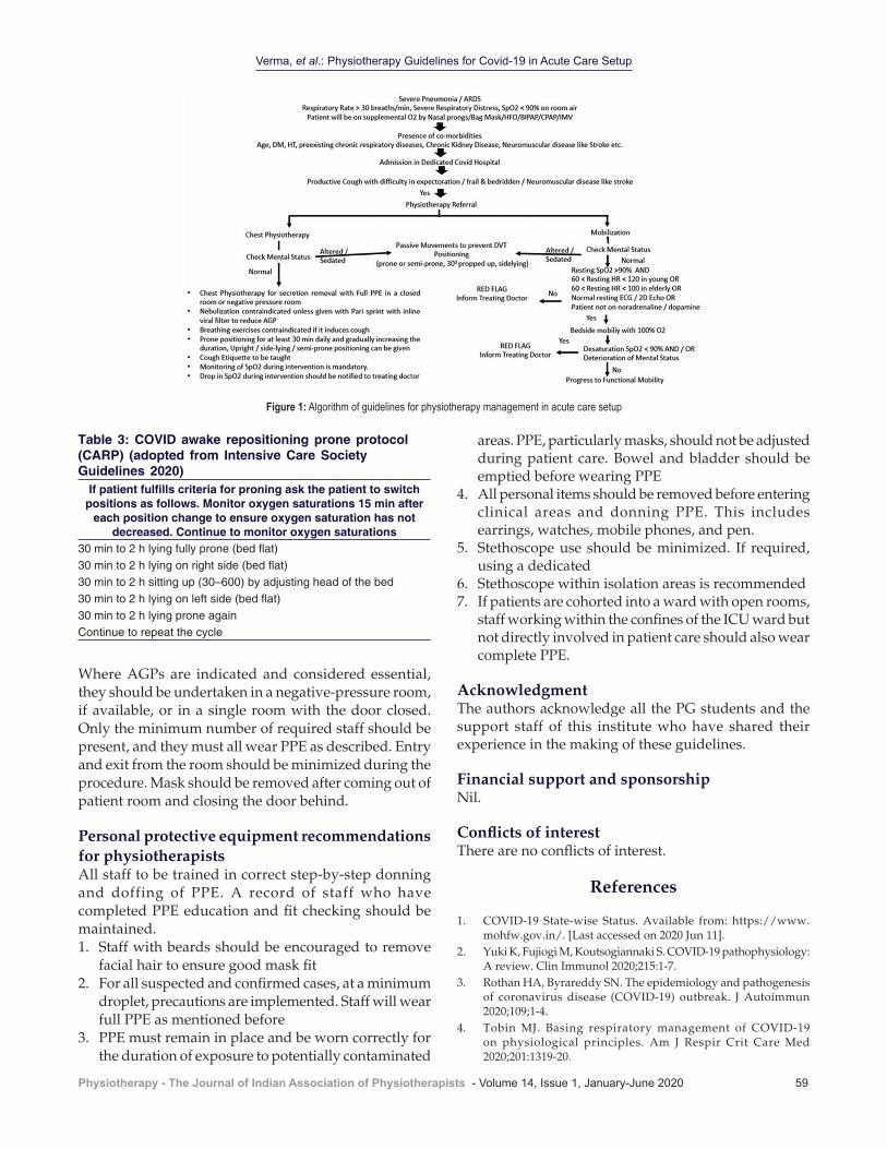
Figure 1: Algorithm of guidelines for physiotherapy management in acute care setup
Verma, et al.: Physiotherapy Guidelines for Covid-19 in Acute Care Setup
Physiotherapy - The Journal of Indian Association of Physiotherapists - Volume 14, Issue 1, January-June 2020 59
Where AGPs are indicated and considered essential, they should be undertaken in a negative‑pressure room, if available, or in a single room with the door closed. Only the minimum number of required staff should be present, and they must all wear PPE as described. Entry and exit from the room should be minimized during the procedure. Mask should be removed after coming out of patient room and closing the door behind.
Personal protective equipment recommendations for physiotherapistsAll staff to be trained in correct step‑by‑step donning and doffing of PPE. A record of staff who have completed PPE education and fit checking should be maintained.1. Staff with beards should be encouraged to remove
facial hair to ensure good mask fit2. For all suspected and confirmed cases, at a minimum
droplet, precautions are implemented. Staff will wear full PPE as mentioned before
3. PPE must remain in place and be worn correctly for the duration of exposure to potentially contaminated
areas. PPE, particularly masks, should not be adjusted during patient care. Bowel and bladder should be emptied before wearing PPE
4. All personal items should be removed before entering clinical areas and donning PPE. This includes earrings, watches, mobile phones, and pen.
5. Stethoscope use should be minimized. If required, using a dedicated
6. Stethoscope within isolation areas is recommended7. If patients are cohorted into a ward with open rooms,
staff working within the confines of the ICU ward but not directly involved in patient care should also wear complete PPE.
AcknowledgmentThe authors acknowledge all the PG students and the support staff of this institute who have shared their experience in the making of these guidelines.
Financial support and sponsorshipNil.
Conflicts of interestThere are no conflicts of interest.
References
1. COVID‑19 State‑wise Status. Available from: https://www.mohfw.gov.in/. [Last accessed on 2020 Jun 11].
2. Yuki K, Fujiogi M, Koutsogiannaki S. COVID‑19 pathophysiology: A review. Clin Immunol 2020;215:1‑7.
3. Rothan HA, Byrareddy SN. The epidemiology and pathogenesis of coronavirus disease (COVID‑19) outbreak. J Autoimmun 2020;109;1‑4.
4. Tobin MJ. Basing respiratory management of COVID‑19 on physiological principles. Am J Respir Crit Care Med 2020;201:1319‑20.
Table 3: COVID awake repositioning prone protocol (CARP) (adopted from Intensive Care Society Guidelines 2020)
If patient fulfills criteria for proning ask the patient to switch positions as follows. Monitor oxygen saturations 15 min after
each position change to ensure oxygen saturation has not decreased. Continue to monitor oxygen saturations
30 min to 2 h lying fully prone (bed flat)30 min to 2 h lying on right side (bed flat)30 min to 2 h sitting up (30–600) by adjusting head of the bed30 min to 2 h lying on left side (bed flat)30 min to 2 h lying prone againContinue to repeat the cycle

Verma, et al.: Physiotherapy Guidelines for Covid-19 in Acute Care Setup
60 Physiotherapy - The Journal of Indian Association of Physiotherapists - Volume 14, Issue 1, January-June 2020
5. Physiotherapy Management for COVID‑19 in the Acute Hospital Setting: Recommendations to Guide Clinical Practice. Ver. 1. (Guidelines endorsed by WCPT, APA, ICCRPT, Canadian Physiotherapy Association, ACPRC; 23 March, 2020.
6. Thomas P, Baldwin C, Bissett B, Boden I, Gosselink R, Granger CL, et al. Physiotherapy management for COVID‑19 in the acute hospital setting: Clinical practice recommendations. J Physiother 2020;66:73‑82.
7. Lazzeri M, Lanza A, Bellini R, Bellofiore A, Cecchetto S, Colombo A, et al. Respiratory physiotherapy in patients with COVID‑19 infection in acute setting: A position paper of the Italian Association of Respiratory Physiotherapists (ARIR). Monaldi Arch Chest Dis 2020;90:163‑68.
8. Greenhalgh T, Javid B, Knight M, Inada‑Kim M. What is the efficacy and safety of rapid exercise tests for exertional desaturation in covid‑19? Centre Evidence Based Med 2020;1‑9.
9. COVID Awake Repositioning Protocol; (CARP). Resuscitation & Acute Critical Care, Janus General Medicine; 2020. last seen 11th Jun 2020. https://emcrit.org/wp‑content/uploads/2020/04/COVID‑CARP‑Protocol‑postable.pdf.
10. Guérin C, Reignier J, Richard JC, Beuret P, Gacouin A, Boulain T, et al. Prone positioning in severe acute respiratory distress syndrome. N Engl J Med 2013;368, 23:2159‑68.
11. Caputo ND, Strayer RJ, Levitan R. Early self‑proning in awake, non‑intubated patients in the emergency department: A single ED’s experience during the COVID‑19 pandemic. Acad Emerg Med 2020;27:375‑8.
12. Intensive Care Society ICS. Available from: https://emcrit.org/pulmcrit/proning‑nonintubated. last seen 11 Jun 2020. https://emcrit.org/wp‑content/uploads/2020/04/2020‑04‑12‑Guidance‑for‑conscious‑proning.pdf.