Quarterly Publication of Indian Dental Association, Kerala ...
54
Index Copernicus ID 5385 ISSN No. 0972-396X Vol 39 | No. 4 October 2016 Case of Bi Cortical Screws used to restore teeth in a patient with Atrophic Maxilla Garres osteomyelitis: a case report Assessment of awareness of the association between periodontitis and systemic conditions/diseases amongst general population Prosthodontic rehabilitation of a patient with amelogenesis imperfecta Oral and Systemic Manifestations of Job Syndrome (HEIS) – A Rare Entity in pediatric population Obesity and periodontal disease Complementary and alternative medicine in paediatric dental practice: A review on current status An overview on retention strategies Fabrication of feeding aid for the management of cleft palate – A case report A comparative evaluation of chamomile and triclosan based tooth pastes on gingival inflammation and plaque accumulation in chronic generalised gingivitis individuals: A clinico-microbiological study Maxillary first molar with two roots and two canals: two case reports Application of the ‘rule 1237’ for success of the dental implant Quiz Email: [email protected] Web: www.idakerala.com SIS Index ID 833 Quarterly Publication of Indian Dental Association, Kerala State Branch
-
Upload
khangminh22 -
Category
Documents
-
view
3 -
download
0
Transcript of Quarterly Publication of Indian Dental Association, Kerala ...

Index Copernicus ID 5385 ISSN No. 0972-396X
Vol 39 | No. 4October 2016
Kerala Dental Journal Vol. 39 | No. 4 | October 2016 ISSN No. 0972-396X
Case of Bi Cortical Screws used to restore teeth in a patient with Atrophic Maxilla
Garres osteomyelitis: a case report
Assessment of awareness of the association between periodontitis and systemic conditions/diseases amongst general population
Prosthodontic rehabilitation of a patient with amelogenesis imperfecta
Oral and Systemic Manifestations of Job Syndrome (HEIS) – A Rare Entity in pediatric population
Obesity and periodontal disease
Complementary and alternative medicine in paediatric dental practice: A review on current status
An overview on retention strategies
Fabrication of feeding aid for the management of cleft palate – A case report
A comparative evaluation of chamomile and triclosan based tooth pastes on gingival infl ammation and plaque accumulation in chronic generalised gingivitis individuals: A clinico-microbiological study
Maxillary fi rst molar with two roots and two canals: two case reports
Application of the ‘rule 1237’ for success of the dental implant
Quiz
Email: [email protected] Web: www.idakerala.com
SIS Index ID 833
Q u a r t e r l y P u b l i c a t i o n o f I n d i a n D e n t a l A s s o c i a t i o n , K e r a l a S t a t e B r a n c h

Kerala Dental JournalVol. 39 | No. 4 | October 2016
Edited by: Dr. K. Nandakumar, Hon. Editor • Published By: Dr. Suresh Kumar G. Hon Secretary • For IDA, Kerala State Branch • Production: Suman Graphics, [email protected]
EDITOR
Dr. K. Nandakumar
ASST. EDITOR
Dr. R.M. Baiju
BUSINESS MANAGER
Dr. Mathew Jose
EDITORIAL CONSULTANTS
Dr. Santhosh SreedharDr. K. Chandrasekharan NairDr. K. George VargheseDr. Ipe VargheseDr. Oommen Aju JacobDr. V.I. PaulDr. Thomas ManjooranDr. Sobha KuriakoseDr. N.O. VargheseDr. T. SreelalDr. Siby Xavier
EX-OFFICIO MEMBERS
Dr Mohammed Sameer P.T.Dr. Suresh Kumar G.Dr. Thomas KCDr. Sabu Kurian
EDITORIAL BOARD
Dr. Dibyendu Mazumder Dr. Ashok DhobleDr. Jayakar Shet tyDr. K.T. SreelathaDr. Sunil SunnyDr. Jayaprasad K.Dr. Ajay Kumar HaridasDr. George VargheseDr. RetnakumariDr. Jolly Mary VarugheseDr. Romel JosephDr. Thomas JosephDr. Kunjamma K.MDr. Vinod R.B.Dr. S. Karthiga KannanDr. P.G. FrancisDr. M. Sadique HussainDr. Saji P.Dr. Joji GeorgeDr. Aby Mathew T.Dr. E. Anuradha Sunil JaganDr. Sobha KuriakoseDr. P.C. SunilDr. ShamsuddinDr. M.K. Mangalam
EDITORIAL OFFICE
Neelambikam, At tukal, Manacaud Trivandrum, Kerala - 695 009
Phone: 0471-2459235 Mobile: 09447066100
e-mail: [email protected] web: www.idakerala.com
OFFICE BEARERS OF IDA KERALA STATE
PresidentDr Mohammed Sameer P.T.
imm. Past President Dr. Thomas KC
President electDr Sabu Kurian
Vice Presidents Dr. Ciju A. Poulose
Dr Eugene Varghese JosephDr Fazil V Hassan
Hon. secretary Dr Suresh Kumar G
Joint secretary Dr Binoy Stanly
asst. secretary Dr. Krisshna Kumar K S
treasurerDr. Santhoshkumar P.U.
editorDr. K. Nandakumar
cde cHairmanDr. Nirmal George Saibu
cdH cHairmanDr. Subash K Madhavan
Index Copernicus ID 5385 ISSN No. 0972-396X
Vol 39 | No. 4October 2016
Kerala Dental Journal Vol. 39 | No. 4 | October 2016 ISSN No. 0972-396X
Case of Bi Cortical Screws used to restore teeth in a patient with Atrophic Maxilla
Garres osteomyelitis: a case report
Assessment of awareness of the association between periodontitis and systemic conditions/diseases amongst general population
Prosthodontic rehabilitation of a patient with amelogenesis imperfecta
Oral and Systemic Manifestations of Job Syndrome (HEIS) – A Rare Entity in pediatric population
Obesity and periodontal disease
Complementary and alternative medicine in paediatric dental practice: A review on current status
An overview on retention strategies
Fabrication of feeding aid for the management of cleft palate – A case report
A comparative evaluation of chamomile and triclosan based tooth pastes on gingival infl ammation and plaque accumulation in chronic generalised gingivitis individuals: A clinico-microbiological study
Maxillary fi rst molar with two roots and two canals: two case reports
Application of the ‘rule 1237’ for success of the dental implant
Quiz
Email: [email protected] Web: www.idakerala.com
SIS Index ID 833
Q u a r t e r l y P u b l i c a t i o n o f I n d i a n D e n t a l A s s o c i a t i o n , K e r a l a S t a t e B r a n c h
Geriatric dentistry
October 1st is observed as the ‘ELDERS DAY’ every year. In this occasion IDA has been carrying out programmes designed at improving the oral health of its senior citizens with a view to giving them a sense of care and dignity. IDA has been implementing a programme, Free denture programme (Prathyasha Project ) under which free dentures are distributed to the needy and edentulous poor elderly, who then would be able to enjoy the comforts of dentition. This project is one of the key activity of the IDA, Kerala Branch. This year in collaboration with all the branches, 600 free dentures has been delivered.
India is home to an increasing proportion of the elderly. One-eight of the world’s elderly population lives in India. In 2014, India’s total population was 1260 million, of which 100 million were above 60 years. By 2021 this figure may reach to a staggering 137 or 143 million. It is time to think – are we adequately equipped to manage the oral health care problems of this increasing number.
The continuing rise in the older population poses serious challenges to health policy planners, particularly because disease patterns will shift concurrently. A National Policy on Senior Citizens 2011 has been prepared by the Union Health Ministry and a National Programme for Health care of the Elderly is in place. It’s time that we should have a national health care programme for the older segment of the population. With the increasing population of the elderly, establishing GERIATRIC DENTISTRY as an important specialized field brooks no delay.

ContentsKerala Dental JournalVol. 39 | No. 4 | October 2016
Contents Preisident’s & Secretary’s Message 250-251
Editorial 252
Case of Bi Cortical Screws used to restore teeth in a patient with Atrophic Maxilla 253 — Prasanth Pillai
Garres osteomyelitis 256 — Saranya George
Assessment of awareness of the association between periodontitis and systemic conditions/diseases amongst general population — Jose Paul 258
Prosthodontic rehabilitation of a patient with amelogenesis imperfecta 261 — Litty Francis
Oral and systemic manifestations of Job Syndrome (HEIS) – A rare entity in pediatric population 264 — Sheela Sreedharan
Obesity and periodontal disease 267 — Sheethel Menon V.
Complementary and alternative medicine in paediatric dental practice 272 — Jeeva P.P.
An overview on retention strategies 276 — Dhanya Menon M.
Fabrication of feeding aid for the management of cleft palate 279 — Sangeeth K Cherian
A comparative evaluation of chamomile and triclosan based tooth pastes on gingival inflammation and plaque accumulation in chronic generalised gingivitis individuals: A clinico-microbiological study 281 — Arjun Jacob Oommen
Maxillary first molar with two roots and two canals: two case reports 286 — Nadira K Rahman
Application of the ‘rule 1237’ for success of the dental implant 289 — P. Rajkumar
Quiz 292 — Jayanthi
Association News 293
KDJ–Vol.39•No.4•October2016 249

Messages from the President and Secretary
Dr.MohamadSameerPT
Dr.SureshKumarG
Dear Doctor,
Greetings to you from the State Office.
As we reach the end of the organisational year nothing gives us more joy than the phenomenal membership growth we have achieved this year. This credit should entirely go to the thirty one branch offices who have tirelessly managed to spread the message of IDA to the fellow professionals.
As we reach a formidable strength we are aware of the increased responsibilities and commitments we owe to the professional community and society at large. At a time when the profession is said to be at cross roads with regard to the future we are extremely happy to note that the members have bestowed their confidence and more than anything else trust in IDA. We at IDA assure you that it would take every step in the interests of its members and is committed to its cause.
The Governments move to implement the Clinical establishment bill in its present form has been taken up at all levels. IDA is a part of the confederation which has been formed to represent to the government on the CE Bill. We believe that any regulation that is implemented without taking the stake holders to confidence has always a reason to fall short of its aims and definitely be a cause of worry.
The continued apathy of the Government and its machinery towards recognizing the importance of Oral health was evident in that there was no representation from the Dental fraternity in the committee to draft the Health policy or the proposed CE Bill.
The time has come when we have to realize that unless dentistry creates an identity of its own we would continue to face this discrimination. It is time to ponder whether the continued administrative clubbing of Dentistry with Medicine is really damaging the interests of the larger section of the professionals and causing a hindrance to its development. This definitely points to the fact that more work needs to be done.
250 KDJ–Vol.39•No.4•October2016

The flagship programme HOPE - Our pillar of social security, now that it has expanded its wings by taking care of the medical expenses of the members and families, IDA is touching every aspect in the life of a dental professional. We are sure that every member would be with us in sharing our heartfelt thanks to the HOPE team who have managed the programme in an exemplary manner.
CDH wing - our face in front of the public has successfully completed the Free denture programme Prathyasha and now has embarked upon a unique programme aimed at Tobacco and substance related abuse “MUKTHI”. The fact that the government and its various agencies have embraced the programme speaks volumes for the CDH wing of IDA. We would expect our members to wholeheartedly participate in the programme.
Continuing education has always been the domain of IDA, which the members have looked upon, with the innovative faculty hunt programme which aims to nurture from within its members and the webinar series which aims to keep us in tune with the advances in knowledge delivery, the members stand to benefit than ever before.
It would be worthwhile to ponder if there is any other organization that gives importance to sports and cultural activities as well as its professional obligations. The wide participation of the branches in sports activities and the upcoming CHILAMBOLI – the cultural extravaganza are testimonies to it. The wide range of activities of the womens wing of IDA (WDC) carries more significance than ever before especially due to the predominantly increasing ratio of women dentists passing out.
The State office takes this opportunity in thanking all who have helped us in our activities and urge you to continue your efforts to develop our association into a stronger and vibrant body. As we look forward in anticipation of a new year expecting to see you all during the 49th Kerala State Dental Conference at Kottayam which promises to be an outstanding event.
Thanking one and all for their contributions.
Dr. Mohamad Sameer P.T. Dr. Suresh Kumar G.President, Hon. Secretary IDA Kerala State IDA Kerala State
Messages from the President and Secretary Kerala Dental Journal
Vol. 39 | No. 4 | October 2016
KDJ–Vol.39•No.4•October2016 251

Editorial
Dr.K.Nandakumar
Better late, than the lateNEET has evolved multiple, perplexing and contradictory responses
from different quarters of the dental profession. College managements are in a thinking mode whether to continue with the functioning of the colleges during the next year. The declining trends of admissions have set in a panic wave amongst the managements and the knee jerk responses have compelled many staff members to leave the jobs. May be, this is a damage control step, to keep up the finances. Post graduate courses which have been started with great effort are facing a planned closure at least in select specialties. In a few years’ time, we should not wonder if the expansive buildings and the five acre land conveniently houses a corporate hospital, five star hotel or a modern high tech school.
Students of dentistry do not find a bright future because of the lack of employment potential in this country or abroad. The only silver lining is that the learning standard of the NEET students, might be good and that would cause an improvement in the standard of graduates. Post graduates also face unemployment because colleges no longer remain as an assured provider of jobs. If there is no job potential for the dental profession, it is needless to elaborate that soon we will lose the sheen of BDS and MDS.
There is no point starting a blame game now. But some realities we have to face. To improve the commercial prospects, the college managements have asked for an increase in seats, endorsed by the respective governments and universities by issuing essentiality certificates. The DCI has indiscriminately complied with the demand without seriously thinking of the future impact. May be they were justifying their deed with mundane impractical health statistics. Producing the dentists and specialists in large number, does not mean that the dental health will improve automatically. Teachers of the profession were also complacent. The associations of the profession were keen only to conduct conferences in international destinations. Foreign countries have realized that we are not serious in shaping our professionals. Nobody has emphasized the social impact of our profession and professional education. Governments have erred in their responsibilities to find out the job opportunities. Number of admissions should have been linked to the job opportunities generated annually. The responsible people should not ask back; who asked you to join this course. Do not say that we are here to just produce seats not jobs. You are here to work for the Indian society. Do find a solution immediately. At least develop sensibility to feel the erosion of the soil under your feet. Better late, than the late.
Dr. K. Nandakumar Editor, KDJ
252 KDJ–Vol.39•No.4•October2016

CaseofBiCorticalScrewsusedtorestoreteethinapatientwithAtrophicMaxilla* Prasanth Pillai, ** Bobby Antony
► IntroductionImplants are the closest replacement
to natural teeth and patients are now choosing implants as the preferred choice of treatment. Implants that are conventionally used in Implant treatment are the two piece crestal implants. For these conventional implants to be successful, there should be adequate crestal bone available and in case of bone deficiency, bone grafting turns mandatory before implant placement. Regardless of the increasing demand, the fact is that more cases are being rejected than being completed with implants due to bone deficiencies. With increasing cases of Oral Cancers and Road Traffic Accidents, prosthetic rehabilitation with conventional crestal implants become daunting tasks fraught with lots of difficulties and failures.
The first designs of the Bicortical screw implants were introduced in the 1970s in Europe. However, they disappeared into obscurity with the advent of conventional, root form, rough surface implants because the promoters of the Bicortical screws did not have clarity on their surgical and prosthetic techniques as well as applications. In spite of all the hurdles, a small group of doctors continued using bicortical screws and Prof. Ihde played a major role in re introducing Bicortical Screws into the world of implantology. Now, no other implant can replace the Bicortical *MaxillofacialSurgeon&Director–TheSmileCentre.in,Kochi,**DentalSurgeon
•CorrespondingAuthor:DrPrasanthPillai,E-mail:[email protected]
Screw implant in “no bone” situations. Such a case where an atrophic maxilla has been rehabilitated with the help of Bi Cortical Screws is being discussed here.
► Case reportA 26 yrs. old lady patient reported to
the clinic with a history of dentofacial trauma three years prior to her reporting to us. She had sustained fractures of her upper anterior teeth in a road traffic accident.
Immediately after the trauma the dentists tried to salvage the remaining upper anterior teeth with root canal procedures and apicoectomies but they were not successful and the patient ultimately had to undergo extractions and was given a removable partial denture which she had been wearing for close to 2 years.
Patient wanted a fixed solution for her missing teeth and did not want fixed partial dentures.
On clinical diagnosis the patient was missing her right central and lateral incisors and right canine. OPG and CBCT were taken for radiographic diagnosis, and they revealed that the left central incisor was having very poor bone support. It was also noticed that the
Maxillary Sinus was mesially extending involving the canine area. The available alveolar bone in the maxilla, in the region where the implants were planned was found to be highly deficient.
A treatment plan was formulated which involved extraction of left central incisor followed by replacement of missing teeth with bicortical screw implants.
Procedures carried out:
Extraction of left central incisor
Bi-Cortical Screw (BCES) implants from Dr IhdeDental, Switzerland, were used employed to provide fixed teeth in the highly atrophied anterior maxilla.
4 BECES implants were placed to replace 21, 11, 12 & 13. Implant in 13 region has been placed trans sinus, engaging the nasal floor. The implants in the 12 region engaged the palatal vault, implant in the 11 region engaged the anterior nasal spine while the implant in the 21 area was placed engaging the nasal floor.
An interim metal-acrylic prosthesis was provided to the patient within 72 hours of the implant surgery, following immediate loading protocols.
KDJ–Vol.39•No.4•October2016 253
KDJ – Kerala Dental Journal IMPlANt
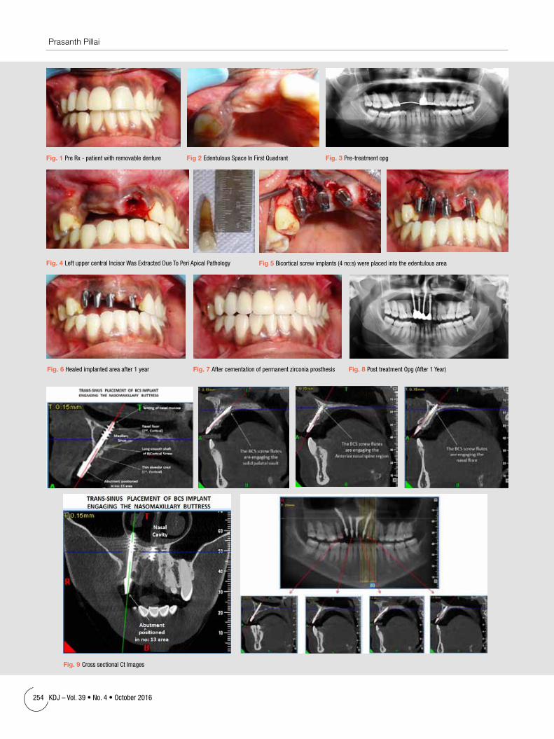
Fig 2 EdentulousSpaceInFirstQuadrantFig. 1 PreRx-patientwithremovabledenture Fig. 3 Pre-treatmentopg
Fig. 4 LeftuppercentralIncisorWasExtractedDueToPeriApicalPathology Fig 5 Bicorticalscrewimplants(4no:s)wereplacedintotheedentulousarea
Fig. 9 CrosssectionalCtImages
Fig. 6 Healedimplantedareaafter1year Fig. 7 Aftercementationofpermanentzirconiaprosthesis Fig. 8 PosttreatmentOpg(After1Year)
254 KDJ–Vol.39•No.4•October2016
Prasanth Pillai

The interim prosthesis was retained for one year after which the prosthesis was replaced with a Zirconia prosthesis.
A CBCT was made and were able to appreciate the engagement of the Bi Cortical screws in the Second corticals (nasal floor, anterior nasal spine, palatal vault), in this case.
► DiscussionThe atrophy of the maxilla makes it difficult to use crestal
implants to rehabilitate the patient following immediate load protocols. Bone grafting followed by implantation after 6 to 8 months is the only option and bone grafting in these areas may not provide us 100% results we seek.
Rough surface implants cannot be placed trans sinus, as the rough surface attracts bacterial colonization leading to sinus infections and also failure of the implants. However, Oral Maxillofacial Surgeons have been regularly using smooth surface plates and screws involving the maxillary sinus in various maxillofacial procedures in management of trauma, orthognathic surgery as well as cancer surgery. Thus use of smooth surface bicortical screw implants involving the maxillary sinuses cannot be of any consequence. In addition, these implants are placed deep in the basal bone which is resistant to resorption, infection and provides extremely strong anchorage. These implants are splinted and functionally loaded within 72 hours of their placement, based on the protocols of Strategic Implantology which emphasizes on splinting of implants even before bone remodelling sets in.
The protocols are based on orthopaedic principles of engaging multiple bone corticals to effect osseofixation of the implant in the bone. The bone physiology is such that once the implants are placed, the bone considers it as a form of trauma. A callus will be formed and it is this callus which is converted into woven bone but this bone remodelling
commences only after 72 hours. Within this time period it is mandatory to establish splinted stabilization of the implants and also to functionally load them.
► ConclusionBicortical screw design implants has been used in
implant dentistry for several decades. But due to lack of proper protocols and imaging systems in the early days, the concepts in their techniques and applications were improper and highly confusing. With the recent reintroduction of the design following proper protocols and 3D imaging of implant engagement, the success rate of such implants have become very much predictable in addition to offering provisions for successful rehabilitation of all types of cases, including “No Bone cases”.
► Citations1. Bocklage, R.: Advanced alveolar crest atrophy: an alternative
treatment technique for maxilla and mandible. Implant Dentistry 2001; 10(1): 30 – 35
2. Ihde, S. Comparision of basal and crestal implants and their modus of application. Smile Dental journal Vol 4 No.1: 36-46, 2009
3. Ihde, S.: Radiological assessments and clinical implications in cases including basal implants; Arab Dental 2: 30-31, 2008
4. Scortecci, G.: Prosthetic colutions with Diskimplants for the treatment of severe bone resorption. Publication scientifique dans la Revue du 2nd International Symposium on Advanced Application of Osseointegration, Padoue, Italie, avril 1991
5. Scortecci, G., Misch C., Odon, G.: Implantologie basale: une approche therapeutique fondee sur la prevue. Implantodontie 12 (2003) 35-47
6. Riben C, Thor A. The Maxillary sinus membrane elevation procedure: Augmentation of bone around dental implants without grafts-A Review of a surgical technique. Int J Dent 2012;2012:105483.
7. Kenji Fujisawa, Go Ohe, Hirokazu Nagai, Susumu Abe, Youji Miyamoto. Effects of Surface Topography of Dental Implants on Bone Loss around the Implants : A Five-Year Evaluation between Implants in an Adjacent Area in Identical Patients. Journal of Japanese Society of Oral Implantology
KDJ–Vol.39•No.4•October2016 255
Case of Bi Cortical Screws used to restore teeth in a patient with Atrophic Maxilla

Fig 1: Extraoralviewshowingdiffuseswellingrightbodyofmandibleregion
Fig 2: Intraoralviewshowingcariesexposed46withmildexpansioninrightbuccalvestibule
Fig 3: Mandibulartrueocclusalviewshowingtheonionskinpatternofperiostealexpansionnotedopposite46,47region
Fig 4: Croppedpanoramicradiographshowingexpansionofperiosteuminferiorto46region.Cariesexposed46withperiapicalradiolucencyalsonoted.Cortexofthemandibleappearsintact.
Garresosteomyelitis*Saranya George, **Girija K L
AbstractGarres osteomyelitis is a chronic non suppurative type of osteomyelitis with proliferative periostitis resulting from mild irritation or infection. It frequently occurs in children and young adults commonly presenting as a bony hard non tender swelling which is slowly progressive and associated with a painful carious tooth. Here a case report of an 11 year old male patient is presented.
Keywords: Garres osteomyelitis, periostitis ossificans, osteomyelitis with proliferative periostitis.
KDJ 2016 | Vol. 39 | No. 4 | Pg 256-257
► IntroductionGarre’s Osteomyelitis is named
after Carl Garre, who first observed the condition in the tibia in 1893. It is a chronic non-suppurative type of osteomyelitis, with proliferative periostitis. It frequently occurs in children and young adults and is caused by inert stimulation from low grade infection.1
The first case of proliferative periostitis affecting the jawbone was described by Pell in 1955. Various terms used to describe this phenomenon include proliferative periostitis of Garré, Garrés osteomyelitis, periostitisossificans, nonsuppurative ossifying periostitis, osteomyelitis sicca, osteomyelitis with proliferative periostitis, and perimandibular ossification.2 The most common site of involvement is the inferior border of the mandible in the first molar region. * Postgraduate student, **Assistant Professor, Dept. of Oral Medicine and Radiology, Government Dental College,
Trivandrum •CorrespondingAuthor:Dr.SaranyaGeorge,Email:[email protected]
► Case reportAn 11 year old male patient reported
to the department of oral medicine and radiology with the complaint of painless swelling of right side of face of one month duration. He gave history of pain in relation to grossly decayed right lower back tooth and diffuse swelling of right cheek three months back for which he
took antibiotics. Pain subsided but the swelling was persisting.
Extraoral examination revealed a diffuse nontender bony hard swelling of size approximately 2*2cm on the inferior aspect of right body of mandible (Fig. 1). Overlying skin appeared normal with no fixity. The regional submandibular
256 KDJ–Vol.39•No.4•October2016
KDJ – Kerala Dental JournalCase RepoRt

lymphnode was enlarged, tender and mobile. Intraorally, the right mandibular first molar was caries exposed (Fig. 2). A bony hard expansion was palpable in the right lower buccal sulcus of 46 region. The borders of the swelling appeared to blend with the normal bone. Considering these facts-diffuse swelling over the mandible of slow progression, non-tender, hard in consistency, associated with dental caries in 46 and regional lymphadenitis, a provisional diagnosis of consolidated dentoalveolar abscess was made along with a differential diagnosis of antibioma.
The patient was subjected to the basic haematological and radiological investigations. Hemogram was within normal limits.
Mandibular occlusal radiograph showed an enlargement of bone in relation to right body of mandible region with expansion of periosteum extending 0.5cm bucally to the first molar with multiple parallel lamellae giving an onion-skin appearance (Fig. 3). Panoramic radiograph revealed an extensive carious lesion on 46 with periapical radiolucency of 0.5cm. Multiple smooth radiopaque lamellae were noted below the inferior border of right mandible separated by fine radiolucent line with a definite cortical outline (Fig. 4). Based on these clinical and radiographic findings a clinical diagnosis of chronic osteomyelitis with proliferative periostitis or garres osteomyelitis was made. The patient underwent extraction of 46 and was given antibiotic coverage. Three months postoperatively the lesion was completely subsided.
► DiscussionGarres osteomyelitis of the jaws generally originates from
an infection of low virulence, such as dental decay, mild periodontitis, periodontal defect, pericoronitis, developing tooth follicle, unerupted teeth, untreated fracture, dental eruption or previous dental extraction in the lesion area or a consequence of infection of the underlying soft tissue that later involved the deeper periosteum.3 This condition generally develops in children and young adults below 25 years of age. Clinically there is a focal non-tender to mildly tender bony hard enlargement of the mandible in the molar region. The overlying skin and mucosa will usually be normal. The clinical observation in the present case was consistent with these findings.
Radiographically periostitis ossificans evolves through three stages. The first consists of an apparent thickening of the periosteum, without radiologic evidence of new bone formation. In the second stage a single layer followed by multiple laminations of new bone are formed between the periosteum and cortex. The third stage occurs during resolution and is characterised as a gross thickening without laminations.4
The differential diagnosis of proliferative periostitis include Ewings sarcoma, fibrous dysplasia, osteogenicsarcoma, infantile cortical hyperostosis, callus exostosis, calcifying hematoma and osteoma.6
Histologically the lesions are supracortical but subperiostealand are composed of reactive trabecular bone and osteoid with an associated cellular fibrovascular connective tissue matrix. The osseous trabeculae are lined with numerous osteoblasts and manifest prominent reversal lines.4
The main treatment goal is to eliminate the etiologic factor, most frequently by extraction of the causative tooth. The role of endodontic therapy is questionable.7 Concurrent antibiotic coverage is often administered. The swelling usually disappears within 2 to 6 months, with a return of the normal bony architecture. At times resolution is protracted over a 1-year period.4
► ConclusionThe Garre’s osteomyelitis is a well-described pathologic
entity. It is rare in occurrence because its development depends on the occurrence of a set of critically integrated conditions; that is chronic infection in a young individual, with a periosteum capable of vigorous osteoblastic activity and equilibrium between the virulence of the infectious agents and the resistance of the host.7 Garre’s osteomyelitis primarily affects younger age group. Hence restoration of the involved tooth by endodontic therapy should be considered as the main treatment goal.
► References1. StephaneSchwartz,Huan Pham.Garre’s osteomyelitis: a case report.
Pediatric Dentistry: Volume 3, Number 3. December 20,19802. Hiroyuki Nakano, Tetsuei Miki, Keiko Aota, TetsuroSumi, Ken
Matsumoto and Yoshiaki Yura. Garrés Osteomyelitis of the Mandible Caused by an Infected Wisdom Tooth. Oral Science International, November 2008, p.150-154
3. ParvindGumber, Asmita Sharma, Kanchan Sharma, Sonal Gupta, BinduBhardwaj, Kamal Kant Jakhar. Garre’ssclerosing osteomyelitis – a case report. Journal of Advanced Medical and Dental Sciences Research, Vol. 4 Issue 2 March - April 2016
4. Robert P Langlais, Olaf E Langland, Christoffel J Nortje. Diagnostic imaging of the jaws. Williams and Wilkins. P477-481. 1995
5. Kawai T, Murakami S, Sakuda M, Fuchihata H Radiographic investigations of mandibular periostitisossificans in 55 cases. Oral Surgery, Oral Medicine, Oral Pathology, Oral Radiology and Endodontics, 1996
6. Yen-Ching Chang a,e, Yi-ShingShiehb,e, Shiao-Pieng Lee a,e,. Yi-Jan Hsia a,e, Chih-Kung Lin c, Shin Nieh c. Huey-Kang Sytwu d, Yuan-Wu Chen a, Chronic osteomyelitis with proliferative periostitis in the lower jaw.. Journal of Dental Sciences (2015)
7. Suma R, Vinay C, Shashikanth M C, Subba Reddy V. Garre’ssclerosing osteomyelitis. J Indian SocPedod Prevent Dent - Supplement 2007
KDJ–Vol.39•No.4•October2016 257
Garres osteomyelitis

Assessmentofawarenessoftheassociationbetweenperiodontitisandsystemicconditions/diseasesamongstgeneralpopulation* Jose Paul, ** Johnson Prakash D’Lima, ***Biju Philip, **** Aswathy Sheela Sudhakar
AbstractIntroduction: Over the last few years, numerous researches have been conducted to prove the periodontal-systemic health link. There is a lack of general awareness among the public regarding this link as well as the need to undergo periodontal treatment in order to establish good oral and general health. Aim: The aim of the study was to determine the level of awareness of the association between periodontitis and systemic conditions/diseases among outpatients attending Annoor Dental College and Government Taluk hospital Muvattupuzha. Materials and Methods: A self-structured questionnaire comprising of 10 questions regarding association between periodontitis and systemic conditions/diseases, was distributed among 180 outpatients. The respondents were instructed to mark any of the choices given as answers to the questions, the choices being yes, no and don’t know. Results: Out of the 180 respondents, 72%, 22%, 25.5% and 30.5 % were aware about the association between periodontitis and diabetes mellitus, cardiovascular diseases, pre term low birth weight infants and stress respectively. 28.8% and 33.3% were aware that treatment of periodontitis can result in better glycemic control and can reduce the risk of ischemic heart disease and stroke respectively. Conclusions: This data points out that adequate awareness regarding the association between periodontitis and systemic conditions/diseases is lacking among the public. Hence integrated individual and community based education programmes are necessary to make the public aware about the association of periodontal disease and systemic diseases.Keywords: Periodontal disease, Systemic disease, Systemic conditions, Diabetes mellitus, Cardiovascular disease, pre term low birth weight infants, Stress
KDJ 2016 | Vol. 39 | No. 4 | Pg 258-260*ProfessorandHOD,**Professor,***SeniorLecturer,****PostGraduateStudent,DepartmentofPeriodonticsandImplantology,AnnoorDentalCollegeandHospital,Muvattupuzha,Kerala,India.•CorrespondingAuthor:DrJosePaul,E-mail:[email protected]
► IntroductionPeriodontitis is predominantly
a Gram negative infection resulting in severe inflammation, in which microorganisms and their products such as lipopolysaccharides (LPS) by vascular dissemination would spread throughout the body1. This results in the spread of infection to different parts of the body resulting in systemic changes as well.
Over the last few years, there has been a keen interest in the relationship between periodontal and systemic health, labelled as periodontal–systemic interlink: a two-way road2. The term Periodontal Medicine was first suggested by Offenbacher denoting a rapidly emerging branch of Periodontology focusing on the evidence relating periodontal diseases with systemic diseases3. This relationship has been mentioned in the Assyrian clay tablet, 17th century, and it was Miller who later proposed the “human mouth as a focus of infection” in 1891, and in 1900 William Hunter designated it with the term “Oral sepsis.
But this focal infection theory fell into disrepute in 1940s due to widespread practice of so called “preventive” or “therapeutic edentulation,” including extraction of otherwise healthy teeth. Resurrection of the theory was seen in the form of Periodontal Medicine when Kimmo Matilla et al. in 1989 examined a possible relationship of oral infection
in contributing to an individual's risk for systemic disease.3 After which, numerous researches have been conducted to prove this dynamic periodontal-systemic health link.
There is a lack of general awareness among the public regarding this link as well as the need to undergo periodontal treatment in order to establish good oral and general health.
Hence the aim of this study was to assess the awareness of the link between periodontitis and systemic conditions/diseases among the outpatients attending Annoor Dental College and Government Taluk Hospital, Muvattupuzha.
► Materials and methodsA self-structured questionnaire was
distributed among 180 patients (135 from Annoor Dental College, Muvattupuzha and 45 outpatients from Government Taluk hospital, Muvattupuzha). Prior permission was obtained from the authorised personnel for conducting the study and a verbal consent from the respondents. The questionnaires were distributed among the subjects after explaining the purpose and terms of the study.
The questionnaire comprised of ten questions regarding the link between periodontitis and systemic conditions/diseases such as diabetes mellitus,
258 KDJ–Vol.39•No.4•October2016
KDJ – Kerala Dental Journal

cardiovascular diseases, pre term low birth weight infants, and stress. The respondents were instructed to mark any of the choices given as answers to the questions, the choices being yes, no and don’t know.
► ResultsOut of the 180 respondents, 72% were aware of the
relation between periodontitis and diabetes mellitus, while 17% were unaware and 10% did not respond (Table 1). 44% were aware that uncontrolled diabetes mellitus can worsen periodontitis but 40% were unaware of this and 15% chose not to answer (Table 2). Only 28% were aware that treatment of periodontal disease can improve the glycemic control in diabetic patients (Table 3).
22% were aware that there is a relation between periodontitis and cardiovascular diseases, while 74.4% were not aware of this association. Only 25.5% were aware of the association between periodontitis and pre term low birth weight infants, while 72.2% were not aware of this relation. 30% respondents were aware of the relation between periodontitis and stress, while 61.65% were not (Table 1).
Also 33.3% knew that treatment of periodontitis can reduce the risk of stroke/ischemic diseases while 63.3% did not (Table 3).
► DiscussionPeriodontal disease is a complex infectious disease resulting
from interplay of bacterial infections and host-response to bacterial challenges. It is estimated that more than 500 different bacterial species are capable of colonising the mouth of an adult. Systemic challenges with the potential vascular dissemination of microorganisms and their products (via the sulcular epithelium) such as Lipopolysaccharides (LPS) throughout the body induce a major vascular response. This host-response may offer explanatory mechanism for the interaction between periodontal infection and a variety of systemic disorders like coronary heart disease; coronary heart disease–related events such as angina, infarction and atherosclerosis, stroke, diabetes mellitus, preterm labour (low birth-weight infants), chronic obstructive pulmonary disease and hospital-acquired pneumonia4.
During the past few years there has been an increased interest in the association between periodontitis and systemic diseases. As the prevalence of periodontitis is high and more studies have correlated the link between periodontitis and systemic diseases, the findings of this study raise important concerns. Given the high prevalence of periodontal disease, its deleterious impact on oral health and its association with systemic disease, patients seeing internal medicine physicians may not be receiving the education and guidance needed5. An increased awareness of certain aspects of periodontal disease and its link to systemic conditions are important, and patients should be counselled about this at each healthcare contact they have, whether it is with a dentist, a physician, or any other healthcare provider. This issue is of extreme importance for India that is being labelled as diabetes capital of the world.
table 1: Awarenessoftherelationbetweenperiodontitisanddiabetesmellitus,cardiovasculardisease,pre-termbirthandlowbirthweightinfants,stress
table 2: Awarenessofthebidirectionallinkbetweenperiodontitisanddiabetes table 3: Awarenessofthetreatmentofperiodontitiscanresultinglycemiccontrolindiabeticpatientsandreducetheriskofischemicheartdisease/stroke.
KDJ–Vol.39•No.4•October2016 259
Assessment of awareness of the association between periodontitis and systemic conditions/diseases amongst general population

► ConclusionThe data obtained from this pilot study points out that there
is only limited awareness among the public regarding the link between periodontitis and systemic health. The current study was only a pilot study comprising of a limited sample size with subjects from a particular geographic location. Further large scale multicentre study required for an accurate assessment of the level of awareness. Measures to improve the awareness should be initiated immediately from an individual basis from patients attending clinics, colleges, to community level and including larger population. Various portals of communication including audio visual aids, posters, printed material including pamphlets, brochures may be used as inexpensive, yet effective methods to raise the awareness among the public.
► References 1. Mealy BL, Klokkevold Perry R. Periodontal Medicine: Carranza's
Clinical Periodontology. 10th ed2. Genco RJ. Current view of risk factors for periodontal diseases.
J Periodontol. 1996;67:1041–93. Williams RC, Offenbacher S. Periodontal medicine: The emergence
of a new branch of periodontology. Periodontol 2000. 2000;23:9–124. Beck JD. Periodontal implications: Older adults. Ann Periodontol.
19965. Bhatia A, Bains SK, Singh MP. To assess knowledge and awareness
of North Indian population towards periodontal therapy and oral-systemic disease link: A cross-sectional survey. J Interdiscip Dentistry 2013;3:79-85
Dr. Jacob Hyson, retired Joint Director of medical education left for his heavenly abode recently. He studied for BDS in Taminadu G o v e r n m e n t D e n t a l College and later took his
postgraduation in Prosthodontics from Government Dental College, Bombay. When he joined Trivandrum Dental College, he had the unique distinction as the first dental postgraduate of Kerala. His entry formally initiated the dawn of Prosthodontics in Kerala. The ascendance of Prof. Hyson in the government hierarchy as Professor, Head of the Dental Wing, Director of Dental College and Joint Director of Medical Education has coincided with the development of the Department of Prosthodontics and the Trivandrum Dental College to one of the finest in the country. He has served the Kerala University in various capacities and the Dental Council of India as its Vice President. His significant contributions include formalizing the inspection protocol and the postgraduate curriculum.
In the early years of Dental College, the syllabus of Prosthodontics was decided by the notes dictated
by Prof. Hyson. Many generations of students copied those notes and graduated without ever referring to a standard text book. Only few books were available in the college library and which we have never considered essential to be referred, to make a pass in the university examination. He had a good team of assistants in the teaching faculty and they carried out the clinical training. Every student remembers him sitting in his office room always writing on the files, correcting thesis of postgraduate students and making discussions with senior staff. He was a silent and sharp observer and could judge the strengths of each individual. He was very reserved in expressing his observations but was never reluctant in giving testimonials which had helped many in getting positions in different parts of the world.
Dr. Jacob Hyson was a legendary professor whose classes were phenomenally popular amongst students. Young teachers emulated him though it was a difficult task. Everyone will remember him for his contributions to the profession. Dentistry in Kerala is indebted to him for its present state and his memory will serve as a guiding light.
Prof. (Dr) Jacob Hyson
260 KDJ–Vol.39•No.4•October2016
Jose Paul

Prosthodonticrehabilitationofapatientwithamelogenesisimperfecta*Litty Francis, **S. Lylajam, ***K. Harshakumar
AbstractAmelogenesisimperfecta (AI) represents a group of developmental conditions, genomic in origin, which affect the structure and clinical appearance of enamel of all or nearly all the teeth in a more or less equal manner and which may be associated with morphologic or biochemical changes elsewhere in the body. Rehabilitation of a patient with AI is a major challenge to the prosthodontist. However, the tremendous advances in the field of dentistry have enabled restoration of function and esthetics to acceptable levels in such cases. The treatment protocol differs depending on the case at hand. The following is a case report of a twenty year old male, with compromised occlusion and poor esthetics owing to Amelogenesis Imperfecta.
Key words: Amelogenesis imperfecta, full mouth rehabilitation, metal ceramic crowns
KDJ 2016 | Vol. 39 | No. 4 | Pg 261-263
► IntroductionAmelogenesis imperfecta (AI) has
been described as a complex group of conditions that disturbs the developing enamel structure and exists independent of any related systemic disorder1-3. This enamel anomaly affects both the primary and permanent dentition1-4. AI is caused by mutation in genes that control Amelogenesis and follow inheritance pattern of autosomal dominant, autosomal recessive or X-linked mode of transmission. The *FormerSeniorResident,**Professor,***ProfessorandHead,DepartmentofProsthodontics,Govt.DentalCollege,
Trivandrum•CorrespondingAuthor:Dr.LittyFrancis,E-mail:[email protected]
incidence of amelogenesis imperfecta has been reported to vary between 1:700 and 1:16,000, depending on the population studied and the diagnostic criteria used4-7. Among the congenital anomalies, amelogenesis imperfecta is an important condition that causes accelerated wear of teeth.
AI has been categorized into 4 broad groups— hypoplastic, hypocalcified, hypomaturation, and hypomaturation-hypoplastic1-4,8-10. All AI patients have similar oral manifestations: teeth sensitivity, poor dental esthetics, and decreased occlusal vertical dimension11. In olden days, treatment of patients with AI has included multiple extractions and fabrication of complete dentures11. These options are psychologically harsh while addressing an adolescent patient12. The treatment plan for patients with AI is related to many factors including the age of the patient, the type and severity of disorder, the socioeconomic status of the patient and the intra-oral situation at the time of presentation.
Complete occlusal rehabilitation in patients having AI is challenging due to the fact that replacement of the lost tooth structure and restoration of the lost vertical dimension of occlusion have to be carried out simultaneously. The contributing factors for excessive wear of teeth are evaluated and should be removed or reduced if possible. The following case report describes the sequenced treatment of a young adult patient with Amelogenesis Imperfecta and decreased vertical height.
► Case ReportA 20-year-old male patient reported
to Dept. of Prosthodontics, Govt. Dental College, Trivandrum with the chief complaints of sensitivity and dissatisfaction with the size, shape, and shade of his teeth. A detailed medical and dental history was recorded. The family history was non-contributory. A thorough intra-oral examination revealed yellowish brown discoloration of all teeth with attrition and hypersensitivity of mandibular incisors and molars (Fig. 1). The interocclusal distance was about 4mm. The oral hygiene was satisfactory. Radiographic examination revealed caries exposure of 47. The patient was diagnosed to have AI. Diagnostic alginate impressions (ALGIPLAST, INDIA PVT.) were made to fabricate study casts. The study casts were then analyzed and a treatment plan was formulated. Patient education regarding the treatment and oral hygiene maintenance was done. The planned treatment included endodontic treatment of 47 followed by full mouth rehabilitation using metal ceramic restorations.
Diagnostic casts were mounted on a semi-adjustable articulator (Dentatus Articulator type ARL) using a face-bow (face bow type AEB) (Fig.2). A hard wax record (Bite registration wax) was taken to increase the vertical dimension by 2mm and this was used to mount the mandibular cast in centric relation. Auto polymerized acrylic resin jig was made so that it could be positioned between the maxillary and mandibular anterior teeth of the articulated cast (Fig. 3). Acrylic jig was used as an index during tooth
KDJ–Vol.39•No.4•October2016 261
KDJ – Kerala Dental Journal Case report

preparation. Canine protected occlusal scheme was planned considering the age and periodontal health of the patient.
The maxillary and mandibular posterior teeth were prepared using the centric jig as an index. Full crown preparations were done on the 14,15,16,24,25,26,34,35,36,44,45,46 and 47. Gingival retraction was done and maxillary and mandibular impressions were made with polyvinyl siloxane (ELITE HD DENTSPLY). The posterior segmental relationship was then registered using bite registration paste (Bitrex, EQUINOX Germany) with the resin jig in place. Shade selection was done (A2 shade was selected-VITA CLASSIC SHADE GUIDE).
Provisional restorations were fabricated with autopolymerising acrylic resin by indirect technique. The patient is allowed to wear these provisional restorations for a period of 2 weeks to confirm the functional acceptance of the occlusal design. They were cemented in place with non-eugenol temporary cement (Freegenol). Patient was recalled after a period of 2 weeks and was found to be comfortable with the increased vertical dimension.
Metal copings (wironet, Germany) were fabricated and tried intra-orally to verify marginal fit and accuracy. Bisque trial for mandibular and maxillary posterior crowns was done. The occlusion was checked in centric and eccentric positions. Once proper occlusion was established, the maxillary & mandibular metal ceramic crowns were glazed and cemented in place with Glass ionomer cement (GC CORP, JAPAN) (Figs. 4 & 5).
The patient was given instruction regarding oral hygiene and diet and to report after 2 weeks. The patient was found to
be comfortable with the restorations. Then the next phase of treatment was undertaken to restore the maxillary and mandibular anterior teeth. Full crown preparations were done for all the six maxillary and mandibular anterior teeth (Figs. 4 & 5). Poly vinyl siloxane impressions (ELITE HD DENTSPLY) were made and poured in type IV dental stone to obtain working casts. Provisional crowns were fabricated for the anteriors using auto-polymerizing acrylic resin. The metal ceramic crowns were fabricated to be in harmony with the pre-established vertical height. Try in of metal copings followed by bisque trial was done. After ascertaining the patients comfort levels, the glazed crowns were cemented into place using Glass ionomer cement (GC CORP, JAPAN).
The patient was educated regarding oral hygiene and maintenance of the crowns. Recall evaluations at 2 months interval was done. The patient was satisfied as his esthetic and functional expectations were met (Fig. 6).
► Discussion Management of AI in the young adult using fixed
prosthodontics is not a novel approach, but is possibly an underutilized one13. Treatment planning for patients with Amelogenesis Imperfecta is related to many factors: the age, socioeconomic status of the patient, the type and severity of the disorder, and the intraoral situation at the time of treatment planning14. Usually the affected teeth show soft enamel of normal thickness that chips and wears easily and has a radiodensity similar to that of dentin. The various dental symptoms include discoloration, pitting and staining of enamel, occlusal wear or
Fig 1: Preoperative Fig 2: FacebowTransfer Fig 3: ResinJig
Fig 4: MaxillaryPosteriorCrownsinplaceandanteriortoothpreparation
Fig 5: MandibularPosteriorCrownsinplaceandanteriortoothpreparation
Fig 6: Satisfiedandconfidentsmile
262 KDJ–Vol.39•No.4•October2016
Litty Francis

chipping, sometimes exposing dentin, tooth sensitivity and a possible loss of vertical dimension of occlusion. The poor appearance in this case was not only due to the innate color of the teeth, but also to the chipping and attrition of the teeth. Occlusal wear is most often attributed to attrition. The causes may be either Amelogenesis or Dentinogenesis Imperfecta or parafunctional activity. Excessive occlusal wear can result in occlusal disharmony, functional and esthetic impairment. Pulpal pathology may also accompany.
When fixed prosthodontic treatment is indicated for all teeth in one or both arches, the dentist must evaluate the existing vertical dimension of occlusion. There has never been a scientific, practical and accurate method by which vertical dimension of the patient could be recorded. Classic techniques have been used to determine the vertical dimension of occlusion like phonetics, interocclusal distance, facial soft tissue contour, cephalometrics, electromyography and patient’s neuromuscular perception15. Dawson stated that even when the teeth have gone down to the gum line, the vertical dimension is not lost because of the eruption of the teeth along with the alveolar bone. The potential problems of restoring the vertical dimension are clenching, muscle fatigue, soreness of teeth, muscles and joints, headache, intrusion of teeth, fracture of porcelain, occlusal instability due to shifting of restored teeth and continual wear16. In such cases, checking and periodic occlusal adjustment must be done upto a year before normal stability returns. Carlsson et al concluded that moderate increase in vertical dimension of occlusion does not create problem provided that occlusal stability is provided17.
There are a number of alternatives for the treatment of teeth affected by AI including inlays, onlays, crowns, laminate veneers, overdentures, implants etc. and the treatment options depend on the severity of the disease. In case of severely affected cases root canal therapy followed by crowns or extraction followed by fixed or removable partial prosthesis may be advised. For many years the most predictable and durable esthetic restoration of anterior teeth has been achieved with jacket crowns18. However, this is an invasive procedure which requires the removal of substantial amounts of tooth structure. Ever since the introduction of porcelain laminates in dentistry, it is considered as the treatment option for anterior teeth as it is conservative and esthetic19. But they have some disadvantages such as lack of marginal adaptation and poor bonding20. So it becomes mandatory to formulate the treatment plan after discussing with the patient.
It is necessary to provide appropriate intercuspation as well as the exact vertical height, which will allow the temporo-mandibular joint to function in a stable & healthy manner. In this case, considering the age of the patient individual metal ceramic crowns with a canine guided occlusion were inserted. During & after the treatment oral hygiene and dietary advice were reinforced to prevent future problems. A periodic
review of the patient’s oral hygiene and periodontal health was stressed and maintained in order to achieve long term success. Psychological health is also an important issue in AI patients which improved following the treatment.
► Conclusion The early rehabilitation of patients with AI is critical to
prevent the progressive loss of vertical dimension of occlusion. Treatment of such cases not only restores the function and appearance of the patient but also helps in building in him a new sense of self confidence.
► References1. Weinmann JP, Svoboda JF, Woods RW. Hereditary disturbances of
enamel formation and calcification. J Am Dent Assoc 1945;32:397-418.2. Aldred MJ, Savarirayan R, Crawford PJM. Amelogenesis imperfecta:
a classification and catalogue for the 21st century. Oral Diseases 2003;9:19-23.
3. Neville BW, Damm DD, Allen CM, Bouquot JE. Oral and maxillofacial Pathology. 2nd ed. Philadelphia: Elsevier; 2002. p. 89-94.
4. Witkop CJ, Rao SR. Inherited defects in tooth structure. Birth Defects Orig Artic Ser 1971;7:153-84.
5. Backman B, Holm AK. Amelogenesis imperfecta: prevalence and incidence in a northern Swedish county. Community Dent Oral Epidemiol 1986;14:43-7.
6. Sundell S, Koch G. Hereditary amelogenesis imperfecta. Epidemiology and clinical classification in a Swedish child population. Swed Dent J 1985;9:157-69.
7. Witkop CJ. Hereditary defects in enamel and dentin. Acta Genet Stat Med 1957;7:236-9.
8. Sundell S, Valentin J. Hereditary aspects and classification of hereditary amelogenesis imperfecta. Community Dent Oral Epidemiol 1986;14:211-9.
9. Witkop CJ. Amelogenesis imperfecta, dentinogenesis imperfecta and dentin dysplasia revisited: problems in classification. J Oral Path 1988;17:547-53.
10. Aldred MJ, Crawford PJM. Variable expression in amelogenesis imperfecta with taurodontism. J Oral Pathol Med 1988;17:327-33.
11. Seow WK. Clinical diagnosis and management strategies of amelogenesis imperfecta variants. Pediatr Dent 1993;15:384-93.
12. Bouvier D, Duprez JP, Bois D. Rehabilitation of young patients with amelogenesis imperfect: A report of two cases. ASDC J Dent Child 1996;63:443-7
13. Mink JR, Okeson JP. Fixed prosthodontics for the young adolescent.In: Goldman HM. Current therapy in dentistry. Vol VI. St. Louis: Mosby;1977. p. 493-5031.
14. Sari T, Usumez A. Restoring function and esthetics in a patient with amelogenesis imperfecta: a clinical report. J Prosthet Dent 2003;90:522-5.
15. Sharry J.J Complete denture prosthodontics 3rd edition16. Dawson D. E. evaluation, diagnosis and treatment of occlusal problems
St. Louis, C.V. Mosby17. Carlsson et al. Effect of increasing vertical dimension on the masticatory
system in subjects with natural teeth. J PROSTHET DENT 1979, vol 41, pg 284-289
18. Peumans M, Van Meerbeek B, Lambrechts P, Vanharle G. Porcelain veneers: a review of the literature. J Dent 2000;28:163-77.
19. Zalkind M, Hochman N. Laminate veneer provisional restorations: a clinical report. J Prosthet Dent 1997;77:109-10.
20. Karlsson S, Landahl I, Stegersjo G, Milleding P. A clinical evaluation of ceramic laminate veneers. Int J Prosthodont 1992;5:447-51.
KDJ–Vol.39•No.4•October2016 263
Prosthodontic rehabilitation of a patient with amelogenesis imperfecta

OralandsystemicmanifestationsofJobSyndrome(HEIS)-Arareentityinpediatricpopulation* Sheela Sreedharan, ** Priyadarsini Geetha Raghuvaran
AbstractJob Syndrome also known as Hyperimmunoglobulin E syndrome (HEIS) is a primary immunodeficiency disease characterized by recurrent skin abscesses, recurrent pneumonia, eczematous dermatitis, and elevated serum IgE levels. HIES manifests as a disease that affects multiple organ systems, including the skeleton, connective tissue, and dentition. The paper describes an unusual case report of Hyperimmunoglobulin E syndrome (HIES) in a 10 year old female child who presented with multiple oral lesions and extraoral pyodermic lesions. Very few literature review is available regarding oral manifestations of Job syndrome.
Keywords: pergammaglobulineamia, pyoderma, Lymphadenopathy.
KDJ 2016 | Vol. 39 | No. 4 | Pg 264-266
► IntroductionHyperimmunoglobulin E syndrome
(HIES) was first described as Job syndrome in 1966, when 2 patients were reported with eczematous dermatitis, recurrent staphylococcal boils, hyperextensible joints/recurrent bone fractures, and distinctive coarse facies1. Biblical references can be traced to Job's syndrome as it was the name given to describe the patients based on the book of Job 2:7, *ProfessorandHead,**SeniorResident,DepartmentofPedodonticsandPreventiveDentistry,GovernmentDental
College,Thiruvananthapuram.•CorrespondingAuthor:DrSheelaSreedharan,Email:[email protected]
“So went Satan forth from the presence of the Lord, and smote Job with some boils from the sole of his feet unto his crown.”2
Autosomal dominant hyper IgE (HIES or Job's) syndrome is a rare primary immune deficiency characterized by eczema, recurrent skin and lung infections, extremely elevated serum IgE, and a variety of connective tissue and skeletal abnormalities. Individuals with HIES share a characteristic facial appearance and many oral manifestations including retained primary dentition, a high arched palate, variations of the oral mucosa and gingiva, and recurrent oral candidiasis.3 It is reported very rarely, with an incidence of 1 in 1,000,000 people.4
In many cases of HIES, skeletal abnormalities include scoliosis, osteopenia, minimal trauma fractures, and craniosynostosis. Dental features include failure of shedding of primary teeth, supernumerary teeth, microdontia, and a high, arched palate. Deficient or delayed root resorption of primary teeth has been reported at a frequency as high as 64%, 72%, and 75% in HIES patients. Reduced resorption of primary tooth roots leads to prolonged retention of primary teeth, which in turn prevents the appropriate eruption of permanent teeth.5 There is however, a paucity of literature
describing oral findings in HIES patients.
► Case report A 10 year old female child reported
in the outpatient Department of Pedodontics, Government Dental College, Thiruvananthapuram with complaints of white patches of tongue, palate and cheek since two weeks. Medical history of the child revealed that she was admitted in the Pediatric ward following recurrent attacks of high grade fever for one and half months coupled with intermittent episodes of abdominal pain. The patient had no relevant dental history, including no previous episodes of extraction or trauma.
The child was first child of nonconsanguinous marriage. Postnatal history revealed history of multiple skin lesions and recurrent pyoderma with orbital cellulitis at 5 years of age. She had coarse facies, recurrent bacterial and fungal pyodermal abscesses, bronchoectasis, mucocutaneous candidiasis for which multiple episodes of incision and drainage was done. She also demonstrated allergic manifestations of eczema, eosinophilic pustules of skin of extremities. Musculoskeletal evaluation revealed osteopenia with pathological hairline fractures, scoliosis and hyperexcitibility. USG abdomen revealed hepatomegaly
264 KDJ–Vol.39•No.4•October2016
KDJ – Kerala Dental JournalCase RepoRt

of 5cm below costal margin and was nontender on palpation. Blood investigations revealed low Hb levels (7.2%), TLC (1.2 lakhs/mm), and elevated IgE levels (19940 Iu/ ml). She also had multiple cervical axillary and inguinal lymphadenopathy.
She was treated for spikes of pyrexia with oral Paracetamol; Bacitracin for pyodermal abscess and Fluconazole was the main antifungal regimen. She was referred to dental department for complaints of loose retained milk teeth and oral ulcers. Extraction of deciduous retained teeth was done under topical anaesthesia. Candid troche application coupled with Betadine mouth gargle was prescribed for pseudomembranous candidiasis.
Extraoral examination revealed pyodermic scars of face, back, neck, and extremities of hands and feet. Intraoral examination revealed scrapable white and erythematous papules in entire buccal mucosa, ventral surface of tongue, attached gingival of maxillary and mandibular anterior teeth and labial mucosa and high arched palatal vault. Lesions were tender with intake of spicy foods. Physiologic mobility of retained 54.55.64.65,72,74,75,84,85 were elicited.
► Discussion Hyperimmunoglobulin E syndrome (HIES, or Job’s
syndrome) is a rare immunodeficiency disorder characterized by chronic eczema, recurrent staphylococcal infections, increased serum immunoglobulin E (IgE; usually >2,000 IU /ml).
The normal serum level of IgE in adults is less than 130 IU per milliliter.4 The primary diagnosis of the illustrated case is derived from the elevated serum IgE levels (19940 Iu/ ml) which triggers significant immune reactions by the release of histamine and cytokines due to antigen-IgE complexes. Vigliante CE et al reported life-threatening cervicofacial infection in a child with hyperimmunoglobulin E syndrome7; Tsang P reported severe periodontitis in a 5-year-old girl with hyperimmunoglobulin E syndrome11. These further emphasizes the need for dental management of HIES patients.
In many cases of HIES, abnormalities in hard tissue are also common. Skeletal abnormalities include scoliosis, osteopenia, minimal trauma fractures which was the prominent musculoskeletal problem in this child2,4 and occasionally craniosynostosis. Dental features included failure of shedding of primary teeth, supernumerary teeth, microdontia, and a high, arched palate7. Deficient or delayed root resorption of primary teeth has been reported at a frequency as high as 64%, 72%, and 75% in HIES patients8. Aldous et al have reported that dental abnormalities are variable and include retention of primary teeth which sometimes results in double rows of teeth upon the eruption of the permanent teeth, and high-arched palates.9 Studies have reported a higher incidence of gingivitis, thrush, and plaque in a group of patients with Job's syndrome and was consistent with the findings of the present case.2
Figure 2. GeneralisedeczemaandpyodermalabscessesontheextremitiesofthepatientwithJobsyndrome(HEIS)
Figure 3. IntraoralviewofthepatientwithJobsyndrome(HEIS)depictingoralcandidiasis,higharchpalate,retained54,55,64,65,72,74,84
Figure 1. CoarsefaciesandpruriticskinabscessesonthetrunkandabdomenofthepatientwithJobsyndrome(HEIS)
KDJ–Vol.39•No.4•October2016 265
Oral and systemic manifestations of Job Syndrome (HEIS) - A rare entity in pediatric population
Although some cases of familial HIES with autosomal dominant or recessive inheritance have been reported, most cases of HIES are sporadic.6
The oral lesions in HIES may represent developmental abnormalities, reactive lesions arising from chronic infections associated with the syndrome, or manifestations of the role of the HIES gene, STAT3, in epithelial development.10 Assessing the prevalence of disease is cumbersome because of its rarity. Prenatal diagnosis may be possible in a child born to parents with known mutations with STAT3 or DOCK8.7
Mutations in STAT3 account for the majority of the cases of autosomal dominant HIES, but the pathogenesis of many varied features remains poorly understood. STAT3-deficient HIES must be distinguished from the distinct syndrome of autosomal recessive HIES which is characterized by extremely elevated serum IgE and severe eczema, often complicated by bacterial and viral super infections. Autosomal recessive HIES does not share the musculoskeletal and dental manifestations of the autosomal dominant disease and has a high incidence of neurologic complications from either infection or vasculitis.
The dental management strategies continue to be: (1) prophylactic antibiotics; (2) timely treatment of infections; and (3) surgical intervention as necessary.11
Recommendations for patients with Job syndrome7
Encourage patients with hyperimmunoglobulinemia E syndrome to exercise actively, attend school. Discourage them from being exposed to smoke, because
further impair pulmonary function. Patients generally benefit from outdoor activities. Good skin care is essential. Given the reasonably good survival rate into adulthood,
healthy activities can help patients not to be emotionally crippled from this immunodeficiency disease.
► ConclusionA proper understanding of the inheritance, systemic and
oral manifestations of hyper IgE syndrome will facilitate early palliative and therapeutic interventions to improve patient care. Further research on HIES and STAT3 is mandatory to decode the ambiguity associated with HIES.
► References 1. Dinauer MC. Disorders of neutrophil function: an overview. Methods
Mol Biol. 2014. 1124:501-15.[Medline] J Indian Soc Periodontol. 2012 Apr-Jun; 16(2): 256–260.
2. Fungal infection of gingiva in a patient with hyperimmunoglobulin-E (Job's) syndrome D. Deepa, K. V. Arun Kumar, Chander Shekhar Joshi, Sanjeev Kumar,1 and Anita Pandey
3. Freeman AF, Domingo DL, Holland SM. Hyper IgE (Job′s) syndrome: A primary immune deficiency with oral manifestations. Oral diseases. 2009;15:2–7.
4. Grimbacher B, Holland SM, Puck JM. HyperIgE syndromes. Immunol Rev 2005;203:244-50.
5. Kamasaki et al. Dental management of a Hyper-IgE syndrome patient.Journal of Dentistry for Children-79:2, 2012
6. Renner ED, Puck JM, Holland SM, et al. Autosomal recessive hyperimmunoglobulin E syndrome: A distinct disease entity. J Pediatr 2004;144:93-9.
7. http://emedicine.medscape.com/article/886988-treatment#d9 Harumi Jyonouchi, MD Faculty, Division of Allergy/Immunology and Infectious Diseases, Department of Pediatrics, Saint Peter's University Hospital.
8. O’Connell AC, Puck JM, Grimbacher B, et al. Delayed eruption of permanent teeth in hyperimmunoglobulinemia E recurrent infection syndrome. Oral Surg Oral Med Oral Pathol Oral Radiol Endod 2000;89:177-85.
9. Aldous JA, Olson GJ, Parkin MJ. Dental observations of hyper IgE disorder. J Clin Pediatr Dent.2007;32:69–72.
10. Freeman AF, Domingo DL, Holland SM. Hyper IgE (Job′s) syndrome: A primary immune deficiency with oral manifestations. Oral diseases. 2009;15:2–7.
11. Severe Periodontitis in a 5-year-old Girl With Hyperimmunoglobulin E Syndrome P. Tsang, DMD G. Derkson, DMD R. Priddy, DDS, MSc A.K. Junker, MD J. Slots, DDS, MS, PhD, DMD, MBA H. Larjava, DDS, PhD. Journal of Pediatric Dentistry – 27:1, 2005
IDA-HOPE (Help Offered to Professionals in Emergencies). Members are requested to contact their respective IDA local branch HOPE representative to receive original application formsJOIN
266 KDJ–Vol.39•No.4•October2016
Sheela Sreedharan

Obesityandperiodontaldisease* Sheethel Menon V., **Arun Sadasivan, ***Elizabeth Koshi
AbstractObesity has been shown to be increasing in its prevalence and is also considered as the fastest growing health related problem in the world. Evidence has shown an increasing link between periodontitis and several systemic diseases. Obesity is one of those systemic disease which can predispose to variety of comorbidities and complications which inturn can affect the overall health. Studies have shown its association with oral diseases, particularly periodontal disease. The aim of this review paper is to provide about the definition and the inflammatory pathways which form a link between the periodontal disease and obesity.
Key words: Obesity, periodontitis, adipokines, inflammation
KDJ 2016 | Vol. 39 | No. 4 | Pg 267-271
► IntroductionObesity have been shown to occur
at an increased rate in recent years.1 Body weight tends to remain stable for long periods of time. This is mainly by a regulatory processes that occur between the dietary intake and energy expenditure.2 But, when there is imbalance between these two processes, it can lead to increased accumulation of fat in the body, thereby leading to overweight and further to obesity.3 Obesity have been recognized as a major predisposing factor to major chronic diseases which range from cardiovascular diseases to cancer by the World Health Organization.4 Subjects with Body Mass Index (BMI) between
*PostGraduateStudent,**Professor,***HeadoftheDepartment,Dept.ofPeriodontics,SreeMookambikaInstituteofDentalSciences,Kulasekharam-629161,TamilNadu,India•Correspondingauthor:Dr.SheethelMenonV, Email:[email protected]
25-29.9 are considered as overweight and BMI equal to or greater than 30 are considered as obese.5 Obesity increases the likelihood of patients to have other associated health and social problem, which inturn can affect the dental services and management.6 There are also studies which show the influence of obesity on oral disease, particularly periodontitis.7,8
Periodontitis is a destructive condition affecting the supporting structures of teeth, which develops through an inflammatory process mainly induced by the presence of a microbial biofilm.9 the quality of host immune response along with the periodontopathogens play an important role in the transition from health to disease. The host response to the bacteria in biofilm and their toxic products triggers an inflammatory response that can cause gingival ulcerations, tissue destruction, alveolar bone loss and finally tooth loss.10 Along with the local production of cytokines and biologic mediators there will be an increase in the systemic inflammatory markers. Evidence indicate a link between periodontitis and other systemic diseases, adverse pregnancy outcomes, osteoporosis, rheumatoid arthritis and also obesity.
► DefinitionObesity has been defined based on
the body mass index (BMI, also called Quetelet Index) which is given by the ratio of body weight (in kg) to body height(in m) squared.11 BMI related to fat mass, morbidity and mortality. BMI does not
assess the body fat distribution. It is also known that abdominal (central, visceral, android) obesity, observed in men, is associated with a higher morbidity than the gluteofemoral (peripheral, gynoid) obesity observed in women. Body fat distribution is based on the waist circumference. Waist circumference shows a close relation with the amount of visceral adipose tissue, which has been shown to be more active metabolically and to secrete far greater amounts of cytokines and hormones compared with subcutaneous adipose tissue. Studies have also shown that measurement of waist circumference or waist-hip ratio can be better disease risk predictor than BMI. But this is still not confirmed which is better and research is still ongoing to assess if BMI or waist circumference or both should be used to assess disease risk.12
Classification and Definition of overweight and obesity (Based on Expert Panel, 1998)11
Classification BMI(kg/m2)
Underweight <18.5
Normal 18.5-24.9
Overweight 25.0-29.9
Obese Class I 30.0-34.9
Obese Class II 35.0-39.9
Obese Class III ≥40
KDJ–Vol.39•No.4•October2016 267
KDJ – Kerala Dental Journal INFORMAtION

► Obesity related diseasesObesity is a risk factor for many chronic disease, like type
2 diabetes, hypertension, coronary heart disease, dyslipidemia, metabolic syndrome, stroke, gall bladder disease, cancer, reproductive abnormalities etc.
HypertensionOverweight and obesity are recognized as important
determinants of elevated blood pressure levels. It is established that weight gain is associated with increased blood pressure, and weight loss can decrease the blood pressure independent of changes in sodium intake. It is shown that obese persons have upto 5 times higher risk of hypertension, and upto 2/3rd of cases of hypertension can be attributed to excess weight.13 similar to adults, the prevalence of hypertension is threefold higher in obese children than in nonobese children.14 the multiple potential mechanism that contribute to the development of higher blood pressure in obese humans include hyperinsulinemia, sympathetic nervous system stimulation, sodium and volume retention, renal abnormalities, abnormal levels of certain adipokines such as leptin and altered spectrum of cytokines acting at vascular endothelial level.15
Type 2 DiabetesDiabetes mellitus and obesity have a complex relationship,
with type 2 diabetes strongly associated with obesity. Obese persons have more than 10 fold increased risk of developing type 2 diabetes compared with normal weight persons. Type 2 Diabetes develops due to an interaction between insulin resistance and beta cells failure. Lipotoxicity, glucose toxicity, obesity-derived cytokines are the possible factors than can explain the process involed.15
Cardiovascular disease & Metabolic SyndromeObese individuals have about 1.5 fold increased risk for
cardiovascular disease, which includes coronary heart disease and cerebrovascular disease. Also about 10-15% of all cases of cardiovascular disease can be attributed to overweight & obesity.16 the association with obesity is stronger, and the population-attributable fraction which is the fraction of cases within population that can be attributed to overweight and obesity, is larger for coronary heart disease than for cerebrovascular disease. Obesity is also associated with an about two-fold higher risk for heart failures and 50% increased risk for atrial fibrillation.
Metabolic Syndrome (MetS) is a complex disorder with high socioeconomic cost that is considered a worldwide epidemic. MetS is defined by a cluster of interconnected factors that increase the risk of coronary heart disease, and other forms of cardiovascular atherosclerotic diseases and diabetes mellitus type 2. Main components involved are dyslipidemia, elevation of arterial blood pressure and dysregulated glucose homeostasis,
while obesity and/or insulin resistance have gained increased attention as the core manifestation of the syndrome.17
The World Health Organization(WHO), the National Cholesterol Education Program(NCEP), and International Diabetes Federation(IDF) proposed algorithms to define the metabolic syndrome
Definition of Metabolic Syndrome
Risk factor Defining Level
Abdominal obesity Waist circumference
Men >102cm
Women >88cm
Triglycerides ≥150mg/dL
HDL cholesterol
Men <40mg/dL
Women <50mg/dL
Blood Pressure ≥130/≥85mmHg
Fasting Glucose ≥110mg/dL
Presence of 3 of any of the above risk factor defines metabolic syndrome
Most studies uses the definition proposed in the ‘Third Report of National Cholesterol Education Programme, Expert Panel in Detection, Evaluation and Treatment of High Blood Cholesterol in Adults (Adult Treatment Panel III) (NCEP-ATP III) which requires presence of atleast 3 of the following metabolic abnormalities before the metabolic syndrome can be defined: abdominal obesity, elevated triglyceride, reduced levels of HDL cholesterol, high blood pressure and high fasting glucose.
Obesity and insulin resistance remain at the core of the pathophysiology of MetS. Other factors involved in the pathogenesis includes chronic stress and dysregulation of hypothalamic-pituitary adrenal axis and autonomic nervous system, increase in cellular oxidative stress, renin-angiotensin-aldosterone system activity, and intrinsic tissue glucocorticoid actions, as well as currently discovered molecules such as microRNAs.17
Other DiseasesObesity also increases the risk of respiratory disorders,
reproductive abnormalities, non-alcoholic steatohepatitis, gall bladder disease, osteoarthritis and certain types of cancers.
268 KDJ–Vol.39•No.4•October2016
Sheethel Menon V.

Inflammatory PathwaysObesity is a chronic metabolic conditions that act as a
risk factor for many diseases such as diabetes, hyperlipidemia, hypertension, atherosclerosis and cardiovascular diseases.
Three main characteristics associated with obesity are
• Hyperinsulinemia- increased production of insulin by beta cells to compensate resistance in tissues.
• Hyperglycemia- due to insulin resistance, circulating glucose is not taken by cells and there is an increase in blood glucose levels.
• Hyperlipidemia- elevated serum levels of cholesterol & triglyceride due to lipid metabolism alteration.
Adipose tissue is also a metabolically active organ that secretes more than 50 bioactive substances, including proinflammatory cytokines such as tumor necrosis factor alpha (TNF-α) and interleukin-6 (IL-6), both of which are main inducers of acute phase hepatic protein production including that of C-reactive protein.
Insulin resistance is found in obesity as a result of an increased secretion of cytokines by adipose tissue and macrophages, which are hyperactive because of hyperlipidemia in obesity.
TNF-α interferes in insulin secretion & blocks translocation of glucose transporter (GLUT-4) to the cell membrane, as well as the assimilation of circulatory glucose by cell; and a state of hyperglycemia can arise.
Insulin resistance inturn interfere with lipid metabolism because the adipocytes insulin resistance cannot assimilate circulating fatty acids. After reaching tissues, fatty acids will be broken down to triglycerides and cholesterol which inturn increase insulin resistance state.
Leptin, a 16kDa nonglycosylated polypeptide acts as both cytokine and hormone, is produced by adipose tissue. It is involved in various biological processes including energy metabolism, endocrine functions, reproduction and bone metabolism. Leptin acts as a circulating appetite suppressant that regulates adipose tissue mass.18 Elevated levels of leptin results in decreased food intake, increased energy expenditure, and a negative energy balance whereas leptin deficiency results in hyperphagia and severe obesity. Also obese individuals generally exhibit high levels of circulating leptin which also suggest a resistance to leptin.19 Leptin also has a significant role in inflammatory process. An increase in leptin production occurs during infection and inflammatory processes and is
therefore implicated in the pathogenesis of chronic inflammatory disease.20,21
Periodontitis, is a chronic inflammatory disease where bacterial origin and resultant endotoxin production trigger host responses at both local and systemic levels. Inflammatory mediators like interleukins (IL-1β, IL-6, IL-8, IL-17 and IL-23) and TNF-α as well as bone related factors are identified at both mRNA and protein levels.22 Proinflammatory actions of TNF-α lead to bone loss and loss of periodontal attachment; action of IL-17 lead to production of other proinflammatory mediators including IL-6 and IL-8 and in turn lead to alveolar bone destruction. An increase in local and systemic inflammatory cytokines like IL-6 & TNF-α has been reported in patients with periodontitis.23 These cytokines can in turn trigger an increase in the production of acute phase proteins such as C-reactive protein which increase the inflammatory response similar to that seen in obesity.
Adipose tissue secrete several bioactive substances known as adipocytokines, which include TNF-α, which can affect the periodontal tissue directly. TNF-α mediates endotoxin-induced injury in various organs including periodontal tissue.24 Plasminogen activator inhibitor-I (PAI-I), which is expressed in visceral fat induces the agglutination of blood and raises the risk of ischemic vascular disease. Therefore PAI-I may also reduce blood flow in periodontium of obese individuals to promote development of periodontal disease. (Fig. 1)
Inflammatory markersAdipose tissue secrete proinflammatory cytokines like TNF-α
and interleukin-6 which are the main inducers of acute phase hepatic protein production, including C-reactive protein. These markers have also shown impairment in intracellular insulin signalling which inturn cause insulin resistance.25
Leptin
Fig. 1 Adipose-TissueDerivedHormonesandCytokines(Adipokines)
KDJ–Vol.39•No.4•October2016 269
Obesity and periodontal disease

Leptin is a pleitropic cytokine, secreted by adipocytes. It is involved in variety of biological processes, including energy metabolism, endocrine functions, reproduction and immunity. It acts a ‘lipostat’ that regulates adipose tissue mass. Leptin deficiency due to mutation in ob gene encoding leptin (rarely observed in humans) results in hyperphagia and severe obesity, whereas its substitution in leptin-deficient mice and humans were able to normalize food intake and body weight.26
Leptin levels are acutely increased by endotoxins (lipopolysaccharides (LPS) and administration of proinflammatory cytokines (TNF-α, IL-1)
Adiponectin, Resistin and other Adipose- tissue derived CytokinesAdiponectin is a circulating hormonesecreted by adipose
tissue that is involved in glucose and lipid metabolism and account for about 0.05% of total serum proteins.27 Contrary to the adipose-derived hormones, adiponectin levels are reduced in persons with obesity, insulin resistance or type 2 diabetes. It improves insulin sensitivity and may have anti-atherogenic and anti-inflammatory properties and low plasma adiponectin levels showed prediction of type 2 diabetes & coronary heart disease in humans.28
Resistin belongs to family of resistin like molecules (RELM) and has been reported to be secreted by adipocytes and cause insulin resistance in animal models.29 In contrast to mice, human resistin is expressed at lower levels in adipocytes and at a higher levels in circulating blood monocytes and evidence suggest that human resistin is more closely related to inflammatory processes than to insulin resistance
Other adipokines include Visfatin, which elicits insulin-like effects and serum retinol-binding protein 4(RBP4). Initially regarded as markers it is mainly related to weight regulation and insulin resistance, it has become clear that hormones like leptin, resistin or adiponectin are involved in a variety of functions and diseases including cardiovascular disease, diabetes and inflammatory diseases.
► ConclusionPeriodontist should be aware of the increasing number of
obese individual and also its significance as a risk factor for overall and oral health. Studies have shown a close relation between obesity and periodontal disease and also with other chronic diseases like type 2 diabetes and cardiovascular disease.
Pro-inflammatory cytokines can act as a multidirectional link among periodontitis, obesity and other chronic diseases. Adipose tissue is a large reservoir of mediators like TNF-α and other adipokines.
Therefore full co-operation and collaboration of all health care professionals are required to educate patients regarding the ramification of obesity and periodontitis and other chronic diseases and to encourage counselling, treatment and intervention strategies as needed.
► References1. Haenle MM, Brockmann SO, Kron M, Bertling U, Mason RA,
Steinbach G, et al. Overweight, physical activity, tobacco and alcohol consumption in a cross-sectional random sample of German adults. BMC Public Health 2006;6:233.
2. Nascimento GG, Leite FRM, Do LG, Peres KG, Correa MB, Demarco FF, Peres MA. Is weight gain associated with the incidence of periodontitis? A systematic review and meta-analysis. J Clin Periodontol 2015; 42:495-505
3. Martinez JA, Navas-Carretero S, Saris WH, Astrup A. Personalized weight loss strategies-the role of macronutrient distribution Nature Reviews. Endocrinology 2014; 10:749-60.
4. World Health Organization. Diet, nutrition and the prevention of chronic diseases. World Health Organ Tech Rep Ser 2003; 916: i-viii, 1-149, backcover.
5. World Health Organization (2000) Obesity: Preventing and Managing the Global Epidemic. Report of a WHO Consultation World Health Organization Technical Report Series. 894, i–xii, 1–253, World Health Organization, Geneva.
6. Tirth A, Tandon V. Role of Obesity in Chronic Periodontal Diseases: A Systematic Review. Int J Sci Stud 2015;2(10):104-7.
7. Saito T, Shimazaki Y, Koga T, Tsuzuki M, Ohshima A. Relationship between upper body obesity and periodontitis. J Dent Res 2001;80:1631-6.
8. Saito T, Shimazaki Y, Kiyohara Y, Kato I, Kubo M, Iida M, et al. Relationship between obesity, glucose tolerance, and periodontal disease in Japanese women: the Hisayama study. J Periodontal Res 2005; 40: 346-53.
9. Van Dyke TE, van Winkelhoff AJ. Infection and inflammatory mechanisms. J Clin Periodontol 2013; 40(suppl 14): s1-s7
10. Krejci CB, Bissada NF. Obesity and periodontitis: a link. General Dentistry 2013; 60-3.
11. Expert Panel. Executive summary of the clinical guidelines on the identification, evaluation, and treatment of overweight and obesity in adults. Arch Intern Med 1998;158:1855-67.
12. Pischon N, Heng N, Bernimoulin JP, Kleber BM, Willich SN, Pischon T. Obesity, Inflammation, and Periodontal Disease. J Dent Res 2007;86(5):400-409.
13. Wolf HK, Tuomilehto J, Kuulasmaa K, Domarkiene S, Cepaitis Z, Molarius A, et al. Blood pressure levels in the 41 populations of the WHO MONICA Project. J Hum Hypertens1997;11:733-42.
14. Kotchen TA. Obesity-related hypertension: epidemiology, pathophysiology, and clinical management. Am J Hypertens 2010;23:1170-78.
15. Stumvoll M, Goldstein BJ, van Haeften TW. Type 2 diabetes: principles of pathogenesis and therapy. Lancet 2005;365(9467):1333-46.
16. Wilson PW, D'Agostino RB, Sullivan L, Parise H, Kannel WB. Overweight and obesity as determinants of cardiovascular r isk: the Framingham experience. Arch Intern Med 2002;162:1867-1872.
270 KDJ–Vol.39•No.4•October2016
Sheethel Menon V.

17. Kassi E, Pervanidou P, Kaltsas G, Chrousos G. Metabolic syndrome: definitions and controversies. BMC Medicine 2011;9:48.
18. Friedman JM. Leptin, leptin receptors, and the control of body weight. Nutr Rev1998;56(2, pt 2):s38-s46; discussion s54-s75.
19. Margetic S, Gazzola C, Pegg GG, Hill RA. Leptin: a review of its peripheral actions and interactions. Int J Obes Relat Metab Disord 2002;26(11):1407-33.
20. Lago R, Gómez R, Lago F, Gómez-Reino J, Gualillo O. Leptin beyond body weight regulation–current concepts concerning its role in immune function and inflammation. Cell Immunol2008;252(1-2):139-145.
21. Otero M, Lago R, Lago F, et al. Leptin, from fat to inflammation: old questions and new insights. FEBS Lett2005;579(2):295-301.
22. Ribeiro, VF, de Mendonca AC, Santos VR, Bastos MF, Figueiredo LC, Duarte PM. Cytokines and bone-related factors in systemically healthy patients with chronic periodontitis and patients with type 2 diabetes and chronic periodontitis. JPeriodontol 2011;82(8):1187-96.
23. Beck J, Garcia R, Heiss G, Vokonas PS, Offenbacher S. Periodontal disease and cardiovascular disease. J Periodontol1996;67(10):1123-37.
24. Saito T, Shimazaki Y. Metabolic disorders related to obesity and periodontal disease. Periodontol 2000 2007;43:254-66.
25. Rotter V, Nagaev I, Smith U. Interleukin-6 (IL-6) induces insulin resistance in 3T3-L1 adipocytes and is, like IL-8 and tumor necrosis factor-alpha, overexpressed in human fat cells from insulin-resistant subjects. J Biol Chem 2003;278(46):45777-84.
26. Margetic S, Gazzola C, Pegg GG, Hill RA. Leptin: a review of its peripheral actions and interactions. Int J Obes Relat Metab Disord 2002;26(11):1407-33.
27. Chandran M, Phillips SA, Ciaraldi T, Henry RR. Adiponectin: more than just another fat cell hormone? Diabetes Care 2003;26:2442-50.
28. Berg AH, Combs TP, Du X, Brownlee M, Scherer PE. The adipocyte-secreted protein Acrp30 enhances hepatic insulin action. Nat Med 2001; 7(8):947-53.
29. Steppan CM, Bailey ST, Bhat S, Brown EJ, Banerjee RR, Wright CM. The hormone resistin links obesity to diabetes. Nature 2001; 409:307-12.
30. Nagaev I, Smith U. Insulin resistance and type 2diabetes are not related to resistin expression in human fat cells or skeletal muscle. Biochem Biophys Res Commun 2001;285:561-4.
31. Fukuhara A, Matsuda M, Nishizawa M, Segawa K, Tanaka M, Kishimoto K, et al. Visfatin: a protein secreted by visceral fat that mimics the effects of insulin. Science 2005;307(5708):426-30.
KDJ–Vol.39•No.4•October2016 271
Obesity and periodontal disease

Complementaryandalternativemedicineinpaediatricdentalpractice* Jeeva P.P., **Kannan V.P., ***Remya Ramachandran
AbstractManagement of behaviour and anxiety of children are key factors in paediatric dentistry. Although many techniques are successful, the attitude of parents and dentists towards these techniques are changing. Increased awareness among parents about the risks of pharmacologic behaviour management and with the global movement towards holistic approach, child friendly Complementary and Alternative Medicine (CAM) techniques are becoming popular. This article highlights certain CAM techniques which are helpful for behaviour management in paediatric dental practice.
Key words: Complementary and Alternative Medicine (CAM) technique, differentially abled children, behaviour management, fear, anxiety, holistic approach.
KDJ 2016 | Vol. 39 | No. 4 | Pg 272-275
► IntroductionPaediatric dental practice is aimed
to build and maintain relationships between child, parent and dentist that will allow the highest quality of dentistry to be delivered. It take necessary steps to help each child to develop the skills and behaviour necessary to willingly seek appropriate lifetime of dental care, not hindered by undue fear
*AssociateProfessor,**Professor,***SeniorLecturer,DepartmentofPeadiatricandPreventiveDentistry,GovernmentDentalCollege,Kozhikode.•CorrespondingAuthor:Dr.RemyaRamachandran,Email:[email protected]
or anxiety. Managing the behaviour and anxiety of children are widely agreed to be a key factor in paediatric dentistry. Although many techniques are successful, the attitude of parents and dentists towards these techniques are changing.1 Awareness of parents about medical risk of pharmacologic behaviour management and with the global movement towards holistic approach, child friendly Complementary and Alternative Medicine (CAM) techniques are becoming popular. For better behaviour, complementary and alternative methods of behaviour management can be adopted as a part of integrative medicine especially in differentially abled children. Most of these method will help the child to reduce stress, fear, anxiety, phobia and improve overall personal wellbeing.
► CAM techniquesComplementary and alternative
medicine has a long history of use with some modalities being in existence for thousands of years. Recently, there is greater awareness of the many benefits of CAM for promoting health and wellness. The National Institute of Health has defined CAM as a group of diverse medical and health care systems, practices and products that are not presently considered to be part of conventional medicine.2 Complementary medicine procedures are used in conjunction with conventional medicine
but alternative medicine procedures are used instead of conventional medicine. When CAM therapies are used with conventional medicine, it is called integrative medicine.3,4 CAM system display a holistic approach based on assumption that wellbeing is intrinsically linked to the integration and balance of the whole person-body, mind and spirit in harmony with the environment and prevailing culture. In contrast to conventional medicine, CAM therapy highlight the central role of patient in regaining and maintaining full health and therefore offer an extra dimension to the healing process.2,3,4
The National Centre for CAM categorized CAM in to five groups.5,6
(1) Alternative medical systems-based on a complete system of theory and practice that has originated separately before the development of conventional western medical practice. eg. homeopathy, ayurveda, naturopathy, ancient medicine.
(2) Mind body interventions this method works by utilizing mind to affect the body and its physical symptoms. eg. meditation, prayer, dance, music, art and cognitive behavioural therapy.
(3) Biologically based therapy like herbal therapy and mineral therapy.
272 KDJ–Vol.39•No.4•October2016
KDJ – Kerala Dental JournalREvIEw

(4) Manipulative and body based therapy-based on application of controlled force to a joint, moving it beyond the normal range of motion in an effort to aid in restoring health. For example Massage therapy, pressure point therapy.
(5) Energy therapy - biofield therapy and bio electromagnetic therapy utilize magnetic, pulsed or direct current for healing.
Aroma therapyThe modern aroma therapy appeared in the early part of
twentieth century. It is the art and science of utilizing extracted aromatic essence from plants to harmonize and promote the health of mind, body and spirit.7,8 This method used to unify the physiological and psychological process to enhance an individual’s healing capacity and wellbeing. Essential oils from plants are mainly used for this therapy. They are usually inhaled or applied topically and rarely administered by oral and rectal route. Therapeutic route and dosage depends up on character of patient, age, condition to be treated, properties of essential oils, professional practice parameters, safety data and patient’s preference. Essential oils had a wide range of actions like analgesic, anti-inflammatory, anti-microbial, antiseptic, relaxant and sedative. The effect of aroma therapy may be due to the binding of chemical components in the essential oil to the receptors in the olfactory bulb, impacting the brains emotional centre, the limbic system and also by the direct pharmacological actions of essential oil. Odours can modulate cognition, mood and behaviour. It also changes pain perception by setting a more pleasant environment or distracting from pain experience. It is proved that aroma therapy with orange oil and lavender oil reduces stress and anxiety in dental patients.9
Music therapy:Music therapy is a clinical modality that uses music as a
central basis of activity. It is a creative art therapy that cross multiple areas of treatment and can be effective in facilitating development in multiple areas of child’s functioning.10 The major elements of music-rhythm, melody, harmony, tempo and loudness are used in specific combinations and varying degrees of intensity to produce change in an individual.11 The music therapist often works as a part of an interdisciplinary care team in many different settings. The use of music based interventions aid in distraction from painful stimuli, lessen behavioural distress and decrease pain threshold in children. It helps to improve attention, concentration, impulse control, social functioning, self esteem, self expression, motivation and cognism of patients.11,12
Colour/chromatherapy:Colour assists the body in its natural ability to balance itself
and has been used for centuries for healing in Asia and in the
ancient civilizations. It was considered that colour is a force of immeasurable and infinite power exerting a tremendous psychological and physiological influence on people. Colour therapy consist of the use of colour in a variety of ways to promote health and healing like coloured light, massaging with coloured oils, visualizing colours, wearing coloured cloths, and eating coloured foods.13,14 Colours with unique wavelength and oscillations, when combined with a light source and selectively applied to impaired organ or system, provide the necessary healing energy required by the body. It generates electrical impulses and magnetic fields of energy that are prime activators of the biochemical and hormonal process in the human body.13
Along with conventional medicine, colour therapy is used to treat neonatal jaundice, seasonal affective disorder, insomnia, drug dependence and to reduce overall level of medications. It is found that pink colour has tranquilizing and calming effect within minutes of exposure and it is used to suppress the hostile aggressive and anxious behaviour.15 Blue light is used in healing of injured tissue, preventing scar formation, pain reduction and for psychological problems. Red and black colour is associated with depression and anger, so its use is limited for better behaviour.13,14,16 Along with conventional treatment, judicious use of colour therapy can help to create positive behaviour outcome in dental setting.
Laughter therapyLaughter is the most frequent and appealing response to
humour and many other positive emotions. It has positive quantifiable physiological and psychological effects on certain aspect of health. Laughter therapy is an application containing breathing exercise and physical movements.1 Physiologically this therapy helps to relax muscle, improve breathing and circulation, and enhance immunity and CNS functions. Psychologically it raise the pain threshold and improve individual wellbeing. Also it reduces stress producing hormones and control anxiety and depression. This CAM technique is well tolerated with less side effects. Unlike other therapies which are more time consuming, committed or expensive, laughter therapy can be a easily implemented, cost effective CAM in dental settings for the health and patient care.18,19,20
Play therapy:It is the systematic use of a theoretical model to establish
an interpersonal process wherein trained play therapists use the therapeutic powers of play to help child to prevent or resolve psychological difficulties and achieve optimal growth and development. It provides a way in young children to express their experience and feelings through a natural safe guided self-healing process. As children experiences and knowledge are communicated through play, it becomes an important way
KDJ–Vol.39•No.4•October2016 273
Complementary and alternative medicine in paediatric dental practice
for them to know and accept themselves and others. Studies showed that play therapy is an effective method to reduce anxiety, distress and to increase children’s coping abilities.21, 22, 23
Art therapyIt is a form of psychotherapy that uses art as its primary
mode of communication. It uses the creative process of art making to improve and enhance the physical, mental and emotional well- being of individuals. It is based on theory that the creative process involved in artistic expression helps people to resolve conflicts and problems, develop interpersonal skills, manage behaviour, reduce stress, and increase self-esteem and self-awareness. It integrates the fields of human development, visual art and the creative process with models of counselling and psychotherapy.24,25 Art therapy is helpful in behaviour management of special children especially autism.25
HypnosisIt is a procedure during which a person is guided by a
therapist to respond to suggestions that allow for changes related to subjective experiences. A deeply relaxed natural altered state of consciousness is achieved in this technique.26 Hypnotic susceptibility varies between individuals. This begin to increase at age of three and peak level attain at the age of 8-12 years and then declines at age of sixteen after which it tends to remain stable for whole life. This CAM method helps to eliminate phobia and fear and anxiety in dental setting. Moreover helps to prevent gagging and aid in habit correction.26, 27, 28
Cognitive behaviour therapy (CBT)CBT is a psychological approach based on scientific
principles and is effective for a wide range of behavioural problems. It aim to directly target distressing symptoms, reduce distress, re-evaluate and promote helpful behavioural responses by offering problem focused skills based treatment interventions.29 CBT is a talking therapy based on principle of guided discovery, which involve exploring and reflecting on the style of reasoning and thinking and possibilities of thinking differently and more hopefully.30 Evidence based studies showed that CBT is effective in children with mental health problem, anxiety and phobia in dental setting.30,31
YogaYoga has ancient root but it has become more popular
for physical and spiritual reasons in the past thirty years.31 It is a psycho somatic spiritual discipline for achieving union and harmony between mind, body, soul and the ultimate union of individual conscious with the universal conscious. It is basically a mind body therapy that has emerged as an educational based intervention and to improve various
medical conditions. Yoga produces many physiological and psychological effects on body.32 Physiologically improve blood circulation, muscle relaxation, improve immunity, reduce blood pressure, increase neuroendocrine and other hormonal activity.33 Psychologically yoga alter mood and reduce pain threshold and help in behaviour management by relaxation exercise. This CAM method helps children to overcome fear and anxiety during dental visits and ultimately improve the quality of life.33,34
► ConclusionIn spite of rapid growth of CAM, its role in mainstream of
modern medicine is still not clear. Many scientific, educational, legal issues exist as challenge to integrate CAM in conventional medical practice. Possible benefits and dangers with many of the CAM techniques are uncertain. More evidence based studies and clinical trials need to be carried out to popularise CAM in the modern world.
► Reference1. Lawrence SM, McTigue BJ, Wilson S, et al. Parental attitudes
toward behavior management techniques used in paediatric dentistry. Pediatr. Dent 1991; 13:151-155
2. Micozzi MS. Integrative medicine in pharmacy and therapeutics. P&T. 2003;28(10):666–672.
3. Cohen KR, Cerone P, Ruggiero R. Complementary/alternative medicine use: Responsibilities and implications for pharmacy services. P&T. 2002;27(9):440–446.
4. Kantor M. The role of rigorous scientific evaluation in the use and practice of complementary and alternative medicine. J Am Coll Radiol. 2009;6(4):254–262.
5. Kwan D, Hirschkorn K, Boon H. U.S. and Canadian pharmacists’ attitudes, knowledge, and professional practice behaviors toward dietary supplements: A systematic review. BMC Complement Altern Med. 2006;6(31):1–10.
6. Waldman SA, Terzic A. Pharmacology and Therapeutics: Principles to Practice. Philadelphia: Elsevier/W. B. Saunders; 2008. p. 1536.
7. Lehrner J, Eckersberger C, Walla P, Pötsch G, Deecke L. Ambient odor of orange in a dental office reduces anxiety and improves mood in female patients. Physiol Behav. 2000;71:83–6.
8. Dobetsberger C, Buchbauer G. Actions of essential oils on the central nervous system: An updated review. Flavour Fragr J. 2011;26:300–16.
9. Lehrner J, Marwinski G, Lehr S, Johren P, Deecke L. Ambient odors of orange and lavender reduce anxiety and improve mood in a dental office. Physiol Behav. 2005;86:92–5.
10. Standley, J. M. (1986). Music research in medical/dental treatment: Meta-analysis andclinical applications. Journal of Music Therapy, 23(2), 56.
11. John Pellitteri Music Therapy in the Special Education Setting Journal of educational and psychological consultation, 11(3&4), 379–391.
12. Aitken, J. C., Wilson, S., Coury, D., & Moursi, A. M. (2001) The effect of music distraction on pain, anxiety and behavior in pediatric dental patients. Pediatric Dentistry, 24(2), 114-118-122
13. Birren, F.: Human Response to Color and Light. Hospitals, pp. 93-96, July 16,1979.
274 KDJ–Vol.39•No.4•October2016
Jeeva P.P.

14. Amber R. Colour Therapy. Sarta Fe, N.M.: Aurora Press; 1964.15. Schauss AG. Tranquilizing effect of colour reduces aggressive
behaviour and potential violence. J Orthomol Psych. 1979;4:218–21.16. Sanadhya Yatishkumar, Sanadhya Sudhanshu, Jain Sorabh R,
Pankaj Aapaliya, Garima choudhary, Nidhi Sharma Importance of chroma therapy in dentistry Indian J Dent Adv 2013; 5(3): 1252-1256.
17. Mora-Ripoll R. Thetherapeutic value of laughter in medicine. Altern Ther Health Med 2010 Nov–Dec;16(6):56-64.
18. Martin RA. Humor, laughter, and physical health: methodological issues and research findings. Pyschol Bull 2001 Jul;127(4):504-19
19. Morse DR. Use of humour to reduce stress and pain and enhance healing in the dental setting.JNJ Dent Asso.2007,7894)32-36
20. Wild JA Laughing all the way. Dent today 1998,17(12):38-4121. Christiano, B. A., & Russ, S. W. (1996). Play as a predictor of
coping and distress in children during invasive dental procedure. Journal of Clinical Child Psychology, 25(2), 130.
22. Clatworthy, S. (1981). Therapeutic play: effects on hospitalized children. Children's Health Care, 9(4), 108-113.
23. Ellerton, M.-L., Caty, S., & Ritchie, J. A. (1985). Helping Young Children Master Intrusive Procedures Through Play. Children's Health Care, 13(4), 167.
24. Hogan, S. (2001). Healing arts: The history of art therapy. London: Jessica Kingsley. p. 135.
25. Kellman, J. (2001) Autism, Art, and Children: The Stories We Draw. Westport, CT: Bergin & Garvey.
26. Richardson, J., Smith, J. E., McCall, G., & Pilkington, K. (2006). Hypnosis for procedure-related pain and distress in pediatric cancer patients: A systematic review of effectiveness and methodology related to hypnosis interventions. Journal of Pain and Symptom Management, 31, 70−84.
27. Accardi, M. C., & Milling, L. S. (2009). The effectiveness of hypnosis for reducing procedure-related pain in children and adolescents: A comprehensive methodological review. Journal of Behavioral Medicine, 32, 328−339.
28. Cohen S, Fiske J, Newton J. Behavioural dentistry: The impact of dental anxiety on daily living. Br Dent J. 2000;189:385–90.
29. Kebriaee F, Sarraf Shirazi A,Fani K, Moharreri F, Soltanifar A,Khaksar Y,Mazhari F Comparison of the effect of cognitive behaviour therapy and inhalation sedation on child dental anxiety. Eur Arch Paediatr Dent.2015 April 16(2):173-9.
30. Phidelma Lisowska, Liana Zoitopoulos Cognitive behavioural therapy and severe needle phobia a case study Journal of Disability and Oral Health (2009) 10/3 135-138
31. Ost L-G, Skarat E (eds). Cognitive behaviour therapy for dental phobia and anxiety. Chichester: John Wiley & Sons Ltd, 2013.
32. Platania-Solazzo A, Field TM, Blank J. Relaxation therapy reduces anxiety in child and adolescent psychiatric patients. Acta Paedopsychiatr. 1992;55:115–120.
33. Slawta J, Bentley J, Smith J, et al. Promoting healthy lifestyles in children: a pilot program of be a fit kid. Health Promot Pract 2006;1–8.
34. Peck HL, Kehle TJ, Bray MA, Theodore LA. Yoga as an intervention for children with attention problems. School Psych Rev. 2005;34: 425– 424
KDJ–Vol.39•No.4•October2016 275
Complementary and alternative medicine in paediatric dental practice

► IntroductionRetention is the last and critical phase
of orthodontic treatment where teeth are held in an aesthetic and functional position. Oppenheim (1934) stated “Retention is the most difficult problem in orthodontia, in fact it is the problem”. Planning for retention should be decided at the time of diagnosis and treatment planning. A satisfactory balance of utility, beauty and stability can often minimize the need for retention by mechanical appliances.
► DefinitionRetention
Holding of teeth in ideal aesthetic and functional position – Riedel
The holding of teeth following orthodontic treatment in the treated position for the period of time necessary for the period of time necessary for the maintenance of the result – Moyers
RelapseChanges in tooth position after
orthodontic treatment – Horowitz and Hixon
Term applied to the loss of any correction achieved by orthodontic treatment – Moyers
A histogenetic and morphogenic response to some anatomical and functional balance - Enlow
*P.G.Student,**HOD,DepartmentofOrthodonticsandDentofacialOrthopedics,MaheInstituteofDentalSciences.Mahe•CorrespondingAuthor:Dr.DhanyaMenonM.,Email:[email protected]
► History of retentionFor many years clinicians did not
agree about the need for retention. Hellman said in summary, “we are in almost complete ignorance of the specific factors causing relapses”. Different philosophies or schools of thought have developed, and present day concepts generally combine several of these theories.
1. The occlusion school (Kingsley 1880)
The occlusion of the teeth is the most potent factor in determining the stability in a new position. Many early writers agreed that proper occlusion was of primary importance in retention.
2. The apical base school (Axel Lundstrom 1925)
He suggested that the apical base was one of the most important factors in the correction of malocclusion and maintenance of a correct occlusion. McCauley suggested that intercanine and intermolar width should be maintained as originally presented to minimize retention problems.
3. The mandibular incisor school (Grieve and Tweed, 1944)
They suggested that the mandibular incisors must be placed and kept upright and over basal bone.
4. The musculature school( Rogers)
He introduced a consideration of the necessity of establishing proper functional muscle balance. Orthodontists have come to realize that retention is not separate from orthodontic treatment, but that is a part of treatment.
Reasons for relapse1. Forces from periodontal and gingival
tissuesi. Periodontal ligament reorganises
over 3 months, while the gingival fibres reorganizes over 6 months and gingival supra crestal fibres reorganize over 232 days after the removal of the appliance.
ii. Transseptal fibres continue to exert compressive forces between mandibular contact points, thereby contributing to post treatment crowding.
2. Occlusal factors and occlusal forcesi. According to McNamara,
the more functionally stable occlusion is the result; the less post-treatment changes are presented.
ii. Andrews assumed that the Curve of Spee tends to deepen with time as a physiological phenomenon.
iii. Finishing to the gnathologic principles of functional occlusion.
iv. The mesiodistal tooth size has been discussed as a causative factor of the late crowding.
Anoverviewonretentionstrategies*Dhanya Menon M., **S. Gopikrishnan
276 KDJ–Vol.39•No.4•October2016
KDJ – Kerala Dental JournalINFORMAtION

3. Forces from oro-facial soft tissues.4. Facial growth and occlusal development.5. Bone adaptation.6. Failure to eliminate the etiology.7. Race 8. Patient’s age 9. Role of third molars.10. Late mandibular growth11. Periodontal health12. Bolton discrepancy
Malocclusions most likely for post treatment relapse1. Diastema2. Rotational correction3. Pretreatment irregularity of the maxillary anterior teeth.4. Expansion of the maxillary anterior arch segment.5. Pre treatment spacing6. Incomplete space closure7. Overbite8. Incomplete alignment during active treatment
Retention protocol1. Self retention or no retention
Corrected cross bite with adequate overbite
2. Short term retention or limited retention (3-6 months)Corrected cross bitesHighly placed canines treated by extractionSerial extractionCorrection of blocked out teethCases treated by maxillary retardation after growth
completion
3. Medium or moderate retention required(1 to 5 years)Class 1 non extraction casesClass 1 or 2 extraction casesCorrected deep overbites in class 1 or 2 malocclusionsEarly correction of rotated teeth to their normal position
4. Permanent or semi permanent retention requiredSevere rotationsMidline diastemaCleft palate casesCase treated with expansion in mandibular archCase with generalized spacing with arch length excessAdult patients with periodontal problems
Retention appliancesThey are passive orthodontic appliances that are used
to hold the teeth moved by orthodontic treatment till the supporting tissues are reorganized.
Ideal requirements1. It should restrain each tooth that has been moved into
the desired position in all directions2. It should be self cleansing as possible3. It should be as inconspicuous as possible.4. It should be sturdy enough to withstand long term use.
► Classification
Removable retainers Fixed retainers
Hawley’s retainer and its modifications
Bonded canine to canine retainer
Begg wrap around retainer Banded canine to canine retainer
Spring or barrer retainer Diastema maintenance
Thermoplastic retainers Anti rotation band
Tooth positioner Band and spur
Head gear Pontic maintenance
Functional appliance
► Removable retainersHawley retainer and modifications
This appliance was designed in 1920 by Charles Hawley which can be made on upper and lower arch. This consists of clasps on molars and a short labial bow of 0.020 inch wire extending from canine to canine having adjustment loops.
Begg Wrap around retainer This appliance was introduced by P Raymond Begg. In
this appliance labial bow extends posterior to the last erupted molar and embedded in the acrylic base plate.
Spring or Barrer retainerThis appliance is used in the lower anterior region, from
canine to canine region. This requires setting of teeth in wax and constructing the retainer on this setup. This appliance is made of a wire frame work that runs labially over the incisors and passes between canine and premolar and is reserved to lie over the lingual surface. It realigns relapsed lower anterior segment.
Thermoplastic retainers (Essix and vaccum formed retainers)
This appliance is fabricated from 1.5 mm poly vinyl chloride sheets by heating to 475 degrees and vacuum pressure of 1.5lb for 50s over working casts. It ensures full coverage of all teeth
KDJ–Vol.39•No.4•October2016 277
An overview on retention strategies

until the half of the second molar. It is usually used for a very short time as it will interfere with settling of the occlusion.
Kesling’s tooth positionerH D Kesling designed this appliance for extraction cases.
These are elastomeric or rubber removable retainers used for minor adjustments for settling of occlusion
Functional appliancesThese are used in patients who have growth left. Activators
and oral screens are mostly used for retention.
Fixed retainersThese are indicated for long term retention of the labial
segments, particularly when there is reduced periodontal support, and for retention of a midline diastema. Evolution of fixed lingual retainers can be broadly classified into three generations
1. Fixed retainers made of blue elgiloy 0.032-0.036 inch introduced in 1944
2. During 1970, 0.032 inch steel wire was chosen, which were soldered to first molar bands or canine bands.
3. Since 1994, lingual fixed bondable retainers are made of 0.030-0.032 inch stainless steel wire which is sandblasted with aluminium oxide to improve micromechanical retention.
Current orthodontic opinion recommends either the use of 0.0215 inch multistrand wire or 0.030-0.032 inch sandblasted round stainless steel wire.
Advantages 1. They are easily and well tolerated by the patient.2. They do not compromise with esthetics3. They do not interfere with speech
Disadvantages 1. The placement is time-consuming2. It interferes with the bite especially in deep overbite3. It may lead to periodontal diseases due to plaque
accumulation.
Adjunctive procedures to aid in retention
1. Interproximal stripping2. Frenectomy3. Pericision or circumferential supracrestal fiberotomy4. Surgical gingivoplasty
► ConclusionRetention plan is part of orthodontic diagnosis and planning
and perhaps the most difficult part of the active orthodontic treatment. Choice of retention appliance and retention protocol varies in each patient, according to malocclusion, treatment mechanics used, extraction pattern, craniofacial morphology, functional anatomy of stomatognathic system and periodontal health besides systemic health.
278 KDJ–Vol.39•No.4•October2016
Dhanya Menon M.

Fabricationoffeedingaidforthemanagementofcleftpalate* Sangeeth K Cherian, **Rajesh Chandran
► IntroductionThe incidence of cleft lip and palate
is very common as the incidence is reported to be 0.28 to 3.74 per 1000 live births1. Squeal of cleft lip and palate include excessive air intake, nasal regurgitation, fatigue, coughing, chocking and gagging. Due to these difficulties the relatives and friends who may be available to offer support to mothers of healthy infants can be reluctant and anxious about helping to feed infants with cleft lip and/or palate. Feeding is a highly emotive area. Mothers of infants with feeding difficulties often experience feelings of inadequacy when unable to feed their infants as they had planned. Constant concern about the amount of feed the infant takes and subsequent growth problems place stress on the family unit. An affected infant cannot produce negative pressure in the oral cavity which leads to difficulty in moving the bolus backwards to the pharynx. Various interventions are identified in the management of cleft patients. These include special feeding equipments, feeding techniques, brest feeding, prostheses and a combination of the above mentioned. To obtain better nutritional intake prior to surgical correction, palatal lift obturators are recommended.
► Case reportA 14 day-old healthy neonate
presenting with a cleft palate and resultant poor feeding ability was referred to the Department of Prosthodontics with
*Professor,DeptofProsthodontics,**Reader,DeptofOrthodontics,NoorulIslamCollegeofDentalSciences,Aralummoodu,Trivandrum-695123.•CorrespondingAuthor:Dr.SangeethKCherian, E-mail:[email protected]
chief complaint of feeding difficulties. Extra-oral and intra-oral examination of the child revealed a unilateral cleft palate (Fig 1). Since the mother of the child was finding it difficult to feed the child it was decided to fabricate a feeding plate for the patient.
► TechniqueA primary impression of the
maxillary arch was made with polyvinyl
siloxane putty material and. The infant was held with his face towards the floor in order to avoid aspiration. Also, it was noted that the infant was crying during the impression-making procedure. This thus ensured a patent airway continuously throughout the procedure.
The impressions was poured in dental stone. The cast was inspected for undercuts in the cleft area, and were
Fig 1 Preoperative Fig 2 FinalImpression
Fig 3 FeedingPlate Fig 4 FeedingPlateinposition
KDJ–Vol.39•No.4•October2016 279
KDJ – Kerala Dental Journal Case report

blocked with wax. A custom tray was fabricated on the primary cast using self cure acrylic resin. A secondary impression was made with medium body addition curing silicon rubber base impression material (Fig 2). Using dental stone a secondary cast was fabricated. The cast was inspected for any undercuts and was blocked with dental plaster. A wax pattern was fabricated on this cast with a 21 gauge orthodontic stainless steel wire extending from it as handle and as a safety mechanism to prevent accidental swallowing. The appliance was acrylised using heat cure acrylic for better tolerance and to reduce the monomer content in the set material (Fig 3). After finishing, the feeding aid is positioned in the patient's mouth and parents were trained about placement and removal of the appliance and its maintenance (Fig 4). Thereafter, the mother was asked to feed the infant and the infant was found to suck more comfortably and with ease. The infant was called on the next day for follow up and to relieve the sore spots.
► Discussion Presence of cleft lip and palate is a major concern in the
early childhood. In order for the child to suck, a negative pressure has to be created intraorally2. Fabrication of a feeding plate closes the cleft and helps the neonate to suck3. Feeding
plates reduce nasal regurgitation, the child suck better, it reduces the feeding time and prevents the placement of tongue in the cleft area which may block the growth of the palatine processes4, 5. It motivates parents attitude towards feeding the child. The advantages of acrylic feeding plate is its ease of fabrication, the necessity of minimal equipments and easy availability. They can be adjusted by trimming and if needed can be relined.
► References
1. McDonald R, Avery D, Dean J. Dentistry for the Child and the Adolescent. 8th Ed St. Louis, Missouri: Mosby; 2004.
2. Choi BH,Klienheinz J, Joos U,Komposch G. Sucking efficiency of early orthopaedic plate and teats in infants with cleft lip and palate. Int J Oral Maxillofac Surg.1991;20(30):167-9
3. Nagda S, Deshpande DS, Mhatre SW. Infant palatel obturator. J Indian Soc Pedo Prev Dent. 1996;14(1):24-5
4. Savion I, Huband ML. A feeding obturator for a preterm baby with Pierre Robin sequence. J Prosthet Dent 2005;93:197-200.
5. Turner L, Jacobsen C, Humenczuk M, Singhal VK, Moore D, Bell H. The effects of lactation education and a prosthetic obturator appliance on feeding efficiency in infants with cleft lip and palate. Cleft Palate Craniofac J 2001;38:519-24.
280 KDJ–Vol.39•No.4•October2016
Sangeeth K Cherian
AbstractObjectives: Various toothpastes are good vehicles for antibacterial substances to exert a substantive clinical effect on the periodontium, there is an increasing societal desire to rely on naturally occurring compounds for health care. Thus this cross sectional study is designed to compare and evaluate chamomile and triclosan based tooth pastes on gingival inflammation and plaque accumulation in subjects with chronic generalised gingivitis.Methods: 22 subjects diagnosed with chronic generalised gingivitis were selected and randomly divided into two groups: Group I – chamomile based tooth paste group, Group II- triclosan based tooth paste group. Clinical evaluation were done using the modified gingival index (MGI), plaque index (PI), gingival bleeding inde (GBI) and microbiological examination was done by counting the anaerobic colony forming units (CFU). Paired t-test was used to compare the difference within the groups and un-paired t-test was used to compare the difference between the groups at baseline and on 21st day. Statistically significance were assumed for value of p≤0.05.Results: At the end of the study, Group I showed 36.3 %, 47.5%, 31.7% reduction in the MGI, PI and GBI when compared to Group II showed 27.3%, 33.3%, 31.4% reduction respectively which was statistically significant. In the microbiological analysis the CFU in Group II showed 37.3% reduction which was statistically significant compared to 30.8% in Group I. However, there was no clear statistically significant difference between the above two groups.Conclusion: It can be concluded that both the chamomile and the triclosan based toothpastes are equally effective in the control of plaque and gingivitis.Keywords: Gingivitis, tooth paste, cross section, gingivitis, chamomile, triclosan.
KDJ 2016 | Vol. 39 | No. 4 | Pg 281-285*P.G.Student,**Professor,***ProfessorandHead,Dept.ofPeriodontology,KrishnadevarayaCollegeofDentalSciencesandHospital,KrishnadevarayaNagar,OffAirportRoad,Bangalore-562127 •CorrespondingAuthor:Dr.ArjunJacobOommen,Email:[email protected]
► IntroductionThe oral cavity is a home ground for
a large number of both pathogenic and non-pathogenic species which co-exist with one another as normal micro biota 12 leading to biofilm establishment and further formation of plaque and calculus in the oral environment. Gingivitis being the most frequent periodontal diseases is closely associated with dental plaque bacteria and their metabolites.
Various oral hygiene measures have been practised by different populations and cultures around the world since antiquity to prevent this biofilm formation. Self–performed mechanical plaque removal is the most accepted methods in controlling plaque and gingivitis.1 Despite the efficacy of many toothpaste formulations with anti-bacterial properties, there is an increasing societal desire to rely on naturally occurring compounds for health care.
In this regard two commonly available tooth pastes available in the market such as the chamomile based tooth paste and triclosan based tooth paste were evaluated. Both have shown considerable efficacy in the control of plaque and gingivitis. However there has been no comparison done to evaluate
the efficacy of above one tooth paste over the other.
Thus the aim of the study is a comparative evaluation of a chamomile based tooth paste with a triclosan based tooth paste on gingival inflammation and plaque accumulation in chronic generalised gingivitis individuals.
► Materials and Methods:It is a cross sectional human clinical
study and the study population consisting of 22 adult subjects who were age (25-40 years) and sex matched visiting the outpatient section of the Department of Periodontics, Krishnadevaraya College of Dental Sciences and Hospital who were randomly screened and recruited for the study.
The study was approved by the institutional review board and the ethical clearance was obtained.
The eligible subjects were informed of the nature, potential risks and benefits of their participation in the study with a inclusion criteria of chronic generalized gingivitis subjects within age group of 25 to 40 years, dentition with at least 20 functioning teeth, who were co-operative and able to attend follow up and who
Acomparativeevaluationofchamomileandtriclosanbasedtoothpastesongingivalinflammationandplaqueaccumulationinchronicgeneralisedgingivitisindividuals:Aclinico-microbiologicalstudy* Arjun Jacob Oommen, ** B.V. Karthikeyan, *** Prabhuji MLV,
KDJ–Vol.39•No.4•October2016 281
KDJ – Kerala Dental Journal RESEARCH

had not received any periodontal treatment in the last six months. Exclusion criteria were patients with aggressive periodontitis, chronic generalised periodontitis, history of allergies, bleeding disorders, chronic medications like oral contraceptives and anti-psychotics, gross oral pathology and suppuration, anomalies of the immune system, pregnancy and lactation, smokers.
► Method of collection of data Twenty two subjects diagnosed with chronic generalised
gingivitis were selected and randomly divided into two groups: Group I – chamomile based group, Group II- triclosan based group using the coin toss method. The gingival examination and recordings were done using the modified gingival index (Lobene et al. 1986), plaque index (Sillness and Loe, 1964), gingival bleeding index (Ainamo J and Bay I,1975).
Analysis of intraexaminer reproducibility Reproducibility of the examiner (AJO) was assessed by
carrying out clinical periodontal data collection on five patients. Each subject was assessed twice in one visit, over a 1-h interval. The second set of recordings were carried out ‘‘blinded’’ to the first assessment. Reproducibility of the data collection was determined by calculation of the percentage of the sites examined where the scores repeated exactly or to an accuracy of 1 mm for each site. Assessment of the mean difference in
the scores (with 85% accuracy) between visits indicates that there would be no systematic bias in measurement. The kappa co-efficient of the plaque index is 0.67, modified gingival index being 0.77, gingival bleeding index is 0.68.
► Study Methodology All the selected subjects fulfilled the clinical criteria of
gingival index scores between 1-2, plaque index scores between 1-2, probing pocket depth ≤3 mm, and with no attachment loss or radiographic evidence of bone loss. Sub gingival plaque was collected using No.40 absorbent points (Dentsply, Maillefer, CH-1338) from tooth with the maximum inflammation and were transferred into test tubes containing thioglycollate broth and was incubated at 370C for an hour and vortexed for 30 seconds under aseptic conditions then 100 microliters of broth was transferred onto a dried brucella blood agar plates and spreaded uniformly using a sterile L-spreader. The plates were incubated in an anaerobic jar with Gas pack at 370C for 48 hours. The colonies were counted using a colony counter in CFU/ml.
After recording the plaque index, gingival index and modified bleeding index a thorough supra gingival and sub gingival oral prophylaxis followed by randomly dispensing the toothpastes (chamomile based or triclosan based) concealed by wrapping it with brown paper.
table 1: MeanandStandardDeviationofmodifiedgingivalindexatbaselineand3weeks.*Statisticallysignificantp≤0.005
Groups Baseline After 3 weeks p-value
Chamomile based 0.44 ± 0.20 0.28 ± 0.14 0.00013*
Triclosan based 0.460.25 0.34± 0.21 0.00094*
table 2: MeanandStandardDeviationofplaqueindexatbaselineand3months.*Statisticallysignificantp≤0.005
Groups Baseline After 3 weeks p-value
Chamomile based 0.82 ± 0.64 0.56 ± 0.40 0.017*
Triclosan based 0.67 ± 0.49 0.46 ±0.23 0.11
Groups Baseline After 3 weeks p-value
Chamomile based 0.80±0.29 0.42 ± 0.179 0.013*
Triclosan based 0.60±0.24 0.40 ± 0.17 0.17
table 3: MeanandStandardDeviationofgingivalbleedingindexatbaselineand3weeks*Statisticallysignificantp≤0.005
282 KDJ–Vol.39•No.4•October2016
Arjun Jacob Oommen

All the selected subjects were instructed to take a pea sized amount of tooth paste onto the medium soft tooth brush and were demonstrated the modified Bass method of brushing technique twice a day for five minutes for 21 days. The subjects were specifically instructed to avoid any other tooth pastes or dental aids. To check the compliance, the participants were asked to return their assigned tubes, so that we could verify the amount of tooth pastes that were used.
After 21 days the plaque modified gingival index, plaque index and gingival bleeding index were recorded and sub gingival plaque were collected from the same tooth and subjected to microbiological analysis and the colonies were counted using the colony counter.
Statistical AnalysisStudents paired’ test and unpaired’ t test were used for
statistical analysis. Paired’ test was used for intra-group comparison and unpaired’ test was used for inter-group comparison. Statistically significance were assumed for those calculations for which value of significance level p≤0.05.
► ResultsThe results of the study in comparative evaluation on of the
chamomile based and triclosan based tooth pastes on chronic generalised gingivitis individuals have shown differences on clinical and microbiological parameters from baseline to 21 days follow up. Both the groups being randomly divided showed homogeneity in their mean age groups as chamomile group (n = 11) showed it as 29.3 ± 4.0 and the triclosan group (n = 11) was 29.5 ± 4.4
Table 1 shows both the chamomile and triclosan based tooth paste showed decrease in the modified gingival index where the chamomile based tooth paste showed a 36.3% reduction in the gingival inflammation when compared to 26.08% (0.16 ± 0.04 Vs 0.12 ± 0.03) reduction in triclosan based tooth paste which both showed statistically significant difference.
Table 2 shows the reduction of plaque scores from baseline to 3 weeks with chamomile based group showing improved results which were statistically significant when compared to the triclosan group. At the end of the study, chamomile based tooth paste showed 47.5% reduction in the plaque index when compared to 33.3% (0.38 ± 0.02 Vs 0.2 ±0.01) reduction in the triclosan based group which was statistically significant.
Table 3 shows reduction in the gingival bleeding index from baseline to 3 weeks on both the chamomile and triclosan groups which are statistically significant. 31.7% reduction in the gingival bleeding index when compared to 31.34% (0.26±0.04 Vs. 0.21±0.02) reduction in triclosan groups which was statistically significant
On analysing the colony forming units in Fig: 1 the chamomile based showed a decrease of anaerobic count from 273.5 ±17.0 to 189±15 whereas the triclosan based group showed a decreased of counts from 308±11 to 193±12 of which the triclosan based group showed a statistical significant difference (p value 0.00040*). In the anaerobic colony counts the triclosan based groups showed 37.3 % reduction compared to 30.8% reduction in the chamomile based groups which was a statistically significant difference.
Inter group analysis done using the un-paired t test which showed similar t values on the different indices from baseline to 3 weeks which were not statistically significant (tables 4) the intra group using the paired t test analysis elicits the “t” value in either groups which are similar in the different indices which were not statistically significant. (table 5)
► DiscussionThere has been a search for years for chemical agents that
could support patient dependent mechanical plaque control and thus reduce or prevent oral disease. Tooth pastes like chamomile based and the triclosan based tooth pastes were
Parameters
Chamomile Group Vs. Triclosan group (t value)
P value
Modified Gingival Index 2.11 0.52
Plaque Index 2.09 0.84
Gingival Bleeding Index 2.13 0.49
Anaerobic count 2.36 0.38
table 4: Intergroupanalysisusingtheunpairedt-testbetweenthegroups.
Parameters
Chamomile based group (t value)
Triclosan based group ( t value)
Modified Gingival Index 2.24 2.27
Plaque Index 2.27 2.26
Gingival Bleeding Index 2.26 2.30
Anaerobic colony forming units 2.44 2.36
table 5: Intragroupcomparisonusingthestudentt-testfrombaselineto3weeks.
KDJ–Vol.39•No.4•October2016 283
A comparative evaluation of chamomile and triclosan based tooth pastes on gingival inflammation and plaque accumulation

developed and commercially available in market to reduce gingival inflammation and plaque control. Both chamomile and triclosan based tooth pastes have showed established safety and efficacy. It becomes imperative that clinical trials verify the efficacy of any new products commercially available and till date, there is no study comparing the efficacy of chamomile over triclosan based tooth pastes in reducing the gingival inflammation and plaque control.
The primary site of triclosan antimicrobial action is the bacterial cytoplasmic membrane which prevents the essential amino acid uptake at bacteriostatic concentrations. At bactericidal concentrations, triclosan causes cytoplasmic disorganization of the bacterial cytoplasmic membrane, and leakage of cellular contents, as triclosan is effective against both gram-positive and gram-negative bacteria.13,3 The triclosan based tooth paste contains Zinc sulphate, Potassium aluminium sulphate (alum), triclosan and xylitol. Zinc sulphate and Potassium aluminium sulphate reduce gingival bleeding, fluoride strengthens teeth, triclosan being antimicrobial prevents plaque build-up. Similarly, chamomile is known for properties such as attack, defend, and fortify. 0.25% Chamomile based tooth paste causes inhibition of saliva glycolysis, acid formation during a 5 hour incubation period. Alkaline environment in chamomile based tooth paste hamper the formation of dental plaque thus adding to the antimicrobial properties of its herbal component.3 Chamomile based tooth paste contains sodium bicarbonate, sodium fluoride, chamomile extract known for its anti-inflammatory properties, salvia extract which decreases tissue bleeding, echinacea extract which stimulates the immune response, myrrh extract a natural antiseptic with sage, rhatany known for its antifungal activity.8
Hence, in this study, we aimed to compare and evaluate the plaque inhibition capacity and assess the clinical and microbiological parameters between the chamomile based Paradontax tooth paste (Glaxo Smith Kline Ltd, Bangalore, India) and the triclosan based Omnident (Group Pharmaceutials, Bangalore, India) toothpaste.
The results of our study showed in the intra group comparison in chamomile based tooth paste showed a mean decrease in MGI of 36.3% (0.44±0.20 Vs 0.28±0.14), PI of 47.5 % (0.82 ± 0.64 Vs 0.54 ±0.40), GBI of 31.7 % (0.80±0.29 Vs 0.42±0.179) and anaerobic counts of 30.8% (273.5±17.0 Vs 189±15). Whereas, triclosan tooth pastes showed a mean decrease in MGI of 26.08 % (0.46±0.25, 0.34±0.21), PI of 33.3% ( 0.67±0.49 Vs. 0.46±0.23), GBI of 31.34% (0.60±0.24 Vs. 0.40±0.17) and anaerobic counts of 37.3 %. (308±11 Vs. 193±12). However, when a inter group comparison was carried out the results revealed that there was no statistical significant difference between the two test groups.
Since, there are no studies comparing the chamomile and triclosan based tooth pastes in the literature an indirect comparison has been done between chamomile and triclosan based tooth paste with other agents. In a study by Vicky Ehlers et al (2011)5 compared Parodontax tooth paste with chlorhexidine gel and found that Parodontax had a greater suppression in the degree of gingival inflammation, amount of plaque accumulation and concentration of a MMP-8 which was comparable to chlorhexidine.12 Similarly, Fabiana Ozaki et al ( 2006)7 and Golpar Rodafshar9 comparing the chamomile based tooth paste with flouride tooth paste demonstrated the effectiveness of chamomile based dentifrice in the control of dental plaque formation over a control flouride containing tooth paste. Likewise, studies by Trombelli and Farina (2013)11 Adams et al (2003)1 and Loveren et al (2000)3 have demonstrated that triclosan based tooth paste are more effective than flouride toothpaste formulations in the control of plaque growth and gingival inflammation.
For the optimization of oral hygiene measures, no oral hygiene instructions like inter dental brushing and flossing were given to patients to exclude the influence of improved oral hygiene practices on the results. To check out for compliance, the participants were asked to return their paste tubes so that the investigators could verify the amount of tooth paste that was used. Further to prevent the bias in clinical estimation examiners were calibrated, the examiner and patients were blinded, to ensure a evident effect of an antimicrobial agent well established gingivitis subjects were recruited and to prevent contamination of microbial samples paper points were used.
However, results of this study should be interpreted with caution as there were some inherent limitations in this study.
The small sample size, short follow up period and attrition of the sample forbid us to arrive at any strong conclusion. Presence of selection bias was indeed present in terms of some attachment loss due to tooth brush trauma but being overlooked and recruited for the study. Collection of Fig. 1 MeanandStandardDeviationofanaerobiccolonyformingunitsatbaselineand
3weeks**Statisticallysignificantp≤0.005
284 KDJ–Vol.39•No.4•October2016
Arjun Jacob Oommen

sub-gingival plaque samples could have been made more standardised by selecting a particular tooth from all the patients thus standardising the amount of sub-gingival plaque collected.
A longitudinal study is called for to evaluate clinical and microbiological parameters along with histological parameters in chronic generalised gingivitis patients and chronic generalised periodontitis patients to validate the effects of each dentifrice in subject individuals.
By this cross sectional study it can be concluded that both the herbal based chamomile based and the triclosan based tooth pastes have shown reduction in clinical and microbiological parameters in chronic generalised gingivitis individuals. Thus the Parodontax (chamomile based) and the Omnident (triclosan based) tooth pastes tested in this study can be recommended to adults with chronic generalised gingivitis for plaque control and to reduce gingival inflammation. Although there is no additional benefit of chamomile dentifrice over the triclosan dentifrice but it can be used as an alternative to conventional formulations for individuals with an interest in naturally based products.
► Conflict of Interest and Sources of FundingThere is no conflict of interest and the study was self
funded
► AcknowledgementsWe thank Dr. Arul Selvam, Professor and HOD,
Department of Microbiology for whole hearted support during microbiological work and Ms. Jeeno Mathews for helping out in statistical analysis work.
► Clinical RelevanceScientific Rationale: Toothpastes are emissary for
antibacterial substances to exert a clinical effect on the periodontium, interest in alternative natural tooth pastes has increased recently. For a clearly evident effect of an antimicrobial agent to be proven, it should be demonstrated on established gingivitis subjects.
Principal findings: Statistically significant reductions in the MGI, PI, GBI and CFUs in both the test groups, although no statistical significant differences between the groups.
Practical implications: Chamomile based natural tooth paste could be used as a alternative to triclosan based toothpaste in reducing plaque and gingival inflammation for people interested in natural products.
► References1. Adams SE, Theobald AJ, Jones NM, Brading MG, Cox TF, Mendez
A et al. The effect of toothpaste containing 2% zinc citrate and 0.3% triclosan on bacterial viability and plaque growth in vivo compared to toothpaste containing 0.3% triclosan and 2% copolymer. Int Dent J. 2003;53(6 Suppl 1): 398-403
2. Adwan G, Salameh Y, Adwan K, Barakat A. Assessment of antifungal activity of herbal and conventional toothpastes against clinical isolates of Candida albicans. Asian Pac J Trop Biomed 2012; 2(5): 375-379
3. C. van Loveren, J. F. Buijs, J. M. ten Cate. The effect of triclosan toothpaste on enamel demineralization in a bacterial demineralization model. J Antimicrob. Chemother; 2000:45 (2): 153-158
4. Ciba-Geigy Corporation Technical bulletin (Irgasan DP 300—Broad Spectrum Antibacterial, 1988.
5. Ehlers V, Helm S, KasajA, Willershausen B. The Effect of Parodontax® on the MMP-8 Concentration in Gingivitis Patients. Schweiz Monatsschr Zahnmed 2011;121: 1041–1046
6. Mullaly BH, James JA, Coulter WA, Linden GJ. The efficacy of a herbal based tooth paste on the control of plaque and gingivitis. J Clin Periodontol 1995;22:686-9.
7. Ozaki F, Pannuti CM, Imbronito AV, Pessotti W, Saraiva L, Maria de Freitas N et al. Efficacy of a herbal toothpaste on patients with established gingivitis – a randomized controlled trial. Braz Oral Res 2006;20(2):172-7
8. Pannuti CM, Pereira de Mattos J, Ranoya PN, Martins de Jesus A, Lotufo RFM, Romito GA. Clinical effect of a herbal dentifrice on the control of plaque andgingivitis. A double-blind study. PesquiOdontol Bras 2003;17(4):314-8.
9. Radafshar G, Mahboob F, Kazemnejad E. A study to assess the plaque inhibitory action of herbal-based toothpaste: A double blind controlled clinical trial. Journal of Medicinal Plants Research 2010; 4(12):1182-1186.
10. Regos J, Hitz HR: Investigations on the mode of action of triclosan, a broad spectrum antibacterial agent. Zbl Bakt Hyg 226:390–401, 1974. 3.
11. Trombelli L, Farina R. Efficacy of triclosan- based tooth pastes in the prevention and treatment of plaque- induced periodontal and peri-implant diseases. Minerva Stomatologica 2013; 62(3):71-88.
12. Verkaik MJ, Busscher HJ, Jager D, Slomp AM, Abbas F b, C. van der Mei H. Efficacy of natural antimicrobials in toothpaste formulations against oral biofilms in vitro. J dent 2 0 1 1; 39: 2 1 8 – 2 2 4
13. Yankell SL, Emling RC (1988). Two month evaluation of Parodontax dentifrice. J. Clin. Dent. (Suppl. A); 1988: A41-A3
A comparative evaluation of chamomile and triclosan based tooth pastes on gingival inflammation and plaque accumulation
KDJ–Vol.39•No.4•October2016 285

Maxillaryfirstmolarwithtworootsandtwocanals:twocasereports*Nadira K Rahman, *Rakhee Ramdas, *Muhammad Abdul Rahman, *Gokul Raj, **Ramesh Kumar, ***Jayasree S.
AbstractA Thorough knowledge about the root canal anatomy and morphology is essential for a successful endodontic treatment. Maxillary first molar has shown wide range of anatomic variations. This case report presents the root canal treatment of an unusual case of maxillary first molar having two root and two root canal.
Keywords: Maxillary first molars, root canal anatomy
KDJ 2016 | Vol. 39 | No. 4 | Pg 286-288
► IntroductionA proper cleaning, shaping,
obturation of the root canal system is essential steps of endodontic treatment. Variations in anatomy and morphology are found in all teeth. A good knowledge about the root canal morphology can enhance the success rate of endodontic treatment.
Maxillary first molar has been extensively studied due to wide range of variation in root canal morphology. It is now generally accepted that the most common form of maxillary first molar has three roots and four canals1. The wide bucco-lingual dimension of the mesiobuccal root and the associated concavities on its mesial and distal surfaces are consistent with the majority of mesiobuccal roots having two canals, whereas there is usually single canal in each of distobuccal and palatal roots2,3. The incidence of two mesiobuccal canals has been reported to
*P.G.Student,**ProfessorandHead,***AssociateProfessor,DeptofConservativeDentistryandEndodontics, GovtDentalCollege,Calicut,India.•CorrespondingAuthor:Dr.NadiraK.Rahman
range from 18% to 96.1%4,5,6. The other variations of maxillary first molar include one6, four7, and five8 roots and unusual morphology of root canal system with individual roots. Cases with five9 and six10 root canals or with C-shaped canal configuration has also been reported.
Only few cases are reported about the two rooted maxillary molar with two canals. These case reports are about the endodontic treatment of maxillary first molar with two root and two canals.
► Case report IA 40 year-old man came to the
department of conservative dentistry and endodontics, Government dental college with a chief complaint of severe tooth ache especially during night. Clinical examination revealed a carious pulp exposure on the mesial aspect of the tooth. The tooth was tender on percussion. The vitality test shows negative response. Intra oral periapical radiograph shows radiolucency at the periapex. Only two roots were seen in the radiograph, one buccal and one palatal root. The tooth was diagnosed as symptomatic apical periodontitis with necrotic pulp. 2% lignocaine with epinephrine 1:100,000 was administered the tooth was isolated with rubber dam. During access cavity preparation mesiobuccal canals were not able to find. The access cavity preparation was further prepared towards the oblique ridge. Almost at the centre of the tooth two orifices were found, one buccal and one palatal. The two orifices were in the same
line. The two canals were negotiated with 10k hand files. Working length was calculated using RAY APEX and confirmed radiographically. Two canals were instrumented up to 20k hand files followed by protaper rotary instrument using crown down technique up to 30 size. The canals were irrigated with 5.25% sodium hypochlorite and dried completely. Calcium hydroxide was given as intra canal medicament and the access cavity was sealed temporarily with cavit. Patient was completely asymptomatic when recalled after 1 week. Master cone radiograph was taken to confirm working length. sealer was applied on the root canal walls and obturation was done with single cone technique. Permanent restoration was done after 1 week
► Case report IIThis is a case of a 39 year old
female patient who reported to the Department of Conservative dentistry and Endodontics complaining of severe pain in relation to upper right tooth. Patient gives history of night pain and sensitivity to thermal stimuli since 1 week. Patient’s medical history was noncontributory. Clinical examination revealed a deep mesiooclusal caries lesion in relation to upper right first molar. Vitality was performed using electric pulp tester and the tooth gave an exaggerated response. The tooth was tender on vertical percussion. Preoperative radiograph revealed deep mesioocclusal lesion approaching the pulp. The outline of the roots was however not very clear on radiograph. On the basis of clinical,
286 KDJ–Vol.39•No.4•October2016
KDJ – Kerala Dental JournalCase RepoRt

radiographic examination and also the result of vitality test, a diagnosis of irreversible pulpitis was made and root canal treatment was planned. Meanwhile root canal therapy of upper right second premolar, which was found nonvital was also performed.
2% lidocaine with epinephrine was administered and
rubber dam was placed. A conventional access cavity was prepared. On exploration of pulp chamber only 2 canal orifices were identified – 1 buccal and 1 palatal. The canal was first instrumented with K file and pulp tissue was extirpated. Working length was determined by Root ZX apex locator and confirmed later by peri apical radiograph. The canals were then further prepared by protaper files working 1 mm short
Fig. 2: (a)Preoperativeradiograph, (b)Workinglengthradiograph, (c)Masterconeradiograph, (d)Obturationradiograph
Fig 3: (e)Preoperativeradiograph, (f)Workinglengthradiograph, (g)Masterconeradiograph, (h)Obturationradiograph
Fig. 1 Accesscavitypreparationa b
c d
e f
g h
KDJ–Vol.39•No.4•October2016 287

of radiographic apex. During preparation, irrigation was performed using normal saline, 2.5% sodium hypochlorite and 17% EDTA. Canals were then dried with paper points and obturated by cold compaction of gutta percha and AH plus resin sealer. The cavity was sealed with a temporary restorative material. Patient’s symptoms resolved when recalled after one week.
► DiscussionVariations in root and root canal morphology are described
in many literatures. Some times detection of hidden canals may create a challenge for the clinician. A detailed knowledge about the tooth morphology and anatomy and careful interpretation of the radiograph may help the clinician for precisely detecting all canals. Maxillary first molar often create some endodontic treatment difficulties due to complex morphology. The presence of additional root canal has been reported and discussed by several authors using a variety of study methods including radiography, magnification, clinical evaluation, dye injection, tooth sectioning, scanning electron microscopy and cone beam computed tomography11. The fusion of buccal roots is one of the most common aberrations of maxillary molars. A total of 0.4 % maxillary molars and 2.25 second maxillary molars having reported to have this anormally12.
Root canal morphology should be examined further during treatment by evaluation of radio graph taken from different horizontal angles. The use of a pre-operative radiograph and additional radiographic views wth 20 degree mesial and distal angulations are good technique for assessment for root canal anatomy and morphology.13,14
► ConclusionA thorough knowledge of the internal anatomy and
morphology of root canal system establishes the success of endodontic treatment. Permanent maxillary first molar has been extensively evaluated as they show great variation with respect to number of roots and root canals. Hence root canal anatomy should be examined by aids of radiograph before and during root canal treatment. Despite the limitation of the study of not using advanced technology like CBCT, however the above provided data may help clinician to get a better idea
and understanding of the root canal morphology of maxillary first molars and its management. Hence a combination of proper diagnosis, proper technique of cleaning, debridement and obturation is inevitable for this kind of root canal anatomy and for attaining success in these cases.
► Reference1. PecoraJD,WoelfelJB, SousaNetoMD,IssaEP.Morphologic study
of the maxillary molars.Part 11:Internal anatomy. Braz Dent J.2. WaltomR, ThorabinejadM.Principals and practice of endodontics
2th edition :Philadelphia:saunders Co:1996.3. Ash M, Nelson. Wheelers Dental anatomy, physiology and
occlusion.8th edition:Philadelphia:saunders:2003.4. Kulid JC, PetersDD.Incidence and configuration of canal systems in
the mesiobuccal root of maxillary first and second molars.JEndod 199;16(7);311-7[pubmed].
5. Buhrley LJ,BarrowsMJ,BeGoleEA, WenkusCS. Effect of magnification on locating the MB2 canal in maxillary molars,JEndod 2002;28(4):324-7.doi:10.1097/00004770-200204000-00016[pubmed].
6. GopikrishnaV,BhargaviN,KandaswamiD.Endodontic Management of a maxillary first molar with a single root and single canal diagnosed with the aid of spiral CT:A Case report. J.Endodn.2006;32(7):687-91.doi:10.1016/j.joen.2005.10.057.
7. ChristieWH, PeikoffMD,FogelHM.Mxillary molars with two palatal roots :a retrospective clinical study.JEndod. 1991;17(2):80-4 doi:10.1016/S0099-2399(06)81613-4[pubmed].
8. BarbizamJV,Ribeiro RG, TanomaruFilhoM. Unusual anatomy of permenant maxillary first molar :a case report. J Endod 2005;950:400-2[pubmed].
9. FergusonDB,kjarKS,HartwellGR. Three canals in the mesiobucccal root of maxillary first molar :a case report. J.Endod.2005;31(5):400-2[pubmed].
10. de Almeida –Gomes F, Maniglia-FerreieaC,Carvalho de Sousa B, Alves dos Santos R.Six root canals in maxillary first molar.Oralsurg Oral med Oral pathol Oral radiol Endodn.2009;108(3):e157-9doi,tripleo 2009.04.032[pubmed].
11. CleghornBM,ChristieWH,DongCC. Root canal morphology of the human permanent maxillary first molar. A literature review.JEndod 2006:32(9);813-21.doi 10.1016/j.joen 2006.04.014[pubmed].
12. Sabala CL,BenenatiFW,Neas BR Bilateral root or root canal abberrations in a dental school patient population. J Endod. 1994;20(1):38-42[pubmed].
13. Yilmaz Z, TuncelB, SerperA,Calt S.C-shaped root canal in a maxillary first molar a case report. intEndod J. 2006:39(2):162-6.doi 10.1111/j.1365-2591.2006.011069x[pubmed].
14. De Moor RJ C –shaped root canal configuration in maxillary first molars. IntEndod J.2002;35(2):200-8[pubmed].
288 KDJ–Vol.39•No.4•October2016
Nadira K Rahman

AbstractThe goal of an implant supported reconstruction is to obtain optimal aesthetics and function. In order to achieve this visualization of the final restorative reconstruction is necessary prior to beginning treatment. It starts with an accurate diagnosis, which will lead to a prognosis of each individual tooth and the overall dentition. This information will help the clinician develop the treatment options suitable for tooth replacement. Understanding the patients expectations is key to a successful outcome. Deciding that these expectations are realistic which requires a correct diagnosis and an inter-disciplinary treatment plan that is logical. Their success depends upon the maintenance of bone to implant interface to prevent mobility. Thus bone density becomes the key factor for success of endosseous implants which should be evaluated during diagnosis.
KDJ 2016 | Vol. 39 | No. 4 | Pg 289-291
*P.GStudents,**ProfessorandHeadoftheDepartment,***SeniorLecturer,****Reader,DeptofProsthodontics, SreeMookambikaInstituteofDentalScience,Kulasekaram,Kanyakumari,Tamilnadu. •CorrespondingAuthor:Dr.P.Rajkumar
osseointegration. But even then failures were there.
Long term success in implant dentistry requires the evaluation of more than 50 dental criteria, many of which are unique to this discipline. The dentist should determine the prosthodontic needs and desires of the patient first, relative to the missing tooth. The most important criteria for implant placement are available bone. Green field already appreciated the importance of available bone for implant. However, it is difficult to evaluate exactly since the bone resorption process occurs soon after tooth extraction, particularly in the posterior maxilla region.
Characteristic bone changes occurs after tooth loss3,4,5. The amount of bone loss that occurs during the first year after tooth loss is almost 10 times greater than in the following years. Ratio of anterior mandibular bone loss is 1:4. The posterior edentulous mandible resorbs at a rate about 4 times faster than the anterior edentulous mandible.
Once the final prosthesis type has been determined, the next consideration is the required size, number and location of endosseous implants necessary to satisfy the prosthodontic requirements.
► DiscussionAvailable bone describes the
volume of bone in the edentulous area
considered for implant placement. It represents the external architecture of the bone.
Bone assessmentThe available bone for implant
placement is evaluated in terms of the following parameters.
Rule 1: Bone width [distance around implant at the crest]
The width of available bone is measured between the facial and lingual plates at the crest of potential implant site, they cannot be quantified on intraoral radiographs, because they are two dimensional and has to be determined clinically.
The crest of the edentulous ridge is composed of dense cortical bone which permits immediate fixation of the implant. It normally has a triangular cross-section and is supported by a wider base. Hence an osteoplasty will provide greater width of bone, although of reduced height.
As a rule 1, a minimum of 0.5mm of bone should be available on each side of the implant at the crest to ensure sufficient bone thickness and blood supply around the implant. Hence 4-mm diameter implant usually requires more than 5-mm of crestal bone width (Fig. 2). However, the anterior maxilla does not follow this rule because most edentulous
Applicationofthe‘rule1237’forsuccessofthedentalimplant*P. Rajkumar, *Vivek B. Chandran, **T. Sreelal, ***Aparna Mohan, ****Giri Chandramohan
► IntroductionReplacement of missing tooth
with various materials dates back to ancient period of Greek and Egyptian civilization. Many materials were introduced later on but unpredictable failures occurred with them due to the lack of firm attachment. In 1952, Dr. Perr Ingvar Branemark developed a threaded implant design made of pure titanium that showed direct contact with bone. This phenomenon was called
KDJ–Vol.39•No.4•October2016 289
KDJ – Kerala Dental Journal Case report
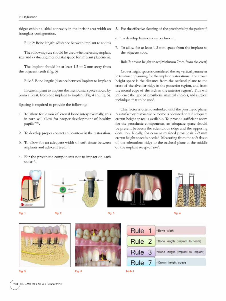
ridges exhibit a labial concavity in the incisor area width an hourglass configuration.
Rule 2: Bone length: (distance between implant to tooth)
The following rule should be used when selecting implant size and evaluating mesiodistal space for implant placement.
The implant should be at least 1.5 to 2 mm away from the adjacent teeth (Fig. 3)
Rule 3: Bone length: (distance between Implant to Implant)
In case implant to implant the mesiodistal space should by 3mm at least, from one implant to implant (Fig. 4 and fig. 5).
Spacing is required to provide the following:
1. To allow for 2 mm of crestal bone interproximally, this in turn will allow for proper development of healthy papilla10,11.
2. To develop proper contact and contour in the restoration.
3. To allow for an adequate width of soft tissue between implants and adjacent teeth12.
4. For the prosthetic components not to impact on each other12.
5. For the effective cleaning of the prosthesis by the patient12.
6. To develop harmonious occlusion.
7. To allow for at least 1-2 mm space from the implant to the adjacent root.
Rule 7: crown height space[minimum 7mm from the crest]
Crown height space is considered the key vertical parameter in treatment planning for the implant restorations. The crown height space is the distance from the occlusal plane to the crest of the alveolar ridge in the posterior region, and from the incisal edge of the arch in the anterior region9. This will influence the type of prosthesis, material choices, and surgical technique that to be used.
This factor is often overlooked until the prosthetic phase. A satisfactory restorative outcome is obtained only if adequate crown height space is available. To provide sufficient room for the prosthetic components, an adequate space should be present between the edentulous ridge and the opposing dentition. Ideally, for cement retained prosthesis 7-9 mm crown height space is needed. Measuring from the soft tissue of the edentulous ridge to the occlusal plane at the middle of the implant receptor site9.
Fig. 1 Fig. 2 Fig. 3 Fig. 4
Fig. 5 Fig. 6 table I
290 KDJ–Vol.39•No.4•October2016
P. Rajkumar

The ideal vertical dimensions of each region are 2 mm for the soft tissue,23 5 mm for the abutment height and 2 mm for the occlusal metal (or) porcelain, the screw retained restorations generally requires lesser crown height space compared to the cement retained prosthesis since it can screw directly onto the implant body (Fig. 6).
Bone height : (to determine the implant length)
The available bone height in an edentulous site is an important consideration because it governs the selection of the height (or length) of the implant. It also influences the available crown height space and consequently force considerations and esthetics.
Bone height is measured from the crest of the edentulous ridge to the opposing limiting anatomical land mark during radiographic assessment. These limiting structures includes the inferior alveolar canal in the mandible and the floor of the nasal cavity and maxillary sinus in the maxilla.
The anterior regions of the jaws have the greatest bone height available. Specifically, the maxillary canine eminence region offers the greatest bone height in the maxilla and the mandibular first premolar region provides the most vertical column of bone in the mandible.
As rule 9, the height of implant also affects its total surface area. The suggested minimum bone height for predictable long term endosteal implant survival is 9 mm.
Branemark screw type implant body and osseointegrated approach was provided only in 3.75 mm width and 9 mm length.
This height requirement is reduced in the very dense bone of the symphysis of an atrophic mandible when the prosthesis is overdenture or increased in the very porous type of bone of the posterior maxilla.
► ConclusionThe key determinant for clinical success of implants is
available bone around that endosteal dental implant. The strength of bone is directly related to bone density. Thus available bone should be properly evaluated in terms of bone width, height, length and angulation also the periodontal status, inclination of adjacent teeth during diagnosis to determine the prognosis of implant placement.
► References1. Branemark PI, Hansson BO, Adell R, et al, Osseointegrated implants
in the treatment of the edentulous jaw. Experience from a 10 year period. Scand J PlastReconstrSurgSuppl 1977;16:1-132.
2. GreenfeldEJ(1913) Implantation of artificial crown and bridge abutments. Dent Cosmos 55:364-359.
3. Atwood DA(1963) Post extraction changes in the adult mandible are illustrated by microradiographs of midsagittal sections and serial cephalometrics roentgenogram. J Prosthet Dent 13:810-824.
4. Atwood DA (1971) Reduction of residual ridges: major oral disease entity. J Prosthet Dent 26:266-279.
5. Atwood DA, Coy WA (1971) Clinical, cephalometric, densitometric study of residual ridge. J Prosthet Dent 26:280-295.
6. Tallgreen A (1972) The continuing reduction of alveolar ridges in complete denture wearer: a mixed longitudinal study covering 25 years. J Prosthet Dent 27:120-132.
7. Misch CE (1999) short versus long implant concept: functional surface areas. Dent Today 18(8):60-65.
8. Misch CE (1990) Density of Bone: effect on treatment plan, surgical approach, healing and progressive bone loading. Int J Oral Implant 6:23-31.
9. Carl E Misch. Contemporary implant dentistry. 3rd ed. St Louis: Mosby: 2010.p.40,130-46,245-50,280.
10. Buser D, Martin W, Belser UC, Optimizing esthetics for implant restorations in the anterior maxilla: Anatomic and Surgical considerations. Int J Oral Maxillofac Implants 2004;19:43-61.
11. Kois JC. Predictable single tooth peri-implant esthetics: Five diagnostic keys, CompendContinEduc Dent 2001;22;199-206.
12. Floyd P, Richard P, Barrett V. Treatment planning for implant restorations. Br Dent J 1999;187:297-305.
13. Berglundh T, Lindhle J. Dimension of the peri-implant mucosa. Biologic Width revisited.JClin Periodontal 1996;23:971-73.
14. Sclar A. Strategies for management of single-tooth extraction sites in aesthetic implant therapy. J Oral MaxillofacSurg 004;6:90-105.
Dr P I Kochukunju, one of the seniormost dentists of Kerala, expired on September 5, 2016 at the age of 77. He completed his graduation from Govt. Dental College, Thiruvananthapuram. He had his practice at Muvattupuzha for nearly four decades. He was an active member of IDA and was the second President of IDA Malanadu Branch (1993-1994). He is survived by his wife Mrs. Valsa Kochukunju, daughters Dr Maya Dinesh and Mrs Manju, and son Dr Manoj K. Poovelil.
Obituary
KDJ–Vol.39•No.4•October2016 291
Application of the ‘rule 1237’ for success of the dental implant

1. A 50 year old patient presented with a swelling in the posterior region of mandible. Radiograph showed multilocular radiolucency and histopathologically numerous giant cells were seen. Blood examination revealed increased parathormone and serum calcium levels. The diagnosis is
a. Central giant cell granulomab. Peripheral giant cell granulomac. Giant cell tumord. Brown tumor
2. A 45 year male patient presented with diffuse black pigmentation of the labial mucosa, buccal mucosa and tongue. Oral pigmentation can occur in all of the following conditions except?
a. Addison’s diseaseb. Peutz Jegher syndromec. Neurofibromatosisd. Cushing’s syndrome
3. The most common congenitally missing permanent teeth following third molars are
a. Mandibular central incisors
b. Maxillary lateral incisors
c. Mandibular caninesd. Maxillary canines
4. A 50 year old female patient complained of burning sensation of the mouth. On examination, the oral mucosa appeared pale and dorsum of tongue was smooth and shiny with loss of papillae. Blood examination showed haemoglobin values of 8gm/dl and peripheral blood smear revealed macrocytic hyperchromic RBCs. The probable diagnosis is
a. Iron deficiency anemiab. Sickle cell anemiac. Pernicious anemiad. Thalassemia
5. Physiological or pathological loss of tooth substance as a result of tooth to tooth contact is termed as
a. Abfractionb. Attritionc. Abrasiond. Erosion
6. A 20 year old male patient complained about difficulty in pronouncing certain letters. On examination, the lingual frenum was attached to the tip of the tongue. The diagnosis is
a. Microglossiab. Ankyloglossiac. Macroglossiad. Lingual varix
7. Which of the following lesions have potential for malignant transformation?
a. Pemphigusb. Pemphigoidc. Reticular lichen planusd. Erosive lichen planus
8. Angular chelitis may be due toa. Loss of vertical
dimensionb. Candidal infectionc. Both the aboved. None of the above
9. Vicker & Gorlin’s criteria refers to formation of ......... from dentigerous cyst
a. Adenomatoid odontogenic tumor
b. Ameloblastomac. Epidermoid carcinomad. All of the above
10. T h e m o s t c o m m o n supernumerary tooth is
a. Paramolarb. Distomolarc. Mesiodensd. Extra lateral incisorAnswers: 1. d, 2. d, 3. b, 4. c, 5. b, 6. b, 7. d, 8. c, 9. b, 10. c
*Jayanthi, **Varun B.R.
*Professor,Dept.ofOral&MaxillofacialPathology,AzeeziaDentalCollege,Kollam;**Reader,DeptofOral&MaxillofacialPathology,PMSDentalCollege,Trivandrum E-mail:[email protected]
292 KDJ–Vol.39•No.4•October2016
KDJ – Kerala Dental JournalQuIz

KDJ–Vol.39•No.4•October2016 293
AssociationNews

Coastal Malabar Branch
North Malabar Branch
7th CDE PROGRAMMEDATE- 27/07/2016 / VENUE- Hotel Juju International, PayyannurFACULTY- Dr. Civy PulayathTOPIC- Tips and Tricks in Successful Dental PracticeAttendance- 53 NosDr. Santhosh Sreedhar, Dr. Rahul Nandakumar and Dr. Ahmed Shafi
attended the fourth State Executive Committee meeting on 31/07/2016, Sunday at IMA Housr, Kochi.
23rd CDH ACTIVITY- ORAL HYGENE DAY CELEBRATIONDate- 01.08.2016, MondayVenue-1. Chinmaya School, Payyannur2. Malabar Rehabilitation Centre For Handicapped, PayyannurThe following programmes were conductedBest smile competition, Healthy teeth contest, Drawing competition,
Elocution competition, Dental health check up, Dental health seminar, Distribution of oral health education chart, Dental health education class for parents, Dental health education class for public, Distribution of powered tooth brush for the disabled school children.
IDA COASTAL MALABAR BRANCH BAGGED ISP NATIONAL AWARD FOR THE BEST ORAL HYGENE DAY CELEBRATIONS.
24th CDH ACTIVITYDate- 13.08.2016, Saturday / Venue- Puthur DYFI Party officeDental awareness class for the general public.25th CDH ACTIVITYDate-15.08.2016, Monday / Venue- Thumbakovval Anganvady, AnnurDental check up was done for 50 patients and dental aid kits were distributed.8th CDE PROGRAMME
Topic- "NEXT-GEN ENDODONTICS"Lecture and Hands on Programme on Rotary EndodonticsFaculty- Dr.M. Abarajithan.MohanTime- 9.30am to 3.30pmVenue- Hotel Vyshak International, Payyannur Date- 21/08/2016, Sunday A Press Conference of IDA Coastal Malabar Branch was held on 24/08/2016,
Wednesday in Connection with Pratyasha-2016.5th EXECUTIVE COMMITTEE MEETINGDate- 31/08/2016 / Venue- Hotel Vyshak International, PayyannurTime- 7.00pm onwardsNinth and tenth CDEs finalised. Venue for PRATYASHA-2016 finalised.9th CDE PROGRAMMEDate- 18/09/2016, SundayVenue- Hotel K.K. Residency, PayyannurTopics 1. Bridge the gap....Perfectly with rpds2. Fundamental tips and trics to predict the outcome of a good dental
prosthesisFACULTIES 1. Prof(Dr). Sreedevi S. Geejai 2. Mr. Tarek.Frank.FeissaliTIME- 9.00am to 1.30pmDistribution of onam kits to five cancer patients was done on 18/09/2016,
Sunday at Hotel K.K. Residency.The release of second edition of our journal "mirror" was done on
18/09/2016 at Hotel K.K. Residency, payyannur by handing over the first copy TO Prof.(Dr)Sreedevi S. Geejai by Dr. Santhosh Sreedhar.
CDE PROGRAMMECDE 6: Lecture on Vaccination Update was conducted on 29th July at
IDA hall Podikundu by Dr M K Nandakumar, Pediatrician, Koyili Hospital. Programme was attended by 39 members.
CDE 7: CDE programmeon INTERDISIPLINARY PERIODONTICS was conducted on 6th September at IDA hall Podikundu by Dr. Anil Meylath, Principal.Mahe Dental College. 47 members attended the programme
CDE 8: One day CDE programme on ALL ABOUT GLASS IONOMER was conducted on 18th September by Dr Aravind Shenoy, Professor at hotel royal omers. 60 members attended the programme.
CDH ACTIVITIESCDH 11: A blood donation camp was conducted at blood bank, Kannur
on 14th June in connection with Blood donorsday. Mr. Sumesh, president, Kannur Jilla panchayat inaugurated the function. Many of our branch members donated blood.
CDH 12: International Yoga Day was celebrated by our branch on 21st June at IDA hall, Podikundu. Many members with their families participated in the programme. Mr. Vijay Iyer Neelakantan and Mr Manoj V V took classes.
CDH 13:IDA NMB conducted a free Td vaccination programme on 11th July at Mother and Child hospital.192 members and Dental Assistants took vaccination.
CDH 14: Hepatitis B vaccination was given for IDA NMB members and Dental Assistants on World Hepatitis Day on 28 July at Mother and Child hospital, Kannur
CDH 15: Oral health reinforcement programme was conducted at Anjarakandy U P School on 1st August. Dr. Faizal C P and IDA members of Kannur dental college participated.
CDH 16: Follow up Camp and Oral health reinforcement programme was conducted at at Kannur Central jail on 11 August. Dr. Jayashree KT, Dr. Roopesh C, Dr. Anil Kumar PK took part in the programme. Dr. Anil Kumar PK took classes on Oral Hygiene maintenance
CDH 17: A dental checkup camp was conducted at Chalad U P school on 21st Augest. Dr Anil Kumar P K, Dr Reshma Jithesh, Dr Roopesh C and Dr Faizal C P participated. Dr Anil Kumar P K took awareness class.150 students attended the camp.
Executive committee meetings: 4thexecutive committee meeting was held at IDA hall Podikundu on 30thAugest, 21members attended the meeting.
WORLD HEPATITIS DAY: World Hepatitis Day was observed by our branch by giving Hepatitis B Vaccination for Dental Surgeons and Dental assistants in Kannur region on 28th July
TEACHERS DAY CELEBRATION : Honouringof teaching members of IDA NMB was conducted on 6th September.
FAMILY MEET: Afamily meet of IDA members was conducted along with Onam celebration on 25th September at hotel Malabar residency.
DENSINFO: Journal of IDA NMB, ’Densinfo’ was released on 25thSeptember.first copy was handed over to Dr C V Pradeep by Dr K C Thomas.
Onam celebration: Onam celebration of IDA north Malabar was conducted on 25th September at hotel Malabar residency around 170 members participated.
294 KDJ–Vol.39•No.4•October2016
Association News

Kodungalloor Branch
1.Family tour to Hongkong and Macao September 13-19.20 members took part.2. Prathyasha free denture program
conducted on october 2nd.15 free dentures distributed.3.Combined meeting with IMA Kodungallur
conducted.Topic-Cleft lip and palate management from
birth to adulthoodDr. Mariya Kuriakose MDS, (AIMS, Kochi)
was the faculty.
Valluvanad Branch
Funfilled family tour to Cherai Beach.General body meeting conducted during the evening.
Maveli Visted our Brach during our Onam celebration. He joied with the kids ans gave away loads of gifts.
Conducted 2 Clinical clubs on practice management and Discussion on consumer court case decisions.
CDE on Current concepts on TMJ pain management.
Many CHH activities performed by the members.
Nedumbassery Branch
4th CDE PROGRAMIDA valluvanad 4thcde program was conducted on july 24th Sunday at
hotel Nakshatraregency, pattambi. The topic was BASIC LIFE SUPPORT. Dr. Yasser Chomayil from MIMS kottakkal was the faculty. Handson program on dummy was conducted in the after noon session. About 23 members participated.
DENTAL SCREENING CAMPIDA valluvanad in association with keralakarshakathozhilali union
manisseryvillage has conducted a dental screening camp on 14th august at AUP school, Thrikkangode. Dr. Shoukathali, Drvishak, and Dr. Rajarajan lead the camp. About 70 patients were screened. Oral hygiene kits distributed.
IDA NORTH ZONE CRICKET MATCHIDA valluvanad has participated in the ida state north zone cricket tournament
held at vadakara. 11 members from our branch participated.
PRATHYASHA 2016IDA Valluvanad has conducted free denture delivery to the poor patient
on October 9th, 2016 as part of IDA kerala state project PRATHYASHA at Snehanilayam Old Age Home, Mudavannor, Thrithala. About 12 dentures are delivered from our branch. Oral hygiene kits were distributed to the patients. Ida valluvanad has sponsored one day food for the people of snehanilayam old age home. Dr. Vishalkorah, Dr. Subashmadhavan, Dr. Shoukathali, Dr. Rajarajan, Dr. Thahseen, Dr. Geethanjali were present with their patients for the program
MIDO 2016IDA valluvanad has participated in IDA kerala state sports meet held at
manjeri on October 9,2016. Dr. sreeshgopal won first prize for 100meters and 200 meters running race.
CDH Programmes:Conducted free denture distribution programe
Prathyasha 2016 On Oct 2nd 2016 at Wynd Valley ResortKalpetta. 55 patients were benefitted by this programe.
Wayanad District Collector B.S. Thirumeni IAS was the chief guest for the function. After Free Denture Distribution branch conducted its Onam
Celebration at the same venue.State Dental Sports Meet:IDA Wayanad became the Overall champions in the
State Dental Sports meet held at Unity Womens College, Manjery, Malappuram on 9. 10. 2016.
Wayanad Branch
1. Anti tobacco awareness programme was conducted on 18th July, at Sub jail Kasargod for the inmates. Talk given by Dr Praveen, Assistant Professor, Century Dental College, Kasargod
2. Executive committee meeting was held on 10th August, Wednesday, at IMA hall Kasargod. Issues and programmes to be conducted in the current year were discussed.
3. CDE programme was conducted on 17th September 2016. Topic was Problem Solving in Endodontics by Dr Abhijith S Shetty, Bangalore.
4. Eventhough our branch didn’t participate in “Prathyasha” programme, our members given 6 complete dentures for needy people. It was done under our branch in respective clinics.
Kasargod Branch
Association News
KDJ–Vol.39•No.4•October2016 295

Trichur Branch
Tellicherry Branch
The VI executive meeting was held on 10-07-2016 at City Dental Clinic Vatakara, 12 members participated.
Meeting decided to conduct 1. A CDE programme on LASER2. To visit ASHANTKETHAN, the school for differently abled at Nandhi
Bazar and St. Vincent Home at Calicut as part of WDC Programme 3. To conduct an awareness programme at Sub jail Vatakara On 22-08-2016, IDA VATAKARA conducted a CDE programme on
LASERS and the faculty was Dr. Jagadish Pai and was attended by 30 members. Same day an awarness programme was conducted at Sub jail, Vatakara. Dr.
Abdul Gafoor, Dr. Susanth and Dr. Salil. paticipated. A short film was screened and awareness class was taken by Dr. Abdul Gafoor.
Branch ONAM celebration was conducted on 4th september at IMA HALL, with a good member participation above 70.
Colourfull ‘ Pookkalam’ was made at the entrance by our members.Mr. Sivadas Purameri, inaugrated the Programme, followed by “ Thiruvathira”
by our own lady members.Games for members and kids, “Onasadhya” followed.
President distributed the prizes.Meeting of Organising committee of North Zone Cricket match was held
at Kallatt Dental Clinic on 22-9-2016 to evaluate the preparation and decided to change the ve nue to Azhiyoor mini stadium, Kunhippalli, due to poor weather Conditions.
Fixture for quarterfinals was prepared in the presence of Dr. Sudheer( Secratary IDA Malabar ) Dr. Firoz & Dr. Sujith ( Prez & Sec. IDA Thalassery)
VII Executive meeting was also held on 22-9-2016 and decided to 1. Participate in the MUKTHI programme of IDA Kerala state by sending
Dr. Sushanth, Dr. Noufal and Dr. Ashin Raj as its representatives.2. The conduct a family tour to Wynad on 22 nd 23rd October.3. to conduct a state CDE programme in the month of December.The North Zone cricket match was well organized at “ Azhiyoor Mini
Stadium” Kunj ipalli, Chobala on 25-9-2016. It was participated by IDA Malabar, IDA Malappuram, IDA Ernad, IDA Thalassery, IDA Valluvanad, IDA Coastal Malabar, IDA Wynad, & IDA Vatakara.
IDA Ernad won the trophy & ida Malabar was runner up. Both teams qualified for state cricket meet.
Senior member Dr. Natrajan distributed the trophies.
4th executive committee meeting was held at parco residency thalassery. It was attended by 11 members.
4th cde programme was held at parco residency. Faculty of the cde was Dr Joby Peter. He conducted the class on "clinical alterations of growing face". Dental council allotted 6 credit points for the cde. 23 members participated in the cde.
5th cde programme was conducted by Dr Tony Thomas. Venue was parco residency. Topic of the cde was "Recent advances in denture making ". It was attended by 30 doctors. 6 credit points were allotted by dental council.
Onam celebration along with 2nd general body meeting and family get together was conducted at Pythalmala resorts. 17 members with family attended the meeting. It was a 2day trip. There was a 2hrs trekking to the hilltop in the morning which almost all the members participated. It was a fun filled trip which was enjoyed by all.
On 24th September Onam celebration was conducted at Hotel Merlin. Most of our members attended the Onamcelebration and the programme was enjoyed by everybody.
On 2nd October 2016, Trichur branch cricket team wwent to Thrippunithara to partcipate cricket match (Central Zone) hosted by Kochi branch.
On 9th October 2016 Denal camp was conducted at Santhwanam charitable trust. CDH representative Dr. Vijith Vijayan, Dr Sajiah Viswhwanathan and a tem of oral maxillofacila surgeons under Dr. Manoj Bhaskaran MDS, OMFS attnded the campl
296 KDJ–Vol.39•No.4•October2016
Association News
Vatakara Branch

Eranad Branch
Malappuram BranchEXECUTIVE MEETING: Our 5 th Executive meeting of MIDA were held
at Hotel Rydgess Inn, Kottakkal,, Malappuram on 16/08/16 at 8 pm onwards.“PRATHYASHA” FREE DENTURE PROGRAMME: MIDA Conducted
prathyasha” free denture Screening Programme for poor and financially backward peoples in malappuram district on 28/8/2016 Sunday at GLP SCHOOL, Malappuarm More than 80 denture needed peoples participated in the programme. The programme was inaugurated by Dr Muhammed Sameer PT, State president, IDA, Kerala.
“MIDAN” IDA MALAPPURAM HALF YEARLY NEWS LETTER: MIDA published first half yearly news letter on 28/8/16. This publication is covered all the conducted programme details and upcoming programme schedule.
9 th CDE PROGRAMME: Our 9 th CDE Programme was held at HOTEL RUBY Residency, Kottakkal on Sunday, 5/9/2016.
7 th CDH CAMP: Our Seventh CDH Camp was held at Pulpatta Gramapanchayath, Manjery on 4/9/2016.
8 th CDH CAMP: Our Eighth CDH Camp was held at Marakkara Gramapanchayath, Kadampuzha on 11/9/2016.
MIDA CHARITY DENTAL CLINIC at GOVT, OLD AGE HOME, THAVANUR: The inauguration of MIDA charity dental clinic is done by Dr KT, Jaleel, Hon, Minister, Local Administration, Kerala, on Sunday, 18/9/16 at GOVT, OLD AGE HOME, THAVANUR. Mrs, Lakshmi, Presidet, BLOCK Panchayath, Ponnani, Dr Fasil V Hassan, Vice President, IDA Kearala were guests in the programme.
ONAM –BAKRID FEST: MIDA Celebrated onam bakrid fest at Govt, Old Age Home, Thavanur along with inmates of old age home. More than 120 inmates, staffs, and MIDA family members participated in the programme.
The programme culminated with GRAND ONAM SADYA. We distributed ONAPPUDAVA to all inmates of Old age home.
IDA KERALA NORTH ZONE CRICKET TOURNAMENT: MIDA Cricket team participated in the IDA KERALA north zone cricket tournament held at Vadakara. We reached in the semifinal.
INTERNATIONAL GERIATRIC DAY OBSERVATION: The CDH team of MIDA visited the Gov. Old age home at Thavanoor on Geriatric day(1/10/2016).
“PRATHYASA-16 “ FREE DENTURE DISTRIBUTION PROGRAMME: IDA Malappuram contributed to the programme by presenting 50 complete dentures to the poor and needy people of our society. More than 50 members of IDA Malappuram cooperated WITH the programme. The distribution of the complete dentures done in the programme conducted at KP Kesava menon Hall, Calicut on Sunday, 2/10/16. Dr Muhamed Sameer PT, President, IDA Kerala was the chief guest. Dr Suresh kumar G, Hon, secretary, IDA Kerala, Dr Subash Madavan, state CDH covenor were also present in the programme.
MIDO -16IDA KERALA STATE SPORTS MEET : IDA Malappuram conducted IDA
Kerala state sports meet on Sunday, 9/10/16 at Unity Womens college, Manjery and Cosmopolitan club, Manjery. Dr Muhamed Sameer PT, State president, IDA Kerala inaugurated the event. Many state and branch leaders of IDA were present on the occasion. The event started with grand colourfull team march past. More thsan 10 branches participated in the sportsmeet. more than 40 individual events conducted in sports meet. IDA Vayanad became overall champions and IDA Malabar became runners up. IDA Malappuram won bronze in tug of war team event and Dr Asha Rajesh won the gold medal in 100 mtr womens sprint event.
ADOPTION OF INSTITUTIONS: IDA MALAPPURAM adopted the Goverment Old Age Home, Thavanoor. Goverment Juvenile Home, Thavanoor, Government Rescue Home, Thavanoor, Prathyasha Bavan, Thavanoor. Government Mahila mandiram, Thavanoor. The adoption programme was inaugurated by Honourable Minister Dr KT Jaleel in the presence IDA state and branch leaders and Goverement officials. The complete dental related treatments will be providing for all above institutions will be done in the IDA Malappuram Charity dental clinic in Old Age Home Thavanoor for the future by members of IDA Malappuram under CDH Wing.
70th Independence Day celebrations was conducted on 15th Aug at GMLP school Mukkatta, Nilambur. Oral health awareness class and sweet distribution was done.
prathyasha free denture screening camp was conducted on31st August 2016 at vyaparabhavan perinthalmanna. About 120 patients screened.30 patients selected on it. Celebrated Onam-Eid on September 11, 2016 at hotel green palace Nilambur. It was really fun and feast for mind and body. There was active participation
from all members. SPORTS-participated in north zone cricket tournament held in vadakara on 25/9/2016. IDA ernad became champions. Dr arshad of Ida ernad became Man of the series. Participated in MIDA sports conducted in manjeri. We got first prize in relay. Dr assainar got second for 200 metres. Dr ajmal got 3rd prize for shotput. Dr Shayar and Dr Assainar got 3rd prize for TT. IDA Ernad distributed their free denture during prathyasha program hosted by ida malabar on Nov 2, 2016 at keshavamenon hall. We distributed 25 dentures.
KDJ–Vol.39•No.4•October2016 297
Association News

Malabar BranchCDH - DENTAL CAMP & AWARENESS CLASS (15/08/16)Dental Screening and awareness class for parents and teacher’s were conducted
at UmbichiHajee Higher Secondary School Chaliyam Kozhikode on 15/08/2016. Dr. Saju NS IPP IDA Malabar branch inaugurated the camp with an awareness class. Around 200 students, 12 teachers and 20 parents were present. in the camp. On behalf of IDA Malabar Dr. Saju NS, Dr. AmeenShahid, Dr. ShameemThaha participated in the camp.
Screening Program forPrathyasha 2016 (21/08/16)Screening was done in the IDA hall for Prathyasha the free denture program
of IDA was done 21/08/2016. Program started at 9.00AM, around 150 participants were there and after screening 80 participants were selected.
” NEST” PAIN & PALLIATIVE CENTRE(Adoption&Inaguration of free Dental Unit)
Nest is a pain and palliative centre situated in quilandy Kozhikode. IDA Malabar adopted the pain and palliative centre and installed a dental unit their and various treatments were given to the patients their by periodic visits of our members. MOU was signed between President IDA Malabar branch and Chairman of pain and palliative centre.
FOLLOW UP VISITS TO HOME OF LOVE (THE ADOPTED OLDAGE HOME) 29/08/16
Multiple visits were made to adopted old age home and four complete denture were delivered to the inmates. Around 20 extractions were done so far and four complete dentures are in process which reached up to trial and will be delivered on Prathyasha 2016.
CDE - BASIC LIFE SUPPORT The eighth CDE of IDA Malabar was held on 04/09/2016 at IDA hall
Ashokapuram Kozhikode in association with Aster MIMS Kozhikode. The topic of CDE was Basic Life Support. The CDE programme was inaugurated by Dr. Nizaro Siyo Past President IDA Kerala State at 9.00 AM in the Presence of Dr. Dinesh KR President IDA Malabar Branch. Faculty was Dr. Venugopalan P.P Director Emergency Medicine Aster MIMS Kozhikode. The CDE was of three sessions, First session began at 9.30 AM which includes a discussion on CPR followed by a hands on training. The second session started at 11.35
AM after a short tea break followed by a lecture on infant CPR and choking. The third session started at 2.00pm after lunch break with a hands on training programme and discussion on trauma care followed by an evaluation session. Around 50participants attended.
ONAM CELEBRATIONSOnam celebrations of IDA Malabar branch was conducted on 11/09/16
at 10.00am. Under the leadership of Dr. Navjeevraj a beautiful Pookallam was made with verity of flowers followed by fancy games for women and children and other musical entertainment. A grand onasadhya was also arranged in the IDA hall. Around 100 members along with their family participated.
PARTICIPATION IN IDA NORTH ZONE CRICKET TOURNAMENT(23/09/16)
IDA Malabar branch participated in the IDA North Zone cricket tournament held in Municipal stadium Vadakara and became the Runners up in the tournament
TV SHOW (20/09/16)One of our senior memberDr. Sujatha Verma delivered a talk in Asianet
Malayalam channel regarding the early diagnosis of malignancy in oral cavity its treatment and prognosis.
INDEPENDENCE DAY (Flag Hoisting)69th Independence day was celebrated in IDA hall. Flag hoisting was done
by Dr. Dinesh KR. President IDA Malabar branch at 8.00 am followed by the National Anthem. Many senior and junior members witnessed the ceremony. Sweets and Payasam were distributed.
INDEPENDENCE DAY CELEBRATIONS (HONOURING A WAR HERO)
In the evening of 69th Independence day IDA Malabar branch celebrated independence day in a different manner by honoring a great soul, a soldier who has fought valiantly in some of the most inhuman terrains of the world.
Brig. P.T. Gangadharan who had suffered a critical injury from a Pakistan Shell attack in 1999 while leading his troupes at LOC, J&K, which had left him paraplegic ever since. It was a privilege listening to & paying back the gratitude, respect & affection to a beloved son of this nation. Many senior and junior members attended the function.
Quilon BranchInstallation ceremony & Family meet : The 24th installation ceremony
and family meet of IDA Quilon branch was held on 20th December 2015 at The Quilon Beach Hotel kollam. Dr B S Sundaresan as the president and other office bearers were installed.
CDE Programmes: Conducted 5 CDE programs: 4 branch & 1 inter branch program.
CDH Programmes: 1st CDH: Conducted a dental and medical camp at Believers Church Mathma central School Eravipuram on 24th January 2016.
2nd CDH programme : of IDA Quilon branch, a medical and dental check up camp, highlighted with a talk on 'oral health in children' by Dr Maneesha. R ( dept. of pedodontics and preventive dentistry, Azeezia dental college) was conducted for school children ( SPC) on 6th February'16 at Meenakshi Vilasam Govt. Vocational Higher Secondary school, Punthalathazham.
3rd CDH Programme: World oral health day programme( March 20th) was conducted on three different levels.
4th CDH : International dentist day: in five levels."Level 1: Community based programme: Cancer detection camp : A cancer
detection camp in association with Cancer Care Centre, Qln and Palliative care unit was conducted at St. Johns School, Eravipuram
Level 2 : Tribute to community: "Salute the silent worker award." Mr Yesudas
Rozario, President of Sahaya Hastham, Palliative care unit, Eravipuram was recognised honoured and awarded for his humanitarian work
Level 3 : Dentist day talk: Dentist day talk by ex state secretary Dr Shibu Rajgopal Level 4:Awards & honours :To honour the dental fraternity of Qln Branch. Honouring of senior dentists, appreciation awards to the members who had represented in state & national level and for their contributions:
Level 5: Family get together with gala musical entertainment, lucky dips & dinner.
7th CDH-WORLD NO TOBACCO DAY PROGRAMMES:Public awareness at Infant Jesus School on27th June'16. Class on WNTD
for nearly 300 students and teachers with power point presentation., Press meet was conducted on 29th May at Press club Kollam.
Cleberated WNTD 2016"by Online Page inFB from 1st May'16.ONAM CELEBERATIONS & FAMILY MEET : Conducted the family
meet and onam celeberation of the branch at Hotel Raviz Kollam.A colourful “ATHAPPOOKKALAM” was prepared by the members,14th CDH programme -the state project " PRATHYASHA " on 2nd Oct'16
at Ferns Hall from 10 am to 1 pm. The chief guest of the event was Hon. MLA of Eravipuram Sri M. Noushad.
298 KDJ–Vol.39•No.4•October2016
Association News

Dear Hope members,As we are moving towards the end of the year, we have got a good
response from our members. This year we have 168 new members to the hope family. 2410 members have renewed and 29 members have not yet renewed their hope membership.
Our Hope member Dr. R N Shenoy of hope no.1425 left us this year. We will pay the fraternity contribution of Rs. 10 lakhs to his family soon.
Regarding Hopemedi, 1011 members have renewed and paid Rs. 148 lakhs as the premium amount. Last year we had 1012 members in hopemedi and we claimed around Rs.175 lakhs.
For this year the claims could be settled either on cashless basis with the hospitals with whom the TPA M/s Vidal Health TPA Pvt Ltd has tie up arrangements or on reimbursement basis. For cashless facility request from the hospital has to be forwarded to the TPA and in turn TPA will accord their approval subject to the terms of the policy. Tie up hospitals are aware of the procedures to be followed.
Though, availing cashless facility is the most convenient method of settlement of claims, it is normally found that billing method followed by hospital are different for cashless and reimbursement for the same treatments. For cashless facility bills are higher which means higher claim outgo.
For reimbursement of hospital expenses following procedures are to be followed
(a) Immediate intimation of hospital admission has to be served to the TPA / M/s Cosmos Insurance Brokers Pvt Ltd
(b) On discharge from the hospital, below mentioned documents are to be send to the address shown
1) Attached Claim form dully completed in all respects and signed2) Medical Bills in original with details and bifurcations (both hospital
and medicines purchased from outside with prescription)3) Discharge Summary from the hospital4) Investigation reports if any5) ID Proof – Diving License / Voters Identity Card or / Adhaar etc6) Bank Account details of the proposer viz, cancelled cheque leaf
with member name printed on it.Documents to be send to: - M/s Cosmos Insurance Brokers Pvt. Ltd, 39/ 2338, 2nd Floor,Sarala Chambers, Durbar Hall Road, Ernakulam - 682 016 Ph: 0484 – 2351432 / 2351433
PLEASE NOTE THE FOLLOWING CHANGES CAREFULLY
1. Sum insured slab of Rs.2 Lac is withdrawn and higher slab of Rs.7 Lac is introduced.
2. Room rent is restricted to Rs.2500/- per day for Rs.3 Lac and 5 Lac sum insured and Rs.3500/- per day for Rs.7 Lac SI.
& 2% of Sum insured for ICU.3. Claim amount is limited to 70% of admissible claim ie.(co-sharing)
exclusively for parents only.4. Expenses for cataract surgery is capped to Rs.25000/- maximum.5. Treatment expenses in hospitals in states other than the state of
Kerala will be considered at par with the reasonable expenses likely to incur in hospitals of Kerala/maximum in the institutions like AIIMS, CMC Vellore and select Government institutions.
6. A maximum amount of Rs150000/- is restricted for Joint replacement-Knee/Hip(per Joint Including Implant)
Please note that 30% co-sharing applicable only for parents.Certain restrictions had to be imposed on payouts because of the tendency
of a very few members taking unfair advantage of our liberal scheme. Viability of the scheme in the larger interest of benefit to the members was the priority for the new restrictions.
7. Please refrain from opting for high end hospitals providing 5* facilities. Hope medi is not liable for full compensation in such cases. Standard average rates for the procedure as applicable in hospitals across Kerala shall apply and only those amounts as decided by TPA shall be reimbursed. If anyone wish to prefer such hospitals, the additional financial requirements shall be borne on their own.
8.Cashless facility is being discouraged as it has been observed that the hospitals tend to inflate the bills up-to 3 times when it is availed. Members shall be provided cashless only in dire situations, subject to scrutiny. Please co-operate.
At the moment we our premium are still below the figures of individuals schemes for families with cover for aged parents. Pre-existing illness and no age bar and no medical are only some of the highlights of the scheme. Exorbitant payouts will cause exceptional rise in premiums in successive years. Members please use your discretion wisely.
For any assistance regarding Hopemedi claims may contact Shri Rajeev P.R. (Cell No: 9995368466).
Thanks to all hope members especially hope reps of all branches who helped us to make it a grand success.
Dear friends,Warm greetings to all Indian dental association has always
been actively involving in conducting dental education programs. Almost all the branches of IDA Kerala state conducted CDE programs. I feel energized to share the thoughts through KDJ by closely watching the activities of local branches during this year. I express my sincere appreciation to all branch office bearers and members for
the success of the programs.The second state level CDE program was conducted at Hotel
Maryland, Kayamkulam on 16/10/2016 hosted by IDA Mavelikara branch. The topic was “ Crown and Bridge” by Dr. P.C. Jacob from Bangalore. IDA State Vice president Dr. Siju Paulose inaugurated the function. The sponsors of the program were 3M ESPE and Colgate
The third state level CDE program was carried out in Hill park Hotel, Pattanamthitta on 22/10/2016 hosted by IDA Pathanamthitta. State vice president Dr. Eugene Varghese presided the function. The topic was “ Esthetic and post endo restorations – current choices” by Prof. Dr. Narasimman Bhardwraj from Chennai. We got financial assistance from GC India and Colgate
The upcoming programs areWeb Seminar on 15/11/2016 at 7.30 pm 4th CDE –Host IDA Costal Malabar 20/11/2016 5th CDE –Host IDA Kollam 27/11/20166th CDE –Host IDA Vadakara 11/12/2016Lets together make it grandWith warm regards
DR NIRMAL GEORGE SAIBUCDE Chairman
IDA Kerala State
DrNirmalGeorgeSaibuConvenorCDE
CDEReport
IDAHopeReportDr. Joseph C.C.
Hon. Secretary
KDJ–Vol.39•No.4•October2016 299
Association News

CDHReport
Dr.SubhashMadhavanChairmanCDH
PRATHYASHAIDA kerala state, this year, has
decided to conduct 'prathyasha', the free denture programme for the poor, in a grand manner
Intimations were sent to all ida branches in the state to conduct screenings camps (to identify deserving beneficiaries) on a convenient date, after giving detailed information of the same in the print media. Deserving individuals
were selected from these screening camps and it was decided to distribute fabricated dentures on Oct 2nd, Gandhijayanti day, from different centres across the state. The valedictory function and state wide inauguration of the programme was decided to be held at kozhikode with idamalabar being the hosts
A press conference was called at calicut press club on 30 th Sept, in which, idaksb president Dr Muhammad sameer, explained the programme details to the medias
On Oct 2nd, at K PKeshavamenon Hall, calicut, statewide inauguration of 'prathyasha 2016'was conducted by Dr. Muhammad sameer,, ida state president, in the absence of our hon transport minister MrM KSaseendran, who was held up in another programme. Hon minister later messaged his wishes. Dentures prepared by the members of IDA Malappuram, Eranad and Malabar were distributed during the programme
On the occasion, Dr Dineshkr, president, idamalabar welcomed the gathering. After the inaugural address, felicitations were offered by Drsureshkumar, Drfazilhassan and representatives of malappuram and eranad branches. State cdh chairman Drsubashmadhavan presented the project report, and Drshashikumar explained the directions of using dentures. Vote of thanks was rendered by Dr. Sudheer K T, programme Co ordinator. Around 200 dentures
distributed in kozhikode. Similar programmes were conducted by other branches, on the same day, namely coastal malabar, wyanad,, karunagappalli and kollam and altogether around 400 distributed, and the programmes conducted in their area was also in grant manner
MUKTHIIndian Dental Association Kerala State has embarked on a
comprehensive programme to PREVENT TOBACCO AND SUBSTANCE ABUSE among youth covering around 1345 Higher Secondary Schools of Kerala in collaboration with Department of Higher Education, Govt. of Kerala, Excise Department and Home Department. The programme is being partnered by Federal bank who is the CSR partner.
The programme known as “MUKTHI” will utilize the services of dental surgeons to involve in training and awareness and intervention components. “MUKTHI” is envisaged as a programme that educates, advocates and collaborates to reduce tobacco and substance related abuse problems in the state among children in the vulnerable age group. It is very important in the current scenario in Kerala that substance abuse prevention programmes for students in 11th and 12thstandard should be introduced.
“MUKTHI” programme includes life-skills training, teaches decision making skills, includes peer resistance training, uses trained teachers, interactive teaching methods, effective content and delivery which targets students prior to onset of drug use, teaches drug harm, teaches community values and is culturally sensitive.
The programme will be undertaken as different modules namely :• Resource persons training programme• Teacher Training• Peer group leadership training• Awareness programmes for parents and children• Awareness campaigns in institutions• Psycho -social support if needed.
The First resource persons training programme was held at IMA House Kochi 0n the 25th and 26th of October. Dr Arun Kandaswamy and Dr Jayakrishnan Menon from Centre for Addiction Medicine (CAD), NIMHANS Bangalore were the faculty for the programme. Around 40 members were trained as Resource persons during the programme.
The Second phase of the programme has since commenced with Peer Group Leadership Training of students with training in 57 centres all over the State.
The Programme is being conducted under the aegis of Council of dental health of IDA Kerala state in tandem with the CDH activity of its Local branches.
Association News
300 KDJ–Vol.39•No.4•October2016