Zinc oxide nanowires in chemical bath on seeded substrates: Role of hexamine
8
J Sol-Gel Sci Techn (2006) 39:49–56 DOI 10.1007/s10971-006-6969-y Zinc oxide nanowires in chemical bath on seeded substrates: Role of hexamine Abhilash Sugunan · Hemant C. Warad · Mats Boman · Joydeep Dutta Received: 2 August 2005 / Accepted: 23 December 2005 / Published online: 11 May 2006 C Springer Science + Business Media, LLC 2006 Abstract We report a study of the role of hexamine in the seeded growth of zinc oxide (ZnO) nanowires by hydrother- mal route. We show that the growth can be made highly anisotropic (aspect ratio > 150) with almost no detectable increase in diameter (with time of hydrothermal growth) of the obtained nanowires. Results indicate that hexamine acts as a shape inducing molecule, by selectively capping the non-polar crystallographic planes of the zincite crystal. We observe nanowires with typical diameters of ∼30 nm and lengths exceeding several microns after a 24 h growth period at 60–95 ◦ C. Our observations, and reports from the litera- ture, suggest that the concentration of the precursors in the chemical bath can significantly affect the growth rate of the wires. By keeping the concentration of the precursors in the bath at 1 mM, we have observed an extremely slow, but highly anisotropic growth of ZnO nanowires. Keywords Nanowire . Hexamine . Anisotropic growth . Colloid . Precipitation . ZnO . Zinc oxide . Nanoparticle 1. Introduction Recent developments in nanotechnology and the demonstra- tion of various quantum size effects in nano-scale particles, implies that many novel devices of the future will be based on properties of nanomaterials [1]. Reports of zinc oxide A. Sugunan · H. C. Warad · J. Dutta Microelectronics, School of Engineering and Technology, Asian Institute of Technology, P.O. Box 4, Klong Luang, Pathumthani, 12120 Thailand M. Boman Materials Chemistry, Angstrom Laboratory, Uppsala University, Box 538, SE-751 21, Uppsala, Sweden (ZnO) nanostructures, especially nanowires, are of interest due to their potential applications in gas and chemical sens- ing [2], micro-lasers [3], field emitters [4], amongst others. Also reported for ZnO nanostructures (piezoelectric mate- rial) are applications as cantilevers, with size scales over 50–100 times smaller than the conventional cantilevers, which show improved sensitivity and mechanical flexibil- ity for applications in scanning probing microscopes [5]. A wide range of strategies have been explored by dif- ferent groups for the synthesis of nanowires of ZnO [6–9]. The conventional synthetic techniques for ZnO nanowires usually involve a high temperature, vapor phase synthesis. It is a catalyst-driven synthesis that relies on the dissolution of the vapor of the semiconductor material like ZnO into metal nanoparticles (usually gold), followed by super-saturation and crystallization [6]. Various modifications of the high temperature vapor-solid method have been reported for the growth of ZnO nanostructures, including nano-belts and nanowires [7–9]. The commercial potential for these processes are constrained by the need for an insulating and/or expensive substrate for proper oriented growth of the nano-rods, and also the complexities and cost of the high temperature and controlled atmospheric systems. The need for deposition of a monolayer of gold nanoparticles on the substrate, to catalyze the epitaxial growth, further increases the complexity of the process. Other techniques to synthesize nanowires of ZnO include template assisted growth [10] and electrophoresis [11]. Alter- native low temperature techniques for synthesis of nanowires of metal oxides usually involve a hydrothermal growth pro- cess inducing an epitaxial, anisotropic crystal growth in a solution [12, 13]. The hydrothermal process is usually sub- strate independent [14], and offers a fairly good control over the morphology of the obtained nanowires. Among the var- ious synthetic techniques for obtaining nanowires of ZnO, Springer
Transcript of Zinc oxide nanowires in chemical bath on seeded substrates: Role of hexamine

J Sol-Gel Sci Techn (2006) 39:49–56DOI 10.1007/s10971-006-6969-y
Zinc oxide nanowires in chemical bath on seeded substrates:Role of hexamineAbhilash Sugunan · Hemant C. Warad · Mats Boman ·Joydeep Dutta
Received: 2 August 2005 / Accepted: 23 December 2005 / Published online: 11 May 2006C© Springer Science + Business Media, LLC 2006
Abstract We report a study of the role of hexamine in theseeded growth of zinc oxide (ZnO) nanowires by hydrother-mal route. We show that the growth can be made highlyanisotropic (aspect ratio >150) with almost no detectableincrease in diameter (with time of hydrothermal growth) ofthe obtained nanowires. Results indicate that hexamine actsas a shape inducing molecule, by selectively capping thenon-polar crystallographic planes of the zincite crystal. Weobserve nanowires with typical diameters of ∼30 nm andlengths exceeding several microns after a 24 h growth periodat 60–95◦C. Our observations, and reports from the litera-ture, suggest that the concentration of the precursors in thechemical bath can significantly affect the growth rate of thewires. By keeping the concentration of the precursors in thebath at 1 mM, we have observed an extremely slow, buthighly anisotropic growth of ZnO nanowires.
Keywords Nanowire . Hexamine . Anisotropic growth .
Colloid . Precipitation . ZnO . Zinc oxide . Nanoparticle
1. Introduction
Recent developments in nanotechnology and the demonstra-tion of various quantum size effects in nano-scale particles,implies that many novel devices of the future will be basedon properties of nanomaterials [1]. Reports of zinc oxide
A. Sugunan · H. C. Warad · J. DuttaMicroelectronics, School of Engineering and Technology, AsianInstitute of Technology,P.O. Box 4, Klong Luang, Pathumthani, 12120 Thailand
M. BomanMaterials Chemistry, Angstrom Laboratory, Uppsala University,Box 538, SE-751 21, Uppsala, Sweden
(ZnO) nanostructures, especially nanowires, are of interestdue to their potential applications in gas and chemical sens-ing [2], micro-lasers [3], field emitters [4], amongst others.Also reported for ZnO nanostructures (piezoelectric mate-rial) are applications as cantilevers, with size scales over50–100 times smaller than the conventional cantilevers,which show improved sensitivity and mechanical flexibil-ity for applications in scanning probing microscopes [5].
A wide range of strategies have been explored by dif-ferent groups for the synthesis of nanowires of ZnO [6–9].The conventional synthetic techniques for ZnO nanowiresusually involve a high temperature, vapor phase synthesis. Itis a catalyst-driven synthesis that relies on the dissolution ofthe vapor of the semiconductor material like ZnO into metalnanoparticles (usually gold), followed by super-saturationand crystallization [6]. Various modifications of the hightemperature vapor-solid method have been reported forthe growth of ZnO nanostructures, including nano-beltsand nanowires [7–9]. The commercial potential for theseprocesses are constrained by the need for an insulatingand/or expensive substrate for proper oriented growth of thenano-rods, and also the complexities and cost of the hightemperature and controlled atmospheric systems. The needfor deposition of a monolayer of gold nanoparticles on thesubstrate, to catalyze the epitaxial growth, further increasesthe complexity of the process.
Other techniques to synthesize nanowires of ZnO includetemplate assisted growth [10] and electrophoresis [11]. Alter-native low temperature techniques for synthesis of nanowiresof metal oxides usually involve a hydrothermal growth pro-cess inducing an epitaxial, anisotropic crystal growth in asolution [12, 13]. The hydrothermal process is usually sub-strate independent [14], and offers a fairly good control overthe morphology of the obtained nanowires. Among the var-ious synthetic techniques for obtaining nanowires of ZnO,
Springer

50 J Sol-Gel Sci Techn (2006) 39:49–56
the sol-gel based strategy involving hydrothermal growthof ZnO particles is probably the most energy efficient, byavoiding the complexities of vacuum environment and theneed for high temperatures.
First reported by Vayssieres et al. [15], this process in-volves epitaxial growth of ZnO rods on various substratesfrom an equimolar aqueous solution of zinc nitrate andhexamine as precursor. The technique relies on the inher-ent anisotropy in the crystal structure of ZnO (hexagonalWurtzite) [15, 16]. Since ZnO exhibits partial polar charac-teristics, an electric dipole is set up between the oppositeends of (001) plane of the zincite crystal, which leads toa high surface energy for the polar plane. Lower energynon-polar planes are more stable, thus in a thermodynami-cally stable growth process, large facets are usually the non-polar planes [17].
Lowering the thermodynamic barrier by providing nucle-ation sites, achieved by fixing pre-synthesized ZnO nanopar-ticles on the substrate (seeding), improves the aspect ratioof the obtained rods [16, 18, 19], and according to recentreports, results in better uniformity [20]. Nucleation sitescan be introduced by depositing a layer of ZnO particlesby sputtering [20], or spin coating a layer of colloidal ZnOnanoparticles obtained by chemical route [14]. Alternatively,annealing a layer of zinc foil results in a dense layer of ZnOand is known to act as a substrate for growth of ZnO nano-rods [21].
Since the growth of nano-rods or wires occur epitaxiallyfrom the seeds, a pre-decided pattern of nanowires on thesubstrate can be achieved by patterning the deposition of theZnO seeds, similar to reports of patterned growth of ZnOrods through the vapor phase route [8]. However, unlike thevapor phase route, the seeded hydrothermal growth can beachieved on a wide variety of substrates and at much lowertemperatures.
Hydrothermal growth carried out in the presence of a ZnOlayer results in nanostructures with dominant (002) planes[14, 15, 20]. Since nano-rods are usually well oriented, thisimplies that the total surface area of the polar facet is theminimum (at the tips), while the lower energy non-polarfacets have the maximum area.
The commonly used precursors for the hydrothermalgrowth process are zinc nitrate (source of Zn2+ ions) andhexamethylenetetramine (HMT), also known as hexamine.Hexamine is a highly water soluble, non-ionic tertiary aminederivative. The growth is carried out in the temperature rangeof 55–95◦C. Variation of the experimental conditions, suchas temperature and the concentration of precursors, allowscertain degree of control on the growth rate and the morphol-ogy of the obtained nano-rods/wires. It was recently reportedthat the role of hexamine in the growth of ZnO nanostructuresin the hydrothermal process is mainly as a pH buffer by aslow release OH− ions, through thermal decomposition [22].
In this work, we have synthesized highly anisotropicnanostructures of ZnO through a seeded hydrothermalgrowth technique to study the role of hexamine as ashape-inducing molecule for the nanowire growth. We re-port on the effect of reactant concentrations and the temper-ature of the chemical bath on the morphology of the grownnanowires. We also present a phenomenological explanationof the epitaxial growth of ZnO nanowires by a hydrothermalgrowth process.
2. Experimental
2.1. ZnO seed particles
Suspension of homogenous ZnO nanoparticles was preparedbased on a method described by Bahnemann et al. [23].In brief, it involved preparation of a 1 mM zinc acetate[Zn(CH3COO)2, Aldrich, 99% purity] solution in 80 ml of2-propanol [(CH3)2CHOH, Aldrich, 99.5 atom% purity] un-der vigorous stirring at ∼50◦C, followed by dilution to a totalvolume of 920 ml and cooling in an ice bath. To this solu-tion, an 80 ml aliquot of 20 mM sodium hydroxide [NaOH,Merck, 99% purity] solution in 2-propanol was added un-der continuous stirring and the mixture was immersed ina pre-heated water bath at 50◦C for 2 h. The transparentcolloidal solution was stored at room temperature and isstable for months, under ambient conditions. The colloidcontained ∼10 nm ZnO particles as observed from scanningelectron microscope (SEM) images. X-ray diffraction anal-ysis (Siemens D5000) and scanning electron microscopy(LEO-1550) was performed on the synthesized nanoparti-cles. FTIR (Fourier transform infrared) spectroscopy wasdone with a Perkin-Elmer FTIR spectroscope (SpectrumOne) in attenuated total reflectance (ATR) mode.
2.2. ZnO nanowires: Hydrothermal growth
To grow nanowires, the colloidal ZnO particles were cen-trifuged at 4000 rpm for 20 min and the supernatant wassmeared evenly on the substrate (glass slide and (111) Siwafer). The coated substrate was oven-dried at 95◦C for12 h, for proper fixation. The substrate was then placed in-verted (seeded surface faced downwards) in chemical bathcontaining an equimolar solution of 1 mM and 0.5 mM zincnitrate hexahydrate [Zn(NO3)2·6H2O, Aldrich, 99% purity]and hexamine [C6H12N4, Sigma, 99% purity]. The growthprocess was carried out in a laboratory oven at 60–95◦C forup to 24 h.
In a separate experiment, the seeded substrate was placedin a chemical bath that was preheated at 95◦C for 7 h, to in-vestigate the influence of thermal decomposition of chemicalprecursors on the first few layers of the seeds on the morphol-ogy of the nanowires. The hydrothermal growth was carried
Springer

J Sol-Gel Sci Techn (2006) 39:49–56 51
out for 18 h in this case, and compared with a sample grownfor the same time period by the standard process (no pre heat-ing of the chemical bath). In another experiment, equimolarsolution of 0.1 M hexamine and zinc nitrate was heated to60 degrees for up to five hours and Fourier transform infra-red spectroscopy (FTIR) was performed in the attenuatedtotal reflection mode to monitor changes in the absorptiondue to stretching vibrations of the tertiary amines in hexam-ine as a indicator of its degradation.
The growth process resulted in uniform nanowires of∼30 nm on the substrate with lengths depending onthe growth time. Samples were characterized with scan-ning electron microscopy (SEM), and X-Ray diffractionanalysis.
3. Results and discussions
The synthesized ZnO colloid was found to consist of fairlyuniform nanoparticles of 10–15 nm as observed from theSEM image (Fig. 1(a)). X-ray diffraction analysis of the col-loidal particles, after repeatedly dropping aliquots of the col-
loidal solution on a (111) Si wafer and drying, shows fairlyequal dominance of (100), (002), (101), and (102) crystallo-graphic planes, as shown in Fig. 1(b). These particles werefound to consist of 2–3 crystallites agglomerated, of averagesize of about 5.4 nm determined from (100) peak employingthe Debye-Scherrer formula.
In general, the most commonly observed morphology ofZnO is needle-like or rod-like structures attributed to theinherent anisotropy of the Wurtzite lattice with an axial c/aratio of 1.602 [20]. Theoretical calculations predict that thesurface energies of the non-polar surfaces of ZnO are small,while the cleavage energy of the polar surface is predicted tobe two times larger than the non-polar surfaces [24]. During ahydrothermal growth in the presence of ZnO seeds, the high-est growth rate is along [0002] direction. This form wouldminimize the total surface energy since the total surface areaof the (001) facet would be limited to the tips and hencevery low. Nevertheless, it is reported that upon prolongingthe growth period, the nanowires grow laterally reducingthe over all aspect ratio [14]. In order to achieve a desiredaspect ratio, the composition of the chemical bath in whichthe growth takes place, becomes important.
Fig. 1 (a) Scanning ElectronMicrograph (SEM) of thecolloidal ZnO particles used asseeds for the hydrothermalgrowth into nanowires. Theparticles are ∼ 10–15 nm indiameters. (b) Indexed X-Raydiffractogram of nanoparticlesof ZnO on a (111) Si substrateshowing the preferentialorientation of the crystallites
Springer
52 J Sol-Gel Sci Techn (2006) 39:49–56
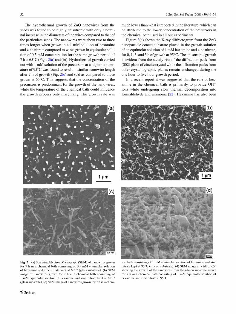
The hydrothermal growth of ZnO nanowires from theseeds was found to be highly anisotropic with only a nomi-nal increase in the diameters of the wires compared to that ofthe particulate seeds. The nanowires were about two to threetimes longer when grown in a 1 mM solution of hexamineand zinc nitrate compared to wires grown in equimolar solu-tion of 0.5 mM concentration for the same growth period of7 h at 65◦C (Figs. 2(a) and (b)). Hydrothermal growth carriedout with 1 mM solution of the precursors at a higher temper-ature of 95◦C was found to result in similar nanowire lengthafter 7 h of growth (Fig. 2(c) and (d)) as compared to thosegrown at 65◦C. This suggests that the concentration of theprecursors is predominant for the growth of the nanowires,while the temperature of the chemical bath could influencethe growth process only marginally. The growth rate was
much lower than what is reported in the literature, which canbe attributed to the lower concentration of the precursors inthe chemical bath used in all our experiments.
Figure 3(a) shows the X-ray diffractogram from the ZnOnanoparticle coated substrate placed in the growth solutionof an equimolar solution of 1 mM hexamine and zinc nitrate,for 0, 1, 3, and 5 h of growth at 95◦C. The anisotropic growthis evident from the steady rise of the diffraction peak from(002) plane of zincite crystal while the diffraction peaks fromother crystallographic planes remain unchanged during theone hour to five hour growth period.
In a recent report it was suggested that the role of hex-amine in the chemical bath is primarily to provide OH−
ions while undergoing slow thermal decomposition intoformaldehyde and ammonia [22]. Hexamine has also been
Fig. 2 (a) Scanning Electron Micrograph (SEM) of nanowires grownfor 7 h in a chemical bath consisting of 0.5 mM equimolar solutionof hexamine and zinc nitrate kept at 65◦C (glass substrate). (b) SEMimage of nanowires grown for 7 h in a chemical bath consisting of1 mM equimolar solution of hexamine and zinc nitrate kept at 65◦C(glass substrate). (c) SEM image of nanowires grown for 7 h in a chem-
ical bath consisting of 1 mM equimolar solution of hexamine and zincnitrate kept at 95◦C (silicon substrate). (d) SEM image at a tilt of 65◦
showing the growth of the nanowires from the silicon substrate grownfor 7 h in a chemical bath consisting of 1 mM equimolar solution ofhexamine and zinc nitrate at 95◦C
Springer

J Sol-Gel Sci Techn (2006) 39:49–56 53
Fig. 3 (a) Comparative X-raydiffractograms of nanowiresgrown for 0, 1 , 3 , and 5 h,showing the rise of thediffraction peak due to the (002)plane. The spectra have beenshifted up for clarity. (b) Acomparison of the Fouriertransform infrared spectrum(FTIR) obtained in theattenuated total reflectance(ATR) mode for a mixture of0.1 M hexamine and 0.1 M zincnitrate heated at 65◦C for 1, 2,and 5 h. All spectra arenormalized to their maximumvalues
used for the synthesis of alumina, zirconia and ceria [25,26]. In aqueous media hexamine does not decompose evenon boiling. Complexation of zinc with hexamine is normallyrare and no proper understanding on this aspect exists in theliterature.
FTIR spectra collected in the ATR mode of a mixtureof 0.1 M hexamine and zinc nitrate (the concentration wasintentionally kept higher than typical concentrations of thereactants used for the nanowire growth reported here, to cir-cumvent the problem with sensitivity of the ATR measure-ments) at 65◦C for different periods of time were compared(Fig. 3(b)). The spectra were recorded using the attenuatedtotal reflectance (ATR) mode, since the solvent was water.A peak due to the stretching vibrations of C–N bonds in
tertiary amines of hexamine at ∼1012 cm−1 was observedin the vibrational spectra [27]. No noticeable degradationof this tertiary amine could be observed even upon heatingthe mixture for up to 5 h under conditions similar to that ofthe chemical bath. We have observed anisotropic growthof ZnO nanowires within 5 h, (X-Ray diffractogram ofFig. 3(a)).
Tada [28] proposed that protonated hexamine[(CH2)6N4H+] undergoes hydrolytic decomposition inacidic media producing protonated ammonia. Under slowthermal decomposition of hexamine into formaldehydeand ammonia, the vibrations of C–N bond in the infra-redspectra was expected to reduce over time upon continuedheating. The IR spectra of the mixture heated for 1 h, was
Springer
54 J Sol-Gel Sci Techn (2006) 39:49–56
almost identical to the spectra recorded after heating for5 h indicating no changes in the tertiary amine structure(Fig. 3(b)).
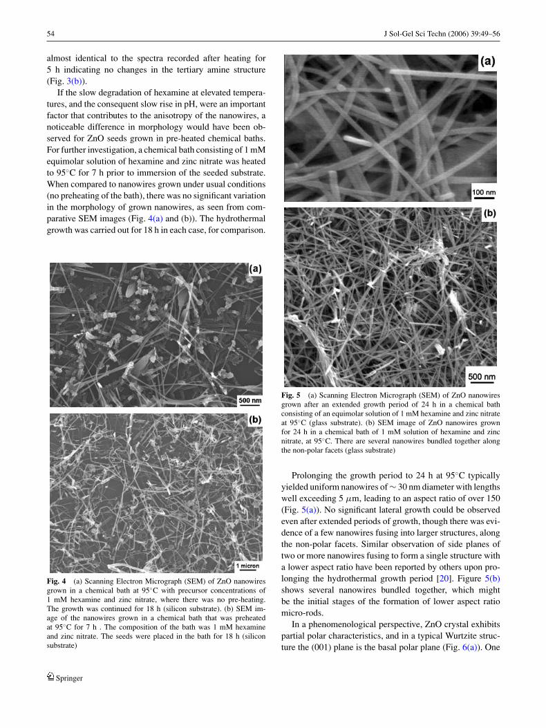
If the slow degradation of hexamine at elevated tempera-tures, and the consequent slow rise in pH, were an importantfactor that contributes to the anisotropy of the nanowires, anoticeable difference in morphology would have been ob-served for ZnO seeds grown in pre-heated chemical baths.For further investigation, a chemical bath consisting of 1 mMequimolar solution of hexamine and zinc nitrate was heatedto 95◦C for 7 h prior to immersion of the seeded substrate.When compared to nanowires grown under usual conditions(no preheating of the bath), there was no significant variationin the morphology of grown nanowires, as seen from com-parative SEM images (Fig. 4(a) and (b)). The hydrothermalgrowth was carried out for 18 h in each case, for comparison.
Fig. 4 (a) Scanning Electron Micrograph (SEM) of ZnO nanowiresgrown in a chemical bath at 95◦C with precursor concentrations of1 mM hexamine and zinc nitrate, where there was no pre-heating.The growth was continued for 18 h (silicon substrate). (b) SEM im-age of the nanowires grown in a chemical bath that was preheatedat 95◦C for 7 h . The composition of the bath was 1 mM hexamineand zinc nitrate. The seeds were placed in the bath for 18 h (siliconsubstrate)
Fig. 5 (a) Scanning Electron Micrograph (SEM) of ZnO nanowiresgrown after an extended growth period of 24 h in a chemical bathconsisting of an equimolar solution of 1 mM hexamine and zinc nitrateat 95◦C (glass substrate). (b) SEM image of ZnO nanowires grownfor 24 h in a chemical bath of 1 mM solution of hexamine and zincnitrate, at 95◦C. There are several nanowires bundled together alongthe non-polar facets (glass substrate)
Prolonging the growth period to 24 h at 95◦C typicallyyielded uniform nanowires of ∼ 30 nm diameter with lengthswell exceeding 5 µm, leading to an aspect ratio of over 150(Fig. 5(a)). No significant lateral growth could be observedeven after extended periods of growth, though there was evi-dence of a few nanowires fusing into larger structures, alongthe non-polar facets. Similar observation of side planes oftwo or more nanowires fusing to form a single structure witha lower aspect ratio have been reported by others upon pro-longing the hydrothermal growth period [20]. Figure 5(b)shows several nanowires bundled together, which mightbe the initial stages of the formation of lower aspect ratiomicro-rods.
In a phenomenological perspective, ZnO crystal exhibitspartial polar characteristics, and in a typical Wurtzite struc-ture the (001) plane is the basal polar plane (Fig. 6(a)). One
Springer

J Sol-Gel Sci Techn (2006) 39:49–56 55
Fig. 6 (a) Illustration of a typical zincite crystal with various planesmarked. The top plane is the basal polar plane (001), the side facets arenon-polar planes. (b) Illustration of the possible role of hexamine in theanisotropy of the obtained ZnO nanoparticles
end of the basal polar plane terminates with partially posi-tive Zn lattice points and the other end terminates in partiallynegative oxygen lattice points [17]. It is believed that, in thechemical bath, hexamine being a non-polar chelating agentwould preferentially attach to the non-polar facets of thenano-wires, thereby exposing only the (002) plane for epi-taxial growth (Fig. 6(b)). Thus a preferential growth alongthe [0002] direction is made possible.
There is a recent report on a reversible reduction of theaspect ratio of ZnO nanorods by introducing acidic anionsin the growth medium to cap the polar plane [19], whereincitrate anions probably cap the (001) which terminateswith charged Znδ+. This shows the dependence of themorphology of the ZnO nanowires on the ionic content ofthe chemical bath, a clear demonstration of the versatilityof the hydrothermal growth process, which can be studiedfurther for controlled nanowire growths.
4. Conclusions
We have studied the growth of ZnO nanowires prepared byhydrothermal synthesis from zinc nitrate in the presence of
hexamine, with different concentrations in the chemical bath.The hypothesis of degradation of hexamine that is linked tothe growth of ZnO nanowires in similar experiments reportedin the literature deviates from what we have observed in ourexperiments. Infra-red spectroscopy shows negligible degra-dation of hexamine over the typical time ranges employedfor the nanowire growth in our experiments. ZnO nanowireswith very high aspect ratio and high uniformity in diametersare obtained due to preferential capping of the non-ionic hex-amine molecule, over the non-polar side facets of a growingZnO crystal. The nanowires evolve via epitaxial growth ofZnO from seeds fixed on glass and silicon (111) substrates,leading to lengths exceeding 5 µm and ∼30 nm in diameter,with an aspect ratio of over 150. It was also observed that thegrowth-rate of the nanowires depends on the concentrationof the precursors in the chemical bath. It will be interestingto carry out a systematic study on the growth of nanowireswith respect to the concentration of the precursors.
Acknowledgments The authors would like to thank Prof. J. G. Hilbornfor enlightening discussions, Mr. Bjorn Atthoff for assistance with someof the microscopy, and Mr. Basse Asplund for assistance with the FTIRspectroscopy. We are grateful for partial financial support from theAsian Institute of Technology and Uppsala University.
References
1. Efros AL, Rosen M (2003) Annu. Rev. Mater. Sci. 30:4752. Lee D-D, Lee D-S (2001) IEEE Sensors Journal 1:2143. Huang M, Mao S, Feick H, Yan H, Wu Y, Kind H, Weber E, Russo
R, Yang P (2001) Science 292:18974. Lee CJ, Lee TJ, Lyu SC, Zhang Y, Ruh H, Lee HJ (2002) Appl.
Phys. Lett. 81:36485. Wang ZL (2003) Adv. Mater. 15:4326. Law M, Goldberger J, Yang P (2004) Annu. Rev. Mater. Res. 34:
837. Li YB, Bando Y, Sato T, Kurashima K (2002) Appl. Phys. Lett.
81:1448. Dong L, Jiao J, Tuggle DW, Petty JM (2003) Appl. Phys. Lett.
82:10969. Kim H, Sigmund W (2002) Appl. Phys. Lett. 81:2085
10. Shingubara S (2003) Journal of Nanoparticle Research 5:1711. Wang YC, Leu IC, Hon MH (2002) J. Cryst. Growth 237:56412. Xia Y, Yang P, Sun Y, Wu Y, Mayers B, Gates B, Yin Y, Kim f,
Yan H (2003) Adv. Mater. 15:35313. Hossain MK, Ghosh SC, Boontongkong Y, Thanachayanont C,
Dutta J (2005) J. Metastable Nanocryst. Mater. 23:2714. Greene LE, Law M, Goldberger J, Kim F, Johnson JC, Zhang Y,
Saykally RJ, Yang P (2003) Angew. Chem. Int. Ed. 42:303115. Vayssieres L, Keis K, Lindquist SE, Hagfeldt A (2001) J Phys.
Chem. B 105:335016. Vayssieres L (2003) Adv. Mater. 15:46417. Kong XY, Wang ZL (2004) Appl. Phys. Lett. 84:97518. Sugunan A, Warad HC, Thanachayanont C, Dutta J, Hofmann
H (2005) In: Vaseashta A, Dimova-Malinovska D, Marshall JM,(eds) Nanostructured and advanced materials for applications insensor, optoelectronic and photovoltaic technology, Proceedingsof the NATO Advanced Study Institute, NATO Science Series II:
Springer

56 J Sol-Gel Sci Techn (2006) 39:49–56
Mathematics, Physics and Chemistry, vol. 204 (Springer, 2005) (inPrint)
19. Tian ZR, Voigt JA, Liu J, Mckenzie B, Mcdermott MJ, RodriguezMA, Konishi H, Xu H (2003) Nature Materials 2:821
20. Liou S-C, Hsiao C-S, Chen S-Y (21005) J. Cryst. Growth 274:43821. Lee JH, Leu IC, Hon MH (2005) J. Cryst. Growth 275:e206922. Govender K, Boyle DS, Kenway PB, O’Brien P (2004) J. Mater.
Chem. 14:2575
23. Bahnemann DW, Kormann C, Hofmann R (1987) J. Phys. Chem.91:3789
24. Meyer B, Marx D (2003) Phys. Rev. B67:03540325. Zhang F, Jin Q, Chan SW (2004) J. Appl. Phys. 95:431926. Haas PA, Pitt Jr. WW, Robinson SM, Ryon AD (1983) I and EC
Product Res. Dev. 22:46127. http://www.sigmaaldrich.com/spectra/rair/RAIR005128.PDF.28. Tada H (1960) J. Am. Chem. Soc. 82:255
Springer