Zebrafish in the sea of mineral (iron, zinc, and copper ...
23
REVIEW ARTICLE published: 06 March 2014 doi: 10.3389/fphar.2014.00033 Zebrafish in the sea of mineral (iron, zinc, and copper) metabolism Lu Zhao 1,2 , Zhidan Xia 1,2 and Fudi Wang 1,2 * 1 Department of Nutrition, Center for Nutrition and Health, School of Public Health, School of Medicine, Zhejiang University, Hangzhou, China 2 Institute of Nutrition and Food Safety, Zhejiang University, Hangzhou, China Edited by: Raffaella Gozzelino, Instituto Gulbenkian de Ciência, Portugal Reviewed by: Andrei Adrian Tica, University Of Medicine Craiova, Romania Constantin Ion Mircioiu, Carol Davila “University of Medicine and Pharmacy” , Romania *Correspondence: Fudi Wang, Department of Nutrition, Center for Nutrition and Health, School of Public Health, School of Medicine, Zhejiang University, 866 Yuhangtang Road, Hangzhou 310058, China e-mail: [email protected]; [email protected] Iron, copper, zinc, and eight other minerals are classified as essential trace elements because they present in minute in vivo quantities and are essential for life. Because either excess or insufficient levels of trace elements can be detrimental to life (causing human diseases such as iron-deficiency anemia, hemochromatosis, Menkes syndrome and Wilson’s disease), the endogenous levels of trace minerals must be tightly regulated. Many studies have demonstrated the existence of systems that maintain trace element homeostasis, and these systems are highly conserved in multiple species ranging from yeast to mice. As a model for studying trace mineral metabolism, the zebrafish is indispensable to researchers. Several large-scale mutagenesis screens have been performed in zebrafish, and these screens led to the identification of a series of metal transporters and the generation of several mutagenesis lines, providing an in-depth functional analysis at the system level. Moreover, because of their developmental advantages, zebrafish have also been used in mineral metabolism-related chemical screens and toxicology studies. Here, we systematically review the major findings of trace element homeostasis studies using the zebrafish model, with a focus on iron, zinc, copper, selenium, manganese, and iodine. We also provide a homology analysis of trace mineral transporters in fish, mice and humans. Finally, we discuss the evidence that zebrafish is an ideal experimental tool for uncovering novel mechanisms of trace mineral metabolism and for improving approaches to treat mineral imbalance-related diseases. Keywords: zebrafish, trace elements, minerals, iron, copper, zinc, metabolism INTRODUCTION As children of the Earth, humans are intimately connected to our surroundings in many ways, and the relationship between humans and minerals is perhaps the most enigmatic. Based on their necessity for life and their limited quantities with the human body, 11 elements are classified as trace minerals, including iron (Fe), zinc (Zn), copper (Cu), selenium(Se), man- ganese(Mn), iodine(I), molybdenum(Mo), fluorine (F), cobalt (Co), chromium (Cr), and vanadium (V) (Fraga, 2005). Metal trace minerals are biologically active primarily as metal- loproteins formed by conjugating or binding with various protein partners. Metalloproteins account for approximately half of all proteins and perform a wide range of biological functions as enzymes, transporters and signal transducers. In metallopro- teins, metal trace minerals are essential components, acting at the enzyme’s active site or by stabilizing the protein’s structure. Trace mineral deficiencies can cause a number of diseases that can be mild, severe, or even fatal. Conversely, excess levels of trace min- erals can be toxic. For example, excess iron or copper produces reactive oxygen species (ROS) via the Fenton reaction, resulting in lipid peroxidation, DNA damage, altered calcium homeostasis, and cell death (Stohs and Bagchi, 1995). Excess levels of redox- inactive metals such as zinc are also harmful; the accumulation of zinc triggers neuronal death in the brain and induces copper deficiency. Thus, the balance of endogenous trace minerals must be tightly regulated. Organisms have evolved comprehensive systems for main- taining trace element homeostasis; these systems are composed primarily of transport proteins, storage proteins, and some hor- mones. Our current knowledge regarding these regulatory factors has come primarily from studies using model organisms rang- ing from yeast to mice. Among these species, the zebrafish (Danio rerio) has been a valuable vertebrate system with several unique advantages. First, their small size, high fertility rate, and rapid development make zebrafish an ideal model for large-scale genetic screens. Secondly, because they are fertilized ex vivo and are optically transparent, zebrafish embryos are ideally suited for experimental techniques such as gene knockout/knockdown and overexpression. Because the embryos develop ex utero, zebrafish are also an excellent model for studying pharmacology and tox- icology in early developmental stages. Finally, the zebrafish is a vertebrate species, and many of its genes and metabolic systems are highly conserved with humans; indeed, 80% of genes and expressed sequence tags (ESTs) are present in conserved synteny groups between fish and humans (Barbazuk et al., 2000). Here, we systematically review the major findings obtained from zebrafish studies of trace element homeostasis, with a focus on iron, zinc, copper, selenium, manganese, and iodine. We also www.frontiersin.org March 2014 | Volume 5 | Article 33 | 1
-
Upload
khangminh22 -
Category
Documents
-
view
2 -
download
0
Transcript of Zebrafish in the sea of mineral (iron, zinc, and copper ...

REVIEW ARTICLEpublished: 06 March 2014
doi: 10.3389/fphar.2014.00033
Zebrafish in the sea of mineral (iron, zinc, and copper)metabolismLu Zhao1,2, Zhidan Xia1,2 and Fudi Wang1,2*
1 Department of Nutrition, Center for Nutrition and Health, School of Public Health, School of Medicine, Zhejiang University, Hangzhou, China2 Institute of Nutrition and Food Safety, Zhejiang University, Hangzhou, China
Edited by:
Raffaella Gozzelino, InstitutoGulbenkian de Ciência, Portugal
Reviewed by:
Andrei Adrian Tica, University OfMedicine Craiova, RomaniaConstantin Ion Mircioiu, Carol Davila“University of Medicine andPharmacy”, Romania
*Correspondence:
Fudi Wang, Department of Nutrition,Center for Nutrition and Health,School of Public Health, School ofMedicine, Zhejiang University,866 Yuhangtang Road,Hangzhou 310058, Chinae-mail: [email protected];[email protected]
Iron, copper, zinc, and eight other minerals are classified as essential trace elementsbecause they present in minute in vivo quantities and are essential for life. Becauseeither excess or insufficient levels of trace elements can be detrimental to life (causinghuman diseases such as iron-deficiency anemia, hemochromatosis, Menkes syndromeand Wilson’s disease), the endogenous levels of trace minerals must be tightly regulated.Many studies have demonstrated the existence of systems that maintain trace elementhomeostasis, and these systems are highly conserved in multiple species rangingfrom yeast to mice. As a model for studying trace mineral metabolism, the zebrafishis indispensable to researchers. Several large-scale mutagenesis screens have beenperformed in zebrafish, and these screens led to the identification of a series of metaltransporters and the generation of several mutagenesis lines, providing an in-depthfunctional analysis at the system level. Moreover, because of their developmentaladvantages, zebrafish have also been used in mineral metabolism-related chemicalscreens and toxicology studies. Here, we systematically review the major findings of traceelement homeostasis studies using the zebrafish model, with a focus on iron, zinc, copper,selenium, manganese, and iodine. We also provide a homology analysis of trace mineraltransporters in fish, mice and humans. Finally, we discuss the evidence that zebrafish isan ideal experimental tool for uncovering novel mechanisms of trace mineral metabolismand for improving approaches to treat mineral imbalance-related diseases.
Keywords: zebrafish, trace elements, minerals, iron, copper, zinc, metabolism
INTRODUCTIONAs children of the Earth, humans are intimately connected toour surroundings in many ways, and the relationship betweenhumans and minerals is perhaps the most enigmatic. Basedon their necessity for life and their limited quantities withthe human body, 11 elements are classified as trace minerals,including iron (Fe), zinc (Zn), copper (Cu), selenium(Se), man-ganese(Mn), iodine(I), molybdenum(Mo), fluorine (F), cobalt(Co), chromium (Cr), and vanadium (V) (Fraga, 2005).
Metal trace minerals are biologically active primarily as metal-loproteins formed by conjugating or binding with various proteinpartners. Metalloproteins account for approximately half of allproteins and perform a wide range of biological functions asenzymes, transporters and signal transducers. In metallopro-teins, metal trace minerals are essential components, acting at theenzyme’s active site or by stabilizing the protein’s structure. Tracemineral deficiencies can cause a number of diseases that can bemild, severe, or even fatal. Conversely, excess levels of trace min-erals can be toxic. For example, excess iron or copper producesreactive oxygen species (ROS) via the Fenton reaction, resultingin lipid peroxidation, DNA damage, altered calcium homeostasis,and cell death (Stohs and Bagchi, 1995). Excess levels of redox-inactive metals such as zinc are also harmful; the accumulationof zinc triggers neuronal death in the brain and induces copper
deficiency. Thus, the balance of endogenous trace minerals mustbe tightly regulated.
Organisms have evolved comprehensive systems for main-taining trace element homeostasis; these systems are composedprimarily of transport proteins, storage proteins, and some hor-mones. Our current knowledge regarding these regulatory factorshas come primarily from studies using model organisms rang-ing from yeast to mice. Among these species, the zebrafish (Daniorerio) has been a valuable vertebrate system with several uniqueadvantages. First, their small size, high fertility rate, and rapiddevelopment make zebrafish an ideal model for large-scale geneticscreens. Secondly, because they are fertilized ex vivo and areoptically transparent, zebrafish embryos are ideally suited forexperimental techniques such as gene knockout/knockdown andoverexpression. Because the embryos develop ex utero, zebrafishare also an excellent model for studying pharmacology and tox-icology in early developmental stages. Finally, the zebrafish is avertebrate species, and many of its genes and metabolic systemsare highly conserved with humans; indeed, 80% of genes andexpressed sequence tags (ESTs) are present in conserved syntenygroups between fish and humans (Barbazuk et al., 2000).
Here, we systematically review the major findings obtainedfrom zebrafish studies of trace element homeostasis, with a focuson iron, zinc, copper, selenium, manganese, and iodine. We also
www.frontiersin.org March 2014 | Volume 5 | Article 33 | 1

Zhao et al. Zebrafish in mineral metabolism
performed a homology analysis of trace mineral transporters infish, mice and humans, and we summarize the available zebrafishmutant models in the field. This review demonstrates thatzebrafish are an ideal experimental tool for investigating novelmechanisms of trace mineral metabolism and for improvingtherapeutic approaches for treating mineral imbalance-relateddiseases.
ZEBRAFISH AND IRON METABOLISMOVERVIEW OF IRON METABOLISMIron is present in nearly all living organisms. As an essential com-ponent of heme and iron-sulfur cluster–containing proteins, ironplays a central role in many biological activities, including oxygentransport, cellular respiration, and DNA synthesis (Muckenthalerand Lill, 2012). Of all the trace elements, the iron homeostasissystem is one of the best characterized, primarily because of iron’srole in erythropoiesis and its causative relationships with iron-deficiency anemia and hematochromatosis. To date, many majorproteins involved in the uptake, transport, storage and release ofiron have been identified (Figure 1).
Under normal conditions, dietary iron is absorbed by ente-rocytes through Divalent Metal Transporter 1 (DMT1); from
there, it is exported to the circulation through Ferroportin1 (Fpn1). In the blood, iron is transported in the form ofTransferrin (Tf)-Fe3+, which is taken up by endocytosis intocells with surface Transferrin receptors (TfRs). Iron in the endo-somes is then released to the cytoplasm and delivered to themitochondria, where it is used to make iron-sulfur (Fe-S) clus-ters, to synthesize heme, or to be stored as Ferritin. Mostof the iron used for producing hemoglobin in erythrocytes isobtained from the recycling iron pool released from senescentred blood cells that are phagocytized by macrophages. Asidefrom transport and storage proteins, Hepcidin—a peptide hor-mone released by the liver—plays an important role in regulatingiron levels by binding to Fpn1 and promoting its internal-ization. Other factors such as oxidoreductases [e.g., DuodenalCytochrome b (Dytb), Ceruloplasmin (Cp), Hephaestin (Heph),and STEAP3) and modulatory proteins (e.g., Hemochromatosis(HFE), Hemojuvelin (HJV), Iron Regulatory Protein (IRP) 1/2,and Transmembrane Serine Protease 6 (TMPRSS6)] also playan active role in iron metabolism (Muckenthaler and Lill, 2012;Srai and Sharp, 2012). An overview of the protein homology andexpression patterns among fish and mammalian iron-regulatingproteins is provided in Table 1.
FIGURE 1 | Generalized overview of iron metabolism in vertebrate cells.
Dietary iron is absorbed by enterocytes through the concerted activity of thereductase DCYTB and the transporter DMT1. Iron is then oxidized by HEPHand exits the enterocytes through the iron exporter FPN1. Iron is transferredas a complex with Transferrin (TF) in the bloodstream and is delivered totarget cells that express Transferrin receptors (TFRs) on their plasmamembrane. TF-Iron-TFR complexes are then endocytosed. In the endosome,
iron is released from TF by STEAP3 and then transported out of theendosome through DMT1. The cytoplasmic iron then enters the labile ironpool and is delivered by MFRN and siderophores to the mitochondria to beused for the synthesis of heme and Fe-S clusters. Excess iron is stored inFerritin. Iron leaves the cell through FPN1, the plasma expression of which isnegatively regulated by Hepcidin. Proteins for which zebrafish knockoutand/or knockdown models are available are written in red.
Frontiers in Pharmacology | Drug Metabolism and Transport March 2014 | Volume 5 | Article 33 | 2

Zhao et al. Zebrafish in mineral metabolism
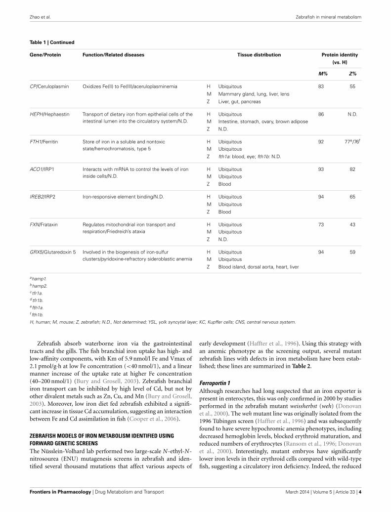
Table 1 | Iron metabolism–related proteins in zebrafish, mice, and humans.
Gene/Protein Function/Related diseases Tissue distribution Protein identity
(vs. H)
M% Z%
SLC11A2/DMT1 Intestinal iron absorption, intracellular ironrelease/Hypochromic microcytic anemia
H Ubiquitous 89 71M Yolk sac, intestine
Z Blood, gill, gut, lens, liver, YSL
SLC40A1/FPN 1 Cellular iron efflux/Hemochromatosis type 4 H Duodenum, macrophages, KCs, placenta,kidney
91 68
M Placenta, intestine, bone marrow,erythrocytes, liver, spleen
Z CNS, yolk syncytial layer, gill, gut, liver
HAMP/Hepcidin Cellular iron homeostasis/Hemochromatosis type 2B H Liver, heart 59 31a/32b
M Liver, lung, heart
Z Hamp1: gut, liver; hamp2: N.D.
HFE/HFE Regulator of Tf-TfR interaction/Hemochromatosistype 1
H Ubiquitous 68 N.D.M Ubiquitous
Z N.D.
HFE2/HJV Modulator of hepcidin expression/Hemochromatosistype 2A
H Ubiquitous 87 44M Skeletal muscle, liver, heart, prostate
Z Skeletal muscle, liver, notochord
TF /Transferrin Transport iron/atransferrinemia H Liver, spinal cord, lung, hypothalamus 72 41
M Liver, spinal cord, cerebellum, lung,placenta, ovary, bladder
Z Liver, trunk musculature
TFRC/TFR1 Cellular iron uptake / N.D. H Fetal liver, pancreas, muscle, placenta,early erythroid cells
77 44c/39d
M Placenta, intestine, muscle, osteoclasts,microglia, bone marrow, liver, kidney
Z Tfr1a: blood, blood island; tfr1b: ubiquitous
TFR2/TFR2 Mediates cellular uptake of Tf-boundiron/Hemochromatosis, type 3
H Liver, early erythroid cells 85 53M Liver, bone, bone marrow
Z Liver
TMPRSS6/TMPRSS6 Iron homeostasis/Iron-refractory iron deficiencyanemia
H Ubiquitous 83 N.D.M Liver
Z N.D.
SLC25A37 /Mitoferrin Mitochondrial iron transport/N.D. H Bone marrow, fetal liver, fetal lung, blood,prostate, early erythroid cells
91 69
M Umbilical cord, spleen, bone, bonemarrow
Z Blood island, lateral plate mesoderm
CYBRD1/DCYTB Dietary iron absorption/N.D. H Ubiquitous 75 58
M Ubiquitous
Z N.D.
STEAP3/ STEAP3 Ferric-chelate reductase activity/Anemia,hypochromic microcytic, with iron overload 2
H Ubiquitous 87 53M Ubiquitous
Z N.D.
(Continued)
www.frontiersin.org March 2014 | Volume 5 | Article 33 | 3
Zhao et al. Zebrafish in mineral metabolism
Table 1 | Continued
Gene/Protein Function/Related diseases Tissue distribution Protein identity
(vs. H)
M% Z%
CP/Ceruloplasmin Oxidizes Fe(II) to Fe(III)/aceruloplasminemia H Ubiquitous 83 55
M Mammary gland, lung, liver, lens
Z Liver, gut, pancreas
HEPH/Hephaestin Transport of dietary iron from epithelial cells of theintestinal lumen into the circulatory system/N.D.
H Ubiquitous 86 N.D.M Intestine, stomach, ovary, brown adipose
Z N.D.
FTH1/Ferritin Store of iron in a soluble and nontoxicstate/hemochromatosis, type 5
H Ubiquitous 92 77e/76f
M Ubiquitous
Z fth1a: blood, eye; fth1b: N.D.
ACO1/IRP1 Interacts with mRNA to control the levels of ironinside cells/N.D.
H Ubiquitous 93 82M Ubiquitous
Z Blood
IREB2/IRP2 Iron-responsive element binding/N.D. H Ubiquitous 94 65
M Ubiquitous
Z Blood
FXN/Frataxin Regulates mitochondrial iron transport andrespiration/Friedreich’s ataxia
H Ubiquitous 73 43M Ubiquitous
Z N.D.
GRX5/Glutaredoxin 5 Involved in the biogenesis of iron-sulfurclusters/pyridoxine-refractory sideroblastic anemia
H Ubiquitous 94 59M Ubiquitous
Z Blood island, dorsal aorta, heart, liver
ahamp1.bhamp2.ctfr1a.d tfr1b.efth1a.f fth1b.
H, human; M, mouse; Z, zebrafish; N.D., Not determined; YSL, yolk syncytial layer; KC, Kupffer cells; CNS, central nervous system.
Zebrafish absorb waterborne iron via the gastrointestinaltracts and the gills. The fish branchial iron uptake has high- andlow-affinity components, with Km of 5.9 nmol/l Fe and Vmax of2.1 pmol/g·h at low Fe concentration (<40 nmol/1), and a linearmanner increase of the uptake rate at higher Fe concentration(40–200 nmol/1) (Bury and Grosell, 2003). Zebrafish branchialiron transport can be inhibited by high level of Cd, but not byother divalent metals such as Zn, Cu, and Mn (Bury and Grosell,2003). Moreover, low iron diet fed zebrafish exhibited a signifi-cant increase in tissue Cd accumulation, suggesting an interactionbetween Fe and Cd assimilation in fish (Cooper et al., 2006).
ZEBRAFISH MODELS OF IRON METABOLISM IDENTIFIED USINGFORWARD GENETIC SCREENSThe Nüsslein-Volhard lab performed two large-scale N-ethyl-N-nitrosourea (ENU) mutagenesis screens in zebrafish and iden-tified several thousand mutations that affect various aspects of
early development (Haffter et al., 1996). Using this strategy withan anemic phenotype as the screening output, several mutantzebrafish lines with defects in iron metabolism have been estab-lished; these lines are summarized in Table 2.
Ferroportin 1Although researches had long suspected that an iron exporter ispresent in enterocytes, this was only confirmed in 2000 by studiesperformed in the zebrafish mutant weissherbst (weh) (Donovanet al., 2000). The weh mutant line was originally isolated from the1996 Tübingen screen (Haffter et al., 1996) and was subsequentlyfound to have severe hypochromic anemia phenotypes, includingdecreased hemoglobin levels, blocked erythroid maturation, andreduced numbers of erythrocytes (Ransom et al., 1996; Donovanet al., 2000). Interestingly, mutant embryos have significantlylower iron levels in their erythroid cells compared with wild-typefish, suggesting a circulatory iron deficiency. Indeed, the reduced
Frontiers in Pharmacology | Drug Metabolism and Transport March 2014 | Volume 5 | Article 33 | 4

Zhao et al. Zebrafish in mineral metabolism
Table 2 | Iron metabolism–related mouse and zebrafish knockout/knockdown models and their phenotypes.
Gene/Protein KO mouse models KO/KD zebrafish models References
SLC11A2/DMT1 Hypochromic microcytic anemiaa,b Hypochromic microcytic anemiac aRussell et al., 1970; bGunshin et al.,2005a; cDonovan et al., 2002
SLC40A1/FPN1 Embryonic lethality; abnormal ironhomeostasisd,e
Hypochromic microcytic anemiaf dDonovan et al., 2005; eZohn et al., 2007;fDonovan et al., 2000
HAMP/Hepcidin Massive iron accumulation in the liver,pancreas, and heartg
N.D. gLesbordes-Brion et al., 2006
HFE/HFE Increased intestinal iron absorption, elevatedhepatic iron load, reduced duodenal ironstoresh,i
N.D. hVujic Spasic et al., 2007; iVujic Spasic etal., 2008
HFE2/HJV Lack of hepcidin expression, severe ironoverloadj,k
N.D. jHuang et al., 2005a; kNiederkofler et al.,2005
TF /TF Hypochromic microcytic anemia, iron-loadingin the liver, pancreas, heart, and brainl
Hypochromic anemiam lBartnikas et al., 2011; mFraenkel et al.,2009
TFRC/TFR1 Anemia, hydrops fetalis, neurologicaldefectsn
Hypochromic anemiao nLevy et al., 1999; oWingert et al., 2004
TFR2/TFR2 Periportal hepatic iron loading, splenic ironsparing, and elevated serum transferrinsaturations p
N.D. pRoetto et al., 2010
TMPRSS6/TMPRSS6 Microcytic anemia, female infertilityq,r N.D. qNai et al., 2010; rNai et al., 2012
SLC25A37 /MFRN No hemoglobinization in the yolk sac andheart; die during organogenesiss
Hypochromic anemia, erythroidmaturation arrestt
sTroadec et al., 2011; tShaw et al., 2006
CYBRD1/DCYTB Alterations in liver weight and liver ironcontentu
N.D. uGunshin et al., 2005b
STEAP3/STEAP3 Anemiav N.D. vOhgami et al., 2005
CP/Ceruloplasmin Iron accumulation in the liver, spleen, brain;iron deficiency anemia, impaired motorcoordinationw,x
N.D. wHarris et al., 1999; xCherukuri et al., 2005
HEPH/Hephaestin Hypochromic anemiay N.D. yVulpe et al., 1999
FTH1/Ferritin Embryonic lethality z N.D. zDarshan et al., 2009
GRX5/GRX5 N.D. Hypochromic anemiaA AWingert et al., 2005
HD/Huntingtin N.D. *Hypochromic anemiaB BLumsden et al., 2007
BDH2/2,5-DHBA N.D. *Hypochromic anemiaC CDevireddy et al., 2010
ARHGEF3/ARHGEF3 N.D. *Hypochromic anemiaD DSerbanovic-Canic et al., 2011
*Knockdown (KD) model.
hemoglobin level in weh mutants can be rescued by intravenousiron-dextran injections, demonstrating that their hypochromia iscaused by inadequate iron in the blood (Donovan et al., 2000). Toidentify the precise location of the gene mutation, chromosomalwalking was performed and revealed a premature stop codon ina novel gene named ferroportin1 (fpn1), consistent with a loss-of-function mutation. Importantly, overexpressing fpn1 in mutantembryos rescued the hypochromia phenotype, suggesting that thefpn1 gene is causally linked to the disease. Fpn1 transcripts arepresent specifically in the zebrafish yolk syncytial layer (YSL),between the developing hematopoietic cells in the intermediatecell mass and the yolk, which contains iron and other nutrientsessential for early embryonic development. This specific expres-sion pattern of fpn1, together with the iron-deficiency phenotypesobserved in weh mutants, suggests that the function of the fpn1protein is to export iron from the yolk into the embryonic circula-tion. This hypothesis was confirmed by performing an iron effluxassay in a Xenopus oocyte expression system, in which oocytesexpressing fpn1 had increased iron efflux (Donovan et al., 2000).
Moreover, both mice and humans have homologs of FPN1 thatare highly conserved with the fish fpn1, and mammalian FPN1is robustly expressed in the placenta, duodenum, and liver, allof which are major sites of iron transport. At the protein level,human FPN1 is concentrated at the basal surface of the syn-cytiotrophoblasts in the placenta, an organ that is functionallysimilar to the zebrafish YSL, indicating that human FPN1 playsa role in maternal-fetal iron export. In mice, Fpn1 is expressedat the basolateral surface of enterocytes, suggesting a role as anintestinal iron transporter (Donovan et al., 2000). This studyserves as a prime example as how genetic screens in zebrafish canlead to the identification of essential novel genes. Nevertheless,Fpn1 remains the only iron exporter that has been identified inall eukaryotic organisms.
Shortly after these findings in the zebrafish weh mutantswere reported (Donovan et al., 2000), two groups independentlycloned Fpn1 (also called Ireg1 or Mtp1) from mouse duodenalepithelial cells and a mouse mRNA library (Abboud and Haile,2000; McKie et al., 2000). Both studies confirmed the essential
www.frontiersin.org March 2014 | Volume 5 | Article 33 | 5

Zhao et al. Zebrafish in mineral metabolism
role of Fpn1 in iron export and the regulation of iron homeosta-sis. Mutations in SLC11A3, the human homolog of FPN1, werelater identified as causing autosomal dominant hemochromato-sis, a disorder characterized by iron overload and multiple organdamage (Montosi et al., 2001; Njajou et al., 2001).
The effect of Fpn1 deficiency on the adult zebrafish systemwas examined in further detail. Although weh homozygotes areembryonic lethal and usually die 7–14 days post-fertilization(dpf), repeated intravenous injections of iron-dextran enables themutants to reach adulthood (Donovan et al., 2000; Fraenkel et al.,2005). These rescued fish are normal until 6 months of age, butdevelop hypochromic blood by 12 months. Compared with iron-injected wild-type fish, the rescued mutants had increased ironstaining in the kidney macrophages at 12 months of age, as wellas increased staining in the intestinal villi at 6 and 12 months,suggesting that the fpn1 mutation impairs iron export in thesetissues. The iron-rescued weh mutants also has hepatic iron over-load, with particularly high iron levels in the liver Kupffer cells(Fraenkel et al., 2005). The role of Fpn1 in iron mobilizationfrom enterocytes, hepatocytes, and macrophages was confirmedby studies using adult tissue-specific Fpn1 knockout mice (Zhanget al., 2011, 2012). Hepcidin, a peptide hormone secreted by theliver, was found to regulate iron by triggering the internalizationof Fpn1 and inhibiting iron efflux (Nemeth et al., 2004). Hepcidinis also conserved in fish, and injecting zebrafish embryos with ironcauses a significant increase in endogenous hepcidin expression.Moreover, a similar iron-stimulated increase in hepcidin expres-sion occurs in weh mutant embryos, suggesting that hepcidinexpression is independent of Fpn1’s normal function as an ironexporter (Fraenkel et al., 2005).
Dmt1The protein DMT1 (also called DCT1 and Nramp2), which con-tains 12 transmembrane domains, was originally isolated in therat duodenum as a divalent ion transporter that is upregulatedby dietary iron deficiency (Gunshin et al., 1997). Shortly afterits discovery, two mammalian hypochromic anemia models—the mk/mk mouse and the Belgrade rat—were found to carryDMT1 mutations (Fleming et al., 1997, 1998). The chardon-nay (cdy) zebrafish mutant revealed a conserved role of DMT1in zebrafish iron metabolism (Donovan et al., 2002). The cdymutant is a hypochromic, microcytic anemia model with reducedhemoglobin expression and delayed erythrocyte maturation. Thecdy mutant has a premature stop codon in the zebrafish homologof dmt1, resulting in a severely truncated protein. The zebrafishhomolog of DMT1 is 73% identical to human and mouse DMT1homologs, and its transcripts are concentrated both in erythroidcells and in the intestine. Direct evidence of the role of zebrafishDMT1 in iron transport came from experiments using a mam-malian cell line; cells that overexpressed wild-type zebrafish dmt1took up nearly 10 times the amount of iron as non-transfectedcontrol cells, whereas the truncated protein produced by thecdy mutation was non-functional. Interestingly, unlike the wehmutants, which die during early development (Donovan et al.,2000), the anemic cdy homozygotes survive and reach adult-hood. The viability of cdy fish may be attributed to additionalpathways for iron absorption (Donovan et al., 2002). Of clinical
relevance, the first identified human mutation in DMT1 wasreported to cause symptoms that include severe hypochromicmicrocytic anemia and iron overload (Mims et al., 2005). Onepossible explanation for the excess iron in these patients is thepresence of an alternate iron absorption route in the duodenum,thereby bypassing DMT1 (Mims et al., 2005).
Tfr1Transferrin receptor 1 (Tfr1) is a membrane-bound protein thatfacilitates iron uptake by binding to the iron carrier Transferrin.Tfr1 was identified as being essential for erythropoiesis andembryonic development in a Tfr1-knockout mouse model (Levyet al., 1999). Tfr1−/− mice develop anemia, have retarded growthand neurological defects, and die during embryogenesis (Levyet al., 1999). Four different zebrafish chianti (cia) mutants wereidentified with various degrees of hypochromic anemia anddefective erythroid differentiation (Haffter et al., 1996; Wingertet al., 2004), and positional cloning revealed that cia allelesare missense and splicing mutations. During early develop-ment, tfr1a transcripts are expressed specifically in erythrocytes.Importantly, cytoplasmic delivery of iron by microinjection atthe 1-cell stage—but not intravenous iron injections—can res-cue the hypochromia phenotypes of cia mutants, indicating thatthe tfr1a mutation prevents erythrocytes from taking up and uti-lizing circulating iron (Wingert et al., 2004). Interestingly, whilecloning tfr1a, a second tfr1-like gene, tfr1b, was also identified(Wingert et al., 2004). This gene duplication phenomenon inzebrafish is believed to have occurred as an evolutionary geneticevent in teleosts (Amores et al., 1998; Postlethwait et al., 1998).The tfr1b gene is expressed ubiquitously throughout embryo-genesis. Notably, although overexpressing tfr1b partially rescuesthe anemic phenotypes of cia mutants, tfr1b morphants (animalsin which the gene has been knocked down by injecting mor-pholino antisense oligonucleotides) have normal hemoglobiniza-tion. Nevertheless, tfr1b morphants have retarded growth anddevelop brain necrosis, a phenotype that is similar to the neu-rologic defects observed in the mouse model (Levy et al., 1999),indicating that tfr1b may be involved in iron uptake throughnon-erythroid tissues (Wingert et al., 2004). Thus, the combinedphenotypes of tfr1a and tfr1b deficient zebrafish embryos appearto recapitulate the entire phenotypic spectrum of Tfr1−/− mice.Therefore, the cia mutant zebrafish is an ideal model for studyingthe function of tfr1 in erythropoiesis without the complication ofother developmental abnormalities.
Grx5The shiraz (sir) zebrafish mutants were originally isolated fromthe Tübingen 2000 screen consortium; these mutants were lateridentified as a typical hypochromic anemia disease model witha deletion in the glutaredoxin 5 (grx5) gene which encodes anantioxidant protein (Wingert et al., 2005). Functional studiesof grx5 in sir zebrafish revealed a novel connection betweenheme biosynthesis and iron-sulfur (Fe-S) cluster formation, twoprimary functions of iron that were previously believed to beindependent processes in vertebrates.
Studies in yeast revealed that GRX5 is required for the mito-chondrial synthesis of Fe-S clusters (Rodriguez-Manzaneque
Frontiers in Pharmacology | Drug Metabolism and Transport March 2014 | Volume 5 | Article 33 | 6

Zhao et al. Zebrafish in mineral metabolism
et al., 2002). Similar to the yeast GRX5, zebrafish grx5 is alsolocalized primarily in the mitochondria, and expression of thezebrafish grx5 gene can rescue a GRX5-deficient yeast strain,suggesting that the function of grx5 is evolutionarily conserved.However, the sir zebrafish mutants have a hypochromic ane-mia phenotype, with no changes in their mitochondrial ironcontent or oxidative stress level (Wingert et al., 2005). Withrespect to iron metabolism, one key difference between yeastand higher eukaryotes is that in the latter, iron regulatory pro-teins 1 and 2 (IRP1/2) control intracellular iron levels by bindingto Iron Response Elements (IREs) in the 5′-UTR of target genetranscripts, thereby blocking their translation. Importantly, theIRE-binding capacity of IRP1 is negatively regulated by Fe-Sclusters. Thus, a possible explanation for the anemic pheno-type in sir zebrafish mutants is that the grx5 mutation reducesFe-S assembly, inappropriately triggering IRP1 activity, whichthen inhibits the expression of select target genes that are crit-ical for heme biosynthesis. In support of this hypothesis, redblood cells in sir zebrafish mutants lack aminolevulinate syn-thase 2 (ALAS2), the first enzyme in the heme biosynthesispathway. Moreover, overexpressing an ALAS2 gene in which theIRE is deleted rescues hemoglobin production in sir mutants; incontrast, overexpressing the wild-type ALAS2 gene does not res-cue hemoglobin production. Interestingly, knocking down theexpression of IRP1 also rescues the sir embryonic phenotype.These compelling results strongly suggest that heme synthesisin vertebrates is regulated via Fe-S cluster levels (Wingert et al.,2005). A conserved role for GRX5 in regulating heme synthesiswas additionally confirmed in human patients (Camaschella et al.,2007). The findings obtained from sir mutants serve to highlightthe advantages of using zebrafish as a vertebrate model systemfor discovering mechanisms that are not necessarily conserved inlower organisms.
MitoferrinThe role of Mitoferrin (Mfrn) in mitochondrial iron uptake wasoriginally discovered in yeast studies (Foury and Roganti, 2002).MRS3 and MRS4, the yeast homologs of Mfrn, increase theefficiencies of both heme formation and Fe-S biosynthesis, thetwo key mitochondrial processes that utilize iron (Muhlenhoffet al., 2003). Studies of frascati (frs) zebrafish mutants furthersupport Mfrn’s role as a principal mitochondrial iron importerin vertebrate erythroblasts. Frs mutants develop defects suchas hypochromic anemia and erythroid maturation arrest (Shawet al., 2006), and positional cloning identified missense mutationsin the mfrn gene in all mutant lines. Importantly, overexpressingmfrn in frs mutants rescued the erythropoiesis deficiency in halfof the injected animals, and mfrn knockdown morphants mim-icked the mutant phenotype, suggesting that mfrn is the disease-causing gene in frs mutants. Expression array analysis revealedthat mfrn is highly expressed in the intermediate cell mass (thetissue in which erythropoiesis occurs for early embryos), furthersupporting mfrn’s role in erythroid heme synthesis. Similar toyeast MRS3/4, when overexpressed in transfected mammalian celllines, zebrafish mfrn localizes to the mitochondria (Shaw et al.,2006). To investigate the function of mammalian Mfrn further,an Mfrn−/− mouse hematopoietic cell line was established. These
cells have impaired terminal erythroid maturation and an inabil-ity to incorporate iron into heme proteins. Furthermore, mouseMfrn rescues the phenotype in zebrafish frs mutants, and fish mfrnrestores the activity of an MRS3/4-deficient yeast strain (Shawet al., 2006). Taken together, these results suggest that Mfrn’s rolein mitochondrial iron uptake is evolutionarily conserved amongeukaryotes.
Transferrin-aZebrafish gav mutant strains carry mutations in their transferrin-a (tf-a) gene, which encodes the principal iron carrier in allvertebrate organisms (Fraenkel et al., 2009). Gav mutants developsevere hypochromic anemia. Importantly, this phenotype can bephenocopied by injecting tf-a morpholinos into embryos, and itcan be rescued by overexpressing tf-a. Together, these findingsconfirm that tf-a is the disease-causing gene. Homozygous gavmutants are generally embryonic lethal and die at approximately14 dpf (Fraenkel et al., 2009). In humans, genetic mutationsin transferrin cause congenital hypotransferrinemia, a rare dis-ease with features that are strikingly similar to the phenotypein gav fish, including hypochromic anemia and premature death(Hayashi et al., 1993; Goldwurm et al., 2000). Thus, the zebrafishgav mutant serves as an ideal model for studying human hypo-transferrinemia. In addition, Fraenkel and colleagues examinedhepcidin expression in the gav mutant, as well as several previ-ously established zebrafish mutants with iron metabolism defects.In 2-dpf zebrafish embryos (the stage in which endogenous hep-cidin expression is least affected by environmental stimuli), thenumber of hepcidin transcripts was measured in various mutantsand morphants either with or without iron injection. The resultssuggest that both tf-a and tfR2 are required for hepcidin expres-sion, whereas tfR1a and dmt1 are required for increasing hepcidinexpression in response to iron loading (Fraenkel et al., 2009).The primary role of Tf in driving hepcidin expression is fur-ther supported by studies performed using a mouse model ofhypotransferrinemia (Bartnikas et al., 2011).
ZEBRAFISH AS A REVERSE GENETICS TOOL IN IRON METABOLISMSTUDIESIn addition to helping identify novel iron transporters andmetabolic mechanisms via forward genetic screens, zebrafish havealso been used as a reverse genetics tool for increasing ourunderstanding of the iron homeostasis system.
Gene knockdown/knockoutGene knockdown/knockout techniques are used as a primarystep in investigating the unknown biological functions of a givengene. In this approach, antisense morpholino oligonucleotidesbind to the targeted gene transcript, thereby inhibiting the gene’sexpression by blocking the initiation of translation or by mod-ifying pre-mRNA splicing. Because zebrafish develop ex utero,microinjection-mediated gene suppression is used widely amongzebrafish researchers.
Hd. Studies performed using hd zebrafish morphants revealed anovel role for Huntingtin (Htt, a protein linked to Huntington’sdisease) in the utilization of iron by erythrocytes (Lumsden
www.frontiersin.org March 2014 | Volume 5 | Article 33 | 7

Zhao et al. Zebrafish in mineral metabolism
et al., 2007). Although it has been known for more than twodecades that Huntington’s patients carry an expanded CAGrepeat in the coding region of the HD gene (Huntington’sDisease Collaborative Research Group, 1993), the normal func-tion of Htt remains unclear. In order to explore the biologicalroles of HD, hd knockdown morphant zebrafish were created(Lumsden et al., 2007). Interestingly, in addition to neurologi-cal defects such as brain necrosis, the hd knockdown fish alsodevelop blood hypochromia (characterized by reduced red pig-ments in the erythrocytes) and decreased hemoglobin staining.Nevertheless, Prussian blue staining revealed that hd morphantshave normal iron levels in their red blood cells, suggesting thatTf-Tfr–mediated iron transport and endocytosis are intact inthe erythrocytes. Intriguingly, Prussian blue only stains ferriciron that is bound to Tf, but does not stain ferrous iron inhemoglobin. Thus, the hypochromia observed in the hd mor-phants is likely due to defects that are downstream of Tf-Tfrendocytosis. This hypothesis was tested by injecting iron-dextrandirectly into the cytoplasm at the 1-cell stage, which circum-vents the Tf-Tfr–mediated iron cycle. Using this approach, thehypochromia defects in hd morphants were largely rescued(Lumsden et al., 2007). This important study revealed an inter-esting role for Htt in making endocytosed iron available for useby the cell; however, the detailed mechanism by which Htt regu-lates iron release and/or downstream utilization must be exploredfurther.
Bdh2. Although the vast majority of intracellular iron is boundby proteins, a small amount of cytoplasmic iron is bound tolow-molecular-weight carriers called siderophores, forming alabile iron pool (Breuer et al., 2008). Enterobactin is a classicbacterial siderophore that binds to Lipocalin 24p3, an iron-trafficking protein that functions as the iron-chelating moiety(Yang et al., 2002). In mouse cell cultures, 2,5-dihydroxybenzoicacid (2,5-DHBA) is the iron-binding moiety of the 24p-associatedmammalian siderophore, the synthesis of which is catalyzedby 3-hydroxybutyrate dehydrogenase type 2 (Bdh2), a dehy-drogenase/reductase family member (Devireddy et al., 2010).In Bdh2-knockdown mouse cells, the intracellular siderophorewas depleted, and the cells accumulated abnormally highamounts of free cytoplasmic iron, resulting in elevated levelsof ROS. Notably, these cells were also deficient in mitochon-drial iron, suggesting that siderophores also participate in thetransport of iron from the cytoplasm to the mitochondria.Importantly, bdh2 zebrafish morphants develop hypochromicblood and have reduced hemoglobin levels—but normal globinexpression—confirming a defect in mitochondrial heme syn-thesis (Devireddy et al., 2010). These findings demonstratethat the function of siderophores in regulating intracellulariron homeostasis is conserved from bacteria to vertebrates. Inthis respect, zebrafish are a convenient model for confirmingand extending findings obtained from studying mammalian cellcultures.
Arhgef3. Genome-wide association and meta-analysis studieshave identified more than 100 independent genetic loci asso-ciated with erythrocytes and platelets (Ganesh et al., 2009;
Soranzo et al., 2009). Because of its advantages with respectto reverse genetics, the zebrafish model was used to investigatethe biological functions of several candidates identified from themeta-analysis. This screen revealed that Rho guanine nucleotideexchange factor 3 (Arhgef3) plays an unexpected role in regulatingiron uptake and driving erythroid cell maturation (Serbanovic-Canic et al., 2011). Silencing arhgef3 expression in zebrafishdisrupts erythroid differentiation and causes hypochromic ery-throcytes, which are indicative of iron-deficiency anemia. Indeed,cytoplasmic iron supplementation significantly rescues thehemoglobinization phenotype in arhgef3 morphants. Moreover,disrupting the arhgef3 target RhoA produces a phenotype thatis similar to arhgef3 morphants, and this can also be rescued bycytoplasmic iron injection. The concerted roles of Arhgef3 andRhoA in regulating the internalization of membrane-bound Tfwas supported by studies in a human cell line (Serbanovic-Canicet al., 2011).
In vivo validation of cis-regulatory elements in mfrn1 usingtransgenic fish. In erythroblasts, Mfrn1, and Mfrn2 are solutecarriers that import cytoplasmic iron into the mitochondria forheme and Fe-S cluster biogenesis (Shaw et al., 2006; Paradkaret al., 2009). In mice, both the Mfrn1 and Mfrn2 genes containCpG-rich promoter regions. The cis-regulatory modules (CRMs)in Mfrn1 form a chromatin immunoprecipitation dataset forGATA-1, the primary erythroid transcription factor (Cantor andOrkin, 2002), suggesting that Mfrn1 is transcriptionally regulatedby GATA-1 via binding at CRM regions. Though quite com-pelling, these bioinformatics results still needed to be validatedfunctionally at the systemic level, and transgenic fish were a con-venient tool for this purpose. Mfrn1 transcripts are concentratedprimarily in hematopoietic tissues, whereas mfrn2 transcripts areexpressed throughout the central nervous system and in somites(Shaw et al., 2006). Transgenic zebrafish expressing GFP-taggedmouse Mfrn1 or Mfrn2 promoter sequences were generated tostudy the expression of these genes. When expressed in fish, theMfrn2 promoter is expressed in a pattern similar to the endoge-nous mfrn2 expression pattern, suggesting that the promoterfunctions appropriately. However, the tagged Mfrn1 promoterfailed to drive detectable GFP expression in fish, suggesting theneed for other transcriptional regulatory elements (Amigo et al.,2011). Interestingly, injecting two of the three predicted mouseMfrn1 CRMs yielded transient transgenic fish that expressed GFPin the same tissues as endogenous mfrn1. The specific expressionpattern was refined further in transgenic fish carrying a con-struct that contains a CRM linked with the CpG-rich promoter.Thus, the critical role of CRMs in regulating Mfrn1 expressionhas been demonstrated in vivo (Amigo et al., 2011). Moreover,the critical role for GATA-1 in regulating Mfrn1 transcriptionwas confirmed by the finding that the Mfrn1-specific expres-sion pattern was abolished after the GATA-1 core binding siteswere mutated in the CRMs of the transgenic fish. Finally, theexpression of endogenous mfrn1 was also markedly reduced inGATA-1 zebrafish morphants (Amigo et al., 2011). This semi-nal study supports the high value of using zebrafish transgenicsto complement and validate findings obtained from in silicoanalyses.
Frontiers in Pharmacology | Drug Metabolism and Transport March 2014 | Volume 5 | Article 33 | 8

Zhao et al. Zebrafish in mineral metabolism
Gene overexpressionThe ex utero development of zebrafish also enables researchers todrive gene overexpression through microinjection. Importantly,the biological mechanisms that underlie dominant-negative genemutations in mammals can be examined readily in zebrafish usinggene overexpression techniques. In humans, FPN1 mutationshave been linked to a form of autosomal dominant hemochro-matosis (Pietrangelo, 2004). Interestingly, two distinct clinicalphenotypes have been characterized: one phenotype includesiron accumulation in macrophages, low transferrin saturation,and iron-limited erythropoiesis, whereas the other phenotypeincludes iron accumulation in hepatocytes and high transferrinsaturation. The diversity of these clinical traits can be explainedby the different natures of the underlying FPN1 mutations (DeDomenico et al., 2006). Mutations that cause a defect in FPN1’scell-surface localization or iron export capacity cause iron loadingin macrophages, whereas mutations that impair FPN1’s sensitiv-ity to Hepcidin (thus impeding FPN1 incorporation) cause ironaccumulation in hepatocytes (De Domenico et al., 2005, 2006;Schimanski et al., 2005). Nevertheless, the clinical complexity ofthe long-term disease process makes it difficult to determine theprecise nature of a given FPN1 mutation. Overexpressing mutantFPN1 alleles in zebrafish is a high-throughput approach for iden-tifying the functional effects of many mutations (De Domenicoet al., 2007). The cDNA of wild-type mouse Fpn1 or previouslyidentified human and mice Fpn1 mutants was injected into 1-cell stage zebrafish embryos. Expressing either wild-type Fpn1or the N144H Fpn1 mutant (a Hepcidin-irresponsive protein)had no effect on endogenous hemoglobinization. However, over-expressing the H32R Fpn1 mutant (which has defective Fpn1membrane localization) or the N174I Fpn1 mutant (which istransport-deficient) led to severe defects in hemoglobin synthesis,and these hemoglobinization defects were rescued by intravenousiron injections, suggesting the existence of iron-deficiency ery-thropoiesis in these mutant-expressing embryos (De Domenicoet al., 2007). This study demonstrates nicely that the functionalconsequences of mammalian Fpn1 mutations can be studiedrapidly and effectively, and it supports the important role ofzebrafish as a valuable vertebrate model in functional studies ofdominant-negative mutations.
ZEBRAFISH AND ZINC METABOLISMOVERVIEW OF ZINC METABOLISMAfter iron, zinc is the second-most abundant trace mineral inhumans. Zinc is essential for the activity of more than 300enzymes and for maintaining the structural integrity of nearly2000 transcription factors. Thus, zinc plays a critical role in cellu-lar homeostasis, the immune response, oxidative stress, apoptosis,and aging (Stefanidou et al., 2006; Prasad, 2012). Zinc defi-ciency can cause a wide range of clinical defects, including growthretardation, hypogonadism, rough skin, weakened immunity, andneurosensory and cognitive disorders (Prasad, 2012). Althoughzinc is a redox-inactive metal, it can be toxic (albeit less toxic thaniron and copper), and both acute and chronic forms of zinc poi-soning have been reported to cause hematopoietic abnormalities,altered lipoprotein metabolism, and impaired immune function(Stefanidou et al., 2006).
In vivo, zinc homeostasis relies primarily on the zinc trans-porter family, which in mammals contains 10 Zinc Transporterproteins (ZnTs, or SLC30) and 14 Zrt- and Irt-like proteins (ZIPs,or SLC39) (Huang and Tepaamorndech, 2013; Jeong and Eide,2013). ZnTs are zinc exporters that facilitate the efflux of zincfrom cells and/or into intracellular vesicles, whereas ZIPs increaseintracellular zinc concentration by driving the uptake of extracel-lular zinc and/or the release of vesicular zinc into the cytoplasm.The concerted actions of ZnTs and ZIPs maintain the balanceof intracellular zinc and deliver zinc to its appropriate proteinpartners (Figure 2). Metallothioneins (MTs) are a group of low-molecular-weight metal-binding proteins that also have a highaffinity for binding zinc. MTs play an important regulatory role inzinc metabolism, possibly by competing with—or supplying zincto—a variety of transporter proteins (Vasak and Hasler, 2000;Chasapis et al., 2012).
ZINC HOMEOSTASIS IS HIGHLY CONSERVED BETWEEN ZEBRAFISHAND MAMMALSThe system that regulates zinc metabolism is highly conservedbetween zebrafish and mammals. The zebrafish zip1 gene wascloned from the zebrafish gill, an ion-transporting epitheliumthat absorbs minerals from the surrounding water (Qiu et al.,2005). The Zip1 protein is conserved both structurally and func-tionally with its mammalian homologs. Zip1 transcripts areexpressed ubiquitously in zebrafish embryos, with the highestexpression in the ovaries. As with human ZIP1 (Gaither and Eide,2001), overexpressing zebrafish zip1 significantly increases zincuptake. Interestingly, because Zip1 does not increase zinc influxat high extracellular zinc concentrations, it has no effect on themaximum endogenous rate of zinc uptake (Qiu et al., 2005).This ceiling effect might be due to the fact that zinc regulatesthe cellular localization of Zip1. Studies in mice suggest that theextracellular zinc concentration mediates the cellular localizationof Zip1 through an endocytosis-mediated pathway (Wang et al.,2004). A second zinc importer in teleosts, zip2, was cloned fromthe gill of a pufferfish species (Takifugu rubripes) and plays a rolein mediating zinc uptake (Qiu and Hogstrand, 2005).
A systematic bioinformatics data-mining approach identi-fied the zebrafish zinc transporter genes from two previouslyreleased zebrafish databases (Zv4 Ensembl 31 and Zv5 Ensembl34), and these genes were phylogenetically assigned to mam-malian orthologs (Feeney et al., 2005). To date, eight ZnT mem-bers (ZnT1, ZnT2, and ZnT4–9) and 11 ZIP members (ZIP1,ZIP3, ZIP4, ZIP6–11, ZIP13, and ZIP14) have been identifiedin zebrafish. Interestingly, the teleost ZIP8 sequence differs frommammalian ZIP8 orthologs to a larger degree than other ZIPgenes. Studies of gene expression patterns revealed that theovaries and intestine—the two organs that have the most dynamicnutrient metabolism—have the highest expression of zinc trans-porters (Feeney et al., 2005). The expression level of each zinctransporter has been examined during zebrafish embryogene-sis under normal maternal zinc conditions (Ho et al., 2012).The results showed that despite a relatively constant level ofendogenous zinc during embryonic development from fertiliza-tion through 120 h post-fertilization (hpf), zinc transporters aredifferentially expressed throughout this period. Nearly all zinc
www.frontiersin.org March 2014 | Volume 5 | Article 33 | 9

Zhao et al. Zebrafish in mineral metabolism
FIGURE 2 | Generalized overview of zinc metabolism in vertebrate cells.
Zinc Transporters (ZnTs) downregulate intracellular zinc levels by exportingzinc through the plasma membrane (ZnT1, ZnT2, and ZnT4) or by transportingzinc into various intracellular compartments, including lysosomes (ZnT2), theGolgi apparatus (ZnT5-7), mammary gland vesicles (ZnT2 and ZnT4), insulingranules (ZnT8), and synaptic vesicles (ZnT3). In addition, ZnT9 can
translocate to the nucleus, where it regulates target gene transcription. Zrt-and Irt-like proteins (ZIPs) upregulate cytoplasmic zinc levels by importingextracellular zinc (ZIP1–6, ZIP8, ZIP10, and ZIP14) and release zinc fromintracellular vesicles (ZIP1 and ZIP13), lysosomes (ZIP3 and ZIP8), the Golgiapparatus (ZIP7, ZIP9, and ZIP13) and the nucleus (ZIP7). Proteins for whichzebrafish knockout/knockdown models are available are written in red.
transporters have their highest expression at 120 hpf, with theexception of ZNT8, which peaks at 48 hpf (Ho et al., 2012).The release of the latest zebrafish database (Zv9 Ensembl 73)has further increased our knowledge of zebrafish genome, con-firming the existence of zebrafish ZNT10 and ZIP5 in zebrafish.The homology of zinc transporters among zebrafish, mice andhumans is summarized in Table 3. An influence of Zn on theuptake and circulatory influx of Cd has been reported in fish, sug-gesting that the uptake of Zn and Cd occurs through commonpathways (Wicklund Glynn, 2001).
ZEBRAFISH MODELS OF ZINC METABOLISMThe current zebrafish and mouse models available for studyingzinc metabolism are listed in Table 4.
Zip6Zip6 is a member of the LIV-1 subfamily of ZIP zinc trans-porters, which in humans consists of nine ZIP members thatcontain a highly conserved metalloprotease motif. Importantly,LIV-1 is regulated by estrogen and has been implicated inmetastatic breast cancer (Taylor, 2000), although how LIV-1mediates cancer metastasis is unclear. Studies using Zip6 (LIV1)zebrafish morphants have been instrumental in addressing thisproblem (Yamashita et al., 2004); the zebrafish zip6 cDNA wascloned by subtraction screening. Interestingly, the expressionpattern of endogenous zip6 mimics the expression of stat3, animportant player in the epithelial-mesenchymal transition (EMT)during gastrulation, organogenesis, wound-healing, and cancer
progression (Sano et al., 1999; Yamashita et al., 2002). Notably,the expression of zip6 is abolished in stat3 zebrafish morphants,suggesting that zip6 is downstream of stat3. The ability of stat3to transcriptionally regulate zip6 was confirmed in studies usingmouse and human cell lines (Yamashita et al., 2004). The roleof zip6 in early embryonic development was assessed furtherin zip6-depleted zebrafish embryos. By the end of gastrulation,these zip6 morphants have malpositioned heads and a short-ened anterior-posterior axis, although early cell-fate specificationwas not affected, suggesting that zip6 plays a critical role incell migration during gastrulation. Cell tracing and cell trans-plantation assays further support the cell-autonomous role ofZip6 in the migration of mesendodermal cells (Yamashita et al.,2004). Phenotypic analyses of zip6 morphants revealed that cell-cell adhesion was not downregulated as occurs normally, thusresulting in severe perturbations in cell migration. These samedefects were also observed in zebrafish morphants in which thezinc-finger protein Snail, a master regulator of EMT, is knockeddown (Batlle et al., 2000; Cano et al., 2000). Moreover, the in vivoactivity of Zip6 is dependent on Snail; Zip6 regulates the nucleartranslocation of Snail (Yamashita et al., 2004). This was the firststudy to use multiple zebrafish knockdown models to establish amolecular link between Stat3, Zip6, and Snail during EMT.
Zip7Zip7 is also a member of the LIV-1 subfamily of zinc trans-porters. Studies using mammalian cell lines suggest that humanZIP7 plays a role in elevating cytoplasmic zinc concentrations
Frontiers in Pharmacology | Drug Metabolism and Transport March 2014 | Volume 5 | Article 33 | 10

Zhao et al. Zebrafish in mineral metabolism
Table 3 | Zinc metabolism–related proteins in zebrafish, mice, and humans.
Gene/Protein Function/Related diseases Tissue distribution Protein identity
(vs. H)
M% Z%
SLC30A1/ZNT1 Plasma Zn exporter/N.D. H Ubiquitous 86 61a/54b
M UbiquitousZ N.D.
SLC30A2/ZNT2 Plasma Zn exporter, transport Zn into mammarygland vesicles/Transient neonatal Zn deficiency
H Ubiquitous 79 N.D.M Adipose, placenta, intestine, prostate,
pancreas, kidney, testis, mammary glandZ Hindbrain, neurons, spinal cord
SLC30A3/ZNT3 Transport Zn into synaptic vesicles/N.D. H Ubiquitous 89 N.D.M Amygdala, cerebral cortex, hippocampus,
spinal cord, testis, pancreasZ N.D.
SLC30A4/ZNT4 Plasma Zn exporter, transport Zn into mammarygland vesicles/Zn deficiency in milk
H Ubiquitous 92 53M Lacrimal gland, mammary gland, placenta,
intestineZ N.D.
SLC30A5/ZNT5 Plasma Zn exporter, transport Zn into Golgi/N.D. H Ubiquitous 95 78M UbiquitousZ Ubiquitous
SLC30A6/ZNT6 Transport Zn into Golgi/N.D. H Ubiquitous 91 12M UbiquitousZ Ubiquitous
SLC30A7 /ZNT7 Transport Zn into Golgi/N.D. H Ubiquitous 96 79M UbiquitousZ CNS, notochord
SLC30A8/ZNT8 Transport Zn into insulin granules/Diabetes mellitus H Pancreatic islet 81 53M UbiquitousZ Ubiquitous
SLC30A9/ZNT9 Plasma Zn exporter, transcriptional regulation innucleus/N.D.
H Ubiquitous 89 73M UbiquitousZ Ubiquitous
SLC30A10/ZNT10 Cation transporter/Hypermanganesemia withdystonia, polycythemia, and cirrhosis
H Ubiquitous 80 46M Stomach, intestine, prostate, liver,
amygdala, cerebral cortexZ N.D.
SLC39A1/ZIP1 Plasma Zn importer, Zn release from vesicles/N.D. H Ubiquitous 94 N.D.M UbiquitousZ Brain, eye, gill, heart, integument, kidney,
musculature system, neural crest, ovary
SLC39A2/ZIP2 Plasma Zn importer/N.D. H Ubiquitous 78 N.D.M UbiquitousZ N.D.
SLC39A3/ZIP3 Plasma Zn importer, Zn release from lysosomes/N.D. H Ubiquitous 84 N.D.M UbiquitousZ Ubiquitous
(Continued)
www.frontiersin.org March 2014 | Volume 5 | Article 33 | 11

Zhao et al. Zebrafish in mineral metabolism
Table 3 | Continued
Gene/Protein Function/Related diseases Tissue distribution Protein identity
(vs. H)
M% Z%
SLC39A4/ZIP4 Plasma Zn importer/acrodermatitis enteropathica H Ubiquitous 72 N.D.M Lung, placenta, uterus, ovary, stomach,
intestine, liverZ Ubiquitous
SLC39A5/ZIP5 Plasma Zn importer/N.D. H N.D. 84 37M Pancreas, intestine, stomach, placenta,
kidneyZ N.D.
SLC39A6/ZIP6 Plasma Zn importer/N.D. H Ubiquitous 88 43M UbiquitousZ N.D.
SLC39A7 /ZIP7 Zn release from Golgi and the nucleus/N.D. H Ubiquitous 87 54M UbiquitousZ Brain, eye, forebrain, gill, muscle, optic
cup, retina
SLC39A8/ZIP8 Plasma Zn importer at the onset of inflammation/N.D. H Ubiquitous 89 54M UbiquitousZ Ubiquitous
SLC39A9/ZIP9 Zn release from Golgi/N.D. H Ubiquitous 93 82M UbiquitousZ Ubiquitous
SLC39A10/ZIP10 Plasma Zn importer / N.D. H Ubiquitous 87 47M UbiquitousZ Anterior axial hypoblast, gill, hatching
gland, kidney, pigment cells, polster
SLC39A11/ZIP11 Cation transport/N.D. H Ubiquitous 89 67M UbiquitousZ Ubiquitous
SLC39A12/ZIP12 Cation transport / N.D. H Ubiquitous 78 N.D.M Spinal cord, hypothalamus, retinal
pigment, ciliary bodiesZ N.D.
SLC39A13/ZIP13 Zn release from Golgi and vesicles/Ehlers-Danlossyndrome-like spondylocheirodysplasia
H Ubiquitous 91 53
M UbiquitousZ Ubiquitous
SLC39A14/ZIP14 Plasma Zn importer/N.D. H Smooth muscle, pancreas islet, liver, lung,intestine
83 67
M UbiquitousZ Notochord, olfactory placode, otic placode,
presumptive telencephalon, somite
aslc30a1a.bslc30a1b.
H, human; M, mouse; Z, zebrafish; N.D., not determined; CNS, central nervous system.
Frontiers in Pharmacology | Drug Metabolism and Transport March 2014 | Volume 5 | Article 33 | 12

Zhao et al. Zebrafish in mineral metabolism
Table 4 | Zinc metabolism–related mouse and zebrafish knockout/knockdown models and their phenotypes.
Gene/Protein KO mouse models KO/KD zebrafish models References
SLC30A1/Znt1 Embryonic lethalitya N.D aAndrews et al., 2004
SLC30A3/Znt3 Age-dependent deficits in learning andmemoryb,c
N.D bCole et al., 1999; cAdlard et al., 2010
SLC30A4/Znt4 Zinc-deficient milk, otolith degeneration,impaired motor coordination, alopecia,dermatitisd
N.D dHuang and Gitschier, 1997
SLC30A5/Znt5 Growth retarded, skeletal defectse N.D eInoue et al., 2002
SLC30A7/Znt7 Reduction in body fat accumulationf N.D fHuang et al., 2007
SLC30A8/Znt8 Reduced islet zinc levels, circulating insulinlevels, and glucose stimulated insulinsecretiong,h
N.D gNicolson et al., 2009; hPound et al., 2009
SLC39A1/Zip1 Abnormal developmenti N.D iDufner-Beattie et al., 2006
SLC39A2/Zip2 Retarded growthj N.D jPeters et al., 2007
SLC39A3/Zip3 No obvious abnormalitiesk N.D kDufner-Beattie et al., 2005
SLC39A4/Zip4 Embryonic lethalityl,m N.D lDufner-Beattie et al., 2007; mGeiser et al., 2012
SLC39A6/Zip6 N.D *Embryonic lethality, shortenedanterior-posterior axisn
nYamashita et al., 2004
SLC39A7/Zip7 N.D *Embryonic lethality, curvednotochord, reduced eye sizeo
oYan et al., 2012
SLC39A13/Zip13 Skeletal abnormalities and dentalabnormalitiesp
N.D pFukada et al., 2008
SLC39A14/Zip14 Decreased body size, torticollis, reducedbone volume, scoliosis, impaired fastinggluconeogenesis, decreased hepatic zinclevelq,r
N.D qHojyo et al., 2011; rBeker Aydemir et al., 2012
*Knockdown (KD) model.
by transporting zinc from the Golgi apparatus to the cytoplasm(Huang et al., 2005b). The systemic expression and function ofZip7 was examined in zebrafish (Yan et al., 2012), and endoge-nous Zip7 was found to be expressed ubiquitously in early stagesof somitogenesis, but becomes concentrated around the retinaafter 24 hpf. In adult fish, zip7 is also highly expressed in theeyes and the brain. Zip7 zebrafish morphants have develop-mental defects that include a curved notochord and small eyes.Moreover, co-injecting zip7 mRNA or supplementing the sur-rounding water with zinc significantly rescues the phenotypicdefects in zip7 morphants, suggesting that the developmentaldefects are caused specifically by zip7 knockdown and are closelyrelated to zinc deficiency (Yan et al., 2012). The distribution pat-tern of zinc was also compared between wild-type embryos andzip7 morphants; the analysis revealed a significant loss of zincin the eyes of the zip7 morphants, and this was rescued by theaddition of exogenous zinc. These results suggest that Zip7 playsa critical role in maintaining intracellular zinc levels in the eyesand demonstrate that exogenous zinc supplementation can com-pensate for Zip7 deletion, possibly through the activity of otherzinc-importing pathways. Indeed, in the zip7 morphants, theexpression levels of several zinc transporters are altered, includ-ing zip3, zip6, znt2, znt5, and znt6 (Yan et al., 2012). This studyrevealed the tissue-specific function of Zip7 and nicely illustratesthe dynamic interaction between environmental nutrient levelsand endogenous transcriptional regulation.
ZINC-REGULATED GENE EXPRESSION IN ZEBRAFISHZinc is required for the function of thousands of transcriptionfactors. Fluctuations in environmental zinc levels actively influ-ence an organism’s various biological activities through transcrip-tional regulation. Zebrafish is a convenient model for studyingthe effect of fluctuating exogenous nutrients on endogenous geneexpression, as the nutrient concentration in the surroundingwater can be easily manipulated. In fish, the gill is a unique struc-ture comprised of polarized epithelial cells; this configuration isessential for the fish’s ability to extract zinc and other mineralsdirectly from the water. Importantly, the zinc transport system ingills is highly conserved with the transport system in mammals.
Recently, two related studies examined the dynamic transcrip-tome profiles of gills in zebrafish that were subjected to eitherzinc depletion or zinc supplementation (Zheng et al., 2010a,b).Juvenile zebrafish were exposed for 2 weeks to water that waseither zinc-enriched (4 µM), zinc-normal (0.25 µM), or zinc-deficient (0.04 µM). From 14 days of treatment, fish gill sam-ples were collected from each group at multiple time pointsand processed through microarray analysis in order to measurechanges in the transcriptome. In the group that received zincsupplementation, most of the changes in the transcriptome wereassociated with “transcription factors,” “steroid hormone recep-tors,” and “development.” Additional data mining suggested thatthese detected changes in the transcriptome were likely to beinduced by only a few key transcription factors, including Mtf1
www.frontiersin.org March 2014 | Volume 5 | Article 33 | 13

Zhao et al. Zebrafish in mineral metabolism
(the principal regulator of zinc-driven metallothionein expres-sion), Jun, Stat1, Ppara and Gata3, reflecting a process similarto hedgehog and bone morphogenic protein signaling. Moreover,the transcriptional changes tended to slow after seven days oftreatment, suggesting that the fish gradually became acclimatedto the elevated zinc in the water (Zheng et al., 2010b). In the zinc-deficient group, the most significant transcriptional changes werefound in genes associated with “developmental processes,” whichaccount for up to 26% of all regulated genes. The expression levelsof genes correlated with diabetes and bone/cartilage developmentwere also significant changed, which is consistent with previouslyreported biological roles of zinc (Huang and Tepaamorndech,2013). Several transcription factors were identified as key coor-dinators of the homeostatic response to zinc depletion, includingHnf4a, Foxl1, Wt1, Nr5a1, and Nr6a1 (Zheng et al., 2010a). Takentogether, these two complementary studies present a systemic,longitudinal overview of the complicated changes that occur inthe transcriptome under abnormal environmental zinc levels.
The effect of changing environmental zinc levels on the regula-tion of zinc transporter expression was also studied in zebrafish byexamining the expression patterns of zinc transporters in varioustissues under zinc-enriched and zinc-deficient conditions (Feeneyet al., 2005). The fish’s gills and intestine—two major sites of zincexchange in fish—had the largest differences in zinc transporterexpression. In contrast, the expression of zinc transporters in themuscle and liver was affected the least (Feeney et al., 2005). Twostudies examined the transcriptional changes of zinc transportersin fish gills in various zinc concentrations, and these studiesreported different sets of genes with altered expression. However,both studies observed increased expression of znt5, zip3, and zip10in the gills under zinc-deficient conditions and reduced expres-sion of zip10 under zinc-enriched conditions (Feeney et al., 2005;Zheng et al., 2008). Moreover, the inverse relationship betweenzinc concentration and zip10 expression may be controlled bymetal-responsive clusters in two distinct promoters in the zip10gene that have opposing regulatory roles in response to zinc avail-ability; this process is potentially mediated by Mtf-1 (Zheng et al.,2008). Interestingly, studies using mice have also suggested anessential role for Mtf-1 in regulating Zip10 expression (Wimmeret al., 2005; Lichten et al., 2011).
ZEBRAFISH AND COPPER METABOLISMOVERVIEW OF COPPER METABOLISMCopper is an essential nutrient that is present in nearly all livingorganisms. Similar to iron, copper is a redox-active metal. Copperfunctions as a key catalytic cofactor in a wide range of enzymesand is therefore essential for many fundamental biological pro-cesses, including cellular respiration, free radical detoxification,connective tissue formation, and melanin production. Despiteits essential role in biology, excess copper is highly toxic due toits high redox potential. Copper overload leads to the produc-tion of ROS such as hydroxyl radicals, and the accumulation ofthese radicals can cause devastating damage to cellular compo-nents, ultimately causing cell death (Pena et al., 1999). In humans,Menkes syndrome and Wilson’s disease are genetic diseases thatare caused by copper deficiency and copper overload, respectively(Ala et al., 2007; Tumer and Moller, 2010).
The endogenous copper metabolism system can be dividedinto three major steps (Figure 3): (i) copper uptake, (ii) intra-cellular distribution of copper, and (iii) and copper export. Thehigh-affinity copper transporter 1 (CTR1) is the primary playerin the uptake of extracellular copper, whereas the low-affinitycopper transporter 2 (CTR2) is primarily intracellular and mayfunction to release copper from vesicles. Upon entry into thecell, copper binds to a variety of cytosolic copper chaperonesand is then transported to specific subcellular destinations. Thethree major copper chaperones are Copper chaperone for super-oxide dismutase (CCS), Cytochrome c oxidase assembly protein17 (COX17), and Antioxidant copper chaperone 1 (ATOX1). CCSdelivers copper to cytosolic superoxide dismutase 1 (SOD1) toactivate its function in mediating superoxide protection. COX17transports copper to the mitochondria and facilitates its incor-poration into Cytochrome c oxidase (CCO), the final enzyme inthe respiratory electron transport chain. ATOX1 carries copperto copper-ATPases in the Golgi apparatus, from which copper isthen transferred to various cuproenzymes via secretory pathways.The secretion of copper is also dependent on copper-ATPases.ATP7A (the disease gene linked to Menkes syndrome) and ATP7B(the gene that is defective in patients with Wilson’s disease)encode two major types of copper exporters. When intracellularcopper levels are high, ATP7A and ATP7B are expressed in closeproximity to the basolateral and apical membranes, respectively,where they export copper via vesicle-mediated fusion (Lutsenko,2010). Approximately 95% of copper in the plasma is bound toceruloplasmin (CP), the principal circulating copper carrier. CPis also a ferroxidase, serving as a molecular link between copperand iron metabolism. The copper metabolism-related proteinsin fish and mammals are summarized in Table 5, and the cur-rently available fish and mammalian models are summarized inTable 6.
ZEBRAFISH MODELS OF COPPER METABOLISMIdentifying copper-deficient phenotypes in zebrafishUnlike iron, which is directly related to anemia and hemochro-matosis, many trace minerals do not cause specific phenotypeswhen they are deficient or in excess. The identification of copper-related phenotypes is essential for identifying animal modelsto study mineral imbalance. A spectrum of distinct develop-mental abnormalities was linked to copper deficiency througha chemical genetic screen (Mendelsohn et al., 2006). Copperhas been proposed to play a role in melanin formation throughthe activity of tyrosinase, a copper-containing oxidase (Rawlset al., 2001). Using copper-induced reversible depigmentationas a screening output, a library of small molecules was eval-uated for their role in interfering with copper metabolism.Notably, in addition to pigment loss, a specific combination ofother abnormalities was observed in all of the molecule-treatedembryos, including a wavy notochord, impaired cartilage andvascular development, lack of hematopoiesis, and defective neu-rogenesis. Adding copper—but not any other trace mineral—tothe water of molecule-treated embryos rescued the phenotypes(Mendelsohn et al., 2006). Establishing these copper deficiency–induced phenotypes in zebrafish greatly facilitated the discoveryof zebrafish with mutations linked to copper metabolism. The
Frontiers in Pharmacology | Drug Metabolism and Transport March 2014 | Volume 5 | Article 33 | 14

Zhao et al. Zebrafish in mineral metabolism
FIGURE 3 | Generalized overview of copper metabolism in vertebrate
cells. Extracellular copper enters the cell through the high-affinity CTR1receptor. The CTR2 receptor primarily mediates the release of copperfrom intracellular vesicles, but is also expressed in low levels in theplasma membrane. Intracellular copper is bound by a variety of copperchaperones and transported to various proteins in the following
intracellular sites: COX17 delivers copper to CCO in the mitochondria;CCS delivers copper to cytosolic SOD1; and ATOX1 delivers copper tocopper-ATPases in the Golgi apparatus. Copper is secreted from thebasolateral and apical sides via ATP7A-mediatedand ATP7B-mediatedexocytosis, respectively. Proteins for which zebrafish knockout/knockdownmodels are available are written in red.
role of copper in notochord development was suggested to berelated to lysyl oxidase, a cuproenzyme that may be impor-tant for maintaining notochord sheath integrity (Csiszar, 2001).Lysyl oxidase was further implicated in notochord develop-ment through studies of zebrafish knockdown models, in whichlysyl oxidase morphants develop a notochord distortion thatis similar to copper-deficient fish (Gansner et al., 2007). It isalso notable that adding exogenous copper at an early stageof development rescues all defects, suggesting that copper—andlikely other metals as well—can enter the embryo from thesurrounding water, presumably via transport through the cellmembrane.
Using the copper deficiency–linked zebrafish phenotypes as ascreening standard (Mendelsohn et al., 2006), a more recent studyexamined nearly 3000 small molecules and identified a novelpanel of copper inhibitors (Ishizaki et al., 2010). Interestingly,the authors combined the zebrafish phenotype screen with ayeast chemical-genetics screen. The molecules that were identi-fied from the zebrafish screen were used to treat a genome-widelibrary of mutant yeast strains in order to identify novel geneticpathways involved in copper metabolism. Select copper-relatedgenes identified from the yeast screen were then verified usingzebrafish knockdown models (Ishizaki et al., 2010). This flexi-ble and powerful combination of zebrafish and yeast chemical-genetics screening approaches will likely be useful in other studiesof diseases with identifiable phenotypes.
Atp7aATP7A plays important roles in exporting excess cytosolic cop-per from the cell and in the delivery of copper to cuproen-zymes via secretory pathways. In humans, mutations in ATP7Acause Menkes syndrome, which has a broad spectrum of clin-ical disorders that are related to copper deficiency, includingprogressive neurodegeneration, connective tissue abnormalities,and kinky, colorless hair (Tumer and Moller, 2010). After theyidentified the copper-deficient phenotypes in zebrafish, the samegroup performed an N-ethyl-N-nitrosourea (ENU) mutagen-esis screen to search for mutants that mimic the chemicallyinduced copper-deficient phenotypes, particularly the pigmentloss and wavy notochord. This screen identified the calamitymutant, which has the same set of developmental defects.Positional cloning revealed that calamity mutants contain asplice variant of the zebrafish homolog of atp7a; this alter-natively spliced product causes a frame-shift and introducesa premature stop codon (Mendelsohn et al., 2006). In fish,the expression pattern of atp7a correlates with the phenotypicdefects, with strong expression in the developing notochord.Moreover, the phenotype of the calamity mutants can be res-cued by overexpressing human ATP7A, suggesting conserva-tion of function (Mendelsohn et al., 2006). In addition, atp7azebrafish morphants have the same hypopigmentation and defec-tive notochord phenotype as calamity mutants. Finally, a role forAtp7a in modulating the expression of sp1 and sod1 has been
www.frontiersin.org March 2014 | Volume 5 | Article 33 | 15

Zhao et al. Zebrafish in mineral metabolism
Table 5 | Copper metabolism–related proteins in zebrafish, mice and humans.
Gene/Protein Function/Related diseases Tissue distribution Protein identity
(vs. H)
M% Z%
SLC31A1/CTR1 High-affinity Cu transporter/N.D. H Ubiquitous 92 72
M Ubiquitous
Z Entire embryo; larval brain liver gut; adultgill, liver, gut, ovary
SLC31A2/CTR2 Low-affinity Cu transporter/N.D. H Salivary gland, placenta, spinal cord,hypothalamus, blood
77 43
M Lacrimal gland, microglia, osteoclasts
Z N.D.
ATP6V0D1/ATP6V0D1 ATP catabolic process; ion transmembrane transport /N.D.
H Ubiquitous 100 94M Ubiquitous
Z CNS, epiphysis, integument,mucus-secreting cells, neurons, pigmentcells, presumptive RPE, kidney, trigeminalplacode
ATP7A/ATP7A Cu-exporting ATPase/Menkes syndrome; occipitalhorn syndrome; spinal muscular atrophy, distal,X-linked 3
H Ubiquitous 90 65M Ubiquitous
Z Neural tube, notochord, entire organism
ATP7B/ATP7B Cu-exporting ATPase/Wilson’s disease H Ubiquitous 82 62
M Ubiquitous
Z Liver
COX17 /COX17 Cu chaperone / N.D. H Ubiquitous 92 75
M Ubiquitous
Z Lens, myotome, pectoral fin musculature,liver, gill
CCS/CCS Cu chaperone/N.D. H Liver, early erythroid cells 87 68
M Liver, kidney
Z Ubiquitous
ATOX1/ATOX1 Cu chaperone / N.D. H Ubiquitous 85 69
M Ubiquitous
Z N.D.
H, human; M, mouse; Z, zebrafish; N.D., not determined; CNS, central nervous system; RPE, retinal pigment epithelium.
suggested based on studies of atp7a morphants (Chen et al.,2011).
Although the mottled mouse is a well-characterized animalmodel of Menkes syndrome and has been studied for nearly 40years (Hunt, 1974; Levinson et al., 1994), thanks to its uniqueproperties, the recently developed zebrafish Menkes model hassignificantly increased our understanding of the underlying dis-ease mechanism. The rapid and ex utero development of zebrafishembryos has greatly facilitated the feasibility of performing exper-iments during early embryogenesis. Embryonic transplantationassays have revealed that transplanted wild-type cells can developmelanin normally in calamity mutants, indicating that Atp7afunctions cell-autonomously. Furthermore, a combination ofcopper-suppressing treatment and atp7a morpholino injections
revealed that the gene dosage of Atp7a determines the animal’ssensitivity to copper deficiency (Mendelsohn et al., 2006). Thesetwo novel findings obtained using the zebrafish atp7a model sug-gest new therapeutic strategies that focus on tissue-specific genereplacement for treating patients with Menkes syndrome. Thezebrafish Menkes model is also a powerful tool for identifyingand screening potential compounds that can restore cuproen-zyme function in the Atp7a mutant background using chemicalscreens.
A rescue assay of two zebrafish atp7a mutants was conductedvia morpholino injection (Madsen et al., 2008). The same ENUscreen described above revealed a second allele of calamity, andanimals bearing this mutation have a phenotype that is similarto animals with the first allele (Mendelsohn et al., 2006; Madsen
Frontiers in Pharmacology | Drug Metabolism and Transport March 2014 | Volume 5 | Article 33 | 16

Zhao et al. Zebrafish in mineral metabolism
Table 6 | Copper metabolism–related mouse and zebrafish knockout/knockdown models and their phenotypes.
Gene/Protein KO mouse models KO/KD zebrafish models References
SLC31A1/CTR1 Embryonic lethality; decreased copperlevels in the blood and several organsa,b
*Embryonic lethality; cell death inthe neural systemc
aLee et al., 2001; bNose et al., 2006;cMackenzie et al., 2004
ATP6V0D1/ATP6V0D1 Mortality; premature agingd Embryonic lethality; pigment losswith copper deprivatione
dMiura et al., 2003; eMadsen and Gitlin, 2008
ATP7A/ATP7A Perturbed copper metabolismf,g Pigment loss; abnormal skeletaland notochord developmenth
fSchlief et al., 2006; gNiciu et al., 2007;hMendelsohn et al., 2006
ATP7B/ATP7B Copper accumulation in various organs,primarily the liver, kidney, and brain; a formof liver cirrhosisi
N.D. iBuiakova et al., 1999
COX17 /COX17 Retarded growthj N.D. jTakahashi et al., 2002
CCS/CCS Increased sensitivity to paraquat andreduced female fertilityk
N.D. kWong et al., 2000
ATOX1/ATOX1 Impaired intracellular copper traffickingand postnatal mortality, retarded growth,hypoactivity, loose skin,hypopigmentation, seizuresl
N.D. lHamza et al., 2001
*Knockdown model.
et al., 2008). Noticing that both calamity mutants cause splic-ing defects, the researchers attempted to rescue the mutants byoverexpressing antisense morpholino oligonucleotides designedto specifically target the splice-site junctions in the two muta-tions. Remarkably, the morpholino injections fully rescued thecopper-deficient defects in the calamity mutants and permittedthe production of wild-type Atp7a protein in all rescued embryos.Nevertheless, although the rescued morphants had decreasedamounts of mutant mRNA, they did not have a significantincrease in wild-type mRNA, suggesting the presence of com-petitive translational regulation (Madsen et al., 2008). This studymade available promising therapeutic options for using gene cor-rection therapy to treat patients with Menkes syndrome, althoughthe feasibility of such an approach is currently limited by severalfactors, including the nature of the mutations and the delivery ofmorpholinos.
Small-molecule copper suppressors used in combination withENU mutagenesis screens identified zebrafish mutants that aresensitive to limited copper availability (Madsen and Gitlin, 2008).After treating with suboptimal doses of copper chelators (whichhas no effect in wild-type embryos), developmental defects(including pigment loss and an undulating notochord) weredetected in certain mutants that are phenotypically silent withadequate copper supply (Madsen and Gitlin, 2008). Two sep-arate copper-sensitive mutants were identified: a hypomorphicallele of calamity and a novel mutant called catastrophe (cto).Interestingly, in the hypomorphic calamity mutants, homozygousembryos from homozygous mothers have reduced pigmentation;this phenomenon does not occur in homozygous embryos fromheterozygous mothers, suggesting that the mutant’s copper sen-sitivity is related to deficient loading of maternal copper into theegg. The cto mutation is homozygous lethal, with homozygotesdeveloping small, punctate melanocytes; moreover, these animalslose all pigments when treated with copper chelators. Cto mutanthave a mutation in Atp6, a vacuolar ATPase that is predicted toplay a role in proton translocation. Embryonic transplantation
of wild-type melanocytes restores normal melanin levels in ctomutants, suggesting that the mutants have an intact copperuptake system. However, a suboptimal dose of Atp7a morpholi-nos completely blocks the production of melanin in the Atp6mutants, suggesting a novel relationship between the protontransport and copper secretory pathways (Madsen and Gitlin,2008). This study revealed the mechanisms that underlie coppermetabolism by studying mutants that were identifiable only withsuboptimal copper supply, and it illustrates the value of using thezebrafish model system to study gene-nutrient interactions.
Ctr1CTR1 is a high-affinity copper importer that was originally iden-tified in yeast (Dancis et al., 1994). Subsequent studies identifiedhuman CTR1 as a potent copper transporter that is located pri-marily in the plasma membrane (Lee et al., 2002). The role ofCTR1 in the absorption of dietary copper was further demon-strated using a tissue-specific Ctr1-knockout mouse model (Noseet al., 2006). The zebrafish Ctr1 protein shares approximately 70%identity with the human homolog, particularly among the aminoacid residues that are essential for copper transport (Mackenzieet al., 2004). Early in development, ctrl1 expression is generalized,but then becomes concentrated in the intestine, which is consis-tent with its role in the uptake of dietary copper. Ctr1-knockdownfish are embryonic lethal, with massive cell death throughout theneural system, including the brain and spinal cord (Mackenzieet al., 2004). However, the mechanism by which Ctrl1 trans-ports copper in zebrafish, and the protein’s role in embryogenesis,requires further study.
ZEBRAFISH AND THE METABOLISM OF OTHER TRACEELEMENTSSELENIUMSelenium plays essential biological functions in the body, primar-ily in the form of selenoproteins, which contain selenocysteineamino acid residues. These functions include antioxidant defense,
www.frontiersin.org March 2014 | Volume 5 | Article 33 | 17

Zhao et al. Zebrafish in mineral metabolism
thyroid hormone production, and cancer prevention, The mech-anisms by which selenium is absorbed and excreted vary basedon its chemical form in foods, and these mechanisms are poorlyunderstood (Mehdi et al., 2013).
Several zebrafish selenoproteins have been identified andhave a function that is conserved with human homologs, mak-ing fish a powerful model for studying selenium metabolism.The expression patterns of more than 20 selenoprotein geneswere analyzed in zebrafish embryos, and all of the examinedgenes have tissue-specific expression patterns, many of whichreflect their known functions in mammals (Thisse et al., 2003).Mutations in the human Selenoprotein N-encoding gene SEPN1cause various forms of congenital muscular diseases calledSEPN1-related myopathies, which are characterized by early-onset hypotonia and weakness (Lescure et al., 2009). Duringsomitogenesis, the zebrafish sepn1 gene is expressed specifi-cally in the somites and notochord, which are the precursorsof skeletal muscle and vertebrae, respectively (Thisse et al.,2003; Deniziak et al., 2007). Seph1-knockdown embryos havereduced motility, poorly coordinated movement, and poorlydelimited myotome boundaries. At the ultrastructural level, mor-phants exhibit pathological muscle changes, including defectsin sarcomere organization and myofiber attachment, as wellas altered myoseptum integrity (Deniziak et al., 2007). Thus,the functions of zebrafish seph1 and mammalian SEPN1 arestrikingly similar, and studies of the zebrafish seph1 modelhave yielded new insights into the pathological changes thatoccur in human SEPN1-related myopathy and may serve asan ideal model for future studies of disease mechanisms andtreatments.
On the other hand, the zebrafish selenium metabolism hasnon-mammalian properties as well. Gene duplication is an inter-esting genetic phenomenon in zebrafish selenium metabolism.Two distinct genes that encode the selenocysteine tRNA[Ser]Sec
have been identified (Xu et al., 1999). Selenocysteine tRNA[Ser]Sec,the principal component in selenoprotein biosynthesis, isencoded by a single-copy gene in mammals and many otherclasses. In zebrafish, the two tRNA[Ser]Sec genes have identicalcoding sequences, and their flanking regions (several hundredbases in length in both directions) are highly homologous, whichlikely reflects evolutionary gene duplication in teleosts (Xu et al.,1999). Similarly, multiple selenoprotein genes have been detectedin fish (Kryukov and Gladyshev, 2000). Two of the zebrafishhomologs of the human selenoproteins SEPT—glutathione per-oxidase 1 and glutathione peroxidase 4—each have two genes inthe zebrafish genome. In addition, the zebrafish sepp gene con-tains duplicated Sec insertion sequence elements and encodes aprotein containing 17 Sec residues, which is the largest numberof Sec residues in any known protein (Kryukov and Gladyshev,2000; Tujebajeva et al., 2000). Finally, a novel family of seleno-proteins called Sepu was identified in fish, chicken, and manyother non-mammalian species, suggesting the divergent evolu-tionary distribution of selenoproteins in eukaryotes (Castellanoet al., 2004). Thus, although the zebrafish is a convenient andpowerful model for studying selenium metabolism, researchersmust be aware of key genetic differences between fish andmammals.
MANGANESEManganese (Mn) is a key trace element associated with bonedevelopment, superoxide elimination, and the metabolism ofamino acids, lipids and carbohydrates. Biologically, manganesefunctions primarily as a cofactor of various enzymes, includ-ing Mn superoxide dismutase (Mn-SOD), glutamine synthetase,and arginase. Mn is transported through the body by transfer-rin, macroglobulins, and albumin (Fraga, 2005). However, themechanism of manganese metabolism in animals is poorly under-stood. The zebrafish Mn-SOD has been cloned and shares 85%sequence identity with the human homolog, including high con-servation of the amino acids located in the Mn-binding sites. Thefish Mn-SOD gene is highly expressed during the early cleav-age stage and has been suggested to be maternally distributed tothe eggs, indicating an essential role in embryonic development(Lin et al., 2009). In addition, a Mn-dependent enzyme, ADP-ribose/CDP-alcohol diphosphatase (ADPRibase-Mn), has beenfunctionally characterized in zebrafish (Rodrigues et al., 2012).ADPRibase-Mn family members may play a role in the immunesystem in vertebrates, as suggested by their expression patternsin the rat (Canales et al., 2008). Similar to the rodent homolog,the zebrafish ADPRibase-Mn is also Mn-dependent and catalyzesthe hydrolysis of ADP-ribose and CDP-alcohol. However, theenzyme’s cyclic-ADP-ribose hydrolysis activity, which is robust inrat ADPRibase-Mn, is negligible in the fish homolog, possibly dueto the lack of any known cyclic ADPR synthesis pathways in fish(Rodrigues et al., 2012).
IODINEIodine is also a non-metal trace mineral. The primary biologi-cal role of iodine is a constituent of thyroid hormones, which areessential regulators of body growth, cell metabolism, and bodytemperature maintenance. Zebrafish are an important modelsystem for studying thyroid development and function. Unlikehumans, zebrafish lack a compact thyroid gland. Nevertheless,zebrafish thyroid tissue expresses genes that are critical for itspatterning and development, and these genes are conserved withmammals (Porazzi et al., 2009). In zebrafish, thyroid hormonesplay a role in regulating the differentiation of the pectoral finsand determining the transition from the larval stage to the juve-nile stage (Brown, 1997). Thyroid hormones are also essentialfor the normal function of several physiological systems in fish,including the cardiovascular system, the skeletomuscular system,and the digestive system (Power et al., 2001). Therefore, zebrafishcan be an ideal model for studying thyroid diseases and iodinemetabolism.
CONCLUSIONSDue to the availability of powerful genetic tools and its devel-opmental advantages, the zebrafish has become an invaluablemodel system for studying mineral metabolism. The gener-ation of various zebrafish knockdown and knockout mod-els has greatly facilitated the identification of novel genesand mechanisms that underlie mineral metabolism, particularlywith respect to iron and copper, which produce characteris-tic phenotypic changes when their concentrations are altered.Moreover, the flexible combination of mutagenesis screening and
Frontiers in Pharmacology | Drug Metabolism and Transport March 2014 | Volume 5 | Article 33 | 18

Zhao et al. Zebrafish in mineral metabolism
metal chelation treatments, for example in studies of coppermetabolism (Mendelsohn et al., 2006; Madsen and Gitlin, 2008),may be useful for identifying representative deficiencies that arerelated to the metabolism of other metals. In addition, becausethe zebrafish is an aquatic organism that develops ex utero, theendogenous mineral levels in zebrafish can be readily altered bychanging the concentration of nutrients in the surrounding water,even as early as embryonic day 0. Thus, the effect of dynami-cally changing trace mineral levels on early development can bestudied quite conveniently in zebrafish (Ho et al., 2012). Thisadvantage also makes zebrafish an ideal tool for studying genetic-nutrient interactions (Madsen and Gitlin, 2008). Furthermore,the zebrafish is a vertebrate organism that is highly conservedwith humans, but is small in size and has rapid development,making the zebrafish an ideal tool for studying the functionalconsequences of gene mutations that have been identified inmammals (De Domenico et al., 2007), for expanding cell-basedfindings to the systemic/organism level (Devireddy et al., 2010),and for confirming findings obtained from studying lower organ-isms (Ishizaki et al., 2010). Finally, the results obtained usingzebrafish disease models provide important directions for treatinghuman patients (Madsen et al., 2008).
The recent breakthroughs in reverse gene-editingtechnologies—such as transcription activator-like effectornucleases (TALENs) and the clustered regularly interspacedshort palindromic repeats (CRISPR)/Cas9 system—have greatlyfacilitated the generation of loss-of-function fish mutants, whichin turn have enormously accelerated our ability to examine thebiological functions of many genes that had a previously elusiverelationship with trace mineral metabolism. Future studies suchas the construction of zebrafish mutant libraries of mineraltransporter proteins will allow researchers to gain importantinsights into the field of mineral metabolism.
ACKNOWLEDGMENTSThis work was supported by research grants from the ChineseNational Natural Science Foundation grants (numbers 31225013,31330036, and 31030039 to Fudi Wang). This work was alsosupported by the Distinguished Professorship Program fromZhejiang University (to Fudi Wang). We are also grateful to themembers of the Wang laboratory for their encouragement andhelpful comments.
REFERENCESAbboud, S., and Haile, D. J. (2000). A novel mammalian iron-regulated protein
involved in intracellular iron metabolism. J. Biol. Chem. 275, 19906–19912. doi:10.1074/jbc.M000713200
Adlard, P. A., Parncutt, J. M., Finkelstein, D. I., and Bush, A. I. (2010). Cognitiveloss in zinc transporter-3 knock-out mice: a phenocopy for the synapticand memory deficits of Alzheimer’s disease? J. Neurosci. 30, 1631–1636. doi:10.1523/JNEUROSCI.5255-09.2010
Ala, A., Walker, A. P., Ashkan, K., Dooley, J. S., and Schilsky, M. L. (2007). Wilson’sdisease. Lancet 369, 397–408. doi: 10.1016/S0140-6736(07)60196-2
Amigo, J. D., Yu, M., Troadec, M. B., Gwynn, B., Cooney, J. D., Lambert, A. J.,et al. (2011). Identification of distal cis-regulatory elements at mouse mito-ferrin loci using zebrafish transgenesis. Mol. Cell. Biol. 31, 1344–1356. doi:10.1128/MCB.01010-10
Amores, A., Force, A., Yan, Y. L., Joly, L., Amemiya, C., Fritz, A., et al.(1998). Zebrafish hox clusters and vertebrate genome evolution. Science 282,1711–1714. doi: 10.1126/science.282.5394.1711
Andrews, G. K., Wang, H., Dey, S. K., and Palmiter, R. D. (2004). Mouse zinctransporter 1 gene provides an essential function during early embryonicdevelopment. Genesis 40, 74–81. doi: 10.1002/gene.20067
Barbazuk, W. B., Korf, I., Kadavi, C., Heyen, J., Tate, S., Wun, E., et al. (2000).The syntenic relationship of the zebrafish and human genomes. Genome Res.10, 1351–1358. doi: 10.1101/gr.144700
Bartnikas, T. B., Andrews, N. C., and Fleming, M. D. (2011). Transferrin is a majordeterminant of hepcidin expression in hypotransferrinemic mice. Blood 117,630–637. doi: 10.1182/blood-2010-05-287359
Batlle, E., Sancho, E., Franci, C., Dominguez, D., Monfar, M., Baulida, J., et al.(2000). The transcription factor snail is a repressor of E-cadherin gene expres-sion in epithelial tumour cells. Nat. Cell Biol. 2, 84–89. doi: 10.1038/35000034
Beker Aydemir, T., Chang, S. M., Guthrie, G. J., Maki, A. B., Ryu, M. S., Karabiyik,A., et al. (2012). Zinc transporter ZIP14 functions in hepatic zinc, iron andglucose homeostasis during the innate immune response (endotoxemia). PLoSONE 7:e48679. doi: 10.1371/journal.pone.0048679
Breuer, W., Shvartsman, M., and Cabantchik, Z. I. (2008). Intracellular labile iron.Int. J. Biochem. Cell Biol. 40, 350–354. doi: 10.1016/j.biocel.2007.03.010
Brown, D. D. (1997). The role of thyroid hormone in zebrafish andaxolotl development. Proc. Natl. Acad. Sci. U.S.A. 94, 13011–13016. doi:10.1073/pnas.94.24.13011
Buiakova, O. I., Xu, J., Lutsenko, S., Zeitlin, S., Das, K., Das, S., et al. (1999).Null mutation of the murine ATP7B (Wilson disease) gene results in intracellu-lar copper accumulation and late-onset hepatic nodular transformation. Hum.Mol. Genet. 8, 1665–1671. doi: 10.1093/hmg/8.9.1665
Bury, N. R., and Grosell, M. (2003). Waterborne iron acquisition by a fresh-water teleost fish, zebrafish Danio rerio. J. Exp. Biol. 206, 3529–3535. doi:10.1242/jeb.00584
Camaschella, C., Campanella, A., De Falco, L., Boschetto, L., Merlini, R.,Silvestri, L., et al. (2007). The human counterpart of zebrafish shiraz showssideroblastic-like microcytic anemia and iron overload. Blood 110, 1353–1358.doi: 10.1182/blood-2007-02-072520
Canales, J., Fernandez, A., Ribeiro, J. M., Cabezas, A., Rodrigues, J. R., Cameselle,J. C., et al. (2008). Mn2+-dependent ADP-ribose/CDP-alcohol pyrophos-phatase: a novel metallophosphoesterase family preferentially expressed inrodent immune cells. Biochem. J. 413, 103–113. doi: 10.1042/BJ20071471
Cano, A., Perez-Moreno, M. A., Rodrigo, I., Locascio, A., Blanco, M. J., DelBarrio, M. G., et al. (2000). The transcription factor snail controls epithelial-mesenchymal transitions by repressing E-cadherin expression. Nat. Cell Biol. 2,76–83. doi: 10.1038/35000025
Cantor, A. B., and Orkin, S. H. (2002). Transcriptional regulation of erythro-poiesis: an affair involving multiple partners. Oncogene 21, 3368–3376. doi:10.1038/sj.onc.1205326
Castellano, S., Novoselov, S. V., Kryukov, G. V., Lescure, A., Blanco, E., Krol, A.,et al. (2004). Reconsidering the evolution of eukaryotic selenoproteins: a novelnonmammalian family with scattered phylogenetic distribution. EMBO Rep. 5,71–77. doi: 10.1038/sj.embor.7400036
Chasapis, C. T., Loutsidou, A. C., Spiliopoulou, C. A., and Stefanidou, M. E.(2012). Zinc and human health: an update. Arch Toxicol. 86, 521–534. doi:10.1007/s00204-011-0775-1
Chen, H. R., Yang, H. C., Hsieh, D. J., Liu, Z., and Tsai, K. J. (2011). Zebrafishsod1 and sp1 expression are modulated by the copper ATPase gene atp7a inresponse to intracellular copper status. Chem. Biol. Interact. 189, 192–197. doi:10.1016/j.cbi.2010.12.003
Cherukuri, S., Potla, R., Sarkar, J., Nurko, S., Harris, Z. L., and Fox, P. L. (2005).Unexpected role of ceruloplasmin in intestinal iron absorption. Cell Metab. 2,309–319. doi: 10.1016/j.cmet.2005.10.003
Cole, T. B., Wenzel, H. J., Kafer, K. E., Schwartzkroin, P. A., and Palmiter, R. D.(1999). Elimination of zinc from synaptic vesicles in the intact mouse brain bydisruption of the ZnT3 gene. Proc. Natl. Acad. Sci. U.S.A. 96, 1716–1721. doi:10.1073/pnas.96.4.1716
Cooper, C. A., Handy, R. D., and Bury, N. R. (2006). The effects of dietaryiron concentration on gastrointestinal and branchial assimilation of both ironand cadmium in zebrafish (Danio rerio). Aquat. Toxicol. 79, 167–175. doi:10.1016/j.aquatox.2006.06.008
Csiszar, K. (2001). Lysyl oxidases: a novel multifunctional amine oxidase family.Prog. Nucleic Acid Res. Mol. Biol. 70, 1–32. doi: 10.1016/S0079-6603(01)70012-8
Dancis, A., Yuan, D. S., Haile, D., Askwith, C., Eide, D., Moehle, C., et al.(1994). Molecular characterization of a copper transport protein in S. cerevisiae:
www.frontiersin.org March 2014 | Volume 5 | Article 33 | 19

Zhao et al. Zebrafish in mineral metabolism
an unexpected role for copper in iron transport. Cell 76, 393–402. doi:10.1016/0092-8674(94)90345-X
Darshan, D., Vanoaica, L., Richman, L., Beermann, F., and Kuhn, L. C. (2009).Conditional deletion of ferritin H in mice induces loss of iron storage and liverdamage. Hepatology 50, 852–860. doi: 10.1002/hep.23058
De Domenico, I., McVey Ward, D., Nemeth, E., Ganz, T., Corradini, E., Ferrara, F.,et al. (2006). Molecular and clinical correlates in iron overload associated withmutations in ferroportin. Haematologica 91, 1092–1095.
De Domenico, I., Vaughn, M. B., Yoon, D., Kushner, J. P., Ward, D. M., andKaplan, J. (2007). Zebrafish as a model for defining the functional impact ofmammalian ferroportin mutations. Blood 110, 3780–3783. doi: 10.1182/blood-2007-07-100248
De Domenico, I., Ward, D. M., Nemeth, E., Vaughn, M. B., Musci, G., Ganz, T.,et al. (2005). The molecular basis of ferroportin-linked hemochromatosis. Proc.Natl. Acad. Sci. U.S.A. 102, 8955–8960. doi: 10.1073/pnas.0503804102
Deniziak, M., Thisse, C., Rederstorff, M., Hindelang, C., Thisse, B., and Lescure,A. (2007). Loss of selenoprotein N function causes disruption of mus-cle architecture in the zebrafish embryo. Exp. Cell Res. 313, 156–167. doi:10.1016/j.yexcr.2006.10.005
Devireddy, L. R., Hart, D. O., Goetz, D. H., and Green, M. R. (2010). A mammaliansiderophore synthesized by an enzyme with a bacterial homolog involved inenterobactin production. Cell 141, 1006–1017. doi: 10.1016/j.cell.2010.04.040
Donovan, A., Brownlie, A., Dorschner, M. O., Zhou, Y., Pratt, S. J., Paw, B. H.,et al. (2002). The zebrafish mutant gene chardonnay (cdy) encodes divalentmetal transporter 1 (DMT1). Blood 100, 4655–4659. doi: 10.1182/blood-2002-04-1169
Donovan, A., Brownlie, A., Zhou, Y., Shepard, J., Pratt, S. J., Moynihan, J.,et al. (2000). Positional cloning of zebrafish ferroportin1 identifies a conservedvertebrate iron exporter. Nature 403, 776–781. doi: 10.1038/35001596
Donovan, A., Lima, C. A., Pinkus, J. L., Pinkus, G. S., Zon, L. I., Robine, S., et al.(2005). The iron exporter ferroportin/Slc40a1 is essential for iron homeostasis.Cell Metab. 1, 191–200. doi: 10.1016/j.cmet.2005.01.003
Dufner-Beattie, J., Huang, Z. L., Geiser, J., Xu, W., and Andrews, G. K. (2005).Generation and characterization of mice lacking the zinc uptake transporterZIP3. Mol. Cell Biol. 25, 5607–5615. doi: 10.1128/MCB.25.13.5607-5615.2005
Dufner-Beattie, J., Huang, Z. L., Geiser, J., Xu, W., and Andrews, G. K. (2006).Mouse ZIP1 and ZIP3 genes together are essential for adaptation to dietary zincdeficiency during pregnancy. Genesis 44, 239–251. doi: 10.1002/dvg.20211
Dufner-Beattie, J., Weaver, B. P., Geiser, J., Bilgen, M., Larson, M., Xu, W., et al.(2007). The mouse acrodermatitis enteropathica gene Slc39a4 (Zip4) is essen-tial for early development and heterozygosity causes hypersensitivity to zincdeficiency. Hum. Mol. Genet. 16, 1391–1399. doi: 10.1093/hmg/ddm088
Feeney, G. P., Zheng, D., Kille, P., and Hogstrand, C. (2005). The phylogenyof teleost ZIP and ZnT zinc transporters and their tissue specific expressionand response to zinc in zebrafish. Biochim. Biophys. Acta 1732, 88–95. doi:10.1016/j.bbaexp.2005.12.002
Fleming, M. D., Romano, M. A., Su, M. A., Garrick, L. M., Garrick, M. D., andAndrews, N. C. (1998). Nramp2 is mutated in the anemic Belgrade (b) rat: evi-dence of a role for Nramp2 in endosomal iron transport. Proc. Natl. Acad. Sci.U.S.A. 95, 1148–1153. doi: 10.1073/pnas.95.3.1148
Fleming, M. D., Trenor, C. C., 3rd., Su, M. A., Foernzler, D., Beier, D. R., Dietrich,W. F., et al. (1997). Microcytic anaemia mice have a mutation in Nramp2, acandidate iron transporter gene. Nat. Genet. 16, 383–386. doi: 10.1038/ng0897-383
Foury, F., and Roganti, T. (2002). Deletion of the mitochondrial carrier genes MRS3and MRS4 suppresses mitochondrial iron accumulation in a yeast frataxin-deficient strain. J. Biol. Chem. 277, 24475–24483. doi: 10.1074/jbc.M111789200
Fraenkel, P. G., Gibert, Y., Holzheimer, J. L., Lattanzi, V. J., Burnett, S. F., Dooley,K. A., et al. (2009). Transferrin-a modulates hepcidin expression in zebrafishembryos. Blood 113, 2843–2850. doi: 10.1182/blood-2008-06-165340
Fraenkel, P. G., Traver, D., Donovan, A., Zahrieh, D., and Zon, L. I. (2005).Ferroportin1 is required for normal iron cycling in zebrafish. J. Clin. Invest. 115,1532–1541. doi: 10.1172/JCI23780
Fraga, C. G. (2005). Relevance, essentiality and toxicity of trace elements in humanhealth. Mol. Aspects. Med. 26, 235–244. doi: 10.1016/j.mam.2005.07.013
Fukada, T., Civic, N., Furuichi, T., Shimoda, S., Mishima, K., Higashiyama, H., et al.(2008). The zinc transporter SLC39A13/ZIP13 is required for connective tissuedevelopment; its involvement in BMP/TGF-beta signaling pathways. PLoS ONE3:e3642. doi: 10.1371/journal.pone.0003642
Gaither, L. A., and Eide, D. J. (2001). The human ZIP1 transporter mediates zincuptake in human K562 erythroleukemia cells. J. Biol. Chem. 276, 22258–22264.doi: 10.1074/jbc.M101772200
Ganesh, S. K., Zakai, N. A., Van Rooij, F. J., Soranzo, N., Smith, A. V., Nalls, M. A.,et al. (2009). Multiple loci influence erythrocyte phenotypes in the CHARGEConsortium. Nat. Genet. 41, 1191–1198. doi: 10.1038/ng.466
Gansner, J. M., Mendelsohn, B. A., Hultman, K. A., Johnson, S. L., and Gitlin, J.D. (2007). Essential role of lysyl oxidases in notochord development. Dev. Biol.307, 202–213. doi: 10.1016/j.ydbio.2007.04.029
Geiser, J., Venken, K. J., De Lisle, R. C., and Andrews, G. K. (2012). A mouse modelof acrodermatitis enteropathica: loss of intestine zinc transporter ZIP4 (Slc39a4)disrupts the stem cell niche and intestine integrity. PLoS Genet. 8:e1002766. doi:10.1371/journal.pgen.1002766
Goldwurm, S., Casati, C., Venturi, N., Strada, S., Santambrogio, P., Indraccolo,S., et al. (2000). Biochemical and genetic defects underlying human congenitalhypotransferrinemia. Hematol. J. 1, 390–398. doi: 10.1038/sj/thj/6200063
Gunshin, H., Fujiwara, Y., Custodio, A. O., Direnzo, C., Robine, S., and Andrews,N. C. (2005a). Slc11a2 is required for intestinal iron absorption and erythro-poiesis but dispensable in placenta and liver. J. Clin. Invest. 115, 1258–1266. doi:10.1172/JCI24356
Gunshin, H., Mackenzie, B., Berger, U. V., Gunshin, Y., Romero, M. F., Boron, W.F., et al. (1997). Cloning and characterization of a mammalian proton-coupledmetal-ion transporter. Nature 388, 482–488. doi: 10.1038/41343
Gunshin, H., Starr, C. N., Direnzo, C., Fleming, M. D., Jin, J., Greer, E. L., et al.(2005b). Cybrd1 (duodenal cytochrome b) is not necessary for dietary ironabsorption in mice. Blood 106, 2879–2883. doi: 10.1182/blood-2005-02-0716
Haffter, P., Granato, M., Brand, M., Mullins, M. C., Hammerschmidt, M., Kane, D.A., et al. (1996). The identification of genes with unique and essential functionsin the development of the zebrafish, Danio rerio. Development 123, 1–36.
Hamza, I., Faisst, A., Prohaska, J., Chen, J., Gruss, P., and Gitlin, J. D. (2001). Themetallochaperone Atox1 plays a critical role in perinatal copper homeostasis.Proc. Natl. Acad. Sci. U.S.A. 98, 6848–6852. doi: 10.1073/pnas.111058498
Harris, Z. L., Durley, A. P., Man, T. K., and Gitlin, J. D. (1999). Targeted gene dis-ruption reveals an essential role for ceruloplasmin in cellular iron efflux. Proc.Natl. Acad. Sci. U.S.A. 96, 10812–10817. doi: 10.1073/pnas.96.19.10812
Hayashi, A., Wada, Y., Suzuki, T., and Shimizu, A. (1993). Studies on familial hypo-transferrinemia: unique clinical course and molecular pathology. Am. J. Hum.Genet. 53, 201–213.
Ho, E., Dukovcic, S., Hobson, B., Wong, C. P., Miller, G., Hardin, K., et al.(2012). Zinc transporter expression in zebrafish (Danio rerio) during devel-opment. Comp. Biochem. Physiol. C Toxicol. Pharmacol. 155, 26–32. doi:10.1016/j.cbpc.2011.05.002
Hojyo, S., Fukada, T., Shimoda, S., Ohashi, W., Bin, B. H., Koseki, H., et al. (2011).The zinc transporter SLC39A14/ZIP14 controls G-protein coupled receptor-mediated signaling required for systemic growth. PLoS ONE 6:e18059. doi:10.1371/journal.pone.0018059
Huang, F. W., Pinkus, J. L., Pinkus, G. S., Fleming, M. D., and Andrews, N. C.(2005a). A mouse model of juvenile hemochromatosis. J. Clin. Invest. 115,2187–2191. doi: 10.1172/JCI25049
Huang, L., and Gitschier, J. (1997). A novel gene involved in zinc transport isdeficient in the lethal milk mouse. Nat. Genet. 17, 292–297. doi: 10.1038/ng1197-292
Huang, L., Kirschke, C. P., Zhang, Y., and Yu, Y. Y. (2005b). The ZIP7 gene (Slc39a7)encodes a zinc transporter involved in zinc homeostasis of the Golgi apparatus.J. Biol. Chem. 280, 15456–15463. doi: 10.1074/jbc.M412188200
Huang, L., and Tepaamorndech, S. (2013). The SLC30 family of zinc transporters -a review of current understanding of their biological and pathophysiologicalroles. Mol. Aspects Med. 34, 548–560. doi: 10.1016/j.mam.2012.05.008
Huang, L., Yu, Y. Y., Kirschke, C. P., Gertz, E. R., and Lloyd, K. K. (2007).Znt7 (Slc30a7)-deficient mice display reduced body zinc status and body fataccumulation. J. Biol. Chem. 282, 37053–37063. doi: 10.1074/jbc.M706631200
Hunt, D. M. (1974). Primary defect in copper transport underlies mottled mutantsin the mouse. Nature 249, 852–854. doi: 10.1038/249852a0
Huntington’s Disease Collaborative Research Group, H. (1993). A novel gene con-taining a trinucleotide repeat that is expanded and unstable on Huntington’sdisease chromosomes. The Huntington’s Disease Collaborative Research Group.Cell 72, 971–983. doi: 10.1016/0092-8674(93)90585-E
Inoue, K., Matsuda, K., Itoh, M., Kawaguchi, H., Tomoike, H., Aoyagi, T.,et al. (2002). Osteopenia and male-specific sudden cardiac death in mice
Frontiers in Pharmacology | Drug Metabolism and Transport March 2014 | Volume 5 | Article 33 | 20

Zhao et al. Zebrafish in mineral metabolism
lacking a zinc transporter gene, Znt5. Hum. Mol. Genet 11, 1775–1784. doi:10.1093/hmg/11.15.1775
Ishizaki, H., Spitzer, M., Wildenhain, J., Anastasaki, C., Zeng, Z., Dolma, S., et al.(2010). Combined zebrafish-yeast chemical-genetic screens reveal gene-copper-nutrition interactions that modulate melanocyte pigmentation. Dis. ModelMech. 3, 639–651. doi: 10.1242/dmm.005769
Jeong, J., and Eide, D. J. (2013). The SLC39 family of zinc transporters. Mol. AspectsMed. 34, 612–619. doi: 10.1016/j.mam.2012.05.011
Kryukov, G. V., and Gladyshev, V. N. (2000). Selenium metabolism in zebrafish:multiplicity of selenoprotein genes and expression of a protein containing17 selenocysteine residues. Genes Cells 5, 1049–1060. doi: 10.1046/j.1365-2443.2000.00392.x
Lee, J., Petris, M. J., and Thiele, D. J. (2002). Characterization of mouse embryoniccells deficient in the ctr1 high affinity copper transporter. Identification of aCtr1-independent copper transport system. J. Biol. Chem. 277, 40253–40259.doi: 10.1074/jbc.M208002200
Lee, J., Prohaska, J. R., and Thiele, D. J. (2001). Essential role for mammalian cop-per transporter Ctr1 in copper homeostasis and embryonic development. Proc.Natl. Acad. Sci. U.S.A. 98, 6842–6847. doi: 10.1073/pnas.111058698
Lesbordes-Brion, J. C., Viatte, L., Bennoun, M., Lou, D. Q., Ramey, G., Houbron,C., et al. (2006). Targeted disruption of the hepcidin 1 gene results in severehemochromatosis. Blood 108, 1402–1405. doi: 10.1182/blood-2006-02-003376
Lescure, A., Rederstorff, M., Krol, A., Guicheney, P., and Allamand, V. (2009).Selenoprotein function and muscle disease. Biochim. Biophys. Acta 1790,1569–1574. doi: 10.1016/j.bbagen.2009.03.002
Levinson, B., Vulpe, C., Elder, B., Martin, C., Verley, F., Packman, S., et al. (1994).The mottled gene is the mouse homologue of the Menkes disease gene. Nat.Genet. 6, 369–373. doi: 10.1038/ng0494–369
Levy, J. E., Jin, O., Fujiwara, Y., Kuo, F., and Andrews, N. C. (1999). Transferrinreceptor is necessary for development of erythrocytes and the nervous system.Nat. Genet. 21, 396–399. doi: 10.1038/7727
Lichten, L. A., Ryu, M. S., Guo, L., Embury, J., and Cousins, R. J. (2011).MTF-1-mediated repression of the zinc transporter Zip10 is alleviated by zincrestriction. PLoS ONE 6:e21526. doi: 10.1371/journal.pone.0021526.
Lin, C. T., Tseng, W. C., Hsiao, N. W., Chang, H. H., and Ken, C. F.(2009). Characterization, molecular modelling and developmental expressionof zebrafish manganese superoxide dismutase. Fish Shellfish Immunol. 27,318–324. doi: 10.1016/j.fsi.2009.05.015
Lumsden, A. L., Henshall, T. L., Dayan, S., Lardelli, M. T., and Richards, R. I.(2007). Huntingtin-deficient zebrafish exhibit defects in iron utilization anddevelopment. Hum. Mol. Genet. 16, 1905–1920. doi: 10.1093/hmg/ddm138
Lutsenko, S. (2010). Human copper homeostasis: a network of interconnectedpathways. Curr. Opin. Chem. Biol. 14, 211–217. doi: 10.1016/j.cbpa.2010.01.003
Mackenzie, N. C., Brito, M., Reyes, A. E., and Allende, M. L. (2004). Cloning,expression pattern and essentiality of the high-affinity copper transporter 1(ctr1) gene in zebrafish. Gene 328, 113–120. doi: 10.1016/j.gene.2003.11.019
Madsen, E. C., and Gitlin, J. D. (2008). Zebrafish mutants calamity and catastrophedefine critical pathways of gene-nutrient interactions in developmental coppermetabolism. PLoS Genet 4:e1000261. doi: 10.1371/journal.pgen.1000261
Madsen, E. C., Morcos, P. A., Mendelsohn, B. A., and Gitlin, J. D. (2008). In vivocorrection of a Menkes disease model using antisense oligonucleotides. Proc.Natl. Acad. Sci. U.S.A. 105, 3909–3914. doi: 10.1073/pnas.0710865105
McKie, A. T., Marciani, P., Rolfs, A., Brennan, K., Wehr, K., Barrow, D., et al.(2000). A novel duodenal iron-regulated transporter, IREG1, implicated inthe basolateral transfer of iron to the circulation. Mol. Cell 5, 299–309. doi:10.1016/S1097-2765(00)80425-6
Mehdi, Y., Hornick, J. L., Istasse, L., and Dufrasne, I. (2013). Selenium in theenvironment, metabolism and involvement in body functions. Molecules 18,3292–3311. doi: 10.3390/molecules18033292
Mendelsohn, B. A., Yin, C., Johnson, S. L., Wilm, T. P., Solnica-Krezel,L., and Gitlin, J. D. (2006). Atp7a determines a hierarchy of coppermetabolism essential for notochord development. Cell Metab. 4, 155–162. doi:10.1016/j.cmet.2006.05.001
Mims, M. P., Guan, Y., Pospisilova, D., Priwitzerova, M., Indrak, K., Ponka, P., et al.(2005). Identification of a human mutation of DMT1 in a patient with micro-cytic anemia and iron overload. Blood 105, 1337–1342. doi: 10.1182/blood-2004-07-2966
Miura, G. I., Froelick, G. J., Marsh, D. J., Stark, K. L., and Palmiter, R.D. (2003). The d subunit of the vacuolar ATPase (Atp6d) is essential for
embryonic development. Transgenic. Res. 12, 131–133. doi: 10.1023/A:1022118627058
Montosi, G., Donovan, A., Totaro, A., Garuti, C., Pignatti, E., Cassanelli, S.,et al. (2001). Autosomal-dominant hemochromatosis is associated with a muta-tion in the ferroportin (SLC11A3) gene. J. Clin. Invest. 108, 619–623. doi:10.1172/JCI13468
Muckenthaler, M. U., and Lill, R. (2012). “Cellular Iron Physiology,” in IronPhysiology and Pathophysiology in Humans, eds G. J. Anderson and G. D.McLaren. (New York, NY: Humana Press), 27–50. doi: 10.1007/978-1-60327-485-2_2
Muhlenhoff, U., Stadler, J. A., Richhardt, N., Seubert, A., Eickhorst, T., Schweyen,R. J., et al. (2003). A specific role of the yeast mitochondrial carriers MRS3/4pin mitochondrial iron acquisition under iron-limiting conditions. J. Biol. Chem.278, 40612–40620. doi: 10.1074/jbc.M307847200
Nai, A., Pagani, A., Mandelli, G., Lidonnici, M. R., Silvestri, L., Ferrari, G., et al.(2012). Deletion of TMPRSS6 attenuates the phenotype in a mouse model ofbeta-thalassemia. Blood 119, 5021–5029. doi: 10.1182/blood-2012-01-401885
Nai, A., Pagani, A., Silvestri, L., and Camaschella, C. (2010). Increased susceptibilityto iron deficiency of Tmprss6-haploinsufficient mice. Blood 116, 851–852. doi:10.1182/blood-2010-04-278655
Nemeth, E., Tuttle, M. S., Powelson, J., Vaughn, M. B., Donovan, A., Ward, D. M.,et al. (2004). Hepcidin regulates cellular iron efflux by binding to ferroportinand inducing its internalization. Science 306, 2090–2093. doi: 10.1126/sci-ence.1104742
Niciu, M. J., Ma, X. M., El Meskini, R., Pachter, J. S., Mains, R. E., and Eipper,B. A. (2007). Altered ATP7A expression and other compensatory responsesin a murine model of Menkes disease. Neurobiol. Dis. 27, 278–291. doi:10.1016/j.nbd.2007.05.004
Nicolson, T. J., Bellomo, E. A., Wijesekara, N., Loder, M. K., Baldwin, J. M.,Gyulkhandanyan, A. V., et al. (2009). Insulin storage and glucose homeosta-sis in mice null for the granule zinc transporter ZnT8 and studies of the type 2diabetes-associated variants. Diabetes 58, 2070–2083. doi: 10.2337/db09-0551
Niederkofler, V., Salie, R., and Arber, S. (2005). Hemojuvelin is essential for dietaryiron sensing, and its mutation leads to severe iron overload. J. Clin. Invest. 115,2180–2186. doi: 10.1172/JCI25683
Njajou, O. T., Vaessen, N., Joosse, M., Berghuis, B., Van Dongen, J. W., Breuning, M.H., et al. (2001). A mutation in SLC11A3 is associated with autosomal dominanthemochromatosis. Nat. Genet. 28, 213–214. doi: 10.1038/90038
Nose, Y., Kim, B. E., and Thiele, D. J. (2006). Ctr1 drives intestinal copper absorp-tion and is essential for growth, iron metabolism, and neonatal cardiac function.Cell Metab. 4, 235–244. doi: 10.1016/j.cmet.2006.08.009
Ohgami, R. S., Campagna, D. R., Greer, E. L., Antiochos, B., McDonald, A.,Chen, J., et al. (2005). Identification of a ferrireductase required for efficienttransferrin-dependent iron uptake in erythroid cells. Nat. Genet. 37, 1264–1269.doi: 10.1038/ng1658
Paradkar, P. N., Zumbrennen, K. B., Paw, B. H., Ward, D. M., and Kaplan,J. (2009). Regulation of mitochondrial iron import through differentialturnover of mitoferrin 1 and mitoferrin 2. Mol. Cell. Biol. 29, 1007–1016. doi:10.1128/MCB.01685-08
Pena, M. M., Lee, J., and Thiele, D. J. (1999). A delicate balance: homeostatic controlof copper uptake and distribution. J. Nutr. 129, 1251–1260.
Peters, J. L., Dufner-Beattie, J., Xu, W., Geiser, J., Lahner, B., Salt, D. E., et al. (2007).Targeting of the mouse Slc39a2 (Zip2) gene reveals highly cell-specific patternsof expression, and unique functions in zinc, iron, and calcium homeostasis.Genesis 45, 339–352. doi: 10.1002/dvg.20297
Pietrangelo, A. (2004). The ferroportin disease. Blood Cells Mol. Dis. 32, 131–138.doi: 10.1016/j.bcmd.2003.08.003
Porazzi, P., Calebiro, D., Benato, F., Tiso, N., and Persani, L. (2009). Thyroid glanddevelopment and function in the zebrafish model. Mol. Cell Endocrinol. 312,14–23. doi: 10.1016/j.mce.2009.05.011
Postlethwait, J. H., Yan, Y. L., Gates, M. A., Horne, S., Amores, A., Brownlie, A.,et al. (1998). Vertebrate genome evolution and the zebrafish gene map. Nat.Genet. 18, 345–349. doi: 10.1038/ng0498-345
Pound, L. D., Sarkar, S. A., Benninger, R. K., Wang, Y., Suwanichkul, A., Shadoan,M. K., et al. (2009). Deletion of the mouse Slc30a8 gene encoding zinctransporter-8 results in impaired insulin secretion. Biochem. J. 421, 371–376.doi: 10.1042/BJ20090530
Power, D. M., Llewellyn, L., Faustino, M., Nowell, M. A., Bjornsson, B. T.,Einarsdottir, I. E., et al. (2001). Thyroid hormones in growth and development
www.frontiersin.org March 2014 | Volume 5 | Article 33 | 21

Zhao et al. Zebrafish in mineral metabolism
of fish. Comp. Biochem. Physiol. C Toxicol. Pharmacol. 130, 447–459. doi:10.1016/S1532-0456(01)00271-X
Prasad, A. S. (2012). Discovery of human zinc deficiency: 50 years later. J. TraceElem. Med. Biol. 26, 66–69. doi: 10.1016/j.jtemb.2012.04.004
Qiu, A., and Hogstrand, C. (2005). Functional expression of a low-affinity zincuptake transporter (FrZIP2) from pufferfish (Takifugu rubripes) in MDCK cells.Biochem. J. 390, 777–786. doi: 10.1042/BJ20050568
Qiu, A., Shayeghi, M., and Hogstrand, C. (2005). Molecular cloning and func-tional characterization of a high-affinity zinc importer (DrZIP1) from zebrafish(Danio rerio). Biochem. J. 388, 745–754. doi: 10.1042/BJ20041807
Ransom, D. G., Haffter, P., Odenthal, J., Brownlie, A., Vogelsang, E., Kelsh, R. N.,et al. (1996). Characterization of zebrafish mutants with defects in embryonichematopoiesis. Development 123, 311–319.
Rawls, J. F., Mellgren, E. M., and Johnson, S. L. (2001). How the zebrafish gets itsstripes. Dev. Biol. 240, 301–314. doi: 10.1006/dbio.2001.0418
Rodrigues, J. R., Fernandez, A., Canales, J., Cabezas, A., Ribeiro, J. M., Costas,M. J., and Cameselle, J. C. (2012). Characterization of Danio rerio Mn2+-dependent ADP-ribose/CDP-alcohol diphosphatase, the structural prototype ofthe ADPRibase-Mn-like protein family. PLoS ONE 7:e42249. doi: 10.1371/jour-nal.pone.0042249
Rodriguez-Manzaneque, M. T., Tamarit, J., Belli, G., Ros, J., and Herrero, E. (2002).Grx5 is a mitochondrial glutaredoxin required for the activity of iron/sulfurenzymes. Mol. Biol. Cell 13, 1109–1121. doi: 10.1091/mbc.01-10-0517
Roetto, A., Di Cunto, F., Pellegrino, R. M., Hirsch, E., Azzolino, O., Bondi, A.,et al. (2010). Comparison of 3 Tfr2-deficient murine models suggests distinctfunctions for Tfr2-alpha and Tfr2-beta isoforms in different tissues. Blood 115,3382–3389. doi: 10.1182/blood-2009-09-240960
Russell, E. S., Nash, D. J., Bernstein, S. E., Kent, E. L., McFarland, E. C., Matthews,S. M., et al. (1970). Characterization and genetic studies of microcytic anemiain house mouse. Blood 35, 838–850.
Sano, S., Itami, S., Takeda, K., Tarutani, M., Yamaguchi, Y., Miura, H., et al.(1999). Keratinocyte-specific ablation of Stat3 exhibits impaired skin remod-eling, but does not affect skin morphogenesis. EMBO J. 18, 4657–4668. doi:10.1093/emboj/18.17.4657
Schimanski, L. M., Drakesmith, H., Merryweather-Clarke, A. T., Viprakasit, V.,Edwards, J. P., Sweetland, E., et al. (2005). In vitro functional analysis of humanferroportin (FPN) and hemochromatosis-associated FPN mutations. Blood 105,4096–4102. doi: 10.1182/blood-2004-11-4502
Schlief, M. L., West, T., Craig, A. M., Holtzman, D. M., and Gitlin, J. D.(2006). Role of the Menkes copper-transporting ATPase in NMDA receptor-mediated neuronal toxicity. Proc. Natl. Acad. Sci. U.S.A. 103, 14919–14924. doi:10.1073/pnas.0605390103
Serbanovic-Canic, J., Cvejic, A., Soranzo, N., Stemple, D. L., Ouwehand, W.H., and Freson, K. (2011). Silencing of RhoA nucleotide exchange factor,ARHGEF3, reveals its unexpected role in iron uptake. Blood 118, 4967–4976.doi: 10.1182/blood-2011-02-337295
Shaw, G. C., Cope, J. J., Li, L., Corson, K., Hersey, C., Ackermann, G. E., et al.(2006). Mitoferrin is essential for erythroid iron assimilation. Nature 440,96–100. doi: 10.1038/nature04512
Soranzo, N., Spector, T. D., Mangino, M., Kuhnel, B., Rendon, A., Teumer, A.,et al. (2009). A genome-wide meta-analysis identifies 22 loci associated witheight hematological parameters in the HaemGen consortium. Nat. Genet. 41,1182–1190. doi: 10.1038/ng.467
Srai, S. K., and Sharp, P. (2012). “Proteins of iron homeostasis,” in Iron Physiologyand Pathophysiology in Humans, eds G. J. Anderson and G. D. McLaren. (NewYork, NY: Humana Press), 3–25.
Stefanidou, M., Maravelias, C., Dona, A., and Spiliopoulou, C. (2006). Zinc: amultipurpose trace element. Arch. Toxicol. 80, 1–9. doi: 10.1007/s00204-005-0009-5
Stohs, S. J., and Bagchi, D. (1995). Oxidative mechanisms in the toxicity ofmetal ions. Free Radic. Biol. Med. 18, 321–336. doi: 10.1016/0891-5849(94)00159-H
Takahashi, Y., Kako, K., Kashiwabara, S., Takehara, A., Inada, Y., Arai, H., et al.(2002). Mammalian copper chaperone Cox17p has an essential role in activa-tion of cytochrome C oxidase and embryonic development. Mol. Cell Biol. 22,7614–7621. doi: 10.1128/MCB.22.21.7614-7621.2002
Taylor, K. M. (2000). LIV-1 breast cancer protein belongs to new family ofhistidine-rich membrane proteins with potential to control intracellular Zn2+homeostasis. IUBMB Life 49, 249–253. doi: 10.1080/15216540050033087
Thisse, C., Degrave, A., Kryukov, G. V., Gladyshev, V. N., Obrecht-Pflumio, S.,Krol, A., et al. (2003). Spatial and temporal expression patterns of selenoproteingenes during embryogenesis in zebrafish. Gene Exp. Pattern 3, 525–532. doi:10.1016/S1567-133X(03)00054-1
Troadec, M. B., Warner, D., Wallace, J., Thomas, K., Spangrude, G. J., Phillips,J., et al. (2011). Targeted deletion of the mouse Mitoferrin1 gene: from ane-mia to protoporphyria. Blood 117, 5494–5502. doi: 10.1182/blood-2010-11-319483
Tujebajeva, R. M., Ransom, D. G., Harney, J. W., and Berry, M. J. (2000). Expressionand characterization of nonmammalian selenoprotein P in the zebrafish, Daniorerio. Genes Cells 5, 897–903. doi: 10.1046/j.1365-2443.2000.00375.x
Tumer, Z., and Moller, L. B. (2010). Menkes disease. Eur. J. Hum. Genet. 18,511–518. doi: 10.1038/ejhg.2009.187
Vasak, M., and Hasler, D. W. (2000). Metallothioneins: new functional andstructural insights. Curr. Opin. Chem. Biol. 4, 177–183. doi: 10.1016/S1367-5931(00)00082-X
Vujic Spasic, M., Kiss, J., Herrmann, T., Galy, B., Martinache, S., Stolte, J., et al.(2008). Hfe acts in hepatocytes to prevent hemochromatosis. Cell Metab 7,173–178. doi: 10.1016/j.cmet.2007.11.014
Vujic Spasic, M., Kiss, J., Herrmann, T., Kessler, R., Stolte, J., Galy, B., et al. (2007).Physiologic systemic iron metabolism in mice deficient for duodenal Hfe. Blood109, 4511–4517. doi: 10.1182/blood-2006-07-036186
Vulpe, C. D., Kuo, Y. M., Murphy, T. L., Cowley, L., Askwith, C., Libina, N.,et al. (1999). Hephaestin, a ceruloplasmin homologue implicated in intesti-nal iron transport, is defective in the sla mouse. Nat. Genet. 21, 195–199. doi:10.1038/5979
Wang, F., Dufner-Beattie, J., Kim, B. E., Petris, M. J., Andrews, G., and Eide,D. J. (2004). Zinc-stimulated endocytosis controls activity of the mouse ZIP1and ZIP3 zinc uptake transporters. J. Biol. Chem. 279, 24631–24639. doi:10.1074/jbc.M400680200
Wicklund Glynn, A. (2001). The influence of zinc on apical uptake of cad-mium in the gills and cadmium influx to the circulatory system in zebrafish(Danio rerio). Comp. Biochem. Physiol. C Toxicol. Pharmacol. 128, 165–172. doi:10.1016/S1532-0456(00)00188-5
Wimmer, U., Wang, Y., Georgiev, O., and Schaffner, W. (2005). Two majorbranches of anti-cadmium defense in the mouse: MTF-1/metallothioneins andglutathione. Nucleic Acids Res. 33, 5715–5727. doi: 10.1093/nar/gki881
Wingert, R. A., Brownlie, A., Galloway, J. L., Dooley, K., Fraenkel, P., Axe, J. L.,et al. (2004). The chianti zebrafish mutant provides a model for erythroid-specific disruption of transferrin receptor 1. Development 131, 6225–6235. doi:10.1242/dev.01540
Wingert, R. A., Galloway, J. L., Barut, B., Foott, H., Fraenkel, P., Axe, J. L., et al.(2005). Deficiency of glutaredoxin 5 reveals Fe-S clusters are required forvertebrate haem synthesis. Nature 436, 1035–1039. doi: 10.1038/nature03887
Wong, P. C., Waggoner, D., Subramaniam, J. R., Tessarollo, L., Bartnikas, T. B.,Culotta, V. C., et al. (2000). Copper chaperone for superoxide dismutase isessential to activate mammalian Cu/Zn superoxide dismutase. Proc. Natl. Acad.Sci. U.S.A. 97, 2886–2891. doi: 10.1073/pnas.040461197
Xu, X. M., Zhou, X., Carlson, B. A., Kim, L. K., Huh, T. L., Lee, B. J., et al. (1999).The zebrafish genome contains two distinct selenocysteine tRNA[Ser]sec genes.FEBS Lett. 454, 16–20.
Yamashita, S., Miyagi, C., Carmany-Rampey, A., Shimizu, T., Fujii, R., Schier, A. F.,et al. (2002). Stat3 controls cell movements during zebrafish gastrulation. Dev.Cell 2, 363–375. doi: 10.1016/S1534-5807(02)00126-0
Yamashita, S., Miyagi, C., Fukada, T., Kagara, N., Che, Y. S., and Hirano, T. (2004).Zinc transporter LIVI controls epithelial-mesenchymal transition in zebrafishgastrula organizer. Nature 429, 298–302. doi: 10.1038/nature02545
Yan, G., Zhang, Y., Yu, J., Yu, Y., Zhang, F., Zhang, Z., et al. (2012). Slc39a7/zip7plays a critical role in development and zinc homeostasis in zebrafish. PLoS ONE7:e42939. doi: 10.1371/journal.pone.0042939
Yang, J., Goetz, D., Li, J. Y., Wang, W., Mori, K., Setlik, D., et al. (2002). Aniron delivery pathway mediated by a lipocalin. Mol. Cell 10, 1045–1056. doi:10.1016/S1097-2765(02)00710-4
Zhang, Z., Zhang, F., An, P., Guo, X., Shen, Y., Tao, Y., et al. (2011). Ferroportin1deficiency in mouse macrophages impairs iron homeostasis and inflammatoryresponses. Blood 118, 1912–1922. doi: 10.1182/blood-2011-01-330324
Zhang, Z., Zhang, F., Guo, X., An, P., Tao, Y., and Wang, F. (2012). Ferroportin1 inhepatocytes and macrophages is required for the efficient mobilization of bodyiron stores in mice. Hepatology 56, 961–971. doi: 10.1002/hep.25746
Frontiers in Pharmacology | Drug Metabolism and Transport March 2014 | Volume 5 | Article 33 | 22

Zhao et al. Zebrafish in mineral metabolism
Zheng, D., Feeney, G. P., Kille, P., and Hogstrand, C. (2008). Regulation of ZIPand ZnT zinc transporters in zebrafish gill: zinc repression of ZIP10 tran-scription by an intronic MRE cluster. Physiol. Genomics 34, 205–214. doi:10.1152/physiolgenomics.90206.2008
Zheng, D., Kille, P., Feeney, G. P., Cunningham, P., Handy, R. D., and Hogstrand, C.(2010a). Dynamic transcriptomic profiles of zebrafish gills in response to zincdepletion. BMC Genomics 11:548. doi: 10.1186/1471-2164-11-548
Zheng, D., Kille, P., Feeney, G. P., Cunningham, P., Handy, R. D., and Hogstrand,C. (2010b). Dynamic transcriptomic profiles of zebrafish gills in responseto zinc supplementation. BMC Genomics 11:553. doi: 10.1186/1471-2164-11-553
Zohn, I. E., De Domenico, I., Pollock, A., Ward, D. M., Goodman, J. F., Liang,X., et al. (2007). The flatiron mutation in mouse ferroportin acts as adominant negative to cause ferroportin disease. Blood 109, 4174–4180. doi:10.1182/blood-2007-01-066068
Conflict of Interest Statement: The authors declare that the research was con-ducted in the absence of any commercial or financial relationships that could beconstrued as a potential conflict of interest.Received: 24 January 2014; paper pending published: 12 February 2014; accepted: 17February 2014; published online: 06 March 2014.Citation: Zhao L, Xia Z and Wang F (2014) Zebrafish in the sea of mineral (iron, zinc,and copper) metabolism. Front. Pharmacol. 5:33. doi: 10.3389/fphar.2014.00033This article was submitted to Drug Metabolism and Transport, a section of the journalFrontiers in Pharmacology.Copyright © 2014 Zhao, Xia and Wang. This is an open-access article distributedunder the terms of the Creative Commons Attribution License (CC BY). The use, dis-tribution or reproduction in other forums is permitted, provided the original author(s)or licensor are credited and that the original publication in this journal is cited, inaccordance with accepted academic practice. No use, distribution or reproduction ispermitted which does not comply with these terms.
www.frontiersin.org March 2014 | Volume 5 | Article 33 | 23