worm Agrotis ipsilon (Huf.) - International Atomic Energy Agency
209
1 Al-Azhar University Faculty of Science (Girls branch) Zoology Department Combined effect of gamma radiation and some fungal control agents on the greasy cut- worm Agrotis ipsilon (Huf.) By Ahlam Gabarty Abd EL- Wahed B.Sc. Faculty of Science, Cairo University. (1992) M.Sc. Faculty of Science, Al-Azhar University(2008) A Thesis Submitted for the Degree of Doctor Philosophy (ph.D) in Entomology (2011)
-
Upload
khangminh22 -
Category
Documents
-
view
3 -
download
0
Transcript of worm Agrotis ipsilon (Huf.) - International Atomic Energy Agency
1
Al-Azhar University Faculty of Science (Girls branch) Zoology Department
Combined effect of gamma radiation and some fungal control agents on the
greasy cut- worm Agrotis ipsilon (Huf.)
By Ahlam Gabarty Abd EL- Wahed B.Sc. Faculty of Science, Cairo University. (1992)
M.Sc. Faculty of Science, Al-Azhar University(2008)
A Thesis Submitted for the Degree of Doctor Philosophy
(ph.D) in Entomology
(2011)

2
Combined effect of gamma radiation and some fungal control agents on the greasy cut-worm Agrotis ipsilon
(Huf.)
A Thesis Submitted for the degree of doctor Philosophy (Ph.D) in entomology
By
Ahlam Gabarty Abd EL- Wahed Ali B.Sc. Faculty of Science, Cairo University (1992).
M.Sc. Faculty of Science, Al-Azhar University(2008).
Under the supervision of Prof. Dr. Mohammed Abd El- Hai Fouda
Professor of Entomology, Faculty of Science,
Al -Azhar University.
Prof. Dr. Afaf Abd El- Wahab Professor of Entomology,
Faculty of Science for Girls, Al -Azhar University
Prof. Dr. Hedayat Allah Mahmoud Salem
Professor of Entomology, Chairman of Biotechnology Division, National Center for Radiation Research and Technology, Atomic
Energy Authority, Cairo, Egypt.
Dr. Ahmad Adly Mohamed Ibrahim Researcher of Insect Pathogen Product Unit, Plant Protection
Research Institute, AgricultureResearch Center.
Faculty of Science for Girls Al- Azhar University
Cairo- Egypt (2011)

3
APPROVAL SHEET
Combined effect of gamma radiation and some fungal control agents on the greasy cut-worm Agrotis ipsilon
(Huf.) By
Ahlam Gabarty Abd EL- Wahed Ali B.Sc. Faculty of Science, Cairo University (1992).
M.Sc. Faculty of Science, Al-Azhar University(2008).
This Thesis for (Ph.D) degree in entomology has been approved by: Supervision Committee: Prof. Dr. Mohammed Abd El- Hai Fouda Professor of Entomology, Faculty of Science, Al- Azhar, University.
Prof. Dr. Afaf Abd El- Wahab Professor of Entomology, Faculty of science for girls, Al- Azhar University
Prof. Dr. Hedayat Allah Mahmoud Salem Professor of Entomology, Chairman of Biotechnology Division, National Center for Radiation Research and Technology, Atomic Energy Authority, Cairo, Egypt.
Dr. Ahmed Adly Mohamed Ibrahim Researcher of Insect Pathogen Product Unit, Plant Protection Research Institute,Agriculture Research Center.
Prof. Dr.Fatma Eid
Head, Department of Zoology,
Faculty of science (for Girls).

4
APPROVAL SHEET
Combined effect of gamma radiation and some fungal control agents on the greasy cut-worm Agrotis ipsilon
(Huf.) By
Ahlam Gabarty Abd EL- Wahed Ali B.Sc. Faculty of Science, Cairo University (1992).
M.Sc. Faculty of Science, Al-Azhar University(2008).
This Thesis for (Ph.D) degree in entomology has been approved by: Prof. Dr. Nawal Zohdy Mohammed Zohdy Professor of Entomology, Faculty of Science, Cairo, University.
Prof. Dr. Ismail Ezzat Ismail Professor of Entomology, Faculty of science for girls, Cairo, University
Prof. Dr. Mohammed Abd El- Hai Fouda Professor of Entomology, Faculty of Science, Al- Azhar, University.
Prof. Dr. Afaf Abd El- Wahab Professor of Entomology, Faculty of science for girls, Al- Azhar, University. Date of examination: /5 /2011
Prof. Dr. Fatma Eid
Head, Department of Zoology, Faculty of science (for Girls).

5
TToo MMyy FFaatthheerr
GGaabbaarrttyy AAbbdd EEll--WWaahheedd AAllii

6
Acknowledgement Firstly, ultimate thanks to Allah.
Words can`t express my deep appreciation to Prof. Dr.
Mohammed Abd El- Hai Fouda, Professor of Entomology, Faculty of Science, Al Azhar University, for suggesting and planning the present research, advice during the course of the study and for his constructive criticisms of the manuscript.
I wish to express my deepest thanks and gratitude to Prof. Dr. Afaf Abd El- Wahab, Professor of Entomology, Faculty of Science for Girls, Al Azhar University, for her valuable supervision of this study, valuable guidance and continuous encouragement.
My special and deep thanks to Prof. Dr. Hedayat Allah Mahmoud Salem, Professor of Entomology, Department of Natural Products Research, National Center for Radiation Research and Technology, Atomic Energy Authority, Cairo, Egypt, for her valuable time and effort in directing this work, her guidance, meticulous supervision and generous dealings.
It is great pleasure to express my deep thanks and sincere
gratitude to Dr. Ahmed Adly Mohamed Ibrahim, Researcher of Insect Pathogen Product Unit, Plant Protection Research Institute, who was almost a partner of this work without his participations, this work was not to be done.
I would like to thank Prof. Dr. Mohamed M.Abou Zaid,
Professor of Aquatic Biology, Faculty of Science, Al Azhar University, for her collaborative effort and continuous support.

7
Deep thanks is also to Prof. Dr. Hussien Abd El-Kareem , Professor and Head of Microbiology Department, National Center for Radiation Research and Technology, Atomic Energy Authority, Cairo, Egypt, for his assistance and helpful.
Great thanks is due to head of Natural Product Department, National Center for Radiation Research and Technology, Atomic Energy Authority, Cairo, Egypt and all staff member of Natural Product Department.
I would like to thank the head of Zoology Department,
Faculty of Science for Girls, Al Azhar University and all staff member of Zoology Department.
Great thanks is due to all members of my family
Finally, Thanks to Allah who only by his guidance this work completed.

8
CONTENTS
Title Page no.
INTRODUCTION 1
REVIEW OF LITERATURE 4
1. Biological activity of gamma irradiation. 4 1.1 Biological effects of gamma irradiation on
certain lepidopterous insects. 4 1.2 Biological effects of gamma irradiation on
Agrotis ipsilon. 11 2. Biological activity of entomopathogenic
fungi. 13 3. Combined effects of gamma irradiation
and entomopathogenic fungi. 28 4. Pathological effects of entomopathogenic
fungi. 29 5. Biochemical effects of entomopathogenic
fungi. 39 MATERIALS AND METHODS
1. Rearing technique 44
2. Irradiation process. 45
3. The susceptibility of the full-grown pupae to gamma irradiation. 45
4. The latent effect of gamma irradiation on some biological aspects of F1 progeny resulted from irradiated male pupae. 45
5. Entomopathogenic fungi used. 46 5.1. Conidiospores production. 47 6. Method of application. 47
7. Toxicological tests. 48
7.1. Susceptibility of larval stage of Agrotis ipsilon to entomopathogenic fungi . 48

9
7.2. The latent effect of investigated entomopathogenic fungi on some biological aspects of Agrotis ipsilon. 48
8. Combined effect of gamma irradiation and entomopathogenic fungi on some biological aspects of Agrotis ipsilon. 49
9. Histopathological effect. 50 9.1. Total & differential count of haemocytes in A.
ipsilon larvae as affected by entomopathogenic fungi (Beauveria bassiana and Metarhizium anisopliae). 50
9.1.1. Preparation of blood film. 51 9.1.2. Preparation of Giemsa stain: (Fadem and Yalow
(l95l). 51 9.1.3. Method of counting. 51 9.2. Preparation of tissue samples for scanning
electron microscopi examination. 52 9.3. Histological Studies. 53 9.3.1 Haematoxyline , and eosin counter stain staining. 53 9.3.2 Gomery Trichrome Staining. 54 10. Biochemical determination. 55 10.1. Determination of total protein. 55 10.2. Determination of total lipids. 57 10.3. Determination of total carbohydrates. 58
10.4. Determination of enzymatic activities. 60 10.4.1 Protease Assay. 60 10.4.2 Chitinase activity. 62
a) Preparation of insect chitinase. 62 b) Chitinase Assay: 63
11. Statistical analysis. 66
RESULTS 67
1. Biological activity. 67
1.1 Susceptibility of Agrotis ipsilon to gamma irradiation. 67
1.2 Susceptibility of A. ipsilon to entomopathogenic 70

10
fungi. 1.3 The latent effect of gamma irradiation on some
biological aspects of F1 progeny resulted from irradiated male pupae. 74
1.4 The latent effect of investigated entomopathogenic fungi on some biological aspects of A. ipsilon. 78
1.5 Combined effect of gamma irradiation and entomopathogenic fungi on some biological aspects of A. ipsilon. 82
2. Pathological effects. 86
2.1 Haematological effects. 86 2.1.1 Normal haemocytes in A. ipsilon larvae. 86 2.1.2 Histopathological changes due to the treatment
with Lc50 of tested entomopathogenic fungi . 90 2.1.2.2 In prohaemocytes Fig. (21, A-I). 90 2.1.2.3 In plasmatocytes Fig. (22, A-I). 93 2.1.2.4 In granulated cells Fig. (23, A-I). 95 2.1.2.5 In spindle cells Fig. (24, A-I). 97 2.1.2.6 In spherule cells Fig. (25, A-H). 99 2.1.2.7 In adipohaemocytes Fig. (26, A-F). 99 2.1.2.8 In oenocytoids Fig. (27, A-F). 102 2.1.2.9 In cystocytes Fig. (28, A-E). 104 2.1.2.10 Mitotic division in Prohaemocytes Fig ( 29, A-E). 104 2.1.3 Determination of total haemocytes count in
treated A. ipsilon larvae . 107 2.1.4 Determination of differential haemocytes count
(DHCs). 110 2.2 Histopathological effects. 115 2.2.1 Scanning electron microscopy of
entomopathogenic fungi B. bassiana and M. anisopliae on A. ipsilon larvae. 115
2.2.2 Light microscopy of histopathology as induced by the entomopathogenic fungi, B. bassiana and M. anisopliae in A. ipsilon larvae. 123
3. Biochemical studies. 130 3.1 The changes in the total tissue content of
proteins, lipids and carbohydrates. 130

11
3.2 Changes in enzymatic activities (protease and chitinase). 139 DISCUSSION
1. Biological activity. 147 1.1. Susceptibility of Agrotis ipsilon to gamma
irradiation. 147 1.2 Susceptibility of A. ipsilon to etomopathogenic
fungi. 148 1.3 The latent effect of gamma irradiation on some
biological aspects of F1 progeny resulted from irradiated male pupae. 149
1.4 The latent effect of investigated entomopathogenic fungi on some biological aspects of A. ipsilon. 151
1.5 Combined effect of gamma irradiation and entomopathogenic fungi on some biological aspects of A. ipsilon. 152
2. Pathological effects. 153 2.1 Haematological effects. 153 2.2 Histopathological effects. 155
3. Biochemical studies. 158 3.1 The changes in the total tissue content of
proteins, lipids and carbohydrates. 158 3.2 Changes in enzymatic activities (protease and
chitinase). 159 SUMMARY 161 REFERENCES 169

12
The greasy cut worm, Agrotis ipsilon (Lepidoptera- Noctuidae) is widely distributed all over the world, particularly in moderate and subtropical countries of the northern and southern hemispheres (Kononenko ,2003). The greasy cut worm causes damage to vegetables, cucurbitaceous and industrial crops. The greatest damage is caused to cotton, essential-oil cultures, maize, tobacco, sunflower, tomatoes, sugar beet and potato. The pest can strongly harm vegetables, and also damage seedlings of tree species (pine, maple, and nut). This pest has solitary habits. They commonly feed on seedlings at ground level, cutting off the stem and sometimes dragging the plants into their burrows.
The continuous use of chemical pesticides against pests, resistance to the action of pesticides had dramatically evolved. Also, the extensive use of these chemicals has given rise to problems such as residual toxicity (pollution) and harmful effects on beneficial insects, which are natural enemies of target or non-target pest species. Such problems have become a cause of search for safety pesticides including microbial agents as fungi, bacteria and viruses (Rashed, 1993).
The use of radiation to induce dominate lethal mutations in the sterile insect technique (SIT) is now as the major component of many large and successful programs for pest suppression and eradication. Adult insects, and their different developmental stages, differ in their sensitivity to the induction of dominate lethal mutation. Care has to be taken to identify the appropriate dose of radiation that produces the required level of sterility without impairing the overall fitness of the released insects.(Sawires, 2005).
This technique would be successful control device for suppressing and combating many lepidopteraus insect pests, including A. ipsilon has been studied (EL- kady et al., 1983,

13
EL-Naggar et al., 1984, Abd El -Hamid 2004 and Gabarty, 2008).
Entomopathogenic fungi that infect insects have received considerable attention by scientists for their potential for biological control of pests. Some insect pathogenic fungi have restricted host ranges while other fungal species have a wide host range for example, Beauveria bassiana ,Metarhizium anisopliae and Paecilomyces fumosoroseus Many researchers have focused on the selection of virulent strains for target pests and their development as biological control agents. ( Castillo et al., 2000, Ekesi et al., 2002, Almeida et al. 2003, Thorne and Lord 2004, Angel-sahagum et at., 2005, Lohmeyer and Miller 2006, Quesada-Moraga et al., 2006, Kivan 2007, Amer et al., 2008, Godonou et al., 2009 and Amora et al., 2010)
Penetration through the host cuticle is the mode of entry for most entomopathogenic fungi. During fungal infection, the first step prior to penetration is the adhesion of fungi to the host cuticle (Holder and Keyhani, 2005 Pucheta et al., 2006, Changjin Dong et al., 2009 and Weimin Liu et al., 2009).
Entomopathogenic fungi caused a dramatic reduction in the major hemocyte class (Shi-Yih and Boucias ,1992) destroyed hemolymph (Changjin Dong et al., 2009) and changed the hemocyte morphology which characterised by extreme spreading ability (Paul Dean et al., 2004).
Surface structure and the chemical composition of the host cuticle are both believed to affect the attachment of fungal propagules to the cuticle. Recognition of a susceptible host can include both chemical and topographical clues. Within the former, the success of fungi to invade insect cuticle might be foreseen by the presence of appropriate chemical signs. Many extracellular cuticle-degrading enzymes have been purified from entomopathogenic fungi (St. Leger et al., 1986a), and their induction in cuticle-containing culture was studied: proteases and esterases are produced first (24 h), while chitinase and lipase activities appear substantially later (4–5 days) than enzymes of the proteolytic complex (St. Leger et al., 1986b).

14
The present study aimed to investigate the efficacy of some entomopathogenic fungi or ∕and substerilizing doses of gamma radiation against the greasy cutworm, A.ipsilon. Also, to investigate the combined effects of gamma radiation and treatment of A. ipsilon larvae with each of the two pathogenic fungi, B. bassiana and M. anisopliae as integrated control agents. The effects included: (1) the biological activity, (2) pathological effects (hematological and histopathological) using light and scanning electron microscopy (SEM), and (3) biochemical effects.

15
1. Biological activity of gamma irradiation:-
1.1 Biological effects of gamma irradiation on certain lepidopterous insects: Complete sterility in lepidopteran species requires high doses
of radiation. These doses invariably cause severe somatic damage, and reduce mating competitiveness of released insects.
The first report was provided by Proverbs (1962 a, b) who found that the progeny of irradiated males of the codling moth Carpocapsa pomonella were sterile. This phenomenon therefore has been accepted in several species of lepidoptera. Although, the progeny of irradiated males were more sterile than their male parents, this result did not occur when females were irradiated. The use of partially sterile males was vantageous because of their highly competitiveness with native moths.
Al-Taweel et al. (1990) irradiated one-day-old Ephestia
cautella (Walker) males with 150, 200 and 250 Gy of gamma radiation and placed in groups (15 pairs) with untreated virgin females. Results showed greater reduction in egg hatchability and the greater ratio of male to female with the initial dose. Radiation effects on F1 males were greater than their effect on P1males or F1 females. These results were confirmed, cytologically, by examination of the developing meiotic nuclei, which carried multiple translocations.
Sallam and Ibrahim (1993) studied the fecundity, fertility and mating ability of normal females of Spodoptera littoralis (Boisd) crossed with males irradiated as 7-days-old pupae with doses of 50, 75, 100, 125, 150 and 200 Gy of gamma radiation.

16
The inherited effects of irradiation were studied until the third generation F3. Increasing the dosage applied to P1 males significantly reduced the fecundity of females. Egg hatchability laid by P1 females was significantly reduced when treated with 125, 150 and 200 Gy as compared with the untreated control. The F1 generation was significantly more sterile than their irradiated parents. Mortality of larvae and pupae in the F1 and F2 generations were high and dose-dependent. The average developmental time from egg hatch to adult emergence as well as sex ratio was not affected and also the sex ratio in the resulting progeny.
Seth and Reynolds (1993) irradiated male pupae of the
tobacco hornworm, Manduca sexta (Linnaeus), with100 Gy . They found decline in the number of eggs laid per mated female, and also a reduction in hatching rate for the matings of both male and female F1 insects. The effect on males was more severe than on females.
Seth and Sehgal (1993) evaluated partial sterilizing gamma
radiation doses (40-200 Gy) to control Spodoptera litura (Fabr).Radio-sensitivity between males and females was apparent in the P1 generation. Male irradiation was considered better than female for producing, behaviorally, more viable, but infertile F1 candidates when a moderate sublethal dose of 130 Gy was tested. A reduction in the insect’s growth was induced and an increase in the level of malformations in F1 progeny was found to be dose-dependent. Oviposition and egg viability were found to be negatively correlated with irradiation when F1 crosses (inter-crosses and out-crosses) were studied. F1 progeny exhibited more sterility than their parent generation.
El-Dessouki et al. (1996) irradiated full-grown male pupae
of the cotton leafworm, Spodoptera littoralis (Boisd.) with low doses of gamma radiation (25, 50, 75 and 100 Gy). Fecundityof normal females mated with F1 or F2 males was significantly reduced for all applied doses. F1 males were more sterile thantheir

17
irradiated parent male, while F2 males were more fertile than F1 males. Irradiation of P1 males did not clearly affect the percentage of mated females or the average number of delivered spermatophores per mated females among the individuals of P1, F1, F2 and F3 generations. Increasing dose to P1 male throughout the first three filial generations graduallydecreased the percent of larvae surviving to reach the adult stage.
El-Shall et al. (1997) irradiated full-grown pupae of the
maize worm Mythimna ioreyi (Dup.) with substerilizing doses and the emerging moths crossed with untreated females or males. Mating, insemination, fecundity and fertility were reduced in both F1 males and females resulting from irradiated male’s parent with 100, 150 and 200 Gy (male line). Inherited sterility was more pronounced when F1 males were crossed with untreated females than when F1 females were crossed with untreated males. In female line, fecundity and fertility of F1 crosses were higher than the corresponding male line at 100 and 150 Gy but still less than those in the control treatment.
Duarte and Arthur (1998) studied the effects of
substerilizing doses of gamma radiation on Spodoptera frugiperda (Smith) pupae and transfer genetic effects to the first and second generations. When males were irradiated with 100, 125, 150 or 175 Gy and crossed with non-irradiated females, the larval viability was between 64 and 94 percent in F1 and F2 generations, respectively. The duration and other life parameters of the pupae and adults did not differ from the control. The egg laying was not affected by doses up to 150 Gy on both sexes. When females were irradiated with doses of 175 and 200 Gy and crossed with non-irradiated males, egg laying was inhibited.
Marec et al. (1999) investigated the potential and the
effectiveness of F1 sterility in population suppression of Ephestia kuehniella irradiated with gamma. They found that egg hatch significantly decreased starting at 150 Gy, but effects of

18
irradiation became evident only at doses higher than 200 Gy. In reciprocal F1 matings, the level of induced inherited sterility correlated, positively, with the irradiation dose. F1 males exhibited a higher level of induced sterility than F1 females. This indicates that induced mutations have a sex-dependent impact. Optimal doses for the induction of F1 sterility in E.kuehniella are in the range of 175-200 Gy.
Pransopon et al. (2000) irradiated mature male pupae of cotton
bollworm, Helicoverpa armigera, from laboratory culture at 0, 50, 100, 150 and 200 Gy with dose rate of 33.6 Gy/min in 60 Co gamma irradiator. The results were found to have: 99.17, 97.50, 98.75, 97.92 and 99.06 % moth emergence; 1.56, 3.54, 2.91, 5.39 and 8.33% moth deformation; 13.35, 10.20, 9.45, 11.65 and 11.10 days’ longevity of P1moths; 27.04, 30.49, 33.12, 48.84 and 62.73% sterility of P1 moths; respectively. Effects of radiation on F1 progeny showed that survival of the immature stages of F1 progeny significantly decreased with increasing doses irradiated to P1 male and the sex ratio of the F1 progeny was significantly skewed in favour of males. Longevity of F1 male moths from male parents irradiated as mature pupae at 0, 50, 100, 150 and 200 Gy were 29.00, 26.13, 24.90, 26.35 and 22.55 days while those of F1 female moths were 17.60, 18.00, 17.55, 17.15 and 17.19 days, respectively. Fecundity of F1 female moths was not significantly different but the sterility of F1 progeny was significantly different compared with untreated moths. The sterility of F1 male moths were 26.17, 52.77, 92.1, 96.84, 100.00% while those of F1 female moths were 26.17, 52.75, 84.76, 98.91 and 100.00%, respectively.
Sallam et al. (2000) studied the inherited sterility inthe spiny bollworm Earias insulana (Boisd). They irradiated male parents with substerilizing doses of 100-200 Gy and crossed with unirradiated females then followed the effect throughout three

19
successive generations. Female reproductive potential decreased at the three doses of irradiation (100, 150 and 200 Gy) throughout P1, F1, F2 and F3 generations as compared to control. The progeny of F1 males were evidently more sterile than their irradiated male parents. The effect continued in the F2 population, however, F3 males almost regained their fertility. The reduction in mating ability was significant only at 200 Gy for P1 males and at 150 and 200 Gy for F1 males as compared to control. Larvae reaching the adult stage decreased in number as the irradiation dose was increased. The effect was more obvious at F1 generation. In general, larval and pupal durations were not significantly affected through F1 F2 or F3 generation, (except at 100 Gy in the F3 generation). The sex ratio was slightly altered in favour of males at F1 and F2 at the dose level of 200 Gy.
Seth and Sharma (2001) irradiated Spodoptera littoralis,
which reared on host plants and on synthetic diet with two substerilizing doses of gamma radiation, 100 and 130 Gy, and examined the inherited sterility. Irradiation affected mating success in the parental (P) and F1 generations. F1 sterility was higher than P sterility, and F1 males inherited more sterility than did F1 females. F1 progeny developed at a slower rate compared with the controls. F1 survival to adulthood decreased with increasing dose of radiation. Sex ratio in F1 moths was skewed towards male.
Ocampo and Leon (2002) studied the effect of irradiating
male Helicoverpa. armigera with a substerilizing rate (100 Gy) of gamma radiation on the growth, development and reproduction of subsequent generations in the laboratory. This rate of gamma radiation had no significant detrimental effects on larval and pupal weights or on the duration of the pupal period in the F1 progeny. However, it lengthened the duration of the larval period by 2 days.

20
In the F2 generation, the progeny of the (treated f1female) x treated f1male) cross had significantly lighter pupae. The effects of this substerilizing rate of radiation and of the resulting inherited sterility on the reproduction of H. armigera were similar to those described for other species of Lepidoptera. No detrimental effects on P1 and F1 female fecundity were recorded. Crosses involving Tf1 females laid only approximately one-half the number of eggs laid by the controls; however, the range in the number of eggs laid by these females fell within the normal range for H. armigera. Fertility of crosses involving P1 males was greatly affected; fertility in these females was only 61% of that exhibited by the controls. This deleterious effect was inherited in the F1 and F2 generations, and was maximally expressed when F1 progeny of the NF x TM cross were inbred. Egg hatch was almost completely inhibited in sibling crosses, while outcrosses of the F1 progeny showed a 64-70% reduction in egg hatch compared to controls.
Suckling et al. (2002) reported that the Australian painted
apple moth, Teia anartoides has been the target of an eradication programme in Auckland. This has included an extensive trapping programme underpinned by moth dispersal studies. Sterilisation of males was considered essential before release to avoid exacerbating the eradication problem. Late stage male pupae were irradiated using 1.25 MeV gamma rays from a Cobalt 60 source, at six doses (60, 80, 100, 120, 140 and 160 Gy). No effects were measurable on male emergence or mating performance in the treated compared to control insects. Significant effects were observed in the F1 generation, with increasing doses producing increased mortality. At the highest doses 100% sterility was achieved in the F2 generation. Male flight in a wind tunnel was not significantly affected by irradiation at 160 Gy. Mark-release

21
recapture experiments were successfully conducted, with the maximum recorded dispersal distance of several kilometers by irradiated sterile male moths.
Bloem et al. (2003) recorded inherited effects in F1
progeny of Cryptophlebia leucotreta resulting from irradiation of parental males with selected doses of radiation. A decrease in F1 fecundity and fertility, increase F1 mortality during development and a significant shift in the F1 sex ratio in favour of males were recorded when increasing radiation doses to the P1 males.
Sawires (2005) exposed full-grown male and female
pupae of the Mediterranean flour moth, Ephestia kuehniella (Zeller) to doses of gamma irradiation ranging from 50 to 450 Gy. Irradiated males were more radio-resistant than females. Reduction in fecundity and egg fertility was dose dependent. Irradiated males or females showed significant shorter life span than unpredicted (check). There were reductions in F1 progeny as result of irradiation male and female parents with sub sterilizing doses, which was more apparent in irradiation of male parents. The average larval-pupal developmental period of F1 male and female progeny was affected. The sex ratio of F1 progeny was shifted in favor of males.
1.2 Biological effects of gamma irradiation on Agrotis
ipsilon:
El-Kady et al. (1983) reported the effect of gamma radiation on certain aspects of the biology of A. ipsilon (Hufn.). Adult emergence was reduced, and the rate of malformation in survivors increased, as the radiation dose increased, the effect being greater

22
in females than in males. Exposure of mature pupae to 200 Gy reduced mating in the ensuing adults and induced sterility in females, whereas 250 Gy was required for male sterility. Female fecundity was reduced proportionately to the treatment dose. Irradiation of females at any given dose always caused greater sterility than did irradiation of males at the same dose, but treatment of both partners of a mating pair reduced fertility more markedly than did treatment of either sex separately.
El-Naggar et al. (1984) irradiated full-grown pupae of A.
ipsilon with 50 or 100 Gy gamma radiation and crossed with unirradiated females. The F1 progeny were more sterile than parents. The percentage of mated females of the F1 generation was greatly reduced while the mating frequency was increased. There were fewer inseminated females among the F1, particularly when the female inherited the sterility. The mortality among larvae of the F1 generation was high and dose-dependent, and that among F2 larvae was even higher. The sex ratio of the F1 progeny was altered in favor of males, while that of the F2 was normal; 100 Gy applied to P1 males were sufficient to inhibit hatching of the eggs produced by F1 adults.
Abd El -Hamid (2004) exposed full-grown male and female
pupae of black cut worm; A. ipsilon to three doses of gamma irradiation 50, 100 and 150 Gy. Increasing the dose of irradiation applied to the parental male gradually reduced the egg hatch. The reduction was significant at all tested doses level when compared to the control treatment.The average number of eggs did not significantly differ from untreated control at 50 and 100 Gy but it was significantly reduced at150 Gy. Also, the data indicated that the percentage of egg hatch was reduced gradually at all tested

23
mating combination of FI in comparison with their untreated control. Full-grown female pupae were exposed to three doses of gamma irradiation; the average number of eggs and percentage of egg hatch of treated female mated with normal male were decreased. However, the data indicated that the percentage of egg hatch was increased at all tested mating combination of F1in comparison with their PI. The results lead to a conclusion that sterility could be inherited by irradiation of full grown male pupae more than irradiated full grown female pupae.
Gabarty (2008) tested three substerilizing doses 50, 100
and 150 Gy of gamma irradiation against full – grown male and female pupae of A. ipsilon. The results showed that fecundity of irradiated females crossed with irradiated males was decreased by increasing irradiation dose. The decrease in egg – hatchability % and increase in sterility % induced by gamma irradiation were found to be positively correlated with the dose. The parentage of larval and pupal mortality increased significantly (p<0.05) with the increase of doses used. In addition, larval and pupal durations were found to be significantly prolonged as a result of gamma- irradiation treatment. In general, the results indicated that the biological activity of gamma irradiation against A. ipsilon larvae was more remarkable when both crossed females and males were irradiated followed by irradiated females crossed with non-irradiated males.
2. Biological activity of entomopathogenic fungi:- Castillo et al. (2000) investigated the effectiveness of seven
strains of entomopathogenic fungi against Ceratitis capitata

24
adults. Adults were susceptible to five of seven aqueous suspensions of conidia Metarhizium anisopliae and strain of Paecilomyces fumosoroseus were the most pathogenic fungi. The extract from M. anisopliae was the most toxic, resulting in about 90% mortality at a concentration of 25 mg/g of diet, also, fecundity and fertility of treated females were reduced by 94 and 53% respectively, compared with untreated controls.
Pramono et al. (2001) reported that the entomopathogenic
fungus Metarhizium flavoviride isolate was effective in reducing numbers of boktor Dorysthenes sp. is a new pest of sugarcane in Indonesia and should be developed as a biological control agent of boktor in the near future.
Ekesi et al. (2002) studied the pathogenicity of 13 isolates of
Metarhizium anisopliae and two isolates of Beauveria bassiana in Ceratitis capitate, Ceratitis Cosyra and Ceratitis var. rose fasciventris exposed as late 3rd instar larvae. Al1 isolates caused a significant reduction in adult emergence and large mortality on the puparia of both species. All isolates also induced large deferred mortality in emerging adults following treatment as late 3rd instar larvae.
Rabie (2002) tested the effect of 4 fungal strains of
Metarhizium anisopliae on the 4th larval instar of the cotton leaf worm, Spodoptera Iittoralis. He found that mortality reached 92 and 94% in strains M32 and FCl5, respectively, since they were highly pathogenic strains. The virulent fungal strains such as FC15 and M32 produced high amounts of dextrin E compared with the others.
Wildey et al. (2002) tested insect specific fungi against

25
beetles, psocids, moths and mites in the laboratory. Some strains of fungi have achieved 100% kill of tested insects and mites 10 days after initial contact. Results to date have indicated levels of activity that could lead to development of a particular mycopesticide against stored product pests.
Almeida et al. (2003) studied the effect of two formulations
of acetamiprid and entomopathogenic fungi in sugarcane plant for control of subterranean termites, Heteroterms tenuis, they found that the treatment acetamiprid 2% GR 10 Kg/ha, showed the better production of cane and the treatment Beauveria bassiana 30 Kg/ha, lowest population of termites in sugar cane plants and in traps. Metarhizium anisopliae 30 Kg/ha did not control H. tenuis in inundate inoculation.
Devi et al. (2003) stated that epizootics caused by Beauveria
bassiana and Nomuraea rileyi have been observed on blood worms and Spodoptera litura in South Indian fields during winter. During the N. rileyi -induced natural epizootics, some boll worms were found surviving without infection. Whether they represent pathogen-resistant genotypes was investigated. Two insect populations, collected 3 months prior to and during the epizootic were established. Their sensitivity to the fungi was compared in laboratory bioassays. No significant difference in sensitivity was observed between the two populations. It was concluded that the boll worm population surviving the epizootic was not genotypically resistant.
Moya et al. (2003) evaluated the effectiveness of a new
control method of Ceratitis capitate using entomopathogenic fungi. They described the design and development of an attractant-contaminant system. This system is selective due to the

26
use of specific attractants of the medfly. In addition, along-time attractant-contaminant effect is obtained because of a controlled-release emitter included in the trap, and because the persistence of the conidia is notably increased due to the humidity control in the trap. Preliminary results in the field show that the system is able to reduce the medfly population.
Bohata and Landa (2004) studied the efficacy of
entomopathogenic fungus Paecilomyces fumosorseus on population of greenhouse whitfly, Trialeurodes vaporariorum. Fungus was applied before adults of pest have been introduced on treated plants. Eggs and early nymphal stages were the most susceptible stages of whitfly; P. fumosoroseus induced high mortality in both. Auto-dissemination of infection by whitfly adults was also recorded.
Scholte et al. (2004) reported that fungal diseases in insects
are common and widespread and can decimate their populations in spectacular epizootics. Virtually all insect orders are susceptible to fungal diseases, including Dipterans. Fungal pathogens such as Lagenidium, Coelomomyces and Culicinomyces are known to affect mosquito populations, and have been studied extensively. There are, however, many other fungi that infect and kill mosquitoes at the larval and/or adult stage.
Thorne and Lord (2004) studied the effect of
entomopathogenic fungi, Beauveria bassiana on the prolonged immature developmental period of saw-.toothed grain beetle, Oryzaephilus sunnamensis, a storage pest. They found that (10 mg) of conidia per kilogram of oats reduced the number of beetle progeny produced by 38- 67% in whole oats, but when adding 150

27
mg per kilogram to cracket or whole oats resulted in a 70-98% reduction, respectively in number of progeny produced, Beauveria baasiana could be used to help control saw toothed grain beetles.
Angel-sahagum et at. (2005) evaluated the susceptibility of
the egg, pupa and adult of the horn fly Haematobia irritans to isolates of the fungi Metarhizium anisopleae, Beauveria bassiana and Paecilomyces fumosoroseus under laboratory conditions. Eggs, pupae and adults were spread with a conidial suspension of the isolates; all the studied stages of horn fly were susceptible to these etomopathogenic fungi. These findings supported the hypothesis that isolates of M. anisopliae and P.fumosoroseus are pathogenic against the different biological stages of horn flies by reducing adult emergence when applied on groups of eggs and pupae and producing mortality when applied to adults.
Cherry et al. (2005) tested twelve indigenous and exotic
isolates of Beauveria bassiana and Metarhizium anisopliae for their virulence and their ability to suppress populations of Callosobruchus maculates in stored cowpea. In both assays B. bassiana was consistently more virulent than M. anisopliae in l Kg baches of cowpea stocked with: 50 adults C. maculates, B. bassiana at both lx107 and 1x10⁸ conidia g-1 grain led a significant adult mortality and reduced F1 emergence relative to untreated populations. At lx108 conidia g-l the effect of the fungus persisted into the F1 generation.
Cuthbertson and Walters (2005) estimated the use of the
entomopathogenic fungus, lecanicillium muscarium to control the sweet-potato whitefly, Bemisia tabaci by biological control

28
agents in the glasshouses. High mortality of second instar B.tabaci was recorded after application of L. muscarium.
Cuthbertson et al. (2005) studied the efficacy of
Lecanicillium muscarium (entomopathogenic fungi) aginst diffedrent instars of Bemisia tabaci after two incubation times (3 and 7 days). Second instar B.tabaci was most susceptible to L.muscarium infection. There was no significant difference in mortality of B. tabaci, second instars after either 3 or 7 days exposure to L . muscarium on either host plant. The importance of speed of pest mortality following treatment and the potential of L.muscarium to be incorporated into an integrated pest management strategy for the control of B. tabaci on tomato and verbena plants are discussed.
Destefano et al.(2005) evaluated the effectiveness of
Metarhizium anisopliae strain E9, isolated from pasture spittlebug Deois-flavopicta against larvae, prepupae and pupae stage and emerged adults of Anastrepha fraterculus. Various concentrations of conidia were incorporated into the soil, the mortality calculated based on the percentage of adult emergence, it was 86% of the highest conidia concentrations.
Yee and Lacey (2005) studied the effects of
entomopathogenic fungus, Metarhizium anisopliae on the mortality of different life stages of the western cherry fruit fly, Rhagoletis indifference (Diptera : Tephritidae). They found that adults exposed to various concentrations of dry spores inside vials, 15 mg (4.59 x 10⁸ spores /10 flies) was the lowest needed for 100% mortality at 7 d post exposure, and resulted in 5.96 x 10⁶ spores adhering to each fly.

29
Lohmeyer and Miller (2006) stated that powder
formulations of three species of entomopathogenic fungi were evaluated for their pathogenic effect upon adult horn fly, Hematobia irritans (Diptera : Muscidae). Flies were treated with conidia and blasto-spores of the entomopathogenic fungi Beauveria bassiana. Metarhizium anisopliae and Paecilomyces fumosoroseus in the laboratory. At 4 d post exposure flies treated with B. bassiana had an average of 98.4% mortality versus 43.5% from M. anisopliae and 13.0% from P. fumosoroseus. At 7 d post exposure, flies treated with B. bassiana has an average of 100% mortality compared with 73.0% from M. anisopliae aud 33.3% from P. fumosoroseus. The three species of entomopathogenic fungi may have the potential for controlling populations of horn flies. These studies indicate that B. bassiana and M. anisopliae were not only pathogenic to adult horn flies, but they caused mortality in a short time.
Muerrie et al. (2006) identified a naturally occurring fungal
pathogen of adult Aethina tumida (Coleoptera: Nitidulidae) from its endemic range in south Africa [Metarhizium anisopliae variety anisopliae strain F1 – 203], the susceptibility of adult beetles to this fungus and to three other generalist entomopathogenic fungal isolates [Metarhizium anisopliae, Beauveria bassiana and Hirsutella illustris] was assessed using spore suspension bioassays. The data revealed significantly increased mortality in the B. bassiana (74.0%) and M. anisopliae (28.0%) tests but not in H. illustris (2.0%) and M. anisopliae (12.0%) groups. The results indicate a potential for entomopathogenic fungi as an alternative control of A. tumida.

30
Quesada-Moraga et al. (2006) studied the pathogenicity of 10 isolates of Beauveria bassiana and 5 of Metarhizium anisopliae towards puparia and adult of the medfly, Ceratitis capitate. A1l the isolates applied via inoculation of the fungal suspensions on the ventral surface of the abdomen were pathogenic to adults, with mortality range (30 - 100%) and average survival times from (5.6 - 8.6 d). When C. capitate puparia were immersed in the conidial suspensions, only B. bassiana and M. anisopliae isolates caused >50% mortality of puparia. The highest pupal mortalities ranged from 52.5 to 70.0% as a function of soil moisture and were caused by EAMa 01/58 - Su and Bu-1333 isolates.
Sengonca et al. (2006) studied the potential of 41
entomopathogenic fungal isolates as biological control against the western flower thrips, Frankliniella occidentalis. A11 isolates were assessed with single concentrations against 1st instar larvae of F. Occidentalis. The efficiency of the isolates with high virulence was investigated against various developmental stages of F. occidenlalis. The virulence of the entomopathogenic fungi differed at different developmental stages of F.accidentalis. The susceptibility of developmental stages of the entomopathogenic fungi decreased from larvae over pupae to adults.
Ezz et al. (2007) evaluated the virulence of the
entomopathogenic fungus, Beauveria bassiana on the soft scale insect, Saissetia coffeae. The pathogenicity of the fungus B. bassiana to nymphal stage was more than the adult females. B. bassiana reduced the population of different stages of S. coffeae where the reduction % on both nymphs and adult females after 30 days from treatment were 74.10 and 69.70%, respectively.

31
Gesraha (2007) evaluated the effect of commercially
formulated entomopathogenic fungi Metarhizium anisopliae and Beauveria bassiana against the desert locust Schistocerca gregaria. Results clarified that M. anisopliae proved significant higher rapid effects than B. bassiana on the pest, either applied by direct spray on young nymphs or indirect, through soil treatment. The inactivity and virulence of M. anisopliae against locust nymphs was much faster than that with B. bassiana. More the 95% mortality was achieved after 10, 12 and 18 days for M. anisopliae and after 10, 18, 22 days for B. bassiana at 4, 2 and 1 g/l concentrations, respectively.
Kivan (2007) studies the pathogenicity of 4 fungal isolates of
Beauveria bassiana and l isolate of Metarhizium anisopliae to adult sun bug, Eurygaster integriceps. A single exposure concentration assay for each isolate were used by immersing the adults in 10 ml of a fungal suspension for 5 sec. Mortality ranged from 40% to 82.5% in B. bassiana, 100% in M. anisopliae at 8 days after treatment. The best isolates caused a significantly different mortality compared to the untreated control at 12 days post-application. Thus, M. anisopliae was most virulent fungus to Eurygaster integriceps adults.
Mythili et al. (2007) reported that Chrysomyia megacephala
is a series myiasis-producing pest of livestock. The fungus, Beauveria bassiana was tested for its entomopathogenicity against larvae of Chrysomyia megacephala. Oi1 suspensions of Beauveria bassiana were highly virulent compared with aqueous suspensions, causing 100% mortality.

32
Amer et al. (2008) examined five entomophathogenic fungi Beauveria bassiana, B. brongniartti, Paecilomyces farmosus, Metarhizium anisopliae and M. flavoviridae as biological control agents for Spodoptera littoralis. The fungi were grown on specific 3 media. Five concentrations were applied for each fungus.2nd and 4th instar larvae of S. littoralis were exposed for 48 h. to treated castor bean leaves by using dipping technique with conidial suspensions. The results showed that, the mortality percentage increased with increasing concentrations and time elapsed after treatment. The conidiospores effectiveness appeared that, M. anisopliae and M. flavoviridae have the most effective isolates. M. anisopliae gave the highest mortality % (60 and 55%) to the 2nd and 4th instar larvae with lethal time (LT50 ) 7 and 10 day, respectively. While B. bassiona and B. brongniartti appeared the lowest mortality % and with the lowest LT50 followed by P. farinasus.
Chouvenc et aI. (2008) studied the effect of the
entomopathogenic fungus, Metarhizium anisopliae against eastern subterranean termite, Reticuliterms flavipes.Termites were treated with a suspension of M. anisopliae conidia and released back into the arenas containing untreated termites. After 5 d, 90% of the treated termites died in the arena, but untreated termites did not exhibit a significant increase in mortality within 90 d after release, indicating no transfer of viable M. anisopliae and no epizootic.
Ekesi et al. (2008) reported that entomopathogenic fungi
include utilization of fungal spores and their toxic metabolites in bait sprays targeting adult flies, application of auto-dissemination by combining conidia with fruit fly attractants in baiting stations

33
and soil incubation targeting pupariating larvae and puparia. Infection by entomopathogenic fungi has also been demonstrated to reduce fruit fly fecundity and fertility and this can contribute to fruit fly suppression in the long term.
Kannan et al. (2008) reported that, 30-50 male and female adult mosquitoes Anopheles stephensi (Malarial vector) were exposed to M. anisopliae (exposed to 1 x 10⁶ conidia/ml of oil or water suspension). A 96% and 94% adult mortality was observed in oil and water formulated conidia of M. anisopliae, respectively. Similarly, adult emergency rate was also decreased with increasing concentration (1x10⁸ conidia/ml). Finally, they conclude that the fungal spores or cells developed within insect cuticle which is suppress the cellular defence system and also fungal grow on the legs and wings to arrest the mosquito movement.
Ondiaka et al. (2008) tested in laboratory the virulence of 8
isolates of Metarhizium anisopliae and 4 isolates of Beauveria bassiana to adult Cylas puncticollis (Coleoptera) .All the isolates tested were pathogenic to C. punctiollis. Mortality varied between 77.5% and 84.2% with B. bassiana, and varied between 62.5 - 89.2% with M. anisopliae, 26 d post treatment. Percentage egg viability differences between control and fungus treatments were significant at all the concentrations tested, 10 days post treatment. These results show that B. bassiana and M. anisopliae were pathogenic to potato sweet weevils C. puncticollis and infection can reduce feeding, fecundity and egg viability.
Quesada-Moraga et al. (2008) examined the horizontal
transmission capacity of the autochthonous M. anisopliae strain EAMa 01/58-Su among C.capitata adults in laboratory tests.
34
Males and females inoculated either with dry conidia or with wet conidia became infected exhibiting 95.0–100.0% mortality rates with mycosis and average survival time values of 8.30–9.30 days. Both the inoculation method and the sex, however, had a significant effect on the effectiveness of transmission; thus, inoculation with dry conidia resulted in higher transmission rates than inoculation with wet conidia. In both inoculation methods, the male-to-female rate of transmission ranging between 90.0% (wet) and 100.0% (dry) was higher than the female-to-male rate, which varied from 60.0% (wet) to 90.0% (dry). While the effectiveness of transmission was highly correlated with the sexual proportion between inoculated males and clean females, the mean number of females infected by males tended to be constant (4.5–5.5). The horizontal transmission potential of M. anisopliae strain EAMa 01/58-Su was evaluated against C. capitata in cage experiments using an experimental autoinoculation device consisting of a plastic mineral water bottle. Adults of C. capitata of both sexes entered and exited the autoinoculation device for the 48 h of exposure and became infected with the fungus with 100.0% male and female mortality followed by mycosis. Moreover, adults of C. capitata that were maintained for 48 h in contact with the autoinoculation device transmitted the fungal inoculums to clean adults of the opposite sex, with 95.0% mortality rates of clean males or females. These results reveal the relevant contribution that horizontal transmission has on the overall efficacy of fungal treatment for med flies, and they indicate that med flies might potentially be controlled through the use of EAMa 01/58-Su strain in an autoinoculation device.
Scorsetti et a1. (2008) reported that whiteflies Bemisia

35
tabaci and Trialeurodes vaporariorum are major crop pests. Entomopathogenic fungi could be considered as biological control agent for these pests. Surveys in Argentina greenhouses and open fields resulted in the recovery and isolation of the following fungi from whiteflies: Lecanicillium lecanii, L. muscarium, L. longisporum, Isaria fumosorosea and I. javanica. Pathogenicity tests were conducted against T. vaporariorum nymphs using a conidial suspension (1x10⁷ conidia/ml) of the fungi. A mortality rate between 26.6% and 76.6% was obtained at 7 days post infection.
Van Hanh et al. (2008) screened twelve strains of
entomopathogenic fungi such as Lecanicillium Iecanii, Paecilomyces farinosus, Beauveria bassiana, Metarhizium anisopliae, Cordyceps scarabaeicola and Normuraea rileyi for aphid control. At 25oC and 75% relative humidity (RH), entomopathogenic fungi, L. lecanii showed the highest virulent pathogenicity for both Myzus persicae and Aphis gossypii and their control values were both nearly 100% 5 and 2 d after treatment, respectively.Moreover, at an RH of 45% and in a wide temperature range (20 - 30OC), L. Iecanii also exhibited the highest virulence to M. persicae. The control value of M. pcrsicae and the 50% lethal time (LT50) decreased significantly as the applied conidial concentration increased. The test entomopathogenic fungi grew in a broad temperature range (15-300C). Lecallicillilum stains showed optimum growth at 25OC.The aerial conidia of L. strains also could germinate in a broad temperature range (15- 30oC) and L. Iecanii was the only strains with conidial germination at 35OC.

36
Ansari et al. (2009) reported that, wireworms, the subterranean larval stage of click beetles (Coleoptera: Elateridae), are an important pests of potatoes throughout the world. Laboratory assays were done to identify virulent strains of entomopathogenic fungi against wireworm, Agriotes lineatus (L.) (Coleoptera: Elateridae).A fungus, Metarhizium anisopliae (Metsch), Sorokin strains V1002 and LRC181A, caused 90 and 100% mortality of A. lineatus, 3 weeks post-inoculation. Other M. anisopliae strains caused mortality ranging between 10 and 70%, whereas strains of Beauveria bassiana (Balsamo) Vuillemin and Paecilomyces fumosoroseus (Wize) were non-pathogenic to A. lineatus. The present results suggest that M. anisopliae strain V1002 has considerable potential for the control of the wireworm tested.
El-Akhadar and Ouda (2009) evaluated the virulence of
five fungal isolates, Tricoderma longibrachiatum, T. harzianium, Aspergillus terreus, A. niger and Penicillium exalicum against the fruit fly, Ceratitis capitate to control or suppress its population before the application of the sterile insect technique (SlT) in the field. Filtrate and spore suspension concentrations of each fungal isolate were applied to the adult flies > 24 h. old. The results revealed that there were significant decreases in the survival of males at all ages. Males were more susceptible than females. Significance decreases in female fecundity when T. longibrachiatum, T. harzianum and , A. niger and P. oxalicum were applied to adults. Significant increases were observed in the male sterility when T. longibrachiatum, T. harzianum, A. niger and P.oxalicum were used.

37
Godonou et al. (2009) tested the virulence of eight isolates of the entomopathogenic fungi Beauveria bassiana and Metarhizium anisopliae indigenous to Benin against larvae of diamondback moth (DBM), Plutella xylostella L.. The B. bassiana isolates tested were Bba14, Bba5644, Bba5645, Bba5653, Bba5654, and Bba5655, and M. anisopliae isolates were Ma178 and Ma182. The isolate Bba5653 caused 94% mortality of DBM larvae, and the mortality was significantly higher than that caused by any of the other isolates. Cabbage yield was 44.1 t/ha for plots treated with water formulation of Bba5653 at 1 kg conidia powder (CP) per hectare and 41.9 t/ha for plots treated with emulsion formulation of Bba5653 at same CP dose. Each of the yields was approximately threefold higher than the yield in plots treated with the insecticide bifenthrin or in untreated plots. In water formulations, 1 kg/ha of the conidia powder of Bba5653 reduced DBM populations at about the same rate as did 0.75 kg and 0.5 kg CP/ha, but significantly more than did 0.25 kg CP/ha.
Ouda and El-Akhdar (2009) evaluated the effect of five
fungal isolates, Trichoderma bongibrachiatum, Trichoderma harzianum. Aspergillus Terreus, A. niger and Penicillium oxalicum, for suppressing and control the immature stages (larvae and pupae) of the medfly Ceratitis capitata. An increase in mortality of the pupae 2 days old than 8 days-old was observed. Meanwhile, A. terreus and P. oxalicum were more effective as microbial at 20% concentration.
Changjin Dong et al. (2009) studied the pathogenicity of a
new China variety of Metarhizium anisopliae (M. anisopliae var. dcjhyium) against the subterranean termite Odontotermes

38
formosanus. Conidia from the M. anisopliae var. dcjhyium were highly virulent for O. formosanus causing approximately 100% mortality 3days post-inoculation in the concentration of 3×10⁸ conidia/ml. The conidial clumps with conidial chains distributed on the cadavers of termite.
Amora et al. (2010) evaluated the effects of the fungus
Metarhizium anisopliae var. acridum on Lutzomyia longipalpis. Five concentrations of the fungus were utilized, 1×10⁴ to 1×10⁸
conidia/ml, accompanied by controls. The unhatched eggs, larvae and dead adults previously exposed to fungi were sown to reisolate the fungi and analysis of parameters of growth. The fungus was subsequently identified by PCR and DNA sequencing. M. anisopliae var. acridum reduced egg hatching by 40%. The mortality of infected larvae was significant. The longevity of infected adults was lower than that of negative controls. The effects of fungal infection on the hatching of eggs laid by infected females were also significant. With respect to fungal growth parameters post-infection, only vegetative growth was not significantly higher than that of the fungi before infection. The revalidation of the identification of the reisolated fungus was confirmed post-passage only from adult insects. In terms of larvae mortality and the fecundity of infected females, the results were significant, proving that the main vector species of VL is susceptible to infection by this entomopathogenic fungus in the adult stage.

39
3. Combined effects of gamma irradiation and entomopathogenic fungi:-
El-Sinary and Rizk (2007) tested that two concentrations of
the entomopathogenic fungus, B. bassiana; 104 and 108 spores ∕ ml against the 4th larval instar of the greater wax moth; G. melonella. There was a positive correlation between the fungal concentration and its lethality for the treated larvae. The larval mortality percentages increased significantly with 108 spores ∕ ml as it reached 75.87%, after 96 h from the beginning of the treatment while it scored 44.83% with 104 spores ∕ ml after 96 h as compared with 3.33% for the untreated control. When three different doses of gamma irradiation were exposed to the 4th larval instar of G. melonella (50, 100 and 150 Gy) combined with the fungal pathogenicity effect, the efficiency of B. bassiana increased especially when the gamma irradiation dose was increased. No adults were produced with both fungal concentrations and 150 Gy gamma irradiation dose. Males were more tolerant than females in all examined treatments.
4. Pathological effects of entomopathogenic fungi:-
Fungi usually attach to the external body surface of insects in the form of microscopic spores (usually asexual, mitosporic spores also called conidia). Under permissive conditions of temperature and (usually high moisture), these spores germinate, grow as hyphae and colonize the insect's cuticle; eventually they bore through it and reach the insects' body cavity (hemocoel). Then, the fungal cells proliferate in the host body cavity, usually as walled hyphae or in the form of wall- less protoplasts (depending on the fungus involved). After some time the insect is

40
usually killed (sometimes by fungal toxin.) and new propagules (spores) are formed in/on the insect if environmental conditions are again permissive.
Fargues (1984) proposed adhesion to occur at three stages:
(1) adsorption of the fungi propagates to the cuticular surface; (2) adhesion or consolidation of the interface between pregerminate propagates and the epicuticle; (3) fungi germination and development at the insect cuticular surface, until appresorium are developed to start the penetration stage.
Thorvilson. et al. (1985) inoculated green cloverworm
larvae. Plathypena scabra, with Nomuraea rileyi by “tumbling” l in a vial of conidia. The ontogeny of the pathogen was followed by using standard histological techniques. N. rileyi conidia germinated on green cloverworm integument within 12 hr after inoculation. Germ tubes penetrated larval cuticle 36 hr after inoculation, then grew parallel to endocuticular laminae. After hyphal penetration of the epidermis ca. 4.5 days after inoculation, hyphal bodies were produced and were transported throughout the hemocoel. Hyphal bodies and hemocytes cohabited the hemocoel, but gut epithelial and muscle tissues were not invaded by Day 5. Hemocytes lysed and mycelia completely ramified throughout all larval tissues by 7 days after inoculation. Death of larvae was followed by conidiogenesis ca. 7.5 days after inoculation.
Gunnarsson (1988) studied histologically and by scanning
electron microscopy the cellular reactions induced in the integument of Schistocerca gregaria by infection with Metarhizium anisopliae or by wounding. The fungal conidia germinated within 12 hr post application (p.a.) to the cuticle,

41
digested and penetrated the epicuticle 12–18 hr p.a., and started to invade the hemocoel by 24 hr. At 12 hr p.a., hemocytes had begun to attach to the basement membrane of the epidermis beneath the infection site. They aggregated and changed their appearance, becoming more spread and having more pseudopodia. The structure formed by 18 hr p.a. showed similarities with encapsulation reactions, with hemocytes in several layers. They also penetrated the basement membrane and mingled with the epidermal cells. All stages of reacting hemocytes from normal hemocytes to cells with a very changed appearance were found at the inflammatory focus. Since the hemocytes were activated before the fungus reached the endocuticle, changes in the properties of the basement membrane, and possibly also a factor(s) released into the hemolymph, are suggested as responsible for the activation of the hemocytes.
Bidochka et al. (1988) reported that, entomopathogenic fungi, Metarhizium anisopliae and Beauveria bassiana are ubiquitously distributed in soils. As insect pathogens they adhere to the insect cuticle and penetrate through to the insect haemocoel using a variety of cuticle-hydrolysing enzymes. Once in the insect haemocoel they are able to survive and replicate within, and/or evade, phagocytic haemocyte cells circulating in the haemolymph.
Hassan and Charnley (1989) reported that, fourth instar larvae of the tobacco hornworm, Manduca sexta, were inoculated with conidia of Metarhizium anisopliae then fed a diet containing the insect chitin synthesis inhibitor Dimilin. Cuticle of Dimilin-treated insects provided reduced resistance to penetration by hyphae of M. anisopliae. Widespread histolysis of postecdysial (Dimilin-affected) cuticle occurred. In addition, although lamellate preecdysial cuticle was not affected by Dimilin, the
42
majority of the cuticle in the vertical cuticular columns was laid down at the same time as the postecdysial cuticle. Therefore, the vertical cuticular columns were areas of weakness in the preecdysial cuticle which as a consequence failed to provide a mechanical barrier to the penetrating fungus.
Shi-Yih and Boucias (1992) reported that, fifth instar,
Spodoptera exigua larvae were found to be highly susceptible to hemocoelic challenge of low dosages (50–500 cells/larvae) of Beauveria bassiana blastospores. At higher dosages (5 × 10³–5 × 10⁴ cells/larvae), fungal challenge caused cessation in larval development and death within 2–3 days postinjection. A dosage of 5 × 10² blastospores/larvae, producing synchronous larval mortality within 72 hr, was selected for phagocytic studies. Total and differential hemocyte counts revealed that infection by B. bassiana caused a dramatic reduction in the major hemocyte class, the granulocyte, by 36 hr postchallenge. Fungal infection was also observed to inhibit filopodial formation and spreading of granulocytes by 24 hr postchallenge. At intervals during the injection cycle, the phagocytic competence of circulating hemocyte was evaluated with a second injection of fluorescent-labeled fungal cells. Results of these assays demonstrated that as the disease progressed, an increasing number of hemocyte were unable to phagocytose labeled fungal cells. Interestingly, at the 36 hr intervals, hemocyte was able to phagocytose a portion of fluorescent-labeled cells but did not recognize the in vivo circulating hyphal body cells. In summary, B. bassiana appears to possess a multifaceted capability for both suppressing and eluding the cellular defense response of S. exigua larvae.
Vilcinskas et al. (1997) explained the phagocytic activity of isolated plasmatocytes from Metarhizium anisopliae- or

43
Beauveria bassiana-infected, Galleria mellonella larvae was examined and compared to that observed from untreated larvae. Mycosis reduced plasmatocyte phagocytic activity against either yeast cells or blastospores of both entomopathogenic fungi.
Gillespie et al. (2000) showed that, topical application of
Metarhizium anisopliae var acridum to the desert locust Schistocerca gregaria resulted in changes in the biochemistry and antimicrobial defenses of the haemolymph. M. anisopliae var acridum colonized the host haemolymph from day two post application. The haemocytes did not attach to, phagocytose or nodulate elements of the fungus. However, the presence of the fungus appeared to stimulate hemocyte aggregation over the first few days of mycosis though the number of aggregates declined subsequently. The total hemocyte count increased two days after application, indicating an overall stimulation of the immune system, but declined to a value below that for uninoculated controls by day four. The differential haemocyte count showed
that the initial increase in total haemocyte count was primarily due to a larger number of coagulocytes. After day two consistent
declines in cell number were observed for all haemocyte classes in mycosed insects.
Da Silva et al. (2000) studied the cellular immune defense
mechanism initiated by the mosquito Culex quinquefasciatus infected with the fungus, Candida albicans. Differences in the hemocyte counts in hemolymph perfused from uninoculated, saline- inoculated, and C. albicans-infected mosquitoes were compared using a light microscope. Phagocytosis was also investigated using electron microscopy. Four types of hemocytes were identified in control Mosquitoes: prohemocytes (9.8%),

44
plasmatocytes (38.8%), granular cells (44.2%), and oenocytoids (7.3%). Between3 and 18 h postinoculation, the total hemocyte count was significantly higher in infected, compared to uninfected, mosquitoes. Differential hemocyte counts from infected mosquitoes at 3, 6, and 18 h after inoculation showed that the relative proportion of plasmatocytes (48.6, 50.7, 45%) was higher and, concomitantly, the proportion of granular cells was lower (38, 36.8, 35%, respectively). Yeast cells were phagocytosed and limited growth was observed within the plasmatocytes. Melanized nodules were found attached to different insect tissues at 24 to 72 h following infection. These results suggest that phagocytosis, followed by nodule formation, was capable of clearing the hemolymph of yeast cells.
Moino et al. (2002) described the external development of
Beauveria bassiana and Metarhizium anisopliae on the subterranean termite, Heterotermes tenuis using Scanning Electron Microscopy (SEM), determining the duration of the different phases of fungal infection. Two fixation techniques for preparing SEM samples were also evaluated. Worker specimens of H. tenuis were inoculated with a 1 x 10⁹ conidia ∕mL suspension of the fungi and maintained at 25±1oC and 70±10% relative humidity. Insects were collected from 0 to 144 hours after inoculation and prepared on SEM stubs for each of the two fixation techniques. The results obtained with the two techniques were compared and duration of the different phases of the infection process were estimated from SEM observations and compared for three fungal isolates. B. bassiana and M. anisopliae have similar development cycles on the termite, but some important differences exist. The penetration, colonization and conidiogenesis phases are relatively faster for M. anisopliae than

45
for B. bassiana, which results in a faster rate of insect mortality. The fixation technique with OsO4 vapor is suitable for preparation of insects to be used in SEM observation of the developmental stages of entomopathogenic fungi.
Paul Dean et al. (2004) reported that infection of the tobacco
hornworm Manduca sexta with Beauveria bassiana showed a new type of hemocyte, not previously observed in healthy insects, was found in hemocyte monolayers. These cells have a distinctive morphology, characterised by extreme spreading ability. They achieve adiameter of up to 120 mm after 1 h on glass coverslips and are therefore extremely thin. These hyper-spreading cells first appear in fungal-infected insects prior to hyphal growth. Their numbers later fall to zero as the pathogen begins to proliferate.
Asensio et al. (2005) studied the parasitism of the red scale
insect of the date palm, Phoenicococcus marlatti by entomopathogenic fungi, using light microscopy (LM), scanning electron microscopy (SEM) and low temperature scanning electron microscopy (LTSEM). Beauveria bassiana, Lecanicillium dimorphum and Lecanicillium cf. psalliotae, were inoculated directly on the scale insects or on insect infested plant material. L. dimorphum and L. cf. psalliotae developed on plant material and on scale insects, making infection structures. B. bassiana was a bad colonizer of date palm leaves Phoenix dactylifera L did not parasite the scale insects.
Holder and Keyhani (2005) reported that, the
entomopathogenic fungus, Beauveria bassiana produces at least three distinct propagutes, aerial conidia, vegetative cells termed blastospores and submerged conidia. These fungal cells were used

46
to quantify the kinetics of adhesion of these cells types to surfaces having various hydrophilic or hydrophilic properties. The variations in the cell surface properties leading to the different adhesion qualities of B. bassiana aerial conidia blastopores and submerged conidia could lead to rational design decisions for improving the efficacy and possibly the specificity of entomopathogenic fungi for host targets to control it.
Kddra and Bogus (2006) studied the relationship between
insects and their pathogens. Fungal metabolites are known to inhibit phagocytosis, whereas components of the fungal cell wall stimulate phagocytosis. To achieve a better understanding of fungal pathogenesis in insects, haemocyte populations of two insect species susceptible to Conidiobolus coronatus infection (Galleria mellonella, Dendrolimus pini ) were compared with haemocytes of the resistant species (Calliphora erythrocephala ). Fungal infection increased phagocytic activity of G.mellonella plasmatocytes 3.3 times and this of D. pini plasmatocytes 2.1 times. Analysis of infected C. erythrocephala larvae did not reveal any influence of C. coronatus upon phagocytic activity.
Harwood et al. (2006) examined the presence of
Hesperomyces virescens fungus on adult Harmonia axyridis using binocular and scanning electron microscopy. Over 80% of aspecies previously reported as having a persistent association with the fungus, were infected. No significant differences were observed in incidence on male and female hosts; however, the distribution of fungus differed between sexes. Female H. axyridis had a greater percentage of infection on their elytron compared to other parts of their body whilst male infection was concentrated around their elytra, legs and abdomen. Although infection rates

47
were significantly lower, the presence of this fungus was report on the hosts Cycloneda munda, Brachiacantha quadripunctata and Psyllobora vigintimaculata.
Pucheta et al. (2006) explained that, fungi begin their infective
process when spores are retained on the integument surface, where the formation of the germinative tube initiates, the fungi starting to excrete enzymes such as proteases, chitinases, quitobiases, lipases and lipoxygenases. These enzymes degrade the insect's cuticle and help in the process of penetration by mechanical pressure that is initiated by the apresorium, a specialized structure formed in the germinative tube. Once inside the insect, the fungi develop as hyphal bodies that disseminate through the haemocel and invade diverse muscle tissues, fatty bodies, Malpighian tubes, mitochondria and hemocytes, leading to death of the insect 3 to 14 days after infection. Once the insect dies and many of the nutrients are exhausted, fungi start micelar growth and invade all the organs of the host. Finally, hyphae penetrate the cuticle from the interior of the insect and emerge at the surface, where they initiate spore formation under appropriate environmental conditions.
Changjin Dong et al. (2009) reported that, when the termite
Odontotermes formosanus was infected by the entomopathogenic fungus M. anisopliae var. dcjhyium, hyphae invaded the integument and body cavity of the termite; well-developed muscles and fat tissue in the thorax of termite were decomposed and absorbed by hyphae, and formed the net structure. Hyphae seriously destroyed hemolymph, various tissues, pipelines and produced large number of conidia in the body of termite.

48
Chouvenc et al. (2009) inoculated Reticulitermes flavipes workers with _10,000 conidia of the entomopathogenic fungus, Metarhizium anisopliae. After being kept in groups of 20 individuals for 1–9 d, histopathological examination showed that termites had an individual immune reaction. The nodule formation at the point of entrance of the fungal hyphae was identified as a cellular encapsulation and the different steps in the nodule formation are described. The relative number of hemocytes per termite increased 24 h after fungal exposure and remained high in the hemolymph for at least 3 d before decreasing back to pre-exposure levels. The role of an individual immune cellular reaction in social insects was discussed.
Weimin Liu et al. (2009) studied pathological changes of
Japanese wax scale, Ceroplastes japonicus Green, by the hyphomycete Lecanicillium lecanii (Zimmermann) Gams & Zare by light, scanning and transmission electron microscopy. The results showed that L. lecanii generally infected the wax scale by penetrating the integument. The anal area, the body margin, around the base of mouthparts and legs, over the stigmatic furrow and the area around the vulva were susceptible places, while the wax test had an inhibitory effect on L. lecanii. Within 24 h after inoculation, conidia became attached to the cuticle, and within 48 h, hyphae adhered to the integument of the scale and their tips differentiated into specialized infection pegs. Penetration of the cuticle occurred within 72 h of inoculation; the fungus caused the insect cuticle to rupture and hyphae entered the insect body through these openings. Within 72 h after inoculation, L. lecanii entered the hemocoele of the scale and formed blastospores. After 96 h, blastospores were dispersed throughout the hemolymph and completely disrupted the hemocytes, resulting in damage of the

49
cell nucleus and agglutination of chromatin. Concomitant to colonization of the hemolymph, the internal organs and tissues, e.g., tracheae, malpighian tubules and muscle fibers, were also infected. As the infection progressed, the wax test and body changed color from white and red, respectively, to yellowish. After 144 h, the internal tissue structure was totally compromised and the insects died. After this time, new conidiophores bearing conidia were produced on the surface of the cadavers.
5. Biochemical effects of entomopathogenic fungi:-
Persson et al. (1984) demonstrated that peptidases are highly specific toward several synthetic chromogenic peptides that found in the mycelia of four arthropod pathogenic fungi, Aphanomyces astaci, Beauveria bassiana, Metarrhizium anisopliae, and Paecilomyces farinosus. A. astaci peptidases had high hydrolyzing activities toward most of the peptides, especially those with arginine in the P1 position, while those of B. bassiana and P. farinosus readily hydrolyzed peptides with valine and arginine, as well as proline and tyrosine in the P2 and P1 positions, respectively. The hydrolyzing capacities of M. anisopliae peptidases were similar to A. astaci, but showed lower specific activities. Casein or azocoll was only hydrolyzed by A. astaci peptidases. B. bassiana and M. anisopliae had a very low hydrolyzing capacity toward casein and could not degrade azocoll. P. farinosus had no hydrolyzing activity toward casein or azocoll. Only peptidases from the crayfish pathogen A. astaci could degrade the crayfish cuticle. The peptidase preparations of A. astaci and B. bassiana hydrolyzing MeO-Suc-Arg-Pro-Tyr-pNA or Bz-Phe-Val-Arg-pNA were of the serine type. The possible

50
importance of peptidase activity of arthropod pathogenic fungi in the infection process was discussed.
St. Leger et al. (1986a) reported that, extracellular
enzymes of Metarhizium anisopliae had considerable affinity for insect cuticle. Binding of proteases immobilized over 70% of soluble enzyme activity, which in vivo could have a significant influence on the extent and nature of cuticle degradation. Adsorbed protease, carboxypeptidase, and N-acetylglucosaminidase activities were recoverable with 0.2 M buffer suggesting
nonspecific ionic binding. Chitinase bound irreversibly as a specific enzyme-substrate complex. Cuticle degradation by an alkaline (optimum pH 9) basic (pH9.5) protease was inhibited by increasing salt concentrations while anilide hydrolysis was unaffected. Inhibition arose from interference with essential electrostatic adsorption of the enzyme on to the cuticle. An anionic detergent enhanced enzymic solubilization of cuticle proteins (probably due to increased electronegativity of cuticle) at the expense of continued proteolysis of released peptides, clearly distinguishing between the two processes. A cationic detergent inhibited cuticle degradation, indicating that salt labile bonds form between the negative (probably carboxyl) groups of cuticle and the positively charged groups of the protease. The significance of these results in understanding the mechanism of cuticle degradation was discussed.
St. Leger et al. (1986b) reported that, several pathogenic
isolates of Metarhizium anisopliae, Beauveria bassiana, and Verticillium lecanii when grown in buffered liquid cultures containing comminuted locust cuticle as composite carbon source

51
(good growth occurred on most monomeric and polymeric cuticular constituents), produced a variety of extracellular enzymes corresponding to the major components of insect cuticle, e.g., endoproteases, aminopeptidase, carboxypeptidase A, lipase, esterase, chitinase, and N-acetylglucosaminidase. Considerable variations occurred in levels of production between species and even within a species, but endoproteases were exceptional as production of them was high with all the isolates. Cuticle-degrading enzymes were produced rapidly and sequentially in culture. The first activities to appear (<24 hr) were those of the proteolytic complex; chitinases were always produced substantially later.
St. Leger et al. (1987) studied two chymoelastases and three
trypsinlike proteases which were separated from culture filtrates of the entomopathogen Metarhizium anisopliae. A chymoelastase (Pr1) (pI 10.3Mr 25,000) and trypsin (Pr2) (pI 4.42, Mr 28,500) were purified to homogeneity by ammonium sulphate precipitation, isoelectric focusing, and affinity chromatography. Inhibition studies showed that both enzymes possessed essential serine and histidine residues in the active site. Pr1 shows greater activity than Pr2 or mammalian enzymes against locust cuticle.
Samuels and Paterson (1995) studied proteases and their similarities, with particular reference to tobacco hornworm, Manduca sexta, and the entomopathogenic fungus, Metarhizium anisoplae. Insects degrades their own cuticle during moulting, which catalysed by acomplex mixture of enzymes. Entomopathogenic fungi infect the insect host by penetration of the cuticle, utilizing enzymatic and ∕ or physical mechanisms. Protein is the major component of insect cuticle and a major recyclable resource for the insect and therefore, represents a
52
significant barrier to invading fungus. To this end, both insects and entomopathogenic fungi produce variety of cuticle degrading proteases.
Strasser et al. (2000) reported that entomopathogenic fungi
are promising alternatives to chemical insecticides. They have the possible toxicity of secreted metabolites; especially secondary metabolites (destuxins, efrapeptins, oosporein, beauvericin and beauveriolides) produced by the important genera, Beauveria, Metarhizium and Tolypocladium. The quantities of secondary metabolites produced by these fungi in vivo are usually much less than those secreted in nutrient rich liquid media.
Murad et al. (2002) evaluated ten M. anisopliae isolates
according to their virulence, correlating chitinolytic, proteolytic and α-amylolytic activities, as well proteomic analysis by two-dimensional gels of fungal secretions in response to an induced medium containing Callosobruchus maculatus shells, indicating novel biotechnological tools capable of improving cowpea crop resistance.
Charnley (2005) stated that insect cuticle comprises up to 70% protein and it is not surprising that extracellular fungal proteases appear to be particularly important in the penetration process. Subtilisins, chemotrypsins, trypsins and metullo proteases, usually with multiple isoforms of each, provide an impressive back-up arsenal. Pathogenic fungi produce a wide variety of toxic metabolites which have been found in diseased insects and caused death.
Shakeri and Foster (2006) investigated two strains of
Trichoderma harzianum , one as an insect pathogen and the other

53
used for biological control of fungal plant for the production of serine protease, chitinase and antibiotic activity in relation to entomopathogenic fungi . Enzymes from both strains had similar characteristics and were produced during the grown phase. Both strains also produced peptaibols active fungi in late growth and stationary phases which differed in then amino-alcohol content. The peptaibols were insecticidal when fed to larvae of Tenebrio molitor or when applied to the cuticle together with the serine protease. The results suggest that the virulence factors involved in biocontrol are the same as those for insect pathogenic. This may affect the use of Trichoderm spp. for biocontrol as there may be effects on non-target insect species.
Pedrini et al. (2007) studied the biochemical interaction
between fungal pathogens and their insect host epicuticle by examining fungal hydrocarbon degrading ability. As a contact insecticide, entomopathogenic fungi invade their host thorough the cuticle, covered by all outermost lipid layer. Strain of Beauveria bassiana and Metarhizium anisopliae (Deuteromycotina: Hyphomycetes), pathogenic both to the blood-sucking bug Triatoma ingestant (Hemiptera :Reduviidae) and the bean weevil Acanthoscelides obtecus (Coleoptra : Bruchidae), were grown on different carbon sources. Alkane grown cells showed a lipid pattern different from that of glucose - grown cells evidenced by a major switch in the triacylglycerol and sterol components. Radiolabelled hydrocarbons were used to investigate the catabolic pathway and the by - product incorporation into fungal cellular components. The relationship between fungal ability to catabolize very long chain hydrocarbons and virulence parameters were studied.

54
Rearing technique:- The greasy cutworm strain used in the present study was
taken from natural control laboratory at the National Center for Radiation Research and Technology, (NCRRT), Nasr City, Cairo, Egypt. It was reared under laboratory constant conditions (22±2˚C and 65±5% relative humidity) and away from any intentional chemical pressure. Laboratory tests revealed its susceptibility.
According to Reham Ali (2005) the eggs were kept separately in wide-mouth plastic jars (1000ml) fitted with filter paper until hatching. Newly hatched larvae were kept into jars and provided with clean castor bean leaves, Ricinus communis for larval feeding. When the larvae reached to the 2nd instar straw was put in the plastic jars to avoid moisture, the plastic jars were changed daily with new straw, when the larvae were reached to the 4th instar were they reared individually in separate unit of plastic cell tray (10.8x3.7cm) to avoid cannibalism.
Each tray contained 18 separate cells (5.2x3.2x3.7cm), each cell having 5 small pores on its outer lateral walls for ventilation and its bottom was covered with glass sheets for preventing larval escape. Fresh castor bean leaves were offered daily until pupation. The pupae were then placed in glass jars fitted with filter paper until adult emergence couples of female and male moths were kept in glass jars (9.5cm diameter, 15cm height) covered with muslin. In each jars 5-7 stripes of porous
55
filter paper were hanged as oviposition sites. Food of moths was provided daily by using a cotton pad soaked in 10% sugar solution, and then eggs were daily collected.
1. Irradiation process:- The irradiation process was performed using Gamma
cell-40 (cesium-137 irradiation unit), at National Center for Radiation Research and Technology, Cairo (NCRRT). The dose rate was 0.59 Gy/min at the time of the present investigation.
2. The susceptibility of the full-grown pupae to gamma irradiation .
Full-grown pupae were irradiated with 6 doses 40, 80, 120. 160. 200 and 240 Gy, the fecundity (No. of eggs laid/♀), fertility (No. of eggs hatched) and % of hatchability were determined according to El-Shazly (1993). By calculation, % of hatchability and % of mortality were determined. Mortality percentages were corrected using Abbotts’ Formula (Abbott, 1925). The (Lc50) and (Lc95) were calculated according to the method of Finney(1971).
Two crossing combination were set up as follows for each dose: 10 replicates of resulted irradiated ♂♂ Χ unirradiated ♀♀, 10 replicates of unirradiated ♂♂ Χ unirradiated ♀♀ was used as control.
Hatchability %= No.of hatched eggs 100No. of eggs laid
3. The latent effect of gamma irradiation on some
biological aspects of F1 progeny resulted from irradiated male pupae.
Full grown pupae of A. ipsilon were exposed to gamma irradiation doses (LD50 and LD95 Gy), to study the effect of

56
irradiation on some biological aspects of unirradiated ♂♂ Χ unirradiated ♀♀ which were used as control, aganist irradiated ♂♂ Χ unirradiated ♀♀.
Each 5 combinations were kept in a separate jar, covered with pieces of thin cloth fixed in with a rubber band, five replicates were performed for each one; the eggs laid by females were recorded and counted daily under a binocular microscope to determine the fecundity. Egg masses were transferred to clean jar 250 c.c capacity; hatched larvae were counted daily and provided with fresh diet. % of hatchability was determined according to El-Shazly (1993). % of sterility was calculated according to Toppozada et al. (1966), larval and pupal mortality, larval and pupal duration, adult emergence and deformation of larvae, pupae, and adults were determined as well.
% Sterility = a b100 ( 100)A B
where a=No. of eggs/female in treatment. b= % hatchability in treatment.
A= No. of eggs/female in control. B= % hatchability in control.
4. Entomopathogenic fungi used:
Entomopathogenic fungi used in the present study were Beauveria bassiana, Metarhizium anisopliae and Paecilomyces fumosoroseus. The first fungus was isolated from the red palm weevil, Rhynchophorus ferruginens in Ismailiya governorate, while the second one was isolated from the white fly Bemisia tabaci in Sharkia governorate (Ibrahim, 2006) and the third fungus was kindly provided by Insect Pathogen Unit (IPU) of Plant Protection Research Institute, Agricultural Research Center.

57
5.2.Conidiospores production:-
Conidia obtained from fungal cultures of B. bassiana , M. anisopliae and P. fumosoroseus were grown at 25 ºC, in dark, on sabouraud dextrose agar (SDA), consisted of peptone 10 g/L, glucose 20 g/L, & agar-agar 20 g/L., (constant volume of 15 ml) in standard Petri-dishes (90 mm diameter). Conidia were harvested from 15 days old plates by scraping into sterile Tween-80 (polyoxyethylene sorbitan monooleate; 1ml/L). The suspension was vortexed for 2 min and agitated for 1.5 h on a flask shaker (Griffen and George Ltd.) at room temperature before filtering through four layers of sterile muslin. The conidial concentration of the resulting stock suspension was estimated using an improved Neubauer bright line hemocytometer (Reichart) under a Leitz Dialu x 20 EB microscope (400 x magnifications). A series of dilutions were made to give range concentrations of 106, 107, 108 and 109 spores/ml. Suspensions were held overnight on ice at 4 ºC and then routinely checked for conidial germination prior to use in bioassays as described by Yeo et al., (2003).
5. Method of application:- The susceptibility of the 2nd instar larvae of A. ipsilon to
the above mentioned entomopathogenic fungi was tested by the use of leaf spraying and dipping technique as follows:
Fresh castor bean leaves, R. communis were sprayed and dipped with different concentrations of entomopathogenic fungi and also the straw which put in the plastic jars were sprayed with different concentrations of entomopathogenic fungi, then the leaves were left until dryness before being offered to the larvae for feeding on it.

58
6. Toxicological tests:-
7.1. Susceptibility of larval stage of Agrotis ipsilon to entomopathogenic fungi :-
To evaluate the toxicity of the chosen entomopathogenic fungi, ten healthy 2nd instar larvae of A. ipsilon (five replications for each) were left to feed on castor leaves treated with different concentrations of entomopathogenic fungi (106, 107, 108 and 109 spores/ml) then put on treated straw with the same conc. in the plastic jars. Untreated leaves and straw were used as control. The larvae were fed on treated leaves for 9 days after that mortality counts were recorded as percentage mortality.
Mortality percentages were corrected using Abbott’ Formula (Abbott, 1925). The (Lc50) and (Lc95) were calculated according to the method of Finney (1971).
7.2. The latent effect of investigated entomopathogenic fungi on some biological aspects of Agrotis ipsilon:-
According to the high mortality percentage, B. bassiana and M. anisopliae were used in the determination of biological activity, pathological effect and biochemistry. Only one concentration (Lc50) was chosen to study the effect of entomopathogenic fungi on some biological aspects.
To evaluate the biological activity of A. ipsilon under laboratory conditions (22±20C and 65 ±5% relative humidity), five replicates of the 2nd instar larvae (20 larvae for each) were fed on castor leaves treated with the Lc50 of the tested entomopathogenic fungi (B. bassiana and M. anisopliae) in wide- mouth plastic jars fitted with moist treated straw. Treated

59
leaves were offered for 9 days and the larval mortality was recorded.
Insects resulting from such treatments were fed on fresh leaves (untreated), reared individually to avoid cannibalism, and maintained under constant temperature and humidity to determine the following biological criteria: larval and pupal mortality, larval and pupal duration, adult emergence and deformation of larvae, pupae, and adults.
The resulting morphologically normal adults were grouped in pairs and replicated 5 times (treated ♂♂Χ untreated ♀♀), and (untreated ♂♂ Χ untreated ♀♀). Each pair was placed in small cage provided with strips of porous filter papers hanged as oviposition sites and 10% sugar solution for feeding. The eggs were collected hatchability and sterility percent were recorded.
8. Combined effect of gamma irradiation and entomopathogenic fungi on some biological aspects of Agrotis ipsilon:-
Only one dose level of gamma irradiation (LD50) was chosen to study the combined effect of gamma irradiation with the (Lc50) of different entomopathogenic fungi (B. bassiana and M. anisopliae) on the mortality and some biological aspects.
For studying the effects on mortality and some biological aspects of A. ipsilon, one experimental group was consisted of the progeny of F1 larvae irradiated as parental full-grown male pupae with (LD50). A parallel group of untreated insects was used as control. The progeny of F1 larvae of each group, were fed on fresh castor leaves Ricinus communis till the 2nd instar, five replicates from the 2nd instar larvae (20 larvae each )were fed on castor leaves for 9 days treated with the (Lc50) of tested

60
entomopathogenic fungi (B. bassiana and M. anisopliae) in wide-mouth plastic jars fitted with treated straw, under laboratory condition (22±20C and 65 ±5% relative humidity), recording the mortality among the larvae.
Larvae resulting from such treatment were fed on fresh leaves (untreated), and reared individually to avoid cannibalism, and maintained under constant temperature and humidity to determine: larval and pupal mortality, larval and pupal duration, adult emergency and deformation of larvae, pupae, and adults.
The resulting morphologically normal adults were grouped in pairs replicated 5times as follows: treated ♂♂x untreated ♀♀ and untreated ♂♂ x untreated ♀♀. Each pair was placed in a small cage provided with strips of porous filter papers hanged as oviposition sites and 10% sugar solution for feeding. The eggs were collected and scored for hatching and percent of hatchability and sterility were recorded.
9. Pathological effect:-
9.1. Total & differential count of haemocytes in A ipsilon larvae as affected by entomopathogenic fungi (Beauveria bassiana and Metarhizium anisopliae)
Experiments were carried out to study the effect of Lc50 entomopathogenic fungi (B. bassiana and M. anisopliae) on total and differential haemocytes count of the 3rd , 4th and 6th instar larvae.
9.1.1. Preparation of blood film:
To determine the total and differential haemocytes count and the pathological changes resulted from the treatment of 2nd instar larvae with the (Lc50) of tested entomopathogenic fungi (B. bassiana and M. anisopliae). Haemolymph smears

61
were taken from 3rd , 4th and 6th larval instars. A drop of blood was spread into a thin film by the aid of a cover slip, air dried, fixed in absolute methanol for five minutes, stained in Giemsa stain for 35 minutes and the stain was flushed off with distilled water (Abdel-Rahman ,1978).
9.1.2. Preparation of Giemsa stain: (Fadem and Yalow (l95l).
1. Weigh 7.6 grams of Giemsa powder stain. 2. Dissolve 7.6 grams of Giemsa powder stain in 500 ml
of glycerol. 3. Incubate the mixture in water bath at 56oC for l20
minutes (mix few times). 4. Add 500 ml of methanol. 5. Keep the mixture at room temperature for 7 days
(mix several times daily). 6. Filter and store at room temperature in dark bottles.
Giemsa solution: 1. Giemsa solution (stock)................. 50 drops. 2. Distilled water.......................... 50.0 ml.
9.1.3. Method of counting:
The proleg on the sixth abdominal segment was cut with a fine pair of scissors, and blood was allowed to ooze on a clean, grease free, glass microscopic slide, the haemolymph was quickly drawn up to the 0.5 mark in a Thoma white blood cell dilution pipette and immediately diluted to 11 mark with Turek solution (1-2 % glacial acetic acid, slightly colored with gentian violet). The first two or three drops of this solution were discarded; the haemocytes were counted in the four corners and multiplied by a factor of 50 to get the number of cells per cubic millimeter. If the cells were clumped or were

62
otherwise unevenly distributed in the chamber, the preparation was discarded, as described by Rizk (1991).
9.2. Preparation of tissue samples for scanning electron microscopi examination:-
To determine the over growth of B. bassiana and M. anisopliae on the dead insect resulted from the treatments of 2nd instar larvae which fed on castor leaves treated with the Lc50 of tested entomopathogenic fungi (B. bassiana and M. anisopliae) inculated with treated straw, transferred to petri dishes, lined with moist filter paper, maintened at 25±1°C and 70±10% relative humidity for 7 days. 1. The insects were fixed by glutheraldhyde 2.5% by
serial dehydration of ethanol using automatic tissue processor (Leica EMTP).
2. The samples drying using co2 critical point drier ( Tousimis – Audosadri- 815).
3. The sample coated by gold sputter coater (SPI- Module).
4. Finally the samples exanimate by scanning electron microscopy (JEOL –JSM- LV) using high vacuum mode at the Regional center of Mycology and Biotechnology, Cairo. Egypt.
9.3. Histological Studies:-
Experiments were carried out for histopathological effect on larvae treated with the Lc50 of entomopathogenic fungi (B. bassiana and M. anisopliae) by making section through the body wall of fungal infected larvae. 1. Treated larvae was fixed by in FAA (formalin acetic
alcohol) for 2 days.

63
2. Dehydration was through ascending graded series of ethanol (30, 50, 70, 80, 90 and 100%) for 30 min. for each was carried out.
3. Clearing of samples was conducted firstly in mixture of celodine and methyl benzoate (1:1) for 1 day and then in (wax + benzene) at cold for 1 day.
4. Specimens were embedded in melted paraffin (wax+ xylen), pure melted paraffin wax then pure melted paraffin wax for 30 min. for each change in an oven set at 65oc.
5. Serial cross sections were cut at 8 µ thicknesses using a manual microtone. Ribbons were mounted and adhered with Meyer′s media (egg albumin and glycerin (1:1)).
9.3.3 Haematoxyline , and eosin counter stain staining:
1. According to Mallory (1944) deparaffinizing of section was conducted firstly in pure xylen.
2. Rehydration was through descending graded series of ethanol (100. 95, 80 and 70%) for 2 min. for each, then washing by dis. water.
3. Sections were stained by haematoxyline for 1 min.
4. Washing by (alk. water then dis. water) then staining of section by eosin counter stain for 3 min.
5. Sections were then dehydrated through ascending graded series of ethanol (70, 80, 95 and 100%) for 4 min. for each.

64
6. Sections were cleared in pure xylen before mounted in Canada balsam.
7. The slides were kept in an oven at 40oC for aweeke before being examined under light microscope.
9.3.4 Gomery Trichrome Staining:
1. According toSweat et al. (1968) deparaffinizing of section was conducted firstly in pure xylen.
2. Rehydration was through descending graded series of ethanol(100. 95, 8o and 70% ) for 2 min. for each, then washing by (dis. water +bouin) for 3 min. and then by dis. water.
3. Staining section in Gomery Trichrome stain for 3 min.
4. Dehydration was through ascending graded series of ethanol (70, 80, 95 and 100%) for 4 min. for each.
5. Section was cleared of in pure xylen before mounted in Canada balsam.
6. The slides were kept in an oven at 40oC for a week before being examined under light microscope.
Finally the stained sections were examined and photographed under different magnification using digital camera mounted on a research microscope.
10. Biochemical determination:-
To evaluate the biochemical activity of A. ipsilon under laboratory conditions (22±20C and 65 ±5% relative humidity), five replicates of the 2nd instar larvae (20 larvae for each) were fed on castor leaves treated by Lc50 of the tested

65
entomopathogenic fungi (B. bassiana and M. anisopliae) in wide- mouth plastic jars fitted with moist treated straw.
Larval supernatant for chemical analysis was obtained from the survived the 3rd, 4th and 6th instar larvae in each treatment. larval supernatant were prepared according to EL Saidy ( 1989) the 3rd , 4th and 6th instar treated larvae were homogenized in water(20mg/ml) and centrifuged in cold at 10.000 g for 15 min. The supernatant was used as enzyme solution.
10.1. Determination of total protein:-
Total protein was determined calorimetrically by the method according to Gornal et al. (1949) using kits purchased from Biodiagnostic Comp., Dokki, Giza, Egypt. The method is dependent on Biuret reaction.
Principle: In the presence of an alkaline cupric sulfate, the
protein produces a violet color, the intensity of which is proportional to their concentration.
Reagents: 1. Standard Albumin 5 g/dl 2. Biuret Reagent:-
Cupric sulfate 6 mmol/L
Sodium potassium tartrate 21 mmol/L
Sodium hydroxide 750 mmol/L
Potassium iodide 6 mmol/L

66
Procedure: 1. The total protein reaction mixture was determined in
sample by taken 0.025ml of larval supernatant haemolymph from(0.1ml of supernatant haemolymph diluted by 0.2 ml distal water), then adding 1.0 ml of working reagent (Biuret Reagent).
2. Standard of total protein mixture was measured by adding 0.025 ml of standard Albumin to 1.0 ml of working reagent (Biuret Reagent).
3. 1.0 ml of working reagent (Biuret Reagent) as blank.
Then all test tubes were mixed well, incubate for ten min. at 370C or 30min. at room temperature. Read the absorbance of the sample (Asample) and standard (Astandard) against reagent blank at 550 nm.
Calculation: The level of total protein was calculated using the following equation: Protein Concentration (g/dL) =
10.2. Determination of total lipids:- Total lipids were determined calorimetrically by the
method according to Zollner and Kirsch (1962) using kits purchased from (Biodiagnostic Comp. , Dokki, Giza, Egypt).
Principle: Lipids react with sulfuric, phosphoric acids and
vanillin to form pink colored complex.
Reagents: 1. Standard 1000 mg/dL
sample
standard5A
A

67
2. Color reagent:
Phosphoric acid 14 mol/L
Vanillin 10 mmol/L
3. Sulfuric acid conc.
Procedure: 1. The total lipids concentration was estimated in sample by
adding 0.025ml of larval supernatant haemolymph and 1.0 ml of sulfuric acid conc.{solution(1)}.
2. Standard total lipids was measured by adding 0.025ml of standard to 1.0 ml of Sulfuric acid conc.{solution(2)}.
Then all test tubes were mixed well, cover the tubes with glass bead, and let stand in boiling water bath for 10 min. then cool and:
3. From above solutions, (1) 0.05 ml was pipetted into dry test tubes and 1.5 ml of color reagent was added.
4. From above solutions (2) 0.05 ml was pipetted into dry test tubes and 1.5 ml of color reagent was added.
5. 0.05 ml of sulfuric acid conc. and 1.5 ml of color reagent was added as blank test.
All test tubes were mixed well, let stand at room temperature, for 30 min. in dark and pour into dry cuvettes. Read the absorbance of the sample (Asample ) and standard (Astandard ) against reagent blank at 545nm.
Calculation: The level of total lipids was calculated using the following equation:

68
Total Lipids Concentration (mg/dl)= sample
Standard1000A
A
10.3 Determination of total carbohydrates:- Total carbohydrates contents of the haemolymph and fat
body tissues were determined calorimetrically according to Singh and Sinha (1977) as follows:
Reagent: Anthrone reagent:- Prepared by addition of 72ml sulfuric
acid (B.D.H) to 28ml distilled (dist.) water, while this mixture was still warm, 50mg of anthrone were added with a vigorous shaking.
Procedure: 1. To determine the total carbohydrate content,( 0.1 ml of
larval supernatant haemolymph was diluted with 1.9 ml distilled water) and from this aqueous sample taken 0.1 ml, which introduced into a test tube and diluted with 1.0 ml distilled water, then treated with 5ml freshly prepared anthrone.
2. Blank was prepared by adding 5ml of anthrone to 1.1ml distilled water.
All test tubes of unknown and blank were placed in aboiling water-bath for 10 min. then it was left to cool for 15min. at room temperature with a dark place. Read the absorbance of the sample (Asample ) and standard (Astandard ) against reagent blank at 620nm.
Calculation: Total carbohydrate concentration (gm/100ml)= sample
standard
A nA
Where:-
69
Asample : multiply in dilution factor (20). n : concentration of standard.
Preparation of standard curve of total carbohydrate: The standard curve was prepared by adding 20 mg of
glucose, to 100 ml of distilled water and from further dilutions were prepared serial concentrations of glucose solution as follows :- 0.1ml from glucose solution/0.9 ml distilled water.
0.2ml from glucose solution/0.8 ml distilled water.
0.3ml from glucose solution/0.7ml distilled water.
0.4ml from glucose solution/0.6ml distilled water.
0.5ml from glucose solution/0.5ml distilled water.
0.6ml from glucose solution/0.4 ml distilled water.
0.7ml from glucose solution/0.3ml distilled water.
0.1 ml from each concentration was pipetted into test tubes. In each tube, 5 ml of anthrone reagent was added.
All test tubes were placed in boiling water-bath for 10 min., and then it was left to cool for 15 min. at room temperature with a dark place. Reading was recorded spectrophotometically at 620 nm. The optical densities are plotted against concentration, thus a curve can be constructed.
10.4 Determination of enzymatic activities:- 10.4.2 Protease Assay:
Principle: The method is based on the incubation of the enzyme
solution with the casine (as a substrate) for 10 min. at 30°C

70
and measuring the release of L.tyrosine (Detmar and Vogels 1971).
Reagent: 50% Trichloroaceticacid (TCA):
Dissolving 25gm of trichloroaceticacid (TCA) in 50ml of dist. water, then take 36 ml of (TCA) solution which added to (220 ml of 1M. sod. acetate solution and 330 ml of 1M acetic acid solution). Dist. water was added to bring the final volume to 1000 ml.
Caseine: To prepare caseine , prepare 0.1% NaOH ( dissolve
0.1 gm NaoH in 100 ml dist. water) and dissolve 0.6 caseine.
Procedure: 1. The protease activity was estimated in sample by adding
1.25 ml of caseine substrate (0.6 gm casine in 100 ml 0.1% NaOH), and 0.25 ml of the enzyme sample.
2. Blank tube was prepared by adding 2.5ml trichloroacetic acid (TCA) and 0.25 ml of the enzyme sample, then.
3. The all test tubes were incubated for 10min. at 30°C.
4. 2.5ml trichloroacetic acid (TCA) was added to stopped the reaction in the enzyme sample.
5. All the tubes were incubated for 20min. at 35°C in water bath.
The solutions were centrifugated and then read the absorbance of the sample supernatant (Asample) against blank at 275nm.

71
Calculation: Calculation of enzymatic activity were carried
according to the equation denoted by Abou El-Ghar et al.(1996),Enzymatic activity ((µg tyrosine /min. /ml tissue ) =
O.D of test- O.D of zero 1 1Conc.of standardO.D. of standard Incubation time(min.) volume(ml.) of sample
Preparation of standard curve of protease: The standard curve was prepared by adding 1 mg of
L.tyrosine, to 10 m1 of distilled water and from further dilutions were prepared serial concentrations of L.tyrosine solution as follows:
Dilute 50µl from L.tyrosine solution till 10 ml distilled water.
Dilute 100µl from L.tyrosine solution till 10 ml distilled water.
Dilute 200µl from L.tyrosine solution till 10 ml distilled water.
Dilute 250µl from L.tyrosine solution till 10 ml distilled water.
Dilute 300µl from L.tyrosine solution till 10 ml distilled water.
Dilute 400µl from L.tyrosine solution till 10 ml distilled water.
Dilute 500µl from L.tyrosine solution till 10 ml distilled water.
The solutions were centrifugated then read the absorbance of the (A standard) against blank at 275nm. The optical densities are plotted against concentration, thus a curve can be constructed.
10.4.2. Chitinase activity:
c) Preparation of insect chitinase:-
The crude enzyme solution was prepared from the larva by the method of Kimura (1981). In brief, the larval
72
body was homogenized in 50 mM citric acid - Na2HPO4 buffer (pH 5.0). The homogenates were centrifuged at 10,000g for 20 min and the supernatant was mixed with ammonium sulfate to give 70% saturation (472gm/L). After centrifugation, the precipitate was dissolved with the same buffer at the concentration of 1.5 larvae per ml, and the solution thus obtained was stored at -80°C and used in the chitinase assay as the enzyme solution. d) Chitinase Assay:
Principle: The method is based on the incubation of the enzyme with
the colloidal chitin suspension (as a substrate) for 18h at 37°C and measuring the release of reducing sugars (Miller, 1959).
Reagent: 3, 5-dinitrosalicylic acid reagent:
Dissolving 1gm of 3, 5-dinitrosalicylic acid in 20ml of 2N NaOH (8gm NaOH/100 ml dist. water) and 50 ml of dist. water aid of magnetic stirring. 30 gm potassium sodium tartarate was added, and magnetic stirring was continued until a clear solution was obtained. Dist. water was added to bring the final volume to 100 ml.
Colloidal chitin:
Colloidal chitin was prepared from chitin powder (Sigma Co.) according to the method described by Reid and Ogryd-Zia (1981). Five grams of chitin powder suspended in 50 ml of 85% phosphoric acid (H2 PO4) and stored at 4°C for 24h., then blended in 2 liter of distilled water using a warring blender. The suspension was centrifuged. This washing procedure was repeated several times till the colloidal chitin suspension was adjusted to pH

73
7.0 with (1N) NaOH and re-centrifuged. The colloidal chitin pellet was stored at 4°C until used.
For. using: 4% w/v aqueous suspension of the colloidal chitin.
Citric acid - Na2HPO4 buffer (pH 5): Add 25.7ml of 0.2 M dibasic sodium phosphate (3.5
gm Na2HPO4 / 500ml dist. Water) and 24.3ml of 0.1 M citric acid (4.8 gm citric acid/500ml dist. Water) (Pearse, 1980).
Procedure: 1. The chitinase activity was estimated in sample by
adding 0.3 ml of substrate (4% w/v aqueous suspension of the swollen chitin), 0.6 ml of 50 mM citric acid - Na2HPO4 buffer (pH 5.0) and 0.1 ml of the enzyme sample.
2. Blank tube was prepared by adding 0.4 ml dist. Water and 0.6 ml of 50 mM citric acid - Na2HPO4 buffer (pH 5.0)
3. The all test tubes were incubated for 18h. at 37°C.
4. 1ml dinitrosalicylic acid was added to stop the reaction.
5. All the tubes were placed in boiling in water bath for 10 min.
The solutions were centrifugated and then read the absorbance of the sample supernatant (Asample) against blank at 540nm.
Calculation: Calculation of enzymatic activity were carried
according to the equation denoted by Abou El-Ghar et al.(1996), Chitinase activity (µg N-acetylglucoseamine /h. /ml tissue ) =

74
O.D of test- O.D of zero 1 1Conc.of standardO.D. of standard Incubation time(min.) volume(ml.) of sample
The percent reduction was computed according to the following formula:
% Reduction= control - treated 100 control
Preaparation of standard curve of chitinase: The standard curve was prepared by adding 500
mg of N-acetyleglucoseamine (NAG), to 50 m1 of distilled water and from further dilutions were prepared serial concentrations of N-acetyleglucoseamine solution as follows:
0. 1 ml from NAG solution/0.9 ml distilled water.
0.2 ml from NAG solution/0.8 ml distilled water.
0.3 ml from NAG solution/0.7ml distilled water.
0.4 ml from NAG solution/0.6ml distilled water.
0.5 ml from NAG solution/0.5ml distilled water.
0.6 ml from NAG solution/0.4 ml distilled water.
0.7 ml from NAG solution/0.3ml distilled water.
1. In the standard tubes add 0.3 ml of substrate (colloidal chitin), 0.6 ml of 50 mM citric acid - Na2HPO4 buffer (pH 5.0) and 0.1 of the NAG sample.
2. Blank tube was prepared by adding 0.4 ml dist. Water and 0.6 ml of 50 mM citric acid - Na2HPO4 buffer (pH 5.0)
3. The all test tubes were incubated for 18h. at 37°C.

75
4. 1ml dinitrosalicylic acid was added to stop the reaction.
5. All the tubes were placed in boiling in water bath for 10min.
The solutions were centrifugated and then read the absorbance of the (A standard) against blank at 540nm. The optical densities are plotted against concentration, thus a curve can be constructed.
11. Statistical analysis:-
The average percent mortality of the tested larvae was calculated and corrected using Abbott's formula (Abbott, 1925). The corrected percentages of mortalities were statistically computed according to the method of Finney (1971). Computed percentage of mortality was plotted versus the corresponding concentrations on logarithmic probability paper to obtain the corresponding Log-concentration probit lines. The lethal concentrations 50 and 95% were determined for established regression lines.
All data obtained for (biological and biochemical studies) were statistically analyzed and the variance ratios were calculated. The method of ANOVA by using (SPSS) computer program calculated at 5% level.

76
1. Biological activity
1.1 Susceptibility of Agrotis ipsilon to gamma irradiation :-
The mortality percentage of larvae resulted from eggs Laid by moths emerged from irradiated full grown male pupae of the greasy cut worm, A. ipsilon as influenced by different doses of gamma irradiation ; 40, 80, 120, 160, 200, 240 Gy are given in Table (1) and illustrated in Fig.(1). The data obtained indicated that the highest mortality percent was caused by the highest dose (240 Gy), where it recorded 93.3%. Meanwhile, the lowest dose (40Gy) caused lowest percentage of mortality (28.2).
From the aforementioned results it is obvious that the susceptibility of male A. ipsilon to different doses of gamma irradiation based on LD50 and LD95 (Table 2 and Fig. 2) was72 Gy for LD50 and 332 Gy for LD95, respectively.
Table (1): The mortality percentage of larvae resulted from eggs Laid by
moths emerged from irradiated full grown male pupae of A. ipsilon.
Dose (Gy) Corrected mortality (%) Control 0
40 28.2 80 52.2
120 71.8 160 78.5 200 85 240 93.3

77
Fig. (1): The mortality percentage of larvae resulted from eggs Laid by moths emerged from irradiated full grown male pupae of A. ipsilon.
Table (2): Toxicity of different doses from gamma irradiation on the
mortality percentage of larvae resulted from eggs Laid by moths emerged from irradiated full grown male pupae of A. ipsilon.
(LD50) values in (Gy).
(LD95) values in (Gy). Slope ± S.E.
72
(63.1 -- 80)*
332
(291—406)* 2.476 ± 0.101
*number between brackets indicates the minimum and maximum doses.
0
20
40
60
80
100
control 40 80 120 160 200 240
mo
rt
ali
ty
(%
)
Dose (Gy)

78
Fig. (2): Doses--- Mortality probit lines of gamma irradiation on the mortality percentage of larvae resulted from eggs Laid by moths emerged from irradiated full grown male pupae of A. ipsilon.
Mo
rt
ali
ty
%
Doses (Gy)
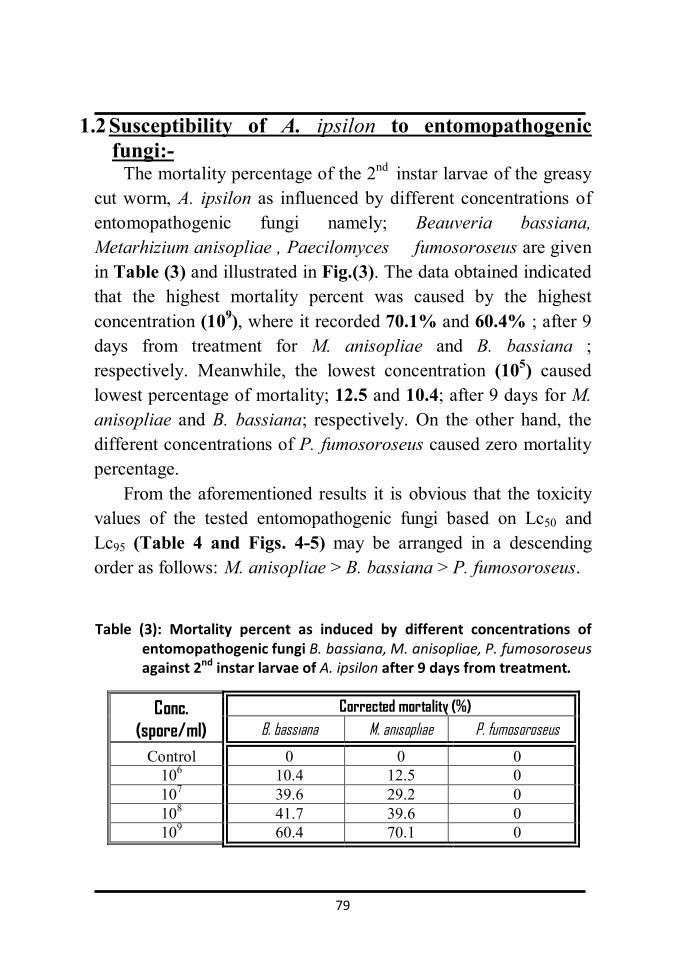
79
1.2 Susceptibility of A. ipsilon to entomopathogenic fungi:-
The mortality percentage of the 2nd instar larvae of the greasy cut worm, A. ipsilon as influenced by different concentrations of entomopathogenic fungi namely; Beauveria bassiana, Metarhizium anisopliae , Paecilomyces fumosoroseus are given in Table (3) and illustrated in Fig.(3). The data obtained indicated that the highest mortality percent was caused by the highest concentration (109), where it recorded 70.1% and 60.4% ; after 9 days from treatment for M. anisopliae and B. bassiana ; respectively. Meanwhile, the lowest concentration (105) caused lowest percentage of mortality; 12.5 and 10.4; after 9 days for M. anisopliae and B. bassiana; respectively. On the other hand, the different concentrations of P. fumosoroseus caused zero mortality percentage.
From the aforementioned results it is obvious that the toxicity values of the tested entomopathogenic fungi based on Lc50 and Lc95 (Table 4 and Figs. 4-5) may be arranged in a descending order as follows: M. anisopliae > B. bassiana > P. fumosoroseus.
Table (3): Mortality percent as induced by different concentrations of entomopathogenic fungi B. bassiana, M. anisopliae, P. fumosoroseus against 2nd instar larvae of A. ipsilon after 9 days from treatment.
Conc. (spore/ml)
Corrected mortality (%) B. bassiana M. anisopliae P. fumosoroseus
Control 0 0 0 106 10.4 12.5 0 107 39.6 29.2 0 108 41.7 39.6 0 109 60.4 70.1 0

80
Fig. (3): Mortality percent as induced by different concentrations of
entomopathogenic fungi B. bassiana, M. anisopliae, P. fumosoroseus against the 2nd instar larvae of A. ipsilon after 9 days from treatment.
Table (4): Toxicity of different entomopathogenic fungi B. bassiana and M.
anisopliae against the 2nd instar larvae of A. ipsilon after 9 days from treatment.
Treatment (Lc50) values in
(spore/ml). (Lc95) values in
(spore/ml). Slope ±
S.E.
B.bassiana 2×10⁸
(6.8×10⁷ – 1.1×109)* 2.1×1012
(7.8×10¹⁰—4.8×1015)* 0.408 ±
0.086
M. anisopliae 1.9×10⁸
(8.03×10⁷ – 6.1×108)* 2.4×1011
(2.5×10¹⁰—2.1×1013)* 0.528 ±
0.091
*numbers between brackets indicate the minimum and maximum concentration.
0
10
20
30
40
50
60
70
80
control 10 6 107 108 109
mo
rt
ali
ty
(%
)
Conc. (spore/ ml)
B. bassiana
M. anisopliae
P. fumosoroseus

81
Mo
rt
ali
ty
%
Dose (spore/ ml)
Fig. (4 ):Concentration-Mortality probit lines of entomopathogenic
fungi B. bassiana against the 2ndinstar larvae of A. ipsilon.

82
Fig. (5): Concentration-Mortality probit lines of entomopathogenic fungi M. anisopliae against the 2ndinstar larvae of A. ipsilon
Mo
rt
ali
ty
%
Dose (spore/ ml)

83
1.3 The latent effect of gamma irradiation on some biological aspects of F1 progeny resulted from irradiated male pupae.
Data given in Table (5) showed the effect of doses of gamma irradiation (LD50 and LD95 Gy) on the biological activity of F1 progeny resulted from irradiated full- grown male pupae.
As shown from the results in Table (5), the fecundity of non - irradiated females crossed with irradiated males resulted from irradiated male pupae was decreased by increasing the irradiation dose. Also, the statistical analysis revealed a significant decrease (p<0.05) in the mean number of eggs laid by non – irradiated females crossed with irradiated males, where it was 83±6.8and 40±6.5 eggs/♀ at LD50(72 Gy) and LD95(332 Gy), respectively vs 360± 19 eggs/♀for the control. The decrease in hatchability % was dose dependent. Moreover, the sterility % which induced by gamma irradiation was found to be positively correlated with the dose, where it recorded 86.6±1.1and 99.3±0.1 at the doses, LD50 and LD95 Gy, respectively (Table 5) and Fig.(6).
The percentage of larval and pupal mortality (Table 5 and Fig. 7) increased significantly (p<0.05) with the increase of doses as compared with the control.
The larval mortality percent was 23.1±2.8% and 100±00% at the doses of LD50 and LD95Gy; respectively compared to 1.7±0.3% for control. Meanwhile, the pupal mortality percent was 24.7±1.2% at the doses LD50 Gy, compared to 1.2±0.2% for control.
As shown in Table (5) the mean duration of the resulted larvae was significantly different (p<0.05) at LD50 Gy, compared to that of the non-irradiated (control) group. Also, the mean duration of the resulted pupae were significantly different (p<0.05) at LD50 Gy, where duration of the resulted larvae prolonged to 35.2±0.8 days compared to 27.2±1.6 days for the

84
control, also pupal duration prolonged to 14.1±0.4 days compared to 12±0.7 days for the control.
The results in Table (5) and Fig. (8) indicated that gamma
irradiation induced a significant (p<0.05) malformation percent among the resulted pupae and adults. The malformation percent was 14.6±1.8% vs. 0.8±0.3% for pupae and; 26.4±3 vs.2.9±0.4 for adults at LD50 Gy, respectively.
A significant decrease (p<0.05) in the percentage of the resulted adults was induced by the gamma irradiation doses used, where the adult emergence % decreased to 75.1±1.2 % at LD50 Gy compared to 98.8±0.2% for control.
The results also in Table (5) and Fig.(9) indicated that the sex ratio among the progeny of irradiated males seemed to be skewed to male side especially at LD50 Gy.

85
Table (5): Effect of gamma irradiation on some biological aspects of F1 progeny resulted from irradiated males pupae of A. ipsilon.
Dose (Gy)
Mean±S.D.
Fecun. (eggs/♀)
Hatch. (%)
Steril. (%)
Larval mor.(%)
Pupal mor.(%)
Pupal mal. (%)
Lrval dur. (D.)
Pupal dur. (D)
Adult mal. (%)
Adult em. (%)
Sex ratio (%)
♂ ♀
Control 360 ±19bc
95.8 ±2.6 bc 0 ±0bc 1.7
±0.3 bc 1.2
±0.2 b 0.8
±0.3b 27.2
±1.6 b 12
±0.7 b 2.9
±0.4 b 98.8
±0.2 b 48.3
±0.4 b 51.7 ±0.4
b
LD 50 72Gy
83 ±6.8ac
55.5 ±2.5 ac
86.6 ±1.1 ac
23.1 ±2.8 ac
24.7 ±1.2 a
14.6 ±1.8a
35.2 ±0.8 a
14.1 ±0.4 a
26.4 ±3 a
75.1 ±1.2 a
57.4 ±3.9 a
41.9 ±3 a
LD95
332Gy 40
±6.5ab 5.7
±1.1 ab 99.3
±0.1 ab 100
±00 ab - - - - - - - -
a significant different for control at (p < 0.05). b significant different for (LD 50 Gy) at (p < 0.05). c significant different for (LD95 Gy) at (p < 0.05).
No. of pupae irradiated was = 25 pupae (11 days old), ( Fecun.= fecundity) – ( Hatch. = Hatchability) - (Steril.= Sterility) – ( mor.= mortality) ( mal.= malformation ) - (dur.= duration ) – ( D.=Days) -) – (em.= emergency).Values represent the mean of 5 replicates for each group.

Results
86
Figs. (6-9): Effect of gamma irradiation on some biological aspects of F1 progeny resulted from irradiated
males pupae of A. ipsilon.
0
20
40
60
80
100
cotrol LD 50 LD95
perc
enta
ge
Dose Gy
Hatchability Sterility
0
20
40
60
80
100
cotrol LD 50 LD95
perc
enta
ge
Dose Gy
Larval mortality
Pupal mortality
05
1015202530
cotrol LD 50 LD95
perc
enta
ge
Dose Gy
pupal malformation adult malformation
0102030405060
cotrol LD 50 LD95
perc
enta
ge
Dose Gy
♀ ♂sex ratio

87
1.4 The latent effect of investigated entomopathogenic fungi on some biological aspects of A. ipsilon.
The biological activity of the tested entomopathogenic
fungi at Lc50 (108 spore∕ml) against the 2nd larval instar of A. ipsilon has been studied. The biological activity of the tested entomopathogenic fungi namely B. bassiana and M. anisopliae included: larval and pupal mortality, larval and pupal duration, adult emergence and morphogenetic effects. The results obtained in (Table 6) and Figs. (10-13) may be arrenged as follows:
Data given in Table (6) and illustrated inFig. (10) showed that the Lc50(108 spore∕ml) of entomopathogenic fungi; B. bassiana and M. anisopliae caused 53± 2.7 and 48.0± 5.7 % larval mortality; respectively which was significantly different (p<0.05) for all tested entomopathogenic fungi as compared with control. In addition, B. bassiana and M. anisopliae caused 21.3± 1.2 and 19.1± 2.6 % pupal mortality, respectively. vs. 3.2±0.9 % for control.
The mean larval duration was significantly (p<0.05) prolonged to 21.6±1.1 and 20.8±0.8 days by the treatment with B. bassiana and M. anisopliae; respectively; vs. 19.4±0.5 days for control. Also, the tested entomopathogenic fungi prolonged significantly (p<0.05) the pupal duration. The duration prolonged to 13.8±0.8 and 12.6±0.5 days by B. bassiana and M. anisopliae; respectively compared to 11.6±0.5 days for control (Table 6 and Fig. 11).
Results recorded in Table (6) and Fig. (12) showed that the entomopathogenic fungi induced a significant (p<0.05) malformation percentages among the resulted pupae and adults. The pupal malformation percentages recorded 18.7±4.3 and 13.1±3.8 for larvae treated with B. bassiana and M. anisopliae; respectively compared to 3.2±2.9 for control. Meanwhile, the adult malformation percentage recorded 13.6± 1 and 11.4 ±0.6 for larvae treated with B. bassiana and M. anisopliae; respectively compared to 0.0±0.0 for the control.

88
Data in Table (6) and Fig. (13) demonstrated the effect of mating of F1 progeny resulting from treated the 2nd instar larvae with Lc50 (108 spore∕ml) of the tested entomopathogenic fungi on fecundity, egg-hatchability and sterility.
The mean number of eggs/♀ laid by untreated females
crossed with treated males were significantly (p<0.05) less than that laid by untreated (control), where it reached to 249.8±11.8 and 316.8±12.5 eggs for the treatment with the Lc50 (108 spore∕ml) of B. bassiana and M. anisopliae; respectively vs. 488.4± 22.8 eggs for the control. Also, the percentage of egg- hatchability was significantly (p<0.05) reduced to 62.7±4.7and 71.1±2.8 as a result of treatment by B. bassiana and M. anisopliae, respectively compared to 94.5±1.6 for control. Moreover, sterility % of untreated females crossed with treated males was significantly (p<0.05) higher than that of control, where it recorded 66.1±2.3and 51.1±2.3% for B. bassiana and M. anisopliae treatment; respectively vs. 0.0±0.0 for the untreated control.

89
Table (6): Effect of Lc50 (108 spore∕ml) of the entomopathogenic fungi, B. bassiana and M. anisopliae on some biological aspects of A. ipsilon.
Treatments
Mean ± S. D.
Larval dur. (days)
Larval mor. (%)
Pupal dur. (days)
Pupal mor.(%)
Pupal mal. (%)
Adult mal. (%)
% of Adult emergence
T♂♂&U♀♀
Fecun. (eggs/♀)
Hatch (%)
Steril. (%)
Control 19.4 ±0.5bc
5 ±0.5 bc 11.6
±0.5 bc 3.2 ±0.9 bc
3.2 ±2.9 bc
0 ±0 bc
96.9 ±2.9 bc
488.4 ±22.8 bc
94.5 ±1.6 bc
0 ±0 bc
B.bassiana 21.6 ±1.1a
53 ±2.7 a
13.8 ±0.8ac
21.3 ±1.2 a
18.7 ±4.3 ac
13.6 ±1 ac
78.7 ±1.2 a
249.8 ±11.8 ac
62.7 ±4.7 ac
66.1 ±2.3 ac
M. anisopliae 20.8 ±0.8a
48 ±5.7 a
12.6 ±0.5ab
19.1 ±2.6 a
13.1 ±3.8 ab
11.4 ±0.6 ab
80.9 ±3.6 a
316.8 ±12.5 ab
71.1 ±2.8 ab
51.1 ±2.3 ab
a significant different for control at (p < 0.05). b significant different for B bassiana at (p < .05). c significant different for M anisopliae at (p < 0.05).
No. of larvae treated was = 20 larvae (2nd instar). Values represent the mean. of 5 replicates for each group., (dur.= duration ) – ( mor.= mortality)- ( mal.= malformation ) ( Fecun.=Fecundity) – ( Hatch. = Hatchability) - (Steril.= Sterility). T: treated , U ; untreated .

90
Figs. (10- 13): Effect of Lc50 of the entomopathogenic fungi, B. bassiana and M. anisopliae on some biological aspects of A. ipsilon.
0102030405060
control B. bassiana M. anisopliae
perc
enta
ge
LC50 Conc.(108 spore ∕ ml)
larval mortality pupal mortality
0
5
10
15
20
25
control B. bassiana M. anisopliae
days
LC50 Conc.(108 spore ∕ ml)
larval duration pupal duration
0
5
10
15
20
control B. bassiana M. anisopliae
perc
enta
ge
LC50 Conc.(108 spore ∕ ml)
pupal malformation adult malformation
0
20
40
60
80
100
control B. bassiana M. anisopliae
perc
enta
ge
LC50 Conc.(108 spore ∕ ml)
Hatchability Sterility

91
1.5 Combined effect of gamma irradiation and entomopathogenic fungi on some biological aspects of A. ipsilon:-
The biological activity of the tested entomopathogenic fungi; B. bassiana and M. anisopliae at (Lc50) against the 2nd larval instar of A.ipsilon resulted from irradiated males crossed with non- irradiated females has been studied. The biological activity of these entomopathogenic fungi included: larval and pupal mortality, larval and pupal duration, adult emergency and morphogenetic effects. The results obtained (Table 7) and Figs.(14-17) may be demonstrated as follows:
Data given in Table (7) and Fig. (14) showed that Lc50 with the two entomopathogenic fungi ; B. bassiana and M. anisopliae caused 69± 6.5, and 59± 4.2 % larval mortality; respectively which was significantly different (p<0.05) as compared to control. In addition, B. bassiana and M. anisopliae caused 55.4± 5.5and 43.6±3.7 % pupal mortality; respectively, vs. 3.2±0.9% for control.
The mean larval duration was significantly (p<0.05) prolonged to 24.8±0.8 and 23.6±1.1 days by the treatment of B. bassiana and M. anisopliae ; respectively vs 19.4±0.5 days for control. Also, B. bassiana and M. anisopliae prolonged significantly (p<0.05) the pupal duration. The duration was prolonged to 15.2±0.8 and 14.2±0.8 days by B. bassiana and M. anisopliae, respectively compared to 11.6±0.5 days (Table 7 and Fig. 15).
The results recorded in Table (7) and Fig. (16) showed that the used entomopathogenic fungi induced a significant (p<0.05) malformation percentage among the resulted pupae and adults. The pupal malformation percentages were 38.7±3.5 and 29±4.2 for larvae treated with B. bassiana and M. anisopliae; respectively compared to 3.2±0.9 for control. Meanwhile, the adult malformation percentages recorded 43.3±2.1 and 22±1.7 for larvae treated with B. bassiana and M. anisopliae, respectively compared to 0±0 for control.

92
Statistical analysis showed that the tested; B. bassiana and M. anisopliae significantly (p<0.05) reduced the percentage of adult emergence as compared with that of untreated control group (Table 7). The adult emergence percent reduced to 44.6±5% and 56.4±3.2 % by B. bassiana and M. anisopliae; respectively vs. 96.9±2.8% for control.
Data in Table (7) and Fig. (17) demonstrated the effect of different mating of F1 progeny resulting from irradiated ♂♂crossed with non-irradiated ♀♀ in combination with the Lc50 (108 spore∕ml) of the tested entomopathogenic fungi against the 2nd instar larvae on fecundity, egg-hatchability and sterility.
The mean number of eggs/♀ laid by treated males crossed with untreated females were 38.2±2.2, and 67.8±1.8 eggs for the treatment with the Lc50 of B. bassiana and M. anisopliae, respectively which significantly (p<0.05) less than of control 448± 12 eggs. The percentage of egg-hatchability for this group was also significantly (p<0.05) less than of the control, where it recorded 0±0 and 8±0.7 for B. bassiana and M. anisopliae treatment, respectively vs. 94.5±1.6 for control. However, sterility % was recorded 100±0 and 98.8±0.2 % for B. bassiana and M. anisopliae treatment; respectively compared to 0.0±0.0% for the control.

93
Table (7): Effect of gamma irradiation (LD50) in combination with the (Lc50) of entomopathogenic
fungi; B.bassiana and M. anisopliae on some biological aspects of A. ipsilon resulted from irradiated males
Treatments
Mean ± S. D.
Larval dur
.(days)
larval mor. (%)
Pupal dur.
(days)
Pupal mor. (%)
Pupal mal. (%)
Adult mal. (%)
% of Adult emergence
T♂♂&U♀♀
Fecun. (eggs/)
Hatch (%)
Steril. (%)
Control 19.4 ±0.5bc
5 ±0.5 bc
11.6 ±0.5 bc
3.2 ±0.9 bc
3.2 ±0.9 bc
0 ±0 bc
96.9 ±2.8 bc
448 ±12 bc
94.5 ±1.6 bc
0 ±0 bc
B.bassiana 24.8 ±0.8a
69 ±6.5ac
15.2 ±0.8 a
55.4 ±5.1 ac
38.7 ±3.5 ac
43.3 ±2.1 ac
44.6 ±5 ac
38.2 ±2.2 ac
0 ±0 ac
100 ±0 ac
M. anisopliae
23.6 ±1.1a
59 ±4.2ab
14.2 ±0.8 a
43.6 ±3.7 ab
29 ±4.2 ab
22 ±1.7 ab
56.4 ±3.2 ab
67.8 ±1.8 ab
8 ±0.7 ab
98.8 ±0.2
ab
a significant different for control at (p < 0.05). b significant different for B bassiana at (p < 0.05). c significant different for M anisopliae at (p < 0.05).
No. of larvae treated was = 20 larvae (2nd instar). Values represent the mean. of 5 replicates for each group., (dur.= duration ) – ( mor.= mortality)- ( mal.= malformation ) ( Fecun.= Fecundity) – ( Hatch. = Hatchability) - (Steril.= Sterility). T: treated , U ; untreated .

94
Figs. (14-17): The effect of gamma irradiation (LD50) in combination with the (Lc50) of entomopathogenic fungi; B.bassiana and M. anisopliae on some biological aspects of A. ipsilon resulted from
irradiated males.
0
20
40
60
80
control B. bassiana M. anisopliae
perc
enta
ge
LC50 Conc.(108 spore ∕ ml)
larval mortality pupal mortality
05
10152025
control B. bassiana M. anisopliae
days
LC50 Conc.( 108spore ∕ ml)
larval duration pupal duration
0
20
40
60
control B. bassiana M. anisopliae
perc
enta
ge
LC50 Conc.(108 spore ∕ ml)
pupal malformation adult malformation
0
50
100
control B. bassiana M. anisopliae
perc
enta
ge
LC50 Conc.(108 spore ∕ ml)
Hatchability Sterility
14 15
16 17

95
2. Pathological effects
2.1 Haematological effects:
2.1.1 Normal haemocytes in A. ipsilon larvae: Haemocytes were distinguished based on morphological
characteristic and staining affinity. In the 3rd instar larvae there was no morphological differences between the different types (Fig. 18, A&B) .Meanwhile, in the 4th and 6th larval instars eight types have been distinguished (Fig. 19, A-P):
Prohaemocytes (A&B in 4th & 6th larval instar, respectively):
are small, rounded to slightly ovoid cells, with undifferentiated cytoplasm. The single, round nucleus with dense, homogenous chromatin is usually enlarged in size leading to formation of macronucleus, which in turn sparing the cellular cytoplasm.
Plasmatocytes (C&D in 4th & 6th larval instar, respectively):
are characterized by variety of cytoplasmic texture ratio which is less than that of prohaemocytes. The general outline is most commonly ovoid or round in shape. The nucleus stains reddish purple, rounded with punctuate or granular chromatin and usually central in position.
Granulated cells (E&F in 4th & 6th larval instar, respectively ):
are larger and thicker than plasmatocytes slightly round, characterized by granulocyte nucleus and cytoplasm dark in color and contained numerous granular inclusions.
Spindle cells (G&H in 4th & 6th larval instar, respectively):
spindle shaped cells distinguished by the presence of very large, distinct, usually spindle nucleus, mostly appearing as a solid purple structure.

96
Spherule cells (I&J in 4th & 6th larval instar, respectively): are rounded with undistinguished nucleus. After Giemsa stain, they appeared in dark pink color surrounded with clear zone.
Adipohaemocytes (K&L in 4th & 6th larval instar,
respectively): are rounded containing variable amounts of refringent fat droplets and several other non lipid inclusions. They characterized by small and approximately centric nucleus.
Oenocytoids (M&N in 4th & 6th larval instar, respectively): are
slightly round characterized by round, large and approximately centric nucleus with homogenous chromatin. The cytoplasm contained numerous granular inclusions and filled with intricated canaliculi.
Cystocytes (O&P in 4th & 6th larval instar, respectively): are
extremely fragile cells with a single, small, irregular nucleus in envelope containing distinct; round; acidophilic inclusions.

97
Fig. (18, A&B) Normal haemocytes in the 3rd instar larvae A. ipsilon (x= 1000).
A
B

98
Fig. (19, A-P): Normal haemocytes of 4th and 6th larval instar in A. ipsilon, nucleus (n) (x= 1000)

99
2.1.2 Histopathological changes due to the treatment with Lc50 of tested entomopathogenic fungi.
Histopathological changes in haemocytes of 4th and 6th larval
instar of A. ipsilon due to the treatment of the 2nd instar larvae with the Lc50 of entomopathogenic fungi tested B. bassiana and M. anisopliae have been studied as follows:
2.1.2.1: In Fig. (20, A-C): showed that the vegetative cell termed blastospores of used entomopathogenic fungi were grown in the body cavity (haemocoel) and invaded the prohaemocytes cells. A: Blastospores attacking prohaemocytes of 4th larval instar
treated with M. anisopliae. B: Plenty growth of blastospores in haemocoel of 4th larval instar
with B.bassiana. C: Blastospores attacking prohaemocytes of 4th larval instar
treated with B.bassiana. 2.1.2.2:In prohaemocytes Fig. ( 21, A-I): A&B: Normal prohaemocytes of 4th & 6th larval instar, appears in oval shape, small, slightly round with large central nucleus. C: Prohaemocytes of 6th larval instar treated with M. anisopliae, has irregular shape; increase in cytoplasm, the nucleus was decreased and lost the central position. E: Prohaemocytes of 6th larval instar treated with B. bassiana, has irregular shape with appearance of vacuoles in cytoplasm.

100
Fig. (20, A-C): Blastospores growth in haemocoel of treated A.
ipsilon. Blastospores(bs) (x=1ooo)
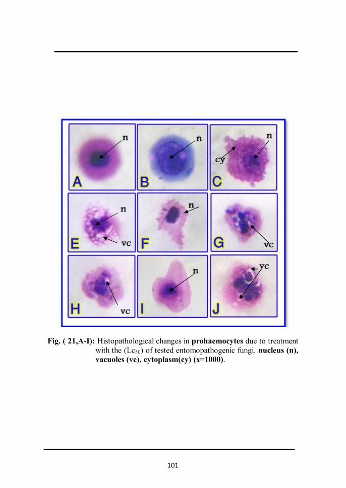
101
Fig. ( 21,A-I): Histopathological changes in prohaemocytes due to treatment with the (Lc50) of tested entomopathogenic fungi. nucleus (n), vacuoles (vc), cytoplasm(cy) (x=1000).

102
F: Prohaemocytes of 4th larval instar treated with B. bassiana, elongated and has irregular shape.
G&H: Prohaemocytes of 4th larval instar treated with M. anisopliae, has irregular shape with appearance of vacuoles in cytoplasm, destruction and appearance of vacuoles in the nucleus.
I: Prohaemocytes of 6th larval instar treated with B. bassiana has, irregular shape, increase in cytoplasm and the nucleus was decreased in size.
J: Prohaemocytes of 4th larval instar treated with B. bassiana.The nucleus increase in size with destruction and appearance of vacuoles, cytoplasm irregular in shape.
2.1.2.3:In plasmatocytes Fig. ( 22, A-I):
A&B: Normal plasmatocytes of 4th & 6th larval instar, appears in round shape, large, with small central nucleus.
C: Plasmatocytes of 6th larval instar treated with M. anisopliae, has irregular shape; increase in cytoplasm, the nucleus was lost the central position.
D: Plasmatocytes of 4th larval instar treated with B. bassiana, has irregular shape, elongated, the nucleus was lost the central position.
E&F: Plasmatocytes of 6th & 4th larval instar treated with M. anisopliae, was increased in size; the nucleus was decreased and lost the central position.

103
Fig. (22, A-I): Histopathological changes in plasmatocytes due to treatment
with the (Lc50) of tested entomopathogenic fungi. nucleus (n), cytoplasm (cy) (x=1000).

104
G: Plasmatocytes of 4th larval instar treated with B. bassiana, has clear destruction in the cell memberane of the cytoplasm, and the nucleus lost the central position.
H: Plasmatocytes of 6th larval instar treated with B. bassiana, was elongated; destruction in cytoplasm and the nucleus was lost the central position.
I: Plasmatocytes of 4th larval instar treated with M. anisopliae, has irregular shape and increase in size.
2.1.2.4:In granulated cells Fig. ( 23, A-I):
A&B: Normal granulated cells of 4th & 6th larval instar, larger and thicker than plasmatocytes slightly round, granulocyte nucleus and cytoplasm dark in color and contained numerous granular inclusions.
C: Granulated cells of 4th larval instar treated with M. anisopliae, have irregular shape; decrease in cytoplasm and has more protrusions; the nucleus was increased in size.
D: Granulated cells of 6th larval instar treated with B. bassiana, has irregular shape; increase in size, cytoplasm has more vacuoles.
E: Granulated cells of 6th larval instar treated with M. anisopliae, has clear destruction in all cell content.
F: Granulated cells of 4th larval instar treated with B. bassiana, has destruction in the cell membrane of the cytoplasm, decrease in cytoplasm and has more protrusions; the nucleus was increased in size.
G: Granulated cells of 6th larval instar treated with M. anisopliae, has irregular shape; increase in size, cytoplasm has more vacuoles.

105
Fig. (23, A-I): Histopathological changes in granulated cells due to
treatment with the (Lc50) of tested entomopathogenic fungi. nucleus (n), vacuoles(vc), cytoplasm(cy) (x=1000).

106
H: Granulated cells of 6th larval instar treated with M. anisopliae, nucleus lost the central position
I: Granulated cells of 6th larval instar treated with B. bassiana, lost the granulocyte shape.
2.1.2.5:In spindle cells Fig. ( 24, A-I):
A&B: Normal spindle cells of 4th & 6th larval instar has spindle shaped cells with large nucleus.
C: Spindle cells of 4th larval instar treated with M. anisopliae, decreased in size and the nucleus lost the central position.
D: Spindle cells of 6th larval instar treated with B. bassiana, has irregular shape approximately with large nucleus.
E: Spindle cells of 4th larval instar treated with M. anisopliae, decreased in size and the nucleus lost the central position and the cell lost the spindle shape approximately.
F: Spindle cells of 4th larval instar treated with B. bassiana. The cell and nucleus increased in size.
G: Spindle cells of 6th larval instar treated with M. anisopliae, has irregular shape; destruction in the cell memberane and the nucleus lost the central position.
H: Spindle cells of 4th larval instar treated with B. bassiana, elongated, decreased in the cytoplasm and nucleus lost the central position.
I: Spindle cells of 6th larval instar treated with B. bassiana, decreased in size and the nucleus lost the central position.

107
Fig. (24, A-I ): Histopathological changes in spindle cells due to treatment with the (Lc50) of tested entomopathogenic fungi nucleus(n), cytoplasm (cy) (x=1000).

108
2.1.2.6:In spherule cells Fig. ( 25, A-H):
A&B: Normal spherule cells of 4th & 6th larval instar. The cell rounded with undistinguished nucleus. After Giemsa stain, they appeared in dark pink color surrounded with clear zone.
C: Spherule cells of 4th larval instar treated with M. anisopliae, has irregular shape and decreased in size.
D: Spherule cells of 4th larval instar treated with B. bassiana, has irregular shape and destruction in the cell memberane and cytoplasm.
E: Spherule cells of 6th larval instar treated with M. anisopliae, decreased in size and destruction in the cell memberane and cytoplasm.
F: Spherule cells of 6th larval instar treated with B. bassiana, has destruction in the cytoplasm and nucleus.
G: Spherule cells of 4th larval instar treated with M. anisopliae,has destruction in the cell memberane and cytoplasm.
H: Spherule cells of 6th larval instar treated with B. bassiana, decreased in size and the nucleus lost the central position.
2.1.2.7:In adipohaemocytes Fig. ( 26, A-F):
A&B: Normal adipohaemocytes of 4th & 6th larval instar. The cell round with with round, small and centric nucleus with variable amounts of refringent fat droplets.
C:Adipohaemocytes of 6th larval instar treated with B. bassiana , has irregular shape and destruction in the cell memberane and nucleus.

109
Fig. (25 , A-H): Histopathological changes in spherule cells due to treatment with the (Lc50) of tested entomopathogenic fungi. nucleus (n), cytoplasm(cy), destructive cytoplasm(d.cy) (x=1000).

110
Fig. (26, A-F): Histopathological changes in adipohaemocytes due to treatment with the (Lc50) of tested entomopathogenic fungi nucleus (n), fat body (f.b) (x=1000).
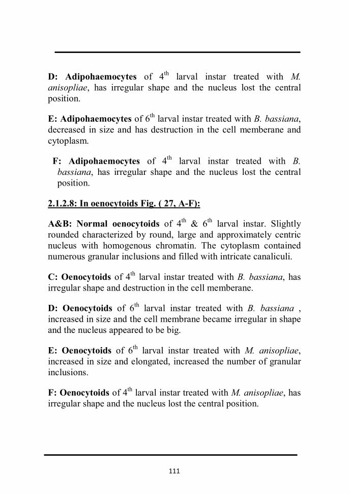
111
D: Adipohaemocytes of 4th larval instar treated with M. anisopliae, has irregular shape and the nucleus lost the central position.
E: Adipohaemocytes of 6th larval instar treated with B. bassiana, decreased in size and has destruction in the cell memberane and cytoplasm.
F: Adipohaemocytes of 4th larval instar treated with B. bassiana, has irregular shape and the nucleus lost the central position.
2.1.2.8: In oenocytoids Fig. ( 27, A-F):
A&B: Normal oenocytoids of 4th & 6th larval instar. Slightly rounded characterized by round, large and approximately centric nucleus with homogenous chromatin. The cytoplasm contained numerous granular inclusions and filled with intricate canaliculi.
C: Oenocytoids of 4th larval instar treated with B. bassiana, has irregular shape and destruction in the cell memberane.
D: Oenocytoids of 6th larval instar treated with B. bassiana , increased in size and the cell membrane became irregular in shape and the nucleus appeared to be big.
E: Oenocytoids of 6th larval instar treated with M. anisopliae, increased in size and elongated, increased the number of granular inclusions.
F: Oenocytoids of 4th larval instar treated with M. anisopliae, has irregular shape and the nucleus lost the central position.
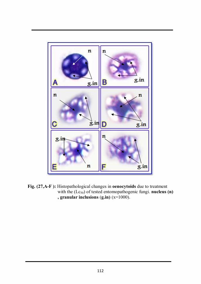
112
Fig. (27,A-F ): Histopathological changes in oenocytoids due to treatment with the (Lc50) of tested entomopathogenic fungi. nucleus (n) , granular inclusions (g.in) (x=1000).

113
2.1.2.9 In cystocytes Fig. ( 28, A-E):
A&B: Normal cystocytes of 4th & 6th larval instar. Fragile cells with a single, small, irregular nucleus in envelope containing distinct, round, acidophilic inclusions.
C: Cystocytes of 6th larval instar treated with M. anisopliae. has irregular shape and destruction in the cell memberane .
D:Cystocytes of 6th larval instar treated with M. anisopliae, decreased in size and the cell membrane became irregular in shape.
E: Cystocytes of 6th larval instar treated with M. anisopliae, has irregular shape and the nucleus lost the central position.
2.1.2.10 : Mitotic division in prohaemocytes Fig ( 29, A-E):
A: Metaphase: Chromosomes are arranged along the cell equator, where each chromosome is connected with one spindle fibers at its centromere.
B: Anaphase: The centromere of each chromosome splits into two halves, so the chromatids separate from each other, two identical groups of chromatids are formed, each group migrate towards one of the cell's pole.
C&D&E : Telophase: A series of adverse changes occur leading to formation of a complete set of chromosomes that have the same number of mother cells chromosomes. Nuclear threads are formed, then a nuclear network leading to the formation of two new separate cells

114
Fig. ( 28, A-E): Histopathological changes in cystocytes due to treatment with the (Lc50) of tested entomopathogenic fungi nucleus
(n) (x=1000).

115
Fig. (29, A-E): Steps of mitotic division in prohaemocytes in A. ipsilon larvae (x=1000).

116
2.1.3 Determination of total haemocytes count in treated A. ipsilon larvae :
Table (8) and Fig. (30) demonstrated the pathological changes in total haemocytes count of 3rd , 4th and 6th larval instar resulted from th 2nd instar larvae treated with the Lc50 of tested entomopathogenic fungi, B. bassiana and M. anisopliae .
The results showed a significant decreased (p<0.05) of total
haemocytes count in the larvae fed on castor leaves treated with Lc50 of the entomopathogenic fungi tested as compared with the larvae fed on untreated castor leaves (control). The percentage of reduction as induced by B. bassiana was 56.4, 80.6 and 69.0 in 3rd, 4th & 6th instar larvae of A. ipsilon, respectively. Meanwhile, the reduction percentage caused by M. anisopliae was 40.7, 70.5 and 53.0 in 3rd, 4th & 6th instar larvae, respectively.
From the aforementioned results, it is seemed that the
percentages of reduction in the total haemocytes count was more pronounced in the 4th instar larvae of A. ipsilon resulted from 2nd instar larvae treated with Lc50 of entomopathogenic fungi tested .

117
Table (8): Total haemocytic count/mm3 in 3rd, 4th and 6th instars resulted from the 2nd instar larvae A. ipsilon treated with the Lc50 of entomopathogenic fungi; B. bassiana and M. anisopliae.
treatments
Mean ± S. D./ Total haemocytic count/mm3
3rd instar reduction
% 4th instar reduction
% 6th instar reduction
%
Control 56.81×10² ±39bc --- 77.55×10²
±46bc --- 95.85×10² ±27 bc ---
B. bassiana 24.75×10² ±29ac 56.4 15.01×10²
±41 ac 80.6 29.65×10² ±31 ac 69
M. anisopliae
33.71×10² ±20ab 40.7 22.86×10²
±24 ab 70.5 45.05×10² ±24 ab 53
a significant different for control at (p < 0.05): b significant different for B. bassiana at (p < 0.05). c significant different for M. anisopliae at (p < 0.05):Values represent the mean. of 5 replicates for each group.

118
Fig. (30): Reduction of total haemocytic blood count/mm3 in 3rd, 4thand 6th
instars resulted from 2nd instar larvae A. ipsilon treated with the Lc50 of entomopathogenic fungi B. bassiana and M. anisopliae.
0
10
20
30
40
50
60
70
80
90
3rd istar 4th instar 6th instar
Redu
ctio
n %
in T
otal
hae
moc
ytic
co
unt/
mm
3
control
B. bassiana
M. anisopliae

119
2.1.4 Determination of differential haemocytes count (DHCs):
Table (9) and Figs. (31&32) illustrated the pathological
changes in differential haemocytes count (DHCs) of 4th and 6th larval instars resulted from 2nd instar larvae of A.ipsilon treated with the Lc50 of tested entomopathogenic fungi; B. bassiana and M. anisopliae.The changes may be arranged as follows;
i. Prohaemocytes in the 4th instar larvae, the percentage of prohaemocytes was increased by B. bassiana and M. anisopliae (84.4 and 78.6, respectively) compared to 55.3 in control. Meanwhile, in 6th larval instar, the percentage of increase prohaemocytes by B. bassiana and M. anisopliae was 82.1 and 81.1, respectively; compared to 48 in control.
ii. Plasmatocytes in the 4th instar larvae, the percentage of
plasmatocytes was decreased by B. bassiana and M. anisopliae (10.2 and 12.5, respectively) compared to 21.1 in control. Meanwhile, in 6th larval instar, the percentage of decrease plasmatocytes by B. bassiana and M. anisopliae was 10.6 and 13.2, respectively; compared to 19.8 in control.
iii. Granulocytes in the 4th instar larvae, the percentage of
granulocytes was decreased by B. bassiana and M. anisopliae (1.1 and 2, respectively) compared to 6.2 in control. Meanwhile, in 6th larval instar, the percentage of decrease granulocytes by B. bassiana and M. anisopliae was 0.7 and 0.5, respectively; compared to 8.1 in control.
iv. Spindle cell in the 4th instar larvae, the percentage of
spindle cell was decreased by B. bassiana and M. anisopliae (0.8 and 0.9, respectively) compared to 5.7 in control. Meanwhile, in 6th larval instar, the percentage of decrease spindle cell by B. bassiana and M. anisopliae was 0.7 and 1.1, respectively; compared to 6.3 in control.

120
v. Spherule cells in the 4th instar larvae, the percentage of
spherule cells was decreased as induced by B. bassiana and M. anisopliae (1.7 and 2.3, respectively) compared to 3.6 in control. Meanwhile, in 6th larval instar, the percentage of decrease spherule cells by B. bassiana and M. anisopliae was 0.3 and 0.4 respectively; compared to 4.5 in control.
vi. Adipohaemocytes in the 4th instar larvae, the percentage of
adipohaemocytes was decreased by B. bassiana and M. anisopliae (0.7 and 1.9, respectively) compared to 2.7 in control. Meanwhile, in 6th larval instar, the percentage of decrease adipohaemocytes cell by B. bassiana was 1.6, while completely disappeared by M. anisopliae compared to 4 in control.
vii. Oenocytoids in the 4th instar larvae, the percentage of
oenocytoids was decreased by B. bassiana and M. anisopliae (1.1 and 1.8, respectively) compared to 5 in control. Meanwhile, in 6th larval instar, the percentage of decrease oenocytoids by B. bassiana and M. anisopliae was 4 and 3.3, respectively compared to 6.6 in control.
viii. Cystocytes in the 4th instar larvae, cystocytes was
completely disappeared by B. bassiana and M. anisopliae compared to 0.1 in control. Meanwhile, in 6th larval instar, the cystocytes completely disappeared by B. bassiana, while the percentage of decreased by M. anisopliae was 0.4 compared to 2.7 in control.

121
Table (9): Effect of entomopathogenic fungi; B. bassiana and M. anisopliae (Lc50) on differential
haemocytes count (DHCs) in A. ipsilon larvae:
Treatments Cell Type (%)
4th instar 6th instar
control B.
bassiana
M.
anisoplia
e control
B.
bassiana
M.
anisoplia
e
Prohaemocytes 55.3 84.4 78.6 48 82.1 81.1
Plasmatocytes 21.1 10.2 12.5 19.8 10.6 13.2
Granulocytes 6.5 1.1 2 8.1 0.7 0.5
Spindle cells 5.7 0.8 0.9 6.3 0.7 1.1
Spherule cells 3.6 1.7 2.3 4.5 0.3 0.4
Adipohaemocytes 2.7 0.7 1.9 4 1.6 -
Oenocytoid 5 1.1 1.8 6.6 4 3.3
Cystocytes 0.1 - - 2.7 - 0.4
Values represent the mean of percentages of 5 replicates for each group.

122
Fig. (31): Effect ofentomopathogenic fungi; B. bassiana and M. anisopliae (Lc50 ) on differential
haemocytes count (DHCs) in 4th instar larvae of A. ipsilon.
controlProhaemocytes
Plasmatocytes
Granulocytes
Spindle cells
Spherule cells
Adipohaemocytes
Oenocytoid
Cystocytes
B. bassianaProhaemocytes
Plasmatocytes
Granulocytes
Spindle cells
Spherule cells
Adipohaemocytes
Oenocytoid
Cystocytes
M. anisopliaeProhaemocytes
Plasmatocytes
Granulocytes
Spindle cells
Spherule cells
Adipohaemocytes
Oenocytoid
Cystocytes

123
Fig. (32): Effect of entomopathogenic fungi; B. bassiana and M. anisopliae (Lc50) on differential haemocytes count (DHCs) in 6th instar larvae of A. ipsilon.
controlProhaemocytes
Plasmatocytes
Granulocytes
Spindle cells
Spherule cells
Adipohaemocytes
Oenocytoid
Cystocytes
B. bassianaProhaemocytes
Plasmatocytes
Granulocytes
Spindle cells
Spherule cells
Adipohaemocytes
Oenocytoid
Cystocytes
M. anisopliaeProhaemocytes
Plasmatocytes
Granulocytes
Spindle cells
Spherule cells
Adipohaemocytes
Oenocytoid
Cystocytes
124
2.2 Histopathological effects 2.2.1 Scanning electron microscopy of entomopathogenic fungi
, B. bassiana and M. anisopliae on A. ipsilon larvae. Fig. (33) illustrated the growth of B. bassiana on the dead A.
ipsilon larvae treated with the Lc50 (2×10⁸spore∕ml) after 7,10, 13 and 16 days. The growth was indicated by white spores. The mycelium started to grow after 7 days from death of infected larvae, then the insect cadaver was covered by mycelium after 10 days.The formation and discharge of spores were found after 13 and 16 days; respectively.
Scanning electron microscopy (SEM) of A. ipsilon larvae
treated with the Lc50 of the fungus, B. bassiana clearly revealed adhesion and penetration structures in the infected larvae (Figs. 34&35). Signs of fungus growth on the larvae were found 9 days after death (Fig. 35). Moreover, signs of hyphal penetration of the insect cuticle as well as proliferation of the cuticle were also apparent (Fig. 36). The spores filled the body cavity of the infected larvae after 7 days from death (Fig. 37).
However, white clusters of B. bassiana mycelim were detected to grow on pupae resulted from the larvae treated with the Lc50 of the fungus after 14 days from death (Figs. 38&39)

125
Fig. (33): Over growth of Lc50 entomopathogenic fungi, B. bassiana on A. ipsilon larvae. (x = 40)

126
Fig.(34) Scanning electron microscopy of 4th instar larvae of A. ipsilon
infected with the fungus, B. bassiana. hyphae(hy) (× = 70).
Fig.(35) Scanning electron micrograph of B. bassiana mycilium growing on
the integument of infected A. ipsilon larva after 9 days from death. conidia (co) and hyphae (hy) (× = 750).

127
Fig.(36) Scanning electron micrograph showing the cuticle proliferation(cu-
pro) as a result of hyphal pentration of B. bassiana into the integument of infected A. ipsilon larva after 11 days from death. (× = 400).
Fig.(37) Scanning electron micrograph into the body cavity of infected A.
ipsilon larva after 7 days from death of infected larvae by B. bassiana.( arrows indicate spores (s) and hyphae (hy)( × = 450).

128
Fig. (38) Scanning electron microscopy (SEM) A. ipsilon pupa resulted
from the 2nd instar larvae treated with the Lc50 of B. bassiana . ( arrows indicate mycelium clumps (my.c) ( × = 18).
Fig.(39) Scanning electron micrograph of B. bassiana mycelium growing
on A. ipsilon pupa resulted from the 2nd instar larvae treated with the Lc50 of fungus.( arrows indicate mycelium clumps(my.c) ) ( × = 220).

129
On the other hand, the growth of M. anisopliae on the cadaver of a larval- pupal intermediate resulted from the 2nd instar larvae treated with the Lc50 (1.9×10⁸spore∕ml) of the fungus is shown in Fig. (40), the fungus growth was characterized by green spores which were formed on cadaver insect after 15 days from larval death.
The fine morphology of hyphae and conidial clumps of M. anisopliae as decleared by SEM is shown in Figs. (41&42). As shown from Fig.(42), the fungal hyphae become dense network together and cause the green spores on the insect cuticle after 15 days from death. The first signs of fungus growth on the larvae were found 7 days from death. Meanwhile, the spores and hyphae of the fungus were seen in the body cavity of larvae after 8 days from dead (43).
Fig. (40): Larval– pupal intermediate of A. ipsilon parasitized by the fungus
M. anisopliae. (x = 40)

130
Fig.(41): SEM of the fungus, M. anisopliae (general view) showing dense
network mycelium growing on the cuticle of A. ipsilon larvae after 15 days from death resulted from the 2nd instar larvae treated with the Lc50 of the fungus (×= 700).
Fig.(42): Close – up of M. anisopliae mycelium showing the dense cover. arrows indicate conidia (co) and hyphae (hy) (×= 800).

131
Fig.(43) Scanning electron micrograph of the interior of A. ipsilon larva
after 7days from death as a result of treatment with the Lc50 of M. anisopliae (arrows indicate spores (s) and hyphae (hy) (×= 350).

132
2.2.2 Light microscopy of histopathology as induced by the entomopathogenic fungi, B. bassiana and M. anisopliae in A. ipsilon larvae:
Histological investigations using light microscopy have
been carried out to observe the mode of action of the two fungi tested, and to study how they were able to colonize and infected the larvae of A. ipsilon. Moreover, the histological damages induced by the two fungi were also observed.
Examination of transversal section of infected larvae
showed a fairly dense of B. bassiana on the insect cuticle. Figs. (44, A&B) showed spores germinating on body wall of the larvae after 5 days from treatment. At 10 days post-treatment, penetration of the integument was observed (Fig.45).This was followed by extensive invasion of the body at 12 days after treatment causing great damage to the insect cuticle a presented by proliferation and canals formation into the cuticle (Figs. 45 & 46). At 15 days the body cavity was filled with spores and hyphal bodies as shown in Figs. (47 &48). After that spores and hyphal bodies were seen in the gut causing damage in the epithelial layer (Fig.48).
On the other hand, histological investigation of the larvae
treated with the Lc50 of M. anisopliae showed the dense coating of mycilium on the larval cuticle after 6 days from treatment (Fig.49). At 9 days post treatment, penetration of the integument was observed causing damage to the insect cuticle presented by proliferation and canals formation into the cuticle (Fig.50). As spores and hyphal bodies filled the body cavity of infected larvae (Fig.51), great destruction in muscles (Fig.52), fat bodies (Fig.53) and epithelial cells of the gut (Fig.54) were observed.

133
Figs. (44, A&B): Transversal section of A. ipsilon 3 rd larval instar resulted from 2nd instar larvae treated with the Lc50 of B. bassiana showing the dense cover of mycelium on the larval integument and spore germination. Spores(s), conidia (co), melanization(mel), mycelium germination (my.ger) (× = 1000 ).

134
Fig.(45 ) :Transversal section of A. ipsilon 4th larval instar resulted from
2nd instar larvae treated with the Lc50 of B. bassiana showing the hyphal penetration (hy.pen) of the larval integument (× =1000 ).
Fig.(46 ) :Transversal section of A. ipsilon 4th larval instar resulted from
2nd instar larvae treated with the Lc50 of B. bassiana showing the proliferation and canal formation into the cuticle as indicated by arrows canal formation(canl f) (× = 1000 ).

135
Fig. (47): Transversal section of A. ipsilon 6th larval instar resulted from
2nd instar larvae treated with the Lc50 of B. bassiana showing the spores inside the body cavity. spors(s), conidia(co) (× = 1000 ).
Fig.( 48) :Transversal section of A. ipsilon 6th larval instar resulted from
2nd instar larvae treated with the Lc50 of B. bassiana showing spores (s) and hyphal bodies (hy) inside the larval midgut (× =1000 ).

136
Fig.(49 ) :Transversal section of A. ipsilon 3rd larval instar resulted from
2nd instar larvae treated with the Lc50 of M. anispliae arrows indicated the dense coating of mycelium(my) on the larval cuticle. (× = 1000).
Fig.( 50 ) :Transversal section of A. ipsilon 3rd larval instar resulted from
2nd instar larvae treated with the Lc50 of M. anispliae showing the proliferation and canals formation(can.for) into the larval cuticle and body (× = 100 ).

137
Fig.(51 ) :Transversal section of A. ipsilon 4th larval instar resulted from
2nd instar larvae treated with the Lc50 of M. anispliae showing spores (s) and hyphal bodies (hy) inside the larval body cavity (× =1000).
Fig.(52 ) :Transversal section of A. ipsilon 4th larval instar resulted from
2nd instar larvae treated with the Lc50 of M. anispliae showing destructed muscles (des.mus )as indicated by arrows spores (s)and conidia(co )(× = 100 ).

138
Figs.(53 ) :Transversal section of A. ipsilon 4th larval instar resulted from 2nd instar larvae treated with the Lc50 of M. anispliae showing destructed fat bodies (des.fb) (× =1000 ) .
Fig.(55 ) :Transversal section of A. ipsilon 6th larval instar resulted from
the 2nd instar larvae treated with the Lc50 of M. anispliae showing destructed of midgut (des. midgut) as indicated by arrows (× = 40 ).

139
3. Biochemical studies 3.1. The changes in the total content of proteins,
lipids and carbohydrates.
Table (10 ) and Fig.(56) demonstrated the changes in the total content of proteins of the 3rd, 4th& 6th instar larvae of A. ipsilon resulted from the 2nd instar larvae treated with Lc50 of entomopathogenic fungi tested (B. bassiana and M. anisopliae). The results showed a significant decrease (p<0.05) in the total content of proteins in the larvae fed on castor leaves treated with Lc50 of the entomopathogenic fungi tested as compared with the larvae fed on untreated castor leaves (control). The percentage of reduction as induced by B. bassiana was 38.6, 46.8 and 25.3 in 3rd, 4th & 6th instar larvae, respectively. Meanwhile, the reduction percentage caused by M. anisopliae was 21.6, 40.2 and 17.9 in 3rd, 4th& 6th instar larvae; respectively.
Data given in Table (11) and illustrated in Fig.(58)
indicated the changes in the total content of carbohydrates in insect of the 3rd, 4th& 6th instar larvae of A. ipsilon resulted from the 2nd instar larvae treated with Lc50 of entomopathogenic fungi tested. The results showed a significant decrease (p<0.05) in the total content of carbohydrates in the larvae fed on castor leaves treated with Lc50 of the entomopathogenic fungi tested as compared with the larvae fed on untreated castor leaves (control). The percentage of reduction as induced by B.bassiana was 55.1, 85.6 and 42.6 in 3rd, 4th& 6th instar larvae, respectively. Meanwhile, the reduction percentage caused by M. anisopliae was 22.7, 68.2 and 24.0 in 3rd, 4th& 6th instar larvae, respectively.

140
Table (10): Changes in the total content of proteins (gm %), of 3rd, 4th& 6th instar larvae of A. ipsilon resulted from 2nd instar larvae treated with (Lc50) of entomopathogenic fungi,
B. bassiana and M. anisopliae.
Mean ± S.D (gm%)
Treatments reduction % 6th instar reduction % 4th instar reduction
% 3rd instar
____ 1.2239±0.062 bc ____ 1.142±0.04 bc ____ 0.6813±0.025bc Control
25.3 0.9146±0.028 ac 48.8 0.5845±0.013 ac 38.6 0.4183±0.053ac B. bassiana
17.9 1.005±0.034 ab 40.2 0.6825±0.015 ab 21.6 0.5343±0.035ab M. anisopliae
a significant different for control at (p< 0.05). c significant different for sunflower oil at (p<0.05). b significant different for canola oil at (p<0.05) values represent the mean. of 3 replicates for each sample.

141
Fig. (56): Changes in the total content of proteins (gm %), of 3rd, 4th& 6th instar larvae of A. ipsilon resulted from 2nd instar larvae treated with (Lc50) of entomopathogenic fungi, B. bassiana and M. anisopliae.
0
0.2
0.4
0.6
0.8
1
1.2
1.4
3rd istar 4th instar 6th instar
tota
l pro
tein
con
c.(g
m%
) control
B. bassiana
M. anisopliae
bc
ac
ab
bc
acab
bc
acab

142
Fig. ( 57 ): Standard curve of total carbohydrate content.
0
0.1
0.2
0.3
0.4
0.5
0.6
0.7
0.8
0.9
0 0.01 0.02 0.03 0.04 0.05 0.06 0.07 0.08 0.09 0.1 0.11 0.12 0.13 0.14 0.15
O.D
(nm
)
Concentration (gm)
Standard Curve of Total Carbohydrate

143
Table (11): Changes in the total content of carbohydrate (gm/100ml), of 3rd, 4th& 6th instar larvae of A. ipsilon
resulted from 2nd instar larvae treated with (Lc50) of entomopathogenic fungi B. bassiana and M. anisopliae.
Mean ± S.D. (gm/100ml)
Treatments reduction
% 6th instar reduction % 4th instar reduction % 3rd instar
____ 0.527±0.076 bc ____ 0.1763±0.02 bc ____ 0.078±0.006 bc Control
42.6 0.3027±0.061a 85.6 0.025±0.003 ab 55.1 0.035±0.0027 ac B. bassiana
24 0.4001±0.053a 68.2 0.056±0.007 ac 22.7
0.0603±0.0032
ab
M. anisopliae
a significant different for control at (p< 0.05). c significant different for sunflower oil at (p<0.05). b significant different for canola oil at (p<0.05) Values represent the mean. of 3 replicates for each sample

144
Fig. (58): Changes in the total content of carbohydrate (gm/100ml), of 3rd, 4th& 6th instar larvae of A. ipsilon
resulted from 2nd instar larvae treated with (Lc50) of entomopathogenic fungi B. bassiana and M. anisopliae.
0
0.1
0.2
0.3
0.4
0.5
0.6
0.7
3rd istar 4th instar 6th instar
tota
l car
bohy
drat
e co
nc.
(gm
/100
ml)
control
B. bassiana
M. anisopliae
ab
acab
bc
abac
bc
a
a

145
From the aforementioned results it is seemed that the percentages of reduction in the total content of proteins and carbohydrates was more pronounced in the 4thinstar larvae of A. ipsilon resulted from the 2nd instar larvae treated with Lc50 of entomopathogenic fungi tested (B.bassiana and M. anisopliae).
Data given in Table (12) and illustrated in Fig. (59 ) indicated the changes in the total content of lipids in the 3rd, 4th & 6th instar larvae of A. ipsilon resulted from the 2nd instar larvae treated with Lc50 of entomopathogenic fungi tested. The results showed a significant change (p<0.05) in the total content of lipids in the larvae fed on castor leaves treated with Lc50 of the entomopathogenic fungi tested as compared with the larvae fed on untreated castor leaves (control). The percentage of increase was 68.0 and 59.3 in 3rd and 4th, instar larvae, respectively. On the other hand, the percentage of reduction was 64.3 in 6th instar larvae as induced by B. bassiana. Meanwhile, the increase percentage caused by M. anisopliae was 73.7 and 66.6 in 3rd & 4th instar larvae, respectively, while the percentage of reduction was 51.9 in 6th instar larvae.

146
Table (12): Changes in the total content of Lipids (mg/dl), of 3rd, 4th& 6th instar larvae of A. ipsilon resulted from
2nd instar larvae treated with (Lc50 ) of entomopathogenic fungi B. bassiana and M. anisopliae.
Mean ± S.D. (mg/dl)
Treatments Reduction % 6th instar Increase
% 4th instar Increase % 3rd instar
____ 235±13 bc ____ 125±7.4 bc ____ 116±5.6 bc Control
64.3 84±7 ab 59.3 307±14.7 ab 68 363±7.6 ab B.bassiana
51.9 113±10 ac 66.6 374±5.6 ac 73.7 441±8.1 ac M. anisopliae
a significant different for control at (p< 0.05). c significant different for sunflower oil at (p<0.05). b significant different for canola oil at (p<0.05) Values represent the mean. of 3replicates for each sample
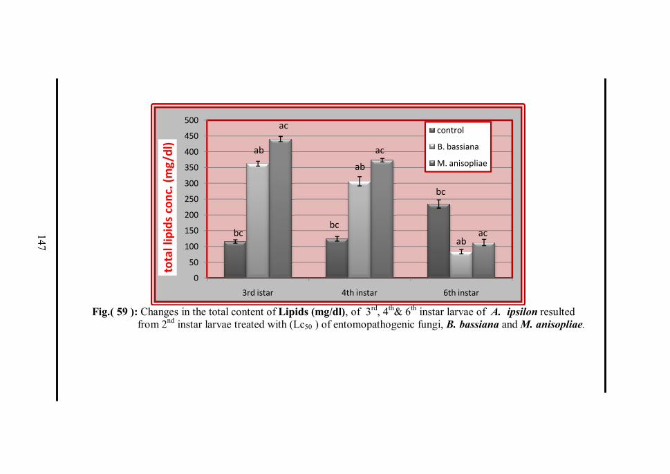
147
Fig.( 59 ): Changes in the total content of Lipids (mg/dl), of 3rd, 4th& 6th instar larvae of A. ipsilon resulted
from 2nd instar larvae treated with (Lc50 ) of entomopathogenic fungi, B. bassiana and M. anisopliae.
0
50
100
150
200
250
300
350
400
450
500
3rd istar 4th instar 6th instar
tota
l lip
ids
conc
. (m
g/dl
)
control
B. bassiana
M. anisopliae
bc
ab
ac
bc
ab
ac
bc
abac
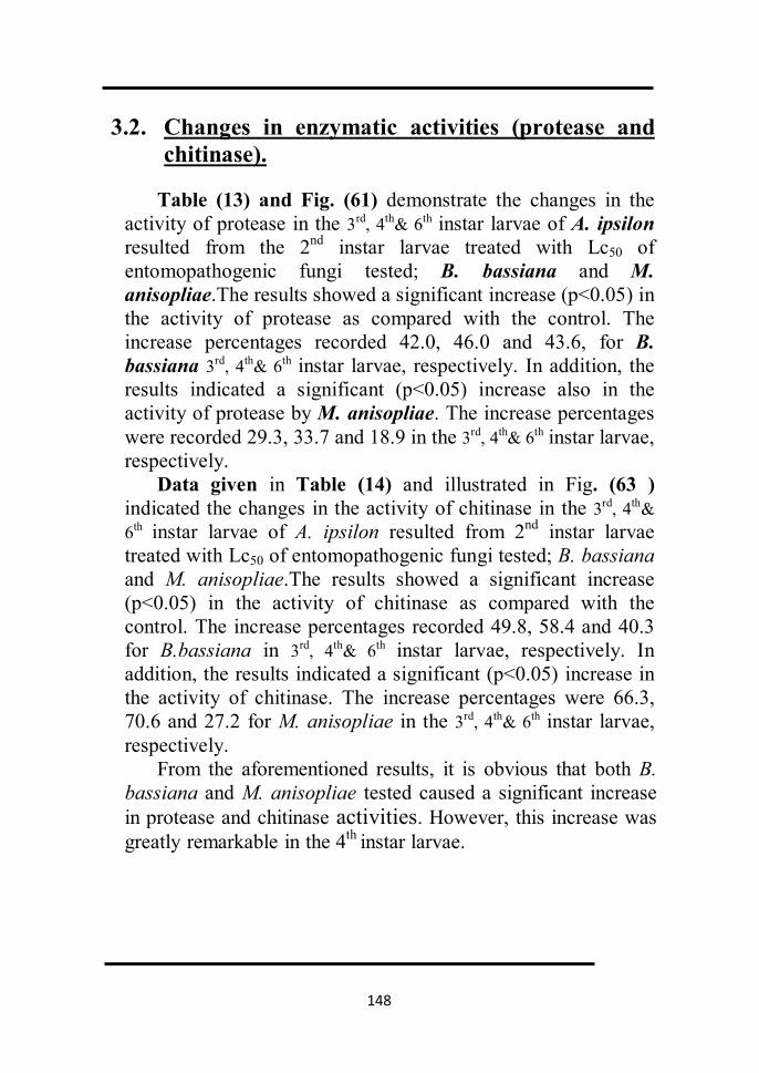
148
3.2. Changes in enzymatic activities (protease and chitinase).
Table (13) and Fig. (61) demonstrate the changes in the
activity of protease in the 3rd, 4th& 6th instar larvae of A. ipsilon resulted from the 2nd instar larvae treated with Lc50 of entomopathogenic fungi tested; B. bassiana and M. anisopliae.The results showed a significant increase (p<0.05) in the activity of protease as compared with the control. The increase percentages recorded 42.0, 46.0 and 43.6, for B. bassiana 3rd, 4th& 6th instar larvae, respectively. In addition, the results indicated a significant (p<0.05) increase also in the activity of protease by M. anisopliae. The increase percentages were recorded 29.3, 33.7 and 18.9 in the 3rd, 4th& 6th instar larvae, respectively.
Data given in Table (14) and illustrated in Fig. (63 ) indicated the changes in the activity of chitinase in the 3rd, 4th& 6th instar larvae of A. ipsilon resulted from 2nd instar larvae treated with Lc50 of entomopathogenic fungi tested; B. bassiana and M. anisopliae.The results showed a significant increase (p<0.05) in the activity of chitinase as compared with the control. The increase percentages recorded 49.8, 58.4 and 40.3 for B.bassiana in 3rd, 4th& 6th instar larvae, respectively. In addition, the results indicated a significant (p<0.05) increase in the activity of chitinase. The increase percentages were 66.3, 70.6 and 27.2 for M. anisopliae in the 3rd, 4th& 6th instar larvae, respectively.
From the aforementioned results, it is obvious that both B. bassiana and M. anisopliae tested caused a significant increase in protease and chitinase activities. However, this increase was greatly remarkable in the 4th instar larvae.

149
Fig. (60): Standard curve of protease.
0
0.1
0.2
0.3
0.4
0.5
0.6
0 5 10 15 20 25 30 35 40 45 50 55 60 65 70 75 80 85
O.D
(nm
)
Concentration (µg/L)
Standard Curve of Protease

150
Table( 13 ): Changes in the activity % of protease of 3rd, 4th& 6th instar larvae of A. ipsilon resulted from 2nd instar larvae treated with (Lc50 ) of entomopathogenic fungi,
B. bassiana and M. anisopliae.
Mean ± S.D. (µg tyrosine /min. /ml tissue )
Treatments Increase % 6th instar Increase
% 4th instar Increase
% 3rd instar
____ 15.4±1.1 bc ____ 17.7±2.7 bc ____ 14.5±0.65
bc Control
43.6 27.3±0.4 ab 46 32.8±1 ab 42 25±2.8 ab B. bassiana
18.9 19±0.6 ac 33.7 26.7±0.8 ac 29.3 20.5±1.3 ac M. anisopliae
a significant different for control at (p< 0.05). c significant different for sunflower oil at (p<0.05). b significant different for canola oil at (p<0.05) Values represent the mean. of 3replicates for each sample.

151
Fig.(61): Changes in the activity% of protease (µg tyrosine /min. /ml tissue ) of 3rd, 4th& 6th instar larvae of A. ipsilon resultedfrom 2nd instar larvae treated with (Lc50 ) of entomopathogenic fu ngi
B. bassiana and M. anisopliae.
0
5
10
15
20
25
30
35
40
3rd istar 4th instar 6th instar
prot
ease
act
ivit
y (µ
g ty
rosi
ne
/min
. /m
l tis
sue
)
control
B. bassiana
M. anisopliae
bc
ab
acbc
ab
ac
bc
ab
ac

152
Fig. (62): Standard curve of chitinase.
-0.2-0.1
00.10.20.30.40.50.60.70.80.9
11.1
0 0.5 1 1.5 2 2.5 3 3.5 4 4.5 5 5.5 6 6.5 7 7.5 8 8.5
O.D
(nm
)
Concentration (mg)
Standard Curve of Chitinase

153
Table(14): Changes in the activity % of chitinase of 3rd, 4th& 6th instar larvae of A. ipsilon
resulted from 2nd instar larvae treated with (Lc50 ) of entomopathogenic fungi, B. bassiana and M. anisopliae.
Mean ± S.D. (µg N-acetylglucoseamine /h. /ml tissue )
Treatments Increase %
6th instar Increase %
4th instar Increase %
3rd instar
____ 702±20 bc ____ 455±10 bc ____ 130±3 bc Control
40.3 1177±16 ab 58.4 1095±13 ab 49.8 259±6 ab B. bassiana
27.2 964±5 ac 70.6 1550±7.5 ac 66.3 386±12 ac M. anisopliae
a significant different for control at (p< 0.05). c significant different for sunflower oil at (p<0.05). b significant different for canola oil at (p<0.05) Values represent the mean. of 3replicates for each sample

154
Fig.(63 ): Changes in the activity % of chitinase (µg N-acetylglucoseamine /h. /ml tissue ) of 3rd, 4th& 6th instar
larvae of A. ipsilon resulted from 2nd instar larvae treated with (Lc50 ) of entomopathogenic fungi, B. bassiana and M. anisopliae.
0
200
400
600
800
1000
1200
1400
1600
1800
3rd istar 4th instar 6th instar
Chit
inas
e ac
tivi
ty (µ
g N
-ac
etyl
gluc
osea
min
e /h
. /m
l tis
sue
)
control
B. bassiana
M. anisopliae
bc
abac
bc
ab
ac
bc
ab
ac

155
The results of the present study may have a great
importance as today the environmental safety of an insecticide is considered to be of paramount importance. The extensive and continuous use of synthetic pesticides in control of agricultural pests have created many problems such as, indiscriminate use let to the three sad R’s (resistance, resurgence and residues), elimination of natural enemies of pests, upsetting the ecological balance and environmental degradation. Such problems have become a cause of search for safely pesticides including microbial agents as fungi, bacteria and viruses (Rashed, 1993). The first microorganisms found to cause diseases in insects were fungi because their specificity for pest control and their high persistence in the environment. Some insect pathogenic fungi have restricted host ranges while other fungal species have a wide host range for example, Metarhizium anisopliae and Beauveria bassiana .In addition gamma irradiation can reduce the viability of successive generations of certain insects, where substerilizing doses of gamma radiation can increase mortalities in the resulting progeny and minimize the hazards of irradiated insect (Sutrisno et al., 1991 and Garcia and Gonzalez, 1993).
The entomopathogenic fungi and gamma-irradiation
doses used in the present study are not toxic to vertebrates as well as they are cheap. The present study showed high bioactivity of the entomopathogenic fungi

156
and gamma-irradiation doses against the greasy cutworm, A. ipsilon, such results may offer an opportunity for developing alternatives to rather expensive and environmentally hazardous organic insecticides .
The obtained results may be discussed in the following aspects:
1. Biological activity
1.2. Susceptibility of Agrotis ipsilon to gamma irradiation.
The mortality percentage of non- hatching eggs resulted from irradiated full grown male pupae was found to be dose dependent i.e. increased as the dose of the gamma irradiation used increased. The data obtained indicated that the highest mortality percent was caused by the highest dose (240 Gy), Meanwhile, the lowest dose (40Gy) caused lowest percentage of mortality. In agreement with the present results, Sallarn and Ibrahim (1993) irradiated 7-days-old male pupae of Spodoptera littoralis (Boisd) with doses of 50, 75, 100, 125, 150 and 200 Gy of gamma radiation. Increasing the dosage applied to P1 males significantly reduced the fecundity of females. Egg hatchability laid by P1 females was significantly reduced when treated with 125, 150 and also 200 Gy as compared with the untreated control. Also Sallam, et al., (2000) reported that female reproductive potential of Earias insulana (Boisd) decreased at the three doses of irradiation (100, 150 and 200 Gy) compared to control.

157
Sawires (2005) found that, reduction in fecundity and egg fertility in Ephestia kuehniella (Zell) was dose dependent. 1.5 Susceptibility of A. ipsilon to etomopathogenic
fungi:- The larval mortality percent was found to be
concentration dependent i.e. increased as the concentration of the entomopathogenic fungi used increased, the toxicity values of the tested entomopathogenic fungi (B. bassiana, M. anisopliae, P. fumosoroseus) based on Lc50 and Lc95 was arranged in descending order as follows : M. anisopliae > B. bassiana > P. fumosoroseus. In agreement with the present results, El-Sinary and Rizk (2007) tested two concentrations of the entomopathogenic fungus, B. bassiana; 104 and 108 spores ∕ ml against the 4th larval instar of the greater wax moth; G. melonella. There was a positive correlation between the fungal concentration and its lethality for the treated larvae.
Amer et al. (2008) examined five entomophathogenic fungi Beauveria bassiona, B. brongniartti, Paecilomyces farmosus, Metarhizium anisopliae and M. flavoviridae as biological control agents for Spodoptera littoralis. The fungi were grown on specific 3 media. Five concentrations were applied for each fungus, 2nd and 4th instar larvae of S. littoralis were exposed for 48 h. to treated castor bean leaves by using dipping technique with conidial suspensions. The results showed that, the mortality percentage increased with increasing concentrations.
158
1.6 The latent effect of gamma irradiation on some biological aspects of F1 progeny resulted from irradiated male pupae.
As shown from the obtained results, gamma irradiation affected the different biological parameters studied (fecundity, egg-hatchability, larval and pupal mortality, larval and pupal duration, adult emergence, malformation and sex ratio) for A.ipsilon. The magnitude of this effect was found to differ according to the irradiation dose. The two doses of gamma-irradiation LD50 (72 Gy) and LD95 (332 Gy)) used in the treatment of full-grown males pupae significantly reduced fecundity egg-hatchability and the percentage of adults emerged. In addition, gamma – irradiation doses used induced a significant malformation percent among the resulted pupae and adults.
The first report was provided by Proverbs (1962 a, b) who found that the progeny of irradiated males of the codling moth Carpocapsa pomonella were sterile. This phenomenon therefore has been accepted in several species of lepidoptera. Although, the progeny of irradiated males were more sterile than their male parents, this result did not occur when females were irradiated. The use of partially sterile males was vantageous because of their highly competitiveness with native moths.
The present study showed that the dose of radiation was inversely proportional to the fecundity and fertility when male of A.ipsilon were exposed as pupae to all tested doses of gamma radiation. Similar results were obtained by Al-Taweel et al., (1990) on Ephestia cautella: Seth and
159
Reynolds (1993) irradiated male pupae of the tobacco hornworm, Manduca sexta (Linnaeus), with 100 Gy. They found decline in the number of eggs laid per mated female, and also a reduction in hatching rate for the mating of both male and female F1 insects. The effect on males was more severe than on females. El-Dessouki et al., (1996) irradiating full-grown male pupae of the cotton leafworm, Spodoptera littoralis (Boisd.) with low doses of gamma radiation (25, 50, 75 and 100 Gy) found that fecundity of normal females mated with F1 or F2 males was significantly reduced for all applied doses. Bloem et al., (2003) recorded inherited effects in F1 progeny of Cryptophlebia leucotreta resulting from irradiation of parental males with selected doses of radiation. A decrease in F1 fecundity and fertility, while an increase F1 mortality during development and a significant shift in the F1 sex ratio in favour of males were recorded when increasing radiation doses to the P1 males.
Also, the different biological parameters studied (larval and pupal mortality, larval and pupal duration, adult emergence, malformation and sex ratio) for A. ipsilon. differed according to the irradiation dose. These results were ingreement with Pransopon et al., (2000) irradiating mature male pupae of cotton bollworm, Helicoverpa armigera, Sallam, et al., (2000) studing the inherited sterility in the spiny bollworm Earias insulana (Boisd) and Sawires (2005) irradiating full-grown male and female pupae of the Mediterranean flour moth, Ephestia kuehniella (Zeller) Also, the present results were in consistent with that of gamma irradiation on different biological parameters in A.ipsilon reported by El-Kady et

160
al. (1983), El-Naggar et al. (1984), Abd El -Hamid (2004) and Gabarty (2008).
1.7 The latent effect of investigated entomopathogenic
fungi on some biological aspects of A. ipsilon.
The biological activity (larval and pupal mortality, larval and pupal duration, adult emergence and morphogenetic effects) of the tested entomopathogenic fungi (B. bassiana and M. anisopliae) at the (Lc50) against the 2nd instar larvae of A. ipsilon has been studied. The results obtained indicated that the (Lc50) of the tested entomopathogenic fungi caused significant larval and pupal mortality as compared with control. In addition, the (Lc50) of entomopathogenic fungi prolonged significantly the larval and pupal duration as compared to the control. Moreover, the (Lc50) of tested entomopathogenic fungi caused malformation percentages among the resulted pupae and adults. Similar findings, were also obtained by many authors using different entomopathogenic fungi, Quesada-Moraga et al. (2006&2008) on larvae and adult, Ceratitis capitate , Gesraha (2007) on Schistocerca gregaria ; Kivan (2007) on sun bug, Eurygaster integriceps ; Chouvenc et aI. (2008) on Reticuliterms flavipes, Ansari et al., (2009) on Agriotes lineatus, Changjin Dong et al. (2009) on Odontotermes formosanus. Moreover, Amora et al.,(2010) evaluating the effects of the fungus , M. anisopliae var. acridum on Lutzomyia longipalpis found that the concentrations of 1×10⁴ to 1×10⁸ conidia/ml, M. anisopliae var. acridum reduced egg hatching by 40%. The mortality of infected larvae was also significant. The

161
longevity of infected adults was lower than that of negative controls. The effects of fungal infection on the hatching of eggs laid by infected females were also significant. With respect to fungal growth parameters post-infection, only vegetative growth was not significantly higher than that of the fungi before infection.
1.5 Combined effect of gamma irradiation and entomopathogenic fungi on some biological aspects of A. ipsilon:- The effect of gamma irradiation LD50 (72Gy) in
combination with the Lc50 (108spores/ml) of entomopathogenic fungi tested, B. bassiana and M. anisopliae against the 2nd instar larvae of A. ipsilon resulted from irradiated males crossed with non-irradiated females, on some biological aspects was studied. As shown from the results obtained the combined effect of gamma irradiation (LD50) and the (Lc50) of entomopathogenic fungi tested markedly affected the different biological aspects of A. ipsilon. The combined effect increased the larval and pupal mortality; prolonged the larval and pupal duration; reduced the percentage of adult emergence and caused malformation percentages among the resulted pupae and adults. Moreover, the combined effect of gamma irradiation and entomopathogenic fungi against the 2nd instar larvae of A.ipsilon drastically decreased the number of eggs/♀ (fecundity), increased the sterility percentage and decreased the egg-hatchability percent. The present study indicated that the combined effect of gamma irradiation and entomopathogenic fungi induced more remarkable effects as compared to gamma irradiation or entomopathogenic

162
fungi each of them alone. These findings are agree with those obtained by El-Sinary and Rizk (2007) using B. bassiana; 104 and 108 spores ∕ ml combined with gamma irradiation against the 4th larval instar of the greater wax moth; G. melonella, the efficiency of B. bassiana increased especially when the gamma irradiation dose was increased, where no adults were produced with both fungal concentrations and 150 Gy gamma irradiation dose. El-Kholy and Shoman (2007), studied the effectiveness of the entomopathogenic fungus, Trichoderma harzianum against the medfly, Ceratitis capitata (Wied.), their laboratory results showed that T. harzianum can be applied in the field to suppress the population of the Mediterranean fruit fly and considered as entomopathogenic bioagent controlling this pest by irradiation techniques.
2. Pathological effects 2.1 Haematological effects:
Insects posses a complex and efficient system of biological defence against pathogens and parasites. An essential component of the insect cellular response is phagocytosis. Main type of haematocytes involved in this process are plasmatocytes and granulocytes (Lakie,1988; Wago,1991). Phagocytosis is one of insect defence responses important for the studies of the relationship between insects and their pathogens. Fungal metabolites are known to inhibit phagocytosis (Vilcinskas et al. 1997), whereas component of the fungal cell stimulate phagocytosis (Gunnarson, 1988).However, hyphal bodies of various entomopathogenic fungi; are reported not to be ingested by host phagocytic hemocytes due to the lack of

163
surface epitopes required for their recognition ( Pendlond et al., 1993; Hayek and St Leger, 1994).
In this respect, the present study has been demonstrated and described the changes (abnormalities) in haemocytes of 4th and 6th larval instar on A. ipsilon due to the treatment of 2nd instar larvae with the LC50 of tested entomopathogenic fungi, B. bassiana and M. anisopliae (see the results). Also, the present study demonstrated the effect of the aforementioned fungi on the total hemocytes count in the 3rd, 4th and 6th larval instar on A. ipsilon. The results obtained showed a significant decrease of total hemocytes count of the fungal infected larvae. These results may be comparable with these of other authors; for example, Bidochka et al. (1988) showed that the entomopathogenic fungi, M. anisopliae and B. bassiana once reach to the insect haemocoel they are able to survive and replicate within, and ∕or invade phagocytic hemocyte cells circulating in the haemolymph. Vilcinskas et al. (1997) reported that M. anisopliae or B. bassiana - infected G. mellonella larvae; mycosis reduced plasmatocyte phagocytic acavity against either yeast cells or blastospores of both entomopathogenic fungi.
Moreover, Gllespie et al. (2000) showed that topical application of M. anisopliae var. acridum to the desert locust, Shistocerca gregaria resulted in changes in the biochemistry and antimicrobial defenses of the haemolymph. The presence of the fungus appeared to stimulate hemocyte aggregation. Also, the total hemodecyte count increased two days after application, indicating an overall stimulation of immune system, declined again after

164
that as the present results decleared. Chouvence et al. (2009) showed that inculation of the termite, Reticulitermes flavipes worker with conidia of the entomopathogenic fungus, M. anisopliae increased the relative number of hemocytes per termite after fungal exposure. 2.2 Histopathological effects.
Beauveria bassiana and Metarhizium anisopliae are naturally fungi that are found in the soil of most fields. These fungi are entomopathogenic which causing disease to insects, and killing them. Fungal infection begins when conidia (asexual spores, the seeds of a fungus) attach to insect’s cuticle, the spores germinate and penetrate the insect’s skin and enter the host (Figs.34&35) .Once the fungus penetrates the host; it produces toxins that overcome the insect immune system. Thereafter, the hyphae penetrate through the cuticle to the outside and cause white (B. bassiana) or green (M. anisopliae) sporulation on the insect’s body (Figs.33&40).
Light and scanning electron microscopy are convenient tools to observe the mode of action of entomopathogenic fungi and to observe how they are able to colonize and infect the host.
SEM allowed to observe B. bassiana and M.
anisopliae adhesion and penetration structure on A. ipsilon larvae. SEM of A. ipsilon larvae treated with the Lc50 of the fungus, B. bassiana revealed adhesion and penetration structures in the infected larvae. Growth of the fungus on the infected larvae and signs of hyphal penetration of insect cuticle as well as proliferation of the cuticle were also

165
appearing. On the other hand, the fungus, M. anisopliae as decleared by SEM showed a dense network together and cause the green spores on the insect cuticle. Also, SEM allowed to observe the spores and hyphae of the fungus in the body cavity of infected larvae.
These observation agree with the SEM observation of
Asensio et al.(2005) for B .bassiana growing on the red scale insect of plams (Phoenicocococcus marlatti) and they are in consistent with the following suggestions:
Infection of insects by fungus, M. anisopliae also
requires adhesion, penetration into the host (St Leger, 1993) and establishment of the pathogen in the host (Charnley, 1989; Sampson et al., 1988). Pentration through the host cuticle is the mode of entry for most entomopathogenic fungi (Charnley, 1984). During fungal infection, the first step prior to penetration – is the adhesion of fungi to the host cuticle (Boucias and Pendland, 1991). Fragues (1984) suggested adhesion to occur at three successive stages: (1) adsorption of the fungi propagules to the cuticular surface; (2) adhesion of the interface between propagules and epicuticle; (3) fungi germination and development at the insect cuticular surface, until appresoria are developed to start the penetration stage.
Moreover, the killing of the insect host is attributed to the production of secondary metabolites by the parasitic fungi (Roberts, 1981). These findings may explain the death of A. ipsilon larvae infected with B. bassiana or M. anisopliae as shown in the histological examinations of the fungal infected larvae of A. ipsilon.

166
Insect pathogenic fungi, and in particular the Deuteromycetes, M. anisopliae and B. bassiana infect their host by a combination of mechanical pressure and enzyme degradation (Charnley, 1984). Paterson et al. (1994) demonstrated that M. anisopliae produces several extracellular cuticle – degrading proteases and evidence is consistent that one of these, a chymoelastase PR1, is determinant of pathogencity. It is produced in high levels by the fungus during host penetration (St Leger et al., 1987). In agreement with these findings, the present results (the biochemical studies) indicated a remarkable increase of protease activity in fungal infected larvae of A. ipsilon.
The histological investigation of the fungal – infected
larvae of A. ipsilon have explained the virulence and efficiency of fungal treatments as they screened the damages which happened to cuticle, epithelial layer, midgut, fat bodies, muscles and showed the spread of fungal pathogen and its germination, penetration, growth and proliferation within the body of the infected larvae of A. ipsilon which were in harmony with those obtained by Ramleo et al.(1996) by B. bassiana and M. anisopliae against Metia plano (Lepidoptera: Psychidae), El- Sinary and Risk(2007) against the greatest wax moth, Galleria mellonella; and Sewify and Hashem (1999) by M. anisopliae against G. mellonella larvae.
In conclution, the results obtained in these experiments
establish the pathogencity of entomopathogenic fungi, B. bassiana and M. anisopliae on larvae of A.ipsilon as biological control agent. Bilogical control with pathogenic

167
fungi is promising alternative to chemical control against the lepidopterous pestes.
4. Biochemical studies
3.1 The changes in the total content of proteins, lipids and carbohydrates.
The major bulk components of the insect cuticle are
protein, lipids and chitin, the outermost epicuticular surface layer comprise a complex mixture of non-polar lipids. Epicuticular lipids play a relevant role in preventing lethal desiccation, affecting insecticide and chemicals penetration. (Juarez, 1994).
The present study investigated the changes in total content of proteins, lipids and carbohydrates of F1 progeny in the 3rd, 4th & 6th instar larvae of A.ipsilon resulted from 2nd instar larvae treated with Lc50 of entomopathogenic fungi tested, B. bassiana and M. anisopliae. The results indicated that entomopathogenic fungi caused a significant decrease in the total content of proteins and carbohydrates. However, this decrease was greatly remarkable in 4th instar larvae of A. ipsilon. Meanwhile, the tested entomopathogenic fungi caused a significant increase in the total content of lipids in the 3rd and 4th, instar larvae, but there was a significant reduction induced by the tested entomopathogenic fungi in 6th larval instar .These results agree with Pedrini et al, (2007) who recorded, the biochemical interaction between fungal pathogens and their insect host epicuticle and the relationship between fungal ability to catabolize very long chain hydrocarbons and virulence parameters, by examining fungal hydrocarbon

168
degrading ability. As a contact insecticide, entomopathogenic fungi invade their host through the cuticle, covered by an outermost lipid layer mainly composed of highly stable, very long chain structures.
4.2 Changes in enzymatic activities (protease and
chitinase).
In addition to the main metabolites, the present study dealt with the changes in activities of the two cuticle-degrading enzymes (protease and chitinase) of F1 progeny in the 3rd, 4th & 6th instar larvae of A.ipsilon resulted from 2nd instar larvae treated with Lc50 of entomopathogenic fungi tested, B. bassiana and M. anisopliae. The results indicated that both fungi tested caused a significant increase in protease and chitinase activities. However, this increase was greatly remarkable in the 4th instar larvae.
Pathogenic fungi infect their hosts by multiple developmental processes requiring the activities of fungal
molecules that are either secreted and directly interact physically with the host, remain on the fungal cell surface or inside the fungus. A few fungal molecules have already been proven critical to the outcome of infection. In general, these molecules can be classified as penetration effectors, toxins, enzymes that reduce host defense, transporters that protect the fungus from host defenses or components of signal transduction pathways that are required for fungal sensing of the host environment (Pedrini et al., 2007). Khachatourians, (1996) reported that, extracellular enzymes whether of mycelia or spore origin are hallmark of fungal infection process. The successful infection relies

169
upon the efficient release of exoenzymes, which have potential to degrade insect cuticle.
The present results may be comparable with those of
St. Leger et al. (1986b) who reported that, several pathogenic isolates of Metarhizium anisopliae, Beauveria bassiana, and Verticillium lecanii when grown in buffered liquid cultures containing comminuted locust cuticle as composite carbon source (good growth occurred on most monomeric and polymeric cuticular constituents), produced a variety of extracellular enzymes corresponding to the major components of insect cuticle, e.g., endoproteases, aminopeptidase, carboxypeptidase A, lipase, esterase, chitinase, and N-acetylglucosaminidase. Considerable variations occurred in levels of production between species and even within a species, but endoproteases were exceptional as production of them was high with all the isolates.Cuticle-degrading enzymes were produced rapidly and sequentially in culture.The first activities to appear (<24 hr) were those of the proteolytic complex; chitinases were always produced substantially later.

170
Bioassays with entomopathogenic fungi can be used to
determine and quantify host – pathogen relationships and to assess the effect of biotic and a biotic factors in insect parasitism. Insect pathogenic fungi and in particular the Deuteromycete, Beauveria bassiana, Metarhizium anisopliae and Paecilomyces fumosoroseus, have great potential for use in pest control.
Gamma irradiation is known to reduce the viability of
successive generations of certain insects. Effect of substerilizing doses of gamma radiation help also in increasing mortalities in the resulting progeny and minimize the hazards of irradiated insects.
The present study was carried out to investigate the
efficacy of some entomopathogenic fungi or ∕ and substerilizing doses of gamma irradiation against Agrotis ipsilon larvae. Moreover, the combined effects of gamma irradiation (substerilizing doses) and treatment of A. ipsilon larvae with each of the two pathogenic fungi; B. bassiana and M. anisopliae as integrated control agents were investigated. The results obtained are summarized as follows:-
4. Biological activity
1.1 Susceptibility of A. ipsilon to gamma irradiation :-
The data obtained indicated that the highest mortality percent was caused by the highest dose (240 Gy), where it

171
recorded 93.3%, Meanwhile, the lowest dose (40Gy) caused the lowest percentage of mortality (28.2). 4.2 Susceptibility of A. ipsilon to entomopathogenic
fungi:- The data obtained indicated that the highest mortality
percent was caused by the highest concentration (109), where it recorded 70.1% and 60.4% ; after 9 days from treatment for M. anisopliae and B. bassiana ; respectively. On the other hand the different concentrations of P. fumosoroseus caused zero mortality percentage . the toxicity values of the tested entomopathogenic fungi based on Lc50 and Lc95 may be arranged in a descending order as follows: M. anisopliae > B. bassiana > P. fumosoroseus.
4.3 The latent effect of gamma irradiation on some biological aspects of F1 progeny resulted from irradiated male pupae.
The substerilizing doses of gamma irradiation (LD50 and LD95 Gy) were tested against full – grown male pupae of A.ipsilon.
The results showed that fecundity of irradiated males crossed with non-irradiated females was decreased by increasing the irradiation dose. The mean number of eggs /♀ was 83±6.8 and 40±6.5 eggs/♀ at LD50 (72 Gy) and LD95 (332 Gy), respectively vs 360±19 eggs/♀for the control.
In addition, the decrease in egg – hatchability % and increase in sterility % induced by gamma irradiation were found to be positively correlated with the dose.

172
The parentage of larval and pupal mortality increased significantly (p<0.05) with the increase of doses used. The larval mortality percent recorded 23.1% and 100 % at the doses of LD50 and LD95Gy; respectively compared to 1.7% for control. Meanwhile, the pupal mortality percent was 24.7±1.2% at the doses LD50 Gy, compared to 1.2% for control.
The mean larval duration significantly prolonged to35.2±0.8 days compared to 27.2±1.6 days for the control, while the pupal duration prolonged to14.1±0.4 days compared to 12±0.7 days for the control.
Gamma irradiation induced a significant (p<0.05) malformation % among the resulted pupae and adults. The malformation percent was14.6% vs. 0.8% for pupae and; 26.4 vs.2.9 for adults at LD50 Gy, respectively.
The percentage of adult emergence reduced to75.1% at LD50 Gy compared to 98.8% for control. The sex ratio among the progeny of irradiated males seemed to be skewed to male side especially at LD50 Gy.
4.4 The latent effect of investigated entomopathogenic
fungi on some biological aspects of A. ipsilon.
Lc50 of entomopathogenic fungi tested (B. bassiana and M. anisopliae) caused 53 and 48.0 % larval mortality, respectively compared to 5 % for the control, meanwhile they caused 21.3 and 19.1 % pupal mortality, respectively. The larval and pupal duration significantly (p<0.05) prolonged by B. bassiana and M. anisopliae as compared with that of untreated control.

173
Entomopathogenic fungi tested (Lc50) induced malformation percentages among the resulted pupae and adults. B. bassiana induced the highest percent.
The (Lc50) of B. bassiana and M. anisopliae fungi significantly (p<0.05) reduced the percentage of adult emergence as compared with that of untreated control group. The adult emergence % reduced to 78.7 and 80.9 by B. bassiana and M. anisopliae entomopathogenic fungi, respectively.
Moreover, the results indicated a significant decrease
(p<0.05) in fecundity and egg – hatchability percent as induced by the (Lc50) of tested entomopathogenic fungi. B. bassiana seemed to be the most effective fungus.
4.5 Combined effect of gamma irradiation and entomopathogenic fungi on some biological aspects of A. ipsilon:-
The effect of gamma irradiation (LD50) in combination with the Lc50 of entomopathogenic fungi tested against the 2nd larval instar of A. ipsilon resulted from irradiated males crossed with non-irradiated females on the biological aspects mentioned before was studied. The results indicated that the combined effect of gamma irradiation (LD50) and the Lc50 of entomopathogenic fungi tested markedly affected the different biological aspects such as:
Lc50 of the two entomopathogenic fungi ; B. bassiana
and M. anisopliae caused 69 and 59 % larval mortality; respectively which was significantly different (p<0.05) as

174
compared to control. In addition, caused 55.4 and 43.6 % pupal mortality; respectively, vs. 3.2±0.9% for control.
The adult emergence percent reduced to 44.6% and 56.4% by B. bassiana and M. anisopliae; respectively vs. 96.9±2.8% for control.
Moreover, the results demonstrated the effect of
different mating of F1 progeny resulting from irradiated ♂♂crossed with non-irradiated ♀♀ in combination with the Lc50 (108 spore∕ml) of the tested entomopathogenic fungi against 2nd instar larvae on fecundity, egg-hatchability and sterility.
The mean number of eggs/♀ laid by treated males
crossed with untreated females were 38.2 and 67.8 eggs for B. bassiana and M. anisopliae, respectively which significantly (p<0.05) less than of control 448 eggs. However, sterility % was recorded 100 and 98.8 % for B. bassiana and M. anisopliae treatment; respectively compared to 0.0% for the control.
5. Pathological effects 5.2 Haematological effects: 5.2.1 Normal haemocytes in A. ipsilon larvae:
Eight types of haemocytes were described in the haemolymph of the 4th and 6th larval instars. They were Prohaemocytes; Plasmatocytes; Spindle cells; Granulated cells; Spherule cells; Oenocytoids; Adipohaemocytes and Cystocytes.

175
5.2.2 Histopathological changes due to the treatment with Lc50 of tested entomopathogenic fungi.
Histopathological changes (abnormalities) in haemocytes of 4th and 6th larval instars of A. ipsilon due to the treatment of 2nd instar larvae with the Lc50 of tested entomopathogenic fungi B. bassiana and M. anisopliae have been described in eight types of haemocytes.
2.1.5 Determination of total haemocytes count in treated A. ipsilon larvae :
Pathological changes in total haemocytes count of 3rd ,
4th and 6th larval instars resulted from the 2nd instar larvae treated with the Lc50 of tested entomopathogenic fungi, B. bassiana and M. anisopliae were studied, the percentages of reduction in the total haemocytes count was more pronounced in the 4th instar larvae of A. ipsilon.
2.1.6 Determination of differential haemocytes count (DHCs):
The pathological changes in differential haemocytes count (DHCs) of 4th and 6th larval instars resulted from 2nd instar larvae of A.ipsilon treated with the Lc50 of tested entomopathogenic fungi; B. bassiana and M. anisopliae were demonstrated. The prohaemocytes were the predominant type and the cystocytes were the rare type. Moreover, the percentage of prohaemocytes was increased, while the other types was decreased, by B. bassiana and M. anisopliae; respectively.

176
2.2 Histopathological effects:
2.2.1 Scanning electron microscopy of histopathology as induced by entomopathogenic fungi , B. bassiana and M. anisopliae in A. ipsilon larvae:
Scanning electron microscopy (SEM) allowed to
observe B. bassiana and M. anisopliae adhesion and penetration structure on A. ipsilon larvae. SEM of A. ipsilon larvae treated with the Lc50 of the fungus, B. bassiana revealed adhesion and penetration structures in the infected larvae. Growth of the fungus on the infected larvae and signs of hyphal penetration of insect cuticle as well as proliferation of the cuticle were also appearing.On the other hand, the fungus, M. anisopliae as decleared by SEM showed a dense network together and cause the green spores on the insect cuticle. Also, SEM allowed to observe the spores and hyphae of the fungus in the body cavity of infected larvae.
2.2.2Light microscopy of histopathology as induced by the
entomopathogenic fungi, B. bassiana and M. anisopliae in A. ipsilon larvae:
The histological investigations of the fungal – infected larvae of A. ipsilon have explained the virulence and efficiency of fungal treatments as they screened the damages which happened to cuticle, epithelial layer, midgut, fat bodies, muscles and showed the spread of fungal pathogen and its germination, penetration, growth and proliferation within the body of the infected larvae of A. ipsilon.

177
6. Biochemical studies
3.1 The changes in the total content of proteins, lipids and carbohydrates:
The results indicated a significant (p<0.05) decrease in the total content of proteins and carbohydrates of the 3rd, 4th& 6th instar larvae of A. ipsilon resulted from 2nd instar larvae treated with Lc50 of entomopathogenic fungi tested (B. bassiana and M. anisopliae). The percentages of reduction in the total content of proteins and carbohydrates were more pronounced in the 4th instar larvae of A. ipsilon. On the other hand, the results showed a significant(p<0.05) increase on the total content of lipids in the 3rd and 4th instar larvae while a decrease in the 6th instar larvae of A. ipsilon treated with Lc50 of tested entomopathogenic fungi was observed.
3.2 Changes in enzymatic activities (protease and chitinase):
The results indicated that entomopathogenic fungi tested (B. bassiana and M. anisopliae) caused a significant (p<0.05) increase in the activity of protease and chitinase in insect tissues of the 3rd, 4th& 6th instar larvae of A. ipsilon; this increase was greatly remarkable in the 4th instar larvae.
178
Abbott, W. S. (1925): A method of computing the
effectiveness of an insecticide. J. Econ. Ent., 18:265-267.
Abd El-Hamid (2004): Effect of gamma irradiation on
certain biological and physiological aspects of black cut worm Agrotis ipsilon (Hufn). M.Sc Thesis. Fac. Agric.Cairo Univ.
Abdel-Rahman, A.M. (1978): The haemocytes of Aedes
caspius. Palls. Bull. Fac. Sci. Cairo University, 51(1): 95 – 100.
Abou El-Ghar, G. E. S.; Khalil, M. E. and Eid, T.M.
(1996): Some biochemical effects of plant extracts in the black cutworm, Agrotis ipsilon (Hufn.) (Lep., Noctuidae). J. Appl. Ent., 120(8) 477-482.
Almeida, J.E.M.; Filho, A.B.; Alves, S.B. and Shitara,
T., (2003): Evaluation of insecticides and entomopathogenic fungi for control of subterranean termites of sugarcane. Arquivos de Instituto Biologico, Sao-paulo, 70(3): 347-353.
Al-Taweel, A .A. Ahmed, M.S.H.; Kadhum, S.S. and
Hameed, A.A. (1990): Effect of gamma radiation on the progeny of irradiated Ephestia cautella (Walker) (Lepidoptera: Pyralidae) males. J. Stored-Prod. Res. 26(4):233-336.
179
Amer, M.M.; T.T. El-sayed; H.K. Bakheit; S.A. Mostafa and Yasmin A. El-sayed (2008): pathogenicity and genetic variability of five entomopathogenic fungi against Spodoptera littoralis. Res. J. Agric. and Biol. Science, 4(5): 354-367.
Amora S. S. A. ; Bevilaquaa C. M. L.; Feij F.M. C. ;
Pereira R. H. M.A.; Alves N. D.; Freire F. A. M.; Kamimura M. T.; Oliveira D. M. and Lima E. L. (2010) :The effects of the fungus Metarhizium anisopliae var. acridum on different stages of Lutzomyia longipalpis (Diptera: Psychodidae) Acta Tropica (113) 214–220.
Angel -Sahagùn, C.A.; A. Lezama-Guierrèz; J.
Molina-ochoa; E. Galindo-velasco; M. Lopez-Edwards ; 0. Rebolledo-Dominguez ; C. Cruz-vazquez; W.P. Reyes-Velazquez ; S.R. Skoda and J.E. Foster (2005): Susceptibility of biological stages of the horn fly, Haematobio irritans to entomopathogenic fungi (Hyhphomycetes). J. of Insect Science, Issn: 1536-.2442.
Ansari M. A. ; Evans M. and Butt T. M. (2009):
Identification of pathogenic strains of entomopathogenic nematodes and fungi for wireworm control. Crop Protection 28, 269–272.
Asensioa L.; Lopez-Llorcaa L.V. and Lo´pez-
Jime´nezb J. A. (2005): Use of light, scanning electron microscopy and bioassays to evaluate parasitism by entomopathogenic fungi of the red scale insect of palms (Phoenicococcus marlatti Ckll., 1899) Micron (36) 169–175.

180
Bidochka M. J.; David C. Clark; Mike W. Lewis and Nemat O. Keyhani (1988): Could insect phagocytic avoidance by entomogenous fungi have evolved via selection against soil amoeboid predators? J. Invert. Pathology Vol. 52(2), 285-293.
Bloem, S.; Crpenter, J.E. and Hofmeyr, J.H. (2003):
Radiation biology and inherited sterility in false codling moth (Lepidoptera: Tortricidae). J. Econ. Entomol. 96(6): 1724-31.
Bohata, A. and Land, Z. (2004): Influence of humidity
on efficacy of entomopathogenic fungi. Case study “Paecilomyces fumosoroseus - Trialeurodes vapoarariorum. Collection of scientific papers Faculty of Agriculture in Ceske Budejovice - Series for crop sciences, 21(2-3): 169-171.
Boucias, D. G. and Pendland , J.C. (1991) : In fungal
cell wall and immune response. Berlin: Springer; p.303–16.
Castillo, M.A.; Moya, P.; Hemandez, E. and Primo-
Yufera, E. (2000): Susceptibility of Ceratitis capitata Wiedman (Diptera: Tephritidae) to entomopathogenic fungi and heir extracts. Biological Control, 19, 274-282.
Changjin Dong; Jiamin Zhang; Hai Huang; Wuguo
Chen and Yuanyang Hu (2009): Pathogenicity of anew China variety of Metarhizium anisopliae (M. anisopliae var. Dcjhyium) to subterranean termite Odontotermes formosanus. Microbio. Rese. 164, 27—35.
181
Charnley, A. K. (1984): Physiological Aspects of Destructive Pathogenesis by Fungi. Cambridge University Press; p.229–70.
Charnley, A. K. (1989):. The Biotechnology of Fungi for
Improving Plant Growth. London: Cambridge University; p.85–125.
Charnley, A. K. (2005): Fungal pathogens of insects:
cuticle degrading enzymes and toxins. University of Bath.
Cherry, A.J.; Abalo, P. and Hell, K., (2005): A
laboratory assessment of the potential of different strains of the entomopathogenic fungi Beauveria bassiana (Balsamo) Vuillemin and Metarhizitm anisopliae (Metschnikoff to control Callosobruchus maculates (Coleoptera: Bruchidae) in stored cowpea. J. of Stored Products Research. 41(3): 295-309.
Chouvenc T.; Nan-Yao Su a and Alain Robert (2009):
Cellular encapsulation in the eastern subterranean termite, Reticulitermes flavipes (Isoptera), against infection by the entomopathogenic fungus Metarhizium anisopliae. J. Inverteb. Pathol.. 101 , 234–241.
Chouvence--Thomas; Nan-Yao Su and Elliott-Monica
L. (2008): Interaction between the subterranean termite Reticulitermes flavipes (lsoptera: Rhinotermitidae) and the entomopathogenic fungi, Metarhizium anisopliae in foraging areas. J. of Econom. Entomol. 101(3): 885-893.

182
Cuthbertson - Andrew, G.S. and Walters- Keith F.A. (2005): Pathogenicity of the entomopathogenic fungus, Lecanicillium muscarium, against the sweet potato whitefly Bemisia tabaci under laboratory and glasshouse conditions. Mycopthologia, 160(4): 315-319.
Cuthbertson, Andrew, G.S.; Walter, Keith, F.A. and
Northing Phil, (2005): The susceptibility of immature stages of Bemisia tabaci to the entomopathogenic fungus Lecanicillium muscarium on tomato and Verbena foliage. Mycopathologio. 159(1): 23-29.
Da Silva J. B.; Cleide M. R. De Albuquerque; Eva
Cristina De Arau´ jo; Christina A. Peixoto, and Hilary Hurd (2000): Immune defense mechanisms of Culex quinquefasciatus (Diptera: Culicidae) against Candida albicans infection. J. of Invert. Pathol. 76, 257–262.
Destefano, R. H. R.; Bechara, I. J.; Messias, C. I. and
Piedrabuena, A. E. (2005): effectiveness of Metarhizium anisopliae against immature stages of Anastrepha fraterculus fruit fly (Diptera: Tephritidae). Brazillian J. of Microbiology. 36(1): 94-99.
Detmar, D.A. and R. J. Vogels (1971): Automated
determination of protease for laundry compounds. J. Am.Chem. Soc. 48: 77-79.
Devi, K. Uma; Mohan, C.H. Murali; Padmavathi, T.
and Ramesh, K. (2003): Susceptibility of fungi of

183
cotton boll worms before and after a natural epizootic of the entomopathogenic fungus Nomuraea rileyi (Hyphomycetes). Biocontrol Science and Technology. 13(3): 367-371.
Duarte-Aguilar, l. A. and Arthur Valter (1998): Effect
of substerilizing doses of gamma radiation on Spodoptera frugiperda (Smith) pupae. J. Nucl. Agric. Biolo. 27(2), 132-138.
Ekesi, S.; Dimbi, S. and Maniania, N.K. (2008): The
role of entomopathogenic fungi in the integrated management of fruit flies (Diptera : Tephritidae) with emphasis on species occurring in Africa. Use of entomopathogenic fungi in biological pest management. 239-274.
Ekesi, S.; Maniana, N.K. and Lux, S.A. (2002):
Mortality in three african tephritid fruit fly puparia and adults caused by the entomopathogenic fungi, Metarhizum anisopliae and Beauveria bassiana. Biocontrol Science and Technology. 12(1):7-17.
El-Akhdar, E.A. and Ouda S.M. (2009): Pathogenicity
of different isolates to the adult stage of the Mediterranean fruit fly, Ceratitis capitata (Wied.). J. of Biological Pest Control ,19(1):5-11.
El-Dessouki, S.A.; El-Awady, S.M.; Sallam, H.A.; El-
Naggar, S.M. and Shible, M.M. (1996): Cotton Leafworm: inherited effects of low doses of gamma

184
radiation on the progeny of irradiated males. 6th Conf. Nucl. Sci. Appl. Cairo, 15-20 March.
El-Kady, E. A.; Salem, Y. S. and Hekal, A. M. (1983):
Effect of gamma irradiation on pupae of the greasy cutworm, Agrotis ipsilon (Hufn.) (Lepidoptera: Noctuidae). Mededelingen – van –de - Faculteit-Landbouwwetenschappen,-Rijksuniversiteit-Gent. 48(2): 385-392.
El-Kholy, E. M. S. and Showman, A. A. (2007).
Suscetibility of the gamma irradiated Mediterranean fruit fly Ceratitis capitta (Wied.) to the entomopathogenic fungus Trichoderma harzianum. Isotope and Rad. Res., 39, 4(Suppl.2), 1361-1368.
El-Naggar, S. ; Megahed, M. M. ; Sallam, H. A. and
Ibrahim, S.M. (1984): Inherited sterility among Agrotis ipsilon laboratory population, exposed to gamma irradiation. Insect Sci. and Application, 5(6): 501-503.
EL Saidy, M.F.; M. Auda and D. Degheele (1989):
Detoxification mechanisms of diflubenzuron and teflubenzuron in the larvae of Spodoptera littoralis (Boisd).Pestic. Biochem. Physiol., 35:211-222.
El-Shall, S. S. A. ; EL-Naggar, S. E. M, and Ibrahim,
S. M. (1997): Radiation induced inherited sterility and mating competitiveness in maize worm, Mythimne Ioreyi (Dup.) .Bull. Entomol.Soc. Egypt. Econ. Ser., 24:150-159
El-Shazly, N. M. (1993): Quantitative evaluation of food
intake and assimilation by Spilostethus pandurus

185
(Scopoli) (Hedmiptera; Lygaeidae). Bull. Entom. Soc. Egypt, 71:109-117.
El-Sinary, N. H. and Rizk, S. A. (2007):
entomopathogenic fungus, Beauveria bassiana (Bals.) and gamma irradiation efficiency against the Greater Wax Moth, Galleria melonella (L.). American-Eurasian J. Scientific Rese. 2 (1): 13-18.
Ezz, N. A.; Hemeida I. A.; El-shabrawy H. A.; El-sahn,
O. M. N. I. and Helmy, E. I. (2007): Evaluation of the entomopathogenic fungus Baeuveria bassiana (Balsamo) as a biocontrol agent against the soft scale insect, Saissetia coffeae (Walker) (Homoptera : Coccidae). Egyptian J. of Biological Pest Control. 18(1): 75-80.
Fadem, R.S. and Yalow, R. (l95l): Uniformity of cell
counts in smears of bone marrow particles. American J. Clinical Pathol. 27: 54l.
Fargues, J. (1984): Adhesion of the fungal spore to the
insect cuticle in relation to pathogenicity. In: Roberts, D.W., Aist, J.R. (Eds.). Infection Processes of Fungi.The Rockefeller Foundation, New York, pp. 90-110.
Finney, D. J. (1971): Probit Analysis 3rd Ed., Cambridge
Univ., Press, London U. K. 318pp. Gabarty, A. (2008): Biological and physiological studies
on the effect of some botanical oils and gamma irradiation on the greasy cut worm Agrotis ipsilon (Hufn) . M.Sc Thesis. Fac. Science (Girls branch). Al-Azhar University.
186
Garcia, J. C. and J. R. Gonzalez (1993): Fsub 1 sterility of Diatraea saccharalis (Fab), (Lepidoptera: Crambidae). 1-Effects on substerilizing dose of reproduction and competitiveness, Proc. Intl. Symp. Vienna, 19-23 Oct., Austria.
Gesraha, M. A. (2007): Impact of entomopathogenic
fungi on the desert locust, Scistocerca gregaria (Forskal). Egyptian J. of Biology Pest controls 1 7(1 -2): 83-89.
Gillespie J. P.; Claire Burnett and A. Keith Charnley
(2000): The immune response of the desert locust Schistocerca gregaria during mycosis of the entomopathogenic fungus, Metarhizium anisopliae var acridum. J. Insect Physiol. 46, 429–437.
Godonou, I.; James B.; Atcha-Ahowe,´ C.; Vodouhe`
S.; Kooyman, C.; Ahanche´de,´ A. and Korie, S. (2009): Potential of Beauveria bassiana and Metarhizium anisopliae isolates from Benin to control Plutella xylostella L. (Lepidoptera: Plutellidae) . Crop Protection 28: 220–224.
Gornal, A. C.; Bardawill C. J. and David M. M.
(1949): Determination of serum protein by means of biuret reaction. J. Biol. Chem. 177:751.
Gunnarson, S. G. S. (1988): Infection of Schistocerca
gregaria by the fungus Metarhizium anisopliae: Cellular reactions in the integument studied by scanning electron and light microscopy. J. Inverteb. Pathol. (1), 9-17.

187
Harwood, J. D.; Ricci C.; Romani R.; Kevin M. PIitz; Weir, A. and John, J. Obrycki (2006): Prevalence and association of the laboulbenialean fungus Hesperomyces virescens (Laboulbeniales: Laboulbeniaceae) on coccinellid hosts (Coleoptera: Coccinellidae) in Kentucky, USA. Eur. J. Entomol. 103: 799–804.
Hassan, A. E. M. and Charnley, A. K. (1989):
Ultrastructural study of the penetration by Metarhizium anisopliae through Dimilin-affected cuticle of Manduca sexta . J. Inverteb. Pathol. . 54, (1) 117-124.
Hayek, A. E. and St Leger, R. J. (1994) Interactions
between fungal pathogens and insect hosts. Ann. Rev. Entomol. 39, 293-322
Holder, J.D. and Keyhani, O.N. (2005): Adhesion of the
entomopathogenic fungus Beauveria (Cordyceps) Bassiana to Substrat. Appl. Environ. Microb.,( 71) 5260-2266.
Ibrahim, A. A. (2006): Action of certain microbes on
cotton leaf worm. Ph.D. Thesis, Faculty of Science, Al-Azhar University, 171 pp.
Juarez, M. P. (1994): Inhibition of cuticular lipid
synthesis and its effect on insect survival. Arch. Insect Biochem. Physiol.25, 177-191.
Kannan, K. S.; Murugan K.; Naresh Kumar A.;
Ramasubramanian N.; and Mathiyazhagan P.(2008): Adulticidal effect of fungal pathogen, Metarhizium anisopliae on malarial vector

188
Anopheles stephensi (Diptera: Culicidae) African J. of Biotechnology 7 (6) 838-841.
Kddra, E. and Mieczysiawa, I. Bogus (2006): The
influence of Conidiobolus coronatus on phagocytic activity of insect hemocytes. J. Inverteb. Pathol., 91, 50–52.
Khachatourians, G. G. (1996): Biochemistry and
molecular biology of entomopathogenic fungi. In: howard, D.H., Miller, J. D.(Eds.), The Mycota VIHuman and Animal Relationships. Springer- Verlage, Berlin Heidelberg, Germany, pp.331-363.
Kimura, S. (1981): The occurance of chitinase in the
alimentary canal of the silkworm, Bombyx mori. J. Sericult. Sci. Jpn. 50: 101-108.
Kivan, M. (2007): Pathogenicity of entomopathogenic
fungi, Beauveria bassiana and Metarhizium anisopliae var anisoplia (Deuteromycotina: Hyphomycetes) against Eurygaster integriceps (Heteroptera: Scutelleridae). Entomologia generalis. 30 (1): 63-69.
Kononenko, V.S. (2003): Noctuidae. In: Ler P.A., ed.
Keys to the insects of the Russian Far East. V. 5(4). Trichoptera and Lepidoptera. Vladivostok: Dal.nauka. 688 p. (In Russian)
Lackie, A.M., (1988): Haemocyte behaviour. Adv. Insect
Physiol. 21, 84–178.
Lohmeyer, K.H. and Miller, J.A. (2006): Pathogenicity of three formulations of entomopathogenic fungi for control of adult Haematobia irritant (Diptera:

189
Muscidae). J. of Economic Entomology, 99(6): 1943-1947.
Mallary, Frank, B. (1944): Pathological technique. Philadelphia: W.B.saunders.
Marec, F.; Kollarova, l. and; Pavelka, J. (1999):
Radiation-induced inherited sterility combined with a genetic sexing system in Ephestia Kuehniella (Lepidoptera: Pyralidae). Ann. Entomol. Soc. Amer. 92(2) 250-259.
Miller, G.L. (1959): Use of dinitrosalicylic acid reagent
for determination of reducing sugar. Anal., Chem,. 31: 426-428.
Moino Jr, A.; Alves, S. B.; Lopes, R. B.; Oliveira P.
M.; Neves J.; Pereira, R. M. and Vieira, S. A. (2002): External development of the entomopathoginic fungi Beauveria bassiana and Metarhizium anisopliae in the subterranean termite Heterotermes tenuis . Scientia Agricola, 59(2).267-273.
Moya, D.; Ibrahim, M.; Navarro, v.; Primo, J. and
Primo - Yufera E. (2003): Susceptibility of Ceratitis capitata to control by entomopathogenic fungi. Bulletin-Oil B/SROP, 26(6): 93.
Muerrie, T. M.; Neumann, P.; Dames, J. f; Hepburn,
H. R. and Hill, M. P. (2006): Susceptibility of adult Aethina tumida (Coleoptera : Nitidulidae) to entomopathogenic fungi. J. of Economic Entomology. 99(1): 1-6.

190
Murad, A. M.; Raul, A. Laumann; Thaina, de A. Lima; Rubia, B.C. Sarmento; Eliane, F. Noronha; Thales, L. Rocha; Maria, C. Valadares-Inglis and Octávio, L. Franco (2006): Screening of entomopathogenic Metarhizium anisopliae isolates and proteomic analysis of secretion synthesized in response to cowpea weevil Callosobruchus maculatus exoskeleton . Compar. Biochem. Physi., Part C (142) 365–370.
Mythili, P.; Gomathinayagam, S.; Dhinakarraj and
John, L. (2007): Biological control of larvae of Chrysomyia megacephala by entomopathogenic fungus Beauveria bassiana. J. of Veterinary Parasitology. 21(2): 187-188.
Ocampo, V. R. and Leon, J. B. de. (2002): The effects
of radiation on the biology and reproduction of Helicoverpa armigera (Lepidoptera: Noctuidae). Inter. Atomic Energy Agency Technical Documents IAEA-TECDOCs. (1283): 29-36.
Ondiaka, S.; Maniania, N. K.; Nyamasyo, G. H. N. and
Nderith, J. H. (2008): Virulence of the entomopathogenic fungi Beauveria bassiana and Metarhizium anisopliae to sweet potato weevil Cylas puncticollis and effects on fecundity and egg viability. Annals of Applied Biology. 153(1): 41-48.
Ouda, S.M. and El-Akdar, E.A. (2009): Efficacy of some fungal isolates and their applications for controlling the immature stages of the medfly, Ceratitis capitate (Wied.) before and during sterile insect technique (SIT). Isotope & Rad. Res. 41(3) p. 671-685.

191
Paterson, I. C.; Charnley, A. K.; Cooper, R. M. and Clarkson, J. M.(1993): Regulation of production of trypsin- like protease by the insect pathogenic fungus Metarhizium ansopliae . FEMS Microbial Lett 109,323-328.
Paul Deana; Elaine, H. Richardsb; John P. Edwards;
Stuart E. Reynolds and Keith Charnley(2004): Microbial infection causes the appearance of hemocytes with extreme spreading ability in monolayers of the tobaccohornworm Manduca sexta. Developmental and Comparative Immunology 28, 689–700.
Pearse, A.G.E (1980) : Histochemistry. Theoretical and
Applied. Vol 1. 4th ed. Preparative and Optical Technology. London, Churchill Livingstone,.
Pedrini, N.; Crespo, R.; Juarez, M. P. (2007) :
Biochemistry of insect cuticle degradation by entomopathogenic fungi. Comparative Biochemistry and Physiology, 146(1-2), pp. 124-137.
Pendland, J.; Lopez-Lastra, D. and Boucias, D. G.
(1993): Laminarin binding sites on cell walls of the entomopathogen Nomurea rileyi associated with growth and adherence to host tissues. Mycoiogia .86, 327-335.
Persson, M.; Häll L; and Söderhäll, K. (1984): Comparison of peptidase activities in somefungi pathogenic to arthropods . J. Invert .Pathology 44(3) 342-348.
Pramono, D; Thohaisman, A; Putranto, D. P.; Juliadi, D. and Achadian, E. M. (2001): Effectiveness of

192
an indigenous entomopathogenic fungus as a biocontrol agent of Dorgsthenes sp. (Coleoptera : Cerambycidae) . International Society of Sugarcane Technologists Proceedings of the XXIV congress. Brisbane- Austeralia, 17-21, Vol.2, 401-4043.
Pransopon, P.; Sutantawong, M.; Hormchan, P. and Wongpiyasatid, A. (2000): Effects of gamma radiation on mature pupae of the cotton bollworm, Helicoverpa armigera (Hubner) and their F1 progeny. Kasetsart J. Natural Sci., 34(3): 401-407.
Proverbs, M.D. (1962 a): Progress on the use of induced
sexual sterility for the control of codling moth, Carpocapsa pomonella. Proc. Entomol. Soc. Ontario, 29:5-11.
Proverbs, M.D. (1962 b): Sterlization of the codling
moth by gamma irradiation. Nature, London, 194:1297.
Pucheta, D. M.; Macias, Antonio Flores; Navarro,
Silvia Rodriguez and de la Torre, Mayra (2006): Mechanism of action of entomopathogenic fungi . Microbiology (156), 2164-2171.
Quesada-Moraga, E.; Ruiz-Garcia, A. and Santiago-
Alvarez, C. (2006): Laboratory evaluation of entomopathogenic fungi Baeuveria bassiana and Metarhizium anisopliae against pupal and adults of Ceratitis capitata (Dipter: Tephritidae). J. Economic Entomology. 99(6): 1955-1966.
Quesada-Moraga, E.; Martin-Carballo, I.; Garrido-
Jurado, I. and Santiago-Avarez, C.(2008):

193
Horizontal transmission of Metarhizium anisopliae among laboratory populations of Ceratitis capitata (Wiedemann) (Diptera: Tephritidae). Biol. Control (47) 115–124.
Rabie, M. M. (2002): Relationship between pathogenicity
of the entomopatlmgenic fungus Metarhizium anisopliae to Spodoptera lettoralis and fungal production of destrluxin E.. Egyptian Journal of Biological Pest Control, 12(2): 115-117.
Ramleo, M.; A. S. R. Ali and W. M. Basri (1996):
Histopathology of Metia plana (Lepidoptera: Psychidae) infected with Beauveria bassiana (Deuteromycetina: Hyphomycetes) Elaeis, 8: 10-19.
Rashed, E. (1993): In the biopesticide Bacillus
thuringiensis and its applications in developing countries Eds. Salama, S.; Morris, O.N. and E. Rashed proceedings of an international workshop organized by NRC-Cairo, Agriculture . Candida and IDRC, 4-6 November, 399.
Reham, Ali (2005): Biochemical and toxicological
studies on A. ipsilon (Hufn.) M.Sc Thesis. Fac. Agric.Cairo Univ.
Reid, J.D. and Ogrydziak, D. (1981): Chitinase-
overproducing mutants of Serratia marcescens. Applied Environment Microbiology 41(3): 664-669.
Rizk, S.A. (1991): Effect of gamma radiation and some insecticides on the cotton leaf worm, Spodoptera littoralis (Boisd.). M.Sc. Thesis, Faculty of Science, Cairo University.

194
Roberts, D.W.(1981) Toxins of Entomopathogenic Fungi. London: Academic Press;.p.441–64.
Sallam, H. A.; El- Shall, S. S. A. and Mohamed, H. F.
(2000): Inherited sterility in progeny of gamma irradiated spiny bollworm, Earias insulana (Boisd) . Arab J. of Nucl. Sci. and Appl., 33(1) 263-270.
Sallam, H.A. and Ibrahim, S.M. (1993): Inherited
sterility in progeny of gamma inadiated male cotton leaf worm, Spodoptera littoralis. Liv Radiation induced F1 sterility in Lepidoptera area-wide control. Proc. final, research Co-ordination meating Pheonix, AZ. 9-13 Sept. 1991 IAEA STI/PUB/929: 81-100 .
Sampson, R. A; Evans, H. C. and Latge, J. P., (1988):
Atlas of Entomopathogenic Fungi. Berlin: Springer.
Samuels, R. I. and Paterson, I. C. (1995): Cuticle
degrading proteases from insect moulting fluid and culture filtrates of entomopathogenic fungi. Comp. Biochem. Physiol. 110B, 661-669.
Sawires, S. G.N. (2005): Biological and biochemical
effects of gamma irradiation on the Mediterranean flour moth, Ephestia kuehniella (Zell). M.Sc. Thesis, Fac. Agric. Ain Shams Univ.
Scholte, E. J. ; Knols B.G.J.; Samson, R.A. and
Takken, W. (2004) : Entomopathogenic fungi for mosquito control: A review. 24pp. J. of Insect Science, 4:19.

195
Scorsetti, Ana-Clara; Humber-Richard, A.; De-Gregorio-Carolina and Lastra, Claudia C. Lopez. (2008) : New records of entomopathogentic fungi infecting Bemisia tabaci and Trialeurodes vaporariorum, pests of horticultural crops in Argentina. Biocontrol. (Dordrecht), 53(3): 787-796.
Sengonca, C.; Thungrabeab, M. and Blasser, P. (2006):
Potential of different isolates of entomopathogenic fungi from Thailand as biological control agents against western flower thrips, Frankliniella accidentalis (Pergande) (Thysanoptera : Thripidae). J. of Plant Diseases and Protection. l 13(2): 74-80.
Seth, R.K. and Reynolds, S.E. (1993): Induction of
inherited sterility in the tobacco horn worm Manduca sexta (Lepidoptera: Sphingidae) by sub sterilizing doses of ionizing radiation. Bull. Entomol. Res., 83:227-235.
Seth, R.K. and Sehgal, S.S. (1993): Partial sterilizing
radiation dose effect on the F1 progeny of Spodoptera litura (Fabr.) In: Management of insect pests: Nuclear and releated molecular and genetic technique. Proc. Symp. held in Vienna 19-23 Oct. 1992 IAEA-SM. 327/40;427-440.
Seth, R. K. and Sharma, V. P. (2001): Inherited sterility
by substerilizing radiation in Spodoptera litura (Lepidoptera: Noctuidae): bioefficacy and potential for pest suppression. Florida Entomologist, 84(2): 183-193.

196
Sewify, G.H. and Y.M. Hashem (1999): Effect of the entomopathogenic fungus (Metarhizum anispoliae.) sorokin on cellular defence response and oxygen uptake of the wax moth Galleria melonella L. (Lepidoptera: Pyralidae). Bull. Ent. Soc. Egypt, Econ. Ser., Vol: 26.
Shakeri, I. and Foster, H.A. (2006):Proteolytic activity
and antibiotic production by Trichoderma harzianum in relation to pathogenicity to insects. Enzyme and Microbial Technology, 40 (4), 961-968.
Shi-Yih, H. and Boucias, D. G. (1992): Influence of
Beauveria bassiana on the cellular defense response of the beet armyworm, Spodoptera exigua. J. Invert. Pathol. , 60, (2) 152-158.
Singh, N. B. and Sinha, R. N. (1977): Carbohydrates,
lipids and protein in the developmental stages of Sitophilus oryzea and Sitophilus grannarius. Ann. Ent. Soc. Ames., 593pp.
St. Leger R. J., (1993): Biology and mechanisms of
invasion of deuteromycete fungal pathogens. Parasites and Pathogens of Insects, Press vol.2. NewYork: Academic Press; p.211–29.
St. Leger, R. J., Charnley, A. K. and Cooper, R. M.
(1986a): Cuticle-degrading enzymes of entomopathogenic fungi: Synthesis in culture on cuticle . J. Inverteb. Pathol. Vol. 48 (1) 85-95.
St. Leger, R. J., Charnley, A. K. and Cooper, R.
M.(1986b): Cuticle-degrading enzymes of entomopathogenic fungi: Mechanisms of interaction

197
between pathogen enzymes and insect cuticle . J. Inverteb. Pathol. Vol. 47 (3) 295-302.
St. Leger, R. J.; Charnley A. K. and Cooper, R.
M.(1987): Characterization of cuticle-degrading proteases produced by the entomopathogen Metarhizium anisopliae J. Biochemistry and Biophysics . 253(1)221-232.
Strasser, H.; Vey, A. and Butt, T. M. (2000): Are there
any risks in using entomopathogenic fungi for pest control, with particular reference to the bioactive metabolites of Metarhizium, Tolypocladium and Beauveria species? Bicon. Sci. tech, 10(6): 717-735.
Suckling, D. M.; Hackett, J. K.; Barrington, A. M. and
Daly, J. M. P. Y. (2002): Sterilization of painted apple moth Teia anartoides (Lepidoptera: Lymantriidae) by irradiation. New Zealand Plant Protection. Vol. 55, proceedings of a conference, Centra Hotel, Rotorua, New Zealand, 7-11.
Sutrisno, S.; M. S. Hoedaya, D. Sutardji and A.
Rahayn (1991): The sterile insect technique and transmission of inherited sterility to control the diamondback moth, Plutella xylostella L. and the cabbage webworm, Crocidoomia binotalis. Zell.Proc. Intl. Symp., 19-23 Oct., 1992, Vienna, Austria, IAEA, Sep. 1993, 469: 363-370.
Sweat, Faye; Puchtler, Holde and Sesta,Y. J.(1968):
‘PAS- phosphomolybdic acid – Sirius supra blue FGL-CF. Archives of Pathology 86:33-39.

198
Thorne - James, E. and Lord, Jeffrey, C. (2004): Control of sawtoothed grain beetles (Coleopter: Silvanidae) in stored oats by using entomopathogenic fungus in conjunction with seed resistance. J. of Economic Ent., 97(5); 1765- 1771.
Thorvilson H. G.; Lewis L. C. and Pedigo L. P.(1985):
Histopathology of Nomuraea rileyi in Plathypena scabra larvae. J. Inverteb. Pathol., 45(1)34-40.
Toppozada, A.; S. Abdallah and M. E. Eldefrawi
(1966): Chemosterilization of larvae and adults of the Egyptian cotton leafworm Spodoptera litura by Apholate. Metepa and Tepa. J. Econ. Ent., 59:1125-1128.
Van Hanh, V.; Suk Hong and Keun Kim (2008):
Selection of entomopahathogenic fungi for aphid. Department of Bioscience and Biotechnology, The University of Suwon, Korea.
Vilcinskas, A.; Matha V. and Gijtz, P. (1997):
Inhibition of phagocytic activity of plasmatocytes isolated from Galleria mellonella by entomogenous fungi and their secondary metabolites. J. Insect Physiol. Vol. 43(5). 475 - 483.
Wago, H. (1991):Phagocytic Recognition in Bombyx mori.
CRC Press, Boca Raton, pp. 215–235. Weimin Liu;Yingping Xie ; Jiaoliang Xue ; Ying Gao ;
Yanfeng Zhang ; Xiaomin Zhang and Jinshan Tan (2009): Histopathological changes of Ceroplastes japonicus infected by Lecanicillium lecanii . J. Inverteb. Pathol., 101, 96–105.

199
Wildey, K.B.; Cox, P.D.; Wakefield, M.; Price, N.R.; Moore, D. and Bell, B.A. (2002): The use of entomopathogenic fungi for stored product pest control the “Mycopest” Project. Bulletin-Oil B/SROP, 25(3): 15-19.
Yee, W.L. and Lacey, L.A. (2005): Mortality of diffrient
life stages of Rhagoletis indifferens (Diptera ; Tephrilidae) exposed to the entomopathogenic fungus Metarhizium anisopliae. J. Ent., Scie., 40(2): 167-177.
Yeo, H.; Pell, J. K.; Alderson, P.G.; Clark, S. J. and
Pye, B. J. (2003): Laboratory evaluation of temperature effects on the germination and growth of entomopathogenic fungi and on their pathogenicity to two aphid species. Pest Management Science 59, 156-165.
Zollner, N. and Kirsch, K. (1962). Uber die quantitative
Bestimmung von Lipoiden (mikromethode) mittels der vielen naturlichen Lipoiden (allen bekannten phospholipoiden) gemeinsamen sulfophosphovallinreaktion. Z. Gesamte. Exp. Med. 135, 545-561.

200
الفطريات على التأثير المشترك ألشعة جاما وبعض "أجروتس إبسلون"حشرة الدودة القارضة
ات الممرضة للحشرات یتم إجراء االختبارات الحیویة بواسطة الفطری
ة أثیرات الحیوی یم الت ك لتق ل وذل ین الممرض والعائ ة ب ة الكمی لتحدید العالقر الفط ال و ل الحشریة و تعتب ي العوائ ة ف رات ال حیوی ة للحش ات الممرض ری
رة دیتیرومیسیت وخاصة عائلة ات كبی دیھا إمكانی ي ل من أھم الفطریات التات ة اآلف ي مكافح ل ف ر مث ى و فط اریزیم انزوبل یانا ،مت ا بس بیوفاری
.بسلومیسیس فیموسوراسیسة ت و ن ستخدم الجرعات تحت المعقم ا م ن وسائل كأشعة جام یلة م وس
ة المكافحة المتكاملة لتقلیل ال المتعاقب قدرة بعض الحشرات على أنتاج األجیادة و بةالزی ة نس وت المئوی ع للم ة م ال العقیم اظ األجی ى ب احتف درتھا عل ق
.منافسة األجیال السلیمة في الحقل ات ض الفطری ة بع ا أو فاعلی عة جام ة أش ة فاعلی اول الدراسة الحالی تتن
رات ة للحش ترك الممرض أثیر المش ة ، والت دودة القارض رة ال ى حش علف ز نص ع التركی تراك م ا باالش عة جام ن أش ة م ت المعقم ات تح للجرع
یلة متاریزیم انزوبلىو بیوفاریا بسیاناى الممیت لفطر دة كوس ى ح كال عل .من وسائل المكافحة المتكاملة
النشاط البیولوجى .1
- :حساسیة حشرة الدودة القارضة الشعة جاما 1.1ة بة المئوی ین النس ة ب ة طردی اك عالق ائج أن ھن یر النت وتتش للم
جراى 240وزیادة الجرعة اإلشعاعیة ولقد سجلت أعلى جرعة إشعاعیة ل نسبة 40بینما سجلت أقل جرعة موتأعلى نسبة 28.2موت جراى أق
%. ات الممرضة 2.1 دودة القارضة للفطری حساسیة یرقات حشرة ال
.للحشراتى ج أن ھناك عالقة طردیة بین النسبة المئویة تشیر النتائ للموت ف
ز ة والتركی دودة القارض رة ال ن حش اني م ر الث ا زاد ( العم ى كلم بمعن

201
بة ز زادت نس وتالتركی ز ) الم ى تركی وحظ أن أعل ث ل ى 109حی أدى إلبة ى نس وتأعل ات م د % 60.4و % 70.1للیرق ة 9بع ن المعامل ام م أیوالي بیوفاریا بسیانا و وبلىمتاریزیم انز ىبكل من فطر ى الت ى .عل وعل
ر ن فط ف م زات المختل إن التركی ر ف ھ األخ یس الوج بسلومیسدثت فیموسوراسیس ي % 0أح بةالف وت نس ة للم ة .المئوی دیر درج وبتق
ت ف الممی ز نص ى التركی ادا عل رة اعتم ات المختب میة الفطری % 50 سى نن التركزیوبمعاملة یرقات العمر الثاني بھذی% 95والممیت وبناء عل
رة ات المختب ن الفطری النتائج المتحصل علیھا أمكن تقدیر مدى سمیة كالم . وتم ترتیبھا تنازلیا كالتالي
)بسلومیسیس فیموسوراسیس >بیوفاریا بسیانا > متاریزیم انزوبلى(
الـتأثیر الكامن الشعة جاما على بعض النواحى البیولوجیة فى 3.1 العذارى الذكور كاملة النضج تشعیع اتج منالجیل األول الن
ر ج لحش ة النض ذكور كامل ذارى ال عیع الع م تش ة ةت دودة القارض الى المجموعة باإلضافةجراى LD95و LD50بالجرعات تحت المعقمة إل
).الكنترول(الضابطة -:أظھرت النتائج المتحصل علیھا األتى لقدو
.مع إناث غیر معاملة باإلشعاع عند تزاوج ذكور معاملة باإلشعاع دد إنخفض عدد البیض مع زیادة الجرعة اإلشعاعیة ، فكان متوسط ع
LD50لكل من الجرعتین 6.5 ± 40 و 6.8 ± 83 البیض لكل أنثى ى LD95و ل أنث یض لك دد الب ة بع والى مقارن ى الت 19±360عل
.للكنترول م إنخفاض فى نسبة فقس البیض مع زیادة فى نسبة ال ادة و عق ك بزی ذل
.الجرعة اإلشعاعیة ع ذارى م ات والع ة لموت الیرق ى النسبة المئوی حدثت زیادة معنویة ف
ات ة لموت الیرق زیادة الجرعة اإلشعاعیة ، حیث بلغت النسبة المئویوالى LD95و LD50لكل من الجرعتین % 100و % 23.1 ى الت عل
نسبة المئویة لموت ، بینما بلغت ال مجموعة الضابطةلل% 1.7مقارنة .لمجموعة الضابطةل% 1.2مقارنة LD50للجرعة % 27.7العذارى
ذارى ین الع ة ب ة ملحوظ وھات معنوی امى تش عیع الج دث التش أحة ة للتشوھات ) الفرشات (والحشرات الكامل ث بلغت النسبة المئوی حی
ة ة بالجرعة نصف الممیت % LD50 ، 14.6فى العذارى عند المعاملة رول و % 0.8مقارن ة % 26.4للكنت رات % 2.9مقارن ى الحش ف .الكاملة

202
عند % 75.1إنخفضت النسبة المئویة لخروج الحشرات الكاملة إلى . المجموعة الضابطةب% 98.8مقارنة LD50الجرعة
كما لوحظ زیادة فى النسبة المئویة لخروج الذكور عن اإلناث عند .LD50التشعیع بالجرعة نصف الممیتة
ة ا 4.1 ات الممرض امن للفطری أثیر الك رات لت ض للحش ى بع عل
.النواحى البیولوجیة فى حشرة الدودة القارضةت ف الممی ز نص تخدام التركی رات Lc50بإس ة للحش ات الممرض للفطری
-: متاریزیم انزوبلىو بیوفاریا بسیاناالمختبرة 48و % 53حدثت زیادة فى نسبة الموت فى الطور الیرقى بنسبة %
ا بسیانا كال من الفطرین ل ى و بیوفاری اریزیم انزوبل والى مت ى الت علابطة ة الض ة بالمجموع بة % 5مقارن ى النس ادة ف دثت زی ذلك ح ، ك
.على التوالى% 19.1و % 21.3المئویة للموت فى الطور العذرى المجموعة ھناك زیادة معنویة فى فترة العمر الیرقى والعذرى مقارنة ب
.الضابطة ة تشیر ا لنتائج إلى حدوث تشوھات فى طور العذراء والحشرات الكامل
ر فطر Lc50المعاملة بالتركیز نصف الممیت للفطریات الختبرة ویعتب .األكثر تأثیرا فى نسبة التشوھات بیوفاریا بسیانا
حدوث أنخفاض معنوى فى نسبة خروج الحشرات الكاملة حیث بلغترى % 80.9و% 78.7 ن فط ل م یانابیوفاریلك متاریزیم وا بس
.على التوالى انزوبلى یض دد الب ى ع وى ف اض معن ى إنخف ائج إل یر النت ك تش ى ذل عالوة عل
ات والنسبة المئویة للفقس عند المعاملة بالتركیز نصف الممیت للفطری .ھو األكثر فاعلیة بیوفاریا بسیاناالمختبرة وقد وجد أن فطر
ا وال 5.1 ات التأثیر المشترك الشعة جام رة فطری الممرضة المختب
:للحشرات على بعض النواحى البیولوجیة تم تقسیم الحشرات الكاملة الناتجة من تشعیع العذارى بالجرعة تحت المعقمة
LD50 إلى. تزاوج ذكور معاملة باإلشعاع مع إناث غیر معاملة باالشعاع. الكنترول(المجموعة الضابطة.(
ى ومن ثم تم معاملة یرقات العمر ا زاوج المجموعة األول لثانى الناتجة من تت ف الممی التركیز نص رین Lc50ب رین المختب ن الفط ال م ا لك بیوفاری
ائج المتحصل . كال على حدة متاریزیم انزوبلىو بسیانا ت النت د أثبت ولق

203
دثت رة ، أح ات المختب ع الفطری ا م عة جام ترك الش اثیر المش ا أن الت علیھ لنواحى البیولوجیة مثل تأثیرا واضحا على جمیع ا
ت ث بلغ ات حی ى الیرق وت ف بة الم ى نس ة ف ادة معنوی دوث زی 59حعلى التوالى متاریزیم انزوبلىوبیوفاریا بسیانا لفطرى % 69و %
ذارى ى الع وت ف ة للم ب المئوی ت النس % 43.6و% 55.4وبلغ . المجموعة الضابطةب% 3.2مقارنة
ة حدوث أنخفاض معنوى فى نسب % 44.6ة خروج الحشرات الكاملى وبیوفاریا بسیانا لكل من فطرى % 56.4و اریزیم انزوبل ى مت عل
.المجموعة الضابطةب% 96.9التوالى مقارنة ب ور زاوج ذك ن ت عالوة على ذلك تشیر النتائج أن الجیل االول الناتج م
التركیز ة باإلشعاع والمعامل ب ر معامل اث غی ع أن معاملة باالشعاع مت ف الممی دد Lc50نص ى ع حا عل أثیرا واض رین ت رین المختب للفط
غ . البیض الموضوع لكل أنثى ونسبة الفقس وكذلك نسبة العقم حیث بلیض ة 67.8و 38.2عدد الب ى مقارن بیضة لكل 448بیضة لكل أنث
م . مجموعة الضابطةأنثى لل ة للعق وبناء على ذلك زادت النسبة المئویت ث بلغ ى ح % 100حی ة ف یانا ال ا بس ة % 98.8 بیوفاری ى حال ف
. متاریزیم انزوبلىو
التأثیر الباثولوجى .2 : التأثیر على خالیا الدم 1.2
خالیا الدم فى یرقات حشرة الدودة القارضة 1.1.2
ادس ع والس ات العمرالراب ى دم یرق ا ف واع للخالی ة أن ین ثمانی م تعی تة دودة القارض رة ال ى ؛لحش ا األولی : وھ ة ، الخالی ا البالزمی ة ، الخالی
ا األنوستیة ، ة، الخالی ا الكروی ة ، الخالی الخالیا المغزلیة ، الخالیا الحبیبی .الخالیا الدھنیة ،والخالیا الحویصلیة
ف 2.1.2 ة نص ة بالجرع ة للمعامل توباثولوجیة نتیج ات الھیس اإلختالف
-:من الفطریات المختبرة Lc50الممیتة
ات ة اإلختالف م دراس توباثولوجیةت وھات( الھیس واع )التش ة أن لثمانیدودة القارضة ع والسادس لحشرة ال ر الراب ات العم من خالیا الدم فى یرق
من الفطریات الممرضة للحشرات Lc50المعاملة بالتركیز نصف الممیت .متاریزیم انزوبلىوبیوفاریا بسیانا المختبرة
204
:ة الدودة القارضة المعاملةلعد الكلى لخالیا الدم فى یرقات حشرا 3.1.2
ر ى العم تم دراسة األختالف الباثولوجى فى العدد الكلى لخالیا الدم فة ة المعامل دودة القارض رة ال ات حش ن یرق ادس م ع والس ث والراب الثال
ن ت م ف الممی التركیز نص یانا ب ا بس ىوبیوفاری اریزیم انزوبل ث مت حیدم ا ال ى لخالی دد الكل ى الع دید ف ص ش دث نق ى ح وح ف ذا بوض ر ھ وظھ
.یرقات العمر الرابع
:العد النوعى لخالیا الدم 4.1.2
ى دم ف ا ال وعى لخالی دد الن ى الع اثولوجى ف تالف الب ة األخ م دراس تة ة المعامل دودة القارض رة ال ات حش ن یرق ادس م ع والس ر الراب العم
ن ت م ف الممی التركیز نص ن ب یانا م ا بس ى وبیوفاری اریزیم انزوبل متت وو جد أن الخالیا األولیة ھى النوع األكثر تواجد فى خالیا الدم بینما كان
اك ك ، ھن ى ذل الوة عل د، ع ادرة التواج ا ن ى الخالی لیة ھ ا الحویص الخالیدم ا ال ع خالی ى جمی ص ف ع نق ة م ا األولی ة للخالی بة المئوی ى النس ادة ف زی
اریزیم ان وبیوفاریا بسیانا بفطر من األخرى عند المعاملة ى مت ى زوبل عل .التوالى
:التاثیرات الھیستوباثولوجیة 2.2
ات الممرضة للحشرات 1.2.2 دراسة التأثیر الھیستوباثولوجى للفطری
ى اریزیم انزوبل یانا ومت ا بس كوب بیوفاری تخدام المیكروس بإس :رضةااإللكترونى الماسح على یرقات حشرة الدودة الق
ح المیكروسكوب اإل إستخدام لقد أتاح ى الماس دراسة إلتصاق لكترون
رى راق فط یانا و إخت ا بس ى وبیوفاری اریزیم انزوبل رة مت ب حش لتركی .الدودة القارضة
ر ة بفط ات المعامل ة الیرق د دراس یانا عن ا بس طة بیوفاری بواسفطر للإختراق، نمو لتصاق،إیكروسكوب األلكترونى الماسح لوحظ مال
د على الیرقات المعاملة مع ظھو و ال یاتر الكون ات للمصاحبة بنم ھیف .على جدار الیرقات المعاملة
ة بفطر ات المعامل ى ولكن عند دراسة الیرق اریزیم انزوبل ودراسة متوحظ ظھور ح ل كوب الماس طة المیكروس رات بواس ر والتغی و الفط نم
داخلى . یتونىزعلى شكل شبكة ذات لون أخضر وبدراسة التجویف ال

205
رة وحظ للحش ر ل ود ج ات وج ح للھیف و واض ل اثیم ونم ة داخ الثانوی . األنسجة الداخلیة
ات الممرضة 2.2.2 أثیر الھیستوباثولوجى للفطری ا دراسة الت بیوفاری
ى بسیانا ومتاریزیم انزوبلى بإستخدام المیكروسكوب الضوئى عل :یرقات حشرة الدودة القارضة
تولوجى أثیر الھیس یح الت م توض رة ع للت ات المختب ات فطری ى الیرق ل
ة ت المعامل ف الممی التركیز نص ة Lc50ب دودة القارض رة ال ى حش فرر الل الض ن خ ات م ة بالفطری ة المعامل وة وفاعلی وح ق ر بوض وظھ
دث ب ل أالمح ة الكیوتیك ى طبق ر عل اثر الفط راق وتك و وأخت ار ونم نتش .فى الیرقات المعاملةوالجسم الدھنى والعضالت و القناة الھضمیة
بیوكیمیائیھدراسات .3
:الكلى للبروتین والدھون والكربوھیدرات فى جمیع الخالیا اإلختالف 1.3
ا ائج المتحصل علیھ ت النت اض أثبت وى إنخف ى معن وى الكل ى المحت فات العمر ى یرق درات ف روتین والكربوھی ع و للب ث والراب ادس الثال الس
ة نالناتج دودة ا م رة ال ن حش انى م ر الث ات العم ة یرق ة معامل لقارضا بسیانا بالفطریات المختبرة ى وبیوفاری اریزیم انزوبل وحظ أن . مت د ل ولق
ات ى یرق ر وضوحا ف ان أكث ع ھذا اإلنخفاض ك ر الراب ت . العم ا أثبت بینمر النتائج زیادة معنویة ملحوظة فى المحتوى الكلى للدھون فى یرقات العم
.الثالث والرابع وإنخفاض فى یرقات العمر السادس
:فى جمیع الخالیا فى نشاط إنزیم البروتیز و الكیتینیزاإلختالف 2.3ن ائج أن كال م ا بسیانا فطرى تشیر النت ى وبیوفاری اریزیم انزوبل مت
ادة أحدثت المختبرة ة معنو زی ن ی ى نشاط كال م ز ف ات البروتی و إنزیمدوده رة ال ادس لحش ع و الس ث والراب ات العمرالثال ل یرق ز داخ الكیتینی
.یرقات العمر الرابعولقد بلغ ھذا اإلنخفاض أقصاه فى . لقارضھا

206
صفحة الموافقھ على الرسالة
على التأثير المشترك ألشعة جاما وبعض الفطريات ''أجروتس إبسلون''حشرة الدودة القارضة
دكتوراه الفلسفة للحصول على درجة رسالة مقدمھ في تخصص علم الحشرات
من
أحالم جبرتى عبد الواحد على )1992(جامعة القاھرة - بكالوریوس العلوم
)2008(جامعة األزھر - ماجستیر العلوم
:الحكم والمناقشة من قبل لجنة مناقشة الرسالة والموافقة علیھاتم نوال زھدى محمد زھدى./ د.ا
القاھرةجامعة -كلیة العلوم -لم الحشراتأستاذ ع إسماعیل عزت إسماعیل/ .د.ا
القاھرةجامعة -كلیة العلوم -أستاذ علم الحشرات
محمد عبد الحى فودة ./د.ا )بنین(جامعة األزھر -كلیة العلوم -أستاذ علم الحشرات
عفاف عبد الوھاب عباس/ .د.ا )بنات(ة األزھرجامع -كلیة العلوم -أستاذ علم الحشرات
یعتمد،،،،،،،، 2011/ 05/ 14تاریخ المناقشة
فاطمة عید . د.أ
كلیة العلوم –رئیس قسم علم الحیوان

207
صفحة الموافقھ على الرسالة
على ياتالتأثير المشترك ألشعة جاما وبعض الفطر ''أجروتس إبسلون''حشرة الدودة القارضة
دكتوراه الفلسفة للحصول على درجة رسالة مقدمھ في تخصص علم الحشرات
من
أحالم جبرتى عبد الواحد على )1992(جامعة القاھرة - بكالوریوس العلوم
)2008(جامعة األزھر - علومماجستیر ال
:تم الموافقة على الرسالة من قبل لجنة اإلشراف محمد عبد الحى فودة./ د.ا
)بنین(جامعة األزھر -كلیة العلوم -أستاذ علم الحشرات عفاف عبد الوھاب عباس/ .د.ا
)بنات(جامعة األزھر -كلیة العلوم -أستاذ علم الحشرات
سالم ھدایة اهللا محمود ./د.ا بالمركز القومى لبحوث -رئیس شعبة التكنولوجیا الحیویة -أستاذ علم الحشرات
ھیئة الطاقھ الذریة –وتكنولوجیا اإلشعاع إبراھیممحمد أحمد عدلى ./ د
مركز البحوث –معھد بحوث وقایة النباتات -باحث بمكون إنتاج المبیدات الحیویة .ةالزراعی
یعتمد،،،،،،،، فاطمة عید. د.أ
كلیة العلوم –رئیس قسم علم الحیوان
208
التأثير المشترك ألشعة جاما وبعض الفطريات على ''أجروتس إبسلون''حشرة الدودة القارضة
دكتوراه الفلسفة درجة للحصول على رسالة مقدمھ في تخصص علم الحشرات
من
أحالم جبرتى عبد الواحد على )1992(جامعة القاھرة - بكالوریوس العلوم
)2008(جامعة األزھر - ماجستیر العلوم
تحت إشراف محمد عبد الحي فوده./ د.ا
)بنین(جامعة األزھر -كلیة العلوم - تاذ علم الحشراتأس
عفاف عبد الوھاب عباس. / د.ا )بنات(جامعة األزھر -كلیة العلوم - علم الحشراتأستاذ
ھدایة اهللا محمود سالم ./د.ا
بالمركز رئیس شعبة التكنولوجیا الحیویة - أستاذ علم الحشرات ھیئة الطاقة الذریة –لبحوث وتكنولوجیا اإلشعاع القومي
إبراھیممحمد أحمد عدلى ./ د معھد بحوث وقایة النباتات -باحث بمكون إنتاج المبیدات الحیویة
.مركز البحوث الزراعیةب )بنات(وم ـكلیـــــــة العل
ــــة األزھــــــــر ــــجامعـ ـــــــــــــــــــــرةــــالقاھـ
جمھوریة مصر العربیة )2011 (

209
لومـــــــــــــة العلیــــــــــــــك )فرع البنات ( جامعة األزھر
وانــــــم الحیـــــــــــقسم عل
التأثير المشترك ألشعة جاما وبعض حشرة الدودة القارضة الفطريات على
''نأجروتس إبسلو''
رسالة مقدمھ من أحالم جبرتى عبد الواحد على
)1992(جامعة القاھرة - بكالوریوس العلوم )2008(جامعة األزھر - ماجستیر العلوم
إلى قسم علم الحیوان
)بنات(جامعة األزھر –كلیة العلوم كتوراه الفلسفة للحصول على درجة د
تخصص علم الحشرات
)2011(