World Gastroenterology Organisation Global Guidelines Review team Contents
24
World Gastroenterology Organisation Global Guidelines Inflammatory bowel disease: a global perspective June 2009 Review team Dr. Charles N. Bernstein (chairman, Canada) Prof. Michael Fried (Switzerland) Drs. J.H. Krabshuis (France) Prof. Henry Cohen (Uruguay Prof. R. Eliakim (Israel) Prof. Suleiman Fedail (Sudan) Dr. Richard Gearry (New Zealand) Prof. K.L. Goh (Malaysia) Prof. Saheed Hamid (Pakistan) Dr. Aamir Ghafor Khan (Pakistan) Drs. A.W. LeMair (The Netherlands) Prof. P. Malfertheiner (Germany) Prof. Qin Ouyang (China) Prof. J.-F. Rey (France) Dr. Ajit Sood (India) Prof. Flavio Steinwurz (Brazil) Dr. Ole Ø. Thomsen (Denmark) Dr. Alan Thomson (Canada) Dr. Gillian Watermeyer (South Africa) Contents 1 Introduction 2 Diagnosis of IBD in adult patients 3 Evaluation 4 Management of IBD
Transcript of World Gastroenterology Organisation Global Guidelines Review team Contents
World Gastroenterology Organisation Global Guidelines
Inflammatory bowel disease:a global perspective
June 2009
Review teamDr. Charles N. Bernstein (chairman, Canada)
Prof. Michael Fried (Switzerland)Drs. J.H. Krabshuis (France)Prof. Henry Cohen (Uruguay
Prof. R. Eliakim (Israel)Prof. Suleiman Fedail (Sudan)
Dr. Richard Gearry (New Zealand)Prof. K.L. Goh (Malaysia)
Prof. Saheed Hamid (Pakistan)Dr. Aamir Ghafor Khan (Pakistan)
Drs. A.W. LeMair (The Netherlands)Prof. P. Malfertheiner (Germany)
Prof. Qin Ouyang (China)Prof. J.-F. Rey (France)
Dr. Ajit Sood (India)Prof. Flavio Steinwurz (Brazil)Dr. Ole Ø. Thomsen (Denmark)
Dr. Alan Thomson (Canada)Dr. Gillian Watermeyer (South Africa)
Contents1 Introduction2 Diagnosis of IBD in adult patients3 Evaluation4 Management of IBD
WGO Global Guideline IBD 2
1 IntroductionInflammatory bowel disease (IBD) represents a group of idiopathic chronic inflammatory intestinal conditions. The two main disease categories the term covers are Crohn’s disease (CD) and ulcerative colitis (UC), with both overlapping and distinct clinical and pathological features.
The pathogenesis of IBD is incompletely understood. Genetic and environmental factors such as altered luminal bacteria and enhanced intestinal permeability play a role in the dysregulation of intestinal immunity, leading to gastrointestinal injury.
Global incidence and East–West differencesUC incidence:
Has been increasing in Western countries since the Second World War; beginning to level off
Has been increasing in (previously) low-incidence areas in eastern Europe, Asia, and developing countries
CD incidence:
< 1 per 100,000 (but probably increasing) in Asia and South America
1–3 per 100,000 in southern Europe, South Africa
16 per 100,000 in New Zealand and Australia, 14 per 100,000 in Canada
7 per 100,000 in the USA (based on data only from Olmsted County, Minnesota)
The prevalence of CD appears to be higher in urban areas than in rural areas, and in higher socio-economic classes. Most studies show that when the incidence first starts to increase, it is mostly among those of higher social class, but that the disease becomes more ubiquitous with time.
If individuals migrate to developed countries before adolescence, those initially belonging to low-incidence populations show a higher incidence of IBD. This is particularly true for the first generation of these families born in a country with a high incidence.
One hypothesis for the difference in incidence between developed and developing nations is the “hygiene hypothesis,” which suggests that persons less exposed to childhood infections or unsanitary conditions lose potentially “friendly” organisms or organisms that promote regulatory T cell development, or alternatively do not develop a sufficient immune repertoire as they do not encounter noxious organisms. Such individuals are associated with a higher incidence of chronic immune diseases, including IBD.
© World Gastroenterology Organization, 2009
WGO Global Guideline IBD 3
In developed countries, UC emerged first and then CD followed. In the past 20 years, CD has generally overtaken UC in incidence rates. In developing countries in which IBD is emerging, UC is typically more common than CD. In India, for example, there are reports of a UC/CD ratio of 8 : 1 (previously 10 : 1).
The peak age of incidence of CD is the third decade of life, with a decreasing incidence rate with age. The incidence rate in UC is quite stable between the third and seventh decades.
There is a continuing trend toward an increasing incidence and prevalence of IBD across Asia (particularly in East Asia). While this is occurring among developing nations, it is also being seen in Japan, a socio-economically advanced country.
While there are more females than males with CD among young children, the incidence rates have been higher among males than females during the past decade, and over time we may see equalization of the sex distribution. However, the sex ratio is already equal in UC.
Differences in presenting features of IBD between East and WestCD is distinguished from UC by disease proximal to the colon, perineal disease, fistulas, histologic granulomas, and full-thickness as opposed to mucosa-limited disease. In CD, granulomas are evident in up to 50% of patients and fistulas in 25%. It is noteworthy that the presentations of CD and UC are quite similar in such disparate areas of the world as North America, South America, Europe, Australia, and New Zealand.
However, there are also differences. In Pakistan, for example, there is much less extraintestinal disease in both UC and CD than is reported from the West (where up to 25% of patients have extraintestinal manifestations if arthralgias are included). In Pakistan, few patients have perianal or fistulizing disease.
In India, for example, the age at presentation of CD is a decade later than in the West, colonic involvement is more common, and fistulization appears to be less common.
2 Diagnosis of IBD in adult patientsThe diagnosis of IBD requires a comprehensive physical examination and a review of the patient’s history. Various tests, including blood tests, stool examination, endoscopy, biopsies, and imaging studies help exclude other causes and confirm the diagnosis.
Clinical history
Ask about symptoms—diarrhea (blood, mucus), abdominal pain, vomiting, weight loss, extraintestinal manifestations, fistulas, perianal disease (in CD), fever.
Inquire as to whether any of the presenting symptoms has occurred at any time in the past (not uncommonly, flares of disease have gone undiagnosed in the past).
© World Gastroenterology Organization, 2009
WGO Global Guideline IBD 4
Duration of current complaints, nocturnal awakening, missing work or usual social activities.
Inquire about possible extraintestinal manifestations—including, but not limited to, arthritis, inflammatory ocular disease, skin diseases, osteoporosis and fractures, venous thromboembolic disease.
Identify whether mood disorders are present.
Recent and past medical problems—intestinal infection.
History of tuberculosis (TB) and known TB contacts.
Travel history.
Medications—antibiotics and nonsteroidal anti-inflammatory drugs (NSAIDs).
Family history (IBD, celiac disease, colorectal cancer).
Cigarette smoking.
SymptomsIBD is a chronic, intermittent disease. Symptoms range from mild to severe during relapses and may disappear or decrease during remissions. In general, symptoms depend on the segment of the intestinal tract involved.
Symptoms related to inflammatory damage in the digestive tract: Diarrhea
— Stool may contain mucus or blood
— Nocturnal diarrhea
— Incontinence Constipation
— Can be primary symptom in UC limited to the rectum (proctitis)
— To the point of obstipation and with no passage of flatus seen in cases of bowel obstruction
Pain or rectal bleeding with bowel movement
Severe bowel movement urgency
Tenesmus
Abdominal cramps and pain— In the right lower quadrant of the abdomen common in CD, or around the
umbilicus, in the lower left quadrant in moderate to severe UC Nausea and vomiting may occur, but more so in CD than UC
General symptoms associated with UC and CD in some cases: Fever
© World Gastroenterology Organization, 2009
WGO Global Guideline IBD 5
Loss of appetite
Weight loss
Fatigue
Night sweats
Growth retardation
Primary amenorrhea
ComplicationsIntestinal complications include: Hemorrhage: profuse bleeding from ulcers in UC. Bleeding less common in CD.
Massive bleeding in CD is more often seen from ileal ulceration than colitis.— 5–10% of persons with CD show ulceration in the stomach or duodenum.— Proximal small-bowel involvement occurs more often in children.
Bowel perforation. Intra-abdominal abscesses in CD. Strictures and obstruction (narrowing of the bowel may be from acute
inflammation and edema, or from chronic fibrosis):— Strictures in CD are often inflammatory
Inflammatory strictures can resolve with medical treatment. Scarring (fixed or fibrotic) strictures may require endoscopic or surgical
intervention to relieve the obstruction.— Colonic strictures in UC are presumed to be malignant until proven
otherwise. Fistulas and perianal disease:
— Hallmark of CD. Surgical intervention is required in cases not responding to vigorous
medical treatment, or when abscesses have developed. High risk of recurrence. Some simple fistulas can be treated surgically if medical therapy is not
available.— Fistulas to the urinary tract or vagina are not uncommon and can lead to
pneumaturia or fecaluria or passage of air from the vagina. This may result in urinary tract infection or gynecological inflammation.
Toxic megacolon:— Relatively rare, life-threatening colitis complication (characterized by
dilation of the colon diagnosed on plain abdominal radiography) requiring aggressive medical therapy and urgent surgical intervention if there is no response within 24 h (more common in UC than CD).
Malignancy:— Significantly increased risk of colon cancer in UC after 8 years of diagnosis;
there is a similar risk in CD if a substantial area of colon is involved. The risk increases relative to disease duration, early age of disease onset, and if there is a family history of sporadic colorectal cancer.
© World Gastroenterology Organization, 2009
WGO Global Guideline IBD 6
— Primary sclerosing cholangitis (PSC) in UC is also associated with an increased risk of cholangiocarcinoma and colorectal cancer. PSC is also increased in Crohn’s, although it is more common in UC.
— There is an increased risk of small-bowel adenocarcinoma in small bowel CD, but it is rare.
Extraintestinal complications: Affect up to 25% of those with IBD, although 15–20% have arthralgias, while the
remainder have frank inflammatory disease in other organ systems. Some complications may antedate the diagnosis of IBD, and some may run an independent course from the IBD (even colectomy in UC does not affect the course of ankylosing spondylitis or primary sclerosing cholangitis, although for many, arthralgia activity parallels the activity of the bowel disease).
May include:— Arthritis, the most common complication.
— Other extraintestinal complications include ankylosing spondylitis, pyoderma gangrenosum, erythema nodosum, iritis, uveitis, episcleritis, and primary sclerosing cholangitis.
— Patients may have multiple extraintestinal complications.
— Osteoporosis, venous thromboembolism, avascular necrosis, and ischemic arterial events are all more frequent in IBD than in the general population.
— Mood disorders such as anxiety and depression are increased in IBD.
— The most common liver disorder is probably nonalcoholic fatty liver disease (NAFLD).
— Nephrolithiasis and gallstones in CD.
Physical examination General:
— General well-being— Pallor— Cachexia— Clubbing— Nutritional status— Pulse rate and blood pressure— Body temperature— Body weight and height
Abdominal region:— Mass— Distension— Tenderness, rebound, guarding— Altered bowel sounds (obstruction)— Hepatomegaly— Surgical scars
Perianal region:
© World Gastroenterology Organization, 2009
WGO Global Guideline IBD 7
— Tags— Fissures— Fistulas— Abscess— Digital rectal examination (assess for anal strictures, rectal mass)
Extraintestinal inspection of the mouth, eyes, skin, and joints:— Aphthous ulcers
— Arthropathy— Uveitis, episcleritis— Erythema nodosum— Pyoderma gangrenosum— Sweet’s syndrome (acute neutrophilic dermatosis)— Primary sclerosing cholangitis (manifestations of chronic liver disease)— Metabolic bone disease
Laboratory tests Stool examination:
— Routine fecal examinations and cultures to eliminate bacterial, viral, or parasitic causes of diarrhea.
— Clostridium difficile (should be considered even in absence of antecedent antibiotics).
— Checking for occult blood or fecal leukocytes if a patient presents without a history of blood in the stool can strengthen the indication for lower endoscopy. Where lower endoscopy is readily available, these tests are rarely indicated.
— Cytomegalovirus (CMV; in those receiving immunosuppressives or chronic steroids).
— Calprotectin, lactoferrin, α1-antitrypsin.*
* Note: These tests are unlikely to be used in developing countries, but may be used in more developed countries with limited access to colonoscopy. These tests can be used effectively to triage those less likely to have intestinal inflammation. They can also be used to follow already diagnosed patients for warning signs of recurrent disease. The key reason for listing them here is their ability to rule out intestinal inflammation, rather than a possible use as a positive diagnostic test.
Blood examination:— Complete blood count (CBC).— Erythrocyte sedimentation rate, C-reactive protein and orosomucoid; levels
correlate imperfectly with inflammation and disease activity.— Electrolytes and albumin, ferritin (may indicate absorption or loss problems),
calcium, magnesium, vitamin B12.— Serum ferritin can be elevated in active IBD and may be in the normal range
even in the face of severe iron deficiency. Transferrin saturation can also be assessed to evaluate anemia. The best test, if available, is soluble transferrin receptor (sTfR) assay, although this is expensive (and also involves an acute-phase protein).
— Decreased serum cobalamine—may indicate malabsorption.
© World Gastroenterology Organization, 2009
WGO Global Guideline IBD 8
— Liver enzyme and function testing—international normalized ratio (INR), bilirubin, albumin.
— Human immunodeficiency virus (HIV). Perinuclear antineutrophil cytoplasmic antibody (p-ANCA) and anti-
Saccharomyces cerevisiae antibodies (ASCA) for cases of unclassified IBD.— Positive p-ANCA antigen and negative ASCA tests suggest UC.— Negative p-ANCA antigen and positive ASCA tests suggest CD.— These tests are unnecessary as screening tests, particularly if endoscopy or
imaging is going to be pursued for more definitive diagnoses. p-ANCA may be positive in Crohn’s colitis and hence may not be capable of distinguishing CD from UC in otherwise unclassified colitis. ASCA is more specific for CD.
Celiac antibody testing should be pursued unless presentations include obviously nonceliac features such as fistulas, perineal disease, and blood in the stool.
To exclude intestinal TB (in areas of high pretest probability):— Tuberculin purified protein derivative (PPD) skin test (in some countries,
such as Brazil, the PPD is considered positive when over 10 mm; in the USA, it is positive when over 5 mm.
— Serum PPD antibody test.— Interferon-γ assays (QuantiFERON-TB, T-SPOT, TB test).
Imaging and endoscopy Plain abdominal radiography:
— Can establish whether colitis is present and its extent in some cases.— Used when bowel obstruction or perforation is expected.— Excludes toxic megacolon.
Barium double-contrast enema/barium small-bowel radiography:— Not typically recommended in severe cases.— Barium small-bowel radiography is still widely used to assess the
gastrointestinal tract to the distal small bowel.— Barium enemas can be helpful in areas in which there is no access to
endoscopy, or when colonoscopy is incomplete, or to delineate the length of a stricture.
Sigmoidoscopy, colonoscopy:— Examine for ulcers, inflammation, bleeding, stenoses.— Multiple biopsies from the colon and terminal ileum.— Colonoscopy in severe or fulminant cases may be limited in extent, due to
the increased risk of perforation.— When there is a lack of response to usual therapy, these examinations can be
used to assess for CMV infection if the patient is receiving chronic immunosuppressant medication or C. difficile infection if stool tests are equivocal.
— Screening colonoscopy for dysplasia surveillance is indicated after 8 years of UC or Crohn’s colitis.
Upper gastrointestinal endoscopy:— In case of upper gastrointestinal symptoms (nausea, vomiting, epigastric
pain). As upper gastrointestinal disease is more common in pediatric CD, this is more routine in children.
© World Gastroenterology Organization, 2009
WGO Global Guideline IBD 9
Cross-sectional imaging: computed tomography (CT), ultrasonography, magnetic resonance imaging (MRI; including CT enteroscopy and MRI enteroscopy).— Helpful for determining the disease extent and severity and to assess for
perforating complications of CD. Ultrasound and MRI are preferred, as the patients are often young and are likely to require repeat imaging over time.
Capsule endoscopy may be helpful in patients with suspected CD and negative work-up.
Push enteroscopy, double-balloon enteroscopy:— To assess for small-bowel disease if this is strongly suspected when other
modalities have been negative.— May be a useful means of reaching small-bowel strictures for balloon
dilation. Magnetic resonance cholangiopancreatography (MRCP) or endoscopic retrograde
cholangiopancreatography (ERCP) if there is evidence of cholestasis. Dual-energy X-ray absorptiometry (DEXA) to assess bone mineral density in
selected cases.
Chest radiography to exclude pulmonary TB and also to look for free air under the diaphragm in case of perforation.
Note: it is important to minimize conventional radiography, due to the potential risk of radiation-induced malignancy.
Cascade: IBD diagnosis
Cascade 1: choices for diagnosis, depending on the resources availableLimited resources available:1. Physical examination.
2. Stool tests for infection, occult blood, fecal leukocytes.
3. CBC, serum albumin.
4. HIV and TB testing in high-risk populations.
5. Flexible sigmoidoscopy or colonoscopy, if available.
6. If endoscopy is not available but barium studies are, then obtain both a small-bowel barium study and a barium enema.
If the resources are available:1. Physical examination.
2. Stool tests for infection.
3. Stool for occult blood, fecal leukocytes (not necessary if endoscopy available).
4. CBC, serum albumin, serum ferritin, C-reactive protein (CRP).
5. HIV and TB testing in high-risk populations.
6. Flexible sigmoidoscopy or colonoscopy, if available.
7. If endoscopy is not available but barium studies are, then obtain both a small-bowel barium study and a barium enema.
© World Gastroenterology Organization, 2009
WGO Global Guideline IBD 10
8. Abdominal ultrasound scan.
9. CT scan of the abdomen.
If more extensive resources are available:1. Physical examination.
2. Stool tests for infection.
3. CBC, serum albumin, serum ferritin, CRP.
4. HIV and TB testing in high-risk populations.
5. Colonoscopy.
6. Abdominal ultrasound scan.
7. Abdominal MRI is preferable to abdominal CT, due to the lack of radiation.
8. During lower endoscopy in areas of high TB prevalence, TB culture is essential.
9. If uncertain about small-bowel disease, then small-bowel barium study.
10. Barium enema if a colonic fistula is expected and not defined by cross-sectional imaging, or if colonoscopy is incomplete.
11. Capsule endoscopy if the diagnosis of Crohn’s disease is still unclear.
3 Evaluation
Diagnostic criteria
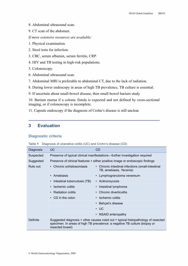
Table 1 Diagnosis of ulcerative colitis (UC) and Crohn’s disease (CD)
Diagnosis UC CD
Suspected Presence of typical clinical manifestations—further investigation required
Suggested Presence of clinical features + either positive image or endoscopic findings
Rule out: • Chronic schistosomiasis • Chronic intestinal infections (small-intestinal TB, amebiasis, Yersinia)
• Amebiasis • Lymphogranuloma venereum
• Intestinal tuberculosis (TB) • Actinomycosis
• Ischemic colitis • Intestinal lymphoma
• Radiation colitis • Chronic diverticulitis
• CD in the colon • Ischemic colitis
• Behçet’s disease
• UC
• NSAID enteropathy
Definite Suggested diagnosis + other causes ruled out + typical histopathology of resected specimen. In areas of high TB prevalence: a negative TB culture (biopsy or resected bowel)
© World Gastroenterology Organization, 2009
WGO Global Guideline IBD 11
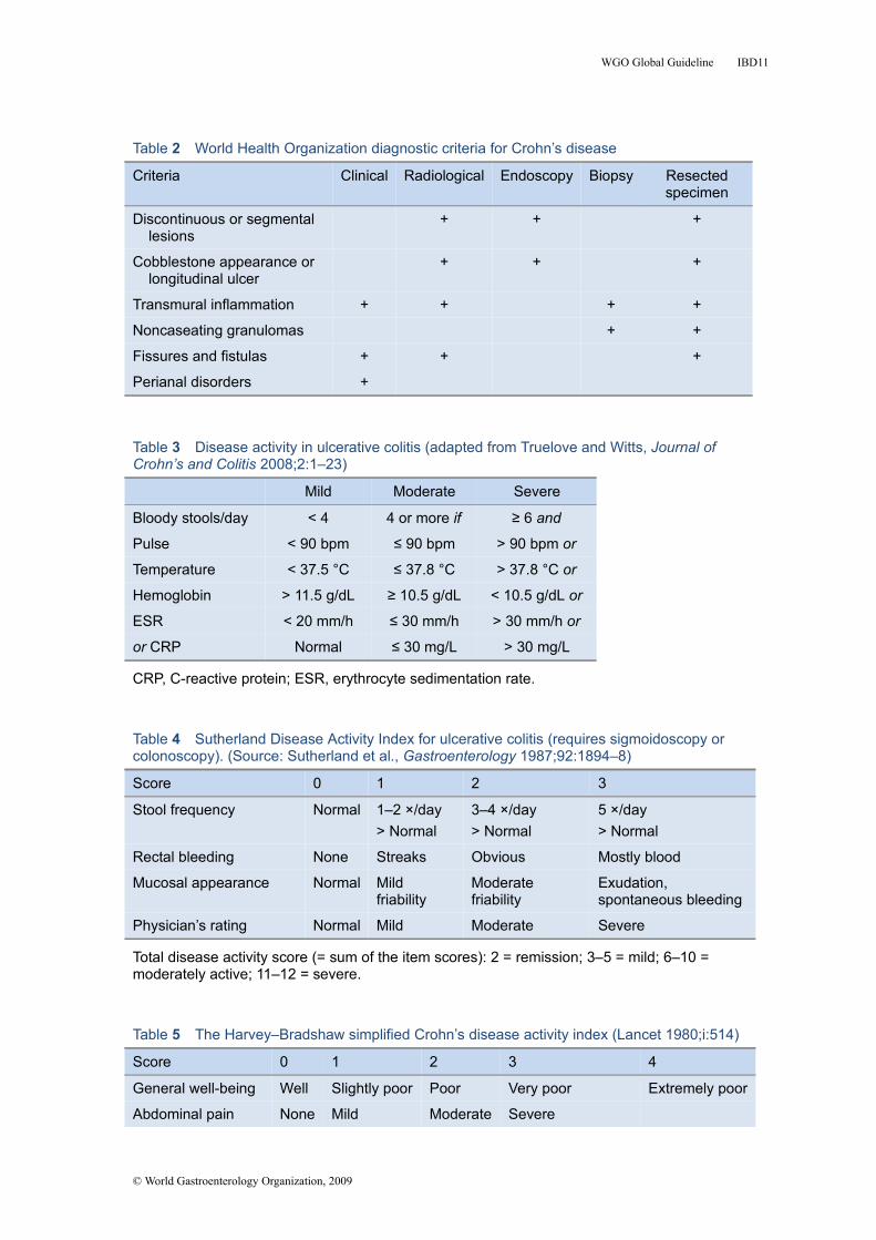
Table 2 World Health Organization diagnostic criteria for Crohn’s disease
Criteria Clinical Radiological Endoscopy Biopsy Resected specimen
Discontinuous or segmental lesions
+ + +
Cobblestone appearance or longitudinal ulcer
+ + +
Transmural inflammation + + + +
Noncaseating granulomas + +
Fissures and fistulas + + +
Perianal disorders +
Table 3 Disease activity in ulcerative colitis (adapted from Truelove and Witts, Journal of Crohn’s and Colitis 2008;2:1–23)
Mild Moderate Severe
Bloody stools/day < 4 4 or more if ≥ 6 and
Pulse < 90 bpm ≤ 90 bpm > 90 bpm or
Temperature < 37.5 °C ≤ 37.8 °C > 37.8 °C or
Hemoglobin > 11.5 g/dL ≥ 10.5 g/dL < 10.5 g/dL or
ESR < 20 mm/h ≤ 30 mm/h > 30 mm/h or
or CRP Normal ≤ 30 mg/L > 30 mg/L
CRP, C-reactive protein; ESR, erythrocyte sedimentation rate.
Table 4 Sutherland Disease Activity Index for ulcerative colitis (requires sigmoidoscopy or colonoscopy). (Source: Sutherland et al., Gastroenterology 1987;92:1894–8)
Score 0 1 2 3
Stool frequency Normal 1–2 ×/day> Normal
3–4 ×/day> Normal
5 ×/day> Normal
Rectal bleeding None Streaks Obvious Mostly blood
Mucosal appearance Normal Mild friability
Moderate friability
Exudation,spontaneous bleeding
Physician’s rating Normal Mild Moderate Severe
Total disease activity score (= sum of the item scores): 2 = remission; 3–5 = mild; 6–10 = moderately active; 11–12 = severe.
Table 5 The Harvey–Bradshaw simplified Crohn’s disease activity index (Lancet 1980;i:514)
Score 0 1 2 3 4
General well-being Well Slightly poor Poor Very poor Extremely poor
Abdominal pain None Mild Moderate Severe
© World Gastroenterology Organization, 2009
WGO Global Guideline IBD 12
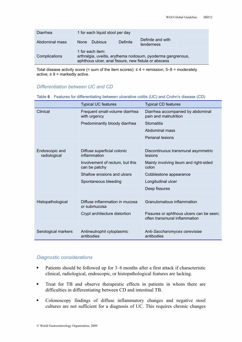
Diarrhea 1 for each liquid stool per day
Abdominal mass None Dubious Definite Definite and with tenderness
Complications1 for each item:arthralgia, uveitis, erythema nodosum, pyoderma gangrenous, aphthous ulcer, anal fissure, new fistula or abscess
Total disease activity score (= sum of the item scores): ≤ 4 = remission; 5–8 = moderately active; ≥ 9 = markedly active.
Differentiation between UC and CD
Table 6 Features for differentiating between ulcerative colitis (UC) and Crohn’s disease (CD)
Typical UC features Typical CD features
Clinical Frequent small-volume diarrhea with urgency
Diarrhea accompanied by abdominal pain and malnutrition
Predominantly bloody diarrhea Stomatitis
Abdominal mass
Perianal lesions
Endoscopic and radiological
Diffuse superficial colonic inflammation
Discontinuous transmural asymmetric lesions
Involvement of rectum, but this can be patchy
Mainly involving ileum and right-sided colon
Shallow erosions and ulcers Cobblestone appearance
Spontaneous bleeding Longitudinal ulcer
Deep fissures
Histopathological Diffuse inflammation in mucosa or submucosa
Granulomatous inflammation
Crypt architecture distortion Fissures or aphthous ulcers can be seen; often transmural inflammation
Serological markers Antineutrophil cytoplasmic antibodies
Anti-Saccharomyces cerevisiae antibodies
Diagnostic considerations
Patients should be followed up for 3–6 months after a first attack if characteristic clinical, radiological, endoscopic, or histopathological features are lacking.
Treat for TB and observe therapeutic effects in patients in whom there are difficulties in differentiating between CD and intestinal TB.
Colonoscopy findings of diffuse inflammatory changes and negative stool cultures are not sufficient for a diagnosis of UC. This requires chronic changes
© World Gastroenterology Organization, 2009
WGO Global Guideline IBD 13
over time (i.e., 6 months, in the absence of other emerging diagnoses) and signs of chronic inflammation histologically.
Surveillance for colorectal cancer should be implemented in patients with long-standing UC and CD colitis.
Differential diagnosis
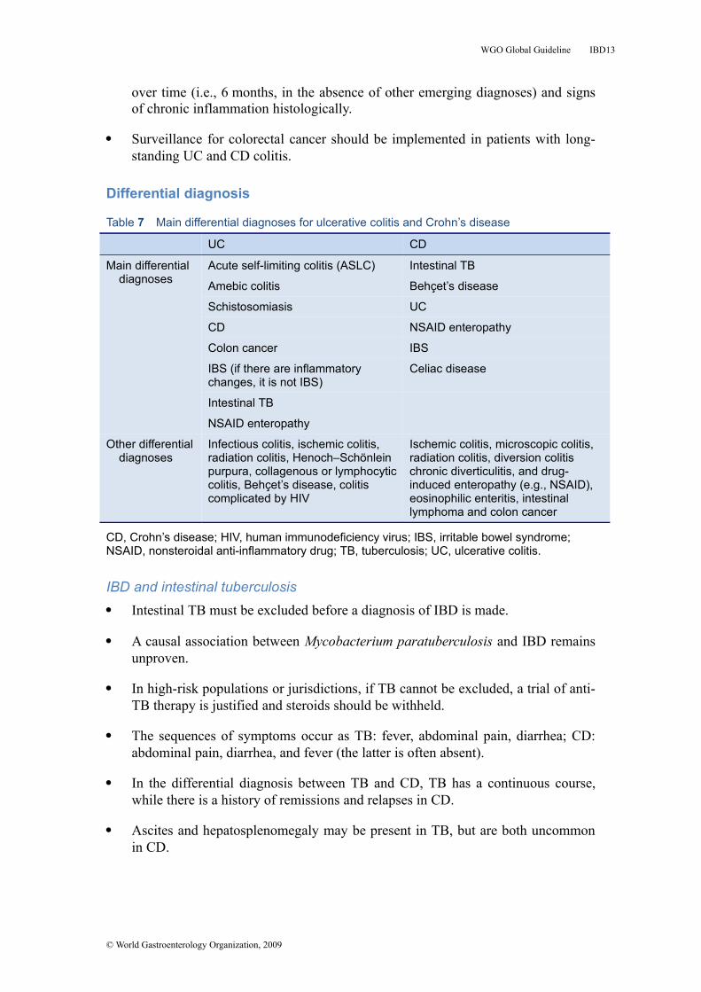
Table 7 Main differential diagnoses for ulcerative colitis and Crohn’s disease
UC CD
Main differential diagnoses
Acute self-limiting colitis (ASLC) Intestinal TB
Amebic colitis Behçet’s disease
Schistosomiasis UC
CD NSAID enteropathy
Colon cancer IBS
IBS (if there are inflammatory changes, it is not IBS)
Celiac disease
Intestinal TB
NSAID enteropathy
Other differential diagnoses
Infectious colitis, ischemic colitis, radiation colitis, Henoch–Schönlein purpura, collagenous or lymphocytic colitis, Behçet’s disease, colitis complicated by HIV
Ischemic colitis, microscopic colitis, radiation colitis, diversion colitis chronic diverticulitis, and drug-induced enteropathy (e.g., NSAID), eosinophilic enteritis, intestinal lymphoma and colon cancer
CD, Crohn’s disease; HIV, human immunodeficiency virus; IBS, irritable bowel syndrome; NSAID, nonsteroidal anti-inflammatory drug; TB, tuberculosis; UC, ulcerative colitis.
IBD and intestinal tuberculosis Intestinal TB must be excluded before a diagnosis of IBD is made.
A causal association between Mycobacterium paratuberculosis and IBD remains unproven.
In high-risk populations or jurisdictions, if TB cannot be excluded, a trial of anti-TB therapy is justified and steroids should be withheld.
The sequences of symptoms occur as TB: fever, abdominal pain, diarrhea; CD: abdominal pain, diarrhea, and fever (the latter is often absent).
In the differential diagnosis between TB and CD, TB has a continuous course, while there is a history of remissions and relapses in CD.
Ascites and hepatosplenomegaly may be present in TB, but are both uncommon in CD.
© World Gastroenterology Organization, 2009
WGO Global Guideline IBD 14
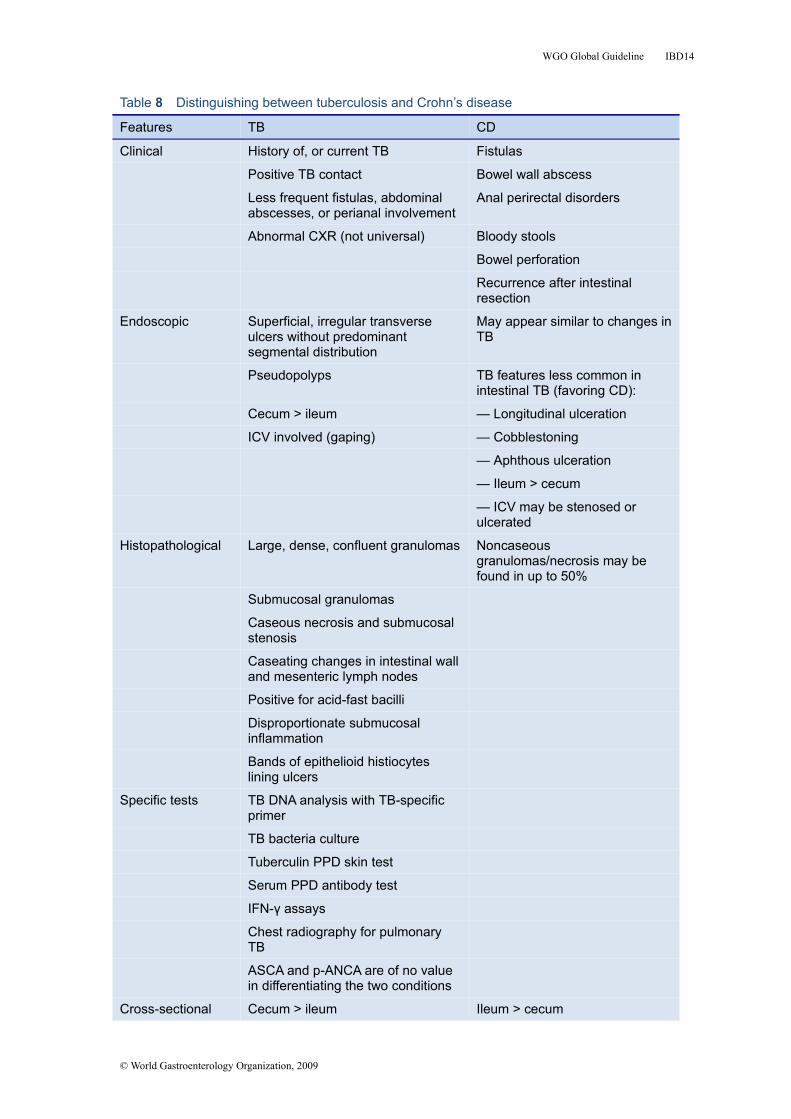
Table 8 Distinguishing between tuberculosis and Crohn’s disease
Features TB CD
Clinical History of, or current TB Fistulas
Positive TB contact Bowel wall abscess
Less frequent fistulas, abdominal abscesses, or perianal involvement
Anal perirectal disorders
Abnormal CXR (not universal) Bloody stools
Bowel perforation
Recurrence after intestinal resection
Endoscopic Superficial, irregular transverse ulcers without predominant segmental distribution
May appear similar to changes in TB
Pseudopolyps TB features less common in intestinal TB (favoring CD):
Cecum > ileum — Longitudinal ulceration
ICV involved (gaping) — Cobblestoning
— Aphthous ulceration
— Ileum > cecum
— ICV may be stenosed or ulcerated
Histopathological Large, dense, confluent granulomas Noncaseous granulomas/necrosis may be found in up to 50%
Submucosal granulomas
Caseous necrosis and submucosal stenosis
Caseating changes in intestinal wall and mesenteric lymph nodes
Positive for acid-fast bacilli
Disproportionate submucosal inflammation
Bands of epithelioid histiocytes lining ulcers
Specific tests TB DNA analysis with TB-specific primer
TB bacteria culture
Tuberculin PPD skin test
Serum PPD antibody test
IFN-γ assays
Chest radiography for pulmonary TB
ASCA and p-ANCA are of no value in differentiating the two conditions
Cross-sectional Cecum > ileum Ileum > cecum
© World Gastroenterology Organization, 2009
WGO Global Guideline IBD 15
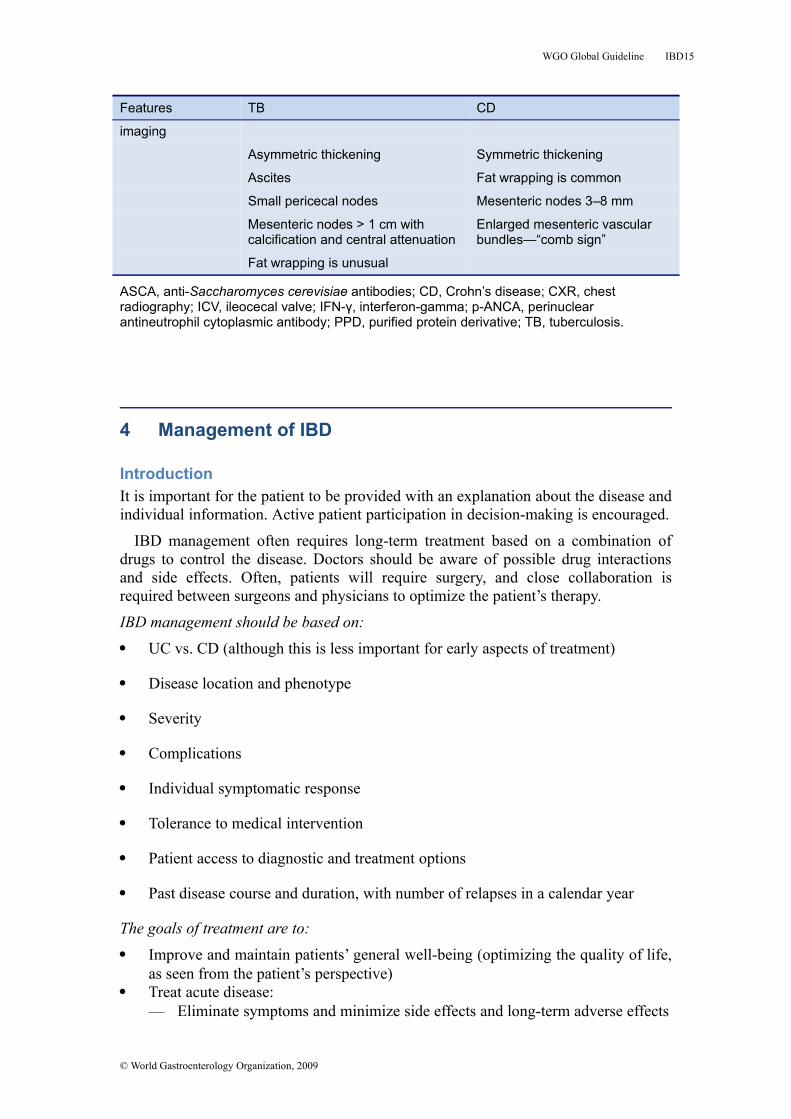
Features TB CD
imaging
Asymmetric thickening Symmetric thickening
Ascites Fat wrapping is common
Small pericecal nodes Mesenteric nodes 3–8 mm
Mesenteric nodes > 1 cm with calcification and central attenuation
Enlarged mesenteric vascular bundles—“comb sign”
Fat wrapping is unusual
ASCA, anti-Saccharomyces cerevisiae antibodies; CD, Crohn’s disease; CXR, chest radiography; ICV, ileocecal valve; IFN-γ, interferon-gamma; p-ANCA, perinuclear antineutrophil cytoplasmic antibody; PPD, purified protein derivative; TB, tuberculosis.
4 Management of IBD
IntroductionIt is important for the patient to be provided with an explanation about the disease and individual information. Active patient participation in decision-making is encouraged.
IBD management often requires long-term treatment based on a combination of drugs to control the disease. Doctors should be aware of possible drug interactions and side effects. Often, patients will require surgery, and close collaboration is required between surgeons and physicians to optimize the patient’s therapy.
IBD management should be based on: UC vs. CD (although this is less important for early aspects of treatment)
Disease location and phenotype
Severity
Complications
Individual symptomatic response
Tolerance to medical intervention
Patient access to diagnostic and treatment options
Past disease course and duration, with number of relapses in a calendar year
The goals of treatment are to: Improve and maintain patients’ general well-being (optimizing the quality of life,
as seen from the patient’s perspective) Treat acute disease:
— Eliminate symptoms and minimize side effects and long-term adverse effects
© World Gastroenterology Organization, 2009
WGO Global Guideline IBD 16
— Reduce intestinal inflammation and if possible heal the mucosa Maintain steroid-free remissions (decreasing the frequency and severity of
recurrences and reliance on steroids) Prevent complications hospitalization and surgery
Maintain good nutritional status
Diet and lifestyle considerations: The impact of diet on inflammatory activity in UC/CD is poorly understood, but
dietary changes may help reduce symptoms:— During increased disease activity, it is appropriate to decrease the amount of
fiber. Dairy products can be maintained unless not tolerated.— A low-residue diet may decrease the frequency of bowel movements.— A high-residue diet may be indicated in cases of ulcerative proctitis (disease
limited to the rectum, where constipation can be more of a problem than diarrhea).
— There are limited data suggesting that a reduction of dietary fermentable oligosaccharides, disaccharides, and monosaccharides and polyols may reduce the symptoms of IBD.
Dietary or lifestyle changes may reduce inflammation in CD:— A liquid diet, pre-digested formula, or nothing by mouth (NPO status) may
reduce obstructive symptoms. Exclusive enteral nutrition can settle inflammatory disease, especially in children.
— Smoking cessation benefits patients with CD in relation to their disease course and benefits UC patients from a general health point of view (smoking cessation is associated with flaring of UC).
Reduction of stress and better stress management may improve symptoms or the patients’ approach to their disease. The assistance of a mental health worker may be useful, and attention to comorbid psychiatric illness is imperative.
Drugs in IBD management Aminosalicylates—anti-inflammatory agents
— Include: 5-aminosalicylic acid (5-ASA), mesalazine Preparations available in the U.S. and western Europe for oral use:
sulfasalazine, mesalamine, olsalazine, balsalazide; and for rectal use: mesalamine enemas (liquid or foam) and suppositories
— Useful both for treating colitis flare-ups and maintenance of remission.— Aminosalicylates for UC treatment during remissions:
Oral or rectal 5-ASA Combination therapy of oral and topical 5-ASA Rectal 5-ASA is superior to rectal steroids
— In CD, sulfasalazine is mainly effective in disease affecting the colon.— Patients receiving sulfasalazine should take folic acid.— Important to use adequate doses: 2.0–4.8 g/day for active disease, ≥ 2 g/day
for maintenance. Corticosteroids (steroids):
— Usually provide significant suppression of inflammation and rapid relief of symptoms.
© World Gastroenterology Organization, 2009
WGO Global Guideline IBD 17
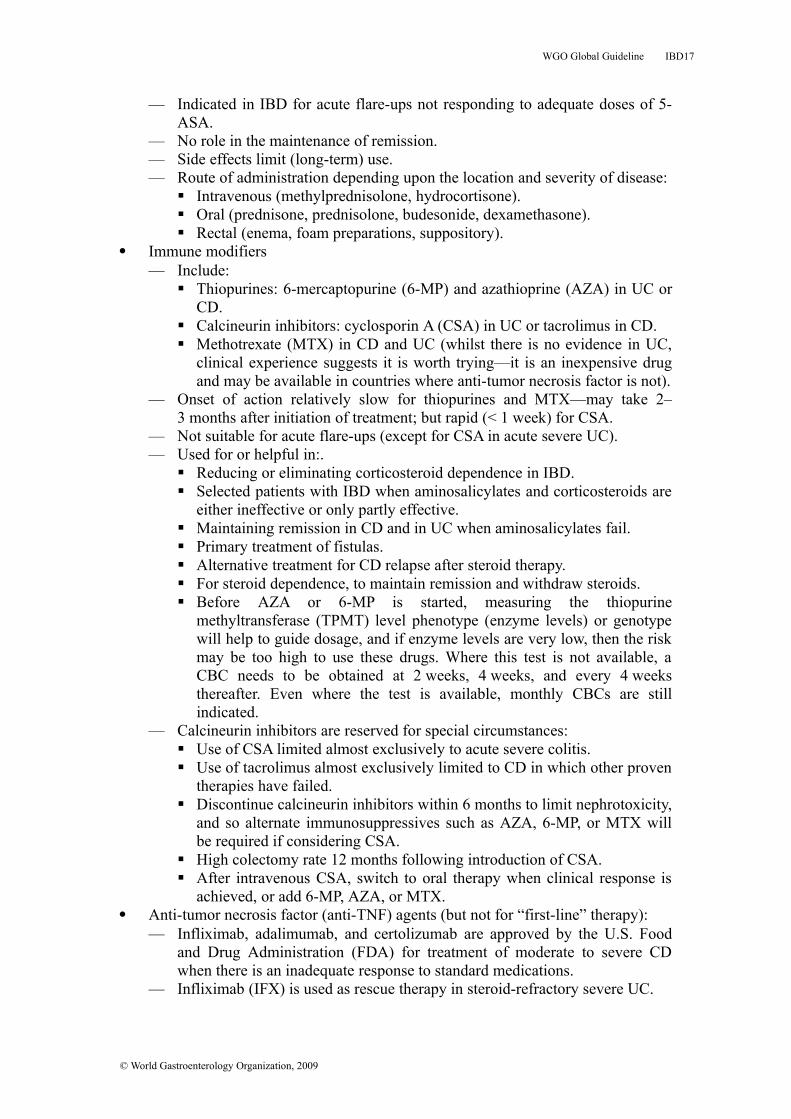
— Indicated in IBD for acute flare-ups not responding to adequate doses of 5-ASA.
— No role in the maintenance of remission.— Side effects limit (long-term) use.— Route of administration depending upon the location and severity of disease:
Intravenous (methylprednisolone, hydrocortisone). Oral (prednisone, prednisolone, budesonide, dexamethasone). Rectal (enema, foam preparations, suppository).
Immune modifiers— Include:
Thiopurines: 6-mercaptopurine (6-MP) and azathioprine (AZA) in UC or CD.
Calcineurin inhibitors: cyclosporin A (CSA) in UC or tacrolimus in CD. Methotrexate (MTX) in CD and UC (whilst there is no evidence in UC,
clinical experience suggests it is worth trying—it is an inexpensive drug and may be available in countries where anti-tumor necrosis factor is not).
— Onset of action relatively slow for thiopurines and MTX—may take 2–3 months after initiation of treatment; but rapid (< 1 week) for CSA.
— Not suitable for acute flare-ups (except for CSA in acute severe UC).— Used for or helpful in:.
Reducing or eliminating corticosteroid dependence in IBD. Selected patients with IBD when aminosalicylates and corticosteroids are
either ineffective or only partly effective. Maintaining remission in CD and in UC when aminosalicylates fail. Primary treatment of fistulas. Alternative treatment for CD relapse after steroid therapy. For steroid dependence, to maintain remission and withdraw steroids. Before AZA or 6-MP is started, measuring the thiopurine
methyltransferase (TPMT) level phenotype (enzyme levels) or genotype will help to guide dosage, and if enzyme levels are very low, then the risk may be too high to use these drugs. Where this test is not available, a CBC needs to be obtained at 2 weeks, 4 weeks, and every 4 weeks thereafter. Even where the test is available, monthly CBCs are still indicated.
— Calcineurin inhibitors are reserved for special circumstances: Use of CSA limited almost exclusively to acute severe colitis. Use of tacrolimus almost exclusively limited to CD in which other proven
therapies have failed. Discontinue calcineurin inhibitors within 6 months to limit nephrotoxicity,
and so alternate immunosuppressives such as AZA, 6-MP, or MTX will be required if considering CSA.
High colectomy rate 12 months following introduction of CSA. After intravenous CSA, switch to oral therapy when clinical response is
achieved, or add 6-MP, AZA, or MTX. Anti-tumor necrosis factor (anti-TNF) agents (but not for “first-line” therapy):
— Infliximab, adalimumab, and certolizumab are approved by the U.S. Food and Drug Administration (FDA) for treatment of moderate to severe CD when there is an inadequate response to standard medications.
— Infliximab (IFX) is used as rescue therapy in steroid-refractory severe UC.
© World Gastroenterology Organization, 2009
WGO Global Guideline IBD 18
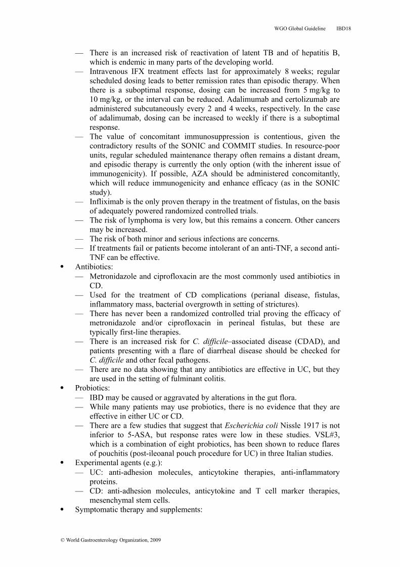
— There is an increased risk of reactivation of latent TB and of hepatitis B, which is endemic in many parts of the developing world.
— Intravenous IFX treatment effects last for approximately 8 weeks; regular scheduled dosing leads to better remission rates than episodic therapy. When there is a suboptimal response, dosing can be increased from 5 mg/kg to 10 mg/kg, or the interval can be reduced. Adalimumab and certolizumab are administered subcutaneously every 2 and 4 weeks, respectively. In the case of adalimumab, dosing can be increased to weekly if there is a suboptimal response.
— The value of concomitant immunosuppression is contentious, given the contradictory results of the SONIC and COMMIT studies. In resource-poor units, regular scheduled maintenance therapy often remains a distant dream, and episodic therapy is currently the only option (with the inherent issue of immunogenicity). If possible, AZA should be administered concomitantly, which will reduce immunogenicity and enhance efficacy (as in the SONIC study).
— Infliximab is the only proven therapy in the treatment of fistulas, on the basis of adequately powered randomized controlled trials.
— The risk of lymphoma is very low, but this remains a concern. Other cancers may be increased.
— The risk of both minor and serious infections are concerns.— If treatments fail or patients become intolerant of an anti-TNF, a second anti-
TNF can be effective. Antibiotics:
— Metronidazole and ciprofloxacin are the most commonly used antibiotics in CD.
— Used for the treatment of CD complications (perianal disease, fistulas, inflammatory mass, bacterial overgrowth in setting of strictures).
— There has never been a randomized controlled trial proving the efficacy of metronidazole and/or ciprofloxacin in perineal fistulas, but these are typically first-line therapies.
— There is an increased risk for C. difficile–associated disease (CDAD), and patients presenting with a flare of diarrheal disease should be checked for C. difficile and other fecal pathogens.
— There are no data showing that any antibiotics are effective in UC, but they are used in the setting of fulminant colitis.
Probiotics:— IBD may be caused or aggravated by alterations in the gut flora.— While many patients may use probiotics, there is no evidence that they are
effective in either UC or CD.— There are a few studies that suggest that Escherichia coli Nissle 1917 is not
inferior to 5-ASA, but response rates were low in these studies. VSL#3, which is a combination of eight probiotics, has been shown to reduce flares of pouchitis (post-ileoanal pouch procedure for UC) in three Italian studies.
Experimental agents (e.g.):— UC: anti-adhesion molecules, anticytokine therapies, anti-inflammatory
proteins.— CD: anti-adhesion molecules, anticytokine and T cell marker therapies,
mesenchymal stem cells. Symptomatic therapy and supplements:
© World Gastroenterology Organization, 2009
WGO Global Guideline IBD 19
— Antidiarrheals such as loperamide (Imodium) if colitis is not fulminant; cholestyramine if the patient has previously undergone ileal resection.
— Analgesics such as acetaminophen, or even codeine if acetaminophen is insufficient.
— Nutritional supplementation for those with malnutrition, or during periods of reduced oral intake.
— Vitamin B12 replenishment for those with deficiency.— Vitamin D supplementation if the local area does not allow sun exposure for
much of the year.— Routine vitamin D and calcium supplementation for steroid users.— Routine multivitamin supplementation for all.— For chronic iron-deficiency anemia, use parenteral iron (either as weekly
intramuscular shots or dosing with intravenous iron) if oral iron is not tolerated.
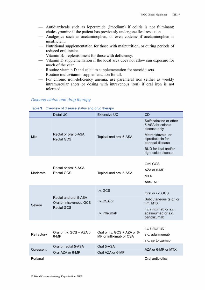
Disease status and drug therapy
Table 9 Overview of disease status and drug therapy
Distal UC Extensive UC CD
MildRectal or oral 5-ASARectal GCS
Topical and oral 5-ASA
Sulfasalazine or other 5-ASA for colonic disease only
Metronidazole or ciprofloxacin for perineal disease
BUD for ileal and/or right colon disease
ModerateRectal or oral 5-ASARectal GCS Topical and oral 5-ASA
Oral GCS
AZA or 6-MP
MTX
Anti-TNF
Severe
Rectal and oral 5-ASAOral or intravenous GCSRectal GCS
I.v. GCSOral or i.v. GCS
I.v. CSA or Subcutaneous (s.c.) or i.m. MTX
I.v. infliximabI.v. infliximab or s.c. adalimumab or s.c. certolizumab
Refractory Oral or i.v. GCS + AZA or 6-MP
Oral or i.v. GCS + AZA or 6-MP or infliximab or CSA
I.v. infliximab
s.c. adalimumab
s.c. certolizumab
QuiescentOral or rectal 5-ASA Oral 5-ASA
AZA or 6-MP or MTXOral AZA or 6-MP Oral AZA or 6-MP
Perianal Oral antibiotics
© World Gastroenterology Organization, 2009
WGO Global Guideline IBD 20
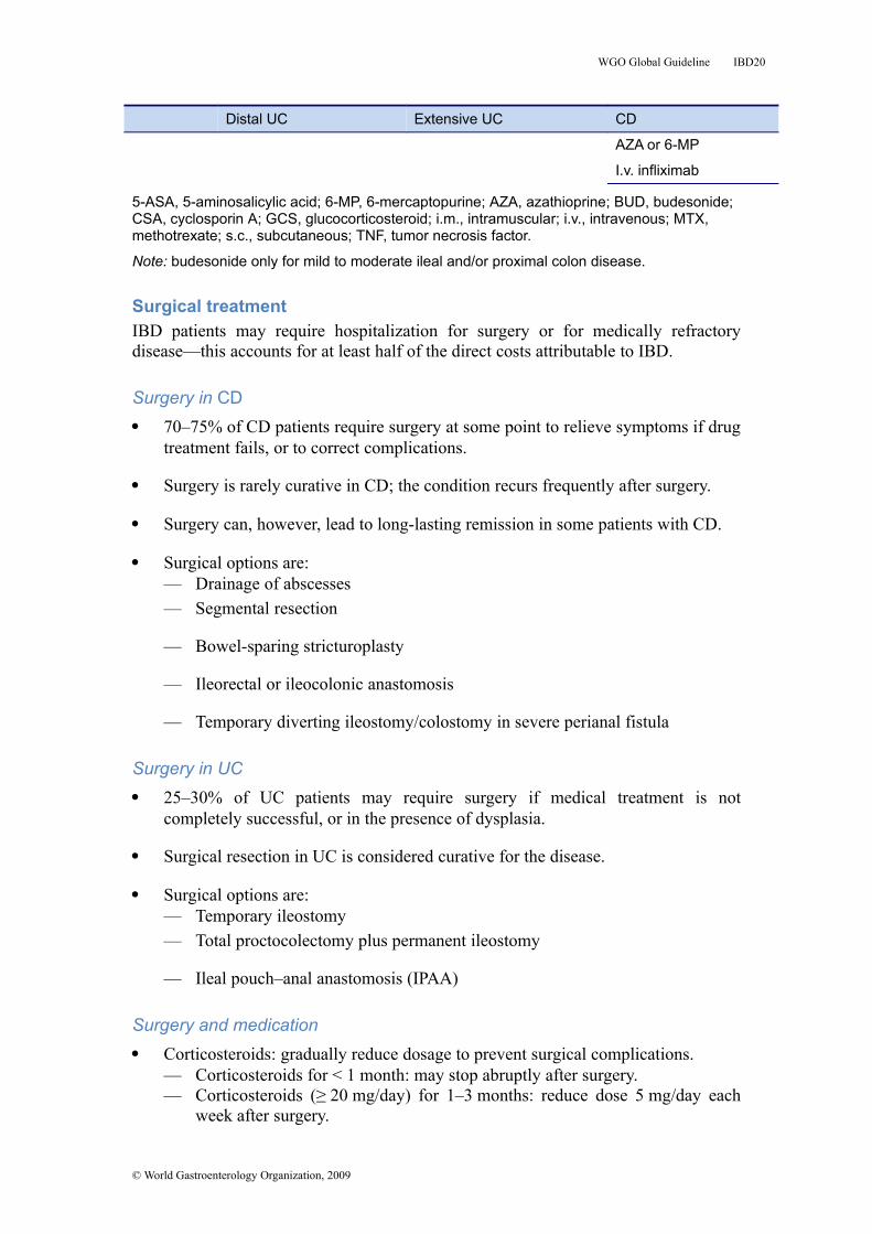
Distal UC Extensive UC CD
AZA or 6-MP
I.v. infliximab
5-ASA, 5-aminosalicylic acid; 6-MP, 6-mercaptopurine; AZA, azathioprine; BUD, budesonide; CSA, cyclosporin A; GCS, glucocorticosteroid; i.m., intramuscular; i.v., intravenous; MTX, methotrexate; s.c., subcutaneous; TNF, tumor necrosis factor.
Note: budesonide only for mild to moderate ileal and/or proximal colon disease.
Surgical treatmentIBD patients may require hospitalization for surgery or for medically refractory disease—this accounts for at least half of the direct costs attributable to IBD.
Surgery in CD 70–75% of CD patients require surgery at some point to relieve symptoms if drug
treatment fails, or to correct complications.
Surgery is rarely curative in CD; the condition recurs frequently after surgery.
Surgery can, however, lead to long-lasting remission in some patients with CD.
Surgical options are:— Drainage of abscesses— Segmental resection
— Bowel-sparing stricturoplasty
— Ileorectal or ileocolonic anastomosis
— Temporary diverting ileostomy/colostomy in severe perianal fistula
Surgery in UC 25–30% of UC patients may require surgery if medical treatment is not
completely successful, or in the presence of dysplasia.
Surgical resection in UC is considered curative for the disease.
Surgical options are:— Temporary ileostomy— Total proctocolectomy plus permanent ileostomy
— Ileal pouch–anal anastomosis (IPAA)
Surgery and medication Corticosteroids: gradually reduce dosage to prevent surgical complications.
— Corticosteroids for < 1 month: may stop abruptly after surgery.— Corticosteroids (≥ 20 mg/day) for 1–3 months: reduce dose 5 mg/day each
week after surgery.
© World Gastroenterology Organization, 2009
WGO Global Guideline IBD 21
— Corticosteroids for 3–6 months: reduce dose 2.5 mg/day each week.— Corticosteroids for > 6 months: reduce dose slowly at ≤ 1 mg/week once at
10 mg/day.— Aim to minimize steroid dosage prior to surgery when possible. Prednisone
doses greater than 30 mg/day preoperatively are associated with poorer postoperative outcomes.
Azathioprine: no increased risk in a perioperative setting. Perioperative anti-TNF-α therapy with infliximab, adalimumab, or certolizumab.
— Suspect an increased risk for emergency colectomy for acute severe colitis.— No increased risk in CD.
Postoperative maintenance in CD with 5-ASA p.o. or 6-MP/AZA to reduce the frequency and severity of recurrences. The best data for maintenance are for metronidazole—it is inexpensive and can be considered in resource-poor settings (although limited by dysgeusia and neuropathy side effects). In contrast, the data for 5-ASA are weak, and it is more expensive.
Emphasize the importance of smoking cessation—the single most effective approach patients can take to reduce recurrence in CD.
Cascades in IBD management
Cascade 2—UC management*Level 1—limited resources:1. In endemic areas and when there is limited access to diagnosis, give a course of anti-ameba therapy.
2. In endemic areas for TB, consider a trial of anti-TB therapy for 1 month to determine the response.
3. Sulfasalazine (least expensive) for all mild to moderate colitis and for maintenance of remission. Different mesalazine preparations are available, including Asacol 800 mg, Lialda (U.S.) or Mezavant (Europe) 1200 mg pills, and Pentasa 2 g sachets. These larger doses can facilitate better adherence, with no sulfa side effects.
4. Steroid enemas for distal colon disease.*
5. Oral prednisone for moderate to severe disease (acute severe disease requires intravenous steroids).
6. If acute severe colitis is unresponsive to intravenous steroids or the patient has chronic steroid-resistant or steroid-dependent colitis, consider colectomy. This decision needs to be made in a timely fashion in acute severe ulcerative colitis. Consider either the Oxford or Sweden predictors of outcome on day 3 of intravenous steroids.
7. CMV should be actively sought in refractory disease.
8. 5-ASA when remission is not maintained. Azathioprine for steroid dependence. Methotrexate can be considered if azathioprine is not available or if there is intolerance.* Steroid enemas can sometimes be made with locally available resources, sometimes at lower cost.
© World Gastroenterology Organization, 2009
WGO Global Guideline IBD 22
Level 2—if resources are available, then:1. Treat TB and parasites when diagnosed first.
2. Sulfasalazine can be used for mild to moderate colitis.
3. Asacol 800 mg, Lialda/Mezavant 1200 mg pills, and Pentasa 2 g sachets are now available and can facilitate better adherence, with no sulfa side effects.
4. 5-ASA enemas or suppositories for distal disease. These can be used for remission maintenance in distal disease in lieu of oral 5-ASA. Steroid enemas are also an option, but typically not for maintenance.
5. Combination therapy with oral and rectal 5-ASA may be more effective in active distal disease or even active pancolitis.
6. If patients fail to maintain remission with 5-ASA, then consider azathioprine or 6-MP/AZA; in case of azathioprine failure, consider methotrexate.
Level 3—if more extensive resources are available:1. Cyclosporine can be considered in acute severe colitis.
2. Infliximab can be considered for acute severe colitis or moderately severe steroid-dependent or steroid-resistant colitis.
3. Azathioprine or 6-MP.* Some traditional Chinese medicines are deemed to be useful as alternative medicines for anemia in China. These are not typically used in the West. Some Chinese agents suggested include powder of natural indigo, powder for treating throat disease (xilei powder), Yunnan white drug, or oral prescriptions such as Pulsatilla decoctions; and some single components in Chinese medicine, such as Pulsatilla root, Coptis root, Amur corktree bark, Baikal skullcap root, and curcumin.
Cascade 3—CD management*Level 1—limited resources:1. In endemic areas and when there is limited access to diagnosis, give a course of anti-ameba therapy.
2. In endemic areas for TB, consider a trial of anti-TB therapy for 1 month to determine the response.
3. Sulfasalazine (least expensive) for all mild to moderate colitis and for maintenance of remission.
4. Steroid enemas* for distal colon disease.
5. Trial of metronidazole for ileocolonic or colonic disease.
6. Oral prednisone for moderate to severe disease.
7. If there is a short segment of small-bowel disease, consider surgery.
8. Azathioprine or methotrexate.
9. Metronidazole for postoperative maintenance.* Steroid enemas can sometimes be made with locally available resources, sometimes at lower cost.
© World Gastroenterology Organization, 2009
WGO Global Guideline IBD 23
Level 2—if resources are available, then:1. Treat TB and parasites when diagnosed first.
2. Sulfasalazine for mild to moderate active colonic CD.
3. Budesonide can be used for mild ileal or ileocolonic disease (right colon).
4. If patients fail to maintain remission after a course of steroids, then consider azathioprine (or 6-MP/AZA); in case of azathioprine failure, consider methotrexate.
Level 3—if more extensive resources are available:1. Infliximab or adalimumab or certolizumab can be considered for moderate to severe steroid-dependent or steroid-resistant disease.
2. Immunosuppressive drugs, such as 6-MP and AZA, can also be very helpful in the treatment of fistulas in CD.
3. Tacrolimus can be considered when anti-TNF fails.* Some traditional Chinese medicines are deemed to be useful as alternative medicines for anemia in China. These are not typically used in the West. Some Chinese agents suggested include powder of natural indigo, powder for treating throat disease (xilei powder), Yunnan white drug, or oral prescriptions such as Pulsatilla decoctions; and some single components in Chinese medicine, such as Pulsatilla root, Coptis root, Amur corktree bark, Baikal skullcap root, and curcumin.
Cascade 4—perineal fistulasLevel 1—limited resources:1. Metronidazole.
1a. Surgery, if an abscess is present
2. Ciprofloxacin.
3. A combination of metronidazole and ciprofloxacin. These antibiotics can be used for maintenance of fistula closure if tolerated over the long term.
4. Surgery—should be considered early and if long-term maintenance of antibiotics is required.
Level 2—more resources available:1. Metronidazole.
1a. Surgery, if an abscess is present.
2. Ciprofloxacin.
3. A combination of metronidazole and ciprofloxacin. These antibiotics can be used for maintenance of fistula closure if tolerated over the long term.
4. Surgery—should be considered early and if long-term maintenance of antibiotics is required.
5. AZA/6-MP for maintenance of fistula closure.
© World Gastroenterology Organization, 2009
WGO Global Guideline IBD 24
Level 3—if more extensive resources are available:1. Metronidazole.
1a. Surgery, if an abscess is present.
2. Ciprofloxacin.
3. A combination of metronidazole and ciprofloxacin. These antibiotics can be used for maintenance of fistula closure if tolerated over the long term.
4. Surgery—should be considered early and if long-term maintenance of antibiotics is required, and particularly if the fistula is simple.
5. AZA/6-MP for maintenance of fistula closure.
6. Infliximab.
7. Adalimumab for infliximab failure, or as an alternative to infliximab primarily.
8. Surgery for complex fistulas.
© World Gastroenterology Organization, 2009