WORLD CONGRESS WILDERNESS MEDICINE I - DTIC
435
I AD-A241 539 The First WORLD CONGRESS on WILDERNESS MEDICINE I I July 14-19,1991 Whistler, British Columbia, Canada * 91-12694 Approved for public release; distribution unlimited. 4%" Seventh Annual Scientific Meeting I , of the Wilderness Medical Society 6_
-
Upload
khangminh22 -
Category
Documents
-
view
1 -
download
0
Transcript of WORLD CONGRESS WILDERNESS MEDICINE I - DTIC

I AD-A241 539
The FirstWORLD CONGRESS
onWILDERNESS MEDICINEI
I July 14-19,1991Whistler, British Columbia, Canada
* 91-12694
Approved for public release;
distribution unlimited.
4%" Seventh Annual Scientific MeetingI , of theWilderness Medical Society
6_

r . %
23 September 1991 Proceedings
Wilderness Medical Society Congress DAMD17-91-Z-1025
Douglas A. Gentile
Wilderness Medical SocietyVanderbilt UniversityNashville, Tennessee 37212
U.S. Army Medical Research & Development CommandFort DetrickFrederick, Maryland 21702-5012
Approved for public release; disLribuLion unlimited
Unlimited Unlimited Unclassified Unlimited
GENERAL INSTRUCTIONS FOR COMPLETING SF 298The Report Documentation Page (RDP) is used in announcing and cataloging reports. It is importantthat this information be consistent with the rest of the report, particularly the cover and title page.Instructions for filling in each block of the form follow. It is important to stay within the lines to meetoptical scanning requirements.
Block 1. Aaency Use Only (Leave Blank) Block 12a. Distribution/Availablity Statement.Denote public availability or limitation. Cite
Block 2. Report Date. Full publication date any availability to the public. Enter additionalincluding day, month, and year, if available (e.g. limitations or special markings in all capitals1 Jan 88). Must cite at least the year. (e.g. NOFORN, REL, ITAR)
Block 3. Type of Reoort and Dates Covered,State whether report is interim, final, etc. If DOD - See DoDD 5230.24, "Distributionapplicable, enter inclusive report dates (e.g. 10 Statements on TechnicalJun 87 - 30 Jun 88). Documents."
Block 4. Title and Subtitle. A title is taken from DOE - See authoritiesthe part of the report that provides the most NASA - See Handbook NHB 2200.2.meaningful and complete information. When a NTIS - Leave blank.report is prepared in more than one volume,repeat the primary title, add volume number,and include subtitle for the specific volume. On Block 12b. Distribution Code.classified documents enter the titleclassification in parentheses. DOD - DOD - Leave blank
DOE - DOE - Enter DOE distribution categoriesBlock 5. Funding Numbers. To include contract from the Standard Distribution forand grant numbers; may include program Unclassified Scientific and Technicalelement number(s), project number(s), task Reportsnumber(s), and work unit number(s). Use the NASA - NASA - Leave blankfollowing labels: NTIS - NTIS - Leave blank.
C - Contract PR - ProjectG - Grant TA -TaskPE - Program WU - Work Unit Block 13. Abstract, Include a brief (Maximum
Element Accession No. 200 words) factual summary of the mostsignificant information contained in the report.
Block 6. Author(s). Name(s) of person(s)responsible for writing the report, performing Block 14. Subject Terms, Keywords or phrasesthe research, or credited with the content of the identifying major subjects in the report.report. If editor or compiler, this should followthe name(s). Block 15. Number of Pages. Enter the total
Block 7. Performing Organization Name(s) and number of pages.Address(es Self-explanatory. Block 16. Price Code Enter appropriate price
Block 8. Performing Organization Report code (NTIS only).Number, Enter the unique alphanumeric reportnumber(s) assigned by the organization Blocks 17.- 19. Security Classifications.performing the report. Self-explanatory. Enter U.S. Security
Classification in accordance with U.S. SecurityBlock 9. Sgonsoring/Monitorina Agency Regulations (i.e., UNCLASSIFIED). If formt4ames(s) and Addresses). Self-explanatory. contains classified information, stamp
Block 10. Spo nsorin/Mon ito ring Agency classification on the top and bottom of the page.Report Number. (If known)
Block 20. Limitation of Abstract. This blockBlock 11. Suoplementary Notes. Enter must be completed to assign a limitation to theinformation not included elsewhere such as: abstract. Enter either UL (unlimited) or SARPrepared in cooperation with...; Trans. of ..., To (same as report). An entry in this block isbe published in .... When a report is revised, (sa as re antry in th block Iinclude a statement whether the new report necessary if the abstract is to be limited. Ifsupersedes or supplements the older report. blank, the abstract is assumed to be unlimited.Standard For 9 Bc Rv.'2.89)

IIIIIi The First
WORLD CONGRESSI on
* WILDERNESS MEDICINE* Medicine and the Spirit of Adventure
* July 14-19, 1991
Whistler, British Columbia, Canada '
I \
ISeventh Annual Scientific Meeting
- oitheWilderness Medical Society-,- .- ,P.O. Box 397
I. . .... Point Reyes Station, CA 9J956USA
'3 .(415) 663-9107
I . ,
I
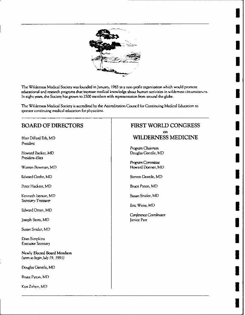
The Wilderness Medical Society was founded in January, 1983 as a non-profit organization which would promoteeducational and research programs that increase medical knowledge about human activities in wilderness circumstances.In eight years, the Society has grown to 2500 members with representation from around the globe.
The Wilderness Medical Society is accredited by the Accreditation Council for Continuing Medical Education tosponsor continuing medical education for physicians.
BOARD OF DIRECTORS FIRST WORLD CONGRESS 3on
Blair Dillard Erb, MD WILDERNESS MEDICINEPresidet P
Program Cort
Howard Backer, MD Douglas Gentile, MD
Program Cmmittee
Warren Bowman, MD Howard Donner, MD
Edward Geehr, MD Steven Gentile, MD 1Pcter Hackett, MD Bruce Paton, MD
Kenneth Iserson, MD Susan Snider, MDSecretary-Teasurer
Eric Weiss, MD
Edward Otten, MD
Confererce CoordnatorJoseph Serra, MD Janice Parr
Susan Snider, MD
Dian SimpkinsExecutive Secretary
Newly Elected Board Members 1(term to begin July 19, 1991)
Douglas Gentile, MD 1Bruce Paton, MD
Ken Zafren. MD
I

I
3 TABLE OF CONTENTS
I Program .Schedule .............................................................................................................................................................. vii
A cknowledgments ........................................................................................................................................................ xiii
I Future Conferences ............................................................................................................................................................ xv
Advertisements ................................................................................................................................................................. xviI Sunday, July 14
The W orld's Environments and the Spirit of Adventure ..................................................................................................... IBarry C. Bishop, PhD
Rescue Oper-.-ions in the Swiss A lps ................................................................................................................................... 3Bruno DiareT, MD
W ilderness W ound M anagement ...................................................................................................................................... 23D. Demitria Zukin, MD
Lyme Disease A Global Perspective ......................................................... 45Douglas A. Gentile, MD
Monday, July 15
Travel M edicine ................................................................................................................................................................ 53Dacid R. Shfim, MD
Controversies in Hypotherm ia: The Afterdrop Phenom enon .................................................................................... 67John Hayward , PhD
Symposia
Desert Survival .................................................................................................................................................................. 69Edward J. (Mel) Otten, MD
H um an Cooling Rates in Extreme Cold ........................................................................................................................... 81John Haywa d, PhD
Co ld W eather Survival ...................................................................................................................................................... 83Warren Bowmnan, MD
Cold W ater Immersion: O perational Considerations ................................................................................................ 89Janes P. Bagian, MD
Can You Enhance Capacivy for Perform ance at M oderate A ltitudes? .............................................................................. 101BeramM D. Levine, MD
Can You Enhance Capacity for Performance in the Cold? ................................................................................. ....... 125Andrew J. Young, MD
Can You Enhance Capacity for Perform ance in the Heat? .............................................................................................. 127Kent B. Pandoa, PhD
Assessment of Performance Capacity for W ilderness Adventure ................................................................................. 129William L. Haskell, PhD
SDiving M ed icine ............................................................................................................................................................. 137Paul S. Auerbach, MD
Ii

I
Diving M edicine .............................................................................................................................................................. 159Kenneth W. Kizt, MD, MPHH igh A ltitude M edicine ......................................................................................................................................... .. . .1931
Peter Hackett, MD and Oswald Oelr, MD
Workshops iW orld Status of W ilde ress M edicine .............................................................................................................................. 201W ilderness M ed icine Research ........................................................................................................................................ 205i
Daid R. Shlbu, MD and Daid N. Taylo, MD
W inter Survival .............................................................................................................................................................. 209Warren Bounman, MD
Litters andL Litter Packaging ............................................................................................................................................. 215Michael V. Callaa=, MSPH
Im provised Self Rescue for the Sport C lim ber ................................................................................................................. 217LannyJohson, RN, EMT-P
H elicopter Rescue in British Columbia ............................................................................................................................ 218BnAc Brwnk and Wayne Flann
W ilderness W ound M anagement .......................................................................................................... (see page) 23Eric Weiss, MD and D. Denerrios Zukin, MD
Tuesday, July 16
M ed icinal Plants of A m azonia ......................................................................................................................................... 221MarkJ. Ptoddkn, PhD
N eurologic Problems at H igh A ltitude ............................................................................................................................. 223Peter Hackett, MD iSymposia
M ed ical Prob lems of Space Flight .................................................................................................................................... 229James P. Bagian, MD
Designing a Health Care System for Space ...................................................................................................................... 231Joey B. Boyce, MD IRecent Advances in H A PE ............................................................................................................................................. 241Osuwald OeLz, MD
Effects of H igh A ltitude on C ardiovascular Diseases ........................................................................................................ 245Herbert N. HuLtgen, MD
Backcountry W ater Disinfection ...................................................................................................................................... 259Ho ard Backer, MD
O rthopedic Injuries ............................................................................................................................ ................. 269Joseph Serra, MD IWorkshops
W orld Status of W ilderness M edicine ............................................................................................................. (see page) 201
Desert Survival .................................................................................................................................................. (see page) 69Edw.ardJ. (Mel) Ouen, MD I
ivI
High Angle Rescue: Recent Developments ..................................................................................................................... 279MdW V. Cahan, MSPH
Basic Backcoun y Evacudtion and Transportation Skills ................................................................................................ 281Lanny Johmson, RN, RMT-P
Wildemess Fracture/Dislocation Workshop .................................................................................................... (see page) 269I Joseph Serra, MD
Wednesday, July 17
Scieniflc Presentations (alphabetical order)Availability of Oxygen-saving Systems for Himalayan Climbing ..................................................................................... 283M. Naksha
Carbohydrate Supplementation for Work at High Altitude: Liquid Versus Solid Focd Supplements ............. 284E.W. Askew
Central Nervous System Changes That May Precede Symptoms of Acute Mountain Sickness (AMS) .......................... 285R.W. Van Boven
Effect of Cooling Time on Survival in Heatstroke: A Meta-Analysis Evaluation ............................................................ 286C.B. Ramsey
I Effect of an Electrical Current on Snake Venom Toxicity ............................................................................................... 287D. Davis
Energy and Nutrient Intakes During High Altitude Acclimatisation ............................................. : ................................ 288C.E. Fenn
Modified Hyperbaric Bag and Inhalator Mask to Study Gas Exchange ............................................................................ 280
W. Bernhard
O rthopedic Field Splinting Devices ................................................................................................................................. 290A. Chisensen
Oxygen Saturation and Neuropsychological Performance:C hanges W ith A ltitude ................................................................................................................................................... 291J. G. Beniez
Pseudoephedrine for the Prevention of Barotitis Media:
A Controlled Clinical Trial in 120 Scuba Divers ............................................................................................................ 292M. Brown
Radio Frequency Rewarming of Hypothermia Victims .................................................................................................. 293M. Nei/er
Respiratory Function Test (RFT) in Normal Subjects During a 7 Day Periodat High Altitude (HA) and Correlation with AMS Score ............................................................................................... 294-- A. Cogo
A Review of EMS Near Drowning Calls in Contiguous North Carolina Beach Communities:Appropriate Use of Basic Assessment and Management Skills Must Be Based on ThoroughUnderstanding of Potential Injuries Sustained in the Marine Environment .................................................................... 295T. Grant
Trekker Medicine: The Impact of Western Medicine on tneHealth of Sherpas in the Khumbu Region of Nepal ......................................................................................................... 296C. Hagen
V

I
Thursday, July 18 IGlobal Health Issues: Impact of International Travel on Developing Countries ............................................................. 297Philip C. Rasoi, MD
Marine Envenomations .................................................................................................................................................... 301John A. Wdlianson, MD
Symposia
Jellyfish Envenomation .................................................................................................................................................... 305Jom A. Wiliarnson, MD IAdvances in Crotalid Antivenom ................................................................................................................................... 309Findlay E. Russell, MD, PhD
Group Cohesion and the Selection of Expedition Members ............................................................................................ 313Daid R. Jones, MD, MPH
Expedition Medicine: Practical Considerations ............................................................................................................... 323 iHoward Donner, MD
Traveler's Diarrhea ....................................................................................................................................................... 333Howard Backer, MD IHeat Illness ...................................................................................................................................................................... 347Eric Weiss, MD
Workshops
W orld Status of W ilderness Medicine ............................................................................................................. (see page) 201 IW hitewater Safety and Rescue ........................................................................................................................................ 359Eric Weiss, MD
Avalanche Res.c#e.: Search Dog, Probe and Beacon Techniques ..................................................................................... 383 IWay" F m and Backcomb Alpine Rescue Specialists
Field Recognition and Management of Exotic Envenomations ....................................................................................... 389Michael V. Callaim, MSPH IField W ater Disinfection Systems ................................................................................................................... (see page) 259Howard Backer, MD IFriday, July 19
W ilderness Medical Liability ........................................................................................................................................... 399Timotrh E. Miller, JD
Dealing with Death: The Psychology of Rescue .......................................................................................... 411Dad R. Jones, MD, MPH
Arthropod Envenomations: Spiders and Scorpions .......................................................................................................... 419Findlay E. Russell, MD, PhD
Bear Attacks: Prevention and Survival ............................................................................................................................ 423 ISunen P French, MD
vi

FIRST WORLD CONGRESS ON WILDERNESS MEDICINEMedicine and the Spirit of Adventure
Chateau Whistlcr ResortJuly 14-19, 1991
Whistler, British Columbia, Canada
Sunday, July 14 - LOCATION: Frontenac Ballroom
1230 pm Welcome and IntroductionDr. Gentile and Dr. Erb
12:45 The World's Environments and the Spirit of AdventureMr. Bishop
1:35 Wilderness Medicine - An Historical PerspectiveDr. Auerbach
2:00 Rescue Operations in the Swiss AlpsDr. Durrer
2:50 6F.F A K-VISIT EXHIBITS - LOCATION: Empress Ballroom
3:20 Wilderness Wound ManagementDr. Zukin
4:10 Lyme Disease - A (11jbal PerspectiveDr Gentile
5:00 ADJOURN
5:30 RECEPTION - LOCATION: Em-',-s Ballroom
Monday, July 15 - LOCATION: Frontenac Ballroom
7:00 COFFEE SERVICE-VISIT EXHIBITS-LOCATION: Empress Ballroom
8:00 Travel MedicineDr. Shlim
9:00 Controversies in Hypothermia: The Afterdrop PhenomenonDr.Hayward
9:50 BREAK-VISIT EXHIBITS
10:20 Symposia (simultaneous sessions)
A. Survival Medicine - LOCATION: Frontenac ADesert Survival - Dr. OttenHuman Cooling Rates in Extreme Cold - Dr HaywardCold Weather Survival - Dr. BowmanCold Water Immersion: Operational Considerations-Dr. Bagian
I vii
B. Enhancing Performance for Wilderness Adventure, LOCATION: Frontenac B ICan you enhance capacity for performance at moderate altitudes? - Dr. LevineCan you enhance capacity for performance in the cold? - Dr. YoungCan you enhance capacity for performance in the heat? - Dr. PandolfAssessment of performance capacity for wilderness adventure - Dr. Ha:kell
C. Wilderness Medicine Core Curriculum I - LOCATION: Frontenac CDiving Medicine - Drs. Auerbach and KizerHigh Altitude Medicine Drs. Hackett and Oelz
12:30 pm LUNCH (on your own)
2:00 Workshops/Demonstrations - (first session)
1. World Status of Wilderness Medicine I:Mountaineering and Environmental StressesLOCATION: Frontenac A IModerators: Drs. Hackett and Oelz
2. Wilderness Medicine Research ILOCATION: Frontenac BDrs. Shlim and Taylor
3. Winter SurvivalLOCATION: Outdoor location TBADr. Bowman
4. Litters and Litter Packaging ULOCATION: Outdoor location TBAMr. Callahan
5. Improvised Self Rescue for the Sport ClimberLOCATION: Outdoor location TBAMr. Johnson
6. Wilderness Wound ManagementLOCATION: Montebello RoomDrs. Weiss and Zukin
7. Helicopter Rescue in British ColumbiaLOCATION: Outdoor location TBAMr. Brink and Mr. Flann
3:30 Workshops/Demonstrations- (second session)LOCATIONS: As indicated above
1. Wilderneq Medicine Research IIDrs. Shlim and Taylor
2. Winter SurvivalDr. Bowman
viil

3. Litters and Litter PackagingMr. Callahan
4. Improvised Self Rescue for the Sport ClimberMr. Johnson
5. Helicopter Rescue in British ColumbiaMr Brink and Mr. Flann
5:00 ADJOURN
7:30 Evening Program: Diving the Rainow ReefsLOCATION: Frontenac BallroomDr. Auerbach
Tuesday, July 16 - LOCATION: Frontenac Ballroom
7:00 COFFEE SERVICE-VISIT EXHIBITS-LOCATION: Empress Ballroom
8:00 Medicinal Plants of AmazoniaDr. Plotkin
9:00 Neurologic Problems at High AltitudeDr. Hackett
9:50 BREAK-VISIT EXHIBITS
10:20 Symposia (simultaneous sessions)
A. Aerospace Medicine - LOCATION: Frontenac AMedical Problems of Space Flight - Dr. BagianDesigning a Health Care System - Dr. Boyce
B. High Altitude Medicine - LOCATION: Frontenac BRecent Advances in HAPE - Dr. OelzEffects of High Altitude on Cardiovascular Diseases - Dr. Hultgren
C. Wilderness Medicine Core Curriculum I1 - LOCATION: Frontenac CBackcountry Water Disinfection - Dr. BackerOrthopedic Injuries - Dr. Serra
12:30 pm LUNCH (on your own)
2:00 Workshops\Demonstrations (first session)
1. World Status of Wilderness Medicine 11:Aquatic Medicine and Environmental StressesLOCATION: Frontenac AModerators: Drs. Auerbach and Kizer
ix

I
2. Desert SurvivalLOCATION: Outdoor location TBADr. Otten
3. High Angle Rescue: Recent Developments:,OCATION: Frontenac BMr. Callahan
4. Basic Backcountry Evacuation and Transportation SkillsLOCATION: Outdoor location TBA IMr. Johnson
5. Wilderness Fracture/Dislocation WorkshopLOCATION: Outdoor location TBADr. Serra
3:30 Workshops/Demonstrations (second session) 1LOCATIONS: As listed above
1. Desert SurvivalDr. Orten
2. High Angle Rescue: Recent DevelopmentsMr. Callahan
3. Basic Backcountry Evacuation and Transportation SkillsMr. Johnson l
4. Wilderness Fracture/Dislocation WorkshopDr. Serra
3:30 Journal of Wilderness Medicine - Editorial Board MeetingLOCATION: Montebello Room (open to all WMS members)
5:00 ADJOURN
7:30 Evening Program: Saving the World's RainforestsLOCATION: Frontenac BallroomDr. Plotkin I
Wednesday, July 17 - LOCATION: Frontenac Ballroom
7:00 COFFEE SERVICE-VISIT EXHIBITS-LOCATION: Empress Ballroom I8:00 Free Scientific Presentations
This session will ,onsist ofbrief communications of current research in all areas of wilderness medicine. I9:50 BREAK - VISIT EXHIBITS
10:20 Free Scientific Presentations (cont.) I12:30 pm ADJOURN
I

I
m Thursday, July 18- LOCATION: Frontenac Ballroom
7:00 COFFEE SERVICE-VISIT EXHIBITS-LOCATION: Empress Ballroom
8.00 Global He.lth Issues: Impact of International Travel on Developing CountriesDr. Rasori
9:00 Marine EnvenomationsDr. Williamson
9:50 BREAK - VISIT EXHIBITS
10:20 Symposia (simultaneous sessions)
A. Wilderness Toxinology - LOCATION: Frontenac A;-Ilvfish Envenomation - Dr. WilliamsonAdvances in Crotalid Antivenom -Dr. Russell
B. Expedition Medicine - LOCATION: Frontenac BGroup Cohesion and the Selection of Expedition Members - Dr. JonesExpedition Medicine: Practical Considerations-Dr. Donner
C. Wilderness Medicine Core Curriculum III -LOCATION: Frontenac CTraveler's Diarrhea - Dr. BackerHear Illness - Dr. Weiss
12:30pm LUNCH (on your own)
2:00 Workshops/Demonstrations (one session only)
I 1. World Status of Wilderness Medicine III:Delivery of Services and Search and RescueLOCATION: Frontenac AModerators: Drs. Bowman and Durrer
2. Whitewater Safety and RescueLOCATION: Outdoor location TBADr. Weiss
3. Avalanche Rescue: Search Dog, Probe and Beacon TechniquesLOCATION: Algonquin RoomMr. Flann and Blackcomb Alpine Rescue Specialists
4. Field Recognition and Management of Exotic EnvenomationsLOCATION: Frontenac BMr. Callahan
I 5. Field Water Disinfection SystemsLOCATION: Frontenac CDr. Backer
I
ImmXi

II
4.00 ADJOURN
7:30 pm Banquet and Special PresentationLOCATION: Frontenac Ballroom
Preservation and the Spirit of AdventureMr. RowellI
Friday, July 19 - LOCATION: Frontenac Ballroom
7.00 COFFEE SERVICE-VISIT EXHIBITS-LOCATION: Empress Ballroom
8:00 Wilderness Medical LiabilityMr. Miller
9:00 Dealing with Death: The Psychology of RescueDr. Jones
9:50 BREAK
10:20 Arthropod Envenomations: Spiders and ScorpionsDr. Russell
11:10 Bear Attacks: Prevention and Survival UDr. French
12:00 noon Closing Session ISummary of the World Status of Wilderness MedicineDrs. Erb and Paton
12:30 pm FINAL ADJOURNMENT
IIIIIII
xii I

i
I ACKNOWLEDGMENTS
The Wilderness Medical Society appreciates the support of the following companies and organizations. You are encour-aged to visits the exhibits in the Empress Ballroom during the breaks to become familiar with their products and services.
i ADVENTURE MEDICAL SEMINARS
ALLEREX LABORATORY, LTD.EPI PEN
CLIMBING MAGAZINE
FAMILY PRACTICE RESIDENCY OF IDAHO
INTEREX INDUSTRIES LTD.
JUST IN CASE
MBB HELICOPTER CANADA LIMITED
MOUNTAIN TRAVEL (THE ADVENTURE COMPANY)/SOBEK EXPEDITIONS INC.
I PATEGONIA, INC.
I POLAR EQUIPMENT
SAWYER PRODUCTS
SHAKLEE CORPORATION
TROPICAL MEDICINE AND TRAVELER'S CLINIC
i US ARMY RESEARCH INSTITUTE OF ENVIRONMENTAL MEDICINE
IIII
I
I
I
i FUTURE WILDERNESS MEDICAL* SOCIETY CONFERENCES
II* SECOND ANNUAL WINTER
WILDERNESS MEDICINE
I March 3-8, 1992
Big Sky, Montana
II* EIGHTH ANNUAL SCIENTIFIC MEETING
OF THEi WILDERNESS MEDICAL SOCIETY
September 20-25, 1992
Keystone, Colorado
II
* MARK YOUR CALENDARS!
xv

" I
Low cut Clarion III hikers• ;'/ .i are designed for maxi-I
mum support and stability•I with minimum weight
You get easy walkingcomfort, plus the Vasque1Variable Fit System"-a custom fit in lustminutes Available innarrow and mediumYOUR , Try on a pair today I
LOAD! J IN i
NORTHWEST EMERGENCY PHYSICIANS ITHE RECOGNIZED LEADER IN EMERGENCY DEPARTMENT STAFFING IN THE
PACIFIC NORTHWEST - OVER A DECADE OF DEDICATED SERVICE
WASHINGTON * OREGON * ALASKA
Rural and Urban Facilities Flexible Scheduling IExcellent Professional Liability Insurance Fee-For-Service Compensation
Earn CME Credit at Educational MeetingsEnjoy Outstanding Recreational Opportunities in all Areas
Physician staff and medical director opportunities are available now in Washington and Alaskalocations. For complete information call or write:
LORETTA POSCHMANNORTHWEST EMERGENCY PHYSICIANS
2001 Western Avenue, Suite 420Seattle, WA 98121
(206) 441-8507/441-8614
vIx'vi

UNDERSEA AND HYPERBARIC MEDICAL SOCIETY
An international professional society whose primary
/ function is to provide scientific information to protect the
health and welfare of sport, military, and commercial
divers, and to improve Hyperbaric Oxygen Therapy
research and treatment protocols.
The Society publishes two scientific journals, Undersea Biomedical Research and the
Journal of Hyperbaric Medicine, as well as a newsletter, Pressure.
Workshops and physicians diving training courses are held regularly. For membership
information, please contact the Society headquarters at 9650 Rockville Pike, Bethesda,
MD 20814. USA., Telephone 301 571 1821, FAX 301 571 1815.
-DEVA
* CHARTERS LTD.WILDERNESS
SAILING ADVENTURES
* FISHING * HIKING* SAILING * WILDLIFE
Sail aboard the luxury 46' yachtDEVA. Cruise the INSIDEPASSAGE & WEST COAST. TheQUEEN CHARLOTTE ISLANDSoffer Ancient Forests,Hot Springs, Wildlife,totem poles.Cruise DESOLATIONSOUND & GULFISLANDS. SPECIALTOURS with NATUR-
ALIST/ARTIST/ Box 798, Duncan,SPHOTOGRAPHER. B.C. V9- 3Y1/ ". 7 . ":(604) 748-5782
THE WORLD'S ENVIRONMENTS AND THE SPIRIT OF ADVENTURE
Barry C. Bishop, Ph.D.Washington, D.C.
In 1888, thirty-three geographers, geologists,explorers, teachers, lawyers, meteorologists, cartographers,military officers, and financiers founded the NationalGeographic Society. All were learned, well-traveled mendistinguished by a love of knowledge and a thirst fordiscovery and achievement. All possessed a passionate senseof adventure which was manifested in their desire toestablish a Society "for the increase and diffusion ofgeographic knowledge."
In all of these efforts it has pursued its mission bybringing the world's environments to its members beginningwith the National Geographic Magazine along with an expandingarray of periodicals, books, maps, and other educationalmaterials. It has sought to instill the spirit of adventurein its members, as well as a sound understanding of the vastand changing array of our world's physical and culturallandscapes.
Since 1890 the Society has also supported more than4,500 research projects and explorations, ranging from RobertE. Peary's North Pole expeditions to a systematicphotomapping of the northern skies. Today the Committee forResearch and Exploration, with an annual budget of more than$5 million, funds research projects in a broad range ofdisciplines, including geology, paleontology, geophysics,oceanography, biology, anthropology, ethnology, andgeographic exploration.
At the same time, the Society has been a significantforce in the conservation movement. Early in its history, ithelped save the giant sequoias of California and establishthe National Park Service.
Over the course of history, science and technology haverapidly advanced as our globe has shrunk. As a result,complex and escalating problems affecting our quality of lifehave become increasingly manifest. Hence, our adventures oftoday and the future are also our concerns that demandattention if we are to maintain and improve our quality oflife.
NOTE: Dr. Bishop preceded his remarks with an audiovisualpresentation, World of Beauty, and concluded withanother, Voyage of Discovery.
RESCUE OPERATIONS IN THE SWISS ALPS
Bruno E.Durrer, Air Rescue doctor / Mountain guide,CH - 3822 Lauterbrunnen, Switzerland
1. HISTORY
In the golden age of alpinism accidents in the high alps mostIiikely ended fatally. At that time a rescue operation usually tookseveral days of hard physical work. The older mountainguides inour villages could tell you tremendous stories of human tragedyand braveness.The technic of mountaineering has certainly changed over the years
and in mountain rescue similar drastic changes have also takenIp lace.In the early 1950s many ground rescues were replaced by theutilisation of fixed wing aircraft. For a short period of timerescuers were even dropped by parachute in remote areas. In the
_ mid 1950"q the first helicopters brought a new dimension intomountain rescue. Finally in the 1960's the new and powerfullturbo- jet helicopters allowed winch rescue missions even at
high altitudes and with adverse meteorological conditions.For rescue operations we use mainly the french helicopters"Alouette III' and "Lama". The *Lama" beeing used for moredifficult rescues.
2. RESCUE ORGANISATIONS IN SWITZERLAND
Switzerland is a very small, highl-y populated, mountaineouscountry with many helicopter companies. Throughout the countrythere are 18 helicopter rescue bases which allow to reach everysite of accident within 15 minutes of flight after the alarm hasbeen raised.There are three helicopter rescue organisations: REGA (Swiss Air
Rescue) a private, professional, nonprofit rescue company andAir Zermatt / Air Glaciers, two helicopter companies in thesouthwest (Valais), doing rescues besides their commercial flyingwork.REGA runs 24 hours a day an alert-station and has a special radiorescue network. REGA rescue helicopters are airborn within 5minutes during the day and 20 minutes at nighttime.
95% of all REGA missions are medic assisted. For the resting 5%paramedics are in charge. The medical equipment carried on boardis sufficient to cover all traumatological and internalemergencies. The equipment consists of resuscitation kits, oxygen,chestdrains, special stretchers and matress for spinal injuriesand mobile electrocardiograph / defibrillator units.
3. SPECIAL EQUIPMENT
Special equipment for difficult crevasse rescues includes a tripodwinch and a compressor for digging a tunnel to the victim throughthe ice.Avalanche accidents demand special equipment for search and
evacuation. The avalanche rescue dog still has priority despiteelectronical devices.
For the rescue of stranded cable cars special rescue devices havebeen developped. There is also special equipment for fire fightingavai labe.For ground rescues and avalanche search there is a closeIcollaboration with the Swiss Alpine Club. P.EGA is responsible forall their medical instruction.i
4. SWISS RESCUE MISSIONS 1990
4.1. GROUND RESCUES IN SWjITZERLAND
The ratio between ground and air rescues in, the Swiss Rlps has notchanged within the last 10 years and still remains with over 90%beeing carried out by air. 5% are combined ground/air rescues andI5% are pure ground rescues. Quite often the helicopter rescuedoctor is part of the ground rescue team.
4.2. AiR RESCUESI
Every year we have at least 3000 helicopter rescue missions in theSwiss mountains, 1200 rescues for road accidents and 3000 hospitaltransfer flights.Swiss Air Rescue (REGA) is responsible for about 2/3 of allhelicopter rescue flights. The other 1/3 is carried out by the two
companies Air Glaciers and Air Zermatt.
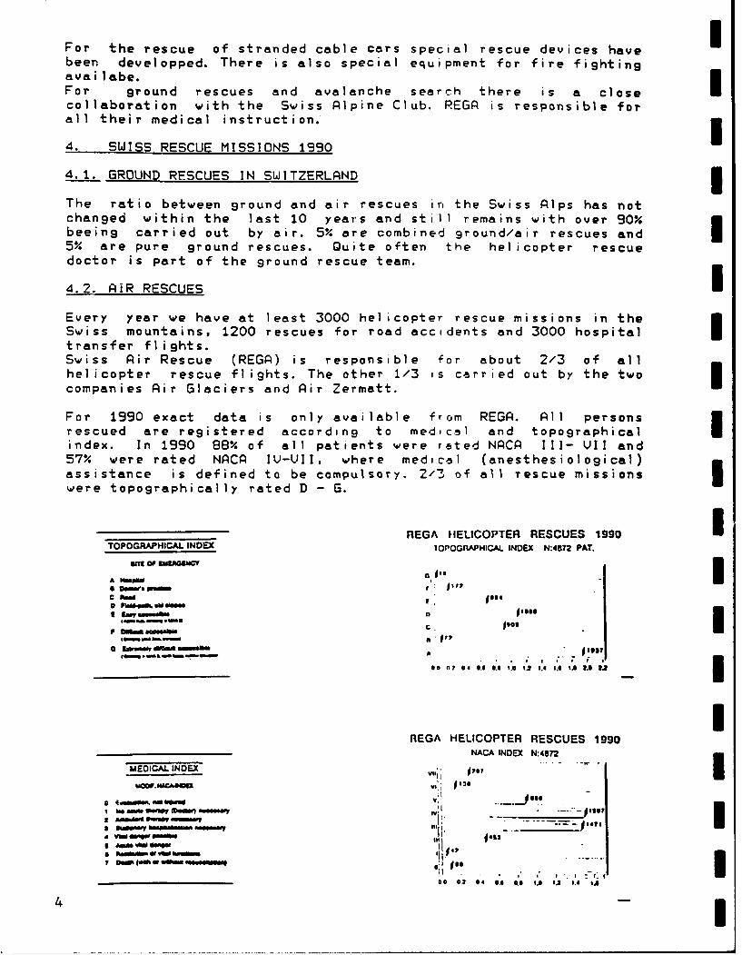
For 1990 exact data is only available from REGA. All personsrescued are registered according to medical and topographicalIindex. In 1990 86% of all patients were rated NACA III- VII and57% were rated NACA IV-VII, where medical (anesthesiological)
assistance is defined to be compulsory. 2/3 of all rescue missionsIwere topographically rated D -G.
_____________ EGA HELICOPTER RESCUES 1990ITOPOGRAPHICAL INDEX TOPOGRAPHICAL INDEX N:4872 PAT.
WMT OF EMEtAGIENCY
A aoP ."
aDemr Ph*1 SM1 .S
I' ~ S es,
00 0~@ .6 0.6 1.6 12 3.4 1.6 S.8 2.0 U
REGA HELICOPTER RESCUES 1990
MEDIAL IDEXNACA INDEX N:4872
MED.ICAINDEX V1i6
pIop Mum -08- : ~ ..~
A VW -1413
7 Doo ".. or
_______________________00 02 .4 S .'6 0.6 1.0 IJ 1.4 1A

In over 80% of all primary missions the heli copter was able toland at the site of accident. Nearly 15"% were winch evacuations
and in max. 5% the helicopter hovered ,-h le the patient waslifted aboard. According to our experience the hovering percedureis a rather dangerous rrission and we try tc- keep this rate as lowas possible.
REGA HEUCOPTER RESCUES 1990EVACUATON OF PATIENTS N: 2944
Hovo|ing 102Evacuall I '3$"
Wwch | 413
EvacualOr
Helseoler ; 429
00 01 1.0 1.S to IS
0000
4.2.1. WINCH RESCUES IN SWITZERLAND
In 1990 about 600-700 (1983/84:n=450/Y) persons were rescued byhelicopter winch in Switzerland (REGA n = 413).
REGA WINCl RISCUES 1990 REGA WINCH RESCUES 1990MA^4012. N - 43 IOPOGRAPOCAL 0N0C: N 413 4
V : " 142
ISI 44 --
0 10 40 so so 100 120 140 __l 180 M 20 0 s 00 ISO 2M
Over 75" of all REGA winch missions were rated NACA III-UII. Over90% of all REGA winch rescues were rated topographically E - G.Over 2/3 of all patients rescued by winch were evacuated fromaccessible and 1/3 from difficult aacc.dent"s site. 2V. wereextremely difficult winch rescues e.g. direct face rescues fromthe Eiger - Northface. Today almost every -.pot in the N-Face canbe reached by extension of the winch cable.
4.2.2. PRACTICAL ASPECTS OF HELICOPTER IJNCH RESCUE
The practical consequences for the treatment and evacuation inwinch rescues depend upon the rescue risks and the typ of injury.The rescue risks are determined by the meteorological, topo-graphical and objective dangers of the mourtans.For a detailled discussion we have to look at the ,,,inch rescuesin relation to the medical and topographical index:
5

II
NACA _0:
REGA WINCH RE..CUES 1990 IMEDICAL I TOPOGRAPHICAL INDEX
NACA 0: N - 43
GI.-, I
Almost 75% of all NACA 0 were rescued from difficult accessiblearea (TOPO F+G).For the rescue of hikers stranded in difficult accessible areas we Iuse special rescue belts or rescue jackets.In extremely difficult areas the climbers are evacuated by winchon the climbing harness without problems. In some cases theblocked climber could hook himselv to the winchcable, withoutlowering a rescuer first. This reduces the rescue riskscons i derabl y.In the last two years paraglider accidents in the AIps haveincreased tremendously. Very often they are not injured but arefound in very difficult situations.
NACA I + I I: I
REGA WINCH RESCUES 1990MEDICAL I TOPOGRAPHICAL INDEX
NACA I + II: N - 53
E ss, I
F ?
The winch rescue of NACA 1+11 patients usually causes no problems.These ambulant cases can be evacuated directly by the climbingharness or rescue belt and the rescuer has not to leave the winch
cable. This percedure lowers the risk considerably whenever safe
belays are lacking or objective dangers threaten the mission.Patients with dislocated shoulder (NACA II) receive sufficientpainkillers prior to the winch evacuation. Exceptionally we evenrelocate the shoulder immediately and winch the patientafterwards.
66 I

NACA III + IV:
FEGA WINCH RESCUES 1990MEDICAL I TOPOGRAPHICAL INDEX REGA WINCH RESCUES
TOOORAP)UC. WO I OSAGMOS4NACAIII + IV: N -243
Av. 11IUA0A* IIJsPIE ABOMELVIS.
0 7 3I S
E 1 61 67 soDr 1' F itsIt
:-.-# !i F ,sn F 34. 3 13
0 3
--_ Over 50% of all winch rescued patients suffer of an injury ratedNACA III + IV. A closer look at the the diagnosis of winch rescuedpersons shows a big number of injuries of head and trunc. Thus aI] proper treatment and fixation prior to the winch evacuationi sessent fal .In difficult accessible areas the rescue doctor often has thedilemma as to whether an immediate recovery or to administerI immediate treatment at the site of accident has priority,especially when stone- or icefall threatens a mission.On the other side severe headbrain- or spinal injuries demandimmediate medical treatment. If the rescue risks allow it, thepatients receive full first medical treatment at the site ofaccident. They are fixated and evacuated either in the horizontal-net or - bag with cervical collar and vacuum matress.if-necessary.I In narrow crevasses the fixation device "KED" is very usefull. Indifficult site of accident (Topo > F) additional helpers may benecesary for the fixation and the evacuation in the net or bag.Prior to the evacuation the patient receives intravenous sedativemed i cat i on.Durin9 the winch evacuation the medic in charge accompanies thepatient. According to our experience this reduces the
psychological rescue stress of the patient considerably.
NACA V + VI:
IIEUA WINCH lIIESGUES 1990MEDICAL / TOPOGRAPHICAL INDEX
NACA V +VI: N 1 g
E 4
I F 3s%
An adequate first medical treatment for severely injured patientsrequires sufficient space to work and often additional helpers. In
difficult accesible sites these patients generally are evacuatedfirst and properly treated later.

IHowever in desperate cases (1,5% of all winch-rescued patients) itcan become necessary to intubate a patient immediately endevacuate him in the net.In difficult areas such rescues are very delicate and demandusually additional helpers. This also applies for resuscitacions 1in pathless areas.
NACA UII:
FnEGA WINCH FiESCUES 1990MEDICAL I TOPOGnAPHICAL INDEX
NACA VII: N-55I
E .-
C ,
G i,
F n~s
Uery often the hel i copter can. not approach close enough to thesite of accident to determine whether a patient is still alive ornot and the doctor has to descend.As soon as the diagnosis rf death is made, the recovery usuallycauses no problems. The victims arehooked to the winch andevacuated. Due to falling stones or ice some death-body recoveriestake place only early in the morning. I5. RESCUE RISKS / DIFFICULT WJINCH RESCUES
For difficult missions (low visibility, strong winds, nightmissions, direct faces, etension of the winch cable) the rescuerisks have to be evaluated in relation to the seriousness of Iinjury.Due to the risk of rock- or ,cefall some rescues can be carriedout only during the early hours of the day. However - often it cannot be determined from the helicopter whether a patient is stillalive or not.J ithin the last 10 years 3 ambulance helicopters have been lostduring rescue missions. There is considerable danger in the Imountains from power cables and transport cables, especially withlow visibility.
Occasionally for some difficult direct face rescues we operate"Lama*-helicopters, especially equipped with a convex "bubble-door'. Thus the pilot has direct visual contact to the rescuerunderneath. This know-how is derived from the helicopter pilot's Iexperience in "helilogging" and allows long line rescues with anextension of up to 70 meters with an extraordinary precision.
For the winch rescue of para- or deltagliders an extension of thevinchcable of up to 70 meters is also necessary to avoid furtherfall due to the downwash of the helicopter. Such a rescue can bevery delicate and demands er, experienced crew.
B!

II
Winch rescues at night (1990 REGA: 58 rescued persons) demandexcellent meteorological conditions and a highly experienced crew.They take place only if exact information about the site ofaccident is available. REGA hel icopters are equipped with special
night vision devices and search ligtits.
G. FUTURE OF HELICOPTER RESCUE IN SWITZERLAND
I For the past ten years 80% of all REGA flights were medic-assisted and only 20V were acompanied by paramedics. For legalreasons today there is the tendency towards 100% medic-assistance.
The single engined Alouette III has been in service for over 25years now. For safety reasons a modern,twin engined helicopter isneeded as replacement. REGA evaluated for this reason threedifferent types of helicopters in the high alps. Finally theItalian Agusta AK 107 was chosen. This should result in safer andmore efficient rescue operations in the future.
7. CONCLUSIONS:
1. Due to the fact that many cl imbers and paragliders nowcarry walky-talkies, the time lapse between accident and alerthas been considerably reduced. Consequently the medicsare confronted more often with severly injured patientswho would not otherwise have survived.
2 Over 75% of all winch rescued persons were rated NACA III -
UII. For this reason we consider it as necessary to have amedic on board.
3. This medic has to be physically fit and must be trained inalpine technics as well, since 2/3 of all 1990 rescuemissions (n:2944) were topographically rated D - G.
4. If the rescue risks allow it and the typ of injury (NACA > 11)demands it, we start to treat injured persons even indifficult accident's sites.The assignment of trained air rescue doctors improved theefficiency of first treatment at the site of accident even indifficult and extremely inaccessible mountain areas.I
II
9
II
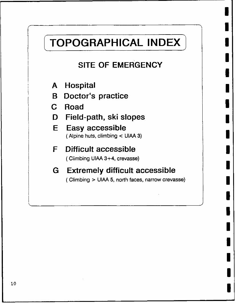
TOPOGRAPHICAL INDEX IXI
SITE OF EMERGENCY IA HospitalB Doctor's practiceC Road ID Field-path, ski slopesE Easy accessible
(Alpine huts, climbing < UIAA 3) I
F Difficult accessible 3(Climbing UIAA 3+4, crevasse)
G Extremely difficult accessible U(Climbing > UIAA 5, north faces, narrow crevasse)
IIIIII
10 I
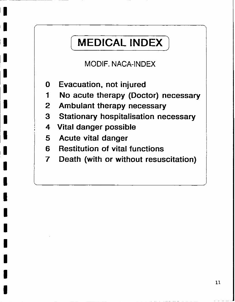
I MEDICAL INDEX]
MODIF. NACA-INDEX
0 Evacuation, not injured1 No acute therapy (Doctor) necessary2 Ambulant therapy necessary3 Stationary hospitalisation necessary4 Vital danger possible5 Acute vital danger6 Restitution of vital functions7 Death (with or without resuscitation)
I1

zi
U)U
ILLI
Q~C'jco
UJ~rC
x' CC)
I~z
< co 0Z~ 0q
(D -
Iw O
0* 00)0
*) c~o(
LU
I LU
0
UmJ
I. CD 0C
0 (
Qa 0
I 13

_ _ _ C\\T
CD~IU
I wo- 0 7
< HQ
< WD
0 4-0
I 0U =2 2 0L) 00(. .CI)
0> Zcz70U0 >>W

Icz
00C6 09- C'J
0
*Uj (0
0
*0" L
U LO
10< 0) t
Lfl 0TOO (
Iw --- -
I 15
9T
10L
0)
l~x 9
~LU
- C.
IQ~ K
C1<1O
ILUJ

~0
LLf
I,,*
LU'
-17

I Nl
zz
CI10L
0)0(0
w
LLU

CI0-01 " 01-0
IwJ
C\CJ
102:1!Lu<
- 19
REGA WINCH RESCUESITOPOGRAPHICAL INDEX / DIAGNOSIS
HEAD/BRAIN TX/SPINE ABD/PELVIS
D 7 113
iE61 57 191
F 34 35 12
3 511
20I

n z X
00
00
IL<- N
C)I v
1~I~oJ
0>1

zz
I 02
0Cz
uj-C) LC)
~LLL
LLU
~LU
Ij

3 WOUNDTI CAIRIE MANUAIL
3 WHlID~IKENES MIECAIL SCIEY
D. D. Zukin, MD
CONTENTS
I. Normal Repair of LacerationsII. Types of WoundsIII. Factors Affecting Wound Infection RateIV. MaterialsV. General Evaluation of the PatientVI. Wound Pre-CareVII. Wound ClosureVIII. AftercareIllustrations
Introduction
The basic principles of wound cleansing and closure remain the same, even out in thewilds. The main problems faced in the wilderness are related to limited supplies. With a minimumof equipment, however, one can close the majority of simple lacerations. This manual sets forththe basic principles of wound closure. The reader will have to modify these guidelines basedupon the resources which he or she has at hand.
1. NORMAL REPAIR OF LACERATIONS
A. RE-EPITHELIALIZATION.
The epidermis, the epithelium of the skin, protects the underlying dermis from bothdesiccation, and invasion by bacteria. When the skin is injured new epidermal cells migrate out toclose over gaps. When a lacerations is repaired so that the wound edges evert, the basal cells ofeach wound edge come into direct contact and a new layer is formed in as little as 12-18 hours;but with inverted edges the new layer will take approximately 72 hours to form (SEEILLUSTRATION).
Re-epithelialization occurs deep to any necrotic tissue, and hence debrides away non-viable
23

II
tissue. IB. NEOVASCULARIZATION.
Neovascularization begins at day 2-3, and peaks at day 4. Capillary buds grow out from eachwound edge, and eventually join in the middle. The proliferation of blood vessels gives thehealing wound an erythematous hue, bu' with minimal pain or induration.
Be careful not to mistake neovascularization for early infection.
C. CONNECTIVE TISSUE REGENERATION.
Fibroblasts and macrophages, like capillary buds, begin to appear in about 2-3 days. Newcollagen is laid down, and increasingly stronger strands bridge the wound gap. The collagenfibers are continually remodeled as the wound matures. In one study a preparation of normal skincould withstand a load of 18 pounds. A similar sample, 10 days after repair could withstand a loadof only 1 pound; at 20 days 2 pounds; at 40 days 6 pounds; and at 5 months 14 pounds. Theninal strength of the wound, once healing is complete, is only about 80% that of normal tissue. ISimilarly, extensibility in a healed wound is not as great as in intact skin. III. TYPES OF WOUNDS
A. LACERATIONS n
1. Shear (sharp cuts caused by metal or glass).
Little tissue damage immediately adjacent to the laceration. Has the highest resistance towound infection. Minimal edema during the healing process. Minimal foreign material andground in dirt.
2. Tension wounds (i.e. a blow to a skin without a bone immediately below the skin).
Irregular edges, increased damage of the tissue directly adjacent to the laceration itself. Higher Iinfection rate and scarring potential than shear wounds.
3. Compression wounds (a bone beneath the skin, struck by blunt object, i.e. stellate wound 3that occurs when the forehead strikes the ground).
Increased edema, infection potential and scarring than shear wounds. l4. Flaps.
Distal flaps (i.e. attached distally, detached proximally) have a more tenuous blood supply Iand hence have a higher potential for necrosis than do proximal flaps (i.e. flaps attachedproximally). However, flaps in children, especially of the distal fingertips, have an amazingcapacity of recovery. I
24 1I

B. PUNCTURE WOUNDS.
Puncture wounds may contain foreign material. For example, bits of fabric and plastic maybecome imbedded in the foot when a patient steps on a nail while wearing running shoes.Puncture wounds of the foot deep enough to reach the bone can result in osteomyelitis, frequentlywith pseudomonas as the causative organism.
C. ABRASIONS.
Loss of some or all of the epidermis. Repair is by migration of new epidermal cells fromadjacent intact skin edges as well as from epidermal appendages (i.e. sweat and sebaceous glands)and hair follicles.D. FOREIGN BODIES.
Suspect 1oreign body in wounds which become infected. Foreign bodies are difficult todetect prior to closure without the use of xray.
III. FACTORS AFFECTING WOUND INFECTION RATE
Several factors affect the wound infection rate. Highly vascular regions, such as the face andscalp, are more resistant to infection. Shear lacerations have a lower infection rate thancompression injuries. Wounds repaired with 8-10 hours of the time of injury have a lowerinfection rate than wounds sutured later than 8-10 hours. However facial lacerations frequentlyheal without infection even when sutured closed more than 24 hours after the injury.
Suturing technique also influences the infection rate. Wounds with tight sutures andwounds with inverted edges are more prone to infection. Wounds that are grossly soiled, such asthose resulting from animal or human bites have higher infection rates than clean wounds.
IV. MATERIALS
A. SUTURE THICKNESS-- the more O's, the finer the suture:
Largest 0, 00, 000, 4-0, 5-0, 6-0 Smallest.
B. SUTURE MATERIALS.
1. Absorbable.
a. Vicryl and Dexon (polyglycolic acid).
Lowest infection rates of absorbables, because breakdown products inhibit bacterialgrowth. Lowest tissue reactivity of absorbables. Good tensile strength. Braided, so hold knotswell, but makes gradual cinching down of ties more difficult. Can take 40 or more days to absorb,but usually lose tensile strength within 14 days. Primarily used for deep and subcuticularclosures.
25

II
Come both dyed and undyed. For 2mergency department use, choose undyed,especially when used near the skin surface.
b. Chromic. I
A gut suture which has been treated with chromic ion to increase its strength. Hightissue reactivity and slightly higher infection rate. Lower tensile strength than vicryl or dexon.Stiffens when dry, hence packed in liquid to make it easier to handle. Non-braided. Takes 14-17days to absorb.
c. Plain gut. 3Most tissue reactivity of the absorbables. Same infection potential as chromic. Low tensile
strength, so breaks easily if knot cinched tightly. Absorbs in 4-10 days, so useful where rapidabsorption required, such as inside the mouth. I
d. Fast -Absorbing Gut.
Heat-treated plain gut loses its tensile strength even faster than plain gut, and is thus useful Ifor placing sutures in areas where suture removal will be difficult.
e. PDS (Polydioxanone) and Maxon (GTMC) i
PDS and Maxon, like Vicryl and Dexon, are degraded primarily by hydrolytic action, andpossess a similar low tissue reactivity. Both are monofilaments. Monofilament materials possessa smoother passage through tissue, and, in studies, a lower incidence of infection rate.
PDS and Maxon are ideal for back-packing trips and other expeditions because, althoughthey are absorbable, they retain tensile strength for-more than two weeks and thus are also suitablefor simple skin closure. Therefore one can take along just one suture material, and still be able toclose both the deep and the skin layer.
2. Non-absorbable.
a. Silk. Standard for all sutures in terms of ease of tying. Braided, so holds knot well. IUnfortunately, the higher infection potential of silk sutures in comparison to monofilaments limitsits usefulness. 3
b. Nylon (Ethilon & Dermalon) Monofilament. Low tissue reactivity, lowinfection rate. Harder to use than silk. Knots tend to unravel, hence use 4- 5 "throws" per knot.
c. Polypropylene (= Prolene). Monofilament. Similar features to nylon. Slightlyeasier to handle than nylon. Blue prolene is especially useful for repairing scalp lacerations in darkhaired individuals, because the sutures are easy to locate for removal. 3
d. Dacron. Braided, easy to use, low tissue reactivity. Comes in a coated form whichpulls through tissue more easily than the uncoated type.
C. NEEDLES. Look on the package.
I26 I

1. Curves. Straight, 3/8th circle (for most uses), 1/2 circle (for web spaces of fingers and toes.
2. Needle cross-sections.
a. Cutting. Triangle with flat base on the outer circumference and cutting point on theinner circumference.
b. Reverse Cutting. Triangle with sharp cutting edge on the outer perimeter, and flatsurface on the inner perimeter, hence less cutting of skin in region where suture tension is thegreatest. In many cases reverse cutting needles are labeled simply as cutting needles. Choose thisneedle configuration for most ER suturing situations.
c. Taper. Circular. For special situations, such as tendon repairs.
3. Needle classes.
Note, the picture on the box reflects the actual size and curvature of the needle. The lettersand numbers stand for different things depending on the manufacturer. For example, M is a seriesnumber used with ethicon. FS = for skin,honed 12 times. P = plastic, honed 24 times. PS =plastic surgeon's, intermediate honing between FS and P. Larger numbers generally refer tosmaller needle sizes. For facial lacerations, use a P3 needle.
3 Recommendations: For expeditions, choose cutting needles of various sizes, small for the face andthe fingers (such as the P-3 needle by Ethicon), and larger needles for the arms, legs and trunk(such as the FS-2 by Ethicon).
D. INSTRUMENTS
1. Skin hooks.
Decrease local tissue damage but do not offer the control of forceps.
2. Forceps.
Smooth and rat-toothed. Toothed forceps cause less crushing of skin because less pressureis required to secure the skin edge.
3. Needle holders.
Hold the needle at the junction of the middle and proximal 1/3rds, near to where the sutureattaches to the needle. Smooth and corrugated surfaces. Smooth best for 5-0 and 6-0 suturematerial. Hold the clamp with the ring finger in a loop and the index finger along the shaft (="palm" the needle holder).
4. Scissors.
Iris scissors are quite adequate for debriding wound edges.
i 5. Scalpels
Scalpels are primarily used for incising specific patterns for plastic wound closures that are
27

II
best not attempted on expedition.
V. GENERAL EVALUATION OF THE PATIENT
Remember not to be distracted by the open wounds of a multiply-injured patient. The 3course of resuscitation still should be:
1. A-C (Airway + Cervical spine immobilization)2. B (Breathing)3. C (Circulation)
Once the patient is stable, then proceed to care for the open wounds. Check the status of the 3circulation, motor function and sensation (CMS) of the injured area. Be sure to check the sensory-motor examination f administering the local anesthetic.
Tetanus prophylaxis: Follow the guidelines at the end of the handout. In an un-immunized patient, both tetanus immune globulin (250 IU given IM) and tetanustoxoid (0.5 cc IM) must be given for tetanus prone wounds such as punctures anddeep lacerations.
VI. WOUND PRE-CARE
A. SHAVING. ILeave your razor blades at home. There is a higher incidence of infection in operative
wounds prepped with shaving as compared to clippers. Therefore clipping away bothersome hairswith a scissors is the preferred method of hair removal.
NEVER SHAVE THE EYEBROWS. 3Eyebrows are slow to grow back, and in addition they serve as valuable landmarks during
suturing. IB. LOCAL ANESTHESIA. I
Whenever possible, local anesthesia should precede irrigation, for the sakeof the patient's comfort.
1. Local infiltration. 3There is no greater infection rate injecting through the open wound edge as opposed to
through intact skin, and it is much less painful to the patient. Raise a weal in the dermis. 3
281 2s I

1 % lidocaine is adequate for most purposes.
Ic. of I% lidlok.aine cvntains ,0 mg ot the drug. Do not exceed 4 mglkg oflidocaine or 0.4 cclkg of the 1 % solution (with a maximum dose of 300 mg). Donot exceed 7 mg/kg of lidocaine with epinephrine or 0.7 cc/kg of the 1 % solution (with amaximum dose of 500 mg). The addition of epinephrine both prolongs the duration of anesthesia,and slows the oozing of blood in vascular areas such as the face and the scalp.
EPINEPHRINE SHOULD NOT BE USED ON THE FINGERS,TOES, TIP OF THE NOSE OR THE PENIS, OR IN WOUNDS WITHTENUOUS PERFUSION (SUCH AS FLAPS) BECAUSEIRREVERSIBLE ISCHEMIA CAN RESULT.
If you are going to bring one anesthetic solution, bring 1%plain (without epinephrine) lidocaine.
Bupivacaine (= Marcaine) 0.25% or 0.5% lasts for several hours, and hence is auseful agent for prolonged procedures. The dose is 1-2 mg/kg, or about 1/2 cc/kg of the 0.25%solution (contains 2.5 mg/cc). Use with caution. Do not give more than the recommendedamount. Always aspirate back on the syringe before injecting, becauseinadvertent intravascular injection has resulted in irreversiblecardiovascular collapse.
2. Regional block (i.e. the digital nerve block for finger lacerations, or the mental nerve blockfor lip lacerations) Ideal because there is no edema and secondary deformity of the area to besutured.
C. IRRIGATION.
1 Forceful (one the order of 7 psi of pressure) irrigation of wounds effectively washes awayboth bacteria and foreign material.
THE METHOD OF CHOICE FOR IRRIGATING OUT SOILEDLACERATIONS IS TO FIT A 20 CC OR 30 CC SYRINGE WITH A16 GAUGE OR 18 GAUGE ANGIOCATH AND PRESS DOWN
I FIRMLY ON THE PLUNGER.
Irrigation from an iv bag or bulb syringe irrigation simply will not generate the pressurenecessary to remove foreign material and bacteria. Syringes and catheters are lightweight, and caneasily be brought along. Saline can be made by adding 1/4th teaspoon salt per cup of boilingwater. (Let the water cool before attempting irrigation.)
Clean wounds, such as those caused by sharp metal or glass, are generally NOT highly
contaminated with foreign material and do not require high pressure irrigation.
29

II
Use caution to ensure that none of the irrigation fluid splashes into your own eyes, placing 3you at risk for catching hepatitis virus (or worse).
D. TOPICAL ANTISEPTICS I1. Betadine Prep Solution (= 10% Povidone-iodine in an aqueous base)
1% povidone-iodine solution (= a 1:10 dilution of the stock 10%Betadine prep solution) is the topical antiseptic of choice for use inthe emergency department. Dilute povidone-iodine solution applied for 30-60 secondsinto the open wound significantly lowers the incidence of wound infection without perceptiblyaltering the course of wound healing.
So, along with your syringes and catheters, bring a small plastic bottle of betadine solutionto leak onto your carefully packed clothes.
Unlike Normal Saline, the Betadine Prep solution is dabbed on
the wound, not irrigated under pressure. 3The same solution is used to paint the surrounding intact skin just prior to draping.
NOTE: The reason for the hesitancy on the part of many to employ povidone-iodine solution tothe open wounds is that povidone-iodine detergent (= Betadine Surgical Scrub, foundin both bottles and the ubiquitous scrub brushes with the attached sponges) isterribly cytotoxic. The fact is that all detergents are highly cytotoxic, so as a general rule:
IF IT LATHERS, DON'T USE IT IN THE SAME ROOM AS ANOPEN WOUND (or at least in the same tent)!
As with most rules, there are exceptions. Green soap-iz recommended for thedecontamination of bite wounds from potentially rabid animals such as skunks, raccoons, foxes Iand cows (the cow, by the way, is now the most commonly rabid domestic animal in the state ofCalifornia).
Povidone-iodine, with repeated use can be absorbed systemically causing mild alterations Iin thyroid function tests (most notable an elevated TSH with a normal T-4) Hence the solutionshould be used with caution in pregnant women, and in infants.
2. Betadine Surgical Scrub (= 10% povidone-iodine in a detergent base).
As mentioned above, Betadine Scrub is cytotoxic, and hence should only be used tocleanse intact skin (i.e. washing your own hands.) I3. Phisohex.
Like betadine scrub, phisohex contains a detergent and consequently should not be
330
I
I
employed near an open wound.
1 4. Alcohols.
Alcohols are highly cytotoxic, essentially fixing the tissues. Like detergents, alcoholsprobably shouldn't be used in the same room as an open wound.
5. Hydrogen peroxide.
All physicians have watched with pleasure the way hydrogen peroxide bubbles away bloodstains from their scrub suits and white jackets. Unfortunately, peroxide causes the sameeffervescence in capillaries, leading to a complete standstill in capillary blood flow when appliedinto an open wound. In addition, peroxide is a poor antiseptic. Therefore peroxide should not bein open wounds.
6. Shur-clens.
Shur-clens is a non-toxic surface active agent (even when administered intravenously)effective for cleaning off dirt or grease. It probably offers little advantage over normal saline.
E. DEBRIDEMENT.
All foreign material must be removed, however tedious this may seem at times. In addition,carefully trimming irregular wound edges will result in a finer final result. Grossly devitalizedtissue should be removed. Debridement can be carried out with either scalpel or scissors.
F. HEMOSTASIS.
The following steps are useful for obtaining hemostasis:
1. Apply direct pressure for 10 to 20 minutes (after all clots first removed.)
2. Epinephrine 1:1000, 1cc diluted with 4-5 cc of saline on a 4X4 gauze, held over thebleeding region for 5 minutes ,will stop small dermal bleeders. Larger vessels must be ligated,however, because the vasoconstriction is temporary. Contraindicated in regions where epinephrine
i is contraindicated (see Anesthesia).
3. Suture the wound. Very effective in wounds such as scalp lacerations which continue toslowly bleed even after local pressure. The wound must be observed for several minutes forhematoma formation, prior to bandaging.
4. Locate, clamp and ligate arterial bleeders. Pressure over a main artery (such as with aBP cuff inflated proximal to an extremity laceration), may facilitate the ligation of arterial bleeders.
Avoid ligating arterial bleeders in the hand because the digital nerves run inclose proximity to the arteries.
31

II
VII. WOUND REPAIR 3A. WOUND TENSION.
The tension within the skin suture loop can be decreased by first bringing together thesubcutaneous tissues with buried sutures. The tension can be further decreased by undermining(see Undermining, below). 3
Sutures cinched with too much tension strangulate the tissue causing local necrosis.Hence the familiar surgical aphorism: 3
APPROXIMATE, DON'T STRANGULATE IB. UNDERMINING.
Undermine underneath the subcutaneous tissue in the natural fascial plane. Use scissors, Ientering with the blades closed, then opening the blades to dissect. Ideally, undermine to the widthof the wound gape on both sides. Especially useful in situations where there has been tissue loss,as well as in regions where the skin is normally taught, such as the lower leg or forearm.
C. SKIN SUTURES. 31. General.
Use monofilament nylon or prolene (Maxon and PDS are acceptable substitutes). 3Enter the skin at about 90 degrees to the skin surface to insure that there will be as much suture atthe base as the top of the suture loop, to aid in eversion.
Use only enough tension to bring the wound edges together.
In complex wounds, first suture the most difficult region. 32. Simple Interrupted Sutures (SEE ILLUSTRATION).
Double loop the first throw of the knot, and single loop the subsequent throws. Lift 3straight up on the sutures just prior to cinching down on the first throw as this simple maneuverserves to enhance wound edge eversion. With the second throw, pull the knot to one side of thewound edge. Use a total of 4-5 throws for monofilaments. Be sure that the suture intertwines inopposite directions with each throw, or else the knots will not square.
For cosmetic closures, use more sutures per centimeter. For facial lacerations place thesutures 2 mm from the wound edge and 2-3 mm apart. However, in the extremities use largerbites and place the sutures further apart.
3. Running Sutures (SEE ILLUSTRATION). 3I
32
I

The first loop is identical as for the interrupted technique, but then one continues down thewound without cutting and tying until the wound is closed. This technique is quicker, and yieldsexcellent results with practice
4. Vertical Mattress Suture (SEE ILLUSTRATION).
The vertical mattress suture insures wound edge eversion and is therefore useful in regionssuch as the web-space between the thumb and index finger where inversion is often a problem.
5. Horizontal Mattress (SEE ILLUSTRATION).
Few indications. May be helpful in regions of thinning skin, such as lower legs in elderlypatients.
6. Half-Buried Horizontal Mattress (SEE ILLUSTRATION).
The half-buried horizontal mattress stitch goes through the subcuticular portions ofangulated flaps in stellate lacerations. It is the method of choice for repairing such injuries becauseit preserves the vascular supply to the flap (remember that the skin vessels run in the dermis, andnot at the dernal- epidermal junction).
D. BURIED SUTURES.
The deep layer serves three vital functions in insuring the optimal cosmetic outcome of asutured facial laceration. First, the deep layer provides up to six weeks additional support to thewound after the skin sutures are out. Thus the scar is less likely to widen with time. . "iond, thedeep layer avoids the development of unsightly pitting in the injured region caused by lack ofhealing of the deep portions of the wound. Third, the deep layer serves to preserve the normalfunctioning of the muscles of facial expression. However, when rapid closure of a wound isnecessary, deep sutures can be kept to a minimum or eliminated entirely.
In the extremities, and the hand in particular, the deep sutures increase the risk of infection,and have the potential of damaging vital nerves, arteries and tendons. Hence, as a rule:
AVOID USING DEEP SUTURES IN THE EXTREMITIES.NEVER USE DEEP SUTURES IN HAND LACERATIONS.
The suture materials most commonly used for the deep layers are dexon, vycril, PDS andMaxon. Use the minimum number of sutures :o close the deep tissue. The more foreign material,the greater the risk of infection.
There are two basic deep stitches used in laceration repairs:
1. The Buried Knot Stitch (SEE ILLUSTRATION).
IBegin and end at the base of the wound, so as to bury the final knot. In some cases thedeep sutures along a wound must all be placed prior to tying, because the tying of one suture canmake the placement of the subsequent deep sutures more difficult.
2. The Buried Horizontal Mattress Stitch (SEE ILLUSTRATION).
II 33

mm
Be careful that both sides of the wound are sutured at even depths. Do not pull tightly, orthe wound will pucker.
E. SKIN TAPES (= STERI-STRIPS) iSkin tapes are useful for shallow, non-gaping wounds. Tapes are not practical for use in
toddlers and young children, who have a tendency to pull them off. Never use steri-strips over the Ijoints.m
Prior to applying skin tapes, wait for full hemostasis. Tapes will not adhere to a moistareas. Next prepare adjacent skin with tincture of benzoin to enhance sticking. Benzoin isavailable in tiny, unit dose containers practical for most expeditions. Allow the benzoin to dry,and become tacky and then apply the strips. Finally, cover with a protective dressing. 3F. SKIN STAPLES.
Disposable skin staplers are significantly faster for repairing lacerations than conventionalsutures because knot tying is not needed. Staplers are light-weight, pre-sterilized and pre-loadedwith staples. Disposable staple removers are also required.
Staples are easy to place, and therefore are ideal during adverse conditions. 3G. Skin Glue (Tissue Adhesives)
Although available in Europe and Canada, tissue adhesives are not yet available for routine 3use in the United States. The two most commonly used adhesives are fibrin glue andcyanoacrylate. In a study of 1500 children, Mizrahi et al noted excellent cosmetic results usingHistoacryl Blau, a butyl cyanoacrylate ( J Ped Surg 23:312-313; 1988). Skin glue is commonlyused by mountain climbers to cover hand wounds. Skin glue may increase slightly the risk ofwound infection.
In using skin glue, first hold the wound edges together. Then apply a thin layer of glue.The glue sets in 1-2 minutes. There may be heat production during polymerization. Use extreme Icaution near the eyes because the glue will adhere to the cornea and lids if it runs into the eye.
II. TREATMENT OF ABRASIONS
Ground in foreign matter must be removed or else an unsightly road tattoowill result.
Abrasions should be dressed either with a topical antibiotic salve such as neosporin orsilvadene, or else with a semi-occlusive dressing such as Duoderm, Tegaderm, Xeroform, or even Ia simple band-aid.
I. TREATMENT OF MINOR 2ND DEGREE BURNS 3The treatment of minor burns is similar to that described already for abrasions. If the
blister is intact, it should not be opened, because re-epithelialization is faster is the blister is notunroofed. If the blister is already un-roofed, then the open area can be cleaned with saline, and thewound covered with bacitracin, silvadene, or a xeroform dressing. Xeroform can usually be left in
I34 I

I
place until the burn has healed.IVII. AFTER CARE.
IA. CLEAN DRESSING.
Conventional band-aids are fine for small lacerations. Slight pressure will aid inhemostasis and decrease local edema. Wounds that are kept covered with an occlusive bandageheal slightly faster than wounds left open to the air.
B. ELEVATE TIlE WOUNDED REGION (when practical).
C. SPLINTS.
Lacerations over joints should be splinted for 5 - 7 days.
D. ANTIBIOTICS.
Prophylactic antibiotics sound like a good idea, but unfortunately they have not beenproved effective in decreasing the incidence of wound infections in simple lacerations. Antibioticshave a place in certain animal and human bites.
E. WOUND CHECK.
In any case with a high potential for infection, check the wound every 2 days for signs ofinfection. In the case of animal bites, the first check should be at 24 hours.
F. SUTURE REMOVAL. (SEE CHART)
Unsightly suture marks occur when epithelial cells grow down the tracks of skin sutures.These tracks can form on the face of children in as little as 5 days. Hence the need for promptremoval of facial sutures. Most suture marks regress and disappear with time, however.
35

UII
Suture Chart
REGION SUTURE SUTURE REMOVAL (days)
CHILD ADULTTHE FACE 6-0 NYLON OR PROLENE (SKIN) 4 4-5
5-0 VYCRIL OR DEXON (DEEP)
THE SCALP 3-0. 4-0. OR 5-0 NYLON 5-7 7OR PROLw'iE
THEHAND 5-0 OR 6-0 NYLON JOINT 10-14 12-16OR PROLENE OTHER 7-10 7-12
NO DEEP SUTURES
EXTREMITIES 4-0 OR 5-0 NYLON JOINT 10-14 12-20OR PROLENE (SKIN) OTHER 7-10 7-12
4-0 VYCRIL OR DEXON (DEEP)
THETRUNK 4-0 OR 5-0 NYLON 7 7-10OR PROLENE (SKIN)
4-0 VYCRIL OR DEXON (DEEP) IORAL MUCOSA 6-0 VYCRIL OR DEXON ABSORBABLEANDTONGUE OR 4-0 OR 5-0 PLAIN
Because of the low tensile strength of the wound during the first 10 -20 days, lacerations on theface, and over joints should be re-enf, -ced with skin tape following suture removal.
IIII
36 1I

TETANUS PROPHYLAXIS
CLEAN WOUNI)S (Clean. superficial abrasions and lacerations)
Tetanus Toxoid Give Give TetanusImmunizations Tetanus Toxoid Immune Globulin
Three or more, No Nolast within 10 yr
Three or more, Yes Nolast > 10 yr
Fewer than three Yes Noor unknown
TETANUS-PRONE WOUNDS (Contaminated, deep punctures, tenuous bloodsupply, extensive lacerations)
Tetanus Toxoid Give Give TetanusImmunizations Tetanus Toxoid Immune Globulin
Three or more, No Nolast within 5 yrs
Three or more. Yes Nolast > 5 yrs
Fewer than threeor unknown Yes Yes
37
REFERENCES
Alexander JW. et al: The Influence of Hair-Removal Methods on Wound Infection. Arch Surg 118:347,1983.
Bennett RG: Selection of Wound Closure Materials. J Am Acad Derm 18:619-637; 1988. UBerk WA, Osbourne DD, Taylor DD: Evaluation of the 'Golden Period' for Wound Repair: 204 Casesfrom a Third World Emergency Department. Ann Emerg Med 17:496-500; 1988.
Dushoff IM: A Stitch in Time. Emerg Med 20:23:57; 1988.
Gravett A. Sterner S, Clinton JE, Ruiz E: A Trial of Povidone-lodine in the Prevention of Infection inSutured Lacerations. Ann Emerg Med 16:167-171; 1987.
Grossman JA: Minor Injuries and Disorders: Surgical and Medical Care. Philadelphia, JB Lippincott; I1984.
Kaplan EN. Hentz VR: Emergency Management of Skin and Soft Tissue Wounds: An Illustrated Guide. 3Boston, Little Brown and Co; 1984.
Mizrahi S, Bickel A, Ben-Layish E: Use of Tissue Adhesives in the Repair of Lacerations in Children. JIPed Surg 23:312-313; 1988.
Tobin GR: Closure of Contaminated Wounds -- Biologic and Technical Considerations. Surg Clin NA64:639; 1984.I
IIIIII
38
I

illustrations P 1
I~A
Technique of undermining using a mosquitoclamp. The extent of undermining (hatched area) on bothsidcs of the wound should roughly equal the gape of thewound. First the clamp enters to the desired depth (A). Nextthc blades are opened to bluntly dissect open the tissuc plane
8 (BI). An iris scissors can be used also.
A. The base of the lImp is not broad enough. and
conscquently there is inversion of the wound cdges. Notice
ho.- basal. regenerative epidermal cells on either side of the
wound do not conic into contact. thus delaying the healing.
HI. There is a broad base to the suture loop, and consequently
the edge% even. Notie that in this inlstance the basal epider-
inat vells do conic into contact. thus facilitating healing.
INVERSION VS EVER51ON UNDERMINING
(Reprinted with permission from Zukin DD, Simon PR: Emergency Wound Care.
Principles and Practice Rockville MD: Aspen Publishers, 1987)
39
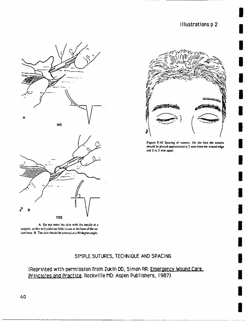
IIllustrations p 2
A
'-- _ . .- -. .-
NO
Figure 5-16 Spacing of sutures. On the face the sutures7should Ibe placed approximately 2 mm nrm the wound edgeand 2 to 3 mm apart.
jI~i
"T III
A. Do not entcr the skin with the needle at atangent. as this will yield too little tissue at the base of the su-ture loop. B. The skin should be entcred at a 90 degree angle. I
SIMPLE SUTURES, TECHNIQUE AND SPACING
(Reprinted with permission from Zukin DD, Simon RR: Emergency Wound Care.Princioles and Practice. Rockvllle MD: Aspen Publishers, 1987)
401 mI

Illustrations p 3
I The instrumernt tic. During the instrument tic, manipulate the needle end of the suture with the nondominant hand
and the needle holder with the dominant hand. To start with. the suture enters the far side and exits the nea'r side of the wound ini the usual fashion. A. Lauy the needle holder on the suture on the near" side, and then wrap the suture around the nedle holder
twice. B. Next reach back with the needle holders and grab the free suture end on the far side. C. Cross hands. pulling the freeend back toward the near side and bringing the nedle end of the suture to th far side. D. Lift up both suture ends and cinchdown the first throw. E. Now lay the needle holdr on the suture on t far sid and loop once. F. Reach back and grab the freesuture end. G. Cross hands and the knot will square. Repeat this pattern for a toWa of four to five throws.
SIMPLE SUTURE, INSTRUMENT TIE(Reprinted with permission from Zukin DD, Simon RR: Emergency Wound Care.Principles and Practice. Rockville MD: Aspen Publishers, 1987)
41

I Ilustrations p 4
RUNNIN SUTUR(Repinte Mtnperissin frm Mi DD
RUNNING. SUTUREepinedfr(Rerite witth permissionn oro Zukin DDs&Wlkn omay
_ _ _ _ _ _ _ _ _ _ _ _ _ _ _ _ _ _ uI')2
42 o R mrec ondCr.Picoe n
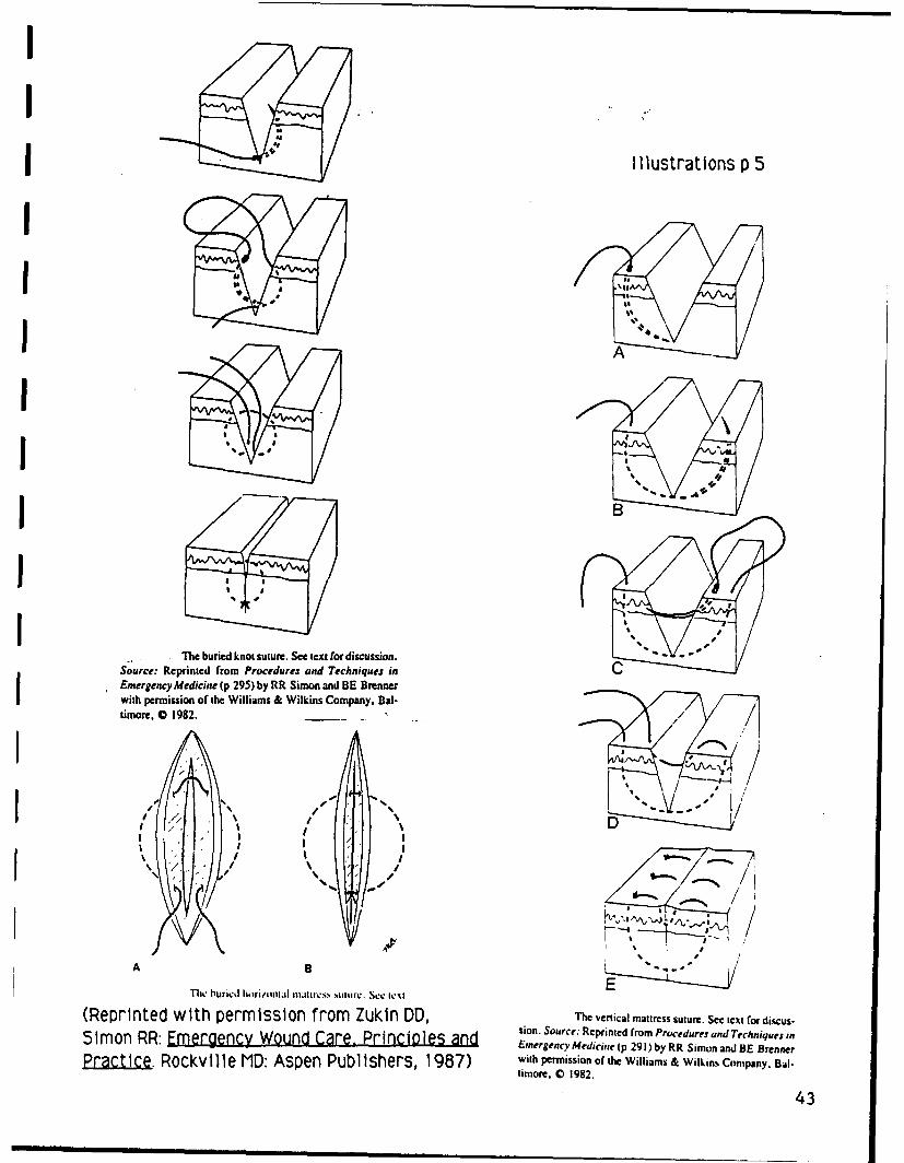
I Illustrations p 5
AAI-
L %U_
I8%
AIVi
A L
(Reprin~~tewihe rsi frmkno D h vria mtrs suture. See text for discus-5mnR:EegnyWudCrRncoean SinSource: Reprinted from Procedures and Techniques in
Emnergency Medicine (p 291) by RR Simon and BE BrennerPrcieR~~leM:ApnPbihr,18)with permission of the Williams & Wilkins Company. Bal
timoremore Cr92 1982.
// 43

I llustratlonsp 6
%I
HALF-BURIED HORIZONTAL MATTRESSI
(Reparn wfi thlai peraision uin aDhl,
Simon RR: Emeroency Wound Care. Principles and
P.racice Rockyille MD: Aspen Publishers, 1987)
44I
LYME DISEASE: A GLOBAL PERSPECTIVEDouglas A. Gentile, MD
OBJECTIVES:
At the completion of this presentation, the participantshould have a clear understanding of:
1. The epidemiology of Lyme disease2. The clinical manifestations of Lyme disease3. The principals of diagnosis of Lyme disease4. The recommendations for treatment of various
manifestations of Lyme disease
45

i
I. IntroductionA. Epidemic form of oligoarticular arthritis
recognized in 1975 in Lyme, Connecticut1. Originally diagnosed as JRA I
B. Epidemiologic evidence suggested arthropod vector1. Geographic clustering2. Rural setting3. Temporal clustering - spring and summer
C. Tick vector1. Erythema migrans - linked to European
syndromes Ia. Ixodes ricinus - 1909
2. Epidemiologic studies implicated Ixodes dammini3. Response to penicillin
D. 1982 Burgdorfer isolated spirochete from mid-gut ofIxodes dammini ticks1. Antibodies in patients with Lyme disease
E. Spirochetal etiology confirmed when isolated fromblood, CSF and skin lesions1. Borrelia burgdorferi
II. Borrelia burgdorferi IA. Fastidious, slow growing
1. Difficult to isolate from patientsB. Outer membrane unique
1. Genes encoding located on plasmids2. Major antigens - OSP-A, OSP-B
C. Differences between American and European isolates1. Morphology, outer surface proteins, plasmids I2. May account for clinical variations
III. Vector and animal hostsA. Transmitted primarily by Ixodes ricinus complex
1. I. dammini - northeastern and midwestern US2. I. pacificus - western US3. I. ricinus - Europe4. I. persulcatus - Asia5. I. scapularis - southeastern US
a. Competent laboratory vectorb. Naturally infected ticks recovered NC I
B. Other ticks and arthropods implicatedC. Lifecycle and preferred hosts
1. Critical that immature stages feed on samehost
2. White-footed mice iajor reservoira. Tolerant to infectionb. Spirochetemic throughout summer I
3. As tick feeds organism becomes systemica. Organisms injected with saliva?
(1) Ribeiro, et al - systemic infection Iafter attachment, before engorgement
(2) Burgdorfer(a) <5% of adults with systemic
infection
46I

(b) Studies show regurgitation ofgut contents - tracers
(3) Animal infected without evidence ofsystemic infection in ticks
b. Ticks often must remain attached 24-48hours before transmission of organism(1) 1 of 14 at 24 hours, 5 of 14 at 48,
13 of 14 at 724. Transmission to humans - larva
a. Small sizeb. No history of tick bite in 70%
5. Preferred adult host - white-tailed deera. Critical to high tick densityb. Deer population correlates with diseasec. Not involved in lifecycle of B.
burgdorferid. Great Island - decrease in ticks after
deer eliminated6. When all features present high infection rates
a. Parts of Northeast over 60% of ticksinfected(1) 1 per square meter on well kept lawns in
Westchester Countyb. West Coast 1-2% I. pacificus infected
IV. EpidemiologyA. 1989 - 8333 cases reported
1. Most common tick-borne infectionB. Most from 3 endemic areas
1. Northeast, Midwest and Pacific Coast2. Sporadic cases identified 43 states
V. Clinical manifestationsA. Multisystem involvementB. Occurs in stages
1. Stage I - early, localized2. Stage II - disseminated3. Stage III - persistent
C. Patients may have one or all stagesD. Mimic other diseases
VI. Early localized infection (Stage I)A. Characterized by erythema migrans
1. Develops 60-80% of patients2. B. burgdorferi spreads locally in skin
B. Description of EM1. Annular, usually centrifugally expanding plaque2. Begin as red macules, maybe nodules3. May have target configuration4. Central area clear, erythematous, vesicular5. Usually asymptomatic
a. May be pruritic or painful6. Size variable
a. 2-60 cm diameterb. Median 15cm at 2 weeks
7. Usually resolve within 3-4 weeksa. "chronicum" misnomer
47

IC. May be accompanied by regional lymphadenopathy,
fever and minor constitutional symptoms IVII. Disseminated infection (Stage II)
A. Within days or weeks spirochete may spread to manysites via blood or lymph1. Recovered from blood during this stage2. Probably initially to all organs3. Sequesters in certain niches
B. Hematogenous spread1. Secondary annular skin lesions
a. Occur in 20-50% of patients in USb. Resemble EM but smaller and migrate less i
2. Other skin lesionsa. Diffuse erythema or urticariab. Evanescent lesionsc. Malar rash
3. Excruciating headache, mild neck stiffnesscommona. Short attacks lasting hoursb. CSF normal
4. Migratory pains in joints, bursae, tendons,muscle and bonea. Last hours or days in given locationb. No joint swelling, frank arthritis
5. Severe malaise and fatigueC. Sequestered infection
1. Neurologic system2. Heart3. Joints
VIII. Neurologic involvement (Stage II)A. 15-20% of patients in US
1. Like other spirochetes adept at invading NSB. Classic triad
1. Lymphocytic meningitis2. Cranial neuritis3. Radiculoneuritis I
C. Central nervous system involvement1. Aseptic meningitis
a. Headache, stiff neckb. Lymphocytic pleocytosis
(1) 100 cells(2) Elevated protein, normal glucose
2. Encephalopathya. Fatigue, memory and cognitive difficulty
(1) Most nonspecific assoc. withfebrile illness I
(2) Some with evidence for mildencephalitis
D. Cranial neuritis1. Facial nerve palsy common
a. May be only abnormalityb. Usually with CSF pleocytosis
2. Also II, III, IV, V, VI, VIIIE. Peripheral nervous system
48I

1. Multifocal axonal neuropathya. Painful radiculitisb. "Guillain Barre-like"c. Brachial/lumbosacral plexitisd. Mononeuritis multiplex
2. Carpal tunnel syndromeIX. Cardiac (4-8%)
A. Fluctuating A-V block1. First degree, Wenckebach, complete heart block
a. Usually brief(1) Complete heart block rarely longer
than one week(2) May require temporary pacemaker
b. Require hospitalization, ECG monitoringB. Myopericarditis
1. ECG changes - ST and T-wave2. Left ventricular dysfunction
X. ArthritisA. 60% of patientsB. Usually several months after onset (mean 6)C. Localized intermittent musculoskeletal pain
1. Joints, periarticular areas, tendons, bursa,enthesis, muscle, bone
2. Individual episodes shorta. Hours to days
3. Migratory - up to 10 different regions4. Often asymptomatic between episodes
D. Transient arthritis with synovialthickening/effusion1. Duration days to months (mean 3 months)
a. Declines over time2. Oligoarticular 70%
a. Knee, shoulder, ankle, elbow, TMJ,wrist, hip, small joints
3. Synovial fluida. 5,000 to >100,000 cells (median 25,000)b. PMN's 80%c. Immune complexes present
XI. Less common featuresA. ARDS, hepatitis, myositis, osteomyelitis, iritisB. In Europe lymphocytoma
XII. Stage III - persistent infectionA. SkinB. Musculoskeletal systemC. Neurologic systemD. Eyes
XIII. SkinA. A..rodermatitis chronica atrophicansB. Primarily Europe
XIV. Chronic arthritisA. 10% of patients with untreated EMB. One year or more of continual joint inflammationC. Begins mean of 12 after EMD. Knees most common - also hips, shoulder, wrist, elbow
49

iE. Severe cases radiographic changes
1. Erosion of cartilage and boneF. Immunogenetic basis
1. HLA-DR4XV. Chronic nervous system involvement
A. Europe I1. Progressive encephalomyelitis
a. Spastic paraparesisb. Ataxiac. Cognitive impairmentd. Bladder dysfunctione. Cranial neuropathy
2. Intrathecal IgG to B. burgdorferiB. U.S.
1. Months to years after EM2. Three syndromes (often overlap) i
a. Encephalopathy(1) Affects memory, mood and sleep(2) Occasional language disturbance
b. Polyneuropathy(1) Sensory symptoms
(a) Distal paresthesia(b) Radicular pain I
(2) EMG - denervatiunc. Leukoencephalitis
(1) MRI - peripheral white matter andperiventricular lesions
(2) Spasticity, bladder dysfunction(3) Normal evoked potentials, absence
of myelin basic protein IXVI. Chronic fatigue, keratitisXVII. Congenital infection
A. Most pregnancies normal iB. Fetal transmission of B. burgdorferi does occur
1. Fetal death may ensueC. Parenteral antibiotics?
XVIII. DiagnosisA. Serology only practical laboratory aidB. Stage I
1. Minimal mononuclear cell response I2. Specific antibody evolves slowly
a. Within 2-4 weeks IgM to 41-kd flagellinb. Capture IgM ELISA more sensitive
3. Diagnosis largely clinicalC. Stage II
1. Heightened mononuclear cell response2. Polyclonal activation of B-cells
a. Increased total serum IgM3. Gradually specific IgG antibody develops
a. Between second and third monthsb. Flagellar and OSP antigensc. May persist for years
4. ELISA more sensitive and specific than IFAa. Elevated in almost all patients after
50 II

first few weeks5. Serologic tests must be correlated with clinical
abnormalitiesa. Serologic testing not yet standardized
(1) Significant variability within andbetween laboratories
D. False negatives1. Early Lyme disease2. Early antibiotics
a. Suppress humoral responseb. T-cell proliferative response
E. False positives1. Cross-reactivity
a. Relapsing feverb. Leptospirosisc. Syphilis
(1) No anti-cardiolipin Ab(2) VDRL normal(3) Cross reaction with FTA-ABS
d. RMSFe. Rheumatoid arthritis, SLE
2. Persistent antibodya. Asymptomatic infections
3. Atypical cases or serology to r/o Lymea. High fal.e positive rates
F. Improvements1. Standardization2. Western blot analysis3. Specific antigens4. Polymerase chain reaction
a. Amplify B. burgdorferi genome sequencesG. Recommendations
1. Early Lyme diseasea. IgM ELIZA
(1) Specific Ag if available (flagellum)(2) Capture antibody
2. Stage II or IIIa. IgM and IgG ELIZA
3. Paired sera to same lab at same time4. Indetr#rminate results - confirm with Western
blotXIX. Treatment
A. Antibiotic sensitivities to B. burgdorferi1. Highly sensitive
a. Tetracyclineb. Ampicillinc. Ceftriaxoned. Erythromycin
(1) In vivo < in vitro2. Moderately sensitive
a. Penicillin(1) Penicillin G > penicillin V
b. Chloramphenicolc. Oxacillin
51

iB. Early Lyme disease (Stage I, hematogenous Stage II)
1. Tetracycline 250mg 4x daily, 10-30 daysI2. Doxycycline 100mg 2x daily, 10-30 days
a. Better tissue levelsb. Longer half lifec. Less GI upset
3. Amoxicillin 500mg 3x daily, 10-30 daysa. Preferred to penicillin V
4. Erythromycin 500mg 3x daily, 10-30 daysa. Alternative for children with PCN allergy
C. Neurologic abnormalities1. Ceftriaxone 2g IV daily, 14 days
a. Long half lifeb. Crosses blood-brain barrier readilyc. Effective in some patients who did not I
respond to PCN2. Penicillin G, 20 million units IV, 6 divided
doses daily, 14 days3. Doxycycline 100mg orally 2x daily, 30 days4. Chloramphenicol 1 g IV 4x daily, 14 days
D. Facial Palsy alone (normal CSF)1. Oral regimens may be adequate I
E. Cardiac abnormalities1. First degree A-V block (PR < 0.3 sec)
a. Oral regimens2. High-degree block
a. Ceftriaxone, Penicillin GF. Arthritis
1. Doxycycline 100mg 2x daily, 30 days2. Amoxicillin and probenecid, 500mg each 4x daily,
30 days3. Ceftriaxone4. Penicillin G
G. Acrodermatitis1. Oral regimens for 30 days
H. Stage III Lyme disease1. Response less predictable, slower
I. Pregnancy1. Penicillin G, 20 million units IV, 14 days2. Erythema migrans alone - amoxicillin 500mg 3x
daily, 21 days
III
52 !

IIII
LEARNING FROM EXPERIENCE: TRAVEL MEDICINE IN KATHMANDU
II
IDavid R. Shlim, M.D.
I1I
From: The CIWEC Clinic
P.O. Box 1340
i Kathmandu, Nepal
Telephone: 977-1-410983
Fax: 977-1-419611
53

iLearning from Experience
Abstract
To understand the risks of illness in a population of
travelers and expatriates in Nepal, we recorded the
diagnoses of every patient who utilized our clinic from 1987 1to 1991. There were 19,616 diagnoses recorded. Visits for
gastrointestinal illness accounted for 31% of the patient
visits; respiratory illness accounted for an additional 21%.
Visits for diarrheal disease were more common among
travelers than foreign residents. The diarrheal disease
rate showed a distinct seasonality, with a peak in April,
May, and June each year. Febrile illnesses such as dengue
fever and enteric fever showed marked longitudinal
variations in illness rates over the four years. Malaria
was an uncommon diagnosis, with only 12 laboratory proven 3cases (all Plasmodium vivax). Typhoid vaccine efficacy was
studied which showed good protection from the parenteral ivaccine, but cast doubt on the efficacy of the oral typhoid
vaccine. Immune serum globulin appeared to offer excellent
protection against hepatitis A. 3
IiIii
54 I

ILearning from ExperienceIIntroduction
Travel medicine is growing rapidly as an area of
special interest within medicine. Most clinics that devote
themselves to travel medicine give routine immunizations and
prophylaxis advice to people before they travel. The
I majority of returning travelers who are ill still seek care
from their personal physicians rather than from these new
clinics. Thus, it is difficult to gather data as to what
actually happens to travelers when they are traveling,
except by utilizing questionnaires that rely on the patients
memory and perhaps incorrect diagnoses abroad.
Until recently, most advice regarding travel-related
illnesses was based on short-term focused studies, or data
that related to local populations. Traveler's illneses may
vary over time, and travelers themselves may differ from
local populations in significant ways. These differences
include the ideas that: 1) Travelers may be generally
healthier and better able to withstand illnesses; 2) They
make more effort to avoid illness; 3) They tend to present
earlier in the course of their illnesses; and 4) They have
access to more expensive medications. Learning which
illnesses travelers acquire most often, how they present,
and which treatments are most effective will help establish
travel medicine as a distinct discipline.
55
ILearning from Experience
Methods
The CIWEC Clinic is an expatriate and tourist oriented
outpatient facility in Kathmandu, Nepal, that has been
operating since 1982. It is ideally lcated to study Iforeigners in Nepal, since the vast majority of foreign
residents reside in Kathmandu, or return there for their
medical care. Ninety per cent of tourists enter and exit
the country from Kathmandu. All serious illnesses or
accidents arc cvacuated to Kathmandu from the rest of Nepal.
From 1 March 1987 through 28 February 1991 we kept a
database which recorded the diagnoses of every patient who Iutilized the clinic. The data was recorded by the
practitioners at the clinic each day as they saw the
patients. Many diagnoses were subjective, but important
diseases were documented by stool examination, blood culture
or serology. Multiple visits for the same illness were Irecorded as one diagnosis. If the same patient returned
later with a separate illness, it was recorded as a separate
visit. The database was entered into a computer using a
commercially available database system. For the purposes of
the study a "resident" is a person who was not a Nepalese Icitizen who was residing in Nepal. A "tourist" is any
person from a country other than Nepal who is temporarily
visiting in Nepal, usually for less than three months.
II
56
I

Learning from Experience
Results
Diagnoses were recorded for 19,616 patient visits
during the four years. Citizens from 76 countries were seen
at the clinic, of which 49% were from European countries,
and 40% were from North America. Only 3% of the patients
were Nepalese citizens. Age and sex data were not recorded.
The overall disease categories for residents and tourists
are shown in Figure 1. There was a significant difference
between the percentage of tourists who presented with a
diarrheal illness and the percentage of residents who
presented with a diarrheal illness (p<0.00001).
The seasonality of three diarrheal diseases is shown in
Figure 2. The transmission of all three illnesses was
increased in the period prior to the peak of the monsoon in
July. The Giardia and Entamoeba hisolytica peaks are
delayed about one month from the bacterial dysentery peak.
This is probably due to the longer incubation period and the
longer time that people tolerate the symptoms of these two
protozoal illnesses before seeking medical care.
The monthly tourist arrival rate has peaks in the
spring and fall seasons, with significant drops in between.
About 200,000 tourists a year arrived in Nepal during the
study period. Even though the fall season sees more
tourists than the spring season, there was no concomittant
peak in diarrheal illness. Two other enterically-
transmitted diseases also peaked just before the heaviest
57

ILearning from Experience
monsoon rains in July: enteric fever, and hepatitis A. Both Ithese illnesses had their highest rates in June.
Thus, every enterically transmitted illness that we
3tudied had a peak transmission rate in the three months
preceding the monsoon rains.
The febrile illnesses in which we were most interested Iwere enteric fever, dengue fever, malaria, and hepatitis. iMalaria was rare, with only 12 cases recorded during the
four years, all of which were Plasmodium vivax.
The monthly and yearly distribution of enteric fever is
shown in Figure 3. There was a large increase in the number Iof enteric fever cases in the fall of 1987. Since then the
diagnosis has been made less frequently. In 1990, we did 48
blood cultures for suspected enteric fever, with only 2 3positive results.
The vaccine status of the enteric fever cases was istudied from 1987 to 1988, and the results reported
elsewhere.[11 We compared a mostly non-vaccinated group of
Israeli travelers (6% received typhoid vaccine), to a mostly
vaccinated group of other Western travelers (90% received
typhoid vaccine). The Israelis had culture proven enteric 3fever at a rate of at least 14 times the other vaccinated
Westerners. Only 10% of Western travelers had used the oral Ityphoid vaccine (Berne Vivotif), but 4 of 7 (57%) of the
culture-proven Salmonella typhi infections in vaccinated
people occurred in those using the oral vaccine.
I58 I

Learning from Experience
Dengue fever cases tended to be clustered in the fall
season. Dengue is not endemic in Nepal, and all cases were
imported from Northern India or Thailand. The fall of 1990
showed a marked increase in cases over the previous three
years, even though tourism was not increased that year.
Viral hepatitis serology was obtained from all
clinically diagnosed hepatitis patients. In a separate
study from 1986 to 1988, 52 hepatitis patients were
identified, of which 22 were travelers.[2] Foreign
residents accounted for 30 cases of hepatitis, of which 5
were proven to be hepatitis E. One of the 22 travelers died
of fulminant hepatitis A. None of the patients diagnosed as
having hepatitis A had received immune serum globulin within
the four months prior to illness.
59
iLearning from Experience
Discussion iDiarrheal illness, respiratory infection, and
dermatologic problems account for 75% of traveler's medical
visits in Nepal. Respiratory illness is common (21%), and
antibiotic treatment of the complications of respiratory
infection is also common. Travelers to Nepal should receive iadvice on how to treat the complications of colds, and carry
appropriate medication in their first aid kits.
Diarrheal disease proved to have a distinct
seasonality, affecting many more people in the spring
trekking season than in the fall. All forms of enteric
illness had increased transmission in these same months,
suggesting a common vector of transmission. The hot season iprior to the monsoon is associated with the greatest amount
of fly activity, and contaminated flies may well account for
the increased diarrheal disease at this time of year. The
heaviest monsoon rains occur in July, and during that month,
the rate of diarrheal illness drops dramatically, and Iremains at that lower rate throughout the rest of the year.
The number of flies decreases after the heavy rains, but
this has not been formally studied. Flies have been shown 3to be capable of carrying enteric pathogens,[3] and
reduction of flies in an army camp setting resulted in Idecreased diarrheal illness.[4]
The implication of these findings is that routine
diarrhea prophylaxis advice to travelers may not be Isufficient protection during times of increased fly
60 II

Learning from Experience
activity, as food could be contaminated right up to the time
of serving. Hotels and restaurants in developing countries
may have to be taught to exclude flies from their kitchens
during these seasons of increased risk.
Two of the febrile illnesses that we studied showed
both seasonal and longitudinal variations over four years.
The longitudinal variation was most striking with the
increase in enteric fever during the fall of 1987, and
decreased diagnosis of that disease in 1990; dengue fever
was also greatly increased in the fall of 1990. These wide
variations of disease rates could prove to be confounders of
short-term studies of returning travelers, leading to
conclusions that either drastically over- or underestimate
the risks of certain diseases. Long-term surveillance will
be necessary to understand the risks to travelers of
important diseases.
The efficacy of immunoprophylaxis advice was also able
to be studied. Parenteral typhoid vaccine was shown to be
highly effective, but questions were raised about the
efficacy of the oral typhoid vaccine. Immune serum globulin
(ISG) protection against hepatitis A appears to be highly
effective, as we did not diagnose a case of clinical
hepatitis A in anyone receiving ISG within the past four
months.
As travel medicine begins to establish itself as a
separate discipline, data such as we have gathered in Nepal
will be necessary to provide a scientific foundation to the
61

ILearning from Experience
advice that is given to travelers, and to facilitate the Idiagnosis of illness in returning travelers. Studies of 3disease risk, and the effectiveness of immunoprophylactic
advice can be ronitored worldwide if travel medicine clinics
could cooperate in keeping a common database for all
travelers utilizing these facilities. Studying travelers Ialso offers the benefit of learning about the diseases
themselves, as our data on the seasonality of enteric
diseases demonstrates. Gathering data on travelers and 3foreigners abroad can help define the disease risks for
travelers to specific destinations. Cooperative data Ugathering should be a priority as travel medicine looks to
the future. IReferences
1. Schwartz E, Shlim DR, Eaton M, Jenks N, Houston R. The Ieffect of oral and parenteral typhoid vaccination on the
rate of infection with Salmonella typhi and Salmonella
paratyphi A among foreigners in Nepal. Arch Intern Med 31990;150:349-351.
2. Schwartz E, Innis BL, Shlim DR, Snitbhan R. Viral 3hepatitis in foreign residents and travelers in Nepal. J
Wild Med. In press. I3. Echeverria P, Harrison BA, Chalard T, McFarland A. Flies i
as a source of enteric pathogens in a rural village in
Thailand. Appl Environ Microbiol 1983;46:32-36. 3I
62 I

Learning from Experience
4. Cohen D, Green M, Block C, Slepon R, Ambar R, Wasserman SS,
Levine MM. Reduction of transmission of shigellosis by control of
houseflies (Musca domestica). Lancet 1991;337:993-997.
63

Gtrointestinal 31%I
Respiratory 21% -
Cardiovascular 2%
Dermatologic 10% Miscellaneous 6%
Orthopedlic 6%Genitourtnary 8% Diseases of interest 8%1
Well Care 8%
;1gure 1: The percentage of visits for various diagnoses by category, for all patients. only first visits for aoarticular diagnosis were counted for thlis chart. N:14. 859.
Rate per 100,000 tourists
500 7'
4001
300 -
200
JAN FEB MAR APR MAY JUN JUL AUG SEP OCT NOV DEC
E hist Rate Giardia Rate Bacterial Rate
Laboratory Proven casesI
- .re 2: T~e rate of da,,.hal J.1lneSS by Mortl, for tnree types of diarrhea. The rate was achieved oy dividing theIruroer cf lacoatory orcven cases by the average number of tourists entering Nepal durirg the specific month,.T IS rate coes rot re-lect the actual attack rate of diarrhea in Nepal, but should be a consistenlt way to
64 comoa'e eacP mol:n, Bacterial diarrmea was assumed when a significant number of white cells were observed in a I£4 stc:' eT3a. Gia-Jia aid Entamoeba-histclytica cases were based or. oositive stool exams.

Number of Cases
I L /
I CJan Feb Mar Apr May Jun Jul Aug Sep Oct Nov Dec
S1990 1999 1988 1987
ur 3: 1 lood culture :roven enteric feyer cases (infection with either Salmonella-typti or Salmonella oaratyphi-A) byI cntl ard year.
I Number of Cases
13 7'/
I 25-
20-
15 -
I 10
0JAN FEB MAR APR MAY JUN JUL AUG SEP OCT NOV DEC
I-:2 19 87 1988 1989 1990
Se:e, cy' ase: cy rof-n a,, ,,ea,. ojq cases were confi1;,ed DY a--ute and convalescent seroiogy, othercases we y .' :,:a; o' oemu'e feve, Clipcalv. 65

CONTRrVERSIES IN HYPOTHERMIA: THE AFTERDROP PHENOMENON
John S. HaywardProfessor of Biology
Victoria, B.C., Canada
Learning Objectives:
Following this presentation, participants will be able tounderstand why there is still confusion and controversy regardingprotocols for first-aid treatment of accidental hypothermia inwilderness situations. With this understanding, participantsshould be able to improve their ability to evaluate varioustreatment strategies and implement a safe and effective protocolof their own.
Outline of Course Material:
" Need to concentrate on problems uf pre-hospital care" Need to concentrate on severe hypothermia
- emphasis on "still alive" victims" Physiological problems of first-aid treatment ofsevere hypothermia
" Emphasis on the basic thermal problem" Limitations of experimental information" Reasons for confusion and controversy" Emphasis on need to understand "afterdrop"
- the two causes and their interaction- afterdrop can be much larger than expected
- case history and experimental evidence" Primary thermal objective of first-aid: "stabilize"
(arrest afterdrop) versus "rewarm"" The "brown fat analogy"" Heat-donation devices to be considered" Basic protocol: the INSTANT response" Need for improving treatment procedures in emergencytransport vehicles (helicopters, ambulances, etc)
" Future research needs
67

DESERT SURVIVAL
Edward J. Otten, M.D.Director, Division of Toxicology
Associate Professor ofEmergency Medicine and Pediatrics
University of Cincinnati College of MedicineCincinnati, Ohio, USA
Following this presentation, participants will be able to identify the hazards
associated with the desert environment and be familiar with the fundamentals of survival
in general and desert survival in particular.
I. Geography
Deserts comprise about 15% of the earth's land area and approximately 8% of the
land area of the United States or 300,000 square miles of desert. Most American deserts
are adjacent to National Parks and Forests and are frequently visited, i.e., Grand Canyon,
Great Basin, Big Bend, Arches, Zion, Organ Pipe, Joshua Tree, et al. An area that has less
than 10 inches (25 cm) of rain, unevenly distributed throughout the year, is considered
desert. Most deserts are found between 30 degrees South and 30 degrees North latitude,
making them hot as well as dry, i.e., Sahara, Arabian, Australian, and Kalahari. There are
several large areas of "cold" desert, i.e., the Gobi, Great Basin, and Patagonia that are found
beyond 40 degrees North and South latitudes and have variable temperatures.
There are several climatic processes that produce desert areas. The most important
of these are the six cells of air currents that descend at the poles and near the Tropic of
Cancer and Tropic of Capricorn. These air currents, driven by the sun and the rotation of
69

Ithe earth, create areas of relatively warm, dry climate. The second important process is the
problem of rain shadows caused by mountain ranges along the western edge of the Icontinents. These areas lie to leeward of the prevailing winds, and moist oceanic air is
unable to rise over the mountains before it cools and loses its moisture on the western
slopes. The dry air then rises over the mountain and descends to the land drying it. The I
Andes shadow the Patagonian Desert, the Sierra Nevada and Cascades shadow the Great
Basin and Mojave Deserts, and the Great Dividing Range in Australia places most of that
continent in a rain shadow. The dry land that is formed is unable to sustain a large amount
of plant life. This lack of vegetation allows the sun's energy to directly heat the ground and
rocks and concentrate in the soil and air directly above it. In a forested region the plants
give off moisture via transpiration which, along with the plants themselves, absorbs most of
the sun's energy before it can heat the soil. The large surface area of the vegetation also
disperses the heat energy so that the temperature on a forest floor rarely rises above 100
F (39C). The combination of solar radiation, high winds, and hot temperatures causes an
increase in the evaporation of any moisture that does reach the desert. The same factors Ithat cause a high temperature during the day allow for a rapid loss of heat during the night.
Temperatures may vary 40 to 45 degrees F in a single 24 hour period. It would seem that
this climate would only allow for a sparse flora and fauna. This is not the case. Death IValley, where air temperatures have been recorded at 134 degrees F, has 600 species of Iplant, 30 species of mammal, 25 species of reptile, and two species of fish. The plants and
animals that have evolved in this environment have developed ingenious methods for Iadapting to the aridity and extremes of temperature. Man, in order to survive in the desert,
must adopt some of the same methods used by the indigenous organisms.
70 II

I1. Preparation
Obviously, the more prepared someone is the more likely he or she will survive, all
other things being equal. Things are never equal, however, and luck is probably the most
important, albeit the most uncontrollable, factor. There are several controllable factors,
namely physical conditioning, clothing, survival kit, and survival skills, that may prevent
needless deaths in the desert.
A. Physical conditioning and acclimatization are probably just as important in
desert travel as in mountaineering. The body's need for water cannot be lessened by these
methods, but the amount of electrolytes lost and the efficiency of the sweating apparatus can
be optimized. Lower body strength may help to prevent a minor injury such as an ankle
fracture that in the desert environment could be fatal if the injured person was unable to
get help or water. Prior to a trip to the desert for the unacclimatized individual, I
recommend a level of fitness at least equivalent to an aerobic workout to 80% of maximum
heart rate for 30 minutes four times a week. Upon arriving in the desert area, the
individual should spend at least three days acclimatizing before starting out on any long
hikes (more than five miles). This will allow for the increased intake of water which may
be three to five gallons per day, the adjustment to the lack of vegetation and increased solar
radiation, and the large temperature variations. Most activity should take place between
dawn and 10 AM and between 3 PM and dusk. Between 10 AM and 3 PM, the hottest time
of the day, it is best to stay in the shade and sleep, read, or handle domestic duties.
The most important conditioning is mental conditioning or "the will to survive."
Throughout the survival literature this factor is constantly cited as the one thing that brought
71

Isurvivors through their ordeal. Unfortunately, this cannot be taught. The potential,
however, is in all of us but may only appear in extreme circumstances. Many medical
personnel develop a sense of "aequanimitas" dealing with emergencies on a daily basis; jet
pilots seem to have a similar trait. While the "will to survive" is not exactly the same as this
trait, it may arise in unlikely individuals just as courage in battle does.
B. The clothing most suitable for the desert is similar to that worn in most
wilderness areas, only the type of material may be different. Polypropylene, wool, pile, and
goretex are the choices of the mountaineer whose enemy is hypothermia. While
hypothermia is a possibility on the desert, hyperthermia and dehydration are more likely.
Ripstop cotton is ideal due to its evaporative ability, and long sleeve shirts and trousers
made from this material are excellent for desert conditions. A light color should be chosen
to help reflect solar radiation. A pile jacket or wool sweater is needed at night in many Idesert areas. Most novices travelling in the desert remark on how cold it is at night and
how ill prepared they were for it. A rule of thumb is "expose as little skin as possible." The
skin must be protected from heat, ultraviolet rays, blowing sand, insects, and water loss. A Ihat is an absolute necessity and should be broad brimmed or a kepi to protect the neck and Iface. A cravat or large handkerchief can be used as an emergency hat if the other is lost.
It can also be used as a towel or soaked in water and placed between the head and the hat Ito act as a solar air conditioner. Sunscreen and chapstick should be used frequently to
protect exposed skin. Sunglasses or goggles are needed to protect the eyes. Ultraviolet
keratitis similar to snow blindness can occur, especially at higher elevations. Corneal Iabrasions from blowing sand are quite common and preventable. Leather gloves protect the
hands from hot objects as well as cactus spines and thorns. Footwear can be leather or
72 II

manmade materials and should be ankle high or higher. Low cut shoes will allow sand to
enter and do not give adequate ankle support. Jungle boots with metal spike protection and
running shoes may get extremely hot in the desert soil and are not good choices for desert
travel. Polypropylene or polypropylene and wool socks seem to decrease the amount of
blistering and give adequate cushioning and insulation to the feet.
C. The survival kit should have the necessary equipment and supplies to help you
survive yet be small enough to be carried with you wherever you hike in the desert. The
principle components'should be water or the means of acquiring it. Unfortunately, water
weighs eight pounds to the gallon and this limits the amount that an individual can carry to
about one day's supply. A solar still should be carried along with water purifying tablets or
iodine. The best way out of a survival situation is to be rescued, therefore signalling
equipment is essential. Survival kit items should have multiple uses and they must be of
high quality. Do not spare expense in purchasing equipment that your life may depend on.
The following list is an example of a basic kit that may be carried in a relatively small pack:
Nylon canteen, 5 quart Penlight
Canteen cup Space blanket
Clear plastic sheet, 5'x5' Sunscreen
Plastic tubing, 5' Signal mirror
Iodine crystals Whistle
Bandaids Sunglasses
Swiss Army knife Parachute cord, 50'
Waterproof matchbox Compass
73

IInsect repellant Hard candy
Safety pins Needle/thread II
When weight is not a consideration, when travelling in a car for example, the
following items should be carried: IPlastic water container, 5 gallon/person Toilet paper
Folding shovel First aid kit
Nylon tarp Gloves
Extra clothing Food
CB radio
Necessity being the mother of invention, many items from vehicles can be used in an
emergency: the rear view mirror can be used to signal, the hubcaps to collect water,
batteries to start fires, oil to produce smudge for signalling.
D. The best survival kit is of no use if the person carrying it does not have the Iskills to use it correctly. These skills must be learned and practiced regularly or they will
be lost. Until you have actually built a solar still you cannot anticipate the problems
encountered. An experienced person ca.i produce a solar still in 15 minutes. An amateur Iwill take 60 minutes and lose more sweat than the still can produce in a week. Direction
finding, fire building, shelter construction, and signalling are all necessary skills that must
be learned before getting into a situation where your life may depend upon them. It is too Ilate to learn to swim after you have fallen out of the boat. Priorities in a survival situation
are based on the "rule of threes." You can live three minutes without oxygen, three hours
I
I
without warmth, three days without water, arid three months without food. Assuming that
there are no immediate medical problems or environmental hazards, e.g., fractured pelvis,
landslide, flash flood, etc., the top priority in a desert survival situation will probably be
water.
1. If there is a limited amount of water available, food should not be eaten
unless the food contains a large amount of water. The metabolism of food digestion and
excretion of waste products requires unnecessary usage of water. Water obtained from
lakes, streams, wells, or springs should be considered contaminated and purified before
drinking. Water may sometimes be found by digging at the outside bend of a dry riverbed
or stream. Vomiting and diarrhea caused by contaminated water could be quickly fatal in
the desert. Rainwater, dew, and water obtained from solar or vegetable stills is relatively
pure. Urine, seawater, or brackish water should never be drunk in a survival situation.
Liquid from radiators is contaminated by glycols and should never be drunk. Many plants
such as barrel cactus and traveller's tree and animals such as the desert tortoise contain
water which can be used in an emergency. A solar still can be made by stretching a 5'x5'
piece of clear plastic over a hole dug in the ground into which vegetation, urine, or brackish
water has been placed. The sun will cause water to evaporate and collect on the underside
of the plastic and then drip back into a container at the bottom of the hole. A tube can be
used to remove the water from the container witho. t dismantling the still. The amount of
water produced will depend on the amount of moisture in the hole; bone dry sand will not
be very productive. Build the still at night to conseive water. Dew, rainwater, and edible
animals may also be collected in the still as a bonus (see illustration).
2. Shelter is essential if the effects of the sun during the midday are to be
75

Iameliorated. The temperature in the desert will vary both above and below the ground.
The temperature at the ground surface will be the highest. It will decre'ase as one goes
either below or above the ground. Therefore, a shelter that protects from direct solar rays Iand has within it either a trench 12 to 18 inches deep or a platform 12 to 18 inches high will
be cooler than one in which you must be in contact with the ground. A second roof
suspended 12 to 18 inches above the first will trap a layer of air and decrease the Itemperature within the shelter. Metal vehicles will be like ovens, and it is better to sit on
a seat cushion in the shade of an automobile or under the wing of an airplane than to be
inside. Try to build the shelter in the shadow of a cactus, tree, or large rock. Avoid dry Istream beds (arroyo, wadi, or dry wash) that may be swept with killer floods in a matter of
minutes after a cloudburst miles away. Desert animals will seek out shelter during the day
also and may venture into your shade. Reptiles may be venomous and mammals may carry
diseases such as rabies or plague. Most of them can be scared off with a stick or rock. 33. One of the earliest decisions that you will have to make is whether to wait for
rescue or to attempt to find your way back to civilization. You need to look at all the
factors that may decide your survival. What is the chance that you will be rescued? If you
filed an itinerary or flight plan, if you have signalling equipment, a radio or emergency
locator transmitter, water, shelter, and food, then you probably should stay where you are.
Your chances for rescue will be much better if you are near an object such as an airplane
or automobile that can be seen by SAR personnel. If you do decide to travel, mark a large
arrow on the ground in the direction of travel and leave a note stating your direction of
travel and plans. Travel in the cool of the night to conserve water, although the footing may Ibe more hazardous. Before travelling in the desert or anywhere, obtain an up-to-date 3
76 II
topographic map of area and learn how to use a compass. Memorize major physical and
manmade features so that if you get lost without your map and compass, you will still be
able to find a road, river, or powerline that may lead to help. Direction can be
approximated by using the shadow tip method or a watch during the day or the stars at night
(see illustration).
Signalling can best be accomplished during the day using a mirror and reflecting the
sun off the mirror towards the horizon in the direction of potential help. If an aircraft is
spotted, do not shine the mirr r directly on the aircraft but rather alternate flashing the
mirror from the ground to the aircraft. A signal such as this can be seen at 30,000 feet. At
night a fire or penlight can be seen at great distances also if shone from a high point. A
mirror cannot be used up but penlight batteries and firewood can, so wait until you hear the
sound of an aircraft or rescuers before signalling by these methods.
Nature is neutral, neither for us or against us. The game of survival like all games
of life is based on our control of nature to the extent that we need to survive. This includes
the nature inside of us as well as outside of us. The first step in any survival situation is to
gain control of ourselves, then try to control those things outside ourselves such as climate
and terrain.
III. Bibliography
A. Adolph EF, Physiology of Man in the Desert. New York, Hafner Press, 1969.
B. Craighead, FC and Craighead, JJ, How to 'urvive on Land and Sea, 4th ed.
Annapolis, MD, naval Institute Press, 1984.
C. MacMahon, JA, Deserts. New York, Alfred Knopf, 1985.
77

ID. U.S. Air Force Publication AFM 64-3, Survival Training Edition,
Headquarters Dept. of the Air Force, Washington, D.C., 1969.
E. U.S. Army Publication FM21-76 Survival. Headquarters Dept. of the Army,
Washington, D.C., 1986
F. Wagner, FH, Wildlife of the Deserts. New York, Harry Abrams, 1984.
IIIIIIIIIIIII
78 I1I

DIGOT TACOR PLACEG TUFOSTURE PRO"uCPF
APR IMA'( \ - BG RRAIN AI. IN _____ _OVRERPOE
xC -i
DIG OUTNGOHELMET
taint,. ~~ ~0 BAG pOlRe RAgrsc a oywse- ob A TION CA
tog tat ba-houeo holt i the so ilm tabt AD inches insiorer out 1or alnhe beow,c ground thvel r The lt a~ldsiameernd fro rinnes d.Adn aon oolr. teee sump ho thoe %hcpe lf an coloceadt rtn wtrnpras
inthe.I celan boto o tolbe toe lccomote sdsfthe Stn-te ls~ ofrmana oewitna nl
hotred a,* n~smaol trcn cont eor roundste siad es, o abfu e Maerete plsti thoe des nate dosch lu
Prac the ost~gc aflins ohe toe toleandhatttaeIrsne soii war Put down ansoil ont the otoinaron the otom of the hahld.
eo'les to hord it inn o-ce. Picoce a rocs, no scorger inonm your It toms about one hour for the air to become soturatedCst inn the c. er cl tMe Platic and lower the picstrc util .t ano start concteising on tne underside of the plostr: carte.
- -~ SHADE TRENCH
SHELTER FORCOLD DESERTS
SHADE SHELTER
DOUBLE LAYER 0T 'PARACHUTECLOTH DRAPED FROM WING
- ,. 0ESEP.T .5;t ra~s . 7

I
Noon shadow Ipaints north ~-
-23.AON_ U
A lT R O T A T E S -i
0 EQUATOR Shadow moves east
Shodowsmove - Ieastword w ! . .
Noon shadow w*
points south -
, . 66.6"S - I
-66.6N- .. Put uP o stick or rod as near to vertical I
as possible in a level place.
Noon shadow 2. Mork the end of the shadow with smalpoints "rth W A sticks or rocks allowing a short period
of time between marks.
-23.40N 3. A line drawn at 90' to a line throughShadow moves ecst the markers will be a north-south line.Shadows W--
movew A. The markers will progress towofd the
eastward east during all seasons anywhere be.tween the Arctic Circles (66 6N to
.-- EQUATOR . 66 6*S). In the Tropics (24 4N.2A 4*S).W this indication of east direction is most
EARTH ROTATES " ji "useful because the noon shadow conEWEST TO EAST be ether north or south depending onahe season. This determination of di.
- 23.4S - rection may be made anytme of the 3day.WI
Noon .hado S. The shortest shedow. which indicatsSNoon shadow . .. ... local noon. will point north anywherepont sot n'"-" orth of 24 4"N lIhtude and south
anywhere south of 24.4*% latitude. The
-66.6S4 us ofth the NOON sun it necessary inIt areas berwees the Act.ic Cicles-. paint sou ,-th-. - ... nrh.f. 4Nltiuean ot.-. and the poles.
•L , - L "<",." *" ." -- . . *.. . .* - -
IStick and Shadow Method to Find Direction
I80 I

HUMAN COOLING RATES IN EXTREME COLD
John S. HaywardProfessor of Biology
Victoria, B.C., Canada
Learning Objectives:
Following this presentation, participants will be able toreasonably predict the actual cooling rates of humans in avariety of extremely cold situations. This knowledge willimprove decision-making concerning: prevention of hypothermia;assessment of the seriousness of hypothermia in a wildernesssituation; need for evacuation of a victim; choice ofhypothermia-treatment protocol; and adequacy of existing searchand rescue facilities with respect to hypothermia risk.
Outline of Course Material:
* The Wet-Cold situation- psychomotor consequences of peripheral cooling- exhaustion-dependent hypothermia
. Inadequacy of data on Dry-Cold situation.* Cold water immersion
- "average" cooling rates and survival times- sources of variation
- swimming and postures- fatness- body size- age (children) and sex- special garments- rafts
. Other poorly-understood situations- trauma victims- alcohol and drug effects- starvation effect- interaction with hypoxia
. Future research needs
81

Cold Weather Survival
Warren D. Bowman, Jr. MD
Billings Clinic, Billings, MT, USANational Medical Advisor, National Ski Patrol
Chairman, Medical Committee, National Association for Search &Rescue (NASAR)Summer, 1991
Objectives: After attending this presentation, the studentshould be able to:
1. List the requirements for human survival2. Discuss the voluntary and involuntary mechanisms that
regulate body core temperature and how to use them to avoiddangerous degrees of cooling.
3. Discuss the requirements for cold weather clothing andgive examples.
4. Understand the principles and techniques of the use ofsnow as insulation in constructing emergency snow shelters.
5. Describe the technique of fire building.6. Discuss the principles of obtaining food and water in a
cold weather survival situation.
For survival, the human body requi "es a constant supply ofoxygen, a body temperature regulated between the relativelynarrow limits of 75oF to 106oF (24oC to 41oC), anadequate supply of water and fooo, and a generous amount of selfconfidence, faith and the will to live. Physical conditioning
and physical integrity (the absence OT disease or illness) arealso important. As a rough rule of thumb, man can survive lessthan 5 minutes without oxygen, perhaps 5 hours of severe weatherwithout shelter, 5 days or so without water, and 5 weeks withoutfood. Although the requirements for survival are listed (anddiscussed) separately, they are interrelated. Since most deathsin the outdoors in winter are due to injury, or hypothermia, orboth, maintenance of body temperature and physical intregity(through accident prevention) are probably the most importantrequirements for cold weather survival. Dehydration, starvation,and exhaustion make temperature maintenance more difficult andinterfere with the rational thought and agility required toprevent accidents. Hypoxia becomes a contributing factor at
extreme altitude, or in the case of misfortunes such assuffocation due to avalanche burial or carbon monoxide poisoningdue to cooking in an unventilated shelter. Abundant food andwater will be of little value to the hypothermic dying frominsufficient clothing and shelter. Lack of self-confidence,faith and the will to live will foster an attitude of panic anddefeatism which tends to prevent the individual from takingtimely survival actions such as preparing a shelter and lightinga fire. Poor physical condition or the presence of illness or
83

Iinjury will interfere with heat production due to shivering; andhamper wood gathering, shelter building, and other physical
activities needed to survive.
WARMTH AND SHELTER
The human body can be thought of as a heat generating and
dissipating machine that must regulate its internal temperatureclosely for survival. This usually can be done successfully
despite ambient temperatures which can vary more than lO0oF(47oC) from the coldest to the hottest seasons of the year intemperate climates. Physiologic mechanisms that protect againstexcessive heat are better developed than those for protectionagainst excessive cold. The most important organ for coldprotection is probably the human brain, since voluntary actions
such as donning clothing and seeking shelter are more important Ithan involuntary actions.
Since death can occur if the human body core cools down to the
temperature of a comfortable summer day, cold weather survival is
an important topic for outdoor recreationists. Dangerousclimactic conditions can exist in the arctic and the highmountains at all times of the year, and during the cooler months
in temperate climates. It is of interest that more cases of Ihvpothermia have been reported from moderately cold regions such
as the Eritih islec, and the State of Kentucky than from arcticregions, possiblv because inhabitants of the latter areas are
more experienced at protecting themselves.Body core temperature can be influenced by both involuntary
and voluntary processes. Resting heat eroduction is the
byproduct of basal biochemical reactions, amounting to 50-70 IKCal./m2/h. It can be increased involuntarily by shivering,
semiconscious activity such as foot stamping, and "nonshivering
thermogenesis", which is probably mediated through increased Isecretion of thyroxin, epinephrine, and norepinephrine. Heatproduction can be increased voluntarily by muscular Pxercise, and
to some extent by eating. Heat can be added from the outside by
warm food and drink, and through a stove, fire or other heatsource. Heat loss can be decreased involuntarily by decreasingsweating, shunting blood away from the skin, muscles and
extremities; and by efforts to decrease body surface area as incurling up into a ball. It can be decreased voluntarily by
putting on additional clothing and seeking shelter.Since the most energy efficient method for avoiding
dangerous body cooling is to decrease heat loss, it will bediscussed in more detail. Heat loss and gain from theenvironment occurs through the five familiar physical mechanisms
of conduction, convection, evaporation, radiation, andrespiration. Heat loss through conduction and convection can beprevented by the intelligent use of insulation and wind-proof
materials. Suitable materials fall into two general groups: Iwoven fabrics and nonwoven fibers. Some nonwoven fibers, such as
polyester pile, are incorporated into a fabric; others, such asdown, are used as a filler to provide loft. Clothing should be
worn in a number of thin layers so as to trap several laye.*rs ofstill air, which are warmed by body heat. The most effective
84I

IIfabrics are those which trap air and prevent its motion, andwhose effectiveness is not reduced by becoming wet. The best of
these currently are wool, polypropylene, and such types oftreated polyester as Carilene (R) and Thermax (R). Cotton has
poor insulating value, especially when wet, and has no place in
the cold outdoor environment. Orlon, polyester, acrylic,and similar synthetics are also good insulators. The above
fabrics are typically worn as the innermost 2 or 3 layers---long
underwear, shirt, sweater, and pants. Outer layers are normallychosen from fibers which produce loft, such as polyester andnylon pile and fleece, down, Dacron (R), Hollofil II (R),
Quallofil (R), Thermoloft (R), Thinsulate (R) and Thermolite (R).
These are made into jackets, vests, and overpants. The outermostlayer is preferably of a windproof and water resistant fabricsuch as nylon/cotton blends, Gore-Tex (R), or similar material.
Heat loss by conduction is avoided by sitting on a log, foampad or other poorly-conducting material rather than in the snow
or on a cold rock, and by not touching metal or other good heatconductors with unprotected skin at low temperatures.
Heat loss by evaporation is avoided by wearing water-repellant outer garments to avoid wetting, by drying off quickly
or changing to dry clothes when wet, and by removing layers when
too warm, in order to avoid excessive perspiring. Heat loss byradiation is avoided principally by wearing a hat, since
circulation to the head is not reduced in the cold, and up to 70%
of body heat production, for example, can be lost at 5oF (-15oC)through an uncovered head. Special attention should be given to
the protection of body parts with a large surface- area to volumeratio, such as the nose, ears, toes, fingers, and extremities ingeneral. Mittens should be worn in preference to gloves. Tightboots and other garments which restrict circulation should beavoided. In cold, windy weather, a face mask, balaclava, or
combination of a ski cap plus a "neck gaiter" should be worn inorder to protect the face. In extreme conditions you should giveup and seek shelter. Wind chill is very important; the adverse
effects of wind when combined with low temperature must beexperienced to be fully appreciated. Wind chill charts show therelationship between actual temperature, wind velocity, and
"effective" temperature at the body surface. Heat loss is not
linear, but is more proportional to the square root of the windspeed. The wind chill concept refers to the rate of cooling
rather than the actual temperature reached.
Anyone who ventures out of doors in ccld weather shouldcarry clothing, either on the body or in the pack, for the mostextreme environmental conditions which are likely to be
experienced.
The ability to improvize a wind and cold-proof survival=.*;,tter is an essential survival skill. If the snow cover isadequate this should be constructed of snow, which is a very goodinsulator with heat conductivity i/10,000 that of copper andabout the same as wool felt. Every cold weather survival kit
should contain a collapsable snow shovel of the small grain-scooptype, and a plastic or nylon tarp measuring about e hy !0 fet.The shovel is used Lo dig a reLtangula Lrench 3 to 4 feet deep,
one end of which is roofed over with the tarp. A fire is built
85

Iin the other end.
The ability to build a fire under adverse conditions isanother important survival skill which needs to be practiced. Acold weather survival kit should contain waterproof matches, a
fire sta.-ter or candle, and a collapsable saw. The fire should
be laid out of the wind--one end of a survival snow shelter is a Igood place. Tender is laid against a large, dead branch inlean-to fashion, with small, dry twigs such as those found on the
dead lower branches of evergreens on the bottom and larger oneson tnp. All the tender is arranged so that air can reach eachpiece. A candle or fire starter is necessary if the wood is
damp; if wet, the wood should be shaved or split as well. The
amount of wood needed to last all night is easy to under- Iestimate, and is two or three times more than you think.
FOOD
Although most persons in a survival situation worry more
about food thasn anything else, food is usually less important
than water or shelter. The body can last many weeks without foodeven in cold weather if water is available and exertionminimized. The amount of wild food found by an untrained
individual will rarely provide enough calories to replenish the 1energy expended in searching for it. Bare ridges, high mountains
above timberline, and dense evergreen forests are difficultplaces to find food, especially in winter. Success is more
likely alonq river and stream banks, lake shores, margins offorests, and natural clearings.
WATER IBecause of the fuel required to melt snow or ice for water,
advantage should be taken of open water when found. It should be Itreated by filtration, chemical disinfection, or boiling ifthere is a question about its purity. Simply bringing water to a
boil is probably effective at any altitude where contamination---
other than human---is a problem. [The boiling point of water at
18,000 feet (5500 meters) is about 179oF (81.6oC)]. Small
streams and springs coming down from high, uninhabited areas atright angles to the main valley drainage pattern and streams
coming from untracked snowfields are more apt to be safer to drink
untreated than larger streams running parallel to the long axis
of a valley. When water supplies are limited, avoid over-
exertion with sweating (Ration your sweat, not your water!).
ADDITIONAL SURVIVAL CONSIDERATIONS
1. Keep in good physical condition with regular exercise.
2. Learn to swim well.
3. Become expert in the use of map and compass, and be able to
find directions .:ithout -, compazs.
4. Know weather patterns in the area to be travelled.
I86 I

I5. Be familiar with wilderness and cold weather medicine,especially with the management of wilderness injuries,
hypothermia, frostbite, and acute mountain sickness.
6. Carry a personal cold-weather survival kit (see below).
7. Persons stranded in cars or dcwned airplanes can survive
using the equipment found in the vehicle. It is best to staywith or near the vehicle rather than try to go for help, sincethe vehicle is more visible to searchers than you are. Cars can
be quite comfortable since they are upholstered, windproof ndwaterproof, but in very cold weather you may do better to build a
shelter adjacent to the car so that a fire can be built. Sincethe fusilages of downed aircraft can be very cold, you areusually better off outside but near the aircraft. Batteries and
cigarette lighters can be used as firestarters; oil and gasolineas fuel.
8. Have a working acquaintance with natural hazards which
commonly create a survival situation, and how to predict andavoid them. These include such things as lightning strikes, snow
avalanches, rockfall, cornice falls, flash floods, deadfalls,severe storms, undercut snowbanks along streams, hazardousanimals and plants, etc.
9. Read and analyze accounts of actual survival experiences,
noting particularly the ingenuous ways in which the victimhandled the unexpected.
10. Never travel in the backcountry alone. Let someone know
where you are going and when you will be back.
I COLD WEATHER SURVIVAL KIT
Shelter Building Equipment:
Plastic or nylon tarp Snow shovelNylon cord, 1/8", 50 feet Folding saw
I Fire Building Equipment:
Waterproof matches Firestarter
Candle Knife
Signalling Equipment:
Whistle Signal mirror
Card with Ground-to-air Signals Flashlight
I Two quarters for phone
Other:
I Compass MapMetal Pot with Bale First Aid KitI
87
I

IToilet Paper SunglassesSunburo Cream Sunburn Creem
Spare Mittens and Socks Canteen (full) IOne Extra Layer: Par-a and
Pile Pants
Optional:
PiLce of Ensolite or Thermorest Sleeping Bag
Stove and Fuel Small Ax(Hudson's Baytype)I
REFERENCES
1. Bowman, W.D., Outdoor Emergenc Care, National Ski Patrol,Denver, 1988. I2. Patterson, Craig. Survivinq in the Wilds, Personal Library
Publishers, Toronto, 1979.
3. Nesbitt, P.H., Pond, A., Allen, W.H. The Survival Book, Funk
& Wagnalls, New York, 1959. I4. Craighead, F.C., Jr., and Craighead, J.J. How to Survive onLand and Sea, U.S. Naval Institute, Annapolis, MD, 1956.
5. Skaggs, S.R., Roder, H.M., Weingart, R.C. The NSPS SkiMountaineering Manual, National Ski Patrol, Denver, 1967.
6. Bowman, W.D. "Wilderness Survival", in Auerbach, P. Geehr, E,
Eds, Management of Wilderness and Environmental Emergencies, C.V.
Mosby Co., St. Louis, Baltimore, Toronto, 1989, p.267. I7. Whitmore, "Papa Bear", Bunstock, J. The W.I.S.E. Man's Guideto Wilderness Survival, Astonisher Press, Lincoln, Neb., 1965.
IIIIII
88
I
Reprint & Copyright © by
Aerospace Medical Association, Washington. DC
Ij TECHNICAL NOTE
Insidious Hypothermia During Raft Use
JONATHAN W. KAUFMAN, M.S., B.Ch.E., and JAMES P.BAGIAN, M.D., B.S.M.E.
KAUFMAN. JW. BAGIAN JP. Insidious hypothermia during raft use. MATERIALS AND METHODSAviat. Space Environ. Med. 1990: 61:569-75.
It is commonly thought that increasing protection against coldwater results in greater physiological efficiency in maintaining Subjects: Four healthy males and one female (Tabk I)core temperature. To examine the relationship of physiological volunteered as subjects after being fully informed of thechanges resulting from cold exposures as a function of increased experimental protocol and associated risks (1).insulation, five subjects (4 males, 1 female) wearing anti- Weight was recorded prior to each test run and theexposure ensembles covering the entire body and head were
exposed in a pool to water temperatures (T"wat) = 4.4"C and air mean was calculated. Body surface area (SA) was cal-temperatures (Tair) = 5.6"C. Trials consisted of subjects undergo- culated from the mean weight and height of each sub-ing either head-out immersions with mean exposure time (t = ject, and percent body fat was determined from esti-150 = 9 S.E.M. min) or enclosed within a raft (t = 398 t 126 mates of body density (I).min). Rectal temperatures were higher, and their relative Materials: The clothing ensemble used in this studychange from baseline (ATrom) smaller, for 3 of the 5 subjects atminute 122 in the water than in the raft. While no correlation was the NASA Launch Entry Suit (LES) (Table 1i) (1).was found between mean weighted skin temperature (Tak), hand The LES consists of a laminated PTFE membrane shell,temperature (Th.nd), or foot temperature (TIwt) with Tim, close which allows the passage of water vapor but not liquid,correlation was found between ATr, and forehead temperature coupled with pressure bladders and cortrollers designed(Tu w) (r = 0.97, p < 0.05) and change in forehead temperature (r= 0.97, p < 0.05). The results suggest that, during cold expo- to provide protection against loss of cabin pressure. In-sure, increased insulation, under specific conditions, may result tegrated flight gloves were designed for use in low pres-in a lower Te. sure environments, but provided minimal thermal pro-
tection. A number of survival mittens were evaluated inthis study, with a neoprene/PTFE type ultimately se-
Io f R AL assumed that increasing insula- lected.tion for persons exposed to cold will help them main- The raft used was a variant of the U.S. Navy LRU-
tain core temperature. This in turn will translate into 18/U one-man raft, modified by the inclusion of a can-their ability to withstand cold exposure longer. Field opy. The canopy consisted of a waterproof fabric coverreports (11) and laboratory studies (5,12,19) of insidious running the length of the raft and designed to come overhypothermia have, however, raised questions regarding the head with a drawstring for sealing the face. To per-the impact of total body insulation in relatively warm mit raft entry, the canopy was split, from the head to thewater of 29"C. A recent evaluation of antiexposure pro- foot of the raft, into two flaps of material. These flapstection produced evidence of insidious hypothermia oc- were designed to be secured along the raft mid-line bycurring in people on rafts during exposure to 4.4C wa- means of a two inch wide Velcro strip. After the firstter (I). What follows is an examination of the data from raft trial, subjects were instructed to sit on the waterthat study and its implications, packets supplied as part of the LES survival equipment
in an attempt to increase the insulation between them-selves and the raft bottom.
The raft bailing system was a small wedge-shapedscoop with a relatively square opening, and fitted with astrap designed to slide over a survival mitten. To ac-
From the Environmental Effects Branch of the Naval Air Develop- complish nearly complete bailing of a raft, a small handment Center. Warminster, PA. and NASA-Johnson Space Center, pump was also provided which consisted of a squeezeHouston. TX. bulb connected to pieces of tubing. The addition of the
This manuscript was received for review in June 1989. The revised hand pump facilitated bailing when the raft canopy wasmanuscnpt was accepted for publication in November 1989. closed and allowed for more complete removal of water
Send reprint requests to Jonathan W. Kaufman. Naval Air Devel-ortment Center. Environmental Effects Branch. Code 6023. Warmin- from the raft while retaining trapped metabolic heat un-s'cr. PA 18974-5000. der the canopy.
Aviation, Space. and Envirnnmenal Medicne - June. 1990 89

INSIDIOLS HYPOTHERMIA-KAUFMAN & BAGIAN
TABLE I. PHYSICAL CHARACTERISTICS OF SUBJECTS. Testing was performed in chamber conditions of watertemperature (Twater) = 4.4 = 0.2'C and air temperature
Surface (Tair) = 5.6 = 0.1°C, with wind velocities of 6.7-11.7Age Height Weight % Body Area
Subject (yrs) (m) (kg) Fat (m:) kmih, overhead spray, and approximately l-i,, choppywaves. Runs consisted of either immersion for the en-
A 36 1.82 75.4 9.3 1.96 tire exposure period or a relatively short imme-sion fol-B 44 1.73 71.5 12.6 1.85C 31 1.83 87.4 14.4 2.10 lowed by entry into a raft. Trials using only a personalD 34 1.88 90.3 18.9 2.17 flotation device consisted of subjects entering the waterE 32 1.65 69.8 29.5 1.77 and attempting to remain immersed for up to 6 h.
Raft tests required the subject to remain in the waterfor 2 min (during a trial in which the raft sank, thesubject was initially in the water for 10 min), after which
TABLE II. CLOTHING WORN AS PART OF THE LAUNCH he or she was handed a raft with its primary air cham-ENTRY SUIT (LES) CONFIGURATION WORN bers inflated. The subject then boarded the raft and in-
DURING TESTS. flated the secondary air chambers. Bailing the raft wasthen initiated, first by use of the canopy, which served
a. Parachute harness to remove large quantities of water quickly, then byb. Life vestI.Parachute pack means of hand-held bailers; this activity was continuedd. Life raft pack until the subject decided sufficient water had been re-e. LES helmet moved to justify closing the raft canopy, after whichf. LES gloves bailing was by means of the small hand pump. Type Tg. Various survival mittens thermc,7ouples were then passed through the raft open-h. Capilene underweari. Polypropylene sox ing located at the feet (which was used for all leads) to
j. Urine coUection device measure changes in the air and water temperaturesk. Flyer's boots within the raft. Subjects were instructed to remain in the I1. Flotation device raft for 24 h or until the trial was terminated.
Runs were terminated early due to a rectal tempera-ture (Tre) = 35°C; hand temperature (Thand) = 10*C ;
Methods: Subjects employed ,.ach configuration foot temperature (Tfoot) = 4.4*C (i.e., equal to Twater);once. The minimum time interval between tests for a heart rate (HR) exceeding 90% of the maximum pre-given subject was 2 d in order to minimize acclimatiza- dicted for age; or if the subject, flight surgeon, or prin-tion effects. cipa investigator requested termination. Potable water
Each subject reported to the laboratory on the morn- was available to subjects in packets carried in the LES,ing of a test and was given a physical examination by the but no food was provided to subjects.attending flight surgeon. A urine specimen was col- Mean weighted skin temperature (Tsk) was calculatedlected and a urinalysis performed. Each subject's base- using the equation: Iline weight was obtained, and ECG electrodes were Tsk 0.1TA) + 0.125(TB + TK) - 0.07(Ms + TO)placed on the subject. Heat flux/temperature transduc- + 0.06(To) + 0.125 (TE) + 0.15(To)ers were attached to the following 10 body sites: (A) + 0.125(TE + TF)/ + 0.05(TH) (Eq. 1)
forehead; (B) left anterior upper chest; (C) left distal where Ti are the measured skin temperatures at loca-upper arm; (D) dorsum of left hand; (E) right anterior tions i = A-K (16). Unweighted mean skin temperaturethigh; (F) left posterior thigh; (G) right shin; (H) right (Tsk, un) was also calculated by summing each of thefoot; (J) right proximal upper arm; and (K) left lower individual sites, with the S.E.M. intended to act as aback. These transducers consisted of a thermopile heat measure of skin temperature uniformity. Meanflux transducer with a thermistor located in the center. weighted skin surface heat flux (HF) was calculatedA rectal thermocouple was inserted at least 8-10 cm weighed skinanterior to the anal sphincter. A detailed description of from the equation:the instrumentation is available elsewhere (I). HF = O.I(HFA) + 0.125(HFB + HFK)
Subjects dressed and type T thermocouples were + 0.07(HFj + HFc) + 0.06(HFD)placed on external LES surface sites corresponding to - 0.125(HFE) + 0.15(HF) 2)the skin surface sites upon which the heat flux/
thermistors transducers were placed. Thermocouple out- where HFi are the measured heat fluxes at locations i =puts, including the rectal probe, were measured with A-K (14,15,16). Cumulative energy losses from theisolated signal conditioners (Ben-Dec, Santa Ana, CA, body were calculated in Joules by:model TC.4). Upon completion of dressing, the subject ZQ= (HF x SA) (Eq. 3)was weighed, followed by a rest period of 20 min whichenabled his temperature and heart rate to return to a where ZQ is the total heat energy and SA is the bodyresting condition before commencing that day's trial, surface area. IThe subject was cooled with a ventilator connected to Statistical Analysis: All data, except exposure dura-
the LES during this 20-min rest period. Laboratory tem- tion, were analyzed at minute 122. as this was the max-perature was maintained at approximately 20"C to min- imum time for which all subjects endured exposures inimize thermal stress during dressing. both configurations. Correlation between physiological
Following the rest period, subjects entered the pool. indices was analyzed using the nonparametric Sipear-
90 Aviation. Space. and Environmental Medicine . June, 1990

INSIDIOUS HYPOTHERMIA-KAUFMAN & BAGIAN
man rank correlation test. Signifikance was determined 39at a probability level of 0.05. Compa-isoa of populations(i.e. with or without evidence of insidious nypothermia)were not analyzed for statistical differences due to thevery small sample sizes (i.e. n = 3 and n = 2, respec-tivelyb. Mean values are given without S.E.M. for the _ 31 A,
same reason when comparing these populations. '0
RESULTS 3A Bt
Three of the fve subjects (A. B. and D) had higher 3 DTre's and smaller ATre's in the immersion trials than in ,the raft trials after 122 min had elapsed (Table III). Thisdifference was established after approximately 20 min 34into the runs for subjects A, B, and D and remained 0 60 120 It0 240 300
until termination, while subject E did not achieve a TIME ( lM.)
higher Tre in the raft than in the immersion trials until Fig. 1. Rectal temperature (Tm) vs. time for subjects A, R, andafter approximately 110 min had elapsed (Fig. 1. 2). D. A higher T was observed for these subjects in immersion
trials compared with raft trials. The subscript R donates raftMean Tre's (and ATre's) for the immersion and raft runs trials.for subjects A, B, and D, i.e. those subjects who dem-onstrated insidious hypothermia (I), were respectively 3936.9°C(-0.4°C) and 35.8"C (- 1.6*C). This compares Iwith means for subjects C and E, i.e. the subjects whodid not produce evidence of insidious hypothermia (NI), 381 --.,,-of 36. I°C (- 1. 1°C) and 36.9°C (-0.8°C) for the immer- . -- " Esion and raft trials. Mean immersion Tre minus raft Trewas positive for I (1.0°C) while negative for NI (- 0.7°C) Z; 3
(Table III). Aa inverse relationship was observed for Ethe mean immersion ATre minus raft ATre, which was 36
negative for I (- 1.2°C) and positive for NI (0.4 0C) (Ta-ble III). These differences among subjects did not cor- " " ,relate with either subject weight or percent body fat. 35-
Mean exposure duration for the immersion runs was C,150 min while the raft runs were 398 min (Table IV). The _ _ _ _ _1
wide disparity in exposure duration observed with the 0 60 120 IS0 240 300raft trials did not correlate with which subjects dis- TIME (MIN.)played higher or lower Tre's in the raft vs. immersion Fig. 2. Rectal temperature ({fe) far subjects C and E. A lower T.,trials measured at 122 min. Subject B endured the long- was observed for these subjects in immersion trials comparedest (801 min) followed by subject E (587 min). All im- with raft trials. The subscript R denotes raft trials.
mersion trials were terminated due to subject discom-fort, with a mean final Tre = 36.5"C. The raft runs for No correlation with either Tre or ATre was observedsubjects C and D were terminated when Tre reached for Tsk, ATsk; Tskun, or S.E.M. of Tsk,un in either im-35.0"C, subject A reached a Tre of 35.1°C before equip- mersion or raft runs (Fig. 3, 4, Table IV). This lack ofment problems caused termination of the run, and the correlation indicates that neither Tsk nor the skin tem-runs of subjects B and E, the two longest runs of the perature gradient measured by S.E.M. of Tsk,un are re-study, were terminated because of subject discomfort. lated to the insidious hypothermia observed in this
study. Other temperature gradients were examined to
TABLE III. RECTAL TEMPERATURE (Tre) BY SUBJECT, determine if they might contribute to the observed phe-ALONG WITH THE MEAN (X) AND STANDARD ERROR OF nomena. No correlation between the skin/garment tem-
THE MEAN (S.E.M.), MEASURED AT 122 MIN DURING perature gradient (Tsk-Tsuit) and either Tre or ATre wasIMMERSION AND RAFT TRIALS. THE CHANGE IN Trm (ATrM) observed (Table V). This was also true for the change in
IS CALCULATED FROM VALUE AT 122 MIN MINUS the (Tsk-Tsuit) gradient, obtained by subtracting theINITIAL VALUE value at minute 122 from the initial value (Table V). The
Tre (C) ATM (*C) core/skin temperature gradient (Tre-Tsk) and the ob-served gradient change also displayed no correlation
Subject imm. raft imm. raft with either Tre or ATre (Table V). !Q, which is relatedA 36.8 36.3 -0.5 - 13 to the garment/environment temperature gradient, wasB 37.0 35.3 -0.1 -1.9 also found to be uncorrelated with either Tre or ATreC 35.4 36.1 -1.5 - 1.I (Table V).D 36.8 35.9 -0.7 -1.6 Extremity temperatures were also examined for cor-E 37.3 37.6 -0.7 -0.4X 36.7 36.2 -0.7 -1.3 relation with Tre or ATre, as it had been suggested that
S. E. M. 0.3 0.4 0.2 0.3 these may be the driving mechanism for thermoregula-tory responses to cold (5.19). No correlations were
.4 1itzon. Space. and Environmental Medicine" June. 1 990 91

1'>SlDlOL'S HY'POTHER.NIlA-1KAL FMAN &- 9 AGIAN
TABLE W., EXPOSURE DURATION. MEAN WEIGHTED SKIN TEMPERATURE (T~i,..L\ E16HTED .ME.\N SKIN T EMPERA\TURE tT,) AND THE STAN? APD ER?.OR OF
THE MEAN OF T-.un (S.EM.. TL~in) FOR IMMERSION AND RAFT TRIALS. THECHANGE IN TsL (A~k IS CALCULATED FROM VALJE AT 122 MIN MINUS INITIAL
VALUE. VALL'rS ARE GIVEN AS OV*ERA-LL MEAN iXa. iN = 5). STANDARD ERROROF THE MEAN z:,.E.M.). MEAN FOR SUBJECTS A. B. AND D (Xis) (N =3). AND MEAN
FOR SUBJECTS C AND E AmN) (N =2).
ExposureIDuration SEM.
(mini Tsk CC) A~s CC) Ts.u (*C) Tik.ua (*C)
imm. raft imm. raft imm. ratl .I rim, ratn imm raft
Xa 150 398 22.7 29.0 -10.0 -4.2 21.1 28.0 2.0 1.5S.E.M. 9 126 0.9 0.4 2.0 1.9 0.4 0.5 0.2 0.2Xi 151 390 23.4 28.7 -9.2 -4.4 21.3 27.8 2.1 1.5
.N1 150 410 21.6 29.6 - 11.3 -4.0 2 0.9 28.2 1.7 1.5I
357 -rnia. correlations with ATre in both the immersion :Ind
i ~raft runs were observed for both Tfore (respectively, r =10.97, p < 0.05 and r = 0.9, p < 0.05) and ATfore (re-
As -------- spectively, r = 0.97, p < 0.05 and r = 0.9, p < 0.05)A- Do- (Table V). In a pattern duplicating that for Tre and ATre, I
the mean immersion Tfore minu-, raft Tfore was positivefor I (2.0'C) and negative for NI (-7.3*C), i.e. Tfore at
-~ '- 122 min was greater during immersion than raft runs forzo~. I and lower for NI. An inverse relationship was ob- I
served for the mean immersion ATfore minus raft A1Tfore,is which was negative for I (-3.9') and positive for NTI
( + 12.2*C), indicating a greater change in Tfore occurredduring raft runs for I and immersion runs for NI.
060 120 1S0 240 300TIME (MIIX.) DISCUSSION
Fig. 3. Mean weighted skin temperaturc (Tsk) time for subjects ThobeatnsdcredavsowfrtefrtA, B, and 0. A higher Tm was observed far these subjects in Thobevtnsdcredaesowfrtefitimmersion trials compared with raft trials. The subscript R de- time the existence of insidious hypother-ua in a non-notes raft trials, diving environment and in a colder arrbient environ-
ment than studied previously. A lower Tre observed ina________________________________ raft than in an open water exposure over a comparable
time for I suggyests that these subjects experienced in-sidious hypothrmia while in the raft. It is striking that Iin the severe test environment used in this stui~v. theenhanced insulation provided by the raft could A'icitsuch a response. Previous cases of accidental or exper-imentally induced insidious hypothermia have been at Iwater temperatures above 29.0*C, compared with the
2T 4.4*C water of this study. Therefore, the question whichshould be addressed is: by what mecha-tism might thetaft induce insidious hypothermia and does this repre- I
14V sen~t a threat to survival?
01It has been postuflated that the mechanism of insidi- mO~t _________________________________ ous hypothermia involves a mild temperature gradient
0 60 120 ISO 240 300 across which heat is lost from the body witb-ut eitaTIMEIMIN.) afferent cold receptor responses (5,11,19). A lack of
Fig. 4. Mean weighted skin temperature (Tsk) VS time for sub- afferent cold receptor signals to the core would be re-jects C and E. A lower Tm was observed for these subjects in sponsible for minimal metabolic response to the net loss Iinmersion trials compared with raft trials. -he subscript II dle- ofbdhet(129)Telakfacoeat nhsnotes raft trials. o oyha 51,9.Telc facreai nti
study between ZQ, Tsk - Tsuit, and Tre - Tsk with Treor ATre, and thus no apparent relationship to the ob-
found for Thand. lThand. or ATfoat in either immersion served insidious hypothermia, points to metabolic inhi- Ior raft trials, though Tfoot was correlated to lTre in the bition as the mechanism' hv which insidious 'ypOtlK.-immersion runs (r = 0.97. p < 0.05) (Table VI). mia occurs . If decreased 'cutaneous vasocor.zmcton
While the existing literature does not suggest a role were responsible, -'ne would expect distinctive c. nges
for forehead temperature MTore) in insidious hypother- in the thermal gradients and in heat losses. Coleshaw et
92 At lotion, Space, and Environmnental Medicine -June. 1990

INSIDIOUS HYPOTHERMIA-KAUFMAN & BAGIAN
TABLE V. TEMPERATURE GRADIENTS. I.E. TEMPERATURE GRADIENT ACROSS SUIT(Tk - T~ut AND BODY TEMPERATURE GRADIENT (Tre - Tsk). ALONG WITH SUMMED
TOTAL HEAT LOSSES (EXCLUDING RESPIRATORY HEAT LOSSES) (!_Q) MEASUREDAT 122 MIN DURING 1 HE !MMERSION AND RAFT TRIALS. THE CHANGES IN
TEMPERATURE AND GRADIFNTS ARE CALCULATED FROM VALUE AT 122 MINMINUS INITIAL VALUE. VALLUS ARE GIVEN AS OVERALL MEAN (Xa) (N = 5)
STANDARD ERROR OF THE MEAN (S.E.M.) MEAN FOR SUBJECTS A. B. AND D Xi)(N = 3). AND MEAN FOR SUBJECTS C AND E (XNi (N = 2).
Tsk - Tswi a(Tsk - Tsu.,) Tre - Tsk D(Tre - Tsk)(*C) (IC) (°C) (°C) ZQ (kJ)
imm. raft . , -aft imm. raft imm. raft Imm. rait
Xau 16.4 13.8 0 -2.9 14.0 7.2 9.3 3.0 - 155 - 125S.E.M. 1.4 0.7 a 1.1 0.9 0.5 0.9 i 0 32 26Xi 17.7 14.5 1.5 -1.7 13.4 7.2 8.8 '8 -136 -114XN1 14.6 12.8 -0.7 -4.7 14.8 7.2 10.2 1 - 185 - 143
TABLE VI. EXTREMITY TEMPERATURES. I.E. HAND TEMPERATURE (Thand). FOOTTEMPE-TURE (Toe), AND FOREHEAD TEMPERATURE (Tfore), MEASURED AT 122 MIN
DURING THE IMMERSION AND RAFT TRIALS. THE CHANGES IN TEMPERATURESARE CALCULATED FROM VALUE AT 122 MIN MINUS INITIAL VALUE. VALUES AREGIVEN AS OVERALL MEAN (Xau( (N = 5). STANDARD ERROR OF THE MEAN (S.E.M.)MEAN FOR SUBJECTS A, B, AND D (Xi) (N = 3), AND MEAN FOR SUBJECTS C AND E
(XNJ( (N = 2).
Thnd (*C) Ahan (*C) Tfoot (*C) ATfoot ('C) Tfore ('C) ATfore (*C)
imm. raft imm. raft imm. raft imm. raft imm. raft imm. raft
Xa" 19.0 26.1 - 14.5 -8.1 18.6 27.6 -13.5 -6.3 25.2 27.7 -7.1 -6.2S.E.M. 1.5 1.0 1.2 0.9 1.8 2.9 1.0 1.7 2.2 2.2 2.0 2.3Xi 17.2 25.4 -15.5 -8.5 16.5 28.8 -14.0 -4.9 26.9 24.9 -5.1 -9.0X.N I 21.8 27.1 -13.1 -7.4 21.9 25.8 -12.8 -8.5 22.7 32.0 -10.2 -2.0
at. (5) and van Someren et al. (19) demonstrated that nisms involved in suppressing metabolic responses tolocal cooling of the hands and feet ;ould elicit metabolic cold (5,11,12,19). The results of this study do not cor-heat production in individuals otherwise experiencing respond to these models, as no discernible relationshipinsidious hypothermia. Keatinge et al. (12) demon- between elevated Tsk, Thand. or Tfoot and su-,'-ts expe-strated a similar metabolic response by the fluctuation riencing insidious hypothermia was observed.of skin temperatures during insilious hypothermia. The difference among subjects might also be ex-
Keatinge et al. (12) and van Someren et al. (19) have plained by N1 having relatively higher Tsk, Thand, Tfootsuggested that uniformity of surface temperatures may in the immersion than the raft runs. While Tfoot andbe the cause of inadequate metabolic responses to ex- Thand for NI were indeed the highest among all subjectscessivc body heat losses. It was postulated (12,19) that in the immersion trials, the relative difference whenthe spatial temperature gradients existing between the compared with raft data was not distinguishable from I.extremities and torso are necessary for maintaining Further, ATfo.,)t and AThand were no greater for NI thanthermoregulatory responses in relatively mild tempera- for I. This suggests that there may be a threshold valuetures. The result- of this study did not support this the- for Tfoot and/or Thand at which metabolic responses areory, since no correlation was found. This was clearly triggered. Such a threshold (i.e. 20"C) has been founddemonstrated by two of the subjects (A and B) exhibit- for some low-temperature cold receptors (17), whiching evidence of insidious hypothermia, and one who did closely corresponds to the Tfoot and Thand for NI duringnot (C) having Increased skin temperature uniformity of immersion. It may be that the lack of evidence of insid-relatively equal magnitude in the raft compared with ious hypothermia for NI is the result of inhibited ther-immersion. moregulatory responses in the immersion runs.
Similarly, it was expected that sone relationship be- Benzinger (2) and Davis (7) have suggested that met-tween Thand, Tfoot, or Tsk with Tre would be found abolic output is a function of both peripheral and centralamong subjects experiencing insidious hypothermia thermal reception. According to Benzinger (2), in-(12.19). It was with some surprise, therefore, that such creased metabolic responses should be expected whena relationship was not found for the subjects in this core temperatures go below 36.9"C when a steady statestudy. The results for Tsk, Thand, and Tfoot do not agree Tsk = 29°C exists. This did not occur in this studs,with the previously stated hypothesis regarding the despite Tre's ie, ching as low as 35C. However, thisdriving mechanism for core temperature preservation. ma- ultimately e ,plain why subject B reached a stableIt would be expected that I would have higher Tsk, Tre 35.1°C at 1.15 min and then gradually increasedThand, and/or Tfoot in the raft tr:' 1 s compared to NI. his Tre over the subsequent 656 min.This would be coristent with the postulated mecha- T:- - observed correlation between Tfore and Tre sup-
A t arion. Space. and Envtron, niu a.'fe~hcine • June. 1990 93

INSIDIOUS HYPOTHERMIA-KAUFMAN & BAGIAN
ports the previously discussed theory that insidious hy- limit to cooling may exist beyond which the body re-pothermia is related to an impairment of the metabolic sponds. Finally, the use of a raft provides survivorsresponse to lowered body temperatures. Because of the with protection against uiuwning and a stable platform Ilarge quantity of heat lost from the head in cold condi- for deploying survival aids. Fatigue would increase thetions (8). it should not be surprising that Tfore is corre- difficulties involved in being rescued as the time in alated to Tre. If this were representative of greater over- survival situation increases, thus making a raft increas-all heat losses across the skin surface, a correlation of ingly valuable and desirable.Tsk and Tre would be expected. Since this was not ob- it is obvious that further study of the phenomena ofserved, greater overall skin surface heat losses do not insidious hypothermia is required in order to fully un-appear responsible fo., the observed insidious hypother- derstand its in'olications. A much larger study needs tomia, and a lower Tfore probably represents a lower met- be performed examining each of the putative contribut-abolic rate. ing factors to insidious hypothermia. As the demands
One must also consider a possible role for Tfore in the for human survival in a cold environment for long pert-control of metabolic response to cold in a manner sim- ods increase, it has become essential that we under- Iilar to that proposed for the feet and hands by Keatinge stand the physiological responses to the microenviron-er al. (12) and van Someren et al. (18). Assuming that ments which we create.the hypothalamic region of the brain is the major centralthermosensitive structure, then hypothalamic tempera- ACKNOWLEDGMENTSture ( Thy) should relate closely to the control of thermal The authors feel it was an honor to be supported by the dedicatedhomeostasis (3). This will be true even assuming inte- group of subjects who made this work possible. In addition, we wouldgrated control of thermal homeostasis, with other affer- like to thank Katherine Dejneka, Peggy Halford, and Walter Sorokaent inputs originating from CNS sites (i.e. spinal cord for their unflagging technical support, and James Barnett, Jr., andand medulla) and cutaneous receptors (3). Tfore might James Schlosser for program support, without which the completion,
of this work would have been impossible. The staff of Code 6025.then have some influence on Thy on the basis of local- Rodney PurselU. Jean Alexander, and Alan Rochford also contributed Uized cooling proposed by Cabanac (4). For this to be greatly to the successful completion of the research. 3true, however, one would expect that the relatively The opinions or assertions contained herein are the private views ofhigher Tfore observed in the NS runs would result in the authors.maintaining Thy. Increased metabolic output elicited bycutaneous receptor afferent signals therefore would be REFERENCESattenuated, as suggested by the null zero theory of Bligh 1. Bagian JP, Kaufman JW. Effectiveness of the Space Shuttle anti-(3). resulting in a lower Tre. This was not observed, thus exposure system in a cold water environment. Aviat. Spacesupporting the findings of Shiraki et al. (18), who found Environ. Med. (in press).a lack of evidence for facial cooling affecting central 2. Benzinger TH. Heat regulation: homeostasis of central tempera-thermoregulation. ture in man. Physiol. Rev. 1969; 49:671-759.
In this study, no relationship between insidious hy- 3. Bligh J. Regulation of body temperature in man and other mam-mals. In: Shitzer A. Eberhart RC. eds. Heat transfer in medi-
pothermia and either body weight or percent body fat cine and biology: analysis and applications, Vol. 1. New York:was observed. One might expect this since Hayward Plenum Press, 1985:15-49.and Keatinge (9) observed that metabolic responses, 4. Cabanac M. Caputa M. Open loop increase in trunk temperatureand thus ultimate duration in the environment, could produced by face cooling in working humans. J. Physiol. 1979;not be predicted by the body fat of a subject in 10C 289:163-74. Iwater. 5. Coleshaw SRK, Keatinge WR. Mincer PJ, van Someren RNM.
Thermoregulatory response to cooling of hands and feet. J.That insidious hypothermia could occur while wear- Physiol. 1982; 325:P38.
ing equipment designed to enhance survival is discon- 6. Coleshaw SRK, van Someren RNM, Wolff AH. Davis HM. Keat-certing. Though the experiences of the North Sea diving inge WR. Impaired memory registration and speed of reasoningcommunity should have suggested such a possibility caused by low body temperature. J. Appl. Physiol. 1983; 55:27-(11), previous cases of insidious hypothermia were ob- 31. -served in much warmer conditions (5,11,12,19) than ex- 7. Davis TRA. Thermogenic factors during cooling and in the stabi- n
lized hypothermic state. Ann. N.Y. Acad. Sci. 1959; 80:500-perienced in this study. Do these findings suggest a po- 14.tential unexpected hazard with use of a raft for long 8 Froese G, Burton AC. Heat loss from the human head. J. Appl.periods? Perhaps, but a number of factors must be ex- Physiol. 1957; 10:235-41.amined before reaching such a conclusion. 9. Hayward MG, Keatinge WR. Roles of subcutaneous fat and ther- e
First, this study employed a garment which totally moregulatory reflexes in deteria-ning ability to stabilize bodyenclosed an individual, including the entire head and temperature in water. J. Physiol. 1981; 320:229-51.neck. Sensory inputs from the face, head, and neck 10. Jacobs I, Romet T, Frim J, Hynes A. Effects of endurance fitnesson responses to cold water immersion. Aviat. Space Environ.were thus minimized. Most working environments do Med. 1984; 55:715-20.not employ such garments, with the result that some 11. Keatinge WR, Hayward MG, Mclver NK. Hypothermia duringportion of the face, head, and neck wot ld be exposed to saturation diving in the North Sea. Br. Med. 1. 1980; 1:291.the environment. Second, there is insufficient under- 12. Keatinge WR, Mason AC, Millard CE, Newstead CG. Effects of Istanding of insidious hypothermia to predict its ultimate fluctuating skin temperature on thermoregulatory responses inconsequences. While insidious hypothermia has been man. J. Physiol. 1986; 378:241-52.demonstrated to result in diminished mental perfor- 13. Keatinge WR, van Someren RNM. Survival by swimmers in ex-dmnstr(6)ad tos reuen imined medivinta eto- itreme low water temperatures. Third International Conferencemance (6) and has been implicated in diving deaths in on Environmental Ergonomics, Helsinki, Finland. August &-the North Sea. Keatinge (13) has suggested that a lower 12. 1988.
94 A #also- Space. and Environmentai Medicine ' June, 1990

INSIDIOUS HYPOTHERMiA-KAUFL-1N & BAGIAN
14. Kuehn LA. Assessment of conective heat loss from humans in the skin. In: Hales JRS. ed. Thermal physiology. New York:cold water. J. Biomech. Enc. 1978. 100:1-7. Raven Press. 1984: 1-I.
15. Layton RP. Mints WH. AnnisJF. Rack MJ. Webb P. Calortmetry 18. Shiraki K. Sagawa S, Tajima F, Yokota A, Hashimoto M,with heat flux transducers: comparison with a suit calorimeter. Brengelmann GL. Independence of brain and tympanic tem-1. Appl. Physiol. 1983: 54:1361-7 peratures in an unanesthetized human. 1. Appl. Physiol. 1988.
16. Oiesen BW. How many sites are necessary to estimate a mean 65:482-6.skin temperature? In: Hales JRS. ed. Thermal physiology. 19. van Someren RNM. Coleshaw SRK, Mincer PJ. Keatinge WR.New York: Raven Press, 1984: 33-8. Reoto;ation of thermoregulatory response to body cooling by
17. Schafer K, Braun LA., Hensel H. Temperature transduction in cooling hands and feet. J. Appl. Physiol. 1982; 53:1228-33.
A .:, Srace. and Entiron entna! M edicine - June. !990 95

Reprint & Copyright C byAerospace Medical Association, Washington, DC
ITECHNICAL NOTE
Effectiveness of the Space ShuttleAnti-Exposure System in a ColdWater Environment
JAMES P. BAGIAN, M.D., B.S.M.E., and JONATHAN W.
KAUFMAN, M.S., B.Ch.E.
BAGIAN JP. KAUFMAN JW. Effectiveness of the Space Shuttle anti- the Earth anywhere between 600 north or south latitudeexposure system in a cold water environment. Aviat. Space Environ. at any time of the year. The worst situation in whichMed. 1990' 61:753-7. w
The purpose of this study was to evaluate the National Aero- crewmembers might find themselves would be that in-nautics and Space Administration (NASA) Space Shuttle Launch volving water survival. The water environment is ex-Entry Suit (LES) and raft for 24 h of protection against cold water tremely hazardous due to the risks of hypothermia andimmersion. Two configurations, the LES and the LES with raft drowning (1). I(Navy LRU-18/U with NASA spray shield) were evaluated for In order to attack the design of an anti-exposure sys-anti-exposure protection. Conditions were selected to simulateworst case water and air temperatures along projected Space tem, it was necessary to first determine the amount ofShuttle ground tracks; i.e., water temperatures = 4.4C (40F), time and under what conditions survival would be re-air temperature = S.6C (421), 1 foot waves (chop), and constant quired. The maximum time the complete system would Ispray. Four males, 31-44 years of age, and one 32-year-old be required to protect individuals was determined to befemale, were studied once in each configuration. Trials with and 24 h. This was based on the rescue community's esti-without a raft were scheduled for up to 24 and 6 h, respectively. 2Mean LES trial durations were 150 ;Z 9 min and final rectal mate of how long it would take them to find and rendertemperature (Tr., f) = 36.5 t 0.3C. Mean LES with raft (LES/r) assistance to a downed crewmember. Most importantly,trial durations were 398 t 126 min and final rectal temperature any equipment used could not have a significant nega-(T.., f)
= 35.6 t 0.4"C. LES and LES/r trials were terminated for tive impact on normal operations. Furthermore, thereaching Tre = 35.0'C or subject-requested termination due todiscomfort. The longest LES and LES/r trials were terminated due anti-exposure system would also have to integrate with Ito subject discomfort. Although not achieving the desired dura- the partial pressure suit to be worn by crewmemberstions, the LES and LES/r did prove capable of potecting individ- which will enable them to tolerate loss of cabin pres-uals, respectively, for up to 3 and 13.5 h. Since the longest LES sure. In view of these requirements, it was elected toand LESir runs were term~nu;ed due to subjective *lerance and combine a dry-type, anti-exposure suit with the partialnot physical incapacitation, actual survival times greater than 3
and 13.5 h, respectively, could be expected for individuals in pressure suit forming what is known as the Launch/these ensembles. Entry Suit (LES). To further enhance anti-exposure
protection, each crewmember would be provided with a Uone-person life raft. The configuration of the LES, con-
INTRODUCTION sisting of expedition-weight CAPILENE ® underwear
W ITH THE ADVENT of an escape system for use worn under a waterproof coverall offered the maximumon the Space Shuttle, it became apparent that insulating value in water compatible with allowable
some sort of anti-exposure system would be needed to cockpit hyperthermic stress and functional operationalmaximize the survival potential of crewmembers after requirements, as determined in previous testing (7). Theexiting the Shuttle. Such a system is necessary because purpose of this testing program was to characterize andthe Shuttle's trajectory may place it over the surface of compare the anti-exposure protection capabilities of the I
LES when used alone and with a raft (LES/r).
From the NASA-Johnson Space Center, Houston, TX (J. P. MATERIALS AND METHODSBagif ancd the Naval Air Development Center Environmental Ef-fects Branch. Warminster. PA (J. W. Kaufman). Subjects: All subjects were volunteers and had been
This manuscript was received for review in March 1989. The re- thoroughly informed as to the test protocols which hadvised manuscript was accepted for publication in November 1989. t i
Address repnnt requests to James P. Bagian. M.D., NASA-JSC. been previously approved by the Human Use Commit-Astronaut Office. CB. Houston. TX 77058. tee and the potential risks associated with participation
96 Atiation. Space. and Eni ironmental Medicine - Aueust. 1990

SHI'T-r'.E ANTI-EXPOSURE SYSTEM-BAGIAN & KAUFMAN
in this study. Prior to being accepted as a test subjecteach individual had to pass a U.S. Navy Class 1I flightphysical and each subject was a!so examined just prior,o any test run. Four males (including one author, J.B.), .Wages 31 to 44, and one female, age 32, were studied oncein each configuration (Table I).
Weight was recorded prior to each test run and themean for each subject calculated. Body surface area(BSA) was calculated (3) from the mean weight andheight of each subject. Percent body fat was determinedrom estimates of body density (2). which were com-
puted from skinfold measurements obtained with LangeSkinfold Calipers (Cambridge Scientific Inc., Cam-bridge, MD) and the equations of Lohman (10), for themale subjects, and Jackson and Pollock (10), for thefemale subject.
Anti-exposure equipment: Each subject was equippedwith a LES and a one-man raft. The LES is a partialpressure suit with a dome-type helmet, which by meansof counter-pressure applied by bladders and restraintmaterial can afford the crewmember protection fromambient pressures corresponding to altitudes in excessof 100,000 feet (Fig. I). Integrated with the partial pres-sure suit in the LES is a dry-type, anti-exposure suitwhich extends the waterproof covering of the crew-member to include the feet and provides a waterproofbarrier at the wrist. The air bladders of the suit coverthe entire trunk and approximately 50% of the limbswith the remainder of the limbs covered by the polytet-rafluoroethylene (PFTE) membrane of the dry suit. Ad-ditional insulation is provided by a pair of long under-wear tops and bottoms made of expedition weightCAPILENE ®, which were worn under the LES. Pro-tection for the hands consisted of NOMEX ® gloves withintegral counter-pressure bladders and 1/8 inch neopreneover-mittens.
Launch/Entry Suit (LES) and ancillary equipment
used in testing were as follows: a. parachute harness; b.life vest; c. parachute pack; d. life raft pack; e. LEShelmet; f. LES gloves; g. survival mittens; h.CAPILENE8 underwear; i. Polypropylene socks; j.urine collection device/DAC; k. boots; and 1. flotationdevice.
A lightweight one-man life raft was provided to the Fig. 1. Launch Entry Suit (LES).subject for those runs requiring the use of a raft. Theraft selected was a U.S. Navy LRU-18/U which was the subject. Heat flux temperature transducers were at-modified by the addition of a spray shield (Fig. 2). tached to the following ten body sites: forehead (TA),
Measurements: Each subject's baseline weight was left anterior upper chest (TB), left distal upper arm (Tc),obtained on a scale accurate to -10 g (Scale-Tronix dorsum of left hand (TD), right anterior thigh (TE), leftmodel 6006SP, Wheaton, IL) and ECG electrodes (3M posterior thigh (Tr), right shin (TG), right foot (TH), rightRed Dot, Minneapolis, MN, along with Gould model proximal upper arm (Ti), and left lower back (TK).4600 series amplifiers, Cleveland, OH) were placed on These transducers consisted of a thermopile heat flux
transducer with a thermistor located in the center (Ham-burg Associates, Jupiter, FL). Analog signals from
TABLE I. PHYSICAL CHARACTERISTICS OF SUBJECTS. these transducers were transmitted via hardwire andwere amplified (Bioinstrumentation Association model
Age Height Weight % Body Surface Area HF-12/Temp-14, San Diego, CA) and stored on the lab-Subject (years) (m) (kg) Fat (m2) oratory's data collection system. A rectal thermocouple
A 36 1.82 75.4 9.3 1.96 (Sensortek model RET-I, Clifton, NJ) was inserted atB 44 1.73 71.5 12.6 1.85 least 8-10 cm anterior to the anal sphincter (later runsC 31 1.83 87.4 14.4 2.10
34 1.88 90.3 18.9 2.17 employed redundant thermocouples). All measure-E 32 1.65 69.8 29.5 I./ ments were sampled at 10-s intervals and their -min
means were stored.
Aviauon. Space. and Environmental Medicine - Aue'ust. 1990 97

SHUTTLE ANTI-EXPOSURE SYSTEM-BAGIAN & KA LFMAN
-- min. a foot temperature of 4-4°C for 5 min. subject re-quest, loss of any of the data used for test terminationcritena, principal investigator request, or medical mon-l~~torre '' .
Statostical analysis: Initial and final T, and exposure- duration data were analvzed using an analysis of vari-ance (ANOVA). Paired-t tests were used to compare
-z variations over time, and for comparing pooled data- hen applicable. Differences were considered signifi-
cant at the level of p < 0.05.i RESULTS
,. None of the zabjects proved able to endure the testconditions for the planned maximum exposure penods.The maximum LES trial duration was 177 min (T,,36.5:C). with termination resulting from discomfort due
-~ to development of suit leakage. In fact. all other LES itnals were terminated due to subject request stemming
Fig. 2. Modified U.S. Navy LRU-181U raft with spray shield, from discomfort and had durations of between 68 andSpray shield is shown partially retracted during boiling opera- 154 min Fig. 3). The maximum LESir trial duration wastion. 801 min and was terminated due to subject discomfort.
Generalized severe discomfort resulting from cold ex-Mean weighted skin temperature (M0 was calculated posure was the cause of trial terminations. It should be
using the equation: noted that in the 801-min exposure, the T, had steadily
0.1-k - 0.125tTB - TO increased from a low of 35. 1C at minute 150 to 35.9C- 0.0"(TJ - Tc) - 0.06TD at trial termination. The second lengthiest run was also
T - 0.125TE - 0.I5TG - 0.125tTE - T- T terminated for subject discomfort at minute 598. Twoother LES/r trials were terminated for reaching a T,, =35CC. The remaining LES:r trial, involving subject A.Where Ti's are the measured skin temperatures at loca- was terminated at Tre = 35. 1 due to a loss of T, moni-
:onsr, i = A throuch K. toring capability. Overall LES,'r runs ranged in lengthE nwere selected from 176 to 801 min (Fig. 4). At the termination of all
to represent as closely as possible the worst expected trials all subjects demonstrated the abilit, to effecti'elywater and air temperatures that might be encountered aid in their own rescue (e.g., by demonstrating the man-along the Space Shuttle ground track. These were T.... ual dexterity required to operate a PRC-90-2 survival i
4.4C and T. = 5.6°C with I-ft waves and constant radio)..'cC spray and were achieved in the Environmental While the change in mean weighted skin temperature
Phvsiology Laboratory of the U.S. Naval Air Develop- (T,.,,f) was not significantly different between LES andment Center. LESir, the mean wgeihted skin temperatures (T0 as a
Test con.Fteurations." In order to gain a better under- function of time for the LES r trials were significantlystanding for the performance of the anti-exposure sys- eeater than the LES trials (p < 0.01. Fig. 5 and 6. Tabletem. two test configrations were chosen. One con- II). A highly significant difference (p < 0.001) was ob-sisted of the subject being immersed to approximately served between the initial and final foot temperature 3the neck while floating with the aid of a life preserver for (Tt:1 f for the LES and LES/r. Similarly, the Ts~d,f wasSmin. Following this the subject would board the raft,
bail it out with a hand-bailer, and close the spray shield(time from boarding to canopy closure = 12.8 = 3.7 min IS. E.M.). The subject would remain that way for the restof the trial, bailing as necessary with a small hand-pump 3Bf LES, r runs). tlThe other run consisted oesf floating ira- > ..,,::-. ,_
mersed approximately to the neck by means of the life s "--. - ---.- -. -- ,preserver until the termination of the trial (LES runs). - - --
Test desi.n: Subjects employed each configuration at Zleast once for a minimum of two total exposures per -2 ,-subIect. performed in a random fashion. Repeated trials A.. -resulted from equipment problems, the additional runs , . -
provided more representative data for analysis. The " Iminimum time interval between tests for a given subject -
was 2 d. to minimize acclimatization effects. LES runs 34
and LES r runs wer- planned to last for a maximum of C Z _.
6 and 24 hours. respectively. A run could be terminated -e. -,
for an'% of the follox ing reasons: a rectal temperature Fig. 3. T-. vs. time for the LES using only personal flotation, by
T.T, of 35-C or less. a hand temperature of 10-C for 5 subject. All trials terminated due to discomfort.

SHUTTLE ALNTI-EXPOSURE SYSTEM-BAGIAN & KAUFMAN
39 TABLE 11. MEAN VALUES OF EXPOSURE DURATION ANDMEAN WEIGHTED SKIN TEMPERATURE. BY
Subject CONFIGURATION. RESULTING FROM EXPOSURE TO3B, • .. EXPERIMENTAL CONDITIONS; i = INITIAL VALUE. f
'- - - - FINAL VALUE.
.- rMean Weighted Skin" 3,7-.. E Exposure Temperature (C)
DurationE- - Configuration (min) i f change6-- w
36f -" ' LES Mean 150 32.7 22.2 -10.5,' - S.E.M. 9 0.5 1.7 1.6
5- LES/r Mean 398 33.3 27.6 -5.7S.E.M. 126 0.7 1.2 1.2
34
0 100 200 300 400 500 600 700 800
Time. minutes TABLE III. MEAN VALUES OF FOOT AND HANDFig. 4. Tr vs. time for the LES with the NASA raft. Trials for TEMPERATURES, BY CONFIGURATION. RESULTING
subjects C and D were terminated because Tre = 35*C. Sensor FROM EXPOSURE TO EXPERIMENTAL CONDITIONS; i -
problems terminated subject A's trial with Tm approximately INITIAL VALUE. f = FINAL VALUE.equal to 35*C. Subjects a and I had trial durations of 801 and587 min, respectively, and their runs were terminated due to Foot Temperature Hand Temperaturediscomfort. (*C) (*C)
Configuration i f change i f change35 LES Mean 34.1 18.3 -15.9 33.6 17.7 -15.9
Subject S.E.M. 0.3 1.6 i.5 0.7 1.7 1.4A LES/r Mean 33.9 26.9 -7.0 34.1 23.6 -10.531. . a
S.E.M. 0.4 0.9 1.1 0.6 0.5 1.1
I .2 27 .
--- 23--- was 1.88*C. This difference, though striking, was not
.- 3{ statistically significant. The inter-individual variationsi = "- f" for each configuration were significantly different (p <
'2 19 0.01).
15 DISCUSSION0 30 60 90 120 150 180
Time, minutes A comparison between LES and LES/r runs showedFig. 5. Tsk vs. time for the LES using only personal flotation, by that the LES/r runs lasted significantly longer. It must
subject. be noted that the reasons for termination of the runsbetween the two configurations was different in a num-
35 ber of cases. Although in the case of the LES no runsj were terminated for reaching a Te = 35.0"C, two LES/r
3 runs were terminated for reaching this temperature, as0 31-, well as one terminated at 35. l*C for a monitoring equip-
S I >' '......... - -. -- ment problem. The TskVf did not significantly differ be-, _
-. 27 tween the two configurations; however, the T,, did sig-nificantly differ as a function of time as did the Tbwwif
Subject and the Tfo0 tv. The larger rate of temperature decay ofA the subjects' hands and feet and Tsk together with the
* C 8 lower resulting absolute temperatures of these sites con-19. 0 tributed in large part to the increased discomfort asso-
ciated with the LES runs. While not unexpected, this
15 1 2 3 0 resulted in the increased rate of trial termination due to0 100 200 300 400 500 600 700 800 discomfort in the LES trials when compared with the
Tie, minutes LES/r trials. Tre did not prove to be a good predictor ofFig. 6. T". vs. time for the LES with the NASA raft, by subject, subject discomfort, as demonstrated by the fact that allsubjects who were terminated for a Tm less than 35"C inthe LES/r configuration had a substantially higher T,,
significantly different between the two configurations (p during the LES trial where they voluntarily requested
< 0.05, Table III). A comparison of change in Te over LES trial termination due to discomfort. The strikingthe first 122 min (the maximum trial duration common difference between Tr, of the LES and LES/r trials as ato all trials in both configurations) showed that the mean function of time with the LES being generally greaterdifference for the LES runs was 0.9"C and for the LES/r than the T, LES/r was unexpected and is counter-
Aviation. Space. and Environmental Medicine • August. 1990 99
SHUTTLE ANTI-EXPOSURE SYSTEM-BAGIAN & KAUFMAN
intuitive. This observation appears to be worthy of fur- for the LES/r. it is likely that individuals wiU demon-ther study, which we hope to report on in the future. strate significantly longer durations in actual survival
It is not uncommon to estimate survival times by lin- situations (6).early extrapolating the T,, data obtained prior to test Itermination (4,5.11.12). While this is often the only av- REFERENCESenue which is available, it is obviously quite flawed in at 1. Brooks CJ. Ship/rig personnel abandonment and helicopter crew/least some instances, as demonstrated by two of the passenger immersion suits: the requirements in the north At- Isubiects in this study' who not ordv showed their Tr, had lantic. Aviat. Space Environ. Med. 1986: 57:276-82.
s n ts s 2. Brozek J. Grande F. Anderson IT, Keys A. Densiometric analy-ceased declining but had. in fact. begun to increase (Fig. ses of body composition: revision of some quantitative as-4). It is not impossible that some cf-lte other subjects in sumptions. Ann. NY Acad. Sci. 1963: 110:113-40.the study may have also dzmonstrated this trend rever- 3. DuBois EF. DuBois D. Measurement of surface area of man.sal had their individual trials not been terminated for Arch. Intern. Med. 1915; 15:868.reaching, the Tee = 35°C ;,--st cut-off point (5. 4. Hayward IS. Thermal protection performance of survival suits in
ice-water. Aviat. Space Environ. Med. 1984; 55:212-5.While the trial durations observed did not equal the 5. Hayward IS. Eckerson JD. Physiologcal responses and survival
objectives which had initially been defined (6 h for LES. time prediction for humans in ice-water. Aviat. Space Environ.and 24 h for LES/r) they still represent some of the Med. 1984: 55:206-12.longest trials ever reported from a laboratory study em- 6. Jacobs 1. Romet T. Frim J, Hynes A. Effects of endurance fitness
on responses to cold water immersion. Aviat. Space Environ.ploying similar equipment and en' ,ronmental condi- Med. 1984: 55:715-20. Itions. How long an individual cou'a endure in such con- 7. Kaufman JW. Deineka K. Evaluation of thermal stresses induced
ditions in an open water situation (9.11) is open to by NASA crew altitude protection system. Warminster. PA:coniecture, but it is not unreasonable to believe that the Naval Air Development Center, 1987; Report No. NADC
88070-60.trial durations which resulted from requested with- 8. Keatinge WR. Coleshaw SRK, Millard CE. Exceptional case ofdrawal would be longer in an actual survival situation survival in cold water. Br. Med. 1. 1980: 292:17,-2.where no such convenient way out would exi.st, all other 9. Light IM. Avery A, Grieve AM. Immersion suit insulation: thethina s being equal (8). This is further supported by the effect of dampening on survival estimates. Aviat. Space Envi-
h s b g eron. Med. 1987: 58:964-9.observation that all subjects were able to properly op- 10. Sinning WE. Dolny DG, Little K.D. Cunningham LN. RacanieUierate their survival equipment and aid in their own res- A. Siconolfi SF. Sholes JL. Validity of "generalized" equa-cue at the termination of all trials, thus demonstrating tions for body composition analysis in male athletes. Med. Sci.an existing functional reserve. The results observed Sports Exerc. 1985: 17:124-30. I
doubt on the practice of linear extrap- 11. Steinman AM. Hayward IS. Nemiroff MI, Kubilis PS. Immersionhere- cast serious hypothermia: comparative protection of anti-exposure gar-olation for estimation of survival time (4.5,11,12) and ments in calm versus rough seas. Aviat. Space Environ. Med.open the question as to what the natural outcome of the 1987; 58:550-8.runs which were terminated for Te = 35°C would be. 12. Steinman AM. Kubilis PS. Survival at Sea: The Effect of Protec-Additional investigation regarding this question appears tive Clothing and Survivor Location on Core and Skin Tem-
peratures. United States Coast Guard, Washington, DC. CG-to be warranted and will be reported separately. D-26-86, November 1986.
Accordingly, although the durations demonstrated in 13. Veghte SH. Cold sea survival. Wright Patterson AFB, OH: Aero-this stud), did not exceed 177 rin for LES, and 801 rin space Med. Research Lab. 1970; AR.L-TR-70-72.
thssuyddno0xed17m0frLS n 0 mm ilin spc. M nd. EResirearc La. ei- 1970;sf A199R- 0.
III
III
100.Aiia::orn. Sonacc. and Ernvironniema! .\1edcine • ...umust. t900

ENHANCING CAPACITY FOR PERFORMANCE AT ALTITUDE
Benjamin D. Levine, M.D.University of Texas Southwestern Medical Center
I. Introduction
Successful performance at high altitude depends on a numberof factors including:
1). Degree of acclimatization2). Physical fitness3). Pre-existing or developing medical problems - underlyingcardiac or pulmonary disease, acute mountain sickness,frostbite, infection, trauma4). Intrinsic physiological characteristics such as thehypoxic ventilatory response and hypoxic pulmonaryvasoconstriction5). Experience and skill with the particular activity6). Environmental factors (absolute altitude achieved,temperature, weather, etc.), and the equipment to deal withthese concerns such as clothing, footwear, technical gear,etc.7). Diet - availability of food and water
In order to successfully complete a wilderness adventure, anindividual must thus have the experience and skills to carryor find adequate food and water, shelter, and protectionfrom a potentially hostile environment. The technicalexpertise and specialized equipment necessary to climb amountain, run a river, or hike in the backcountry are alsocritical to a safe and enjoyable expedition.
After experience and external factors have been accountedfor however, there remain important physiological variablesthat may determine work capacity and performance ataltitude. The ability to modify these factors will be thefocus of this presentation.
II. Physical Work Capacity
A. Key Concept - maximal oxygen uptake (VO2max) is a keyphysiological parameter determining aerobic performance.
1 - is a function of both oxygen delivery and peripheralextraction
a. at sea level is primarily limited by cardiac outputb. at altitude may be further limited by diffusion inthe lung
2 - has both genetic and environmental (training) components3 - decreases with ascent to altitude approximately 1% forevery 100 meters above 1500 meters
a. may decrease to a greater extent in very welltrained athletes (but not to an absolute level less
101

Ithan an untrained individual!) n
B. Concept of "maximal steady state" - that submaximalworkload beyond which work cannot be sustained for prolongedperiods of time. 31 - derived from relationship between V02 and ventilation,lactate, or heart rate2 - usually occurs at a specific fraction of max
C. Concept of submaximal endurance - as long as workintensity is below the maximal steady state, endurance forlong duration activities is determined primarily by localfactors such as muscle strength and substrate availability
III. Altitude Acclimatization IA. If altitude exposure is prolonged, a complex series ofphysiological adjustments takes place which gradually iimproves the ability to perform work at altitude.
1 - increases in ventilation increase alveolar oxygentension and improves systemic oxygenation I
a. importance of hypoxic ventilatory responseb. may be blunted in trained athletes
2 - increases in hemog]obin and hematocrit increase oxygen mdelivery3 - increased capillary density, mitochondrial number, andtissue myoglobin concentration facilitate peripheral oxygenuptake4 - increased mobilization of free fatty acids anddependence on blood glucose spares muscle glycogen, thusimproving substrate utilization.
B. Gradual acclimatization also reduces the incidence ofhigh altitude illness 3IV. Training for Altitude Performance
A. Endurance training improves VO2max by approximately 15-40%, depending on duration and intensity1 - remarkably similar to altitude acclimatization2 - depends on baseline level of fitness I3 - Key concept - Training at either sea level or mild highaltitude, improves maximal oxygen uptake both at sea leveland at altitude by the same amount I
a. does not affect the hypoxic ventilatory responseb. does not confer any protection from acute mountainsickness 3
B. Endurance training can improve submaximal work capacityand endurance by much more than it can VO2max, in the rangeof 100 - 200% or more.
102 II

I1 - if training is done at the same relative intensity,endurance training at either sea level or altitude willincrease endurance at both sea level and altitude.2 - if training is done at high intensity at altitude, workcapacity and endurance at altitude are increased more thancan be obtained by sea level training
3 - concept of specificity of training
I V. Conclusion
There is no substitute for appropriate training andexperience in wilderness adventures. It is also obviouslyimportant to have the appropriate equipment, includingadequate food and water to ensure a safe and successfulexperience. In addition, there is good evidence that basicendurance training at sea level can increase both maximalaerobic power and endurance for altitude activities. Suchconditioning is critical to deal with the harshenvironmental stress and physical demands of many wildernessventures. However a high level of fitness does not protectagainst high altitude illness, and an appropriately gradedascent is the best insurance against acute mountainsickness.
Finally, for the athlete competing at altitude, it isbeneficial to train for a period of time at altitude,primarily to obtain the advantages of acclimatization. Ifit is not possible to acclimatize substantially beforecompetition, then training in a pressure chamber may improvealtitude endurance and enhance performance.
103

Work and Training at Altitude
Benjamin D. Levine, Robert C. Roach, Charles S. Houston
University of Texas Southwestern Medical Center, The Lovelace Medical
Foundation, and the University of Vermont Medical School
Acute exposure to altitude results in a decrease in the amount of oxygen
available to do physical work, and a reduction in maximal aerobic power of
approximately 1% for every 100m above 1500m. 1 Chronic exposure to this
hypoxic stress stimulates acclimatization, which includes several physiological
adaptations that markedly improve the ability to perform submaximal work at
altitude. [2] Increases in hemoglobin and hematocritl 3,4,5 improve the oxygen
carrying capacity of the blood. Peripheral uptake of oxygen by exercising muscle is
facilitated by increased capillary density 6,7] mitochondrial number l8 and tissue
myoglobin concentration. 91 Substrate utilization is improved by increasing
mobilization of free fatty acids and increasing dependence on blood glucose thus
sparing muscle glycogen. [1 ,1' 11 This is manifested by decreased accumulation of
metabolites such as lactate 1 'I ° or ammonial' 12 during submaximal exercise.
These changes of altitude acclimatization are remarkably similar to those
induced by endurance training.11 31 Increases in muscle mitochondria, capillary
density, and oxidative enzyme capacity have all been well described after exercise
training, [141 resulting in decreased lactate accumulation during submaximal
exercise, and increased work performance. Greater reliance on fatty acids over
glycogen as the primary fuel for exercising muscle also occurs with physical
training.[141
Elite athletes, in their constant search for ways to improve performance, have
focused on these similarities between altitude acclimatization and endurance
105

I
training and many spend considerable resources to train at altitude. 15 1 The
evidence supporting this practice however is controversial. !161 Most studies in this
area are limited by four major problems: 1) small subject numbers, 2) lack of a Iclearly defined training program at altitude, 3) use of subjects of widely differing 3degrees of fitness, and 4) inadequate controls undergoing the same testing and
training programs at sea level. How exercise training specifically interacts with
altitude, whether by increasing the training stimulus either centrally or
peripherally,[ 171 by superimposing acclimatization on endurance training,118' by Ialtering the perception of exercise intensity associated with ventilatory effort, or
perhaps even by reducing training intensity, resulting in deconditioning," 91 thus
remains unclear. 3In the following review, we will first provide a historical perspective on the
problems of work at altitude. We will then divide the modern research on the
question of altitude training into two eras: up to and surrounding the Olympics in
Mexico City, which took place at an altitude of 2250m in 1968; followed by Isubsequent research during the last two decades. 3HISTORICAL PERSPECTIVE
That exertion at altitude was more taxing than at sea level was recognized in I
the eighth century, by Japan's greatest Buddhist priest, Kukai, describing the efforts
of a monk named Shodo to climb a small mountain near Nikko. He wrote: "...when Ihe had gone halfway up, his body was exhausted, his strength had left him..." Earlier
Chinese traders, traveling to the Middle East across several high mountain ranges,
were worried more about bandits and the headache of altitude sickness than about 3the labor of climbing. Mirza Haider, a Mongol Chieftain of the sixteenth century
described the shortness of breath and palpitations of the heart which his men Iexperienced while campaigning on the high Tibetan Plateau, and severe altitude
sickness killed many of his men and horses. 1201
106 II

But only a few men ventured into high mountains and such effects went
largely unnoted. Possibly the first scientist to describe the fatigue which limited
work was De Saussure who wrote in 1787 of 3660m on Mont Blanc: "...these sturdy
men for whom seven or eight hours of walking...were absolutely nothing, had hardly
lifted five or six shovel fulls of snow when they found it absolutely impossible to
continue."
When mountain climbing became fashionable a century later Whymper, the
most celebrated climber of his time, wrote of his experience at 4880m on
Chimborazo in the Andes: "...our incapacity was nelLher due to exhaustion or to
deficiency of bodily strength...or to weakness from want of food, but it was caused by
the whole of our attention being taken up in efforts to get air..."'[21
During the golden age of alpine climbing attention focussed on the symptoms
caused by what Acosta in 1590 called "thinne aire'"221 and which Paul Bert proved
were due to lack of oxygen.J23 ] Many dramatic accounts of weakness, often
incapacitating, were written by mountaineers. The aggravating effect of exertion
was shown by Kronecker who showed that nine subjects carried to the 2743m high
Jungfraujoch were much less affected by the altitude than a comparable group who
climbed. A Parisian astronomer compared the well-being of subjects hauled up in a
sled with others who walked to altitude and confirmed Kronecker's findings.
Early in this century, expeditions went to Pike's Peak (4267m), Morococha
(4572m) and Teneriffe (3352m), Cerro de Pasco (4360m), and, when the Nepal
Himalaya had been opened to outsiders, on Cho Oyu, Makalu, and Everest (from
5790m to 7620m). Altitude illness was dissected into its different components, and
the intricate relationships between ventilation, circulation and tissue utilization
began to be understood. The "oxygen cascade" and the "oxygen transport system"
were described; methods for calibrating effort expended were standardized and
widely used.
107

IAcclimatization 3
Centuries earlier both Haider and Acosta had seen that natives living a,
altitude were less affected there than were new arrivals. The benefits of
acclimatization were recognized by Zuptz and Loewy12 41 before 1895 although its
importance was not fully appreciated until the expeditions to Everest in the 1920's,
and the mechanisms are even now not fully grasped.
Haldane's expedition to Pike's Peak in 1911 produced the first hard data
defining this process, though he mistakenly attributed it in part to the secretion of 3oxygen by adapted lungs. [2 52 61 A graphic description of work performance by
acclimatized native miners at Cerro de Pasco (4328m high in the Andes), was Iwritten in 1925 by Barcroft: "Every few minutes like a bee out of some 3hive...someone would appear from the mouth of the mine. He would be much out
of breath, he would take frequent pauses on they way up, but the weight on his back 3would be one hundred pounds". r271
The outbreak of WWII brought aircraft which could climb higher than their Ipilots could, even if breathing pure oxygen. The armed forces of several nations
found that the acclimatized flyer breathing oxygen had a higher "ceiling" than one
who was not acclimatized. After the war the same principle was applied to climbers 3attcmpting Mt. Everest and other great peaks: Soviet, Japanese and German
scientists tried to acclimatize mountaineers using different schedules in 3decompression chambers, but their data were challenged by many. For the men and
women who would soon venture into space, Balke added a sta" a decompression Ichamber on top of Pikes Peak
The altitude proved to be surprisingly benign; several world records in speed
events were surpassed (presumably secondary to the decreased wind resistance at
altitude), and one of the official medical officers reported: n longer events times
were slower than at sea level, but in many instances less prolonged than anticipated.
The remarkable performance of the Kenyan team (who live at 1675m) suggests the
possibility that prolonged training at higher elevations may confer special benefit.
Other special populations include the Tarahumara Indians, for example, who
demonstrate remarkable altitude endurance and run great distances at elevations of
1500 to 2500m.1 291 However these altitude natives have never been successful at
sea level competitions. Furthermore, no sea level records were set in endurance
events in competitions which took place in the period immediately following these
Olympics, raising the question of whether altitude training conferred any special
advantage on performance at sea level.
TRAINING AT HIGH ALTITUDE FOR SEA LEVEL PERFORMANCE
The Mexico City Olympics Era
One of the first attempts at scientific study of whether training at altitude
improves performance at sea level was the anecdotal but intriguing report by
pioneer physiologist Bruno Balke. In 1965, Balke wrote in English summarizing a
study done in the Austrian Alps in 1943.[30] He described three men who trained
for one month at 600m, and then for one month at 3000m. Maximal aerobic power
increased by about 11 % ifter their altitude sojourn.
The 1968 Olympic Games held in Mexico City (2250m) were an important
stimulus for a flurry of research in the late 1960's and early 1970's regarding training
for performance at altitude, as well as altitude training for sea level performance.
For this review, we will focus primarily on the latter and broader question.
109

I
Studies in Untrained Subjects
Balke, Nagle and Daniels reported in 1965 studies in five untrained men
before and after ten days of training at 2800m. [311 VO 2Max increased in all Usubjects after altitude training, as were times in 400m and one mile time trials. 3Blood volume, hemoglobin, and hematocrit were all increased suggesting increased
oxygen transport as a benefit of acclimatization. Klausen et al. [32 l made similar 3observations; a 14% increase in maximal aerobic power at sea level in 5 subjects
after a 5 week stay at 3800m. Unfortunately, no statistical analyses were reported Iand there were no controls. Their subjects ranged from a trained middle distance
runner to a sedentary, heavy cigarette smoker, with similar responses in all subjects.
In both these studies the use of untrained subjects without controls is an important 3limitation, and a simple training effect related to the increased physical activity at
altitude, as well as the training and testing procedures can easily explain their 3observations. In the only study of this era to include a sea level control group,
Hansen et al. studied 16 untrained subjects before and after 2 weeks at 4300m.151 IHalf of these were randomly assigned to "increased physical activity" consisting of 3calisthenics (pull ups, sit ups, knee bends, and timed runs) or "reduced activity". A
third group of 8 subjects lived at sea level and presumably performed similar 3exercises, though their activity is was not specified. There was no greater increase in
VO 2 Max in the altitude activity group compared to the sea level group. This well Icontrolled study elegantly described the physiological responses to exercise at
altitude, including invasive hemodynamic measurements, and suggested that activity
at altitude does not improve fitness above similar activity at sea level. However, the 3training protocol was almost certainly inadequate to produce an optimal endurance
training effect. UII
110
I

Studies in Athletes
To control for a bias due to the level of training, Faulkner, Daniels and
Balke studied five well-conditioned runners and fifteen highly conditioned college-
age swimmers. 1331 Subjects began a uniform exercise regime six weeks before
altitude training. The runners then spent 23 days at 2300m, and the swimmers 14
days at the same altitude. Post altitude training evaluation was done 3 and 21 days
after the return from high altitude. VO2Max (1/m) and maximum workload
significantly increased in all runners after altitude training. The swimmers showed
no benefit of altitude training. The limitation to pulmonary ventilation in prone
swimming may complicate the interpretation of the results in the swimmers in this
study. The authors were not rigorous about documenting that all of the runners had
reached a training plateau before their altitude training. Attainment of a plateau in
VO2Max, or other indices of training, remains a clear prerequisite for informative
studies in this field.
The authors made the interesting observation that some individuals had
better race times after altitude training in spite of no changes in VO2Max. They
attributed the improved race times to enhanced aerobic work capacity. Similar
uncontrolled studies on trained athletes were performed by Dill and Adams [34 and
Buskirk et al. 1351 with divergent results. Dill and Adams studied six male high
school students who were all conference or district high school track champions.
They lived at 3090m (530 torr) for 17 days. While they reduced the speed of their
running, by training twice daily, they actually increased their total mileage compared
to the 6 weeks immediately preceding the study. Sea level VO2Max increased
significantly by 4.2%. Buskirk on the other hand showed no improvement in
performance either in the laboratory or on the track in 6 well-conditioned runners
after two months in Peru at 4000m. These athletes followed no specific training
111

program at altitude and were additionally exposed to the stresses of third world
travel.
In a well-designed study conducted at several different altitudes, Faulkner et Ial. used college track athletes "who were in a steady state of training at sea level
when their study began".1361 Although the authors began the study with a uniform
group, they did not prescribe uniform training intensity and duration at the different 3altitudes; the athletes trained at will. Three of five men that spent 34 days at 2300m
had increased VO2Max on return to sea level while none of the group that spent ten Idays at 4300m had improved VO2Max. These subjects experienced mountain
sickness during their initial stay at 4300m which largely prevented them from
training. Saltin found that performance was unchanged or impaired in Swedish 5Olympic athletes after a stay at 2300m and hypothesized that for the most highly
trained athletes, exercising at altitude forces a reduction in training workload which
may minimize any benefit of altitude exposure.1371 Daniels and Oldridge attempted
to address this question by interspersing periods of sea level training during a 10 Iweek program of altitude training at 2300m in 6 world class runners1381 Such a 3strategy would theoretically allow the runners to maintain the intense training
workloads necessary for competing at a world class level. They observed a trend 3toward a 5% increase in VO 2Max after training and noted that 14 personal best
times were run in post-altitude races. However, the change in VO 2Max did not Ireach statistical significance when tested by repeated measures ANOVA (statistics
not reported in the original manuscript), and there was no control group performing
similarly intense exercise entirely at sea level. 3Chamber Studies
An important problem highlighted by the above studies was in determining 3the appropriate altitude at which to train. Roskamm et al.139l addressed this
question by studying 18 untrained male students randomly assigned to train for four i
I112i

weeks in a hypobaric chamber, either at sea level, at 2250 meters or at 3450 m. The
exercise program was completely individualized for each subject and standardized
among groups. Each man trained for 30 minutes a day, 6 days a week in the altitude
chamber for 4 weeks. The greatest increase in VO2Max (17.5%) was seen in the
group who trained at the intermediate altitude. This increase however was not
statistically different from the 6.4% increase seen in the group training at sea level.
The increase seen in the group training at the highest altitude (10%) was less than
the intermediate group, but did reach statistical significance. This curious finding
raises the question of very divergent responses among different subjects which
cannot be answered without the individual data. Type II error of course is also
possible in a study of this size. Although it is not specifically stated in the report, it
appears that the subjects were only exposed to altitude during the training period.
Thus this study addresses the question of graded hypoxic exercise, rather than
altitude acclimatization, and suggests that moderate altitude might allow exercise at
sufficient intensity to optimize training workload, but still derive the advantages of
altitude exposure. No increase in hemoglobin was observed in any of the three
groups, thus providing further evidence for the absence of acclimatization.
Chamber studies allow for more carefully controlled conditions and another
ingenious approach to experimental design was used in a small study by Loeppky
and Bynum140 l when they had nine fit subjects exercise at either a control altitude
of 628 torr (1575m) or 523 torr (3050m) in a hypobaric chamber, with the subject
blinded as to the altitude. Subjects exercised for one hour/day; treadmill running,
cycle ergometry and calisthenics were done at progressively increasing altitudes
from 3050m to 4270m. The altitude trained group (n =5) had a greater increase in
VO 2 Max than the control group (n =4), though this was not statistically significant.
Total running time on the incremental test used to measure VO2 Max did improve
to a significantly greater extent in the altitude trained group compared to control,
113
I
suggesting an increase in oxygen debt and possibly anaerobic capacity. Like
Roskamm et al., these authors reported no changes in hemoglobin or hematocrit.
After the rush of study before, during and after the 1968 Olympic Games, Iseveral conclusions apparently rested in firm ground in 1970, although some would
soon be challenged:
1. Maximal work capacity decreases roughly 1% for each 100m above i500mI
but returns slightly toward sea level values during an extended stay at high altitude.
2. Long residence at high altitude does not enhance endurance performance Iat sea level.
3. In endurance competition at medium altitudes, athletes who have spent a Nlifetime at medium or high altitude appear to have a physiological advantage over
those born and raised at low altitude.
4. In brief, intense exertion neither prior acclimatization nor altitude makes
an appreciable difference.
5. No agreement existed about whether intermittent altitude exposure is Iadverse or beneficial in training for competition sports or for slow paced climbing. 3
6. Immense differences in speed and in work capacity between individuals
had been demonstrated. Further differences were thought to arise from the type of
work or exercise, level of training, and length of time spent at altitude. Many of
these factors are unpredictable and probably explain the contradictory results found Uin different studies. They do help explain why some of these beliefs were soon to be
altered.
The Post-Mexico City Era 3In 1975, the most carefully controlled study up to that date was performed by
Adams and colleagues, who reported an elaborate study of altitude training in well 3conditioned college middle-distance runners.J4 11 Matched groups trained at sea
level, or 2300m for 3 weeks and then crossed over to the opposite training site. I
I114
Training intensity and duration were carefully controlled at the same relative
workload at each altitude (6.5% slower pace at 2300m compared to sea level; i.e.,
lower absolute workload). They report no improvement in either maximal aerobic
power, or 2 mile time trials, after altitude training upon return to sea level. They
caution the reader that their results apply only to very well-conditioned athletes at
this specific altitude. They suggest that the benefit observed by others may be due
to an altitude threshold, time of testing after return to sea level, training intensity at
altitude and the aerobic power of the athletes studied. It also re-emphasizes the
question of whether the absolute training workload is the critical issue with altitude
training. In 1978, Bannister and Woo reported on a complex training regimen
consisting primarily of interval type, intensive training breathing a hypoxic gas
mixture (12% 02). [421 Both aerobic power and anaerobic capacity appeared to
improve, though the lack of controls undergoing similarly intensive training make
this study difficult to interpret.
Recent Training Studies
In the past few years, a number of well controlled training studies have been
performed and have shed some light on the physiological effects and practical utility
of altitude training. Terrados et al. 1431 studied 4 competitive cyclists training daily
in a hypobaric chamber at 2300m for 3-4 weeks, compared to 4 similarly matched
cyclists training at sea level. They strictly controlled training intensity, and
measured work capacity and skeletal muscle metabolic activity after training. They
found that work capacity at sea level was improved by the same degree regardless of
training altitude. Work capacity at altitude however was increased more by altitude
training (33% vs 14%). Muscle capillarity was increased and glycolytic capacity was
reduced, which paralleled a reduction in lactate concentration at submaximal
workloads at altitude. Levine et al. addressed the issue of training intensity by
randomly dividing 21 fit but untrained subjects into 3 matched groups: one group
115

Itrained at sea level at 70% of measured sea level VO 2 Max; a second group trained
in a hypobaric chamber at 2500m at the same relative workload (i.e., 70% of
measured altitude VO 2 Max); and the third group trained at altitude at the absolute Iworkload that would require 70% VO2Max at sea level, i e., same absolute but
greater relative workload.1441 Endurance time, both at sea level and altitude was
also measured at 85% of altitude specific VO 2 Max. They observed that both sea
level and altitude VO2Max improved by approximately 14% regardless of training
altitude or intensity. Endurance however was improved most at the training altitude.
Mizuno et al. added an important wrinkle by studying a group of 10 elite cross-
country skiers after 2 weeks of training at 2700m.J451 They ensured that their
athletes had reached a training plateau by repeatedly measuring VO2Max over a
number of months prior to the study to document stability of aerobic power and
anaerobic capacity. They observed no change in maximal oxygen uptake, but noted
an increase in maximal 02 deficit and running time to exhaustion after the training
period. By examining muscle biopsies in both the upper and lower extremities, theyI
were able to sort out the relative effects of altitude exposure (changes in both
gastrocnemius and triceps) from muscle specific training with cross-country skiing.
The triceps, which were used relatively more intensively at altitude (skiing > 3running) demonstrated increases in capillarity, while the gastrocnemius, used less
with skiing than with running demonstrated decreases in mitochondrial enzyme 3activities, consistent with relative detraining. However, both muscle groups showed
increases in buffei" capacity, which the authors hypothesized was the primary ialtitude specific effect that could explain the observed increase in anaerobic
capacity and running time. Finally, Levine et al. have recently described a unique
approach of studying two groups of trained runners (n = 10). One group lived at i
altitude (2500m) and the other lived near sea level (1280m) with both groups
training together near sea level.f181 They hypothesized that the athletes living atI
I.16 I

altitude but training near sea level would develop the salutary changes of
acclimatization, but still train at a high enough aerobic power to maintain their high
level of fitness. Training intensity was carefully controlled and monitored by testing
of heart rate, lactate and V0 2 in the field during training sessions. The athletes
who lived at altitude improved their VO2Max by approximately 5%, associated with
almost 30 seconds faster 5000m performance on the track. They also identified a
500 ml increase in blood volume and hypothesized that acclimatization rather than
I hypoxic exercise is the key to altitude training and serves to increase oxygen
transport (a natural form of "blood doping"). No changes were seen in the athletes
who lived near sea level.
Basic Mechanisms
Several recent studies have provided some insight into the nature of the
I interaction between hypoxia and exercise. Connett has reviewed the current
understanding of the way in which hypoxia acts at the cellular level to alter
intracellular processes. 146] While the details of this concept are beyond the scope of
the current review, this paper emphasizes the importance of viewing hypoxia as an
interaction between the cellular subsystems of oxidative phoshphorylation, the
Krebs cycle, glycolysis, substrate supply, and cell energetics, to match ATP demand
and aerobic ATP production. Under most atmospheric conditions under which
altitude training might occur, true skeletal muscle 02 limited cytochrome turnover,
termed "dysoxia" is unlikely to exist. In fact, local metabolism is probably less
affected during hypobaric hypoxic exercise than expected from the decrease in
I arterial P02.[471 Rowell and colleagues have demonstrated that hypoxemia is a
potent stimulus to skeletal muscle blood flow during exercise at any given oxygen
I consumption, even at very high workloads.1 48 l They observed relatively high
femoral venous 02 content with maximal quadriceps exercise during hypoxia,
suggesting that muscle is overperfused relative to its oxygen uptake. Furthermore,
117

02 delivery to muscle was essentially constant at any V0 2 under either normoxic or
hypoxic conditions, providing evidence that 02 delivery to muscle may somehow be
a regulated variable. These concepts are important in understanding the recent iobservations of Terrados et al.,1171 who performed an elegant study examining ten
subjects who trained one leg under normobaric conditions, and the other under
hypobaric conditions (2300m). Each leg was trained at the same absolute work load
and therefore the same degree of oxygen turnover and substrate flux through
mitochondrial aerobic enzyme systems. They observed a striking increase in citrate isynthase activity (a marker of the carboxylic acid cycle) in the leg trained under
hypobaric conditions, along with an increase in myoglobin content. This study
provides evidence for a specific synergistic interaction between exercise and altitude 3which increases the magnitude of the training stimulus. Because the substrate flux
and metabolic demands were the same for each leg, the authors hypothesized that ilowered blood oxygen content (along with increased blood flow) might be the
primary stimulating factor. An alternative explanation could also be that the same
absolute workload under hypobaric conditions, was a greater relative workload, the
latter being the most important factor determining the cardiovascular response to
exercise. [491
Finally, two other unique studies deserve a brief mention. The first was a
study by Withey and colleagues who demonstrated both sodium and water retention,
with an increase in plasma volume of 0.76 liters when altitude exposure (up to 33629m) was combined with prolonged, low grade exercise (hill walking for
7hr/day). I0 1 This increase in plasma volume with an increase in extracellular fluid,
was at the expense of the intracellular space which decreased by approximately I
liters. The mechanism was thought secondary to an observed rise in plasmaI
aldosterone concentration and renin activity, in contrast to the changes usually seen
with altitude exposure without exercise. The second study was by Mairburl et al.
I118 I

who observed a decrease in hemoglobin oxygen affinity (shift of oxyhemoglobin
dissociation curve to the right), manifested by an increase in the P-50 value (oxygen
Ipressure at 50% saturation of hemoglobin) and an increase in 2,3 DPG that was
significantly greater in untrained subjects who exercised at altitude (2300m)
Icompared to subjects who remained sedentary.151 These changes were associated
with an increase in exercise capacity at altitude, presumably facilitating an increase
in A-V 02 difference.
Conclusions
In summary, after more than 25 years of specific investigation, the effect of
altitude training is becoming clearer, though by no means fully settled. Both
altitude acclimatization and hypoxic exercise appear to play important roles in
improving oxygen carrying capacity of the blood, and altering the training stimulus
at the skeletal muscle level. It is reasonable to conclude that for performance at
altitude, acclimatization and training at altitude carry important benefits. The
situation for performance at sea level is probably divergent, depending on the level
of fitness and training, the training altitude, and the specifics of the training
program. For untrained individuals, there is probably minimal benefit to training at
Ialtitude compared to equivalent training at sea level with regard to improving
maximal aerobic power. Endurance and work capacity however are likely to be
Imost improved at the training altitude. For elite athletes, training at altitudes above
3000m probably limits absolute training intensity and precludes any favorable
Iimpact of altitude acclimatization. Training at lower altitudes, if sufficiently intense
may carry added advantages, though this remains to be proved. The most efficient
technique may well be living at altitude and training at sea level, though logistical
Idifficulties may limit the utility of this approach in practice.
II
119
I

IREFERENCES
1. Buskirk ER. Physiology and performance of track athletes at various altitudes in
the United States and Peru. In: Goddard RF, Editor. The Effects of
Altitude on Physical Performance Chicago, IL: Athletic Inst; 1966:65-72.
2. Maher JT; Jones LG; Hartley LH. Effects of high altitude exposure on Isubmaximal endurance capacity of men. J App! Physiol 1974;37(6):895-
898.
3. Eckardt K; Boutellier U; Kurtz A; Schopen M; Koller E; Bauer C. Rate of
erythropoietin formation in humans in response to acute hypobaric
hypoxia. J App! Physiol 1989;66(4):1785-1788. U4. Hannon JP; Shields JL; Harris CW. Effects of altitude acclimatization on blood
composition of women. J Appi Physiol 1969;26(5):540-547. I5. Hansen JE; Vogel JA; Stelter GP; Consolazio CF. Oxygen uptake in man during
exhaustive work at sea level and high altitude. J App] Physiol
1967;26(4):511-522. 36. Banchero N. Capillary density of skeletal muscle in dogs exposed to simulated
altitude. Proc Soc Exp Biol Med 1975;148:435-439. I7. Tenney SM; Ou LC. Physiological evidence for increased tissue cappilarity in rats
acclimatized to high altitude. Respir Physiol 1970;8:137-140.
8. Ou LC; Tenney SM. Properties of mitochondria from hearts of cattle acclimatized 3to high altitude. Respir Physiol 1970;8:151-159.
9. Reynafarje C; Faura J; Villavicencio D. Oxygen transport of hemoglobin in high Ialtitude animals. J AppI Physiol 1975;38:806-810.
10. Young Al; Evans WJ; Cymerman A; Pandolf KB; Knapik JJ; Maher JT. Sparing
effect of chronic high-altitude exposure on muscle glycogen utilization. J 3App Physiol 1982;52(4):857-862.
II
120 I
11. Brooks GA; Butterfield GA; Wolfe RR; Groves BM; Mazzeo RS; Sutton JR;
Wolfel EE; Reeves JT. Increased dependence on blood glucose after
acclimatization to 4,300 m. J Appl Physiol 1991;70(2):919-927.
12. Young PM; Rock PB; Fulcc CS. Altitude acclimatization attenuates plasma
ammonia accumulation during submaximal exercise. J Appl Physiol
1987;63:758-764.
13. Grover RF, Weil JV. Reeves JT. Cardiovascular adaptation to exercise at high
altitude. In: Pandolf KB, Editor. Exercise and Sport Sciences Review 14.
New York, NY: MacMillian Publishing Co.; 1986:269-302.
14. Holloszy JO; Coyle EF. Adaptations of skeletal muscle to endurance exercise
and their metabolic consequences. J AppI Physiol 1984;56:831-838.
15. Crothers M. Sports Illustrated 1990;March.
16. Jackson CG; Sharkey BJ. Attitude, training and human performance. Sports
Med 1988;6:279-284.
17. Terrados N; Jansson E; Sylven C; Kaijser L. Is hypoxia a stimulus for synthesis of
oxidative enzymes and myoglobin? J App Physio1 1990;68(6):2369-2372.
18. Levine BD; Stray-Gunderson J; Duhaime G; Snell PG; Frieuinan DB. Living
high-training low: the effect of altitude acclimatization/normoxic
training in trained runners. Med Sci Sports Exer 1991;23:In Press.
19. Saltin B. Aerobic and anaerobic work capacity at an altitude of 2,250 meters. In:
Goddard RF, Editor. The Effects of Altitude on Physical Performance
Chicago, IL: Athletic Inst; 1966:97-102.
20. Haidar M. A history of the Moguls of Central Asia. In: Elias N, Translator. The
Tarikh-I-Rashida Lahore: Book Traders; 1896.
21. Whymper E. The first as.ent of Chimborazo. In: West JB, Editor. High Altitude
Physiology Stroudsberg, PA: Hutchinson Ross: 1981:20-26.
121

I22. Acosta 1. Of some mervellous effects of the windes, which are in some partes of
the Indies. In: West JB, Editor. High Altitude Physiology Stroudsberg,
PA: Hutchinson Ross; 1981:10-15.
23. Bert P. Barometric Pressure. Bethesda, MD: Undersea Medical Society; 1978.
24. Zuntz N, Loewy A, Muller F, Caspari W. Atmospheric pressure at high altitudes. IIn: West JB, Editor. High Altitude Physiology Stroudsberg, PA:
Hutchinson Ross; 1981:78-80.
25. Haldane JS. Acclimatisation to high altitudes. Physiol Rev 1927;7(3):363-383.
16. Haldane JS, Priestley JG. Oxygen secretion in the lungs. In: West JB, Editor.
High Altitude Physiology Stroudsberg, PA: Hutchinson Ross; 1981:302- 1308.
27. Barcroft J. Respiratory Function of the Blood. Part I. Lessons from High
Altitude. New York, NY: Cambridge Univ Press; 1925. 128. Balke B. Man In Space. Experimental studies on physiological aspects of training
and selection for manned extraterrestrial flights. Randolph Air Force iBase, TX: Air University, School of Aviation Medicine; 1959. 59.
29. Balke B; Snow C. Anthropological and physiological observations on
Tarahumara endurance runners. Am J Phys Anthropol 1969;23:293-302.
30. Balke B. Work capacity and its limiting factors at high altitude. In: Weihe WH,
Editor. Physiological Effects at High Altitude New York, NY:
MacMillan; 1964:233-240.
31. Balke B; Nagle FJ; Daniels JT. Altitude and maximum performance in work and isports activity. JAM/ 1905; 194(6): 176-179.
32. Klausen K; Robinson S; Michael ED; Myhre LG. Effect of high altitude on
maximal working capacity. J Appl Physiol 1966;21(4):1191-1194.
33. Faulkner JA; Daniels JT; Balke B. Effects of training at moderate altitude on
physical performance capacity. J Appl Physiol 1967;23(1):95-89. II
122
I

34. Dill DB; Adams WC. Maximal oxygen uptake at sea level and at 3,090-m
altitude in high school champion runners. J AppI Physiol 1971;30(6):854-
859.
35. Buskirk ER; Kollias J; Akers RF; Prokop EK; Reategui EP. Maximal
performance at altitude and return from altitude in conditioned runners.
J Appl Physiol 1967;23(2):259-266.
36. Faulkner JA; Kollias J; Favour CB; Buskirk ER; Balke B. Maximum aerobic
capacity and running performance at altitude. J App! Physiol
1968;24(5):685-691.
37. Saltin B. Aerobic and anaerobic work capacity at 2300 meters. Med Thorac
1967;24(4):205-210.
38. Daniels J; Oldridge N. The effects of alternate exposure to altitude and sea level
on world-class middle-distance runners. Med Sci Sports Exer
1970;2(3): 107-112.
39. Roskamm H; Landry F; Samek L; Schlager M; Weidemann H; Reaindeli H.
Effects of a standardized ergometer training program at three different
altitudes. J Appl Physiol 1969;27(6):840-847.
40. Loeppky JA; Bynum WA. Effects of periodic exposure to hypobaria and exercise
on physical work capacity. J Sports Med Phvs Fitness 1970;10:238-247.
41. Adams WC; Bernauer EM; Dill DB; Bomar JB. Effects of equivalent sea-level
and altitude training on VO2max and running performance. A122
Physiol1975;39(2):262-265.
42. Bannister EW; Woo W. Effects of simulated altitude training on aerobic and
anaerobic power. Eur J App! Physiol 1978;38:55-69.
43. Terrados N; Melichna J; Sylven C; Jansson E; Kaijser L. Effects of training at
simulated altitude on performance and muscle metabolic capacity in
competitive road cyclists. Eur J App! Physiol 1988;57:203-209.
123

I
44. Levine BD; Engfred K; Friedman DB; Kjaer M; Saltin B. High altitude Iendurance training: Effect on aerobic capacity and work performance
(Abstract). Med Sci Sports Exer 1990;22:S35.
A5. Mizuno M; Juel C; Bro-Rasmussen T; Mygind E; Schibye B; Rasmussen B;
Saltin B. Limb skeletal muscle adaptation in athletes after training at
altitude. J Appl Physiol 1990;68(2):496-502.
46. Connett RJ; Honig CR; Gayeski TE; Brooks GA. Defining hypoxia: A systems
view of V02, glycolysis, energetics, and intracellular P02. J App Physiol I1990;68:833-842.
47. Raynaud J; Douget D; Legros P; Capderou A; Raffestin B; Durand J. Time
course of muscular blood metabolites during forearm rhytmic exercise in
hypoxia. J App Physiol 1986;60:1203-1208.
48. Rowell LB; Saltin B; Kiens B; Christensen NJ. Is peak quadriceps blood flow in Uhumans even higher during exercise with hypoxemia? Am J Physiol
1986 ;251:H1038-H1044.
49. Lewis SF; Snell PG; Taylor WF; Hamra M; Graham RM; Pettinger WA;
Blomquist CG. Role of muscle mass and node of contraction in
circulatory responses to exercise. J Appl Physio1 1985;58(1):146-151. I50. Withey WR; Milledge JS; Williams ES; Minty BD; Bryson El. Fluid and
electrolyte homeostasis during prolonged exercise at altitude. JAp_1
Physiol 1983;55(2):409-412.
51. Mairbaurl H; Schobersberger W; Humpeler E; Hasibeder W; Fischer W; Raas
E. Beneficial effects of exercising at moderate altitude on red cell oxygen
transport and on exercise performance. Pflugers Arch 1986;406:594-599.
III
124I

CAN YOU ENHANCE CAPACITY FOR PERFORMANCE IN THE COLD?
ANDREW J. YOUNG, PH.D.RESEARCH PHYSIOLOGIST
US ARMY RESEARCH INSTITUTE OF ENVIRONMENTAL MEDICINENATICK, MA, USA 01760-5337
INTRODUCTION AND LEARNING OBJECTIVES
A. UNDERSTAND HUMAN THERMOREGULATORY RESPONSES TO COLD,AND PATTERNS OF ADAPTATION
B. UNDERSTAND HOW ACCLIMATIZATION, PHYSICAL FITNESS, AGING,HYDRATION AND NUTRITION STATUS AFFECT RESPONSE TO COLD
II. ACUTE COLD STRESS
I A. REVIEW OF HUMAN THERMOREGULATORY RESPONSES TO COLDB. EFFECTS OF AGING ON THERMOREGULATORY RESPONSE TO COLDC. EFFECTS OF PHYSICAL TRAINING ON THERMOREGULATORY
RESPONSE TO COLDD. EFFECTS OF HYDRATION AND DIET ON THERMOREGULATORY
* RESPONSE TO COLD
III. HUMAN PHYSIOLOGICAL ADAPTATIONS TO REPEATED AND/OR CHRONIC
* COLD STRESS
A. DEFINITIONS
1. COLD ACCLIMATIZATION2. COLD ACCLIMATION3. COLD HABITUATION
B. DIFFERENT TYPES/PATTERNS OF HUMAN COLD ADAPTATION
1. THERMOREGULATORY RESPONSES TO WHOLE BODY COLDEXPOSURE
a. METABOLIC ADAPTATIONb. HYPOTHERMIC ADAPTATIONc. INSULATIVE ADAPTATION
* 2. REGIONAL (HANDS AND FEET) RESPONSES TO REPEATEDLOCALIZED COLD EXPOSURE
3. CLASSICAL CONDITIONING FOR INDIVIDUALS WITH RAYNAUDSSYNDROME OR PREVIOUS COLD INJURY
1I 125

C. RELATIVE IMPORTANCE OF PHYSIOLOGICAL ADAPTATIONSCOMPARED TO BEHAVIORAL ADAPTATIONS FOR DEFENSE OF BODYTEMPERATURE AND PREVENTION OF INJURY DURING EXPOSURETO COLD
1. WHOLE-BODY COLD EXPOSURE2. LOCALIZED COLD EXPOSURE
a. HANDS
b. FEET
IV. SUMMARY AND CONCLUSION
IIIIIIIIIIIII
126I

CAN YOU ENHANCE CAPACITY FOR PERFORMANCE IN THE HEAT?
KENT B. PANDOLF, Ph.D.
DIRECTOR, ENVIRONMENTAL PHYSIOLOGY & MEDICINE DIRECTORATEU.S. ARMY RESEARCH INSTITUTE OF ENVIRONMENTAL MEDICINE
NATICK, MASSACHUSETTS 01760-5007 USA
I. LEARNING OBJECTIVESA. UNDERSTAND HOW HEAT ACCLIMATION STATE, LEVEL OF AEROBIC
FITNESS, HYDRATION LEVEL AND SKIN DISORDERS AFFECTTHERMOREGULATORY RESPONSES AND EXERCISE PERFORMANCE INTHE HEAT
B. UNDERSTAND WHETHER GENDER AND AGING AFFECT EXEFRCISE-HEAT TOLERANCE
II. INTRODUCTIONA. FACTORS INCREASING HEAT PRODUCTION AND HEAT LOSSB. HEAT ILLNESSES AND THEIR INFLUENCE ON HUMAN PERFORMANCE
III. EXERCISE-HEAT ACCLIMATIONA. CLASSIC PHYSIOLOGICAL ADJUSTMENTS DURING ACCLIMATION
1. REDUCED HEART RATE2. LOWERED SKIN AND CORE TEMPERATURES3. POTENTIATED SWEATING RESPONSES4. GREATLY IMPROVED EXERCISE-HEAT TOLERANCE
B. PERCEPTION OF EFFORT AND THERMAL SENSATION GREATLYIMPROVED DURING ACCLIMATION
C. COMPLETE ACCLIMATION OCCURS AFTER 10-14 DAYS EXPOSURED. 75% OF PHYSIOLOGICAL ACCLIMATION RESPONSES DEVELOPED BY3 END OF FIRST WEEKE. REGULAR HEAVY EXERCISE IN THE HEAT MOST EFFECTIVE METHOD
IN DEVELOPING ACCLIMATIONF. DAILY 100 MIN EXPOSURES OPTIMAL TO INDUCE ACCLIMATION
PROCESS
IV. AEROBIC FITNESSA. HIGH AEROBIC FITNESS ACHIEVED THROUGH ENDURANCE TRAINING
REDUCES PHYSIOLOGICAL STRAIN DURING EXERCISE IN THE HEATB. HIGH AEROBIC FITNESS IS MAJOR FACTOR IN RETENTION OF HEAT
ACCLIMATION BENEFITS AND RAPID REACCLIMATIONC. IMPROVED AEROBIC FITNESS BY ENDURANCE TRAINING MUST BE
RELATED TO SIGNIFICANT ELEVATIONS IN CORE TEMPERATUREDURING TRAINING TO IMPROVE EXERCISE-HEAT TOLERANCE
127

IKENT B. PANDOLF, Ph.D. (Cont'd)
V. HYDRATION LEVELA. DEHYDRATION DEGRADES ENDURANCE PERFORMANCE IN THE HEATB. PERFORMANCE DEGRADED WHEN BODY WEIGHT LOSS EXCEEDS 2% lC. DEHYDRATION LEVELS GREATER THAN 2% ASSOCIATED WITH
ELEVATIONS IN CORE TEMPERATURE AND HEART RATE WHILESWEATING RESPONSE IS REDUCED
VI. SKIN DISORDERSA. HEAT RASH (MILIARIA RUBRA) AND SUNBURN RESULT IN EXERCISE-
HEAT INTOLERANCE1. HEAT RASH
(a) HEAT RASH CAUSES REDUCTIONS IN PERFORMANCETIME DURING EXERCISE IN THE HEAT
(b) ELEVATES MEAN BODY TEMPERATURE AS WELL ASHEAT STORAGE WHEN COMPARED TO RESPONSES IN ANON-RASH STATE
(c) EXERCISE-HEAT INTOLERANCE DEMONSTRATED WITH20% OF BODY SURFACE RASHED
(d) INTOLERANCE PERSISTS FOR UP TO THREE WEEKS2. SUNBURN
(a) RESPONSIVENESS AND CAPACITY OF THE SWEAT GLANDTO DELIVER SWEAT TO THE CUTANEOUS SURFACEIMPAIRED
(b) PERCEPTION OF EFFORT AND THERMAL SENSATIONELEVATED AFTER SUNBURN SUGGESTING ENHANCEDSUGGESTIVE DISTRESS
(c) PREVENTION OF THE EGRESS OF SWEAT FROM THEGLAND AFTER SUNBURN PROBABLE CAUSE
VII. GENDER CONSIDERATIONSA. IN GENERAL, WOMEN THERMOREGULATE LESS EFFECTIVELY THAN
MEN WHEN EXPOSED TO ACUTE HEAT STRESSB. MEN AND WOMEN SIMILAR IN AEROBIC FITNESS AND SELECTED
MORPHOLOGICAL FACTORS DO NOT DIFFER, DRAMATICALLY INEXERCISE-HEAT TOLERANCE
VIII. EFFECTS OF AGINGA. IN GENERAL, MIDDLE-AGED AND OLDER MEN AND WOMEN DISPLAY
GREATER EXERCISE-HEAT INTOLERANCE THAN THOSE YOUNGERB. OLDER AND YOUNGER MEN AND WOMEN MATCHED FOR AEROBIC
FITNESS AND SELECTED MORPHOLOGICAL FACTORS DO NOT DIFFER IIN EXERCISE-HEAT TOLERANCE
I
128
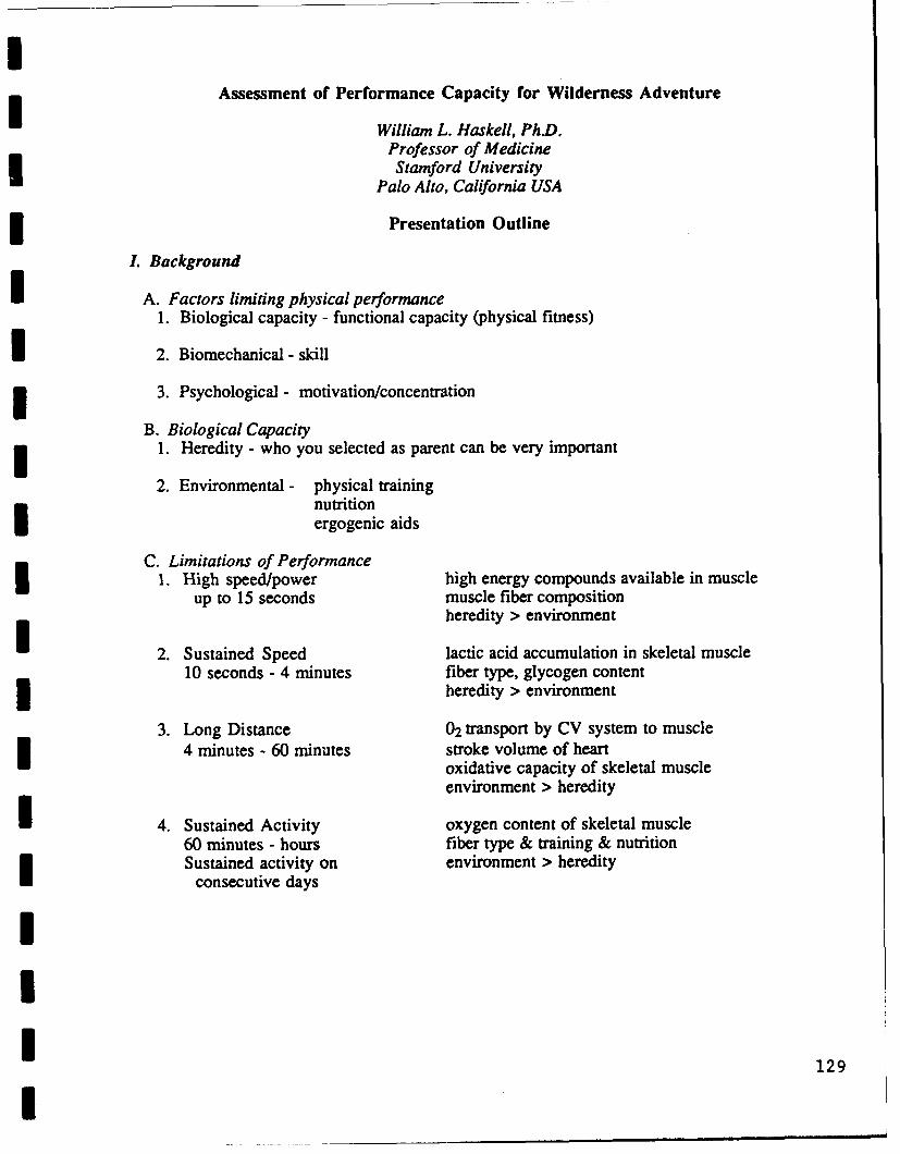
Assessment of Performance Capacity for Wilderness Adventure
William L. Haskell, Ph.D.Professor of MedicineStamford University
Palo Alto, California USA
Presentation Outline
I. Background
A. Factors limiting physical performance1. Biological capacity - functional capacity (physical fitness)
2. Biomechanical - skill
3. Psychological - motivation/concentration
B. Biological Capacity1. Heredity - who you selected as parent can be very important
2. Environmental - physical trainingnutritionergogenic aids
C. Limitations of Performance1. High speed/power high energy compounds available in muscle
up to 15 seconds muscle fiber compositionheredity > environment
2. Sustained Speed lactic acid accumulation in skeletal muscle10 seconds - 4 minutes fiber type, glycogen content
heredity > environment
3. Long Distance 02 transport by CV system to muscle4 minutes - 60 minutes stroke volume of heart
oxidative capacity of skeletal muscleenvironment > heredity
I 4. Sustained Activity oxygen content of skeletal muscle60 minutes - hours fiber type & training & nutritionSustained activity on environment > heredity
consecutive days
1II 129
I

I
11. Clinical Exercise Testing to Assess Functional Capacity
A. Factors to consider in making decision to test1. clinical status of participant - risk factors, medical history
2. age
3. nature of physical & psychological demands & exposure or adventure
B. Objectives of testing1. To determine functional capacity (workload, METS, V02, etc.)
2. To elicit abnormal response (ischemia, BP, arrhythmia, claudication, etc.)
3. To provide individualized exercise training or participation guidelines
C. Exercise Testing Protocol Considerations I1. Pre-test medical evaluation - exclusion criteria
2. Exercise test device - treadmill > cycle > steps I3. Selection of workload profile - continuous, multistage
4. Test termination criteria - submaximal vs. maximal
5. Measurements recorded - ECG, BP, RPE, VE, V02, other ID. Test Interpretation and Recommendations
1. Measurement of functional capacity - age and gender specific values
2. Abnormal test results - what advice should be given regarding participation inwilderness adventure?I(a) myocardiac ischemia(b) left ventricular dysfunction(c) significant dysrhythmias(d) exercise hypertension
3. Translation of results to wilderness adventure activities(a) Physical demands(b) Environmental demands(c) Psychological/risk demands I
III
* 30 I
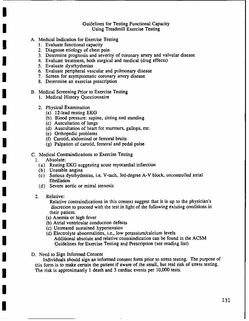
Guidelines for Testing Functional CapacityUsing Treadmill Exercise Testing
A. Medical Indication for Exercise Testing1. Evaluate functional capacity2. Diagnose etiology of chest pain
3. Determine prognosis and severity of coronary artery and valvular disease4. Evaluate treatment, both surgical and medical (drug effects)5. Evaluate dysrhythmias6. Evaluate peripheral vascular and pulmonary disease7. Screen for asymptomatic coronary artery disease8. Determine an exercise prescription
B. Medical Screening Prior to Exercise Testing1. Medical History Questionnaire
2. Physical Examination(a) 12-lead resting EKG(b) Blood pressure: supine, sitting and standing(c) Auscultation of lungs(d) Auscultation of heart for murmurs, gallops, etc.(e) Orthopedic problems(f) Carotid, abdominal or femoral bruits(g) Palpation of carotid, femoral and pedal pulse
C. Medical Contraindications to Exercise Testing1. Absolute:
(a) Resting EKG suggesting acute myocardial infarction(b) Unstable angina(c) Serious dysrhythmias, i.e. V-tach, 3rd-degree A-V block, uncontrolled atrial
fibrillation(d) Severe aortic or mitral stenosis
2. Relative:Relative contraindications in this context suggest that it is up to the physician'sdiscretion to proceed with the test in light of the following existing conditions intheir patient.
(a) Anemia or high fever(b) Atrial ventricular conduction defects(c) Untreated sustained hypertension(d) Electrolyte abnormalities, i.e., low potassium/calcium levels
Additional absolute and relative contraindication can be found in the ACSMGuidelines for Exercise Testing and Prescription (see reading list)
D. Need to Sign Informed ConsentIndividuals should sign an informed consent form prior to stress testing. The purpose of
this form is to make certain the patient if aware of the small, but real risk of stress testing.The risk is approximately 1 death and 3 cardiac events per 10,000 tests.
131

IE. Test Protocol Selection
1. Bruce: ostensibly healthy, asymptomatic individuals2. Mod-Balke: symptomatic and asymptomatic individuals3. Mod-Bruce/Naughton: high risk and/or symptomatic individuals
It is apparent that the population being evaluated and time will dictate whatprotocol is suitable.
F. Instruction to Patient1. Patients should abstain from food, tobacco, alcohol, and caffeine for at least 3 hours
prior to testing.2. Women should bring loose fitting blouse that buttons down the front with short
sleeves and should avoid restrictive undergarments such as panty hose or a girdle.It is best to wear a snug fitting bra made of a material other than nylon.
3. Clothing should permit freedom of movement and include a comfortable walking/running shoe.
4. Correct walking technique on treadmill with objective, if possible, of no gripping ofbars with hands.5. Patient should be instructed to indicate any type of discomfort including shortness Iof breath, leg pain, arm pain, jaw pain, etc.
G. EKG Records, Prior, During and Following Stress Test I1. 12-lead EKG
(a) Supine(b) Standing I(c) Standing/sitting hyperventilation: for purpose of inducing ST segment !epression
owing to a variation in autonomic nervous system(d) Minimum of 1 record per stage of exercise I(e) Peak/immediate post-exercise record(f) One record for first 3 minutes of recovery followed by 2 minute intervals until the
patient returns to pretest baselines (approximately 6-10 minutes).
H. Blood Pressures1. Same as EKG recordings
I. Criteria for Test Termination1. Patient requests to stop2. Pre-determined endpoint is reached (Borg Scale 17-20)3. Progressive angina (on scale of 104 stop at 3+)4. 1.0 mm horizontal or downsloping ST-depression or elevation5. Sustained supraventricular tachycardia6. Ventricular tachycardia7. Significant drop (10 mmHg) or failure of the systolic blood pressure to rise with
increase in exercise8. Systolic > 250 mmHg, or diastolic > 120 mmHg9. RonTPVC10. Multifocal PVC's11. Patient is confused, dizzy, losing coordination on treadmill, etc.
II
132
I

Suggested Readings for Exercise Testing to Evaluate Functional Capacity
1. American College of Sports Medicine. Guidelines for Exercise Testing and Prescription
(3rd Edition) Lea and Febiger, Philadelphia, PA, 1986.
2. Froelicher, VF. Exercise and the Heart: Clinical Concepts. Book Medical Publishers,Chicago, I11, 1989.
3. Exercise Standards: A Statement for Health Professionals from The American HeartAssociation. American Heart Association, Dallas, TX, 1991.
I 4. American College of Sports Medicine. Resource Manual for Guidelines for ExerciseTesting and Prescription. Lea and Febiger, Philadelphia, PA, 1988.
I 5. Skinner J. Exercise Testing and Exercise Prescription for Special Cases. Lea andFebiger, Philadelphia, PA, 1987.
I
IIIIIIII13
I

CW4X4 Ci "CAL 0, COST BICYICLE RAK.POTCLAS$ STATUS 5&A(KGM# 11ETS EF40W.TER AT
SAUCE K.ATTUS "ALM ELLESTAD USAFSAM SLOW, MCHENY Stv*E' ADI WAIT . - WARE USAFSAM
POS 3 IN % GRAD 3M,3- 2 OR 3 -IRASTAGES AT I I GRADE AT
MPHWt %GA 33 MPH STAGES SIAGES AT 2 P5 5 T 20- 14^ MP
FOR 0 KGSTAGES
I~~~ z 4 PHG
490 iw 4 2 33 7- 1
2 455 3 42 16 1420 1 135 4 I 33U22 1
38S T 12 21 PH %GR 15 2 0 1
_5 _0 _. 2U3
R 2sk f Sudde Cada AreI Duin Exr 1s Tr2 is3ining Ia245 25 1 1 3 1 10 1 0 10 17 1U210Sudden car33acz 175 10 42 arrestsStud Sta5u Acivt Mosorn Su1vsin eens 5
n 9 ih geerlpoua0o
Cnethmpso ear normMdial jogghesIng, 197n1.Rponued nihprson . /9,
V asd eo f S u d n r a l A r o ggD u i n g E x r i e T a n n e n o n ee c a r d i ac0courtrgames
Anvtegf general population1/6,0U
Fliete and Catno"crdial scounying ineme n pen 1/60,000Leachn et al" cardiac jogging inemttn n 1/37,000
Thmpsod et a59 cardiac jogging ineme n pen 1/6,000Hartley" cardiac jogging ineme nopeen 1/68000
Hosrace ao n ealpopulardatjgigooeprsn 1/565,085
Flhrandllw"l cardiac mogied intermittent present 1/26,00
Va Cam andPeeron cardiac mogied cntriuous present 1/16,333Hartley" cardiac mogied cntriuous present 1/6.00
VansCemp' cardiac mixed intermittent present 1/22.9558
Hartley* cardiac bicycling intermittent none 1/0000
Fletcher' cardiac mixed intermittent present 0W70,200
Average all cardiac 1/59,142I*Unpublished data.
134I

Approximate Energy Requirements in METs ForHorizontal and Gradc Walking
ni-h 1 1.7 2.0 2.5 3.0 3.4 3.75% Grade
re rain' 45.6 53.7 67.0 80.5 91.2 100.5
0 2.3 2.5 2.9 3.3 3.6 3.92.5 2.9 3.2 3.8 4.3 4.8 5.25.0 3.5 3.9 4.6 5.4 5.9 6.57.5 4.1 4.6 5.5 6.4 7.1 7.8
10.0 4.6 5.3 6.3 7.4 8.3 9.112.5 5.2 6.0 7.2 8. 9.5 10.415.0 5.8 6.6 8.1 9.5 10.6 11.717.5 6.4 7.3 8.9 10.5 11.8 12.920.0 7.0 8.0 9.8 11.6 13.0 14.2
22.5 7.6 8.7 10.6 12.6 14.2 15.525.0 8.2 9.4 11.5 13.6 15.3 16.8
Approximate Energy Requirements in METS forHorizontal and Uphill Jogging/Running
a. Outdoors on solid surface
m-h-, 5 6 7 7.5 8 9 10% Grade
m-mir' 134 161 188 201 215 241 268
o 8.6 10.2 11.7 12.5 13.3 14.8 16.32.5 10.3 12.3 14.1 15.1 '6.1 17.9 19.75.0 12.0 14.3 16.5 17.7 18.87.5 13.8 1 18.9
10 0 15.5 1_.5
b. On the treadmill
rnh-, 5 6 7 7.5 8 9 10% Grade
remmin- 134 161 188 201 215 241 268
0 8.6 10.2 11.7 12.5 13.3 14.8 16.32.5 9.5 11.2 12.9 13.8 14.7 16.3 18.05.0 10.3 12.3 14.1 15.1 16.1 17.9 19."7.5 11.2 13.3 15.3 16.4 17.4 19.4
10.0 12.0 14.3 16.5 17.7 18.812.5 12.9 15.4 17.7 19.015.0 13.8 16.4 18.9
Table D-4. Approximate Energy Expenditure in METsDuring Bicycle Ergometry
Body i'eight Exercise Rate Ag.m-min-' and Vatts/
kg lbs 300 450 600 750 900 1050 1200 (kg-m-min-'j.50 75 100 125 150 175 200 (Watts/
50 110 5.1 6.9 8.6 10.3 12.0 13.7 15.460 132 4.3 5.7 7.1 8.6 10.0 11.4 12.970 154 3.7 4.9 b.1 7.3 8.6 9.8 11.080 176 3.2 4.3 5.4 6.4 7.5 8.6 9.690 198 2.9 3.8 4.8 5.7 6.7 7.6 8.6
100 220 2.6 3.4 4.3 5.1 6.0 6.9 7.7
NOTE:VN2 for zero load pedaling is approximately 550 mlrmin- I for 70 to 80kg subjects.
135

++ 4++4* N N N qw + +
en 00 C11o1m IC- - 4ccL 4u) , '
X
r4 to - -
cr.:
Z E - B '
mu a-co
to
t E ~ E EE E r:
-2; m~4 +- r c - >M 8 0 EN+
ISICl)d
o ~ ~ L Cd to EnI" V~jN ) 1 1 1 ) W w 04
-'+ +~il +jN + r~I~ *1
f
u,
cc'
>U
V) ICO I0 cp0 .-
0. .- 0 r- S- w .
m~ cc 0.OOO ccmc u w
136I

CORE CURRICULUM: HAZARDOUS MARINE LIFE
WILDERNESS MEDICAL SOCIETYPAUL S. AUERBACH, M.D.
The following comprises the content of the teaching slide setentitled "Hazardous Marine Life" to be copyrighted to anddistributed by the Wilderness Medical Society. No portion of thismaterial should be reproduced without the express permission of theWilderness Medical Society
DANGEROUS MARINE LIFE
TRAUMATOGENICSTINGINGPOISONOUS ON INGESTIONSHOCKING
BIOTOXICITY: POISONS, VENOMS AND DEFENSE
ORAL TOXINS: poisonous to eat
PARENTERAL TOXINS: venoms produced in specialized glands
injected mechanically with spines, teeth andfins
CRINOTOXINS: venoms produced in specialized glandsadministered without the aid of "devices"administered with slimes, gastric secretions, etc.
MARINE VENOMS
Denature membranesCatabolize cyclic 3',5'-AMPDegranulate mast cellsProvoke histamine releaseInitiate arachidonate metabolismAccelerate coagulopathies
-- Disrupt cellular transportImpede neuronal transmissionInduce anaphylaxis and shock
GENERAL FIRST AID PRINCIPLES
Control the AirwaySupport Breathinq
HAZARDOUS MARINE LIFE SLIDE SET - WMS (AUERBACH, 1991)
137
IMaintain the CirculationTreat AnaphylaxisDebride and Irrigate Wounds
THE MARINE ENVIRONMENT
Ocean water: a saline milieu for microbes
Sodium chloride iSulphatesMagnesiumPotassium iBicarbonateBromineBoric acid iStrontiumFluorine IMARINE BACTERIA
Generally halophilicheterotrophicmotileGram negative rodsfacultative anaerobeshighly proteolytic i
BACTERIA ISOLATED FROM MARINE WATER, SEDIMENTS, MARINE ANIMALS ANDMARINE-ACQUIRED WOUNDS
More than 65 species, including genera:
AcinetobacterActinomycesAeromonasAlcaligenesAlteromonasBacillus
BacteroidesChromobacterium IClostridiumDeleyaEnterobacterErysipelothrixLegionellaMycobacteriumPasteurella IProteusPseudomonas
HAZARDOUS MARINE LIFE SLIDE SET - WMS (AUERBACH, 1991)
I138 U

SalmonellaStaphylococcusStreptococcusVibrio
MARINE BACTERIA: GROWTH IN CULTURE
Most marine bacteria which are pathogenic to man can be readilyrecovered on standard media
IThiosulfate-citrate-bile salts-sucrose agar is recommended fordetection of marine Vibrio species
I Antibiotic susceptibility testing requires addition of NaCl 2.3% tothe broth or agar used for disk diffusion
MARINE BACTERIA: ANTIBIOTIC THERAPY I
MINOR ABRASIONS OR LACERATIONS
1. Prophylactic antibiotics not required in normal host
2. Persons who are chronically ill (e.g., diabetes, hemophilia,thalassemia), immunosuppressed (e.g., leukemia, AIDS, chemotherapy,corticosteroids) or with serious liver disease (e.g., hepatitis,cirrhosis, hemochromatosis require prophylactic antibiotics
3. Use ciprofloxacin, trimethoprim-sulfamethoxazole ortetracycline/doxycycline
MARINE BACTERIA: ANTIBIOTIC THERAPY II
INJURIES WITH HIGH INFECTION POTENTIAL
Large lacerationsExtensive burnsDeep puncture wounds, particularly hand/footRetained organic foreign material
* 1. Use prophylactic antibiotics.
2. If surgery necessary, parenteral antibiotic choices includetrimethoprim-sulfamethoxazole, tobramycin/amikacin/gentamicin, orcefoperazone/cefotaxime/ceftazidime
3. If outpatient, use ciprofloxacin, trimethoprim-sulfamethoxazoleor tetracycline/doxycycline
HAZARDOUS MARINE LIFE SLIDE SET - WMS (AUERBACH, 1991)
139

II
MARINE BACTERIA: ANTIBIOTIC THERAPY III
INFECTED WOUNDS: ANTICIPATE VIBRIO INFECTION
Rapidly progressive cellulitis and/or myositis indicates Vibriovulnificus or V. parahaemolyticus infection
1. Culture for aerobes and anaerobes
2. Pending culture results, parenteral choices include:
Imipenem-cilastatinTobramycin/amikacin/gentamicin plus cefoperazone or
piperacillinChloramphenicol plus trimethoprim-sulfamethoxazole I
ERYSIPELOTHRIX REUSOPATHIAE INFECTION
Classic erysipeloid appearanceCommonly follows seafood handling
Antibiotics: erythromycin, penicillin or cephalexin
MARINE ANTIVENINS
Chironex fleckeri (box-jellyfish) antivenin, from CommonwealthSerum Laboratories (CSL), Melbourne, Australia: versus Chironexfleckeri and Chiropsalmus quadrigatus
Enhydrina schistosa (beaked sea snake) and Notechis scutatus(terrestrial tiger snake) polyvalent antivenin, from CSL; versusmost sea snakes
Notechis scutatus antivenin, from CSL: versus most sea snakes
Enhydrina schistosa monovalent antivenin, from the HaffkineInstitute in Bombay, India: versus most sea snakes
Synanceja trachynis (stonefish) antivenin, from CSL: versus Istonefish and some scorpionfishes ITRAUMATOGENIC MARINE HAZARDS
SharksBarracudaMoray EelsSea Lions
HAZARDOUS MARINE LIFE SLIDE SET - WMS (AUERBACH, 1991)
I140 I

SHARK LIFE AND HABITS
Found in oceans, tropical rivers and lakesRange in size from 10 cm to over 15 m and 18,000 kgApproximately 32 out of 350 species implicated in human attacks50-100 annual attacks worldwide; less than 10% fatalExquisite ocular perception of motion and olfactory detection of
blood
Most danQerous sharks:
Great whiteMakoBullDuskyHammerheadGrey reef
GREAT WHITE SHARK: CARCHARODON r:ARCHARIAS
The most dangerous of all sharksMay attain a length of 30 feet and weight of 7000 poundsBiting force of up to 18 tons/square inch
Commonly found in waters of southern Australia, east coast of SouthAfrica, middle Atlantic coast of North America, and the AmericanPacific coast north of Point Conception, California
SHARK FEEDING AND ATTACK
Two feeding patterns: subdued (solitary) or frenzied (mob)
Some sharks can achieve 20-40 mph underwater
More than 70% of victims are bitten only once or twice
Lower teeth used first in feeding
Aggression seems to be directed at frightened or wounded victim
SHARK ATTACK
Odds of attack along North American coastline: 1/5,000,000
Danger greatest: during summer months* in recreational areas
HAZARDOUS MARINE LIFE SLIDE SET - WMS (AUERBACH, 1991)
141

Iduring late afternoon and evening (feeding)in murky/warm waterin deep channels or drop-offs Iupon surface swimmers
CLINICAL ASPECTS OF SHARK BITES UMassive tissue lossHemorrhageShockDeath
Extremity bites most commonHigh propensity for infection because of contamination with sea
water, sand, plant debris, shark teeth and mouth flora
SHARK BITE TREATMENT
Manage bleedingAdminister prophylactic antibioticsExplore wounds in the operating room IIrrigate extensivelyClose loosely around drains or pack open for delayed primary
closure drains
SHARK ATTACK PREVENTION 31. Avoid shark infested water, particularly at dusk and night2. Do not swim with domestic animals in shark waters3. Swim in groups. Pay attention to your companions4. Avoid turbid water, dropoff s, deep channels and sanitation
waste outlets5. Do not bleed in the water6. Avoid shiny metal adornments and brightly colored swimwear7. Do not carry tethered (dead or injured) fish8. Be alert for sharks when fish act erratically9. Do not tease or corner a shark I10. Do not panic or splash at surface
BARRACUDA LIFE AND HABITS IDistributed from Brazil north to Florida, and in the Indo-Pacificfrom the Red Sea to the Hawaiian Islands
Only the great barracuda Sphyraena barracuda has been implicated inhuman attacks
Grows to 10 feet and 100 pounds
HAZARDOUS MARINE LIFE SLIDE SET - WMS (AUERBACH, 1991) I142 I
I

Solitary swimmer with canine mouth and large, knifelike teeth
Attracted to motion and shiny objects (e.g., metal anklet)
MORAY EEL LIFE AND HABITS
Found in tropical, subtropical and temperate waters
Muscular, powerful bottom dwellers up to 3 m in length
Reside in holes, ledges and crevices
Attack when cornered or provoked
MORAY EEL BITE CLINICAL ASPECTS
Multiple puncture wounds from long, sharp, retrorse and fangliketeeth
Tenacious bite; to release the animal, it may be necessary toperform decapitation
High propensity to infection, particularly of hand
MORAY EEL BITE TREATMENT
1. Remove the animal2. Locate all puncture wounds3. Perform neurovascular exam4. Debride/irrigate wounds5. Explore wounds for retained teeth6. DO NOT SUTURE PUNCTURE WOUNDS; large defects may be approximated
loosely7. Administer prophylactic antibiotics
SEA LION BITES
Generally mild-mannered animals, except during breeding season
Males are more aggressive, except when females defend pups
Biting and butting injuries
Propensity for post-traumatic wound infections
HAZARDOUS MARINE LIFE SLIDE SET - WMS (AUERBACH, 1991)
143
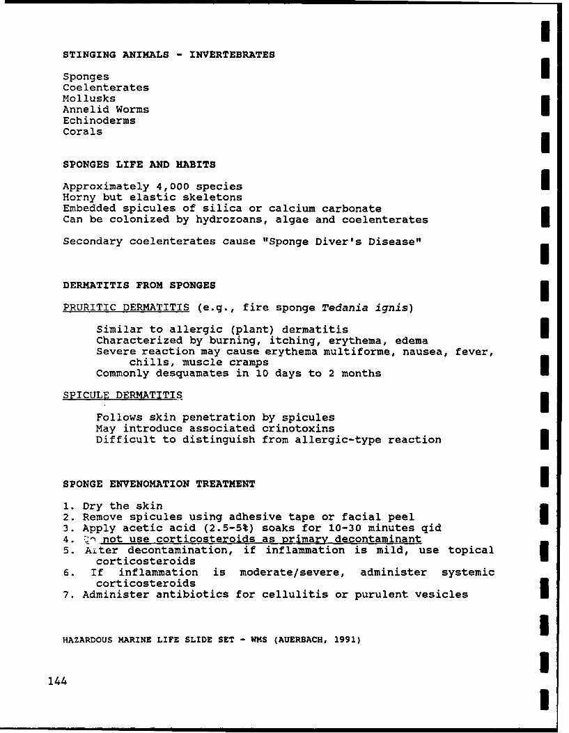
ISTINGING ANIMALS - INVERTEBRATES
SpongesCoelenteratesMollusksAnnelid WormsEchinodermsCorals
SPONGES LIFE AND HABITS
Approximately 4,000 species IHorny but elastic skeletonsEmbedded spicules of silica or calcium carbonateCan be colonized by hydrozoans, algae and coelenterates ISecondary coelenterates cause "Sponge Diver's Disease"
DERMATITIS FROM SPONGES
PRURITIC DERMATITIS (e.g., fire sponge Tedania ignis)
Similar to allergic (plant) dermatitisCharacterized by burning, itching, erythema, edemaSevere reaction may cause erythema multiforme, nausea, fever,
chills, muscle crampsCommonly desquamates in 10 days to 2 months
SPICULE DERMATITISI
Follows skin penetration by spiculesMay introduce associated crinotoxinsDifficult to distinguish from allergic-type reaction I
SPONGE ENVENOMATION TREATMENT n
1. Dry the skin2. Remove spicules using adhesive tape or facial peel I3. Apply acetic acid (2.5-5%) soaks for 10-30 minutes qid4. >) not use corticosteroids as primary decontaminant5. After decontamination, if inflammation is mild, use topical
corticosteroids6. If inflammation is moderate/severe, administer systemic
corticosteroids7. Administer antibiotics for cellulitis or purulent vesicles
IHAZARDOUS MARINE LIFE SLIDE SET - WMS (AUERBACH, 1991)
I144 I

COELENTERATE CLASSIFICATIONS
APPROXIMATELY 9,000 SPECIES, OF WHICH AT LEAST 100 ARE DANGEROUS TOHUMANS
Cnidaria: possess stinging cells (nematocysts)Acnidaria: do not possess nematocysts
CNIDARIA: Hydrozoans (e.g., Portuguese man-of-war)Scyphozoans (e.g., true jellyfish)Anthozoans (e.g., anemones, soft corals)
COELENTERATE VENOM APPARATUS
Nematocyst is a stinging cell produced by cnidocyteMillions of cnidocytes on tentacles or near the mouthStinging cell is venom-filled and contains a coiled thread tubeThread tube may attain length of 200-400 micronsExocytosis of tube stimulated by contact and hypotonic stateVelocity of ejection estimated at 2 m/sec, acceleration 40,000 gSkin is struck by "injectors" at 2-5 lb/sq inUpper dermis penetrated; venom diffuses into circulation
NEMATOCYST VENOM COMPONENTS
BradykininHemolysinSerotoninHistamineProstaglandinsAdenosine triphosphataseNucleotidasesHyaluronidaseAlkaline proteaseAcid proteasePhosphodiesteraseAcid phosphataseAlkaline phosphataseFibrinolysinLeucine aminopeptidaseRNAaseDNAase
CLINICAL ASPECTS OF HYDROID ENVENOMATION
Instantaneous mild burning, itching and urticaria
HAZARDOUS MARINE LIFE SLIDE SET - WMS (AUERBACH, 1991)
145

IErythematous miliary skin irritationRare delayed (12-14 hrs) papular, hemorrhagic or zosteriform
reactionBurning sensation in conjunctivae, oral mucous membranes
THERAPY FOR HYDROID ENVENOMATION I1. Rinse the skin with sea water or 3N saline (not with fresh
water)2. Apply brief soak of acetic acid 5% or isopropyl alcohol 40-70%3. If inflammation is residual, apply topical corticosteroidi
FIRE CORAL CLINICAL ASPECTS
Millepora species are not true corals IThey are the most ubiquitous stingers in the marine environmentNumerous configurations, so adverse encounters are frequent
Immediate intense burning, with occasional central radiationPainful pruritus, with large wheals (worsened by rubbing)Maximum discomfort in 30-60 minutes IUntreated, rash flattens over 12-24 hoursTotal resolution in 3-7 days, with residual hyperpigmentationRegional lymphadenopathy in severe cases
THERAPY FOR FIRE CORAL STING
1. Rinse the skin with sea water or 3N saline (not with freshwater)
2. Apply 15 min soak of acetic acid 5% or isopropyl alcohol 40-70%3. If inflammation is residual, apply topical corticosteroid4. If rash is indolent or lymph nodes are inflamed, administer 2
week tapering course of oral corticosteroids or parenteralcorticosteroid injection
PORTUGUESE MAN-OF-WAR CLINICAL SYNDROME
1. Immediate pain, itching, erythema, "tentacle prints"2. Rare anaphylaxis3. Weakness, dizziness, nausea, vomiting, diarrhea, abdominal pain4. With large surface area involvement, may develop headache,
mononeuritis, delirium, seizures, hemolysis, hypotension,arrhythmias, bronchospasm, pulmonary edema, erythemamultiforme, renal failure, parasympathetic dysautonomia, coma
5. Persistent or recurrent skin lesions with minimal contactsuggest immune sensitization I
HAZARDOUS MARINE LIFE SLIDE SET - WMS (AUERBACH, 1991) II
146
I

SCYPHOZOAN MEDUSAE AND JELLYFISH
Armed with some of the most venoms in existenceMost highly dangerous species are located in Indo-Pacific waters
Examples include:
Chironex fleckeriCyanea capillataChiropsalmus quadrigatusPelagia noctilucaCarukia barnesiTamoya haplonemaChrysaora quinquecirrha
CLINICAL ASPECTS OF SCYPHOZOAN ENVENOMATION
Similar to Portuguese man-of-war, with markedly increased severity
Multiple stings from the box-jellyfish (Chironex fleckeri) withtentacle contact in excess of 6-7 m can cause death fromcardiorespiratory arrest in 60 seconds
Tentacle prints may have frosted, cross-hatched appearance withprominent blistering
Skin wounds frequently become necrotic despite topical therapy
CLINICAL ASPECTS OF ANEMONE ENVENOMATION
Similar to Portuguese man-of-war, but with more pronounced skinurticaria or ischemic appearance
Hemorrhagic perimeter to sting sites common, followed byulcerations, intense local vesiculation with anemones Sagartia,Actinia, Anemonia, Actinodendron, Triactis
Systemic effects are rare, and include fever, chills, somnolence,malaise, weakness, nausea, vomiting
DERMATOLOGICAL THERAPY FOR COELENTERATE ENVENOMATION - I
1. Rinse the wound with sea water or 3N saline. DO NOT RINSE WITHFRESHWATER. DO NOT RUB THE WOUND. Wear protective gloves(double glove).
2. Remove gross tentacle fragments with forceps.3. Apply acetic acid 5% as topical decontaminant (ABSOLUTE with
Chironex fleckeri) for at least 30 min. Alternatives:
HAZARDOUS MARINE LIFE SLIDE SET - WMS (AUERBACH, 1991)
147
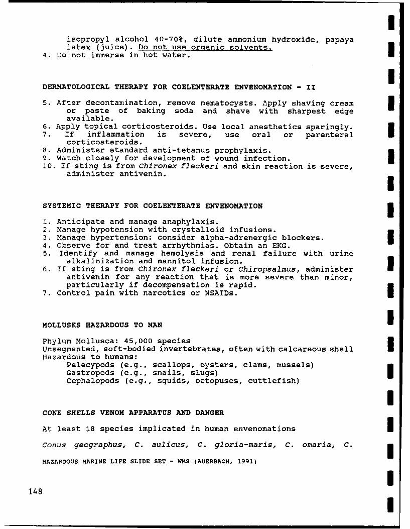
Iisopropyl alcohol 40-70%, dilute ammonium hydroxide, papayalatex (juice). Do not use organic solvents.
4. Do not immerse in hot water.
IDERMATOLOGICAL THERAPY FOR COELENTERATE ENVENOMATION - II
5. After decontamination, remove nematocysts. A pply shaving creamor paste of baking soda and shave with sharpest edgeavailable.
6. Apply topical corticosteroids. Use local anesthetics sparingly.7. If inflammation is severe, use oral or parenteral
corticosteroids.8. Administer standard anti-tetanus prophylaxis.9. Watch closely for development of wound infection. U10. If sting is from Chironex fleckeri and skin reaction is severe,
administer antivenin.
SYSTEMIC THERAPY FOR COELENTERATE ENVENOMATION
1. Anticipate and manage anaphylaxis.2. Manage hypotension with crystalloid infusions.3. Manage hypertension: consider alpha-adrenergic blockers. I4. Observe for and treat arrhythmias. Obtain an EKG.5. Identify and manage hemolysis and renal failure with urine
alkalinization and mannitol infusion.6. If sting is from Chironex fleckeri or Chiropsalmus, administer
antivenin for any reaction that is more severe than minor,particularly if decompensation is rapid.
7. Control pain with narcotics or NSAIDs. I
MOLLUSKS HAZARDOUS TO MAN IPhylum Mollusca: 45,000 speciesUnsegmented, soft-bodied invertebrates, often with calcareous shellHazardous to humans:
Pelecypods (e.g., scallops, oysters, clams, mussels)Gastropods (e.g., snails, slugs) ICephalopods (e.g., squids, octopuses, cuttlefish) I
CONE SHELLS VENOM APPARATUS AND DANGER
At least 18 species implicated in human envenomations IConus geographus, C. aulicus, C. gloria-maris, C. omaria, C.
HAZARDOUS MARINE LIFE SLIDE SET - WMS (AUERBACH, 1991)
I148
• , .! PI

striatus, C. textile, C. tul 'a are worst offenders
Inject venom with detachable, dartlike radular tooth used as a"harpoon"
Proboscis which launches dart can extend all the way back to thethick crown of the shell
CONE SHELL ENVENOMATION
Mild stings resemble wasp sting
Initial symptoms: localized wound ischemia and numbnessburning and stingingparesthesiasdysphagiaweaknesssyncope
Severe reaction: generalized muscular paralysisrespiratory distresscardiac failurecomacerebral edemadisseminated intravascular coagulopathy
THERAPY FOR CONE SHELL ENVENOMATION
1. Apply pressure immobilization technique2. Observe for 2-4 hours3. Manage hypotension with crystalloid infusion4. Administer oxygen5. Be prepared to manage respiratory distress6. Anticipate coagulopathy
PRESSURE IMMOBILIZATION TECHNIQUE FOR VENOM SEQUESTRATION
1. Place a cloth or gauze pad of approximate dimensions 6-8 cm x6-8 cm x 2-3 cm (thickness) directly over bite site (optional)
2. Wrap the extremity with an elasticized bandage at lymphatic-venous occlusive tension
3. Distal fingertips and toes should be pink; arterial lsationsshould be present (proper capillary refill)
4. Keep bandages in place until the victim is brought to medicalattention
5. Anticipate swelling and loosen bandages if arterial circulationis compromised
HAZARDOUS MARINE LIFE SLIDE SET - WMS (AUERBACH, 1991)
149
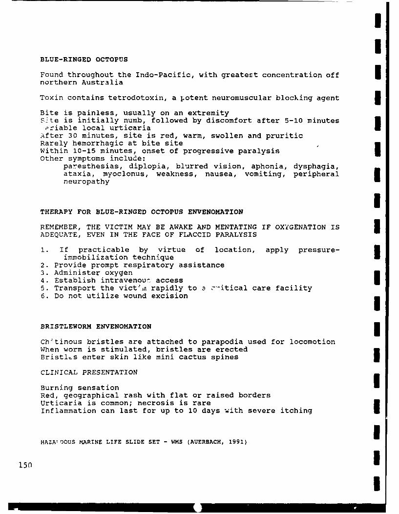
II
BLUE-RINGED OCTOPUS
Found throughout the Indo-Pacific, with greatest concentration off Inorthern Australia
Toxin contains tetrodotoxin, a potent neuromuscular blocking agent
Bite is painless, usually on an extremitySite is initially numb, followed by discomfort after 5-10 minutesariable local urticaria
After 30 minutes, site is red, warm, swollen and pruriticRarely hemorrhagic at bite siteWithin 10-15 minutes, onset of progressive paralysis IOther symptoms include:
paresthesias, diplopia, blurred vision, aphonia, dysphagia,ataxia, myoclonus, weakness, nausea, vomiting, peripheralneuropathy
ITHERAPY FOR BLUE-RINGED OCTOPUS ENVENOMATION
REMEMBER, THE VICTIM MAY BE AWAKE AND MENTATING IF OXYGENATION IS 3ADEQUATE, EVEN IN THE FACE OF FLACCID PARALYSIS
1. If practicable by virtue of location, apply pressure-immobilization technique
2. Provide prompt respiratory assistance3. Administer oxygen4. Establish intravenou- access I5. Transport the victi rapidly to 3 c-itical care facility6. Do not utilize wound excision
BRISTLEWORM ENVENOMATION
Ch-tinous bristles are attached to parapodia used for locomotionWhen worm is stimulated, bristles are erectedBristlt.s enter skin like mini cactus spines ICLINICAL PRESENTATION
Burning sensation IRed, geographical rash with flat or raised bordersUrticaria is common; necrosis is rareInflammation can last for up to 10 days with severe itching
IHAZARDOUS MARINE LIFE SLIDE SET - WMS (AUERBACH, 1991)
150 I
I

THERAPY FOR BRISTLEWORM ENVENOMATION
1. Remove all large visible bristles with forceps2. Dry the skin3. Remove embedded bristles with adhesive tape or facial peel4. Apply acetic acid 2.5-5% or isopropyl alcohol 40-70%5. Apply topical corticosteroids6. If inflammation and discomfort are severe, administer oral or
parenteral corticosteroids
ECHINODERMS
StarfishSea urchinsSea cucumbers
CROWN-OF-THORNS STARFISH LIFE AND HABITS
Found in the coral reef communities of the Great Barrier reef,throughout the Pacific and Indian Ocean, in the Red Sea, and in theeastern Pacific from the Galapagos to the Gulf of California
Slimy venom produced in glands within or under the epidermisAttains 25-35 cm in diameter7-23 arms, with spines of up to 6 cmMagnesium calcite spine can easily penetrate a diving glove or
neoprene suit
m CLINICAL ASPECTS OF STARFISH ENVENOMATION
Immediate intense pain, bleeding and edemaWound may be dusky or discolored
Multiple punctures lead to systemic effects:
ParesthesiasNausea, vomitingLymphadenopathyLocal muscle paralysisDelayed granuloma (foreign body)
1THERAPY FOR STARFISH ENVENOMATION1. Immerse the wound(s) into nonscalding hot water to toleranceI (1130 F or 450 C) for 30-90 minutes or until there is
significant pain relief-- 2. Use local anesthetic for additional pain control
HAZARDOUS MARINE LIFE SLIDE SET - WMS (AUERBACH, 1991)
I 151
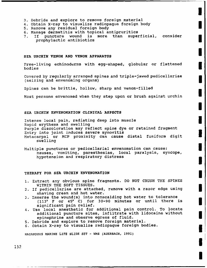
I3. Debride and explore to remove foreign material4. Obtain X-ray to visualize radiopaque foreign body5. Remove any residual foreign body6. Manage dermatitis with topical antipruritics7. If puncture wound is more than superficial, consider
prophylactic antibiotics
SEA URCHIN VENOM AND VENOM APPARATUS IFree-living echinoderms with egg-shaped, globular or flattenedbodies
Covered by regularly arranged spines and triple-jawed pedicellariae(seizing and envenoming organs) ISpines can be brittle, hollow, sharp and venom-filled
Most persons envenomed when they step upon or brush against urchin II
SEA URCHIN ENVENOMATION CLINICAL ASPECTS
Intense local pain, radiating deep into muscle IRapid erythema and swellingPurple discoloration may reflect spine dye or retained fragmentEntry into joint induces severe synovitisMetacarpal or MCP proximity can cause distal fusiform digit
swelling
Multiple punctures or pedicellarial envenomation can cause:nausea, vomiting, paresthesias, local paralysis, syncope,hypotension and respiratory distress
THERAPY FOR SEA URCHIN ENVENOMATION
1. Extract any obvious spine fragments. DO NOT CRUSH THE SPINESWITHIN THE SOFT TISSUES.
2. If pedicellariae are attached, remove with a razor edge using Ishaving cream and hot water.
3. Immerse the wound(s) into nonscalding hot water to tolerance(1130 F or 450 C) for 30-90 minutes or until there issignificant pain relief.
4. Use local anesthetic for additional pain control. To locateadditional puncture sites, infiltrate with lidocaine withoutepinephrine and observe egress of fluid.
5. Debride and explore to remove foreign material.6. Obtain X-ray to visualize radiopague foreign bodies.
HAZARDOUS MARINE LIFE SLIDE SET - WMS (AUERBACH, 1991)
152 II

7. Operate to remove all spines near joints or significantneurovascular structures.
8. Manage dermatitis with topical antipruritics.9. If puncture wound is more than superficial, consider
prophylactic antibiotics.
CLINICAL ASPECTS OF SEA CUCUMBER ENVENOMATION
Tentacular organs of Cuvier produce "holothurin", a cantharidin-like substance
Contact with tentacles or fragments produces dermatitisCorneal inflammation if fragments enter the eyesSevere reaction may lead to blindness
Sea cucumbers may eat nematocysts and excrete coelenterate venom
Holothurin is a potent cardiac glycoside
THERAPY FOR SEA CUCUMBER ENVENOMATION
1. If skin is involved, decontaminate with acetic acid 5% orisopropyl alcohol 40-70%
2. If eye is involved, anesthetize with proparacaine HCl 0.5%, thenirrigate with 250 ml normal saline
3. Stain the cornea and inspect for corneal abrasion/ulceration4. Treat inflammatory keratitis with cycloplegic and mydriatic
agents. Topical corticosteroid ophthalmic solution may behelpful
CORAL CUTS AND POISONING CLINICAL ASPECTS
Initial reaction is stinging pain, erythema and pruritus
"CORAL POISONING":
indolent wound associated with poor healing, persistentexudate, regional lymphadenopathy, reactive bursitis, malaise, lowgrade fever
ITHERAPY FOR CORAL CUTSIHAZARDOUS MARINE LIFE SLIDE SET - WMS (AUERBACH, 1991)
I153I

n1. Vigorously irrigate the wound with fresh or disinfected water or
normal saline2. Scrub the wound with nontoxic soap or antiseptic solution3. Repeat wound irrigation4. Debride macerated edges and explore to remove foreign material5. If the wound is painful in a manner that suggests coelenterate I
envenomation, consider decontamination with acetic acid 2.5%or isopropyl alcohol 20% in an extremely brief application,followed by irrigation i
6. DO NOT SUTURE WOUNDS CLOSED IF AT ALL POSSIBLE. If edges must beapproximated, use surgical adhesive strips
7. Debride daily, utilizing wet-to-dry dressings or topicalantiseptics (nonadherent dressings) I
8. Administer antibiotics at earliest signs of infection ISTINGING ANIMALS - VERTEBRATES
Stingrays IScorpionfishes ISTINGRAYS LIFE AND HABITS
Most commonly incriminated group of fishes involved in humanenvenomations
11 species found in U.S. coastal waters iOne to four venomous stings on the dorsum of caudal appendageSpines are retroserrate vasodentine with associated venom glands IInjuries are commonly to lower extremity with both a laceration andenvenomation
Integumentary sheath is ruptured, releasing venom into wound I
STINGRAY ENVENOMATION CLINICAL ASPECTS
Immediate intense pain as puncture occurs, with central radiationSpine tip(s) may break off and remain in the wound(s)Thorax or abdomen may be penetratedWound is edematous and hemorrhagic, occasionally dusky or mottledRapid fat and muscle hemorrhage and necrosisSystemic effects include:
weakness, nausea, vomiting, diarrhea, diaphoresis, vertigo,headache, seizures, syncope, fasciculations, paralysis,
HAZARDOUS MARINE LIFE SLIDE SET - WMS (AUERBACH, 1991)
154
I

hypotension, arrhythmias and death
THERAPY FOR STINGRAY ENVENOMATION
1. Extract any obvious spines, spine fragments, or pieces ofintegumentary sheath.
2. Immerse the wound(s) into nonscalding hot water to tolerance(1130 F or 450 C) for 30-90 minutes or until there issignificant pain relief.
4. Use local anesthetic infiltration (without epinephrine) foradditional pain control. If necessary, perform a regionalnerve block.
5. Debride and explore to remove foreign material. Irrigate withwarmed saline.
6. Obtain X-ray to visualize radiopaque foreign bodies.
7. Operate to remove all retained foreign matter.8. Do not suture wounds closed if at all possible. Leave open for
delayed primary closure.9. Administer prophylactic antibiotics.10. Observe all victims for at least 4 hours for onset of systemic
effects.
CATEGORIES OF SCORPIONFISHES
Genus Pterois: zebrafish, lionfish, tigerfish, turkeyfish,scorpionfish, firefish
Genus Scorpaena: scorpionfish, bullrout, sculpin
Genus Synanceja: stonefish, stargazer, warty ghoul
SCORPIONFISHES LIFE AND HABITS
Venom organs: 12-13 (of 18) dorsal, 2 pelvic and 3 anal spines
Pectoral spines are not venomous
Each spine is covered with integumentary sheath and carriesassociated venom glands
Animals are often extremely well camouflaged and difficult tolocate, even for experienced divers
SCORPIONFISH ENVENOMATION CLINICAL ASPECTS
HAZARDOUS MARINE LIFE SLIDE SET - WMS (AUERBA'A, 1991)
155
IImmediate pain from puncture woundsPain peaks at 60-90 minutes and persists for up to 12 hoursWound initially ischemic, then cyanotic, then red, warm and swollen mVesicles may formWith severe sting, may have tissue necrosis and sloughingIndolent wounds require months to heal
Systemic effects include:headache, tremors, maculopapular skin rash, nausea, vomiting,diarrhea, abdominal pain, diaphoresis, pallor, delirium,seizures, paralysis, lymphangitis, arthritis, fever,hypertension, respiratory distress, arrhythmias, congestiveheart failure, pericarditis, hypotension, syncope and death
THERAPY FOR SCORPIONFISH ENVENOMATION i
1. Extract any obvious spines, spine fragments, or pieces ofintegumentary sheath.
2. Immerse the wound(s) into nonscalding hot water to tolerance(1130 F or 450 C) for 30-90 minutes or until there issignificant pain relief. i
4. Use local anesthetic infiltration (without epinephrine) for
additional pain control. If necessary, perform a regionalnerve block. Localize all puncture wounds with anestheticinfiltration, observing for egress of fluid.
5. Debride and explore to remove foreign material. Irrigate withwarmed saline.
6. Obtain X-ray to visualize radiopaque foreign bodies.7. Operate to remove all retained foreign matter.8. Do not suture wounds closed if at all possible. Leave open for
delayed primary closure. i9. Administer prophylactic antibiotics.10. Observe all victims for at least 4 hours for onset of systemic
effects.
OTHER MARINE VERTEBRATES THAT STING AND CAUSE INJURIES SIMILAR TOSCORPIONFISHES
WeeverfishesCatfishesDragonfishesVenomous (spined) sharksRatfishesToadfishesRabbitfishesStargazers iSquirrelfishesSea robins
HAZARDOUS MARINE LIFE SLIDE SET - WMS (AUERBACH, 1991) I156 n
I
Flying gurnardsGoosefishes
SEA SNAKES LIFE AND HABITS
The most abundant reptiles on EarthAt least 52 species, all venomous
Distributed in tropical and warm temperate Pacific and IndianOceans
There are no indigenous sea snakes in the Atlantic Ocean or
Caribbean Sea
Carry 2-4 hollow maxillary fangs with associated venom glands
Venom is a neurotoxin, with additional hemolysins and myotoxiccompounds (phospholipase A)
CLINICAL ASPECTS OF SEA SNAKE BITES
DIAGNOSIS OF SEA SNAKE BITE IS BASED UPON:
1. Location of incident. One must be in the water or handling a seasnake to have been bitten.
2. Absence of pain. Most bites are initially painless.3. Fang marks. These are multiple, hypodermic-like and number up to
20.4. Identification of the snake.5. Characteristic symptoms: euphoria, malaise, anxiety, slurred
speech, facial paralysis, parotid gland swelling, myalgia,paralysis, arthralgia, trismus, blurred vision, dysphagia,drowsiness, vomiting, ptosis. Symptoms occur within 6-8 hours.
6. With significant envenomation, demise is rapid within first 2-3hours. Symptoms and signs include myoglobinuria, paralysis,respiratory distress, aspiration, hyperkalemia, acute renalfailure, myonecrosis, hypotension.
THERAPY FOR SEA SNAKE BITES
1. Apply the Extractor device if within the first 3 minutes postbite.
2. Apply the pressure immobilization technique for venomsequestration.
3. Immobilize the affected limb.4. Obtain intravenous access in 2 sites.5. With any evidence of envenomation, administer antivenin.
HAZARDOUS MARINE LIFE SLIDE SET - WMS (AUERBACH, 1991)
157

I6. Monitor for hemoglobinuria or myoglobinuria. If evident, promote
diuresis with mannitol infusion and alkalinize urine withbicarbonate.
7. Administer oxygen. Observe closely for signs of respiratoryfailure. Be prepared to provide mechanical ventilatoryassistance.
IiIIIiIii
Ii
HAZARDOUS MARINE LIFE SLIDE SET - WMS (AUERBACH, 1991) i
158 Ii

m ESSENTIALS OF DIVING MEDICINE
byKenneth W. Kizer, M.D., M.P.H., F.A.C.E.P., F.A.C.P.M., F.A.C.O.M.
Director, California Department of Health Servicesand
Associate Clinical Professcr of Medicine and Community HealthUniversity of California, Davis
ISynopsis: The aquatic environment presents numerous hazards for theunderwater worker or sportsman. This presentation will familiarizepractitioners with the environmental hazards confronting scuba divers,focusing especially on those conditions due to increased atmosphericpressure. After attending this lecture one should have an increasedunderstanding of diving, in general, and a better understanding of thepathophysiology, clinical manifestations and treatment of barotrauma,dysbaric air embolism and decompression sickness, in particular.
LECTURE OUTLINE
I. A Brief Overview of Diving
II. Basic Physics and Physiology of Diving
A. Pressure and its measurement
B. Gas laws
C. Cardiorespiratory physiology of submersion and hyperbaria
III. Primary Civing Medicine Problems
A. Environmental exposures
B. Problems related to the breathing medium
C. Dysbarism
D. Hazardous marine life
E. Miscellaneous
159

IKizer... Diving Medicinepage 2
IV. Barotrauma - local injury resulting from inadequate pressureequalization between air-containing cavities and theambient atmosphere; by far the most common dysbaricproblem of divers
A. Otolaryngologic IB. Gastrointestinal
C. Pulmonary
D. Other
V. Dysbaric Air Embolism
A. Pathophysiology
B. Clinical manifestations
C. Treatment
VI. Decompression Sickness - a multisystem disorder resulting from theliberation of inert gas from solution andthe resultant formation of gas bubbles intissue
A. Pathophysiology
B. Clinical manifestations
C. Treatment I
VII. Differential Diagnosis of Diving Accidents
A. Diving accident history
B. Sources of consultation I1. Diving Alert Network (DAN) - 919:684-8111 32. U.S. Navy Experimental Diving Unit - 904:234-4351
3. Naval Medical Research Institute - 202:295-1839
2. Local and regional sources
I160I
I

Symposium on Environmental Emergencies
I Diving Medicine
Kenneth W,1. Kizer, M.D., M.P.H.*
I The oceans are the last fr-ontier on earth, and in recent y'ears there haslbeen a virtual explosion in the number of div'ers exploring their depths. ItI is estimated that there are about three million recreational scuba divers inthe United States, and more than 200,000 new sport divers are trainedeach year.43 In addition, there are man,. thousands of commercial, scientificI and military divers.
The medical problems of diving are primarily due to the intrinsichazards of the aquatic environment and the breathing of compressed as.I that is. air or other gas mixtures at more than one atmosphere of pressure.A useful way of categorizing the man\, disparate medical problems relatedto diving is shown in Table 1. Since it is beyond the scope of this article toreview all of these problems. this discussion w~ill focus on the severalpressure-related syndromes that are peculiar to diving and collectivelyknow"n as clvsbarism. Emergency physiciami should be familiar with themaaeetIfteepolm eaueteee-nraignme fmieanmnt the pr oblems beaseteivr-nresngnmbro
diversand tebur eonn diig vacation industry, wvhich accounts forI manyv injured dlivers presenting to emergency departments far removedfrom the diving site. make these problems increasingly common.
I PHYSICAL PRINCIPLES
I Humian beinzs did not evolve for a marine existence, and], indeed,the\- are not wyell adapted for functioning in the aquatic environment.Consequently. divers encounter many- adverse physical conditions, such ascold. wetness,. changes in lig-ht and sound conduction, increased density ofthe surrounding environment. and increased atmospheric pressure. Of
I these. pr-essur-e is by fill the Single most important environmental factor in
I)i rvt( Emvur,_,enc\ MIedical scr\ ces AlIthorit. Staite of Caiiria. Sacrainento. Presideit -VIVO.t \WildlerIle NIVedical SOCICt. MIill V&d1C\. Cilliforna
IN)1twil (if till, articr were takeni froum Kizer K\V NMana,-zenlenlt of, d\ "barkc imi, casual~ties.L Ii t '1 e d (:1111 N o rth A ~ l. : 6 9 - 7 . 9 :3.
1 6 1
Fm, -r--cm -I/.Abcdwim ClI,,u% 'if .\~rl Anmria-Vol. 2. No. 3. .Xi-ziit 1"~4

KENNETH W. KIZER
Table 1. MeIdical Problems of Divers*
E nviron mental Exposure ProblemsIMotion sicknessNear drownint, and other immersion syndromiesHy~pothermia and heat illnessSunburnIPhototoxic and photoallergic reactionsIrritant and other dermiatitidesInlectious diseasesPhvsical trauma
DysbarisinBarotraumna
Dysbaric air embolismDecompression sickness
Breathing Gas-Related ProblemsInert gas narcosisI
Oxygen toxicityH ypercapnia
Carbon monoxide poisoningNitrogen oxide toxicityHigh d pressure nervous syndromeCutaneous isobaric counterdiffusionI
Hazardous Marine LifeMIiscellaneous
Hy'perv'entilationDvsbaric osteonecrosisCompression arthralgiaHyperbaric ceplialgiaIHearing lossCarotogenic blackoutPanic and] other psychologfical problems
*From Kizer K\V: Management of' dysbaric diving casualties. Emerg Med Cliii NorthAm. 1:6594670. 19S3.
diving,. with its effects either directly' or indirectly' accounting for themiajority of serious diving mnedical problems. Therefore, having ail under-standing of basic pressure physics and physiology is essential to understand-Iing divingo diseases.
Pressure
Pressure is defined as force per unit area and is mneasured in a numberof different units (Table 2). Since we are adapted to standard terrestrialatmlosp~heric conditions. we usually do not think about thle density andpressi re of' the air- we breath . Actually, though, the weight of air- at sealevel is eqlual to 14.7 pounds per' square inch (psi) or one atmospher-eabl)Sltc ATAf.
\\'lr a diver (-oes under water. the pressure increases b)ecause of thewiltof' the water. and since water is much more dense than air, large
chaiiues ill pressuire will accompany small fluctuations inl depth. Thus. asshown ill FP._iire 1. at at d1epth of' 33 feet of sea water (fswv) the pressure is2 ATA ailci at 165 f'iw\ it is 6 ATA. Inl addition. it should be clear fr-om theI
162 Figiurc that the proportionate chang!e inl pressure per unit depth chanlge is

DIVING MEDICINE
Table 2. Units of Pressure Measuremnent*
1 atmosphere absolute = 33 feet salt water (fsw)t= 34 feet fresh water (Ow)= 5.5 fathoms of sea water= 14.7 pounds per square
inch (psi)= 760 mm Hg= 29.9 in Hg= 1.033 kg/cm= 1.013 bar= 10.06 meters= 0 atmosphere gauge
*From Kizer KXV: Management of dysbaric diving casualties. Emerg Med Clin NorthAm, 1:659-670, 1983.
't1 fsw = 0.445 psi = 0.0303 atmospheres
greatest near the surface. Most scuba diving is done at pressures less than6 ATA.
Since the tissues of the body are composed mostly of water, which isnot compressible, they are not significantly affected by pressure changes.However, gases are compressible and, consequently, the gas-filled organsof the body are directly affected by changes in ambient pressure.
Gas Laws
Three gas laws are fundamental to diving medicine and must beunderstood by persons treating dysbaric diving casualties.
The first of these gas laws is Boyle's law, which states that the volumeof a gas is inversely proportional to its pressure at a constant temperature.This is depicted by the following equation:
PV= k,
where P is pressure, V is volume, and k is a constant. As shown in Figure
Gauge AbsoluteDepth Pressure Pressure Gas Bubble(fsw) (atmos) (atmos) Volume Diameter
air ( 0 1 100% 100%
sea water 33 1 2 50% 79%
66 2 3 33% 69%
99 3 4 25% 63%
132 4 5 20% 58%
165 5 6 17% 54%
IFiauire 1 Pressure-\olume relationships according to Bovle's la".. Gaulle pressure is
awa\s one atmosphere less than absolute pressure. Bubble diameter is probably a more111r atmconsideration than \o nte when assessing the ability of reconlpression to restore
circolatioli to a ias emliolized blood vessel. Fre Kizer K\V: .manaiement of d'sbaric diving 163c(",,Hdtie.N, Emer,2 Mled Clin North A-m. 1:661. lzoc,3, 1
KENNETH ,W. KIZER
1, when the pressure is doubled, the volume of a unit of gas is halved andthe converse is also true. Boyle's law explains the basic mechanism of alltypes of barotrauma.
The second key g._ law is Dalton's law. According to this law, thepressure exerted by each gas in a mixture of gases is the same as each gas Iwould exert if it alone occupied the same volume, or, alternatively, thetotal pressure of a mixture of gases is equal to the sum of the partialpressures of the component gases. This can be mathematically stated as Ifollows:
P(t) = P(O,) + P(N,) + P(x)
where P(t) is the total pressure, P(O,) is the partial pressure of oxygen,P(N2) is the partial pressure of nitrogen and P(x) is the partial pressure ofthe remaining gases ;n the mixture. Since the biologic effects of a gasdepend on partial pressure, which changes in proportion to ambientpressure even though concentration re, "ns constant, this law is funda-mental to the understanding of decompression sickness and other breathinggas-related problems.
Henry's law is the third basic gas law of diving physiology. It statesthat the amount of gas dissolved in a given volume of fluid is proportionalto the pressure of the gas with which it is in equilibrium. This can bedepicted mathematically as follows:
%Y = [P(Y)/P(t)] x 100
where %Y is the amount of gas dissolved in a liquid, P(Y) is the partialpressure of gas Y, and P(t) is the total atmospheric pressure. This lawexplains why, for example, more inert gas, nitrogen, dissolves in the diver'sbody as ambient pressure is increased with descent and, conversel, is Ireleased from tissue %kith ascent.
The pressure-related diving syndromes can be divided into problemscaused by the mechanical effects of pressure (that is. barotrauma) andproblems caused '\, breathing gases at elevated partial pressures (that is,gas tcxicities and decompression sickness). Fundamentally, these can beviewed as the direct and indirect effects of pressure, respectively.
DIRECtA EFFECTS OF PRESSURE I
1.,e gas pressure in the air-filled spaces of the body is normally inequilibrium with the environment; however, if something should obstructthe various portals of gas exchange for these spaces. a pressure disequili-brium will develop. If the air-filled space i not collapsible, the pressure Iimbalance v'ill cause tissue distortion, vascular engorgement and mucosaledema. hemorrhage. and other tissue damage that is collectively known asbarotraunia. Overall. barotrauir'i is the most common affliction of divers. Iand for the purposes of discussion it can he divided according to whetherit occurs during descent or ascent.
164 I
DwINc; MEDICINE
Barotrauma of Descent
Barotraunia of descent, or "squeeze" as it is known in common divingparlance, results from the compression of gas in enclosed spaces as ambientpressure increases with descent underwater.
Otolarungologic Barotrauima. Aural barotrauma is the most commontype of barotrauma and is a major cause of morbidity among divers, beingexperienced by essentially all divers at one time or another. ' Three typesof aural barotrauma may occur, depending on which part of the ear isaffected, either singly or in combination.
Ear Canal Squeeze. The first type of aural barotrauma involves theexternal auditory canal and is generally referred to as external ear squeeze.The external ear canal normally communicates with the environment;consequently, the air in the canal is replaced by water when submerged.However, if the external ear canal is occluded by cerumen, ear plugs,exostoses, or other material, water entry is prevented, and compression ofthe enclosed air with descent will be compensated for by outward bulgingof the tympanic membrane and swelling or hemorrhage of the soft tissuesof the canal. This is typically manifested by pain or bloody otorrhea, orboth.
Examination of the ear canal may reveal petechiae, blood-filled blebs,and ervthema or rupture of the tympanic membrane. As with other typesof ear canal injury, treatment for this type of squeeze involves keeping thecanal dry, prohibition of swimming or diving until healed, and, sometimes,antibiotics and analgesics.
Middle Ear Squeeze. The overall most common type of aural baro-trauma is middle ear squeeze or "barotitis media." This results from afailure to equalize the middle ear and ambient environmental pressuresbecause of occlusion or dystunction of the eustachian tube.
The eustachian tubes normally open and allow equalizaton of middleear pressure when the pressure differential between the middle ear andthe phyarynx reaches about 20 mm Hg. This can be facilitated by yawning.swallowing, or utilizing various autoinflation techniques (for example. the
I Valsalva or Frenzel maneuver). If middle ear pressure equalization is notachieved, the diver will notice discomfort or pain when the pressurediffereintial across the tympanic membrane reaches 100 to 150 mm Hg or.'oughlv. when there has been a 20 per cent reduction in middle ear gasvolume. As the pressure differential further increases. mucosal engorgement
I and edema. hemorrhage, and inw.ard bulging of the tyvmpanic membrane
develop. If descent is not halted, these things will not be able to compensatefor the gas volume contraction. and the tympanic membrane will rupture.Frtunatelv. this deizree of injury is not c.ommon.
A, iiiber of things may impair eustachian tube function (for example,mucosal con gestion secondary to upper respiratory infection, allergies, orsiokimn,. mucosal polyps: excessively vigorous autoinflation maneuvers. andprcviou imaxillofacial trauma!. and divers with such conditions are atilcl(_rc'astd risk of aural barotraunma.
As would lc e\pected firon the way that pressure changes with depth'Wt! Fig_,. 1. mowst mi(ldle ear S(lucezes (ocur near the surface Dikers 165I

KENNETH1 W. KIZER
having this problem usually complain of ear fullness or pain, which istypically severe enough that it causes the diver to abort the dive. If not, itwill continue to worsen until the ear drum ruptures. at which time thediver may feel bubbles escaping from the ear and experience disorientation,nausea, and vertigo secondary to the caloric stimulation of cold water Ientering the middle ear. This sequence of events has accounted fornumerous cases of panic and near drowning.
The otoscopic appearance of the tympanic membrane in cases of middle Iear squeeze varies according to the severity of the injury and can be gradedaccording to the amount of hemorrhage in the tympanic membrane,' 2
running from grade 0 (symptoms only) to grade 5 (gross hemorrhage and Itympanic membrane rupture). Utilizing this grading scheme (Fig. 2) facili-tates communication when describing these injuries. Physical examinationmay also disclose blood around the nose or mouth and a mild conductivehearing loss.
Treatnent of middle ear squeeze requires abftinence from diving untilthe condition has resolved, along with the use of decongestants and, ifthere is an allergic component to the eustachian tube dysfunction, antihis-tamines. Combining an oral decongestant preparation with a long-actingtopical nasal spray is usually most effective, at least for the first two orthree days. Antibiotics should be used when there is a tympanic membranerupture, a pre-existing infection, or the dive was in polluted waters. Clearly,no diving should be done until a perforated ear drum has healed. Oralanalgesics or topical aural anesthetics (for example, Auralgan otic solution)may be needed initially, although ear drops should not be used when there Iis a tympanic membrane perforation, Ideally, an audiogram should beobtained in anyone complaining of decreased hearing, and serial audiogramsshould be obtained in patients having hearing loss. I
Most middle ear squeezes will resolve without complication in threeto seven days, however, prevention of the problem is clearly preferable.This usually' can be achieved if the individual refrains from diving whenunable to easily equalize pressure in the ears and heeds warning signs ofear pain.
Inner Ear Squeeze. The third type of aural barotrauma of descentaffects the inner ear and involves rupture of the round or oval windows,resulting in the development of a perilymph fistula (PLF). This essentiallyalways involves the round window. Although more than one set of circum- I
stances may result in a perilymph fistula, the basic requisite condition isthe sudden development of markedly different pressures between themiddle ear and the inner ear. 12 2:-1- 3 This can occur after a rapid descentwithout adequately equilibrating the middle ear, resulting in excessiveinward movement of the ear drum and stapes so that the oval window is Iimploded. Conversely. a forceful Valsalva maneuver may abruptly raise thecerebrospinal fluid pressure (which is transmitted to the perilymph) somuch so that it causes an explosive outward rupture of either the round or Ithe oval window. Perilymph fistula may also develop during ascent.Whatever the specific circumstances, though, development of a labv'rinthinefistumla and consequent leakage of perilymph fluid will result in irreversible
166 I

I DIVING MEDICINE
Grade 0. Symptoms only
IGrade 1. Erythema over malleus \
Grade 2. Erythema of malleus plus mildI hemorrhage of tympanic membrane
*Grade 3. Gross hemorrhage throughouttympanic membrane
GrdI. Fe lodi ideerA
Grade 4. Free blood inth middle ear pu
perforation of the tympanic membrane
167Fv-,zurc 2 G(.rlindiIzliume for Ifl(Mic~ eair ha~rotratima~ j, slwwnVI Acldaptecl fiom UnEdmoIw

KENNETH W. KIZER
cochlear damage if the condition is not recognized and treated withoutdelav.
Patients with inner ear barotrauma may complain of "blockage" in theaffected ear, tinnitus, vertigo, disorientation, ataxia, or hearing loss, butthe classic triad of symptoms is tinnitus, vertigo, and deafness. Examinationtypically reveals findings of middle ear barotrauma, sensorineural hearingloss, and vestibular dysfunction.
Management of perilymph fistula is somewhat controversial. Some Iauthorities prefer a trial of bed rest and symptomatic measures for vertigobefore attempting surgical correction, whereas others advocate promptexploration and repair of the fistula. Whichever route is taken, though, Ithere is unanimity that an individual should not ever attempt further divingafter suffering a perilymph fistula.
Sinus Squeeze. As with the ears, the paranasal sinuses may fail toequalize pressure during descent, resulting in a sensation of fullness orpressure in the affected sinus, pain, or hemorrhage. Again, predisposingconditions for barosinusitis include upper respiratory infections, sinusitis,nasal polyps, or anything else that impairs the free flow of air from sinuscavity to nose. The maxillary and frontal sinuses are most often affected.
Sinus squeeze is treated much the same as middle ear squeeze,although antibiotics are probably indicated when the frontal sinuses areinvolved.
Lung Squeeze. When breath-holding divers descend below the depthat which their total lung volume is reduced to less than residual volume,they may develop lung squeeze. This usually occurs at depths greater than I120 to 130 fsw. As occurs in other types of barotrauma of descent, theunderventilated lung air spaces fill with tissue fluids and blood in an attemptto relieve the negative pressure. Clinical manifestations include chest pain, Icough, hemoptysis, dyspnea, and other signs of acute pulmonary edema.Treatment includes administration of 100 per cent oxygen, fluid replace-ment, and other supportive measures as clinically indicated. Owing to theintrinsic lung injury and consequent potential for gas embolism, positive-pressure breathing, for example, positive end-expiratory pressure (PEEP)or continuous positive airway pressure (CPAP) should be avoided if at allpossible. Overall, though, very few divers attempt to free dive to depthslikely to cause lung squeeze, and its occurrence is rare.
Other Types of Squeeze. Barotrauma of descent can also affect anyother gas space that does not equilibrate with ambient pressure. Forexample, conjunctival and scleral hemorrhage may result from wearinggoggles when diving. The same thing can happen if the diver fails to exhaleinto his mask during descent, resulting in telltale erythema, ecchvmosis,and petechiae of the part of the face enclosed by the facemask, that is"facemask squeeze." Similarly, if an area of skin is tightly enclosed by adry diving suit, a "suit squeeze" may occur. Although the appearance ofthese injuries may be spectacular, no special treatment is usually required.
Barotrauma of Ascent
If there has been adequate equilibration of the pressure in the body's I168 air-filled spaces during descent, the gas in those spaces will expand I

DIVINc. MEDICINE
according to Boyle's law as ambient pressure decreases with ascent. Theresulting excess gas is normally vented to the atmosphere. However, if th,sis prevented by obstruction of the air passages, the expanding gases willdistend the tissues surrounding them, the resulting damage is known asbarotrauma of ascent and is the reverse process of squeeze.
Otolaryngologic Barotrauma of Ascent. It is unusual for the ears orsinuses to be affected by barotrauma of ascent, since impediment of airegress is unlikely if pressure equalization can be achieved with descent.However, middle ear barotrauma of ascent, or "reverse squeeze," as it isusually called, can occur in divers having upper respiratory congestion thatis treated with a short-acting topical nasal spray, the vasoconstrictive effectof which wears off while the diver is submerged. Similarly, "alternobaricvertigo" (ABV) resulting from asymmetric vestibular stimulation due tounequal middle ear pressure ma' occur during ascent.- b .4 -' Althoughusually only transient, alternobaric vertigo ma' be severe enough to causepanic, and., occasionally, symptoms may last for several hours or days aftera dive.
Gastrointestinal Barotrauma of Ascent. This type of barotrauma, whichis also known as aerogastralgia or "gas in the gut,-2
h. 33 is caused by expansionof intraluminal bowel gas as ambient pressure is decreased during ascent.It occurs most commonly in novice scuba divers, who seem more prone toIaerophagia other predisposing conditions include repeated performance ofthe Valsalva maneuver in the head down position (which forces air into thestomach), drinking carbonated beverages or eating n heavy meal beforediving (expecially one containing legumes or other substances causingflatulence), or chewing gum while diving.
Symptoms of' gastrointestinal barotrauma include abdominal fullness,colicky abdominal pain, belching. and flatulence. It is rarely severe becausemost divers will readily vent any excess bowel gas during ascent; hcwever,it has been known to cause syncopal and shock-like states."3 Only one caseof actual gasti ce rupt';re from gastrointestinal barotrauma is known to haveoccurred. and this was a very unusual case of a scuba diver who swalloweda large volume of water secondary to near drowning. '"
Pulnonary Barotrauma of Ascent. The most serious type of barotrau maof ascent is pulmonary barotrauma (PBT), which may result in severaldifferent injuries. These are collectively referred to as the pulmonaryoverpressurization syndrome (POPS) or "burst lung."
Breathing compressed air underwater puts the diver at risk of POPSbecause the compressed gas wvill expand during ascent according to Boyle'slaw. If the diver does not allow the expanding gas to escape f'on his lungs.
will rupture the alveolae and allow air to escape into extra-alveolarlocations. resulting in pneumomediastinum, subctaneous emphysema.pneinnopericardiutm. pneuimothorax. pneminoperitoeumn. pulmonary inter-stitial Cnphysema. oi- systemic air emibolism. Clinicall\. the result of suchinjurv \".ill depend on the location and amount of escaped ias. SymptomsllhlV develop immediately upon surfacing or be delayeC! for see eral hours.
.lediastinal and subcutaneous empliysemna are the most cominion formsof the POPS and usually present with aradually inc:'easin- hoarseness. ,ieckfullmC.sS. and silstClmMl chest pain several hours after divin. Dyspnea. 169

KENNETI V. KIZER
dysphagia. syncope, and other symptoms may be present as well. Thehistory is usually diagnostic. although radiographs should be obtained to'erifv the location of gas and exclude a pneumothorax.
Although quite unusual, the occurrence of a pneumothorax whilediving is an especially serious problem, for intrapleural gas cannot bevented to the environment and will progress to a tension pneumothoraxduring ascent. This possibility must always be considered when treatingcases of suspected air embolism, and no patient should ever be recoin-pressed with an untreated pneumothorax.
Except for pneumothorax, which may require needle aspiration or tubethoracostornv, treatment of uncomplicated pulmonary overpressurizationtypically requires only observation, rest and, if severe, supplementaloxygen. Recompression of these patients is potentially dangerous andnecessary only in extremely severe cases. for example, if mediastinal orsubcutaneous emphysema is so severe that it compromises the airway.Conversely, though, patients with air embolism require rapid recompres-sion. and all patients with pulmonary barotrauma should be carefully Iexamined for evidence of this potentially serious condition.
Air Embolism
The most feared complication of pulmonary overpressurization issystemic air embolism. Indeed, dysbaric air embolism (DAE) is one of themost dramatic and serious injuries associated with diving and is a majorcause of death and disability among sport divers.
Dvsbaric air embolism results from the entry of gas bubbles intopulmonary veins via ruptured alveolae. After passing through the heartthese bubbles are disseminated more or less randomly through the systemiccirculation and lodge in small arteries, occluding the more distal circulation..The resulting manifestations will depend on the location of the occlusion,and. depending on the site, even minute quantities of gas can havedisastrous consequences (for example. if in the coronary arteries).
Dvsbaric air embolism usually presents immediately after the individ-ual surfaces firom a dive. at which time the high intrapulmonic pressureresulting fnom lung overexpansion is relieved and allows bubble-laden blood Ito return to the heart. it is axiomatic that symptoms of dvsbaric air embolismalways develop within 10 minutes after surfacing fiom a dive. Although theclassic history is that the diver ascends rapidly because of running out of Iair. panic. or a similar situation. this is not always the case, and localizedoverinflation may also result from focally increased elastic recoil of thelungs in some divers."
The manifestations of dvsbaric air embolism are usually dramatic.Coronary occlusion and cardiac arrest ,.nay occur." although the brain is byfAr most often affected.' 3 3, ,, Neurologic manifestations are tvp-ical of an acute stroke, for example. monoplegia or multiplegia. focalparilysis. sensor,, disturbance. blindness or other visual aberration. deaf- Iness. verti,). dizziness. confusion. convulsions, or aphasia. Sudden loss of
comsciousness upon surfacing, should always be assumed to be due to gasemlbolisni until proved otherwise. Although henoptysis is often mentioned I
170 in standard refcrences as a presentim- si,_n of dvsbaic air embolism, this I

DIVING MEDICINE
was noted in only 2 of 42 cases reviewed by me.- Likewise, hemiplegiaand other purely unilateral brain syndromes that are often taught as beingcharacteristic of dvsbaric air embolism are infrequent, with asymmetricmultiplegias being the most common presentation. Not surprisingly, thedifferentiation of dvsbaric air embolism from severe neurologic decompres-sion sickness is often impossible because of the multiplicity of deficits.- -3'Other reported clinical findings such as visualization of bubbles in theretinal arteries or Liebermeister's sign (a sharply circumscribed area ofglossal pallor) are exceedingly rare.
Although the majority of patients with dysbaric air embolism will havesigns of neurologic injury when evaluated, some patients who were initiallyvery symptomatic may be much improved by the time they arrive at theemergency department or hyperbaric treatment facility.' The mechanismof spontaneous recovery is not clear, but it is consistent with clinicalexperience with iatrogenic air embolism. Nonetheless, these patients shouldstill be referred for recompression because it is impossible to totally excludeneurologic impairment in the acute care setting, and waiting to do definitivediagnostic studies may allow subtle dysbaric injuries to become irreversible.
INDIRECT EFFECTS OF PRESSURE
Several diving-related problems may develop as a result of breathinggases at higher than normal atmospheric pressure. Chief among these arenitrogen narcosis and decompression sickness.
Nitrogen Narcosis
Nitrogen has an anesthetic effect at elevated partial pressures. Conse-quently, nitrogen narcosis, or "rapture of the deep" as it has been dubbedby Jacques Cousteau, is an inescapable concomitant of breathing com-pressed air when diving. The narcotic effects are similar to those of alcoholintoxication (that is, giddiness, impaired judgment, poor concentration, and
I so forth) and become evident in most divers between 80 and 100 fsw. Mostdivers are so impaired at 200 fsw that the' can do no useful work, andmans' will lose consciousness at depths deeper than 300 fsw. Althoughthese narcotic effects are entirel" reversed as the P-2 decreases with ascent,nitrogen narcosis is not an uncommon precipitating factor in diving accidentsand may impair a diver's memory of the circumstances leading up to theaccident. This should always be considered when taking the history of adiv'ing accident.
Decompression Sickness
Decompression sickness (DCS) was first reported in the mid-1800samong caisson workers and tunnelers. Subsequently. the similarity ofcaisson disease" and "diver's paralysis" became evident, but it has been
only in the last few y'ears that a real understanding of decompressionsickness has evol'ed.
In essence. decompression sickness is a multisy'stern disorder resultingfriom the liberation of inert gas fi'om solution and the resultant formation of 171

KENNETH WV. KIZER
gas bubbles in blood and body tissues when ambient pressure is decreased.The critical factor in its pathogenesis is increased tissue absorption of inertgas, which in most diving situations is nitrogen.
As an air-breathing diver descends underwater, the ambient pressureincreases, the partial pressure of respirable nitrogen rises, and a positivegradient for nitrogen flow from alveoli to blood to tissue develops. After aperiod of time at depth, the tissues absorb more nitrogen and this gradientdiminishes, eventually becoming zero as a new equilibrium is reached. Thetime that it takes for the new equilibrium to be achieved will depend onthe gradient of alveolar to tissue inert gas, the tissue blood flow, and theratio of blood to tissue inert gas solubility. Consequently, the rate at whicha diver reaches a new inert gas equilibrium will be an exponential functionof the diffusion and perfusion characteristics of the different tissues.
Although tissue uptake of increased inert gas is the primary conditionrequired for the occurrence of decompression sickness, the absorption ofincreased gas by itself does not cause decompression sickness; it is onlywhen ambient pressure is decreased too rapidly to remove the excess body Iburden of inert gas that decompression sickness occurs. Indeed, this is thebasis for the observation of early caisson workers that "one pays only onleaving." I
Although decompression sickness is due to the development of gasbubbles in tissue and blood, the occurrence of bubbles alone cannot explainthe varied manifestations of decompression sickness. In fact, it has nowbeen well demonstrated by ultrasonic techniques that many divers developintravascular bubbles without experiencing any apparent illness; these havebeen called "silent bubbles."3" . 40. 42, 43 Therefore, the pathophysiology ofdecompression sickness should be viewed as a dual phenomenon involvingboth mechanical and biophysical effects of bubbles (Fig. 3). 1 13-.15.44
The major mechanical effect of bubbles in decompression sickness isvascular occlusion; however, the bubbles in decompression sickness formprimarily in the venous circulation and thus impair venous return. Inaddition, the bubbles in decompression sickness can form in the lymphatics,intracellularlv, and essentially anNwhere else, resulting in lymphedema,cellular distention and rupture, an similar mechanical effects. Also, venous Igas emboli may cause paradoxical arterial embolization via intrapulnonicand intracardiac shunts.
Bubbles also exert a variety of biophysical effects due to blood-bubble Isurface interaction. Bubbles are viewed by the immune system as foreignmatter and, consequently, incite an inflammatory reaction.6 The key stepin the process is activation of Hageman factor, which, in turn, activates theintrinsic clotting, kinin, and complement systems, resulting in plateletactivation, cellular clumping, lipid embolization. increased vascular perme-ability. interstital edema, and microvascular sludging. 1 6. 13. 14 24 The neteffect of these processes is decreased tissue perfusion and ischernia.
The clinical manifestations of decompression sickness are protean. withseveral different forms being recognized (Table 3). Overall, the joints andspinal cord are most often -affected. Technically, the term "bends" refersonly to the musculoskeletal form of decompression sickness, but it is
172 commonly used in the generic sense to mean any type of decompression
I

DIV'ING MEDICINE
Tissue Inert GasSupersaturation
BUBBLE FORMATION
Biophysical Effects ofBlood-Bublle Surface Interactions Mechanical Effects
Activation of Hageman Factor.Intrinsic Clotting. Kinin
and Complement Systems, and Vascular and Lymphaticother inflammator' processes Obstruction
Platelet and Cellular Aggregation Endothelial Injury
Lipid Embolization
\ /Transcapillary Fluid Loss
Increased Blood Viscosity
3Nicrovascular H 'poperfusion
ISCHEMIC TISSUE DAMAGE
Figure 3. The pathogenesis of decompression sickness is schematically represented. (FromKizer KW: Management of dvsbaric diving casualties. Emerg Med Clin North Am. 1:665.1983.)
Table 3. Types of Decompression Sickness
Cutaneous C"skin bends")Lymphatic.lusciloskeletal (the "bend%** or -pain onl\ bends")Nelirologic
spinal cordcerebralcerebellar (the "stacgers')inner ear (the "staggers')peripheral nerves
hln()nar\ (the "chokes"/Cardio'ascular ("decompression shock"'VI\Ceil 173

KENNETH W. KIZER
sickness. The various forms of decompression sickness have also beenarbitrarily categorized as either types I or 11, with type I referring to themild forms of decompression sickness (skin, lymphatic, and musculoskeletal) Iand type II including the neurologic and other serious types. Although thelatter categorization is widely used, it is clinically more meaningful to referto the systems affected when discussing patients with decompression Isickness.
More than half of all patients with decompression sickness will becomesymptomatic in the first hour after diving, with most of the rest becomingsymptomatic within six hours. '3 25.-26..29. 4 A few patients (1 to 2 per cent)may note their first symptoms 24 to 48 hours after diving, and these patientsoften present a diagnostic dilemma.
Cutaneous manifestations of decompression sickness include pruritus;formication: subcutaneous emphysema; and scarlatiniform, erysipeloid, ormottled rashes. Similarly, localized swelling or peau d'orange may resultfrom lymphatic obstruction.
joint bends" or "pain only bends" are what most people think asdecompression sickness, and, indeed, periarticular joint pain is the singlemost common symptom of decompression sickness. The pain is typicallydescribed as a deep dull ache, although it may be throbbing or sharp. IThere may be a vague area of numbness or dysesthesia around the affectedjoint. which should not be confused with neurologic decompression sick-ness. Movement of the affected extremity usuallv aggravates the pain, but Iinflation of a blood pressure cuff around the involved joint may relieve thepain for as long as the cuff is inflated. The shoulders and elbows are mostoften affected in scuba divers, although essentially any joint may be Iinvolved. 13. 25. 2b. 29. 41. 44
Neurologic decompression sickness may be manifested by a vast arrayof symptoms and signs because of the random nature in which the neurologicinjury occurs, and essentially any symptom is compatible with neurologicdecompression sickness. Classically, however, neurologic decompressionsickness most often involves the lower thoracic, lumbar, and sacral portionsof the spinal cord and produces paraplegia or paraparesis, lower extremityparesthesias. and bladder dysfunction. Historically, urinary retention wassuch a firequent manifestation of spinal cord decompression sickness that aurethral catheter used to be part of the diver's standard equipment. 4
Recently, Hallenbeck et al. have demonstrated that spinal cord decompres- Ision sickness results from infarction of the cord due to obstruction of venousdrainage in the epidural vertebral venous plexus. 4
Pulmonary decompression sickness, or the "chokes," results from Idiffuse venous air embolization and usually becomes symptomatic when 10per cent or more of the pulmonary vascular bed is obstructed. Overtmanifestations include tachypnea, chest pain. cough, dyspnea, and signs of Icirculatory compromise: radiographs may show pulmonary edema ' Thesepatients often have a fllminant downhill course. as do victims ofdecompres-sion shock. which results from the (eneralized liberation of gas bubbles inthe circulation. Fortunately, both of these conditions are rare.
Althou.dh a \ariety of ialoratorv abnormalities may be demonstrated
174 in decompression sickness most of them have little clinical relevance to the
I

DIVING .MEDICINE
acute care of these patients. However, two tests that are useful are theurine specific gravity and hlematocrit, since intravascular volume depletionand hemoconcentration are common in serious decompression sickness.Other diagnostic tests are typically not necessary in the initial managementof uncomplicated decompression sickness. However, the patient shouldalways be carefully evaluated for lifc-threatening nondvsbaric injuries aswell. This is one of the most important things that the emergency physiciancan do before sending a patient to the hyperbaric chamber.
TREATMENT OF SERIOUS DYSBARIC INJURIES
Dvsbaric air embolism and decompression sickness are true medicalemergencies that should be treated in a hyperbaric chamber as soon aspossible. However, since relatively few recompression chambers are avail-able and the\' are often not located near popular diving areas, most victims
ill be first treated via the emergency medical services system. Thus,emergency personnel should be familiar with the basic management ofthese casualties, especially insofar as being able to determine when re-compression is needed. This has been discussed previously, 32 and, ingeneral, it is wise to consider all symptoms that begin after diving withcompressed air as being due to decompression sickness or dysbaric airembolism unless the' can be readily explained otherwise.
The resuscitation of the victim of a dvsbaric diving casualty shouldbegin as soon as a problem is suspected, with the specific measuresdepending on the circumstances. Obviously, the victim should be rescuedfrom the water and basic life support measures (that is, establishing anairway, cardiopulmonar' resuscitation, and so forth) commenced, asneeded. Thermal balance should be maintained as for an' aquatic accidentvictim.
If dvsbaric air embolism is suspected, the victim should be placed ineither the Trendelenburg or Durant position. The head down position isbelieved to decrease the chance of additional gas emboli traveling to thebrain, and it may cause cerebral vasodilatation by increasing venouspressure. which should facilitate passage of bubbles tlrough to the venouscirculation. Whether this really happens or whether addition of the leftlateral decubitus position (the Durant position) is of true benefit has notbeen well demonstrated, although theoretical reasoning and anecdotal datasuggest that these positions are beneficial. In any case, unless transport toa hyperbaric chamber will take longer than 30 to 60 minutes, thesemaneuvers should cause no harm. However, the head down position doesincrease intracranial pressure, and if the patient is left in this position forvery long. it may cause or worsen cerebral edema.
Supplemental high flow 100 per cent oxygen should always be givenIto suspected victims of dvsbaric casualties as soon as possible. This facilitatesoff-gassing of the nitrogen bubbles and improves oxygenation of damagedtissues.
Depending on local circumstances, patients with suspected dvsbaricair embolism o- decompression sickness may be taken directly to the 175I

KENNETH V. KIZEB
recompression chamber or may need to go via the emergency department.Whichever the case, transportation should he as rapid as is safe. In casesof air transportaiton, the patient should be subjected to the least possible Ipressure reduction so as not to cause any further gas expansion. Either alow-flying helicopter or light airplane (that is, one capable of flight at 1000feet or less) or aircraft that can be pressurized to 1 ATA (for example, a ILear jet or Hercules C-130) can be used.
Advanced life support drugs should be administered according to thevictim's condition and standard protocols. In general, most decompressionsickness victims will be at least mildly volume depleted, so parenteral andoral (if the patient is alert) fluids should be given at a brisk rate unlesscontraindicated for other reasons. High dose parenteral corticosteroids areprobably beneficial 6 11. 27 and should be given as soon as possible.
If the physician is uncertain about the need for recompression or thelocation of the nearest hyperbaric treatment facility, assistance is available24 hours a day through the National Diving Alert Network at DukeUniversity (919-684-8111).
Space does not allow for a discussion of recompression treatment here.Suffice it to say that hyperbaric oxygen is the keystone of treatment forboth decompression sickness and dysbaric air embolism and that it isadministered according to well-established protocols. Various types ofhyperbaric chambers may be utilized for treatment, and the relative meritsof one type or another are beyond the scope of this discussion. I
The outcome of recompression treatment will depend on the severityof the disease, the delay in commencing hyperbaric treatment, and thevictim's health prior to the accident. Overall, 80 to 90 per cent success Irates have been reported,"- 13. . 2... 2 4 and even though recompression ismore likely to be beneficial the sooner that it commences after the onsetof symptoms, dramatic recoveries have been reported after treatment delaysof se'en to nine da's. 1 .31 I
REFERENCES
1. Ackles KN (ed. ): Blood-Bubble Interaction in Decompression Sickness. Downsview,Ontario. Defense and Civil Institute of Environmental Medicine. 1973.
2. Ah-See AK: Review of arterial air embolism in submarine escape. In Smith G (ed.):Proceedings of the Sixth International Congress on Hyperbaric Medicine. Aberdeen.Scotland. Aberdeen University Press. 1977.
3. Behnke AR: Decompression sickness following exposure to high pressures. In Fulton JF(ed.): Decompression Sickness. Philadelphia, W.B Saunders Company, 1951.
4. Blick G: Notes on diver's paralysis. Br Med J, 4:1796-1798. 1909.5. Bond GF: Arterial gas embolism. In Davis JC, Hunt TK ieds.): Hyperbaric Oxygen
Therap\. Bethesda. Maryland. Undersea Medical Society, 1977.6. Bo\e AA: The basis for drug therapy in decompression sickness. Undersea Biomed Res
9:91-111. 1982.Brown FM: 'ertigo due to increased middle ear pressure: Six year experience of the
aeromedical consultation service. Aerospace Med. 42:999-1001, 1971.S. Cales RH. Humplhreys N. Pilnanis AA. et al: Cardiac arrest from gas embolism in scuba
dining. Ann Enierg Med. 10:589-592. 1981.9 Colebatch HIM. Smith MM. Ng CKY: Increased elastic recoil as a determinant of
176 puhnionar\ barotraiuma in divers. Respir Phvsiol 26:55-64. 1976.

DIVING. MEDICINE
10. Cramer FS, Heinihac) 11I): Stomach rupture as a1 result of gastroinltestinal barotratima ina scuba diver. J Traumia. 22:238--240. 1982.
11. Davis JC ted.)I: Treatment of Serious Decompression Sickness and Arterial Gas Embolism.Bethesda. Mary land. U ndersea MIedical Society. Inc.. 1979.
12. Edmonds C. Freeman P, Thomas R. et A Otoloizical Aspects of IDiving., Glebe. N.S.W..Australia, Australasian MIedical Publishing Company. Ltd.. 1973.
13. Edmionds, C. Lowry C. Pennefather J teds.): 1)ivinuzg and Su!haquatic Medicine. 2nd ed.Niosman. N.S.W~.. Australia, Diving Miedical Centre. 1981.
14. Elliott DH. Hallenbeck JM. Bove AA: Acute decompression sickness. Lancet.2:1193-1199. 1974.
13. Elliot DH, Hallenbeck INI: The pathophysiology of decompression sickness. In BennettPB. Elliott DH teds.): Thle Phvsiology and Medicine of Diving and Compressed AirWork. 2nd ed. Baltimore. Williams and Wilkins Company. 1975.
16. Enders. LJ. Rodriquez-Lopez E: Aeroinedical consultation service case report: Alterno-lbaric vertigo. Aerospace Med 41:200-202, 1970.
17. Farmer IC: Diving injuries to the inner ear. Candidate's thesis to thle AmericanLaryngological. Rhinolozical and Otological Society. 1978.
18. Freeman P. Edmonds C: Inner ear barotrauma. Arch Otolarvngol, 95:556-563. 1972.19. Freeman P. Tonkins I. Edmonds C: Rupture of the round wvindow membrane in inner
ear barotrauma. Arch Otolarvneol. 99:437-442. 1974.20. Gillen HWV: Svm ptomatology of cerebral gas embolism . Neurology. 18:507-512, 1968.21. Gillis MF. Peterson PL. Karagiones NIT: In vivo detection of circulating gas emnboli
associated with decompression sickness using the Doppler flo\wneter. Nature.217:965-967, 1968.
22. Goldin 'g FC. Griffiths P. Hempleian HN'. et al: Decompression sickness during construc-tion of the Dartford tunnel. Br J Industr Med. 17:167-180, 1960.
23. Goodhill V: Leaking labrvrinth lesions, deafness, tinnitus and dizziness. Ann Utol.90:99-106. 1981.
24. Hallenbeck J\1. Bove AA. Elliott DH: Mechanisms underlying spinal cord damage indecompression sickness. .Neurology. 25:308,-316. 1973.
25. How 1. WVest D. Edmonds C: Decompression sickness in diving. Singapore Med I.17:92-97. 1976.
26. Kizer KWV Dysbarism in paradise. Hawaii Med I. 39:109-116. 1980.27. Kizer KWV: Corticosteroids in the treatment of serious decompression sickness. Ann Emerg
Med 10:485-48S. 1981.2S. Kizer KWV: Gastrointestinal barotrauma. West J Med. 134:449-450. 1981.29. Kizter K\V': Epidemniologic and clinical aspects of dysharic diving accidents in Hawvaii.
Presented to thle Sixthl Annual Conference onl the Clinical Application of HyperbaricOx 'gven. Long Beach. California, Memorial Hospital Medical Center. S--10 Junie. 1981.
30. Kizer K\V': Dvsbaric air embolism in Hawaii. Presented to the Seventh Annual Conferenceonl the Clinical Application of Hyperbaric Oxygen. Long Beach. California. MemorialHospital Medical Center. 9-11 Julne. 1982.
31. Kizer KN\': Delayed treatment of dvsbarism. a retrosp~ective review,\ of 50 cases. JANIA.247 *2555-2558. 1982.
32. Kizer KWV: Management of dvsbaric diving casualties. Emerg Med Cliii North Am.1:6539-670. 1983.
33. Kizer KWV. Goodman PG: Radiographic manifestations of venous air embolism. Radiology.I 144:35--39. 1982.34. Lundgren CEG: Alteriiobaric vertigo--a diving hazard. Br Med J. 2:511-513. 19653.3. Lundaren CEC. Ornhagen HL: Nausea and abdominal di scomifort -possi ble relation to
aerophagia during divine: Ani epidemiologic stud\. Undersea Bionied Res. 2:153-160.
1975e% 19412. Lunderen CEG. Tiernstromn 0. 177znH lenoai etz n hai itr
37.lcCormiick IG. Holland WB. Brainier RWV. et a]: Sudden hearing loss due to diim and
3S. emkimi M. Scliwartzmnanm Rl: Cerebral air embholismn Arch Neurol. 34:168,-170. 1977.3 I Miller I Nluaa eniemt of dI~Mns- accidents-author's response. Emiergenc\ Mledicine.
1-3:2.-24. 19SI.17

KENNETH W\. KIZER3
40. Powell MIR. Spencer MIP: The Pathophysiologv of Decompressio-" 1"-1"'ss an'1 zhv Effectsof Doppler Detectable Bubbles. Seattle, Li~stitute of Applied Ph' siologv and Medicine,1981. 1
41. Rivera JC; Decominpres sion sickness among dive; ,: An analysis of 935 cases. NMilit Med.17:314-334, 1964.
42. Smith KH. Spencer NIP: Doppler indices of decompression sickness. the evaiuation anduse. Aerospace Med. 41:1396-1401. 1970.
43. Spencer NIP. Campbell SD: Development of bubbles iii venous and arterial blood duringhyperbaric decompression. Bull Mason Clin. 22:26-32. 1968.
44. Strauss RH: Divingz Medicine. ewYork. Crune and Stratton. 1976.I45. Strauss RH ' Diving medicine. Am Rev Respi r Dis. 11,4:1001-1023. 1979.46. Waite. CL, Niazzone \%F, Greenwood ME. et Al Dvsharic cerebral air embolism. In
Larnbertson Cj (ed.): Proceedings of the Third Symposium on Unjlerwater Physiology.
Baltimore. Williams and Wilkins Compan\. 1967.
1600 Ninth StreetRoom 400Sacramento. Galfornia 95814
178

IORIGINAL CONTRIBUTION
embolism, air, dysbaric
I
I Dysbaric Cerebral Air Embolism in Hawaii
Cerebral air embolism is a maior cause of death and disability among sport Kenneth W Kizer. MD, MPH,scuba divers. To better define the epidemiologic and clinical manifestations FACEP. FACPMof this infrequently encountered disorder, the records of all recompression Sacramento. Californiatreatments in Hawaii from 1976 through 1979 were reviewed. Forty-twocases of dysbanc air embolism (DAE) were identified on the basis of clinical From the California Department of Healthcritena, accounting for 18% of the patients undergoing recompression treat- Services, and the University of California.ment for diving-related disorders during this four-year period. In 22 patients Davis(52%). DAE was part of a dysbansm syndrome that involved one or moreforms of decompression sickness and/or in which DAE could not be differ- Received for pubication January 14. 1986entiated from neurologic decompression sickness. The presenting signs and Revision received September 15. 1986
symptoms varied, with asymmetric multiplegia being the most common Accepted for publication September 24.
finding. Two patients died, giving a case fatality rate of 5% for those who 1986
survived until reaching the recompression chamber. Overall, 78% of the Address for reorints Kenneth W Kizer,cases manifested either complete (61%) or substantial (17%) recovery with MD 3740 Clover Valley Road, Rocklin.recompression and adiunctive medical measures. Traditional concepts of California 95677.dvsbanc cerebral air embolism are not adequate to explain the spectrum ofclinical manifestations encountered in this condition. [Kizer KW: Dysbariccerebral 01T embolism in Hawaii. Ann Emerg Med May 1987;16:535-541.1
INTRODUCTIONScuba diving has become increasingly popular in recent years. There are
now about 3 million recreational scuba divers in the US, and more than
300,000 new sport divers are trained each year. In addition, compressed air
diving has become an integral part of many occupations, accounting formany thousands of commercial, scientific, and military divers. Illustrative ofthe rise in popularity of diving is the fact that nearly 100,000 people are
employed in the scuba diving industry in this country, with the industrygenerating revenues of $600 million in 1983.
Cerebral air embolism is the most dramatic and serious medical emergen-cy associated with compressed air diving, and is a major cause of death and
disability among recreational scuba divers. Most literature on dysbanc airembolism (DAE) has focused on fatal cases or has been based on US Navyexpenence with submarine escape training. This is not entirely analogous tothe diving situation, in which there may be problems associated with inert
gas loading and other diving-related factors.This study was undertaken to define the characteristics of DAE in Hawaii,
where dysbanc diving accidents are unusually frequent and where, until1980, essentially all cases were treated by the US Navy Undersea Medicine
Service at Pearl Harbor. The epidemiologic and clinical aspects of dysbariccerebral air embolism in Hawaii have not been reported previously.
MATERIALS AND METHODSThe records of all recompression treatments in Hawaii from 1976 through
1979 were reviewed. Forty-two cases meeting cliucal criteria for DAE wereidentified. These cntena included a compatible diving history (most often arapid or uncontrolled ascent to the surface), sudden onset of symptoms with-
in five rmnutes of surfacing from a compressed air dive, and characteristicsymptoms and signs ftypically, those of a sudden brain injury). These casesaccounted for 18% of the patients who underwent recompression treatmentfor diving-related disorders during this four-year penod.
16 5 May 1987 Annai. ,. .mergency Medicine 179

AIR EMBOLISMKizer
I
FIGURE. Cross-sections of the brain sion JODI of 35 minutes) or in which ers had been diving around the fourof a 31-year-old male sport diving in- the patient manifested signs and main Hawaiian Islands 1Oahu, 58%;structor who died four days after suf- symptoms of decompression sickness Maui, 18%; Hawaii, 18%; Kauai, 6%1,fering an apparent cerebral air embo- (DCS) and/or in which DAE could not while one case occurred in Tonga butlsm (although he also omitted 38 be differentiated from neurologic DCS was not treated until the patient ar-minutes of required decompression). :16 cases, average OD of 85 minutes). nved in Hawaii two days later.Bubble-induced infarcts were found in The cases were analyzed according to The majonty of divers (54%) werethe distribution of both the carotid host characteristics, dive factors, recreational scuba divers, althoughand vertebral arteries. The immediate clinical presentation, and treatment 46% were involved in some sort ofcause of death was hemorrhage from methods and outcome. commercial activity: spearfishing ora perforated gastroduodenal ulcer and Treatment outcome was divided underwater gill net fishing in 14 cases,Gram-negative sepsis. A) The eftect of in-to four levels of recovery according gathering black coral in three, andbubbles embolizing the lenriculate to the patient's assessment of im- cleaning a ship's hull in one. Thestriate hirteries. These are end arteries. provement (eg, how much sensation three nondiving US Navy personneland multiple discrete hemorrhagic in- or strength had returned) and the find- suffered their accident as part of for-farcts can be seen corresponding to ings observed on physical examina- mal submanne escape training.the area of tissue supplied by modi- tion. Complete recovery was defined Information on the depth and bot-t-dual embolized vessels. B) A large as essentially complete resolution of tom time of the dives was availableinfarct of the corpus callosum and all symptoms and signs; substantial for all the scuba divers, although thescattered other infarcts. C) Infarcts recovery was defined as some residual accuracy of these parameters was un-adiacent to both lateral ventricles, problem but less than 30% of the pre- certain in some instances (Table 1).but more severe on the right side. D) treatment impairment. If 30% to 70% The greatest depths reported forDiffuse infarction of the brainstem. of the original manifestations per- Group 1 patients ranged from 15 to
sisted after treatment, the results were 180 feet of sea water (fsw, with aNot included were several other pre- described as moderate recovery. If no mean of 111 fsw. The depths reported
sumed cases of DAE that occurred demonstrable improvement occurred for Group 2 patients ranged from 80 toduring this period but that resulted in or if more than 70% of the original more than 250 fsw (mean, 140 fsw).death before recompression treatment symptoms remained after treatment, Information on the diver's trainingcould be commenced because of scant then minimal recovery was said to and expenence was available on 36 ofinformation. Thirty-nine of the pa- have occurred. Although this categori- the 39 scuba divers (92%). Twenty-sixtients (93%) were scuba divers, while zation was arbitrary it corresponded 172%) were certified sport divers; fourthree cases (/%) occurred in nondiv- well with the degree of improvement also had graduated from a military oring US Navy personnel undergoing judged by clinical assessment, and has commercial diver training program.submanne escape training, been used previously Eight divers had never received any
The patients were divided into two formal training. Two cases (6%) oc-groups. Group 1, with 20 patients RESULTS curred in neophyte sport divers who148%, had classical cerebral arterial Thirty-seven patients (88%) were were participating in ocean trainingair embolism Group 2, with 22 pa- men. The ages of the afflicted divers sessions. Of the 34 divers not part of atients )52%V, had clinically apparent ranged from 18 to 57 years, with a training program, six (17%) had beencerebral artenal air embolism, but in mean of 30. Except for a relative pre- diving for less than one year since cer-these DAE occurred as part of a dys- ponderence of cases dunng the three tification, and five (14%) had one tobansm syndrome in which either the months of September through No- four years of expenence Nine patientspatient had omitted substantial vember (40%1, the cases were evenly (25%I reported diving expenence rang-amounts of required decompression distmbuted throughout the year. mg from five to rune years, while thetsix cases, average omitted decompres- Thirty-eight (97%) of the scuba div- remaining 14 divers (39%) each re-
Annals of Emergency Medicine 16 5 May 1987180I

I
IIr
metric diplegia of the lower ex-TABLE 1. Depth of dives tremities was present in rune patients
121%). Hemiplegia and monoplegiawere present in eight (19%) and five
Depth (Isw) Group 1 (%) Group 2 (%) Total (%) 112%) patients, respectively. Varying15 -40 4 (24) -- 4 (10) degrees of quadriplegia or triplegia60 - 90 4(35) 2(9) 8(21) were present i the other five patents
110 - 180 7 (41) 18(82) 25 (64) (12%). Four patients (10%) demon-strated a unilateral facial nerve palsy
> 200 -2 (9) 2 (5) in combimation with motor deficits inthe extremities. Some degree of senso-ry disturbance was usually present inthe same general anatomic dtstnbu-
ported more than ten years of experi- reported as being due to cquipi ent tion as the motor dysfunction, al-ence. Most of these latter patients malfunction. though some patients manifestedwere diving fishermen. The symptomatic status of the pa- seemingly pure motor or sensory find-
All of the Group I divers had made tients on arrival at the recompression ings. The sensory disturbance wasonly one dive, while 64% of the chamber was reported in 36 patients. usually described as "tingling" orGroup 2 patients had made multiple All three of the submarine escape "numbness."dives las many as six in the 24 hours trainees were symptomatic on surfac- Although 26% of patients com-preceding their accident. The amount ing from their practice ascent and, plained of chest pain or dyspnea, onlyof required decompression that had consequently, were immediately two had hemoptysis lone was a scubabeen omitted, according to the Stan- placed into the on-site recompression diver; the other patient was undergo-dard US Navy Decompression Tables2 chamber. Twenty of the 36 divers ing submarine escape training), andfor the reported dive profiles ranged J56%) had either partial (36%) or com- only one had radiographic evidence oftrom 0 to 300 minutes. Almost all of plete (20%) resolution of their initial pulmonary over-pressunzation (pneu-the Group I divers 17 of 20) omitted symptoms by the time they arnved at momediastinum). There were no casesno required decompression, and the the recompression chamber (Table 21. of pneumothorax. Most patients didmean omitted decompression for Nonetheless, all patients were re- not have chest radiographs prior to re-these cases was less than one minute. compressed. compression because this capabilityConversely, all of the Group 2 divers The primary presenting symptoms was not available at the hyperbarichad omitted some required decom- and signs varied (Table 3), although treatment facility, and the majority ofpression, with the mean omitted de- the recorded symptoms surely under- patients presented directly to the re-compr2ssion for Group 2 being 72 state the actual array of presenting compression chamber instead of firstminutes. manifestations because the need for being seen at a hospital where such di-
In 14 of the 25 cases (56%) in which rapid initiation of recompression agnostic evaluation might have beenthe information was available, the al- treatment, the spontaneous resolution performed.rected diver reported running out of of some symptoms, the stoic nature of The most common visual distur-air at depth and making an emergency many of the affected persons, and lan- bance was "blurry vision," occurringtree ascent. This was more common guage barriers precluded obtaining a in seven of the 11 patients having a vi-n Group I than in Group 2 162% ver- detailed history in a number of cases. sual aberration. Two patients initiallysus 50%). Two cases involved buddy Paresis or paralysis of one or more were totally blind.breathing; in one the diver with the extremities was the most common Treatment for the two groups wasa:: supply -zmaning pamncked and finding in this series, occurring in not notably different and consisted'shot' to the surtace. No cases were nearly two-thirds of the cases. Asym- primarily of recompression lie, hyper-
1 5 May 1987 Annals of Emergency Medicine 181
I

AIR EMBOLISMKi?er
banc oxygen) and hydratien. Steroids,mannitol, and/or diazepam were used TABLE 2. Symptoms present on arrival at the recompression chamberin some cases. Several patients hadmedicated themselves with aspirin,codeine, and alcoholic beverages pnor Symptoms* Group 1 (%) Group 2 (%) Total (%)to seeking medical care. When the Resolved 5 (31) 2 (10) 7 (20)three submarine escape trainees (who Decreased 5 (31) 8 (40)1 13 (36)were recompressed immediately after
manifesting symptoms) and two div- Unchanged 6 (38) 7 (35) 13 (36)ers who were first seen more than 24 Worsened 0 3 (15) 3 (8)hours after the onset of symptoms Does not include the three submarine escape tainees or three divers, for whom the Iwere excluded, the average delay in information was not available
commencing recompression was three 'One of these patients h1 complete resolution of all neurologic symptoms but con-hours. tinued to have hip pain typical of decompression sickness
Two patients arrived at the re- Icompression chamber in extrernis and•,ubsequently d-ed, giv:.g 2 cast fatahry rate of 5% for patients arriving TABLE 3. Primary manifestations of dysbanc air embolismat the chamber alive (Figure).
The maionty of patients (60%1 whowere still symptomatic on arnval at Clinical Findings* Frequency (%)the recompression chamber were re- Paresis'paralysis 27 (64)compressed to six atmospheres abso- Sensory disturbance 26 (62)lute ATA), in accordance with US
Navy Treatment Tables 5A, 6A, or 4, Dizziness or vertigo 18 (43)while the remainder were recom- Aftered mental statust 17 (40)pressed to 2.8 ATA, in accordance Generalized weakness or fatigue 11 (26) Iwith Treatment Table 6.2 The choiceof treatment table was at the discre- Visual disturbances 11 (26)
ton of the treating physician. (Six dif- Nausea or vomiting 11 (26)ferent physicians supervised the care Chest pain or "tightness" 11 (26) Uof these 42 cases.) The recompression Joint pain 10(24)schedule was extended m one-third ofthese latter cases. Headache 8 (19)
Overall, 78% of the patients man- Loss of consciousness 7 (17) Uifested either complete 161%) or sub- Slurred speech 4 (10)stantial (17%) recovery (Table 4).When the recovery rates were corn- Shortness of breath 4 (10)pared according to whether steroids Back pain 4 (10) Iwere used (Table 5) and whether the Abdominal pain 3(7)patients were recompressed to 6 ATAor 2.8 ATA (Table 6) no definite dif- Apnea 3 (7)
ferences could be discerned. Unfortu- Nystagmus 2 (5) Inately when treatment outcome was Anisocona 2 (5)evaluated according to all of the rele-vant factors leg, symptomatic status Hemoptysis 2 (5)on amval at the recompression chain- Neck pain 2 (5)ber, recompression schedule, use of *Because most patients had multiple symptoms the percentages add up to more thansteroids or other drugs, delay in begin- 100% and indicate the proportion of all cases having that particular manifestationning recompression, and severity of 'This was variously described as confusion, disorientation. decreased cognition, im-the patient's condition), the small paired memory, agitation, and drowsinessnumber of patients in each group ofconditions precluded any statisticalanalysis of the specific treatment regi- mains a rather rare and poorly under- known because little information ismens. stood disorder available on the number of dives made
The epidemiologic and clinical char- per year by recreational divers. How-DISCUSSION acteristics of DAE in scuba divers ever, at active diving accident treat-
Next to drowning, cerebral air em- have not been well defined. Among ment centers in the US, DAE typ-bolism is the leading cause ot death in Navy submarine escape trainees, DAE ically constitutes about 40% of thesport scuba divers, accounting for has been reported to occur with a fre- case load, ranging from 12% toabout 30% of recreational scuba div- quency of about one per 10,000 as- 55%.' 12 Despite the relatively largemg fatalities. It is generally regarded cents, 5% to 15% of those accidents number of cases treated dunng thisas the most serious of the dysbaric are fatal. 3 -6 The incidence of DAE four-year penod, the fact that air em-diving diseases. Nonetheless, it re- among sport scuba divers is not bohsm contributed a relatively small
Annals of Emergency Medicine 16 5 May 1987182

tion of the lung due to localized in-TABLE 4. Treatment outcome for symptomatic cases creased elastic recoil.'8 Whatever the
exact precipitating cause, however, theend result is the same; that is, the se-
Group 1 (%) Group 2 (%) Total (%) quential passage of air bubbles fromRecovery (n = 15) (n = 20) (n = 35) the ruptured alveoli into the pulmo-Complete 10 (67) 11 (55) 21 (61) nary veins, left atrium, left ventricle,Substantial 4 (27) 2 (10) 6 (17) and systemic circulation, where they
cause arterial occlusion and resultantModerate 1 (6) 2 (10) 3 (8) anoxic injury.Minimal 0 5 (25) 5 (14) These data support previous obser-
vations that diving air embolism isusually associated with violations ofbasic principles of safe diving leg, div-
TABLE 5. Treatment outcome according to use of steroids ig too deep or not monitoring one'sair supply) and that DAE should be apreventable problem. The relatively
Steroids Used (%) Steroids Not Used (%) increased frequency of cases foundRecovery (n = 17) (n = 17) among experienced divers is interest-
o e8(46) 13(76) ing, but is probably atypical of scubaComplete 8divers in general and is more likelySubstantial 3 (18) 2 (12) attributable to the capricious divingModerate 3 (18) 0 iractices of Hawai's diving fish-Minimal 3 (18) 2 (12) er.nen. 1, 14-16 Conversely, the relative
paucity of cases among sport divingtrainees, which are especially numer-ous in Hawaii, attests to the safetyconsciousness of the local scuba div-
TABLE 6. Treatment outcome according to recompression regimen ing instructors.DAE typically presents immediately
on, or soon after, surfacing from a6 ATA (%) 2.8 ATA (%) dive, at which time the diver exhalesRecovery (n = 14) (n= 19) and relieves the high intrapulmonicComplete 9 (64) 12 (64) pressure buildup resulting from lungSubstantial 1 (8) 5 (26) overexpansion, allowing bubble-laden
pulmonary venous blood to return toModerate 2 (14) 1 (5) the heart. Symptoms of DAE occurMinimal 2 (14) 1 (5) within ten minutes of surfacing from
a dive. Much more common, however,is the onset of symptoms of DAE less
porportion (18%) of the total case load resulting in liberation of inert gas than five minutes after surfacing,probably reflects the frequency with from solution and formation of gas which was the case with all patientswhich decompression sickness occurs bubbles in blood and body tissues. In in this series.in Hawaii.13- 16 scuba divers, DCS most often affects Manifestations of DAE are myriad,
DAE is the most feared complica- the musculoskeletal and central ner- depending on the exact location of thetion of pulmonary barotrauma of as- vous systems. The critical factor in arterial occlusion, the volume of gascent or, as it is also known, the pul- the pathogenesis of DCS is increased disseminated, and the rate at whichmonary over-pressurization syndrome tissue absorption of inert gas, which is gas gains access to the left ventricleiPOPSI.17 POPS, which is a graphic a time- and pressure- (ie, depth) re- and systemic circulation. The present-clinical manifestation of Boyle's law, lated phenomenon dictated by Dal- ing symptoms are typically qite dra-occurs when compressed gas breathed ton's law of partial pressures and Hen- matic, with the brain being mostunderwater expands during ascent to ry's law of gas solubility. In most often affected. Loss of consciousness,the surface at a faster rate than it can diving situations the inert gas is nitro- convulsions, blindness or other visualbe vented from the lung, resulting in gen because compressed air is disturbances, aphasia, confusion, diz-overmflation, lung rupture and, conse- breathed. ziness, vertigo, headache, focal weak-quently, spread of gas into extra-al- Classically, DAE occurs when a div- ness or hemiparesis, and various sen-veolar locations. This may produce er ascends while breathholding and sory disturbances have been the mostpneumomediastinum, subcutaneous traps the expanding intrapulmonic gas commonly reported manifesta-emphysema, pneumopericardium, until it ruptures the lung. However, tions.3.6,12 20 24 Sudden loss of con-pneumopentoneum and/or artenal air DAE also occurs in divers who have sciousness in a scuba diver immedi-embolism exhaled properly during ascent.12,18 .19 ately on surfacing should always be
in contrast, decompression sickness Such cases, which may be more nu- considered due to cerebral air embo-is a multisvstem disorder that occurs merous than previously believed, lism until proved otherwise. Less fre-when ambient pressure is decreased, probably result from focal overuifla- quent manifestations of DAE include
16 5 May 1987 Annals of Emergency Medicine183

AIR EMBOLISMKizer
cardiac arrest duc to coronary artery even after apparently successful re- achieved at 2.8 ATA, to 54%, which isocclusion and abdominal pain due to compression treatment and is prob- achieved at 6 ATA.visceral miury 12.25 ably due to cerebral edema. 12.211.2, Given the current proliferation of I
The frequency of asymmetrc mul- When these observations are consid- monoplace (ie, single lock) hyperbanctqpdegia, the %,Je airay of neurologic ered along with the difficulty in un- chambers in this country, and the con-symptoms, and toe high proportion of covering subtle neurologic or neuro- tinued paucity of mulhiplace chain-cases in which DAE could not be psvchologic deficits in the acute care bers li, hyperbaric chambers having Iclearly diffeicntnated from DCS in this setting and the possible deleterious ef- two or more locks that can be inde-series support the view that tradi- fects that may result from delaying re- pendently pressunzed), this issue doestional clinical concepts about diving compression treatment, as well as the have important implications forair embolism are too limited.1 -2 6 In- rarity of untoward effects associated where and after what delay air embo- Ideed, the seemingly random and dii- with hyperbaric treatment, it seems lism patients will be treated.37,3,fuse distribution of ai. bubbles in the clear that anyone suspected of suffer- Unfortunately, no conclusions couldcerebral circulation was well demon- ing from a dysbaric air embolism be drawn from this senes about the ef-strated by one of the two fatalities in should be referred immediately for re- fectiveness of various first aid and Ithis series (Figurel. Whether these compression treatment. field care or of adjunctive medicalDAE-DCS syndromes resulted from Although computenized tomography measures for treating DAE. Placementpulmonary barotrauma and resultant may have a role in the post-recom- of suspe,'ted DAE patients in thesystemic air embolhzation, arterializa- pression management of DAE,3n the Trendelenburg ur Durant positionsI .tion of venous gas emboli, or a com- initial diagnosis o- DAE and DCS are continues to be a recoin,.eT-,t im-bination of both processes could not made solely on the basis of history mediate post-accident interventio,.,be discerned clinically. and clinical findings. As demonstrated but its true efficacy remains unclear.
Az-erialization of venous gas bub- by many patients in this senes, how- Also, its ability to cause or worsen ce-bles may result from transpulmonary ever, the distinction between these rebral edema if maintained longermigration of air emboli through the entities may not be possible. This can than 30 to 60 minutes has becomepulmonary capillary bed, migration make management problematic be- recognized in recent years, as well asthrough arteriovenous shunts it the cause the traditional hyperbaric treat- the increased difficulty in ventilatingpulmonary or bronchial circulation, ment advocated for these two condi- patients in this position. 12- 7 Al-and/or migration from right to left tions is difterent.2,i2,aI 33 though statistical analysis was notatria through a patent or potentially Recent evidence, however, suggests possible, it seemed that the sooner pa-patent foramen ovale.12.2' All of these that recompression to 2.8 ATA with tients were administered oxygen inprocesses are more likely to occur in hyperbanc oxygen may be as effective the field, the better was their out-the presence of pulmonary hyperten- in treating DAE as the conventional come. Whether this was artifactual,sion, which is generally believed to recompression to 6 ATA on air.3 3-3 6 due to spontaneous resolution oroccur in decompression sickness as a The treatment outcomes achieved in some other cause, could not be deter-result of diffuse pulmonary venous this series also suggest that re- mined, although this has been ob-embohzation. compression to only 2.8 ATA may be served previously.19
One particular finding that ib often adequate for treating DAE patients The importance of hydrating dys-mentioned as a cardinal sign of DAE who are seen after delays of several baric casualties seems to be well es-is hemoptysis: however, this was un- hours, as well as those patients having tablished, 312,14.32,3 9,-43 although thereusual in this senes, occurnng in only combined dysbarism syndromes, but is the same concern about overhydrat-two 15%) of the patients. no definitive conclusions can be ing DAE victims as there is with
Although the maionty of DAE pa- drawn from these data. other brain-iniured patients. Thetients will have definite signs and Interestingly, in Waite's classical usefulness of corticosteroids and van-symptoms of neurologic injury when work,24 which is often cited as the ous other pharmacologic agents in theevaluated, at least temporary spon- basis for recommending recompres- treatment of DAE remains anecdotal,taneous recoveries may occur. This sion to 6 ATA for cerebral air embo- although steroids may be beneh-has been rarely described in the past, lism, maximal clearing of intra- cial.3 2.a -4
3-" Overall, however, there areprobably because most of the liter- vascular bubbles during recompres- no adequate replacements for re-ature is based on submarine escape sion occurred before reaching 4 ATA. compression and hyperbaric oxygen intank trainees who are typically re- It may be that recompression to 6 the treatment of dysbaric air embo-compressed before this might occur. ATA is optimal for submanne escape lism.However, it is clear from this senes trainees, who will be recompressedthat this phenomenon is not rare within a very few minutes of suffenng CONCLUSIONamong sport scuba divers suffenng ce- a DAE and, therefore, have pnmanly a Emergency physicians should be fa-rebral arterial air emhnism. This is simple mechanical bubble problem. mihar with the variable clinical pre-consistent with what often has been However, in patients presenting with a sentation of dysbarTic air embolismobserved after iatrogenic artenal air several-hour history of anoxic brain and its management because of the in-embolism, injury, with its associated edema, mi- creasing frequency of this problem
Even though the initial overt man- crovascular sludging, etc, there may due to the growing number of sportitestations ot DAE may spontaneously be more benefit in administenng pure scuba divers and the burgeoning div-resolve, many patients will later dete- ox)gen at 2.8 ATA than in reducing ing vacation industry, which accountsriorate, often to a worse condition the diameter of any residual intra- for many injured divers presenting tothan tirst experienced. This may occur vascular bubbles from 70%, which is emergency departments far removed
Annals of Emergency Medicine 16 5 May 1987184

from the diving site. All patients sus- Presented to the Sixth Annual Conference on rebral air embolism Aerospace Med 1968,pected of suffering from this disorder the Clinical Application of Hyperbaric Oxygen, 39 709-713must be referred for recompression as Long Beach. Caliornia, June 10, 1981. 32 Davis IC ledl Treatment of Serious De-rapidly as possible. 16 Farm FP Havashi EM Beckman EL Diving compression Sickness and Arterial Gas Embo
and Decompression Sickness Treatment Prac. hsm Bethesda, Maryland. Undersea Medicaltices Among Hawaiis Diving Fishermen Urn. Society, 1979, p 3-9, 25-35, 75-82REFERENCES versity of Hawaii Sea Grant Program. Honolulu,
I Kizer KW. Delaed treatment of dysbarism Hawaii. Publication UHIHI-SEAGRANT-TP-86- 33 Letch DR, Greenbaum I r Hallenbeck IM- a retrospc,.ive review of 50 cases. JAMA 01, 1986. Cerebral arteiI air embolism I Is there bene-1982,24".2555-2558 tit in beginning HBO treatment at 6 bar Under-
17 Kizer KW Management of dysbaric diving sea Biomed Res 1984,11221.2352. Department of the Navy US Navy Diving casualties Emerg Med Chn North Am 1983,1:Manual Washington, DC, Navy Department. 659-670 34. Letch DR. Greenbaum L, Hallenbeck IM1977. Cerebral arterial air embolism II Effect of pres-
18 Colebatch HIM. Smith MM. Ng CKY: In- sure and time on cortical evoked potential re-3 Ah-See AK: Review of artenal air embolism creased elastic recoil as a determinant of pul- coverv Undersea Biomed Res 1984;11.237-248.in submanne escape, in Smith G (edl Proceed- monarv barotrauma in divers Respir PhYsiolings ot the Sixth international Congress on H. 1976.26 55-64. 35 Leltch DR, Greenbaum LI, Hallenbeck IMperbaric Medicine Aberdeen, Scotland, Aber- Cerebral arterial air embohsm 111. Cerebraldeen University Press. 19"17 p 349-351. 19 Dick APK, Massey EW Neurologic presen- blood flow alter decompression from various
tation of decompression sickness and air embo- pressure treatments. Undersea Biomed Res4 K ese tL Air embolism as A result o sub- lism ,in sport divers. Neurology 1985;35: 1984;11.249-263manne escape training US Armed Forces Med 1 667-6-711954,5,243-255. 36. Bove AA. Clark IM. Simon Al, et al: Suc-
20 Menkin M, Schwartzman RI Cerebral air cessful therapy of cerebral air embolism with5. Liebow AA, Stark I,, Vogel I, et al Intra- embolism. Arch Neurol 1977;34:168-170. hyperbaric oxygen at 2.8 ATA. Undersea Bio-pulmonary air trapping in submarine escape ted Res 1982.9:75-80.training casualties. US Armed Forces Med 1 21 Hallenbeck IM, Greenbaum LI feds) Ar.1959,10:265-289. teral Air Embolism and Acute Stroke Beth- 37. Hart GB, Strauss MB, Lennon PA: The
esda, Maryland. Undersea Medical Society, treatment of decompression sickness and air6. Elliott DH, Harrison IAB, Barnard -- P 1977, p 1-7, 8-19 embolism in a monoplace chamber / HyperbarClinical and radiological features of 88 cases ofdecompression barotrauma, in Sh:ibng CW, 22. Gillen HW: Svmptomatology of cerebral ga Med 1986;l:-7Beckett MW ieds): Proceedings of the Sixth embolism. Neurology 1968;18:507-512. 38. Kizer KW Monoplace chamber treatmentSymposium on Underwater Physiology Federa. 23. Pierce EC Cerebral gas embolism lartenal) o1 dysbaric diving diseases. I Hyperbar Medtion of American Societies for Experimental Bi- with special reference to tatrogenic accidents. 1986,1:137.138.ology. Bethesda, Maryland, 1978, p 527-535 HBO Review 1980;1:161-184. 39 Kizer KW: Compressed air diving accidents,
- Personal communication, McAnitt 1, Na- 24 Wate CL Malone WF Greenwood ME. et in Nelson RN, rgund DA Keller MD edsL.tibnal Underwater Accident Data Centcr, Urn- a Dysbarc cerebralEnvronmenal mergences Philadelphia. WBversity of Rhode Island, April 12. 1982. bertson CI ledl Proceedings of the Third Svm. Saunders Co, 1985, p 115-138.8 Personal communication, Rutkowski D, posium on Underwater Physiology Baltimore, 40 Davis IC, Elliott DH: Treatment of de-NOAA/Flonda Underwater Council Recompres- Williams & Wilkins Co, 1967, p 205-215. compression disorders, in Bennett PB, Elliottsion Facility Miami, lanuary 1981. 25 Cales RH. Humphreys N, Pilmanis AA et DH jedsl The Physiologv and Medicine of Div.9 rlAttori T Scuba accidents on the Monterey al Cardiac arrest from gas embolism in scuba iog San Pedro. Calioia Best Publishing Co.Peninsula and experience with a single lock diving. Ann Emerg Med 1981;10:589-592. 1982, pp 473-487.chamber. SPUMS I 1983;13:10-15. 26. Miller I Management of diving accidents - 41. Brunner FP Frick PC, Buhlmann AA: Post-10 Gallagher TI, Samson R, Bell GC. Hyper- author's response. Emergency Medicine 1981, decompression shock due to extravasaton ofbane chamber for sport diving accidents, in Frey 13:23-24. 42plasma. Lancer 19641:1071-1073,R. Safar P jedsl Disaster Medicine. vol 2 New 27. Butler BD, Hills BA Transpulmonar p 42. Heimbecker RD. Koven 1, Richards K: TheYork, Springer-Verlag, 1980, p 144-146. sage of venous air emboh. I Appi Physiol role of gas emnolism in decompression sick-11 Personal communication, Sipsey 11 Univer- 1985,59:543-547. ness, in Ackles KN led): Blood-Bubble Interac-
non in Decompression Sickness Downsview,sirv of Southern Caliomia Catalina Marine Sci- 28 Pearson RR, Goad RF: Delayed cerebral Ontario. Defense and Civil Institute of Environ-ence Center, June 30, 1980. edema complicating cerebral arterial gas embo- mental Medicine, Department of National De-
12 Pearson RR Diagnosis and treatment of gas lism Case histories. Undersea Biomed Res fense, 1973, p 218-226.embolism, in Shilling CW, Carlston CB, 1982,9.283-296. 4.3 Bove AA The basis for drug therapy in de-Mathias RA iedsl The Physicjans Guide to 29 Leitch DR, Greenbaum LI, Hallenbeck IM compression sickness. Undersea Biomed ResDiving Medicine New York, Plenum Press, Cerebral arterial air embolism IV Failure to re- 1982.9:91-1ll.1984. p 333-367 Cover with treatment, and secondary deteriora-13 Erde A. Edmonds C Decompression sick- tin Undersea Biomed Res 1984;11265 K zer IW Corticosteroads in the treatmentA9 -274 o serious decompression sickness Ann Emergness A clinical series I Occup Med 1975, 30 Kizer KW The role of computed tomogra- Med 1981;10:485-4881' 324"328 phv in the management of dysbaric diving acci-14 Kizer KW Dvsbarism in paradise. Hawaii dents. Radiology 1981;140:705-707. 45 Dutka Ail A review oi the pathophvsologand potential application of experimentalMed 1 1980,39 109-116 31 Van Genderen L, Waite CL Evaluation of therapies lot cerebral ischemia to the treatment1, Kizer KW Epidemiologic and clinical as- the rapid recompression-high pressure oxygena- of cerebral arterial gas embolism. Undersea Bio-pects of dvsbaric diving accidents in Hawaii. tion approach to the treatment of traumatic ce- med Res 1985,12 403-421.
Reprinted from May 1987 Annals of Emergency Medicinec
16 5 May 987 Annals of Emergency Medicine 185

Reprinted from the Journal of the American Mecicail AssociationMay 74 1982 Volume 247
Copyright 1982. American MeOical Association
Delayed Treatment of Dysbarism
A Retrospective Review of 50 Cases
Kenneth W. Kizer, MD, MPH
0 Fifty cases of decompression sickness are reviewed in which recom- Ninety-two percent of the cases involvedpression treatment was delayed for 12 hours or longer after the onset of divers who had been diving around thedysbaric symptoms. Twenty-four patients (48%) had pain only. The other 26 main Hawaiian Islands (Oahu. 60,; Maui,patients (52%) had more serious decompression sickness; two patients may 14',; Kauai. 14" ' and Hawaii, 4 ); the
have had air embolisms. Ninety percent of the patients recovered either remaining four cases i8, i came from
completely (66%) or substantially (24%) with recompression and associated other islands in the western Pacificbasin.
treatment, although standard recompression protocols had to be lengthened The outcome of treatment was divided
in many of the cases. into four levels of recovery according to(JAMA 1982;247:2555-2558) the patient's assessment of his improve-
ment leg, how much of the pain hadresolved) and the findings observed on
IT IS estimated that there are about 3 they are victims of diving accidents phsia exam inin oee ophysical examination. Complete recovery
million recreational scuba divers in far removed from a hyperbaric treat- was defined as essentially complete resolu-the United States now, anti about ment facility. Consequently, treat- tion of all symptoms, while substantial200,000 new sport divers are certified ment of such cases is often delayed. recovery was defined as some residual buteach year. In addition, there are The efficacy of delayed recompres- less than 30, of the pretreatment symp-thousands of commercial, scientific, sion treatment of DCS and DAE has toms. If 30, to 70, of the originaland military divers. Concomitant not been well established. Some case manifestations persisted after treatment.with the increased participation in reports'- and limited data from larger the results were described as moderate
underwater activities in recent years series' have indicated that it may be recovery. If no demonstrable improvement
there has been an increased occur- effective after long delays, but, in occurred or if more than 70 of the
rence of dysbaric diving diseases. general, there are scant published original symptoms remained after treat-- daa o ths sbjec. Te pese ment, then minimal recover% was said to
Recompression and hyperbaric oxy- data on this subject. The present have occurred. Although this categoriza-gen therapy are the primary treat- study was undertaken to assess the tion was arbitrary it corresponded wellment for decompression sickness effectiveness of delayed treatment of with the degree of improvement judged by(DCS) and diving air embolism these maladies in Hawaii, where dys- clinical assessment.(DAE), and this treatment is more baric diving accidents are unusuallylikely to be effective the sooner that it frequent. RESULTS
commences after the onset of dys- Ninety-four percent (47) of thebaric symptoms. Illustrative of this is MATERIALS AND METHODS patients were men. The ages of thethe 100/ success rate that Bayne' The records of all recompression treat- afflicted divers ranged from 19 to 52recently reported among a group of ments in Hawaii from 1976 through 1979 years, with a mean of 30 years. TheNa-y divers suffering from decom- were reviewed. Delayed treatment was mean age for each of the four groups
pression sickness who were aggres- defined as treatment that commenced 12 is listed in Table 1.sively treated after minimal delays. or more hours after the onset of dysbaric The patients had been sport divingsymptoms. Fifty cases meeting this crite- Th pate aden ort ii ngTypically, however, civilian diving is on were identified These cases wereof the cases. Forty-six percentmuch less disciplined than military divided into two groups according to type of the cases involved various sorts ofdiving, and it also differs in other of dysbarism: group 1, mild or pain-only commercial diving: spearfishing orways. In particular, many recreation- DCS (the "bends"); and group 2. serious gill-net fishing in 18 cases, harvestingal divers do not appreciate the seri- DCS, a DAE, or both These two groups black coral in three cases, gatheringousness and consequences of DCS, or were then subdivided according to the valuable shells in one case, and col-
length of delay in beginni.-g treatment: lecting tropical fish in one case. Thegroups IA and 2A. 12 to 24 hours: and one case involving a military diver
From the Medical Department. US Navy Explo- groups l B and 2B, more than 24 hours. The has been discussed elsewhere.',,e Ordnance Disposal Group One, Barbers longest delays for groups 1B and 2B were h nm ion o lehof in
Poinj Halwaii and the US Navy Undersea Med, Information on length of divingcine Service, Pearl Harbor. Hawaii 16 and 103 hours, respectively. The num-
The Opinions and asserions contained herein ber of cases and mean delay for each group experience was available in 49 cases.are the private ones o, the author and should not are listed in Table 1. The cases were then In general, the divers were experi-oe construed as representing those of the Depart. analyzed according to host characteristics, enced, with the reported diving expe-men of the Navy
Reprint requests to to San Benito Way. Novalo. dive factors, clinical presentation, and rience being less than one year inCA 94947 (0D Kizer) treatment methods and outcome. 14" , between one and four years in
JAMA. May 14. 1982-Vol 247, No 18 Dysbarism-Kizer
187
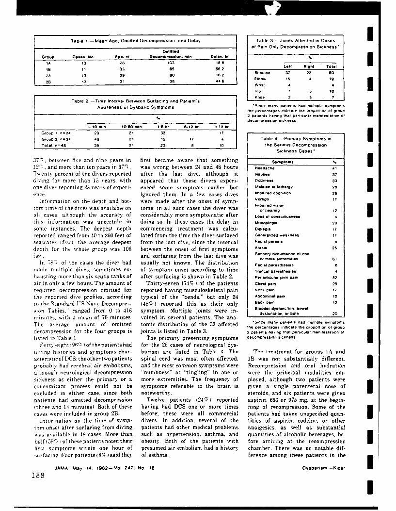
Table I -Mean Age, Omitted Decompression, and Delay Table 3 -Joints Aflected in Cases Iof Pain-Only Decompression Sickness*
OmittedGroup Cases, No. Age. yr Decompression. min Delay, hr %
IA 13 28 103 158 L15Left Right Total
2A 13 29 80 16? SroUid. 37 23 60
28 13 33 38 s46 Elbow 15 4 19_Wrist 4 I
Hip 7 3 10Knee 2 5 7
Table 2 -Time Interval Between Surfacing and Patient's
Awareness u! Cy~baric Symptoms 'Since many patients had multiple symptomsthe percentages indicate the prooon of group2 patients having tia particular manitestation ofdecompression sickness
< 10 min 10-60 min 1-6 hr 6-12 hr >12hr
Group 1 n=24 29 21 33 17
Group 2. n=24 46 21 12 17 4 Table 4 -Primary Symptoms in
Total. n=48 38 21 23 8 10 the Serious Decompression
Sickness Cases*
37(; between five and nine years in first became aware that something Symptoms %
12-, and more than ten years in 37'. was wrong between 24 and 48 hours Headache
Twenty percent of the divers reported after the last dive, although it Nausea 37diving for more than 15 years, with appeared that these divers experi- Dizziness 33
one diver reporting 26 years of experi- enced some symptoms earlier but Mataile or lethargy 28 mence. ignored them. In a few cases dives Impsied cognition 28
Information on the depth and bot- were made after the onset of symp- Vertigo 17Impaired VIs.~ I,
tota time of the dives was available or, toms: in all such cases the diver was Irpearig v2
all cases, although the accuracy of considerably more symptomatic after Loss of consciousness 4this information was uncertain in doing so. In these cases the delay in Monoplegia 20some instances. The deepest depth commencing treatment was calcu- Diplegia 7
reported ranged from 40 to 260 feet of lated from the time the diver surfaced Generalized weakness 17 Iseawater fswi; the average deepest from the last dive, since the interval Facial paresis 4depth for the whole g-oup was 106 between the onset of first symptoms Ataxis 25
fsw. and surfacing from the last dive was Sensory disturbance of oneor more extremities 61
In 7S' of the ca ,es the diver had usually not known. The distribution Facial paresthesias 8made multiple dives, sometimes ex- of symptom onset according to time Truncal pareattlesiss 4
hausting more than six scuba tanks of after surfacing is shown in Table 2. Periarticular joint pain 52air in only a few hours. The amount of Thirty-seven (74c, of the patients Chest pain 29required decompression omitted for reported having musculoskeletal pain Neck pain 17
the reported dive profiles, according typical of the "bends," but only 24 Abdominal pain 12To t li .Standard V-, Navy Decompres- (48, I renorted this as their only Back pain 12
sion Tables. ranged from 0 to 416 symptom. Multiple joints were in- Bladder dystunctcn. bowel •minutes, with 4 nitdn of 70 minutes. volved in several patients. The ana- dysfunction. or both 20
T Since many patients had multiple symptomsThe averag~e amount of omitted tomic distribution of the 53 affected the percentages indicate the proponion of groupdecompression for the four groups is joints is listed in Table 3. 2 patients having that paricular manifestation of
listed ir Table 1. The primary presenting symptoms decompression sicknessF'ort-,-eiglt t9f; ;ofth- natients had for the 26 cases of neurological dys-
diving histories and symptoms char- barism are listed in Tab), I T,, T1-p trretment for groups IA andacteristicofDCS:theothertwopatients spinal cord was most often affected, 1B was not substantially different.
probably had cerebral air embolisms, and the most common symptoms were Recompression and oral hydrationalthough neurological decompression "numbness" or "tingling" in oae or were the principal modalities em-
sickness as either the primary or a more extremities. The frequency of ployed, although two patients wereconcomitant process could not be symptoms referable to the brain is given a single parenteral dose ofexcluded in either case, since both noteworthy. steroids, and six patients were givenpatients had omitted decompression Twelve patients (247 I reported aspirin, 650 or 975 mg, at the begin-ithree and 14 minutesl Both of these having had DCS one or more times ning of recompression. Some of the
cases were included in group 2B. before; these were all commercial patients had taken unspecified quan- IInformation on the time of symp- divers. In addition, several of the tities of aspirin, codeine, or other
tom onset after surfacing from diving patients had other medical problems analgesics, as well as substantialwas available in 46 cases. More than such as hypertension, asthma, and quantities of alcoholic beverages, be-half 59r i of these patients noted their obesity. Both of the patients with fore arriving at the recompressionfirst symptoms within one hour of presumed air embolism had a history chamber. There was no notable dif-surfacing. Four patients(8 said they of asthma. ference among these patients in the
JAMA, May 14. 1982-Vol 247. No 18 Dysbarism-Kizer
188 I

cases is shown in Table 5. In 46 casesTable 5 -Percent Overall Recovery Rates cae is sh on of T e as
(92' ,, ), the amount of recovery was
Group complote Substantial oeagute Minimal that which was achieved with theIA. n-13 62 is a 15 initial recompression and adjunctivei. n- 70 30 measures. The remaining four cases2A n-13 84 a 8 (8, were treated with additional2. n 13 54 38 8 hyperbaric oxygen therapy (HBOTI"Total, n=50 s6 24 4 6 on a daily basis for as long as the
patient demonstrated improvementamount of recovery experienced by steroid-treated group, there were no that could be attributed to the HBOT.those who had only recompression gross differences between the steroid- The number of such treatmentsand hydration and those who had treated and non-steroid-treated ranged from two to eight. Both of therecompression, hydration, and ste- groups It seems notable, then, that patients with presumed air embolismroids or aspirin, more of the steroid-treated patients underwent HBOT. but neither fully
Three group I patients. all of whom recovered fully. Ten (F-1 ! of thesc -ecovcred. 'One recovered substan-had only minimal symptoms when patients recovered completely, while tially, and the other improved moder-recompression commenced, achieved one recovered substantially and the ately I Overall, two thirds of thecomplete relief of their pain with US other moderately. Conversely. only patients recovered completely. andNavy Treatment Table 5.- One other seven (54" ) of the non-steroid- 905 demonstrated at least substan-patient demonstrated no improxe- treated group recovered completely, tial improvement. Unfortunately.ment with Treatment Table 5 and, for and five 13S',, ) recovered substantial- long-term follow-up on the patientsunclear reasons, was not given the ly. The one other patient in this group with residual impairment was notbenefit of additional recompression. recovered minimally. ?vailanle. Not surprisingly, the leastAll of the remaining patients re- With one exception, the group 2 beneficial results were achieved inquired longer recompression accord- patients were treated according to US the neurological cases that were mosting to US Navy Treatment Table 6. Navy Treatment Table 6..: although delayed.which was often extended by several more than half of these patients (52/ Coxygen breathing periods, to achieve required extension of the recompres- COMMENTmaximal pain relief. Overall, 905c of sion schcdule to achieve a stable level General epidemiologic and clinicalgroup 1 patients recovered completely of recovery. (The two possible cases of characteristics of DCS and DAE inor substantially. air embolism were also treated with Hawaii have been presented else-
Detailed treatment records were the Table 6 protocol. presumably where."' '.. The 50 cases reported hereavailable on 25 of the 26 neurological because the treating physician saw no constituted 9; of the cases treatedcases. As with group 1. treatment was benefit to pressurization to 6 atmo- during this four-year period, and theynot subsar:ially different for groups spheres absolute on air, as required did not substantially differ from the2A and 2B. -;ghtly less than half by Navy Treatment Table 6A: after larger group in age, sex, dive factors,14, ) of s patients wp-c treated such long delays. as well as there presenting symptoms, treatmentwith parenteral steroids un addition being some question about whether methods, or overall recovery rate. ' 'to recompression and hydrtun, Al- these were cases of neurological DCS The capricious diving practices ofthough no definite , - ter cu L be or air embolism.) In general, the more Hawaii's diving fishermen and thedi3cerned as to the i %tivaie .'*r pre- severe cases required lengthening of high proportion of serious DCS-ibine steroids (seei.'ing to rclate :o the treatment table, while the less treated in Hawaii have been notedindividual physician habits more than serious cases responded to an unmod- before.' ......anything else. there was a tendency ified Table 6. This seems to be The reasons for delayed treatmentto give steroids to the more severely reflected in the recovery rates as well, in this series were not always clear.affected divers. Dexamethasone was since 631 of the patients treated with The few cases that occurred outside ofused in aii cases in which cortico- at. e.xtendid Tnble 6 ,-eeovered vom- the Hawaiian Islands usually in-steroids were prescribed, usually as a pletely., while 60c7 of the patients volved complicated and lengthy10- or 12-mg intravenous bolus before treated with a straight Table 6 fully. transportation arrangements. Morerecompression and followed by 4 mg recovered, often, though, the delay was due toevery six hours for up to three days. Interestingly. when the recovery the diver failing to recognize theIn a few cases high-dose hydrocorti- rates of the patients treated with an importance of postdive symptoms orsone sodium hemisuccinate (1.000 mg) extended Treatment Table 6 are com- appreciate the need for recompres-was given along with dexamethasone pared according to whether steroids sion treatment. A number of theas an initial bolus; when this was were used or not, there appeared to he diving fishermen first attempted self-done the initial dose of dexametha- a benefit associated with the use of treatment by in-water recompressionsone was usually decreased to 4 or 6 corticosteroids. Six (75? I of eight with compressed air and, after thatmg. In no instance was there any steroid-treated patients recovered failed, employed various combina-adverse effect that could be associ- completely, while only two (407, 1 of tions of alcoholic beverages and anal-ated with the use of steroids. five non-steroid-treated patients re- gesics before seeking recompression.
Except for there being relatively covered fully. The futility of in-water recompres-more severely affected patients in the The treatment outcome for all 50 sion with compressed air is well
JAMA, May 14, 1982-Vol 247. No IS Dysbarism-Kizer 189
.. .... 189

known " ite need for further educa- Navy divers who had been doing steroids and osmotic agents have attion of the diving community about controlled diving. No recompression times produced apparent therapeuticthese things is clearly evident treatment tables have ever been effects independent of recompres-
In general, treatment of these developed specifically for civilian sion.- Thus, the greater recoverypatients was conduct-" according to sport divers, ie, ones that take into rate observed in the steroid-treated mthe guidelines pru,;lgated in stan- account poor physical conditioning, cases in this series was notable anddard referen',., *However, during the presence of concomitant medical supports the notion that their usethe four-Y-- ,eriod covered by this problems, uncertain depth and hot- may have a beneficial effect in neuro-study a: least eight different physi- tom time factors, and other factors logical DCS. It is important to note,cia-x, were involved in treating these that complicate the treatment of however, that the retrospective and
-it) patients, and the primary treating civilian diving casualties. Nonethe- uncontrolled nature of this study, asphysician usu-!ly did not consult with less, the Na,y treatment tables have well as the lack of long-term follow-an% other physician before commenc- worked reasonably well for treating up. precludes deriving any definiteing treatment. Thus, treatment was civilian divers over the years, having conclusion about the efficacy ofin no way controlled and reflected the overall success rates of 801 to 90, in steroids in these diseasesindividual biases of the treating phy- patients who have been treated with References Isician. delays of less than 12 hours.' -..
When evaluating the efficacy of which is comparable with the results h Strauss RH ti).,ng medicine A10 Rv'-• R~ksp, [is., i.4SL] iii ')-023
treatment for these diseases it should achieved in this series of cases having 2 Bane CG Acuti, decompression Sicknessbe remembered that the diagnoses of more delayed treatment. When these 50 cases JA CEP 1979.7 351-35-4H J J. Chan G Management of delayed[-CS and DAE are purely clinical results are viewed together with the case, of decompression sickness-three case
diagnoses based on the occurrence of rarity of untoward effects associated reports Sinijpor, Mtd J 19;3.i4 532-535
characteristic symptoms and signs with recompression treatment, it is o Pooskii Vs Two cases of late treatment
afe 'decompression sickiivs6 Gigj 7T- froy Zawolafter appropriate diving or other clear that all persons suspected of 197A 44-45pressure exposures, there being no having untreated DCS must be 5 Kizer ,. Larsern RT Deiavd treatmentspecific laboratory or other diagnos- referred for recompression even if ,if type I decompression sickness Press ,r 19sd
tic tests, and that recompression such has been delayed for several f, Kizer KV, Corticosteroids in treatment of Ireatment is a dynamic process in days or, possibly. longer. serious decompressionsickness A, ,i9 .i.1 45*5-4 s
wvhich the variables of pressure and It is apparent from this series. -, Greenstein A. Sherman D. Me amed YoN.gen breathing time, which deter- though, that a larger oxygen dose Chokes-favorabie response to delayed recom-mine the o::vgen dose," are titrated may be needed when treating delayed pressiorn therapy A case report -ria Spart
• " " Eniron Mu'd 19SI.52,559-5SMaccording to the patient's response cases. Except for the three patients l, DavsiJCtedi Treatme'vofS-.-,.owrfco-.
Although several different recom- who had minimal symptoms remain- pri's.ior Sickee-s and Anerta Go. Embolism
pression protocols have been advo- ing when recompression commenced, Bethesda. Md. Undersea Medialo Societ Inc.•1979I
coted for the treatment of DCS, the US Navy Treatment Table 5 was not 9 Rivera JC Decompression sickness amongones most widely used in this coun- sufficient to achieve a maximal thera- divers An analysis of 93.5 cases Vilit Mcd
1 WA . 129 316-334rr are the oxygen treatment tables peutic effect in the other 21 cases of 1 Erde A. Admonds C Decomp-.ession sick-promulgated by the US Navy.:: All pain-only DCS. Likewise. Treatment ness A chnical z rics JOM 1975.17.324-326 Ipatients in this series were recom- Table 6 needed extension by two or 11 Kizer KW Dsbarsn in paradise Hoaaii
Me d.1 19"40.30 109-116pressed according to these protocols, more oxygen periods in more than 12 I'S N'au Diving Manual, NAVSHIPSexcep: for many of the patients being half of the seriouF DCS cases. Overall. 1...4-W)01.9)0 I'S Dept of Defense. Dept of the
given additional oxygen breathing the treatment table had to be length- \a%,. 9.5 a13 Kizer KW Epiderniotogic and clinical
periods When treating DCS the phy- ened in 697 of the cases (34 of the 49 aspects of dvsbarism in Hawaii Read before the
sician may extend the recompression cases in which information was avail- Sixth Annual Conference on the Clinical Appl-cations of Hyperbaric Oxygen Therap . Long
scheduie at a given pressure if the able 1. Whether recompression at Peach. Calif. June 13, 19S1patient demonstrates continued im- higher pressures with Nitrox, a 50" 14 Kizer KW. MilroN WC Dsharsm associ-provement at that pressure, which combination nitrogen-oxygen mix- ated with alcohol abuse A case report Huwai,
Med J 19S1.40 12-15would he either 2.S or 1.9 atmospheres ture, or with saturation treatment, as I-) Kizer KNV. Pegg J Scuba emergencies
abrsoiute when Navv Tables 5 or 6 are has been advocated for difficult cases Hi,,,, Phisir,o, 19SU1o-14-36used In a multiplace recompression hy some investigators," would be 16 Strauso RH D197n6 Mtedicint Neu York,
• (.,rune & Stratton Inc, 1976
chamber, several additional oxygen helpful in cases of delayed treatment 1. Edmonds E. Lowr, C. Pennefather J Di, -breathing periods can be given be- has not been investigated. ,1o an? Suoaqunt .ledicine. ed 2 Mosman.
cause of the ability to give periodic Although recompression is the es- Australia. Diving Medical Centre, 191sair breaks, which decrease the risk of sential treatment of DCS and DAE, avnd Medicm or' Dirin and (ompressed Air
oxygen toxicity As many as four or several adjunctive pharmacologic ir Baltimore. Williams & Wilkis Co. 197519 Meiamed Y. Ohrv A The treatment, and
five extensions were given in some of agents have b-,n recommended for ti,. neurological aspects of dvinc accidents inthe cases in this series. treating neurological dysbarism in Israel Parapegia iNWo. 127-132
Another factor that must be con- recent years. In particular, cortico- 2ou Miller J\. Fagraeus L. Bennett PB. et atat taen aroren-ot\iclr saturation therap% in serious
sidered when evaluating the efficacy steroids have been widely recom- cases of compressed-air decompression sickness
of recompression treatment is that mended, although their efficacy hqs Lo, ," 197s,.2 169-171cnr21 Saimarez RC. Bolt JF, Gregor% P1 Neuro-,he tS Nav- treatment tables were never been determined by controlled gi,,cal decompression sicknes- treated ithout
derived for health,. Aell-conditioned studies. Interestingly. though. recompression h- Mcd J 1973.1 151-152
190 JAMA. May 14. 1982-Vot 4- No 18 Dysbatisn-Kizer
P~rtea one~ P,,S?7eC itt Met lUn,teC States o' Ame',c,,"1.90 I

DIVING EMERGENCIES
Kenneth W Kizer, M.D., M.PH.
A \, henever humans breathe compressed air in the E. Dvsbaric air embolism (DAE) is the most feared com-unoervsater environment they are subject to various plication ot pulmonary barotrauma and is a malorph.sical and biologic hazards, the most unique of cause ot death and disabiltv among sport divers.-.hhicn are the pressure-related maladies. Most div- Symptoms of DAE always occur within 10 minutesing-reiated emergencies can be properly diagnosed alter surfacing from a dive, and usually within the firstbv history and pnvsical examination alone. The his- 2 minutes. The neurologic manifestations are typicaltory is most important and, at a m;nimum, should of an acute stroke, asymmetric multiplegia being thedetermine the type of dr.i ig done (e.g., scuba diving most common presentation overall.,.ersus snorkeling); the equipment used (e.g., depth F. Even though a diver may report no omitted decom-gauge, diving watch); the number, depth, and sub-mergence time of all dives in the 48 to 72 hours pression, this is sometimes unreliable ie.g., when aprececing symptom onset; the surface interval(s) depth gauge and a diving watch are not used). Lik-between repetitive dives: whether in-water recom- wise, a very small number at people may surfer de-pression %%as taken, and if so, how much: the specific compression sickness despite having adhered to the
site ot diving and its environmental conditions ,e.g., diing tables.
presence ot a strong current and water temperature), G. Gas pressure in the various air-filled spaces of theprimarv diving activity engaged in (e.g., photogra- body is normally in equilibrium with the environ-prt,.. speartishingi; whether the dive was comph- ment: however, if something e.g., mucosal inflam-catect b,, running out of air. marine an;.mal injury, mation or a nasal polyp) should obstruct the portalstrauma. or other notable event; what the diver did of gas exchange for these spaces a pressure disequi-after ai ing; and. importantly, when the first svmp- librium will develop. Barotrauma is the term used iotoms .,,ere noticed and how they have changed, c: denote the tissue damage resulting trom such an im-ther spontaneously or as a result of something the balance.
i\,.er did. Many ot the most serious diving emergen- H The ears and paranasal sinuses are most oten at-cies are caused by formation of bubbles of nitrogen tected by barotrauma.in the ci,,er s tissues.
.,itroaengasuptakeduringdivingdependsondepth .Pulmonarv barotrauma may occur during ascentanc time. When sate diving depth-time limits are ex- from depth with a closed glottis (as when holdingceeced, specitic time must be taken for "decom- one's breath) or as a result ot a Tocal proolem ie.g.. anression ' to allow excess dissolved nitrogen in the mucus plug or granuloma). The most common mani-oi, er ; tissues to be onf-gassed. This must be done to festations ot pulmonary barotrauma are mediastinala'oil bubble tormation in tissues, the penultimate emphysema and subcutaneous emphysema; how-concition tor decompression sickness. The L.S. ever. pneumopericardium, pneumoperitoneum.\a,.,. :!tancard Decompression Tables are the most and/or (rarelvi pneumothorax may occur singlv or in.ae e,. accepted protocols tor determining whether combination. Recrmpression is contraindicated un-The eeriocf or required decompression has been ace- less there is concomitant decompression sickness orcuate air embolism. Management of the extra-alveolar air
C Decompression sickness IDCS) is a multisvstem dis- depends on its location and amount.
orcier resulting from the liberation of inert gas (e.g.. J Although gastrointestinal air accumulation is gener-nitroven' Trom solution and the resultant formation allv easilv relieved and self-limited, gastric rupturent gas bubbles in blood and body tissues when am- has occurred in rare instances.bient pressure is decreased. The pathogenesis oDCS in. oives both bubble formation and the activa-tiun ot "ageman factor and other biophvsical phe- Referencesnomena at the blood-bubble interface. Most oftenafnected b. DCS are the musculoskeletal and neuro- Davis IC. Kizer KW. Diving medicine. In: Auerbach PS,lo ic s stems. When more than about 10 percent of Geehr EC. eds. Management ot sdderness dnd environ-!ne c)uJmonar\, venous system is occ!uded by intra- mental emergencies. 2nd ed. St Louis. CV Mosbv. 1989
,ascuiar )ubbles. a serious condition known as "the Edmonds C. Lo\,rv C. Pennetather I. eds Diving and sub-nokes results About half of persons suffering DCS aquatic medicine. 2nd ed. Mosman, N S.W. Autralia
ce,.eop s,.mptoms within 10 minutes ot surfacing Diving Medical Centre. 1981!•rv- i.ciinv: more than 95 percent have svmptor's Kizer KW Dvsbaric cerebral air embolism in Hawaii. Ann
,tnin nours atter diving. Rarely. the onset ot svmp- Emerg Med 1987 16:535 -341
bins ma'. be delayed tar as long as 24 hours after Kizer KW. Management of d'.sbaric di'.ing casualties EmergMed Clin North Am 1983. 1:659 - 670.
ci. inp Shilling C'. Carlston CB. Mathias RA, eds The phvsician s
D n some cases, neurologic decompression sickness is guide to diving medicine. New York: Plenum Pressnot readi,, distinguishable from air embolism. 1984
191

COMPRESSED AIR DIVING EMERGENCIES I
® History -Physical examination
Pain, difficult breathing, or neurologic deficit
Assess:Period of decompr )ssion
Omitted decompression No omitted decompression
_ _ _ _ _ _ _ _ _II() Decompression © Combined air embolism () Air embolism () Barotrauma
sickness and decompressionsickness (T) Decompression
I sickness IHyperaric Oxygen Co MC nsider:Treatment ! Marine envenomationMusculoskeletal injury
or overuse syndromeTraumaHyperventilationNear drowning
II
Ear Sinus Tooth Q Pulmonary ( Gastrointestinal Skin
F Decongestants Dental Chest film Observe Observe Ievaluation
Abstinence fromdiving untilresolved
IIII
192 I

I
MAN AT ALTITUDEADAPTATION AND MALADAPTATION
WILDERNESS MEDICAL SOCIETY, 1991
Dr. Peter H. Hackett
Director, Denali Medical ResearchAffiliate Associate Professor,
University of Washington School of Medicine andUniversity of Alaska Anchorage
EDUCATIONAL OBJECTIVES
1. To understand the normal physiological adaptations that take place onascent to altitude.
2. To appreciate the limitations imposed upon exercise at highaltitude and how to maximize performance.
3. To understand the pathophysiology of the problems of high altitude, includingacute mountain sickness, the edemas of altitude, and retinal hemorrhage.
4. To gain a working knowledge of the prevention, diagnosis and treatment of theseconditions.
OUTLINE
I. THE PHYSIOLOGY OF ACCLIMATIZATION
A. Struggle responses (first few days)
1. Hyperventilation
a. Attempt to maintain alveolar P0 2
b. Produces hypocapnic alkalosis, secondary bicarbonate diuresis
i9 3

,Ic. Determined partly by carotid body function (hypoxic ventilatory response) and
CO2 sensitivity, attenuated by alkalosis, alcohol, sleeping meds. Augmented byrespiratory stimulants.
2. Increased cardiac output (mostly increased heart rate)
3. Fluid shifts 3a. Increased intracellular fluid
b. Diuresis secondary to veno-constriction with central shift of blood, and Ibicarbonate excretion
c. Loss of 15% of plasma volume in first few days, reduces stroke volume.
4. Increased hemoglobin con-entration Ia. Caused initially by decrease in plasma volume
b. Increases oxygen carrying capacity per unit of blood.
c. Increased erythropoeisis within hours, but weeks to increase red cell mass I5. Changes in cerebral blood flow depend upon interaction of 02 and CO2-- 3
generally increases 25-40% at 14000 feet, day one, then returns to normal overfive days. 3
6. Cellular metabolic changes
a. Some cells (endothelial) produce hypoxic associated proteins (HAP) Ib. Increased oxidative enzymes (muscle) 3
B. Adaptive changes (weeks to months)
1. Hyperventilation continues but renal bicarbonate excretion brings pH towardnormal 3
2. Cardiac output decreases
3. Increased RBC mass
4. Increased density of capillaries in tissues and mitochondria in cells.
194 I!

III. EFFECTS OF ALTITUDE ON EXERCISE PERFORMANCE
A. VO2 max. decreases by 3% per 1000 feet, starting at 5,000 feet, improves withacclimatization very slowly and not by much.
B. Submaximal exercise (endurance time)--starts to increase at day 5, up 50% by day
15.
C. Factors limiting exercise are not well understood. Possiblities:
1. Decreased motor meuron drive (CNS) to muscles.
2. Decreased diffusion of 02 to muscle cells.
I D. Efforts to increase performance at altitude include blood doping, nutrition,hyperbaric and hypobaric training, and various drugs. Acclimatization seems best.
I E. Altitude exposure may benefit sea level performance.
III. ACUTE MOUNTAIN SICKNESS (AMS)
A. General
1. Incidence of 15-30% of Colorado resort skiers, 50% McKinley climbers, 70%Rainier climbers, 70-100% if flown to 14,000 ft.
2. Related to RATE OF ASCENT, altitude reached, SLEEPING ALTITUDE, and
contributory factors. No relationship to physical fitness. Younger may be moresusceptible.
3. Individual susceptibility and reproducibility. Low vital capacity, low HVR,exaggerated pulmonary hypertension in response to hypoxia all contribute toillness. Past history a risk factor.
B. Diagnosis--THINK AMS!
1. Setting: rapid ascent to a higher altitude in unacclimatized persons.
2. Symptoms: headache, anorexia, dizziness, nausea, insomnia, lassitude, dyspnea.Note: mostly neurological symptoms. Periodic breathing common, not a sign ofAMS. Early AMS exactly like a hangover.
195

I3. In early stages, a lack of physical findings. Advanced: findings of pulmonary and
cerebral edema. Ataxia and nailbed cyanosis most useful indicators of seriousillness.
4. Differential diagnosis: dehydration, exhaustion, CO poisoning infections of lung or
brain, hypothermia, drugs, psychiatric problems.
C. Pathophysiology
1. Ambient hypoxia leads tc rterial hypoxemia and cerebral hypoxia. Ia. Hypoxia at a given altitude depends upon degree of hyperventilation. The lower
the PACO2, the greater the PA0 2.
b. Brain is sensitive to arterial P0 2, not ambient P0 2 .
2. Inadequate ventilation (low HVR) causes greater hypoxemia.
3. Impaired gas exchange leads to even greater hypoxemia-increase alveolar-arterialoxygen difference.
4. Fluid retention and redistribution.
a. Diuresis with acclimatization, antidiuresis with AMS. Ib. Role of ADH, renin-angiotensin-aldosterone systems.
c. Brain and lungs overhydrated.
5. Brain Swelling.
a. Cytotoxic edema--shift of fluid into cells. Ib. Vasogenic edema--permeability vs increased capillary filtration. 3c. Role of cerebral blood flow--too much or too little?
6. Theory of generalized "capillary leak syndrome"--lung, brain, kidney, retina.
D. Treatment I1. Descent--1,000ft may be adequate--as far as necessary for results. I
196 II

I
I 2. Acclimatization at same altitude ok for self-limited illness--sick person never leftbehind alone.
3. Oxygen if available, especially good for headaches and encephalopathy.
4. Analgesics--acetaminophen, aspirin, codeine.
5. Anti-emetics--prochlorperazine (Compazine) 10mg IM, mg po (increases HVR).Benadryl for extrapyramidal reactions.
6. Acetazolamide 125-250mg every 8-12 hours (a sulfa drug).
7. Dexamethasone 4mg po, IM, IV every 6 hours if ataxia or change in consciousnessI present.
8. May re-ascend with staged acclimatization, acetazolamide.
I E. Prevention
1. Slow ascent; climb high, sleep low.
2. High carbohydrate diet--> 70%, improves respiratory quotient.
3. Avoidance of respiratory depressants (esp. sleeping pills) and use of alcohol inI small amounts only.
4. Chemoprophylaxis:
a. indications are forced rapid ascent or history or recurrent illness.
I b. Acetazolamide 5mg/kg/day divided into 2 or 3 doses, for one day prior and oneday after ascent.
I c. Dexametherone 4mg every 6 to 12 hours--for those intolerant of acetozolamide,
or for insertion to extreme altitude.
IV. HIGH ALTITUDE PULMONARY EDEMA (HAPE)
I A. General
1. Strikes 1-2% above 12,000 ft.
I197I

2. Most common on 2nd night.
3. Related to rate of ascent, exertion, use of sleeping medications, cold.
4. Many (most?) people have transient, milder forms of illness. IB. Diagnosis
Early: dry cough, increased heart rate, decreased exercise performance, shortness ofbreath with exercise and increased exercise recovery time.
Late: dyspnea at rest, tachycardia, tachypnea, cyanosis, productive cough, rales.
Atypical presentations: sudden death, cerebral manifestations only (esp. ataxia),acclimatized person, mixed with respiratory infection, bronchospasm.
C. Management
1. Rest, keep victim warm I
2. Oxygen
3. Descent
4. CPAP mask
5. Medications which may be helpful (usually not necessary) Ia. Nifedipine 10 mg po stat, 30 mg extended release tid
b. Acetazolamide 5 mg/kg/day
c. Furosemide single dose of 40 mg
d. Morphine small incremental doses I6. Victim may reascend when HAPE resolved 3
D. Pathophysiology
1. Non-cardiogenic; normal wedge pressures, high pulmonary arterypressure.
198 I

2. May be a form of neurogenic pulmonary edema (NPE).
3. May be a combination of high pressure and high-permeability edemas.
E. Prevention same as for AMS--acetazolamide apparently effective, also nifedipinerecently shown good for prophylaxis.
V. HIGH ALTITUDE CEREBRAL EDEMA (HACE)
A. Diagnosed by ominous progression of cerebral AMS symptoms and findings of ataxiaand change in consciousness.
B. Only effective therapy is descent; oxygen temporizes, drugs of little value, exceptperhaps Decadron given early.
C. Mechanism same as AMS-more advanced.
D. Prevention same as for AMS.
VI. RETINAL HEMORRHAGES
A. Common, affect vision only if in macula (rare).
B. Probably an hypoxic mechanism, leak is in capillary bed.
C. No treatment, no known prevention (acetazolamide?), clear spontaneously in 7-14days.
199

BIBLIOGRAPHY
Hackett, P.H. and Roach, R.C.: Medical therapy of high altitude illness, Ann Emerg Med,16(9): 980-986, 1987. I
Hackett, P.H., Mountain Sickness: Prevention. Recognition and Treatment. Nev.' York:American Alpine Club, 1980
Singh, I., P.K. Kanna., M.C. Srivastava et al. Acute mountain sickness, New England J.Med. 280:175-184, 1969
Houston, C.S. Going Higher. Burlington: Houston, 1983 n
Hackett PH, Roach RC, Sutton JR. Medical problems of high altitude. IN: Auerbach P,Geehr E, Editors. Management of Wilderness and Environmental Emergencies St. Louis,MO: Mosby; 1988:1-34
Hackett PH, Hornbein TF. Disorders of high altitude. IN: Murray JF, Nadel JA, Editors.Textbook of Respiratory Medicine Philadelphia, PA: WB Saunders; 1988:1646-1663
Hackett PH; Roach RC. High altitude pulmonary edema. J. Wilderness Med 1990;1:3-26
Hackett PH. Medical research on Mt. McKinley. Ann Sports Med 1989;4(4):232-244
Hultgren HN. The safety of trekking at high altitude after coronary bypass surgery (Letter).JAMA 1988;260(15)2218 3Johnon TS; Rock PB. Acute mountain sickness. N Engl J Med 1988;319(13):841-845
Oelz 0; Maggiorini M; Ritter M; Waber U; Jenni R; Vock P; Bartsch P. Nifedipine forhigh altitude pulmonary edema. Lancet 1989;2:1241-1244
Sutton JR, Coater G, Remmers JE, Editors. Hypoxia: The Adaptation Philadelphia, PA:BC Dekker; 1990 3
III
200 II

THE WORLD STATUS OF WILDERNESS MEDICINE
Coordinators: Blair Dillard Erb, M.D. and Bruce C. Paton, M.D.
Moderators: Peter Hackett, M.D., Oswald Oelz, M.D., Paul C. Auerbach, M.D.
Kenneth Kizer, M.D., Bruno Durrer, M.D., and Warren D. Bowman, M.D.
The First World Congress on Wilderness Medicine has been structured to address
issues related to health and medical care of individuals who venture into remote
and/or hazardous environments wherever they may be.
Wilderness is global in scope and knows no national boundaries. Since the aims
and goals of medicine are the same the world around, and because nations do not
contain identical geographic, environmental or medical features, a series of three
1 hour sessions has been designed for participants to share their experiences and
their perception of the principles of wilderness medicine with other health
professionals from around the world.
Titled "The World Status of Wilderness Medicine", these sessions 3re an effort to
bring together the various elements that constitute the diverse fields of
wilderness medicine. Since wilderness medicine is traditional medicine with no
single discipline, this is an opportunity for the different specialties such as
Cardiology, Pulmonology, Family Medicine, Emergency Medicine, Sports Medicine,
etc., to examine their individual roles in wilderaess medicine with a focus on the
uniqueness of the requirements of medical care under the circumstances. Various
organizations, institutions, centers, agencies, and leaders will discuss tht
current status of their activities in research, education, and service in
wilderness medicine.
Attendees are invited to participate actively in these sessions. The objectives
of the sessions will be:
1) To define the current world status of various aspects of wilderness
medicine.
2) To outline needs for development of the field of wilderness medicine.
3) To propose mechanisms for enhancing research, education, and service in
wilderness medicine.
201

Representatives from Mountaineering Medicine, Aquatic Medicine, Human Performance,
Environmental Medicine (including heat, cold, and altitude stress), Medical
Services, Public Health, and others will review their special areas of interest
and stimulate discussion by participants.
Emphasis during these three World Status sessions will be on planning for
cooperation for future development of medical resources in the broad field of
wilderness medicine. 3Please consider during your personal deliberations the question, "Should the World
Congress on Wilderness Medicine evolve into a consortium concerned with wilderness
medicine?". Perhaps some form of loosely knit cooperative consortium could arise
as a "United Nations" involved in wilderness medicine.
Please complete the enclosed questionnaire and bring it with you to the sessions.
From this we hope to develop a directory. These are your sessions, and the
product will be a result of your participation. 3
SCHEDULE 3WORLD STATUS OF WILDERNESS MEDICINE 3
Monday, July 15 (2:00-3:30 p.m.)- Mountaineering and Environmental Stresses.Moderators: Oswald Oelz, M.D.I
Peter Hackett, M.D.
Tuesday, July 16 (2:00-3:30 p.m.)- Aquatic Medicine and Environmental Stresses.Moderators: Kenneth Kizer, M.D.
Paul Auerbach, M.D.
Thursday, July 18 (2:00-3:30 p.m.)- Delivery of Services including Search and IRescue and Emergency Care.Moderators: Bruno Durrer, M.D.
Warren Bowman, M.D.
Friday, July 19 (12:00-12:30 p.m.)- Summary of the Proceedings.Moderators: Blair D. Erb, M.D.
Bruce C. Paton, M.D.
I202 I
I

QUESTIONNAIRE
WILDERNESS MEDICINE ORGANIZATIONS
This questionnaire will assist in developing a directory of organizations and
centers involved in wilderness medicine. It focuses on two types of groups:
1) Those whose primary mission is medicine in the wilderness setting, and 2) Those
involved in wilderness activities which have a significant interest in medicine.
Division into these groups is based on the degree of emphasis on the medical
aspect of wilderness activity.
Each individual or organization is asked to complete the questionnaire and return
to the Wilderness Medical Society staff. Thdnk you.
I. Division (Check One)
Medical Organization with Wilderness Interest.
Wilderness Organization with Medical Interest.
II. Organization
Name:
Address:
III. Chief Executive Officer of Organization
Name:
Address:
IV. Aim of the Organization (Check All That Apply)
Education Service
Research Other (Specify)
203

IV. Category (Check All That Apply)
Mountaineering Search and Rescue i
Environmental Stresses Research
Aquatic Medicine Education iAdministration Other (Specify) I
VI. Purpose (Please Describe) 3III
VII. Other organizations with which you are familiar that should be included in a
directory: 3Ii
VIII. For individual completing questionnaire:
Your Name:
Your Address: _
Your relationship to the organization you described:
Please return to: 3Blair Dillard Erb, M.D.
President, Wilderness Medical Society
1729 North Highland Avenue
.ackson, Tennessee 38301
USA 3(901) 422-0330
204 I

NOTES FOR THE WILDERNESS MEDICINE RESEARCH SEMINAR
I. Find a question worth answeringA. Attend conferences
1. Find out what areas need investigatingB. Observe in your own areaC. Test hypothesesD. Try to study something within your area of interest
or expertise
II. Figure out if it's feasible to studyA. Can denominators be calculated?B. Do people all use one medical clinic?C. Do they start and finish at the same point?D. Can the problem be defined (such as rock-climbing
hand injuries)?E. Would it be too expensive to study?
III. Fund it yourself if it's under $2000.A. Figure out your fixed expenses
1. Printing, copying, mailing2. Computer software or computer expert3. Secretarial or research assistant help4. Having figures made (for publication)
IV. Design the studyA. Define your study populationB. Define a "case"C. Define the numeratorD. Decide if it will be prospective or retrospectiveE. Who will collect the data?F. Design a questionnaireG. Design your computer database before you finish your
quesionnaire. Make sure you understand the type of data youwill collect at each point in the database. [Example: Areyou in good physical condition? Give scale 1 to 10, wordssuch as sedentary, occasionally exercise, exerciseregularly, fanatic]
H. Decide how long the study will runI. Define the authors in advance, and designate first
author if that is possible. Get everyone's agreement inadvance. This can cause a lot of trouble later if leftunspoken.
V. Analyze your dataA. Figure out every angle of what you have learned
1. Tabulate, take means, etc.B. Don't go fishing for statistical significance [Give
examples ]C. Calculate statistical significance only when you had
a hypothesis to test. [For example: You may have postulatedthat men have more skiing injuries than women because they
205

tend to ski faster. This can be tested statistically at theend of your study. But if you happen to detect that people iwho wear red jackets get hurt more often, you can'tnecessarily assume significance to that finding]
D. Decide on your major findings1. You don't have to report on everything you
learned
VI. Write your paperA. Write a first draft including everything that you
think of to say. You can always take it out later.B. Organize the paper strictly along the lines of
Introduction, Methods, Results, and Discussion. Read otherpapers with this in mind. Do not mix these sections. Thisis the most common mistake of new paper writers. U
C. Make the introduction shortD. Do not justify your methods in the methods section,
unless there is an overwhelming reason to do so.E. Give the results as plainly as possible, using
appropriate graphs, charts, etc. Simplify wheneverpossible.
F. Use the discussion to point out what you havelearned, the strengths and weaknesses of the study, itssiqnificance to the world of wilderness medicine, possibleadvice to the study population based on your research, and Ipossible future studies.
G Resist witticisms and the desire to be too clever,unless you are so overwhelmingly clever that it can't beresisted.
VII. Submit your paperA. You can only submit to one journal at a timeB. The review process can take several monthsC. So choose carefully where the article might have a
good chance ID. Follow the "instructions for authors" for the
selected journal carefully. Write a cover letter explainingwhy your paper is significant, and have your co-authors signthe required statements, if any. I
E. Don't get angry at the reviewer's comments; they aretold to be critical.
F. Expect to have to change a lot of the article, or Ishorten it drastically.
VII. Article or letter?A. Articles much more significant in terms of CVB. Letters much easier to produce and to get published.C. Letters get published more quicklyD. Letters best medium for brief case presentationsE. Articles carry more weight because they are peer
reviewed.
DETAILS:
206 I

U
1 1. Computer database programs2. Statistical programs, such as Epiinfo3. Choosing the right statistical test: review very basictests, such as comparing two ratios, or comparing means.4. Informed consent and releases, if necessary5. How to make figures, graphs, etc. (Harvard Graphics orhire an artist)6. Epidemiologic methods: mechanisms of bias in selection ofstudy groups, populations, clinics, control groups. Forexample: do all ski injuries go to the first aid hut first,or do the more serious ones go straight off the mountain?
3 Work through an example, such as the one I suggested,studying whether men lose more weight on trek than women.In general keep the whole presentation simple, direct,practical. We have two 90 minute sessions. The firstsession could be an overview as outlined above. The secondsession could be working through a sample study, and dealing3 with details and questions.
III
20
I

I
3 WORKSHOP ON COLD WEATHER SURVIVALFIRST WORLD CONGRESS ON WILDERNESS MEDICINE
Whistler Mountain, British Columbia,July, 1991I Warren D. Bowman, MD
The cold weather survival workshop will be conducted in the snow
as much as possible. If snow conditions are unsuitable for snowshelter construction, model shelters will be demonstrated.Please try to attend the didactic session on cold weathersurvival or at least read the syllabus material before attendingthis workshop.
Objectives: After attending this workshop, the student should beable to:
1. Explain the layer system of clothing and describepreferred fabrics, clothing items, and layering sequences for coldweather outdoor travel.
2. Appreciate the insulating capacity of snow; describe and
demonstrate the principles and building techniques of emergencysnow trenches and multiday oversnow travel snow trenches, snowcaves, igloos, and lean-tos.
3. Describe and demonstrate the techniques of gatheringfuel and starting a fire under winter conditions.
4. List recommended items of winter survival equipment.
Since the end of World War II, more and more people have beenventuring out into the winter wilderness to pursue hunting,snowmobiling, cross-country skiing, winter mountaineering, alpine
skiing, winter camping, and other cold weather sports. Growth inexpeditionary mountaineering places many climbers in regionswhere winter exists all year round. When modern conveniences areleft behind, life is reduced to the basics and individuals becomecompletely dependent on their own resources. Our ancLstors'hard-won knowledge of how to survive in the cold may need to berelearned, sometimes at considerable cost.
This workshop will emphasize preplanning, anticipation, alwayscarrying a minimum of emergency survival equipment, andprevention of environmental emergencies rather than panic-3 stricken improvization.
The most important human organ for survival is the brain, sincevoluntary actions such as preparedness, regulation of energyexpenditure, adjustment of clothing, and seeking shelter are moreimportant than involuntary mechanisms of adaptation to heat orcold. Since regulation of core temperature is the most importantshort-term goal in cold weather survival, the subject must eitherbe carrying enough insulation or know how to improvise enoughinsulation and/or provide enough external heat to maintain asatisfactory core temperature for an indefinite period of time.
Since decreasing heat loss through intelligent use of insulation
i 209
I

Iis the most energy efficient method of avoiding dangerous degreesof cooling, it will be discussed and demonstrated in detail.
Clothing should be worn in a number of thin layersso as to trap several layers of still air, which are warmed bybody heat. The most effective fabrics are those which trap air Iand prevent its motion, and whose effectiveness is not reduced bybecoming wet. The best of these currently are wool,polypropylene, and such types of treated polyester as Capilene
and Thermax. Cotton has poor insulating value, especially whenwet, and should be avoided in cold weather. Orlon, polyester,acrylic, and similar synthetics are also good insulators. The
above fibers are typically worn as the innermost 2 or 3 layers--- Ilong underwear, shirt, sweater, and pants. Outer layers arenormally chosen from fibers that produce loft, such as polyesterand nylon pile and fleece, down, Dacron, Hollofil II, Quallofil,
Thinsulate, and Thermolite. These are made into jackets, vests,and overpants. The outermost layer is preferably of a windproofand water resistant fabric such as Gore-Tex, a nylon/cotton
blend, or similar material. Two or three layers are used when Iskiing, hiking or building shelters, with a fourth layer addedwhen stopping for lunch or a rest.
SAMPLE COLD WEATHER CLOTHING:a 4-layer system
Underwear: 100% wool or 85% wool/15% nylon is the gold Istandard, but hard to obtain. Thermax, Capilene, polypropyleneand blends of these are the new standard. Combinations such as
Duofold and Thermolactyl are less warm. IShirt: Wool, Capilene, polypropylene and similar materials
are good; they should open completely in front or at least have a Ihalf-zipper.
Pants: The hard-finish wool pants found in military surplus
stores are durable and reasonably priced. Knickers are preferredby some.
Parka: The most versatile combination is a windproof andwater-resistant shell worn over a pile jacket. The pile jacketshould have a nylon outer cover to prevent snow sticking to itwhen it is worn alone. The shell should have a hood with
drawstring closure and be fingertip length. Metal zipper pullsand metal snaps should be shielded or situated so they do nottouch bare skin and zippers should have a weather flap. "Pit-
zips" are useful. Handwarmer pockets should be included and themain pockets should be accessible when you are wearing a fannypack or backpack with the waist belt fastened.
Wind Pants: These are a must for cold, windy weather or fordigging a snow cave. They should be made of windproof and water-resistant material.
Fourth Layer: for very cold conditions, a down, Dacron,
210
I

Thinsulate, etc., filled quilted garment such as a jacket or
vest, and a pair of pile pants.
Socks: A good combination is a pair of polypropylene socksnext to the skin with 1-2 pairs of heavy, wool socks over them.
Hat: A wool, polypropylene or acrylic stocking-type that
can be pulled down to cover the eers. Unless a neck gaiter is
worn also, choose a hat with a face mask or balaclava feature.
Mittens or Gloves: Mittens are warmer than gloves but are
less useful when delicate finger movements are required. Several
manufacturers make three-layer mitten and glove sets that includewindproof shells with leather palms and two sets of removable
pile inner layers that velcro in place. Polypropylene glove
liners are useful when working on ski bindings and doing other
delicate work.
Boots: The choice depends on the type of activity and the
expected temperature. Double boots generally are best for coldtemperatures. They can be winter mountaineering boots, telemarking
boots, ski mountaineering boots, or non-technical boots such as
the Sorel.
Rain Gear: In moderate climates or very wet conditions
where rain or wet snow may be encountered, you may prefer to
carry one set of windproof outer garments plus a second set ofwaterproof ones.
Vapor Barrier Garments: These are waterproof socks,
underwear tops and bottoms, and sleeping bag liners worn over athin polypropylene garment. This system traps a warm film of
moisture next to the skin, theoretically decreases waterrequirements by reducing sweating, and maintains the insulating
properties of outer garments by keeping sweat out of them. Try
them under non-survival conditions before you commit yourself to
them since some don't like them.
Anyone who ventures out of doors in cold weather should carry
spare clothing for the most extreme environmental conditions
likely to be experienced.
The ability to improvize a wind and cold-proof survival shelteris an essential survival skill. If the snow cover is adequate,
this should be constructed of snow, which is a very good
insulator with heat conductivity 1/10,000 that of copper and
about the same as wool felt (Table 1). Satisfactory survivalsnow shelter designs should allow quick construction without
wetting or chilling the builders. Every cold weather
survival kit should contain a collapsable snow shovel of the
small grain-scoop type, and a plastic or nylon tarp measuring
about 8 X 10 feet. With the shovel, a triangular trench 4-6 feetwide, 6-8 feet long, and 2-3 feet wide at the narrow end can be
dug iii about 20 minutes. The wide end is roofed over with the
tarp and a fire built at the narrow end. You lie snug and warm
211

under the tarp on your pack or natural insulation such as
evergreen branches.
Snow caves are comfortable and warm but the classic typewith a small, narrow entrance takes at least two hours
to dig during which the builders usually become quite wet. You Ishould have both a small grain scoop and a short, flat shovel of
the French type for best results. A better and faster survival
cave is an open snow-hole dug into the side of a hill, with the
opening closed with snow blocks. If this option is elected, asnow saw is a useful tool since cutting blocks with a shovel or
ski tail is difficult. Igloos are the Taj Mahal ofsnow shelters but require practice and some engineering skillto erect properly and quickly.
If the snow cover is light, the best emergency shelter is 3probably an open lean-to made of the tarp, with a reflector fire
built in front and the sides closed with brush.
In the author's opinion, the most practical snow shelter for Imultiday deep snow camping is a special trench design that
allows for the maximum of snow around and above the occupants.
Such a trench can be built on the level or a slight incline. The Idoor, which opens onto an entranceway where packs are kept, is
narrow like that of a snow cave. The trench is 4X8 feet at the
surface, and is undercut so that the bottom is 6XlO feet. It
should be at least 3 feet deep. Two pairs of skis and ski poles areplaced on top and the tarp laid over these, with snow piledaround its edges to hold it down. If the trench will be used as
a basecamp, poles can be cut and used instead of skis and skipoles. Above timberline, snow blocks can be laid over the skisinstead of the tarp.
With the entrance closed with a poncho, two candles alone Iwill warm the inside of the trench to >20oF no matter what the
outside temperature. You can safely cook inside the trench ifyou place a ventilation hole above the stove. The author has
slept comfortably in such a trench at -19oF.
The ability to build a fire under adverse conditions is another
important survival skill that needs to be practiced. A cold Iweather survival kit should contain water proof matches, a
candle, firestarter, a sturdy knife, and a collapsable
saw. The fire should be laid out of the wind. Tinder---shavings
or the small dead branches on lower trunks of evergreens---is laid
against a large dead branch in lean-to fashion, with the small
twigs on the bottom and larger ones on top. All the tender is
arranged so that air can reach each piece. A candle or fire
starter is needed if the wood is damp; if wet, the wood should be
shaved or split as well. The amount of wood needed to last all
night is easy to under-estimate, and is two or three times more Ithan you think.
Because of the time and fuel required to melt snow or ice for water,
advantage should be taken of open water when found. It should be
treated by filtration, chemical disinfection, or boiling if there
I212

Iis a question about its purity. Simply bringing water to a boilis effective in killing Riardia cysts, pathogenic bacteria andIviruses at altitudes below 18,000 feet (5500 meters).
IIIIIIIIIIIIIIII 213
I
III
LITTERS AND LITTER PACKAGINGIMichael V. Callahan, MSPH
Ministrie des Montag nas-Alpi ne GuardNational Association for Search and Rescue
I Cambridge, Massachusetts U.S.A.
Ii OBJECTIVES
Following this practical skills workshop, participants will beI able to:
1. Properly assess and package the injured patient in soft,rigid, and improvised rescue litters.
2. Construct semi-rigid, vertically-loaded litters fromexpedition materid (Vauhgn system).
3. Develop a coordinated litter evacuation plan with regard topatient injuries, team resources, and terrain.
4. Transport the loaded litter over moderate terrain withI rescue and improvised equipement
IIIIII 215
I

II
I IMPROVISED SELF RESCUE FOR THE SPORT CLIMBER
Lanny Johnson, RN, EMT-P
One of the worst situations a mountaineer can face ishaving a leader fall with an injured climber hanging on therope above the belayer.
This workshop will cover techniques for rescuing thefallen/incapacitated climber with common climbing gear.
Participants will observe demonstrations of the "CounterBalance Rescue" system as well as some simple raising andlowering systems using climbing slings and pulleys.
Ob~iectives
1. Become familiar with technical self rescue techniques.
I 2. Learn knots to use during self rescue operations.
2IIIIIIII
217I

IAN OVERVIEW OF HELICOPTER RESCUE IN BRITISH COLUMBIAI
British Columrbia, located on the west coast of Canada, is a regionof arid plain, timbered hills and snow capped mountain peaks. Theprovince extends over an area of approximately 800,000 sq. km. andextremes of weather and terrain may be encountered at any time ofthe year.
The majority of British Columbia's 3.25 million inhabitants livein the south western corner of the province and in a number ofsmaller centres. The industry and recreation of the province createa venue where accidents are fairly common and when rescue can beassisted by helicopter, a multi-agency, task force rises to theoccasion.
National Search and Rescue Program
Canada is a member of both the International Maritime andInternational Civil Aviation Organizations. As signatory to theconventions on life saving established by these organizations, thefederal government maintains a highly efficient search and rescue(SAR) program.
In Canada, the lead federal ministry for SAR is the Department ofNational Defence. This organization is responsible for providingthe aviation component of SAR, with marine SAR assets beingprovided by Transport Canada Coast Guard.
Armed Forces SAR helicopters are based in each of the four SARregions in Canada. The Pacific Region, which includes all ofBritish Columbia as well as a substantial area of open ocean is
approximately 1.6 million square Km. in size.
The 442 Transport and Rescue Squadron, located at Comox onVancouver Island, is equipped with five Boeing Labrador Helicoptersand five DeHavilland Buffalo STOL aircraft. In addition, electronictop cover is frequently provided by Aurora anti-submarine aircraftalso based at Comox.
In 1990, the 442 Rescue Squadron responded to 45 marine emergenciesand approximately 45 rescue incidents ranging from injured climbersto aircraft accidents.
In the National Parks, the Canadian Parks Service conducthelicopter rescue with Bell 206 and Hughes 500 aircraft undercommercial contract.
In 1990, the Park Warden Rescue Service carried out 12 helicopterflight rescue system (HFRS) extractions in the British Columbiaside of the Rockie Mountains and the Pacific Rim National Park onthe West Coast of Vancouver Island.
219

Civilian Helicopter Rescue in British Columbia
Because of the distances involved in BriLish Columbia the SARprogram provided by the federal government is frequently augmentedby aircraft and personnel from the commercial sector.
Rescue capability is usually in the form of a fixed-rope suspension Isystem (HFRS) attached to Bell 206 Jetranger aircraft. Thisrequires that the rescuers and patients be transported on the endof the rope system outside the aircraft. An improvement over HFRSis now being offered by one helicopter company which is operatinga MBB BK 117 aircraft equipped with a personnel rescue hoist.
These aircraft may be chartered by the Royal Canadian Mounted IPolice, The Rescue co-ordination centre (military), ProvincialAmbulance Service, or the Provincial Emergency Program.
In British Columbia, Workers Compensation Board regulations makethe employer responsable for the initial evacuation of injuredworkers to medical attention. Accidents in resource basedindustries such as logging and Hydro electric projects may requirehelicopter rescue (HFRS) and a number of companies have developedthis capability. IThe British Columbia Forest Service has a well established forestfire attack program which utilizes helicopter rappelling for rapidresponse. A HFRS capability is also available and although intended Ifor the rescue of Forest Service personnel, can be used in theevent of non-fire related emergencies.
Already offering a highly efficient air ambulance service, the IProvincial Government is presently investigating a province widerescue/medical helicopter program. Currently, the British ColumbiaAmbulance Service conducts more than 6200 helicopter and fixed wing Imedevacs each year.
Over the years, the task of providing an efficient and safehelicopter rescue response has evolved into a co-operative multi-agency effort. It is indeed reassuring to know that in BritishColumbia, when a request comes in for the rescue helicopter it canbe answered with all the dedication, skill and speed possible.
IIII
220
I

o '--,HE E LING'! -6FORE ST
8 Y M iARK J. PLOTKIN
The rate of extinction is increasing at an alarming pace. The majority"o of the world's threatened species -both animal and plant -inhabit the
tropical forests. These forests cover only 2% of the earth's surface, but
are believed to contain more than half view these recent extinctions as naturalthe world's species. Among the devastating is to misinterpret the geological record.
The Search impacts of the current destruction of E.O. Wilson of Harvard University postu-tropical forests will be the loss of raw lates that the present rate of global speciesmaterials for future medicines and the loss extinctions is 400 times faster than in theof the knowledge needed to utilize them. recent geological past and that this rate
4 Rain forest plants are complex chemical is rapidly accelerating. The only similar* For New storehouses that contain many undis- examples in the history of this planet were
covered biodynamic compounds with the massive species die-outs at the end ofunrealized potential for use in modern the Paleozoic and Mesozoic eras.I. medicine. We can gain access to these A striking feature of these historic
materials only if we study' and natural disasters, however, is that theconserve the species that contain extinctions were of primarily animalthem. It is estimated that some rather than plant species. In the past.250.000 species of flowering plants were presumably more resistant toplants exist worldwide, 90% of extinction than the dominant animal lifewhich are already known to forms such as dinosaurs. Consequently.science. Nevertheless. expedi- plant diversity has increased through time.tions to the tropics, particularly The unpleasant conclusion is that manthe At,--jn rs.A _.. . i, causing the first major reduction of glo-bring back new species. bal plant diversity since the origin of life.
The flora of the tropics faces serious PL-.NTS AND MEDICINEthreats. For example, as much as 95% of Plants have traditionally served as man'sthe Atlanticcoastal forest of eastern Brazil most important weapon against the
jung e has already been destroyed, according to bacteria that cause disease. It seems thatRussell A. Mittermeier of the World Wild- even the Neanderthals made use oflife Fund. On the island of Madagascar. medicinal plants. As early as 2000 B.C..
where it is believed that 80% of the the Chinese were using molds to treatflowering plants exist nowhere else on festering ulcers, and the ancient Egyptians
M e d i c i n e s earth. well over half of the original forest are known to have applied moldy breadcover has been removed or seriouslv dis- to open wounds.turbed. says Mittermeier. In the Hawaiian It is only relatively recently. with thearchipelago, where more than 90% of the advent of modem technology and syn-plant species exist nowhere else. as much thetic chemistry, that we have been ableas 14% are believed to be extinct, to reduce our almost total dependence
NATURAL & UNNATURAL EXTINCTION on the plant kingdom as a source ofExtinction is a natural process. Since medicine. Nonetheless, we continue to
the origin of life three billion years ago. rely on plants to a much greater degree0 many species have disappeared. Yet to than is commonly realized. Almost half
4 ZooViw 221
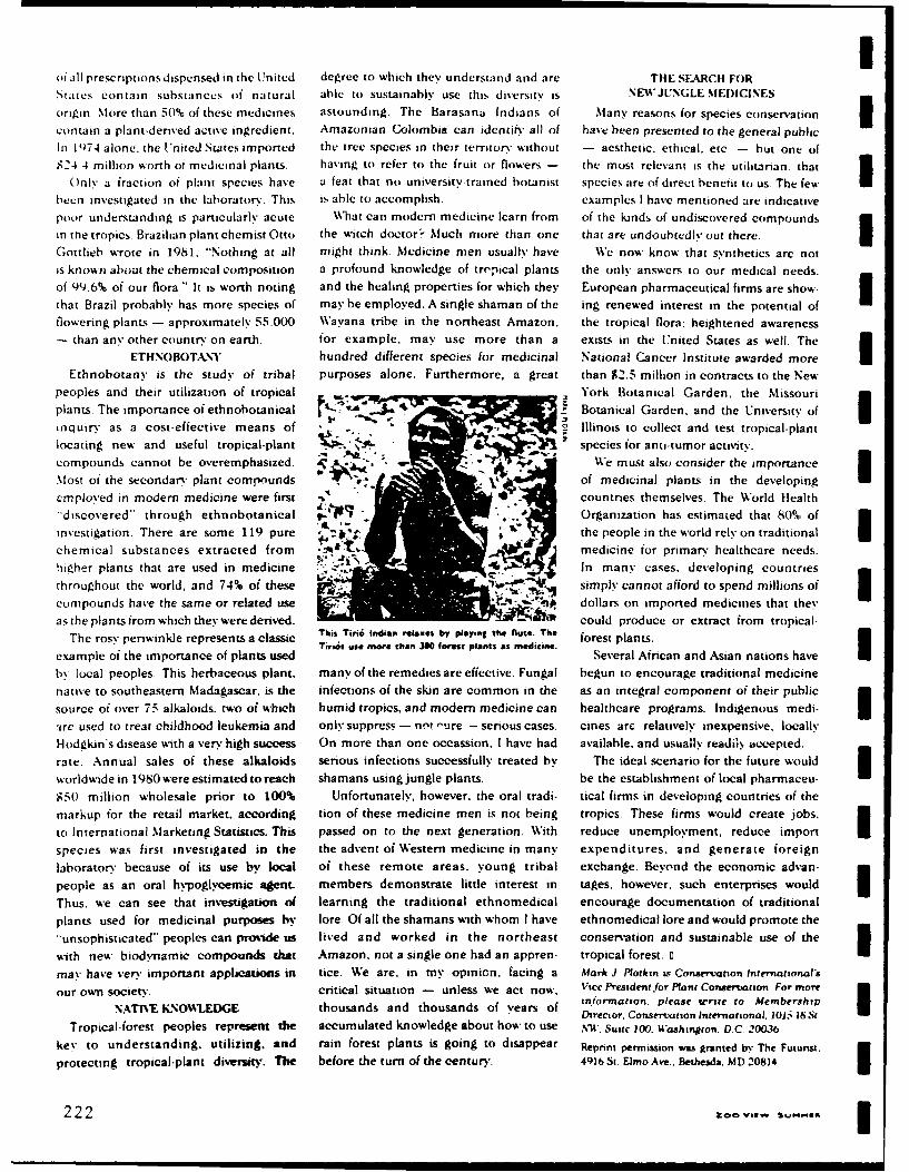
of all prescriptions dispensed in the United degree to which they understand and are THE SEARCH FOR
States contain substances of natural able to sustainably use this diversity is NEWJLNGLE MEDICINES
origin. More than 50% of these medicines astounding. The Barasana Indians of Many reasons for species conservation Icontain a plant-derived active ingredient. Amazonian Colombia can identify- all of have been presented to the general public
In IQ74 alone, the United States imported the tree species in their territory without - aesthetic, ethical, etc. - but one ofS24 4 million worth of medicinal plants. having to refer to the fruit or flowers - the most relevant is the utilitarian. that
Only a fraction of plan species have a feat that no university-trained botanist species are of direct benefit to us. The fewbeen investigated in the laboratory. This is able to accomplish, examples I have mentioned are indicativepoor understanding is particularly acute What can modern medicine learn from of the kinds of undiscovered compounds
in the tropics. Brazilian plant chemist Otto the witch doctor? Much more than one that are undoubtedly out there. IGottlieb wrote in 1981, "Nothing at all might think. Medicine men usually have We now know that synthetics are not
is known about the chemical composition a profound knowledge of tropical plants the onlv answers to our medical needs.of 99.6% of our flora." It is worth noting and the healing properties for which they European pharmaceutical firms are show- 3that Brazil probably has more species of may be employed. A single shaman of the ing renewed interest in the potential offlowering plants - approximately 55.000 Wayana tribe in the northeast Amazon, the tropical flora: heightened awareness
- than any other country on earth. for example, may use more than a exist, in the United States as well. The
ETHNOBOTANY hundred different species for medicinal National Cancer Institute awarded moreEthnobotany is the study of tribal purposes alone. Furthermore, a great than $2.5 million in contracts to the New
peoples and their utilization of tropical York Botanical Garden. the Missouri 3plants. The importance of ethnobotanical tL,:% Botanical Garden, and the University of
inquiry as a cost-effective means of -. " 2 Illinois to collect and test tropical-plant
iocating new and useful tropical-plant , species for anti-tumor activitv.compounds cannot be overemphasized. We must also consider the importance
Most of the secondary plant compounds - of medicinal plants in the developing
employed in modern medicine were first , - countries themselves. The World Health
"discovered" through ethnobotanical Organization has estimated that 80% of
investigation. There are some 119 pure the people in the word rely on traditional m
cmedicine for pnmar healthcare needs.higher plants that are used in medicine -.7 In many cases, developing countriesthroughout the world, and 74% of these . 1 simply cannot afford to spend millions ofcompounds have the same or related use , dollars on imported medicines that they
as the plants from which they were derived. -m could produce or extract from tropical-
The rosy periwinkle represents a classic This Tiriti Indian relaxes by playing the flute. The forest plants.• ~Trsois us. more than 30 foretst piants as medicine.
example of the importance of plants used Several African and Asian nations haveby local peoples. This herbaceous plant, many of the remedies are effective. Fungal begun to encourage traditional medicine
native to southeastern Madagascar. is the infections of the skin are common in the as an integral component of their public
source of over 75 alkaloids, two of which humid tropics, and modern medicine can healthcare programs. Indigenous medi- I3re used to treat childhood leukemia and only suppress - not ,'ure - senous cases. cines are relatively inexpensive, locally
Hodgin's disease with a very high success On more than one occassion. I have had available, and usually readiy accepted.rate. Annual sales of these alkaloids serious infections successfully treated by The ideal scenario for the future would
worldwide in 1980 were estimated to reach shamans using jungle plants. be the establishment of local pharmaceu-S50 million wholesale prior to 100% Unfortunately, however, the oral tradi- tical firms in developing countries of the
markup for the retail market, according tion of these medicine men is not being tropics. These firms would create jobs,
to International Marketing Statistics. This passed on to the next generation. With reduce unemployment, reduce import
species was first investigated in the the advent of Western medicine in many expenditures, and generate foreign
laboratory because of its use by local of these remote areas, young tribal exchange. Beyond the economic advan-
people as an oral hypoglycemic agent, members demonstrate little interest in tages. however, such enterprises would
Thus. we can see that investigation of learning the traditional ethnomedical encourage documentation of traditionalplants used for medicinal purposes by lore. Of all the shamans with whom I have ethnomedical lore and would promote the.unsophisticated" peoples can provide us lived and worked in the northeast conservation and sustainable use of the
with new biodvnamic compounds that Amazon. not a single one had an appren- tropical forest. o
may have very important applications in tice. We are. in my opinion, facing a Mark J Plotkin is Conservation International's
our own society. critical situation - unless we act now. Vice President for Plant Consemvation For more
NATr%'E KNOMEDGE thousands and thousands of years of sntormation, please write to. Membership IA EDirector. Conservatton International. 101i3 S St
Tropical-forest peoples represent the accumulated knowledge about how- to use ,W. Suite 100. W'ashington. D.C 20036.
key to understanding, utilizing, and rain forest plants is going to disappear Repnnt permission was granted by The Futurist.
protecting tropical-plant diversity. The before the turn of the century. 4916 St Elmo Ave.. Bethesda. MD 20814 I222 Zoo, view SUMa s

HIGH ALTITUDE NEUROLOGIC PROBLEMS
Peter H. Hackett M.D.
Director, Denali Medical ResearchAffiliate Associate Professor
University of Washington School of Medicine andUniversity of Alaska Anchorage
Staff Physician Emergency Department Humana Hospital Alaska
LEARNING OBJECTIVES:
Following this presentation, participants will be able to:
1. Recognize clinically the neurologic syndromes that may present at high altitude,including high altitude cerebral edema, cerebral vascular spasm, stroke, and TIA.
2. Understand the pathophysiology of neurologic syndromes at altitude.
3. Discuss the proven and hypothetically effective treatments for these neurologicsyndromes.
4. Appreciate the current research on pathophysiology and the new methodologies beingemployed to study these syndromes.
I. NEUROLOGIC SYNDROMES CAUSED BY HIGH ALTITUDE
A. Acute mountain sickness (AMS) and high altitude cerebral edema (HACE)
1. Pathophysiology
a. Probably the same pathophysiology, with HACE being the end-stage.
b. Possible brain swelling mechanisms:
- Intracellular Na+ and H20 accumulation.
- Vasogenic edema: Cause of permeability?
- Role of cerebral blood flow
B. High altitude neurophysiology
1. Intracranial dynamics
a. Intracranial contents
- Average brain 1500 mls
223

- Average blood 75 mls
-Average CSF 75 mis U- Semi-elastic membranes not very distensible m
- Volume-pressure relationship
- Volume can increase some without increased pressure
Intracranial 3Pressure - Volume Curve
7ID b
a
Volume m
b. Cerebral blood flow m
- If C02 constant, CBF inversely related to SaO2%
- At altitude CBF varies widely, since CO2 as well as SaO2 depend on ventilatoryresponse
- Xenon techniques show 20-40% increase of CBF in days 1-3, then return closeto baseline despite hypocapnia
- Transcranial doppler shows little change in CBF m
c. Cerebral blood volume
- Function of vascular tone I- Increased with vasodilation m
d. Intracranial pressure (ICP)
- Increase ini ICP in animals at altitude quite variable
224
I

- ICP clearly elevated in severe acute mountain sickness
- Both cytotoxic and vasogenic mechanisms may be operant
- Preliminary study indicates ICP not cause of mild AMS
2. Cerebral 02 delivery
a. 02 delivery = CBF X Ca0 2 (arterial 02 content)
-CaO2 = Hb X 1.34 X Sa02%
- Example: a 10% drop in SaO2 and 10% increase in CBF = no net change incerebral 02 delivery
b. Is the brain always hypoxic at high altitude?
- 02 delivery may be maintained
- Cerebral venous P0 2 normal until SaO2 < 70% (5,000 meters) in normocapnia,but hypocapnic vasoconstriction at altitude changes this.
- 02 extraction varies among individuals, may adapt.
c. Matching of 02 delivery with CMRO 2
- Animal studies show CMRO 2 goes up transiently but perfusion and 02 deliveryI exceed needs: "luxury perfusion" or damaging overperfusion?
3. Effects of hypoxia on CNS
- Neurotransmitter failure; e.g., tryptophan hydroxylase
- Cell membrane dysfunction, cell swelling
I - Synthesis of various peptides, proteins: "hypoxic shock" model
- Effect cn energetics? ATP probably not decreased
C. Neurologic Syndromes
U 1. Acute mountain sickness and high altitude cerebral edema
a. Neurologic symptoms and signs
- Headache, vomiting, sleep disorder, ataxia, altered mental status
b. Pathophysiology
- Overhydration of brain, hypoxic brain, or both?
- Fluid retention systemically; ADH, ACTH, aldosterone
I 225
I

- Fluid shift into cells (cytotoxic); Na + /K + pump dysfunction?
- Leakage from vessels from overperfusion (vasogenic) or permeability change?
- Direct effects of hypoxia or brain fluid dynamics? I- Hypoxic effects on neurotransmitters or metabolism
D. Treatment
- Descent or 02
- Dexamethasone very effective, earlier the better I- Acetazolamide effective early; less dramatic improvement than dexamethasone
- Diuretics (furosamide) also shown effective
E. Prevention I
- Acclimatization
- Supplemental 02
- Acetazolamide or dexamethasone
II. FOCAL NEUROLOGIC CONDITIONS IA. Stroke
- Apparently high incidence at high altitude
- Probably multifactorial
- Dehydration, polycythemia, brain compression? I- Possible role of vasoconstriction, vasospasm
- Cerebral venous thrombosis more common
B. Reversible neurologic deficits
1. Symptoms and signs II
226
I.
S- Cortical blindness
- Hemiparesis and hemiplegia
- Aphasia
I - Various reversible focal signs
2. Pathophysiology
- "Hypoxic/eschemic/thromboembolic"
- Migraine equivalent?
I - True TID?
- Hypocapnic vasoconstriction?
3. Treatment
- 02 or CO2 breathing (or both)
* - Descent
- NSAID's?
- Complete evaluation to rule out cerebrovascular pathology
4. Conditions exacerbated by high altitude
a. Seizure disorder
- Reports of 1st onset seizure at altitude
- Increased seizures in those not on medication
S- No reports of increased seizures in those controlled on medication
- Mechanism: Hyperventilation?
b. Multiple Sclerosis
227
I- Any alterations of blood brain barrier may exacerbate MS
- Few case reports, need study
c. Brain tumors
- Become symptomatic at altitude
- May be true of any space occupying lesion
- Probably secondary to brain swelling/vasodilation
d. Cerebrovascular lesions m
- AVS's and aneurysm may bleed
- Secondary to vasodilation, increase venous pressure?
- Perhaps a contraindication to sleeping of 3,000 meters
e. Cerebral arteriosclerosis I- Case reports of patients marginally compensated at sea level becomingsymptomatic on ascent to altitude.
-Normal area of brain may vasodilate, "steal" from area of fixed circulation.
2I.IIIII
228
I

MEDICAL PROBLEMS OF SPACE FLIGHT
James P. Bagian, MDJohnson Space Center
Houston, Texas
Despite the highly technological aspects associated withmanned spaceflight and its emphasis on precise control, thereare some facets over which we have little influence and yetwhich have large impact on the way we operate. While we areable to control environmental factors such as the totalpressure and composition of the cabin atmosphere there is onefactor we don't control at all - gravity. This absence ofgravity turns out to be the root cause of virtually all thephysiologic problems a crewmember is likely to encounterduring flight. The major areas of concern and theoperational problems to which they contribute follow.
VESTIBULARFirst-time exposure to O-g (weightlessness) has resulted inan attack rate of Space Motion Sickness of 67% in Shuttlecrews. The etiology of this syndrome is still not clear.Sensory conflict seems to be the most likely cause althoughfactors such as fluid shift may play a role. The severity ofsymptoms is highly variable but most are manifest andsubsequently resolve within the first 2-3 days of flight.The symptoms resemble those of motion sickness though certainsignificant differences occur.
CALCIUMBone demineralization is known to occur on exposure to O-gand continues at a steady rate for many months. There issome evidence that a new equilibrium is reached after manymonths but this information is only preliminary. While thebone demineralization which occurs poses no problem while thecrewmember is in a O-g environment, return to a 1-genvironment after a long exposure to O-g would have a higherassociated risk of spontaneous (or pathologic) fractures.While this problem is not significant for Space Shuttleoperations, future Space Station and interplanetary missionswill be tremendously impacted by this problem.
CARDIOVASCULARExposure to O-g causes a redistribution of intravascularvolume within the oody. This fluid shift begins immediatelyupon achieving the weightless condition. The body adapts tothis O-g environment rapidly, in less than 2 days, and as aresult the intravascular volume shrinks. It is alsopostulated that with the loss of orthostatic stress in O-gthe autonomic response to an orthostatic load on return to1-g becomes blunted. Although this reduction inintravascular volume and autonomic response to orthostatic 229

II
stimuli is not a problem while at 0-9, return in aprecipitous fashion to a 1-g environment (i.e. re-entry andlanding of the Space Shuttle) can have catastrophic results.As a result, countermeasures are used to minimize thesymotors crewmembers exp~rience on return to 1-g. Thesecountermeasures include fluid loading and anti-g suit use.While these countermeasures are effective, furtherinvestigation into other methods of protection is ongoing.
At the present time our ability to function during SoaceShuttle flights is not significantly impacted; however abetter understanding of the previously mentioned physiologicchanges would serve to make the flight environment morehospitable. While this increased insight is desirable forShuttle flights, it will become absolutely mandatory forlong-duration operations such as those contemplated for SpaceStation or a Mars Mission. 3
2IIIIIIIII
230
I

II
Designing a Health Care System for Space Flight
Joey B. Boyce M.D.Former Manager, NASA Health Aaintenance Facility
Houston, Texas, USA
Learning Objectives: Following this presentation, participantswill be able to -
1. Have a better understanding of the issues and dilemmasinvolved in development of a health care system for long durationspace flight.
2. Understand the limitations of microgravity medical care andthe effects of space flight on humans.
* 3. Appreciate where some of their tax dollars are going.
231

I
Designing a Health Care System for Space 11ight I
.Joey B. Boycc M.D. IFormer Maia;:r . N.ASA Health Maintcnaace Faci, i ty
Houston, Texas, USA
Abstract -
The development of a heal th :eare -;stem for space environmentsdemands many modifications of existing tcrres'rial practice-.Weight, power consuimption. , arid personnrl! resirizion s.; forc,-.adaptation of equipment, tcchniques. and protocolz for Ipreveni;vv. diagnostic, and therapzuic medical care. Maximizinreturn from scarce resource allotments, re';irc:, :umercuz tradestudies and hab.accig' ,f :..-, a' , 1 h nef It p: :,r t: Idevelopment of bas;c, sy.stem rcqui rements. Cost of providing acertain level of care nus: alu.3 o be balaaced against largcrprogram goals and capabilities. This paper will discuss some ofthese i ssues, an:d c:i'rcr: ;jipproa :le.., Io medical care in thedevelopment of the Internal;onal Spa(- St,.;oi. Freedom CrewHealth Care Sy.;:c;m ("'"i ' "
Introduction - IThe Crew Ilealth Care System (CH.,S) of the proposred 'rAternaliona!Space Station Freedom wi]'. provide essential operationalbiomedical support need. for Ihe ! rtec'om crews. inel]ding medicalcare, environmental habitat monitoring, and countermeasures forthe potentially maladaptive phy-ic.ogic effec..: of 3pace flight.To meet these challenges, ClteCS Ai' consist of three integral Iparts, the Health Mainte:aoc.: aci. i ty (1IMF), 4he EnvironrmentalHealth System (EHS), and the Exercise Countermeasures Facility(ECF). The unique microg:'avit,- tosed environment of SpaceStation Freedom will force adaptation of standard Eartli basedprocedures in each one of hc,. ar-as. In addition, each C'!eCSarea will operate under significant constraints of power, weight,volume, and other operationa] limitations. At present, design Ireq-.irements have been defined by NASA for CHeCs which will alloy.construction and operation no later than the permanent mannedcapability (PMC) phase, of the l:eedom Station in 1999 (1). The IMcDonnell Douglas Space Station Corporation has been chosen asthe prime contractor for development of the CHeCS equipment, withtechnical and scient;fic gaidance provided by N.ASA. Many area;that must also be considered for a complete picture of SpaceStation Freedom Mdical Operations will not he discussed in depth
I232 I
I
in this paper. A brief background of previous long duration spaceflight, physiologic effects of space flight, and the SpaceStation Freedom program is needed for understanding the basisupon which CHeCS is founded.
BackgroundThe United State's experience to date in long duration spaceflight is limited to the three Skylab flights, the longest ofwhich was 84 days. The Soviet MIR program has had flights overotie year, and has gained ijvaiuable experience as a resuit.Planned CHeCS equipment and technology will be significantly moresophisticated and more capable than either of these programs. ForSkylab, crew medical officers were limited to only rudimentarymedical procedures and first aid. No extended inpatient care waspossible, and any medical event beyond a minor problem might haverequired a return to Earth for all crewmembers. Environmentalmonitoring of the 5 psia, 70% oxygen/30% nitrogen Skylab cabinatmosphere was limited to oxygen and carbon dioxide sensors onorbit, with detailed post mission analysis of other conslitaents.In addition, life support monitoring did not have to take intoaccount the recycling of resources that will he an essentialfeature of Space Station Freedom. Exercise facilities andmonitoring on Skylab were likewise less capable in compari.;o1with Freedom, with little real time physiologic monitoring ofcrew's performance
Despite the previous work done during US and Soviet flights, noneof the physiologic changes that accompany space flight are wellunderstood at this point. On short term flights, approximatelytwo-thirds of U.S. space travelers have been afflicted by SpaceMotion Sickness (SMS), a self limiting malady that varies inseverity from mild headache to nausea and vomiting (2).Fortunately, SMS typically last no more than three to four days,and should not be a problem during longer flights. Microgravity
exposure over a period of more than one to two weeks causesseveral physiologic changes, including progressive bone minerallosses and musculoskeletal deconditioning. At present, exerciseis the "prescription" of choice as a countermeasure to thisdeconditioning, although it is not clear how effective it will bein preventing long term losses or performance decrements. Theeffects of space flight must be added to the "routine"occupational and medical risks of the crew for a complete medicalrisk appreciation.
Although only approximately 250 nautical miles away from theEarth's surface in low Earth orbit, the Freedom Station will bean extremely isolated habitat that must be self sufficient. Asshown in Figure 1, Freedom Station will consist of 4 cylindricalmodules connected by "nodes" in a building block fashion overmany flights of the Space Shuttle. Two of the smaller modules andall the nodes will be supplied by the United States, with theEuropean Space Agency (ESA) and Japan's NASDA providing one fullsize module each. Essential station support services (power,thermal control, etc) and launch systems via the Space Shuttle
233
Iwill be provided by the United States in this internationalcooperative effort. The Freedom systems are targeted for a 30year lifespan at a minimum. Missions may last from 45 to 180 daysor more. In the event of a contingency or missed resupplymission. essential backup supplies and equipment (including iCHeCS) will be distributed throughout the Station. At present,there are no provisions for a return to Earth capability otherthan the Space Shattle, although the NASA is currently Iinvestigating a', Assured Crew Return Vehicle (ACRV) "lifeboat"for crew return in the event of contingencies. Life .,upport forthe Freedom Station will be supplied by the Environmental Controland Life Su:pport Sys:em (ECLSS), which has been describedpreviou;sly (3). This system will provide a respirable atmosphere,hygiene and potable water, and other essential needs for the crewwhile recycling crew wastes and by-products to the maximum extent Ipossible.
The Health Maintenance Facility
The medical care arm of CHeCS is provided by the HealthMaintenance Facility (HMF), a clinical care facility forinpatient care of one crewmember and outpatient care of an entirecrew of four for no more than three days. In presentconfigurations, the HMF is located in a center node. EssentiaiMMF equipment will take up the majority of one Space Station Idouble rack. A diagram of tIMF equipment in the currentanticipated configuration is shown in Figure 2. The HMF will alsobe able to provide medical care in the Freedom hyperbaric airlockat up to 2.8 atmospheres absolute pressure for treatment ofdecompression sickness and air embolism :uffered duringextravehicular activity (EVA). IThe subsystems that make up the IIMF and their components areshown in Table 1. The determination of the appropriate mix andtypes of equipment for the HMF has been the subject of Iconsiderable effort since the inception of the Freedom program.Despite considerable demand for a numerical cost/benefitdetermination for each piece of equipment, it was not appropriateto "pare down" the myriad number of medical scenarios and basethe HMF equipment list on what is needed in an arbitrary "topten" (or one hundred). Instead, the focus was on general medicalfunctions that would be needed in selected medical fields (i.e. Iequipment needed for basic cardiopulmonary diagnosis andtreatment). In effect, a "rule out" process has been used toexclude equipment or functions that were not feasible. Forinstance, computerized tomography (too large), many specializedlaboratory procedures (rare use), and major surgical treatments(appropriate personnel and equipment not feasible) are all beyondthe scope of practical medical care in the Freedom Stationprogram. In addition, the personnel may also have limitedtraining. The TIMF will support, however, the vast majority ofmedical problems that may arise. I
234 I
I

The Environmental Health SystemTo ensure a safe and healthy environment for station crewmembers,the Environmental Health System (EHS) will regularly monitor thecabin atmosphere, potable water supplies, and other generalenvironmental health parameters.The EHS will be composed of six subsystems: Microbiology,Toxicology, Water Quality, Radiological Health, Vibroacoustics,and Barothermal Physiology (4). In present plans, the EHS systemhas one rack in a central node location, with portable monitoringequipment distributed throughout the Station's main elements forcontinuous monitoring of combustible products and totalhydrocarbons. The node rack mounted E1S equipment will provideanalysis of samples of air, water, microbial swabs, and othersamples from various station sites on a regular schedule. Specialmonitors for specific toxins or experiments are also expected toaugment the more general monitoring equipment.
Freedom environmental sample's results will be evaluated usingadaptations of Earth based standards. Because of the limitedenvironmental reserves of atmospheric gases and water, however,responses and recovery from environmental contingencies will belimited. For example, current Station contingency response plansmay include evacuation of a toxic module's atmosphcrc followed byrepressurization. Even if this were 100% effective in eliminatingthe hazard, the repressurization would use a large percentage ofavailable gases for a mission, and further repress/depressesmight not be possible. Operational modeling of EHS and ECLSSsystems will provide a better understanding of optimal sensorlocation, number, functional requlirements, and other importantdata in the future.
I Exercise Countermeasures FacilityLong duration space flight has well documented deconditioningeffects on the cardiovascular, mu,;c:lozikeletal, and neurosensorysystems (5). To counteract these effects, the ExerciseCountermeasures Facility (ECF) of SS Freedom will provideexercise equipment, physiologic monitors, and other facilities inone double rack located in a node. Present plans call forapproximately two hours of exercise per crewmember for sixdays/week, with a capability for two crewmembers to exercisesimultaneously. Motivational aids and ground monitoringI coaching" will be provided to assist the crew and optimize Iheexercise regimen for crewmembers. The exercise equipment willutilize three primary devices: a treadmill, a multi-functionalexercise device, and a resistive exercise device, along withportable exercise devices. These devices will provide loadsnecessary for maintenance of aerobic fitness, strength, andskeletal integrity. The performance of the crewmembers will bemonitored via video links and physiologic parameters includingpulse, blood pressure, ECG, and metabolic gas monitoring.
235

I
The three CHeCS areas of HMF, EHS, and ECF must fit within the Iplanned operational environment of Space Station to allowfunction with minimal demand on the scarce station resources. Irnfact, the C~leCS equipment has been downsized many times as the UFreedom program str.ggled to meet const:raito imposed by Congressand the available budget. The largest "scrub" occuvre : 1990,when CIHUCS went from a seven double rack design to Khe three rack Isystem just described. Numerous developmental systems that uilleventually be necessary for isolated long duration space flightwere delayed or canceled. The regolar use of CHeCS equipment aswe learn about man's responses to space will requ.r- asignificant portion of the available power, crew timc, andattention. Each of the systems will have man, intrfaces with avariety of Freedom distributed systems including the data Imanagement systcm, environmental :offrl, powce, ,bhevma' ::5;tems,and the fluid management systems. Each system will also requireground support from the Space Station Contrcl ,enter so acontinual basis. At present, plans call for a 24 hour groundmonitor of CIleCS systems and performance. in thz vcnt of acontingency or special event, additionai expertise will beavailable via a ground :ciemedical network.
The CHeCS facilities on Space Station rreedom will play asignificant role in the advancement of medical care in space Ifligh:. The provision of health care, environmental monitoring,
and exercise countermeasures will be essential operational needsin this new environment. For future missions such as mannedexploration of the moon and Mars, it will be necessary to buildupon these capabilities and go on to develop mere completeanswers to the many potentially limiting problems faci~g mannvdexploration of the cosmos. CHeCS is a first step in providing Icomprehensive biomvdical care and monitoring for a long durationcrew. This endeavor will continue to provide nume:'ous benefitsfor Earth based science hy the constant demand for improved Iequipment, methods, and understanding of the biomedical issuesinvolvred.
References I1. Requirements for an Inflight Crew Health Care System (CHeCS).JSC 31013 Rev B. NASA Johnson Space Center, Houston, Tx. 1989.
2. Nicogossian, A.E., Huntoon, C.L.: and Pool, S.L.. SpacePhysiology and Medicine. Second Edition. Lea and FebigerPhiladephia, 1989.
3. Schunk, R.C. and llumphries, W.R. "Environmental Control andLife Support Testing at The Marshall Space Flight Center.''Proceedings of the 17th intersociety Cnference on EnvironmentalSystems, Seattle, Wash. July 1987, Papcr No. 871453.
I236 I
I

II
4. Richard, E.E. and Ruisso, D. An Overview of the Space StationFreedom Environmental Health System. Proceedings of the 19thIntersociety Confercnce on Life Support Systems, San Diego. Ca.July 1989, Paper No. 891538.
5. harris, B.A. and Stewart, D.S. Worlshop on 1xercisePrescription for Long-Duration Space Flight. NASA ConferencePublication 3051, 1989.
237

HMF SUBSYSTEMS
-prevention, diagnosis, treatment and transport
1.- Anesthesia - Penipheral Nerv Stimuliator
2. Dental - Dental Hand Drill, Dental Instrument Tray, Lainar Flow/Suction Particle Containmrent Device
3. Fluid Therapy - Sterle Water for injection System (SWIS), IV Solution Reconstitution Device. Largeand Small Volumre Pareriteral Bags. Flid Administration Kit. I Catheters. Powered and Non-powered IfusionIpumps. Accessories Mit Blood Collection and Administration It. Parenteral Nutrition Kit
4. Hyperbaric Therapy -Suit in Breathing Units
5. Imaging. -Diagnostic Radiographic Imaging System (DRIS), Macroscopic Imaging System, Microscopicimaging System
6. Medical Analytical Lab - Clinical Chemistry Analyzer, Blood Gas Analyzer, HematolgyAnalyzer. Coaglation Analyzer. Reagent Supplies Module, Sample Acquiition and Processing Module.Centnifuge, Prep Tent. (Microscope. Incubaor. Slide stainer, Microbial Analysis system)
7. Medical Decision Support - Medical Database. Diagnostic Support System, Medical LibraryReference System. Support Hardware, Medical Comrmnication System. Medical Mobile ComnputerI
8. Medical Life Support - Advanced Life Support PacK. Defibrillator, Doppler Flow Probe. PatientMonitor (EKG), Pneumatic Anti-Shock Glarment. Pulse Oximeter U9. Pharmacy & Central Supply.- Pharmacy, Central supply
10. Physician's Instruments - Non-powered hand-held diagnostic instrumnts. Powered hand-held diagnostic instruments. EIeconi Stethoscope System
11. Respiratory Support. -Airway management equipment, Automated Ventilator equipment.Portable oxygen supply, Pulmonary manual resuscitator, Respiratory Monitoring
12. Safe Haven - Safe Haven equipment. Medical supplies. Pharmaceutical supplies
13. Surgery.- Cautery device. Task Lighiting. Medical Restraint System (MRS). Surgica Instrument andSupplies
14. Transport - Transport Monior, Transport Aspirator
15. Waste Management - Air-Fluid Separator/Suction. Sharp Trash Container, Soft Trash
Container, Fecal Collection Bag, Urine Collection system, Body Bag3
TABLE 13
238

IIIIIIINUIUIIIIIIII 239

I
ag++I I '
"WI ___,, . #Co"A SIIPLY
Wcalu " u A
*PR~ PULOU CU V
L B t'WtA£$L V, #c
v0 UGHTU
# NBS& BI "TON I+ DEPLOY, ""
I
MLGUR 2CAERAL LAOT N UNTO AL OF
LEGENE
POOE M4O D SP a IOCKU
I240
I
i mmiii |Im lP I mi

IRECENT ADVANCES.IN HIGH ALTITUDE PULMONARY EDEMA (HAPS)
Oswald Oelz, M.D.
3 university Hospital, CH-8091 ZUrich, Switzerland
I Objectives
At the completion of the lecture on recent advances in HAhE,the participant will be able to:
1. Delineate the epiCemiology and prevalence of HAPE.
2. Discuss the pathophysiology, clinical presentation andlaboratory findings of HAPE.
i 3. Discuss the prophylaxis by natural meanc an well as bynifedipins.
4. Discuss the various options of treatment for HAPE whichare descent, oxygen, nifedipine and the pressure bag.
iiIiiI
II
3241
I" ••

IPREVALENCE OF HIGH ALTITUDE PULMONARY EDEMA:
The reported prevalence of HAPE varies with altitude, rate mof ascent, physical activity and altitude of residence. Skiersin the Alps very rarely, if ever, develop HAPE, since they ge-nerally stay overnight below 2000 m. In contrast nne in 10'000 mskiers in Colorado develops serious HAPE at altitudes of 2500to 3000 m. Regiments of the Indian army flown above 3300 m hadan attack rate of 15%. The prevalence of HAPE in trekkers inNepal was reported to be 2.5 to 4.5% in the late seventies andhas since decreased due to educational measures and awarenessof the disease. The reported prevalence of 1 to 2% in climberson Mount McKinley has also declined in recent years. In the ISwiss Alps approximately 10 patients with HAPE per year have tobe evacuated by air rescue organizations.
RISK FACTORS FOR THE DEVELOPMENT OF HAPE:
These include rapid ascent without prior acclimatization, mcontinued ascent in spite of worsening symptoms of acute moun-tain sickness, i history of previous HAPE and male sex. In moststudies males were 5 to 10 times more frequently affected byHAPE than females. The risk to develop HAPE upon very rapidascent to '550 m is 5 to 10 times higher in persons with ahistory of HAPE than in those with a good tolerance to high maltitude in the past.
PATHOPHYSIOLOGICAL SEQUENCE OF HAPE: U1. Hypoxia:3
a) More pronounced hypoxemia in HAPE susceptible subjectsdue to rel ive alveolar hypoventilation (bluntedhypoxic ve ,4iletory response).
b) Exercise-induced oxygen desaturation.
2. Sodium and Water Retention: IHAPE-susceptible subjects are characterized by:
a) More pronounced hypoxemia upon ascent to high altitude.
b) Enhanced ACTH-secretion particularly during exercise. 3C) Enhanced secretion of norepinephrine, epinephrine and
cortisol at rest and during exercise. m
d) Enhanced secretion of renin and angiotensin at rest andexercise.
a) Enhanced secretion of aldosterone at rest and duringexerciSe. 3
242
I

f) Enhanced secretion of ADH during exercise.
g) Enhanced secretion of ANP at rest.
As a consequence HAPE-susceptible subjects retain water andsodium, gain weight and develop peripheral and periorbitaledemas.
3. Hypoxic Pulmonary Hypertension:
Marked hypoxic pulmonary hypertension develops in HAPE-sus-ceptible subjects as a consequence of alveolar hypoxia.There seems to be exaggerated pulmonary vasoconstriction.
Uneven pulmonary vasoconstriction leads to over-perfusioninjury of non-vasoconstricted vascular areas of the lung andthus to permeability edema. This could be either due to rup-tur of endothelial structures or be solely a consequence ofenhanced filtration pressure.
4. Leak:
The protein content of the edema fluid in HAPE is comparableto that of serum thus characterising HAPE as a permeabilityedema.
CLINICAL AND LABORATORY CHARACTERISTICS OF MAPE:
People developing HAPE initially, suffer from a non-produc-tive cough, shortness of breath, particularly in the supineposition, weakness, fatigue and other symptoms of acutemountain sickness. Lateron tachypnea develops. Subsequentlythe cough may become more productive with watery and some-times bloody sputum. Rhonchi and gurgling sounds can even-tually be heard without a stethoscope. However, the commonclinical signs are crepitant rales, rhonchi, tachypnea,cyanosis and tachycardia. Later on, patients become ir-rational and hallucinating due to hypoxia and/or accompaningcerebral edema. This is rapidly followed by coma and death.
The systolic pulmonary artery pressure is markedly elevatedin the range of 40 to 130 mm Mg. Arterial blood gas analysisshows marked hypoxemia and relative hypoventilation. TheAaD02 is elevated above 12 mm Hg. The radiomorphology is un-characteristic with alveolar and interstitial edema. Theright lower lung field is significantly more affected thanother lung fields.
I 243

PREVENTION OF HAPE:
HAPE can in most cases be avoided by graded slow ascent, mrest days when minor symptoms of acute mountain sickness deve-lop and by descent, if symptoms of acute mountain sickness getworse. There is circumstantial evidence that prophylaxis withacetazolamiae anda/or mUxd L abon y protaot HZNPE-cusceptih16subjects to some extent, however, there have been no controlledstudies with these medications. m
In a recent study mountaineers with a history of radio-graphically documented HAPE were randomly allocated to receive20 mg of a nifedipine slow relase preparation or identical pla-cebo every 8 hours while ascending from low altitude within 22hours to 4559 m and during the subsequent stay of 3 days atthis altitude. Diagnosis of HAPE was based on chest radio- mgraphs. Pulmonary artery pressure was assessed by Doppler echo-cardiography and AaDO2 was measured in simultaneously sampledarterial blood and end-expiratory air. 7 of 11 subjects on Iplacebo, but only one of 10 subjects on nifedipine developedHAPE (p-0.01). Systolic pulmonary artery pressure, AaD02 andsymptom score of acute mountain sickness were significantlylower in subjects receiving nifedipine than in those receivingplacebo. Thus prophylactic administation of nifedipine iseffective in lowering pulmonary artery pressure in preventingHAPE in susceptible subject.. This observation should not leadto uncontrolled intake of nifedipine for prophylaxis of HAPE.Intake of drugs in mountain sports can be consideredacceptable, only when the most important preventive measure, mwhich is slow ascent to high altitude, has failed.
TREATMENT OF HAPE:I
HAPE is best managed by descent, evacuation and/or if im-possible by oxygen in a flow rate of 1 to 4 liters per minute.In mild cases bed rest has been tested in a controlled setting.Furosemide has been advocated but in other hands it renderedpatients susceptible to circulatory troubles, pulmonary embo- Ilism and cerebral edema. Morphin, another inadequately testedremedy, could do more harm than good by depressing the respi-ratory centres at a time when more respiration is needed. Posi-tive end-exapiratory pressure has been applied briefly but itlong-term effects may be harmful. Another possible means ofshort-term improvement is postural drainage. 3
Treatment of HAPE patients in a laboratory at 4559 m withnifedipine 10-20 mg sublingually, followed by 20 mg of a slowrelease preparation every 6 hours resulted in clinical improve- mment, better oxygenation, reduction of AaD02 as well as pul-monary artery pressure and progressive clearing of alveolaredema. In these patients no other therapeutic measures weretaken and they continued their mountaineering activities ataltitudes above 4000 m. Thus nifedipine offers a potentialemergency treatment for NAPE, when descent or evacuation is
impossible and oxygen is not available.244

iII
EFFECTS OF ALTITUDE UPON CARDIOVASCULAR DISEASES
3 Herbert N. Hultgren, M.D.
Professor of Medicine (Emeritus)
Stanford University School of Medicine
Palo Alto, California
iIIe
i
m
I
245
I

IEffects of Altitude Upon Cardiovascular Diseases 3
Learning Objectives: i
1. Immediate and long term effects of high altitude upon thecardiovascular system. 3
2. Aggravation of angina pectoris by ascent to altitude includingresponsible mechanisms, prevention and treatment. 3
3. Increase in blood pressure in hypertensive patients going tohigh altitude. Responsible mechanisms, prevention andtreatment. 3
4. Effect of high altitude upon pulmonary hypertension in patientswith primary pulmonary hypertension, congenital heart diseaseand mitral stenosis: Methods of evaulation, prevention andtreatment.
i{iIIi{III
246 i
I
IU
Effects of Altitude Upon Cardiovascular Diseases
3 Herbert N. Hultgren, M.D.Professor of Medicine (Emeritus)
Stanford University School of Medicine3 Stanford, California U.S.A.
Physicians should be aware of sea level cardiovascular problemsthat may be aggravated by exposure to high altitude. Increasingnumbers of middle aged and elderly individuals are visiting highaltitude areas to ski, trek or attend conferences. Many Elder Hostelprograms are held in mountain environments. A recent survey of 1909adult visitors to Keystone, Colorado 9,300 feet (2837 m) revealed that
48% were 40 to 60 years of age and 15% were over 60. Sixty-six percentwere males (1). Approximately 10% of trekkers in Nepal were 50 years
- of age or older (2). The prevalence of many cardiac conditions risesrapidly after 50 years. For example the mean age of entry of 2234 meninto 3 large randomized studies of surgery in coronary artery diseaseU was 50-51 years (3).
This presentation will review altitude induced changes that affectthe circulatory system and examine 3 cardiovascular conditions that areadversely affected by altitude. These will include coronary arterydisease, systemic hypertension and pulmonary hypertension. Highaltitude for the purpose of this presentation is defined as elevationsexceeding 8,000 feet (2440 m), since altitude illness and altitudeaggravation of sea level conditions are rare below this altitude.
1. Effect of Hijh Altitude Upon The Circulation
Several important circulatory changes occur during exposure tohigh altitude (4,5). These include:
1. Increase in resting and exercise heart rate.2. Increase in cardiac output and velocity of cardiac contraction3. Increase in systemic vascular resistance and systemic blood
pressure.4. Contraction of veins and vessels in the skin, muscles and
viscera with an increase in central blood volume.
The sum effect of these changes is an increase in cardiac work,cardiac oxygen consumption and an increase in coronary blood flow.These changes are largely due to an increase in sympathetic activitymediated by the effect of hypoxia upon the chemoreceptors in the carotidbodies (5; 6). Epinephrine secretion is transiently increased andNorepinephrine secretion is increased for several days (7).
Pulmonary ventilation is increased in proportion to the altituderesulting in a respiratory alkalosis which is partially corrected afterseveral days. A prompt decrease in plasma volume occurs with ascent
247

resulting in a 10 - 20% decrease in total blood volume with a resultingrise in hemoglobin and hematocrit (4). After 2 - 3 weeks increased redcell production restores the total blood volume to normal pre-ascentvalues (8). Maximal physical working capacity as measured by themaximal oxygen consumption (V02 max) is decreased (9). The decrease isproportional to the altitude and amounts to a decrease ofapproximately 3% for every thousand feet of altitude above 5,000 feet.
A modest increase in pulmonary vascular (pre-capillary) resistanceoccurs with a resulting increase in pulmonary artery pressure. In somepatients with an elevated pulmonary artery pressure at sea level amarked rise in pulmonary artery pr-essure may occur.
After 4 - 8 days at high altitude most circulatory changes subsideand during a longer period of altitude exposure, several values willfall to levels below those seen at sea level. For example resting Iand exercise cardiac output are decreased (10). Left ventricular andatrial chamber size decrease without changes in cardiac function(11,12). The systemic blood pressure decreases and after a prolonged Istay at altitude the blood pressure may fall to below sea level values(13,14). These effects are due to a decrease in sympathetic nervoussystem activity probably in part due to a decrease in peripheral receptors(15). Pulmonary artery pressure remains elevated however.
2. Coronary Arterv DiseaseThe increase in cardiac work associated with ascent to high I
altitude may result in an increase in severity of angina, the new
onset of angina or unstable angina.
An example of angina made worse by exposure to high altitudeis represented by the following case report: A 70 year old businessmanhad angina pectoris since a myocardial infarct 22 years previously.His angina had progressed slowly so that he experiences chest pain withminimal physical effort. By limiting his activity he is able to carryon a sedentary existence with only 1-2 episodes of chest pain a day.He occasionally had nocturnal angina. His medications include Nitro- Iglycerine prn, aspirin 325 mg/day, Propanolol 40 mg t.i.d. and Diltiazem60 mg t.i.d.. He has never had hypertension. He has had frequenttreadmill tests but stops after 1-2 minutes, because of chest pain. Hehad not had coronary arteriography or coronary bypass surgery.Recently he and his wife travelled to Denver, Colorado, where theyspent the night. No increase in symptoms was noted. Thefollowing day they drove to Vail, Colorado altitude 8200 ft.(2500 a). IUpon crossing Loveland Pass, altitude 11,900 ft. (3630 m) the patientbegan to experience frequent episodes of angina and dyspnea relievedby nitroglycerine. His wife noted he was quite blue and clearly indistress. Upon arrival at Vail his anginal attacks continued, but hewas able to sleep moderately well. The following day repeatedepisodes of chest pain and dyspnea caused them to return to Denver.Once over Loveland Pass the patient felt very much better and upon
248 I

I
Ireturn to sea level his original attacks returned to the pre-ascentfrequency.
Comment:
With such severe angina and limitation of effort at sea level thepatient should not have gone to Vail. When his angina became worse atVail a physician should have been called. It is very possible that lowflow oxygen might have substantially relieved his symptoms. It is ofinterest that he did not experience an increase in symptoms at Denver5400 feet (1647 m).
An analysis of 21 coronary events occurring within 5 days ofarrival at Keystone, Colorado has recently been completed by thisauthor. Seventeen patients had severe angina, two experienced anacute infarction and there were two sudden deaths. Fifteen patientswere males. The mean age of all patients was 58 years (range 40 to74). An elevated blood pressure and heart rate was present in mostcases. Symptoms subsided with bed-rest, medical therapy, oxygen anddescent. One patient (RM) developed severe angina upon arrival atKeystone. The pain subsided upon descent to a lower altitude andbecame severe upon return to Keystone 5 days later. Theelectrocardiogram revealed T wave inversion in the precordial leadsduring chest pain. After medical treatment a normal coronary arterio-gram was found and a diagnosis of altitude induced coronary spasm wasmade. Coronary spasm can be produced by sympathetic stimulation andalkalosis, both of which occur at high altitude (16).
The most likely mechanism of altitude aggravated angina is theincrease in cardiac work primarily due to the increase in systolicblood pressure and heart rate resulting in an increase in the heartrate/systolic blood pressure product which is an estimate of cardiacwork. Morgan and his associates studied nine men with angina by gradedtreadmill testing at 5248 ft. (1600 m) and upon arrival at 10,150 ft.(3100 a). Mean maximal oxygen uptake was reduced by 11%. Anginaand/or ST segment depression occurred at the same heart rate/systolicpressure product but at lower work loads and a shorter duration oftreadmill time (17). The effect of hypoxia was via the increase incardiac work and not an impairment of myocardial function. Ifmyocardial function had been impaired by hypoxia, angina would haveoccurred at a lower double product than at the lower altitude.
The prevalence of coronary disease among the skiing population atKeystone is unknown. An estimate of the prevalence however can be madefrom telemetry electrocardiograms obtained by Grover in 149 men duringrecreational skiing at altitudes above 10,150 ft. (3100 a).Tachycardia was significant and heart rates exceeded 80% of thepredicted maximum in two-thirds of the subjects. Five men developedabnormal ST segment depression during or immediately after exercise.All five were older than 40 years. Thus the incidence of ST abnormal-
249

ities in this skiing population was 5.6%. This is similar to theincidence in asymptomatic men during submaximal exercise at low altitude. 3Grover concluded that mountain skiing does not appear to pose a greatercoronary stress than does comparable exercise at low altitude in menwithout known heart disease. Only one of his subjects had coronarydisease (18). The risks of trekking in patients with coronary diseasehas been reviewed by the author (19).
In summary ascent to 9300 ft. (2837 u) may precipitate coronary 3events within a few days of arrival. Accelerated angina or new onsetangina are the most common presentation, but acute infarction or suddendeath may occur. Systemic hypertension and a rapid heart rate arecommonly present and may be causative factors. Acute coronary spasmmay occur. Many patients had experienced chest pain or infarctionprior to ascent. Physicians should caution such patients that symptomsmay appear or become worse with rapid ascent to high altitude. The use Iof anti-anginal or anti-hypertensive medications should be employed inappropriate patients. Acclimatization for a few days at an intermediatealtitude (6,000 ft. - 7,000 ft. (1830 m to 2135 m) may be helpful. aRestricted activity for a few days after arrival may minimize symptoms.Bed rest, low flow oxygen or descent may be necessary if symptoms aresevere. Asymptomatic patients without known heart disease may ski ataltitude without a greater prevalence of myocardial ischemia thanperforming a similar level of moderate to heavy exercise at a lowerelevation.
Systemic Hypertension
A. Normal Subjects
Normal subjects usually exhibit a modest rise in blood pressureduring the first week or two after ascent to high altitude. Severalstudies have indicated an elevation of both systolic and diastolic Upressures as well as an increase in heart rate during high altitude-yposure. Systemic vascular resistance is increased (4). Kamatobserved a rise in blood pressure in 31 of 32 subjects who ascended to Ian altitude between 11,500 ft. (3506 m) and 13,000 ft. (3906 i).
Systolic pressure rose from 115 mm Hg to 125 mm Hg and diastolicpressure rose from 78 mm Hg to 93 mm Hg. In 7 young men exposed to14,000 ft.(4300 m) for 21 days blood pressur rose from 124/71 mm to145/88m (mean values). Systemic resistance increased by 65 percent (8).
Blood pressure has been shown to rise even at lower altitudes. 3Palatini and his workers recorded 24-hour blood pressures at sea levelafter arrival at 3,969 ft. (1210 m) in 12 normotensives and 12 patientswith borderline or mild hypertension. Modest increases in daytime Iblood pressures were observed in both groups. Systolic pressure wasincreased by 6 mm Hg in both groups with an increase in diastolicpressure only in the hypertensives of 5.5 mm Hg. Individualvariations were present with some individuals exhibiting increases of
I250 I

I
17 to 16 mm Hg in systolic and diastolic pressures respectively.Heart rates were increased by 3 to 4 beats per minute in both groups.Plasma Norepinephrine and Epinephrine levels increased suggesting thatthe changes observed could be due to sympathetic stimulation (21).
Hypertensive Patients
Patients with hypertension at sea level may experience a furtherrise in blood pressure with ascent to high altitude. In someindividuals a marked rise in pressure may occur. This is illustratedby the following case report: A 62 year old Caucasian woman whoresides in Florida had moderate, asymptomatic hypertension for 5 years.Her blood pressure was controlled on Enalapril maleate 5 mg. daily.Her pressure on this regimen varied between 115/75 - 120/82 mm Hg. Shehad no complications from her hypertension. There was no history ofstroke, angina or left ventricular failure. The electrocardiogram wasnormal. Her renal function was normal. She had no retinal abnormalities.She was not obese weighing 127 lbs. She had a ventricular demand pace-maker implanted 2 years previously for a "sick sinus syndrome".Upon arrival at Aspen, Colorado 8,000 feet (2440 m) she had adiffuse, moderately severe headache during the first 2 days afterarrival. Her blood pressure rose to 180/110 mm Hg. She consulted a localinternist who administered Nifedipine sublingually and startedNifedipine (slow release tablets) 20 mg daily. Clonidine 5 mg patcheswere started daily. On this regimen her blood pressure was controlled
to 110/80 - 130/100 mm Hg and she no longer had a headache. She has hadprevious similar rises in blood pressure upon visiting Aspen. On oneoccasion her pressure rose to 200/140 mm Hg. She experiences only mildacute mountain sickness with each ascent.
The effect of altitude upon hypertension is illustrated by arecord of twice daily blood pressures taken by a physician duringa motor trip to the Rocky Mountains. The data are shown in Figure 1.Systolic and diastolic pressures rose with the altitude attained anddecreased to subnormal levels after descent. There was no significantchange in heart rate. The patient continued his usual medication con-sisting of Propanolol 40 mg. daily throughout the trip. At 9,300 ft.(2850 m) the morning blood pressures were higher than evening pressures.This is a reversal of the usual diurnal variation and could be relatedto a greater degree of hypoxia during sleep. Blood pressure elevationsin hypertensive patients going to high altitude are not a trivialproblem. Eight percent of adult visitors to Keystone, Colorado gave ahistory of prior hypertension and 26% of these individuals were takingmedications for their hypertension (22).
Patients with hypertension who go to high altitude should consulttheir physician in advance so that appropriate preventive measures canbe employed. Usually an increase in medication, a low salt diet andincreased rest during the first few days of the altitude stay maysuffice. If patients record their own blood pressure a physician
251

II
should be consulted if high pressures are encountered. Low flow oxygenand rest may be helpful. The appropriate anti-hypertensive medicationsfor altitude aggravated hypertension has not been determined. It is Ipossible that beta blockers may not be effective in controlling highaltitude blood pressure elevations. Altitude hypertension may beprimarily related to increased sympathic activity, and release of IEpinephrine and Norepinephrine. Beta blockade lowers blood pressureprimarily by reductions in cardiac output and plasma renin activity.Clonidine may be more effective in the management of altitude hyper-tension since it produces a diffuse inhibition of sympatheticneural outflow of the central nervous system (23). Prazosinhydrochloride may also be more effective than beta blockers since thispreparation blocks alpha-adrenoceptors and the risc in blood pressure Iduring altitude exposure probably principally involves alpha stimulation.Terazosin is a new alpha blocking agent which has a longer duration(half life-12 hours), than Prazosin. Twenty mg. once daily has a similar 3hypotensive effect to that of Prazosin. Syncope may occur, but can beobviated by omitting the night dose (24). Calcium channel blockers mayalso be effective. Nifedipine may be used if hypertension in severeand symptoms are present and rapid reduction in blood pressure isdesired. It is evident that controlled trials of several forms ofmedication at high altitude are indicated to develop more specificrecommendations. IPulmonary Hypertension
Several congenital and acquired cardiac conditions are associatedwith an increase in pulmonary vascular (pre-capillary) resistance andan elevated pulmonary artery pressure. Most important of theseconditons are: primary pulmonary hypertension, several forms ofcongenital heart disease and mitral stenosis. Many patients with theseconditions will exhibit a rise in pulmonary artery pressure when exposedto high altitude and some may have substantial increases in pressure even Iat moderate altitudes. Not all patients however may exhibit unpleasantsymptoms and many may tolerate altitude exposure with little adversecirculatory effects. 3Primary Pulmonary Hypertension
Most patients with primary pulmonary hypertension will exhibit an 5increase in symptoms upon ascent to high or even moderate altitudes.Increased dyspnea, weakness on exertion and an increase in syncopalepisodes oay occur. Breathing a low oxygen mixture will result in arise in pulmonary artery pressure in many of these patients. Thepress-ze rise can now be non-invasively determined by echo-dopplerstudies (25). In many patients Diltiazem or other calcium channelblockers may prevent the rise in pressure (26). If patients with Iprimary pulmonary hypertension must visit a higher altitude the use ofpulmonary vasodilAtors, reduced physical activity after arrival and lowflow oxygen may prevent or ameliorate symptoms. Patients who live at I
2252
U

IN
moderate or high altitudes may experience considerable relief ofsymptoms by moving to sea level and by the use of selected pulmonaryvasodilators as illustrated by the following case report (27). A 20year old man who resided in Denver, Colorado 5,400 feet (1647 a)experienced increasing exertional dyspnea and frequent episodes ofexertional near and true syncope. Hemodynamic studies had establishedthe diagnosis of primary pulmonary hypertension. The patient moved tothe San Francisco Bay Area upon the advice of his physician. Afterarrival his symptoms were greatly relieved and his walking distancegreatly improved. Hemodynamic studies indicated a moderate reductionin pulmonary artery resistance and pressure. Intravenous andsublingual Isoproterenol further reduced pulmonary artery resistanceand improved walking distance as evaluated by a treadmill test. Thepatient was followed for 6 years. Symptomatic benefit from 20 mg. ofIsoproternol 6 to 12 times daily persisted, but slow progression of hisdisease and symptoms were noted (27,28).
Congenital Heart Disease
It has long been known that several varieties of congenitalcardiac conditions may be associated with an increased pulmonary arterypressure due to an increased pulmonary vascular resistance. Theseconditions include ventricular septal defects, patent ductus arteriousand atrial septal defects. Patients with secundum atrial septaldefects born at moderate to high altitudes have a higher pulmonaryartery pressure than patients born at lower elevations (29). Patientswith ventricular septal defects born at Denver, Colorado have pulmonaryvascular resistances twice as high as patients at sea level (30). Manyof these patients will exhibit an increase in pressure with exposure toaltitude.
While most of the above congenital cardiac conditions can betreated surgically some patients cannot be operated on because ofsevere pulmonary hypertension. Such patients may experience adverseeffects at high altitude due to a rise in pulmonary artery pressure andan increase in cyanosis due to a greater right to left shunt. Rightheart failure may occur. The effect of oxygen and pulmonary vasodilatorsupon pulmonary vascular resistance should be evaluated prior to ascentand, if effective, employed to minimize symptoms at altitude. Adecrease in physical activity is mandatory.
Mitral Stenosis
Symptomatic mitral stenosis is now rarely seen in the UnitedStates due to the wide application of surgical treatment. Howeversevere mitral stenosis is common especially in young patients in manyother parts of the world, including India and Mexico. Of the variousacquired valvular lesions mitral stenosis is the only lesion commonlyassociated with an increased pulmonary vascular resistance andpulmonary hypertension. In some patients systemic levels of pulmonary
253

II
artery pressure has been observed. Patients with mitral stenosisexposed to hypoxia commonly exhibit an increase in vascular resistance(31). Such patients may exhibit an increase in symptoms during Ihigh altitude exposure. Increased dyspnea may be due to the increasein heart rate with a resulting higher left atrial pressure. Rightheart failure may occur due to the increased afterload. Atrialfibrillation may be precipitated. Precautions are similar to those inother patients with pulmonary hypertension. Restricted activity,oxygen administration and pulmonary vasodilators may relieve symptoms.If atrial fibrillation is present additional Digitalis or Verapamil may mbe necessary for control of the heart rate.
In summary ascent to high altitude may have significant effectsupon certain varieties of cardiovascular disease. The most commoncomplication is an increase in angina or new onset angina which occurswithin a day or two after arrival. Systemic blood pressure may becomeelevated especially in individuals with pre-existing hypertension.These complications are largely related to increased activity of thesympathetic nervous system. Patients with pulmonary hypertension dueto congenital or acquired heart disease may experience an increase in Isymptoms at altitude due to hypoxic pulmonary vasoconstriction and arise in pulmonary artery pressure. Physicians should be aware of thesepotential effects of altitude so that appropriate preventive andtherapeutic measures can be employed.
2I
II
III
254 II

IIUI
S60~
I ! °40L- 4170- o Sea Level
170 o High Altitude A.M.160 + High Attitude P.M.
140-
0
81 0
I H~Ear ae systolic andisoibloprsuena64yrod
UE80-
(1650m)
Figure 1.
3 Heart rate, systolic and diastolic blood pressure in a 64 year old
physician at sea level and during an auto tour of the Rocky Mountains.
Note the rise in systolic and diastolic pressure related to altitude.
3 At 9,300 feet morning pressures were higher than afternoon pressures.
Propanolol 4 0 mg. daily was continued throughout the trip.
III 255
I

References
1. Honigman, B., Theis, K., and Yip, R., Adverse effects of moderatealtitude on a tcririt populnticwn. JAMA, 1991 (to be published).
2. Shlim, D. and Houston, R., Helicopter rescues and deaths amongtrekkers in Nepal. JAMA 1989:261; 1017-19.
3. VA Cooperative Study of Medical versus Surgical Treatment forStable Angina - Progress in CV Diseases. 1985-86:28; 213-401.
4. Grover, R., Weil, J. and Reeves, J., Cardiovascular adaptation toexercise at high altitude. In: Exercise and Sport Sciences Review Ied., K. Pandolf (New York:MacMillan Co.) 1986: 269-302.
5. Heistad, D. and Abboud, F., Circulatory adjustments to hypoxia.Dickinson, W., Richard's Lecture. Circulation 1980:61; 463-470. I
6. Kahler, R., Goldblatt, L., and Braunwald, E., The effects of acutesystems and on myocardial contractile force. IJ. Clin. Invest., 1962:41;1553-1563
7. Cunningham, W., Becker, E. and Kreuzer, F., Catecholamines in plasmaand urine at high altitude. J. Appl. Physiol. 1965: 20;607-610.
8. Wolfel, E., Groves, B., Brooks, G. et al: Oxygen transport duringsteady - state submaximal exercise in chronic hypoxia.J. Appl. Physiol. 1991: 70; 1129-1136.
9. Sutton, J. Jones, N. and Pugh, L. Exercise at altitude.Annu. Rev. Physiol 1983: 45; 427-437.
10. Alexander, J., Hartley, H., Modelski, M. and Grover, R., Reductionof stroke volume during exercise in man following ascent to 3100 maltitude. J. Applied Physiol 1967:23; 849.
11. Fowles, R. and Hultgren, H., Left ventricular function at highaltitude examined by systolic time intervals and M-mode echocardio-graphy. Am. J. Cardiol. 1983: 52;862-866.
12. Suarez, J., Alexander, J. and Houston, C., Enhanced leftventricular systolic performance at high altitude during operationEverest II. Am. J. Cardiol. 1987: 60; 137-142.
13. Hultgren, H., Reduction of systemic arterial blood pressure athigh altitude. Adv. Cardiol. (Karger Basal/Munchen/Paris/New York)1970: 5; 49-55. I
14. Scholing, W., High altitude climate: Adaptation of healthy personsand hypertensives. Med. Klin. 1985: 76; 518-523.
256 iI

II
15. Voelkel, N., Hegstrand, N. et al, Effects of hypoxia on density ofB-adrenergic receptors. J. Appl. Physiol. 1981: 50;363-366.
16. Yasue, H., Nagao, M., Omote, S., et al: Coronary arterial spasmand Prinzmetal's variant form of angina induced by hyperventilationand tris-buffer infusion. Circulation, 1978:58;56-62.
17. Morgan, B., Alexander, J., Nicoli, S. and Brammell, H., The patientwith coronary heart disease at altitude: Observations during acuteexposure at 3100 meters. J. Wilderness Med., 1990: 1; 147-153.
18. Grover, R. Tucker, C., McGroarty, S. and Travis, R., The coronarystress of skiing at high altitude. Arch. Int. Med. 1990: 150;1205-1208.
I 19. Hultgren, H., Coronary heart disease and trekking.J. Wilderness Medicine, 1990: 1; 154-161.
20. Kamat, S. and Banerji, B. Study of cardiopulmonary function onexposure to high altitude: Acute acclimatization to an altitude of3500 to 4500 meters. Am. Rev. of Resp. 1972: 106;404-413.
21. Palatini, P., Businaro, R., Berton, G. et al: Effects of lowaltitude exposure on 24 hour blood pressure and adrenergic activity3 Am. J. Cardiology 1989: 64;1379-1382.
22. Tuck, M., The sympathetic nervous system in essential hypertension.American Heart J., 1985: 112; 877-886 (proceedings of a symposiumon New Considerations in Mild Hypertension).
23. Titmarsh, S. and Monk, J., Terazosm. A review of itspharmacodynamic and pharmacokinetic properties and therapeuticefficacy in essential hypertension. Drugs, 1987: 33; 461-477.
24. Currie, P., Seward, J., Chan, J. et al: Continuous wave Dopplerdetermination of right ventricular pressure: A simultaneousDoppler catheterization study in 127 patients.J. Am. Coll. Cardiol. 1985:6; 750-756.
25. Weir, E., Diagnosis and management of primary pulmonaryhypertension. In: Pulmonary Hypertension, Weir, E. and Reeves, J.3 Eds., Futura, New York, 1984.
26. Shettigar, U., Hultgren, H. Specter, M., Martin, R. and Davies H.,Primary pulmonary hypertension: Favorable effeffect ofIsoproterenol. New Engl. J. Med. 1976:295; 1414-15.
27. Hultgren, H. and Shettigar, U., Isoproterenol in primary3 hypertension. New Engl. J. Med., 1980: 45;529.
257
I

II
28. Vogel, J., McNamara, D. and Blount, S., Role of hypoxia in Idetermining pulmonary vascular resistance in infants with ventricularseptal defects. Am. J. Cardiol. 1967: 20;346-349.
29. Yu, P., Beatty, D., Lovejoy, F. Jr., Nye, R. and Joos, H., Studiesof pulmonary hypertension: VII Hemodynamic effects of acute hypoxia
patients with mitral stenosis. Am. Heart J., 1956: 52;683-694.
I
II
II'IIIII
258I
I

IIII
WATER DISINFECTION PRODUCTS
IHoward Backer, MD
IIIIIIIIIIII
259
I

Water Disinfection Products 3Page 1A
Product/Manufacturer Price Structure/Function IKetadyne U.S.A., Inc. 0.2 micron ceramic filter;3020 N Scottsdale Rd silver impregnated to decrease eScottsdale, AZ 85251 bacterial growth.(602) 990-3131
Ketadyne pocket filter $185 40" intake hose & strainer,Replacement filter element zipper case; size: 10"x2";
$90 wgt: 1 lb 9 oz; hand pump,flow: 3/4 qt/min.i
Handpump filter $600 Steel stand and large handpump; size (packed in case):
Replacement filter element 23x6x8"; weight: 10lbs 13oz;
$40 flow: 11/2-3 qt/min.
Drip filter $300 Gravity drip from one plastic Icontainer to another through 3
(same filter element as filter elements;handpump) size: 18" high x 11" dia. I
(26" high when assembled);weight: 8lbs 5oz;flow: 1 pt/hr. 3
General Ecology, Inc. 0.4 micron (absolute) filter--151 Sheree Blvd. carbon matrix, silverLionville, Pa. 19353 impregnated; cannister(215) 363-0412 removable:
First-Need purifier $39.95 hand pump with intakestrainer; 800 pint capacity;
extra cannister $24.95 size: 6"x6", wgt: 12 oz.pre-filter replacement $6.50 flow: 1 pint/min;
Seagull IV (X-1P) $320 Stainless steel casing andhand pump; capacity 1000 gal;
replacement cartridge $50 size 4.8x5.4"; weight: 3 lbs;flow: 3-4 qts/min I
Timberline Filter 2 micron fiberglass andPO Box 12007 polyethylene matrix;Boulder, CO, 80303
Timberline filter $24 hand pump;flow: 1 qt in 1.5 min;
replacement element $12 wgt: 6 oz; size: 9"xl-3".
Outback Filter $29.95 I qt water bottle with drink-(Distributed by Hiker Supply through filter element fromof Washington) timberline filter.
260 Howard Backer, M.D. 1991 I

Water Disinfection Products
* Page 1B
Claims Comments
I Removes bacterial pathogens, Effective for claims.protozoan cysts, parasites, Expensive, but durable.nuclear debris. Clarifies Complete virus removal cannotcloudy water. If filter be expected, although it isclogs, flow can be restored by true that viruses usuallybrushing filter element--this clump to larger particles thatcan be done hundreds of times can be filtered. Silverbefore needing to replace impregnation probably does notfilter element. Claims for prevent all bacterial growthremoval of viruses not made in in filter; recommend flushingUSA, although testimonials with bleach solution afteroffered implying effective in each trip.all polluted waters.
0.1 micron retention (0.4 Reasonable design, cost, andabsolute) microstrains effectiveness. Most testingbacteria, cysts, parasites; no with E. coli and Giardia cystsspecific claims for viruses, show complete removal."Ionic charges from surface Charcoal matrix will removechemistry.. .remove particles chemical pollutants. Willeven smaller than clarify cloudy water. Despitemicrofiltration alone can testimonials for effectivenessremove." Carbon adsorbers in undeveloped countries,remove chemicals and organic recommend caution wherepollutants that cause color viruses may be a problem;and taste. Does not remove prior disinfection withall dissolved minerals and halogen would guaranteesalts. disinfection, and carbon would
remove halogen.
Removes Giardia cysts. Effective for claims; intendedNo claims for bacteria or only for domestic backcountryviruses, use where Giardia is the
primary contaminant. Lightestpump filter available. Drink-through filters likely to givefrustratingly slow flow andrequire high pressures asparticles begin to clog.
261

Water Disinfection Products IPage 2A
Product/Manufacturer Price Structure/Function
Water One $49.95 Cartridge containing charcoal 3(Available in camping stores.) block, 0.5 micron filter;
prefilter sponge 30-50 micron.Calco Ltd Bulb hand-pump generates 3-57011 Barry Ave lbs pressure; can back-wash toRosemont unclog. Size: 3"x6" cartridgeIllinois 60018 with about 7 ft of tubing and(312) 296-6615 bulb pump; Wgt: 1 lb. Capacity I
400 gallons or 3 yrs.
Water Works Four filter elements of i
Total Filtration System decreasing pore size: porous$139.95 foam, 10 micron stainless
Dromedary Beverage Bag steel wire mesh screen,$12.95-19.95 activated carbon filter, then
(All filter elements and parts 0.1 micron absolute membranereplaceable.) filter. Hand pump with inlet IMountain Safety Research tubing. Storage bag (2 or 4Box 3978 liter) attaches directly toTerminal Station outlet of pump. Size: 9x4x3";Seattle, WA 98124 Wgt 18.4 oz; Flow rate: 1(206) 624-7948 liter/90 sec.
Sierra Water Purifier Chlorine crystals (calcium i2801 Rodeo Rd Suite B-518 hypochlorite) and 30% hydrogenSanta Fe, NM 87505 peroxide in separate small(505) 986-0617 plastic bottles with dropper
and scoop. Uses extremelyhigh concentrations ofchlorine for disinfection,then dechlorination withperoxide, which causesformation of soluble calciumchloride (non-toxic). Excessperoxide bubbles off asoxygen.
Sierra Water Purifier Total weight: 5 oz; Istarter kit (hikers) $12.95 treats 160 gallons
marine and RV kit $13.00 treats 720 gallons 3Refills available
(reduced price) 3
2262 Howard Backer, M.D. 1991 U

IWater Disinfection Products
* Page 2B
Manufacturer and Claims Comments
I Removes Giardia; no claims for Should be effective for claimsbacteria, viruses, made. Charcoal will removeUnbreakable, 2 year guarantee. most chemical pollutants andComes with dye to test filter halogens--so filter could beintegrity, used as second stage after
halogen disinfection of water* which may contain bacteria and
viruses.
Removes protozoa (including Excellent filter design.Giardia), bacteria, Prefilters protect morepesticides, herbicides, expensive inner, fine porechlorine, discoloration, filters. Should be effective
Design and ease of use are for claims made. No claimsdistinct advantages. Filter made for viruses. Many wouldcan be easily maintained in be removed by clumping andthe field; maintainance kit adherence to larger particles,and all replacement parts but this should not beavailable. considered reliable for highly
polluted waters in developingcountries. Attaching bag is anice addition.
Kills all microorganisms. Sound use of chlorination andVoluminous experimental dechlorination. Minorevidence exists for efficiency disadvantage of two-stepof chlorination, especially process. Peroxide is titratedwith use of high to estimated amount ofconcentrations. Hydrogen chlorine; measurements do notperoxide is also a weak need to be exact, but takesdisinfectant. Treated water some experience to balance thehas no chlorine taste; in two and achieve optimalfact, taste is improved from results. 30% peroxide isoxygenation. extremely corrosive, and burns
skin; use cautiously. Verygood technique for highlypolluted or cloudy waters andfor disinfecting largequantities. By far the besttechnique for storing water onboats: maintaining high levelsof chlorination preventsgrowth of algae or bacteriaI during storage, thendechlorinate when ready to
-- use.
263
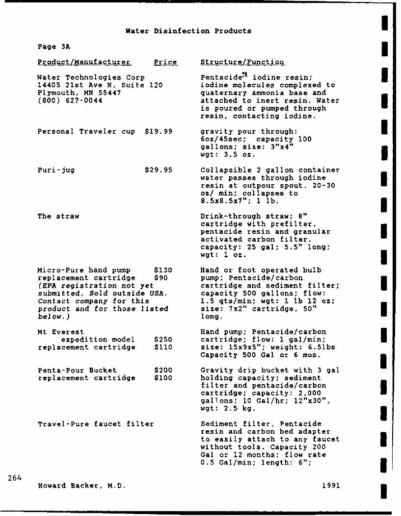
Water Disinfection Products l
Page 3A
Product/Manufacturer Price Structure/Function
Water Technologies Corp Pentacide" iodine resin;14405 21st Ave N, Suite 120 iodine molecules complexed toPlymouth, MN 55447 quaternary ammonia base and(800) 627-0044 attached to inert resin. Water
is poured or pumped throughresin, contacting iodine.
Personal Traveler cup $19.99 gravity pour through: I6oz/45sec; capacity 100gallons; size: 3"x4"wgt: 3.5 oz.
Puri-jug $29.95 Collapsible 2 gallon containerwater passes through iodineresin at outpour spout. 20-30oz/ min; collapses to8.5x8.5x7"; 1 lb.
The straw Drink-through straw; 8"cartridge with prefilter,pentacide resin and granular Iactivated carbon filter.capacity: 25 gal; 5.5" long;wgt: 1 oz. 3
Micro-Pure hand pump $130 Hand or foot operated bulbreplacement cartridge $90 pump; Pentacide/carbon(EPA registration not yet cartridge and sediment filter; Isubmitted. Sold outside USA. capacity 500 gallons; flow:Contact company for this 1.5 qts/min; wgt: 1 lb 12 oz;product and for those listed size: 7x2" cartridge, 50"below.) long.
Mt Everest Hand pump; Pentacide/carbonexpedition model $250 cartridge; flow: 1 gal/min;
replacement cartridge $110 size: 15x9x5"; weight: 6.5lbsCapacity 500 Gal or 6 mos.
Penta-Pour Bucket $200 Gravity drip bucket with 3 galreplacement cartridge $100 holding capacity; sediment
filter and pentacide/carboncartridge; capacity: 2,000gallons; 10 Gal/hr; 12"x30",wgt: 2.5 kg.
Travel-Pure faucet filter Sediment filter, Pentacideresin and carbon bed adapterto easily attach to any faucetwithout tools. Capacity 200Gal or 12 months; flow rate0.5 Gal/min; length: 6";
264 Howard Backer, M.D. 1991I

Water Disinfection Products
* Page 3B
Claims Comments
Resin releases iodine "on Test data is convincing; resindemand", on contact with appears to be majormicroorganisms; minimal iodine breakthrough in waterdissolves in water: effluent disinfection. Organisms1.0-2.0 ppm iodine. Charcoal effectively exposed toremoves any residual dissolved extremely high iodineiodine. Tested effective for concentrations when passingbacteria, giardia, through resin, which is whyshistosomiasis, viruses-- minimal holding time isincluding hepatitis. necessary. Iodine apparently
binds to microorganisms aidedby electrostatic forces andpenetrates more readily thanin a dilute iodine solution,but the exact mechanism ofiodine transfer to organismsis not known. The company hasdeveloped a large number ofnew products applicable todifferent size groups andvarious settings. The resindisinfectant has already beenapproved by the EPA, howevereach product must beindividually submitted andtested for approval--a longand arduous process. Whileall these products are alreadyproduced and sold in Europe,most are not yet available inthe U.S. However, most areavailable through the company;contact the sales department.The most rational products arethe hand pump models, thePuri-jug and pour bucket, andthe faucet filter. The travelcup is much too slow and givesonly small quantities. Thestraw is for survivalists.
265

Water Disinfection Products IPage 4A
Product Price Structure/Function
Polar Equipment Iodine crystals, 8 gm in 3 oz12881 Foothill Lane bottle; separate 30-50 micron
Saratoga, Ca. 95070 prefilter provided. "Trap" inbottle to catch crystals when
Polar Pure $9.75 pouring off water; bottle cap(Widely available in camping is used to measure--directionsstores.) and dot thermometer on bottle
(temperature affects iodine Iconcentration in bottle).Recommends warming water to20C (68F) before adding iodine(to shorten contact time).Capacity: 2000 quarts; Wgt: 5oz. I
Polar Pure Plus 2 oz plastic dropper bottle of(new product) concentrated iodine/alcohol
solution; thermometer and Igraph for determining dosageand contact time. I
Potable Aqua $3.25 Iodine-containing zLablets(sold widely in camping (tetraglycine hydroperiodide)stores) release approximately 7-8 mg
iodine when added to water.
Emergency Germicidal Drinking Instur'ions direct one tableWater Tablets $3.99 added to one quart of water.
In cloudy or cold water, addtwo tablets. Contact timeonly 10 minutes in clear, warmwater, more in cold, cloudywater. Refer to table.Weight: 2 oz.
I
Candle Filter in a bag;Basic Designs ceramic element and charcoal5815 Bennet Valley Rd core. Gravity drip or squeeze ISanta Rosa, C1 95404 bag. Size: 3.5" x 8"; Wgt: 8(707) 575-1220 oz.
Ceramic Water Filter $65 II
266Howard Backer, M.D. 1991

H Water Disinfection Products
* Page 4B
Claims Comments
U Destroys all waterborne Extensive data exists forpathogens; yields 4 ppm iodine effectiveness of iodine.when appropriate dose is added Commercial source ofto one quart of clean water. crystalline iodine, an
excellent source of iodinesolution. Recommendationsadequate for clear, warmwater, but it may beimpractical to warm water.Reports of glass bottlesfreezing (and breaking) insubzero temperatures. PolarPure Plus uses disinfectionchemistry to determine theminimal dose necessary forchosen contact time. Thisallows control over dose (andtaste), but requires someintelligence in the user.
Kills bacteria, viruses, and Method developed by theprotozoan cysts, including military for troops in thegiardia, field. Good data for
effectiveness of iodine atthese concentrations.AdvanLages of unit dose andshort contact time, but theseconcentrations create strongtaste that would not beacceptable to many wildernessusers. May add one tablet totwo quarts of clear water toyield about 4 mg/L with aminimum contact time of 20minutes in warm water and atleast one hour in cold water.In cloudy water, use one ortwo tablets per quart (betteryet, clarify water first).
Removes Giardia, bacteria, New product--not all technicalparasites, cysts and bad data received prior to writingtaste. this. Should be effective for
claims; note virus removal isnot claimed. Filtration ratemay be slow.
267

Joseph B. Serra, M.D.WMS First World CongressWhistler, B.C. 1991I
MANAGEMENT OF DISLOCATIONS & FRACTURES
* IN THE WILDERNESS ENVIRONMENT
I GENERAL COMMENTS
Trauma to the musculoskeletal system, in the form of disloca-tions and fractures, is on the increase due simply to thefact that there are more people involved in the wildernessexperience of remote travel, climbing, cross-country skiing,trekking, and major expeditions. The fractures and disloca-tions are the same as those seen in the urban and hospitalsetting; however, there are distinct differences in methodsI of diagnosis and acute management. It is a well known factthat the sooner a dislocation is reduced, or the soonera fracture is realigned and splinted, the more comfortableand stable the victim will be. This presentation is directedat physicians, nurses, paramedical personnel, trek leaders,and anyone else who is involved in remote travel wheretransport to a medical facility is not available, for whateverreason. These may include distance, environmental riskfactors or extreme terrain. In the situation where acutemanagement and early transport is feasible, this shouldbe the treatment of choice. Fractures should be realignedas well as possible and splinted prior to transport; thearea of question involves dislocations. I would advocatereduction of the more common dislocations as soon afterinjury as possible, if the rescuer is comfortable withattempting a reduction and if the victim is agreeable.Identification of the injury as a dislocation or fracture;good clinical judgment regarding management; a good commonsense approach to the problem are all important factors.These will be discussed and demonstrated.
DISLOCATIONS
SDislocations of most joints may be easily identified andare quite incapacitating to the victim. Some of the majoradvantages in early reduction of dislocations are as follows:1. Reduction is easier immediately after injury, before
swelling and muscular spasm have developed.2. Reduction most often results in dramatic relief of pain.3. Transport of the victim is easier after reduction.4. Immobilization of the injured joint is much easier dnd
stable after reduction.5. Safety of the entire party may be jeopardized during
the evacuation of a victim with a major joint dislocation.6. Early reduction reduces the circulatory and neurologic
risks to the involved extremity.
II 269

I
DIAGNOSIS
The major joints discussed are the shoulder, elbow, digits,hip, knee, patella, and ankle. There are many helpful signsin identifying a dislocation. There is nearly always restric-tion of motion; obvious deformity in comparison with theuninvolved extremity; often a typical, identifiable postureof the dislocated joint that the victim will maintain to Iminimize pain. Crepitus or grating of bone fragments;and gross instability are generally absent.
The provider of care or rescuer should not be concernedabout causing additional damage to any fractures associatedwith a dislocation. At times, there are avulsion typefractures within the joint that has dislocated, but these Ifractures will generally be improved in alignment withreduction of the dislocation. The same is true of vesseland nerve impairment associated with a dislocation; reduction Iwill reduce the impingement and traction injury to these
structures, as well. In the situation in which there isa major long bone fracture associated with a dislocation,the dislocation may not even be diagnosed, in view of themore apparent major fracture. In these cases, splintingof the fracture is the treatment of choice and the dislocation,for all practical purposes, becomes a secondary issue andwill, most likely, not be identified until x-rays are obtained.
DIAGNOSTIC TIPS FOR SPECIFIC JOINTS
SHOULDER - Anterior-inferior dislocation of the shoulder ismost common, accounting for 95% of shoulder dislocations.Victim will stabilize the upper extremity in the most comfort-able position; usually with the upper arm held away fromthe body and supported by the uninvolved arm. The dislocatedextremity cannot be brought across the chest wall, i.e.sling position. Observe and palpate the involved shoulder,comparing it with the uninvolved side. There will be a Iloss of normal contour to the deltoid and a palpable defectwhere the humeral head should be. Check circulation, motorand sensory function to the hand and, also, sensory functionalong the outer aspect of the shoulder, and document findings.Mechanism of injury most commonly external rotation, abductionand extension. In the case of recurrent anterior dislocations,the victim will identify the problem and can be quite helpfulin its reduction.
ELBOW - Obvious deformity is present when compared withuninvolved side. Restricted, painful motion is present.Most commonly posterior with resultant bony prominenceof the olecranon process.
I270I
o . . | m I
IDIGITS - Obvious deformity at the joint with limited
motion are main findings.
HIP - The majority of dislocations are posterior. Theinvolved extremity will be moderately flexed, internallyrotated and adducted. Any attempt to extend the extremityfor splinting or easier transport will be resisted by thevictim and mechanically impossible to obtain. The mechanismof injury is most often a fall in which. the hip is flexed
and the forces are transmitted longitudinally through theknee and femur, driving the femoral head posteriorly fromthe acetabulum - a dashboard injury is a good example.Anterior dislocations of the hip are less common and usuallyoccur as the result of a fall directly on the lateral sideof the hip, driving the femoral head anterior and medially.The posture is extension or mild flexion with externalrotation and abduction of the thigh. Again, it is nearlyimpossible to bring the hip into full extension and neutralposition.
PATELLA - Most often occur laterally with the knee heldin moderate flexion for comfort. Position of patella incomparison to uninvolved side is diagnostic. May be recurrentwith usual mechanism pivoting on a partially flexed, weightbearing knee.
KNEE - Truly a disaster with the probability of majorligamentous disruption. Vascular impairment is a majorthreat. Peripheral pulses, motor and sensory function atthe foot and ankle should be evaluated and documented.The knee may not be dislocated at the time of initial examina-tion, but gross instability is the major clue.
ANKLE - Vascular impairment to the foot is a major risk.Associated fractures are common. Prompt reduction or thebest improvement of alignment should be performed immedidtely.
METHODS OF REDUCTION
SHOULDER - Two methods will be discussed: The abductiontraction method, and the prone traction method. The firstis steady traction applied to the shoulder with the upperarm abducted 900. The victim is supine and, preferably,at waist level. Continual communication with the victimregarding the procedure and the need for relaxation isvital. Gently bring the upper arm 900 away from the sideof the body; have an assistant apply countertraction inthe axilla; while you gently apply traction using yourbody weight as the traction force. Create a loop, utilizinga jacket or shirt with the arms tied together, avalanchecord, belt or webbed strapping. With the victim's elbowflexed 900, the loop is placed in the antecubital fossa
271
~I
and around the waist of the reducer. Then, leaning backsteadily, adequate traction is applied to the shoulder.Padding in the antecubital fossa and axilla prevents damageto neurovascular structures. Gentle, steady leaning backwardusing your body weight creates the traction to reduce theshoulder. Continue traction for two to three minutes, while Igently internally and externally rotating the humerus,all the while communicating with the victim to attain maximumrelaxation1 . Reduction is usually signalled by a clunkingof the humeral head into the glenoid and noticeable reliefof the victim. Proof of reduction is the ability to bringthe arm across the chest wall. The extremity is immobilizedwith a sling and swathe.
The prone traction method consists of the victim in a proneposition with the arm hanging downward and a 15-20 pound Iweight secured to the wrist. Relaxation is extremely impor-tant. This may require 15-20 minutes and, at times, isnot feasible in rainy, cold, windy weather.
In first dislocation, immobilization is recommended forthree weeks to allow adequate soft tissue healing. Inrecurrent dislocations, the victim will usually decidewhen to start using the shoulder again. Individual circum-stances will dictate the need for evacuation of the victim.
ELBOW - Traction applied to the forearm with the elbowin a partially flexed position while countertraction isapplied to the upper arm by an assistant. Slow, steadypull with medial or lateral pressure, if appropriate.Reduction is successful if the elbow can be flexed to 900.Elbows can be quite difficult to reduce; give it your besttry, but don't be disappointed if not successful. If notreduced, splint in a position most comfortable to the victim.Check peripheral pulses.
DIGITS - Reduction of phalanges is accomplished by tractionapplied to a partially flexed digit while actually pushingthe base of the dislocated phalanx back into place. Thisis more successful than attempting to apply straight tractionto the digit. After reduction and if the victim desires,buddy tape the reduced digit so that partial function canbe maintained. Don't attempt reduction of a dislocated Imetacarpophalangeal joint of the index finger, since openreduction of this dislocation is necessary. Dislocationof the metacarpophalangeal joint of the thumb is often Iquite difficult to reduce, but should be attempted. Inopen dislocations, thoroughly cleanse the wound and proceedwith reduction; leave the wound open; and apply a steriledressing. Antibiotic therapy, if available, is indicated.
HIP - Although reducing a dislocated hip may be difficultand painful to the victim, it is worth doing, if at all
272 II

Ipossible. The victim will be more comfortable after reduc-tion, transport is facilitated, and impairment of circulationto the femoral head is reduced. Demerol, Morphine or Valiumgiven intravenously is quite helpful, if available. Twopeople are required with one applying countertraction tothe pelvis as the other reduces the dislocation. The victimis placed supine on the ground; the involved hip and kneeare gently and slowly flexed to 900. The reducer straddlesthe victim and applies steady traction in an upward direction.victim's lower leg is placed between rescuer's thighs;hands locked behind the knee; and as the rescuer assumesa sitting position while applying traction upward, significantleverage is applied to the hip. This position also allowsfor internal and external rotation of the hip, as needed.The anterior dislocation is reduced by steady, gentle,longitudinal traction with internal and external rotation.Once reduced, the injured extremity must be splinted tothe uninvolved extremity and the victim transported ina supine position.
PATELLA - Everyone should learn to do this reductionbecause it is so easy; provides immediate relief; and convertsthe victim to an ambulatory state. The knee will be partiallyflexed viith the patella laterally displaced. Gently extendthe knee and gently push the patella back towards its normalposition. Splint the knee in extension from ankle to groin.Various materials are appropriate, including Ensolite padwrapped around the leg, down parka wrapped in circular fashionseveral times around the knee, metal pack stays, or treebranches. It is safe for the victim to walk and far better thanattempting to carry the victim. An ice axe or ski pole
* is a helpful crutch.
KNEE - Apply gentle traction to realign the joint aswell as possible. Generally, this is easily done due tothe massive ligamentous disruption. The advantages arereduction of vascular impairment. Splint the entire extremity;check pulses and document findings. The victim must becarried.
ANKLE - Dislocations of the ankle are almost alwaysassociated with fractures. The fractures and dislocatedjoint both benefit by reduction of the deformity as soonas possible. Circulation may be compromised and peripheral
* pulses should be evaluated.
Apply gentle traction to the foot and ankle, using thevictim's leg as the countertraction, and this will signifi-cantly improve the alignment. Anatomical alignment isno. necessary. By improving alignment, splinting is morestable and comfortable. These fracture-dislocations maybe well splinted with a down parka or other comparable gear,
I273I
having the affect of a pillow splint and being securelywrapped or pinned in place. 3
FRACTURES
GENERAL COMMENTS
Diagnostic skills are most important in the suspected fracture iand management should be dictated by the circumstancesat hand. The needs of the victim must be considered inrelation to his/her desire to ambulate on a suspiciousankle injury; the ability and availability of people tocarry; the type of terrain involved in transpcrt; and theneed or desire of the victim to continue carrying a load. IFracture deformities should be reduced as well as possibleand adequate splinting applied. Rest, elevation, ice andcompression are part of basic fracture care. Improvisationis often required for splinting techniques and there areusually many materials available to be used. These includeskis, ski poles, Ensolite pads, metal pack frames, straps,webbing, rope, tree branches, parkas, and maps, to mention Ia few.
UPPER EXTREMITY FRACTURES
SHOULDER GIRDLE - Gently palpate towards the area ofpain; compare with uninvolved side; look for deformity.Sling is adequate immobilization. Ice, snow, cold compressesare helpful to reduce pain and swelling.
HUMERUS - The shaft of the humerus is palpable throughoutits entire length along the inner aspect of the upper arm.Any area of point tenderness with significant trauma should Ibe considered a fracture until checked radiologically.Apply sling and swathe. If there is obvious deformityin the humeral shaft, allow gravity to realign the fractureand apply appropriate splinting. Check radial nerve functionby asking the victim to extend wrist, digits and thumb,and docu-;ent findings. i
ELBOW - May be grossly unstable. Apply appropriatesplint, posterior and/or U-shaped, with elbow approximately80-90, and then sling. 3
FOREARM - Realign as well as possible and splint jointabove and below; U-shaped splint is best. Check peripheralpulses. Sling, elevate and ice, if available.
WRIST - These are best splinted in the position found,unless there is a compromise of circulation. Place the
274 iI

hand in a position of function with soft material, suchI as rolled up glove or socks, in the palm.
HAND OR PHALANX - Realign and splint in position offunction with all joints in a partially flexed, relaxed posi-tion. Example would be holding a rolled Ace bandage inthe palm of the hand. Digits should not be splinted inan extended position due to rapid onset of joint stiffness.Consider buddy taping, whenever feasible, to allow forpartial usage of digits.
LOWER EXTREMITY FRACTURES
PELVIS - Pelvic fractures are suspected when there hasbeen significant trauma to the pelvic region and tendernessto palpation over bony prominences. Crepitus is usuallynot present. Pain with weight bearing may also suggestan acetabular fracture. Suspected pelvic fractures requiretransport, unless pain and tenderness are mild over pubisor ischium, in which case, this may be a small, stablefracture and the victim may desire to ambulate with theaid of ice axe or ski pole. The general rule would beto transport victim with a suspected pelvic fracture.If the trauma is massive, be aware of the possibility ofshock due to retroperitoneal bleeding.
HIP - Symptoms are pain, inability to bear weight.Fractures of the femoral neck may not reveal any deformityor instability, but rather pain with hip motion. Unstablehip fractures often reveal shortening and external rotationof the extremity. Immobilization of the injured extremityto the uninjured with padding between the thighs and underthe knees is usually sufficient. If there is significantpain with transport, then traction may be helpful.
FEMORAL SHAFT - All femoral shaft fractures requiretraction to provide stability, reduce hemorrhage, preventfurther vascular damage, and reduce pain. Temporary tractionshould be applied as soon as the injury is diagnosed.There are excellent lightweight traction devices whichare available and should be strongly considered as standardequipment for expeditions. The Kendrick and Sager splints
are very good; lightweight, easy to apply, and very efficient.Become familiar with their usage to facilitate their usage,if the need should arise. If makeshift traction is tobe used, become familiar with the materials you plan touse and practice applications prior to their field applica-tion. It is difficult to remember the necessary steps.A rule of thumb for the amount of traction is 10% of bodyweight; but an equally good indicator is when the victimfeels comfortable and the fracture feels more stable.Flex both knees 5-100 with padding beneath the knees.U
275
L

This will not affect the traction and makes transport muchmore comfortable. Monitoring peripheral pulses and sensibil-ity should be done every thirty minutes and documented.
An extended transport over very difficult terrain requiresa minimum of eight to ten people to alternate in carryingthe victim and also to carry packs. Therefore, if thereis reasonable alternative, including helicopter evacuation,it should be strongly considered. Keep the victim warm Iand monitor for shock during all transport or while awaitingrescue.
PATELLA - Patellar fractures result from direct trauma ito the patella. If there is tenderness, but no palpabledefect or deformity, the knee can be wrapped in a cylindersplint, composed of an Ensolite pad or foam mattress pad,and the victim be allowed to ambulate, if possible. Iceaxes or ski poles are helpful.
TIBIA - These fractures are often easily diagnosed dueto deformity, crepitus and immediate swelling. Gentlycorrect angular deformities and apply adequate splinting.An U-shaped splint running from the inner knee beneaththe foo as a stirrup and up the outside of the leg tothe knee with adequate wrapping is an excellent tibialsplint. In an open fracture, the area should be gentlycleansed with Betadine or any other antiseptic available orwith soap and water; a sterile bandage or the cleanestavailable material placed over the wound and the fracture Ithen treated with realignment and splinting. Broad spectrumantibiotic therapy is indicated, if available. The victimshould not bear weight with a suspected tibial fracture.
FIBULA - In suspected fibular fractures, the victimmay be allowed to ambulate once the fracture is adequatelytaped, wrapped or splinted. Walking aids are helpful.
ANKLE - Swelling and tenderness about the ankle maydenote a fracture if directly over bony prominences. They iare sometimes difficult to differentiate from ankle sprains.
As a general rule, the fracture tenderness immediatelyafter injury is over bone and not ligament. Therefore,treat all ankle injuries as if there may be a fractureinvolved. The ankle should be securely taped or wrappedand an U-shaped, stirrup splint applied. Weight bearingshould be at the discretion of the victim, if the painis not severe. A victim barely able to place weight onan injured ankle, should be advised not to do so.
TARSALS & PHALANGES - These are often stable fracturesand, with adequate wrapping, weight bearing may be allowable,as tolerated. Walking aids are most helpful.
I276
i

hand in a position of function with soft material, suchas rolled up glove or socks, in the palm.
HAND OR PHALANX - Realign and splint in position offunction with all joints in a partially flexed, relaxed posi-tion. Example would be holding a rolled Ace bandage inthe palm of the hand. Digits should not be splinted inan extended position due to rapid onset of joint stiffness.Consider buddy taping, whenever feasible, to allow forpartial usage of digits.
LOWER EXTREM±TY FRACTURES
PELVIS - Pelvic fractures are suspected when there hasbeen significant trauma to the pelvic region and tendernessto palpation over bony prominences. Crepitus is usuallynot present. Pain with weight bearing may also suggestan acetabular fracture. Suspected pelvic fractures requiretransport, unless pain and tenderness are mild over pubisor ischium, in which case, this may be a small, stablefracture and the victim may desire to ambulate with theaid of ice axe or ski pole. The general rule would beto transport victim with a suspected pelvic fracture.If the trauma is massive, be aware of the possibility ofshock due to retroperitoneal bleeding.
HIP - Symptoms are pain, inability to bear weight.Fractures of the femoral neck may not reveal any deformityor instability, but rathe: pain with hip motion. Unstablehip fractures often reveal shortening and external rotationof the extremity. Immobilization of the injured extremityto the uninjured with padding between the thighs and underthe knees is usually sufficient. If there is significantpain with transport, then traction may be helpful.
FEMORAL SHAFT - All femoral shaft fractures requiretraction to provide stability, reduce hemorrhage, preventfurther vascular damage, and reduce pain. Temporary tractionshould be applied as soon as the injury is diagnosed.There are excellent lightweight traction devices whichare available and should be strongly considered as standardequipment for expeditions. The Kendrick and Sager splintsare very good; lightweight, easy to apply, and very efficient.Become familiar with their usage to facilitate their usage,if the need should arise. If makeshift traction is tobe used, become familiar with the materials you plan touse and practice applications prior to their field applica-tion. It is difficult to r, member the necessary steps.A rule of thumb for the amount of traction is 10% of bodyweight; but an equally good indicator is when the victimfeels comfortable and the fracture feels more stable.Flex both knees 5-100 with padding beneath the knees.
275

IThis will not affect the traction and makes transport muchmore comfortable. Monitoring peripheral pulses and sensibil-ity should be done every thirty minutes and documented.
An extended transport over very difficult terrain requiresa minimum of eight to ten people to alternate in carryingthe victim and also to carry packs. Therefore, if thereis reasonable alternative, including helicopter evacuation,it should be strongly considered. Keep the victim warm Iand monitor for shock during all transport or while awaitingrescue.
PATELLA - Patellar fractures result from direct trauma Ito the patella. If there is tenderness, but no palpabledefect or deformity, the knee can be wrapped in a cylindersplint, composed of an Ensolite pad or foam mattress pad,and the victim be allowed to ambulate, if possible. Iceaxes or ski poles are helpful.
TIBIA - These fractures are often easily diagnosed dueto deformity, crepitus and immediate swelling. Gentlycorrect angular deformities and apply adequate splinting.An U-shaped splint running from the inner knee beneaththe foof as a stirrup and up the outside of the leg tothe knee with adequate wrapping is an excellent tibialsplint. In an open fracture, the area should be gentlycleansed with Betadine or any other antiseptic available orwith soap and water; a sterile bandage or the cleanestavailable material placed over the wound and the fracturethen treated with realignment and splinting. Broad spectrumantibiotic therapy is indicated, if available. The victimshould not bear weight with a suspected tibial fracture.
FIBULA - In suspected fibular fractures, the victimmay be allowed to ambulate once the fracture is adequatelytaped, wrapped or splinted. Walking aids are helpful.
ANKLE - Swelling and tenderness about the ankle maydenote a fracture if directly over bony prominences. Theyare sometimes difficult to differentiate from ankle sprains.As a general rule, the fracture tenderness immediatelyafter injury is over bone and not ligament. Therefore,treat all ankle injuries as if there may be a fractureinvolved. The ankle should be securely taped or wrappedand an U-shaped, stirrup splint applied. Weight bearingshould be at the discretion of the victim, if the painis not severe. A victim barely able to place weight onan injured ankle, should be advised not to do so. I
TARSALS & PHALANGES - These are often stable fracturesand, with adequate wrapping, weight bearing may be allowable,as tolerated. Walking aids are most helpful.
276
I

I
SPLINT MATERIALS
In addition to improvised splinting materials, there arecommercial products available which provide excellent immobi-lization and are lightweight. The Sam splint is an excellentaddition to any wilderness pack. For more extended traveland major expeditions, air splints should be considered.The zippered variety with screw type air tube is mostefficient. Keep in mind the variations in pressure, dueto gain or loss in altitude, and monitor circulation onan hourly basis. They do provide excellent immobilizationand stabilization of many fractures. The adult long legand adult long arm sizes are adequate.
I The commercially available air stirrup splint for ankleinjuries is excellent and lightweight. This splint willoften allow the victim to ambulate in relative comfortand stability.
All splints add weight to your medical pack but, if needed,their efficiency to provide adequate splinting and comfortfar outweigh the disadvantages.
I SUMMARY
Fractures and dislocations do occur in the wilderness environ-ment. An understanding of the injury and quick, efficienthandling of the problem will often convert a very painful,unstable and difficult injury into one which can be managedmore comfortably for the victim and all concerned.
o2r7II
IIU
277
I

III
i HIGH ANGLE RESCUE:RECENT DEVELOPMENTS
Michael V. Callahan, MSPHMinistrie des Montagnas-Alpine Guard
National Association for Search and RescueCambridge, Massachusetts, U.S.A.
II
OBJECTIVES
i Following this workshop participants will be able to:
i 1. Identify factors surrounding alpine accidents throughthe analysis of recovered mountaineering equipement
i 2. Describe mountain rescue operations for severalagencies in Europe and North America.
i 3. Describe the role of prototype medical and rescue geardeveloped for mountain rescue.
IIII
279
I

PRACTICAL EVACUATION SKILLS
Lanny Johnson, RN, EMT-P
I Moving an injured or incapacitated mountaineer over roughterrain can be a difficult and strenuous task.
Participants will do hands-on practice with improvised carrysand formal litter carry on non-technical terrain.
Objectives
1. Learn improvised carrying techniques.
2. Be able to estimate manpower needed during evacuationsinvolving carry outs.
3. Learn techniques for carrying and belaying litters safelyon non-technical terrain.
281

IABSTRACT FORM
FIRST WORLD CONGRESS ON WILDERNESS MEDICINEJuly 14-20, 1991
Whistler, British Colu-Dia
TILETITLE: The Availability of Oxygen-saving Systems for Himalayan Climbing.
BOD" OF ABSTRACT: The abstract must be typed double-spaced in no less than 10-point type size and no more than
12 characters per inch. It must be Limited to the space on this page. Do not include references, ilLustrations, or funding
sources. TabLes, when appropriate, are acceptable. Do not List authors or institutions on this page.
[Introduction] Although 02-less high altitude climbing is prevailing
among elite climbers, Himalayan trecking has became more popular
among non-career mountaineers. For the laters, 02-less climbing is
not recomendable. But oxygen is very expensive. So, availablity for
himalayan use of several clinical 02-saving systems were tested at
the Kyoto University Medical Research Expedition to Xixabangma 1990.
[Materials and Methods] Used apparatuses were; 1)Pendant 02 -conserv-
ing nasal cannula(Pendant), 2) Thermo-sensitive demand 02 delivery
system(Synchoxy), 3)Baro-sensitive demand O2 delivery system (Sanso-
saver), 4)Vinyl face mask with constant 02 flow as the control. They
were tested at rest at Base Camp(BC)(5020m). The sabjects were 8
climbers. It was evaluated by elevation of arterial 02 saturation
measured by puls-oxymeter (Sp02). And also they were tested on exer-
cise at Advanced Base Camp(ABC)(5640m). Thm subject was only one and
it was evaluated by walking speed on foot on the hill.
[Results] Average SP02 elevation by 0.51/min.of 02 inhalation for 10
minutes at rest at BC were as follows; Pendant:ll.7%,Synchoxy:11.8%,Sansosaver:12.3% and face mask:3.3%. The walking speed were measured
by consuming time on foot on the hill behind ABC with the elevation
of about 40m.The results obtained were as follows; Air:5 min.22 sec.
Pendant:4 min.28 sec.,Synchoxy:4 min.38sec.,Sansosaver:3 min.55sec..
[Discussion]Several clinical reports suggested that these apparatus-
es could save the 02 consumption to about from 1/2 to 1/3 with con-
siderable elevation of SpO2 .Our results supports their observations.[Conclusion] It was suggested that the Pendant nasal cannula and the
demand oxygen delivery systems, Synchoxy and Sansosaver, could be
used effectively and relatively economicaly for Himalayan climbing.
283

IABSTRACT FORM
FIRST WORLD CONGRESS ON WILDERNESS MEDICINEJuly 14-20, 1991 I
Whistler, British Columbia
TITLE: Carbohydrate Supplementation for Work at High Altitude: Uquid Versus
Solid Food Supplements
BODY OF ABSTRACT: The abstract must be typed double-spaced in no Less than 10-point type size and no more than
12 characters per inch. It must be Limited to the space on this page. Do not include references, iLlustrations, or funding
sources. Tables, when appropriate, are acceptable. Do not List authors or institutions on this page.
The anorexia associated with rapid ascent to high terrestrial elevations is well
known to climbers and trekkers. Even with a gradual ascent and acclimation,
appetite does not keep pace with energy demands associated with work in
these high altitude environments. Carbohydrate is believed to be an especially Icritical energy source for work at high altitude. We studied energy intakes and
expenditures of soldiers consuming liquid or solid carbohydrate food supplements
while undergoing military training at several high altitude locations (Mauna Kea, IHI; Pikes Peak, CO; Mt. Rainier, WA; and Potosi, Bolivia) ranging from 3000-
4300 m elevation. Our results indicate that thirst is less negatively affected than
appetite by high altitude. Consequently, sweetened beverages containing 3sucrose and/or glucose polymers were readily consumed and enabled the
soldiers to ingest up to 200 g/man/day additional carbohydrate over that level
consumed from the basal ration alone. However, when a solid rather than a
liquid carbohydrate supplement was used, the soldiers ate less of the basal diet
and did not receive significantly more total daily carbohydrate. The results ofI
these studies srrongly suggest that high altitude field operations are best
supported by a liquid carbohydrate supplement.
284 I

ABSTRACT FOn?1FIRST WORLD COWIORESS ON WITDFRRNESS HEDICINE
July 14-20, 1991Whistler, British Columbia
TITLE: CENTRAL NERVOUS SYSTEM CHANGES THAT MAY PRECEDE SYMPTOMSOF ACUTE MOUNTAIN SICKNESS (AMS)
S O Y OF ABSTRACT: lh nh-trnct n1,it be tyrd dolthle--pnced In no Ier th.- lO-roint typw nize niv no moro ti-n12 chnrncterR rr Inch. It fmist be ilmitrd to the rpnce? on this pnge. Do not Include reference., Ittustrntions, or fundirigsource-. lnbeq, when aerproprinte, nre occeptnble. Do not list n'zthors or InrtItutfons on this page.
A previous report has demonstrated that latencies of visualevoked potentials increase at high altitudes (Neurosurg. 21:352, 1987). Furthermore, it was suggested that more prolongedlatencies may occur in patients suffering from AMS. We soughtto 1) determine if neural responses would also be altered athigh altitudes in subjects taking acetazolamide (500 mg OD)and dexamethasone (4mg BID) or acetazolamide alone (500 mg OD)and 2) establish whether a relationship exists between AMS andchanges in visual evoked potentials.Visual evoked potentials and symptom reports were recorded
for twenty-two subjects participating in a climbing expeditionin Bolivia, S.A. Data were collected at sea level (day 0),3,689 m (day 2) and 4,970 m (day 5). The second negative (N2)and positive wave (P2) brain responses to flashed-lightstimuli were recorded over the occipital cortex. Latenciesfor the overall group were studied for changes upon ascent(ANOVA). Furthermore, subjects taking acetazolamide anddexamethasone were compared to those on acetazolamide alone(double-blind, randomized) for latency changes from sea levelto 3,689 m and 4,970 m (T-test). Cumulative cerebral symptomscores (sums of intensity grading scores for symptoms present)at each altitude were compared between the two drug groups(Wilcoxon signed-rank test).The overall group did not demonstrate a significant
difference in waveform latencies upon ascent. However, thosesubjects taking acetazolamide alone exhibited 1) asignificantly greater N2 latency change (eight percent) thanthe combined drug regimen at 3,689 m 2) and subsequentlydeveloped higher (p=.02) mean cumulative symptom scores at4,980 m. No significant differences between the drug groupsat 4,980 m existed for either the absolute latencies or forlatency changes from sea level.These data suggest that changes in evoked potential
latencies in the acetazolamide only group at 3,689 m may bea subclinical sign of the developirg pathophysiology but thelatency impairment may be resolved when AMS symptoms arepresent.
285

I'ABSTRACT FORM
FIRST WORLD CONGRESS ON WILDERNESS MEDICINEJuly 14-20, 1991 I
Whistler, British Columbia
TTLE: Effect of Cooling Time on Survival in Heatstroke: A Meta-Analysis
Evaluation
3ODY OF ABSTRACT: --e aostract must be typed ouoe-sDacec in no Less tnan 10-=oin: :,'e size arc no more :Zan I:naraczers cer incn. : mst De Limited to the space on this page. Do not inctuce references, itustrations, or 4uricang
£:urces. TaOLes, wnen aororriare, are acceotable. Do not list authors or institutions on this page. 1
Although rapid cooling is considered a cornerstone of the
treatment of heatstroke, there is little data to correlate
rapid cooling with improved outcome. Two previous studies Iusing similar methodologies demonstrated decreased mortality
,with rapid cooling but were unable to demonstrate statistical
significance, probably secondary to small sample sizes. We
performed a similar analysis on 31 patients with classical
!heatstroke and again demonstrated a much lower mortality (11%)
in rapid coolers vs delayed coolers (33%) but were also unable
to achieve statistical significance (P=.10). Because of the
consistent pattern of improved survival with rapid cooling,
meta-analysis of the 3 data sets was undertaken to explore
whether a real difference in outcome existed between rapid and I
delayed cooling groups. Meta-analysis demonstrated that
heatstroke patients that cool rapidly do have improved Isurvival. Meta-analysis has not been commonly used to assess 3clinical studies of environmental emergencies, and may play an
important role in clarifying treatment approaches to these
infrequently encountered problems.
I286
I

ITHE EFFECT OF AN ELECTRICAL CURRENT ON SNAKE VENOM TOXICITY
D. Davisi, K. Branch' N B. Egen2, F.E. Russell3,K. Gerrish and P.S. Auerbach'
'Division of Emergency Medicine, Department of Surgery, Section ofSurgical Sciences, Vanderbilt University, Nashville,TN, USA.
'Department of Veterinary Science and 'Department of Pharmacologyand Toxicology, University of Arizona, Tucson, AZ, USA.
ABSTRACTAn electric current (20, 11 amp, 7000 volt spikes per second for 90seconds) from a commercial stun gun was applied directly to arattlesnake venom solution in a three compartment electrolysiscell, in order to evaluate the effect that high voltageelectroshock might have on the lethality of the venom. Using LD,determinations in mice, there was no measurable inactivation of thevenom when it was electroshocked for 18 times longer thanrecommended by stun gun manufacturers. Using a constant voltagepower supply, a venom sample was electrolyzed at a lower voltage,but 4 to 5 times the charge delivered from the stun gun. Thisinactivated the venom at the electrodes, but not within a centralcompartment, which simulated the condition of a human envenomation.
Key words: snake venom, envenomation, stun gun, electrical current
287

IE NFWG Y, AND N ;T I INT INTAKES i)Uit IN'; H IIH AI'IT .1IE AC(L I MA IS -LT 'O N
ABSTRACT. USuccess of a high-altitude expedition may be limited by the Inutritional status of the expedition members. This studydescribes a dietary survey carried out by members of The EverestBase Camp Clean Up Operation. These individuals were not Imountaineers but the walk-in period completed by the volunteersis similar to that of most Himalayan expeditions. Ten healthyunaculimaisa-- male subjects walked from an altitude of 2430 m toEverest Base Camp (5400 m) in 10 d. The averge increase inaltitude was 300 m/d. All food and fluids consumed during the 10d walk-in period were weighed using digital dietary scales andrecorded in food record books. The diet during this time wasbased on fresh, locally available foods such as potatoes, onions,cabbage, buffalo, goat and yak meat, dahl, barlotti beans andflour. Energy and nutrient intakes were calculated using food Icomposition tables. The mean daily energy intake was 10.03 (SE1.26) MJ. Once an altitude of 3475 m had been reached there was asignificant decrease in percentage energy from fat (36 (SE 2)% onday 3 to 27 (SE 1) on day 10) with a corresponding increase inenergy from carbohydrate (46 (SE 4) on day 3 to 61 (SE 1) on day10, p<0.001). Mean daily intakes of vitamins and minerals aregiven in the Table.
Mean SE RDAThiamin (mg) 1.3 0.2 1.3Riboflavin (mg) 1.7 0.3 1.6 INiacin (rag) 37 6 18Folic acid (ug) 147 15 300Ascorbic acid (mg) 37 1 30Iron (mg) 13 1 10Zinc (mg) 9 1 -
(RDA, recommended daily amount, DHSS, 1979). IThe RDA values refer to healthy populations but not to thoseexposed to high-altitude when nutrient requirements may beincreased. The results indicate that if an expedition diet isbased on locally available foods for at least 10 d, a vitamin andmineral supplement may be beneficial in order to prevent possibledeficiencies.
IIII
288I
7 T1RTRACT FOP?!
FIRST WORLD COaITIESn Off WI,flrlIIES9 HiEDICIIIE!Jtti.y 14-20t 1991.
Whitler, British Columia
TITLE*
A Modified Hyperbaric Bag and Inhalator Mask to Study Gas ExchangeP 6-D F \n0T R-ACT! Iit! n.trnct mt b! tyrped d,ehte-!pnced In o o I .tnn Wpolnt tYPI 02e MIA no Mori th,-
12 r+AtCte?!t' Ir 1I1ch. It fmi.Y t be wIltrd tn the rrnre ot, thil ri ge. Do m.t I,,c It refete e et, 1tustrntlo-s, or IundlqImble. ?nblet, Mi-n tirprerInte, nre saceptnble. Do not list nithotrs or Instftutforis on thIs page.
A pilot study of hyperbaria was performed utilizing
a Gamow hyperbaric bag (HB) modified with a penetrating
plug during the 1990 American Andes Biomedical Research
Expedition (AABRE) climb to 4,970 m in Bolivia, SouthAmerica.
ISix subjects (4 at sea level without medication and 4 at
high altitude taking medication for acute mountain
sickness (AMS) prophylaxis) underwent three consecutivetwenty minute exposures in the modified HB. During the
first exposure subjects breathed compressed ambient air
whilst in the portable chamber. Then each subject
breathed 100% oxygen (02) on demand or compressed air
through a modified Scott II inhalator mask using a
nonrebreathing system. Sa0O, TcO,, TcC02 and chamber COwere measured. Gas analyses were done with both
polarographic and chemical methods.
Sa02 , Tc02, and TcC02 increased 14%, 50%, and 75%
I respectively, at the end of 20 minutes in subjects
breathing compressed air in the HB at 4,970 m (bag
pressure was maintained at 110 torr above ambient
barometric pressure). P1C02 increased .85 mm Hg in the
bag when measures to preventC02 rebreathing were not
taken. Sa02 and Tc0 increased 26% and 700%
respectively, when 100% 02 was breathed in the HB at high
altitude.
Further studies are necessary to determine whether
I combining hyperbaria with 100% 0 delivered on demand
through a mask is more effective in increasing Sa0 2 and
ameliorating AMS symptoms than either therapy alone.
289

IOrthopedic Field Splinting Devices
This presentation will demonstrate new field splinting devices for upper
limb and neck injuries and for lower limb injuries. The splint for upper limb Iand neck injuries is made out of plastic and exceeds the capabilities of other
available products. Performance highlights include: the splint does not
become brittle at low temperatures, it is completely reusable and does not
weaken with multiple use, it is simple to use, it is light weight, it rolls
into a small compact cylinder shape, and it is economical. IThe device for lower limb injuries is improvised out of available
materials and therefore has no additional costs or additional weight. The
splint is put together from internal stays from four expedition packs, a
therma-rest sleeping pad or similar inflatable pad, webbing or cord, and wind
or rain pants.
IIIIIIIII
290
I

ABSTRACT FORMFIRST WORLD CONGRESS ON WILDERNESS MEDICINE
July 14-20, 1991Whistler, British Columbia
TITLE:
Oxygen Saturation and Neuropsvchological F-rformance Chanpes 4ith AltitudeBODY OF ABSTRACT: The abstract must be typed doubLe-spaced in no Less than 10-point type size arc no more than
12 characters per inch. It must be Limited to the space on this page. Do not incLude references, ilLustrations, or furcing
sources. TabLes, when appropriate, are acceptabLe. Do not List authors or institutions on this page.
Exposure to hypoxic environments, as at altitude, may acutely cause
decreased neuropsychological functioning. We questioned whether any
deficit in performance can be easily measured at different altitudes
during a climb. A pilot group of four recreational mountaineers
participated in a climb to 5700 meters. Pulse, 02-saturation, Pretrails,
Trails A, Trails B, and Symbol Digit Modalities were evaluated at four
different altitudes. These are well-standardized tests with established
norms and are easily administered at altitude. Because This was a pilot
study with a small number of subjects, only trends were identified. As
expected, pulse rate increased and oxygen saturation decreased with
altitude. Trails A seemed to be a sensitive test in that it did show
deterioration with altitude. This effect was not due to exhaustion or
cold because Pretrail speed remained corstant at all altitudes. Trails B
did not show this pattern, but there was a single outlier that may have
skewed the results. Symbol digit modalities did not improve until return
to 2240 m; this may indicate that the anticipated improvement in scores
with repeated testing (learning effect) did not occur and may reflect an
altitude effect. Altitude-induced neuropsychological dysfunction may 'e
evaluated with this battery of simple tests. Larger numbers of subjects
need to be studied for validation of these trends.
291

IABSTRACT FORM
FIRST WORLD CONGRESS ON WILDERP'ESS MEDICINEJUly 14-20, 1991
Whistler, British Columbia
!ITLE: Pseudoephedrine Vor The Prevention of Barotitis Media:
A Controlled Clinical Trial in 120 Scuba Divers
ODY (F ABSTRACT: The abstract must be typed doubte-spaced in no tess than 10-point type Size and no more than '2harecters per inch. It wJst be timIted to the space on this page. Do not include references, Iltustrath,.", or funading-urces. Tobtes, when appropriate, are acceptable. Do not List authors or institutions on this page.
Purpose: To detcrmine the efficacy and safety of decongestant prophylaxis
among scuba divers in the prevention of barotitis media (middle eai squjeeze).
Methods: A prospective, double-blind clinical trial was completed with 120 1voluntzer scuba divers under the sur arvision of certified instructors. Followin
randomization, each subject received a 60mg tablet of pseudoephedrine or
placebo, 60 minutes prior to diving. Prospective data were collected, including
subject demographics, signs and symptoms of middle ear squeeze during the
dive, and possible drug side effects. The otoscopic appearance of the tympanic 3membrane was graded according to the amount of hemorrhage in the eardrum.
with TEED scores running from 0 (normal) to 5 (gross hemorrhage and rupture)
X 2 , t test, and the Mann-Whitney U test were applied with significance at P<.05.
Results: A total of 116 subjects met the inclusion criteria and completed the
study; 60 received 60mg pseudoephedrine and 56 received placebo. Treatment
groups were similar with rega.1 to age, sex, medical history and depth of the
first dive (P>0.5). Ear discomfort during the dive was present in 8% (5/60) of Ithose receiving pseudoephedrine vs 32% (18/56) of the control group (P=.001).
Likewise, the pseudoephedrine group tended to have smaller TEED scores after
diving than the control subjects (P=.003). No adverse effects were noted.
Conclusions: These results -uegest that use of an oral decongestant prior to
diving is safe and decreases the incidence and severity ofI__ sq
I292

ABSTRACT FORKFIRST WORLD CONGRESS ON WILDERNESS MEDICINE
July 14-20, 1991Whistler, I ritish Columbia
TITLE:Radio freqv ., rewarming of hypothermia victims
BODY OF ABSTACT: r. absrract ,,n.,t - typed double-spaced in no less thKn nl-nnit type size and no nore than12 ctaracters per inch. .. ---- -_ t - - to the spece On this pege. Do not irIrel- references. 1I1tijtrations, or furcin;sources. TabLet , when appropriate, are ect-pzable. Do not list authors or institut , on thir pA0-.
The efficiency of warming after hypothermia depends on the method ofrewarming and the control of temperature afterdrop. Rewarming in fieldconditions is often less efficient because of logistical or equipment limitations. Toimprove both efficiency and field appli'ability, rewarming with a radio-frequt-cy(KF) coil operating at 13.56 MHz has been evaluated. In one study, anesthetizedrhesus monkeys were used to compare the temperature effects and enzymeresponse to anesthesia only, rewarming using a surgical heating pad circulating35°C and 40'C water, or RF rewarming from rectal temperatures (Tn) of 280 to30'C. Rewarming to 35°C typically required only 50 minutes for RF vs 137 minfor heating-pad rewarming with no significant differences in serum CPK, LDH orAST between the methods. Six additional sessions, performed at lower T", werenot significantly different, except for one which resulted in cardiovascular collapseat T. = 19.3°C, with spontaneous cardioversion upon RF rewarming. RFrewarming produced T. that were significantly higher than heating pad rewarningbeginning with the third minute of rewarming, and included a large reduction inthe magnitude and .uration of temperature afterdrop. RF has also been shown toproduce more rapid rewarming to 30C than warm humidified air (43°C, 450 ccmin-'-kg') (58± 13 vs 280± 114 min respectively) with anesthetized dogs at stablecore temperatures of 25C. Likewise, in mildly hypothermic humans with 0.5'Creduction in esophageal temperatures (T,), RF has been shown to produce morerapid rewarming than either an insulated cocoon or rewarming in 41'C water(AT, 1.15 ± 0.220C/h, 0.18 ± 0.090C/h, and 0.37 ± 0.16°C/h respectively). RF hasbeen shown to be an efficient, field applicable method of rewarming fromhypothermia and a vest based on this patented method has been approved by theFDA as an investigational device for human use.
293

A1BSTRACT FORMFIRST WORLD CONGRESS ON WILDERNESS MEDICINE
July 14-20, 1991Whistler, British Columbia
TITLE: Respiratory function test(RFT) in normal subjects during a 7 days Iperiod at high altitude(HA) and correlation with A.MS score.
BODY OF ABSTRACT: The abstract must be typed doubLe-spaced in no tess than 10-point type size and no more tman12 characters per inch. It must be limited to the space on this page. Do not include references, illustrations, ot fundingsources. TabLes, when appropriate, are acceptable. Do not List authors or institutions on this page. 3
kIM of the study is the monitoring of flow-volume curves(FV)
during a 7 days stay at HA and the correlation with AMS score
METHODS:5 subjects(2F,3M)mean age 36±4 performed 3 FV at dif-
ferent altitude:sea level(sl),3600m in 3 days(Ai,A2,A3),4550m
in 4 days both in the morning and in the afternoon(Blm Bla,B2 3m B2a,B3m B3a,B4m B4a).Al and B1 were 'climbing days' and the
values were not used for the analysis.The best according to
FEVI was selected for the statistical analysis(paired t test
and ANOVA).RESULTS(mean%of expected value) :PEF sl 102% A 120%
B 138%;FEVl sl 96%,A3 B2a90%,B3a 96%;FVC sl 104%,A3 B2a 100%,
33a 108%;MEF25 sl 76%,A3 B2a 70%,B3a 79%;MEF25 B2m 64% B2a 70
3m 72% B3a 79%. Statistical analysis:significant increase in
PEF(p<.01)between sl and HA;significant decrease(p<.0l)in FVC
-EVI,MEF25 between sl and B2a.In B3a and B4a the values impro
re;the analysis between B2m-B2a and B3m-B3a showed a signifi-
:ant reduction(p<.01)in MEF25 in the morning vs afternoon.In
32 B3 a even light AMS score was recorded.CONCLUSIONS the HA Iaigher PEF is due to decreased air density;the decreased FEVl
FVC,MEF25 in A3 and B2 could be due to the higher lung tissue Ifluid(as pointed out by AMS score).The monitoring of MEF25
twice a day can detect a higher lung tissue fluid in the mor-
ning even when afternoon RFT are improved(defective acclimatinn)
294 I

IA REVIEW OF EMS NEAR DROWNING CALLS IN CONTIGUOUS NORTHCAROLINA BEACH COMMUNITIES: APPROPRIATE USE OF BASIC
ASSESSMENT AND MANAGEMENT SKILLS MUST BE BASED ON A THOROUGHUNDERSTANDING OF POTENTIAL INJURIES SUSTAINED IN THE MARINE
* ENVIRONMENT
Near drownings are a subset of environmental emergenciesfrequently seen in beach communities. This study retrospectivelyreviewed all EMS calls for the calendar year 1990 in the ruralsetting of Dare County, North Carolina. Several management problemswere identified and protocols will be modified to positively affectoutcome. Prehospital care was provided at the EMT-AdvancedIntermediate (endotraclzcal intubation, intravenous access and basicACLS drugs) level. Charts reviewed identified 109 EMS calls relatedto near drownings. Of these, 78 (71.6%) were male and 55 (50.5%) were30 years or older, while only 5 (4.6%) were less than age 6.Eighteen (16.5%) victims were found in cardiopulmonary arrest, and 2(1.8%) were considered dead. Surf and tidal rip were identified assignificant factors in near drownings in 16 patients (14.7%), whichis previously unreported. Spinal immobilization, a significantconcern, was documented in only 11 (10.1%) patients, and for only 2(1.8%) of the patients injured in the surf. Endotracheal intubationwas accomplished in only 8 (44.4%) arrest victims. Hypothermia wasdocumented in 19 (17%), with 12 (11%) occurring in June, July andAugust. These data suggest that specific education on near drowningand the water environment should be incorporated into EMS trainingfor providers in water-related areas. Airway management and spinalimmobilization skills must be stressed in relation to these injuries.
295

II
ABSTRACT 3
UTREKKER MEDICINE:
The Impact of Western Medicine on the Health of Sherpas
Iin the Khiumbu Region of Nepal
Catherine A. Hagen. M.D.Ian D. Schokking, K.D, C.C.F.P.
IEach year, approximately 10,000 foreign trekkers visit Sagarmatha
National Park region of Nepal (the Mount Everest Trek). Problems ofhealth among trekkers have been documented: high altitude sickness,trauma, bacterial and protozoal infection. Less attention has been paidto the impact foreigners have had upon the health of the local 3,000Sherpas.
Many visitors are doctors; all like to share the contents of well-stocked first aid kits with local residents. Unfortunately, the habit oftrailside prescribing can undermine a system of primary health careestablished through Kunde Hospital and a system of village healthclinics. Better communication could be fostered between visiting healthprofessionals and Sherpa patients, in the interest of supporting WHOgoals of primary health care through essential drugs and locally trainedworkers.
In a slide presentation, Doctors Hagen and Schokking document twoyears of primary health care practice and education among the Sherpas ofthe Mt. Everest region.
IIII
296 I
I

GLOBAL HEALTH ISSUES
Philip C. Rasori, M.D.
II
"Modern education is competitive,nationalistic and separative. It hastrained the child to regard materialvalues as of major importance, tobelieve that his nation is also ofmajor importance and superior toother nations and peoples. Thegeneral level of world informationis high but usually biased,influenced by national prejudices,serving to make us citizens of ournation but not of the world."
ILAEiRT EINsTEI.4
20, ;u_7 I L TOTAL
1"s F'T 5.7 WORLD
M rI X 5. POPULATION
i 1135 r'lf" 4.
Ilm I'- 1- - 4.4
i 1975 rt_- 4.1
1970 riT " 3.7
195 * 1.3 34
13H " u 3.3
115 ;,9-9 2.3I1350 r*,l 2.5om LauC2 , C' m 4.
Cmeinm toae bd kM
I A prejed of wMrtd PPUbtion Comparing the growth of develoemuntnhS fthat of less developed countrie.
I297I

DaeEstimated World Population DobigTm
8000 B. C. 5 milion 1, 500yearsA. D. 1650 500 million 200 yearsIA. D. 1850 1, 000 million (1 billion) 80 yearsA. D. 1930 2,000 million (2 billion) 45 years
A. D. 1975 4, 000 million (4 billion) 35 yearsIA. D. 2010 8 billion? 30 yearsA. D. 2040 16 billion?
Figure 10. 2 Doubling times forhumnan population (After Keyfittz. 1962)
FIGURE P9. WORLD POPULATION GROWTH THROUGH HISTORY
Billions of Prople3
6IWaU
W--a 5go.p
2-S Millon 8000 D.C. 6000 aBc. 4000 B.C. 20008 ac. 9 C. A.D. 2000Years B.C. ADM
Sowme Jean van der TLk cart Hauab. anid Elaine Muarphy. 'Out Poculaiion Predicament A New Look: Piiatai fullfiae 3, no.5iDecemoer!979i 2.
Table 9-1. Potential Reduction in Infant and Child Deaths with Proven Disease-Control
TechnologiesI
LivesEstimated Potentially
Disease Deaths Interventions Effectiveness Saved(million) (percent) (million)
Diarrhea 5 Oral Rehydration 50-75 3Immunizable Diseases 5 Vaccines 80-93 4IPneumonia/Lower 4 Penicillin 50 2
Respiratory InfectionLow Birth %**eight. 3 Maternal Supplements; 30 1
Malnutrition Treat Infections.
Contraception
Total 17 10I298 sovit3CZJ.E. Rohde. "%%h% the Other Half Dies."Ai4stinmit CArddrs' (UNICEF/Cene'al. No. 61-62. 1983.

Fig. I Causes of death in children under five years.
Acute respiratory inftections
Diarrhoeal diseases 27%
- Tetanus
I Foreign Economic Aidand Military Expenditures ~by Developed Countries Every gun that is made, every
-warship launched, every rocketI ___________ so.fired signifis, in the findl sense, a theftfr-om those who hunger and are not fed,I __________those who are cold and are not clothed.
This world in arms is notspending mnoney alone-I -. It is spending the sweat ofU its Laborers, the genius ofits scientists, the hope of
-- usits children..Th7is is not a way of life at
all in any tresense. Under the cloud ofI threatening wa, it is humnanityhanging from a cross of iron.
Acmpariseriel developed nalias'W se"adi N mu ei
secoomec aid with I iti iltarY aepfdtute
299

I MARINE ENVENOMATIONS
I Dr John WilliamsonDirector of Hyperbaric Medicine
Royal Adelaide HospitalAdelaide
South Australia
1i. Learning Obiectives:
Following this presentation, participants will:
a) have a perception of the internationalincidence and significance of the commonesthuman envenomations resulting from marinecreatures.
b) be able .o develop a systematic approach to thediagnosis, and first-aid and medical managementof the more serious and the more commonenvenomations.
c) begin to have an understanding of theprinciples of prevention of such envenomations.
d) develop insight into the more pressing areaswhere research is needed to advance thissubject.
2. Course Material:
i Brief historical introduction of marineenvenomations as they have impacted upon humanbeings over the centuries shows that mythology,superstition and inaccurate observations havekept this subject in the realms of folkloreuntil this century. Halstead has been a pioneerin bringing systematic documentation ofobservations *o the subject.
I ii Display of existing world epidemiological datarelating to the incidence and identification ofhuman stingings and envenomations indicatesthat the events affect sea shores the worldover, but have their greatest preponderance inthe warmer tropical and sub-tropical oceans(e.g. the Indo-Pacific region). As with allenvenomations. children are a particularlysusceptible risk group, with also relatively
I high exposure.
iii Illustration of the more common and/or moreserious envenomations (Chirodropid andCarybdeid jellyfish, Physalia stings, sea
snakes, blue-ringed octopus, sting ray
301

Iinjuries, spinefish, Crown-of-thorns injuries,coral abrasions), using first-hand slidepictures and case reports enables recognitionof injury patterns. Reference will also be madeto Ciguatera poisoning. Human deaths mostcommonly result from Chirodropid stings in the IIndo-Pacific regions. Lethal Physalia stingingsare occurring in the United States.
iv Within the animal groupings, a suggested Isystematic approach to diagnosis of close skinsting or injury observation, nematocystidentification from skin scrapings (where Iapplicable), and serum immunoglobulin Gtitres). First-aid management, retrieval, andsubsequent medical treatment of the Ienvenomation groupings is taking on a logicalbasis. Vinegar dousing and the controversialplace of compression bandaging versustourniquets will be discussed. A possible newadvance in the treatment of acute ciguaterapoisoning using intravenous mannitol infusionwill be described.
v Reference to international specific marineantivenom availability, and usage principlesincludes correct animal diagnosis, selection ofappropriate cases for indicated antivenomadministration, appropriate route ofadministration, and suggested methods ofavoidance of the potentially seriouscomplications of systemic animal proteinexposure.
vi An outline of current toxinological andimmunological techniques which permit rationalidentification and management of humanpatients. The leading laboratory is that ofProfessor Joseph W. Burnett, MD, andcolleagues, within the Department ofDermatology at the University of MarylandMedical School, in Baltimore, USA. n
vii A discussion of specific prevention principles,which include public education, knowledge ofrisk times and areas of the coastlines, in-water behaviour, protective clothing, seashorepatrolling, and safe enclosures.
viii A conclusion which addresses the need for Ibetter reporting of stings, envenomations andpoisonings occurring in and from the seasworldwide, for high quality colour photographs Iof fresh stings and injuries, and for accuratefirst hand observations of envenomation casesto be documented. Further advances are now also
302

contingent upon the collection and isolation ofthe marine animal venoms, and their validcharacterisation, both toxicologically andimmunologically. Only then will specifictherapeutic measures become possible foraffected patients.
I ix The International Consortium for JellyfishStings is currently working to the above ends,and seeks to establish an internationalreporting network, with some success to date.Countries participating at present are USA,Australia, Japan, Republic of China, Malaysia,Thailand, Pakistan, India, South Africa,Yugoslavia, Switzerland, Portugal, UnitedKingdom, and Russia. A collaborativeimmunological research effort between USA,Malaysia, India and Australia has resulted inseveral publications. It is hoped more fundingby respective governments will be forthcomingfor marine envenomation research - especiallyfor scientists and clinicians working in thecountries bordering the Indo-Pacific region.
3. Publications in the past 7 years:
Burnett JW, Calton GJ, Othman IB, Russo AJ and
colleagues, and Russell FE and colleagues, in"Toxicon", "Ann Emerg Med", and "J Comp Biochem
l Physiol".
Endean R, Rifkin J and colleagues, in "Toxicon".
l Edmonds C, in Med J Aust, and in book form("Dangerous marine creatures", Reed Books Pty Ltd, 2Aquatic Drive, French's Forest, NSW 2086,I Australia.)
Williamson JA, Fenner PJ, Callanan VI, Hartwick RFand colleagues, in Med J Aust, and in several bookpublications (e.g. Queensland Museum Publications,and Queensland State Centre Inc., Surf Life SavingAssociation of Australia, Brisbane, Queensland,Australia).
4. Classical Historical Publications:
l Pioneer publications by Light, Halstead, Barnes,Cleland, Southcott, Flecker, Freeman, Crone andcolleagues throughout the Australian and Americanmedical and toxicological literature.
303

IJELLYFISH ENVENOMATION
I in a Symposium onWilderness ToxinologyIDr John Williamson
Director of Hyperbaric MedicineRoyal Adelaide Hospital
AdelaideI South Australia
1i. Learning Obiectives:
At the conclusion of this Symposium Session, participantswill: a) appreciate better the world wide incidence, and
frequency of jellyfish envenomation
I b) learn of the diagnostic and management criteriathat are presently understood
c) have an appreciation of the diverse clinicaleffects of jellyfish venoms, together with thecommon features of venoms from differentspecies
d) be able to get a "handle" on the components ofthe venoms, their clinical characterisation,their toxicological and immunologicalpropensities, and the direction of currentresearch.
e) perceive the international research needs ofthe present and the future, and its possiblerelation to parallel research in other venomtoxinology.
2. Course Material:
i An overview of jellyfish envenomation, based onthe International Consortium for JellyfishStings' reporting system shows that theseevents are common, world-wide in distribution,frequently serious, and sometimes fatal.Serious jellyfish envenomation is the commonestmarine mishap after drowning, near-drowning andimmersion, and has been a neglected area ofstudy.
ii Advances in biological understanding (e.g. thearchitecture of jellyfish tentacles, nematocystfunction) and improved on-the-beach correlationof stinging case reports have permitted a
I305

Idevelopment of a systematic approach to therecognition of sting patterns on human skin.
Barnes' original observation, complimented bythe original work of Cleland and Southcott, hasevolved the technique of sting skin scraping Iand the harvesting of adherent nematocystswhich permit "fingerprint-like" specificidentification of the stinging species in theacute situation. This permits appropriateclinical response.
Advances in venom characterisation, especially"lethal" fractions enables more specificapplication of resuscitation and managementpractice at the clinical level.
An emerging appreciation of the antigenicproperties of jellyfish venoms (complexmixtures of polypeptides and enzymes) isrevealing a syndrome group of allergicreactions to jellyfish stings, including bothsevere (anaphylactic) and delayed effects.
iii Antigenic commonalities are being uncoveredbetween components of jellyfish venoms, andother terrestrial venoms and toxins (e.g. thebrown recluse spider and the cholera vibriotoxin). The significance of this observationremains obscure.
iv Titration of the patients days-old serumagainst specific venom antigens in vitro Ipermits the identification of the offending
venom with increasing accuracy. This not onlyassists clinical management, but throws lighton the epidemiology of jellyfish stinging worldwide.
Immunoglobulins G in human sera respond to Ijellyfish venom exposure by specific rise inantibody titres. These appear in a few days,and persist for many months. Significant Inumbers of patients exhibit cross reactingantibodies to other jellyfish venoms. Noconsistent pattern has yet been discerned.
v One of the most studied jellyfish venoms isthat of the northern Australian lethal box-jellyfish Chironex fleckeri. This complex Imixture contains "lethal", dermatonecrotic,savage pain-producing, and haemolytic fractionsat least. THe lethal component is probablydirectly toxic to the human myocardium, causingcessation of contraction in systole? Thisaction is dose- and may be calcium ion-
306 I

dependent? There is animal evidence thatcalcium channel blockade may offer someprotection against the myocardial effects.
The existing Australian Commonwealth SerumLaboratories' specific sheep antiserum Chironexfleckeri antivenom is a powerful analgesic forthe sting of this animal, probably protectsagainst skin damage also, and may offer somemyocardial protection. This latter seems to beenhanced in animals by the addition ofverapamil.
vi Laboratory studies in Baltimore havesuccessfully employed adsorptionimmunochromatography to separate and l irtiallycharacterise various world jellyfish venoms,including the North American Sea Nettle(Chrysaora quinquecizrrha), the world-widePortuguese Man-o'-war (Physalia spp), and someof the Australian and South American jellyfish.
vii A dramatic new syndrome, the "IrukandjiSyndrome", caused by the sting of a tinytropical cubomedusan group (including Carukiabarnesi) is characterised by delayed severemuscular pains and prostration, severe acutehypertension and other features suggestive of acatecholamine storm, and occasional progressionto an acute global cardiac dilatation andpulmonary oedema with life-threatening hypoxia.Venom and clinical research is proceeding.Patients are assisted by the intravenousadministration of phentolamine, and whereindicated inotropic cardiac support.
viii There is evidence that certain jellyfishenvenomations are accompanied by acute, severe,and directly localised arterial spasm anddistal ischaemia of stung limbs followingstinging. The pharmacokinetic explanation forthis phenomenon is unknown.
ix The capture of jellyfish venoms, theirpreservation for transport, and their validreconstitution and separation into componentsis a challenge which is currently beingaddressed. Freeze-drying techniques seemsuperior.
x Past toxicological studies may have been misledby the partial denaturation that resulted inthese highly labile venom mixtures, from thecrude fractionation techniques that were usedat that time.
307

Ii
xi Happily a number of countries in the tropicalregions whose shores are most commonlyfrequented by venomous jellyfish, are beginningtoxicological research. Collaboration,experience sharing, training, and publicationnotification via the channels offered by the IInternational Consortium for Jellyfish Stingsis facilitating these advances. The UnitedStates and Australia presently lead these Iactivities, but it is hoped increasedgovernment and private research funding in thedifferent countries will stimulate theseefforts in the future.
3. Recent Publications: iBurnett JW, Calton GJ, Othman IB, Russo AJ andcolleagues, and Russell FE and colleagues, in I"Toxicon", "Ann Emerg Med", and "J Comp BiochemPhysiol".
Endean R, Rifkin J and colleacues, in "Toxicon". iEdmonds C, in Med J Aust, and in book form("Dangerous marine creatures", Reed Books Pty Ltd, 2AquatIc Drive, French's Forest, i4SW 2086,Australia.)
Williamson JA, Fenner PJ, Callanan VI, Hartwick RF,Burnett JW and colleagues, in Med J Aust, and inseveral book publications (e.g. Queensland MuseumPublications, and Queensland State Centre Inc., SurfLife Saving Association of Australia, Brisbane,Queensland, Australia).
IIIII
308 I

WILDERNESS MEDICINE -- July 1991Advances in Crotalid Antivenin will be taken from the attached paper and thereview, Snake Venom Immunology: Historical and Practical Considerations.J. Tox. Toxin Rev. 7(1) 1-82, 1988, and updated.
Findlay E. Russell, MD, PhD, The University of Arizona, Tucson, Arizona 8.721
t3IOLOGY -nakes. Fewer than 12 fatalities per year occur, mostly:n children, in untreated or mistreated cases, or in
In the USA. ioout 20 ol the 120 species ot snakes are members of religious sects wno handle venomoussluicientlv \enomous to be a danger to humans. Most Of nerpents. Rattlesnakes account for about 65% ofthese reptiles can he divided into the pit vioers venomous snake bites and for almost all of the deaths.Crotalidaei and the coral snakes Elaoaae Their Most other venomous snake bites can be attributed to theistribution is shown in 1able i, and their habits and copperhead, and to a lesser extent the cottonmouth.habitats are aescribed in the relerences. Coral snakes inflict less than 1% of all bites. Imported
CHEMISTRY, PHARMACOLOGY, snakes found in zoos, schools, snake farms, and amateurAND PATHOLOGY and professional collectic-s account for at least 15 bites
Snake venoms are complex mixtures, chiefly per
proteins, many naving enzymatic activity Althougn the SYMPTOMS. SIGNS AND DIAGNOSISenzymes contribute to the deleterious effects of the Bites by venomous snakes are medical emergenciesvenom, the lethal property ,ind some other toxic effects esbyiveomous aes are me eerenciesmay he due tO certain of .the relatively small requiring immediatoe atteri~ion and the exercise of con-
siderable judgment Before treatment is started, it ispolypeptides. These polypeptides appear to hive specific essential to determine whether the snake was venom-cnemical and physiologic receptor sites and activities. ous, and whether or not venom was injected; snakes may
In addition to the activities of .arious venom bite and not inject venom. iNo poisoning develops incomponents and their metabolites, envenomation may ne ibout 20 to 30% of crotalid bites, and in about 40 to 50%complicated by the release of autopnarmacoiogic u" cobra and certain elpid bites.) When no enven-substances that can make diagnosis and treatment ormation occurs, or if the bite is inflicted by a nonven-difficuit. Thus. the arbitrary grouping of snake venoms omous snake, it should be treated as a puncture wound.into categories such as 'neurotoxins." "hemotoxins." and It is advised that the necessary antitetanus agent becardiotoxins" is pharmacoiogically superficial and can given
lead to grave errors in clinical judgment. A so-called Positive diagnosis for snake venom poisoningneurotoxin can produce marked cardiovaicular changes requires identili,.,..on of the snake and evidence ofor direct hematologic effects. ['he so-called hemolytic envenomatin. Although the identity of the offendingvenoms can also produce changes in the nervous system :nake can be suggested by the fang marks, these shouldor in vascular dynamics. A patient with snake venom never be relied on for positive identification. "Typical"poisoning may present several toxic reactions - iang mark patterns ore oiten based on the anatomy ofhematologicai. cardiovascular, and neurological. the snake s jaw and may not occur under field conditions.
Rattlesnake and many viper venoms produce local Rattlesnake bites may leave one or two fang marks astissue camage. nlood cell changes, coagulation aefects, well as other tooth marks. sinvle ftng punctures are verybioou vessel iniurv, and changes in vascular resistance. common and are not uncommon in bites by someThe lict ma', ,il rapidly, althougn hemoconcentration nonvenomous snakes Double strikes also occur.may occur auriny the verv early stages. rhrombocvto- Numerical grading of rattesnake bites is sometimespenia is common. ,-xcept in minimal envenomations described in the literature, hut I, is wiser to describePulmonary eciema in common in evere p oioning, and cases as minimal, moaeraie. or severe, depending on aUlbleeding may occur in the lungs, peritoneum, kidneys, the symptoms, signs, and laboratory Gndings ratherbrain and heart 'hese changes are often iccompanied than on numerical grading: I. 2, 3. 4, or 5, as based onby alterations in cardiac and renal function Renal fail- p bpain and swelling. Bites by the Nlojave rattlesnake, forure may occur because of a critical deficit in glomerular example, can give rise to minimal edema, local tissue!iltratLion secondary to hypotension, or It iay be due to changes and pain, and therefore be graded as 1. Thethe effects of hemolysis. or to the direct effects of the conseauence can be the administration of insufficientvenom components Although cardiac avnamics may be antivenin resulting in a poor teven fatal) outcom,.disturbed, the early caroiovascuiar coilapse seen in dn Diagnosis must be established on the basis of alloccasional patient bitten by a rattlesnaie is caused, for manif'aLions.the most part. bv a fall in circulating blood volume. Thisac-ears to be dsocialed with a loss o1 blood plasma and Pit Viper Envenomationprotein through tne ,ebel wails, and to I)lood poolig. The symptoms and signs of crotalid poisoning varyMost North \merican crotalid '.enoms produce consarelatively minor cnanges in neuromuscular trans- erably, depending on the species of snake, themission The '.enom of the Mojave rattlesnake is the amount of venom injected, and other factors. If there ismajor exception evidence of poisoning immediately after a bite, the
possible consequences must not be underes+imated.Most 2laoa veno, is cause changes in neuromus- Bites by rattlesnakes, cottonmouths, and copperheads
cular transmission. in nerve conduction, and to a much usually cause immediate swelling, edema, and pain; butlesser extent in the CNS Some elapid venoms. however, contrary to popular opinion, severe pain is not a constantcause local tissue damage and necrosis, blood changes, Finding, Generally, however, some degree of painand severe renai Complications immediately follows envenomation, and swelling and
EPIDEMIOLOGY edema usually appear within 2(0 minutes. They arerarely delayed for more than 30 minutes, but this may
\lthougn 45.000 people per 'vear are bitten by sn.,Kes occur. By the Lime the patient arrives at the doctorSin the LSA, only about 8.000 cases are due to venomous office, a ( agnosis ofcrowalid bite with envenomation can
309

usually he made (or envenomation excluded) on the basis Suction, using Sawyer's "Extractor," applied directlyof fung marks, swelling and edema, pain, and in bites by over incisions or even over the fang punctures is of valuesome species, tingling or numbness periorally or in the during the first 30 to 60 minutes following the bite. Thefingers or toes, or a metallic or rubbery taste. wound should be cleansed and covered with a sterile
dressing. The -affected part should be immobilized atUntreated, the edema progresses rapidly and may heart level and in a functional position. The patientinvolve the entirc extremitv within several hours. There shouid be kept warm, at rest, rings and bands removed,
map be lymphangtis and enlarged, tender regional and should be given reassurance. If the patient is withinivmph nodes. Skin temperature over the injured part is 30 minutes ofa medical facility, he shouid be transported 1usually elevated. ,dthougn the patient may complain of there, as quickly as possible, given reassurance, and thechills. Weakness, syncope, sweating and nausea may he affected part immobilized.present. Vomitinlg Iiuv occur arid the pulse may be rapidand weak, BP often drops ana, in severe cases, shock At the hospital, if antivenin is needed, a skin test formay develop early. Particularly following bites by the horse serum sensitivity should be performed a: described IMojave rattlesnake, respiratory distress may occur, and in the antivenin brochure. If the patient is mildlymuscle fasciculations, spasms and weakness are not sensitive to horse serum, diphenhydramine IV isuncommon. True paralysis may be seen in severe cases. indicated before giving the antivenin. Some physiciansThe patient may complain of dtfficuity in swallowing, routinely administer this drug prior to antivenin. A Iheadache, blurred vision, ptosis. and marked thirst. tourniquet, U.), epinephrine, and other drugs and equip-
ment for treating anaphylaxis should be available dur-':cchymosis is common in most cases of moderate or ing antivenin administration. If the skin test is strongly
severe rattlesnake poisoning and usually appears sensitive, antivenin should be given ordy iflife or limb isaround the bite within 3 to 6 hours It is severe following at stake, and then only under very controlled conditions.b .es by eastern and western diamondbacks, the prairie
nd Pacific rattlesnakes, and less severe following The amount of Antivenin (Crotalidae) Polyvalentc pperhead bites. The skin may appear tense and [Wyethl to he given depends on many factors, mostdiscolored. vesicles liuv form IF) the area of the bite important of which are the severity and progression ofWitnin the !irst 8 hours, often tecoming blood-filled, the symptoms and signs. In minimal rattlesnake venom
North American rattlesnake bites tens to be relatively poisoning. 50 to 80 ml (5 to 8 vials), of antivenin (recon-.uperficial. ecrosis is common around the bite area in stituted) will usually suffice. Moderate cases mayiintreated cases, and supcrficial blood vebsels in the area require 80 to 120 ml (8 to 12 vials); severe cases may ITiav be thrombosed Most of the effects produced by need 150 to 400 ml (15 to 40 vials), or more. Water moc-
snake venom reach their peak by the 4th day. casin poisoning usually requires lesser doses; with cop-perhead bites, antivenin is usually required only for U
There may be hemorrhage from the gums, children and the elderly. Reconstituted antivenin shouldhematemesis, melena, and hematuria. Bleeding and be diluted in sterile isotonic saline or 5% dextrose andclotting times are prolonged and plutelet counts muy fall given by IV drip in most cases. If necessary to inject IM,sharply in moderate and severe envenomations. In most give in the buttocks. Never inject antivenin into a toecases, an initial rise in the packed cell volume may occur, or finger. Measuring the circumference of thealthough in moderate or severe cases subsequent extremity at 3 points increasingly proximal to the bitehemolysis may cause a rapid fall in the ilIct. and recording the measurements every 15 to 30 minutes
Coral Snake Envenomation provides a guide to antivenin dosage. If additionalantivenin is needed, it is added to the IV drip and ven
The bite is usually associated with little or no pain, over 3 to 4 hours. Antivenin is probably of less va ue if,ftentransitory. Swelling is eli herabsent or very minor. not administered within the first 12 hours. After 28Paresthiheia it often noted around the bitten area, and hours it is of questionable vulue, although it may reversesome weakness of the part becomes evident within coagulation deficits even at 30 hours. IV fluids should beseveral hours. Muscular incoordinatlon may subse- kept to a minimum, except when shock or hypovolemiaquently develop, and the patient may complain of are present.marked weakness and lethargy. There mav he increased,alivation, difficultics in swallowing and phonation, and The appropriate antitetanus agent should always bevisual disturbances. Respiratory distress and failure given, and a broad-spectrum antimicrobial adminis- Imay ensue. In fatal cases, shock, leading to complete tered in serious cases. In many cases, either early or latecardiovascular failure, usually precedes death. in the poisoning, blood volume decreases and perfusion
fails, often with concomitant lysis of RHCs and plateletLABORATORY TESTS destruction, necessitating transfusions and parenteral
IrI all but trivial cases, a CBC, platelet count, and fluids. Plasma or albumin may be used to treat hypovo-urinalysis are essential. The following tests should also lemia. If there is a decrease in RBC mass, either throughbe obtained: ryping and hold. P. lYT7, and fibrinogen; lysis or bleeding, packed cells or whole blood should beIther tests might include I'SR. Na. K, CI, and Ca blood given. When these complications are accompanied bylevels, and CO2 combining power An ECG is .ndicaed defects of hemostasis -- i.e., abnormal clotting or lysis oflellcs ecells or clots, or a disturbance of platelet activity --
all cases except minor ones. replace with specific clotting factors: fresh frozen plasma
TR EATM ENT or platelet transfusions.
Pit Viper At the first sign of respiratory distress, 02 should be
If the patient is seen within 5 minutes of being given and preparations made to provide mechanicalandiseseveralthoursefromithe hosiutl, and teg support. Tracheal intubation or tracheostomy may bebitten, and is several hours from the hospital, and there indicated, particularly if trismus, laryngeal spasm, or
is evidence of envenomation, a constriction band should excessive salivation is present. Aspirin or codeine mayhc placed close to the bitc above the first joint proximal. be used for pain, and meperidine or morphine if in isThe band should be tight enough to occlude lymph flow severe. Mild sedation with diazepam is indicated in allbut not tight enough to impede venous or arterial flow,. eeebtsi eprtr dpeso sntapolm
severe bites if respiratory depression is not a problem
310

Sedation should reduce the amount of analgesic Coral Snakenecessary. The general principles for pit viper envenomation
The wound should be cleansed and covered with a should also be considered in coral snake bites. The valuesterile dressing. The injured part should be immobilized of suction and other such first aid measures have notin a position of function. Surgical debridement of blebs, been determined. Three vials of antivenin (Micrurusbloody vesicles, or superficial necrosis, if present, should fuluius) should be given when a diagnosis of coral snake
be carried out between the 3rd and 10th day, and may envenomation has been established. If symptomsneed to be done in stages. The injured part should be develop, 3 to 5 additional vials may be indicated. Thesoaked in 1:20 Burow's solution t.i.d. for 15 minutes. physician should contact a poison control center, zoo, orAntihistamines and corticosteroids are of no proven Wyeth Laboratories for the nearest source of thisvalue during the acute stages of poisoning and the latter antivenin. In severe cases, cardiopulmonary andmay be contraindicated. Antihistamines may be of value intensive care may be indicated.as a sedative in children. All data in this review are taken from:
Fasciotomy should be discouraged. It is usuallyunnecessary and reflects the use of insufficient antivenin 1. Snake Venom Poisoning, Scholium International,during the first 12 hours of the poisoning. It may be Port Washington, N.Y. 11050, 1983.necessary, however, when there is objective evidence 2. Merck Manual, 15th & 16th ed., 1988, 1990.(wck measurements) of a constant, elevated intracom-partment pressure (above 40 mml/Hg) over an extended 3. Conns Current Therapy, Saunders, Philadelphia,period of time and other evidence of severe vascular 1988.embarrassment. These, however, are very rare findings. 4. Snake Venom Immunology. J. Toxicol Toxin Reu.
Follow-up care is of the utmost importance. 7:11-82,1988.Contractures can be avoided by early corrective meas- 5. Aids, Cancer & Snakebite. West. J. Med. 148: 84,ures and exercises. Within several days of the bite, a 1988.complete evaluation should be made of joint motion,muscle strength, sensation measurements and girth.Follow-up care should include sterile whirlpool bathsand daily cleansing of the wound with 3% hydrogen Extractor" Pump Kits can be obtained from Sawyerperoxide followed by 15 minute soaks in 1:20 Burow's Products, Box 188, Safety Harbor, FL 34695.solution.
311

IT
cn .0
&~cis
-aI
40
fn Co /icc-cc c,
E1 -v c Cc 0. 0-a
~ 4: -Uo %.__ - -T- iWI t3E Uzk

1GROUP COHESION AND THE SELECTION OF EXPEDITION MEMBERS
David R. Jones, M.D., M.P.H.Aeropsych Associates
One Clermont Court, Oakwell FarmsSan Antonio, TX, USA 78218-1741
This paper concerns the way expedition members are selected, and how thisprocess may affect group cohesion. Selecting people for dangerous orchallenging enterprises is as old as history. The Bible describes how Gideondefeated the enemy with 300 men selected from an army of over 30,000.Greek history tells of Leonidas and 300 Spartans holding out to the last managainst Xerxes's huge force at Thermopylae; the ultimate cohesion. On hisexpedition in search of the Golden Fleece, Jason took "fifty heroes of Greece"with him (apparently his selection procedures were heavily influenced bypublic affairs officers). Each of Columbus's four expedition recruited sailors
I differently, dependir2g on supply and demand; at times picking throughcrowds of volunteers, and at times recruiting convicts. Col. William Travis,trapped in the Alamo by Santa Anna's superior forces, allowed each man tochoose for himself whether to leave, or to stay and fight to the death.
For the "Endurance" expedition of 1914, Shackleton selected 56 membersfrom over 5000 volunteers. Thirty-five years later, Sir Edmund Hilary,describing the selection of the members of the expedition which finallyconquered Mt. Everest, observed rather cynically that when it came tochoosing between two well-qualified applicants for the final assault team,the fact that one had enough money to help finance the expedition was thetie-breaker. Jimmy Doolittle asked American bomber crews to volunteer tofly with him on a secret mission which he would lead, and only after trainingwas complete and they were en route to the target did he reveal theirmission, to bomb Tokyo. Morale was so high that the stand-by crewmembers who did not get L3 go on the extremely dangerous flight were quite
disappointed.
Finally, even today, the selection of a new class of U.S. astronauts involvesstarting with some 3000 volunteers and ending with about 20 astronauttrainees.
How is this accomplished, this process which begins with a group ofstrangers and ends with a group bound so tightly that its members willsacrifice their lives for each other? We will not consider physical fitnesscriteria, or developing the proper technical skills. Instead, using a
313

Ibackground of aerospace medicine and psychiatry. we will look at howcriteria of personality and motivation may be addressed in enterpriseswhich may involve deadly danger, where members may depend utterlyupon each other fo: survival. 3CRITERIA AND MATHEMATICS
Aerospace medicine is the medical specialty involved in the selection,medical care, and health maintenance of aviators and, today, astronauts.Aeromedical standards generally pertain to two factors, safety and health,which may be translated into statements about selection for expeditions:standards should pertain to 1) accomplishing the expedition competently,dependably and safely, and 2) maintaining the health and safety of theindividual member. Among the first challenges faced by early flightsurgeons was that of medical and psychological selection of volunteers forIflying training. This process began about the time of the First World War,when the early practice of transferring men worn out or unfit for armyservice into the air service led to a disastrously high accident rate.Something had to be done to select men who were truly fit to fly.
Early U.S. standards were based on the normal values from a textbook of Uphysiology, but after about two months the flight surgeons found that theserequirements were so severe that no one had been able to pass them, and so3the standards had to be changed. Much attention was given to eye, ear, noseand throat examinations, as well as tests of equilibrium and resistance tohypoxia. In addition, applicants in some countries underwent a variety of Isomewhat more exotic tests: timed response to colored lights, tremor inresponse to an unexpected bomb blast as a measure of emotional control,ability to raise a board which supported a rod standing on end withouttoppling the rod, counting simulated artillery positions on a panorama rolledpast a viewing slit, ability to hold one's breath against 50 mm Hg pressure,and so on. (Armstrong, p. 35 ff.)
Psychological fitness was determined through a semi- structured half-hour 3interview, designed to test the quality of the applicants motivation to fly aswell as to judge whether his personality fit the current ideas of what sort ofperson an aviator should be. In fact, most of these standards were based onthe medical officers' ideas of what fliers should be like, with little or novalidation of the standards against reality. Did they really discriminatebetween successful and unsuccessful pilot trainees--or safe and unsafeaviators--and how did one know? The few research protocols intended todetermine this were inconclusive. Still, the morale of the fliers wasigenerally good, as any reading of biographical or historical documents of the
I314 I

period will show. Late in the 1930s, the Army Air Corps only had some3000 aviators, but they regarded themselves as a special breed, sharingskills and experiences beyond those of those who did not fly.
With the vast expansion of aviation training at the outbreak of World War 11,the mathematics of selection changed from one in which the Air Corps had
I the luxury of selecting only the best of the many applicants. Now U.S. Armyand Navy aviation needed tens of thousands of men. One of the greatmathematical lessons of selection quickly became clear: the larger thenumber of standards used for selection, or the more rigorous those standardsare, then the smaller the number of people who can meet them all.
I If you wish to choose four people who will be guaranteed successful, basedon qualities that may be found in only one percent of the population, thenyou must examine 400 people and reject 396 to get your four. This may bepractical if you have lots of applicants and lots of examiners, but not if youare short of one or the other. If you are willing to choose your people basedon criteria that are wrong 20% of the time, but which involve qualities foundin 50% of the applicants, then you need to examine ten, accept five, and washone out later. Your life will be a lot simpler, especially if you only have 10applicants! Let me remind you again that the more criteria you have, themore difficult your job will be, since the person with the really good visionmay not be clever enough, while the one who is clever may wear glasses.How good must the vision be? How clever must the applicant be? Cancontact lenses be substituted for the glasses? How do you know these
I criteria have anything to do with the success of your project? By askingsuch questions, the number or the severity of the selection criteria May bediminished, and the pool of potential selectees enlarged. For this reason, theselection criteria for aviation training have gotten less strict over the years,and many criteria have been dropped altogether; e.g., right bundle branch
I block.
Another mathematical fact is that the specificity (false positives) andselectivity (false negatives) of your criteria, calculated against theprevalence of the disorder, must be considered. The rarer the condition, themore expensive your testing will be, the more false positives you willencounter, and the more applicants you will lose. I do not propose to discussthis further, but anyone developing a set of medical selection criteria shouldask a statistician whether Bayes's Theorem applies.
315

I
MOTIVATION AND PERSONALITY IPeople going on an expedition need to develop cohesion--to bond together, to 3accept the expedition as a valued personal experience, and to accept its goalsas their own so strongly that they will choose to act for the good of theexpedition over their own personal interests, perhaps even to the point of Isacrifice. Many of the traditions of such choices arise from militaryexperiences, where the individual feels helpless to affect the outcome of abattle, but becomes bold when able to identify with someone or somethingperceived as stronger or more powerful, such as a charismatic leader, or atightly knit group, or a powerful piece of equipment. Borrowing power fromsucn a source, the individual once more becomes master of the situation.Imagine the difference in spirit of a fighter pilot flying in an F-16, and thesame pilot caught on the ground in a mortar attack.
Coping skills
People bonded by dramatic experiences feel they're special--different--better. They are more willing to forego ordinary pleasures and rewards towin the esteem of a valued leader or comrades. Some feeling of atruism--that others will benefit from the project, justifying one's sacrifice--is seen asbeing of greater value or good than any benefit to one's self. Thus, the3difficulty or suffering involved in achieving the goal is accepted.
Everyone who plans for endeavors such as expeditions understands the need ito anticipate what may come--to plan, to think ahead and link what oneknows to what might happen. Another mature coping skill is a healthysense of humor, frequently self-effacing, that keeps things in perspective.Look carefully at the quality of an applicant's humor, though. One must keepin mind that humor used as a weapon (sarcasm) may be quite destructive tothe bonding process. Those who prepare for endeavors such as expeditionsunderstand the need to anticipate what may come--to plan, to think aheadand to link what one knows to what might happen. Some people borrow Ienergy from one part of their lives to use for another purpose, the process ofsublimation. Some are able to suppress unpleasant realities which theycannot control--remember Scarlett O'Hara's "I'll think about it tomorrow."These five coping mechanisms--altruism, anticipation, humor, sublimationand suppression--are discussed by Vaillant in the reference given. They areamong the most adaptive used by mature and capable individuals, and, Ibelieve, should be among those used in selection for stressful endeavors.
I316
I

IHow might this information be of use to someone selecting members of anexpedition? (Remember, we are not addressing physical criteria or technicalskills, although the process is probably similar in those areas.) The first
I tentative selection criteria should come from those who know somethingfirsthand about the expedition and its goals. Intuitively, one might look forsomeone who's already done it. What problems were encountered, and howwere they handled? What sorts of people did well, and did not do so well?Why did they volunteer--what sorts of interest seemed to hold up? Whatmixes of motivation and personality seemed best? From such data, theinitial criteria may be deduced. Selection for such stressful activities asoverwintering in the Antarctic have confirmed the usefulness of factors of
I occupational ability, psychological stability, and social compatibility. (Taylor,1987). People have to be resilient enough to deal with boredom and socialisolation, and these qualities may not be prominent in people who volunteer
I out of a need for adventure or excitement. There are some very unhealthyreasons for wanting to be alone for six months!
I As for the matters of motivation and personality that underlie groupcohesion, a number of questions arise: What sort of job is it? How long will itlast? What size group will be formed? How closely knit will it be? Willmembers be able to have privacy? Will success depend more on cooperativeefforts or on individual requirements? Will some aspects require leadersand followers to trade roles? If both men and women are chosen, what sortsof problems may arise, and how will they be handled? Will sexual partnersbe specifically chosen or excluded? As criteria are decided upon, are thesetechnically measurable, or do they depend on interpersonal assessment byleaders or other expedition members?
One factor that is clear from military experience is that the tougher the
selection criteria, and the more rigorous the training, the better morale islikely to be. Members will be assured of each others' competence anddetermination, and be more willing to trust each other. Training itself is agreat source of bonding--NASA takes people with various disciplines andturns them into astronauts with various disciplines. Training should betough and realistic, clearly related to the job to be done. Harassment for itsown sake is not as effective. The tougher the training, the better people feelabout their ability to deal with tough realities. This is a part of effectiveleadership, surely a crucial part of group cohesion, and one much toocomplex to discuss further in this presentation.
Ethics and relevance
317

ITo summarize, the more standards used for selection, or the more rigorousthose standards are, the smaller the number of people who can meet themall. Medical standards should generally pertain to two factors: 1) competent,dependable and safe accomplishment of the expedition, and 2) maintainingthe health and safety of the individual member. Since failing to meet astandard removes a person from a chance to go on the expedition, justifyingthat standard should also include the concept of fairness, and of equal iopportunity. When selecting expedition members, then, the standardsthemselves must meet the meta-standards of safety health, dependability.competence and fairness. Of course, the ultimate aroiter of the validity ofany such standards is the success of the member and of the expedition, butthis may not be known until it's all over.
Validity
Some traditional medical standards have been around for a long time, andthe reasons behind them have been lost. Minimum weight standards andautomatic chest x-rays are good examples. We may have to decide whetherto continue to use such standards, to try to figure out what they're for, orscrap them. Bureaucratic process being what it is, the burden of proof isupon the one who wishes to change accepted standards. This makes itdifficult to apply new information or techniques to the dynamic process ofselection and health maintenance. Refining standards is hard enough in, 1say, the technically-oriented field of cardiology. It has been even harder insubjective processes such as evaluating the mental fitness of candidates, ordetermining how well they should get along together.
Resistance of applicants 3Doing clinical tests on people who are not patients may lead to difficulty.Using Magnetic Resonance Imaging (MRI) to look at old head injuries, forexample, might show you some unidentified bright objects (UBO) compatiblewith multiple sclerosis in a person who has never had any symptoms. Nowwhat do you do with this person if she's an astronaut candidate? People Imay refuse to volunteer for academic studies because they fear that, usingnew techniques, we might find something which could disqualify them. Lackof assured confidentiality can also lead some not to volunteer as subjects: forexample, fliers may fear the possible loss of flying status because of findingsincidental to research. This might become an issue in getting the necessary 3informed consent.
Select-in and select-out 3I
318
I

One way to look at selection standards involves two concepts known asselect out and select in. Working with mental health standards has beensimplified by the recent development of more objective criteria forpsychiatric diagnosis (DSM-III-R, 1987), which allow the examinerreasonable and reproducible means of establishing the major psychiatricdiagnoses.
Some mental health standards are fairly self-evident. Most people wouldconsider a person with a psychotic disorder to be neither safe nordependable on an expedition. Psychotropic medications might render aperson unsafe under some conditions, either because of the nature of thecondition requiring their use, or because of their effects. Conditions formerlyknown as neurotic (e.g., phobias, generalized anxiety disorders, functionalamiesias) might generally be considered disqualifying on the basis of theirinterference with safe and effective interpersonal interactions. Drug andalcohol abuse are obviously dangerous. However, diagnosis or rationalstandards may not be so easily established in other areas, specifically thepersonality disorders. Some of the most difficult decisions would concernthe amount of disturbed behavior (obsessive neatness, passiveaggressiveness) necessary to disqualify a person from an expedition.
The NASA experience
Once those with mental problems are selected out, those who remain arepresumably qualified. Now the job is to determine which of these mentallyfit persons should be selected into the group. What normal qualities aredesirable, or undesirable? One of the most rigorous selection processesinvolves picking astronaut trainees from the huge pool of volunteers. NASAhas had 13 selection cycles from 1959-1989: of about 25,000 applicants:about 200 were chosen and about 115 have flown in space. Thus, the chanceof selection runs about 1%. Lets look at NASA's methods.
The original 7 Mercury astronauts were chosen in the early I 960s from 508military pilot applicants.
The 1965 selection included emphasis on academic achievement; 6 werechosen from 909 applicants with doctorates in science, medicine, andengineering.
Today, astronauts are chosen in three categories: pilot astronauts, missionspecialist astronauts, and payload specialist astronauts. About 3000 people
I may respond to an announcement that a new group of astronauts is to bechosen. The selection process goes through four phases:
I319I

I
PHASE 1. The civilian applicants basic qualifications are evaluated by the IAstronaut Selection Office: about 60-90% are "qualified" and move forward.
PHASE 2. Applicants are re-evaluated within their own disciplines byspecific boards. [Military applicants enter here, having been screened bytheir respective services.] Each is scored as Qualified, Well Qualified, or IHighly Qualified. Those in the last category move forward (about 13-30% ofthe group).
PHASE 3. Applicants are rank-ordered within their own specialty group.Medical records are screened, and security clearance is evaluated. TheAstronaut Selection Board (comprised mainly of experienced astronauts, withequal opportunity representative and personnel managementrepresentatives) pick the top 20-25% (2-5% of the original applicants) to Icome to Johnson Space Center (JSC) for medical and psychiatric examinations,and interviews. The actual number to be selected is based on the projectedneeds of the Astronaut Office for the next three years, including attrition ofthe current astronauts.
PHASE 4. This five-day process at JSC includes extensive medical andmpsychiatric examinations, orientation to NASA, tours, social events, and aninterview.
The medical examination is rigorous, and includes 24 hour Holter cardiacmonitoring and 24 hour blood pressure monitoring, as well as cardiac fitnesstesting. The mental health examination involves a structured diagnosticinterview, psychological testing, and a discussion of each applicant within ameeting of all the examiners, where strengths and vulnerabilities arediscussed. In a recent selection cycle, 9 of 106 candidates (8.5%) hadpsychiatric diagnoses (two disqualifying: depression and a personality 3disorder) and 12 more (11.3%) had "near-diagnoses, meeting at least half ofthe criteria necessary to make a particular diagnosis. (Santy, personalcommunication.) Both the medical and mental health selection processesnlead to a simple decision--the applicant is qualified, or is not. This is theonly information available to the Astronaut Selection Board. n
The interview (or, more specifically, HEINTER VIEW) is conducted by theFlight Crew Operations Director, some current astronauts, and equal 3opportunity and personnel management representatives. Actual selectiondepends about 60-80% on the interview ranking, although the medical andpsychiatric evaluations may disqualify an applicant. About 15% of those seen3
320 II

at JSC are selected for astronaut training, representing about I % of theoriginal applicants.
Thus, this most rigorous of selection processes for what some regard as theultimate expedition considers educational, professional, medical and mentalhealth factors. A number of highly trained experts spend a great deal oftime and effort to determine which applicants should be disqualified.However, once an applicant is found fit in all other ways, the human elementre-enters, to determine which of these qualified people will be chosen. Inthe last analysis, a face-to-face interview with current astronauts and a fewother specialists is what really counts, an interview which heavily weightsinterpersonal and other intangible factors. A lot of expertise goes ilitopresenting this board with people who are qualilYed to be astronauts, butthe board of peers chooses those who will become astronauts. Gideon,Leonidas, Columbus, Shackleton and Jimmy Doblittle would understand.
REFERENCES
American Psychiatric Association. (1987). Diagnostic and statistical manualof mental disorders. 3rd ed., revised. American Psychiatric Association.Washington, DC.
Armstrong, HG. (1943). Principles and Practice of Aviation Medicine. (2nd.ed.) Williams and Wilkins. Baltimore.
Taylor AJW. (1987). Antarctic Psychology. Dept of Scientific and IndustrialResearch. Wellington, New Zealand.
Vaillant GE. ( 977). Adaptation to life. Little, Brown & Company. Boston.
321

EXPEDITION MEDICINE: PRACTICAL CONSIDERATIONS
HOWARD DONNER, MD
OBJECTIVES:
Following this presentation, oarticipants will be able to:
1. Identify skills necessary to practice emergency care in extremely remote wild.rnessenvironments.
2. Compare the fundamental philosophical differences betwpen urban and third worldmedical care.
3. Become familiar with medical problems commonly encountered in the expeditionenvironment.
4. Design and organize the remote expedition medical kit.
323
Wilderness Medicial SocietyJuly 1991
UEXPEDITION MEDICINE: PRACTICAL CONSIDERATIONS
Howard Donner, MD IMariane Gilbert, FNP
Round Mountain, Califoinia, USA i
There's good news and there's bad news.. . The good news is that if you'reinvolved in primary care, you're already equipped to handle the majority of medical
problems that come up in most expeditionary medical scenarios. The only bad newsis that there are skills which you'll need to bone up on before embarking, as medicalofficer, to parts unknown. Fortunately the following list of skills can be easily learnedwithin a short period of time by any motivated health care practitioner.
PREREQUISITE SKILLS: 3a You are well advised to take an Advanced Trauma Life Support (ATLS) course iwhich includes a basic review of the 'ABCDE's, and provides a general approach todealing with the multiple trauma patient. The skills reviewed in ATLS are key toproviding a reasonable level of trauma support on an expedition. 3° You should be well versed in preparation for foreign travel. You will generallybe considered the expert source for any questions regarding appropriateimmunizations, malaria prophylaxis, etc. 3° Familiarize yourself with the endemic diseases common in the area of theexpedition, for example the local malaria risk, exctic diseases, local animal andarthropod hazards (e.g. bears, snakes, scorpions, leeches).* Prepare yourself for the all-pervasive travelers diarrhea. Learn how to helpprevent this problem with appropriate hygiene. Learn how to differentiate clinicallybetween dysenteric and non dysenteric diarrheas, including when to treat Iconservatively and when to incorporate antiinfectives. Become expert in waterdisinfection methods for the group.- Environmental concerns, for example, knowledge in the area of cold injury,including prevention and treatment of hypothermia and frostbite for those headingtowards cold environs. A comprehensive understanding of altitude problems, such asAMS, HAPE and HACE is requisite for those "going higher".• You will need to be comfortable improvising splints for common orthopedicinjuries including the use of the 'SAM' splint or other improvised splinting systems.The improvisation and application of femoral traction should also be learned. Theuse of fiberglass splints (such as the 3M 'One Step') is helpful. It is key to developfundamental skills in the use of adhesive tape for stabilizing the, inevitable sprainsand strains of expedition life (i.e. thumbs, ankles etc.) Learn to recognize and treatcommon overuse syndromes that inevitably present as aches and pains on the trail(i.e. "sahibs knee", tendonitis, etc.).
324
• Familiarize yourself with basic patient transportation and evacuation systemsincluding improvised methods if necessary.* Develop some basic dental skills, for example the use of temporary fillingmaterial, i.e. cavit, and perhaps some more sophisticated skills such as simpleextraction of abcessed teeth. An excellent book for the dentally naive is Where ThereIs No Dentist. put out by the Hesperian Foundation.• Become familiar with the acute management of epistaxsis. This should includeboth posterior and anterior nasal packing (see medical equipment below).• The medical officer should be familiar with the management of corneal foreignbodies and corneL abrasions, as well as the management of UV keratitis(snowblindness).• Wound care management of basic abrasions and lacerations in the backcountry,including the use of suturing material (both deep and skin). Optimal treatment ofinfected blisters and burns, as well as surgical management of ingrowntoenails and I&D of abcesses.• Some basic nursing skills are essential. This may frighten the over-pamperedphysician. You must learn how to administer all of the IM and intravenous medicationsand fluids carried.
Remember: a creative and common sense approach toproblerai solving often proves more useful than pure medicalknowledge (which too often is based on sea level dogma).
PRE-TRIP CONSIDERATIONS:
• It is crucial to determine the pre-existing medical problems of each expeditionmember. A record should be compiled for each person including his/ her blood group(if "inter-group" transfusion capabilities are opted for), chronic illnesses, prior surgery(e.g. appendectomy?), prior injuries, drug allergies and medications. Appropriatevaccinations should be obtained and expedition members should carrydocumentation on an International Certificate of Vaccination.0 Members should be encouraged to obtain a dental exam prior to leaving as well astreatment of any minor medical or surgical problems (e.g. hemorrhoids, athletes foot,troublesome toenails, etc.)• The medical expertise of the group should be determined. This information may bevery useful when managing a medical emergency or orchestrating a rescue.• Another consideration is getting the medical kit through customs. This usuallypresents minimal difficulty if the medical equipment is being carried by a doctor ornurse. Be sure to carry your medical license for verification. Horror stories do exist ofmedical kits being confiscated by customs. Be forewarned.
325
I m mm mm m mm m mmm m mmm mm l

* Pre-arrange for professional rescue and become familiar with avenues to takeshould the need arise. For example, in Nepal all helicopters are operated by theRoyal Nepal Army. If a request for rescue is sent, a helicopter typically will not leaveKathmandu until someone has paid in advance for the flight. This can be arrangedthrough a trekking agency or at times the embassy of the country sponsoring theexpedition. This requires registering with your embassy on arrival in Kathmandu. Thebottom line is ... pre-arrangement can greatly expedite the rescue process should theneed arise.
• Although many high altitude expeditions are getting away from the traditional use ofclimbing oxygen, it is certainly prudent to bring medical oxygen on most large scaleexpeditions or treks. Remember that there are stringent regulations involving the 3shipping of medical oxygen via aircraft. Oxygen is considered a hazardous material.Your alternatives are: I) Ship your oxygen by surface at least three months inadvance, 2) Plan on purchasing or renting your oxygen equipment in the country ofyour expedition (note: its next to impossible to obtain in many third world countries),3) Pre-arrange to have the oxygen shipped via air freight utilizing the assistance of ahazardous materials shipper. Inquire well in advance!
ADDITIONAL CONSIDERATIONS: 3As medical officer your primary responsibility is your expedition group.
However, in most third world countries, you will invariably be called upon to help thelocals. Most of us instinctively wish to help, however there are a few concepts youshould consider prior to administering to the local sick. In many third world countries,such as Nepal, the government attempts to establish and maintain local health posts. =Delivering wholesale medicine on the trail can destoy the confidence of natives inlocal health care systems. Additionally, inappropriate or inadequate trailside treatmentmay destroy confidence in western medicine leading villagers away from appropriatecare in the future.
Remember that many third world people have little understanding of the basis ofwestern medical practice (i.e. the word for germ in Nepali is the same as that forinsect). The use of pills is often misunderstood. More significance may be attributed tothe shape or color of the pill than the medicine itself. Patients may attempt to collectmedicines which are indiscriminantly distributed to others at a later time.
Consider teaching "self-care" methods, e.g. wound hygiene, hydration withdiarrhea, rather than being eager to dispense medication. Be certain that if treatmentis provided, that full explanation and written instructions are given (using symbols orvia interpreter). Perhaps more importantly, do not indulge in "heroic fantasy". The factthat you are trekking through does not imply that you must take on the responsibility forthe village healthcare.
An often neglected aspect of expeditionary medicine is the health care of theporters. A common misconception is that porters are natives and thus immune to the Iproblems that affect westerners. Unfortunately serious illnesses and deaths amongstporters have occurred because they were not observed for symptoms of hypothermia,frostbite, altitude illness, etc. Remember, as medical officer, your responsibilities Iextend to your ancillary staff as well as your western expedition members!
326 II

THE EXPEDITION MEDICAL KIT:
Organizing the medical equipment for an expedition requires an enormousamount of planning and forethought. No matter how much equipment is hauled in,one cannot oossibly prepare for every nonceivable illness ot accident.
What should one take? There is a wide range of opinions ranging from "nothingbut a triangular bandage and swiss army knife", to a collection which would staggerthe local porters. The surgical types inevitably anticipate trauma, while medicalpeople favor pills and shots of all types. Remember, when medical kits are too bulkyand heavy they are likely to be left behind and not be available when needed.
The specific contents of the medical kit clearly depends on many factors,including:
• the environmental extremes of the tripU endemic diseases* medical expertise of the medical officer* medical expertise of the expedition members• number on trip* responsibility for local health care* length of tripI distance from definitive medical care
availability of rescue (e.g. helicopter, professional SAR, etc.)
The expedition doctor must recognize that is impossible to anticipate everyproblem. Furthermore there is a wide variation in approach to most medical problemswhich generates a healthy requirement for "artistic license".
The medical kit for a climbing expedition is typically divided into two majorgroups: 1) medical kit-Jre, and 2) medical kit-climb, The medical kit for the climb isfurther divided into 1) personal kit, 2) base camp kit, and 3) a mobile camp kit (oneper camp).
The personal kit should be carried by each expedition member. It includesthe most commonly used items and might contain the following:
1) non-narcotic analgesics2) anti-inflammatories3) throat lozenges4) sunscreen and lip protection5) water disinfection6) blister care7) minor wound care8) insect repellent9) malaria prophylaxis (if risk exists)10) vitamins11) personal medications (for pre-existing problems)
327

UThe contents of the mobile camp kit will depend on many factors of bulk and
weight constraints and is often loaded into small polyethylene containers(tupperware).
The remainder of this section will concentrate on the supplies for the mainmedical kit. There are infinite methods for organizing this kit into subgroups. Usewhatever method seems logical to you.
1. ANTIBIOTICSEven in a hospital setting two doctors seldom agree on the optimum antibiotic
for treating a given disease. Prepare for the common problems:* Upper and lower respiratory infections- your choice* Skin and soft tissue infections- consider a first generation oral cephalosporin;Duricef (cefadroxil)- is preferable because of the q12-24 hr. dosing schedule (i.e. less Ibulk)* Bacterial diarrheas- remember that many enteric pathogens are currently resistant tothe old standby Septra; therefore a quinalone antibiotic should be included (i.e. Cipr I• Giad- Tinidazole or metronidazole* Rickettsial illness rare)- Tetracycline* Helminthic infections (hookworm, roundworm and tapeworm)- Mebendazole orother antihelminthic* Malaria (for prophylaxis or presumptive treatment) - mefloquine or chloroquineand fansidar(Note: injectables will be covered later)
2. ANALGESICS IPercocet (or other strong p.o. narcotic)NSAIDs (non-steroidal anti-inflammatories) e.g. Naprosyn 500- should be carried inlarge 'bucket' to provide for the many aches and pains developed on the trailAcetominophen- for those wishing to avoid NSAIDs
3. RESPIRATORY iAlbuterol metered dose inhaler or spinhaler- commonly overlooked medicalitem; essential for cold or exercise-induced brochoconstriction, a common malady on imountaineering expeditions at higher altitudes; also serves as an important adjunct inthe treatment of lower respiratory tract infections
4. ENTRhinorockets- light-weight, simple method for anterior nasal packingAfrin or other topical nasal decongestant- a coveted item on mountaineering Iexpeditions; essential for upper airway congestion which often occurs; also helpful foreustachian tube dysfunction during altitude changesCortisporin otic or other topical anti-infective ISeldane or other non-sedating antihistamine which have a myriad of uses for allergicsymptomsThroat lozenges or hard candy for the omnipresent "altitude throat" (bring lots)Sudafed or other oral decongestant
I328 I

I5. EYETobrex or other potent topical antibioticCyclogyl (cyclopentolate HCL) or other intermediate-acting cycloplegic for relievingthe ciliary spasm of uveitis (i.e secondary to U.V. photokeratitis)Tetracaine-essential for corneal examination; also useful in extreme conditions forallowing a climber with disabling photokeratitis to get off the mountain (suboptimaltreatment!).Fluorescene strips for staining corneal defectsEye patches- for corneal abrasions, photokeratitis
I 6. CMS
ValiumHaldol- note: psychotic breaks are not uncommon under the extremes of expeditionlife (imagine seven days in a snow cave with someone you dislike)Caffeine- useful for a little extra 'get-up-and-go' or to relieve caffeine withdrawalheadachesDexedrine- very controversial, though may be life-saving under extreme conditions,i.e. 'get off the mountain under your own power or die' (not normally recommended).Transderm Scopolamine- motion sickness
7. CARDIOVASCULARInderal LA- chest pain or MINitrostat/ Transderm Nitro- chest pain or MINifedipine- HAPE, angina, hypertension, and effective for Raynauds phenomena
8. GYNMonistat suppositoriesUrine pregnancy test (conspicuously absent in most expedition kits)- veryimportant for ruling out ectopic pregnancy in the reproductive age female with pelvicpain and/or vaginal bleedingSanitary napkins
9. GI/ PROCTOLOGYPeptobismol tablets- for symptomatic treatment of non-dysenteric diarrheaImmodium- irreplaceable for long bus rides or summit bids (avoid indiscriminate use)Colace and Metamucil for "freeze dried megacolon"Compazine suppositories (melt at high temperatures) and tablets- added benefit ofrespiratory stimulant at altitudeAntacid tablets- reflux common at altitudeOral rehydration solution (dehydrated packettes), e.g. "Jeevan jal" or "Infalyte"(homemade ORS can also be prepared)Annusol HC and Tucks pads (containing 50% witchhazel)- hemorrhoids are extrerrcommon on expeditions
329

I10. AL IT.L.DEDiamox- usual dose 250mg BID or one 500mg spansule; smaller doses are effectivefor improved sleep, i.e. 125mg. q HSDecadron 4mg tablets- for treatment of cerebral edemaLasix 40mg. tablets- treatment of peripheral edema
11. TOPICALSNizoral (or other antifungal) IKenalog .1% cream (or other intermediate potency steroid)Insect repellant with DEETSilvadene cream- for burns ITopical antibiotic (e.g. Bactroban, Polysorin)Vaseline or other ointment such as Aquaphor or Blistex for treatment of chapped lipsor fever blisters (some choose to include Zovirax ointment for this) IAloe Vera Gel- for superficial burnsLabiosan (or other bomb-proof sun protection for lips)Betadine or Hibiclens IK-Y Jelly- for rectal or vaginal examsFootpowder
1 2. BLISTERSMoleskin- tons of it!Adhesive foam- for fashioning donuts and padding for bootsSpenko Second Skin- expensive, but indispensable for painful, ulcereated basesof de-roofed blisters; also helpful for small burns
13. WOUND SUPPLIESSteristrips (multiple sizes)Tegaderm or Opsite- ior abrasionsSutures- multiple sizes, both nylon and absorbableSuperglue- for instantaneous treatment of painful skin fissures (don't quote us)Misc. gauze, bandaids, etc.
14. SURGERY14G. Angiocath- emergency tube thoracostomySplinter Froceps (best are Uncle Bill's tweezers)Disposable skin stapler (Ten Shot Precise, 3M)- especially useful for scalp Ilacerations#11 scalpels for I&DInstruments: bandage scissors, tissue scissors, needle drivers, and forcer U10 or 20 cc. syringes with 18 G. angiocaths for high-pressure wound irrigationTB syringes- for administration of lidocainePaper drapes (optional) ISterile glovesShaversSafety pins- "16 uses"!
330 II

I15. ORTHOPEDICSSAM splints- both full-length and finger sizes (great for improvising just aboutanything)Kendrick Traction Device- very light-weight design for femoral tractionAdhesive tape- for splinting ankles, fingers, etc.Ensolite pads or Thermarest pads- no, you don't stick these in the medical kit, butthey're mentioned here because they make excellent improvised universal kneeimmobilizers, ankle splints, C-collars, etc.Ace wraps- essentially worthless for stabilizing 'blown-out' joints, however they servean important function by providing compression; also useful for holding pressure overtaped extremities (Coban works well too)Fiberglass splints (optional) e.g. 3M "One Step"Stiff Neck Extrication Collar (optional)- if room permits (this can be improvised)"Air Casts" or "Gel casts"- light-weight splint for sprained ankles which allows nearnormal ambulation and can be used inside of boot
16. INJECTABLE MEDICATIONS(Note: Always consider individual drug stability under environmental extremes)
II Epinephrine 1:1000- a must!Antibiotics- multiple alternatives here; these are included to provide temporizingtreatment in the unlikely event of meningitis or an intraperitoneal infection, i.e. ruptured
I appendix or bowel perforationa. Primaxin (imipenem-cilastatin sodium)b. Rocephin (Nt=: these are very expensive medications; those on a tighterbudget may want to consider good old-fashioned chloramphenicol)
Morphine SulfateNarcan- where there's MS, there's NarcanDecadron- essential for HACEPhenerganCompazineBenadrylValium- consider rectal administration if difficult IV accessDepo-Medrol- trigger point injection or severe tendonitisLidocaine 1-2%Toradol IM (Ketorolac Tromethamine)- nQi: current problems with availability
17. LaVMultiple sizes of angiocaths (16, 18, & 20G.)Intraosseous needleBlood solution administration setsMicrodrip administration sets"Heparin" locks (with flush sets)Bacteriostatic normal saline &/or heparin flushesAnticoagulated blood collection bags (if group transfusion is considered)IV fluids- crystalloid for volume resuscitation, e.g. normal saline or lactated ringers(possible role here for hypertonic saline); D5W (or any available IV fluid) foradministration of medications (use Heparin lock when possible)
331

I18. AIRWAYOral airwaysEndotracheal tubes (7.5, 8.0) for blind nasotracheal intubations (n=: someexpeditions carry laryngoscopes; they're heavy and bulky--disposable plastic unitsmay be an alternative)V Vac suction device (optional)- somewhat bulky but very effectivePocket maskMedical oxygen tanks with regulators, nasal cannulas and ' nX% non-rebreathing mIwith reservoir
19. DENTAL KITMinimal kit includes:
Cavit (no mix temporary filling)Eugenol (topical anesthetic, anti-inflammatory) IDental floss- multiple uses including reinforcing splint for avulsed teeth
More extensive kit to include:Benzocaine in orobase (topical analgesic) iIRM powder for recementing crownsDental syringe with marcaine ampulesMouth mirror and explorerFilling instrumentProbeUniversal extractorElevator
20. MISCELLANEOUS MEDICAL IHypothermia thermometer (Zeal subnormal)Stethoscope (often ear to chest is adequate)Snakebite kit (Sawyer extrator works best)Condoms- for improvising one-way "Heimlich" valve for use with tube thoracostomyor chest tube (rubber glove works too, but is less exotic)Paper and pencil- essential for communicationHeadlamp- essential for any nightime operations or surgical procedures requiringextra lighting (e.g. dental procedures)Urine Chemstrips (include leukocyte indicator)Tongue bladesSterile applicatorsFoley catheter- 16 French with 30cc balloon, can be used as urinary catheter,improvised chest tube, posterior nasal packWater disinfection system (the choice is yours) IFluorescent surveyors tape- marking helicopter landing zone, finding your way bacthe victim (like Hansel and Gretel), etc.Spare sunglasses (protective eyewear can also be improvised if necessary) iOptional: (ne : rarely will these instruments change the initial impression or plan)
oto-opthalmoscopeblood pressure cuff
I332 I

~TRAVELER'S DIARRHEA
Howard Backer, M.D. 1991
(Adapted from Backer H. Infectious diarrhea from wilderness andforeign travel. In: Eds, Auerbach PA, Geehr EC. Management ofWilderness and Environmental Emergencies. Chapter 29, p 759-804, CVMosby, 1989.)
INorth American travelers to underdeveloped countries aresusceptible to infectious diarrhea. Virtually all travelerseventually will suffer an intestinal illness; most do not have towait long. Traveler's diarrhea (TD) is a syndrome, not a specificdisease, which may be caused by any pathogenic entericmicroorganism. The spectrum of illness may vary as much as theetiology. Transmission is by fecal-oral contamination; water andfood are the most common vehicles and the level of sanitation isthe most important factor determining transmission. Aside fromimmune responses, natural barriers to enteric bacterial infectionsinclude gastric acid, normal gut motility, and normal bacterialflora.
IRiskAttack rates of illness are highest (20-50%) among people from
the "industrialized or developed" countries in North America andnorthern Europe who travel to "undeveloped" countries in Africa,Asia, and South America. Intermediate risk occurs for short-termtravelers to resorts in the Caribbean, northern Mediterranean,Pacific, and countries such as Japan and Israel. Susceptibletravelers experience highest risk soon after arrival, with peakincidence of diarrhea on days 2-5. Previous travel to an area withhigh rates of traveler's diarrhea does not decrease risk ofillness. Multiple episodes may occur on the same trip. Attackrates remain high for up to one year, then decrease, but not tolevels of local inhabitants. Complete immunity is unusual for mostinfections.
EtiologyEnteric bacteria cause most cases of diarrhea. E. coli
bacteria that produces an enterc-oxin (ETEC) account for 25-75% ofillnesses; Shigella and Campylobacter bacteria are responsible for10-20%. A few other bacteria (Salmonella, Vibrio cholera, Vibrioparahemolyticus) cause sporadic cases. Viruses may be responsiblefor 5-10% of cases. Protozoa, mainly Giardia lamblia and Entamoebacoli account for 2% each--but in certain places this can be muchhigher. 10-20% of cases are mixed infections. In 25-45% of cases,an etiologic agent cannot be indentified, even in the bestlaboratories. The list changes as advances in laboratory techniques"discover" new enteric pathogens. One such protozoan organism,Cryptosporidium, is the apparent cause of diarrhea in someimmunocompetent travelers; but the oocysts are commonly found instool samples from around the world and serologic evidence of pastor recent infection is more common, even in asymptomatic persons.
333

II
The pathogenicity of some organisms, such as Blastocystis hominis,is still being debated.
Clinical syndromeThe typical clinical syndrome experienced by travelers
resulting from infection with ETEC (and many of the other causes) Ibegins abruptly with watery diarrhea and abdominal cramping. Mostcases are mild, consisting of 2-4 episodes of diarrhea per day withcramping but no other symptoms. Another 30% experience moderate Iillness with malaise, nausea, and vomiting accompanying thediarrhea. Only 10-20% experience severe illness with more than 4stools per day, malaise, myalgias, nausea, vomiting, chills, andfever. While the average duration is 3 days, half of cases resolvewithin 48 hours, 10% last longer than one week, and 1-2% last onemonth or longer.
5-10% of victims describe dysenteric symptoms with fever and Ibloody stools. Fever is common, as well as lower abdominal crampsand tenesmus (rectal urgency). Stools are typically liquid andsmall volume, containing gross blood and leukocytes. An extensivestudy of "several hundred thousand" Swiss travelers found noreported deaths from TD. Less than 1% were admitted to a localhospital while travelin;, and only 4% consulted a local physician.However, 30-40% were confined to bed or needed to alter theirtravel plans because of illness.
EpidemiologyThe relative importance of food and water is difficult to
determine for traveler's diarrhea. However, several investigationshave shown high rates and amounts of enteric pathogens in foodstested from markets in Asia and Mexico. Some infections (i.e.,Salmonella and Campylobacter) are definitely associated with meats.Diseases with a small infectious dose (shigellosis, amebiasis,glardiasis, and ascariasis) may also be spread by direct person-to- Iperson contact. Prevention of enteric infections includes adequatehand washing and personal hygiene.
Risk of illness appears to be lowest when the majority of Imeals are cooked by oneself, intermediate when eaten in privatehomes or restuarants, and highest from street vendors. Guestsstaying at four-star hotels have only sightly reduced risk. Thecleanliness of the food is no better than that of the handspreparing it.
Except in one instance, most studies evaluating risk havefound little correlation between routine precautions and illness.Thus, the following standard dietary recommendations for preventionare based more on known potential vehicles for transmission ofillness than strong epidemiologic evidence. I
Safe: Bottled and carbonated drinks; commercially bottledbeer and wine; boiled or otherwise disinfected water.Unsafe: tap water and ice made from untreated water; bottledwater that is not carbonated should be suspect.Comment: Bottled carbonated beverages are considered sate due
to antibacterial effects of the low acidity. Home-made beverages
334
I
Traveler's Diarrhea H. Backer, MO
cannot be guaranteed. Most enteric organisms can survive freezingand melting in ccmmon drinks, so ice is not considered safe unlessmade from treated water. (Alcohol in mixed drinks does notdisinfect.) Ice in block form is often handled with unsanitarymethods.
Safe: Pateurized dairy productsUnsafe: unpasteurized dairy products.Conment: These may be the source of infection with Salmonella,
Campylobacter, Brucella, Listeria monocytogenes, Mycobacteriumtuberculosis, and others.
Safe: peeled fruits and vegetables, and cooked food.Unsafe: raw vegetables, salads, meats and seafood.Comment: Raw vegetables in salads may be contaminated by
fertilization with human waste or by washing in contaminated water.Anything that can be peeled or have the surface removed is safe.Fruits and leafy vegetables can also be disinfected by immersionand washing in icdinated water or by contact with boiling water for30 seconds.
Raw sea food has been associated with increased risk ofdiarrheal illness in travelers and is suspected to be the cause ofVibrio parahaerrolyticus infection in Asiatic travelers. Shellfishconcentrate enteric organisms from contaminated water and can carryhepatitis A, Norwa!1 virus, Aeromonas hydrophila, Yersiniaenterocolitica, Vibrio cholera, and Vibrio parahaemolyticus. Rawf-A, can carry parasites such as Anisakis simplex, Clonorchissinensis, and Metagonimus yokogawai. Raw crustacea are the sourceof Paragonimus westermani. Raw meat is the source of Salmonella,Campylobacter, Trichinella, Taenia saginata and T. salium (beef orpork tapeworm), and Sarcocystis. Adequate cooking will kill allmicroorganisms and parasites. However, if food is left at roomtemperature and contaminated prior to serving, it can incubateSalmonella, E. coli, or Shigella.
Prophylactic medication for prevention of TDVaccines are available for typhoid and cholera, but these
offer only limited protection. Vaccines for Shigella, ETEC androtavirus are being developed with strong economic and publichealth incentive.
10-25% of European travelers to high risk areas and one thirdof U.S. travelers to Mexico take prophylactic medication to preventTD. In one survey, 42% took prophylactic or therapeutic intestinaldrugs, and 22% took more than one agent.
Lactobacilli was shown in one study to provide a 40%protection rate, but other studies have not substantiated this.Antimotility drugs, such as diphenoxylate, have adverse effectswhen used for prophylaxis.
Hydroxyquinolones, mainly iodochlorhydroxyquin or clioquinol(available commercially as Entero-vioform) are widely available inmany parts of the world. While they are primarily useful asamebicides, they have been widely used for prevention and treatmentof TD. This medication is not recommended because of itsassociation in Japan with subacute myelooptic neuropathy leading to
335

iI
blindness.Of the non-antibiotic drugs, only bismuth subsalicylate (BSS)
has been shown by controlled studies to offer reasonable protection(40-70%) and safety. The currently recommended dose is 2 tabletsfour times a day (2.1 g/d). The precise mechanism by which BSSPrevents d:arrhSa :s not known. Salicylate has some antisecretoryeffect, and bismuth products have some antimicrobial effect.
Mild side effects include constipation, nausea, blackenedtongues or stools. Although it is not known if this salicylate icross reactt with aspirin, BSS should not be used by someone witha history of aspirin allergy. Caution is recommended in smallchildren, patients with gout, renal insufficiency or those takinganticoagulantz, probenecid, methotrexate, and other aspirincontaining prcducts. Although levels of bismuth have been detectedafter 2-3 weeks cf ESS use in travelers, these were well below thetoxic range. ESS concurrent with doxycyline should be avoided,since it appears to chelate this antibiotic, and may decreaseahsorption.
Antimicrobial prophylaxis of travelers diarrheaSeveral antimicrobial agents have been shown highly effective
(?0-90%) i.: -. reve:.ting travelers' diarrhea when given over shortperiods.
--. mos-t exterier,ce has been obtained withdoxycycline I0Z mg/day and trimethoprim 160 mg withSfametho xa e C!0 mg (TMP-SYZ) once daily. Both preventeddiarrhea fro. susceptible strains of ETEC and Shigella, butprotection wa: h!igher than could be explained on the basis of theseorganisms alone, sugaeZting that other susceptible organisms may be ifound to cause susceptible diarrhea. With both, the effect lastedonly a: long a- the drug was continued. Subjects who remain in ahigh risk area, experience an increased incidence of diarrheadurIng the week fclL:owing cessation of prophylaxis. In areas withantibiotic-resistant ETEC, protection was considerably less (60-70%).
Severa recent studies have shown very good protection rates(90-95%) using quinolone-carboxylic acid derivatives, norfloxacin(400 mg) or ciprofloxacin (500 mg) taken prophylactically in asingle daily dose for one week.
Despite dramatic protection against diarrhea, investigators donot recommend widespread use of these medications by travelers.Several reasons are cited:
1) Side effectsDoxycycline caused no major problems in these studies, but
some persons experienced gastrointestinal side effects.Photosensitivity is potentially a serious problem. Pregnant women Iand children should not use doxycycline for weeks at a time due topotential for teeth staining. TMP-SMX caused rash requiringdiscontinuation of drug in 12% of subjects. With larger numbers ofpeople using these drugs, more serious side effects (e.g., Stevens-Johnson syndrome and hemolytic or aplastic anemia) might cause ahigher risk of morbidity from prophylaxis than from illness. The
336 I

Traveler's Diarrhea H. Backer, MB
quinolones have few serious side effects, but should not be used inpregnant women or children.
2) Selection cf drug resistant bacteria.Emergence of doxycycline and trimethoprim-resistant ETEC has
o:curred during prophylactic use. Multiply resistant stains ofETEC and shigella are becoming common. However, it is noteworthythat a recent study in Mexico demonstrated the acquisition ofantibiotic resistant E. ccli in feces of travelers from the U.S.without prophylactic or therapeutic antibiotic use, indicating thatresistant strains are very common in underdeveloped parts ot theworld and travelers become colonized with these strains within afew week: (not all had diarrhea).
Hopes that norfloxacin and ciprofloxacin, may not promoterapid development of resistant strains -nay be premature; plasmid-mediated resistance to nalidixic acid in Shigella dysenteriae hasnow been reported.
3) Alteration of normal bacterial floraVaginal candidiasis and gastrointestinal side effects,
includin; diarrhea, are ccrnon with antibiotic therapy. Severecol:tis caused by colonic overgrowth with Clostridia difficile hasoccurred following therapy with many antibiotics, includingerythromycin and TMP-SMX. Studies using norfloxacin endciprofloxacin have shown eradication of most gram negative aerobicgut bacteria during treatment "hat returns to normal within sixwee.s. So far n: deleterious .,sults have been noted from thisdramatic alteration in intestinal flora.
4) Decreased preventionTravelers taking antibiotics may relax their vigilance for
dietary precautions and increase their risk of acquiring otherinfections, such as hepatitis A or typhoid fever.
Although there is consensus that anti 'Ji.;ic prophylaxis shouldnot be used by all travelers, there is no agreement as to whoshould use them. The potential candidate would be a resident of alow risk country going to a high rislk area for a few weeks whocannot accept the potential disruption of traveler's diarrhea, whomay not be able to take reasonable dietary precautions andunderstands the limitations and potential side effects ofantimicrobial prophylaxis (e.g. politicians, business andconsultant travelers, athletes, military personnel). Underlyinghealth should be another consideration: those who could nottolerate dehydration and electrolyte loss (patients on diuretic ordigoxin therapy, or with renal insufficiency); those at higher riskof infection (patients with achlorhydria, gastric resection orantacid use); patients with inflammatory bowel disease,immunodeficiency, or with a past history of frequent severe TD.
Evaluation and Laboratory UseIn the field, evaluation of the history, clinical exam
findings and gross appearance of the stool are sufficient todetermine fluid needs and to decide who should receive presumptiveantibiotic therapy. In the clinic or hospital, judicious use oflaboratory and diagnostic tests may allow a definitive diagnosis,
337

Ihowever, these tests are not necessary in the majority of patientswith acute infecticus diarrhea.
Exam. Most imrcrtant is assessment of the patient's generalcondition. The level of hydration should be clinically evaluatedby vital signs with orthostatic pulse and blood pressure, mentalstatus, skir turgor, mucous membranes and urine output. Theabdominal exam often shows mild tederness, but should notdemonstrate signs of peritoneal irritation.
Diagnosis by clinical means. Studies have attempted to Icorrelate stool appearance with the presence of bacterial rinvasive infectaon, indicating which patients should be culturedand started on antibiotics. crossly bloody stools did not correlate Ias well as the .esence of fecal leukocytes. Large number (>25) ofpoiymorphonuclear leukocytes (PMNL) per high power field (hpf)correlate significantly with invasive Shigella and E. histolyticainfecti.n. The test can be simply done in an outpatient clinicsett-na on a fresh stoc sample. A fleck of mucous, or liquidstool is mixed on a slide with a drop of methylene blue andobserved under a miczoscope. Leukocytes are easily seen, althoughthey can be confused with protozoal cysts.Laboratory. Specific diagnosis of bacterial infection is made bystool culture. Howeve:, relatively few pathogens are identified by Irout:n6 stool testing. It is generally agreed that only one cultureis necessar., aithough this does not pick up all infections.
aaos of protozoan infections is usually made byexamnatc:. of the stool for cysts and trophozoites. Intermittentsheddin- cnd sca.t numrrbers of organisms :n the stool may makediagnos.s difficult. Microscopy of one stool sample identifiesonly 50% of infectionF, so three stools at 24-48 hour intervals are iconsidered standard. If these are negative, and suspicion is high,three more are indicated.
Special diagnostic procedures. When dysentery is present,sigmoidoscoEy o colono copy is the best method for evaluatingcolonic lesic-s and c-llectirng samples for culture and microscopy.Barium studies may show nonspecific signs of enteritis or colitis,but are not diagnostic for enteric infection. Blood counts andelectrolytes are seldom helpful, except when complications such asanemia, severe dehydration, or underlying disease are present.Common enteric protozoa do not cause ecsinophilia. Antibody- Ispeciflc serologIc tests are now widely used for invasiveamebiasis, and will become available for other infections in thefuture.
TREATMENTEven among the potentially severe infections, almost all are
self-limited, requiring nonspecific treatment to prevent Idehydration. Culture results are not conclusive for at least 24-48hours. By this time most patients have begun treatment orspontaneously improved, so therapy is freauently not changed by the Iculture results. Invasive organisms causing dysentery and systemictoxicity should be empirically treated with antibiotics in thefield. If therapy does not leid to rapid improvement, diagnostic
338 I
Traveler's Diarrhea H. Backer, MD
measures are important.
Fluid and dietary treatmentThe most significant advance in the past 25 years in the
treatment of diarrheal illness has been oral rehydration. TwocbServations account for the successful use of ORS even with highfluid losses from cholera:
1) glucose enhances absorption of sodium through theintestinal mucosa (other electrolytes are absorbed non-selectively);2) abscrption remains intact despite the presence ofenterotoxin and concurrent secretory losses of water andelectrolytes.Watery diarrhea, often caused by production of an enterotoxin,
has electrolyte composition similar to plasma, varying somewhatwith the type of infection and age of the patient. The formulapackaged and promoted by the World Health Organization and UNICEFcontains powder to be mixed with one liter of disinfected water:sodium 90 mEq, pota3sium 20 mEq, chloride 80 mEq, bicarbonate (ortrisodium citrate) 30mEq, glucose 111 mmol (2%). If balancedelectrolyte salts are not available, similar solutions, can be madeIfro~ commcn ingredients.
Fruit juice, coconut milk, simple sugar and salt solutions, ordiluted cola drinks are fine for mild dehydration, partial-- ints:nance, supplementation, or if nothing else is available.However, for m-derate losses, these do not contain correct
clectroyte balance and osmolality. Many glucose electrolytes:'lution. sold commercially are not ideal for replacement ofdiarrheal losses: electrolyte contents are often insufficient andhigh concentrations of glucose may increase diarrheal fluid loss.
ORS does not prolong duration cr increase rate of diarrheacompare- to intravenous replacement. Repair of fluid deficit isaccomplished within 12 hours in 9C% of patients.
ORS works well for mild to moderate (5-10%) dehydration.Severe dehyration or shock, obtundation or seizures should betreated first with intravenous fluids with ORS instituted as soonas possible. Protracted vomiting or gastric distention precludesiLnitial use of ORS. However, a few episodes of vomiting duringearly treatment should not discourage oral therapy. Nasogasticadministration of ORS is recommended when intravenous fluids arenot available or the patient will not drink enough to replace fluidlosses. 90% of patients can be treated with ORS alone. Failurerates are as low as 1%, most often due to stool losses exceedingoral intake.
Most adults and children can ingest ORS ad libidum, with extrawater or juices provided in addition. In travelers diarrhea, adultsrequrementE will probably be only 3-4 liters in 24 hours. Infantsand small children may be given one part water, half strength milkformula, or juice to each two parts ORS.
Adults and children should be discouraged from fasting.Supplemental nutrition is beneficial and can be given as soon asfluid deficit losses are replaced, usually after the first 4 hours.
339

II
Breast feeding of infants should be resumed as soon as possible.Except for an occasional patient with carbohydrate intolerance,staple foods such as cereals, bananas, lentils, potatoes, and othercooked vegetables are well tolerated and can be continued duringdiarrhea. Food high in starch may even decrease diarrhea. Onlyfood and drink that prolong diarrhea or increase intestinalmotility should be avoided. These contain lactose, caffeine,alcohol, high-fiber, and fats.
Recent research has centered on food-based oral rehydrationsalt solution. The electrolyte concentration is the same asstandard ORS, but the glucose is replaced with 50-60 gm of cerealpowder (rice appears best, but other starches can be used). Thestarches are digested in the intestinal lumen, generating glucosegradually, without causing an excessive osmolar load that pullsfluid into the gut. Compared to glucose-based formulas, cereal-based fornua>_.c decrease diarrhea substantially, so cereal formulasrehydrate nc.e efficiently for a given intake volume. Theadditional calories and amino acids is an obvious nutritionalbenefit. Furthermore, these cereals are already a dietary staple Iin the underedeveloped world so, they are well accepted by thelocal people. And finally, since the cereal formula is brought toa bo during preparation, the water is disinfected.
Nonspecific therapyTherapiE3 that alter intestinal flora (lactobacillus) and
adscrb:.:s (Kaopectate and activated charcoal--which bindnonspecifically to water and other intraluminal material), areharmless but dc not alter the course of illness.
Antimotility agepts (narcotic analogiies related to opiates: Idiphenoxylate-LomctiY , loperamide-lmodium", tincture of opium,paregoric, codeine) are the most widely prescribed drugs fordiarrhea, but there is debate concerning their safety and benefit.
These agents reduce the number of stools and relieve cramps,in illness caused by ETEC and nondysenteric shigellosis. Sideeffects are infrequent and include mild drowsiness, dizziness, andconstipation. There is little evidence that these agents prolong Ior worsen clinical illness. They may be used in combination withantibiotics (see below).
Currently, narcotic antimotility agents are recommended forsymptomatic relief in watery diarrhea, but should be avoided whenhigh fever ordysentery aretpresent. Dosage should be limited to3-4 doses in one 24 hour period, used only after a loose bowelmovement. They should not be given to children under the ages of2 years due to the danger of central nervous system depression.
Antisecretory therapy is appealing, since increased secretionof water and electrolytes is the major physiologic derangement in Iacute watery diarrhea. Although aspirin and other nonsteroidalantiinflammatories have been found to inhibit secretion, theirusefulness is still limited.
Bismuth subsalicyclate (BSS) is the best studied non-antibiotic drug. When was used to treat TD (30 ml or two tabletsevery half hour up to a total of eight doses), the number of stools
U

Traveler's Diarrhea H. Backer, MB
(but not total weight or water) and subjective complaints ofcramping were decreased for at least 48 hours. Taken on twosuccessive days, this same dose decreased the number of stools by5C%. The effect was mainly noted for infections caused by ETEC,with minimal effect in nondysenteric shigellosis. Similarimprovement in stool frequency, consistency and diarrhea durationwere noted with 1050 mg liquid hourly to a total of four doses intwo days. BSS in such high doses should not be given to children,and the same precautions apply for treatment as noted forprophylactic use.
Antimicrobial treatmentAntibiotic treatment with TMP-SXZ or trimethoprim alone can
markedly reduce the symptoms and duration of illness in ETEC andshigeliosis. Flouroquinolones (norfloxacin and ciprofloxacin) arenow becoming the drug of choice in suspected or proven bacterialenteritis, although TMP-SXZ has the advantage of cost andwidespread availabilibty. Ciprofloxacin 500 mg bid was equallyeffect:re in treating TD compared to TMP-SXZ. It appeared todecrease the duration of fever and diarrhea in campylobacteriosisand salmcne lcsis as well as shigellosis. Potential advantagesi1Clude a high degree of in vitro activity against all etiologicagents tested (including Campylobacter) and the potential for lessbacterial resistance.
If a fu.q o is not available or is contraindicated,
sulfa-allergic patients can use a trimethoprim 200 mg bid ordcx ycyc.-e C0 mg bi d. Ciprofloxacin is not currently recommended
in young; children due to drug-related arthropathy in laboratoryanimals. Small children can also be treated with TMP 4mg/kg/dayrnd SMX 20 rr./kg:/day in 2 divided doses.
Recent studies on the treatment of traveler's diarrheacompared an antimotility agent (loperamide) with an antibiotic(TMF-SXZ), each given alone or together: the combination wassignificantly more effective for resolving diarrhea thanantibiotics alone. Loperamide is more effective in decreasingsymptoms in the first 24 hours, after which antibiotics are moreeffective in shortening illness. Antibiotics are much moreeffective in dysentery.
Treatment Recommnendations.Travelers should carry an antimotility agent (loperamide or
diphenoxylate) as well as an antibiotic: TMP-SMX, ciprofloxacin,norfloxacin or nalidixic acid (the latter is much less expensivethan the newer derivatives). Mild diarrhea requires no treatment,or can be treated with antimotility agents or BSS. In the event ofmoderately severe illness (more than 6 loose stools in 24 hours or3 stools in 8 hours associated with other symptoms) or whensymptomatic therapy has failed after 48 hours, begin treatment withboth an antimotility agent and an antibiotic. BSS is a reasonablealternative. When fever and signs of dysentery are present, beginantibiotics; antimotility agents should be avoided or used withcaution. Minimum antibiotic doses and durations have not been
341

II
determined. Three days are recommended, but two or less may besufficient. I
Antibiotics will not be effective against diarrhea caused byviruses, Vibric parahaerrtlyticus, G. lamblia, E. histolytica, andother protozoan, parastic, or non-infectious causes. They have notbeen shown to shorten the illness associated with uncomplicatedCampylobacter or Salmonella enteritis, although ciprofloxacin mayprove to be clinically effective in these illnesses. Therefore,ant:biotics should not be continued in the face of persistent or Iworsening diarrhea.
Giardia as more commonly isolated from stools if diarrhea haspersisted more than one or two weeks. When diagnostic facilitiesare not available, it is reasonable to treat persistent diarrheathat has not responded to antibiotics with metronidazole 250 mg tidfor 7-10 days or tinidazole 2 gm in a single dose for 1-3 days.This is effective treatment for giardiasis and at least partialtreatment for invasive amebiasis.
Chronic diarrhea in travelers. 3Persistent diarrhea in the returned traveler can be a
difficult problen,. Most of these patients have already tried acourse of antibiotic therapy. Several causes must be considered:
1) Persistent infection with a bacterial or protozoal pathogenor an infectic:i with a parasitic roundworm, cestode or nematode.Diarrhea may bc a prominent symptom in schistosomiasis, Pfalciparun miail-.r, leishmaniasis, and African trypanisomiasis.
2) Delaved resolution and malabsorption: Followingeradication of microbial pathogens, bowel habits may not return tonormal fsr several weeks. Postinfective malabsorption can persistfor weeks to months after acute diarrhea. Finally, acute diarrhealillness may induce temporary or permanent lactose intolerance,characterized by abdominal pain, flatulence, and diarrhea afteringestion of lactose-containing foods.
3) Tropical jejunitis: Also known as tropical sprue, thismalabsorption syndrome presents as chronic diarrhea, fatigue,cramps, and weight loso. Onset usually follows an episode of acute Ienteritis. It is associated with level of hygiene and length ofstay, thus is not common in short term travelers.
4) Pre-existing condition: An underlying condition such asinflammatory bowel disease, irritable bowel syndrome, or celiacsprue may become more symptomatic following an episode of acuteenteritis.
The approach to chronic diarrhea in travelers should beginwith stool culture for all common enteric pathogens, as well as forClostridium difficile. Three stools should be examined for ova andparasites. If all tests to this point are negative, and stoolscontain leukocytes, sigmcidoscopy and barium enema may beperformed, along with empiric treatment for shigellosis. If thereare no leukocytes, duodenal mucous is examined for G. lamblia,followed by empiric treatment. Dietary modification in all casesshould include avoidance of lactose. The next step is to performtests for malabsorption and biopsy the small bowel mucosa.
342 I

Traveler's Diarrhea H. Backer, ID
I Selected References: Travelers' Diarrhea
Black RE. The prophylaxis and therapy of secretory diarrhea. MedClin N Amer, 66:611-621, 1982.
CDC. Health information for international travel. HHS PublicationNo. (CDC) 89-8280, 1989.
Claeson ME, Clements ML. Ridding the world of hydroxyquinolines[letter]. BMJ 1999;299:527-8.
Cutting WAM. Oral rehydration in diarrhoea: appliedpathophysiology. Trans Roy Soc Trop Med Hyg; 74:30-5, 1980.
I Dickens DL, DuPont HL, Johnson PC. Survival of bacterialenteropathogenz in the ice of popular drinks. JAMA; 253:3141-3,
i 1985.
Du Pont H,. Nonfu:d Therapy and Selected Chemoprophylaxis of AcuteDiarrhea. American Journal of Medicine. 78:81-90 June 28, 1985.
I DuPont HI., Ericsson CD, Robinson A, Johnson PC. Current problems inantimicrcbial therapy for bacterial enteric infection. Am J Med; 82supp]4A:324-8, 1987.
DuPont HL, et al. Prevention of travelers' diarrhea withtrimethoprim-sulfazoxazole and trimethoprim alone. Gastoenterology84:75-80, 1983.
DuPont HL, et al. Prevention of travelers' diarrhea by the tabletformulation of bismuth subsalicylate. JAMA; 257 -347-50, 1987.
Echeverria P, et al . Travelers' diarrhea among American Peace Corpsvolunteers in rural Thailand. J Infect Dis, 143:767-771, 1981.
Echeverria P, et al. Prophylactic doxycycline for travelers'diarrhea in Thailand: further suportive evidence of Aeromonashydrophila as an enteric pathogen. Am J Epidemiol; 120:912-21,1984.
Ericsson CD, DuPont HL, Mathewson JJ et al. Treatment of traveler'sdiarrhea with sulfamethoxazole and trimethoprim and loperamide.JAMA 1990;263:257-261.
Ericsson CD, et al. The role of location of food consumption in theprevention of travelers diarrhea in Mexico. Gastroenterol, 79:812-816, 1980.
I Ericsson CD, et al. Treatment of traveler's diarrhea withsulfamethoxazole and trimethoprim and loperamide.
Ericsson CD, et al. Ciprofloxacin or trimethoprim-sulfamethoxazole343

II
as initial therapy for travelers' diarrhea. Ann Int Med; 106:216-20, 1987.
Finch MH, Ycunoszai KM. Oral rehydration therapy. Southern 3 Med;80:609-13, 1987.
Giannella RA. Chronic diarrhea in travelers: diagnostic andtherapeutic decisions. Rev Inf Dis; 8 (suppl 2):223-6, 1986.
Gorbach, S.L., Hoskins, D.W.: Traveler's Diarrhea. Disease a Month27(1) Oct 1980.
Harris 3. Are bottled beverages safe for travelers? (editorial)AJPH 1982;72:787-788.
Johnson PC, et al. Comparison of loperamide with bismuthsubsalicylate for the treatment of acute travelers' diarrhea. JAMA,255:757-760, 1986.
Johnson PC, DuPont ML, Ericsson CD. Chemoprophylaxis andchemotherapy of trvelers' diarrhea in children. Ped Inf Dis; 4:620-1, 1985.
Kean BH. The diarrhea of travelerr to Mexico. Ann Int Med; 59:605-14, 1963. IKean BH, Gorbach SL (editors). International conference on thediarrhea of travelers-new directions in research: a summary. J InfDis; 137:355-68, 1978.
Levine MM. Modern Vaccines: Enteric infections. Lancet 1990;i:958-961.
Medical Letter. Oral fluids for dehydration. The Medical Letter;29:63-64, 1987. IMedical Letter. Travelers' warning: enterovioform abroad. TheMedical Letter; 17:105, 1975.
Merson MH. Prevention of travelers' diarrhea (editorial).Gastroenterology; 84: 424-426, 1983Medical Letter. Advice for travelers. The Medical Letter; 29:53-56, I1987.
Molla AM, Molla A, Nath SK, Khatun M. Food-based oral rehydration Isalt solution for acute childhood diarrhoea. Lancet 1989;ii:429-31.
Murray BE, Rensimer ER, DuPont HL. Emergence of high-leveltrimethoprim resistance in fecal Escherichia Coli during oraladministration of trimethoprim or trimethoprim-sulfamethoxazole.4 EJM, 306:130-135, 1982.
I

Traveler's Diarrhea H. Backer, ZD
Murray BE, Mathewson JJ, DuPont HL, Ericsson CD, Reves RR.Emergence of resistant fecal Escherichia coli in travelers nottaking prophylactic antimicrobial agents. Antimicrob AgentsChemotherapy 1990 (April); 515-518.
National Institutes of Health. Gorbach SL, Edelman R, editors.Traveler's diarrhea: consensus development conference (summarystatement). JAMA; 253:2700-4, 1985. [complete papers: Rev Inf Dis;8 Suppl 2, 1986.]
Pichle: HET, Diridl G, Stickler K, Wolf D. Clinical efficacy ofciprofloxacin compared with placebo in bacterial diarrhea. Am JMed; 82 (suppl 4A): 329-32, 1987.
Rademaker CMA, Hoepelman IM, Wolfhagen HM, Beumer H, Rozenberg-Arska M, Verhoef J. Results of double-blind placebo-controlledstudy using ciprofloxacin for prevention of travelers' diarrhea.Eur J Clin Micorbiol Infect Dis 1989 8(August):690-694.
Rosenstein G, et a!. Warning: the use of Lomotil in children.Pediatrics; 51:132-4, 1973.
Sack RB, et al. Doxycycline prophylaxis of traveler's diarrhea inHonduras, an area where resistance to doxycycline is common amongenterotoxigenic Escherichia coli. Am J Trop Med Hyg; 33:460-6,1984 .
Sack DA, et al. Prophylactic Doxycycline for travelers' diarrhea:results of a prospective double-blind study of peace corpsvolunteers in Kenya. NEJM, 298:758-763, 1978.
I Satterwhite TK, DuPont HL. Infectious Diarrhea in office practice.Medical Clinics of North America. 67:203-220, Jan 1983.
Steffen, R, et. al.: Epidemiology of Diarrhea in Travelers. JAMA;249:1176-1180, 1983.
Steffen R, Gsell 0. Prophylaxis of travellers' diarrhea. J Trop MedHyg, 84:239-242, 1981.
Steffen R, Heusser R, Tschopp A, DuPont H. Efficacy and side-effects of six agents in the self-treatment of traveller'sdiarrhoea. Tray Med Int 1988; 6:153-7.
j Taylor DN, Houston R, Shlim DR et al. Etiology of diarrhea amongtravelers and foreign residents in Nepal. JAMA 1988; 260:1245-1248.
Taylor DN, et al. Polymicrobial aetiology of travellers' diarrhoea.Lancet; 381-3, Feb 16, 1985.
Tjoa WS, et al. Location of food consumption and travelers'diarrhea. Am J Epidemiol; 106:61-6, 1977.I4

II
Ungar BLP, Muliaan RN, Nutman TE. Serologic evidence of UCryptosporidium infection in US volunteers before and during PeaceCorps service in Africa. Arch Int Med 1989;149:894-897.
W!O Workina Group. Intestinal immunity and vaccine development.Bull WHO; 57:719-34, 1979.
W!:" Scientific Working Group. Parasitic related diarrhoeas. Bull IWF2: 58:819-39, 1990.
WI-1. -!'anual for the treatment of acute diarrhea. (WHO/DCC/SER/80.2) IWorld Health Crgar, zation, Geneva 1980.
3IIIIIIIIII
346 I
I

ENIOMNAIHA LNSERCAIESMD
JUYI19TRCECLFRI
34
I
CASE PRESENTATION
George is a 49-year-old executive who has driven from Atlanta,Georgia along with three of his friends to raft the ChattoogaRiver in Northern Georgia. They had heard that this was theriver where the movie "Deliverance" had been filmed. George hadplayed lineman for the University of Georgia and has maintaineda hard-driving attitude in his business. He recently had an EKGstress test which was suggestive of coronary artery disease andhe has been advised to see a cardiologist. His past medicalhistory is significant for hypertension. Socially, his wife isfiling for divorce b'cause he drinks too much, works most of thetime, and prefers to spend whatever free time he has "with theguys." He takes Inderal for his hypertension, Elavil fordepression, and Actifed for hay fever. George and his friendsarrive at the rafting company at 9 a.m., having already consumedthree six-packs of their favorite beverage, "Bud", and are eagerto "ride the river." The temperature is 82 degrees Fahrenheitand the relative humidity is 90%.
QUESTIONS TO PONDER
1. What are the environmental factors which predispose someoneto heat illness?
2. How does the body compensate for these factors?
3. Can the relative risk of developing heat illness be objec-tively measured?
4. Does George have any pre-existing medical problems thatincrease his risk for developing heat illness?
I 5. Do any of George's medications predispose him to heatstroke?
6. Why is George's raft guide about to have a bad day?
349

II
MECHANISMS FOR ACCUMULATING HEAT
1. Basal metabolism alone can create a heat load of 65 to 85KCAL/hr, which would raise the body temperature 1.1 degreesCentigrade/hr if mechanisms for dissipating heat did notexist.
2. Moderate work could increase this temperature rise by 500%(300-600 KCAL/hr). 3
3. An individual in bright sun may gain 150 KCAL/hr.
4. Prolonged exposure in hot tubs, saunas, and steam rooms can n
raise body temperature.
5. Elevated body temperature in turn imposes its own intrinsic nadditional heat load since cellular metabolism will increaseby 13% for every 1-degree Centigrade rise in bodytemperature. I
6. Studies done in runners have shown that dehydration aloneis capable of elevating body temperatures. This is probablydue to an increased activity of the cellular sodium pump,which accounts for 20-45% of the basal metabolic rate(BMR).
7. When the air temperature is greater than the body tempera- iture, radiant heat gain is possible.
MECHANISMS FOR DISSIPATING HEAT
1. Radiation, the transferring of heat from the body to acooler environment, accounts for 65% of cooling as long asthe air temperature is lower than the body temperature.
2. Normally, 30% of cooling results from evaporation of sweat.For each 1.7 ml of sweat evaporated, the body loses 1 KCAL Iof heat. When the ambient temperature approaches 95degrees Fahrenheit, evaporation prevails as the onlymechanism that the body has to dissipate heat. If thehumidity level should exceed 75%, evaporative heat losspotential will decrease. Sweat that drips from the skinonly exacerbates dehydration without providing aly coolingbenefit.
I
350I
U

* **** * ****** ** ******
* TAKE HO IE MESSAGE *
- The combination of high temperature and high humidity blocksthe two main mechanisms that the body has to dissipate heat.The conditions are right for "heat stroke."
PHYSIOLOGICAL RESPONSES TO HEAT
1. Cutaneous blood vessels dilate to increase the surfacecooling area.
2. To maintain blood pressure, in the face of this greatlydecreased peripheral vascular resistance, cardiac outputmay double or quadruple, placing a strenuous load on theheart.
3. There is a compensatory vasoconstriction in the splanchnic
vessels.
4. Sweat volume will increase.
5. Acclimatization to heat.
a. In contrast to hypothermia, physiologic acclimatizationto heat is possible. It usually takes one 8-11 days toreach maximum benefit and requires some degree ofexercise (at least 1 1/2 to 2 hours) each day.
b. The mechanisms, although poorly understood, seem to bemediated through activation of the Renin-Angiotensinsystem with increased production of aldosterone. Thisresults in sodium conservation in both urine and sweatwith concomitant losses of potassium.
c. Sweating is initiated at lower core temperatures andthe amount of sweating may more than double.
d. Three cardiovascular adaptations which result inenhanced delivery of heated blood from the core to thesurface have been shown to occur: (1) An increasedcardiac output; (2) A decreased peak heart rate; and,(3) An increased stroke volume.
e. There occurs a marked increase in the density ofmitochondria per unit muscle mass. This allowsincreased potential for oxygen utilization.
351

III
INDIVIDUAL CHARACTERISTICS WHICHPREDISPOSE SOMEONE TO HEAT
1. The elderly are less able to increase cardiac output forheat dissipation and are often dehydrated. Intrinsicdiseases of the heart such as CAD, CHF, or previous MI Uwould limit the ability to compensate for peripheralvasodilatation.
2. Neonates lack thermoregulatory and sweating capabilities.
3. Obese individuals have more insulation and less surfacearea-to-volume ratio with which to dissipate heat. m
4. Hyperthyroidism can markedly increase metabolic rate with arise in endogenous heat production. i
5. Dermatologic disorders, as well as burns affecting largesurface areas, may limit heat dissipation by sweating.
6. Various medications and drugs may predispose one tQ environ-mental heat illness. i
a. Beta-blockers will inhibit compensatory increases incardiac output.
b. Amphetamines, PCP, cocaine, and other stimulants canincrease muscular activity with a resultant increasedendogenous heat load. Amphetamines and LSD also actdirectly on the hypothalamus to produce elevatedtemperatures.
c. Anticholinergics such as phenothiazines, lithium, 3tricyclic antidepressants, antihistamines, and anti-spasmodics reduce sweating and can disrupt hypothalamicfunction.
d. Diuretics may produce dehydration.
HEAT EXHAUSTION VERSUS HEAT STROKE(See Table I)
Heat exhaustion and heat stroke are often discussed separately,implying that they are two different and distinct pathophysio-logic entities. This is misleading because, with few exceptions,they define a continuum of one disease process. Heat exhaustion
352 U

often presents with flu-like symptoms, including malaise,headache, anorexia, nausea, vomiting, and muscle cramps. Coretemperatures are usually less than 41 degrees Centigrade (106dtirees Fahrenheit) and often are normal. Dehydration is almostAtways manifest. Clinical signs may include orthostatic hypoten-sion, tachycardia, diaphoresis, and moderate pyrexia. Dehydra-tion, hypokalemia, and relative hypoglycemia have all beenimplicated as precipitating factors in the development of heatstroke. It represents late stages of heat exhaustion as compen-satory mechanisms for dissipating heat are failing. Decreasedsweating due to dehydration and high-output cardiac failurecontribute to a decompensating system for heat dissipation. Coretemperatures may rise rapidly, producing cellular damage. A keypoint in differentiating heat exhaustion from heat stroke is thatCNS function remains essentially intact in heat exhaustion. CNSdysfunction, such as delirium, ataxia, seizures and coma, wouldsuggest heat stroke and mandate aggressive cooling measures. Theexact temperatures at which cellular damage starts to occur isnot clear, but oxidative phosphorylation becomes uncoupled attemperatures above 42 degrees Centigrads (107.6 degrees Fahren-heit). The resultant damage is both a function of the hightemperature as well as the exposure time. Patients with highertemperatures for shorter periods may do better than thoseindividuals who maintain more moderate temperatures for longerperiods. Dry, hot skin is not maiatory to make the diagnosisof heat stroke. In ine study of -i[4ary recruits, 50% of thepatients with heat stroke main ::ne1 ,eir ability to sweat.Anhidrosis may be a late manifesjt'cn. .s a result of profounddehydration and necrotic pluggii., c- weat gland ductulss.Patients with heat stroke almos. Lways manifest signs oftachypnea, hypotension and sinus tach,a-dia. The cerebellum ismost sensitive to heat and ataxia may : an early clue.
* TAKE HOME MESSAGE *
- Heat exhaustion and heat stroke are probably a continuum ofheat disorders rather than distinct pathophysiologic entities.In the setting of heat illness, patients with CNS dysfunctionshould be treated for heat stroke.
- Sweating may still be present in heat stroke.
353

iiI
COMPLICATIONS OF HEAT STROKE
1. Decreased renal perfusion can lead to acute tubular necrosis iand renal failure.
2. Damage to muscle and rhabdomyolysis can produce myoglo-
binuria and exacerbate the nephropathy.
3. Hypoglycemia and hypocalcemia may occur. 34. Although hyperkalemia may be seen initially, total body
potassium is usually decreased.
5. Markedly elevated liver enzymes are often seen, suggestinghepatocellular injury. SGOT, SGPT, and LDH values may be inthe tens of thousands after 24 hours. Elevations toseveral thousand are often found even in some patients withheat exhaustion.
6. Bleeding, secondary to a consumptive coagulopathy, may ioccur. The precipitating factor is thermal damage tovessel endothelial cells with exposure Type III basementmembrane collagen. A syndrome of DIC may occur at 1-3 days Iafter onset of heat stroke.
7. The only organ not reported to be directly damaged inpatients suffering from heat stroke is the pancreas.
TREATMENT OF HEAT EXHAUSTION 31. Fluids, rest in a cool environment, glucose and conservative
measures of cooling are essential treatment modalities. IUrine should be monitored for rhabdomyolysis and a thoroughneurological exam should be performed.
TREATMENT OF HEAT STROKE
pre-Hosnital Care: i
1. Begin cooling immediately at the scene. Remove clothing,spray any available liquid on the patient and fan topromote evaporative cooling. If nothing else is available,even urine can be used as a liquid medium. The fluid doesnot need to be cold to produce evaporative cooling.
1354 I

2. Ice bags, chemical ice packs, or cold compresses should beplaced in areas where large blood vessels come near thesurface such as the neck, axilla, groin, and scalp.Studies using :his technique have documented cooling ratesof .1 degree Cantigrade/minute (5 times that of controls).This technique avoids generalized cutaneous vasoconstrictionand shivering.
3. Nothing should be given orally.
4. Aspirin and Tylenol are not effective, and aspirin iscontraindicated because of its effect on platelets andclotting. The hypothalamic set-point is not elevated as itis in fever.
5. Patients should be transported as quickly as possible to amedical facility.
-- * TAKE HOME MESSAGE *
* -Heat stroke should be considered a life threatening emergency.Several studies have shown mortality rates from 30 to 80%.
Emergency Room Care: The optimum method for cooling is contro-versial. Some large centers still use ice baths while others relymore on evaporative techniques. The different methods, alongwith experimental data on cooling rates, have been listed inTable II.
If the emergency facility is air-conditioned or in a non-humidI] environment, I prefer the evaporative techniques for cooling.The patient is fully undressed and sprayed with warm water tokeep the skin temperature at approximately 32 degrees Centigrade.Fans are used to maximize evaporation. Ice packs can be simulta-neously placed along the neck, axilla, and groin. This techniqueis safe and effective, and requires a minimum of preparation.It is also more practical for managing monitors, I.V.'s, endo-tracheal tubes and complications that might occur. In addition,it minimizes avoidance behavior and shivering. Most patientscan be cooled to temperatures of 101 to 102 degrees Fahrenheit inunder 40 minutes using either the ice bath or evaporativetechniques. Hyperthermia may recur due to thermoregulatory
instability, requiring additional cooling at a later time.Cooling should be discontinued when the temperature falls below102 degrees Fahrenheit in order to prevent hypothermia fromoccurring.
355

III
Administration of I.V. fluids, consisting of D51/2 normalsaline or lactated Ringer's solution, should be guided by urineoutput, central venous or wedge pressures, and blood pressure.Initial laboratory work should include a blood gas, CBC, electro-lytes, BUN, creatinine, liver enzymes, CPK, calcium, plateletcount, PT, PTT, and FDP.** ** ** ********* **** **
TAKE HOME MESSAGE *
* ** * **** **** ** *****
- Heat stroke victims should be cooled as rapidly as possible.The more rapid the cooling, the lower the mortality.
IIIIIIII1I
356I
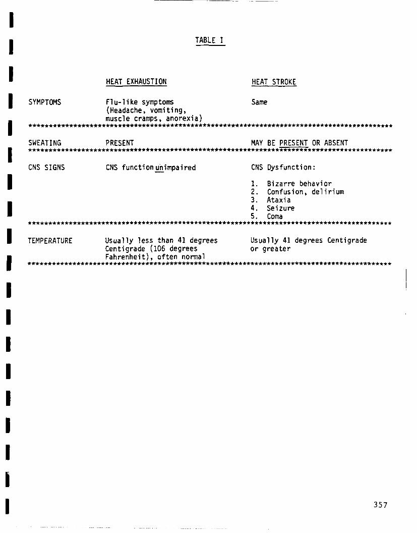
II TABLE I
I HEAT EXHAUSTION HEAT STROKE
I SYMPTOMS Flu-like symptoms Same(Headache, vomiting,muscle cramps, anorexia)
SWEATING PRESENT MAY BE PRESENT OR ABSENT
CNS SIGNS CNS functionunimpaired CNS Dysfunction:
1. Bizarre behavior2. Confusion, delirium3. Ataxia4. Seizure5. Coma
I TEMPERATURE Usually less than 41 degrees Usually 41 degrees CentigradeCentigrade (106 degrees or greaterFahrenheit), often normal
357

I04.)
.&J u -4-4 CE .04- 0
S 0 C * -u 0.6-1 Lac
(n CC c c O. 0C *J a) w c ul -4f
O uo~. CLQ wc os0 0s- w0 CP'J w u
c U) 0 ~.. M.>0
-w 3 C I- (n CL
u 0CC
04 0
~ C0. 0
Ul tC . 4 - *1 -iLU,
E a -
LI C(D aC
(0 *4 4
Li i C3( EL
-. a a0 L.4
LC ts 0
L C2
4 a C-4
0 0.
I- CC u1L i
Ina W

IHTWTRMCNERCAIEIS 4
JUYI19TRCECLFRI
35

I!II
INTRODUCTION
Whitewater rafting, kayaking, and canoeing have attracted a
growing number of participants in the past ten years. With the
present level of 200,000 whitewater enthusiasts growing by an
estimated 15% each year, it is not surprising that there has also
been an alarming increase in related accidents and deaths.
This article will review both the inherent risks manifest in
whitewater sports and its medical ramifications. Recommendations
for prevention and treatment will be discussed.
HISTORICAL OVERVIEW
I Whitewater boating as recreation began in the late 19th
Century, when early guides attempted to emulate Major Wesley
Powell's Colorado River expedition by rowing wooden boats down many
of the West's large rivers. Inflatable rafts became popular
immediately after the Fecond World War when military-surplus
I neoprene assault boats and life rafts became available for civilian
use. Less than twenty years later, while John F. Kennedy was
running the nation, his brother Bobby was running a raft down the
Colorado River. At that time, fewer than 500 people per year ran
the Colorado River through the Grand Canyon. Now, more than 500
I people a day travel the same route.361

I
Rafting did not become popular In the Eastern part of the IUnited States until the early 1960s. In 1968, commercially-guided
raft trips were offered for the first time on the New River in West
Virginia, while the Chattooga River in Georgia attracted many
rafters after the movie Deliverance was filmed there In 1971.
Today, even mountaineering and rock-climbing legends such as 1
Yvon Chouinard and Royal Robbins spend much of their time performing
first descents on rivers rather than first ascents on rock faces.
River running has been revolutionized by vast improvements in
equipment. Electronically-welded plastic is replacing rubber as
the primary material employed In raft construction--making rafts
lighter, stronger, and more easily repaired. The advent of the
light and maneuverable self-balling raft has made it common for Irafters to run rivers that prior to 1983 had been considered too
difficult and dangerous. This greater mobility has been paralleled
by a marked increase in the number of accidents far away from
modern medical care. IA major innovation in kayaking was the invention of the
plastic kayak, first manufactured by the Holloform Company In 1972. IToday it is rare to see anything but plastic kayaks except at
races, where fiberglass and kevlar boats are still popular. The
advent of plastic boats has resulted in a substantial Increase in 3serious injuries and deaths caused by vertical ginnino. boh ,
and e. IWhen a kayaker plunges over a drop, the front end cf the boat
can become trapped between rocks beneath the surface; this results
in what is called a vertical oin. Instead of breaking away as
362 I

fiberglass boats tended to do In the past, plastic kayaks remain
steadfast--or, worse, fold over on themselves, pinning the occupant
upside down underwater.
The term k c SbiD refers to the process whereby the
unrelenting force of water traps a craft against an obstacle such
as a boulder. This can cause a plastic kayak to fold or its front
deck to collapse, trapping the paddler's legs and preventing
escape. During the last two years, a number of kayakers have
suffered serious leg Injuries in this way, and at least three have
I died.
The sport's enormous popularity has led to the exponential
growth of professional guide services. In the face of increased
competition, guide services have been taking inexperienced clients
with little training and few skills down dangerous rivers farther
from medical care. Evidence of this alarming trend appeared on the
front pages of many newspapers last suinier when five major United
States business executives died after their raft flipped on the
Chilco River in British Columbia. The exact cause of their death
is unclear, but major contributing factors must have been their
I sudden and prolonged immersion in ice-cold glacier runoff water,
i compounded by a lack of sufficiently insulating clothing. One of
the survivors was quoted as saying, "We looked at whitewater as
sort of a roller coaster ride."
The dynamic and unpredictable nature of rivers can turn any
mishap into a serious disaster with little chance for recovery.
For this reason, our initial goal in whitewater medicine must be an
ever-present emphasis on safety precautions and accident prevention.
SAFETY EOUIPMENT
363

IAccording to the United States Coast Guard's boating accident
statistics, the three most common contributing factors to
whitewater-related deaths are cold water, inadequate swimming
ability, and failure to wear a PFD (personal flotation device or
life Jacket).
The Coast Guard classifies PFDs into five types; of these,
only two are commonly used in whitewater sports. Most paddlers
wear Type III devices, which permit mobility and comfort but
provide the minimum amount of flotation required for safety. Type
V jackets are predominantly used by commercial outfitters because Ithey provide more flotation and are asymmetrically constructed with
a collar theoretically designed to turn the unconscious wearer face
up.
Beyond flotation, life iackets have many other benefits that
make them highly useful in wilderness settings. They add extra Iwarmth and insulation to help prevent hypothermia, and they can act
as padding in falls on slick rocks. In addition, they are excellent
material for improvised splints. They can readily be fashioned
into cervical collars for suspected neck injuries, and can easily
be converted Into cylindrical splints for the knee.
Another vital piece of equipment is a safety rope. This
should be accessible in each craft, and secured in a manner which Ifacilitates rapid employment and prevents entanglement. In the
past five years, self-contained throw bags have virtually replaced
the once-popular coiled ropes. Throw bags, though they take less 3time to deploy, reduce control over the amount of rope thrown.
However, they are less likely to become loose and tangled during a Iraft flip.
364 I

Knives should be readily accessible. A fixed knife is
preferable to a folding one, in the event that only one hand is
free. Double-edged blades are preferred by many since they can cut
in any direction and thus require minimal handling in precarious
situations. Many modern knives designed for kayakers feature a
serrated edge which can cut through plastic boats during
entrapments.
Helmets should be worn by all whitewater boaters.
Approximately 10% of all kayaking accidents involve head or facial
trauma, as capsized boaters strike the rocks which are an inevitable
component of all rivers.
Whistles should also be worn; they enable paddlers to alert
others that an accident has occurred. Since paddlers are often
spread out over the course of a rapid, attempting to alert someone
downstream by yelling over the roar of the rapids can be a
frustrating and fruitless endeavor.
Functional, insulated clothing should also be considered a
mandatory safety item. Hypothermia is now recognized as a major
cause of death among outdoor travelers. More significantly, a
growing number of accidents are believed to have resulted from
hypothermia-induced impairment of Judgement and coordination.
Immersion hypothermia is a most serious threat to paddlers during
the Spring months when air temperatures are cooler and snow melt-
swollen rivers run very cold. Many rivers, especially in the West,
are controlled by dams which release water from far beneath the
surface and thus remain cold throughout the season.
Studies have determined that water conducts heat away from
the body 20 to 25 times faster than does air at the same
365

Itemperature. Placing adequate barriers between our bodies and the
environment is therefore of paramount importance in aquatic sports.
The insulating value of clothing is quantified by a comparative
thermal resistance unit termed the CL__factor. The CLO value of
cotton, for instance, is three times lower than that of wool or
polyester; cotton loses all of its insulating properties when wet.
On the other hand, newer synthetics such as polypropylene and
polyester pile absorb no more than 1% of their weight in water and
maintain their thermal insulating qualities even when wet. When a
nylon paddling Jacket is added, the combination of materials
affords both insulation and wind protection.
Wet suits at one time were considered optimal garments for Ipaddlers in extreme conditions. Today the dry suit has become the
gold standard for cold water boating. Dry suits, however, are
expensive and must be protected from ripping. 3RIVER HAZARDS
The most fundamental hazards found on rivers are hydra u
Other names for these phenomena are: pour-overs, holes, suck-holes, Ireversals, and rollers. A hydraulic is created when water flows
over an obstacle, causing a depression and producing a vacuum
within which the downstream water recirculates. Hydraulics can
provide enormous thrills and entertainment; they can also be Ideathtraps. Rafts and kayaks can be turned upside down by the
force of a hydraulic and, if the reversal currents are strong
enough, crafts and their passengers can become trapped in the
recirculatory flow.
The best way to avoid dangerous hydraulics is to scout the Irapid beforehand and to portage when necessary. Vhen proceeding
366 I
into a rapid which contains a hazardous hydraulic, always pre-set a
rope below the hole to facilitate rescue.
A swimming escape from a keeper hydraulic may necessitate
staying submerged and resisting the urge to regain the surface.
Surfacing too early can result in recirculation. Hydraulics
release water downstream from beneath the surface; often this is
the only avenue of escape for the swimmer. Fortunately, most
hydraulics eventually spit people out regardless of what action
they may take.
Novice paddlers often misjudge the force of hydraulics. It is
not the size of the drop which reflects the re:irculating potential,
but rather the shape and angle of the obstruction causing the drop,
combined with water volume and downstream currents.
While hydraulics may be the most obvious hazards, statistically
the biggest killers are undercut rocks and s.
Undercut rocks are boulders or rock formations which have been
eroded away just beneath the surface of the water. They pose a
significant risk of entrapment and drowning for both swimmers and
kayakers. Unfortunately, they are difficult to recognize from the
shore. A number of rafters tave died as a result of flipping on
undercut rocks and subsequently being pinned between craft and
rock. In February of 1986 a kayaker on a commercial trip on the
Rio Jatate in southern Mexico died when his upside-down kayak
washed onto an undercut rock, pinning him beneath the surface. It
took rescuers almost 24 hours to free the body.
Strainers--obstacles such as fallen trees, bridge debris, or
driftwood lodged between rocks--will allow water to pass through
but will trap the swimmer or boater. Strainers are notorious for
367

Ihaving caused the deaths of some of America's premier paddlers
while they were attemptinq to run rivers during floods. Flooding,
of course, increases the likelihood Lf strainers by washing 3riverbank debris Into the flow.
Yet another kind of entrapment can occur when a swimmer 1
attempts to stand up and walk in swift-moving current. A foot can
become wedged between rocks beneath the surface, causing the
unfortunate victim to lose his or her balance and fall face down
into the river. This type of mishap has caused drownings in water
that was less than three feet deep.
Swimmers in rapids should assume a position on their backs,
with their feet at the surface pointed downstream to serve as shock
absorbers. This minimizes the potential for foot entrapment as
well as head and neck trauma.
Entrapment accounted for a threefold increase in whitewater-
related deaths from 1985 to 1986. This statistic reflects more
than an increase in the numbers of whitewater boaters; it also 1
reflects the 1986 Western drought, which lowered river levels and 1exposed many more indercut rocks and hazardous areas.
Man-made hazards represent yet another threat to river
runners. Low-head dams or weirs form perfect hydraulics with
enormous recirculating r:*°enial. In the Binghamton Dam d'saster Iof 1975, a l. 1/2-foot Boston whaler with a 20 horsepower engine
was sucked into a hydraulic while attempting a rescue; this
resulted in the deaths of three fir.finhters. Other man-made
hazards include bridge pilings, submerged automobiles, and low-
hanging power lines. I1
368
I

IWHITEWATER FATALITIES
Although exact statistics are not readily available due to the
inconsistent collection of data by various agencies, we know that
most deaths occurring on rivers result from submersion accidents.
I Each year, accounts uf drownings and accidents are compiled by the
River Safety Task Force of the American Canoe Association;
unfortunately, many accidents go unreported and even some deaths
are not recorded.
We have seen that submersion accidents can result from a
variety of mechanisms: most commonly, underwater entrapment by
strainers, undercut rocks, hydraulics, vertical pins, or wedging of
feet. Avoiding entrapment, therefore, must be emphasized as a
safety measure, since rescue options are often limited by
constraints of time, resources, and ability to reach the victim.
Importaat preventative measures include:
1. Prior scouting of rapids
2. Pre-setting of safety ropes and rescue boaters
3 3. Swimming in rapids on one's back with feet downstream and near
the surface, thereby using the feet as shock absorbers and
minimizing the risk of foot entrapment
4. Questioning local paddlers about hidden hazards or going with a
I- guide who knows the river well
5. Rescuing swimmers before chasing down equipment
6. Avoiding r'vers when they are in flood or at an unusually low
level
7. Respecting the power of the river.
11 Regardless of the mechanism of thL submersion incident, once
* extrication has been achieved the gold standard for first aid is
369
I

still immediate and aggressive initiation of ventilation and
perfusion. Evidence of trauma should be noted and precautions
taken in the event of a cervical 5pine Injury. Hypoglycemia and 3seizures have contributed to drownings during whitewater activities;
these must be considered. Because apnea usually precedes cardiac Iarrest if ventilation is provided, circulatory resumption may occur
spontaneously. Supplemental oxygen should be provided as soon as
possible.
An important group of submersion accident victims who seem to
defy predictions for outcome are those who have experienced
prolonged submersion in very cold water. The designation "cold
water drowning" has led to some confusion since the mechanisms Uprolonging survival are not entirely understood. Nor has anyone
positively defined "cold water," There have been at least 19
reported cases of submersion estimated at greater than 15 minutes 3with good neurological outcome. Fourteen of these victims were
less than 19 years of age, and of the recorded water temperatures, iall were 100 Centigrade or lower. 3
Acute Submersion Hypothermia--defined as the rapid development
of hypothermia during fresh water drowning due to core cooling from 3pulmonary aspiration and absorption of cold water--has been
proposed by Alan Conn, an anaesthesiologist at Toronto Hospital for ISick Children, as an explanation. If enough cold water were 3aspirated, cerebral cooling might occur prior to circulatory
collapse, thus delaying permanent brain injury. Whatever the 3mechanism behind prolonged survival, the overt implication is the
value of prolonged CPR after submersion in very cold water. II
370
I

There is no logic in attempting to drain water from the lungs
in fresh water immersion since the water has rapidly been absorbed
into the circulation. The Heimlich maneuver is not recommended
unless there is no response to ventilation due to an obstructed
I airway.
TRAUMA
One of the most common traumatic injuries occurring in
whitewater sports is the sprained ankle. Ironically, this usually
occurs off the water--while one walks along the banks on loose,
wet, slippery rocks during scouting or portaging, or upon entering
or leaving the river. Ankle injuries also occur on the river when
rafters are thrown about in rapids. Twice I have suffered foot and
I ankle injuries when the person sitting across from me was tossed
into the middle of the raft onto my leg, which was stretched out in
a bracing position. Kayakers are prone to ankle injuries from
sudden inversion or dorsiflexion when the bow of the boat hits an
obstruction such as a submerged rock. The feet are held against
the narrow braces while the heels are pushed underneath or the
entire ankle is inverted.
Management of foot and ankle injuries encompasses the triad of
ice, elevation, and compression to reduce swelling. River water is
usually substituted for ice. Splinting is important for reduction
of pain and edema and to limit extension of the injury. At one
time most rafting companies carried pneumatic splints in their
first aid kits. While providing adequate support, protection, and
compression, air splints are also liable to overinflate when
heated by the sun. Their zippers often malfunction as a result of
rusting from moisture or Jamming by sand particles. Neurovascular
371

1integrity must be checked frequently with this type of splint. I
prefer either to improvise a pneumatic splint from my kayak float Ibags, or to fashion a "pillow splint" from an extra life Jacket or 1an article of clothing.
Head, facial, end dental trauma are more common in kayakers
and decked canoeists than in rafters because of the potential for
flipping upside-down while still in the craft. Minor abrasions, 3lacerations, and contusions are common; serious head injury with
loss of consciousness is fortunately rare. Head and facial trauma 1can be minimized by wearing protective helmets and tucking forward 3while rolling, instead of leaning backward. Cervical spine injuries
accompany 5 to 10% of serious head trauma and should always be
considered in the assessment.
Most kayakers suffer frequent abrasions and contusions to the
fingers and knuckles of the hand while hanging upside-down after
flipping. Oar frames, oars, paddles, and the ammo boxes which are Iused to keep supplies dry can all inflict injury when rafts are
capsized or tossed about in turbulent water. Significant injury
can occur when a swimmer is suddenly sandwiched between a downstream 3boulder or obstruction and the upstream raft, canoe, or kayak that
he or she has exited. The rise in popularity of kayak wave-surfing Iin the ocaan has increased the number of incidents in which paddlers
and surfboarders alike have been speared by the front of a kayak.
Visceral injury is possible and I have seen one case of a
pneumothorax by this mechanism. Less serious injuries such as
fractured ribs and chest wall contusions frequently result, 3probably because padded life jackets offer some protection.
I372 1
I

Back injuries from strain and overuse are frequently seen among
whitewater paddlers. Rafters are prone to such injuries while
portaging, pushing stuck rafts off rocks, or carrying crafts to and
from the river. Raft guides frequently suffer back strain when
pulling capsized customers--who usually weigh more than the guides
themselves--back into the rafts. The awkward task of bending over
a bulky side compartment and lifting a heavy Individual out ot the
water produces enormous stress on the lumbar musculature.
Unfortunately, the construction of rafts tends to limit support
from the legs while lifting. Kayakers and canoeists more commonly
injure their backs when lifting water-laden boats to empty them, or
l when loading craft onto automobile roofs. Using the legs for
lifting, and sharing the load with another individual, will reduce
the likelihood of such injuries.
1 The repetitive dorsiflexion of the dominant wrist required to
operate feathered kayak paddles often produces tendonitis and
synovitis. Using a paddle constructed with a 750-800 offset
instead of the traditional 900 can limit stress to this joint.
Aspirin or other noiAsteroidal anti-inflarnmatory medications taken
30 minutes prior to paddling have been found to be beneficial.
Wrist supports provide limited relief.
i The injury most associated with kayaking Is the anterior
shoulder dislocation. According to one survey of Eastern kayakers,
over 10% had suffered at least one shoulder dislocation. The
maneuver most notorious for precipitating this injury in kayakers
is the high bra-j. Often used while supporting oneself in
l hydraulics, surfing on waves, or preventing capsizing, the high
brace entails abduction of the humerus with external rotation of
373
I
1the glenohumeral Joint. If the arm should become extended behind
the midline plane of the body either by the force of the current or
when the paddler lifts the head and turns the body away from the 3bracing paddle, the triad of forces leading to an anterior
dislocation are produced. The humeral head is driven forward and
frequently avulses the cartilaginous glenoid labrum and capsule
from the anterior aspect of the glenoid cavity. The paddler will Iusually be aware that something has "gone out of place" and will Ihold the extremity away from the body, being unable to bring the
arm across the chest. The shoulder may appear square due to the
anterior, medial, and inferior displacement of the humeral head
into a subcoracoid location. 1
Prevention: The preferred method of bracing which minimizes
the risk of shoulder dislocation is the l in which the arm
is held in internal rotation and close to the body (adduction). 3This type of brace, while initially more awkward for the novice
paddler, is inherently stronger and more versatile as it allows back Ipaddling out of hydraulics. 3
The shoulder joint itself depends for its stability upon the
joint capsule and surrounding musculature. Exercises which 3strengthen the rotator cuff, deltoids, triceps, and pectorals dill
inherently reinforce the glenohumeral joint. IT After the shoulder has been dislocated, the longer 3
it remains out, the harder eventual reduction will be due to
increasing muscle spasm. Although on-scene reduction of 3dislocations is still controversial, the immediate relief of pain,
curtailment of ongoing injury to the Joint, and subsequent ability 1of the victim to function more actively in facilitating his or her
374
I

Iown evacuation are all strong reasons in favor of the attempt. Many
techniques have been advocated for reduction. The key element is
the ability to maintain traction-countertraction while maximizing
muscle relaxation in an improvised setting. Always monitor
circulation, motor and sensory function to the hand before and
after attempting reduction.
Blisters are a ubiquitous problem in paddlers. Among kayakers
they develop most often between the metacarpalphalangeal (MCP)
Joint and the interphalangeal joint of the thumb along the ulnar
aspect. Prior taping and eventual callous formation will reduce
the incidence of this potentially incapacitating problem. Common
sites of blister formation in rafters and canoeists are the proximal
palmer surfaces of the MCP Joints of the hands.
Infections: Blisters, along with abrasions and lacerattons,
are always at increased risk of becoming infected in an aquatic
environment. Maceration from prolonged immersion in water and
exposure to pathogens are predisposing factors. Abscess formation
and cellulitis requiring incision, drainage, and antibiotics are
common during expedition boating when participants spend extended
I periods on the river. A recent study of the bacteriology of the
freshwater environment published by Paul S. Auerbach, M.D. in T
IAnnals of Emergency Medicine yielded a diverse collection ofi pathogens including Aeromonas, Pseudomonas, and Vibrio species.
Antibiotics that were found to be effective against these gram-
negative organisms included: Trimethoprim-Sulfamethoxazole,
Tetracycline, Ciprofloxacin, Imipenem, and Ceftaz.dine.
375

ifirst-generation Cephalosporins, Ampicillin, and Erythromycin all
provided poor coverage'.
An outbreak of Staphylococcus Aureus skin infections among 3raft guides in Georgia and South Carolina nearly led to the closing
of two rafting companies. Sharp grommets on the thwarts of the
rafts had caused repeated lower-extremity abrasions. The causative
organism was cultureable from rafts up to 48 hours after use.
Daily raft disinfecting became a standard practice which kept the 3companies from being forced to close.
Otitis externa (swimmer's ear) is a common problem among
paddlers. Water exposure to the ear canal both macerates the
epithelium and neutralizes the normally acidic pH of the canal,
predisposing to infection. The bacteria most commonly cultured are
Pseudomonas Aeriginosa, Proteus Vulgaris, and Staphylococci.
Purulent discharge from the external auditory canal in a patient,
with pain aggravated by traction on the auricle or pressure on the
tragus, will confirm the diagnosis. Antibiotic ear drops with or 3without hydrocortisone are widely available and very useful.
Irrigation of the canal with commercially-available solutions
containtng acetic acid and alcohol will help prevent infection by
lowering the pH and drying the canal. Tie drops should be applied
after each outing. IThe recent publicity given to water contamination by Giardia 3
is now reinforced by statistics from the Federal Centers for
Disease Control which report Giardia Lamblia to be the most common 3pathogenic intestinal parasite in the United States. Giardia cysts i
'Auerbach, P. S. et al. 1987. Bacteriology of the freshwaterenvironment: implications for clinical therapy. Ann. Emera. Med.16:1016-1022.
376I
I

flourish in mountain streams and rivers which were once considered
sources of pristine water. They even thrive in very cold water and
lend no taste or smell to the water they infest. Rivers are usually
contaminated by animals that defecate in or near the water.
Studies by the Wild Animal Disease Center at Colorado State
University have identified more than 30 species as carriers.
Physicians should be aware that onset of symptoms may occur one to
three weeks after ingestion, and should not overlook this diagnosis.
Whitewater rafting in foreign countries is becoming
increasingly popular as adventure travel companies promote more
international trips. Unprepared participants are thus exposed to
infectious diseases not encountered in the United States. This was
exemplified by a recent report of Schistosomiasis in a group of
eleven rafters on a Sobek Expedition in Ethiopia. Schistosomiasis
is endemic to large areas of Africa, South America, and the
Caribbean, and is transmitted to humans who swim or come into
contact with fresh water containing the larval stage. Rafters going
to areas in which this disease is endemic should be screened with
serologic testing, since up to 50% of infected cases can remain
asymptomatic.
ENVIRONMENTAL HAZARDS
Whitewater participants are exposed to an array of
environmental hazards, most of which can ba readily avoided or
prevented. Sunburn and other effects of solar radiation are
magnified by the water's ability to reflect up to 100% of the UV
light, depending on the time of day. Sand can reflect up to 17% of
harmful UV radiation. The "paddler's tan," outlined by the life
jacket, is not only painful but can predispose to neoplasms from377

Irepeated exposure. Sunscreens must be applied frequently, as they Iare prone to washing off in the water. PABA-containing sunblocks,
applied a couple of days prior to exposure, will absorb into the
epidermis, affording a cumulative and more stable protective
barrier. Zin, oxide and other agents more resistant to water are
preferable.
Eye protection, often overlooked by paddlers, is important to Ishield from the chronic effects of UV exposure as well as the more
acute problem of photokeratitis. Most rivers are situated in the
mountains, where every additional 1000 feet of altitude gained
increases the amount of UV exposure by 3 to 4%. Polarizing lenses
are helpful in reducing glare from the water but are not by Ithemselves adequate filters of UV and infrared radiation.
The second most common environmental affliction suffered by
paddlers is the dreaded "weed." Rhus dermatitis from poison oak or
poison ivy is especially ubiquitous in the Spring, when the vine
itself is very potent but the characteristic leaves have not yet Uappeared--making Identification of the plant difficult. The 3unfortunate paddler is not often aware of his or her exposure until
days later when the characteristic dermatitis develops.
Venomous snakes--most notably the pit vipers--along with
scorpions, spiders, and fire ants, are frequently encountered by Iriver runners and should be considered a potential hazard.
Reported cases of envenomation, however, are rare.
The beauty and adventure offered by our nation's rivers,
combined with opportunities for spiritual growth, camaraderie, and Iphysical and mental challenge, are luring greater numbers of
378
I

people to participate In the sport of whitewater boating. Becoming
more familiar with the inherent dangers and medical ramifications
of the sport will aid physicians both in advising patients about
accident prevention and in treating their Injuries.
379

osc
LU
C CL. (a C40 V 4) 0) 6
.j x 4)Cl) Q >
0
E
-0 E Z(Do4) 10 c
11v o
0 0 x
LU
rl.n Irv

0 ~ a
00
000
01
381

*000
Lid0
..0.....
-coI0 p/

ii
i AVALANCHE RESCUE
Search Dog, Probe and Beacon Techniques
W. Wayne Flann BBAAvalanche ForecasterRescue Specialisti
IiiIii
Wayne Flann Blackcomb Skiing Enterprises P.O. Box 98
Whistler British Columbia Canada VON IBO
383

IINTRODUCTION
IAvalanche rescues, for the most part are associated with
body recovery. Many buried -victims do not survive, but
rescue attempts must be performed, for there is always hope! IThe primary cause of death is suffocation, followed by
collision with obstacles! then hypothermia and shock.
Chances of a live recovery diminish rapidly with time and
depth of burial. Fifty percent of buried victims perish I
within thirty minutes. This figure includes persons buried
in buildings and vehicles. IA quote form the book Kampf uber die Gletschern (Battle over
the Glaciers) by W. Schmidkunz says all, "The Snowy Torrents
are like the deep sea; they seldom return their victims
alive." I
Avalanche rescue operations are only effective with a
written plan, adequate equipment, and organized trained
rescurers. Persons pursuing backcountry travel in avalanche Iprone areas should be equipped for self rescue. Organized
rescue groups are usually too late for a live recovery.
The following tables from the Avalanche Handbook summarize
survival and causes of death in an avalanche.
III
384
I
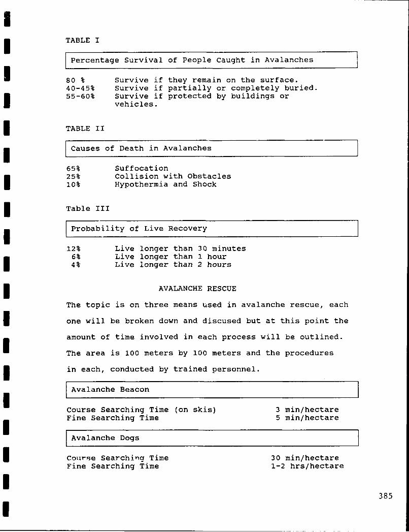
TABLE I
Percentage Survival of People Caught in Avalanches
80 % Survive if they remain on the surface.40-45% Survive if partially or completely buried.55-60% Survive if protected by buildings or
vehicles.
TABLE II
* Causes of Death in Avalanches
65% Suffocation25% Collision with Obstacles10% Hypothermia and Shock
Table III
Poility of Live Recovery
12% Live longer than 30 minutes6% Live longer than 1 hour4% Live longer than 2 hours
AVALANCHE RESCUE
The topic is on three means used in avalanche rescue, each
one will be broken down and discused but at this point the
amount of time involved in each process will be outlined.
The area is 100 meters by 100 meters and the procedures
in each, conducted by trained personnel.
SAvalanche Beacon
Course Searching Time (on skis) 3 min/hectareFine Searching Time 5 min/hectare
Avalanche Dogs
Course Searching Time 30 min/hectareFine Searching Time 1-2 hrs/hectare
385
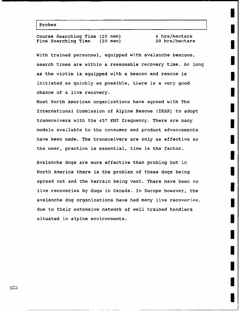
IProbesICourse Searching Time (20 men) 4 hrs/hectareFine Searching Time (20 men) 20 hrs/hectare
With trained personnel, equipped with avalanche beacons,
search times are within a reasonable recovery time. As long ias the victim is equipped with a beacon and rescue is
initiated as quickly as possible, there is a very good
chance of a live recovery.
Most North American organizations have agreed with The
International Commission of Alpine Rescue (IKAR) to adopt itransceivers with the 457 KHZ frequency. There are many
models available to the consumer and product advancements
have been made. The transceivers are only as effective as
the user, practice is essential, time is the factor. IAvalanche dogs are more effective than probing but in
North America there is the problem of these dogs being
spread out and the terrain being vast. There have been no
live recoveries by dogs in Canada. In Europe however, the iavalanche dog organizations have had many live recoveries.
due to their extensive network of well trained handlers
situated in alpine environments.
III
I

In reccnt years, The Banff National Park wardens and dog
handlers in the Whistler area have used the Helicopter
Flight Rescue System (HFRS) to deploy dog and handler onto
slide paths. The HFRS system uses a fixed line attached to
the under carriage of a helicopter to transport the rescue
party. This procedure is timely and reduces the risk of
exposing other rescuers to further hazards.
It is important to remember that burial sites should be
kept as scent free as possible if a dog is being used.
Probing is the least desirable means of recovery. It
requires 20 rescurers to be effective and is a slow and
tedious procedure. Probe lines must be managed in an
orderly and systematic fashion. For further information
I consult the Avalanche Handbook.
IIIIIIII
387I

I
REFERENCES II
Ronald I. Perla and M. Martinelli Jr. Avalanche HandbookU.S. Dep. Agric. Handb. 1975
David McClung and Peter Schaerer Avalanche Handbook I(Book in Draft) 1991
E. R. LaChapelle The ABC of Avalanche Safety IThe Mountaineers 1985
IIUIIIIIIIIII
388
I

Field Recognition and Managementof Exotic Snake Envenomation
Michael V. Callahan, MSPHDivision of Geographic Medicine
Tufts-New England Medical CenterBoston, Massachusetts, U.S.A.
OBJECTIVES
Following this presentation, participants will be able to:
1. Describe the taxonomy and natural history of venomous snakes.2. State factors that increase the risk of human-snake encounters.
3. Delineate methods for preventing snakebite in the wilderness.
I4. Describe appropriate treatment for the envenomated patient in
the wilderness setting.
II
389
I

IIntroduction;Snake venom poisoning is a medical emergency that requires sound judgement, accurate
assessment and definitive treatment with appropriate antivenin. Managing these injuries inaustere and wilderness environments is, with few exceptions, limited to first aid measuresthat vary with the type of snake. Many first aid measures previously recommended are nowknown to be useless or even detrimental to the patient. In all cases of confirmed snakeenvenomation in the backcountry, the priorities are basic life support, restriction ofsystemic spread of venom, and expedient evacuation of the patient to a hospital forimmunotherapy with appropriate antivenin.
Epidemiology,Venomous snakebite continues to be an important medical and economic problem fordeveloping countries. Underreporting of snakebite occurs in many countries due tcdependence on hospital admission reports (which are biased in favor of severe cases). Inone survey in northern Nigeria only 8.5% of people bitten by cobras sought medical care1 .Mortality due to snakebite is similarly underestimated due to reliance on hospital records. InIndia the reported number of annual fatalities from all provinces has exceeded 20,000 over
the last number of years 2. Brazil reports over 2000 deaths per year from all species 3 .Snakebite is responsible for 5% of deaths among the Waorani tribes of eastern Ecuador and I2% of the Yanomamo of the southern Amazon basin 4. Snakebite is the fifth most commoncause of death in Burma 5. In the United States there are over 7000 venomous snakebitesrecorded each year with 9-14 deaths6 . INatural History;An understanding of taxonomy is important for the treatment of reptile envenomationbecause venoms from reptiles of the same genus often produce similar clinical effects.Secondly, scientific names for species are important because they are employedinternationally to describe specificity of many antivenins.
Approximately 550 of the 2787 currently known species of snakes, are known to bevenomous. The venomous snake families are the Elapidae, including the cobras, mambas,and kraits; Hydrophobidae, including Australian terrestrial snakes and seasnakes:iViperidae, the vipers and pit vipers; and Atractaspididae or "asps", which were previouslyclassed with the Viperids. Over 200 species belonging to these families have causedhuman deaths. Over 45 species of a fifth family, the Colubridae, are now known to becapable of human envenomation.
Venomous reptiles are distributed throughout the world. Venomous snakes are found as farnorth as the arctic circle (Vipera anmalon, V. b. beres) and at altitudes exceeding 5100 m I(Agkistrodon himalayanus). There are no venomous snakes in New Zealand, Ireland,Madagascar, Atlantic, many Carribean and Pacific islands and polar regions.Reptile activity patterns are dictated by the behavior of each species, temperature, circadianand diurnal rhymes. All venomous snakes are thigmotrophic to some extent, preferring toconceal themselves from predators through protective coloration and hiding. Prey animalsare caught by questing (sea snakes, certain elapids) or by ambushing food species from asecure hiding place (certain viperids). Changes in populations of venomous snakes occur Inaturally with changes in prey animal populations and climate, or through humanencroachment and development of wilderness areas. In particular, flooding, and suddenchanges in ecotone (e.g. deforestation) have been attributed with displacing venomoussnakes-often with increases in the number of snake bite. In Burma peak incidence of snakebites occur in June when rice is planted and in November when it is harvested 5. Theaggressiveness of each species also plays a role in the incidence of snakebite. Rattlesnakes,cobras, and mambas and members of the Atricaspididae strike readily when cornered,
390 II

Iwhile coral snakes, kraits, sea snakes and the Gaboon Viper and it's relatives (Bitis) appearreluctant to strike. In developing countries the majority of snakebites result frominadvertently disturbing the snake, usually by stepping on the animal in darkness.Encounters such as these, where the victim is ignorant of the snake's presence are referredto as legitimate bites. In contrast, 30% of venomous snakebites in Alabama are illegitimatebites, that is, the snake is known to be venomous prior to the bite, and over 50% ofsnakebites in adults are associated vith alcohol consumption 7.
I Prevention;Prevention of snakebite is insured through common sense and simple precautions(resources that may be in short supply among certain wilderness travellers). Snakes shouldnot be disturbed or handled without first determining that they are nonvenomous. Indiv-iduals insistent on approaching snakes in the wild should first learn about the naturalhistory of local species from a responsible source. Never grab exposed tails in -ock screeor leaf litter without first determining where the animal's head is located. Many allegedly"dead" snakes have regained their senses while being handled, or in the emergencydepartment. "Rules" for identifying venomous snakes in one region may prove inaccuratein other regions. Appropriate clothing should be worn in snake infested areas, includingbaggy trousers, stout boots and socks. Flashlights should be used after dark, and whencollecting firewood in dense cover. A long walking stick can be used to probe heavyundergrowth and loose sand prior to passage. When travelling through low canopy, lookahead for the dangling vine-like representatives of the genus Dispholidus (Boomslang) andThelotornis (vine snakes). Rock climbers should be cautious of the presence of snakessunning themselves on overhead ledges. Fisherman should wear stout rubber gloves andboots when emptying nets. Swimmers should avoid overgrown beaches and bodies ofwater with floating aquatic plants. Undercut rocks and logs should first be inspected beforedislodging them. Particular care should be taken in the days following heavy rain, andearthquakes as snakes may have been driven from their hiding places.
Envenomation kinematics;Snakes do not have to be coiled to strike. Lateral strikes by extended snakes and bites from"cold" or torporous snakes have resulted in envenomation. Given sufficient warning,elapids (mambas, kraits, cobras) rear up to 1/3 of their length above the ground. This canpiuv ,,u Lxciting vvhei tie serpent -:, 3n, cobra (Ophiophagus hannah). Elapid strikesstart high and end low, resulting in a high percentage of bites to the lower extremity.Defensively posturing Viperidae display lateral coiling of the forward body, often with thehead and neck held off the ground. This defensive position produces faster strikes andgreater range. Certain species of pit vipers are particularly pugnacious, strikingIaggressively ard repeatedly over half their body length.
Venom is introduced through wounds made by chewing, or directly through specializedfangs, canaliculated or grooved teeth. Members of the Viperidae family, subfamiliesCrotalinae (e.g. rattlesnakes) and Viperinae (e.g. old world vipers), have hinged fangs. Atrest the fangs are covered by a protective sheath and lie flat against the divided mixilla. TheElapidae, including the cobras, kraits, mambas and seasnakes of the genus Laticauda, andthe Hydrophobidae, including venomous Australian snakes and other sea snakes, allpossess short, non-retractable front fangs. Evolution of fangs varies from enlargedcanaliculated posterior maxillary teeth in the genus Rhabdophis (yamakagashi), to fixedrear fangs in Dispholidus (boomslang) to hollow retractable fangs of the viperidae.Viperidae and Atractaspididae are constantly replacing fangs. Often one to as many as fourfangs are functionally positioned. The presence of more than two fang marks at the bite sitedoes not necessarily indicate multiple bites.
I391

IWith the exception of colubrids, all venomous snakes produce venom in specialized glandslocated below the eye and spanning posterior to the mandibular joint. Venom is forced fromthe glands by compressor muscles through specialized ducts which transverse the mucosaand terminate at the anterior base of the fangs. Venom travels the length of the fang inpartially or completely enclosed canals, exiting through an opening near the tip. In certainspecies of cobras, the canal opens into a groove which allow these species to spray a fine Imist of venom during exhalation. Venom can be sprayed or "spat" up to 2.5m, and is usedfor defensive purposes. Rear fanged representatives of the Colubridae produce venomtoxins Zn a modified region of the superior labial gland known as the Duvernoy's gland.During envenornation venom is discharged and transmitted to the bite wound throughgrooved teeth. In order to introduce sufficient venom Colubrids must hold on and chew thetissue.
Not all bites by venomous snakes are accompanied by envenomation. The bite and thedelivery of venom are independently innervated events. This may explain the highpercentage of trivial or asymptomatic bites from certain species. Approximately 20% ofCrotalus and Bothrops bites 6 , 50% of C. rhodostoma and V. russellii bttes5 and over80% of sea snake bites are accompanied by trivial or no envenomation 8 .
Snake venom biochemistry;Snake venom, as with snake behavior and environmental adaptation, attests to theremarkable evolution of these animals. The eastern diamondback rattlesnake (C.adamantus) for example, possesses a venom particularly toxic to rabbits, the preferred Iprey. Similarities between snake venoms, and therefore certain clinical effects produced bythese venoms, correlate reliably with genera level of classification. However, the age,geographic location, season and even the sex of snakes are known to influence the toxicityof venom.
All snake venoms are comprised of numerous enzymes, polypeptide toxins, and glyc-proteins Many venoms contain biogenic amines such as 5-hydroxytryptamine andhistamint. These small molecular weight agents are believed responsible for the painimmediately follov, ing envenomation by viperidae and certain elapids. Snake venoms areoften described as e;tl,,r neurotoxic or hemotoxic; a oversimplification that neglects the Imultiple targ, ., a; '-1 , ierty of the venoms. For example, verom from cobras andAustralian snakes !irc known for their neurotoxicity, yet bites from these species also causedevastating tissue necrosis rivaling that observed in viper bites. Conversely, severalcrotalids such as C. durissus terrificus , C. scutulatus and V. russelli spp. possesspotent neurotoxins. Several medically significant toxins found in snake venoms are listed inTable 1.
Pathophysiology of snake venoms;Envenomation in humans is an aberrancy of the effects observed in small prey. In general,the greater mass and blood volume of humans seems to be beneficial for surviving the acuteeffects of the venom. For example, untreated human victims of C. adamantus (E.diamondback) envenomation may survive the massive intravascuiar coagulation that killsthe rabbit, but may die days later from hemmorhagic shock or cerebral hemorrhage.Pathophysiology of envenomation may be abstracted into three concurrent, consecutive Iand/or synergistic actions; These are
1.) injury caused by the direct effects of venom2.) autopharmacologic effects due to the release of
inflammatory mediators and vasoactive compounds.3.) secondary complications such as infection and disabling
sequelae.
392 i
I.

II. i_.,jaevenoms;AnP amately 85% of Viperidae venom by weight is water. Viperidae venoms also contain
a enzymes , 2-16 non-enymatic peptides, and low molecular weight substancesincluding amines 9 ,10 . Viperidae toxins show no particular affinity for plasma proteins andin the case of subcutaneous bites, tend to be spread by lymphatics. There is considerablevariation in tissue affinity and eventual elimination of viperidae venom components.Agkistrodon (cottonmouth) venom is bound to lung tissue and several Crotalus(rattlesnake) venoms have been shown to be concentrated in the liver and excreted in thebile9 .
Many viperidae peptides lyse cell membranes and damage the vascular endotheliumpermitting vascular fluid exudation into surrounding tissue. Progressive plasmaextravasation into tissues and vasodilation induced by venom kininogenases, histamine andprobably cytokines, lead to hypotension, hemoconcentration, and acidosis. In unpublishedobservations, our laboratory has measured high levels of the "shock cvtokines" TNF-cu
and IL-lP in rabbits artificially envenomated with Crotalus venoms, and in plasmasamples front patients envenomated by pit vipers. Viperid venoms also contain significantamounts of phospholipase A2 enzymes. which induce hemolysis through the release oflysolecithin.
Viperidae venoms also induce coagulopathies. These clotting aberrancies are the result ofmultiple sites of enzyme activity and include platelet aggregation, thrombocytopenia.hypofibrinogenemia, DIC, and myriad DIC-like syndromes 11 Several species ofvipendae also possess neurotoxic peptides such as crotoxin of C. durissus terrificus (So.American rattlesnake) and Mohave venoms A and B of C. s. scutulatus (Mohaverattlesnake).
II. Elapidae and Hydrophobidae venom;The toxic polypeptides of the Elapidae (cobras, mambas, kraits) and Hydrophobidae(seasnakes and Australian snakes) are small molecules that are rapidly absorbed into theblood stream. The principle toxins of medical importance are small molecular weightpolypeptide neurotoxins. Elapidae neurotoxins can be divided into two groups: 1.) thedisulfide bridged peptides which include the cell membrane toxins and postsynaptic (X-
neurotoxins, and 2.) the phospholipase A2 toxins, including presynaptic 3-neurotoxins andmyotoxins12, 13 . Many elapid and hydrophobid venoms also contain acetylcholinesteraseactivity.
Several Elapidae and Hydrophobidae venoms induce tissue necrosis. In particular, bitesfrom Naja n. nigricollis (Black-necked Spitting cobra), and Hydrophobidae (Australiansnakes and beaked sea snakes) cause extensive tissue injury. Venom from Hydrophobidaeand many species of elapidae induce generalized rhabdomyolysis, resulting inmyoglobinurea and danger of renal failure. Several important Elapidae and Hydrophobidaetoxins, and the species that produce them are listed in Table II
Clinical presentation:The victim of snakebite may prescnt with symptoms resulting from damaging "first aid"measures, from emotional anxiety, or from th" actual effects of the venom. Anxiety andfear can smoke screen accurate assessment and interfere with management. Tourniquets canocclude blood flow to a bitten extremity resulting in numbness, tingling, pain, congestion,swelling, and loss of distal pulses. Cruciate incisions, scarification and currettage can cause
393

istructural damage to underlying nerves and vessels leading to uncontrolled bleeding, lossof function, secondary infection. Cryotherapy may provide pain relief, but often at theexpense of increased tissue damage. Electrical shock therapy for snakebite has no scientificor medical basis, but has rekindled the depressed "stun gun" market in the United States.
Unabaring pain , edema and the presence of one or more fang marks provide the mostreliable indication of Viperid envenomation. Cobra and mamba envenomation (Elapidae)also produces pain and variable edema, but fang marks may not be obvious. Localervrhema and swelling become noticeable within minutes following bites by the Viperids,Australian snakes, cobras (Naa) and mambas (Dendroaspis). Care should be takenconcerning bites by PanAmeiican coral snakes (Micrurus and Aicroroides) and Africanand Asian kraits (Bungarus) because of the localized anaesthetic effects of the venom.Krait and coral snake venoms produce little local change, and the first indication of Ienvenomation could be a systemic symptom. Blood may be totally defibrinogenated in 15
minutes following bites by the Malayan Pit Viper C. rhodostorna 14 and respiratoryparalysis present 30 minutes following bites by mambas and cobras. There are recorded Ideaths within 20 minutes following bites by cobras, mambas and Australian snakes. Thesecases are, however, exceptional, the usual time for death being hours follow, ing Elapidaeand Hydrophobidae envenomation, and days following Viperidae envenomation 2, 15 3Multiple assessment schemes are used to grade the severity of envenomation. The simplestand most applicable scheme was described by Russell (way back when) for pit viperenvenomation. This scheme seperates the severity of envenomation into trivial; minimal.involving swelling and echvmosis at the bite sight; moderate, involving the extremity; and Isevere, the presence of any systemic symptom (e.g.perioral paresthesias, changes inclotting values)
Death from Viperid venoms is usually due to circulatory collapse associated with a drop inblood volume due to extravasation through capillary endothelium and pooling of blood inmicrovasculature. Death from Hydrophobidae and Elapidae envenomation is usually due torespiratory paralysis induced by curariform effects on neuromuscular junctions.
Wilderness ManagementFirst Aid: iGet the victim away from the snake. Attempt snake identification in regions wherespecies-specific (monovalent) antivenin is used, and where Viperid and Elapid speciesoverlap. Sacrifice the snake only when I.D. is questionable and when it can beaccomplished safely.
Manage ABCs. Management of airway, breathing and circulation takes priority.Obstruction of the upper airway by the tongue or inhaled vomitous occurs in envenomation lby Australian snakes, inambas, kraits and several species of cobras, and can precipitaterespiratory arrest. Breathing difficulty has been recorded in less than 1 hr followingenvenomation by a cobra 16 . In unresponsive patients, airway can be managed by proper ipositioning of the head, and positioning the patient on their side. In anaphylactic reactions.and bites that cause swelling of the upper airway, an oralpharyngeal airway (if available) orairway improvised from available materials can be used. In elapid and hydrophobidenvenomation, respiratory paralysis may develop with intercostal muscles affected first. Inthe absence of antivenin and anticholinesterases, patients supported by manual ventilationor ventilator have recovered sufficient diaphramatic movement to bread on their own in 1to 4 days 15 ,20 . Several ambubags (BVMs) such as the Svenchen (Zurich, Switzerland) Ihave been developed for use in extended field care . The Svenchen BVM weighs .4 kg andhas sufficient volume for hyperventilation of adulhs.
3 94 i
I

IAssess the patient. Calming the patient reduces faintness and hyperventilation whichhinder accurate assessment. Fang marks, pain, swelling, erythema, or in elapids, tinglingat the bite sight, or the presence of any systemic symptoms not attributed to excitation,indicate envenomation. Assuring the patient may also decrease heart rate, with the potential
I! benefit of slowing the spread of venom.
Immobilize the bitten extremity. Animal experiments have demonstrated thatimmobilization of the envenomated extremity delays systemic spread of crotalid and elapidvenom 16, 17 . Immobilization is effectively accomplished in the wilderness by improvis-ation. Excellent immobilization is accomplished using SAM splints TM, internal frame staysfrc .n packs and crevat sling and swaths. EVA pads can also be tailored to immobilizeextremities. Patients with bites to the abdomen, neck and face should be backboarded ifpossible. Spine immobilizers improvised from external pack frames or KEDT extricationdevices are suitable for this purpose.
Initiate mechanical suction over the fang marks. The ExtractorTM (SawyerProducts, Safety Harbor, FL, USA) is a portable pump that generates up to oneatmosphere of negative pressure. Animal studies using the Extractor have demonstrated upto 30% removal of crotalid venom proteins if used within three minutes following thebite 18 . In other animal studies using C. horridus horridus and C. horridus atricaudatusvenom, suction continued to remove radiolabelled venom until swelling of tissuesprevented an effective seal between the pump bell and skin 19. Rubber suction cups such asthose included in the Cutter Snakebite Kit (Cutter Industries, Newark, NJ) are compactalternatives but lack the high suction pressures produced by the Extractor. Suction units canbe improvised in the wilderness by cutting a large irrigation syringe (60 cc) with a heatedknife. The suction seal is improved by shaving the area around the bite and coating thesuction cup with a small amount of petroleum-based ointment. Viperid bites leak blood andserosanguinous from the fang marks. Be prepared to empty fluid from the suction bell andUreapply suction regularly. The Extractor may prove effective for the removal of elapid andAustralian snake venoms as well. The shorter fangs of these snakes result in the superficialdeposition of venom-potentially resulting in a more efficient retrieval of venom withnegative pressures. There are numerous cases of oral suction being used on snakebite asare there cases where the individual providing the suction experienced mild symptoms ofvenom poisoning. The risk-benefit associated with oral suction remains controversial.
. Pressure wraps and lymphatic-occlusive dressings;A circumferential pressure wrap has been shown to delay the spread of hydrophobid(Australian snake) venom from the bite site. This technique involves the application ofcrepe or elastic bandage directly over the bite, and continuing the wrap proximally up theextremity. The extremity is then splinted as described above. The principle for thistechnique is attributed to compression of subcoutaneous microvasculature and lymphatics,thereby compartmentalizing venom from spreading 16. The pressure wrap was also found tobe effective at reducing the spread of crotalid venom (C. adamantus) in animals.Unfortunately, following removal of the pressure wrap, the animals developed suddenelevations in prothrombin and partial thromboplastin time and the affected extremitydeveloped massive edema, ecchymosis and discoloration 17. Currently the pressure wrapis not recommended for Viperid envenomation 17 .
For Viperid venoms (pit vipers, old world vipers), the spread of venoms can bereduced by the application of - iartially-occlusive dressing that impedes lymphatic flow.The lymphatic "tourniquet" should be placed immediately proximal to the fang marks. Thedressing should be wide enough to distribute pressure evenly, and should not interfere withvenous return. The tightness of the lymphatic dressing should be monitored as tissues
395

Iswell. If the dressing becomes too tight, a second dressing should be applied proximal and
superior, prior to removal of the original dressing. Remova of rings, watches, andbracelets prevent loss of perfusion as tissues swell. Determine progression of injury bycomparing cicumferential measurements at the bite site with measurements on theunaffected extremity, and recording changes over time. ISnake venom ophthalmia; care for patients sprayed or "spat" at by cobras requireaggressive irrigation of the eye and mucous membranes with water or other safe fluic. Theeve should be closed with a soft dressing and a topical antimicrobial should be applied.Patients should be quickly transported to a hospital and the eye should be examined usingfluorescent stain.
Supportive CareNephrotoxic and myotoxic venoms; patients bitten by certain rattlesnakes (C.horridus, C.d. terrificus) several vipers (V. russellii spp), cobras, sea snakes, andAustralian snakes may develop rhabdomyolysis. Early symptoms include headache, fever,vomiting and thirst. Myoglobinuria appears between 2-8 hr of the bite. Complicationsinclude renal failure and cardiac arrest from hyperkaelemia. Some venoms (C. d.terrificus, V. russellii, Bothrops jararaca) can induce renal failure directly or throughvasoconstriction, DIC, and ischemia to the kidneys. Forced drinking (up to I ltfhr) andparenteral solutions have been recommended to maintain renal function. Suspectedrhabdomyolysis and the presence of myoglobin in urine has been treated with forced lalkaline diuresis.
Circulatory Collapse; envenomated patients who are hypotensiven should be treatedfor hypovolemia if possible. Hypotensive patients envenomated by Vipera russelli andhave been successfully treated with dopamine alone.
Paralytic snakebite; due to the low avidity of snake neurotoxins to targets, allparalytic 3effects of snake venom are reversible with time. Patients have recovered after manualventilatation (by relays) for 30 days. Neurotoxic symptoms from a variety of elapidvenoms (cobra, krait, mamba) have improved with Neostigmine and relatedanticholinesterases. The current recommendation for neurotoxic envenomation isedrophonium chloride (10 mg IV) after atropine (.6 mg) 20.
Antivenin; IDefinitive treatment of envenomation requires rapid administration of the appropriateantivenin. Unfortunately, the majority of antivenins are of equine origin, and thereforeprone to induce hypersensitivity reactions. Lyophilized antivenins are also dilute, Isometimes requiring tens of vials to reverse systemic symptoms. The use of a single vialincurs all the risk of dangerous side effects with marginal benefit and has been likened to "asniff of penicillin". Effective immunotherapy also requires that the antivenin beadministered in high bulk intravenous diluent. The effectiveness of lyophilized antivenins isshort lived without continuous cold chain. These shortcomings to their use in the field, andthe effectiveness of prevention, together preclude the addition of antivenin in all but themost elaborate field kits. Paramedical personnel belonging to select Indian and Thai Imilitary patrols at increased risk of snakebite carry a antivenin kit containing 6-10 vials ofpolyvalent antivenin against local species, I.V. kits and supportive medication. The Thai kitweighs approximately 2 kg, and 1 kit is supplied for every 70 soldiers. More kits arecarried if the risk of snakebite is felt to be greater. Several groups are currently working todevelop antivenins of greater stability, fabulated (Fab'2 and Fab') antivenins, and humanmonoclonal antivenir.s.
396
I

references1. Warrell DA et al: The importance of bites by the saw scaled viper (Echis carinatus):Epidemiological studies in Nigeria and a review of world literature. Acta Trop 33:307,19762. Warrell DA: Animal Poisons, in Manson-Bahr and Bell DR (ed.) Manson's TrooicalDiseases, 19th ed. Bailliere Tindall, pp 997-1019, 19793. Warrell DA. Venomous and Poisonous Animals in Warren KA, Mahmoud A (ed)Tropical and Geographic Medicine, 2nd ed. MacGraw Hill, NY1990, pp 533-556.4. Cobras Venenosas do Brasil (report 1989), Instituto Butantan-Medicina do Trabalhoy Ministerio da Saude. Sao Paulo-Sp; pp 25. Myint-Lwin et al; Bites by Russell's Viper (Vipera russelli siamensis) in Burma:haemostatic, vascular and renal disturbances and response to treatment. Lancet; 1259-64, 19856. Russell FE; Snake Venom Poisoning, Philadelphia, Lippincott 19807. Pitts RM et al., Alabama Center for Envenomation Research/Poison Control;Summary Pit Viper Envenomation 1988-19908. Reid HA, Seasnake bite. A survey of of fishing villages in north-west Malaya. Br Med J2:1266, 1957.9. Mebs D; Pharmacology of reptile venoms, in Gans C, Gans K. (ed.) Biology of theReptilia. London, Academic Press, 1978, vol 8, pp 437-560.10. Sitprija V, Snake venoms and nephrotoxicity, in Lee, C-Y (ed); Snake Venoms.Berlin, Springer-Verlag, 1979, pp 997-101811. Kornalik F; The influence of snake venoms enzymes on blood coagulation. Pharmacol.Ther. vol 29:353-405, 198512. WHO; progress in the characterization of venoms and antivenoms. WHO publ. No. 58,Geneva, 198113. Sutherland SK; Australian animal toxins. The creatures. their toxins and care of thepoisoned patient. Melbourne, Oxford Univ. Press, 198314. Warrell DA et al. Clinical significance of venom antigen levels in patientsenvenomated by the Malayan Pit Viper (Calloselasma rhodostoma). Am. J Trop med Hyg,35:579-587, 198615. Warrell DA. Venomous and Poisonous Animals in Warren KA, Mahmoud A (ed)Tropical and Geographic Medicine, 2nd ed. MacGraw Hill, NY1 990, pp 533-556.16. Sutherland SK, Harris RD and Coulter AR; Rationalization of first-aid measures forelapid snakebite. Lancet 14, pp183-6 197917. Sutherland Sk and Coulter AR; Early management of bites by the Eastern DiamonbackRattlesnake (Crotalus adamantus); studies in monkeys (Macaca faxciularis). Am J TropMed Hyg 30:(2):497, 198118. Bronstein AC et al: negative pressure suction in field management of rattlesnakebite, (abstract) AACT, Kansas City, 198519. Pitts RM, and Callahan MV; Recovery of radiolabelled Viperidae venoms usingnegative pressures in a rabbit model (in preparation).20. Campbell CH; Venomous snakebite in Papua and its treatment with tracheostomy,artificial ventilation and antivenene. Trans R Soc Trop Med Hyg; 58:263-73, 196421. Watt G et al. Positive response to edrophonium in patients with neurotoxicenvenomating by cobras (Naja naja philippinensis). A placebo controlled study. N.Eng JMed; 315:1444-48, 198b
397
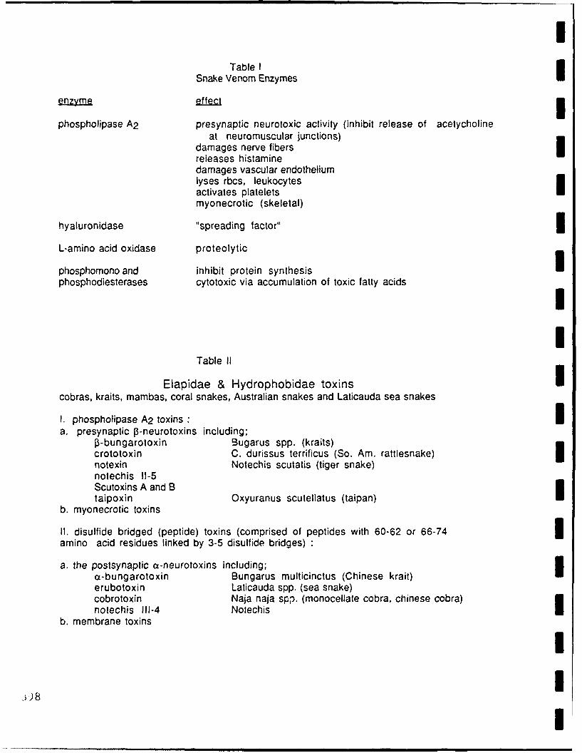
iTable I
Snake Venom Enzymes
enzyme effec Iphospholipase A2 presynaptic neurotoxic activity (inhibit release of acetycholine
at neuromuscular junctions)damages nerve fibers Ireleases histaminedamages vascular endotheliumlyses rbcs, leukocytesactivates plateletsmyonecrotic (skeletal)
hyaluronidase "spreading factor"
L-amino acid oxidase proteolytic
phosphomono and inhibit protein synthesis Uphosphodiesterases cytotoxic via accumulation of toxic fatty acids I
ITable II
Elapidae & Hydrophobidae toxins Icobras, kraits, mambas, coral snakes, Australian snakes and Laticauda sea snakes
I. phospholipase A2 toxins : Ia. presynaptic P-neurotoxins including;
P-bungarotoxin Bugarus spp. (kraits)crototoxin C. durissus terrificus (So. Am. rattlesnake) Unotexin Notechis scutatis (tiger snake)notechis 11-5Scutoxins A and Btaipoxin Oxyuranus scutellatus (taipan)
b. myonecrotic toxins
II. disulfide bridged (peptide) toxins (comprised of peptides with 60-62 or 66-74amino acid residues linked by 3-5 disulfide bridges)
a. the postsynaptic a-neurotoxins including;a-bungarotoxin Bungarus multicinctus (Chinese krait) Ierubotoxin Laticauda spp. (sea snake)cobrotoxin Naja naja spp. (monocellate cobra, chinese cobra)notechis 111-4 Notechis
b. membrane toxins II
. :)8

WILDERNESS MEDICAL LIABILITY
LEARrCNG OBJECTIVES
Following this presentation, participants will have:
1. Acquired a basic understanding of the general lecalprinciples of medical malpractice claims and theirapplication to expedition settings.
2. Become aware of the areas in which a physician is exposed toa medical malpractice claim in an expedition setting.
3. Obtained suggestions and recommendations to help lessen thephysician's exposure to liability.
Timothy E. MillerAttorney at Law
Martin, Bischoff, Templeton, Langslet & Hoffman3100 First Interstate Tower
Portland, Oregon USAI 97201
399

mWILDERNESS MEDICAL LIABILITY
Timothy E. Miller'Attorney at Law
Martin, Bischoff, Templeton, Langslet & Hoffman3100 First Interstate Tower
Portland, Oregon USA97201
I. Introduction
Participation by a physician in an expedition or otherwilderness outing unquestionably exposes the physician to therisk of liability for professional negligence. The probabilityof a claim being made appears to be small, probably because thetype of people who participate in such activities know they areassuming some risk and are more likely than the general public to
accept responsibility for the consequences. The lck of casesreported in United States courts supports this conclusion. Whilethe probability of a claim may be small, the financialconsequences of a successfully prosecuted claim could bedevastating. If the claim is covered by your professionalmalpractice insurance, the insurer will pay to defend the claimas well as to indemnify you for any resulting judgment orsettlement, up to your limit of liability. If your insurancedoes not cover the claim, you will bear the costs of defense andbe personally liable for any resulting judgment or settlement.
Many of you may recall that there was a death on the 1986SINO-USA Upper Yangtze River Expedition. The expedition stillphotographer died at approximately 14,500 feet on the TibetanPlateau from the combined effects of high altitude pulmonaryedema and cerebral edema. His widow sued the expedition leaders,the documentary film company and the film company's president.The cost of successfully defending the expedition leaders alonewas approximately $175,000 and that amount did not include somesubstantial costs assumed by the film company, such as expertwitness fees. The expedition doctor was not sued, but hisI conduct was a central issue in the case.
The purpose of this paper is to highlight the generalconcepts of medical malpractice liability and apply thoseconcepts to the activities of a physician who participates in anexpedition or other wilderness outing. For ease of presentation,
i John W. Knottnerus, an attorney employed in the author'sfirm, provided substantial assistance in the preparation of thispaper.
I401I

I
the example of an expedition physician will be employed. The Iprinciples are applicable to other less organized situations aswell.
This paper is not a comprehensive review of malpracticeliability, which varies from state to state and country tocountry. The pirpose of this paper is to make you think aboutyour exposure to claims and provide suggestions on how to avoidthem.
II. Basics of Medical Malpractice Liability
A brief explanation of the general legal principles involvedin a agneric medical malpractice claim is necessary in order toappreciate and understand the specific issues raised by aphysician's involvement in an expedition.
A. Elements of a Claim
A professional malpractice case involves elements generallyapplicable in any tort or negligence action. In the normal tortaction, the plaintiff2 must prove: (1) a duty on the part of thedefendant to conform to a specific standard of conduct for theprotection of the plaintiff; (2) breach of that duty by the Idefendant; (3) the breach of the duty was the actual andproximate cause of plaintiff's injury; and, (4) damages.3 Thefactor that distinguishes medical malpractice cases (as well as Uother professional malpractice cases) from most tort actions isthe "standard of care" to which the defendant's conduct iscompared.
A general duty of care is imposed, in tort law, on all humanactivity. However, if a defendant has knowledge, skill, or evenintelligence superior to that of the ordinary person, the lawwill demand of that defendant conduct consistent with suchattributes.4 Professional persons (doctors, lawyers, engineers,etc.) in ge:neral, and those who undertake any task calling for
2Note that the "plaintiff" is not always the person to whom
the professional provided service. For example, a spouse,children, parents, or a decedent's estate may be the onesbringing an action against a doctor for the actions the doctortook in diagnosing and treating the patient. Therefore, even ifthe participant was willing to take the risk and was satisfiedwith your actions, the family left behind may have differentideas. I
3 Prosser & Keeton on Torts § 30 (5th Ed. 1984).
' Restatement (Second) of Torts § 289, comment m. II
402
I

I special skill, are required not only to exercise reasonable carein what they do, but also to possess a standard minimum ofspecial knowledge and ability.
B. Physician's Standard o Care
The standard of reasonable conduct for medical professionalsis the range of accepted responses to a given set ofcircumstances by others with a similar practice in the same orsimilar community--commonly known as the "community standard."5
With respect to "the range of responses," the law contemplatesthat in any particular set of circumstances there may be morethan one approach that would meet the physician's standard ofcare. For example, even if another physician would not responOto a given set of circumstances in the same manner, neitherphysician is negligent if both responses are within the range ofaccepted responses in same or similar community.6 An examplewould be surgery to treat a thyroid problem instead of drug
therapy.
The physician's community and his or her practice can becriticdl to a determination of liability. On- who holds him- orherself out to the public as a specialist in a particular fieldof medicine is held to a higher standard of care than a generalpractitioner who renders the same type of service as a part ofhis or her practice.7 The specialist must conform to thestandard of that segment of the profession in which the physicianspecializes.
The place where the physician practices is also relevant tothe community standard. In some courts, a physician is entitledto have his or her conduct tested by how others in the same orsimilar places would conduct themselves.8 In other words, thelaw recognizes that a general practitioner in New York City mayrespond differently than a general practitioner in Two Dot,
Montana (U.S.A.).
The physician's medical community, geographic community, andspecialty (if any) define the standard of care to which thephysician will be held. The law sets the standard of care and
5 McCoid, The Care Requirement of Medical Practitioners, 12Vand. L. Rev. 549 (1959).
6 See e.g., Willard v. Hutson, 234 Or. 148, 378 P.2d 966(1963).
7 Prosser & Keeton on Torts § 32 (5th Ed. 1984).
8 See e.g., ReeQ v. Shauqhnessy, 570 F.2d 36J ki0th Cir.
1978).
403
I
negligence is determined by whether the standard is met as the Iresult of what one does or does not do, rather than by what oneknows or does not know.
C. Informed Consent
Aside from liability arising from actions that may falloutside the "community standard," there is another area ofpotential liability called, "informed consent." As a generalrule, a physician has a legal duty to warn a patient of the risksof a procedure and advise of alternative approaches if theevidence discloses that: (1) the risk of injury inherent in theprocedure is material; (2) there are feasible alternative coursesof action available; and, (3) the plaintiff can be advised of the Irisks and alternatives without detriment to his or her well-being.9
The patient can bring an action against the physician if thephysician did not obtain informed consent. Implicit in the casesinvolving informed consent is the requirement that the patientestablish that he or she would not have consented to theproueduie if adequately advised.10
III. Areas of Exposure in Wilderness Medicine HWith some of the basic legal principles applicable to
medical malpractice cases in mind, the circumstances unique to Uexpedition medicine, under which these principles will beapplied, can be outlined. The potential theories of negligencethat might be advanced by a patient's attorney are endless.However, experience from the Yangtze River case and common sensereveal several likely areas of exposure.
A. Pre-Expedition Screening & Examination
A physician's exposure to a medical malpractice claim mayarise even before the expedition departs. Most prudentexpedition leaders will require that all participants, as acondition to participation, obtain a physical and provide amedical history. As the expedition physician, you may be asked Ito conduct the physicals, which would include a review of theparticipants' medical histories. More likely, the expeditionphysician will be responsible for reviewing the results of the Iphysicals and the medical histories. In most cases, prudence
9 2 Louisell & Williams, Medical Malpractice § 22.01 et. Iseq. (1989).
10 Waltz and Scheuneman, Informed Consent to Therapy, 64 NW
U. L. Rev. 638 (196;).
I404 I

I
Iwill require that the expedition physician, who is fully aware ofthe specific health risks of the expedition, prepare a detailedquestionnaire for each participant that seeks informationpertinent to the specific risks.
If a participant suffers serious injury, illness or deathfrom the complication of a pre-existing condition, which shouldhave been a contraindication to participation, the physician who
performed the physical and the reviewing or expedition physicianare exposed to a claim. Likely theories are failure to inquireabout health problems later deemed relevant to the known risks ofthe expedition, failure to recognize the risks, failure to reportcontraindications to the expedition leaders and failure to advisethe participant of contraindications.
B. Expedition Diagnosis & Treatment
A physician's exposure to malpractice claims formisdiagnosis and treatment during the expedition encompasses manyof the same areas for which the physician would be exposed to amalpractice claim in his or her everyday practice. Basically,anything that went "wrong" in the diagnosis and treatment phasescould be a potential malpractice claim. The specific legalissues raised by diagnosis and treatment during the expeditionare discussed below.
C. Equipment & Supply Deficiencies
The expedition physician is customarily called upon toassemble, sometimes at his or her own expense, the medical "kit"for the expedition. While the expedition leaders or otherparticipants may have some suggestions bpsed on past experience,the physician, for the most part, exercises unrevieweddiscretion.
If a participant is injured, or succumbs to an illness, andthe right type or quantity of supplies or drugs are notavailable, the physician is exposed to a claim. Because manyexpeditions today are equipped with sophisticated equipment, theexpectations of participants and their families are that themedical supplies will likewise be "state of the art." Rememberthat in the case of a death, the claim will likely come from aspouse or parent. Their expectations are frequently higher,usually due to assurances made by the now-deceased participant,and they will not understand or accept the practical expeditionreality that there are limits on what can be carried, preservedor possibly even taken into a foreign country.
The standard of care imposed on the physician assembling themedical kit is more likely to be based on what physicians havetaken on similar expeditions to similar locales than on a surveyof physicians in the community where the expedition physician
405

i
practices. Consultation with those who have gone before you and 1equipping accordingly will lessen your exposure. Document yourcommunications with those physicians.
D. Informed Consent
The second major branch of medical malpractice liability isinformed consent. Under certain circumstances, an expeditionphysician may have a duty to advise an injured or ill participantof the risks and alternatives of a proposed treatment orprocedure, before administering it. There is no logicalimpediment to applying this concept to treatment in theexpedition setting in exactly the same form it is applied in ahospital before surgery.
IV. Legal Issues Raised by Expedition Medicine
We have not found any reported cases involving claims ofmedical malpractice on expeditions, so there is not a great dealof guidance on how the courts would deal with such a case.Although there are no definite answers, in the legal sense, thefollowing are some of the legal issues raised by expeditionmedical care.
A. Standard of Care
The physician's general duty of care, under the communitystandard, takes into account the physician's area of practice,including, in some states, the geographical location of hispractice. With respect to the physician who performs the pre-expedition physical, there should not be an issue with respect tothe applicable community, particularly if it is the participant'spersonal physician. The standard of care applied to the Iexpedition doctor is more difficult. The standard couild be drawnfrom the community where the physician practices, from theconduct of expedition physicians on similar expeditions tosimilar locales or from a specialized practice, such as highaltitude medicine or the treatment of hypothermia.
Advances in the field of wilderness medicine, including the iexistence of physicians whose entire "practice" is limited towilderness and expedition medicine, raises the issue of whetherthe physician is a specialist, which is a familiar concept inUnited States law relating to the standard of care. One whoholds him or herself out to the public as a specialist in aparticular field of medicine is held to a higher standard of carethan a general practitioner who renders the same type of serviceas a part of his or her practice. Therefore, the expeditionphysician may be subject to the standards, knowledge and skill ofother physicians specializing in wilderness medicine, or certain Idiscrete areas, such as high altitude medicine, whether or not
I406
I
the particular physician actually has that knowledge or skill.If you participate in a climbing expedition, as the expeditionphysician, you may be judged by a standard of care developed fromthe work of people like Dr. Hackett, if the work is published anda reasonable inquiry would have discovered it. In other words,you should attempt to familiarize yourself with the state of theart on medical issues likely to arise, given the specific natureof the expedition.
B. Good Samaritan Statutes
Good Samaritan statutes offkr total immunity from liability--even if the physician fails the community standard. The theoryof Good Samaritan immunity has been accepted in all of the UnitedStates." All states have legislatively adopted some type ofstatute exempting certain bystanders or "rescuers" from allliability for any negligent act or omission arising out of theirrescue attempts. Many, if not all, of these statutes were passed
for the express purpose of protecting physicians whoaltruistically render services in emergency situations. Somestates also extend the protection to other medical and emergencypersonnel (nurses, EMTs, paramedics, firemen, etc. )12 and some toanyone rendering emergency assistance.
Every state's statute is different and the applicablestatute must be consulted for its exact language andrequirements. The statute most likely to apply is that of thestate where you practice, the state where the participant resides(or resided) or the state where the injury occurred, and not thelaw of a foreign country. Even though the statutes differ, somebroad statements with regard to the conditions placed on aphysician's immunity under most Good Samaritan statutes can bemade. 13 First, many statutes require a good faith state of mindon the part of the rescuer rendering emergency assistance.Second, many statutes require that the care be rendered"gratuitously" which has been interpreted to mean that there isno pre-existing duty to render such service. Third, some
11 See, Good Samaritan Laws--The Legal Placebo: A CurrentAnal, is, 17 Akron L. Rev. 303 (1983).
12 For an example of an statute whose coverage extends to
nurses, see Cal. Bus.&Prof. Code § 2727.5 (West 1974). For acomplete outline of every state's statute, see the Akron LawReview article cited in the prior footnote.
13 These generalizations are drawn from: Annotation,
Construction and Application of "Good Samaritan" Statutes, 68ALR4th 294 (1989); 2 Louisell & Williams, Medical Malpractice,chptr 21 (1989); and, the Akron law review article cited above.
407

I
statutes specify or limit the places in which the emergency aidmust be given (i.e. "at the scene") for immunity to attach.Fourth, the statutes will not afford immunity to a physician whohas been grossly negligent, reckless, or has engaged in willfulor wanton misconduct. Finally, the emergency situation must nothave been created by the person rendering assistance.
For the most part, it seems that the conditions imposed bymost Good Samaritan statutes could be met by the expeditionphysician. This is particularly true in situations where an Iunexpected accident or injury occurs. The most apparent reasonto prevent immunity from attaching is the requirement that thephysician perform the service gratuitously with no pre-existingduty.
Even though the physician may not have been "hired" as theexpedition physician and receives no compensation, the presence Iof a physicirn on most expeditions is no accident. Participants
are told, or may assume, that you are there to provide themmedical services. Expedition leaders and participants expectthat if something happens the physician will act. The purpose ofthe physician's participation and the expectations of theexpedition members may give rise to a pre-existing duty to renderservices. If a court or jury found that duty to exist, the Iimmunity offered by a Good Samaritan statute might not apply.
C. Informed Consent
As described earlier, the informed consent doctrine centersaround the physician's duty to advise the patient of the natureand risks of a proposed medical treatment, in order to enable thepatient to make an intelligent and informed decision as to thetreatment.
Two exceptions to the physician's duty to inform and obtainconsent are applicable to the expedition setting. First, theduty is not applicable in an emergency situation where the Ipatient is unconscious or otherwise incapable of consenting andthe harm from a failure to treat is imminent and outweighs anyharm threatened by the proposed treatment. 14
The second exception, often termed the "therapeuticprivilege," eliminates the duty to inform and obtain consent insituations where the physician believes that the patient's Iemotional state is so fragile that the very act of revealing the
I14 2 Louisell & Williams, Medical Malpractice § 22.06 I
(1989). I'08
I

risks may: (1) cause the patient to refuse the treatment that thephysician thinks is needed; or, (2) create physiologicalreactions that would increase the dangers of the procedure. 15
The usual situation for application of this doctrine, theseverely emotional patient, is not likely to present itself on anexpedition. It is more likely that a physician will be presentedwith the risk of harm to other members of the expedition, and tohimself or herself, than a risk to the "patient" if treatment isrefused. This problem is very real in the expedition setting,has not been discussed as a consideration in the reported cases,and raises ethical problems which are beyond the scope of thispaper.
E. Insurance Coverage
As with any other situation, the expedition physician shouldbe concerned with whether his or her medical malpracticeinsurance policy will cover a claim arising from acts oromissions on the expedition. Your policy should be carefullyexamined before participating in any expedition. The first thingto look for is a geographical limitation. The second is languagein the policy suggesting that only activities normally associatedwith your stated practice are covered. Careful attention shouldbe paid to all exclusions and endorsements, which frequentlymodify the terms of coverage otherwise extended in the maininsuring agreements.
If you have doubts about coverage, consult a lawyer beforeyou consult your insurance agent. Disclosure to the agent mayresult in issuance of an exclusion defeating any coverage for theexpedition. This will eliminate a later argument that anambiguous policy does provide coverage.
You should also find out if the expedition leader hasobtained insurance specifically for the expedition. If so,obtain a copy and determine, with the help of a lawyer if needed,if the policy covers you. If it does not, request to be added asan additional insured.
D. Releases of liability
The Yangtze River case wds won on the strength of anassumption of risk, or release, document signed by the deceasedparticipant. The expedition leaders retained a lawyer to preparethe release and required each participant to sign one as a
'5 2 Louisell & Williams, Medical Malpractice § 22.06(1989).
409

I
condition of participation. The Yangtze River release did not Iexpressly release the expedition physician, but it could havebeen drafted to do so.
You should discuss releases with the expedition leader andrequire, as a condition of your participation, that eachparticipant execute a release that covers you as well as theleader. There is no standard forr release. Be aware thatreleases from liability, especially for physicians, are notfavored in the law and will not automatically apply to protectyou from a claim in every case. You and the leader should retaina lawyer to assist you in drafting the release.
V. Conclusion
The threat of liability should not chill your participationin expeditions or other wilderness activities. Rather, if youkeep the issues and suggestions presented in this paper in mindwhen you are preparing for an expedition or other activity, youwill lessen, or perhaps eliminate, your exposure to claims.
IIIIIi
II
410 I
I

DEALING WITH DEATH:THE PSYCHOLOGY OF RESCUE
David R. Jones, M.D., M.P.H.Aeropsych Associates
One Clermont Court, Oakwell Farms,San Antonio, TX 78218-1741
INTRODUCTION:This presentation assumes that the people involved in deadly situations aregenerally in good health, both physically and mentally, before the mishap. Itwill deal with the victims--those saving themselves, or needing rescue--andwith the rescuers themselves.
IHE WILL TO SURVIVESurvival is one of the basic instincts in every animal, including humans. If weare physically threatened, we respond instinctively in order to survive, byfight, flight, or fright (freezing). If the thing we do (our behavior) helps us
I survive, we remember it and tend to use it if the same situation threatens usagain. This is learning by experience.
I Another way animals acquire survival behavior is through simulation; welearn through planning and practicing. Much of the playing that young
* animals do is for this purpose.
One of the unique things humans do is to learn survival behavior before theI event by mental rehearsal--learning through imagination. This includes
listening to how others have dealt successfully with potentially deadlyexperiences, or imagining how we would deal with such a situation. Thus, wecan "remember" how to deal with things that we have never actually faced,because we have thought them through ahead of time. Aviators do a lot ofthis.
However, one element which is impossible to simulate, rehearse or plan for isthe emotional component of danger, fear. Fear is to the mind as pain is to thebody: it repels us from danger. Our natural instinct is to avoid things thathurt us or frighten us. Fear changes the way we think, feel and act, actingpartly through the mind, but mostly through the autonomic nervous system(ANS). Of course, we can use will power to overcome these natural aversions,but doing so takes a certain amount of psychological energy.
411

i
Some people seem to have what Tom Wolfe called "the right stuff," the abilityto continue to think clearly and act correctly during a severely threateningsituation, even one which is new to them. No one knows how much of thisability is inborn, and how much is learned. This quality is difficult to predict Ior to identify, except by observation. At any rate, these people can thinkunder stress and control their behavior in situations where most people mightstop thinking clearly and, instead, freeze up or panic. Panicky behaviorinvolves thoughtless and possibly counterproductive behavior that mayactually increase the danger. For example, a person may panic under fire incombat and stand up to run away, thus causing needless exposure to enemybullets.
Danger may not only be immediately threatening, but it may also involve alonger lasting situation which requires a more enduring response, as was thecase with the woman with the broken leg who was rescued from deep in acave in April 199 1. After dealing with the initial mishap, the person may bephysically exhausted, injured, and utterly alone in a hostile environment.Instinct and immediate response have worked so far, but now a new set ofskills is called for, involving very clear thinking: assessment of the situation,control of emotions and actions, and planning a series of moves as in a chess igame. The person's status is far from the usual, familiar situation, and theperson may feel overwhelmed by the need for innovation and flexibility, andmay be tempted to give up or to wait passively for whatever happens next.Indeed, "lying low" may be the best course of action in such a situation, but,once rested, the person must start thinking ahead of the situation again,planning moves, considering "What will I do if.such-and-such happens?"
In this situation, prior planning, listening to others' experiences, mental oractual rehearsing, and other such preparation may pay off with cool, correctaction in the long-term survival situation. The will to survive has involvedboth instinctive and cognitive mental processes.
Once the person is safe, through his or her own actions or through the effortsof rescuers, some psychological reactions may occur. One common reaction isto deny the emotional component, to "get right back on the horse that threwyou" and prove that the individual, no longer a victim, is back in control, Thismay well be effective, and is much used by aviators who fly again as soon aspossible after a crash or other brush with death. The problem with suchdenial, however, is that the individual may feel that any emotional reaction issomehow unworthy, and that one should be cool and emotionless in everysituation. Thus, one may actually repress emotions, such as survival guilt,which are so strong that they will affect other areas of life, causing later
412
I

problems such as irrational fears (phobias), acute stress reactions, or
psychosomatic symptoms.
How should one deal with an individual who has survived a potentially lethal
situation? Using an outline form, we will consider three elements: EarlyDetection of Problems, Rapid Treatment, and Rehabilitation.
I. EARLY DETECTION OF PROBLEMS IN VICTIMSA. Understand that fear is normal, and serves the same purpose for
the mind that pain does for the body. A fearful response to a realthreat is not the same as acute anxiety or mental illness.
B. Reactions may be adaptive or maladaptive, immediate or delayed,and acute or chronic. Maladaptive behavior in otherwise uninjuredvictims may require some therapeutic response.
C. Reactions may include anxiety, disorientation, misunderstanding, confusion,, withdrawal, irritability, increased startle reaction, difficulties withmemory, concentration or sleep; also psychophysiological symptoms.These may simply represent normal responses to an abnormalsituation, and usually require merely supportive care.
D. Mishap victims may have brief maladaptive reactions of psychoticproportions, but these are usually transient.
II. RAPID TREATMENTA. Avoid creating an unnecessary "patient" role for victims.B. Provide basic needs: shelter, protection, warmth, food, comfort and
support. Allow regressive dependency to run its course. Useshort-acting sedation (temazepam, for example) only if necessary, sincemedication reinforces the "patient" role.
C. Provide accurate information about the situation.D. Ventilate emotions--describing what happened helps victims get their own
thoughts in order.E. Allow or encourage victims to express their fears ("I don't know how you
feel, but I would be really thinking hard about almost dying. What areyou telling yourself about the accident?"). Reassure that such thoughtsand feelings are normal. Explore the realities of what happened, andhow they survived.
F. If psychological defenses were overwhelmed, expect to see either denial,depression, loss of sense of humor, withdrawal and psychologicalnumbing ("I don't feel anything") or intrusive symptoms such asreliving the experience, flashbacks, nightmares, or agitation. Thesepolar reactions, withdrawal and agitation, may alternate for a while and
413

Ithen begin to fade away as the individual comes to grip wth whathappened. Professional help may be useful during this period.
G. In multiple-victim mishaps, let victims help each other as they are able.. This establishes reassuring adult roles in place of dependent ones.
2. The volunteer effort.saved may be better used tisewhere.H. Brief reactive psychosis.
1. Produced by an overwhelming stressor.2. Symptoms include emotional turmoil, and at least one psychoticsymptom: incoherence or loosened associations, grossly disorganizedbehavior, delusions and/or hallucinations.3, Again, avoid use of psychotropic medications if possible, since theyreinforce the "patient" role, and may mask neurologic symptoms. Useshort-acting medications such as haloperidol if necessary. i
I. Physically injured patients should be followed psychologically in aconsultation/liaison model with their primary physicians, since theywill have had the same stressors and losses.1. Psychotropic medication may mask psychological symptoms for awhile, and may interfere with the normal grieving process. It may alsomask head injury symptoms.2. Injured patients need ventilation too, but not excessively. They mayneed to be buffered from the press, dignitaries, and curious staff.3. Bad news about family members or others involved in the mishapshould be handled carefully and thoughtfully.
III. REHABILITATIONA. Mental health consultants may switch from crisis intervention to
treatment of post-traumatic stress disorder as time passes.B. Although careful early follow-up may diminish the incidence of
Post Traumatic Stress Disorders, case-finding efforts by the Red Crossand similar agencies may be useful in reaching victims who were notprimarily involved, such as family members of actual disaster victims.
C. All victims may have troubling symptoms for a while, but many will ihandle these through normal grieving processes. Symptoms mayalternate between periods of psychic numbing, which protects fromoverwhelming emotion, and intense emotional pain, which may feelintolerably threatening, as if the victims were "losing their minds" or
Agoing crazy." 3D. A stable environment, allowing the processing of the situation in small
emotional increments and accepting the ups and downs of the recoveryperiod, helps people in processing the disaster and the losses. Expect3some inappropriate anger, often directed at rescuers or helpingagencies.
414 4].4 i

1. Therapy should be aimed at re-establishing control over one's ownlife, and dealing effectively with the post-disaster realities.2. Tearfulness, both expected and unexpected, is part of the healing,
and will probably continue for up to a year. Frequency and intensitywill diminish with time.3. Through talking about their losses, victims may relieve some of thefeeling of isolation in grief. Like tears, the need to ventilate and thusseek support will diminish after a year or so.4. Dreams about what happened may represent one means of naturaldesensitization to the event.5. Psychosomatic symptoms, including appetite, libido, or sleeppatterns, may be disturbed, but should gradually return to normal.Memory and concentration may go through similar disturbances.Intrusive thoughts should also gradually lessen, although all thesesymptoms may be exacerbated by anniversaries, sights, sounds, andother associations. Victims should be made aware of these and similarpattrns of reactions as normal phenomena, similar to somatic healingprocesses, and should be reassured that they are not signs of mentalillness, weakness, or lack of moral fiber.
E. During this interval of recovery, victims should be made aware of aneed not to self-impose additional stresses. For example:1. Respect emotional waves. If one occurs, for example, while driving,pull over and stop as soon as safety allows, and wait until it passesbefore resuming driving. Allow a little extra time. Travel with afriend, if possible.2. Be especially careful if work requires alertness; e.g., aroundmachinery.3. Watch out for physical health; stress can affect resistance to disease.Consult a physician for somatic symptoms. However, keep in mind thatgrief can also be a factor in such symptoms. Beware of overmedication.
4. Delay making decisions which will cause added stress, if possible. Ifnot, separate such decisions in time. (Therapists should keep theHolmes-Rahe Life Change Events tables in mind.)5. Share problems, concerns and cares with responsible familymembers, trusted friends, and/or mental health professionals.
RESCUERSA considerable literature exists on the care of rescuers, and others who
may have been only pe,'ipherally involved in the disaster, such as reporters,morgue attendants, and medical staff. A sensitive and professionallymoderated post-incident debriefing procedure may help to elicit troublingthoughts and emotions which otherwise would not be recognized for whatthey are. This process should not be undertaken without prior specific
415
1training, and should be carried out by persons who were not involved in the 3primary disaster or response. See the articles by Jones and by Mitchell in thebibliography for further details.
BIBLIOGRAPHY IAdler A. Neuropsychiatric complications in victims of Boston's Cocoanut Grovedisaster. 1943; JAMA 123: 1098-1101.
Ahearn FL, Cohen RE. Disaster and mental health: an annotated bibliography.1984; Washington, DC. Center of Mental Health Studies of Emergencies, NIMH.
Baker GW, Chapman DW. (eds) Man and society in disaster. 1962; New York. UBasic Books.
Cobb S, Lindemann E. Neuropsychiatric observations during the Cocoanut IGrove fire. 1943, Ann. Surg. 117: 814-24.
Cohen RE, Ahearn FL. Handbook for mental health care of disaster victims. I1980; Baltimore, MD. Johns Hopkins University Press.
Cohen RE. The Armero tragedy: lessons for mental health professionals. 1987;Hosp. Commun. Psychiat. 38: 1316-2 1.
Farberow N, Gordon NS. Manual for child health workers in major disasters.1980; Washington, DC. Government Printing Office. 3Glass, AJ. Psychological aspects of disaster. 1959; JAMA 171: 222-5.
Hartmann K, Allison J. Expected psychological reactions to disaster in medicalrescue teams. 198 1: Mil. Med. 146: 323-7.
Hoiberg A, McCaughey BG. The traumatic aftereffects of collision at sea. 1984;Am. J. Psychiat. 141: 70-3. I
Holmes TH, Rahe RH. The social readjustment rating scale. J. PsychosomaticRes. 1967 ; 11: 213-8. I
Horowitz MJ, Kaltreider NB. Brief psychotherapy of stress responsesyndromes. In Specialized techniques in individual psychotherapy, (Ed. IKarasu T, Bellak L) 1980: New York. Brunner/Mazel, Inc.
1416
3

Jones DR. Emotional reactions to military aircraft accidents. 1982;Aviat. Space Environ Med. 53: 595-8.
Jones DR. Secondary disaster victims: the emotional effects of recovering andidentifying human remains. 1985; Am. J. Psychiat. 142: 303-7.
Kolb LC A neuropsychological hypothesis explaining posttraumatic stressdisorders. 1987; Am. J. Psychiat. 144: 989-95.
Laube J. Psychological reactions of nurses in disaster. 1973; Nurs. Res. 22:343-7.
Lif ton RJ, Olson E. The human meaning of disaster: the Buffalo CreekI experience. 1976; Psychiat. 39: 1-18.
Lindy JD, et al. Psychotherapy with survivors of the Beverly Hills Supper Clubfire. 1981; Am. J. Psychother. 37: 593-610.
Mangelsdorff AD. Lessons learned and forgotten: the need for prevention andmental health interventions in disaster preparedness. 1985; J. Commun.Psychol. 13: 239-57.
Michael S, et al. Rapid response mutual aid groups: a new response to social
crises and natural disasters. 1985; Social Work 85: 245-52.
Mitchell JT. When disaster strikes...the critical incident stress debriefingprocess. 1983; J. Emerg. Med. Serv. 36-9.
Raphael B, Middleton W. Mental health responses in a decade of disasters:Australia, 1974-1983. 1987; Hosp. Commun. Psychiat. 38: 133 1-37.
Sanner PH. Wolcott BW. Stress reactions among participants in mass casualtysituations. 1983; Ann. Emerg. Med. 12: 426-8.
Slaby A , Emergency psychiatry: an update. 1981; Hosp, Commun. Psychiat.32: 687-98.
Shore JH, Tatum EL, Vollmer WR. Psychiatric reactions to disaster: the MountSt. Helens experience. 1986; Am. J. Psychiat. 143: 590-5.
Tyhurst JS. Individual reactions to community disaster: the natural history ofpsychiatric phenomena. 195 1: Am. J. Psychiat. 107: 764-9.
417

IWise MG. Post traumatic stress disorder: the human reaction to catastrophe.1983 (Mar.); Drug Ther. (no vol.) 2 10-7.
INSTITUTIONAL RESOURCES
Center for Mental Health Studies of EmergenciesNational Institute of Mental HealthParklawn Building, Room 6C 125600 Fishers LaneRockville, MD 20857Tel. 301-443-1910
National Organization for Victim Assistance717 D. Street, N.W.Washington, DC 20004 UTel. 202-393-6682
The Society for Traumatic Stress Studies IP.O. Box 1564Lancaster, PA 17603-1564 3Tel. 717-396-8877
Disaster Preparedness in the Americas 3Pan American Health Organization525 23rd Street, N.W.Washington, DC 20037
[Note: this bibliography and resource list was derived from a more extensive Ulist prepared by A. David Mangelsdorff, Ph.D., M.P.H., Technical Director,Health Care Studies and Clinical Investigation Activity, Health ServicesCommand (Attn: HSHN-T), Fort Sam Houston, TX 78234-6060; Tel. 512-221- 35671/251 1.1
IIIU
418i
I
WILDERNESS MEDICINE -- JULY 19-91VENOMOUS ARTHROPODS
Findlay E. Russell. M.D., Ph.D.The University of Arizona, Tucson, Arizona 85721
BIOLOGY spiders, 80% were found tW be caused by urthropods other
l'he phylum Arthropoda is the largest phylum in the than spiders, 10% were probably caused by Loaosceiesanimal kingdom. It contains the spiders, scorpions, sp., and 10% by other disease states. The arthropods
most frequently involved in the misdiagnoses were ticksuisvcts, tI.cks, kissing bugs, water hugs, caterpillars,oets, butterflies, grasshoppers, centipedes, iand (including their embcdded mouth parts), kissing bugs,
r:,p.cebutterngieshershSopearhropos stinged ees, n mites, bedbugs, fleas (infected flea bites), lepidopterousntilipedes, among others. Some arthropeds sting pbees, insects (lepidopterous blisters), flies, beetles (vesicaLing
'nts, scorpions, etc., others bite (spiders, centipedes, beetles), water bugs, and various stinging Hymencptera.kissing bugs, etc.), while still others discharge a Reviews on spider venom poisoning will be found insecretion that is toxic (millipedes, caterpillars, etc.). Bettini i , Maretic and Lboi 2] Southcott [4, Russell
Although the number of arthropods that are sufficiently [5I and Gertsch 161.,enomous to be of'a potential danger to humans is notknown, these animals are implicated in far more bites Lactrodectus Species (Widow Spiders)and stings than all other phyla combined. Almost all ofthe 20,000 species of spiders are venomous, but luckily These spiders are commonly known as the black
o"ur man, only relatively small numbers have fangs long widow, brown widow, or red-legged spider, depending onand strong enough to penetrate the human skin. There the species. They have many other common aames inare some 500 species of scorpions and all are venomous, English: hourglass, poison lady, deadly spider, red-aithough only a small number are of a danger to bottom spider, T-spider, gray lady spider, or shoe-button!)umans. In the order ilymenoptcra, the bees, wasps, spider. The eastern black widow spider is L. mactans,eilow jackets and ants, numerous species are involved whereas the western is L. hesperus.
in serious envenomataons. Among the other arthropods Although both male and female widow spiders areare numerous animals of medical importance. venomous, only the latter have fL--s large and strong
The number of deaths from arthropod stings and enough to penetrate the human skin. Mature femalesP)iLes is not known, nor do most countries keep records of range in body length from 10 to 18 mm, whereas malesLhe incidence of such injuries. In Mexico, parts of are from 3 to 5 mm.Central and South America, and in North Africa, deaths In most patients, there is a history of having received.rom scorpion stings may exceed several thousand a a sharp, pinprick-like bite, but in some cases the incident
e.uar Spider bites probably account for no more than 200 is so minor that it goes unnoticed. The initial pain islc'aths a year, worldwide. The number of deaths from sometimes followed by a dull, occasionally numbing pain
artnropod bites or stings in the temperate countries is far in the affected extremity, and by pain and some cramps_reater than the number of deaths from snakebite. in one or several of the large muscle masses. MuscieAlmost. all ofL these deaths, however, are anaphylactic in fasciculations can sometimes be seen within 30 minutes• lture In ihc underdeveloped countries of the tropics, a of the bite. Sweating is common and the patient may.Ar L;reaLer number of deaths from arthropod bites or complain of weakness, as well as pain in the regionai.. in,,s are due to the direct effects oithe venom. lymph nodes. Lymphadenitis is frequently observed.
CIlEMISTRY ANI) PIIAIMACO[LOGY Pain in the low back, thighs, or abdomen is common andrigidity of the abdominal muscles is seen in most cases in
'lie venunis of the various urthropods are very which envenomation has been severe. Severe parox-niifrent. aria. if the spider venoms so far studied are an vsmal muscle cramps may occur, and arthralgia has
AIUiCaULon, these poisons may prove to be more compLex been reported...an oric'inaily suspected. [-or instance, the venom of
6i iack w )wow ,spider lalrodecius muciuo uncontains at The extent of the local tissue reaction around the bite.it 15 proteins, arid preliminary work on the brown may vary, depending on the species involved. In most
-sroir (l.oxosceies) as well as other spider venoms, cases in the United States, there is no reaction at the bitenUIctUs tna tthey may all be equallV as cumplex. Like site and the puncture wounds may be impossible to lina.'ne nnake venoms, the arthropod poisons exert their In some cases, there is an immediate 2 to 4 mm blanched11uhILultuI efectsit th, ic cl:lular lvel. anid sincue area around the puncture wounds, surrounded bv a
.rhrupods bite while others sting, the passage of the slightly erythematous ureu. which in time becomesxin across membranes, its absorption at specific pallid. This may persist for 2U-30 minutes. In children.
.icctor sites. metabolism and excretion are quite there may be locaiized dil'use edema.:fiTrent., rrletctiniz the teleology and Ievodition 4,f the I.1ocal Sweating nay be evident, although the:)1no n The intcreLed reader will lfind the texts by .rythev tLous plaque usually ruinainh dry. PilocrectLon
and .Maretic and lbez 121 particularly around the bite area and local changes in the sensitivity,,)',rtant with respect tw the composition and moues 01 ol the skin have been reportea following the bites of some
.-cion ol artnropoa venoms. species. Opisthotonos, bizarre head and neck move-.I')l'.S ments, elevation of body temnerature, mydriasis.
increued salivtion, anorexia. oliguriu. and convulsions'! eare at least 21.0 species of' 'piders that have have all been reported but are not common.
.'_.l i:1pliCateU in signilicant bites on hrumans 131. Hypertension is a characteristic finding in moderate to-inortunatelv, spiders otten get the "bum rap" for the severe poisonings. It may not be evident during the first
i i(., 4,,r -Trinv. oiniher arthropods, or for dermatoln ical hour fo;o-.'-in the bite in untreated cases. it is oftenS,:iru truceuole to etiolgies other than urthropods. present between the second and fourth hours. In somei nroximately 600 suspected spider bites seen in one cases, it may develop rapidlv and become acute.
. i cases, excluding those caused by the widow warranting immeaiate attention. There may also b2',o , q-,-, 41 ,# . , ¢I9

,',A 1ges in the electrocardiogram, varying from simple jaundice, spleen enlargement, hemolysis, hematuria, 35.- .segment depression and prolongation of the QT and thrumbocytopenia. Fatal cases, while rare, areinterval to a complete block, usually preceded y intravascular hemolysis, hemolytic
anemia, thrombocytopenia, hemoglobinuria, and renal UThere are no first-aid measures of value for the failure.
widow spider bites. If the pain is intense, ice can be f
placed over thl. -. ,nd Lnt.H, t, patient arrives at a There ace no flrzA, aid measures of demonstratedmedical facility. If the symptoms and signs are trivial, value. In fact, all first-aid procedures should be avoided,no stiecific treatment is indicated. In most cases, 10 mg because the natural appearance of the lesion is most mcalcium gluconate intravenously, or a relaxant such as important in determining the diagnosis. An ice pack,methocarbomai, by slow push, will relieve the muscle however, may he placed over the wound if the pain isCramps and pain. the calcium glucunuALe will often need aevere. Some phy siciuns excise the bite area if the• o be repeated at four-hour intervals for optimum effect. patient is seen within several hours of the bite and there)iazepam can be used in the less severe poisonings, is no doubt of a Loxosceles spider being involved. This m
Meperidine hydrochloride or morphine sulfate have been procedure, however, is becoming less and less practicedused where respiratory depression was not a problem. in the United States. If a child presents with aSimple hot baths may uTord relief in some cases. Mild significant lesion and/or systemic manifestations, one ofsedation is sometimes indicated. Hypertensive crises are several courses can be followed. The patient may be ILreated with nitroprusside. Atropine, ergot prepara- placed on a corticosteroid, such as prednisone I mg/kgtions, and physostigmine have all been tried in black daily for five days, during the acute phase. If thewidow poisoning but with indifferent results. poisoning is severe, dexamethasone 0.4 mg/kg/day in U
four divided doses may be given. King and Reese (71Theue of antivenin is usually restricted to the more have suggested the use of dapsone for the iystemic
severe poisonings that do not respond to the above manifestations of L. reclusa bites, whereas Russell andmeasures or when pregnancy is present. Antivenin for Ilansen 181 have had some success with colchicine. The U
U mactarts is prepared in Argentina and the United former drug must be used with extrome care.States and is said to mitigate the effects produced by all Antihistamines are of questionable value. The anti-North, Central, and South American Latrodectus venins prepared in Peru and Brazil for Lozoscele#species. The author advises one vial of antivenin, given envenomaion have not been used to any extent in the 3intravenously in 10 to 50 ml of dilutant over a five- United States. In the author's experience, they have notminute period, proved of value in L. arizonica bites. A spocies-spocific
L/ozosceles Species tViolin Spiders) antivenin is currently being evaluated by King and
These spiders are variously known in North America Reese, and early experience indicates its efficacy.
as the fiddle-back, brown, or brown recluse spiders. SteatodaSpecies(CobwebSpiders)There are over 100 species of I.oxosceles. The most These spiders, variously called the false blackwidely distributed, L. ruescens. is cosmopolitan, and has window, combfooted, or cupboard spiders, are abundantbeen imported into North America. Loxosceles Iaeta is in the Old World and reached the Americas throughchiefly a South American species, but it has been trade sources. These spiders are often mistaken for blackintroduced into Central America as well as small areas widow spiders. The female of S. grossa differs from thatin Cambrid,e. Massachusetts. and Sierru Mudre. and Of Latrudectus mactans and L. hesperus in that it has a.\lhambra, California. The abdomen of these spiders purplish-brown abdomen rather than a black one, and itsaries in color from grayish through orange and reddish- abdomen is less shiny and more oval than the round
.rown to dark 6rown. The violin on the cepnalothorax is abdomen of Latrodectus.,rnwn to bIacKish and distinct 1rnm the pale yellow toieudish-bruwrn oackgruun of the cephulothorax. Thit The literature on poisoning by these species is-rider has six eves grouped in three diads, forming a ,canty. According to Maretic and Lebez 2, bite, '- Irecurved row. Femaies average 6 to 12 mm in body humansbvS. grossaorS. fulua could be quite serious. Inlength whereas males average 6 to 10 mm. IHoth male the United States, however, bites by the former may,ind I'cmaicbpidcrsare venomous produce no more than local pain. induration, pruritus,
The bite of this spider produces about the same and the near breakdown of tissue at the bite site. Ilegree o' pain as that of an ant sting, but patients may Phidippus Species (Jumping Spiders)ne completely unaware of the bite. In most cases, a1.,-alized borning sensal on develops about the injury These spiders, variously known as crab spiders orrhis may lust f r 3U to 601 mmniutcs. 1ruritus is often eyebrow spiders, are large eyed jumping spiders, usually;)resent, and the area begins to appear red with a small less than 20 mm in length, and have a somewhat!)Iancned area around the immeaiate bite site. rhe elevated, rectangular cephalothorax that tends to bereddened area enlarges during the subsequent 1-8 hours, blunt anteriorly. In some species, the cephalothorax is mIt olten becomes irregular in shape and as time passes, larger than the abdomen whereas in the others, the two1 etnorrhages may develop throughout the area. A small are about equal. The abdomen is often oval or elongated.)leb or vesicle I -orms a. the bite site and increases in size. There is a great deal of variation in the color of these
It suosequently ruptures and a pustule forms. The red, spiders. This genus includes the heaviest and hairiest of mhemorrhagic area continues to enlarge, as does the the American jumping spiders.D)ustule. The wnulc area becomeb swollen and painful, The bite of this spider produces a sharp pinprickLnd lvmphudenuoathv may develop. During the early pain. and the area immediately around the wound may-rages. the lesion uiten takes on a bull's-eye appearance. become painful and tender. The pain usually lasts 5 to Uwith a central wlite vesicle surrounded by the reddened 10 minutes. An erythematous wheal slowly develops. A.Area, ringed by the whitish or bluish border. The central dull, sometimes throbbing pain may subsequently;)Ljitulc ruptures and necrosis to various depths some develop over the injured part but it rarely requires!.mes develops. rhe necrosis can invade the underlying attention. A small vesicle may form at the bite site. •muscle. Systemic symptoms and signs include fever, Around this is an irregular, slightly hyperemic area,malaise, stomach cramps, nausea and vomiting,
420 m

which in turn may be surrounded by a blanched area SCORPIONStender to touch and pressure. Generally, there is onlymild lymphadenitis. Swelling of the part may be diffuse Approximately 75 of the 800 species of scorpions canand is usually accompanied by some pruritus. The be considered of sufficient importance to warrantsymptoms and signs usually abate within 48 hours, medical attention. In the United States, members o' the
genera Hadrurus, Vejovia, and Uroctonus are capable ofChiracanrhium Species (Running Spiders) inflicting a painful and often erythematous lesion. In
Tlhe 160 species of this genus enjoy an almost the United States, only species of the genus Centrurmideacircuiglobal distribution, although only four or five are sufficiently dangerous to warrant definitive medicalspecies have been implicated in bites on rive care. Most stings, however, are inflicted by members ofsarecs had een nmpiam e d i bits on humans. the genus Vejours. The problems of scorpion stings haveMarLic "and Lcbz name C. puncda rius, C. inci sum, C. been reviewed by Mello-Leitao [9], Vachon (101, Baloetmldei, and C. diierum as the species most often I11, daKeeganll21.implicated in envenomations. The abdomen is convexand egg-shaped and varies in color from yellow, green, or Centruroides Speciesgreenish-white to reddish-brown, and the cephalothoraxis usually slightly darker than the abdomen. The There are approximately 30 species of this genuschelicerae are strong and the legs are long, hairy, and confined to the New World. Of these, about seven are ofdelicate. The spider ranges in length from 7 to 16 mm. considerable medical importance, and most of these
species are found in Mexico. In the United States, theyThe author's experiences with nine bites by C. are commonly referred to as "bark scorpions' because of
species have been very similar. Like Phidippus, but he preference for hiding under the loose bark of trees, ineven more so, Chirocanthium tends to be tenacious and dead trees, or logs. Their general color is .straw tomust sometimes be removed from the bite area. F'or this yellowish-brown or reddish-brown, and they Ire oftenreason, there is a high degree of identification of these easily distinisahable from other scorpions in the samespiders. The patient usually describes the bite as sharp habitat by their long thin telson, or tail, the pedipalps, orand painful, with the pain increasing during the 'irst 30- pincer-like claws, and the subaculear tooth. Adults of46 minutes. The patient complains of some restlessness, this genus show a considerable differenm in length. C.and a dull pain over the injured part, often accentuated ezilcauda (sculptuurcaus in the southwestwn Unitedby movement. A reddened wheal with a hyperemic States and adjacent Mexico reaches a lcngth of approx-border develops. Small petechiae may appear near the imately 55 mm.center of the wheal. The injured area is tender to touchand pressure. Skin temperature over the lesion is often In children, there is often an initial complaint ofelevated. Lymphadenitis and lymphadenopathy may some pain, although it rarely appears to be severe. Somedevelop. Vital signs are normal, as are blood findings. children, however, do not complain of pain and areAbdominal cramps may develop in patients in whom the unaware of the injury. The area becomes sensitive tobite has been on the lower extremity, and there may be touch and merely pressing lightly over the injury willsome diarrhea. Pruritus is common. elicit an immediate reaction. Usually there is little local
swelling or erythema. Children become tense, restless,L cosa Species (Wolf Spiders) and display abnormal and random head and neck move-
These relatively large spiders are variously known ments, as well as roving eye movements. Tachycardiaor wolf spiders. They vary in size may often be evident within 45 minutes, andus hunting spiders o the spiers They haveize hypertension, although it is not seen in children as earlyF'rom the 5 mm Piratas to the 5 cm Lycosa. Tlhey have an
Oval abdomen and a large elongated cephalothorax, or as severe as in adults, may be present one hour afterwhich is often elevated and narrow anteriorly. On te the sting. Respiratory rate is increased and, by 90the minutes postbite, the child may appear quite ill.lower part of the face, set close together, is a row of four Farim1, ti,,na mq'- , seen over the face or large muscleetes pointing forward and slightly to each side. Ao,, masses, and the child may complain of generalized',iese are two very large eyes that point forward, and weakness and display some ataxia or motor weakness.further back are two large eyes pointing upward. The The respiratory distress may proceed to respiratorylegs and chelicerae are rohust. These spiders vary in paralysis. Excessive salivation is often present and maycolor from an abdomen that may be yellow to brown with further embarrass respiratory function. Slurring ofa dark brown to dark-brown cephalothorax and a speech may be present, and convulsions have beenzternum that is often black. reported. y
A considerable number of animal studies have been The history of the treatment for scorpion stings willdone on the venoms of the different species of lycosids,and along with some human cases, it has become provide the reader with a fascinating touch of mythol-andaen thatithesre hana cnsdee i bencoein ogy, folklore, hunches (educated and otherwise), and aapparent that there can be a considerable difference listing of all sorts of therapeutic devices from electro-the clinical picture of lycosid venom poisoning. Most shock to mechanical compression bandages. The list ofbites, however, result in little more than localized pain, drugs that have been advised for scorpion stings includewelling about the inacula area, and some itching. atropine, barium, digitalis, epinephrine, heparin,
Other Spider Envenomations hyoscyamine, iodine, prccaine, morphine, physostig-mine, reserpine, steroids, antivenins (snake, spider, and
There are no specific therapeutic measures for these scorpion), vitamin C, ice, hot water, cold water, and allbites. They should be dcbrided if necessary, cleansed and manner of fruit juices, papain, and petroleum products.covered with a sterile dressing. Methdiluzine HCL 8 mg Other than the scorpion antivenins, there is no clinicaltid may be effective. Where no skin break occurs, apply evidence that any of these substances is of specific value.Itch Balm Plus,',a an analgesic/antahistamine/cortico-steroid cream that is appropriate for these and all There are no first-aid measures of value, althougharthropod bites and :,tings where there is no skin break there is no harm in placing a piece of ice over the stingur infection, area to reduce pain. In any severe scorpion sting by one
of the species known to be dangerous, the specific orItchBaimPlua ,umarktLbySgwvSrPr~duct .Box 188.Sifvt suggested antivenin should be used. The authorI 1aibu, t' Iu¢i J46W5.
421

generally gives twice the recommended dose and gives it multiple bee stings, death has been attributed to as teintravenously and slowly, unless otherwise indicated, cardiovascular collape.The antivenin should be diluted with 100-200 ml of 5%dextrose in water or physiological saline. Mild sedation The stings of many Hymenoptera may remain in theis often indicated. Valium is the drug of choice for skin and should be removed by teasing or scrapingconvulsions. Intravenous phenobarbital should be used rather than pulling. An ice cube placed over the stingbut only with the greatest of care. Nitroprusside is the will reduce pain. Persons with known hypersensitivitydrug of choice in the acute hypertensive cases. Assisted to such stings should carry a kit containing an antdhis-ventilation may be necessary. Digitalis and diuretics tamine and epinephrine when in endemic areas.have been used in the rare decompensated infant or Desensitization can be carried out using whole-bodychild. Atropine and steroids are of questionable value antigens or, preferably, whole-venom antigens.and do not seem indicated. TICKS AND MITES
Vetovis Species Ticks are vectors of many diseases. In North
This genus is closely related to Uroctonus, with America, some species of Dermacentor and Amnblyommnawhich it is sometimes confused. Both genera are ground cause tick paralysis. Symptoms and signs includescorpions and whereas Uroctonus usually inhabits anorexia, lethargy, muscle weakness, incoordination,mountain habitats from southern California into nystagmus, and ascending flaccid paralysis. Bulbar orOregon, Vejovis has a wide distribution from the respiratory paralysis may develop. The bits of somesouthern portions of Canada, south though Wyoming Orithadorus ticks ("pajaroello"), found in Mezice andand Colorado to Texas, and west to California. Some the southwestern United States, cause a local vesila-species may measure up to 86 mm but most are from 30- lion, pustulation, rupture, ulceration a e _y with55 mm. Vejovis spinigerus, medically the most impor- varying degrees of local swelling and pain. -Similar
tant bpecies, is often called the striped-tail scorpion. reactions have been observed following the bitm dtherHadrurus Species ticks.
Treatment of tick paralysis is symptm.O '
This genus contains seven or eight species and is and respiratory assistance may be need.. An u hinative to North America. These are the longest and most is presently under study. Psjaroello tick leiimsuaheavy-bodied of our scorpions, generally termed the be cleansed, soaked in 1:20 Burow's solution, debrided,giant hairy scorpions because of their size and the and painted with the aqueous triple dye usedf pit viperconspicuous bristles on their legs, pedipalps, and cauda. bites. Corticosteroids are of value in severe reactions.Adults of some species may measure up to 135 mm. Infections are common during the ulcer stage, but rarelyThese are burrowing s ecies and may be found as deep as require more than local antiseptic measures.two feet in sandy soi. These are native to Arizona, R ECalifornia, and parts of Utah, Nevada, and Idaho, as well REFERENCESas Mexico. I. Bettini S. Arthropod Venoms. Berlin: Springer-
The stings of these and related species can be quite Verlag, 1978. Ipainful in children, leading to anxiety and restlessness. 2. Maretic' Z, Lhie D. Araneism. Washington, DC.Often there is some diffuse swelling and erythema National Library of Medicine, 1979.around the sting but, in general, this is not severe. In 3. Russell FE. A confusion of spiders. Emerg Medseveral children seen by the author, there has been a 1988; 15: 6.marked increase in salivation following Vejovus stings,as well as marked and painful swelling of the part. 4 Southcott RV. Arachnidism and allied syndromesT'!ere may be some pruritus and few other significant in the Australian region. Rec Adelaids Child Hospfindings. The local swelling may persist for several days. 1976: 1:97. I
Stings of all scorpions in the United States, other 5. Russell FE. Venomous animal injuries. In:than Centruroides species, are not sufficiently dangerous Schachner LA, Hansen RC, eds. Pediatricto warrant definitive medical care, but all such stings in Dermatology, volume 3. New York: Churchill- Ichildren should be watched carefully during the first Livingstone, 1988.four hours, for an occasional child will react adversely. 6 Gertsch WJ. American Spiders. New York: VanIn such cases, the treatment is symptomatic. Itch Balm Nostand, 1979.Plus- can be applied to the bitten area in the event of Upruritus, pain, and swelling. King LE, Reese RS. Dapsone treatment of a brown
recluse bite. JAMA 1983; 250: 648.BEES, WASPS, HORNETS. AND ANTS 8 Russell FE, Hansen RC. Dapsone use for Lozosceles
The venoms of these insects (order Hymenopleral envenomation treatment. Vet 11um Toxscol 1984;contain, among other components, peptides and 26: 260.nonenzymatic proteins (e.g., apamin and melittin and/or 9 Mello-Leitao C de. Escopioes Sul-Amerwanos. Riokinins), enzymes (e.g, phospholipase A and B, and deJanerio: Ar. Mus. Nac., 1945.hyaluronidase), and amines (e.g., histamine and 5- 10. Vacnon M. Eiudes sur les Scorpions d'AlgUrie,hydroxytryptamine. While it may Lake over 100 bees to Alger: Institut Pasteur, 1952.inflict a lethal dose of venom in most adults, one stingcan cause a fatal anaphylactic reaction in a hyper- I1. Balozet L. Scorp ionism in the Old World. in:sensitive person. There are three to four times more Bucherl W, Buckley E, eds. Venomous Animals anddeaths in the United States from bee stings than from Their Venoms, volume 3. Oxford: Pergamon, 1971. Usnakebites. In a few fatalities that have resulted from 12. Keegan HL. Scorpions of Medical Importance.
Jackson, MI: University Press, 1980.
422 3

GRIZZLY BEAR ATTACKS: PREVENTION AND SURVIVAL
Steven P. French, M.D.Research Director
Yellowstone Grizzly Foundation581 Expedition Drive
Evanston, Wyoming 82930
LEARNING OBJECTIVES
Following this presentation, participants will be able to:
1. Reduce the chances of having a close encounter with a grizzly bear
2. Reduce the chances of being attacked if you have a close encounter
3. Reduce the severity and type of injuries if you are attacked by agrizzly bear
4. Reduce the chances of being preyed upon by a grizzly bear
5. Understand the principles of treating victims of grizzly bearattacks
423

GRIZZLY BEAR ATTACKS: PREVENTION AND SURVIVALSteven P. French, M.D. 3
II. PERSPECTIVE
Approximate Number of Human Deaths from Animal AttacksWorldwide Annually
M an ................................. 100,000'sSnakes ........................... 60,000Tigers, Crocodijes ...... 1,000 (each) IDomestic Livestock .. 1,000Hippos, Elephants ..... 500 (each)Leopards, Lions ......... 500 (each)Grizzly Bears ............... < I
Grizzly Bear Attacks Resulting in Injury iGrizzly bear attacks are relatively rare and sporadic. A total of
only 126 attacks were reported from 1900-1979 in all national parks Iin North America. From 1970-1979 there were 16 grizzly attacksreported in Yellowstone National Park. 3
Injury Rates 3Calculation of an accurate injury rate remains elusive. Not only
were earlier records incomplete, but it has always been difficult todefine and quantify those at risk. The following are estimates ofinjury rates for all visitors to these national parks in the 1970's:
Glacier National Park-1 injury/1.3 million visitorsYellowstone National Park-1 injury/1.5 million visitors
The injury rate for registered backcountry users in Yellowstoneduring the same period was one per 59,300 backcountry use days. 3
I4,24
I

I
I Seasonal Variations
There is a predictable seasonal variation in bear attacks (bothblack and grizzly) that correlates with human use of bear habitat
during the active (non denning) period for bears. The followinggraph demonstrates this seasonal variation for 77 bear attacks thatoccurred in Yellowstone National Park from 1970-1990:
II ACTIVE BEAR SEASON
n20
IZW N=77
IL 10 7 7....-.. .- .IJ F M A M J JA SO0N D
MONTH
Although grizzly attacks are rare, the psychological impact ofwidespread media attention appear to inflate their frequency andsignificance, particularly when they sensationalize the horror and
Igore. Every attack, regardless of the extent of injuries, istraditionally referred to as a "mauling" and this term itself supportsthe "grizzly bearanoia" that many people feel about grizzlies,Iparticularly when they visit grizzly country. Being injured andsometimes killed by a wild animal is not considered to be a socially
I acceptable form on injury by most members of our society today.
425
I<.,<.:<"<'. .,< L

III. CIRCUMSTANCES of GRIZZLY BEAR ATTACKS iA. Surprise Encounters
1. Terrain and the environment: can limit the sensory detectionof one another until they are in close proximity. i
2. Hunters of other game: tend to move around quietly therebynot allowing a bear detect them readily.
3. Sows with young: only 20% of a population is made up ofsows with young but they account for up to 2/3 of grizzly bearattacks. Defense of young, or just a greater opportunity for humanencounters since sows with young are more frequently active whenhumans are during the daytime?
4. Carcass: a significant number of injuries have occurred whenpeople have come across a grizzly either feeding on, or situatednearby, a carcass. Is this a defense of carcass, or simply a closeencounter when a bear is anchored and preoccupied with a food 3item?
IB. Intentional Encounters
1. Hunters: they obviously violate basic safety rules for the ichance to hunt bears. Wounded bears represent an additional risk.
2. Harassment: this particularly applies to the modern increase Uin photographers, both commercial and amateur.
3. Handling: research and management requires trapping,anesthetizing, and releasing of grizzlies with a significant risk ofmorbidity and mortality to both handlers and bears.
4. Predation (the intent is by the bear): there is little evidenceto suggest that grizzlies in North America stalk humans as preyduring the daytime, although there are several documented cases ofpolar and black bears stalking and preying on humans as a daytime 3event.
4426
I

IC. Predation
It is important to understand that most people attacked bygrizzly bears are not killed because the intent of the bear is simply toremove a perceived threat, not to prey on the individual. From1900-1979 there were 19 human deaths from grizzly attacksIdocumented in the national parks in North America and an additional22 deaths in Alaska outside the parks. Some were victims ofdefensive attacks and probably would have survived if currentmedical management techniques had been available, such as ATLS.However, some of these deaths occurred as a result of predatoryattacks. The point that has puzzled me the most is not that grizzliesoccasionally prey on humans, but why they don't do it more often.As a potential prey species, humans are predictable and abundance,are easy to catch and easy to kill, and are easy to consume for agrizzly. So why don't they prey on us as part of their routine feeding
1behavior?There is little historical evidence to suggest that grizzly bears
preyed upon humans routinely, except in unusual circumstances. In1860 a smallpox epidemic struck a small band of Stonie Indians(Assiniboin Tribe) who were camped in the Yarrow Creek drainage inAlberta, Canada. Grizzlies began scavenging on the dead who wereleft on the ground as the tribe moved to the next drainage. Grizzliesfollowed them to their next encampment and began preying on thosewho had survived. For several years thereafter, the Indians avoidedthis area for fear of being eaten by grizzlies who had "learned toprey" on humans.
Since reasonably accurate records have been kept (roughly1900), predatory attacks on humans by grizzly bears has beengenerally rare, sporadic, and isolated events. However a disturbingtrend appears to have begun in recent times. Since 1967 there havebeen 8 cases of human predation in or adjacent to Glacier andYellowstone National Parks in the U.S. and Banff National Park inCanada. In all cases the grizzly involved was judged to be eitherhabituated to the presence of humans and/or conditioned to humanfoods. It should be pointed out that during this same time periodthere have been perhaps thousands of bears with these behavioraltraits who did not prey on humans. Nonetheless it is possible thatconditioned and/or habituated behavior appears to predispose somegrizzlies to prey on humans under certain circumstances.
427

IIII. CASE HISTORIES
What can we learn from other's mistakes? It is important torealize that the information is gathered from victims who generallyare inexperienced with respect to bear behavior and whoseinterpretations of the events reflect their cultural biases. Also,victims often become instant media celebrities and I know of severalcases where the circumstances surrounding the attack "changed"significantly with each telling, generally reducing their ownculpability. And because of potential litigation, some victims haveonly told their stories through an attorney.
Despite these potential limitations in the history given by thevictim, there is often valuable information that can be gathered byinvestigating the indirect evidence at the scene and along with thevictim's account, especially their first one.
IIV. CLASSIFICATION OF GRIZZLY-HUMAN INTERACTIONS I
Our notions of how best to avoid grizzly bear attacks have havepreviously been drawn primarily from what attack victims did 3"wrong". But since most people who live, work and recreate ingrizzly country for a significant part of their lives never get injured,it is equally important to understand what it is they have done 3"right". Unlike bear attack victims, these people have successfullynavigated throughout grizzly country without being injured. Howhave they done it? Unfortunately this information is not as readilyavailable as attack records, but it nonetheless remains critical to ourknowledge of grizzly-human interactions. 3
As an example, from 1900-1985 there were 115 humaninjuries from combined black, polar and grizzly bear attacks in 3Alaska, but only 2 victims were natives. This strongly suggest thatthe behavior of people is very important in determining the marnerin which they coexist with bears. The following model wasdeveloped to illustrate the relationship between people and bearswho occupy the same habitat.
428 IU

IIMODEL OF GRIZZLY-HUMAN INTERACTIONS
BEARDoesn't Know KnowsI
I Doesn't IIKnow
HUMAN 4Knows
III IV
I Aggression
Class I: People and grizzly bears using the same habitat butI are not aware of each other's specific presence at a given time.
Class II: The grizzly bear knows that a person(s) is nearby,I either by sight, sound, smell, or a combination. Since successful
bears have avoided humans most will retreat if given thei opportunity and the person(s) never knows they had an encounter!
Class III: A person(s) typically sees a grizzly but the bear isunaware of their presence. If the person(s) leave the area withoutdisturbing the bear, a potentially aggressive encounter is avoided.
I Class IV: Now the person(s) and the grizzly are aware of eachother. Even at this stage, bears generally retreat without causinginjury. Nonetheless, circumstances that lead to entry into this classof interaction should be avoided when possible.
429

IV. SAFETY IN GRIZZLY COUNTRY
Concerning safety in grizzly country, there are four areas that
warrant special consideration:
A. Avoidance
You can significantly reduce your chances of having a closeencounter by doing three simple things:
1. Make noise2. Remain alert3. Use good judgement
B. Don't provoke an attack IEven if you have a close encounter (class IV), there are still 3
several things you should and should not do that can reduce yourchances of being injured:
1. Identify yourself2. No sudden movements3. Don't rivet4. Attempting to climb a tree is generally inappropriate5. Animal math-safety in numbers 36. If the bear charges, stand your ground7. Prepare mentally for an attack8. Submission versus vulnerability
C. If you are attacked-" Damage Control" IIf an attack is imminent, you still have important actions to
consider. What you do immediately before, during and after anattack will influence the ty.pe and severity of your injuries.
This is where the data from attack victims is very useful.Humans are rarely killed during an attack precipitated by a surprise, Iclose encounter even though they are physically capable of easilyand quickly doing so. Why? My interpretation of the attack data isthat during these type of attacks, grizzlies are only trying to removea perceived threat and their intent is only to use as much force as isnecessary. It is also very important to understand that grizzly bearsare head-oriented during agonistic intraspecific social behavior and
430 U

they commonly direct their aggrcssion towards humans in the samemanner, i.e. towards the head Therefore the general rules to followduring an attack is to "help" the bear remove the perceived threatand to protect your vital areas:
1. Don't run, try to climb a tree, fight or scream2. Protect the head and neck by interlocking your hands behind
your head (ear level) and flexing your head forward, either in thefetal position or flat on the ground face down. Use elbows to coverthe face if the bear turns you over.
3. Never look at the bear during an attack4. After the attack, stay down until you are sure the bear has
completely left the area- this is an extremely important point.People who have gotten up before the bear has left generally getsignificantly more severe injuries during the second attack.
5. When you believe the bear has left the area, peek aroundwhile moving as little as possible, try to determine which way thebear left, evaluate your options and leave the area.
Special Considerations
1. Companions-all stay down during the attack unless you areabsolutely sure you can get safely away.
2. Bear spray- we do not recommend its use for severalreasons. It is usually spayed into the wind during a surpriseencounter, limiting its range and most likely spraying yourself.Using it during a charge exposes you to significant injury. It is notlikely to be available when you really need it. It gives a false senseof security which can alter the good judgement needed to avoid theclose encounter to begin with. It is not aerosol brains in a can!
3. Guns- many of the same arguments can be made against itsuse as was made for bear spray but there are two additionalconcerns. First is the kill target for a grizzly is small. The brain isnarrow and long and the bear is a quickly accelerating moving target.And secondly is that if you injure the bear you now have an enragedwounded bear that is likely to deliver a more severe if not fatalinjury. Most attacks are so sudden that people relying on guns don'thave a chance to fire them. If you decide to use a gun, make sureyou are well experienced and make sure you kill the bear before itgets to you- it is a very big gamble, and your life is at stake.
431

ID. REDUCE THE CHANCES OF PREDATION
Don't Attract Bears to Your Campsite
1. Avoid travel corridors/seasonal feed sites i2. Avoid sites that have been "trashed"3. Proper food storage4. Reduce odors5. Don't leave a hidden mess for the next campers:
Have a contingency plan
1. Become familiar with the area I2. Sleep in a tent, don't zip up bag3. Flashlight; bear spray; gun?4. Group effort- explain
MenstruationIn August 1967, two women were killed in separate events on
the same night by different grizzly bears in Glacier National Park. Onpostmortem exam it was discovered that one had been menstruating.That menstruation may be a precipitating factor in attacks and/orpredation has unfortunately become solidly ingrained into theopinions of most people who ponder the questior,. It has been acomplex journey to answer the question with any scientific basis,burdened with strong cultural biases, enforced with hysterical massmedia coverage, and finally left dangling by both scientists andbureaucrats alike.
In 1985 B.S. Cushing presented the results of his master's 3degree study "Response of Polar Bears to Human Menstrual Odors" tothe International Bear Association. Although the study was designedinadequately to test the hypothesis that menstruating women were Imore likely to be either attacked or preyed upon by grizzly bears,the press nonetheless came to this conclusion. The InteragencyGrizzly Bear Committee then gave the ambivalent caution in thegovernment's official grizzly bear pamphlet (Bear Us In Mind )thatsaid "Women may choose to stay out of bear country during theirmenstrual period". There remains no scientific evidence that evensuggests that menstrual odors precipitate grizzly bear attacks. Keepin mind that there has been only one documented serious attack on a Uknown menstruating woman (August, 1967) in North America, andeven the official investigating team concluded that menstruation didnot appear to have play a major role in that attack!
432 I

VI. INJURIES FROM GRIZZLY BEAR ATTACKS
Severity: Ranges from trivial, typically treated on an outpatientbasis, to severe, requiring hospitalization, surgery, and resulting insignificant cosmetic and/or functional disability.
Character Sources1. Tearing 6. Shearing 1. Teeth2. Crushing 7. Punctures 2. Claws3. Blunt 8. Occult 3. Paws4. Dirty 9. Old 4. Trees5. Gravity 10. Ballistics 5. Guns
Wound managementThe specifics of initial wound treatment will be determined in
part by the available medical equipment and location in which youfirst receive the victim. Stabilization of the patient remains theprimary objective using the principles of ATLS. Consider all grizzlybear attacks as major trauma and proceed with appropriate care forthe facility, advancing upwards as indicated. Injuries can often beoccult. Internal injuries are not uncommon, either from directpenetration (claws, teeth) or blunt trauma. Neuro-vascular injuriesmust be considered with trauma to the extremities, and neuro-sensory and cosmetic injuries are very common with trauma to theface. Remember that wounds are usually old and contaminated.
AntibioticsThe awesome mystic of the grizzly's power seems to have
invaded our sense of the bacteria they must harbor, as if to assumethat "you gotta be one bad dude (bacteria) to live on a griz!"
There are no studies of the normal grizzly flora and onlylimited data for black bears. Nonetheless there does not appear to beany anecdotal evidence that attack victims develop unusual or rareseptic complications from unknown pathogens.
It is important to remember that the blunt trauma, deeppunctures, and shearing/tearing forces create significant tissueischemia and necrosis that is not apparent initially.
Rabies immunizationThere is no documented case of rabies in a grizzly bear.
However the CDC recommends rabies immunization to victimsattacked by wild carnivores. Therefore informed consent of the risksvs benefits for rabies immunization is in order.
433

IX. REFERENCES
Auerbach, P.S. and Geehr, E.C.: Management of Wilderness andEnvironmental Emergencies. C.V. Mosby Company, 1989.
Crammond, M.: Killer Bears. Outdoor Life Books, Charles Scribner' andSons, Ne-v "Y7ork, 1981. 3
Floyd, T., Manville, A.M., and French, S.P.: Normal oral flora in blackbears: guidelines for antimicrobial prophylaxis following bearattacks. J. Wilderness Medicine, 1, 47-49, 1990.
French, S.P. and French, M.G.: Yellowstone Grizzly Journal. NewsletterPublication of the Yellowstone Grizzly Foundation, 1988-.
Gowans, F.R.: Mountain Man and Grizzly. Mountain Grizzly IPublications, Orem, Utah, 1986.
Herrero, S.: Bear Attacks- Their Causes and Avoidance. WinchesterPress, 1985.
Herrero, S. and Fleck, S.: Injury to people inflicted by black, grizzly,and polar bears: recent trends and new insights. Int. Conf. BearRes. and Manage., 25-32, 1990.
Kaniut, L.: Alaska Bear Tales. Alaska Northwest Publishing Company,Anchorage, 1983.
Middaugh, J.P.: Human Injury from bear attacks in Alaska, 1900-1985. Alaska Medicine, 121-126, Vol. 29. No. 4, 1987.
Mills, E.A.: The Grizzly. Comstock Edition, Inc., 1919. URussel, A.: Grizzly Country. Alfred A. Knopf, 1985. 3Schullery, P.: The Bears of Yellowstone. Roberts Rhinehart, Inc., 1986.
Storer, T.I. and Trevis, L.P.: California Grizzly. University of CaliforniaPress, Berkeley, 1955.
Wright, W.H.: The Grizzly Bear. University of Nebraska Press, 1977.
II
434 I