WO 2014/172390 A2 B
131
(12) INTERNATIONAL APPLICATION PUBLISHED UNDER THE PATENT COOPERATION TREATY (PCT) (19) World Intellectual Property Organization International Bureau (10) International Publication Number (43) International Publication Date 23 October 2014 (23.10.2014) WO 2014/172390 A2 PO PCT (51) International Patent Classification: (81) Designated States (unless otherwise indicated, for every C12Q 1/68 (2006.01) kind of national protection available): AE, AG, AL, AM, AO, AT, AU, AZ, BA, BB, BG, BH, BN, BR, BW, BY, (21) International Application Number: BZ, CA, CH, CL, CN, CO, CR, CU, CZ, DE, DK, DM, PCT/US20 14/034245 DO, DZ, EC, EE, EG, ES, FI, GB, GD, GE, GH, GM, GT, (22) International Filing Date: HN, HR, HU, ID, IL, IN, IR, IS, JP, KE, KG, KN, KP, KR, 15 April 2014 (15.04.2014) KZ, LA, LC, LK, LR, LS, LT, LU, LY, MA, MD, ME, MG, MK, MN, MW, MX, MY, MZ, NA, NG, NI, NO, NZ, (25) Filing Language: English OM, PA, PE, PG, PH, PL, PT, QA, RO, RS, RU, RW, SA, (26) Publication Language: English SC, SD, SE, SG, SK, SL, SM, ST, SV, SY, TH, TJ, TM, TN, TR, TT, TZ, UA, UG, US, UZ, VC, VN, ZA, ZM, (30) Priority Data: ZW. 61/812,212 15 April 2013 (15.04.2013) US 61/935,269 3 February 2014 (03.02.2014) US (84) Designated States (unless otherwise indicated, for every kind of regional protection available): ARIPO (BW, GH, (71) Applicant: CEDARS-SINAI MEDICAL CENTER GM, KE, LR, LS, MW, MZ, NA, RW, SD, SL, SZ, TZ, [US/US]; 8700 Beverly Boulevard, Los Angeles, Califor UG, ZM, ZW), Eurasian (AM, AZ, BY, KG, KZ, RU, TJ, nia 90048 (US). TM), European (AL, AT, BE, BG, CH, CY, CZ, DE, DK, EE, ES, FI, FR, GB, GR, HR, HU, IE, IS, IT, LT, LU, LV, (72) Inventors: DI VIZIO, Dolores; 457 North Ogden Drive, MC, MK, MT, NL, NO, PL, PT, RO, RS, SE, SI, SK, SM, Los Angeles, California 90036 (US). FREEMAN, Mi¬ TR), OAPI (BF, BJ, CF, CG, CI, CM, GA, GN, GQ, GW, chael R.; 5550 Wilshire Boulevard, #543, Los Angeles, KM, ML, MR, NE, SN, TD, TG). California 90036 (US). MORELLO, Matteo; 150 North Clark Drive, Apt. 1, Beverly Hills, California 9021 1 (US). Published: MINCIACCHI, Valentina R.; 1128 South Hayworth, Los — without international search report and to be republished Angeles, California 90035 (US). upon receipt of that report (Rule 48.2(g)) (74) Agents: VAKHARIA-RAO, Hema et al; Nixon Peabody LLP, Gas Company Tower, 555 West Fifth Street, 46th Floor, Los Angeles, California 90013 (US). (54) Title: METHODS FOR DETECTING CANCER METASTASIS Figure 14 A B E CK18 CD CD63 < GADPH © β -actin (57) Abstract: The invention provides methods for isolating large oncosomes and determining cancer metastasis based on the pres - ence of large oncosomes in a subject in need thereof.
-
Upload
khangminh22 -
Category
Documents
-
view
1 -
download
0
Transcript of WO 2014/172390 A2 B

(12) INTERNATIONAL APPLICATION PUBLISHED UNDER THE PATENT COOPERATION TREATY (PCT)
(19) World Intellectual PropertyOrganization
International Bureau(10) International Publication Number
(43) International Publication Date23 October 2014 (23.10.2014)
WO 2014/172390 A2P O P C T
(51) International Patent Classification: (81) Designated States (unless otherwise indicated, for everyC12Q 1/68 (2006.01) kind of national protection available): AE, AG, AL, AM,
AO, AT, AU, AZ, BA, BB, BG, BH, BN, BR, BW, BY,(21) International Application Number: BZ, CA, CH, CL, CN, CO, CR, CU, CZ, DE, DK, DM,
PCT/US20 14/034245 DO, DZ, EC, EE, EG, ES, FI, GB, GD, GE, GH, GM, GT,(22) International Filing Date: HN, HR, HU, ID, IL, IN, IR, IS, JP, KE, KG, KN, KP, KR,
15 April 2014 (15.04.2014) KZ, LA, LC, LK, LR, LS, LT, LU, LY, MA, MD, ME,MG, MK, MN, MW, MX, MY, MZ, NA, NG, NI, NO, NZ,
(25) Filing Language: English OM, PA, PE, PG, PH, PL, PT, QA, RO, RS, RU, RW, SA,
(26) Publication Language: English SC, SD, SE, SG, SK, SL, SM, ST, SV, SY, TH, TJ, TM,TN, TR, TT, TZ, UA, UG, US, UZ, VC, VN, ZA, ZM,
(30) Priority Data: ZW.61/812,212 15 April 2013 (15.04.2013) US61/935,269 3 February 2014 (03.02.2014) US (84) Designated States (unless otherwise indicated, for every
kind of regional protection available): ARIPO (BW, GH,(71) Applicant: CEDARS-SINAI MEDICAL CENTER GM, KE, LR, LS, MW, MZ, NA, RW, SD, SL, SZ, TZ,
[US/US]; 8700 Beverly Boulevard, Los Angeles, Califor UG, ZM, ZW), Eurasian (AM, AZ, BY, KG, KZ, RU, TJ,nia 90048 (US). TM), European (AL, AT, BE, BG, CH, CY, CZ, DE, DK,
EE, ES, FI, FR, GB, GR, HR, HU, IE, IS, IT, LT, LU, LV,(72) Inventors: DI VIZIO, Dolores; 457 North Ogden Drive,MC, MK, MT, NL, NO, PL, PT, RO, RS, SE, SI, SK, SM,
Los Angeles, California 90036 (US). FREEMAN, Mi¬TR), OAPI (BF, BJ, CF, CG, CI, CM, GA, GN, GQ, GW,
chael R.; 5550 Wilshire Boulevard, #543, Los Angeles,KM, ML, MR, NE, SN, TD, TG).
California 90036 (US). MORELLO, Matteo; 150 NorthClark Drive, Apt. 1, Beverly Hills, California 9021 1 (US). Published:MINCIACCHI, Valentina R.; 1128 South Hayworth, Los — without international search report and to be republishedAngeles, California 90035 (US).
upon receipt of that report (Rule 48.2(g))(74) Agents: VAKHARIA-RAO, Hema et al; Nixon Peabody
LLP, Gas Company Tower, 555 West Fifth Street, 46thFloor, Los Angeles, California 90013 (US).
(54) Title: METHODS FOR DETECTING CANCER METASTASIS
Figure 14
A B
E
CK18
CD
CD63
< GADPH
© β-actin
(57) Abstract: The invention provides methods for isolating large oncosomes and determining cancer metastasis based on the pres -ence of large oncosomes in a subject in need thereof.

METHODS FOR DETECTING CANCER METASTASIS
GOVERNMENT RIGHTS
[0001] The invention was made with government support under Grant Nos. CA131472,
CA131471 and CA1 43777 awarded by the National Institutes of Health. The government
has certain rights to the invention.
FIELD OF INVENTION
[0002] The invention is in the field of cancer biology. Provided herein are methods for
detecting cancer metastasis and treating cancer.
BACKGROUND
[0003] All publications herein are incorporated by reference to the same extent as if each
individual publication or patent application was specifically and individually indicated to be
incorporated by reference. The following description includes information that may be useful
in understanding the present invention. It is not an admission that any of the information
provided herein is prior art or relevant to the presently claimed invention, or that any
publication specifically or implicitly referenced is prior art.
[0004] Cancer metastases may result from cancer cells growing directly into the tissue
surrounding the tumor, cancer cells traveling through the blood stream to distant locations
and/or cancer cells traveling through the lymphatic system to nearby or distant lymph nodes.
There is no single test for detecting tumor metastasis in general. Routine blood tests
detecting specific markers may indicate metastasis but the blood tests are often normal in
subjects with advanced forms of cancer. Existing imaging techniques have their limitations
as well and are expensive, time consuming. There is a need in the art for tests simple blood
to detect cancer metastasis.
[0005] Herein, inventors show that cancer cells shed large oncosomes. Presence of large
oncosomes or an increase in the number of large oncosomes in a sample obtained from a
subject that has or had cancer may be indicative of tumor metastases. The large oncosomes
include proteins and nucleic acids (such as DNA, micro RNA) that are either enriched in
large oncosomes compared to smaller microvesicles or that are uniquely expressed in large
oncosomes compared to smaller microvesicles.

SUMMARY OF THE INVENTION
[0006] The present invention is based on the discovery that cancer cells shed microvesicles
such as large oncosomes which contain molecular material such as nucleic acids, proteins and
lipids. The large oncosomes may be detected without labeling the isolated large oncosomes
or may be detected by labeling the molecular content of the large oncosomes.
[0007] The invention provides methods for isolating large oncosomes from a sample
obtained from a subject. The method includes obtaining a sample (for example, a blood
sample) from the subject, obtaining platelet-poor plasma from the sample, eliminating cells
and debris from the platelet-poor plasma (for example, by centrifugation), filtering the
platelet-poor plasma to separate large oncosomes from other microvesicles and collecting the
large oncosomes, so as to isolate large oncosomes from the sample obtained from the subject.
[0008] The invention also provides methods for identifying large oncosomes in a tissue
sample. The method includes providing a tissue sample from the subject, fixing and
embedding the tissue (for example, formalin fixing and paraffin embedding tissue) and
staining the tissue sample, wherein large oncosomes are identified as large membrane sacs
attached to and surrounding the cells.
[0009] The invention further provides methods for determining the likelihood of cancer
metastasis in a subject in need thereof. The method includes providing a sample from a
subject that has or had cancer and detecting large oncosomes in the sample by the methods
described herein. A presence and/or an increase in the number of large oncosomes in the
sample from the subject relative to the number of large oncosomes in a reference sample is
indicative of increased likelihood cancer metastasis in the subject. In some embodiments, an
increase in the numbers of large oncosomes and/or the presence of specific proteins and/or
enrichment of specific proteins in the large oncosomes relative to the reference sample is
indicative of increased likelihood of cancer metastasis. In some embodiments, an increase in
the number of large oncosomes and/or the presence of specific micro RNAs in the large
oncosomes and/or enrichment of specific micro RNAs in the large oncosomes is indicative of
increased likelihood of cancer metastasis in the subject.
[0010] Also provided are methods for treating cancer in a subject in need thereof. The
methods include providing a biological sample from a subject, detecting large oncosomes by
the methods described herein, determining if there is increased likelihood of cancer

metastasis in the subject by the methods described herein and prescribing a therapy if the
subject has increased likelihood of cancer metastasis.
[0011] In some embodiments, the subject has cancer. In some embodiments, the subject had
cancer at some point in the subject's lifetime. In various embodiments, the subject's cancer
is in remission, is re-current or is non-recurrent.
BRIEF DESCRIPTION OF FIGURES
[0012] Figure 1 depicts, in accordance with an embodiment of the invention the
characterization and detection of shed tumor cell-derived vesicles. (A) Prostate cancer (PC3,
DU145), bladder cancer (253J), and glioblastoma (U87) cell lines stained with FITC-Cholera
Toxin B (CTxB) and imaged by confocal microscopy (scale bar: ΙΟµιη) . (B) CTxB-labeled
DU145 cells showing large, bulbous membrane protrusions, and several large vesicles
released into the surrounding environment. (C) Nucleic acid extracted from vesicles shed
from LNCaP/MyrAktl cells. Total RNA/DNA was treated with RNase A or DNase 1 and
samples electrophoresed through 2% agarose. (D) FACS analysis of vesicles, isolated from
the medium of WPMY-1 and LNCaP/MyrAktl cells treated with EGF (50 ng/ml), fixed,
permeabilized, and stained with PI. The dot plots depict FSC and FL2 (PI). The dotted lines
surround Pi-positive events, graphed on the right; p<2.2e-16. (E) Left panel:
photomicrograph of FITC-labeled gelatin matrix loaded with a biochemical preparation of
vesicles shed from LNCaP/MyrAktl cells (40X), showing large zones of proteolytic
clearance. Trypsin was used as a positive control; a general protease inhibitor and vehicle
served as negative controls. Right panel: shed vesicles (SV) were analyzed via gelatin
zymography (substrate gel electrophoresis), showing active MMP-9 and MMP-2. (F) Top,
LNCaP/MyrAktl cells stained with FITC-CTxB. Note the large size of 2 membrane vesicles.
Bottom, FITC-labeled gelatin after exposure to SV. (G) CTxB-labeled membrane blebs
(n=150) and gelatin degradation spots (n=135) were measured by AxioVision Rel. 4.5, and
plotted graphically (mode: 3.1 - 4 µιη) . (H) The CTxB-labeled membrane blebs (n=150) and
gelatin degradation spots (n=135) are measured by AxioVision version 4.5 (Zeiss) and
plotted graphically (mode, 3.1 to 4µιη) .
[0013] Figure 2 depicts, in accordance with an embodiment of the invention that large
oncosomes are bioactive and can be identified by FACS. (A) LNCaP/MyrAktl -derived
vesicles, stained with HA-FITC-labeled antibody to detect MyrAktl, were analyzed by

FACS. Unstained vesicles were used as a negative control (grey-shaded). LNCaP/MyrAktl
cells, stained with Alexa-594-CTxB and FITC-HA-labeled antibody to detect MyrAktl
(40X). Arrowheads point to a shed large oncosome. (B) Shed vesicles (SV) and/or cell
lysates (C) from LNCaP/MyrAktl and DU145 cells were blotted with the indicated
antibodies. The micrographs show DU145 shRNA control and DU145 DIAPH3 -silenced
(shRNA) cells stained with CTxB, revealing large oncosomes produced by DIAPH3
silencing. (C) Mouse dermal endothelial cell (MDEC) and tumor endothelial cell (TEC)
migration induced by incubation with LNCaP/MyrAktl -derived vesicles. The image depicts
a heat map of a 24-well plate showing an increase in CellTracker fluorescence (red) from
migrated cells, in response to treatment with LNCaP/MyrAktl vesicles or vehicle.
Experiments were performed in technical duplicate and biological triplicates (Exp. A, B, and
C). Treatment with SV increased migration rates in both cells lines significantly: p=0.038
and p=0.028, respectively. (D) Fold change in CellTracker fluorescence of DU145 incubated
with SV or vehicle; SV induced significantly higher migration than vehicle in DU145
prostate cancer cells p=0.01 1. (E) Fold change in CellTracker fluorescence of DU145 cells
migrating in response to WPMY-1 stromal cells as attractant. WPMY-1 cells were incubated
with SV or vehicle, and DU145 cell migration toward the stromal cells monitored as in (D)
p=0.021. (F) Protein extracts from SV (lane 1), cells (lane 2) and cells + SV (lane 3) were
blotted with the indicated antibodies. Lane 1 : Protein extracts from LNCaP/MyrAktl -derived
SV; Lane 2 : Protein extracts from WPMY-1 cells before exposure to SV; Lane 3 : Protein
extracts from WPMY-1 cells after exposure to LNCaP/MyrAktl -derived SV. (G) Wild type
mouse prostate fibroblasts were incubated with SV and analyzed by qRT-PCR (RT2 profiler
PCR array (Qiagen)) for BDNF, CXCL12, osteopontin, and IL-6 mRNA levels. Exposure to
SV resulted in a significant upregulation of these pro-metastatic factors in comparison to
vehicle (Ctrl). Changes are shown as relative units of gene expression in comparison with a
pool of 5 housekeeping genes.
[0014] Figure 3 depicts, in accordance with an embodiment of the invention the analysis of
shed vesicles and detection of large oncosomes by FACS and microscopy. (A) Purified
vesicles derived from LNCaP/MyrAktl cells, stained with a FITC-conjugated HA antibody,
were plotted by setting FSC vs. PW signal on a linear scale (left panel). A schematic
representation of two signal pulses from the flow cytometer detector, corresponding to single
particles (bottom) and doublet particles (top) is shown in the right panels. Gated single

events (within the dotted line on the left panel) were visualized at the microscope and
considered for further analysis. Signal pulses generated from aggregates (outside of
thedotted line) were excluded. (B) Fluorescence and electron micrographs of MyrAktl -
positive particles sorted using size beads smaller and larger than Ιµιη. The arrowhead
highlights the lipid bilayer structure of the vesicle-encapsulating membrane.
[0015] Figure 4 depicts, in accordance with an embodiment of the invention the identification
of large oncosomes in vivo. (A) Tumor growth and tumor take in LNCaP/MyrAktl and
LNCaP/LacZ xenografts (growth, p<0.01); (sc sites free of tumor, p=0.01). (B) Aktl
immunostaining of paraffin sections of the indicated xenografts. Note the prominent Akt
membrane staining in the MyrAktl tumors, in contrast to its diffuse cytosolic staining in the
LacZ tumor sections. (C) Left, FACS analysis of 1-10µιη MyrAktl -positive vesicles purified
from the plasma of mice carrying LNCaP/MyrAktl and LNCaP/LacZ xenografts. The dot
plot depicts FSC and FL1 (MyrAktl), and the dotted lines indicate positive events. (D)
Quantitative evaluation of MyrAktl -positive events. A cutoff corresponding to the 99th
percentile of vesicles isolated from the plasma of mice with LNCaP/LacZ tumors was chosen
to segregate negative and positive events. (E) In mice with LNCaP/MyrAktl tumors, the
percentage of MyrAktl -positive vesicles correlated with tumor weight (p-value: 0.007).
(F) Mouse dermal endothelial cells (MDEC) exhibited increased migration when exposed to
oncosomes in comparison to vehicle. In the left panel, the vesicles were isolated from the
plasma of mice with MyrAktl tumors. In the right panel, vesicles were derived from the
medium of LNCaP/MyrAktl cells. (G) Tumor sections of LNCaP/MyrAktl xenografts
stained with hematoxylin and eosin (H&E); arrowheads point to large vesicles, similar in
appearance to large oncosomes. Sections were also immunostained with an Aktl antibody,
which identified Aktl at the plasma membrane and allowed visualization of large vesicles.
(H) Tumor sections of LNCaP/MyrAktl xenografts imaged by TEM. Membrane blebs
protruding from tumor cells near blood vessels are highlighted.
[0016] Figure 5 depicts, in accordance with an embodiment of the invention the large
oncosome-like vesicles in metastatic prostate cancer. (A) Representative paraffin section of
human core biopsies of patients with prostate cancer (Gleason score 4+3) immunostained
with CK18 antibody. Arrowheads point to structures resembling large oncosomes. (B)
Quantitative analysis of the distribution of ARF6 positive large oncosome-like vesicles
among the diagnostic categories, showing vesicles are significantly more abundant in

Gleason score > 7 than in Gleason score < 7 (p=0.020) and in Mets than in organ-confined
tumors (PCa) (p=0.001). (C) Representative sections of an additional prostate cancer TMA
stained with ARF6 showing absence (left) and presence (right) of large oncosome-like
vesicles in the right panel (arrowheads). (D) The presence of structures resembling large
oncosomes significantly discriminates between normal and tumor samples (p<0.0001), and
between organ confined disease and metastasis (p=0.0008).
[0017] Figure 6 depicts, in accordance with an embodiment of the invention the large
oncosome-like structures containing Cav-1 identify aggressive prostate cancer. (A) FACS
analysis of 1-1 Οµιη Cav-1 positive vesicles purified from the plasma of TRAMP mice (n=15)
and non-transgenic littermates (WT) (n=10), plotted in a FSC histogram, and analyzed with
respect to the diagnostic categories. (B) Large oncosome-like structures, isolated from the
plasma of TRAMP and WT mice, were analyzed by western blot. (C) The abundance of
Cav-1 -positive large oncosome-like structures (1-10µιη) was significantly increased in
tumor-bearing mice than in controls p=0.0007, and dramatically correlated with disease
progression (almost 30-fold difference between mice with organ-confined tumors vs. mice
with lung metastases), p=0.0002. (D) The abundance of Cav-1 -positive events < 1µιη did not
reflect changes across the diagnostic categories. (E) Representative paraffin section of a
TRAMP metastatic tumor stained with ARF6 antibody. Arrowheads point to large
oncosome-like structures.
[0018] Figure 7 depicts, in accordance with an embodiment of the invention that ARF6 is
overexpressed in human prostate cancer. (A) EGF-treated blebbing PC3 cells labeled with
FITC-CTxB and propidium iodide (PI) were imaged using an Axioplan 2 microscope (scale
bar: ΙΟµιη) . (B) 9 independent expression data sets comparing normal prostate tissue and
prostate adenocarcinoma consistently show significant upregulation of ARF6 expression
levels in prostate cancer. Of these, 4 studies rank ARF6 expression levels in the top fifth
percentile of all upregulated genes and 3 studies rank ARF6 in the tenth percentile. (C-F) Box
plots showing AFR6 expression levels in normal prostate, prostate adenocarcinomas and
metastatic lesions of 4 representative and high-powered expression profiles (Tomlins at al.,
Yu et al, Lapointe et al, Singh et al.) (G) Elevated ARF6 expression correlates with disease
recurrence at 5 years (Holzbeierlein et al.)

[0019] Figure 8 depicts, in accordance with an embodiment of the invention the
establishment of LNCaP cells overexpessing Cav-l/GFP. (A) LNCaP cells were engineered
to overexpress a Cav-l/GFP fusion protein or vector only (Vo)/GFP and photographed by
fluorescence microscopy (40X). (B) Whole cell lysates from LNCaP expressing Vo/GFP and
Cav-l/GFP were blotted using Cav-1 antibody. (C) Whole cell lysates (C) and SV from
LNCaP expressing Vo/GFP and Cav-l/GFP were blotted using the indicated antibodies. (D)
Quantitative analysis of bleb formation in LNCaP cells overexpressing Vo/GFP and Cav-
l/GFP (p=0.03).
[0020] Figure 9 depicts, in accordance with an embodiment of the invention that particles
purified from LNCaP cells overexpressing Cav-l/GFP can be sorted by flow cytometry and
visualized by microscopy. (A) Purified particles from LNCaP cells overexpressing Cav-
l/GFP were stained with a Cav-1 antibody, sorted by flow cytometry, and examined by
microscopy (scale bar: ΙΟµιη) . (B) Purified particles from LNCaP cells overexpressing Cav-
l/GFP (left) and DU145 cells (right), stained with a-Cav-1, were analyzed by flow
cytometry. A Cy3 -conjugated secondary antibody was used for detection. Unstained
particles were used as a negative control (grey shaded).
[0021] Figure 10 depicts, in accordance with an embodiment of the invention the filtration-
based isolation protocol.
[0022] Figure 11 depicts, in accordance with an embodiment of the invention a comparison
between ultracentrifugation and filtration-based protocols. (A) Comparative analysis, by
flow cytometry (clear bars), of EV isolated from the medium of the glioblastoma cell line
U87, using ultracentrifugation-based protocols (10,000 and 100,000g) versus the filtration-
based protocol, showing that our new method allows detection of large oncosomes in the
upper chamber. The comparative analysis was also performed by Nanoparticle Tracking
Analysis (NTA) using the NanoSight (black bars) showing that our new method excludes
exosomes and microvesicles <lmm. (B) Snapshot and NTA profile of particles isolated
using ultracentrifugation protocol (100,000g). (C) Using the new method, we show that Cav-
1 positive large oncosomes discriminate metastatic prostate cancer.
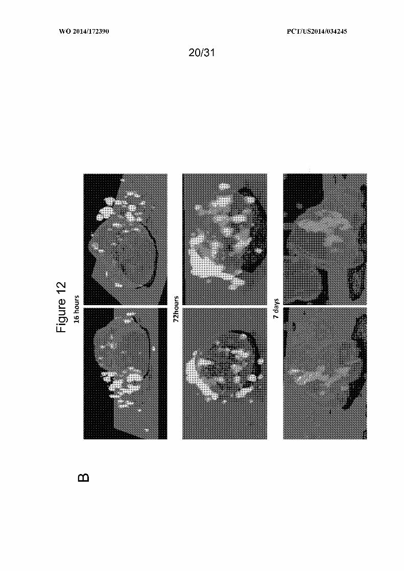
[0023] Figure 12 depicts, in accordance with an embodiment of the invention, that large
oncosomes are internalized in stromal cells. (A) Top panel shows a dose-dependent response
of internalization of large oncosomes, showing increased signal with increased concentrations

of large oncosomes. The bottom panel shows confocal images of sorted fibroblast positive
for large oncosome internalization. (B) Shows the follow up of fibroblasts that have been
sorted after the treatment with large oncosomes. The cells were fixed and imaged by confocal
respectively 16h, 72h and 7 days after sorting. 3D reconstruction was performed, as shown, to
understand the position of large oncosomes once they enter into the cell.
[0024] Figure 13 depicts, in accordance with an embodiment of the invention, the
identification of common and unique proteins in large oncosomes and smaller EV. (A)
Schematic representation of DIAPH3 -silenced DU145 cells, double-labeled with light and
heavy medium before undergoing isolation of large oncosomes and smaller EV respectively,
for SILAC analysis. (B). List of proteins specific for large oncosomes and small EV. (C)
Volcano plot showing proteins enriched in large oncosomes (right) and small EV (left) (D)
Gene ontology analysis showing enrichment of the indicated biological processes for
differentially expressed proteins (DEPs) in large oncosomes and smaller EV.
[0025] Figure 14 depicts, in accordance with an embodiment of the invention, the validation
of the proteomics data using immuno-flow cytometry and western blotting. (A) Fibronectin 1
(FN1) is significantly overexpressed in large oncosomes in comparison with smaller EV, in
DU145 cells silenced with shDIAPFB. The control is DU145 cells, not silenced with
shDIAPFB. (B) CK18 is only detected in large oncosomes, whereas CD81 and CD63,
commonly used markers for exosomes are significantly overexpressed in the small EV.
[0026] Figure 15 depicts, in accordance with an embodiment of the invention, proteins that
are highly enriched in large oncosomes, with a known functional role in cancer metastasis
and response to the therapy.
[0027] Figure 16 depicts, in accordance with an embodiment of the invention, a list of
proteins enriched in large oncosomes and smaller EV.
[0028] Figure 17 depicts, in accordance with an embodiment of the invention, miRNA
profiling of prostate cells and derived EVs (Discovery). (A, B ) Oncosome formation in
CTxB-FITC labeled RWPE-1 and RWPE-2 cells, was quantitatively analysed in presence or
absence of full medium (FM), serum free medium (SFM) and EGF. (C ) Expression levels of
847 miRNAs in cells and extracellular vesicle. Scatter plots data show the correlation
between: RWPE-2 versus RWPE-1 cells (R=0.944), EV2 versus EV1 (R=0.895), EV1 versus
RWPE-1 cells (R=0.706) and EV2 versus RWPE-2 cells (R=0.649). The difference between

EV2 versus RWPE-2 cells and EV1 versus RWPE-1 cells indicate an active selection and
secretion of miRNA into EV.
[0029] Figure 18 depicts, in accordance with an embodiment of the invention, qRT-PCR
validation of the discovery experiment using the filtration method, and relative quantitation
of miRNAs detected in both EV and donor cells. TaqMan qRT-PCR levels of miRNAs in
EV2 versus RWPE-2 cells (A), and EV1 versus RWPE-1 cells (B ) . (C ) Relative quantitation
of the miRNAs with at least 1.5 -fold expression change (dotted lines) in EV2 versus RWPE-2
cells (left panel), and in EV1 versus RWPE-1 cells (right panel); boxes at the top show the
top 5 expressed miRNAs and boxes at the bottom show the bottom 5 expressed miRNAs.
[0030] Figure 19 depicts, in accordance with an embodiment of the invention, quantitation of
miR-1227. (A) qRT-PCR analysis using miR-1227 specific primers was performed on total
miRNA isolated from large and small EVs (10 000 and 100 000 g) from RWPE -2
overexpressing miR-1227 mimic. This transient overexpression in cells results in the
enrichment of the miRNA in large oncosomes but not in the small EVs. (B) Migration of
cancer associated fibroblasts was enhanced by large EVs overexpressing miR-1227.
DETAILED DESCRIPTION OF THE INVENTION
[0031] All references cited herein, including the references cited therein, are incorporated by
reference in their entirety as though fully set forth. Unless defined otherwise, technical and
scientific terms used herein have the same meaning as commonly understood by one of
ordinary skill in the art to which this invention belongs. The practice of the present invention
will employ, unless indicated specifically to the contrary, conventional methods of molecular
biology and recombinant DNA techniques within the skill of the art, many of which are
described below for the purpose of illustration. Such techniques are fully explained in the
literature. Allen et al., Remington: The Science and Practice of Pharmacy 22nd ed.,
Pharmaceutical Press (September 15, 2012); Hornyak et al, Introduction to Nanoscience and
Nanotechnology, CRC Press (2008); Singleton and Sainsbury, Dictionary of Microbiology
and Molecular Biology 3rd ed., revised ed., J . Wiley & Sons (New York, NY 2006); Smith,
March 's Advanced Organic Chemistry Reactions, Mechanisms and Structure 7th ed., J . Wiley
& Sons (New York, NY 2013); Singleton, Dictionary of DNA and Genome Technology 3rd
ed., Wiley-Blackwell (November 28, 2012); and Green and Sambrook, Molecular Cloning: A
Laboratory Manual 4th ed. , Cold Spring Harbor Laboratory Press (Cold Spring Harbor, NY

2012), provide one skilled in the art with a general guide to many of the terms used in the
present application. For references on how to prepare antibodies, see Greenfield, Antibodies
A Laboratory Manual 2nd ed., Cold Spring Harbor Press (Cold Spring Harbor NY, 2013);
Kohler and Milstein, Derivation of specific antibody-producing tissue culture and tumor lines
by cell fusion, Eur. J . Immunol. 1976 Jul, 6(7):5 11-9; Queen and Selick, Humanized
immunoglobulins, U. S. Patent No. 5,585,089 (1996 Dec); and Riechmann et al, Reshaping
human antibodiesfor therapy, Nature 1988 Mar 24, 332(6 162):323-7.
[0032] One skilled in the art will recognize many methods and materials similar or equivalent
to those described herein, which could be used in the practice of the present invention.
Indeed, the present invention is in no way limited to the methods and materials described.
For purposes of the present invention, the following terms are defined below.
[0033] "Beneficial results" may include, but are in no way limited to, lessening or alleviating
the severity of the disease condition, preventing the disease condition from worsening, curing
the disease condition, preventing the disease condition from developing, lowering the
chances of a patient developing the disease condition and prolonging a patient's life or life
expectancy. In some embodiments, the disease condition is cancer.
[0034] "Cancer" and "cancerous" refer to or describe the physiological condition in
mammals that is typically characterized by unregulated cell growth. Examples of cancer
include, but are not limited to B-cell lymphomas (Hodgkin's lymphomas and/or non-
Hodgkins lymphomas), brain tumor, breast cancer, colon cancer, lung cancer, hepatocellular
cancer, gastric cancer, pancreatic cancer, cervical cancer, ovarian cancer, liver cancer,
bladder cancer, cancer of the urinary tract, thyroid cancer, renal cancer, carcinoma,
melanoma, head and neck cancer, brain cancer, and prostate cancer, including but not limited
to androgen-dependent prostate cancer and androgen-independent prostate cancer.
[0035] "Chemotherapeutic drugs" or "chemotherapeutic agents" as used herein refer
to drugs used to treat cancer including but not limited to Albumin-bound paclitaxel
(nab-paclitaxel), Actinomycin, Alitretinoin, All-trans retinoic acid, Azacitidine,
Azathioprine, Bevacizumab, Bexatotene, Bleomycin, Bortezomib, Carboplatin,
Capecitabine, Cetuximab, Cisplatin, Chlorambucil, Cyclophosphamide, Cytarabine,
Daunorubicin, Docetaxel, Doxifluridine, Doxorubicin, Epirubicin, Epothilone,
Erlotinib, Etoposide, Fluorouracil, Gefitinib, Gemcitabine, Hydroxyurea, Idarubicin,

Imatinib, Ipilimumab, Irinotecan, Mechlorethamine, Melphalan, Mercaptopurine,
Methotrexate, Mitoxantrone, Ocrelizumab, Ofatumumab, Oxaliplatin, Paclitaxel,
Panitumab, Pemetrexed, Rituximab, Tafluposide, Teniposide, Tioguanine,
Topotecan, Tretinoin, Valrubicin, Vemurafenib, Vinblastine, Vincristine, Vindesine,
Vinorelbine, Vorinostat, Romidepsin, 5-fluoro uracil (5-FU), 6-mercaptopurine (6-
MP), Cladribine, Clofarabine, Floxuridine, Fludarabine, Pentostatin, Mitomycin,
ixabepilone, Estramustine, or a combination thereof.
[0036] "Isolated" or "purified" large oncosomes as used herein refers to large oncosomes that
are not in their natural milieu. No particular level of purification is required. For example, an
isolated large oncosome may be removed from its native or natural environment.
[0037] "Large Oncosomes" as used herein refer to tumor-derived microvesicles that transmit
signaling complexes between cell and tissue compartments. Large oncosomes are about Ιµιη
to about ΙΟµιη in size. In some embodiments, large oncosomes are shed by amoeboid tumor
cells. Large oncosomes comprise lipids, nucleic acids and proteins, each or a combination of
which may be used to detect and/or quantify large oncosomes. In various embodiments, the
size of the large oncosomes may be about Ιµιη to ΙΟµιη, about 2µιη to ΙΟµιη, about 3µιη to
ΙΟµιη, about 4µιη to ΙΟµιη, about 5µιη to ΙΟµιη, about 6µιη to ΙΟµιη, about 7µιη to ΙΟµιη,
about 8µιη to ΙΟµιη or about 9µιη to ΙΟµιη in size.
[0038] "Patient outcome" refers to whether a patient survives or dies as a result of treatment.
A more accurate prognosis for patients as provided in this invention increases the chances of
patient survival.
[0039] "Poor Prognosis" means that the prospect of survival and recovery of disease is
unlikely despite the standard of care for the treatment of the cancer (for example, prostate
cancer), that is, surgery, radiation, chemotherapy. Poor prognosis is the category of patients
whose survival is less than that of the median survival.
[0040] "Good Prognosis" means that the prospect of survival and recovery of disease is
likely with the standard of care for the treatment of the disease, for example, surgery,
radiation, chemotherapy. Good prognosis is the category of patients whose survival is not
less than that of the median survival.
[0041] A "recurrence" means that the cancer has returned after initial treatment.

[0042] "Non-recurrent" or "recurrence-free", as used herein means that the cancer is in
remission; being recurrent means that the cancer is growing and/or has metastasized, and
some surgery, therapeutic intervention, and/or cancer treatment is required to lower the
chance of lethality. The "non-recurrent subjects" are subjects who have non-recurrent or
recurrence-free disease, and they can be used as the control for recurrent subjects who have
recurrent disease or recurrence.
[0043] "Subject" or "individual" or "animal" or "patient" or "mammal," is meant any
subject, particularly a mammalian subject, for whom diagnosis, prognosis, or therapy is
desired. In some embodiments, the subject has cancer. In some embodiments, the subject
had cancer at some point in the subject's lifetime. In various embodiments, the subject's
cancer is in remission, is re-current or is non-recurrent.
[0044] "Mammal" as used herein refers to any member of the class Mammalia, including,
without limitation, humans, domestic animals, farm animals, zoo animals, sport animals, pet
animals such as dogs, cats, guinea pigs, rabbits, rats, mice, horses, cattle, cows; primates such
as apes, monkeys, orangutans, and chimpanzees; canids such as dogs and wolves; felids such
as cats, lions, and tigers; equids such as horses, donkeys, and zebras; food animals such as
cows, pigs, and sheep; ungulates such as deer and giraffes; rodents such as mice, rats,
hamsters and guinea pigs; and so on. In certain embodiments, the mammal is a human
subject. The term does not denote a particular age or sex. Thus, adult and newborn subjects,
as well as fetuses, whether male or female, are intended to be included within the scope of
this term.
[0045] "Treatment" and "treating," as used herein refer to both therapeutic treatment and
prophylactic or preventative measures, wherein the object is to prevent or slow down (lessen)
the targeted pathologic condition, prevent the pathologic condition, pursue or obtain
beneficial results, or lower the chances of the individual developing the condition even if the
treatment is ultimately unsuccessful. Those in need of treatment include those already with
the condition as well as those prone to have the condition or those in whom the condition is to
be prevented. Examples of cancer treatment include, but are not limited to, active
surveillance, observation, surgical intervention, chemotherapy, immunotherapy, radiation
therapy (such as external beam radiation, stereotactic radiosurgery (gamma knife), and

fractionated stereotactic radiotherapy (FSR)), focal therapy, systemic therapy, vaccine
therapies, viral therapies, molecular targeted therapies, or a combination thereof.
[0046] "Tumor," as used herein refers to all neoplastic cell growth and proliferation, whether
malignant or benign, and all pre-cancerous and cancerous cells and tissues.
[0047] "Tumor derived microvesicles (TMV)" or "microvesicles (MV)" as used herein refer
to a mixture of exosomes, which are about 20nm to about 80nm in size, and large oncosomes
which are about Ιµιη to about ΙΟµιη in size. "Oncosomes" as described in DiVizio et al,
{Cancer Res. 2009, Vol 69(13) pages 5601-5609) refer to "microvesicles" and contain a
mixture of cell-derived vesicles including but not limited to exosomes and large oncosomes.
Microvesicles may also include shedding microvesicles, microparticles, nanovesicles,
apoptotic bodies, nanoparticles and membrane vesicles.
[0048] "Therapeutic agents" as used herein refers to agents that are used to, for example,
treat, inhibit, prevent, mitigate the effects of, reduce the severity of, reduce the likelihood of
developing, slow the progression of and/or cure, a disease. Diseases targeted by the
therapeutic agents include but are not limited to carcinomas, sarcomas, lymphomas,
leukemia, germ cell tumors, blastomas, antigens expressed on various immune cells, and
antigens expressed on cells associated with various hematologic diseases, autoimmune
diseases, and/or inflammatory diseases.
[0049] Large oncosomes are tumor-derived microvesicles that transmit signaling complexes
between cell and tissue compartments. Herein, the inventors show that amoeboid tumor cells
export large oncosome (1-10µιη diameter) vesicles, derived from bulky cellular protrusions,
which contain metalloproteinases, R A, proteins (for example, caveolin-1 (Cav-1) and the
GTPase ARF6), and are biologically active toward tumor cells, endothelial cells and
fibroblasts. The inventors also describe methods whereby large oncosomes can be selectively
sorted by flow cytometry and analyzed independently of vesicles smaller than Ιµιη . Large
oncosomes were identified in the circulation of different mouse models of prostate cancer and
their abundance correlated with tumor progression. Similar large vesicles were also
identified in human tumor tissues, but they were not detected in the benign compartment.
They were more abundant in metastases.

[0050] Accordingly, the invention is based, at least in part, on the finding that large
oncosomes can be visualized and quantified in biological samples (for example, in tissues and
in circulation), and isolated and characterized using clinically-adaptable methods. These
findings also suggest a mechanism whereby migrating tumor cells condition the tumor
microenvironment and distant sites, thereby potentiating advanced disease. The present
invention addresses the need for determining the likelihood of cancer metastasis and
prognostication of cancer, such as prostate cancer. The invention provides assays and
methods for isolating large oncosomes, determining the likelihood of cancer metastasis based
on the number and/or molecular content of the large oncosomes and for treating cancer in
subjects based on the number and/or molecular content of large oncosomes. In an
embodiment, the cancer is prostate cancer.
[0051] Specifically, provided herein is a process comprising obtaining a biological sample
from a subject that has or previously had cancer (for example, prostate cancer), processing
the sample to obtain platelet-poor plasma, optionally further centrifuging the platelet-poor
plasma to eliminate cells and cellular debris, filtering the supernatant using a filter with an
appropriate pore size to separate large oncosomes from other microvesicles and collecting the
large oncosomes. In an embodiment, the large oncosomes may be in the upper chamber of
the filtration unit and the exosomes and/or other microvesicles may be in the lower chamber
of the filtration unit. An embodiment of the process is depicted in Figure 10.
[0052] In an embodiment, the invention provides a method for isolating large oncosomes
from a blood sample obtained from a subject that has or previously had cancer. In an
embodiment, the cancer is prostate cancer. The method includes processing the blood sample
obtained from the subject to obtain platelet-poor plasma and optionally further eliminating
cells and cellular debris (for example by centrifugation). The platelet-poor plasma is filtered
using a filter with an appropriate pore size to separate large oncosomes from other
microvesicles and the large oncosomes are collected. In an embodiment, the large
oncosomes may be in the upper chamber of the filtration unit and the exosomes and/or other
microvesicles may be in the lower chamber of the filtration unit. The filtration-based method
for purifying large oncosomes is depicted in Figure 10.
[0053] In an embodiment, the invention provides a method for isolating large oncosomes
from a blood sample obtained from a subject that has or previously had prostate cancer. The

method includes processing the blood sample obtained from the subject to obtain platelet-
poor plasma and optionally further centrifuging the platelet-poor plasma at 2800g for 10
minutes at 4°C so as to eliminate and cells and cellular debris. The supernatant is filtered
using a filter with a 0.2µιη pore size as depicted in Figure 10 and the large oncosomes are
collected. The large oncosomes may be collected from the upper chamber of the filtration
unit.
[0054] For the filtration-based method described herein to isolate the large oncosomes also
described herein, the type and size of filters and columns that may be used will be apparent to
a person of skill in the art. For example centrifugal ultracentrifugation devices covering
sample volumes from 500 µΐ to 2 ml with any type of low-protein retentions and wide pH-
range (e.g. 2 - 12 pH) filter membranes, such as polyethersulfone (PES) can be used. The
nominal size of the pores can range between any one or more of 0.1 µιη to Ιµιη, 0.2µιη to
Ιµιη, 0.3 µι ίο Ιµιη, 0.4µι ίο Ιµιη, 0.5 µι ίο Ιµιη, Ο. µιη to Ιµιη, 0.7µιη to Ιµιη, 0.8 µι ίο
Ιµιη or 0.9µιη to Ιµιη .
[0055] The pore size of the filter is selected so as to enrich for large oncosomes, wherein
enriching for large oncosomes comprises obtaining a greater number of large oncosomes
relative to other microvesicles. For example, to separate the large oncosomes from other
microvesicles such as exosomes, the pore size of the filter may be any of 200nm, 300nm,
400nm, 500nm, 600nm, 700nm or 800nm. In an embodiment, the pore size is 200nm.
[0056] In various embodiments, large oncosomes are purified or enriched-for, if after
filtration, the yield is about 100% large oncosomes, 90% large oncosomes, 80%> large
oncosomes, 70%> larger oncosomes, 60%> large oncosomes or 55% large oncosomes.
[0057] Methods for obtaining platelet-poor plasma will be apparent to a person of skill in the
art. For example, platelet-poor plasma may be obtained by the methods described in Floryan,
K . and Berghoff, W. (AORN Journal October 2004, Vol 80(4), pages 667-674) and/or Cai et
al. (IntJRadiat Oncol Biol Phys 2010 Vol 77(3), pages 867-76).
[0058] Provided herein are methods for identifying large oncosomes in a tissue sample. The
method includes providing a tissue sample from the subject, fixing and embedding the tissue
(for example, formalin fixing and paraffin embedding tissue) and staining the tissue sample,
wherein large oncosomes are identified as large membrane sacs attached to and surrounding

the cells. In some embodiments, the tissue is stained with any one or more of hematoxylin
and eosin (H&E), Alizarin red S (ARS), Periodic acid-Schiff (PAS), Masson's trichrome,
Romanowsky stains, Silver stain, Sudan stain, or a combination thereof. In various
embodiments, staining the tissue sample encompasses contacting the embedded tissue sample
with reagents that react with any one or more of lipids, proteins, DNA, RNA or a
combination thereof present in the large oncosomes. The reagent may be any one or more of
a nucleic acid, an antibody, a small molecule, a lipid or a combination thereof. In some
embodiments, the sample is contacted with a label to produce a signal so as to detect any one
or more of proteins, DNA, RNA or a combination thereof present in the large oncosomes. In
exemplary embodiments, the tissue is stained for any one or more of cytokeratins, PSA,
PMSA, Cav-1, MCL2, CK18, FN1, membrane proteins, lipids, carbohydrates or a
combination thereof. In some embodiments, the tissue may be stained for any one or more of
the proteins that are described in Figures 13, 15 or 16.
[0059] Also provided herein are methods for determining the likelihood of cancer metastasis
in a subject in need thereof. The method includes providing a sample from a subject that has
or previously had cancer and detecting large oncosomes in the sample. If the sample is a
tissue sample, the method comprises detecting large oncosomes by staining the tissue,
wherein the large oncosomes may be identified as large membrane sacs attached to and
surrounding the cells. If the sample is a blood sample, the method comprises isolating large
oncosomes by the methods described herein (for example, depicted in Figure 10) and
quantifying the isolated large oncosomes. In various embodiments, the subject has an
increased likelihood of cancer metastasis if there is an increase in the number of large
oncosomes in the sample from the subject relative to a reference sample, or the subject has a
decreased likelihood of cancer metastasis if the number of large oncosomes in the sample
from the subject is the same as or decreased relative to the reference sample. In some
embodiments, increased likelihood of cancer metastasis may be indicative of poor prognosis.
[0060] Further provided are methods for determining the likelihood of prostate cancer
metastasis in a subject in need thereof. The method includes providing a blood sample from
a subject that has or previously had prostate cancer, detecting large oncosomes in the blood
sample by isolating large oncosomes according to the methods described herein (for example,
depicted in Figure 10), quantifying the isolated large oncosomes, and determining that the
subject has an increased likelihood of prostate cancer metastasis if there is an increase in the

number of large oncosomes in the sample from the subject relative to a reference sample, or
determining that the subject has a decreased likelihood of prostate cancer metastasis if the
number of large oncosomes in the sample from the subject is the same as or decreased
relative to the reference sample, so as to determine the likelihood of cancer metastasis in the
subject. In some embodiments, increased likelihood of prostate cancer metastasis may be
indicative of poor prognosis
[0061] Also provided are methods for determining the likelihood of cancer metastasis (for
example, prostate cancer metastasis) in a subject in need thereof. The method includes
providing a tissue sample from a subject that has or previously had cancer (for example,
prostate cancer), detecting large oncosomes in the tissue sample by identifying large
oncosomes attached to the cells in the sample or not-attached but surrounding the cells in the
sample and determining that the subject has an increased likelihood of cancer metastasis (for
example, prostate cancer metastasis) if large oncosomes are present and/or there is an
increase in the number of large oncosomes in the sample from the subject relative to a
reference sample, or determining that the subject has a decreased likelihood of cancer
metastasis (for example, prostate cancer metastasis) if large oncosomes are absent and/or
there is a decrease in the number of large oncosomes in the sample from the subject relative
to the reference sample, so as to determine the likelihood of cancer metastasis in the subject.
In an embodiment, the tissue sample is a prostate tissue sample. In another embodiment, the
tissue sample is a sample from any other organ suspected of being cancerous.
[0062] Provided herein is a method for determining the likelihood of cancer metastasis in a
subject in need thereof. The method includes providing a sample from the subject with
cancer, isolating large oncosomes from the samples, detecting the presence of proteins in the
large onosomes, wherein the proteins are any one or more of GLS, ATP50, PPP2R1A,
SFXN1, ECI, HBA1, HINT2, TUFM, ANP32A, PGLS, ETFA, CRYZ, MGST1, CYB5R3,
TMEM33, PHB, TOP2A, KIF5B, APEX1, RDH1 1, or a combination thereof and
determining the likelihood of cancer metastasis in the subject. In some embodiments, the
subject has an increased likelihood of cancer metastasis if the sample from the subject
comprises an increased numbers of large oncosomes, and/or the proteins in the large
oncosomes, and/or increased levels of the proteins in the large oncosomes relative to the
reference sample. In some embodiments, the subject has a decreased likelihood of cancer
metastasis if the sample from the subject has decreased numbers of large oncosomes, and/or

the proteins are absent in the large oncosomes and/or the subject has decreased levels of the
proteins relative to the reference value. In some emboidments, the large oncosomes are
isolated by the methods described herein, for example in Figure 10.
[0063] Also provided herein is a method for determining the likelihood of cancer metastasis
in a subject in need thereof. The method includes providing a sample from the subject with
cancer, isolating large oncosomes from the samples, detecting the presence of proteins in the
large onosomes, wherein the proteins are any one or more of SLC25A6, PHB2, ATP5B,
SLC25A3, VDAC1, GOT2, STOML2, MDH2, HSPA9, VDAC2, SLC25A5, FN1, SHMT2,
H2AFX, HSPD1, ECH1, PRDX3, SLC25A1 1, HNRNPM, HIST2H2BF, TMX1, MMYH10,
NCEHl, GPI, CK18 or a combination thereof and determining the likelihood of cancer
metastasis in the subject. In some embodiments, the subject has an increased likelihood of
cancer metastasis if the sample from the subject comprises an increased numbers of large
oncosomes, and/or the proteins in the large oncosomes, and/or increased levels of the proteins
in the large oncosomes relative to the reference sample. In some embodiments, the subject
has a decreased likelihood of cancer metastasis if the sample from the subject has decreased
numbers of large oncosomes, and/or the proteins are absent in the large oncosomes and/or the
subject has decreased levels of the proteins relative to the reference value. In some
emboidments, the large oncosomes are isolated by the methods described herein, for example
in Figure 10.
[0064] Also provided herein is a method for determining the likelihood of cancer metastasis
in a subject in need thereof. The method includes providing a sample from the subject with
cancer, isolating large oncosomes from the samples, detecting the presence of micro RNAs in
the large onosomes, wherein the micro RNA is any one or more of miR-491-3p, miR-34b,
miPv-1228, miR-647, miR-1227 or a combination thereof and determining that the subject has
an increased likelihood of cancer metastasis if the sample from the subject comprises
increased levels of large oncosomes and the micro RNAs in the large oncosomes relative to
the reference sample, or determining that the subject has a decreased likelihood of cancer
metastasis if the sample from the subject comprises decreased levels large oncosomes and the
micro RNA relative to the reference value so as to determine the likelihood of cancer
metastasis in the subject. In some embodiments, the large oncosomes are isolated by the
methods described herein, for example, in Figure 10.
[0065] In some embodiments, an increase in the number of large oncosomes in combination
with the presence of any one or more of GLS, ATP50, PPP2R1A, SFXN1, ECI, HBA1,
HINT2, TUFM, ANP32A, PGLS, ETFA, CRYZ, MGST1, CYB5R3, TMEM33, PHB,
TOP2A, KIF5B, APEXl, RDHl 1, or a combination thereof in large oncosomes isolated from
the subject is indicative of increased likelihood of cancer metastasis in the subject.
[0066] In some embodiments, an increase in the number of large oncosomes in combination
with an increase in the amount of any one or more of SLC25A6, PHB2, ATP5B, SLC25A3,
VDAC1, GOT2, STOML2, MDH2, HSPA9, VDAC2, SLC25A5, FN1, SHMT2, H2AFX,
HSPD1, ECH1, PRDX3, SLC25A1 1, HNRNPM, HIST2H2BF, TMX1, MMYH10, NCEH1,
ECHS1, C19orfl0, SSR4, PPIB, GPI, PLEC, PRKCSH, PSMB1, LRRC59, GPT1,
HNRNPU, RPN1, KRT18, SRSF3, CALR, PDIA3, HIST1H4A, NPM1, PSMB5, P4HB,
PDIA4, PSMB3, LRPPRC, HSPA5, ERP29, MYH9, MDH1, TKT, LDHB, GAPDH, RAN,
PDIA6, RPS5, HNRNPC, CK18 or a combination thereof in large oncosomes isolated from
the subject is indicative of increased likelihood of cancer metastasis.
[0067] In some embodiments, an increase in the number of large oncosomes in combination
with an increase in the levels of one or more of miR-491-3p, miR-34b, miR-1228, miR-647,
miR-1227 or a combination thereof in large oncosomes isolated from the subject is indicative
of increased likelihood of cancer metastasis.
[0068] In some embodiments, an increase in the number of large oncosomes in combination
with the presence of any one or more of GLS, ATP50, PPP2R1A, SFXN1, ECI, HBA1,
HINT2, TUFM, ANP32A, PGLS, ETFA, CRYZ, MGST1, CYB5R3, TMEM33, PHB,
TOP2A, KIF5B, APEXl, RDHl 1, or a combination thereof in large oncosomes isolated from
the subject, and increased levels of miR-491-3p, miR-34b, miR-1228, miR-647, miR-1227 or
a combination thereof in large oncosomes isolated from the subject, is indicative of increased
likelihood of cancer metastasis.
[0069] In some embodiments, an increase in the number of large oncosomes in combination
with the presence of any one or more of SLC25A6, PHB2, ATP5B, SLC25A3, VDAC1,
GOT2, STOML2, MDH2, HSPA9, VDAC2, SLC25A5, FN1, SHMT2, H2AFX, HSPD1,
ECH1, PRDX3, SLC25A1 1, HNRNPM, HIST2H2BF, TMX1, MMYH10, NCEH1, ECHS1,
C19orfl0, SSR4, PPIB, GPI, PLEC, PRKCSH, PSMB1, LRRC59, GPT1, HNRNPU, RPN1,
KRT18, SRSF3, CALR, PDIA3, HIST1H4A, NPM1, PSMB5, P4HB, PDIA4, PSMB3,

LRPPRC, HSPA5, ERP29, MYH9, MDH1, TKT, LDHB, GAPDH, RAN, PDIA6, RPS5,
HNRNPC, CK18 or a combination thereof in large oncosomes isolated from the subject, and
increased levels of miR-491-3p, miR-34b, miR-1228, miR-647, miR-1227 or a combination
thereof in large oncosomes isolated from the subject, is indicative of increased likelihood of
cancer metastasis.
[0070] In some embodiments, an increase in the number of large oncosomes in combination
with the presence of any one or more of caveolin-1, CK18, FN1, miR-1227 or a combination
thereof, is indicative of increased likelihood of cancer metastasis.
[0071] In some embodiments, an increase in the number of large oncosomes in combination
with increase in the level of of any one or more of caveolin-1, CK18, FN1, miR-1227 or a
combination thereof relative the the reference value, is indicative of increased likelihood of
cancer metastasis.
[0072] The invention also provides methods for treating cancer (for example, prostate cancer)
in a subject in need thereof. The method comprises providing a biological sample from the
subject, isolating and/or detecting large oncosomes in the sample from the subject by the
methods described herein, determining the likelihood of cancer metastasis in the subject by
the methods described herein and prescribing a therapy and optionally administering the
therapy to the subject if the subject has an increased likelihood of cancer metastasis. In some
embodiments, the sample is blood or tissue. In an embodiment, the subject has an increased
likelihood of cancer metastasis if large oncosomes are present or if there is an increase in the
number of large oncosomes in the sample from the subject relative to a reference sample.
[0073] In various embodiments, the samples for use with the methods and processes
described herein are obtained from human subjects that have cancer or have had cancer. In
an embodiment, the cancer is prostate cancer. In various embodiments, the sample is any one
or more of blood, plasma, tissue, urine or a combination thereof.
Detection of Large Oncosomes
[0074] As described herein, the determination of likelihood of cancer metastasis (for
example, prostate cancer metastasis) in a subject and/or treatment of cancer in a subject (for
example, prostate cancer and/or prostate cancer related metastasis) includes detecting and/or

quantifying large oncosomes in samples obtained from the subject. In various embodiments,
the samples are blood, tissue or a combination thereof.
[0075] Further, as described herein, large oncosomes comprise lipids, nucleic acid and
proteins (collectively terms the "molecular content"), each of which or a combination thereof
may be used to not only detect and/or quantify large oncosomes but may also be used to
identify the type of cancer that may be metastasizing.
[0076] The nucleic acid component of the molecular content of large oncosomes includes
DNA and/or variants thereof such as single-stranded DNA (ssDNA), double-stranded DNA
(dsDNA), cDNA and/or genomic DNA. The nucleic acid component of the molecular
content of large oncosomes also includes RNA and its variants including but not limited to
mRNA, rRNA, tRNA, siRNA, miRNA and/or non-coding RNA. The nucleic acid
component of the molecular content of large oncosomes also includes tandem repeats (such
as satellite DNA/RNA, microsatellite DNA/RNA or minisatellite DNA/RNA), interspersed
repeats (such as transposons (transposable elements), Short Interspersed Nuclear Elements
(SINEs, such as Alu's), Long Interspersed Nuclear Elements (LINEs such LINE-1), global
direct repeats, local direct simple repeats, local direct repeats, local direct repeats with spacer
and/or endogenous retroviruses (endogenous viral elements). Examples of nucleic acids that
may be found in the large oncosomes are shown in Table 1. Examples of microRNAs that
may be found in large oncosomes are shown in Table 2 and Figure 18.
[0077] For example, large oncosomes, isolated from prostate cancer cell lines, also contain
genomic DNA. DNA has been described in exosomes, where it is present in small amounts
and largely fragmented. The amount of DNA contained in large oncosomes is more than 30
times of that found in exosomes, whereas the ratio between single and double strand is 5:1 in
both. Large oncosomes can be an important source of genomic information about the tumor,
in a dynamic manner. For example, large oncosomes from PC3 cells report Myc
amplification on chromosome 8 and PTEN deletion on chromosome 10.
[0078] The protein component of the molecular content of the large oncosomes includes
peptides, polypeptides and/or variants thereof. As described herein, the presence of large
oncosomes in a sample is indicative of increased likelihood of cancer metastasis. In some
exemplary embodiments, the presence of PSA or the presence of Caveolin-1 in the large
oncosomes from the subject may be indicative of prostate cancer metastasis or the presence

of Arf6 in the large oncosomes may be indicative of breast cancer metastasis or the presence
of both PSA and Arf6 may be indicative of prostate cancer metastasis. Other proteins present
in large oncosomes may also be indicative of various cancer metastasis, including prostate
cancer metastasis. In some embodiments, presence or increased presence of proteins in
Figure 15 and 16 in large oncosomes is indicative of increased likelihood of cancer
metastasis.
[0079] Polypeptides may be modified or unmodified. Modifications to the polypeptides
include but are not limited to any one or more of myristoylation, palmitoylation, prenylation,
farnesylation, geranylgeranylation, glypiation, glycosylphosphatidylinositol (GPI)
lipoylation, addition of flavin moiety (FMN or FAD), addition of heme C,
phosphopantetheinylation, diphthamide formation, addition of ethanolamine
phosphoglycerol, hypusine formation, acylation, alkylation, amide bond formation,
butyrylation, gamma-carboxylation, glycosylation, ,hydroxylysine, polysialylation,
malonylation, hydroxylation, iodination, nucleotide addition (such as ADP-ribosylation),
oxidation, phosphate ester (O-linked) or phosphoramidate (N-linked) formation,
phosphorylation, adenylylation, propionylation, pyroglutamate formation, S-
glutathionylation, S-nitrosylation, Succinylation, sulfation, selenoylation, ISGylation,
SUMOylation, ubiquitination, Neddylation, Pupylation, citrullination, deamidation,
eliminylation, carbamylation, disulfide bridges formation, proteolytic cleavage,racemization
or a combination thereof.
[0080] The lipid component of the molecular content of the large oncosomes includes but is
not limited to any one or more of fatty acids, glycerolipids, glycerophospholipids,
sphingolipids, sterol lipids, prenol lipids, saccharolipids, polyketides, phosphoglycerides,
glycolipids, or a combination thereof.
[0081] Large oncosomes may be isolated from biological material including but not limited
to any one or more of tissue, cells, blood, plasma, serum, urine, sputum, spinal fluid, pleural
fluid, nipple aspirate, lymph fluid, fluid of the respiratory tract, fluid of the intestinal tract,
fluid of the genitourinary tract, fluid of the lymphatic system, semen, cerebrospinal fluid,
tears, saliva, intra-organ system fluid, tumor cyst fluid, amniotic fluid or a combination
thereof.

[0082] In some embodiments, large oncosomes are purified from a sample obtained from a
subject and detected and/or quantified without labeling the large oncosomes. In an
embodiment, unlabeled large oncosomes may be quantified using flow cytometry techniques,
for example, using forward scatter flow cytometry. In an embodiment, forward scatter flow
cytometry is used with Ιµιη to ΙΟµιη beads to enrich for detection large oncosomes.
Methods for performing forward scatter flow cytometry would be apparent to a person of
skill in the art and may be performed as described in, for example, Di Vizio et al. {Cell Cycle.
2009 Aug; 8(15):2420-4), Dragovic et al. (Nanomedicine. 201 1 Dec;7(6):780-8),
Wvsoczynski M and Rataiczak MZ (Int J Cancer. 2009 Oct 1;125(7): 1595-603). Broadly,
flow cytometry analysis of large oncosomes may be performed by setting forward scatter
(FSC) and side scatter (SSC) voltages as low as that the background noise, determined by
running double 0.2 µιη filtered PBS at the highest flow rate available (in some embodiments,
no higher that 0-4 events/second. After data acquisition, large oncosomes can be analyzed by
setting FSC and SSC on a logarithmic scale.
[0083] In some embodiments, the labeled large oncosomes may be detected using
microfluidic systems as described in Shao et al. {Nature Medicine Dec 2012 Vol 18(12)
pages 1835-1841). The methods described in Shao et al. are applied to exosomes but as
would be apparent to a person of skill in the art, these methods may be applied to large
oncosomes as well. The larger size of the large oncosomes may facilitate better capture of
the large oncosomes.
[0084] In some embodiments, the isolated/purified large oncosomes obtained from a sample
from a subject may be labeled and then quantified and/or detected. In such instances, the
nucleic acids, lipids and/or proteins in the large oncosomes are labeled. In an embodiment,
flow cytometry is used to detect and quantify the labeled large oncosomes. In an
embodiment, large oncosomes are labeled with antibodies that bind to specific proteins of
interest. For example, in order to detect and/or quantify large oncosomes obtained from a
subject that has or had prostate cancer, the isolated large oncosomes may be labeled with
antibodies that bind antigens including but not limited to PSA, PMSA, PSMA, FASN, Cav-1
or a combination thereof. In an embodiment, the large oncosomes labeled with antibodies are
detected and/or quantified using flow cytometry. Additional proteins that may be labeled
with antibodies to quantify and/or detect large oncosomes are depicted in Figures 15 and 16.

[0085] In further embodiments, the isolated/purified large oncosomes may be denatured and
the denatured material may be used as an analyte to detect the presence of one or more
proteins of interest in the large oncosomes. For example, specific antibodies may be used to
detect the presence of one or more proteins of interest. Any suitable immunoassay method
may be utilized, including those which are commercially available, to ascertain the presence
of, and optionally quantify the amount of, the protein of interest present in the analyte. The
presence (and optionally the amount) of the protein of interest in the analyte is indicative of
the presence of said protein in large oncosomes. In various embodiments, the proteins of
interest may be the cancer specific markers, including but not limited to the markers
described herein. In various embodiments, the antibody is any one or more of a monoclonal
antibody or fragment thereof, a polyclonal antibody or a fragment thereof, chimeric
antibodies, humanized antibodies, human antibodies, and a single chain antibody. Extensive
discussion of the known immunoassay techniques is not required here since these are known
to those of skill in the art. Typical suitable immunoassay techniques include Western blots,
sandwich enzyme-linked immunoassays (ELISA), radioimmunoassays (RIA), competitive
binding assays, homogeneous assays, heterogeneous assays, etc. Various known
immunoassay methods are reviewed, e.g., in Methods in Enzymology, 70, pp. 30-70 and 166-
198 (1980).
[0086] Further, "sandwich-type" assays may be used with the methods described herein.
Some examples of sandwich-type assays are described in U.S. Pat. No. 4,168,146 and U.S.
Pat. No. 4,366,241. Alternatively, "competitive-type" assays may be used with the methods
described herein. In a competitive assay, the labeled probe is generally conjugated with a
molecule that is identical to, or an analog of, the analyte. Thus, the labeled probe competes
with the analyte of interest for the available receptive material. Competitive assays are
typically used for detection of analytes such as haptens, each hapten being monovalent and
capable of binding only one antibody molecule. Examples of competitive immunoassay
devices are described in U.S. Pat. No. 4,235,601, U.S. Pat. No. 4,442,204 and U.S. Pat. No.
5,208,535.
[0087] Antibodies, that may be used to detect one or more proteins of interest in a large
oncosome, may be labeled. In some embodiments, the detection antibody is labeled by
covalently linking to an enzyme, labeled with a fluorescent compound or metal or is labeled
with a chemiluminescent compound. For example, the detection antibody can be labeled

with catalase and the conversion uses a colorimetric substrate composition comprises
potassium iodide, hydrogen peroxide and sodium thiosulphate; the enzyme can be alcohol
dehydrogenase and the conversion uses a colorimetric substrate composition comprises an
alcohol, a pH indicator and a pH buffer, wherein the pH indicator is neutral red and the pH
buffer is glycine-sodium hydroxide; the enzyme can also be hypoxanthine oxidase and the
conversion uses a colorimetric substrate composition comprises xanthine, a tetrazolium salt
and 4,5-dihydroxy-l,3-benzene disulphonic acid. In one embodiment, the detection antibody
is labeled by covalently linking to an enzyme, label with a fluorescent compound or metal, or
label with a chemiluminescent compound.
[0088] Direct and indirect labels can be used in immunoassays. A direct label can be defined
as an entity, which in its natural state, is visible either to the naked eye or with the aid of an
optical filter and/or applied stimulation, e.g., ultraviolet light, to promote fluorescence.
Examples of colored labels which can be used include metallic sol particles, gold sol
particles, dye sol particles, dyed latex particles or dyes encapsulated in liposomes. Other
direct labels include radionuclides and fluorescent or luminescent moieties. Indirect labels
such as enzymes can also be used according to the invention. Various enzymes are known for
use as labels such as, for example, alkaline phosphatase, horseradish peroxidase, lysozyme,
glucose-6-phosphate dehydrogenase, lactate dehydrogenase and urease. For a detailed
discussion of enzymes in immunoassays see Engvall, Enzyme Immunoassay ELISA and
EMIT, Methods of Enzymology, 70, 419-439 (1980).
[0089] The antibody can be attached to a surface. Examples of useful surfaces on which the
antibody can be attached for the purposes of detecting the desired antigen include
nitrocellulose, PVDF, polystyrene, and nylon. The surface or support may also be a porous
support (see, e.g., U.S. Patent No. 7,939,342). The assays can be carried out in various assay
device formats including those described in U.S. Pat. Nos. 4,906,439; 5,051,237 and
5,147,609 to PB Diagnostic Systems, Inc.
[0090] In some embodiments of the processes and methods described herein, detecting the
presence and/or level of antibodies reactive to cancer specific markers (for examples, cancer
specific proteins) present in large oncosomes includes contacting the isolated large
oncosomes from the cancer patient with an antibody or a fragment thereof that specifically
binds to the cancer specific marker of interest, forming an antibody-protein complex between
the antibody and marker present in the sample, washing the sample to remove the unbound
antibody, adding a detection antibody that is labeled and is reactive to the antibody bound to
marker in the sample, washing to remove the unbound labeled detection antibody and
converting the label to a detectable signal, wherein the detectable signal is indicative of the
presence and/or level of the cancer specific marker in the sample from the patient. In some
embodiments, the effector component is a detectable moiety selected from the group
consisting of a fluorescent label, a radioactive compound, an enzyme, a substrate, an epitope
tag, electron-dense reagent, biotin, digonigenin, hapten and a combination thereof. In some
embodiments, the detection antibody is labeled by covalently linking to an enzyme, labeled
with a fluorescent compound or metal, labeled with a chemiluminescent compound. The
level of the marker may be obtained by measuring a light scattering intensity resulting from
the formation of an antibody-protein complex formed by a reaction of marker in the sample
with the antibody, wherein the light scattering intensity of at least 10% above a control light
scattering intensity indicates the likelihood of presence of the cancer specific marker and
thereby, presence of large oncosomes in the sample and increased likelihood of cancer
metastasis in the subject.
[0091] In some embodiments, the isolated/purified large oncosomes obtained from a sample
from a subject may be labeled and then quantified and/or detected. In such instances, the
nucleic acids, lipids and/or proteins in the large oncosomes are labeled. In some
embodiments, the nucleic acids are labeled. For example, mRNA encoding PSA, PMSA,
Cav-1 or a combination thereof may be detected with a polynucleotide capable of hybridizing
with PSA specific mRNA, PMSA specific mRNA and/or Cav-1 specific mRNA, under
stringent hybridization conditions. In various embodiments, micro RNA (miRNA) may also
be used to detect and/or quantify large oncosomes. In some embodiments, some miRNAs
such as miR-31, miR-145, miR-18a, miR-135a, which have been shown to be de-regulated in
various types of cancers including bladder, prostate, breast, and colorectal cancer, may be
markers for specific types of cancers may be markers for specific types of cancers. In certain
embodiments, the RNA (for example, mRNA or miRNA) expression level is determined
using microarray, SAGE, blotting, RT-PCR, quantitative PCR or qNPA.
[0092] Techniques that may be used to assess the amount of nucleic acid encoding cancer-
specific marker of interest present in the large oncosomes isolated from a sample obtained
from a subject include but are not limited to in situ hybridization (e.g., Angerer (1987) Meth.

Enzymol 152: 649). Preferred hybridization-based assays include, but are not limited to,
traditional "direct probe" methods such as Southern blots or in situ hybridization {e.g., FISH
and FISH plus SKY), and "comparative probe" methods such as comparative genomic
hybridization (CGH), e.g., cDNA-based or oligonucleotide -based CGH. The methods can be
used in a wide variety of formats including, but not limited to, substrate (e.g. membrane or
glass) bound methods or array-based approaches. Probes that may be used for nucleic acid
analysis are typically labeled, e.g., with radioisotopes or fluorescent reporters. Preferred
probes are sufficiently long so as to specifically hybridize with the target nucleic acid(s)
under stringent conditions. The preferred size range is from about 200 bases to about 1000
bases. Hybridization protocols suitable for use with the methods of the invention are
described, e.g., in Albertson (1984) EMBO J . 3 : 1227-1234; Pinkel (1988) Proc. Natl. Acad.
Sci. USA 85: 9138-9142; EPO Pub. No. 430,402; Methods in Molecular Biology, Vol. 33: In
situ Hybridization Protocols, Choo, ed., Humana Press, Totowa, N.J. (1994), Pinkel, et al.
(1998) Nature Genetics 20: 207-21 1, and/or Kallioniemi (1992) Proc. Natl Acad Sci USA
89:5321-5325 (1992).
[0093] Methods of "quantitative" amplification are well known to those of skill in the art.
For example, quantitative PCR involves simultaneously co-amplifying a known quantity of a
control sequence using the same primers. This provides an internal standard that may be used
to calibrate the PCR reaction. Detailed protocols for quantitative PCR are provided in Innis,
et al. (1990) PCR Protocols, A Guide to Methods and Applications, Academic Press, Inc.
N.Y.). Measurement of DNA copy number at microsatellite loci using quantitative PCR
anlaysis is described in Ginzonger, et al. (2000) Cancer Research 60:5405-5409. The known
nucleic acid sequence for the genes is sufficient to enable one of skill in the art to routinely
select primers to amplify any portion of the gene. Fiuorogenic quantitative PCR may also be
used in the methods of the invention. In fiuorogenic quantitative PCR, quantitation is based
on amount of fluorescence signals, e.g., TaqMan and sybr green.
[0094] Other suitable amplification methods include, but are not limited to, ligase chain
reaction (LCR) (see Wu and Wallace (1989) Genomics 4 : 560, Landegren, et al. (1988)
Science 241:1077, and Barringer et al. (1990) Gene 89: 117), transcription amplification
(Kwoh, et al. (1989) Proc. Natl. Acad. Sci. USA 86: 1173), self-sustained sequence
replication (Guatelli, et al. (1990) Proc. Nat. Acad. Sci. USA 87: 1874), dot PCR, and linker
adapter PCR, etc.

[0095] In certain embodiments, other techniques may be used to determine expression of a
polynucleotide gene product, including microarray analysis (Han, M., et al, Nat Biotechnol,
19: 631-635, 2001; Bao, P., et al, Anal Chem, 74: 1792-1797, 2002; Schena et al, Proc. Natl.
Acad. Sci. USA 93:10614-19, 1996; and Heller et al, Proc. Natl. Acad. Sci. USA 94:2150-55,
1997) and SAGE (serial analysis of gene expression). Like MPSS, SAGE is digital and can
generate a large number of signature sequences (see e.g., Velculescu, V. E., et al, Trends
Genet, 16: 423-425., 2000; Tuteja R. and Tuteja N . Bioessays. 2004 Aug; 26(8):916-22),
although orders of magnitude fewer than that are available from techniques such as MPSS.
[0096] In certain embodiments, the term "microarray" includes a "nucleic acid microarray"
having a substrate-bound plurality of nucleic acids, hybridization to each of the plurality of
bound nucleic acids being separately detectable. The substrate can be solid or porous, planar
or non-planar, unitary or distributed. Nucleic acid microarrays include all the devices so
called in Schena (ed.), DNA Microarrays: A Practical Approach (Practical Approach Series),
Oxford University Press (1999); Nature Genet. 21(1) (suppl): 1-60 (1999); Schena (ed.),
Microarray Biochip: Tools and Technology, Eaton Publishing Company/BioTechniques
Books Division (2000). Nucleic acid microarrays may include a substrate -bound plurality of
nucleic acids in which the plurality of nucleic acids are disposed on a plurality of beads,
rather than on a unitary planar substrate, as described, for example, in Brenner et al., Proc.
Natl. Acad. Sci. USA 97(4): 1665-1670 (2000). Examples of nucleic acid microarrays may be
found in, for example, U.S. Pat. Nos. 6,391,623, 6,383,754, 6,383,749, 6,380,377, 6,379,897,
6,376,191, 6,372,431, 6,351,712 6,344,316, 6,316,193, 6,312,906, 6,309,828, 6,309,824,
6,306,643, 6,300,063.
[0097] In further embodiments, the large oncosomes isolated from samples (for example,
tissue, blood, plasma or a combination thereof) may be identified by detecting cancer specific
(also referred to herein as cancer-specific) markers including but not limited to any one or
more of cytokeratins (for example basic or neutral cytokeratins (including CK1, CK2, CK3,
CK4, CK5, CK6, CK7, CK8 and CK9) and/or acidic cytokeratins (CK10, CK12, CK 13,
CK14, CK16, CK17, CK18, CK19 and CK20)), prostate-specific antigen (PSA), prostate-
specific membrane antigen (PSMA), caveolin-1 (Cav-1), MCL2, bladder-tumor antigen
(BTA), NMP22, carcinoembryonic antigen (CEA), CA 125, CA 19-9, TPA, hormone
receptors (for example estrogen and progesterone), HER2/neu antigen, CA15-3, CEA, CA27-
29, bcr-abl, CA19-9, EGFR, human chorionic gonadotropin (HCG), alpha fetoprotein (AFP),

neuron-specific enolase (NSE), epidermal growth factor receptor (EGFR), TA-90, S-100,
immunoglobulins or free light chains, Bence Jones proteins, monoclonal protein (M-protein),
CA 125, CA 72-4, LASA-P, HCG, CA 72-4, Lactate dehydrogenase (LDH), FASN or a
combination thereof. Additional cancer specific markers include proteins encoded by genes
set forth in Table 1. In various embodiments, the aforementioned markers may be detected in
the isolated/purified large oncosomes by detecting the nucleic acids that encode the
aforementioned cancer-specific markers or by detecting the proteins that encode the
aforementioned cancer-specific markers. Additional embodiments of proteins that are
present in large oncosomes or enriched in large oncosomes and used to identigy large
oncosomes are depicted in Figure 13, 15 and 16.
[0098] Suitable methods for assaying for the expression of various cancer-specific markers
present in isolated large oncosomes (for example, non-denatured large oncosomes or
denatured large oncosomes) isolated from samples obtained from subjects include but are not
limited to any one or more of DNA sequencing, comparative genomic hybridization (CGH),
array CGH (aCGH), SNP analysis, mR A expression assay, RT-PCR, real-time PCR, or a
combination thereof. In various embodiments, the assay for detection of nucleic acids
encoding cancer-specific markers or protein levels of cancer-specific markers present in the
isolated large oncosomes include any one or more of Northern blot analysis, Southern blot
analysis, reverse transcription-polymerase chain reaction (RT-PCR), polymerase chain
reaction (PCR), enzyme-linked immunosorbent assay (ELISA), radio-immuno assay (RIA),
sodium dodecyl sulfate polyacrylamide gel electrophoresis (SDS-PAGE), Western blot
analysis, mass spectrometry immune assays (MSIA), stable isotope standard capture with
anti-peptide antibodies (SISCAPA), two-dimensional electrophoresis, PROTOMAP, which
combines SDS-PAGE with shotgun proteomics, matrix-assisted laser desorption/ionization
(MALDI), fast parallel proteolysis (FASTpp), yeast two-hybrid analysis, protein microarrays,
immunoaffinity chromatography, immunoaffinity chromatography followed by mass
spectrometry, dual polarization interferometry, microscale thermophoresis, phage display
method, stable isotope labeling by amino acids in cell culture (SILAC) or a combination
thereof. In some embodiments, the presence of cancer-specific markers in isolated large
oncosomes may be ascertained by measuring the substrate upon which the marker may act,
such that the substrate serves as a surrogate marker for the cancer specific marker.

[0099] In various embodiments, tissue samples obtained from a subject with cancer (for
example, prostate cancer), may be analyzed for large oncosomes. Large oncosomes in the
tissue sample may be identified by staining the tissue using any one or more of hematoxylin
and eosin (H&E) stain, Periodic acid-Schiff (PAS) stain, Sudan stain, cytostain, Papanicolaou
stain, Nissl stain, azocarmine stain, neutral red or janus green. In further embodiments, the
tissue sample may be analyzed by immuno-histochemical staining. Immuno-histochemical
staining (IHS) may provide information about the presence, location and distribution of large
oncosomes that may be bound to the tissue sample or may be present surrounding the tissue
sample. Antigens for HIS include proteins, peptides, nucleic acids, small molecules or any
other molecule that may be specifically recognized by an antibody. In various embodiments,
the antibodies may specifically bind the cancer specific markers described herein. Unbound
antibody may be removed by washing. The specifically bound antibody may be detected by
treating the bound antibody with, for example a labeled secondary antibody or labeled
avidin/streptavidin. Suitable labels for immunohistochemistry include but are not limited to
fiuorophores such as fluoroscein and rhodamine, enzymes such as alkaline phosphatase and
horse radish peroxidase, and radionuclides such as P and 1 5I . Examples of gene product
markers that may be detected by immuno-histochemical staining include but are not limited
to cytokeratins (for example basic or neutral cytokeratins (including CK1, CK2, CK3, CK4,
CK5, CK6, CK7, CK8 and CK9) and/or acidic cytokeratins (CK10, CK12, CK 13, CK14,
CK16, CK17, CK18, CK19 and CK20)), prostate-specific antigen (PSA), prostate-specific
membrane antigen (PSMA), caveolin-1 (Cav-1), MCL2, bladder-tumor antigen (BTA),
NMP22, carcinoembryonic antigen (CEA), CA 125, CA 19-9, TPA, hormone receptors (for
example estrogen and progesterone), HER2/neu antigen, CA15-3, CEA, CA27-29, bcr-abl,
CA19-9, EGFR, human chorionic gonadotropin (HCG), alpha fetoprotein (AFP), neuron-
specific enolase (NSE), epidermal growth factor receptor (EGFR), TA-90, S-100,
immunoglobulins or free light chains, Bence Jones proteins, monoclonal protein (M-protein),
CA 125, CA 72-4, LASA-P, HCG, CA 72-4, Lactate dehydrogenase (LDH) or a combination
thereof. Additional examples of gene-product markers that may be detected by immuno-
histochemical staining include but are not limited those listed in Figure 13, 15 and 16.
Additional cancer specific markers include proteins encoded by genes set forth in Table 1.

Reference Values
[00100] In various embodiments of the processes and methods described herein, the
reference value is based on the presence and/or number of large oncosomes in a sample
obtained from a subject having cancer (for example, prostate cancer). In one embodiment,
large oncosomes are isolated from a blood sample obtained from a subject that has cancer or
had cancer. In another embodiment, large oncosomes are detected in a tissue sample
obtained from a subject that has cancer of had cancer. As used herein, "subjects that had
cancer" refers to subjects that had cancer at any point in their lifetimes and the cancer is in
remission.
[00101] In some embodiments, the reference value is the mean or median number of
number of large oncosomes and the molecular content (such as proteins, nucleic acids, lipids)
of the large oncosomes in a population of subjects that do not have cancer. In such subjects,
complete absence of large oncosomes may be expected.
[00102] In some embodiments, the reference value is the mean or median number of
large oncosomes and the molecular content (such as proteins, nucleic acids, lipids) of the
large oncosomes in a population of subject that have cancer in remission.
[00103] In an additional embodiment, the reference value is the mean or median
number of large oncosomes and the molecular content (such as proteins, nucleic acids, lipids)
of large oncosomes in one or more samples from the subject wherein the one or more samples
are obtained at a different (for example, an earlier) time point, such as during diagnosis,
before treatment, after treatment or a combination thereof. In an embodiment, the cancer is
prostate cancer.
[00104] Exemplary embodiments of the molecular content of large oncosomes include
proteins depicted in Figures 13, 15 and 16, micro R As depicted in Figure 18 or a
combination thereof.
[00105] In various embodiments, the large oncosomes in the cancer subject compared
to the reference value is increased by at least or about 10%, 20%, 30%>, 40%>, 50%>, 60%>,
70% , 80%o, 90% or 100%. In various embodiments, the large oncosomes in the cancer
subject compared to the reference value is increased by at least or about 1-fold, 2-fold, 3-fold,
4-fold, 5-fold, 10-fold, 15-fold, 20-fold, 25-fold, 30-fold, 35-fold, 40-fold, 45-fold, 50-fold,
55-fold, 60-fold, 65-fold, 70-fold, 75-fold, 80-fold, 85-fold, 90-fold, 95-fold, 100-fold or a
combination thereof.
Therapies
[00106] In accordance with various embodiments of the invention, the therapies that
may be prescribed to a subject with increased likelihood of cancer metastases may be
selected, used and/or administered to treat a cancer patient (for example a prostate cancer
patient). In various embodiments, the therapy may be any one or more of surgery, radiation,
chemotherapy, immunotherapy, vaccine or combinations thereof.
[00107] In some embodiments, chemotherapeutic agents may be selected from any one
or more of cytotoxic antibiotics, antimetabolities, anti-mitotic agents, alkylating agents,
arsenic compounds, DNA topoisomerase inhibitors, taxanes, nucleoside analogues, plant
alkaloids, and toxins; and synthetic derivatives thereof. Exemplary compounds include, but
are not limited to, alkylating agents: treosulfan, and trofosfamide; plant alkaloids: vinblastine,
paclitaxel, docetaxol; DNA topoisomerase inhibitors: doxorubicin, epirubicin, etoposide,
camptothecin, topotecan, irinotecan, teniposide, crisnatol, and mitomycin; anti-folates:
methotrexate, mycophenolic acid, and hydroxyurea; pyrimidine analogs: 5-fluorouracil,
doxifluridine, and cytosine arabinoside; purine analogs: mercaptopurine and thioguanine;
DNA antimetabolites: 2'-deoxy-5-fluorouridine, aphidicolin glycinate, and
pyrazoloimidazole; and antimitotic agents: halichondrin, colchicine, and rhizoxin.
Compositions comprising one or more chemotherapeutic agents (e.g., FLAG, CHOP) may
also be used. FLAG comprises fludarabine, cytosine arabinoside (Ara-C) and G-CSF. CHOP
comprises cyclophosphamide, vincristine, doxorubicin, and prednisone. In another
embodiments, PARP (e.g., PARP-1 and/or PARP-2) inhibitors are used and such inhibitors
are well known in the art (e.g., Olaparib, ABT-888, BSI-201, BGP-15 (N-Gene Research
Laboratories, Inc.); INO-1001 (Inotek Pharmaceuticals Inc.); PJ34 (Soriano et al., 2001;
Pacher et al, 2002b); 3-aminobenzamide (Trevigen); 4-amino-l,8-naphthalimide; (Trevigen);
6(5H)-phenanthridinone (Trevigen); benzamide (U.S. Pat. Re. 36,397); and NU1025
(Bowman et al.).
[00108] In various embodiments, therapies include use of chemotherapeutic agents to
treat prostate cancer. Such agents include but are not limited to Abiraterone Acetate,
Cabazitaxel, Degarelix, Denosumab, Docetaxel, Enzalutamide, Jevtana (Cabazitaxel),

Leuprolide Acetate, Lupron (Leuprolide Acetate), Lupron Depot (Leuprolide Acetate),
Lupron Depot-3 Month (Leuprolide Acetate), Lupron Depot-4 Month (Leuprolide Acetate),
Lupron Depot-Ped (Leuprolide Acetate), Prednisone, Prolia (Denosumab), Provenge
(Sipuleucel-T), Radium 223 Dichloride, Sipuleucel-T, Taxotere (Docetaxel), Viadur
(Leuprolide Acetate), Xgeva (Denosumab), Xofigo (Radium 223 Dichloride), Xtandi
(Enzalutamide), Zytiga (Abiraterone Acetate)or a combination thereof.
[00109] In various embodiments, therapies include, for example, radiation therapy.
The radiation used in radiation therapy can be ionizing radiation. Radiation therapy can also
be gamma rays, X-rays, or proton beams. Examples of radiation therapy include, but are not
limited to, external-beam radiation therapy, interstitial implantation of radioisotopes (1-125,
palladium, iridium), radioisotopes such as strontium-89, thoracic radiation therapy,
intraperitoneal P-32 radiation therapy, and/or total abdominal and pelvic radiation therapy.
For a general overview of radiation therapy, see Hellman, Chapter 16: Principles of Cancer
Management: Radiation Therapy, 6th edition, 2001, DeVita et al., eds., J . B. Lippencott
Company, Philadelphia. The radiation therapy can be administered as external beam radiation
or teletherapy wherein the radiation is directed from a remote source. The radiation treatment
can also be administered as internal therapy or brachytherapy wherein a radioactive source is
placed inside the body close to cancer cells or a tumor mass. Also encompassed is the use of
photodynamic therapy comprising the administration of photosensitizers, such as
hematoporphyrin and its derivatives, Vertoporfm (BPD-MA), phthalocyanine, photosensitizer
Pc4, demethoxy-hypocrellin A; and 2BA-2-DMHA.
[00110] In various embodiments, therapies include, for example, immunotherapy.
Immunotherapy may comprise, for example, use of cancer vaccines and/or sensitized antigen
presenting cells. The immunotherapy can involve passive immunity for short-term protection
of a host, achieved by the administration of pre-formed antibody directed against a cancer
antigen or disease antigen (e.g., administration of a monoclonal antibody, optionally linked to
a chemotherapeutic agent or toxin, to a tumor antigen). Immunotherapy can also focus on
using the cytotoxic lymphocyte-recognized epitopes of cancer cell lines.
[00111] In various embodiments, therapies include, for example, hormonal therapy,
Hormonal therapeutic treatments can comprise, for example, hormonal agonists, hormonal
antagonists (e.g., flutamide, bicalutamide, tamoxifen, raloxifene, leuprolide acetate

(LUPRON), LH-RH antagonists), inhibitors of hormone biosynthesis and processing, and
steroids (e.g., dexamethasone, retinoids, deltoids, betamethasone, Cortisol, cortisone,
prednisone, dehydrotestosterone, glucocorticoids, mineralocorticoids, estrogen, testosterone,
progestins), vitamin A derivatives (e.g., all-trans retinoic acid (ATRA)); vitamin D3 analogs;
antigestagens (e.g., mifepristone, onapristone), or antiandrogens (e.g., cyproterone acetate).
[00112] The duration and/or dose of treatment with anti-cancer therapies may vary
according to the particular anti-cancer agent or combination thereof. An appropriate
treatment time for a particular cancer therapeutic agent will be appreciated by the skilled
artisan. The invention contemplates the continued assessment of optimal treatment schedules
for each cancer therapeutic agent, where the genetic signature of the cancer of the subject as
determined by the methods of the invention is a factor in determining optimal treatment doses
and schedules.
[00113] In various embodiments, the subject for whom predicted efficacy of an anti
cancer therapy is determined, is a mammal (e.g., mouse, rat, primate, non-human mammal,
domestic animal such as dog, cat, cow, horse), and is preferably a human. In another
embodiment of the methods of the invention, the subject has not undergone chemotherapy or
radiation therapy. In alternative embodiments, the subject has undergone chemotherapy or
radiation therapy. In related embodiments, the subject has not been exposed to levels of
radiation or chemotoxic agents above those encountered generally or on average by the
subjects of a species. In certain embodiments, the subject has had surgery to remove
cancerous or precancerous tissue. In other embodiments, the cancerous tissue has not been
removed, e.g., the cancerous tissue may be located in an inoperable region of the body, such
as in a tissue that is essential for life, or in a region where a surgical procedure would cause
considerable risk of harm to the patient, or e.g., the subject is given the anti-cancer therapy
prior to removal of the cancerous tissue.
[00114] The invention provides a system for determining the likelihood of cancer
metastasis wherein large oncosomes are isolated from biological samples obtained from
cancer patients. The system includes a sample analyzer configured to produce a signal for
mRNA encoding a cancer marker of interest or an miRNA that may be a cancer marker
present in the isolated large oncosomes and a computer sub-system programmed to calculate,

based on the mR A or miR A, whether the signal is greater than or not greater than a
reference value.
[00115] The invention also provides a system for determining the likelihood of cancer
metastasis wherein large oncosomes are isolated from biological samples obtained from
cancer patients. The system comprises a sample analyzer configured to produce a signal
when a cancer marker specific antibody binds the cancer marker in the isolated large and a
computer sub-system programmed to calculate, based on the antibody binding whether the
signal is greater than or not greater than a reference value.
[00116] In some embodiments, the computer sub-system is programmed to compare
the aforementioned signals to reference values to determine a likelihood of cancer metastasis
based on an algorithm that classifies the patient as likely to have metastasized form of cancer
if the presence of large oncosomes is increased in the subject relative to a reference value.
[00117] The invention further provides a computer program product embodied in a
computer readable medium that, when executed on a computer, performs steps comprising
detecting cancer marker expression in a sample comprising isolated large oncosomes
obtained from a cancer patient and comparing the cancer marker expression to a reference
value.
[00118] The invention further provides combinations and kits comprising isolated
large oncosomes and one or more reagents to react with the lipids, carbohydrates, proteins,
DNA, RNA or a combination thereof, in the large oncosomes. The reagent may be any one
or more of a nucleic acid, an antibody, a small molecule, a lipid or a combination thereof.
The reagent may further comprises a label to produce a signal so as to detect the proteins,
DNA, RNA or a combination thereof in the oncosomes. The label may be any one or more of
a radiolabel, a chromophore, a fluorophore or a combination thereof.
Advantages
[00119] The "oncosomes" described in Di Vizio et al. (Cancer Res 2009 Vol 69(13)
pages 5601-5609), are a mixture exosomes and large oncosomes. Specifically, as shown in
Figures 10 and 11(a), the oncosome purification method described in DiVizio et al. results in
a mixture of exosomes (Figure 11(a) middle panel black bar) and large oncosomes (Figure

11(a), middle panel white bars), compared to the filtration method described herein (Figure
11(a) right panel) which yields an isolated population of large oncsomes.
[00120] As described herein, presence of large oncosomes in a subject is indicative of
increased likelihood of cancer metastasis. The isolation methods described herein, such as (i)
the filtration method described in Figure 11(a), optionally followed by fluorescence-activated
cell sorting (FACS) with size beads of Ιµιη to ΙΟµιη, and (ii) differential centrifugation
followed by FACS with size beads of Ιµιη to ΙΟµιη, rapidly and selectively enrich for large
oncosomes. The purified large oncosomes are selected away from other microvesicles,
including exosomes to yield essentially purified large oncosomes (Figure 1 and (Figure 11(a)
right panel)). Since presence and/or an increase in the number of large oncosomes are
indicative of increased likelihood of cancer metastasis, the invention provides a quick and
simple test (for example a blood test) that may be used to detect cancer metastasis. Since, as
described herein, the large oncosomes are produced by cancer cells only and may be detected
even without a label, metastasis of any cancer that results in formation and circulation of
large oncosomes, may be detected by a simple blood test. Exosomes in contrast may be
produced by any cell type (Cocucci et al, 2009; Momen-Heravi F et al. 2013) and exosomes
are not necessarily indicative cancer metastasis (compare figures 6C and 6D).
[00121] As shown in the exosome field, in vitro results are not necessarily predictive
of in vivo events. For example, (see Peinado et al. {Nature Medicine 2012 Vol 18(6) pages
883-891) page 884 left column line 16). Di Vizio et al. described in vitro observations in a
mixture of microvesicles shed by cultured cancer cells, but it wasn't until the large
oncosomes were isolated by the methods described herein that it was apparent that not only
are the large oncosomes not an in vitro artifact but are in fact, biologically active comparably
if not superior to exosomes and are a marker for cancer metastasis.

EXAMPLES
Example 1
Methods of the invention
Cell culture
[00122] LNCaP, DU145, PC3 and WPMY-1 were from the American Type Culture
Collection (ATCC) (Di Vizio D et al., Oncosome formation in prostate cancer: association
with a region of frequent chromosomal deletion in metastatic disease, Cancer Res 2009,
69:5601-5609; Adam RM et al, Cholesterol sensitivity of endogenous and myristoylated Akt,
Cancer Res 2007, 67:6238-6246; Di Vizio D et al., Caveolin-1 interacts with a lipid raft-
associated population of fatty acid synthase, Cell Cycle 2008, 7:2257-2267). LNCaP/LacZ
and LNCaP/MyrAktl lines were described (Adam RM et al., Cholesterol sensitivity of
endogenous and myristoylated Akt, Cancer Res 2007, 67:6238-6246). For the generation of
LNCaP /Vo and LNCaP/Cav-1, parental LNCaP were plated at 70-80% confluence and
transfected using Lipofectamine™ 2000 Tranfection Reagent (Invitrogen). Stable
populations were isolated following selection with Gentamicin G418 0.5 mg/ml. LNCaP
were cultured in RPMI-1640 medium supplemented with 10%> fetal bovine serum (FBS;
Valley Biomedical) 2 mmol/L L-glutamine, 100 U/ml penicillin, and 100 µg/ml streptomycin
(all from Invitrogen). DU145 and PC3 were cultured in DMEM supplemented with 10% and
5% (WPMY-1) FBS, 2 mmol/L L-glutamine, 100 U/ml penicillin, and 100 µg/ml
streptomycin.
DNA and RNA extraction
[00123] Total nucleic acid was isolated by the TRIzol (Invitrogen) extraction method,
and concentration determined by NanoDrop 2000c spectrophotometer (Biolab). RNA and
DNA were fractionated by gel electrophoresis on a 2% agarose gel (Invitrogen) and nucleic
acid purity was evaluated by RNase A (Invitrogen) and DNase 1 (Sigma) digestion. lOOng
nucleic acid from vesicles obtained from 2xl0 7 LNCaP/MyrAktl cells was suspended in 15
µΐ buffer for gel electrophoresis.
Immunoblot analysis
[00124] Cells and purified vesicles were lysed and analyzed by SDS-PAGE and
western blotting with the following antibodies: rabbit polyclonal Cav-1 (N-20) (Santa Cruz);
HA.l l mAb, clone 16B12 (Covance); Aktl and p-Aktl (S473), clone D9E (Cell Signaling),
at a dilution of 1:1000; β-actin mAb, clone AC- 15 (Sigma), at a dilution of 1:5000. ARF6
antibody was obtained from Brigham and Women Hospital, Boston, MA (Powelka AM et al.,
Stimulation-dependent recycling of integrin betal regulated by ARF6 and Rabl l , Traffic
2004, 5:20-36; Di Vizio D et al., Oncosome formation in prostate cancer: association with a
region of frequent chromosomal deletion in metastatic disease, Cancer Res 2009, 69:5601-
5609). 15 µg protein per lane was loaded for western blotting.
Immunofluorescence microscopy
[00125] Cells were incubated on ice with FITC-conjugated cholera toxin B (CTxB)
subunit (Sigma) or Alexa 594-conjugated CTxB (Molecular Probes) and analyzed as
previously described (Di Vizio D et al., Caveolin-1 interacts with a lipid raft-associated
population of fatty acid synthase, Cell Cycle 2008, 7:2257-2267). For selected experiments,
LNCaP/MyrAktl were then fixed in 4% paraformaldehyde (PFA), stained with FITC
conjugated HA (MyrAktl) tag (Santa Cruz Biotechnology), and imaged using an Axioplan 2
microscope (Zeiss) (Di Vizio D et al, Caveolin-1 interacts with a lipid raft-associated
population of fatty acid synthase, Cell Cycle 2008, 7:2257-2267).
Animal models and tumor xenografts
[00126] All experiments were performed in compliance with the guidelines of the
Institutional Animal Care and Use Committee, Children's Hospital Boston. We established
subcutaneous xenografts injecting 2 x 10⁶ LNCaP/LacZ or LNCaP/MyrAktl cells into the
flanks of 8 and 16 SCID mice, respectively. Tumor growth was monitored 3 times per week
from detection of the first palpable tumors and the mice were sacrificed before reaching the
maximum tumor burden of 2,000 cm3 at all 4 sites (6 weeks post implantation). At necropsy,
tumors were removed, weighed, fixed in 10% buffered formalin and embedded in paraffin for
histological analysis. The colony of TRAMP and age-matched wild-type mice were
maintained in a C57BL6/J background and genotyped as described (Wang S et al.prostate-
specific deletion of the murine Pten tumor suppressor gene leads to metastatic prostate
cancer, Cancer Cell 2003, 4:209-221; Chen Z et al., Crucial role of p53-dependent cellular

senescence in suppression of Pten-deficient tumorigenesis, Nature 2005, 436:725-730). In
some experiments, TRAMP mice from an F l C57BL6/J X FVB/N cross were used.
Pten KO/Trp53 KO and Pten K0 /KRAS mice were maintained as described(Wang S et
al.,Prostate-specific deletion of the murine Pten tumor suppressor gene leads to metastatic
prostate cancer, Cancer Cell 2003, 4:209-221).
Large Oncosome isolation
[00127] Large oncosomes were purified from conditioned medium or from 500µ1 of
platelet-poor plasma as previously described (Di Vizio D et al., Oncosome formation in
prostate cancer: association with a region of frequent chromosomal deletion in metastatic
disease, Cancer Res 2009, 69:5601-5609). Briefly, cells and debris were eliminated by
centrifugation at 2,800xg per 10 min. The supernatant was centrifuged at 100,000xg for 80
min. Purified oncosomes were washed in PBS, stained with the indicated antibodies, fixed
and permeabilized in ethanol, then analyzed by FACS and sorted using 1 and ΙΟµιη bead
standards, followed by microscopic examination. In selected experiments, unfixed large
oncosomes were sorted using size gates with 1 and ΙΟµιη bead standards and used for
biological experiments.
Gelatin zymography and in vitro degradation assay
[00128] Lysed particles isolated from growth media were analyzed by gelatin
zymography (substrate gel electrophoresis) (Braunhut SJ and Moses MA. Retinoids modulate
endothelial cell production of matrix-degrading proteases and tissue inhibitors of
metalloproteinases (TIMP), J Biol Chem 1994, 269:13472-13479). The gelatin degradation
assay was performed by seeding isolated particles (1-5 µg) on FITC-labeled gelatin coated
coverslips for 1.5 hr, at 37 °C using a modification of a published protocol (Ramachandran A
et al., An Akt- and Fra-1 -dependent pathway mediates platelet-derived growth factor-induced
expression of thrombomodulin, a novel regulator of smooth muscle cell migration, Am J
Pathol 2010, 177:1 19-131).
Migration assays
[00129] lxlO 5 mouse dermal endothelial cells (MDEC), tumor endothelial cells (TEC),
or DU145 cells treated with vesicles shed from cells (10 µg/mL) were stained with
CellTracker RED CMPTX (Invitrogen), and assayed in Transwell FluoroBlok inserts for 16

hours. Cell migration was measured as described (Ramachandran A et al., An Akt- and Fra-
1-dependent pathway mediates platelet-derived growth factor-induced expression of
thrombomodulin, a novel regulator of smooth muscle cell migration, Am J Pathol 2010,
177:1 19-131). Alternatively, WPMY-1 cells (1.5xl0 5) were seeded into the bottom of 24-
well plates and treated for 24 hr with 10 µg/mL LNCaP/MyrAktl vesicles. DU145 cells
(lxl 05), labeled with CellTracker, were seeded into FluoroBlok inserts placed into the
WPMY-1 -conditioned media and migration monitored as above. Vesicles were unfixed
when used in migration assays. Results represent fold-changes from the average of biological
triplicates performed in technical duplicate.
Flow cytometry
[00130] Purified vesicles were stained with propidium iodide (PI) Staining Buffer (BD
Bioscience) or with the indicated antibodies, were processed on a Moflo High-Speed Cell
Sorter (Beckman-Coulter) and analyzed using FlowJo software (Treestar). 1 and ΙΟµιη bead
standards (Spherotech Inc.) were used to set size gates. At least 3,000 events were recorded
(Wersto RP et al., Doublet discrimination in DNA cell-cycle analysis, Cytometry 2001,
46:296-306). The following antibodies were used: HA-probe (F-7) FITC (Santa Cruz), at a
dilution of 1:200, Cav-1 (N-20) (Santa Cruz), at dilution 1:200.
Electron microscopy
[00131] Large oncosomes . Purified material was adsorbed to glow-discharged,
carbon-coated copper grids and stained with 0.75% (w/v) uranyl formate as described (Ohi M
et al. Negative Staining and Image Classification - Powerful Tools in Modern Electron
Microscopy, Biol Proced Online 2004, 6:23-34). Images were collected on a Philips CM10
electron microscope (FEI) operated at an acceleration voltage of 100 kV. Images were
recorded on a IK CCD camera (Gatan) at a magnification of 50,000x and a defocus of about
-1.5 µιη.
[00132] Human xenografts . Sections from the paraffin embedded subcutaneous
tumors were deparaffinized and fixed for 2 hours in Karnovsky fixative before post-fixation
with 1% Os04 and 0.03g/4ml of potassium ferrocyanide. Sections were then stained
overnight with 2% uranyl acetate, dehydrated and embedded overnight with a 1:1 mixture of
LX 112 resin and 100% ethanol. Electron micrographs were collected on a JEOL 1400 TEM
operated at an acceleration voltage of 120kV.

Immunohistochemistry
[00133] Sections from the paraffin embedded mice tumors, human core biopsies, and
human TMA were stained (Adam RM et al., Cholesterol sensitivity of endogenous and
myristoylated Akt, Cancer Res 2007, 67:6238-6246; Di Vizio D et al., Caveolin-1 interacts
with a lipid raft-associated population of fatty acid synthase, Cell Cycle 2008, 7:2257-2267Di
Vizio D et al, Caveolin-1 is required for the upregulation of fatty acid synthase (FASN), a
tumor promoter, during prostate cancer progression, Cancer Biol Ther 2007, 6:1263-1268)
with the following antibodies: Akt mAb (Cell Signaling) at a dilution of 1:500; HA. 11 mAb,
clone 16B12 (Covance), at a dilution of 1:100, CK18 rabbit polyclonal Ab (Epitomics), at
dilution of 1:200.
Patients and samples
[00134] Three human tissue cohorts were used: 1) core biopsies with localized prostate
cancer from 15 patients from the Department of Pathology of University of Naples, Italy; 2) a
TMA from Ulm, Germany, containing 26 multifocal specimens, with at least 2 distinct cancer
foci in the prostate gland and 1 lymph node metastasis; 3) a TMA containing 36 benign
prostate samples, 36 organ confined, and 36 metastatic tumors, of which 11, 20 and 23
samples, respectively, were available for the analysis (Di Vizio D et al., Caveolin-1 interacts
with a lipid raft-associated population of fatty acid synthase, Cell Cycle 2008, 7:2257-2267;
Di Vizio D et al., An absence of stromal caveolin-1 is associated with advanced prostate
cancer, metastatic disease and epithelial Akt activation, Cell Cycle 2009, 8:2420-2424;
Mukhopadhyay NK et al., Heterogeneous nuclear ribonucleoprotein K is a novel regulator of
androgen receptor translation, Cancer Res 2009, 69:2210-2218; Cinar B et al., The pro-
apoptotic kinase Mstl and its caspase cleavage products are direct inhibitors of Aktl, EMBO
J 2007, 26:4523-4534). Informed consent was obtained from all subjects.
Statistical analysis
[00135] Comparisons between experimental groups were performed using two-tailed,
unpaired Student's t test. Pearson's chi-square test was used to assess the correlation
between percentage of MyrAktl -positive events and tumor weight. Fisher exact test was
applied to assess association between presence of large oncosome-like structures and Gleason
score groups and metastases. In all experiments, a p<0.05 was considered significant.

Protein separation and digestion
[00136] Proteins were separated and digested as described (Yang, W. et al, Molecular
& cellular proteomics: MCP 201 1, 10(6), MHO 007492). SILAC-labeled proteins were
mixed at a 1:1 ratio, separated by 10% SDS-PAGE, and stained with Coomassie Brilliant
Blue solution (Bio-Rad). Each lane was cut into ten gel slices of similar size and further cut
into about 1 mm3 particles. Proteins were reduced by 10 mM dithiothreitol (DTT), alkylated
by 55 mM iodoacetamide, and digested with mass spectrometry-grade trypsin (Promega) at
37°C for 16 h . Tryptic peptides were successively extracted with 100 of 5%> acetic acid,
100 of 2.5% acetic acid and 50% acetonitrile, and 100 of 100% acetonitrile. Samples
were dried in a SpeedVac concentrator (Thermo Scientific) and stored at -80°C until MS
analysis.
LC-MS/MS analysis
[00137] Tryptic peptides were redissolved with 20 µΐ 1.5% acetic acid and 5%>
acetonitrile. Samples (10 each) were analyzed by online C18 nanoflow reversed-phase
HPLC (Eksigent nanoLC-2D™) connected to an LTQ Orbitrap XL mass spectrometer
(Thermo Scientific) as described previously (Yang, W et al, Molecular & cellular
proteomics: MCP 2010, 9(1), 54-70.27; Dowal L. et al, Blood 201 1, 118, (13), e62-73).
Briefly, samples were loaded onto a 15 cm nanospray column (75 µιη inner diameter, Magic
CI8 AQ, 3 µιη particle size, 20θΑ pore size, Precision Capillary Columns) and separated at
300 nL/min with a 60-min gradient from 5 to 35% acetonitrile in 0.1% formic acid. MS data
were acquired in a data-dependent manner selecting the fragmentation events based on the
precursor abundance in the survey scan (350-1600 Th). The resolution of the survey scan was
set at 30,000 at m/z 400 Th. Dynamic exclusion was 90 s . After each survey scan, up to ten
collision-induced dissociation (CID) MS/MS spectra were acquired in the linear ion trap.
The mass spectrometry proteomics data have been deposited to the ProteomeXchange
Consortium (http://proteomecentral.proteomexchange.org) via the PRIDE partner repository
(Vizcaino et al, Nucleic Acids Res. Jan 2013; 41(D1): D1063-D1069) with the dataset
identifier PXD000776.
Protein identification and quantitation
[00138] The MS data were analyzed using MaxQuant (vl.3.0.5) (Cox, J . et al, Nature
biotechnology 2008, 26(12), 1367-72). Proteins were identified by searching MS/MS spectra

against the Uniprot database for Human (released on 09/1 1/2012, 84,680 entries) combined
with 262 common contaminants by the Andromeda search engine (Cox, J . et al., Journal of
proteome research 201 1, 10(4), 1794-805), with second peptide identification enabled.
Carbamidomethylation of cysteines was set as a fixed modification, oxidation of methionines
and acetylation of the protein N-terminus as variable modifications. Trypsin allowing for
cleavage N-terminal to proline was chosen as the enzyme specificity. A maximum of two
missed cleavages were allowed. The minimum peptide length was specified to be seven
amino acids. The maximal mass tolerance in MS mode was set to 20 ppm for first search and
6 ppm for main search, and in MS/MS mode 0.7 Da. The maximum false discovery rates
(FDR) for peptide and protein identifications were specified as 0.01.
Identification of differentially expressed proteins (DEPs)
[00139] To identify differentially expressed proteins (DEPs) in large oncosomes (10K
fraction) compared to small EVs (100K fraction), we first normalized the intensities of
proteins identified with more than two sibling peptides in at least one out of two replicates, in
either the large or small EV fractions, using a quantile normalization method (Bolstad, BM et
al, Bioinformatics 2003, 19(2), 185-93). From these normalized intensities, the proteins
differentially expressed between large and small EVs were identified using an integrative
statistical testing method, as previously reported (Moon, PG et al, Proteomics 201 1, 11(12),
2459-75; Lee HJ et al, The Journal of biological chemistry 2010, 285(12), 9262-72). Briefly,
for each protein, we performed 1) two independent tests, a test and a log2 median ratio test;
2) adjusted P values from each two-tailed tests were computed using an empirical distribution
of the null hypothesis (that the means of the genes are not different), which was obtained
from random permutations of the samples; 3) the individual adjusted P values were combined
to compute the FDR using Stouffer's method (Hwang D et al, PNAS 2005, 102, (48), 17296-
301). Note that when using this integrative statistical testing, for the peptides for which the T-
test is not applicable due to the availability of only one intensity value in a given condition,
the adjusted P-value from the log2 median ratio test becomes the FDR. We identified large
oncosome and small EV proteins, with a FDR<0.05 and a fold change > 2 . In addition, to
identify proteins uniquely detected in large oncosomes or small EVs, we employed the
following criteria: 1) being identified in at least one replicate with more than two sibling
peptides and 2) being quantified in one single condition, not quantified in the other condition.
Functional enrichment analysis
[00140] To explore cellular functions and localizations of proteins that were identified
and differentially regulated, we performed functional enrichment analysis using the Database
for Annotation, Visualization and Integrated Discovery (DAVID) software (Dennis, G. Jr. et
al, Genome biology 2003, 4(5), P3). Gene Ontology (GO) terms (Biological Processes and
Cellular Compartments) and Kyoto Encyclopedia of Genes and Genomes (KEGG) pathways
enriched in all identified proteins and DEPs were selected with P<0.05.
Example 2
Filtration -based isolation protocol
[00141] Serum-free conditioned media or human platelet-poor plasma are centrifuged
at low speed (2,800g/10min) to eliminate cells and cell-debris. In comparison with the ultra-
centrifugation based protocol, in which extracellular vesicles (EV) are recovered from the
supernatant after centrifugation at 10,000g/30min or 100,000g/60min, in our new protocol
supernatant is filtered at 8,000g/30sec using Vivaspin ultrafiltration spin columns. Large
oncosomes are recovered form the top of the filter membrane (upper chamber), whereas small
EV, are eluted (lower chamber) (Figures 10 and 11).
[00142] Tumor cells that exhibit the amoeboid feature of membrane blebbing can
release large, plasma membrane-derived vesicles into the extracellular space (Di Vizio D et
al., Oncosome formation in prostate cancer: association with a region of frequent
chromosomal deletion in metastatic disease, Cancer Res 2009, 69:5601-5609) (Figures 1A,
IB, 2A). To determine whether these vesicles can be isolated and analyzed, shed vesicles
were purified from the conditioned medium of LNCaP human prostate cancer cells stably
expressing the potent oncogene MyrAktl (Adam RM et al., Cholesterol sensitivity of
endogenous and myristoylated Akt, Cancer Res 2007, 67:6238-6246), and from WPMY-1
prostate stromal cells. The contents of the purified vesicles reacted with propidium iodide
(PI) (Figure 7A), consistent with the presence of RNA (Figure 1C). This allowed detection
of a Pi-positive vesicle population by fluorescence activated cell sorting (FACS). The
stromal cells produced substantially fewer vesicles than LNCaP/MyrAktl cells (p<0.001)
(Figure ID). Biochemical purification coupled with FACS sorting was thus in agreement
with results using a visual quantitative bleb-formation assay (Di Vizio D et al., Oncosome
formation in prostate cancer: association with a region of frequent chromosomal deletion in

metastatic disease, Cancer Res 2009, 69:5601-5609). Shed vesicles from LNCaP/MyrAktl
cells produced zones of proteolytic clearance in a fluorescent gelatin matrix assay (Ayala I et
al, Multiple regulatory inputs converge on cortactin to control invadopodia biogenesis and
extracellular matrix degradation, J Cell Sci 2008, 121:369-378) (Figure IE) in a similar size
range (2-4µιη) and modal distribution to > 1µιη diameter membrane blebs observed
microscopically (Figures IF, G). MMP zymography showed that the vesicle preparations
contained bioactive MMP9 and MMP2, two key proteases involved in tumor cell invasion
(Figure IE).
[00143] Shed vesicles isolated from LNCaP/MyrAktl cells contained abundant,
membrane-localized MyrAktl as demonstrated by FACS and immunofluorescence staining
(IF) (Figure 2A) and by western blotting (Figure 2B). These results indicated that MyrAktl
is a potentially useful marker with which to track large shed vesicles produced by
LNCaP/MyrAktl cells in vivo. Vesicles shed and purified from U87 glioma cells, which
exhibit amoeboid properties, contained Caveolin-1 (Cav-1), similar to DU145 prostate cancer
cell-derived vesicles (Di Vizio D et al, Oncosome formation in prostate cancer: association
with a region of frequent chromosomal deletion in metastatic disease, Cancer Res 2009,
69:5601-5609). DU145- and LNCaP/MyrAktl vesicle preparations also contained ARF6
(Figure 2B), a GTPase shown recently to mediate large microvesicle shedding from tumor
cells and to be enriched in TMV (Muralidharan-Chari V. et al., ARF6-regulated shedding of
tumor cell-derived plasma membrane microvesicles, Curr Biol 2009, 19:1875-1885). As
reported previously (Di Vizio D., et al., Oncosome formation in prostate cancer: association
with a region of frequent chromosomal deletion in metastatic disease, Cancer Res 2009,
69:5601-5609), silencing of the GTPase-regulated form in DIAPH3 in DU145 cells
potentiated shedding of large microvesicles (Figure 2B). As reported in another recent study,
DIAPH3 loss in DU145 and other tumor cell backgrounds results in amoeboid behavior 10 ,
providing further evidence that shedding of large TMV are a feature of the amoeboid tumor
phenotype.
[00144] Sustained ARF6 activation enhances tumor cell invasion in cell and animal
model systems and vesicle shedding may be a potential mechanism for ARF6-induced
acquisition of invasive potential. Interrogation of publicly available prostate cancer
expression data sets demonstrated that ARF6 mRNA levels are higher in prostate cancer in
comparison with benign tissue (Figure 7). ARF6 ranked in the top 10 percentile of

overexpressed genes in these data sets, and ARF6 mRNA was significantly upregulated in
metastases and in recurrent disease.
[00145] The MMP activity seen with large vesicles shed from LNCaP/MyrAktl cells
(Figure 1F-H) indicates that they are bioactive. In separate bioactivity tests, these vesicle
preparations stimulated migration of mouse tumor endothelial cells (TEC), mouse dermal
endothelial cells (MDEC) (Dudley AC et al., Calcification of multipotent prostate tumor
endothelium, Cancer Cell 2008, 14:201-21 1) (Figure 2C) and DU145 cells (Figure 2D).
LNCaP/MyrAktl vesicles also activated WPMY-1 stromal cells to produce factors that
stimulated migration of DU145 cells (Figure 2E). LNCaP/MyrAktl vesicles also activated
Aktl in WPMY-1 stromal cells (Figure 2F). Finally, incubation of mouse prostatic
fibroblastic cells with LNCaP/MyrAktl vesicles resulted in increased expression of the pro-
metastatic factors BDNF, CXCL12, and osteopontin (Figure 2G). Collectively, these data
indicate that LNCaP/MyrAktl cells shed oncosomes, including particles considerably larger
than nanosized microvesicles, with the potential to contribute to oncogenic signaling by
acting as paracrine effectors of inter-tumor cell, tumor cell-endothelial cell, and epithelial-
stromal reaction.
Example 3
Large oncosomes in the circulation and in situ
[00146] Microvesicles range from about 20nm to about ΙΟµιη. Exosomes (a type of
microvesicles) range from about 20nm to about lOOnm. Large oncosomes (another type of
microvesicles) range in size from about Ιµιη to about ΙΟµιη. To distinguish large oncosomes
from smaller TMV unequivocally, ultracentrifugation and immuno-flow cytometry were used
in combination with sizing beads to create a calibrated domain of analysis based on size.
Following biochemical purification, shed vesicles were stained with DAPI to verify the
absence of cellular contamination and subsequently labeled with an HA antibody, for
detection of membrane-localized MyrAktl. MyrAktl -negative and -positive populations
were then distinguished on a cell sorter, with gates set using 1 and ΙΟµιη beads (Figure 3A).
Because aggregates may potentially be a significant component of the vesicle preparation, we
excluded them from the analysis. To do this, forward scatter signal (FSS) was plotted against
pulse width (PW) of the same signal on a linear scale (Figure 3A). Electronic signal pulses
are composed of pulse height (PH) and PW, and the latter is a function of the time necessary

to traverse the laser beam, which is shorter for single particles. Electric pulses from a flow
cytometer detector with narrow PW identify a singlet (bottom), whereas those with broader
PW identify doublets or possibly clumps (top). In order to restrict our analysis to single
vesicles, we delineated a region in which only single events were analyzed based on their
smaller and more homogeneous PW signals and their corresponding forward scatter signals
(Figure 3A, dotted line). Only these events were gated and considered for further analysis
(Figure 3A, right bottom panel). Electric pulses generated from aggregates, as indicated by
increased PW (Figure 3A, right top panel), were excluded from the analysis, as validated
microscopically (Figure 3A, B).
[00147] Sorted single events < 1µιη and > 1µιη were visualized by both fluorescence
and electron microscopy (EM), indicating that large, single vesicles can be distinguished
within a mixed population. Microscopic analysis of retrieved vesicles following
ultracentrifugation and other sorting procedures indicated that they were not aggregates of
smaller particles and they had a definable structure (Figure 3A,B). These results indicate that
large vesicles can be isolated as discrete events using a tumor cell-specific marker.
[00148] Having demonstrated that membrane blebs shed by cells can be sorted by
FACS as discrete large vesicles, a similar approach was used to isolate and quantify these in
the circulation of SCID mice carrying LNCaP/MyrAktl xenograft tumors. All mice injected
subcutaneously with either LNCaP/MyrAktl or LNCaP/LacZ control cells in Matrigel
developed tumors; however, LNCaP/MyrAktl tumors were significantly larger than
LNCaP/LacZ tumors (Figure 4A). Aktl was membrane localized in LNCaP/MyrAktl tumors
as detected with both Aktl and HA antibodies (Figure 4B). Plasma from mice carrying
LNCaP/MyrAktl tumors contained MyrAktl -positive events in the 1-10µιη size range that
were sorted with the HA antibody (Figure 4C, D). The number of MyrAktl events detected
by FACS correlated with tumor weight (p<0.01) (Figure 4e). Because the HA antibody does
not recognize an endogenous epitope in the mouse, these data indicate that tumor-derived
large vesicles from the subcutaneous xenografts gain access to the circulation.
[00149] MyrAktl -positive vesicles isolated from the blood of mice with MyrAktl
tumors stimulated migration of normal endothelial cells (Figure 4F, left panel), suggesting the
capability of oncosomes in the circulation to evoke progression-related changes in the
microvasculature. A similar result was obtained with MyrAktl -positive vesicles sorted by

FACS in the l - ΙΟµηι size range from the medium of LNCaP/MyrAktl cells (Figure 4F, right
panel), indicating that large oncosomes are bioactive.
[00150] High magnification microscopy of LNCaP/MyrAktl tumors revealed
membrane vesicles within a similar size range to the large oncosomes identified in vitro and
in the circulation of the same animals (Figure 4G). To our knowledge, this type of membrane
vesicle has not been previously described in paraffin sections of prostate tumors. Large
oncosome-like structures were evident when sections of LNCaP/MyrAktl tumors were
stained with hematoxylin and eosin, and immunostained with Aktl and HA antibodies
(Figure 4G). Morphological features with the appearance of intravasating amoeboid cells
were identifiable by immunostaining with Aktl antibody (Figure 4G, right panels). Aktl-
positive vesicles in tumor tissues were also positive for ARF6, thus resembling large
oncosomes secreted from LNCaP/MyrAktl, and DU145 cells (Figure 2B), and in accordance
with the amoeboid character of these cell lines. Transmission electron microscopy (TEM) of
LNCaP/MyrAktl tumor sections showed that the tumor cells produced large, bulbous
structures with the appearance of membrane sacs, often in the proximity of vascular lacunae
(Figure 4H). Higher magnification revealed internal structures with features resembling
polyribosomes. These results suggest that MyrAktl -positive vesicles detected in the
circulation of tumor-bearing mice arise from membrane blebs similar or identical to
amoeboid blebs produced in cell culture.
Example 4
Large oncosome-like vesicles as afeature of metastatic prostate cancer
[00151] Identification of similar large vesicles in human prostate cancer specimens
was investigated. ARF6 staining revealed vesicular structures in the 1-10µιη size range.
Initially a small cohort of human core biopsies (n=15) with localized prostate cancer was
examined, and ARF6-positive large oncosome-like structures in the tumor tissue of 3
specimens were identified. Significantly, similar vesicular structures were also identified
using a CK18 antibody, which detects prostate luminal epithelial cells (Figure 5A). To
determine whether the identification of ARF6-positive vesicles in tissues may reflect clinical
status, a tissue microarray (TMA) containing over 120 punches from foci of localized and
metastatic human prostate cancer was examined (Perner S et al., ERG rearrangement
metastasis patterns in locally advanced prostate cancer, Urology 2010, 75:762-767). High

magnification revealed ARF6- positive vesicles more frequently in foci of Gleason score >7
in comparison with foci of lower Gleason score (p=0.02), suggesting that identification of
large oncosome-like structures in situ may indicate tumor progression. Notably, metastatic
foci exhibited significantly more vesicles (p=0.001) (Figure 5B). These characteristics
represent a previously unrecognized histologic feature of aggressive prostate cancer.
[00152] To determine whether the presence of vesicular structures identified using
similar criteria can differentiate indolent vs. fatal cancer, two models of Pten-deficient mice
with greatly different biological course were used. Pten KO/Trp53 KO mice develop prostate
adenocarcinoma that remains organ-confined within 10-16 weeks after gestation. In
comparison, Pten K0 /KRAS mice develop distant metastases in the liver and lung, and also
develop castrate resistance following androgen ablation. ARF6-positive vesicles, similar in
appearance and size to those seen in the LNCaP/MyrAktl tumors, were abundant in
Pten K0 /KRAS tissues but were absent in the Pten KO/Trp53 KO prostatic tissues. These
findings suggest that large oncosome features in prostate tumors may generally report
aggressive disease.
[00153] Also analyzed was an additional human prostate TMA containing benign
samples, organ confined tumors, and metastatic tumors. Figure 5C shows representative
images of absence (left) and presence (right) of large oncosome-like structures detected using
anti-ARF6. These features were present in less than 30% of the tumors and were not detected
in benign prostate tissue. A significant correlation between the presence of large oncosome-
like features and metastasis (p=0.0008) was observed. The presence of these features also
discriminated between benign and tumor tissue (p<0.0001) (Figure 5D).
Example 5
Large oncosomes containing caveolin-1 identify aggressiveprostate cancer
[00154] To determine whether large oncosomes can be identified using an endogenous
tumor progression marker, large shed vesicles were sorted using Cav-1, a validated prostate
cancer biomarker found in the circulation of patients with advanced disease (Tahir SA et al.,
Secreted caveolin-1 stimulates cell survival/clonal growth and contributes to metastasis in
androgen-insensitive prostate cancer, Cancer Res 2001, 61:3882-3885). Cav-1 was present in
membrane vesicles produced by DU145 cells, suggesting that large shed vesicles are a mobile
vehicle for circulating Cav-1 (Figure 2B). Cav-l-null LNCaP cells stably expressing Cav-1-

GFP produced membrane vesicles that were shed and which contained the fluorescent GFP
fusion protein (Figure 8A-C). Enforced expression of Cav-1 in LNCaP cells significantly
increased the rate of membrane blebbing and shedding (Figure 8D), suggesting that Cav-1
may play an active role in vesiculation. The ability to sort Cav-1 -GFP-positive particles by
FACS in the 1-10µιη size range was validated with a Cav-1 -Cy3 antibody, which decorated
large oncosome-like GFP-positive vesicles (Figure 9A). Using the same approach,
endogenous Cav-1 in shed vesicle preparations from DU145 cells was detected (Figure 9B).
These results suggest that Cav-1 is a viable target for identifying large oncosomes.
[00155] Cav-1 -positive vesicles from the plasma of transgenic mice with
autochthonous prostate tumors (TRAMP) were sorted. TRAMP prostate tissues express high
levels of intra-tumoral Cav-1 (Di Vizio D et al., Caveolin-1 is required for the upregulation of
fatty acid synthase (FASN), a tumor promoter, during prostate cancer progression, Cancer
Biol Ther 2007, 6:1263-1268; Williams TM et al., Caveolin-1 promotes tumor progression in
an autochthonous mouse model of prostate cancer: genetic ablation of Cav-1 delays advanced
prostate tumor development in tramp mice, J Biol Chem 2005, 280:25134- 25145). Age-
matched, non-transgenic littermates were used as controls. Analysis was focused on vesicles
of 1-10µιη in diameter. As a validation method, Cav-1 -positive vesicles >2-3 µιη detected in
plasma were sorted by FACS and visualized by direct optical imaging. Vesicles meeting
these criteria were detected in plasma from TRAMP mice with prostate cancer, but levels
were negligible in plasma of non-transgenic animals and of TRAMP animals with prostate
intraepithelial neoplasia (PIN) (Figure 6A), as confirmed by western blotting (Figure 6B).
These vesicles were substantially more abundant in the circulation of TRAMP mice with
lymph node and lung metastases (p=0.03) (Figsure 6A, C). These results suggest that large
oncosomes containing Cav-1 reach the circulation of mice with prostate cancer and their
presence can reflect the extent of disease progression. Notably, the presence of Cav-1 -
positive vesicles < 1µιη, which were also detected in the plasma, were not correlated with
disease progression in this model (Figure 6D). Significantly, large oncosome-like vesicles
recognized using ARF6 staining were also identified in situ in metastatic TRAMP tumors
(Figure 6E), whereas they were not detected in organ-confined tumors or in benign tissue.

Example 6
[00156] In order to understand the degree to which the large oncosomes resemble or
are distinct from smaller microvesicles, next generation sequencing (whole genome and
RNA-seq) and quantitative proteomics studies (LC-MS/MS SILAC) were performed. The
results demonstrated the feasibility of RNA-seq analysis of microvesicles purified from
human platelet-poor plasma and have obtained a minimum of 50 million paired end reads per
sample. Mapping to RefSeq-annotated human gene loci and quantification as FPKM
(fragments per kilobase of exon per million fragments mapped) identified a number of
transcripts as differentially expressed between patients with breast cancer and normal
subjects (FDR <0.05). Whole genome paired-end sequencing (Illumina) of large oncosome
DNA demonstrated that single nucleotide mutations, insertions/deletions, and translocations
present in donor cells can be identified in the large oncosome DNA. Stable isotope labeling
by amino acids in cell culture (SILAC) profiling of tumor cell-derived large oncosomes in
comparison with smaller microvesicles demonstrated enrichment in large oncosomes of cell
cycle, anti-apoptosis, and cell motility-associated proteins. Among classes of microvesicle
proteins, significant cohorts of the exosome biomarkers were enriched in large oncosomes.
These findings identify large oncosomes as a novel class of mircovesicles that harbor
clinically relevant biomarkers.
Example 7
LC-MS/MS SILAC analysis of large oncosomes versus small E Vfrom amoeboid cancer cells
reveals specific oncosome markers
[00157] . We have previously demonstrated that DIAPH3 silencing induces a rapidly
migratory, amoeboid phenotype in prostate cancer cells, which undergo a massive non-
apoptotic membrane blebbing. This results in the shedding of atypically large bioactive
extracellular vesicles (EV), termed large oncosomes, whose abundance in tumor tissues and
in the circulation correlates with tumor aggressiveness. The finding of a correlation of Cav-1
positive large oncosomes with prostate cancer progression in animal models and patients will
prostate cancer prompted us to perform a deeper characterization of these vesicles in an
attempt to identify a prognostic signature of disease progression.
[00158] We performed a robust proteomic analysis of large oncosomes using a
quantitative SILAC approach. Of the 400 proteins identified in large oncosomes and 387

proteins identified in small EV, 20 were uniquely attributed to large oncosomes and 7 to
small EV, based on identification of each peptide in at least one replicate with more than two
sibling peptides, with an FDR < 0.05 (Figure 13). Most of the proteins that were identified
exclusively in large oncosomes are involved in metabolic/catabolic processes (i.e. ECU and
PGLS) and in redox processes (i.e. MGST1 and CYB5R3), as demonstrated by GO analysis.
Some of the large oncosome proteins have been described as mitochondrial, although most
can also shuttle through different subcellular compartments. For example, PHB, while
mitochondrial-associated, is also described as a transcription factor with pro-proliferative
abilities. Interestingly, PPP2R1A, one of the regulatory subunits of PP2A in mammals,
mediate aberrant DNA duplication and aneuploidy. TOP2 is often overexpressed in cancer
and has been suggested as a therapeutic target. KIF5B is a microtubule binding protein with
motogenic activity, and ANP32A, highly expressed in prostate cancer is involved in
proliferation and mRNA trafficking. Additional prostate cancer-related targets are CYB5R3,
involved in cholesterol synthesis, and RDHl l , exhibiting high expression levels in PCa.
Given the low number of proteins identified exclusively in the small EV population (n=7), we
were not able to perform GO analysis. Interestingly, LAMB2 is involved in cell
differentiation, adhesion, migration, GPR126 is a member of the adhesion family of G-
protein coupled receptors, ITGA6, another well known adhesion molecule, plays a role in
prostate cancer and has been identified as an abundant protein in exosomes.
[00159] Large oncosomes are internalized by stromal cells (Figure 12). Large
oncosomes isolated from LNCaP MyrAktl cells, a prostate cell line overexpressing
Myristoilated Aktl, were used for the treatment of WPMY-1, human immortalized
fibroblasts. In all the experiments the fibroblasts were treated with large oncosomes for l h at
37°C. Top panel of Figure 12A shows a dose-dependent response to internalization of large
oncosomes. The bottom panel of Figure 12A shows confocal images of sorted fibroblast
positive for large oncosome internalization. Figure 12B shows the follow up of fibroblasts
that were sorted after the treatment with large oncosomes. The cells were fixed and imaged
by confocal respectively 16h, 72h and 7 days after sorting. 3D reconstruction was performed
to understand the position of large oncosomes once they enter into the cell and as shown,
more large oncosomes are internalized.
[00160] Oncosome formation in CTxB-FITC labeled RWPE-1 and RWPE-2 cells, was
quantitatively analysed in presence or absence of full medium (FM), serum free medium

(SFM) and EGF. As shown in Figures 17A and 17B, cancer cells lines (RWPE-2) form more
large oncosomes than non-cancer cell lines (RWPE-1). Figure 17C depicts the expression
levels of 847 miRNAs in cells and extracellular vesicle. Scatter plots data show the
correlation between: RWPE-2 versus RWPE-1 cells (R=0.944), EV2 versus EV1 (R=0.895),
EV1 versus RWPE-1 cells (R=0.706) and EV2 versus RWPE-2 cells (R=0.649). The
difference between EV2 versus RWPE-2 cells and EV1 versus RWPE-1 cells indicate an
active selection and secretion of miRNA into EV.
[00161] Figure 18 depicts qRT-PCR validation of the miRNA reults using the
filtration method, and relative quantitation of miRNAs detected in both EV and donor cells.
Figure 18A shows the TaqMan qRT-PCR levels of miRNAs in EV2 versus RWPE-2 cells.
Figure 18B shows the TaqMan qRT-PCR levels of miRNAs in EV1 versus RWPE-1 cells.
Figure 18C depicts the relative quantitation of the miRNAs with at least 1.5-fold expression
change (dotted lines) in EV2 versus RWPE-2 cells (left panel), and in EV1 versus RWPE-1
cells (right panel); boxes at the top show the top 5 expressed miRNAs and boxes at the
bottom show the bottom 5 expressed miRNAs.
[00162] Figure 19 shows the quantitation of miR-1227. In Figure 19(A) qRT-PCR
analysis using miR-1227 specific primers was performed on total miRNA isolated from large
and small EVs (10 000 and 100 000 g) from RWPE -2 overexpressing miR-1227 mimic.
This transient overexpression in cells results in the enrichment of the miRNA in large
oncosomes but not in the small EVs. Figure 19B shows that migration of cancer associated
fibroblasts was enhanced by large EVs overexpressing miR-1227.
[00163] Large oncosomes, isolated from prostate cancer cell lines, also contain
genomic DNA. DNA has been described in exosomes, where it is present in small amounts
and largely fragmented. The amount of DNA contained in large oncosomes is more than 30
times of that found in exosomes, whereas the ratio between single and double strand is 5:1 in
both. Whole genome paired end sequencing (Illumina HiSeq-2000) of large oncosomes and
their donor cells identified, in large oncosomes, 527 out of 5027 indels (insertion, deletion or
translocation) present in the donor cells, suggesting that large oncosomes carry about 10% of
the genomic alterations present in the donor cell. These results suggest that large oncosomes
can be an important source of genomic information about the tumor, in a dynamic manner.

For example, large oncosomes from PC3 cells report Myc amplification on chromosome 8
and PTEN deletion on chromosome 10.
[00164] As demonstrated herein, large oncosomes represent a distinct population of
extracellular vesicles. We can selectively identify specific molecules (such as proteins)
carried in large oncosomes which are tumor specific extracellular vesicles. Large oncosomes,
identified using specific markers, can be stronger reporters of disease status in the body
(plasma). We have a large signature that comprises 20 proteins that are uniquely identified in
large oncosomes and over 50 proteins that are significantly enriched in large oncsomes; this
provides several markers which may be used as biomarkers in the clinic, and that may be
involved in the biogenesis and function of large oncosomes.
[00165] We then focused on additional proteins that were found significantly enriched
in one of the two EV populations and identified 57 proteins with a significantly higher
SILAC ratio in large oncosomes and 44 proteins with a significantly higher SILAC ratio in
small EV (FDR < 0.05) (Figure 16). GPI, LDHB, GOT, FNl and CK18 were significantly
enriched in large oncosomes, whereas the SILAC ratio for CD9, CD81 ITGA3, ITAGV and
TGFBl was higher in small EV. The different roles that these proteins have in the cellular
biology suggest a specialized function of the two different vesicles preparation. Enrichment
of CK18 and FNl in large oncosomes and of CD9 and CD81 in small EV was validated by
western blotting and flow cytometry (Figure 14). Importantly, several proteins that were
highly enriched in large oncosomes, have been shown to play a functional role in cancer
metastasis and response to the therapy (Figure 15). Interestingly, proteins belonging to
categories such as membrane proteins, GTPases, chaperones, and RNA-binding proteins,
identified in and frequently considered specific for exosomes, appeared in both EV
populations, and occasional were enriched in large oncosomes.
Example 8
[00166] This study describes a type of bioactive membrane vesicle previously
unrecognized in vivo, which can originate from amoeboid tumor cells. This class of
microvesicle is large (~1-10 µιη diameter) in comparison to other types of bioactive vesicles
(<1 µιη), in particular, the substantially smaller exosome-sized vesicles (Duijvesz D et al,
Exosomes as biomarker treasure chests for prostate cancer, Eur Urol 201 1, 59:823-831) (30-
lOOnm). Consequently, we propose the term "large oncosome" as a descriptor.

[00167] The data herein describes several independent approaches for the detection of
large oncosomes in the circulation and in formalin-fixed paraffin embedded (FFPE) sections
from mouse models and human tumors. Unlike smaller particles, shown herein is that large
oncosome-like structures are visible in tissues using conventional methods, including
detection with a clinical biomarker (CK18, Figure 4D) in FFPE tissue sections, suggesting
that a method for detecting these pathologic features could be adapted for routine
histopathology. Also described herein is that large oncosomes can be quantified in platelet
poor plasma. In several prostate cancer models, as well as in human TMAs and core
biopsies, large oncosome-like structures are a feature associated with high Gleason grade and
metastatic disease. The results described herein using the LNCaP/MyrAktl model, in which
the oncosomes can be followed from the point of secretion in tissues to circulation in the
blood stream in vivo, support the conclusion that large oncosomes are produced by
aggressive tumor cells and gain access to the circulation. The present study focused on
prostate cancer models. However, because similar processes occur in other solid tumors, we
believe it is likely that similar particles may be detected in other malignancies using
comparable approaches.
[00168] Using specific markers and a FACS method that allowed sorting of vesicles of
1-10µιη diameter and exclusion of aggregates, the results herein show a correlation between
the abundance of large oncosomes in the plasma and tumor weight, in mice carrying
LNCaP/MyrAktl xenograft tumors. In tissues, large vesicles were detected by direct
inspection of H&E-stained tumor sections. ARF6 immunostaining facilitated the detection of
similar structures in paraffin-embedded tissue, including in Pten K0 /KRAS metastatic
prostate tumors, whereas they were not observed in organ-confined Pten K0 tumors.
Pten K0 /KRAS mice develop resistance to androgen ablation, suggesting that quantification
of large oncosome-like features may facilitate assessment of tumor aggressiveness at initial
presentation or disease progression during therapy. In TRAMP mice, the abundance of Cav-
1-positive circulating large oncosome-like structures was highly associated with the presence
of metastatic tumors, and similar vesicular structures were observed in paraffin sections only
from advanced tumors (Figure 6). Notably, the number of smaller events (<1 µιη) in the
circulation of TRAMP mice did not correlate with disease progression.
[00169] Using independent cohorts of human prostate cancer tissues, the findings
herein demonstrate that the large oncosome features discriminate between tumor and benign

tissue, including benign prostatic hyperplasia (BPH), and also between organ- confined and
metastatic tumors. Identifying predictive criteria for prostate tumor progression has been
extremely problematic, and current paradigms, including PSA levels, Gleason score and
clinical stage, are inadequate as predictive tools. In particular, serum PSA can be elevated in
benign conditions such as inflammation and BPH. Collectively, these observations indicate
that assessment of large oncosome features in tumor sections or in the circulation may inform
clinical evaluation. Identification of proteins other than Cav-1 and ARF6 in oncosomes, as
well as discrete RNA species, may result in additional biomarker tools applicable to clinical
medicine. Cav-1 is expressed by normal endothelial cells and has been identified as a
constituent of circulating nanovesicles. For this reason, it is possible that gating particle size
above Ιµιη when Cav-1 is used as a sorting marker will provide information about disease
course that collection of smaller TMV may not.
[00170] Quantitation of circulating tumor cells (CTCs) is currently being evaluated to
assess the risk of disease progression for prostate and other types of cancer. However, the
clinical significance of CTCs remains to be established because of their extremely small
number in peripheral blood as compared with the number of blood cells (Park Y, et al.,
Expected clinical applications of circulating tumor cells in breast cancer, World J Clin Oncol
201 1, 2:303-310). We have demonstrated that large oncosome-like structures can be
separated from plasma in a manner that does not require the capture of CTCs or other cells.
The molecular characterization of large oncosomes may potentially offer a more sensitive and
specific 'liquid biopsy' than CTCs for patient selection, monitoring of treatment efficacy and
assessment of drug-resistance.
[00171] Table 1: Examples of cancer genes

Gene Description Chr Chr Band Tumor Types
AF15Q14 AF15ql4 protein 15 15ql4 AML
ALL 1-fused gene fromAF1Q 1 lq21 ALL
chromosome l q
SH3 protein interactingAF3p21 with Nek, 90 kDa (ALL1 3 3p21 ALL
fused gene from 3p21)
ALL1 fused gene fromAF5q31 5 5q31 ALL
5q31
A kinase (PRKA) anchorAKAP9 7 7q21-q22 papillary thyroid
protein (yotiao) 9
v-akt murine thymoma breast, colorectal, ovarian,AKT1 14 14q32.32
viral oncogene homolog 1 NSCLC
v-akt murine thymoma 19ql3.1-AKT2 19 ovarian, pancreatic
viral oncogene homolog 2 ql3.2
aldehyde dehydrogenase 2ALDH2 12 12q24.2 leiomyoma
family (mitochondrial)
anaplastic lymphoma ALCL, NSCLC,ALK 2 2p23
kinase (Ki-1) neuroblastoma
AL017 KIAA161 8 protein 17 17q25.3 ALCL
colorectal, pancreatic,adenomatous polyposis of
APC 5 5q21 desmoid, hepatoblastoma,the colon gene
glioma, other CNS
RHO guanine nucleotideARHGEF12 exchange factor (GEF) 12 11 l lq23.3 AML
(LARG)
RAS homolog gene family,ARHH 4 4pl3 NHL
member H (TTF)
AT rich interactive domain clear cell ovarian carcinoma,ARID 1A 1 lp35.3
1A (SWI-like) RCC
AT rich interactive domainARID2 12 12ql2 hepatocellular carcinoma
2
aryl hydrocarbon receptorARNT 1 lq21 AML
nuclear translocator

Gene Description Chr Chr Band Tumor Types
alveolar soft part sarcomaASPSCR1 chromosome region, 17 17q25 alveolar soft part sarcoma
candidate 1
ASXL1 additional sex combs like 1 20 20ql l.l MDS, CMML
malignant melanoma of softactivating transcription
ATF1 12 12ql3 parts, angiomatoid fibrousfactor 1
histiocytoma
5-aminoimidazole-4-carboxamide
ATIC ribonucleotide 2 2q35 ALCLformyltransferase/IMPcyclohydrolase
ataxia telangiectasiaATM 11 l lq22.3 T-PLL
mutated
alpha thalassemia/mentalPancreatic neuroendocrineATRX retardation syndrome X- X Xq21.1tumours, paediatric GBM
linked
colorectal, endometrial,prostate and hepatocellular
AXIN1 axin 1 16 16pl3.3carcinomas, hepatoblastoma,sporadic medulloblastoma
BRCA1 associated protein-3p21.31- uveal melanoma, breast,
BAP1 1 (ubiquitin carboxy- 3p21.2 NSCLC, RCC
terminal hydrolase)
BCL10 B-cell CLL/lymphoma 10 1 lp22 MALT
BCL1 1A B-cell CLL/lymphoma 11A 2 2pl3 B-CLL
B-cell CLL/lymphoma 11BBCL1 1B 14 14q32.1 T-ALL
(CTIP2)
BCL2 B-cell CLL/lymphoma 2 18 18q21.3 NHL, CLL
BCL3 B-cell CLL/lymphoma 3 19 19ql3 CLL
BCL5 B-cell CLL/lymphoma 5 17 17q22 CLL
BCL6 B-cell CLL/lymphoma 6 3 3q27 NHL, CLL
BCL7A B-cell CLL/lymphoma 7A 12 12q24.1 BNHL

Gene Description Chr Chr Band Tumor Types
BCL9 B-cell CLL/lymphoma 9 1 lq21 B-ALL
retinoblastoma, AML, APLBCOR BCL6 corepressor X Xpl l.4
(translocation)
BCR breakpoint cluster region 22 22ql l.21 CML, ALL, AML
folliculin, Birt-Hogg-DubeBHD 17 17pl l.2
syndrome
baculoviral IAP repeat-BIRC3 11 I lq22-q23 MALT
containing 3
BLM Bloom Syndrome 15 15q26.1
bone morphogeneticBMPR1A 10 10q22.3
protein receptor, type IA
melanoma, colorectal,papillary thyroid, borderline
v-raf murine sarcoma viralBRAF 7 7q34 ovarian, NSCLC,
oncogene homolog B 1cholangiocarcinoma, pilocyticastrocytoma
familial breast/ovarianBRCA1 17 17q21 ovarian
cancer gene 1
familial breast/ovarianBRCA2 13 13ql2 breast, ovarian, pancreatic
cancer gene 2
lethal midline carcinoma ofBRD3 bromodomain containing 3 9 9q34
young people
lethal midline carcinoma ofBRD4 bromodomain containing 4 19 19pl3.1
young people
BRCA1 interacting proteinBRIP1 17 17q22
C-terminal helicase 1
B-cell translocation gene 1,BTG1 12 12q22 BCLL
anti-pro liferative
BUB 1 budding uninhibitedBUB IB by benzimidazoles 1 15 15ql5
homolog beta (yeast)
chromosome 12 openC12orf9 12 12ql4.3 lipoma
reading frame 9

Gene Description Chr Chr Band Tumor Types
chromosome 15 openC15orf21 15 15q21.1 prostate
reading frame 2 1
chromosome 15 openC15orf55 15 15ql4 lethal midline carcinoma
reading frame 55
chromosome 16 openC16orf75 16 16pl3.13 PMBL, Hodgkin lymphoma
reading frame 75
chromosome 2 openC2orf44 2 2p23.3 NSCLC
reading frame 44
calmodulin binding lp36.31- epithelioidCAMTA1 1
transcription activator 1 p36.23 haemangioendothelioma
calcium activatedCANT1 17 17q25 prostate
nucleotidase 1
caspase recruitmentCARD 11 7 7p22 DLBCL
domain family, member 11
CARS cysteinyl-tRNA synthetase 11 l lpl5.5 ALCL
core-binding factor, runtCBFA2T1 domain, alpha subunit 8 8q22 AML
2;translocated to, 1 (ETO)
core-binding factor, runtdomain, alpha subunit 2;
CBFA2T3 16 16q24 AMLtranslocated to, 3 (MTG-16)
core-binding factor, betaCBFB 16 16q22 AML
subunit
Cas-Br-M (murine)CBL ecotropic retroviral 11 l lq23.3 AML, JMML, MDS
transforming
Cas-Br-M (murine)CBLB ecotropic retroviral 3 3ql3.1 1 AML
transforming sequence b
Cas-Br-M (murine)CBLC ecotropic retroviral 19 19ql3.2 AML
transforming sequence c
CCDC6 10 10q21 NSCLCcoiled-coil domain

Gene Description Chr Chr Band Tumor Types
containing 6
eyei n Bl interactingCCNB1IP1 protein 1, E3 ubiquitin 14 14ql l.2 leiomyoma
protein ligase
CCND1 cyclin Dl 11 l lql3 CLL, B-ALL, breast
CCND2 cyclin D2 12 12pl3 NHL,CLL
CCND3 cyclin D3 6 6p21 MM
CCNE1 cyclin El 19 19ql2 serous ovarian
programmed cell death 1CD273 9 9p24.2 PMBL, Hodgkin lymphoma
ligand 2
CD274 CD274 molecule 9 9p24 PMBL, Hodgkin lymphoma
CD74 molecule, majorhistocompatibility
CD74 5 5q32 NSCLCcomplex, class II invariantchain
CD79a molecule,CD79A immunoglobulin-associated 19 19ql3.2 DLBCL
alpha
CD79b molecule,CD79B immunoglobulin-associated 17 17q23 DLBCL
beta
cadherin 1, type 1, E-CDH1 cadherin (epithelial) 16 16q22.1 lobular breast, gastric
(ECAD)
cadherin 11, type 2, OB-CDH1 1 16 16q22.1 aneurysmal bone cyst
cadherin (osteoblast)
CDK12 cyclin-dependent kinase 12 17 17ql2 serous ovarian
CDK4 cyclin-dependent kinase 4 12 12ql4
CDK6 cyclin-dependent kinase 6 7 7q21-q22 ALL
cyclin-dependent kinasemelanoma, multiple other
CDK 2A inhibitor 2A (pl6(INK4a)) 9 9p21tumour types
gene

Gene Description Chr Chr Band Tumor Types
cyclin-dependent kinasemelanoma, multiple other
CDKN2a(pl4) inhibitor 2A- pl4ARF 9 9p21tumour types
protein
cyclin-dependent kinaseCDK 2C inhibitor 2C (pi 8, inhibits 1 lp32 glioma, MM
CDK4)
caudal type homeo boxCDX2 13 13ql2.3 AMLtranscription factor 2
CCAAT/enhancer bindingCEBPA 19 19ql3.1 AML, MDS
protein (C/EBP), alpha
CEP1 centrosomal protein 1 9 9q33 MPD, NHL
coiled-coil-helix-coiled-CHCHD7 coil-helix domain 8 8ql l.2 salivary gland adenoma
containing 7
CHK2 checkpoint homologCHEK2 22 22ql2.1
(S. pombe)
cysteine-rich hydrophobicCHIC2 4 4ql l-ql2 AML
domain 2
extraskeletal myxoidCHN1 chimerin (chimaerin) 1 2 2q31-q32.1
chondrosarcoma
oligodendroglioma, softCIC capicua homolog 19 19ql3.2 tissue sarcoma
class II, majorCIITA histocompatibility 16 16pl3 PMBL, Hodgkin lymphoma
complex, transactivator
clathrin, heavy polypeptideCLTC 17 17ql l-qter ALCL, renal
(He)
clathrin, heavyCLTCL1 22 22ql l.21 ALCL
polypeptide-like 1
chemokine orphan receptorCMKOR1 2 2q37.3 lipoma
1
CCR4-NOT transcriptionCNOT3 19 19ql3.4 T-ALL
complex subunit 3

Gene Description Chr Chr Band Tumor Types
dermatofibrosarcomaCOL1A1 collagen, type I, alpha 1 17 17q21.31-q22 protuberans, aneurysmal bone
cyst
core promoter elementCOPEB 10 10pl5 prostate, glioma
binding protein (KLF6)
cytochrome c oxidaseCOX6C 8 8q22-q23 uterine leiomyoma
subunit Vic
clear cell sarcoma,cAMP responsive element
CREB1 2 2q34 angiomatoid fibrousbinding protein 1
histiocytoma
cAMP responsive elementCREB3L1 11 l lpl l.2 myxofibrosarcoma
binding protein 3-like 1
cAMP responsive elementCREB3L2 7 7q34 fibromyxoid sarcoma
binding protein 3-like 2
CREB binding proteinCREBBP 16 16pl3.3 ALL, AML, DLBCL, B-NHL
(CBP)
cytokine receptor-like Xp22.3; B-ALL, Downs associatedCRLF2
factor 2 X,Y Ypl l.3 ALL
CREB regulated salivary glandCRTC3 15 15q26.1
transcription coactivator 3 mucoepidermoid
colorectal, ovarian,catenin (cadherin- hepatoblastoma, pleomorphic
CTNNB1 3 3p22-p21.3associated protein), beta 1 salivary gland adenoma, other
tumour types
familial cylindromatosisCYLD 16 16ql2-ql3 cylindroma
gene
DNA segment onD10S170 chromosome 10 (unique) 10 10q21 papillary thyroid, CML
170, H4 gene (PTC1)
pancreatic neuroendocrinedeath-domain associated
DAXX 6 6p21.3 tumour, paediatricprotein
glioblastoma
damage-specific DNADDB2 11 l lpl2
binding protein 2

Gene Description Chr Chr Band Tumor Types
DNA-damage-inducible 12ql3.1-DDIT3 12 liposarcomatranscript 3 ql3.2
DEAD (Asp-Glu-Ala-Asp)DDX10 11 I lq22-q23 AML*
box polypeptide 10
DEAD (Asp-Glu-Ala-Asp)DDX5 17 17q21 prostate
box polypeptide 5
DEAD (Asp-Glu-Ala-Asp)DDX6 11 l lq23.3 B-NHL
box polypeptide 6
DEK oncogene (DNADEK 6 6p23 AML
binding)
sex cord-stromal tumour,dicer 1, ribonuclease type
DICERl 14 14q32.13 TGCT, embryonalIII
rhabdomyosarcoma
DNM2 dynamin 2 19 19pl3.2 ETP ALL
DNA (cytosine-5-)-DNMT3A 2 2p23 AML
methyltransferase 3 alpha
DUX4 double homeobox, 4 4 4q35 soft tissue sarcoma
EBF1 early B-cell factor 1 5 5q34 lipoma
epithelial cell transformingECT2L 6 6q24.1 ETP ALL
sequence 2 oncogene-like
epidermal growth factorreceptor (erythroblastic
EGFR 7 7pl2.3-pl2.1 glioma, NSCLCleukemia viral (v-erb-b)oncogene homolog, avian)
eukaryotic translationEIF4A2 initiation factor 4A, 3 3q27.3 NHL
isoform 2
E74-like factor 4 (etsELF4 domain transcription X Xq26 AML
factor)
ELK4, ETS-domainELK4 protein (SRF accessory 1 lq32 prostate
protein 1)

Gene Description Chr Chr Band Tumor Types
ELKS ELKS protein 12 12pl3.3 papillary thyroid
ELL gene ( 11-19 lysine-ELL 19 19pl3.1 AL
rich leukemia gene)
ELN elastin 7 7ql l.23 B-ALL
echinoderm microtubuleEML4 2 2p21 NSCLC
associated protein like 4
300 kd ElA-Binding colorectal, breast, pancreatic,EP300 22 22ql3protein gene AML, ALL, DLBCL
epidermal growth factorEPS 15 receptor pathway substrate 1 lp32 ALL
15 (AFlp)
v-erb-b2 erythroblasticleukemia viral oncogene
breast, ovarian, other tumourERBB2 homolog 2, 17 17q21.1
types, NSCLC, gastricneuro/glioblastoma derivedoncogene homolog (avian)
excision repair cross-complementing rodentrepair deficiency, 19ql3.2-
ERCC2 19complementation group 2 ql3.3(xeroderma pigmentosumD)
excision repair cross-complementing rodentrepair deficiency,
ERCC3 2 2q21complementation group 3(xeroderma pigmentosumgroup B complementing)
excision repair cross-complementing rodent
ERCC4 16 16pl3.3-pl3.13repair deficiency,complementation group 4
excision repair cross-complementing rodent
ERCC5 repair deficiency, 13 13q33complementation group 5(xeroderma pigmentosum,complementation group G
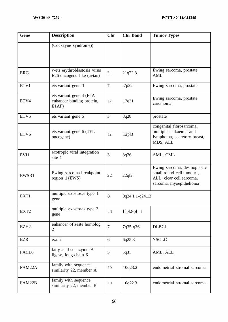
Gene Description Chr Chr Band Tumor Types
(Cockayne syndrome))
v-ets erythroblastosis virus Ewing sarcoma, prostate,ERG 2 1 21q22.3
E26 oncogene like (avian) AML
ETV1 ets variant gene 1 7 7p22 Ewing sarcoma, prostate
ets variant gene 4 (El AEwing sarcoma, prostate
ETV4 enhancer binding protein, 17 17q21carcinoma
E1AF)
ETV5 ets variant gene 5 3 3q28 prostate
congenital fibrosarcoma,ets variant gene 6 (TEL multiple leukaemia and
ETV6 12 12pl3oncogene) lymphoma, secretory breast,
MDS, ALL
ecotropic viral integrationEVI1 3 3q26 AML, CML
site 1
Ewing sarcoma, desmoplasticEwing sarcoma breakpoint small round cell tumour ,
EWSR1 22 22ql2region 1 (EWS) ALL, clear cell sarcoma,
sarcoma, myoepithelioma
multiple exostoses type 1EXT1 8 8q24.1 1-q24.13
gene
multiple exostoses type 2EXT2 11 l lpl2-pl l
gene
enhancer of zeste homologEZH2 7 7q35-q36 DLBCL
2
EZR ezrin 6 6q25.3 NSCLC
fatty-acid-coenzyme AFACL6 5 5q31 AML, AEL
ligase, long-chain 6
family with sequenceFAM22A 10 10q23.2 endometrial stromal sarcoma
similarity 22, member A
family with sequenceFAM22B 10 10q22.3 endometrial stromal sarcoma
similarity 22, member B

Gene Description Chr Chr Band Tumor Types
family with sequenceFAM46C 1 lpl2 MM
similarity 46, member C
Fanconi anemia,FANCA 16 16q24.3
complementation group A
Fanconi anemia,FANCC 9 9q22.3complementation group C
Fanconi anemia,FANCD2 3 3p26complementation group D2
Fanconi anemia,FANCE 6 6p21-p22
complementation group E
Fanconi anemia,FANCF 11 l lpl5complementation group F
Fanconi anemia,FANCG 9 9pl3
complementation group G
FBXOl l F-box protein 11 2 2pl6.3 DLBCL
F-box and WD-40 domain colorectal, endometrial, T-FBXW7 protein 7 (archipelago 4 4q31.3
ALLhomolog, Drosophila)
Fc fragment of IgG, lowFCGR2B affinity lib, receptor for 1 lq23 ALL
(CD32)
FEV protein -FEV 2 2q36 Ewing sarcoma
(HSRNAFEV)
fibroblast growth factorFGFR1 8 8pl l.2-pl l.l MPD, NHL
receptor 1
FGFR1 oncogene partnerFGFRIOP 6 6q27 MPD, NHL
(FOP)
fibroblast growth factorFGFR2 10 10q26 gastric, NSCLC, endometrial
receptor 2
fibroblast growth factor bladder, MM, T-cellFGFR3 4 4pl6.3
receptor 3 lymphoma
FH fumarate hydratase 1 lq42.1

Gene Description Chr Chr Band Tumor Types
pleomorphic salivary glandFHIT fragile histidine triad gene 3 3pl4.2
adenoma
idiopathic hypereosinophilicFIP1L1 FIP1 like 1 (S. cerevisiae) 4 4ql2
syndrome
Friend leukemia virusFLU 11 l lq24 Ewing sarcoma
integration 1
BX648577, FLJ27352FLJ27352 15 15q21.3 PMBL, Hodgkin lymphoma
hypothetical LOC 145788
fms-related tyrosine kinaseFLT3 13 13ql2 AML, ALL
3
formin binding protein 1FNBP1 9 9q23 AML
(FBP17)
granulosa-cell tumour of theFOXL2 forkhead box L2 3 3q23
ovary
FOX01A forkhead box 0 1A (FKHR) 13 13ql4.1 alveolar rhabdomyosarcoma
FOX03A forkhead box 0 3A 6 6q21 AL
FOXP1 forkhead box PI 3 3pl4.1 ALL
follistatin-like 3 (secretedFSTL3 19 19pl3 B-CLL
glycoprotein)
far upstream elementFUBP1 1 lpl3.1 oligodendroglioma
(FUSE) binding protein 1
liposarcoma, AML, Ewingfusion, derived from
sarcoma, angiomatoid fibrousFUS t(12;16) malignant 16 16pl l.2
histiocytoma, fibromyxoidliposarcoma
sarcoma
follicular lymphomaFVT1 18 18q21.3 B-NHL
variant translocation 1
GAS7 growth arrest-specific 7 17 17p AML*
GATA binding protein 1megakaryoblastic leukaemia
GATA1 (globin transcription factor X Xp 11.23of Downs syndrome
1)
GATA2 GATA binding protein 2 3 3q21.3 AML (CML blast

Gene Description Chr Chr Band Tumor Types
transformation)
GATA3 GATA binding protein 3 10 10pl5 breast
guanine monphosphateGMPS 3 3q24 AML
synthetase
guanine nucleotide bindingGNA1 1 protein (G protein), alpha 19 19pl3.3 uveal melanoma
11 (Gq class)
guanine nucleotide bindingGNAQ protein (G protein), q 9 9q21 uveal melanoma
polypeptide
guanine nucleotide bindingprotein (G protein), alpha
GNAS 20 20ql3.2 pituitary adenomastimulating activitypolypeptide 1
golgi autoantigen, golginGOLGA5 14 14q papillary thyroid
subfamily a, 5 (PTC5)
golgi associated PDZ andGOPC coiled-coil motif 6 6q21 glioblastoma
containing
GPC3 glypican 3 X Xq26.1
GPHN gephyrin (GPH) 14 14q24 AL
GTPase regulatorassociated with focal
GRAF 5 5q31 AML, MDSadhesion kinaseppl25(FAK)
H3F3A H3 histone, family 3A 1 lq42.12 glioma
HCMOGT-1 sperm antigen HCMOGT-1 17 17pl l.2 JMML
HEAB ATP GTP binding protein 11 l lql2 AML
homocysteine -inducible,endoplasmic reticulum
HERPUD1 16 16ql2.2-ql3 prostatestress-inducible, ubiquitin-like domain member 1
HEY1 8 8q21hairy/enhancer-of- split mesenchymal

Gene Description Chr Chr Band Tumor Types
related with YRPW motif 1 chondrosarcoma
huntingtin interactingHIP1 7 7ql l.23 CMML
protein 1
HIST1H3B histone cluster 1, H3b 6 6p22.1 glioma
HIST1H4I histone l , H4i (H4FM) 6 6p21.3 NHL
HLF hepatic leukemia factor 17 17q22 ALL
HLXB9 homeo box HB9 7 7q36 AML
microfollicular thyroidhigh mobility group AT-
HMGA1 6 6p21 adenoma, various benignhook 1
mesenchymal tumours
lipoma, leiomyoma,high mobility group AT-
HMGA2 12 12ql5 pleomorphic salivary glandhook 2 (HMGIC)
adenoma
heterogeneous nuclearHNRNPA2B1 7 7pl5 prostateribonucleoprotein A2/B1
HOOK3 hook homolog 3 8 8pl l.21 papillary thyroid
HOXA1 1 homeo box A l l 7 7pl5-pl4.2 CML
HOXA13 homeo box A13 7 7pl5-pl4.2 AML
HOXA9 homeo box A9 7 7pl5-pl4.2 AML*
HOXC1 1 homeo box CI 1 12 12ql3.3 AML
HOXC13 homeo box C13 12 12ql3.3 AML
HOXD1 1 homeo box Dl 1 2 2q31-q32 AML
HOXD13 homeo box D13 2 2q31-q32 AML*
v-Ha-ras Harvey ratinfrequent sarcomas, rare
HRAS sarcoma viral oncogene 11 l lpl5.5other tumour types
homolog
HRPT2 hyperparathyroidism 2 1 Iq21-q31 parathyroid adenoma
heat shock 90kDa proteinHSPCA 14 14q32.31 NHL
1, alpha

Gene Description Chr Chr Band Tumor Types
heat shock 90kDa proteinHSPCB 6 6pl2 NHL
1, beta
isocitrate dehydrogenase 1IDH1 2 2q33.3 glioblastoma
(NADP+), soluble
socitrate dehydrogenase 2IDH2 15 15q26.1 glioblastoma
(NADP+), mitochondrial
MM, Burkitt lymphoma,immunoglobulin heavy
IGH@ 14 14q32.33 NHL, CLL, B-ALL, MALT,locus
MLCLS
immunoglobulin kappaIGK@ 2 2pl2 Burkitt lymphoma, B-NHL
locus
immunoglobulin lambda 22ql l.l-IGL@ 22 Burkitt lymphomalocus ql l.2
IKAROS family zincIKZF1 7 7pl2.2 ALL, DLBCL
finger 1
IL2 interleukin 2 4 4q26-q27 intestinal T-cell lymphoma
IL21R interleukin 2 1 receptor 16 16pl l NHL
interleukin 6 signalIL6ST transducer (gpl30, 5 5ql l hepatocellular carcinoma
oncostatin M receptor)
IL7R interleukin 7 receptor 5 5pl3 ALL, ETP ALL
interferon regulatory factorIRF4 6 6p25-p23 MM
4
immunoglobulinIRTA1 superfamily receptor 1 lq21 B-NHL
translocation associated 1
ITK IL2-inducible T-cell kinase 5 5q31-q32 peripheral T-cell lymphoma
JAK1 Janus kinase 1 1 Ip32.3-p31.3 ALL
JAK2 Janus kinase 2 9 9p24 ALL, AML, MPD, CML
acute megakaryocyteJAK3 Janus kinase 3 19 19pl3.1
leukaemia, ETP ALL

Gene Description Chr Chr Band Tumor Types
juxtaposed with anotherJAZF1 7 7pl5.2-pl5.1 endometrial stromal tumour
zinc finger gene 1
JUN jun oncogene 1 Ip32-p31 sarcoma
potassium inwardly-KCNJ5 rectifying channel, 11 l lq24 adrenal
subfamily J, member 5
lysine (K)-specificKDM5A 12 12pl l AML
demethylase 5A, JARIDIA
lysine (K)-specificXp 11.22-
KDM5C demethylase 5C X clear cell renal carcinomap l l.21(JARID1Q
lysine (K)-specificKDM6A X Xpl l.2 renal, oesophageal SCC, MM
demethylase 6A, UTX
vascular endothelial growthKDR 4 4ql l-ql2 NSCLC, angiosarcoma
factor receptor 2
KIAA1549 KIAA1549 7 7q34 pilocytic astrocytoma
KIF5B kinesin family member 5B 10 10pl l.22 NSCLC
v-kit Hardy-Zuckerman 4 GIST, AML, TGCT,KIT feline sarcoma viral 4 4ql2 mastocytosis, mucosal
oncogene homolog melanoma
KLF4 Kruppel-like factor 4 9 9q31 meningioma
kallikrein-related peptidaseKLK2 19 19ql3.41 prostate2
v-Ki-ras2 Kirsten rat pancreatic, colorectal, lung,KRAS sarcoma 2 viral oncogene 12 12pl2.1 thyroid, AML, other tumour
homolog types
kinectin 1 (kinesinKTN1 14 14q22.1 papillary thyroid
receptor)
lymphoid nuclear proteinLAF4 2 2ql l.2-ql2 ALL, T-ALL
related to AF4
LASP1 LIM and SH3 protein 1 17 17ql l-q21.3 AML
LCK 1 Ip35-p34.3 T-ALLlymphocyte-specific

Gene Description Chr Chr Band Tumor Types
protein tyrosine kinase
lymphocyte cytosolic 13ql4.1-LCP1 13 NHL
protein 1 (L-plastin) ql4.3
leukemia-associatedLCX protein with a CXXC 10 10q21 AML
domain
lipoma HMGIC fusionLHFP 13 13ql2 lipoma
partner
leukemia inhibitory factorLIFR 5 5pl3-pl2 salivary adenoma
receptor
LIM domain only 1LMOl 11 l lpl5 T-ALL, neuroblastoma
(rhombotin 1) (RBTN1)
LIM domain only 2LM02 (rhombotin- like 1) 11 l lpl3 T-ALL
(RBTN2)
LIM domain containingLPP preferred translocation 3 3q28 lipoma, leukaemia
partner in lipoma
leucine-rich repeats andLRIG3 immunoglobulin-like 12 12ql4.1 NSCLC
domains 3
lymphoblastic leukemia 19pl3.2-LYL1 19 T-ALLderived sequence 1 pl3.1
Homo log of Drosophilacolorectal, pancreatic, small
MADH4 Mothers Against 18 18q21.1intestine
Decapentaplegic 4 gene
v-maf musculoaponeuroticMAF fibrosarcoma oncogene 16 16q22-q23 MM
homolog
v-maf musculoaponeurotic20ql l.2-MAFB fibrosarcoma oncogene 20 MMql3.1homolog B (avian)
MALT1 mucosa associated 18 18q21 MALTlymphoid tissue lymphoma

Gene Description Chr Chr Band Tumor Types
translocation gene 1
mastermind-like 2 salivary glandMAML2 11 I lq22-q23(Drosophila) mucoepidermoid
mitogen-activated protein 15q22.1- NSCLC, melanoma,MAP2K1 15
kinase kinase 1 q22.33 colorectal
mitogen-activated proteinMAP2K2 19 19pl3.3 NSCLC, melanoma
kinase kinase 2
mitogen-activated proteinMAP2K4 17 17pl l.2 pancreatic, breast, colorectal
kinase kinase 4
MAX Myc associated factor X 14 14q23 pheochromocytoma
Mdm2 p53 binding protein sarcoma, glioma, colorectal,MDM2 12 12ql5homolog other tumour types
Mdm4 p53 binding protein glioblastoma, bladder,MDM4 1 lq32
homolog retinoblastoma
myelodysplasia syndromeMDS1 3 3q26 MDS, AML
1
myelodysplasia syndromeMDS2 1 lp36 MDS
2
mucoepidermoid salivary glandMECT1 19 19pl3
translocated 1 mucoepidermoid
mediator complex subunitMED 12 X Xql3 uterine leiomyoma
12
parathyroid tumours,multiple endocrine
MEN1 11 l lql3 pancreatic neuroendocrineneoplasia type 1 gene tumour
met proto-oncogenepapillary renal, head-neck
MET (hepatocyte growth factor 7 7q31squamous cell
receptor)
microphthalmia-associatedMITF 3 3pl4.1 melanomatranscription factor
megakaryoblastic leukemia acute megakaryocyteMKL1 22 22ql3
(translocation) 1 leukaemia

Gene Description Chr Chr Band Tumor Types
MLF1 myeloid leukemia factor 1 3 3q25.1 AML
colorectal, endometrial,MLH1 E.coli MutL homolog gene 3 3p21.3
ovarian, CNS
myeloid/lymphoid ormixed-lineage leukemia
MLL 11 l lq23 AML, ALL(trithorax homolog,Drosophila)
myeloid/lymphoid orMLL2 12 12ql2-ql4 medulloblastoma, renal
mixed-lineage leukemia 2
myeloid/lymphoid orMLL3 7 7q36.1 medulloblastoma
mixed-lineage leukemia 3
myeloid/lymphoid ormixed-lineage leukemia
MLLT1 (trithorax homolog, 19 19pl3.3 ALDrosophila); translocatedto, 1 (ENL)
myeloid/lymphoid ormixed-lineage leukemia
MLLT10 (trithorax homolog, 10 10pl2 ALDrosophila); translocatedto, 10 (AF10)
myeloid/lymphoid ormixed-lineage leukemia
MLLT2 (trithorax homolog, 4 4q21 ALDrosophila); translocatedto, 2 (AF4)
myeloid/lymphoid ormixed-lineage leukemia
MLLT3 (trithorax homolog, 9 9p22 ALLDrosophila); translocatedto, 3 (AF9)
myeloid/lymphoid ormixed-lineage leukemia
MLLT4 (trithorax homolog, 6 6q27 ALDrosophila); translocatedto, 4 (AF6)

Gene Description Chr Chr Band Tumor Types
myeloid/lymphoid ormixed-lineage leukemia
MLLT6 (trithorax homolog, 17 17q21 ALDrosophila); translocatedto, 6 (AF17)
myeloid/lymphoid ormixed-lineage leukemia
MLLT7 (trithorax homolog, X Xql3.1 ALDrosophila); translocatedto, 7 (AFX1)
meningioma (disrupted inMN1 22 22ql3 AML, meningioma
balanced translocation) 1
myeloproliferativeMPL leukemia virus oncogene, 1 p34 MPD
thrombopoietin receptor
MSF MLL septin-like fusion 17 17q25 AML*
colorectal, endometrial,MSH2 mutS homolog 2 (E. coli) 2 2p22-p21
ovarian
MSH6 mutS homolog 6 (E. coli) 2 2pl6 colorectal
musashi homolog 2MSI2 17 17q23.2 CML
(Drosophila)
MSN moesin X Xql l.2-ql2 ALCL
mature T-cell proliferation T cell prolymphocyticMTCP1 X Xq28
1 leukaemia
MUC1 mucin 1, transmembrane 1 lq21 B-NHL
lp34.3-MUTYH mutY homolog (E. coli) 1
lp32.1
v-myb myeloblastosis viralMYB 6 6q22-23 adenoid cystic carcinoma
oncogene homolog
v-myc myelocytomatosis8q24.12- Burkitt lymphoma, amplified
MYC viral oncogene homolog 8q24.13 in other cancers, B-CLL
(avian)
MYCL1 v-myc myelocytomatosis 1 lp34.3 small cell lung carcinomaviral oncogene homolog 1,

Gene Description Chr Chr Band Tumor Types
lung carcinoma derived(avian)
v-myc myelocytomatosisviral related oncogene,
MYCN 2 2p24.1 neuroblastomaneuroblastoma derived(avian)
myeloid differentiationMYD88 3 3p22 ABC-DLBCL
primary response gene (88)
myosin, heavy polypeptide 16pl3.13-MYH1 1 16 AML
11, smooth muscle pl3.12
myosin, heavy polypeptideMYH9 22 22ql3.1 ALCL
9, non-muscle
MYST histoneacetyltransferase
MYST4 10 10q22 AML(monocytic leukemia) 4(MORF)
nascent-polypeptide-NACA associated complex alpha 12 12q23-q24.1 NHL
polypeptide
Nijmegen breakageNBS1 8 8q21
syndrome 1 (nibrin)
nuclear receptorNCOA1 2 2p23 alveolar rhabdomyosarcoma
coactivator 1
nuclear receptorNCOA2 8 8ql3.1 AML, chondrosarcoma
coactivator 2 (TIF2)
nuclear receptorNCOA4 coactivator 4 - PTC3 10 10ql l.2 papillary thyroid
(ELE1)
N-myc downstreamNDRG1 8 8q24.3 prostate
regulated 1
neurofibromatosis type 1NF1 17 17ql2 neurofibroma, glioma
gene
neurofibromatosis type 2 meningioma, acousticNF2 22 22ql2.2
gene neuroma, renal

Gene Description Chr Chr Band Tumor Types
nuclear factor (erythroid-NFE2L2 2 2q31 NSCLC, HNSCC
derived 2)-like 2 (NRF2)
adenoid cystic carcinoma,NFIB nuclear factor I/B 9 9p24.1lipoma
nuclear factor of kappalight polypeptide gene
NFKB2 10 10q24 B-NHLenhancer in B-cells 2(p49/pl00)
ninein (GSK3B interactingNIN 14 14q24 MPD
protein)
NKX2-1 NK2 homeobox 1 14 14ql3 NSCLC
non-POU domainNONO containing, octamer- X Xql3.1 papillary renal
binding
Notch homolog 1,NOTCH 1 translocation-associated 9 9q34.3 T-ALL
(Drosophila) (TAN1)
marginal zone lymphoma,NOTCH2 Notch homolog 2 1 lpl3-pl l
DLBCL
nucleophosmin (nucleolarNPM1 phosphoprotein B23, 5 5q35 NHL, APL, AML
numatrin)
nuclear receptor subfamilyextraskeletal myxoid
NR4A3 4, group A, member 3 9 9q22chondrosarcoma
(NOR1)
neuroblastoma RAS viral melanoma, MM, AML,NRAS 1 lpl3.2
(v-ras) oncogene homolog thyroid
nuclear receptor bindingNSD1 5 5q35 AML
SET domain protein 1
NT5C2 5'-nucleotidase, cytosolic II 10 10q24.32 relapse ALL
neurotrophic tyrosineNTR 1 1 Iq21-q22 papillary thyroidkinase, receptor, type 1
neurotrophic tyrosine congenital fibrosarcoma,NTR 3 15 15q25
kinase, receptor, type 3 secretory breast

Gene Description Chr Chr Band Tumor Types
nuclear mitotic apparatusNUMA1 11 l lql3 APLprotein 1
nucleoporin 214kDaNUP214 9 9q34.1 AML, T-ALL
(CAN)
NUP98 nucleoporin 98kDa 11 l lpl5 AML
oligodendrocyte lineageOLIG2 transcription factor 2 2 1 21q22.1 1 T-ALL
(BHLHB1)
OMD osteomodulin 9 9q22.31 aneurysmal bone cyst
purinergic receptor P2Y, Xp22.3; B-ALL, Down syndromeP2RY8
G-protein coupled, 8 X,Y Ypl l.3 associated ALL
platelet-activating factorPAFAH1B2 acetylhydrolase, isoform 11 l lq23 MLCLS
lb, beta subunit 30kDa
partner and localizer ofPALB2 16 16pl2.1
BRCA2
PAX3 paired box gene 3 2 2q35 alveolar rhabdomyosarcoma
paired box gene 5 (B-cellPAX5 lineage specific activator 9 9pl3 NHL, ALL, B-ALL
protein)
lp36.2-PAX7 paired box gene 7 1 alveolar rhabdomyosarcoma
p36.12
PAX8 paired box gene 8 2 2ql2-ql4 follicular thyroid
clear cell renal carcinoma,PBRM1 polybromo 1 3 3p21
breast
pre-B-cell leukemiaPBX1 1 lq23 pre B-ALL, myoepithelioma
transcription factor 1
pericentriolar material 1PCM1 8 8p22-p21.3 papillary thyroid, CML, MPD
(PTC4)
proprotein convertasePCSK7 11 l lq23.3 MLCLS
subtilisin/kexin type 7
PDE4DIP phosphodiesterase 4D 1 lql2 MPDinteracting protein

Gene Description Chr Chr Band Tumor Types
(myomegalin)
platelet-derived growthfactor beta polypeptide 22ql2.3-PDGFB 22 DFSP(simian sarcoma viral (v- ql3.1sis) oncogene homolog)
GIST, idiopathicplatelet-derived growth
PDGFRA 4 4ql l-ql3 hypereosinophilic syndrome,factor, alpha-receptorpaediatric glioblastoma
platelet-derived growthPDGFRB factor receptor, beta 5 5q31-q32 MPD, AML, CMML, CML
polypeptide
period homolog 1 17pl3.1-PERI 17 AML, CMML
(Drosophila) 17pl2
PHF6 PHD finger protein 6 X Xq26.3 ETP ALL
PHOX2B paired-like homeobox 2b 4 4pl2 neuroblastoma
phosphatidylinositolPICALM binding clathrin assembly 11 l lql4 TALL, AML,
protein (CALM)
phosphoinositide-3 -kinase, colorectal, gastric,PIK3CA 3 3q26.3
catalytic, alpha polypeptide glioblastoma, breast
phosphoinositide-3 -kinase,glioblastoma, ovarian,
PIK3R1 regulatory subunit 1 5 5ql3.1 colorectal(alpha)
PIM1 pim-1 oncogene 6 6p21.2 NHL
pleomorphic adenomaPLAG1 8 8ql2 salivary adenoma
gene 1
PML promyelocytic leukemia 15 15q22 APL, ALL
PMS1 postmeioticPMS1 segregation increased 1 (S. 2 2q31-q33
cerevisiae)
PMS2 postmeioticPMS2 segregation increased 2 (S. 7 7p22
cerevisiae)

Gene Description Chr Chr Band Tumor Types
paired mesoderm homeoPMX1 1 lq24 AML
box 1
PNUTL1 peanut-like 1 (Drosophila) 22 22ql l.2 AML
POT1 protection of telomeres 1 7 7q31.33 CLL
POU domain, class 2,POU2AF1 associating factor 1 11 l lq23.1 NHL
(OBF1)
POU domain, class 5,POU5F1 6 6p21.31 sarcoma
transcription factor 1
peroxisome proliferativePPARG 3 3p25 follicular thyroid
activated receptor, gamma
protein phosphatase 2,PPP2R1A 19 19ql3.41 clear cell ovarian carcinoma
regulatory subunit A, alpha
papillary renal cellPRCC carcinoma (translocation- 1 lq21.1 papillary renal
associated)
PR domain containing 1,PRDM1 6 6q21 DLBCL
with ZNF domain
PRDM16 PR domain containing 16 1 Ip36.23-p33 MDS, AML
perforin 1 (pore formingPRF1 10 10q22
protein)
protein kinase, cAMP-dependent, regulatory, type
PRKAR1A 17 17q23-q24 papillary thyroidI, alpha (tissue specificextinguisher 1)
PRO 1073 protein renal cell carcinomaPRO 1073 11 l lq31.1(ALPHA) (childhood epithelioid)
PC4 and SFRSl interactingPSIP2 9 9p22.2 AML
protein 2 (LEDGF)
Homolog of Drosophila skin basal cell,PTCH 9 9q22.3
Patched gene medulloblastoma
phosphatase and tensinPTEN 10 10q23.3 glioma, prostate, endometrial
homolog gene

Gene Description Chr Chr Band Tumor Types
protein tyrosinePTPN1 1 phosphatase, non-receptor 12 12q24.1 JMML, AML, MDS
type 11
rabaptin, RAB GTPaseRAB5EP binding effector protein 1 17 17pl3 CMML
(RABPT5)
ras-related C3 botulinumtoxin substrate 1 (rho
RAC1 7 7p22 melanomafamily, small GTP bindingprotein Racl)
RAD51-like 1 (S.RAD51L1 14 14q23-q24.2 lipoma, uterine leiomyoma
cerevisiae) (RAD51B)
v-raf-1 murine leukemiaRAF1 3 3p25 pilocytic astrocytoma
viral oncogene homolog 1
ral guanine nucleotideRALGDS 9 9q34.3 PMBL, Hodgkin lymphoma,
dissociation stimulator
RANBP17 RAN binding protein 1 5 5q34 ALL
RAP1, GTP-GDPRAP1GDS1 4 4q21-q25 T-ALL
dissociation stimulator 1
retinoic acid receptor,RARA 17 17ql2 APLalpha
retinoblastoma, sarcoma,RBI retinoblastoma gene 13 13ql4 breast, small cell lung
carcinoma
RNA binding motif protein acute megakaryocyteRBM15 1 lpl3
15 leukaemia
RECQL4 RecQ protein-like 4 8 8q24.3
v-rel reticuloendotheliosisREL viral oncogene homolog 2 2pl3-pl2 Hodgkin lymphoma
(avian)
medullary thyroid, papillaryRET ret proto-oncogene 10 10ql l.2 thyroid, pheochromocytoma,
NSCLC
RNF43 Ring finger protein 43 17 17q22 cholangiocarcinoma, ovary,

Gene Description Chr Chr Band Tumor Types
pancreas
v-ros UR2 sarcoma virusROS1 oncogene homolog 1 6 6q22 glioblastoma, NSCLC
(avian)
RPL10 ribosomal protein L10 X Xq28 T-ALL
ribosomal protein L22RPL22 1 lp36.31 AML, CML
(EAP)
RPL5 ribososomal protein L5 1 lp22.1 T-ALL
RPN 1 ribophorin I 3 3q21.3-q25.2 AML
RUN domain containingRUNDC2A 16 16pl3.13 PMBL, Hodgkin lymphoma
2A
runt-related transcriptionRUNX1 2 1 21q22.3 AML, preB- ALL, T-ALL
factor 1 (AML1)
runt-related transcriptionRUNXBP2 factor binding protein 2 8 8pl l AML
(MOZ/ZNF220)
Shwachman-Bodian-SBDS 7 7ql l
Diamond syndrome protein
SDC4 syndecan 4 20 20ql2 NSCLC
chromosome 11 openSDH5 11 l lql2.2
reading frame 79
succinate dehydrogenaseSDHB complex, subunit B, iron 1 Ip36.1-p35
sulfur (Ip)
succinate dehydrogenasecomplex, subunit C,
SDHC 1 lq21integral membrane protein,15kDa
succinate dehydrogenaseSDHD complex, subunit D, 11 l lq23
integral membrane protein
SEPT6 septin 6 X Xq24 AML

Gene Description Chr Chr Band Tumor Types
SET SET translocation 9 9q34 AML
SETBP1 SET binding protein 1 18 18q21.1 atypical CML
SETD2 SET domain containing 2 3 3p21.31 clear cell renal carcinoma
splicing factor 3b, subunitSF3B1 2 2q33.1 myelodysplastic syndrome
1, 155kDa
splicing factorproline/ glutamine
SFPQ 1 lp34.3 papillary renalrich(polypyrimidine tractbinding protein associated)
splicing factor,SFRS3 6 6p21 follicular lymphoma
arginine/serine-rich 3
MPD, sAML, erythrocytosis,SH2B3 SH2B adaptor protein 3 12 12q24.12
B-ALL
SH3-domain GRB2-like 1SH3GL1 19 19pl3.3 AL
(EEN)
TALI (SCL) interruptingSIL 1 lp32 T-ALL
locus
solute carrier family 34SLC34A2 (sodium phosphate), 4 4pl5.2 NSCLC
member 2
solute carrier family 45,SLC45A3 1 lq32 prostate
member 3
SWI/SNF related, matrixassociated, actin dependent
SMARCA4 19 19pl3.2 NSCLCregulator of chromatin,subfamily a, member 4
SWI/SNF related, matrixassociated, actin dependent
SMARCB1 22 22ql l malignant rhabdoidregulator of chromatin,subfamily b, member 1
SWI/SNF related, matrixassociated, actin dependent
SMARCE1 17 17q21.2regulator of chromatin,subfamily e, member 1

Gene Description Chr Chr Band Tumor Types
smoothened homologSMO 7 7q31-q32 skin basal cell
(Drosophila)
suppressor of cytokineSOCS1 16 16pl3.13 Hodgkin lymphoma, PMBL
signaling 1
SRY (sex determining NSCLC, oesophagealSOX2 3 3q26.3-q27
region Y)-box 2 squamous carcinoma
SLIT-ROBO Rho GTPaseSRGAP3 3 3p25.3 pilocytic astrocytoma
activating protein 3
serine/arginine-richSRSF2 17 17q25 MDS, CLL
splicing factor 2
synovial sarcomaSS18 translocation, chromosome 18 18ql l.2 synovial sarcoma
18
synovial sarcomaSS18L1 translocation gene on 20 20ql3.3 synovial sarcoma
chromosome 18-like 1
spectrin SH3 domainSSH3BP1 10 10pl l.2 AML
binding protein 1
synovial sarcoma, X Xp 11.23-SSX1 X synovial sarcoma
breakpoint 1 p l l.22
synovial sarcoma, X Xp 11.23-SSX2 X synovial sarcoma
breakpoint 2 p l l.22
synovial sarcoma, XSSX4 X Xp 11.23 synovial sarcoma
breakpoint 4
signal transducer andactivator of transcription 3 T-cell large granular
STAT3 17 17q21.31(acute-phase response lymphocytic lymphomafactor)
serine/threonine kinase 11STK1 1 19 19pl3.3 NSCLC, pancreatic
gene (LKB1)
STL Six-twelve leukemia gene 6 6q23 B-ALL
suppressor of fusedSUFU 10 10q24.32 medulloblastoma
homolog (Drosophila)

Gene Description Chr Chr Band Tumor Types
suppressor of zeste 12SUZ12 17 17ql l.2 endometrial stromal tumour
homolog (Drosophila)
MDS, peripheral T-cellSYK spleen tyrosine kinase 9 9q22lymphoma
TAF15 RNA polymeraseII, TATA box binding 17ql l.l- extraskeletal myxoid
TAF15 17protein (TBP)-associated ql l.2 chondrosarcoma, ALLfactor, 68kDa
T-cell acute lymphocytic lymphoblasticTALI 1 lp32
leukemia 1 (SCL) leukaemia/biphasic
T-cell acute lymphocyticTAL2 9 9q31 T-ALL
leukemia 2
transcription elongationTCEA1 8 8ql l.2 salivary adenoma
factor A (SII), 1
transcription factor 1, hepatic adenoma,TCF1 12 12q24.2
hepatic (HNF1) hepatocellular
transcription factor 12Extraskeletal,myxoid
TCF12 (HTF4, helix-loop-helix 15 15q21 chondrosarcomatranscription factors 4)
transcription factor 3 (E2ATCF3 immunoglobulin enhancer 19 19pl3.3 pre B-ALL
binding factors E12/E47)
TCF7L2 transcription factor 7-like 2 10 10q25.3 colorectal
T-cell leukemia/lymphomaTCL1A 14 14q32.1 T-CLL
1A
T-cell leukemia/lymphomaTCL6 14 14q32.1 T-ALL
6
telomerase reverseTERT 5 5pl5.33 melanoma
transcriptase
tet oncogene familyTET2 4 4q24 MDS
member 2
transcription factor binding papillary renal, alveolar softTFE3 X Xp 11.22
to IGHM enhancer 3 part sarcoma, renal
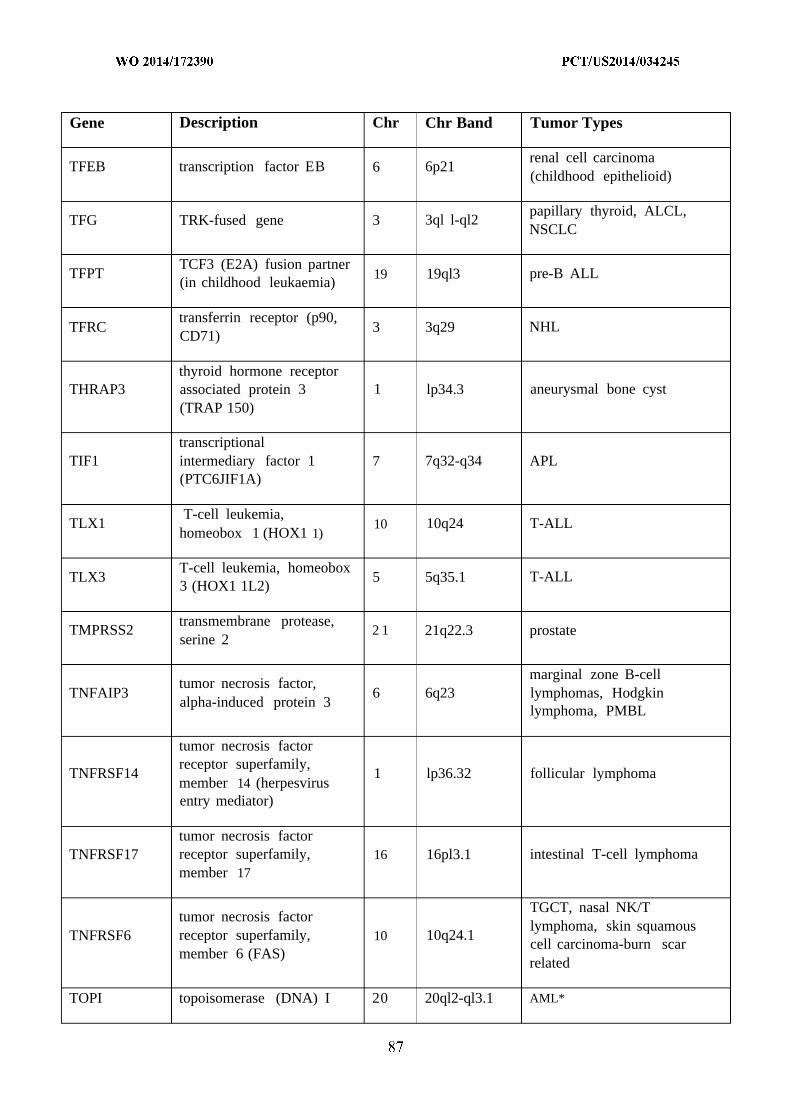
Gene Description Chr Chr Band Tumor Types
renal cell carcinomaTFEB transcription factor EB 6 6p21
(childhood epithelioid)
papillary thyroid, ALCL,TFG TRK-fused gene 3 3ql l-ql2
NSCLC
TCF3 (E2A) fusion partnerTFPT 19 19ql3 pre-B ALL
(in childhood leukaemia)
transferrin receptor (p90,TFRC 3 3q29 NHL
CD71)
thyroid hormone receptorTHRAP3 associated protein 3 1 lp34.3 aneurysmal bone cyst
(TRAP 150)
transcriptionalTIF1 intermediary factor 1 7 7q32-q34 APL
(PTC6JIF1A)
T-cell leukemia,TLX1 10 10q24 T-ALL
homeobox 1 (HOX1 1)
T-cell leukemia, homeoboxTLX3 5 5q35.1 T-ALL
3 (HOX1 1L2)
transmembrane protease,TMPRSS2 2 1 21q22.3 prostate
serine 2
marginal zone B-celltumor necrosis factor,
TNFAIP3 6 6q23 lymphomas, Hodgkinalpha-induced protein 3
lymphoma, PMBL
tumor necrosis factorreceptor superfamily,
TNFRSF14 1 lp36.32 follicular lymphomamember 14 (herpesvirusentry mediator)
tumor necrosis factorTNFRSF17 receptor superfamily, 16 16pl3.1 intestinal T-cell lymphoma
member 17
TGCT, nasal NK/Ttumor necrosis factor
lymphoma, skin squamousTNFRSF6 receptor superfamily, 10 10q24.1
cell carcinoma-burn scarmember 6 (FAS)
related
TOPI topoisomerase (DNA) I 20 20ql2-ql3.1 AML*

Gene Description Chr Chr Band Tumor Types
breast, colorectal, lung,sarcoma, adrenocortical,
TP53 tumor protein p53 17 17pl3glioma, multiple other tumourtypes
papillary thyroid, ALCL,TPM3 tropomyosin 3 1 Iq22-q23
NSCLC
TPM4 tropomyosin 4 19 19pl3.1 ALCL
translocated promoterTPR 1 lq25 papillary thyroid
region
TRA@ T cell receptor alpha locus 14 14ql l.2 T-ALL
tumour necrosis factorTRAF7 16 16pl3.3 meningioma
receptor-associated factor 7
TRB@ T cell receptor beta locus 7 7q35 T-ALL
TRD@ T cell receptor delta locus 14 14ql l T-cell leukaemia
tripartite motif-containingTRIM27 6 6p22 papillary thyroid
27
tripartite motif-containingTRIM33 1 lpl3 papillary thyroid
33 (PTC7,TIF1G)
thyroid hormone receptorTRIP 11 14 14q31-q32 AML
interactor 11
renal cell carcinoma, bladderTSC1 tuberous sclerosis 1 gene 9 9q34
carcinoma
pulmonarylymphangioleiomyomatosis
TSC2 tuberous sclerosis 2 gene 16 16pl3.3 (LAM), renalangiomyolipoma and headand neck cancer
thyroid stimulatingTSHR 14 14q31 toxic thyroid adenoma
hormone receptor
TTL tubulin tyrosine ligase 2 2ql3 acute lymphocytic leukaemia
U2 small nuclear RNA chronic lymphatic leukaemia,U2AF1 2 1 21q22.3
auxiliary factor 1 myelodysplastic syndrome

Gene Description Chr Chr Band Tumor Types
ubiquitin specific peptidaseUSP6 17 17pl3 aneurysmal bone cyst
6 (Tre-2 oncogene)
von Hippel-Lindau renal, haemangioma,VHL 3 3p25
syndrome gene pheochromocytoma
vesicle transport throughVTI1A interaction with t-SNAREs 10 10q25.2 colorectal
homolog 1A
WAS Wiskott-Aldrich syndrome X Xpl l.23-p 11.22
Wolf-Hirschhorn syndromeWHSC1 4 4pl6.3 Multiple myeloma
candidate l(MMSET)
Wolf-Hirschhorn syndromeWHSC1L1 8 8pl2 AML
candidate 1-like 1 (NSD3)
pleomorphic salivary glandWIF1 WNT inhibitory factor 1 12 12ql4.3
adenoma
Werner syndromeWRN 8 8pl2-pl l.2
(RECQL2)
Wilms tumour, desmoplasticWT1 Wilms tumour 1 gene 11 l lpl3
small round cell tumour
family with sequenceWTX similarity 123B X Xql l.l Wilms tumour
(FAM123B)
WW domain containing epithelioidWWTR1 3 3q23-q24
transcription regulator 1 haemangioendothelioma
xeroderma pigmentosum,XPA 9 9q22.3complementation group A
xeroderma pigmentosum,XPC 3 3p25complementation group C
exportin 1 (CRM1XPOl 2 2pl5 CLL
homolog, yeast)
tyrosine 3-monooxygenase/tryptophan
YWHAE 17 17pl3.3 edometrial stromal sarcoma5-monooxygenaseactivation protein, epsilonpolypeptide (14-3-3
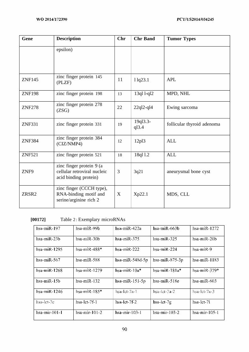
Gene Description Chr Chr Band Tumor Types
epsilon)
zinc finger protein 145ZNF145 11 l lq23.1 APL
(PLZF)
ZNF198 zinc finger protein 198 13 13ql l-ql2 MPD, NHL
zinc finger protein 278ZNF278 22 22ql2-ql4 Ewing sarcoma
(ZSG)
19ql3.3-ZNF331 zinc finger protein 331 19 follicular thyroid adenoma
ql3.4
zinc finger protein 384ZNF384 12 12pl3 ALL
(CIZ/NMP4)
ZNF521 zinc finger protein 521 18 18ql l.2 ALL
zinc finger protein 9 (aZNF9 cellular retroviral nucleic 3 3q21 aneurysmal bone cyst
acid binding protein)
zinc finger (CCCH type),ZRSR2 RNA-binding motif and X Xp22.1 MDS, CLL
serine/arginine rich 2
[00172] Table 2 : Exemplary microRNAs

hsa-mir- 107 hsa-mir- 1Oa hsa-mir- 1Ob hsa-mir- 122 hsa-mir- 1226
hsa-mir-1249 hsa-mir- 125a hsa-mir- 125b- 1 hsa-mir- 125b-2 hsa-mir- 126
hsa-mir- 129-1 hsa-mir- 129-2 hsa-mir- 1291 hsa-mir- 1295 hsa-mir- 1296
hsa-mir- 132 hsa-mir- 133a- 1 hsa-mir- 133a-2 hsa-mir- 133b hsa-mir- 134
hsa-miR-107 hsa-miR-767-3p hsa-miR-422a hsa-miR-1263 hsa-miR- 181a
hsa-mir- 100 hsa-mir- 106b hsa-mir- 124-3 hsa-mir- 128-2 hsa-mir- 130b
hsa-let-7f hsa-miR-662 hsa-miR-325 hsa-miR-1207-5p hsa-miR-519a*
hsa-let-7c hsa-mir- 1-2 hsa-mir- 106a hsa-mir- 124-2 hsa-mir- 128-1
hsa-let-7d hsa-mir- 135b hsa-mir- 136 hsa-mir- 137 hsa-mir- 13 8-1
hsa-mir- 139 hsa-miR-139-5p hsa-mir- 140 hsa-mir- 141 hsa-mir- 142
hsa-mir- 144 hsa-mir- 145 hsa-mir- 146a hsa-mir- 146b hsa-mir- 147
hsa-mir- 148b hsa-mir- 149 hsa-mir- 150 hsa-mir- 151 hsa-mir- 152
hsa-mir- 153-2 hsa-mir- 154 hsa-mir- 155 hsa-mir- 15a hsa-mir- 15b
hsa-mir- 16-2 hsa-mir- 17 hsa-mir- 181a-1 hsa-mir- 181 a-2 hsa-mir- 18 lb- 1
hsa-mir- 181c hsa-mir- 181d hsa-mir- 182 hsa-mir- 183 hsa-mir- 184
hsa-mir- 186 hsa-mir- 187 hsa-mir- 188 hsa-mir- 18a hsa-mir- 18b
hsa-mir- 191 hsa-mir- 192 hsa-mir- 193a hsa-mir- 193a hsa-mir- 193b
hsa-mir- 194-2 hsa-mir- 195 hsa-mir- 196a- 1 hsa-mir- 196a-2 hsa-mir- 196b
hsa-mir- 198 hsa-mir- 199a- 1 hsa-mir- 199a-2 hsa-mir- 199b hsa-mir- 19a
hsa-mir- 19b-2 hsa-mir-200a hsa-mir-200b hsa-mir-200c hsa-mir-202
hsa-mir-204 hsa-mir-205 hsa-mir-206 hsa-mir-208a hsa-mir-20a
hsa-mir-2 1 hsa-mir-2 10 hsa-mir-2 11 hsa-mir-2 12 hsa-mir-2 14
hsa-mir-2 16a hsa-mir-2 17 hsa-mir-2 18-1 hsa-mir-2 18-2 hsa-mir-2 19-1
hsa-mir-22 hsa-mir-220a hsa-mir-22 1 hsa-mir-222 hsa-mir-223
hsa-mir-23a hsa-mir-23b hsa-mir- 24- 1 hsa-mir- 24-2 hsa-mir-25
hsa-mir-26a- 1 hsa-mir-26a-2 hsa-mir-26b hsa-mir-27a hsa-mir-27b
hsa-mir-296 hsa-mir-299 hsa-mir-29a hsa-mir-29b- 1 hsa-mir-29b-2

hsa-mir-301a hsa-mir-302a hsa-mir-302b hsa-mir-302c hsa-mir-302d
hsa-mir-30b hsa-mir-30c-l hsa-mir-30c-2 hsa-mir-30d hsa-mir-30e
hsa-mir-32 hsa-miR-32* hsa-miR-320 hsa-mir-320a hsa-mir-323
hsa-mir-325 hsa-mir-326 hsa-mir-328 hsa-mir-329-1 hsa-mir-329-2
hsa-mir-331 hsa-mir-335 hsa-mir-337 hsa-mir-338 hsa-mir-339
hsa-mir-33b hsa-mir-340 hsa-mir-342 hsa-mir-345 hsa-mir-346
hsa-mir-34b hsa-mir-34c hsa-mir-361 hsa-mir-362 hsa-mir-363
hsa-mir-365-2 hsa-mir-367 hsa-mir-369 hsa-mir-370 hsa-mir-371
hsa-mir-373 hsa-mir-374a hsa-mir-374b hsa-mir-375 hsa-mir-376a-l
hsa-mir-376c hsa-mir-377 hsa-mir-378 hsa-mir-379 hsa-mir-380
hsa-mir-382 hsa-mir-383 hsa-mir-384 hsa-mir-409 hsa-mir-4 10
hsa-mir-42 1 hsa-mir-422a hsa-mir-423 hsa-mir-424 hsa-mir-425
hsa-mir-43 1 hsa-mir-432 hsa-mir-43 3 hsa-mir-449a hsa-mir-449b
hsa-mir-45 1 hsa-mir-452 hsa-mir-453 hsa-mir-454 hsa-mir-455
hsa-mir-484 hsa-mir-485 hsa-mir-486 hsa-mir-487a hsa-mir-487b
hsa-mir-489 hsa-mir-490 hsa-mir-49 1 hsa-mir-492 hsa-mir-493
hsa-mir-495 hsa-mir-496 hsa-mir-497 hsa-mir-498 hsa-mir-499
hsa-mir-501 hsa-mir-502 hsa-mir-503 hsa-mir-505 hsa-mir-508
hsa-mir-509-3 hsa-mir-5 10 hsa-mir-5 11-1 hsa-mir-5 11-2 hsa-mir-5 12-1
hsa-mir-5 13a- 1 hsa-mir-5 13a-2 hsa-mir-5 15-1 hsa-mir-5 15-2 hsa-mir-5 16a- 1
hsa-mir-5 16b- 1 hsa-mir-5 16b-2 hsa-mir-5 17a hsa-mir-5 17b hsa-mir-5 17c
hsa-mir-5 18a-2 hsa-mir-5 18b hsa-mir-5 18c hsa-mir-5 18d hsa-mir-5 18e
hsa-mir-5 19a- 1 hsa-mir-5 19a-2 hsa-mir-5 19b hsa-mir-5 19c hsa-mir-5 19d
hsa-mir-520a hsa-mir-520b hsa-mir-520c hsa-mir-520d hsa-mir-520e
hsa-mir-520g hsa-mir-520h hsa-mir-521-1 hsa-mir-521-2 hsa-mir-522
hsa-mir-524 hsa-mir-525 hsa-mir-526a-l hsa-mir-526a-2 hsa-mir-526b
hsa-mir-532 hsa-mir-539 hsa-mir-542 hsa-mir-543 hsa-mir-548o

hsa-mir-550-1 hsa-mir-551a hsa-mir-561 hsa-mir-566 hsa-mir-567
hsa-mir-572 hsa-mir-574 hsa-mir-575 hsa-mir-576 hsa-mir-582
hsa-mir-589 hsa-mir-590 hsa-mir-593 hsa-mir-598 hsa-mir-60 1
hsa-mir-605 hsa-mir-608 hsa-mir-61 1 hsa-mir-621 hsa-mir-622
hsa-mir-627 hsa-mir-629 hsa-mir-632 hsa-mir-634 hsa-mir-635
hsa-mir-639 hsa-mir-642 hsa-mir-644 hsa-mir-646 hsa-mir-648
hsa-mir-650 hsa-mir-652 hsa-mir-657 hsa-mir-658 hsa-mir-659
hsa-mir-66 1 hsa-mir-662 hsa-mir-663 hsa-mir-663b hsa-mir-67 1
hsa-mir-7- 1 hsa-mir-7-2 hsa-mir-7-3 hsa-mir-708 hsa-mir-744
hsa-mir-767 hsa-miR-768-3p hsa-mir-874 hsa-mir-886 hsa-mir-888
hsa-miR-9 hsa-mir-9-1 hsa-mir-9-2 hsa-mir-9-3 hsa-mir-923
hsa-mir-92a-2 hsa-mir-92b hsa-mir-93 hsa-mir-95 hsa-mir-96
hsa-mir-99a hsa-mir-99b hsa-miR-99b* mmu-mir- 1-1 mmu-mir- 100
mmu-mir- 103-2 mmu-mir- 106a mmu-mir- 106b mmu-mir- 10a mmu-mir- 1Ob
mmu-mir- 125b- 1 mmu-mir- 126 mmu-mir- 13Ob mmu-mir- 133a- 1 mmu-mir- 133b
mmu-mir- 139 mmu-mir- 140 mmu-mir- 143 mmu-mir- 145 mmu-mir- 150
mmu-mir- 15a mmu-mir- 16-1 mmu-mir- 17 mmu-mir- 181a-1 mmu-mir- 181c
mmu-mir- 184 mmu-mir- 18a mmu-mir- 192 mmu-mir- 193b mmu-mir- 196b
mmu-mir- 19b- 1 mmu-mir- 19b-2 mmu-mir-203 mmu-mir-207 mmu-mir-20a
mmu-mir-2 10 mmu-mir-22 mmu-mir-222 mmu-mir-223 mmu-mir-23a
mmu-mir-25 mmu-mir-27a mmu-mir-27b mmu-mir-290 mmu-mir-292
mmu-mir-298 mmu-mir-29a mmu-mir-29c mmu-mir-301a mmu-mir-3 Ob
mmu-mir-3 1 mmu-mir-322 mmu-mir-3 3 mmu-mir-346 mmu-mir-3 5 1
mmu-mir-365-1 mmu-mir-3 75 mmu-mir-378 mmu-mir-450b mmu-mir-466a
mmu-mir-468 mmu-mir-486 mmu-mir-500 mmu-mir-503 mmu-mir-542
mmu-mir-669a- 1 mmu-mir-669c mmu-mir-674 mmu-mir-680-1 mmu-mir-681
mmu-mir-686 mmu-mir-69 1 mmu-mir-699 mmu-mir-706 mmu-mir-7 10
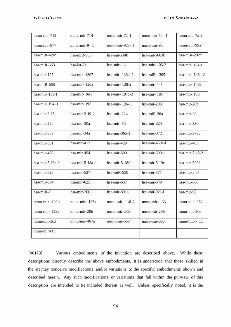
mmu-mir-712 mmu-mir-714 mmu-mir-72 1 mmu-mir-7a- 1 mmu-mir-7a-2
mmu-mir-877 mmu-mir-9- 1 mmu-mir-92a- 1 mmu-mir-93 mmu-mir-99a
hsa-miR-424* hsa-miR-605 hsa-miR-34b hsa-miR-663b hsa-miR-202*
hsa-miR-663 hsa-let-7b hsa-mir- 1-1 hsa-mir- 105-2 hsa-mir- 124-1
hsa-mir-127 hsa-mir- 1307 hsa-mir- 135a- 1 hsa-miR-1305 hsa-mir- 135a-2
hsa-miR-668 hsa-mir- 130a hsa-mir- 138-2 hsa-mir- 143 hsa-mir- 148a
hsa-mir- 153-1 hsa-mir- 16-1 hsa-mir- 18lb-2 hsa-mir- 185 hsa-mir- 190
hsa-mir- 194- 1 hsa-mir- 197 hsa-mir- 19b- 1 hsa-mir-203 hsa-mir-20b
hsa-mir-2 15 hsa-mir-2 19-2 hsa-mir- 224 hsa-miR-26a hsa-mir-28
hsa-mir-29c hsa-mir-30a hsa-mir- 31 hsa-mir-324 hsa-mir-330
hsa-mir-33a hsa-mir-34a hsa-mir-365-1 hsa-mir-372 hsa-mir-376b
hsa-mir-381 hsa-mir-412 hsa-mir-429 hsa-mir-450a-l hsa-mir-483
hsa-mir-488 hsa-mir-494 hsa-mir-500 hsa-mir-509-1 hsa-mir-5 12-2
hsa-mir-5 16a-2 hsa-mir-5 18a- 1 hsa-mir-5 18f hsa-mir-5 19e hsa-mir-520f
hsa-mir-523 hsa-mir-527 hsa-miR-550 hsa-mir-571 hsa-mir-5 84
hsa-mir-604 hsa-mir-625 hsa-mir-637 hsa-mir-649 hsa-mir-660
hsa-miR-7 hsa-mir-766 hsa-mir-891a hsa-mir-92a-l hsa-mir-98
mmu-mir- 103-1 mmu-mir- 125a mmu-mir- 13 8-2 mmu-mir- 155 mmu-mir- 182
mmu-mir- 199b mmu-mir-20b mmu-mir-23b mmu-mir-296 mmu-mir-30e
mmu-mir-363 mmu-mir-467a mmu-mir-652 mmu-mir-685 mmu-mir-7 11
mmu-mir-805
[00173] Various embodiments of the invention are described above. While these
descriptions directly describe the above embodiments, it is understood that those skilled in
the art may conceive modifications and/or variations to the specific embodiments shown and
described herein. Any such modifications or variations that fall within the purview of this
description are intended to be included therein as well. Unless specifically noted, it is the

intention of the inventors that the words and phrases in the specification and claims be given
the ordinary and accustomed meanings to those of ordinary skill in the applicable art(s).
[00174] The foregoing description of various embodiments of the invention known to
the applicant at this time of filing the application has been presented and is intended for the
purposes of illustration and description. The present description is not intended to be
exhaustive nor limit the invention to the precise form disclosed and many modifications and
variations are possible in the light of the above teachings. The embodiments described serve
to explain the principles of the invention and its practical application and to enable others
skilled in the art to utilize the invention in various embodiments and with various
modifications as are suited to the particular use contemplated. Therefore, it is intended that
the invention not be limited to the particular embodiments disclosed for carrying out the
invention.
[00175] While particular embodiments of the present invention have been shown and
described, it will be obvious to those skilled in the art that, based upon the teachings herein,
changes and modifications may be made without departing from this invention and its
broader aspects. It will be understood by those within the art that, in general, terms used
herein are generally intended as "open" terms (e.g., the term "including" should be
interpreted as "including but not limited to," the term "having" should be interpreted as
"having at least," the term "includes" should be interpreted as "includes but is not limited
to," etc.).

IN THE CLAIMS
1. A method for determining the likelihood of cancer metastasis in a subject in need
thereof comprising:
(i) providing a sample from the subject with cancer;
(ii) isolating large oncosomes from the samples comprising:
a . obtaining platelet-poor plasma from the sample;
b. centrifuging the platelet-poor plasma to eliminate cells and debris;
c . filtering the supernatant using a size exclusion column; and
d . collecting the large oncosomes from the upper chamber;
(iii) detecting the presence of proteins in the large onosomes, wherein the proteins
are any one or more of GLS, ATP50, PPP2R1A, SFXN1, ECI, HBA1, HINT2,
TUFM, ANP32A, PGLS, ETFA, CRYZ, MGST1, CYB5R3, TMEM33, PHB,
TOP2A, KIF5B, APEX1, RDH1 1, or a combination thereof; and
(iv) determining that the subject has an increased likelihood of cancer metastasis if
the sample from the subject comprises increased levels of large oncosomes and
the proteins in the large oncosomes relative to the reference sample, or
determining that the subject has a decreased likelihood of cancer metastasis if
the sample from the subject comprises decreased levels large oncosomes and the
proteins relative to the reference value so as to determine the likelihood of
cancer metastasis in the subject.
2 . A method for determining the likelihood of cancer metastasis in a subject in need
thereof comprising:
(i) providing a sample from the subject with cancer;
(ii) isolating large oncosomes from the samples comprising:
a . obtaining platelet-poor plasma from the sample;
b. centrifuging the platelet-poor plasma to eliminate cells and debris;
c . filtering the supernatant using a size exclusion column; and
d . collecting the large oncosomes from the upper chamber;
(iii) detecting the presence of proteins in the large onosomes, wherein the proteins
are any one or more of SLC25A6, PHB2, ATP5B, SLC25A3, VDAC1, GOT2,
STOML2, MDH2, HSPA9, VDAC2, SLC25A5, FN1, SHMT2, H2AFX,

HSPD1, ECH1, PRDX3, SLC25A1 1, HNRNPM, HIST2H2BF, TMX1,
MMYH10, NCEH1, GPI, CK18 or a combination thereof; and
(iv) determining that the subject has an increased likelihood of cancer metastasis if
the sample from the subject comprises increased levels of large oncosomes and
the proteins in the large oncosomes relative to the reference sample, or
determining that the subject has a decreased likelihood of cancer metastasis if
the sample from the subject comprises decreased levels large oncosomes and the
proteins relative to the reference value so as to determine the likelihood of
cancer metastasis in the subject.
A method for determining the likelihood of cancer metastasis in a subject in need
thereof comprising:
(i) providing a sample from the subject with cancer;
(ii) isolating large oncosomes from the samples comprising:
a . obtaining platelet-poor plasma from the sample;
b. centrifuging the platelet-poor plasma to eliminate cells and debris;
c . filtering the supernatant using a size exclusion column; and
d . collecting the large oncosomes from the upper chamber;
(iii) detecting the presence of micro R A in the large onosomes, wherein the micro
R A is any one or more of miR-491-3p, miR-34b, miR-1228, miR-647, miR-
1227 or a combination thereof; and
(iv) determining that the subject has an increased likelihood of cancer metastasis if
the sample from the subject comprises increased levels of the large oncosomes
and the miR-1227 in the large oncosomes relative to the reference sample, or
determining that the subject has a decreased likelihood of cancer metastasis if
the sample from the subject comprises decreased levels of the large oncosomes
and the miR-1227 relative to the reference value so as to determine the
likelihood of cancer metastasis in the subject.
The method of claims 1, 2 or 3, wherein the sample is blood, plasma or a combination
thereof.
The method of claims 1, 2 or 3, wherein the subject has prostate cancer.

6 . The method of claims 1, 2 or 3, wherein the sample from the subject is obtained
before, during or after cancer treatment.
7 . The method of claims 1, 2 or 3„ wherein the subject is human.
8. The method of claims 1, 2 or 3, wherein the reference value is the mean or median
number of large oncosomes and/or the protein levels and/or micro R A levels in a
population of subjects that do not have cancer.
9 . The method of claims 1, 2 or 3, wherein the reference value is the mean or median
number of large oncosomes and/or the protein levels and/or micro RNA levels in a
population of subjects that have cancer in remission.
10. The method of claims 1, 2 or 3, wherein the reference value is the mean or median
number of large oncosomes and/or the protein levels and/or microRNA levels in one
or more samples from the subject wherein the one or more samples are obtained from
a different time point.
11. A method for treating cancer in a subject in need thereof comprising:
(i) determining the likelihood of cancer metastasis by the methods of claims 1, 2 or
3; and
(ii) prescribing a therapy to the subject if the subject has increased likelihood of
cancer metastasis.
12. The method of claim 11, wherein the therapy is any one or more of surgery, radiation,
chemotherapy, immunotherapy, vaccine or a combination thereof.
13. The method of claim 12, further comprising administering an effective amount of any
one or more of radiation, chemotherapy, immunotherapy, vaccine or a combination
thereof.
14. The method of claim 11, wherein the cancer is prostate cancer.

The method of claim 11, wherein the subject is human.