Genome-wide distribution of Auts2 binding localizes with active neurodevelopmental genes
ARCHIVAL REPORT
Widespread Reductions in Cortical ThicknessFollowing Severe Early-Life Deprivation:A Neurodevelopmental Pathway to Attention-Deficit/Hyperactivity Disorder
Katie A. McLaughlin, Margaret A. Sheridan, Warren Winter, Nathan A. Fox, Charles H. Zeanah,and Charles A. NelsonBackground: Children exposed to early-life psychosocial deprivation associated with institutional rearing are at markedly elevated riskof developing attention-deficit/hyperactivity disorder (ADHD). Neurodevelopmental mechanisms that explain the high prevalence ofADHD in children exposed to institutionalization are unknown. We examined whether abnormalities in cortical thickness and subcorticalvolume were mechanisms explaining elevations in ADHD among children raised in institutional settings.
Methods: Data were drawn from the Bucharest Early Intervention Project, a cohort of children raised from early infancy in institutions inRomania (n ¼ 58) and age-matched community control subjects (n ¼ 22). Magnetic resonance imaging data were acquired whenchildren were aged 8 to 10 years, and ADHD symptoms were assessed using the Health and Behavior Questionnaire.
Results: Children reared in institutions exhibited widespread reductions in cortical thickness across prefrontal, parietal, and temporalregions relative to community control subjects. No group differences were found in the volume of subcortical structures. Reducedthickness across numerous cortical areas was associated with higher levels of ADHD symptoms. Cortical thickness in lateral orbitofrontalcortex, insula, inferior parietal cortex, precuneus, superior temporal cortex, and lingual gyrus mediated the association ofinstitutionalization with inattention and impulsivity; additionally, supramarginal gyrus thickness mediated the association withinattention and fusiform gyrus thickness mediated the association with impulsivity.
Conclusions: Severe early-life deprivation disrupts cortical development resulting in reduced thickness in regions with atypical functionduring attention tasks in children with ADHD, including the inferior parietal cortex, precuneus, and superior temporal cortex. Thesereductions in thickness are a neurodevelopmental mechanism explaining elevated ADHD symptoms in children exposed to institutionalrearing.
Key Words: Attention-deficit/hyperactivity disorder (ADHD),brain development, childhood adversity, cortical development,deprivation, institutionalization
Attention-deficit/hyperactivity disorder (ADHD) is a commonneurodevelopmental disorder estimated to affect approx-imately 5% of children worldwide (1–3). Children with
ADHD exhibit deficits in numerous aspects of executive function-ing including working memory, response inhibition, attentionaland motor control, and planning (4–9). Meta-analyses of func-tional magnetic resonance imaging studies have identifiedabnormalities in neural function among children with ADHD,including blunted activation in right hemisphere dorsolateralprefrontal cortex (PFC), striatum, and thalamus during inhibitionand attention tasks; reduced inferior parietal cortex, precuneus,and superior temporal cortex activation during attention tasks;
From Boston Children’s Hospital (KAM, MAS, WW, CAN), Harvard MedicalSchool, Boston; and Harvard Center on the Developing Child (KAM,MAS, CAN), Cambridge, Massachusetts; Department of Human Devel-opment and Quantitative Methodology (NAF), University of Maryland,College Park, Maryland; and Department of Psychiatry (CHZ), TulaneUniversity School of Medicine, New Orleans, Louisiana.
Address correspondence to Katie A. McLaughlin, Ph.D., University ofWashington, Assistant Professor of Psychology, Box 351525, Universityof Washington, Seattle, WA 98195; E-mail: [email protected].
Received Apr 1, 2013; revised Aug 16, 2013; accepted Aug 18, 2013.
0006-3223/$36.00http://dx.doi.org/10.1016/j.biopsych.2013.08.016
and hypoactivation in left hemisphere frontal-parietal-cerebellarcircuits during timing tasks (10,11).
Attention-deficit/hyperactivity disorder is also associated withatypical neural structure, including smaller volume of the PFC andbasal ganglia (12–14) and reductions in cortical thickness acrossprefrontal, parietal, and temporal cortex (15,16). Children with ADHDexperience 2- to 5-year delays in reaching peak cortical thickness inthese regions (17), and cortical thickness in children with ADHD doesnot catch up to levels seen in typically developing children in mostareas (16,18). Children with ADHD whose developmental trajectoryof cortical thickness is more similar to that of typically developingchildren have better functional outcomes than children withpersistent thickness reductions (16), suggesting that this pattern ofcortical development may be central to the pathophysiologyof ADHD.
What factors lead to these neurodevelopmental deficits inchildren with ADHD? The high heritability of the disorder andearly age of onset suggest strong genetic underpinnings (19,20).However, early-life psychosocial deprivation is also associated withADHD (21–23), indicating that adverse early experiences maycontribute to atypical patterns of brain development. The preva-lence of ADHD among children raised in institutional settings is 4to 5 times higher than in the general population, raising questionsabout neurodevelopmental mechanisms involved in ADHD follow-ing psychosocial deprivation (21–23). Institutional rearing is asso-ciated with atypical structural development that might contributeto ADHD risk in previously institutionalized children. Reducedcerebral and cortical white and gray matter volumes have beenobserved in institutionally reared children (24,25), as well as white
BIOL PSYCHIATRY 2014;76:629–638& 2014 Society of Biological Psychiatry

630 BIOL PSYCHIATRY 2014;76:629–638 K.A. McLaughlin et al.
matter microstructure abnormalities in tracts linking the PFC totemporal and parietal regions (26–28). Larger right amygdalavolume was reported in one study of institutionally reared children(24), and another found larger amygdala volume among late-adopted children compared with early-adopted and controlchildren (29). Reduced cerebellar volume has also been observedin previously institutionalized children (30).
We investigated whether atypical neural structure is respon-sible for elevations in ADHD among children raised in institutionalsettings. We anticipated that institutional rearing would beassociated with reduced cortical thickness and subcortical volumein regions implicated in ADHD pathology, including the dorso-lateral PFC, inferior parietal cortex, superior temporal cortex, andstriatum. In addition, we hypothesized that reduced corticalthickness and subcortical volume in these regions would beassociated with ADHD pathology. Finally, we investigatedwhether disrupted cortical and subcortical development is amechanism explaining the association between early psychoso-cial deprivation and ADHD.
Methods and Materials
SampleThe Bucharest Early Intervention Project is a longitudinal study
of early institutionalization of young children in Bucharest,Romania (31). A sample of 136 children (age range 6–30 months,mean [M] ¼ 23 months) was recruited from each of the sixinstitutions for young children in Bucharest, excluding partic-ipants with genetic syndromes (e.g., Down syndrome), fetalalcohol syndrome, and microcephaly (31). An age-matchedsample of 72 community-reared children was recruited frompediatric clinics in Bucharest and comprised the never-institutionalized group (NIG). Half of the children in the institu-tionalized group were randomized to a foster care intervention,resulting in two groups: the foster care group (FCG) and thegroup who received care as usual (prolonged institutional care[CAUG]). The study design and methods have been described indetail previously (31).
Structural magnetic resonance imaging was acquired whenchildren were between 8 and 10 years of age for all children whoseguardians provided consent for imaging. Of the 86 children who
Table 1. Sociodemographic and Developmental CharacCommunity Control Subjects in the Bucharest Early Int
Ever InstitutionalizGroup (n ¼ 58)
M SD
Female, No. (%) n ¼ 28 (48.3%)Age (Months)Age at study entry 17.7 (7.Age at MRI scan 116.3 (9.Age at HBQ assessment 103.2 (4.
Birth Weight (grams) 2780.0 (623Head Circumference at Birth (cm) 46.07 (2.6Full-Scale IQ 72.0 (15Intracranial Volume 1,456,490 (132,Cerebral Gray Matter 790,429 (71,1Cortical Gray Matter 577,432 (57,1
HBQ, MacArthur Health and Behavior QuestionnaireaSignificant at the p � .05 level, two-sided test.
www.sobp.org/journal
completed magnetic resonance imaging assessments, 80 wereincluded in analysis: 31 CAUG children (15 female participants), 27FCG children (13 female participants), and 22 NIG children (12female participants). Four participants were excluded from analysisbecause of poor scan quality (2 CAUG, 1 FCG, and 1 NIG) and twochildren were excluded due to frank neurological abnormality (1FCG, 1 NIG). Four participants were taking stimulant medication forADHD at the time of the scan (3 CAUG, 1 FCG).
No differences in ADHD symptoms of inattention, t51 ¼ .46,p ¼ .646, or impulsivity, t51 ¼ .69, p ¼ .497, or in cortical thick-ness or subcortical volume were observed at age 8 to 10 yearsbased on foster care placement. As such, children in the FCG andCAUG were collapsed into one ever-institutionalized group (EIG)for all analyses. No differences in gender distribution or age wereobserved for EIG and NIG children, although differences in IQ,birth weight, and cerebral and cortical gray matter were presentacross groups (Table 1).
Image AcquisitionStructural magnetic resonance images were acquired at Regina
Maria Health Center on a Siemens Magnetom Avanto 1.5 TeslaSyngo System (Siemens, Munich, Germany). Images were obtainedusing a transverse magnetization-prepared rapid gradient-echothree-dimensional sequence (echo time ¼ 2.98 msec, inversiontime ¼ 1000 msec, flip angle ¼ 81, 176 slices with 1 � 1 � 1 mmisometric voxels) with a 16-channel head coil. The repetition time(TR) for this sequence was 1710 milliseconds for most participants(n ¼ 59) and varied between 1650 milliseconds and 1910 milli-seconds for remaining participants. Four subjects were acquired inthe sagittal plane; one was acquired in the coronal plane.Acquisition parameters did not differ by group membership norwere they associated with scan quality; all scans were thereforeconsidered together and a covariate for TR length was included inall analyses.
Image ProcessingCortical reconstruction and volumetric segmentation were
performed with FreeSurfer (Version 5.0, http://surfer.nmr.mgh.harvard.edu). Technical details of these procedures have beendescribed previously (32–36). Gray/white matter and gray matter/cerebrospinal fluid (CSF) boundaries are constructed using spatialintensity gradients across tissue classes. A segmentation process
teristics Among Children Reared in Institutions andervention Project (n ¼ 80)
ed Never InstitutionalizedGroup (n ¼ 22) Group Difference
M SD F p Value
n ¼ 12 (54.5%) χ21 ¼ .25 .617
8) 20.0 (7.2) 1.21 .2760) 117.9 (10.6) .1 .8166) 101.4 (4.0) 2.49 .149.3) 3150.0 (411.8) 4.41a .0401) 46.5 (2.08) .04 .843.8) 107.9 (14.67) 96.49a .001948) 1,499,091 (109,367) 1.79 .18460) 833,849 (62,887) 6.31a .01420) 613,899 (48,391) 7.04a .010
; MRI, magnetic resonance imaging.
K.A. McLaughlin et al. BIOL PSYCHIATRY 2014;76:629–638 631
is used to identify subcortical gray matter structures. Followingreconstruction, the cerebral cortex is parcellated into regionsbased on the structure of gyri and sulci (34,37). Intensity andcontinuity information are used to generate measurements ofcortical thickness, calculated as the closest distance from thegray/white boundary to the gray/CSF boundary at each vertex onthe tessellated surface (33). The resulting surface maps are notrestricted to the voxel resolution of the original data and arecapable of detecting submillimeter differences between groups.
FreeSurfer morphometric procedures have demonstratedgood test-retest reliability across scanner manufacturers and fieldstrengths (38,39), and methods for measuring cortical thicknesshave been validated against manual measurement (40,41) andhistological analysis (42) and have been used in studies ofchildren aged 8 to 10 years (25,29,43). The results of theautomated segmentation and parcellation process were manuallyinspected for all participants. Where necessary, manual edits wereperformed as recommended to optimize accurate placement ofgray/white and gray/CSF borders based on shifts in the imageintensity gradient (32,33). No differences were present in thedegree to which manual edits were required across groups.
ADHDThe MacArthur Health and Behavior Questionnaire (HBQ) (44)
was completed by the primary teacher of each child when theywere between 8 and 10 years old (M ¼ 8.5 years, SD ¼ .4 years).The HBQ assesses emotional and behavior problems and hasbeen widely used in studies of children ranging from preschoolage to adolescence, including previously institutionalized children(45). The ADHD subscale assesses inattention and impulsivity.Teacher reports of ADHD behaviors on the HBQ have demon-strated excellent test-retest reliability in community and clinicalsamples, acceptable concordance with parent reports, and highdiscriminant validity (44,46).
Statistical AnalysisWe investigated whether elevations in ADHD symptoms
among institutionalized children relative to control subjects wereaccounted for by differences in brain structure using standardtests of statistical mediation. To provide evidence for mediation,four criteria must be met (47,48). First, an association between theexposure and outcome must be established. Here, we examineddifferences in ADHD symptoms between children reared ininstitutions versus the community using univariate analyses ofvariance with group (EIG, NIG) as a between-subjects factor.
Second, the exposure must be associated with the mediator.We examined group differences in brain structure using the Qdecsurface-based group analysis tool (Version 1.4) in FreeSurfer.Following spatial normalization to an averaged spherical surfaceand smoothing with a 10-mm full-width at half maximumGaussian kernel, Qdec applies a general linear model (GLM) tocortical thickness at each vertex, separately by hemisphere. Adiscrete variable for group was included in the GLM, along withcovariates for age, gender, total brain volume, and TR. Nointeractions were found between group and any of thesecovariates. To reduce type I error associated with multiplecomparisons, we applied a false discovery rate (FDR) correction(49). Group differences in subcortical volume were examinedusing univariate analyses of variance with group as a between-subjects factor and the covariates outlined above for the caudate,putamen, globus pallidus, nucleus accumbens, amygdala, hippo-campus, thalamus, and cerebellum. False discovery rate correc-tion was applied to correct for multiple comparisons.
Third, the mediator must be associated with the outcome.Here, we examined the associations of cortical thickness andsubcortical volume with ADHD symptoms using linear regression.We examined the association of cortical thickness with ADHDsymptoms in each cluster that differed between children raised ininstitutions versus the community after FDR correction. To do so,we created a region of interest (ROI) for each FDR-correctedcluster that was significantly different between groups. Thisnormalized ROI was mapped back to each participant (usingdeformation tools in FreeSurfer) to generate a mean thicknessvalue for that ROI for each participant. Gender, age, total brainvolume, and TR were included as covariates.
Finally, we tested the significance of the indirect effect using abootstrapping approach that provides bias-corrected confidenceintervals and is appropriate for use in small samples (50).Confidence intervals that do not include zero indicate significantmediation. We required that a brain region differed in thicknessor volume as a function of institutionalization and be associatedwith ADHD symptoms at the FDR-corrected threshold to beincluded in the mediation analysis.
Results
Institutionalization and ADHDAttention-deficit/hyperactivity disorder symptoms varied as
a function of institutionalization for inattention, F1,70 ¼ 29.48,p � .001, and impulsivity, F1,69 ¼ 17.94, p � .001. Children withhistories of institutional rearing (EIG) exhibited higher levels ofinattention (M ¼ 6.46, SD ¼ 2.86) and impulsivity (M ¼ 8.73, SD ¼5.53) than community-reared children (inattention M ¼ 1.90,SD ¼ 2.86; impulsivity M ¼ 3.14, SD ¼ 4.33).
Institutionalization and Cortical ThicknessResults from the left hemisphere GLM revealed 34 clusters
that differed significantly in thickness as a function of institution-alization. Institutionally reared children had reduced thicknesscompared with never-institutionalized children in all 34 clusters.Table 2 provides the Montreal Neurologic Institute coordinates andpeak of each cluster, and Figure 1 displays results. Signi-ficant differences in cortical thickness were observed in multipleclusters and were most pronounced in the superior and inferiorparietal cortex (5 and 4 clusters, respectively), precuneus (4clusters), superior temporal gyrus and sulcus (3 clusters), precentralgyrus (2 clusters), and posterior cingulate (2 clusters). Significantdifferences were also present in the superior frontal gyrus, middlefrontal gyrus (MFG), fusiform gyrus, supramarginal gyrus, lateralorbitofrontal cortex (OFC), lateral occipital cortex, and insula.
The right hemisphere GLM revealed 27 clusters that differedsignificantly between groups, with institutionally reared childrenexhibiting reduced cortical thickness compared with controlsubjects in all clusters (Table 3, Figure 1). Findings mirrored thosefrom the left hemisphere, with the exception of greater differ-ences in the MFG. Areas with multiple significant clusters and thelargest group differences were the MFG (2 clusters), superior andinferior parietal cortex (3 and 4 clusters, respectively), precuneus(4 clusters), supramarginal gyrus (2 clusters), and superior tem-poral gyrus and sulcus (2 clusters). Additional regions differing inthickness included the superior frontal gyrus, inferior temporalgyrus, frontal pole, lateral OFC, lateral occipital cortex, fusiformgyrus, lingual gyrus, and insula.
We also examined the association of duration of institution-alization with cortical thickness in institutionally reared children.
www.sobp.org/journal
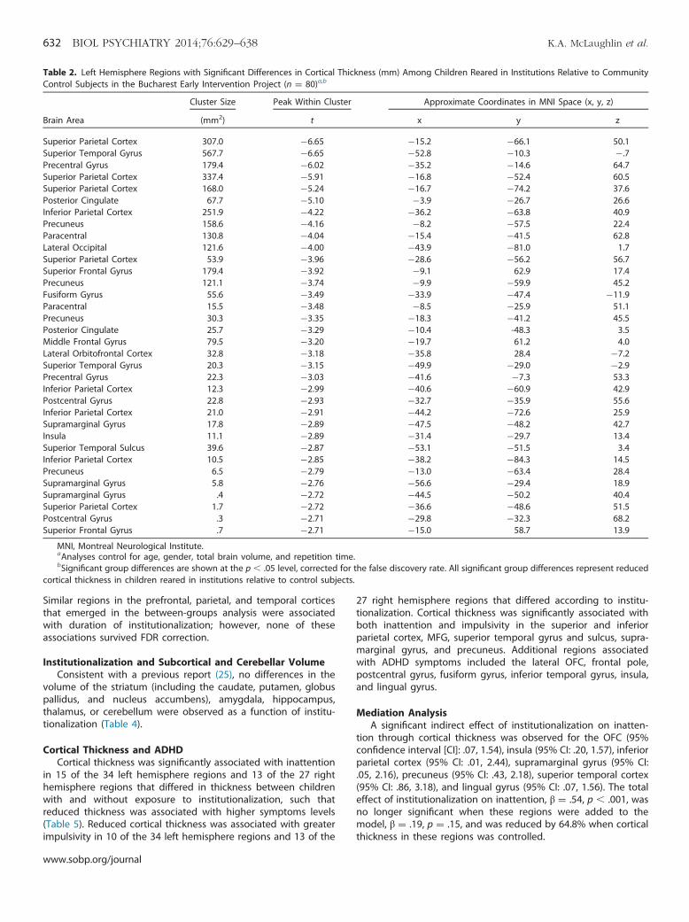
Table 2. Left Hemisphere Regions with Significant Differences in Cortical Thickness (mm) Among Children Reared in Institutions Relative to CommunityControl Subjects in the Bucharest Early Intervention Project (n ¼ 80)a,b
Cluster Size Peak Within Cluster Approximate Coordinates in MNI Space (x, y, z)
Brain Area (mm2) t x y z
Superior Parietal Cortex 307.0 �6.65 �15.2 �66.1 50.1Superior Temporal Gyrus 567.7 �6.65 �52.8 �10.3 �.7Precentral Gyrus 179.4 �6.02 �35.2 �14.6 64.7Superior Parietal Cortex 337.4 �5.91 �16.8 �52.4 60.5Superior Parietal Cortex 168.0 �5.24 �16.7 �74.2 37.6Posterior Cingulate 67.7 �5.10 �3.9 �26.7 26.6Inferior Parietal Cortex 251.9 �4.22 �36.2 �63.8 40.9Precuneus 158.6 �4.16 �8.2 �57.5 22.4Paracentral 130.8 �4.04 �15.4 �41.5 62.8Lateral Occipital 121.6 �4.00 �43.9 �81.0 1.7Superior Parietal Cortex 53.9 �3.96 �28.6 �56.2 56.7Superior Frontal Gyrus 179.4 �3.92 �9.1 62.9 17.4Precuneus 121.1 �3.74 �9.9 �59.9 45.2Fusiform Gyrus 55.6 �3.49 �33.9 �47.4 �11.9Paracentral 15.5 �3.48 �8.5 �25.9 51.1Precuneus 30.3 �3.35 �18.3 �41.2 45.5Posterior Cingulate 25.7 �3.29 �10.4 -48.3 3.5Middle Frontal Gyrus 79.5 �3.20 �19.7 61.2 4.0Lateral Orbitofrontal Cortex 32.8 �3.18 �35.8 28.4 �7.2Superior Temporal Gyrus 20.3 �3.15 �49.9 �29.0 �2.9Precentral Gyrus 22.3 �3.03 �41.6 �7.3 53.3Inferior Parietal Cortex 12.3 �2.99 �40.6 �60.9 42.9Postcentral Gyrus 22.8 �2.93 �32.7 �35.9 55.6Inferior Parietal Cortex 21.0 �2.91 �44.2 �72.6 25.9Supramarginal Gyrus 17.8 �2.89 �47.5 �48.2 42.7Insula 11.1 �2.89 �31.4 �29.7 13.4Superior Temporal Sulcus 39.6 �2.87 �53.1 �51.5 3.4Inferior Parietal Cortex 10.5 �2.85 �38.2 �84.3 14.5Precuneus 6.5 �2.79 �13.0 �63.4 28.4Supramarginal Gyrus 5.8 �2.76 �56.6 �29.4 18.9Supramarginal Gyrus .4 �2.72 �44.5 �50.2 40.4Superior Parietal Cortex 1.7 �2.72 �36.6 �48.6 51.5Postcentral Gyrus .3 �2.71 �29.8 �32.3 68.2Superior Frontal Gyrus .7 �2.71 �15.0 58.7 13.9
MNI, Montreal Neurological Institute.aAnalyses control for age, gender, total brain volume, and repetition time.bSignificant group differences are shown at the p � .05 level, corrected for the false discovery rate. All significant group differences represent reduced
cortical thickness in children reared in institutions relative to control subjects.
632 BIOL PSYCHIATRY 2014;76:629–638 K.A. McLaughlin et al.
Similar regions in the prefrontal, parietal, and temporal corticesthat emerged in the between-groups analysis were associatedwith duration of institutionalization; however, none of theseassociations survived FDR correction.
Institutionalization and Subcortical and Cerebellar VolumeConsistent with a previous report (25), no differences in the
volume of the striatum (including the caudate, putamen, globuspallidus, and nucleus accumbens), amygdala, hippocampus,thalamus, or cerebellum were observed as a function of institu-tionalization (Table 4).
Cortical Thickness and ADHDCortical thickness was significantly associated with inattention
in 15 of the 34 left hemisphere regions and 13 of the 27 righthemisphere regions that differed in thickness between childrenwith and without exposure to institutionalization, such thatreduced thickness was associated with higher symptoms levels(Table 5). Reduced cortical thickness was associated with greaterimpulsivity in 10 of the 34 left hemisphere regions and 13 of the
www.sobp.org/journal
27 right hemisphere regions that differed according to institu-tionalization. Cortical thickness was significantly associated withboth inattention and impulsivity in the superior and inferiorparietal cortex, MFG, superior temporal gyrus and sulcus, supra-marginal gyrus, and precuneus. Additional regions associatedwith ADHD symptoms included the lateral OFC, frontal pole,postcentral gyrus, fusiform gyrus, inferior temporal gyrus, insula,and lingual gyrus.
Mediation AnalysisA significant indirect effect of institutionalization on inatten-
tion through cortical thickness was observed for the OFC (95%confidence interval [CI]: .07, 1.54), insula (95% CI: .20, 1.57), inferiorparietal cortex (95% CI: .01, 2.44), supramarginal gyrus (95% CI:.05, 2.16), precuneus (95% CI: .43, 2.18), superior temporal cortex(95% CI: .86, 3.18), and lingual gyrus (95% CI: .07, 1.56). The totaleffect of institutionalization on inattention, β ¼ .54, p � .001, wasno longer significant when these regions were added to themodel, β ¼ .19, p ¼ .15, and was reduced by 64.8% when corticalthickness in these regions was controlled.
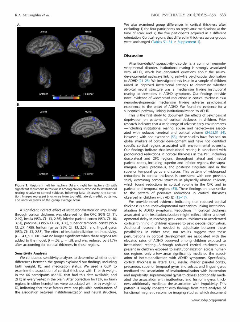
Figure 1. Regions in left hemisphere (A) and right hemisphere (B) withsignificant reductions in thickness among children exposed to institutionalrearing relative to control subjects, following false discovery rate correc-tion. Images represent (clockwise from top left), lateral, medial, posterior,and anterior views of the group average brain.
K.A. McLaughlin et al. BIOL PSYCHIATRY 2014;76:629–638 633
A significant indirect effect of institutionalization on impulsivitythrough cortical thickness was observed for the OFC (95% CI: .11,2.49), insula (95% CI: .13, 2.36), inferior parietal cortex (95% CI: .10,3.61), precuneus (95% CI: .68, 3.30), superior temporal cortex (95%CI: .27, 4.08), fusiform gyrus (95% CI: .13, 2.53), and lingual gyrus(95% CI: .13, 2.33). The effect of institutionalization on impulsivity,β ¼ .43, p � .001, was no longer significant when these regions areadded to the model, β ¼ .08, p ¼ .58, and was reduced by 81.7%after accounting for cortical thickness in these regions.
Sensitivity AnalysisWe conducted sensitivity analyses to determine whether other
differences between the groups explained our findings, includingbirth weight, IQ, and medication status. We used a GLM toexamine the association of cortical thickness with 1) birth weightin the 66 participants (82.5%) that had this data available; and2) IQ in every vertex in the brain. After correction for FDR, no brainregions in either hemisphere were associated with birth weight orIQ, indicating that these factors were not plausible confounders ofthe association between institutionalization and neural structure.
We also examined group differences in cortical thickness afterexcluding: 1) the four participants on psychiatric medications at thetime of scan; and 2) the five participants acquired in a differentorientation. Cortical regions that differed in thickness across groupswere unchanged (Tables S1–S4 in Supplement 1).
Discussion
Attention-deficit/hyperactivity disorder is a common neurode-velopmental disorder. Institutional rearing is strongly associatedwith ADHD, which has generated questions about the neuro-developmental pathways linking early-life psychosocial deprivationto ADHD (21–23). We investigated this issue in a sample of childrenraised in deprived institutional settings to determine whetheratypical neural structure was a mechanism linking institutionalrearing to elevations in ADHD symptoms. Our findings providenovel evidence of widespread reductions in cortical thickness as aneurodevelopmental mechanism linking adverse psychosocialexperience to the onset of ADHD. We found no evidence for asubcortical pathway linking institutionalization to ADHD.
This is the first study to document the effects of psychosocialdeprivation on patterns of cortical thickness in children. Priorresearch indicates that a wide range of adverse early environments—including institutional rearing, abuse, and neglect—are associ-ated with reduced cerebral and cortical volume (24,25,51–54).However, with one exception (53), these studies have focused onglobal markers of cortical development and have not identifiedspecific cortical regions associated with environmental adversity.Our findings indicate that institutional rearing is associated withpronounced reductions in cortical thickness in the PFC, includingdorsolateral and OFC regions; throughout lateral and medialparietal cortex, including superior and inferior regions, the supra-marginal gyrus, precuneus, and posterior cingulate; and in thesuperior temporal gyrus and sulcus. This pattern of widespreadreductions in cortical thickness is consistent with one previousstudy examining cortical structure in physically abused children,which found reductions in cortical volume in the OFC and inparietal and temporal regions (53). These findings are also similarto the pattern of pervasive reductions in cortical thicknessobserved in children with ADHD (15–17).
We provide novel evidence indicating that reduced corticalthickness is a neurodevelopmental mechanism linking institution-alization to ADHD symptoms. Reductions in cortical thicknessassociated with institutionalization might reflect either a devel-opmental delay in reaching peak cortical thickness or acceleratedcortical thinning in children exposed to psychosocial deprivation.Additional research is needed to adjudicate between thesepossibilities. In either case, our results suggest that theseperturbations in cortical development are associated with theelevated rates of ADHD observed among children exposed toinstitutional rearing. Although reduced cortical thickness waspresent in children exposed to institutionalization across numer-ous regions, only a few areas significantly mediated the associ-ation of institutionalization with ADHD symptoms. Specifically,cortical thickness in lateral OFC, insula, inferior parietal cortex,precuneus, superior temporal gyrus and sulcus, and lingual gyrusmediated the association of institutionalization with inattentionand impulsivity; supramarginal gyrus thickness additionally medi-ated the association with inattention; and fusiform gyrus thick-ness additionally mediated the association with impulsivity. Thispattern is largely consistent with findings from meta-analyses offunctional magnetic resonance imaging studies, which document
www.sobp.org/journal

Table 3. Right Hemisphere Regions with Significant Differences in Cortical Thickness (mm) Among Children Reared in Institutions Relative to CommunityControl Subjects in the Bucharest Early Intervention Project (n ¼ 80)a,b
Cluster Size Peak Within Cluster Approximate Coordinates in MNI Space (x, y, z)
Brain Area (mm2) t x y z
Middle Frontal Gyrus 197.7 �5.26 18.7 53.7 21.4Inferior Parietal Cortex 353.6 �5.23 41.7 �55.9 41.1Superior Frontal Gyrus 223.1 �5.10 20.6 23.4 55.1Inferior Temporal Gyrus 136.5 �4.80 47.3 �45.1 �15.3Supramarginal Gyrus 142.3 �4.76 40.7 �27.2 40.3Superior Parietal Cortex 330.8 �4.71 26.7 �55.4 59.6Frontal Pole 200.6 �4.47 10.0 64.0 �8.0Precuneus 173.1 �4.33 16.2 �42.9 55.0Superior Temporal Gyrus 73.0 �4.11 57.1 �4.8 �2.6Lateral Orbitofrontal Cortex 62.9 �4.03 40.1 25.2 �13.7Lateral Occipital Cortex 181.7 �4.02 40.6 �70.3 �1.9Fusiform Gyrus 58.9 �3.83 29.3 �66.1 �13.8Precuneus 22.0 �3.59 8.3 �54.7 52.1Middle Frontal Gyrus 34.9 �3.51 36.9 26.8 43.4Inferior Parietal Cortex 47.1 �3.49 42.2 �77.4 24.9Insula 1.2 �3.29 31.0 13.6 �12.4Inferior Parietal Cortex 24.1 �3.26 41.9 �71.0 35.0Precuneus 27.6 �3.23 22.0 �67.3 15.1Precuneus 26.6 �3.13 15.3 �69.8 39.0Inferior Parietal Cortex 17.1 �3.11 51.7 �56.1 13.2Lingual Gyrus 32.6 �3.11 15.7 �73.0 �5.8Superior Parietal Cortex 26.0 �3.09 18.4 �66.3 46.8Supramarginal Gyrus 8.9 �3.05 55.5 �34.5 35.2Precuneus 19.9 �2.94 10.7 �66.1 34.9Superior Temporal Sulcus 3.5 �2.93 49.8 �41.8 14.3Superior Parietal Cortex 5.9 �2.92 24.1 �84.3 30.7Insula .3 �2.92 29.6 13.4 �13.7
MNI, Montreal Neurological Institute.aAnalyses control for age, gender, total brain volume, and repetition time.bSignificant group differences are shown at the p � .05 level, corrected for the false discovery rate. All significant group differences represent reduced
cortical thickness in children reared in institutions relative to control subjects.
634 BIOL PSYCHIATRY 2014;76:629–638 K.A. McLaughlin et al.
blunted activation in dorsolateral PFC, inferior parietal cortex,precuneus, and superior temporal cortex during attention tasks inADHD (11). These regions are integral to cognitive processesdisrupted in ADHD, including working memory storage, targetdetection, attentional orienting, and attention allocation (55–59).Additionally, the precuneus and inferior parietal lobule are centralnodes in the default mode network (60,61). Fluctuations in defaultmode network activation have been linked to attention lapses
Table 4. Group Differences in the Volume of SubcorticReared in Institutions and Community Control Subjects i
Ever InstitutionalizedGroup (n ¼ 58)
M SD
StriatumCaudate 8338.2 865.1Putamen 11,742.7 1052.2Globus pallidus 4242.6 485.5Nucleus accumbens 1447.2 216.3
Amygdala 3759.2 395.8Hippocampus 9254.7 792.5Thalamus 14,465.7 1459.8Cerebellum 130,490.5 12,289.3
aAnalyses control for age, gender, total brain volumefalse discovery rate.
www.sobp.org/journal
(62), and some have hypothesized that this network underliesattention to external stimuli (63); it is possible that atypicalcortical structure in regions associated with the default modenetwork are related to the attentional deficits that underlie ADHD.This possibility warrants examination in future research. Finally,the OFC is involved in emotion regulation, social behavior, anddecision making in situations involving reward or other emotion-ally salient cues (64,65). Children with ADHD exhibit impulsivity
al Structures and the Cerebellum Among Childrenn the Bucharest Early Intervention Project (n ¼ 80)a
Never InstitutionalizedGroup (n ¼ 22) Group Differencea
M SD F p Value
8693.7 878.1 1.03 .51211,804.0 1058.4 .31 .7634417.8 484.2 1.00 .5121389.5 187.7 3.68S .4723893.8 336.4 1.43 .5129382.8 749.7 .00 .992
15,046.2 1187.1 1.47 .512134,281.3 13,773.0 .19 .763
, and repetition time and p values corrected for the

Table 5. Cortical Regions Significantly Associated with Symptoms ofInattention and Impulsivity in the Bucharest Early Intervention Project(n ¼ 74)a
Inattention Impulsivity
Brain Area Regionb β
FDRCorrectedp Value β
FDRCorrectedp Value
Left HemisphereSuperior Parietal Cortex 1 �.38 .013 �.29 .027Superior Temporal Gyrus 2 �.45 .001 �.36 .009Superior Parietal Cortex 5 �.27 .036 �.33 .013Inferior Parietal Cortex 7 �.34 .013 �.27 .051Superior Parietal Cortex 11 �.43 .001 �.34 .012Precuneus 16 �.36 .007 �.35 .011Posterior Cingulate 17 �.25 .040 �.20 .098Middle Frontal Gyrus 18 �.34 .009 �.25 .053Lateral Orbitofrontal 19 �.37 .009 �.31 .035Inferior Parietal Cortex 22 �.32 .011 �.30 .018Postcentral Gyrus 23 �.27 .036 �.19 .133Supramarginal Gyrus 25 �.27 .036 �.24 .063Superior Temporal Sulcus 27 �.43 .001 �.35 .009Inferior Parietal Cortex 28 �.25 .049 �.15 .211Superior Parietal Cortex 32 �.30 .018 �.12 .314
Right HemisphereMiddle Frontal Gyrus 1 �.33 .008 �.31 .018Inferior Parietal Cortex 2 �.43 .001 �.42 .002Inferior Temporal Gyrus 4 �.33 .014 �.35 .013Supramarginal Gyrus 5 �.36 .005 �.34 .011Superior Parietal Cortex 6 �.43 .001 �.38 .009Frontal Pole 7 �.36 .007 �.36 .012Precuneus 8 �.45 .001 �.41 .002Superior Temporal Gyrus 9 �.40 .002 �.30 .023Fusiform Gyrus 12 �.34 .008 �.36 .009Insula 16 �.43 .001 �.37 .009Lingual Gyrus 21 �.40 .002 �.36 .009Superior Temporal Sulcus 25 �.41 .001 �.26 .045Insula 27 �.40 .002 �.27 .039
FDR, false discovery rate.aAssociations reported for symptoms of inattention and impulsivity are
based on linear regression controlling for age, gender, total brain volume,and repetition time.
bRegions from analysis of group differences in cortical thicknesspresented in Tables 2 and 3, with the first region in the list having themost pronounced thickness difference as a function of institutionalization,the second region having the next most pronounced thickness difference,and so on.
K.A. McLaughlin et al. BIOL PSYCHIATRY 2014;76:629–638 635
and problems in decision making, particularly in situations withhigh-reward salience (66), which may be related, in part, toabnormalities in the structure of the OFC.
In contrast, institutionalization was unrelated to the volume ofsubcortical structures, including the striatum, or to cerebellarvolume. These findings are surprising for several reasons. First,institutional rearing has been associated with amygdala andcerebellum volume in previous studies (24,29,30). Second, meta-analyses have identified the caudate and other divisions of the basalganglia as regions that differ in structure among those with ADHDrelative to control subjects (13,14). Third, previous research suggestsimportant functional differences in the basal ganglia, particularly thecaudate, among children with ADHD compared with controlsubjects (67–71). Abnormalities in frontostriatal function are centralto theoretical conceptualizations of cognitive deficits in ADHD,including working memory and response selection and inhibition
(9,72,73). It is possible that striatal contributions to ADHD sympto-matology reflect predominantly genetic and prenatal influences,whereas cortical mechanisms reflect a combination of both prenataland postnatal influences. Future research is needed to evaluate thispossibility empirically. It is important to acknowledge, however, thatthe lack of differences in striatal volume as a function of institution-alization may have resulted from measurement error, given theoptimization of FreeSurfer algorithms for cortical analysis.
Children exposed to institutionalization exhibited reductionsin cortical thickness in numerous regions of the prefrontal,parietal, and temporal cortex, and this atypical pattern of neuro-development was a mechanism linking institutionalization toADHD. These findings have important implications for under-standing the role of psychosocial experience in the developmen-tal neurobiology of ADHD. Theoretical conceptualizations arguethat ADHD involves fundamental deficits in the ability to generateaccurate predictions about the type and timing of environmentalevents and to engage top-down control processes to alterbehavior following experiences that violate predictions (9). Thedeprived social environment of institutions may contribute tothese deficits by affording children few opportunities to detectand learn environmental contingencies to facilitate accuratepredictions about future events. Moreover, associative learningthat occurs in the highly structured and atypical environment ofinstitutions might impair prediction ability once children leaveinstitutional care. In either case, children are provided limitedexperience engaging top-down control systems to regulatebehavior in novel or unexpected circumstances. These experi-ences likely result in pervasive underutilization of multiple areasin association cortex, which may ultimately lead to the wide-spread reductions in cortical thickness observed here.
Identifying the specific aspects of psychosocial experience thatpredict disruptions in cortical development is an important goal forfuture research to elucidate mechanisms linking other types ofadverse environments with ADHD. Executive functioning deficitsand ADHD are common among children raised in families with lowsocioeconomic status (74,75) and those exposed to other types ofpsychosocial adversity (76,77). Determining whether the same corticalpathways are involved in these associations warrants examination infuture studies. Conversely, other types of experience that lead to thepattern of cortical maturation observed here may also increasepropensity for ADHD (e.g., preterm birth). Finally, the degree towhich early intervention can mitigate the effects of adverse environ-mental experiences on cortical development is unknown.
The lack of intervention effect on ADHD and cortical structureamong children randomized to foster care in this sample issurprising, given marked improvements resulting from the inter-vention in other cognitive and psychosocial domains (21,78). Aprevious study of children adopted out of Romanian institutionsobserved no elevations in ADHD among children placed before6 months of age (22,23). No children were placed in foster carethis early in the Bucharest Early Intervention Project, suggestingthat psychosocial experience very early in life might exert alasting influence on cortical development that influences risk ofADHD and is not ameliorated by later intervention.
Several limitations are worth noting. First, ADHD symptomswere assessed using a teacher report measure rather than adiagnostic interview. However, ADHD behaviors frequently mani-fest in the school setting, and in this sample, teacher reportsprovide a more standardized method of reporting ADHD symp-toms than caregiver reports, given variation across groups in thelength and quality of caregiver relationships. Teachers have aunique perspective in having substantial amounts of time in which
www.sobp.org/journal

636 BIOL PSYCHIATRY 2014;76:629–638 K.A. McLaughlin et al.
to observe children of a given age and to evaluate individualdifferences. Future research is nevertheless needed to replicatethese findings in predicting ADHD diagnoses based on structuredinterviews. Second, the number of control participants was smallrelative to the number of children exposed to institutional rearing.Third, several previously institutionalized children were on medi-cations for ADHD at the time of scan. However, sensitivity analysisindicated no difference in results when these children wereexcluded. Fourth, group differences in ADHD may be related tofactors other than postnatal rearing environments, such as prenatalmalnutrition, exposure to alcohol or other toxins, or genetic factors.Though we cannot rule out genetic and prenatal differencesbetween children with and without exposure to institutionalization,results were unchanged when we controlled for birth weight.Additionally, although meaningful IQ and birth weight differencesexist across study groups, IQ and birth weight were unassociatedwith cortical thickness, indicating that they are not plausibleconfounders of the observed associations with neural structure.Finally, differences in scan acquisition or motion may havecontributed to our findings. However, neither differences in scanparameters nor rejection of scans due to artifact differed acrossgroups, reducing concern about this possibility. Moreover, TR wasincluded as a covariate in all analyses and a sensitivity analysisindicated that removing the five subjects acquired in a differentorientation did not change the pattern of results.
We present novel evidence for a neurodevelopmental mech-anism linking institutional rearing to ADHD symptomatology.Children reared in institutions exhibited widespread reductionsin cortical thickness. Reductions in thickness in the prefrontal,parietal, and temporal cortices explained, at least in part,inattention and impulsivity observed in these children. Early-lifepsychosocial deprivation appears to disrupt cortical development,culminating in heightened risk of ADHD. Future research isneeded to determine whether interventions targeted very earlyin the life course ameliorate these aberrant patterns of braindevelopment and their behavioral consequences.
This research was supported by a grant from the John D. andCatherine T. MacArthur Foundation Research Network on EarlyExperience and Brain Development (Charles A. Nelson, NetworkChair) and the National Institutes of Health (R01-MH091363 to CAN,K01-MH092526 to KAM, and K01-MH092555 to MAS). These fundersprovided support for all data collection and analysis.
We thank Sebastian Koga for overseeing the project in Romania;Hermi Woodward and members of the MacArthur FoundationResearch Network on Early Experience and Brain Development forinput regarding the conceptualization, design, and implementationof the project; the caregivers and children who participated in thisproject; and the Bucharest Early Intervention Project staff for theirtireless work on our behalf.
KAM is currently affiliated with University of Washington, Seattle,Washington.
All authors, including Drs. McLaughlin, Sheridan, Fox, Zeanah,and Nelson and Mr. Winter, declare that they have no biomedicalfinancial interests or potential conflicts of interest.
Supplementary material cited in this article is available online athttp://dx.doi.org/10.1016/j.biopsych.2013.08.016.
1. Polanczyk G, Silva de Lima M, Horta BL, Biederman J, Rohde LA (2007):The worldwide prevalence of ADHD: A systematic review andmetaregression analysis. Am J Psychiatry 164:942–948.
www.sobp.org/journal
2. Scahill L, Schwab-Stone ME (2000): Epidemiology of ADHD in school-age children. Child Adolesc Psychiatr Clin N Am 9:541–555.
3. Faraone SV, Sergeant JA, Gillberg C, Biederman J (2003): The world-wide prevalence of ADHD: Is it an American condition? WorldPsychiatry 2:104–113.
4. Martinussen R, Hayden J, Hogg-Johnson S, Tannock R (2005): A meta-analysis of working memory impairments in children with attention-deficit/hyperactivity disorder. J Am Acad Child Adolesc Psychiatry 44:377–384.
5. Konrad K, Neufang S, Hanisch C, Fink GR, Herpertz-Dahlmann B (2006):Dysfunctional attentional networks in children with attention deficit/hyperactivity disorder: Evidence from an event-related functionalmagnetic resonance imaging study. Biol Psychiatry 59:643–651.
6. Lijffijt M, Kenemans JL, Verbaten MN, van Engeland H (2005): A meta-analytic review of stopping performance in attention-deficit/hyper-activity disorder: Deficient inhibitory motor control? J Abnorm Psychol114:216–222.
7. Nigg JT, Blaskey LG, Huang-Pollock CL, Rappley MD (2002): Neuro-psychological executive functions and DSM-IV ADHD subtypes. J AmAcad Child Adolesc Psychiatry 41:59–66.
8. Willcutt EG, Doyle AE, Nigg JT, Faraone SV, Pennington BF (2005):Validity of the executive function theory of attention-deficit/hyper-activity disorder: A meta-analytic review. Biol Psychiatry 57:1336–1346.
9. Nigg JT, Casey BJ (2005): An integrative theory of attention-deficit/hyperactivity disorder based on the cognitive and affective neuro-sciences. Dev Psychopathol 17:785–806.
10. Hart H, Radua J, Mataix-Cols D, Rubia K (2012): Meta-analysis of fMRIstudies of timing in attention-deficit hyperactivity disorder (ADHD).Neurosci Biobehav Rev 36:2248–2256.
11. Hart H, Radua J, Nakao T, Mataix-Cols D, Rubia K (2013): Meta-analysisof functional magnetic resonance imaging studies of inhibition andattention in attention-deficit/hyperactivity disorder: Exploring task-specific, stimulant medication, and age effects. Arch Gen Psychiatry 70:185–198.
12. Mostofsky SH, Cooper KL, Kates WR, Denckla MB, Kaufman WE (2002):Smaller prefrontal and premotor volumes in boys with attention-deficit/hyperactivity disorder. Biol Psychiatry 52:785–794.
13. Nakao T, Radua J, Rubia K, Mataix-Cols D (2011): Gray matter volumeabnormalities in ADHD: Voxel-based meta-analysis exploring the effectsof age and stimulant medication. Am J Psychiatry 168:1154–1163.
14. Frodl T, Skokauskas N (2012): Meta-analysis of structural MRI studies inchildren and adults with attention deficit hyperactivity disorderindicates treatment effects. Acta Psychiatr Scand 125:114–126.
15. Narr KL, Woods RP, Lin J, Kim J, Phillips OR, Del'Homme M, et al.(2009): Widespread cortical thinning is a robust anatomical marker forattention-deficit/hyperactivity disorder. J Am Acad Child AdolescPsychiatry 48:1014–1022.
16. Shaw P, Lerch JP, Greenstein D, Sharp W, Clasen L, Evans A, et al.(2006): Longitudinal mapping of cortical thickness and clinical out-come in children and adolescents with attention-deficit/hyperactivitydisorder. Arch Gen Psychiatry 63:540–549.
17. Shaw P, Eckstrand K, Sharp W, Blumenthal J, Lerch JP, Greenstein D,et al. (2007): Attention-deficit/hyperactivity disorder is characterizedby a delay in cortical maturation. Proc Natl Acad Sci U S A 104:19649–19654.
18. Gogtay N, Giedd J, Rapoport JL (2002): Brain development in healthy,hyperactive, and psychotic children. Arch Neurol 59:1244–1248.
19. Faraone SV, Biederman J (1998): Neurobiology of attention-deficithyperactivity disorder. Biol Psychiatry 44:951–958.
20. Faraone SV, Doyle AE (2001): The nature and heritability of attention-deficit/hyperactivity disorder. Child Adolesc Psychiatr Clin N Am 10:299–316.
21. Zeanah CH, Egger HL, Smyke AT, Nelson CA, Fox NA, Marshall PJ,Guthrie D (2009): Institutional rearing and psychiatric disorders inRomanian preschool children. Am J Psychiatry 166:777–785.
22. Stevens SE, Sonuga-Barke EJS, Kreppner JM, Beckett C, Castle J, ColvertE, et al. (2008): Inattention/overactivity following early severe institu-tional deprivation: Presentation and associations in early adolescence.J Abnorm Child Psychol 36:385–398.
23. Kreppner JM, O’Connor TG, Rutter M, Beckett C, Castle J, Croft C(2001): Can inattention/overactivity be an institutional deprivationsyndrome? J Abnorm Child Psychol 29:513–528.

K.A. McLaughlin et al. BIOL PSYCHIATRY 2014;76:629–638 637
24. Mehta MA, Golembo NI, Nosarti C, Colvert E, Mota A, Williams SC, et al.(2009): Amygdala, hippocampal and corpus callosum size followingsevere early institutional deprivation: The English and RomanianAdoptees Study Pilot. J Child Psychol Psychiatry 50:943–951.
25. Sheridan MA, Fox NA, Zeanah CH, McLaughlin KA, Nelson CA (2012):Variation in neural development as a result of exposure to institution-alization early in childhood. Proc Natl Acad Sci U S A 109:12927–12932.
26. Kumar A, Behen ME, Singsoonsud P, Veenstra AL, Wolfe-Christensen C,Helder E, Chugani HT (2013): Microstructural abnormalities in lan-guage and limbic pathway in orphanage-reared children: A diffusiontensor imaging study [published online ahead of print January 28].J Child Neurol.
27. Eluvathingal TJ, Chugani HT, Behen ME, Juhasz C, Muzik O, Maqbool M(2006): Abnormal brain connectivity in children after early severesocioemotional deprivation: A diffusion tensor imaging study. Pedia-trics 117:2093–2100.
28. Govindan RM, Behen ME, Helder E, Makki MI, Chugani HT (2010):Altered water diffusivity in cortical association tracts in children withearly deprivation identified with tract-based spatial statistics (TBSS).Cereb Cortex 20:561–569.
29. Tottenham N, Hare T, Quinn BT, McCarry TW, Nurse M, Gilhooly T, et al.(2010): Prolonged institutional rearing is associated with atypicallylarger amygdala volume and difficulties in emotion regulation. Dev Sci13:46–61.
30. Bauer PM, Hanson JL, Pierson RK, Davidson RJ, Pollak SD (2009):Cerebellar volume and cognitive functioning in children who experi-enced early deprivation. Biol Psychiatry 66:1100–1106.
31. Zeanah CH, Nelson CB, Fox NA, Smyke AT, Marshall PJ, Parker SW,Koga S (2003): Designing research to study the effects of institution-alization on brain and behavioral development: The Bucharest EarlyIntervention Project. Dev Psychopathol 15:885–907.
32. Dale AM, Fischl B, Sereno MI (1999): Cortical surface-based analysis. I.Segmentation and surface reconstruction. Neuroimage 9:179–194.
33. Fischl B, Dale AM (2000): Measuring the thickness of the humancerebral cortex from magnetic resonance images. Proc Natl Acad Sci US A 97:11050–11055.
34. Fischl B, van der Kouwe A, Destrieux C, Halgren E, Segonne F, SalatDH, et al. (2004): Automatically parcellating the human cerebralcortex. Cereb Cortex 14:11–22.
35. Fischl B, Sereno MI, Dale AM (1999): Cortical surface-based analysis. II.Inflation, flattening, and a surface-based coordinate system. Neuro-image 9:195–207.
36. Fischl B, Salat DH, Busa E, Albert M, Dieterich M, Haselgrove C, et al.(2002): Whole brain segmentation: Automated labeling of neuro-anatomical structures in the human brain. Neuron 33:341–355.
37. Desikan RS, Segonne F, Fischl B, Quinn BT, Dickerson BC, Blacker D,et al. (2006): An automated labeling system for subdividing thehuman cerebral cortex on MRI scans into gyral based regions ofinterest. Neuroimage 31:968–980.
38. Han X, Jovicich J, Salat D, van der Kouwe A, Quinn B, Czanner S, et al.(2006): Reliability of MRI-derived measurements of human cerebralcortical thickness: The effects of field strength, scanner upgrade andmanufacturer. Neuroimage 32:180–194.
39. Reuter M, Schmansky NJ, Rosas HD, Fischl B (2012): Within-subjecttemplate estimation for unbiased longitudinal image analysis. Neuro-image 61:1402–1418.
40. Kuperberg GR, Broome MR, McGuire PK, David AS, Eddy M, Ozawa F,et al. (2003): Regionally localized thinning of the cerebral cortex inschizophrenia. Arch Gen Psychiatry 60:878–888.
41. Salat DH, Buckner RL, Snyder AZ, Greve DN, Desikan RS, Busa E, et al.(2004): Thinning of the cerebral cortex in aging. Cereb Cortex 14:721–730.
42. Rosas HD, Liu AK, Hersch S, Glessner M, Ferrante RJ, Salat DH, et al.(2002): Regional and progressive thinning of the cortical ribbon inHuntington's disease. Neurology 58:695–701.
43. Ostby Y (2009): Heterogeneity in subcortical brain development: Astructural magnetic resonance imaging study of brain maturationfrom 8 to 30 years. J Neurosci 29:11772–11782.
44. Essex MJ, Boyce WT, Goldstein LH, Armstrong JM, Kraemer HC, KupferDJ (2002): The confluence of mental, physical, social, and academicdifficulties in middle childhood. II: Developing the MacArthur Healthand Behavior Questionnaire. J Am Acad Child Adolesc Psychiatry 41:588–603.
45. Wiik KL, Loman MM, Van Ryzin MJ, Armstrong JM, Essex MJ, Pollak SD,Gunnar MR (2011): Behavioral and emotional symptoms of post-institutionalized children in middle childhood. J Child Psychol Psychia-try 52:56–63.
46. Lemery-Chalfant K, Schreiber JE, Schmidt NL, Van Hulle CA, Essex MJ,Goldsmith HH (2007): Assessing internalizing, externalizing, andattention problems in young children: Validation of the MacArthurHBQ. J Am Acad Child Adolesc Psychiatry 46:1315–1323.
47. Baron RM, Kenny DA (1986): The moderator-mediator variabledistinction in social psychological research: Conceptual, strategic,and statistical considerations. J Pers Soc Psychol 51:1173–1182.
48. MacKinnon DP, Lockwood CM, Hoffman JM, West SG, Sheets V (2002):A comparison of methods to test mediation and other interveningvariable effects. Psychol Methods 7:83–104.
49. Benjamini Y, Hochberg Y (1995): Controlling the false discovery rate: Apractical and powerful approach to multiple testing. J R Stat Soc SeriesB 57:289–300.
50. Preacher KJ, Hayes AF (2008): Asymptotic and resampling strategiesfor assessing and comparing indirect effects in multiple mediatormodels. Behav Res Methods 40:879–891.
51. De Bellis MD, Keshavan MS, Clark DB, Casey BJ, Giedd JN, Boring AM,et al. (1999): Developmental traumatology Part II: Brain development.Biol Psychiatry 45:1271–1284.
52. Carrion VG, Weems CF, Eliez S, Patwardhan A, Brown W, Ray RD, ReissAL (2001): Attenuation of frontal asymmetry in pediatric posttraumaticstress disorder. Biol Psychiatry 50:943–951.
53. Hanson JL, Chung MK, Avants BB, Shirtcliff EA, Gee JC, Davidson JRT,Pollak SD (2010): Early stress is associated with alterations in theorbitofrontal cortex: A tensor-based morphometry investigation ofbrain structure and behavioral risk. J Neurosci 30:7466–7472.
54. De Bellis MD, Keshavan MS, Shifflett H, Iyengar S, Beers SR, Hall J,Moritz G (2002): Brain structures in maltreatment-related posttrau-matic stress disroder: A sociodemographically matched study. BiolPsychiatry 52:1066–1078.
55. McNab F, Klingberg T (2008): Prefrontal cortex and basal gangliacontrol access to working memory. Nat Neurosci 11:103–107.
56. Le TH, Pardo JV, Hu X (1998): 4 T-fMRI study of nonspatial shifting ofselective attention: Cerebellar and parietal contributions. J Neuro-physiol 79:1535–1548.
57. Kiehl KA, Laurens KR, Duty TL, Forster BB, Liddle PF (2001): An event-related fMRI study of visual and auditory oddball tasks. J Psychophysiol15:221–240.
58. Stevens MC, Pearlson GD, Kiehl KA (2007): An fMRI auditory oddballstudy of combined-subtype attention deficit hyperactivity disorder.Am J Psychiatry 164:1737–1749.
59. Tamm L, Menon V, Reiss AL (2006): Parietal attentional systemaberrations during target detection in adolescents with attentiondeficit hyperactivity disorder: Event-related fMRI evidence. Am JPsychiatry 163:1033–1043.
60. Buckner RL, Andrews-Hanna JR, Schacter DL (2008): The brain's defaultnetwork: Anatomy, function, and relevance to disease. Ann N Y AcadSci 1124:1–38.
61. Fox MD, Raichle ME (2007): Spontaneous fluctuations in brain activityobserved with functional magnetic resonance imaging. Nat RevNeurosci 8:700–711.
62. Weissman DH, Roberts KC, Visscher KM, Woldorff MG (2006): The neuralbases of momentary lapses in attention. Nat Neurosci 9:971–978.
63. Gilbert SJ, Dumontheil I, Simons JS, Frith CD, Burgess PW (2007):Comment on “Wandering minds: The default network and stimulus-independent thought.” Science 317:43.
64. Bechara A, Damasio H, Damasio AR (2000): Emotion, decision makingand the orbitofrontal cortex. Cereb Cortex 20:295–307.
65. O’Doherty JP, Kringelbach ML, Rolls ET, Hornak J, Andrews C (2001):Abstract reward and punishment representations in the humanorbitofrontal cortex. Nat Neurosci 4:95–102.
66. Garon N, Moore C, Waschbusch DA (2006): Decision making inchildren with ADHD only, AHDH-anxious/depressed, and controlchildren using a child version of the Iowa Gambling Task. J AttenDisord 9:607–619.
67. Castellanos FX, Lee PP, Sharp W, Jeffries NO, Greenstein DK, Clasen LS,et al. (2002): Developmental trajectories of brain volume abnormalitiesin children and adolescents with attention deficit/hyperactivity dis-order. JAMA 288:1740–1748.
www.sobp.org/journal
638 BIOL PSYCHIATRY 2014;76:629–638 K.A. McLaughlin et al.
68. Castellanos FX, Giedd JN, Marsh WL, Hamburger SD, Vaituzis AC,Dickstein DP, et al. (1996): Quantitative brain magnetic resonanceimaging in attention-deficit hyperactivity disorder. Arch Gen Psychiatry53:607–616.
69. Casey BJ, Castellanos FX, Giedd JN, Marsh WL, Hamburger SD,Schubert AB, et al. (1997): Implication of right frontostriatal circuitryin response inhibition and attention-deficit/hyperactivity disorder.J Am Acad Child Adolesc Psychiatry 36:374–383.
70. Durston S, Tottenham N, Thomas KM, Davidson MC, Eigsti I-M, Yang Y,et al. (2003): Differential patterns of striatal activation in youngchildren with and without ADHD. Biol Psychiatry 53:871–878.
71. Casey BJ, Epstein JN, Buhle J, Liston C, Davidson MC, Toney ST,et al. (2007): Frontostriatal connectivity and its role in cognitivecontrol in parent-child dyads with ADHD. Am J Psychiatry 164:1729–1736.
72. Castellanos FX, Sonuga-Barke EJS, Milham MP, Tannock R (2006):Characterizing cognition in ADHD: Beyond executive dysfuntion.Trends Cogn Sci 10:117–123.
www.sobp.org/journal
73. Halperin JM, Schulz KP (2006): Revisiting the role of the prefrontalcortex in the pathophysiology of attention-deficit/hyperactivity dis-order. Psychol Bull 132:560–581.
74. Noble KG, McCandliss BD, Farah MJ (2007): Socioeconomic grad-ients predict individual differences in neurocognitive abilities. Dev Sci 10:464–480.
75. Noble KG, Norman MF, Farah MJ (2005): Neurocognitive correlates ofsocioeconomic status in kindergarten children. Dev Sci 8:74–87.
76. Biederman J, Milberger S, Faraone SV, Kiely K, Guite J, Mick E, et al. (1995):Family-environment risk factors for attention-deficit hyperactivity dis-order: A test of Rutter's indicators of adversity. Arch Gen Psychiatry 52:464–470.
77. Biederman J, Faraone SV, Keenan K, Knee D, Tsuang MT (1990): Family-genetic and psychosocial risk factors in DSM-III attention deficitdisorder. J Am Acad Child Adolesc Psychiatry 29:526–533.
78. Nelson CA, Zeanah CH, Fox NA, Marshall PJ, Smyke AT, Guthrie D(2007): Cognitive recovery in socially deprived young children: TheBucharest Early Intervention Project. Science 318:1937–1940.
Copyright © 2022 FDOKUMEN