Wide diversity in structure and expression profiles among members of the Caenorhabditis elegans...
19
BioMed Central Page 1 of 19 (page number not for citation purposes) BMC Genomics Open Access Research article Wide diversity in structure and expression profiles among members of the Caenorhabditis elegans globin protein family David Hoogewijs †1 , Eva Geuens †2 , Sylvia Dewilde 2 , Andy Vierstraete 1 , Luc Moens 2 , Serge Vinogradov 3 and Jacques R Vanfleteren* 1 Address: 1 Department of Biology and Center for Molecular Phylogeny and Evolution, Ghent University, B-9000 Ghent, Belgium, 2 Department of Biomedical Sciences, University of Antwerp, B-2610 Antwerp, Belgium and 3 Department of Biochemistry and Molecular Biology, Wayne State University School of Medicine, Detroit, Michigan 48201, USA Email: David Hoogewijs - [email protected]; Eva Geuens - [email protected]; Sylvia Dewilde - [email protected]; Andy Vierstraete - [email protected]; Luc Moens - [email protected]; Serge Vinogradov - [email protected]; Jacques R Vanfleteren* - [email protected] * Corresponding author †Equal contributors Abstract Background: The emergence of high throughput genome sequencing facilities and powerful high performance bioinformatic tools has highlighted hitherto unexpected wide occurrence of globins in the three kingdoms of life. In silico analysis of the genome of C. elegans identified 33 putative globin genes. It remains a mystery why this tiny animal might need so many globins. As an inroad to understanding this complexity we initiated a structural and functional analysis of the globin family in C. elegans. Results: All 33 C. elegans putative globin genes are transcribed. The translated sequences have the essential signatures of single domain bona fide globins, or they contain a distinct globin domain that is part of a larger protein. All globin domains can be aligned so as to fit the globin fold, but internal interhelical and N- and C-terminal extensions and a variety of amino acid substitutions generate much structural diversity among the globins of C. elegans. Likewise, the encoding genes lack a conserved pattern of intron insertion positioning. We analyze the expression profiles of the globins during the progression of the life cycle, and we find that distinct subsets of globins are induced, or repressed, in wild-type dauers and in daf- 2(e1370)/insulin-receptor mutant adults, although these animals share several physiological features including resistance to elevated temperature, oxidative stress and hypoxic death. Several globin genes are upregulated following oxygen deprivation and we find that HIF-1 and DAF-2 each are required for this response. Our data indicate that the DAF-2 regulated transcription factor DAF-16/FOXO positively modulates hif-1 transcription under anoxia but opposes expression of the HIF-1 responsive globin genes itself. In contrast, the canonical globin of C. elegans, ZK637.13, is not responsive to anoxia. Reduced DAF- 2 signaling leads to enhanced transcription of this globin and DAF-16 is required for this effect. Conclusion: We found that all 33 putative globins are expressed, albeit at low or very low levels, perhaps indicating cell-specific expression. They show wide diversity in gene structure and amino acid sequence, suggesting a long evolutionary history. Ten globins are responsive to oxygen deprivation in an interacting HIF-1 and DAF-16 dependent manner. Globin ZK637.13 is not responsive to oxygen deprivation and regulated by the Ins/IGF pathway only suggesting that this globin may contribute to the life maintenance program. Published: 4 October 2007 BMC Genomics 2007, 8:356 doi:10.1186/1471-2164-8-356 Received: 23 April 2007 Accepted: 4 October 2007 This article is available from: http://www.biomedcentral.com/1471-2164/8/356 © 2007 Hoogewijs et al; licensee BioMed Central Ltd. This is an Open Access article distributed under the terms of the Creative Commons Attribution License (http://creativecommons.org/licenses/by/2.0 ), which permits unrestricted use, distribution, and reproduction in any medium, provided the original work is properly cited.
Transcript of Wide diversity in structure and expression profiles among members of the Caenorhabditis elegans...

BioMed CentralBMC Genomics
ss
Open AcceResearch articleWide diversity in structure and expression profiles among members of the Caenorhabditis elegans globin protein familyDavid Hoogewijs†1, Eva Geuens†2, Sylvia Dewilde2, Andy Vierstraete1, Luc Moens2, Serge Vinogradov3 and Jacques R Vanfleteren*1Address: 1Department of Biology and Center for Molecular Phylogeny and Evolution, Ghent University, B-9000 Ghent, Belgium, 2Department of Biomedical Sciences, University of Antwerp, B-2610 Antwerp, Belgium and 3Department of Biochemistry and Molecular Biology, Wayne State University School of Medicine, Detroit, Michigan 48201, USA
Email: David Hoogewijs - [email protected]; Eva Geuens - [email protected]; Sylvia Dewilde - [email protected]; Andy Vierstraete - [email protected]; Luc Moens - [email protected]; Serge Vinogradov - [email protected]; Jacques R Vanfleteren* - [email protected]
* Corresponding author †Equal contributors
AbstractBackground: The emergence of high throughput genome sequencing facilities and powerful highperformance bioinformatic tools has highlighted hitherto unexpected wide occurrence of globins in thethree kingdoms of life. In silico analysis of the genome of C. elegans identified 33 putative globin genes. Itremains a mystery why this tiny animal might need so many globins. As an inroad to understanding thiscomplexity we initiated a structural and functional analysis of the globin family in C. elegans.
Results: All 33 C. elegans putative globin genes are transcribed. The translated sequences have theessential signatures of single domain bona fide globins, or they contain a distinct globin domain that is partof a larger protein. All globin domains can be aligned so as to fit the globin fold, but internal interhelicaland N- and C-terminal extensions and a variety of amino acid substitutions generate much structuraldiversity among the globins of C. elegans. Likewise, the encoding genes lack a conserved pattern of introninsertion positioning. We analyze the expression profiles of the globins during the progression of the lifecycle, and we find that distinct subsets of globins are induced, or repressed, in wild-type dauers and in daf-2(e1370)/insulin-receptor mutant adults, although these animals share several physiological featuresincluding resistance to elevated temperature, oxidative stress and hypoxic death. Several globin genes areupregulated following oxygen deprivation and we find that HIF-1 and DAF-2 each are required for thisresponse. Our data indicate that the DAF-2 regulated transcription factor DAF-16/FOXO positivelymodulates hif-1 transcription under anoxia but opposes expression of the HIF-1 responsive globin genesitself. In contrast, the canonical globin of C. elegans, ZK637.13, is not responsive to anoxia. Reduced DAF-2 signaling leads to enhanced transcription of this globin and DAF-16 is required for this effect.
Conclusion: We found that all 33 putative globins are expressed, albeit at low or very low levels, perhapsindicating cell-specific expression. They show wide diversity in gene structure and amino acid sequence,suggesting a long evolutionary history. Ten globins are responsive to oxygen deprivation in an interactingHIF-1 and DAF-16 dependent manner. Globin ZK637.13 is not responsive to oxygen deprivation andregulated by the Ins/IGF pathway only suggesting that this globin may contribute to the life maintenanceprogram.
Published: 4 October 2007
BMC Genomics 2007, 8:356 doi:10.1186/1471-2164-8-356
Received: 23 April 2007Accepted: 4 October 2007
This article is available from: http://www.biomedcentral.com/1471-2164/8/356
© 2007 Hoogewijs et al; licensee BioMed Central Ltd. This is an Open Access article distributed under the terms of the Creative Commons Attribution License (http://creativecommons.org/licenses/by/2.0), which permits unrestricted use, distribution, and reproduction in any medium, provided the original work is properly cited.
Page 1 of 19(page number not for citation purposes)
BMC Genomics 2007, 8:356 http://www.biomedcentral.com/1471-2164/8/356
BackgroundGlobins constitute a large superfamily of heme-bindingproteins that are encountered in all the kingdoms of life[1,2]. Globin polypeptides typically comprise 145 to 155amino acid residues that are folded into a characteristicthree dimensional structure, the globin fold: six to eight α-helical segments connected by short loops form a helicalsandwich that encloses non-covalently bound hemewithin a cavity of hydrophobic residues. Single globinunits can aggregate or fuse with each other or with otherpolypeptide chains to form a bewildering complexity ofquaternary structures including monomers, dimeric, tetra-meric and polymeric forms, multi-subunit and multi-domain, multi-subunit proteins, ranging from 17 to 3600kDa in size [3]. The evolution of these high molecularweight structures is likely linked with their extracellularoccurrence to avoid elimination from the extracellularfluid by excretory processes. The A and G helices of manyannelid and vestimentiferan globins contain cysteine res-idues that readily form cystine bridges with other globinunits, and linker polypeptides forming high molecularweight aggregates [4-7]. Concatenation of globin domainsresulting from gene duplication also results in increases inMr and can be combined with further aggregation. Thesestructures are found in nematodes, some bivalve molluscsand crustaceans [8-10] (for a comprehensive review see[3]).
Globins have been called respiratory proteins becausetransport and facilitation of diffusion of oxygen to themitochondria are the predominant and first establishedfunctions of vertebrate globins. A role in NO metabolismwas only recently demonstrated for these proteins [11-14]. Functions identified for nonvertebrate globins aremuch more diverse and also include oxygen sensing, stor-age or scavenging reactions with sulphate, and oxidaseand peroxidase activities (for an extensive review see [3]).
Nematodes express several globins, including cellular,perienteric and cuticular isoforms. Some of them likelyfunction in facilitating respiration. Enoplus brevis expresseslarge quantities of globin in the pharynx which enablesthis species to feed efficiently at low Po2. Nippostrongylusbrasiliensis, an intestinal parasite of rat and mouse con-tains 2 globin isoforms, one localized in the body walland the other in the cuticle. Both globins have oxygenaffinities 100-fold higher than the rodent host's hemo-globins and likely serve to shuttle oxygen from the worm'sgut to its tissues [15]. Other nematode globins haveunprecedented functions. The globin molecule which isabundantly present in the perienteric fluid of Ascaris lum-bricoides is an octamer of didomain globin polypeptides.It binds molecular oxygen more than 10,000 fold strongerthan vertebrate myoglobin [16], excluding a function inO2 transport. Its function is still unknown. Suggested roles
include providing heme to the oocytes (nematodes areunable to synthesize heme and must acquire it from theirfood ([17,18]), assisting sterol biosynthesis [19], acting asa NADPH-dependent reductase for cytochrome c [19] andscavenging oxygen by oxygenating NO [20]. Mermisnigrescens, a parasite nematode of grass hoppers, has twocellular globin isoforms, that are 84% identical. The bodyisoform presumably acts as a myoglobin, but the otherisoform is very highly expressed in the ocellus of maturephototactic females, where it forms intracellular crystalsand likely acts as a shading pigment [21].
The finding of a first globin gene in the genome of C. ele-gans more than a decade ago [22,23] was surprising sincethese small animals were generally thought to rely entirelyon diffusion for gaseous exchange. However, careful in sil-ico analysis of the genome of C. elegans has revealed thepresence of more than 30 proteins that are predicted toexhibit or to contain globin or globin-like domains andcould be aligned so as to fit determinants of the globinfold [24]. C. elegans is a small (1.2–1.5 mm long and 50–70 μm wide) free-living soil nematode. This species hastwo sexes, hermaphrodites and males, but the latter com-prise only about 0.1%–0.2% of the population. The lifecycle consists of four larval stages (referred to as L1-L4)and the adult stage, separated by molts. When exposed tounfavorable conditions of crowding, high temperatureand scarcity of food, a second stage larva can enter dia-pause and molt to a dauer larva, a facultative and special-ized L3 stage that does not feed, is resistant to harshenvironmental conditions and can survive up to eighttimes the normal 3-week life span of animals that havebypassed this stage. Entry and exit from the dauer stage iscontrolled by interacting neuroendocrine signaling,including TGF-β, Ins/IGF-1, cyclic nucleotide and gonadalsignaling. The metabolic adaptations of dauer diapauseare controlled by Ins/IGF-1 signaling. Insulin-like pep-tides can bind on the unique Ins/IGF-1-like receptorencoded by the daf-2 gene. Upon binding insulin ligand,DAF-2 (genes are written in italics; their protein productsin non-italicized capitals) activates a signaling cascadethat phosphorylates the FOXO transcription factor DAF-16. DAF-16 resides in the nucleus but relocates to the cyto-plasm and is inactive when phosphorylated. Thus DAF-16is negatively regulated by DAF-2. In the nucleus DAF-16can activate an enhanced life maintenance program, char-acterized by alterations in energy and intermediate metab-olism, elevated stress resistance and prolonged survival.Animals carrying partial loss-of-function alleles (e.g.e1370) of daf-2 form dauers constitutively. e1370 is a tem-perature-sensitive allele permitting normal developmentat the permissive temperature (15°C). When the culturetemperature is raised to 25°C after development to L3, theanimals grow to adults that exhibit several characteristicsof the dauer stage, and live twice as long as wild-type ani-
Page 2 of 19(page number not for citation purposes)

BMC Genomics 2007, 8:356 http://www.biomedcentral.com/1471-2164/8/356
mals [25-27]. A signal from the gonad also regulates lifespan in C. elegans. A lipophilic hormone synthesized inthe somatic gonad by the cytochrome P450 homologDAF-9 binds and activates the nuclear receptor DAF-12 inresponsive cells, where DAF-12 and DAF-16 co-regulatetranscription of downstream effectors of diapause. Thispathway also enables integration of signals from thereproductive system with DAF-16 activation to modulatelife span [28-30].
Here, we investigate the structure of the globin genes andthe amino acid sequence of the gene products by compu-tational analysis of the genomes of C. elegans and of C.briggsae [31], one of its closest relatives, and by sequenceanalysis of the transcribed messages. We examine theexpression profile of the globin family during the progres-sion of the life cycle and we find that distinct subsets ofthe globins are differentially regulated in dauers and long-lived daf-2(e1370) mutant animals. Several globin genesare upregulated following anoxia and we find that bothHIF-1 and DAF-2 function is required for this response tooxygen deprivation.
ResultsIdentification of putative globins and validation of the covalent structuresA previous report by Hoogewijs et al. [24] revealed thepresence of 35 putative globins in the genome of C. ele-gans. A more stringent analysis using the sequence-struc-ture homology recognition tool FUGUE [32] reduced thisnumber to 33, still a number by far not reached in anyother organism studied to date [1], second only to the lar-vae of the insect Chironomus with over 40 globin genes[33]. All putative globin genes have orthologous genes inC. briggsae with identities ranging from 67.8 % to 99.7 %.Screening of the recently published draft genomicsequence of C. remanei also indicated the presence of anortholog for each candidate globin gene. The putativeglobin genes are distributed over all 6 chromosomes andno clusters are found, with the exception of C18C4.1 andC18C4.9 which are separated by only 4 kb. Ten of the 33putative globins are located on chromosome V (Fig. 1).
Comparison of the predicted C. elegans and C. briggsaeorthologs was very helpful in delineating potentiallywrongly predicted portions. The globin orthologs fromthese species have BLAST E-values ranging from about e-69 to e-215 and the aligned sequences show very fewsequence changes. Portions that are conspicuously dis-similar delineate inaccurate sequence prediction. Wefound that all the genes listed in Table 1 are transcribed,but that 7 genes are partially wrongly predicted. Internalannotation errors could be readily corrected by RT-PCR. 5'and 3' RACE experiments were needed for correcting N-and C-terminal annotation errors. The predicted ORFs for
the genes Y58A7A.6 and F21A3.6 were corrected in succes-sive WormPep versions in WormBase in the course of thisstudy as well. The ORF for the gene C18C4.1 was alsochanged, but we found that it was still mispredicted. Acomparison of the C26C6.7 predicted genomic sequenceand our cDNA sequence revealed that the first Met residuein the N-terminal sequence of C26C6.7 predicted by Gen-efinder is incorrect. The predicted sequence of C28F5.2was corrected at the amino terminus (extra first exon anddifferent position of the second exon). Several of thewrongly predicted genes needed multiple corrections.C18C4.1 needed correction at the N-terminus and inser-tion of an additional internal exon, and a large C-terminalportion of the annotated gene turns out to be part of a 3'UTR, due to a frame shift caused by a 10 bp insertion com-pared to the predicted sequence. The predicted sequenceof T06A1.3 needed an internal correction in the F helixand incorporation of the annotated T06A1.4 gene at itsamino terminus. The predicted sequence of Y22D7AR.5(lacking an F, G and H helix) was corrected at its C-termi-nus by subsuming Y22D7AR.4 into it. The C. briggsaeortholog found in WormBase also lacks F, G and H helix,but its TWINSCAN prediction (cb25.fpc2587.0.059.a)aligns very well with our corrected sequence. Altogether,the predictions for the genes Y58A7A.6, F21A3.6,C18C4.1, C26C6.7, C28F5.2, T06A1.3 and Y22D7AR.5were fully corrected. Eventually we could validate the pri-mary structures of all 33 putative globins. Interestingly,several of these show homology to vertebrate neuroglobinand cytoglobin, as revealed by BLAST searches (Table 1).
Wide diversity in the primary structuresThe profiles of all candidate globins are listed in Table 1.The total length ranges from 159 (ZK637.13) to 542(Y75B7AL.1) amino acid residues. A typical globindomain comprises ~150 residues. The increase in length iscaused by N- and/or C-terminal and, more exceptionally,internal extensions. A sequence alignment of all 33 glob-ins is provided in additional file 1. Nine sequences wereextracted from this alignment block and are shown in Fig.2 to illustrate structural features discussed in the text. Allsequences can be aligned to fit the globin fold, displayingF8His and several other globin signatures, includingCD1Phe, E7His or Gln. However, six putative globinshave Leu (R01E6.6), Ile (T22C1.2), Tyr (T06A1.3), Met(C26C6.7, Y57G7A.9) or Val (Y75B7AL.1) instead of Pheat CD1 and four have Ile (T06A1.3, Y75B7AL.1,C09H10.8) or Val (C06E4.7) instead of His or Gln at E7.C-terminal extensions (up to 120 amino acids inC09H10.8) are found in 10 putative globins, whereas N-terminal extensions (372 amino acids inY75B7AL.1) arefound in 12 putative globins. Interestingly, several predic-tion programs (TMHMM server v. 2.0 [34], TMpred [35]and DAS [36]) revealed that the N-terminal portion ofY75B7AL.1 has the structural characteristics of a G-cou-
Page 3 of 19(page number not for citation purposes)

BMC Genomics 2007, 8:356 http://www.biomedcentral.com/1471-2164/8/356
pled receptor-like containing 7 putative transmembranehelices. Internal GH interhelical extensions of unusuallength are seen in F49E2.4 (40aa), R102.9 (32aa),C52A11.2 (21aa), C06E4.7 (21aa), C09H10.8 (22aa) andY22D7AR.5 (17aa). T19C4.5 displays an alternative splicevariant and features F8Asn instead of F8His, whereas its C.briggsae homolog contains F8His. Moreover amino acidsequence comparison of both orthologs reveals that theglobin domain differs only in 6 aa and FUGUE stilldefines it as a globin. As the functionally essential histi-dine at helix position F8 has been replaced by asparagine,precluding proper heme binding, T19C4.5 was omittedfrom our globin set.
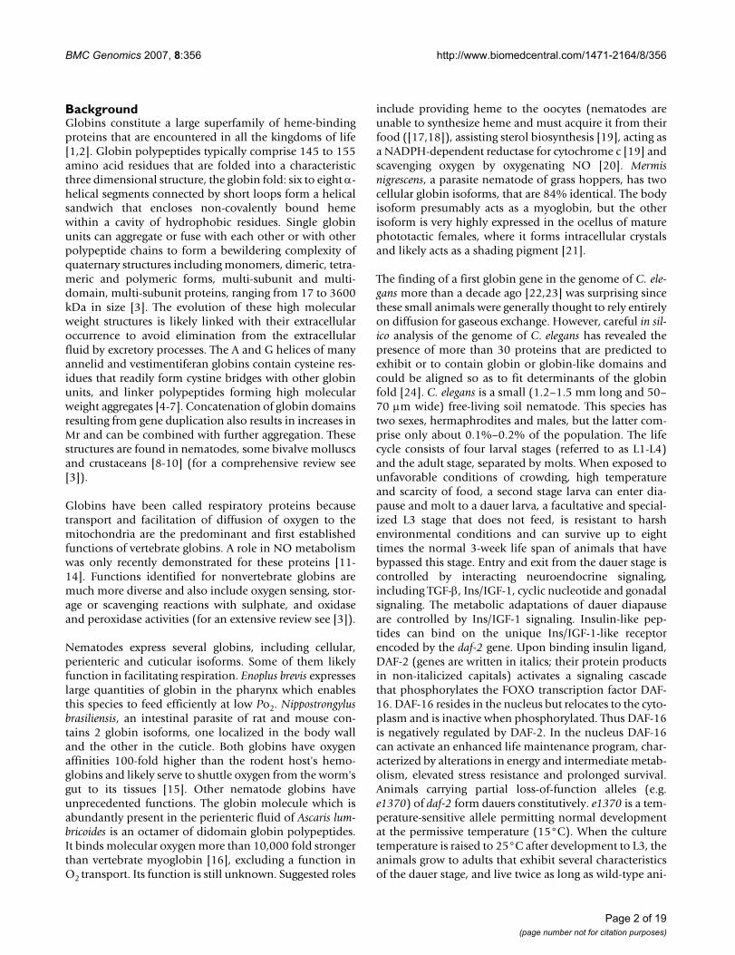
No conservation of intron insertion patternsVertebrate globins genes typically have two intronsinserted at conserved positions B12.2 (intron locatedbetween codon positions 2 and 3 of the 12th amino acidof globin helix B) and G7.0, whereas plant globin geneshave an extra intron at E15.0 and most insect globin genescontain no intervening sequences [37,38]. We have com-pared the exon/intron patterns among all 33 putativeglobin genes in Fig. 3 and Fig. 4. Strikingly, no conserva-tion is discernible neither in the number of introns (1–9)and exons (2–10) nor in the exon/intron boundaries.Examination of the intron insertion positions showed aneven more remarkable diversity. The number of introns
Chromosomal distribution of the C. elegans globin genesFigure 1Chromosomal distribution of the C. elegans globin genes.
Page 4 of 19(page number not for citation purposes)

BMC Genomics 2007, 8:356 http://www.biomedcentral.com/1471-2164/8/356
that interrupt the globin domain ranges from 1(ZK637.13) to 5 (C06E4.7); 1–5 and 1–3 introns areinserted in the pre-A and post-H portions of the proteins,respectively. In the globin domain, 76 different insertionpositions can be found of which most are unprecedented.Many positions are found in interhelical segments, 9 inthe GH interhelix from which 5 are located in GH interhe-lical extensions. Only one globin gene (F21A3.6) featuresboth conserved vertebrate insertion sites B12.2 and G7.0.Fourteen of the 33 putative globin genes display 3 introninsertions in the globin domain, 7 of them feature 2 inter-ruptions in the globin domain.
Developmental expression patternTo establish the pattern of globin gene expressionthroughout the life cycle of the animal, we studied highlysynchronized developmental staged C. elegans using RT-PCR, including embryo's (eggs), L1, L2, dauers, L3, L4,and young adults (1–2 days post the L4 to adult molt. Wefound that a large portion of globins are expressed
through all stages of development (Additional file 2). Dif-ferences in expression levels between developmentalstages were evaluated more accurately using quantitativeRT-PCR. We examined the expression levels of the globinfamily in L3 and dauers, relative to wild-type youngadults. Biological replicates were done on 3 independentworm cultures. Several globin genes (C06E4.7,C09H10.8, C36E8.2, C52A11.2, F52A8.4, R01E6.6,R13A1.8, R90.5, and W01C9.5) are similarly upregulatedin L3 and dauers relative to young adults, although somereach significance in dauers only (Fig. 5). Many genesexhibited more than 2- fold upregulation but didn't reachstatistical significance because strong upregulation wasonly seen in 2 biological replicates (Additional file 3).Remarkably, we observed a significant downregulation inL3 stage relative to young adults for C26C6.7, T22C1.2and ZK637.13. A similar trend was seen in dauers. Inter-estingly, C26C6.7 was the only globin which wasexpressed at a significantly higher level in dauers relativeto L3.
Table 1: Overview of structural characteristics
Gene Accession no. C. briggsaehomolog
Motif/similarity Total length Pre A helix Post H helix GH Inter-helix
C06E4.7 AAA82476 CBG05824 globin-like 230 26 26 21C06H2.5 CAA99771 CBG23115 globin 209 32 25C09H10.8 CAA90439 CBG02965 globin-like 344 - 120 22C18C4.1 EF471982 CBG09371 globin 358 34 162C18C4.9 AAK52180 CBG09369 globin 389 195 40C23H5.2 AAP82661 CBG10551 ngb (Xenopus) 5.4 e-07 278 5 115C26C6.7 EF471981 CBG11881 cygb (human) 3 e-05 404 193 22C28F5.2 EF471980 CBG13047 globin 194 10 42C29F5.7 AAC46826 CBG02622 globin 198 26 16C36E8.2 CAA84649 CBG03635 globin-like 254 41 71C52A11.2 CAA86764 CBG03023 globin 266 30 56 21F19H6.2 CAA92164 CBG00138 ngb (human) 2.8 e-09 231 48 31F21A3.6 CAB04152 CBG18593 cygb (human) 8.2 e-12 236 83 -F35B12.8 CAA98468 CBG23302 globin-like 234 34 48F46C8.7 AAF99945 CBG16720 globin-like 322 59 99F49E2.4 CAA86423 CBG16082 cygb (Danio) 7.9 e-06 216 12 16 40F52A8.4 CAA95823 CBG11915 cygb (human) 0.00014 238 68 11F56C4.3 CAE17840 CBG04428 ngb (Xenopus) 4.1e-05 214 - 47R01E6.6 CAA92187 CBG07422 globin-like 311 94 75R102.9 CAE17922 CBG17640 globin 196 15 - 32
R11H6.3 CAB07647 CBG04577 cygb (human) 0.06 387 132 87R13A1.8 AAA81477 CBG05809 globin 342 159 21R90.5 CAD31696 CBG09511 ngb (Xenopus) 0.006 322 141 24
T06A1.3 EF471978 CBG24799 globin-like 189 31 -T22C1.2 CAA99921 CBG08252 globin 183 25 6W01C9.5 CAA90269 CBG00571 globin 224 22 51Y15E3A.2 CAB60330 CBG07681 globin-like 217 55 23Y17G7B.6 CAA19459 CBG21021 cygb (human) 0.01 216 36 20
Y22D7AR.5 EF471979 CBG11736 ngb (human) 0.0001 272 6 103 17Y57G7A.9 AAC26293 CBG07112 globin-like 169 8 6Y58A7A.6 AAK84620 CBG08670 globin 230 88Y75B7AL.1 AAK68603 CBG06424 globin 542 372 8ZK637.13 CAA77458 CBG06867 globin 159 - 5
Page 5 of 19(page number not for citation purposes)

BMC Genomics 2007, 8:356 http://www.biomedcentral.com/1471-2164/8/356
We also used quantitative real-time RT-PCR experimentsto compare the relative abundance of all 33 globins inwild type adults (Table 2). Results demonstrate thatT22C1.2 and ZK637.13 are expressed at substantiallyhigher levels. The difference with the other globins rangeswithin 1–3 orders of magnitude.
Anoxia-induced globin expressionTo investigate which globins might be upregulated inresponse to oxygen deprivation we subjected young adultworms (1–2 days of adult age) to anoxic conditions for 12h. RNA was subsequently prepared and amplified byquantitative RT-PCR. A separate viability assay showedthat more than 90% of the worm population remainedviable under these conditions, and they recovered within24 h under normoxic conditions. Our criteria for differen-tial regulation were: (1) two-fold difference in expressionwith matched samples, and (2) non-overlapping 95%confidence intervals. Six genes (C26C6.7, F21A3.6,Y17G7B.6, R13A1.8, C18C4.1 and C36E8.2) met boththese criteria and are referred to as anoxia-responsive.T22C1.2 and C18C4.9 exhibited greater than 2-foldupregulation by anoxia but didn't reach statistical signifi-cance (p < 0.06 and p < 0.07, respectively) as 1 biologicalreplicate showed only moderate upregulation. Expressionof W01C9.5 and Y75B7AL.1 was induced 1.96 and 1.89,respectively. These four genes are referred to as likelyanoxia-responsive (Fig. 6). None of the 33 globin tran-
scripts in N2 worms showed reduction in expressionunder anoxia.
Three (C26C6.7, F21A3.6 and Y17G7B.6) out of the 6anoxia-responsive genes displayed sequence similarity tovertebrate cytoglobin, whereas none of the five genes thatshowed homology to vertebrate neuroglobin was inducedby oxygen deprivation (Additional file 4). Interestingly, asimilar lack of hypoxic response was reported for verte-brate neuroglobin [39-41].
HIF-1 dependent globin genesTo find out whether HIF-1 (hypoxia-inducible factor) wasrequired for regulation of the anoxia-responsive genes, wemeasured the expression levels of the proven and likelyanoxia-responsive genes listed in Fig. 6 in hif-1 mutantsgrown in normoxic and anoxic conditions. ZK637.13,which is anoxia-insensitive in wild-type worms butstrongly induced in adult daf-2(e1370), was also includedin this assay. The non-globin, HIF-1 dependent geneF22B5.4 (unknown function) was used as a positive con-trol [42,43]. F22B5.4 was expressed at lower levels underboth normoxia and hypoxia in hif-1 defective animals, asexpected [43], but to our surprise all hypoxia-sensitiveglobins tended (statistically significant for C26C6.7,T22C1.2, F21A3.6, C36E8.2, W01C9.5 and Y17G7B.6) tobe expressed at higher levels in hif-1 compared to wild-type animals. However none of them was differentially
Alignment of 9 representative globins and Sperm whale (SW) myoglobinFigure 2Alignment of 9 representative globins and Sperm whale (SW) myoglobin. ZK637.13 is the canonical globin. It was the first reported globin species in C. elegans and features all characteristics of a standard globin. The next 6 globins have an unusual amino acid residue at CD1. Y75B7AL.1 is a chimeric protein consisting of a G-coupled receptor domain (not shown) and a globin domain. F21A3.6 and F19H6.2 show homology with mammalian cytoglobin and neuroglobin, respectively. Numbers between brackets indicate the number of amino acids preceding and following the globin domain. The residues at position CD1, E7 and F8 are marked in yellow, green and red, respectively.
Page 6 of 19(page number not for citation purposes)

BMC Genomics 2007, 8:356 http://www.biomedcentral.com/1471-2164/8/356
Page 7 of 19(page number not for citation purposes)
Genomic structuresFigure 3Genomic structures.
BMC Genomics 2007, 8:356 http://www.biomedcentral.com/1471-2164/8/356
regulated under anoxia in hif-1 defective mutant worms,indicating that they are HIF-1 dependent (Fig. 7).
Additionally, candidate regulatory regions were examinedfor putative hypoxia-responsive sequence elements(HREs). Results indicate the presence of candidate HIF-1binding elements in the genomic region of all anoxia-induced globin genes.
DAF-2- and DAF-16-regulated globin gene expressionSince daf-2(e1370) mutant worms are hypoxia tolerant[44,45], we anticipated that some globin genes might beconstitutively upregulated in these animals. To our sur-prise we found only minor changes in transcription levelscompared to wild-type worms (Additional file 5), exceptfor ZK637.13 which was significantly upregulated by 4-fold. None of the HIF-1 dependent globin genes was sig-nificantly induced by anoxia in the daf-2 animals. Threeglobin genes (F21A3.6, C18C4.9 and C26C6.7) were sig-nificantly downregulated under normoxic conditions(Fig. 8).
To understand how DAF-2 exerts its effect on ZK637.13we compared the expression level of this globin in daf-16and daf-2;daf-16 mutant worms relative to daf-2 (Fig. 9).We found that the expression of ZK637.13 was reduced by4-fold in daf-16 and daf-2;daf-16 animals indicating thatZK637.13 is regulated by DAF-2 in a DAF-16 dependentfashion. Computational analysis of the ZK637.13genomic region detected the presence of a DAF-16 bind-ing element at position -350, providing additional sup-port for DAF-16-mediated regulation.
Since the HIF-1 dependent globin genes were not inducedby anoxia in daf-2 mutants we expected that DAF-16would not mediate or rather oppose their induction uponanoxia. We found that all anoxia inducible globin genesare upregulated in daf-16 animals under anoxic condi-tions, albeit at a lower level relative to wild-type worms(statistically significant and more than 2-fold forC18C4.9, W01C9.5; significant and less than 2-fold forC36E8.2, T22C1.2, F21A3.6 and Y17G7B.6; borderlinesignificant for R13A1.8, Fig. 10).
Lack of conservation in the intron insertion positionsFigure 4Lack of conservation in the intron insertion positions. Phase 0 introns (55%) are inserted between 2 successive codons; phase 1 (9%) and phase 2 introns (36%) are inserted following the first and second base of a codon, respectively.
Page 8 of 19(page number not for citation purposes)

BMC Genomics 2007, 8:356 http://www.biomedcentral.com/1471-2164/8/356
DAF-16 modulates hif-1 expressionTo learn more about the role of DAF-16 in the response toanoxia we measured the expression of hif-1 in daf-16(m26) and wild-type animals. daf-16(m26) is a verysevere mutation that nearly fully disrupts the function ofDAF-16. We found that transcription of hif-1 in the ani-mals lacking daf-16 activity was lower under normoxia ineach of four independent trials, but this difference was notstatistically significant (not shown). However, expressionof hif-1 was reduced by more than 2.5-fold (P < 0.025)under anoxic conditions in these animals (Fig. 11).
DiscussionStructural diversityThe finding of a globin sequence in the genome ofCaenorhabditis elegans was unexpected because it was gen-erally felt that due to the small size of this organism suffi-cient quantities of oxygen could reach the sites of oxygenconsumption by simple diffusion, and for about a decadeit was thought that globin ZK637.13 was the only globinspecies expressed in C. elegans [22,23]. However, the com-pletion of the genome sequence of this species and thedevelopment of powerful gene prediction tools has nowled to the identification of at least 33 putative globingenes that are all expressed. So, the paramount questionarises whether these globins have distinct functional prop-erties, or represent structural variations of the core globin
and exhibit large redundancy. Usually such multigenefamilies cluster together and the individual membersbecome substrates for purifying or positive selectiondepending on the evolving function. Chironomid larvaelive in low oxygen habitats and selection likely favoredhigh copy number of similar genes to synthesize morehemoglobin. Several of these are stage-specific [33,46].Because of the small size and terrestrial habitat of C. ele-gans it is likely that most globins in this species wereselected for reasons other than enhancing hemoglobinsynthesis capacity. The low transcript abundance of theglobin genes also argues against such a role. Even globinZK637.13, which has the second largest transcript abun-dancy is expressed in a subset of cells [47]. We found noevidence of recent gene duplication events, and the hugediversity seen in the primary structures of the proteins sug-gests a long evolutionary history. Taken together, theseaspects of low expression and huge sequence diversitymay indicate that most, if not all, of these globin genes arecell or stage specific, and that they have diverged to per-form specialized functions.
The wide diversity in the gene structures, as shown by themarked variability of intron insertion patterns is in linewith this view. Vertebrate globins genes typically have twointrons inserted at conserved positions B12.2 and G7.0,whereas plant globin genes have an extra intron at E15.0.
Levels of globin expression in third stage larvae (L3) and the alternative dauer (diapause) stage, relative to expression in young adult animalsFigure 5Levels of globin expression in third stage larvae (L3) and the alternative dauer (diapause) stage, relative to expression in young adult animals. The expression ratio's are the average values from 3 replicate cultures (biological repeats). Bars indicate the 95% confidence interval of the mean. Only globins that showed a significant (borderline for C36E8.2) difference in one of the three comparisons are displayed.
Page 9 of 19(page number not for citation purposes)

BMC Genomics 2007, 8:356 http://www.biomedcentral.com/1471-2164/8/356
It was speculated that the 4 exons, 3 introns arrangementis ancestral and that evolution sometimes led to elimina-tion of introns, particularly in bacteria, yeast and someinsects [48,49]. As the positions of these canonical intronsappear to divide the globin gene into functional domains,it was hypothesized that they witness the joining of func-tional exons to create the globin core unit early in evolu-tion [48,50]. Although later studies demonstrated thatthere is no such general correlation of exon boundaries ingenes with domain boundaries in proteins [51], it wasthought that introns in eukaryotic genes witness the geneorganization of the last universal common ancestor of cel-lular life, and that evolution sometimes led to their total(prokaryotes) or partial (eukaryotes) elimination [48,52].This introns-early view was challenged by the intron-latehypothesis [53] which holds that introns in eukaryotegenomes evolved after the split of prokaryotes and eukary-otes. This debate still persists [54,55]. The central questionis how the complex eukaryotic spliceosomal machinerycould have evolved. Self-splicing group I and group IIintrons are found in prokaryotes, but spliceosomalintrons are a hallmark of eukaryotic genomes. A reconcil-ing hypothesis holds that spliceosomal introns co-evolvedwith the origin of the eukaryote ancestor: an archaebacte-rial host acquired an α-proteobacterial symbiont (themitochondrial ancestor). Group II introns from this sym-biont invaded the hosts' genome and evolved to spliceo-somal introns [56,57]. This initial intron invasion wouldhave ended when the original intron-encoded protein wasno longer required and was exposed to mutationaldemise. It is likely that introns that are inserted at very
conservative positions throughout the evolution (e.g.B12.2, G7.0 and E15.0 in globin genes) are descendants ofthese founder introns. These, and many other intronshave been lost and many other introns have beenacquired more recently in nematodes. It is thought thatthese introns were gained by a fundamentally differentprocess, likely reverse splicing of preexisting introns[54,58,59]. The lack of any conspicuous pattern of intronspositioning in the globin genes of C. elegans reflects adynamic pattern of intron insertion events, consistentwith this hypothesis. Minor variability in intron position-ing has been described for the cytochrome P450 [60] andDEAD helicase gene families [61] of C. elegans, however.The mechanisms controlling the stringent or more relaxedexon-intron pattern is currently not understood. A com-prehensive analysis of the evolutionary history of theglobin protein family in the genus Caenorhabditis will bedescribed elsewhere.
Anoxia-responsive globinsAs a first approach to establish their functions, we havestudied the expression profiles of these putative globinsunder normoxia and anoxia. C. elegans is a soil inhabitingnematode which relies on oxygen consumption by themitochondria to maintain normal metabolic function. Inthe laboratory the worms are usually grown on agarplates, where they are exposed to normal atmospheric air,which contains 21 % oxygen or approx. 300 mg oxygen/Lair. Oxygen supply will be lower in moist soil, mainly dueto the poor solubility of oxygen in water, which is reducedto approx. 9.1 mg/L water at 20°C. C. elegans is able to
Ten genes are upregulated in young adult wild-type animals following 12 h of anoxiaFigure 6Ten genes are upregulated in young adult wild-type animals following 12 h of anoxia. The expression ratio's are the average val-ues from 3 replicate cultures (biological repeats). Bars indicate the 95% confidence interval of the mean.
Page 10 of 19(page number not for citation purposes)

BMC Genomics 2007, 8:356 http://www.biomedcentral.com/1471-2164/8/356
maintain a constant metabolic rate when the oxygen con-centration in air is lowered to approx. 3.6 %, and reducesrespiration by 50% at 1 % oxygen (or 51.4 and 14.3 mgoxygen per liter air, respectively [62]). Thus water-loggedor flooded soils rapidly become hypoxic and, not surpris-ingly, C. elegans has developed a response to cope withreduced oxygen supply. The animals can continue todevelop and reproduce down to 0.1–0.25 % gaseous oxy-gen by activation of a hypoxia responsive pathway[63,64]. Below this threshold they can relay on suspendedanimation for survival under anoxia and hif-1 is notrequired [63,65]. However, our findings demonstrate thatsome globins are upregulated under conditions of totaloxygen deprivation. One possible explanation of thisfinding is that the induction of these genes by hypoxia isquite fast and 'frozen' in this state under anoxia.
Only 6, perhaps 10, out of the 33 globins were anoxia-responsive. Three out of 6 cytoglobin-like and none of the5 neuroglobin-like proteins were upregulated under con-ditions of oxygen deprivation, in agreement with most
mammalian globin expression studies [39-41,66]. Thelack of significant difference in expression under nor-moxic and anoxic conditions in hif-1 mutant worms leadsto the conclusion that all anoxia responsive globin genesare regulated by a HIF-1 mediated mechanism. Computa-tional analysis of the globin genomic regions detected thepresence of putative hypoxia-responsive sequence ele-ments in all anoxia-induced globin genes, providing addi-tional support for HIF-1-mediated upregulation. Theseglobins are also induced under normoxia in hif-1 mutants(Fig. 7), suggesting a more complex regulation that is ableto compensate for the loss of hif-1 function.
Y75B7AL.1 was mildly induced by anoxia, reaching signif-icance in one set of experiments only (Figs 6 and 7), andrequired intact hif-1 activity for adaptation to anoxia. Pre-diction programs identify Y75B7AL.1 as a chimeric pro-tein consisting of a G-coupled receptor domaincontaining 7 transmembrane helices and a globindomain. Its structural properties suggest that this proteinmay act as an oxygen sensor. The oxygen dependent
Table 2: Relative expression levels. Values are average percentages and standard deviations from 6 independent experiments
Globin gene Relative expression Standard Deviation
C06E4.7 0.267 0.174C06H2.5 0.262 0.030C09H10.8 0.272 0.094C18C4.1 0.328 0.446C18C4.9 2.245 1.626C23H5.2 0.304 0.147C26C6.7 0.196 0.171C28F5.2 0.554 0.490C29F5.7 1.799 0.805C36E8.2 1.611 1.208
C52A11.2 0.279 0.300F19H6.2 1.235 0.641F21A3.6 0.421 0.170F35B12.8 0.431 0.380F46C8.7 0.164 0.044F49E2.4 0.604 0.196F52A8.4 0.339 0.096F56C4.3 1.103 0.298R01E6.6 2.697 0.311R102.9 0.052 0.043
R11H6.3 0.463 0.180R13A1.8 0.449 0.149
R90.5 0.558 0.349T06A1.3 0.395 0.309T22C1.2 43.659 8.788W01C9.5 0.547 0.269Y15E3A.2 0.237 0.166Y17G7B.6 1.443 0.508
Y22D7AR.5 0.191 0.223Y57G7A.9 0.172 0.083Y58A7A.6 0.981 0.468Y75B7Al.1 4.329 0.852ZK637.13 31.409 10.324
Page 11 of 19(page number not for citation purposes)

BMC Genomics 2007, 8:356 http://www.biomedcentral.com/1471-2164/8/356
expression of this globin is surprising since such regula-tion is unprecedented to date. A chimeric protein com-prising a guanylate cyclase and a haem binding domainwas recently reported to sense oxygen and to regulate aer-otaxis responses [67,68].
Expression of globin ZK637.13 is regulated by insulin/IGF-1 signaling and not induced by anoxiaSince both dauers and daf-2 mutant animals are hypoxiaresistant [44] we anticipated that they might constitutivelyexpress a subset of globin genes at higher levels. However,we found to our surprise a rather dissimilar pattern ofglobin regulation in these animals, as summarized inTable 3. Several globins were borderline (not statisticallysignificant) upregulated in dauers (relative to L3), but notin daf-2 mutant worms. ZK637.13 was substantiallyupregulated in daf-2(e1370) adults in a daf-16-dependentfashion. This globin molecule was expressed at lower lev-els in L3 and borderline (no statistical support) upregu-lated in the alternative dauer stage. Interestingly,regulation of transcription of this globin species was notsensitive to anoxia. Since suspended DAF-2 signalinginduces enhanced life maintenance it is tempting to antic-ipate that ZK637.13 contributes to these processes.
Multiple pathways control expression of the anoxia responsive globin genesSurprisingly, none of the HIF-1 responsive globin geneswas induced when daf-2(e1370) mutant animals wereexposed to 12 h of oxygen deprivation (Table 3). Thus
induction of these genes by anoxia requires both HIF-1and DAF-2 function. This could indicate that HIF-1 medi-ates induction of these genes upon anoxia treatment andthat HIF-1 activity is modulated by insulin/IGF-1 signal-ing, or that HIF-1 and DAF-2 each act to regulate theexpression of these globin genes.
The data presented in Figures 10 and 11 provide supportfor the first model: reduced levels of hif-1 mRNA weremeasured in animals lacking daf-16 function and theinduction of the globins in response to anoxia was weak-ened in these mutants. The finding that anoxic treatmentfails to induce the HIF-1 responsive globin genes in ani-mals lacking daf-2 function leads to an apparent contra-diction, however: DAF-16 activity which is negativelyregulated by DAF-2 is expected to be fully active in theseanimals and hif-1 would be expected to be fully expressed.Yet the globin genes were not induced upon anoxic treat-ment in these mutants. To explain this we propose amodel in which DAF-16 opposes the expression of theseglobins, directly or indirectly. Direct inhibition couldresult from competition of HIF-1 and DAF-16 for the HIF-1 responsive promoters. This explanation would implythat DAF-16 can bind to but not activate these promoters.This is opposite to globin ZK637.13 which is clearlyinduced by DAF-16 but not HIF-1.
ConclusionThis work provides the first comprehensive analysis of theglobin gene family of C. elegans. We illustrate the remark-
hif-1 mediates induction of the anoxia responsive genesFigure 7hif-1 mediates induction of the anoxia responsive genes. The expression ratio's are the average values from 3 replicate cultures (biological repeats). Bars indicate the 95% confidence interval of the mean. F22B5.4 is an established hypoxia responsive gene of unknown function [43] and is included as a positive control. Globin ZK637.13 is not sensitive to anoxia and was included as a negative control.
Page 12 of 19(page number not for citation purposes)

BMC Genomics 2007, 8:356 http://www.biomedcentral.com/1471-2164/8/356
able structural diversity of this gene family, pointing to afunctional variety. In this study we demonstrate signifi-cant differential expression of the C. elegans globin genefamily upon anoxia. Studying expression profiles in differ-ent mutant worms has enabled us to provide initial evi-dence of linked HIF-1 and Ins/IGF-1 signaling for globinregulation under severe hypoxic conditions in the nema-tode.
We assume that in a natural environment HIF-1 respon-sive globins are "on" when DAF-2 is "on" i.e. when theconditions for growth and reproduction are favorable.Scarcity of oxygen in otherwise permissive conditions isremedied by the action of HIF-1. Adverse conditions forgrowth and reproduction (crowding, high temperature,...)lead to reduced DAF-2 signaling and increasing silencingof the HIF responsive globin promoters by the DAF-16FOXO transcription factor. In contrast, globin ZK637.13is induced and may contribute to the life maintenanceprogram that is deployed under these conditions.
MethodsCulture techniquesThe wild-type strain N2 and the mutant strains daf-2(e1370), daf-16(m26), daf-2(e1370);daf-16(m26) and
hif-1(ia04) were obtained from the Caenorhabditis Genet-ics Center. Synchronous populations were initiated fromeggs prepared by alkaline hypochlorite treatment ofgravid adults and grown at 24°C on cholesterol supple-mented Nutrient Agar (OXOID) plates containing a lawn
Expression levels of globin genes in daf-16 and daf-2;daf-16 young adults relative to daf-2 young adult wormsFigure 9Expression levels of globin genes in daf-16 and daf-2;daf-16 young adults relative to daf-2 young adult worms. The expression ratio's are the average values from 3 replicate cul-tures (biological repeats). Bars indicate the 95% confidence interval of the mean.
Expression levels of globin genes in daf-2 young adults under normoxia or following 12 h exposure to anoxic conditions, rela-tive to wild-type young adult worms under normoxiaFigure 8Expression levels of globin genes in daf-2 young adults under normoxia or following 12 h exposure to anoxic conditions, rela-tive to wild-type young adult worms under normoxia. The expression ratio's are the average values from 3 replicate cultures (biological repeats). Bars indicate the 95% confidence interval of the mean. Only globins that showed a significant (borderline for T22C1.2) difference normoxia N2 vs. normoxia daf-2 are displayed.
Page 13 of 19(page number not for citation purposes)

BMC Genomics 2007, 8:356 http://www.biomedcentral.com/1471-2164/8/356
of freshly grown E. coli K12 cells [69]. For the study of lifecycle specific globin expression experiments worm sam-ples were harvested after 0(unfed L1), 6(L1), 12(L1),18(L2), 24(L2), 30(L3), 36(L4), 42(L4) and 48(youngadult) hours of growth. When the worms reached thefourth juvenile stage FUdR was added at 200 μM finalconcentration to prevent progeny production. At harvest,worms were washed off the plates, cleaned using Percolland dense sucrose [70], flash frozen and stored at -75°Cuntil use. daf-2 (e1370) is a temperature-sensitive consti-tutive dauer former: L2 larvae enter diapause at 24°C butmolt to the normal third larval stage at lower temperature.To prevent dauer formation the cultures were incubated at17°C and shifted to 24°C after the animals had molted tothe fourth larval stage. Dauers were grown by spreading150,000 eggs, 1010 heat killed E. coli cells and 1mg haemo-globin (from a 5% stock solution in 0.1N KOH, auto-claved for 10 min) on 10 cm agar (made up withcholesterol supplemented S buffer, pH 7.0) plates. Theseconditions induce almost 100% dauer formation. Platescontaining less than 99% dauers were discarded. hif-1mutant worms were grown at 20°C.
The worms were subjected to anoxia in the respirometercells of a six channel respirometer from Strathkelvin(Glasgow, Scotland) equipped with Clark electrodes.Young adult (1–2 days after the L4 to adult molt) animalswere washed off the plates. Samples containing 20000–40000 worms in 2 ml of S buffer were transferred to the
respirometer cells and sealed with rubber stoppers. Theworm suspensions were stirred at the appropriate temper-ature and the oxygen concentration was continuouslymonitored. Oxygen concentration decreased rapidlybelow 0.1 μM (= 3.2 μg/L, which is in the order of about0.001% of the atmospheric concentration) within 15 minand was stable for the next 12 h. Three replicate worm cul-
Expression level of hif-1 under anoxic conditions in daf-16 young adults relative to N2 young adult wormsFigure 11Expression level of hif-1 under anoxic conditions in daf-16 young adults relative to N2 young adult worms. The expres-sion ratio's are the average values from 4 replicate cultures (biological repeats). Bars indicate the 95% confidence interval of the mean.
Expression levels of globin genes in daf-16 young adults following 12 h exposure to anoxic conditions relative to normoxic con-ditionsFigure 10Expression levels of globin genes in daf-16 young adults following 12 h exposure to anoxic conditions relative to normoxic con-ditions. The expression ratio's are the average values from 3 replicate cultures (biological repeats). Bars indicate the 95% con-fidence interval of the mean. Only globins that showed a significant (borderline for R13A1.8) difference are displayed.
Page 14 of 19(page number not for citation purposes)

BMC Genomics 2007, 8:356 http://www.biomedcentral.com/1471-2164/8/356
tures were grown and 3 samples from each culture weresubjected to anoxia to account for experimental variation.
Identification of globin-like genesPutative globins and globin domains were identified inthe genomes of C. elegans and C. briggsae as described ear-lier [1,2,24]. Briefly, putative globins were identifiedusing a library of hidden Markov models [71] listed on theSUPERFAMILY website [72]. Sequences that matched theglobin motifs with E-values > 10-5 or shorter than 100 aawere discarded and the remaining sequences werechecked employing the WormBase Release WS150 [73].Orthologous sequences were manually aligned followingthe procedure used earlier in the alignment of over 700globins [74]. This procedure is designed to fit the putativesequences to the myoglobin fold [75], the pattern of 37conserved, hydrophobic residues, including 33 intra-heli-cal residues A8, A11, A12, A15, B6, B9, B10, B13, B14, C4,
E4, E7, E8, E11, E12, E15, E18, E19, F1, F4, G5, G8, G11,G12, G13, G15, G16, H7, H8, H11, H12, H15, and H19,the three inter-helical residues at CD1, CD4 and FG4 andthe invariant His at F8 [74,76]. Although earlier align-ments [74] had suggested that globins were characterizedby two invariant residues, F8His and CD1Phe, morerecent information on globin sequences [1,2] indicatethat other hydrophobic residues, such as Tyr/Met/Leu/Ile/Val can occur at the CD1 position as well as Ala/Ser/Thr/Leu at the distal E7 position, in addition to His and Gln.
RNA isolation and cDNA synthesisTotal RNA was isolated from harvested C. elegans worm-susing the RNeasy Midi Kit from Qiagen (Hilden, Ger-many), according to the manufacturer's instructions. ADNase I (Zymo Research, Orange, California) digestionstep was subsequently performed to remove genomicDNA. First strand cDNA was synthesized using an
Table 3: Overview of differential globin regulation. Brackets denote substantial, but not significant, difference in expression
Globin gene dauer vs L3 dauer vs adult
L3 vs adult daf-2 vs N2 N2 anoxia vs normoxia
daf-2 anoxia vs normoxia
hif-1 vs N2 daf-16 anoxia vs normoxia
C06E4.7 (up) upC06H2.5 (up) (up)C09H10.8 (up) upC18C4.1 (up) (up) up (up) (up)C18C4.9 down up (up) upC23H5.2C26C6.7 up down down up upC28F5.2 (up)C29F5.7C36E8.2 (up) (up) (up) up up upC52A11.2 (up) up (up)F19H6.2F21A3.6 (up) (up) down up up upF35B12.8F46C8.7 (up)F49E2.4F52A8.4 (up) upF56C4.3R01E6.6 (up) up upR102.9 (up)R11H6.3 (up) (up)R13A1.8 (up) up up (up) (up) (up)R90.5 up upT06A1.3 (up)T22C1.2 (down) (down) down (down) up up upW01C9.5 (up) up up up upY15E3A.2Y17G7B.6 up up upY22D7AR.5 (up) (up)Y57G7A.9 (up)Y58A7A.6 (up) (up)Y75B7AL.1 up (up) upZK637.13 (up) down up
Page 15 of 19(page number not for citation purposes)

BMC Genomics 2007, 8:356 http://www.biomedcentral.com/1471-2164/8/356
oligo(dT) primer and a Moloney murine leukemia virusreverse transcriptase (Fermentas, Vilnius, Lithuania).
Expression analysiscDNA was used as a template to amplify mRNA from eachputative globin in a PCR reaction using gene-specific for-ward and reverse primers. The reverse transcription reac-tion conditions were 1 μM forward and reverse primer,200 μM dNTP, 2.5 mM MgCl2, and 2.5 units of Taq DNApolymerase (Qiagen, Hilden, Germany) in a volume of 50μl. The cycling conditions were 95°C for 2 min followedby 40 cycles of 95°C for 1 min, 54°C for 30 sec and 72°Cfor 30 sec.
The 5' cDNA end of several sequences was identified usinga RACE (Invitrogen, Carlsbad, California) experiment.First strand cDNA was synthesized using a gene-specificprimer. A polyC tail was added to the 3' end of the cDNAwith terminal deoxynucleotide transferase. The 5' end wasthen amplified using an oligo(dG) adaptor and a gene-specific nested primer in a PCR of 35 cycles of 94°C for 1min, 54°C for 30 sec and 72°C for 30 sec, followed by afinal extension for 5 min at 72°C. The amplified productswere sequenced on both strands using BigDye terminatorchemistry and an ABI 377 sequencer (Applied Biosystems,Foster City, California).
Real-time quantitative RT-PCRPrimers (Invitrogen) were designed using Primer3 soft-ware [77] and tested for specificity using NCBI BLAST. Thesequences are available upon request.
The targets amplified by the primer pairs were evaluatedwith MFOLD software [78] in order to control for the for-mation of secondary structures at the site of primer bind-ing. MFOLD analysis was performed using default settingsand 50 mM Na+, 3 mM Mg2+ and a temperature of 60°C(which is the annealing temperature of the primers).
Quantitative RT-PCR was carried out using a Rotor-Gene2000 centrifugal real-time cycler (Corbett Research, Mort-lake, Australia) using the Platinum SYBR Green qPCRSuperMix-UDG (Invitrogen). Each reaction contained:12.5 μl of the Platinum SYBR Green qPCR SuperMix-UDG, 100 nM, 200 nM or 500 nM of forward and reverseprimers and 5 μl cDNA (1:40 RNA dilution), to a final vol-ume of 25 μl. Amplification was performed in 0.1 ml real-time PCR tubes (Corbett Research) placed in the 72-wellrotor of the Rotor-Gene instrument. The cycling condi-tions were as follows: 50°C for 2 min, initial denaturationat 95°C for 2 min, followed by 45 cycles of 15s at 95°C,30s at 60°C, and 30s at 72°C (gain set at 8 for SYBRGreen). Following the final cycle, melting curve analysiswas performed to examine the specificity in each reactiontube (absence of primer dimers and other nonspecific
products). The Rotor-Gene software (version 6.0) allowsautomatic melting curve analysis for all tested samples ina given run. SYBR Green fluorescence of the generatedproducts was continuously monitored throughout thetemperature ramp from 60 to 99°C. The temperature rosein 1° increments with a 5-s hold at each degree. A singlemelt peak for each reaction confirmed the identity of eachPCR product. Each assay included a no-template controlfor every primer pair. All PCR reactions were performed intriplicate. Real-time PCR efficiencies for each reactionwere calculated using the formula: Efficiency (E) = [10(1/
slope)] – 1, from the slope values given by Rotor-Gene soft-ware. Amplicons from each reaction mixture were alsoanalyzed by agarose gel electrophoresis.
Quantification and data analysisThe threshold cycle (Ct) values of the Rotor-Gene softwareversion 6.0 (Corbett Research) were exported to Excel(Microsoft) for further analysis. Reaction efficiency esti-mates were derived from standard curves that were gener-ated using serial dilutions of the corresponding cDNA.These were then used to transform the Ct values to relativequantities, which were normalized using the geometricmean of 3 reference genes identified by the geNorm 3.4software [79]. The geNorm VBA applet for Microsoft Exceldetermines the most stable reference genes from a set ofgenes in a given panel of cDNA samples. We evaluated aset of 8 candidate reference genes, comprising act-1, csq-1,mua-6, tba-1, mlc-3, pat-10, unc-15 and T22B11.5. Thethree most stably expressed genes were used to calculate anormalization factor for each of the cDNA samples.Expression levels of all globin genes were determined in atleast 3 independent replicate cultures. Differential geneexpression was considered significant when the 95% con-fidence interval of the mean expression levels did notoverlap (equivalent to P < 0.05).
Analysis of potential regulatory elementsAll 33 globin genomic regions comprising 2000 bpupstream of the translation initiation sites, all introns and500 bp downstream were evaluated for the presence ofputative HIF-1 (Hypoxia Responsive Elements, HRE, con-sensus motif RCGTG) and DAF-16 (TTG/ATTTAC andTGATAAG, [80]) binding sites in both directions. Repeatswere masked using RepeatMasker.
Competing interestsThe author(s) declares that there are no competing inter-ests.
Authors' contributionsDH and JVR conceived and designed all experiments; DH,EG and AV performed experiments, DH, EG, LM, SD, SVand JVR analyzed data; DH and JRV wrote the manuscript.All authors read and approved the final manuscript.
Page 16 of 19(page number not for citation purposes)

BMC Genomics 2007, 8:356 http://www.biomedcentral.com/1471-2164/8/356
Additional material
AcknowledgementsWe thank Jo Anne Powell-Coffman and Iqbal Hamza for critical reading of the manuscript. This work was supported by grants from the Fund for Sci-entific Research Flanders (G.0331.04) and the European Commission (QLG3-CT-2002-01548). SD is a postdoctoral fellow of the Fund for Scien-tific Research Flanders (FWO). Some nematode strains used in this work were provided by the Caenorhabditis Genetics Center, which is funded by the NIH National Center for Research Resources (NCRR).
References1. Vinogradov SN, Hoogewijs D, Bailly X, Arredondo-Peter R, Gough J,
Dewilde S, Moens L, Vanfleteren JR: A phylogenomic profile ofglobins. BMC Evol Biol 2006, 6:31.
2. Vinogradov SN, Hoogewijs D, Bailly X, Arredondo-Peter R, GuertinM, Gough J, Dewilde S, Moens L, Vanfleteren JR: Three globin line-ages belonging to two structural classes in genomes from thethree kingdoms of life. Proc Natl Acad Sci U S A 2005,102(32):11385-11389.
3. Weber RE, Vinogradov SN: Nonvertebrate hemoglobins: func-tions and molecular adaptations. Physiol Rev 2001,81(2):569-628.
4. Strand K, Knapp JE, Bhyravbhatla B, Royer WE Jr.: Crystal struc-ture of the hemoglobin dodecamer from Lumbricus erythro-cruorin: allosteric core of giant annelid respiratorycomplexes. Journal of molecular biology 2004, 344(1):119-134.
5. Green BN, Gotoh T, Suzuki T, Zal F, Lallier FH, Toulmond A,Vinogradov SN: Observation of large, non-covalent globin sub-assemblies in the approximately 3600 kDa hexagonal bilayerhemoglobins by electrospray ionization time-of-flight massspectrometry. J Mol Biol 2001, 309(3):553-560.
6. Royer WE Jr., Strand K, van Heel M, Hendrickson WA: Structuralhierarchy in erythrocruorin, the giant respiratory assem-blage of annelids. Proc Natl Acad Sci U S A 2000, 97(13):7107-7111.
7. Kuchumov AR, Taveau JC, Lamy JN, Wall JS, Weber RE, VinogradovSN: The role of linkers in the reassembly of the 3.6 MDa hex-agonal bilayer hemoglobin from Lumbricus terrestris. J Mol Biol1999, 289(5):1361-1374.
8. Manning AM, Trotman CN, Tate WP: Evolution of a polymericglobin in the brine shrimp Artemia. Nature 1990,348(6302):653-656.
9. Trotman CN, Manning AM, Bray JA, Jellie AM, Moens L, Tate WP:Interdomain linkage in the polymeric hemoglobin moleculeof Artemia. J Mol Evol 1994, 38(6):628-636.
10. Dewilde S, Van Hauwaert ML, Peeters K, Vanfleteren J, Moens L:Daphnia pulex didomain hemoglobin: structure and evolu-tion of polymeric hemoglobins and their coding genes. MolBiol Evol 1999, 16(9):1208-1218.
11. Flogel U, Merx MW, Godecke A, Decking UK, Schrader J:Myoglobin: A scavenger of bioactive NO. Proc Natl Acad Sci U SA 2001, 98(2):735-740.
12. Merx MW, Godecke A, Flogel U, Schrader J: Oxygen supply andnitric oxide scavenging by myoglobin contribute to exerciseendurance and cardiac function. FASEB J 2005, 19(8):1015-1017.
13. Herold S, Shivashankar K, Mehl M: Myoglobin scavenges perox-ynitrite without being significantly nitrated. Biochemistry 2002,41(45):13460-13472.
14. Herold S, Fago A, Weber RE, Dewilde S, Moens L: Reactivity stud-ies of the Fe(III) and Fe(II)NO forms of human neuroglobinreveal a potential role against oxidative stress. J Biol Chem2004, 279(22):22841-22847.
15. Blaxter ML, Ingram L, Tweedie S: Sequence, expression and evo-lution of the globins of the parasitic nematode Nippostrongy-lus brasiliensis. Mol Biochem Parasitol 1994, 68(1):1-14.
16. Okazaki T, Wittenberg JB: The hemoglobin of Ascaris peri-enteric fluid. 3. Equilibria with oxygen and carbon monoxide.Biochim Biophys Acta 1965, 111(2):503-511.
17. Hieb WF, Stokstad EL, Rothstein M: Heme requirement forreproduction of a free-living nematode. Science 1970,168(927):143-144.
18. Rao AU, Carta LK, Lesuisse E, Hamza I: Lack of heme synthesis ina free-living eukaryote. Proc Natl Acad Sci U S A 2005,102(12):4270-4275.
19. Sherman DR, Guinn B, Perdok MM, Goldberg DE: Components ofsterol biosynthesis assembled on the oxygen-avid hemo-globin of Ascaris. Science 1992, 258(5090):1930-1932.
20. Minning DM, Gow AJ, Bonaventura J, Braun R, Dewhirst M, GoldbergDE, Stamler JS: Ascaris haemoglobin is a nitric oxide-activated'deoxygenase'. Nature 1999, 401(6752):497-502.
21. Burr AH, Hunt P, Wagar DR, Dewilde S, Blaxter ML, Vanfleteren JR,Moens L: A hemoglobin with an optical function. J Biol Chem2000, 275(7):4810-4815.
22. Kloek AP, Sherman DR, Goldberg DE: Novel gene structure andevolutionary context of Caenorhabditis elegans globin. Gene1993, 129(2):215-221.
23. Mansell JB, Timms K, Tate WP, Moens L, Trotman CN: Expressionof a globin gene in Caenorhabditis elegans. Biochem Mol Biol Int1993, 30(4):643-647.
24. Hoogewijs D, Geuens E, Dewilde S, Moens L, Vierstraete A, Vinogra-dov S, Vanfleteren J: Genome-wide analysis of the globin genefamily of C. elegans. IUBMB Life 2004, 56(11-12):697-702.
25. Kenyon C, Chang J, Gensch E, Rudner A, Tabtiang R: A C. elegansmutant that lives twice as long as wild type. Nature 1993,366(6454):461-464.
Additional file 1Alignment of the 33 C. elegans globinsClick here for file[http://www.biomedcentral.com/content/supplementary/1471-2164-8-356-S1.pdf]
Additional file 2Globin expression throughout the life-cycle using RT-PCRClick here for file[http://www.biomedcentral.com/content/supplementary/1471-2164-8-356-S2.tiff]
Additional file 3Expression levels of globin genes in third stage larvae (L3) and the alter-native dauer (diapause) stage, relative to expression in young adult ani-mals. The expression ratios are the average values from 3 replicate cultures (biological repeats). Bars indicate standard error of the mean.Click here for file[http://www.biomedcentral.com/content/supplementary/1471-2164-8-356-S3.tiff]
Additional file 4Expression levels of globin genes in young adult wild-type animals follow-ing 12 h of anoxia, relative to expression in normoxia. The expression ratios are the average values from 3 replicate cultures (biological repeats). Bars indicate standard error of the mean.Click here for file[http://www.biomedcentral.com/content/supplementary/1471-2164-8-356-S4.tiff]
Additional file 5Expression levels of globin genes in daf-2 young adults under normoxia or following 12 h exposure to anoxic conditions, relative to wild-type young adult worms under normoxia. The expression ratios are the average values from 3 replicate cultures (biological repeats). Bars indicate standard error of the mean.Click here for file[http://www.biomedcentral.com/content/supplementary/1471-2164-8-356-S5.tiff]
Page 17 of 19(page number not for citation purposes)
http://www.ncbi.nlm.nih.gov/entrez/query.fcgi?cmd=Retrieve&db=PubMed&dopt=Abstract&list_uids=2250721
http://www.ncbi.nlm.nih.gov/entrez/query.fcgi?cmd=Retrieve&db=PubMed&dopt=Abstract&list_uids=8083888
http://www.ncbi.nlm.nih.gov/entrez/query.fcgi?cmd=Retrieve&db=PubMed&dopt=Abstract&list_uids=7891734
http://www.ncbi.nlm.nih.gov/entrez/query.fcgi?cmd=Retrieve&db=PubMed&dopt=Abstract&list_uids=5879483
http://www.ncbi.nlm.nih.gov/entrez/query.fcgi?cmd=Retrieve&db=PubMed&dopt=Abstract&list_uids=5879483
http://www.ncbi.nlm.nih.gov/entrez/query.fcgi?cmd=Retrieve&db=PubMed&dopt=Abstract&list_uids=5417058
http://www.ncbi.nlm.nih.gov/entrez/query.fcgi?cmd=Retrieve&db=PubMed&dopt=Abstract&list_uids=5417058
http://www.ncbi.nlm.nih.gov/entrez/query.fcgi?cmd=Retrieve&db=PubMed&dopt=Abstract&list_uids=1470914
http://www.ncbi.nlm.nih.gov/entrez/query.fcgi?cmd=Retrieve&db=PubMed&dopt=Abstract&list_uids=8325507
http://www.ncbi.nlm.nih.gov/entrez/query.fcgi?cmd=Retrieve&db=PubMed&dopt=Abstract&list_uids=8401321

BMC Genomics 2007, 8:356 http://www.biomedcentral.com/1471-2164/8/356
26. Kenyon C: The plasticity of aging: insights from long-livedmutants. Cell 2005, 120(4):449-460.
27. Braeckman BP, Vanfleteren JR: Genetic control of longevity in C.elegans. Exp Gerontol 2007, 42(1-2):90-98.
28. Gerisch B, Weitzel C, Kober-Eisermann C, Rottiers V, Antebi A: Ahormonal signaling pathway influencing C. elegans metabo-lism, reproductive development, and life span. Dev Cell 2001,1(6):841-851.
29. Motola DL, Cummins CL, Rottiers V, Sharma KK, Li T, Li Y, Suino-Powell K, Xu HE, Auchus RJ, Antebi A, Mangelsdorf DJ: Identifica-tion of ligands for DAF-12 that govern dauer formation andreproduction in C. elegans. Cell 2006, 124(6):1209-1223.
30. Rottiers V, Motola DL, Gerisch B, Cummins CL, Nishiwaki K, Man-gelsdorf DJ, Antebi A: Hormonal control of C. elegans dauer for-mation and life span by a Rieske-like oxygenase. Dev Cell 2006,10(4):473-482.
31. Stein LD, Bao Z, Blasiar D, Blumenthal T, Brent MR, Chen N, Chin-walla A, Clarke L, Clee C, Coghlan A, Coulson A, D'Eustachio P, FitchDH, Fulton LA, Fulton RE, Griffiths-Jones S, Harris TW, Hillier LW,Kamath R, Kuwabara PE, Mardis ER, Marra MA, Miner TL, Minx P,Mullikin JC, Plumb RW, Rogers J, Schein JE, Sohrmann M, Spieth J, Sta-jich JE, Wei C, Willey D, Wilson RK, Durbin R, Waterston RH: Thegenome sequence of Caenorhabditis briggsae: a platform forcomparative genomics. PLoS biology 2003, 1(2):E45.
32. Shi J, Blundell TL, Mizuguchi K: FUGUE: sequence-structurehomology recognition using environment-specific substitu-tion tables and structure-dependent gap penalties. J Mol Biol2001, 310(1):243-257.
33. Green BN, Kuchumov AR, Hankeln T, Schmidt ER, Bergtrom G,Vinogradov SN: An electrospray ionization mass spectromet-ric study of the extracellular hemoglobins from Chironomusthummi thummi. Biochim Biophys Acta 1998, 1383(1):143-150.
34. TMHMM [http://www.cbs.dtu.dk/services/TMHMM/]35. TMPRED [http://www.ch.embnet.org/software/
TMPRED_form.html]36. DAS [http://www.sbc.su.se/~miklos/DAS/maindas.html]37. Rozynek P, Broecker M, Hankeln T, Schmidt E: The primary struc-
ture of several hemoglobin genes from the genome of Chi-ronomus tentans. In Structure and function of invertebrate oxygencarriers Edited by: Vinogradov SN, Kapp OH. New York , Springer;1991:297-312.
38. Stoltzfus AF, Doolittle WF: Molecular evolution: slippery intronsand globin gene evolution. Curr Biol 1993, 3:215-217.
39. Mammen PP, Shelton JM, Goetsch SC, Williams SC, Richardson JA,Garry MG, Garry DJ: Neuroglobin, a novel member of theglobin family, is expressed in focal regions of the brain. J His-tochem Cytochem 2002, 50(12):1591-1598.
40. Fordel E, Geuens E, Dewilde S, Rottiers P, Carmeliet P, Grooten J,Moens L: Cytoglobin expression is upregulated in all tissuesupon hypoxia: an in vitro and in vivo study by quantitativereal-time PCR. Biochem Biophys Res Commun 2004,319(2):342-348.
41. Hundahl C, Stoltenberg M, Fago A, Weber RE, Dewilde S, Fordel E,Danscher G: Effects of short-term hypoxia on neuroglobin lev-els and localization in mouse brain tissues. Neuropathol ApplNeurobiol 2005, 31(6):610-617.
42. Bishop T, Lau KW, Epstein AC, Kim SK, Jiang M, O'Rourke D, PughCW, Gleadle JM, Taylor MS, Hodgkin J, Ratcliffe PJ: Genetic analysisof pathways regulated by the von Hippel-Lindau tumor sup-pressor in Caenorhabditis elegans. PLoS biology 2004, 2(10):e289.
43. Shen C, Nettleton D, Jiang M, Kim SK, Powell-Coffman JA: Roles ofthe HIF-1 hypoxia-inducible factor during hypoxia responsein Caenorhabditis elegans. J Biol Chem 2005,280(21):20580-20588.
44. Scott BA, Avidan MS, Crowder CM: Regulation of hypoxic deathin C. elegans by the insulin/IGF receptor homolog DAF-2. Sci-ence 2002, 296(5577):2388-2391.
45. Mendenhall AR, LaRue B, Padilla PA: Glyceraldehyde-3-phosphatedehydrogenase mediates anoxia response and survival inCaenorhabditis elegans. Genetics 2006, 174(3):1173-1187.
46. Gruhl M, Kao WY, Bergtrom G: Evolution of orthologous intron-less and intron-bearing globin genes in two insect species. JMol Evol 1997, 45(5):499-508.
47. Lynch AS, Briggs D, Hope IA: Developmental expression patternscreen for genes predicted in the C. elegans genomesequencing project. Nat Genet 1995, 11(3):309-313.
48. Gilbert W: Why genes in pieces? Nature 1978, 271(5645):501.49. Lewin R: Surprise finding with insect globin genes. Science
1984, 226:328.50. Blake CC: Do genes-in-pieces imply proteins-in-pieces? Nature
1978, 273:267.51. Stoltzfus A, Spencer DF, Zuker M, Logsdon JM Jr., Doolittle WF:
Testing the exon theory of genes: the evidence from proteinstructure. Science 1994, 265(5169):202-207.
52. Gilbert W: The exon theory of genes. Cold Spring Harb SympQuant Biol 1987, 52:901-905.
53. Cavalier-Smith T: Selfish DNA and the origin of introns. Nature1985, 315(6017):283-284.
54. Roy SW, Gilbert W: The evolution of spliceosomal introns: pat-terns, puzzles and progress. Nat Rev Gen 2006, 7(3):211-221.
55. Rodriguez-Trelles F, Tarrio R, Ayala FJ: Origins and evolution ofspliceosomal introns. Annu Rev Genet 2006, 40:47-76.
56. Cavalier-Smith T: Intron phylogeny: a new hypothesis. TrendsGenet 1991, 7(5):145-148.
57. Martin W, Koonin EV: Introns and the origin of nucleus-cytosolcompartmentalization. Nature 2006, 440(7080):41-45.
58. Rogozin IB, Wolf YI, Sorokin AV, Mirkin BG, Koonin EV: Remarka-ble interkingdom conservation of intron positions and mas-sive, lineage-specific intron loss and gain in eukaryoticevolution. Curr Biol 2003, 13(17):1512-1517.
59. Coghlan A, Wolfe KH: Origins of recently gained introns inCaenorhabditis. Proc Natl Acad Sci U S A 2004,101(31):11362-11367.
60. Gotoh O: Divergent structures of Caenorhabditis elegans cyto-chrome P450 genes suggest the frequent loss and gain ofintrons during the evolution of nematodes. Mol Biol Evol 1998,15(11):1447-1459.
61. Boudet N, Aubourg S, Toffano-Nioche C, Kreis M, Lecharny A: Evo-lution of intron/exon structure of DEAD helicase familygenes in Arabidopsis, Caenorhabditis, and Drosophila. GenomeRes 2001, 11(12):2101-2114.
62. Van Voorhies WA, Ward S: Broad oxygen tolerance in the nem-atode Caenorhabditis elegans. J Exp Biol 2000, 203(Pt16):2467-2478.
63. Shen C, Powell-Coffman JA: Genetic analysis of hypoxia signal-ing and response in C. elegans. Ann N Y Acad Sci 2003,995:191-199.
64. Nystul TG, Roth MB: Carbon monoxide-induced suspendedanimation protects against hypoxic damage in Caenorhabdi-tis elegans. Proc Natl Acad Sci U S A 2004, 101(24):9133-9136.
65. Padilla PA, Nystul TG, Zager RA, Johnson AC, Roth MB: Dephos-phorylation of cell cycle-regulated proteins correlates withanoxia-induced suspended animation in Caenorhabditis ele-gans. Mol Biol Cell 2002, 13(5):1473-1483.
66. Schmidt M, Gerlach F, Avivi A, Laufs T, Wystub S, Simpson JC, NevoE, Saaler-Reinhardt S, Reuss S, Hankeln T, Burmester T: Cytoglobinis a respiratory protein in connective tissue and neurons,which is up-regulated by hypoxia. J Biol Chem 2004,279(9):8063-8069.
67. Gray JM, Karow DS, Lu H, Chang AJ, Chang JS, Ellis RE, Marletta MA,Bargmann CI: Oxygen sensation and social feeding mediatedby a C. elegans guanylate cyclase homologue. Nature 2004,430(6997):317-322.
68. Cheung BH, Cohen M, Rogers C, Albayram O, de Bono M: Experi-ence-dependent modulation of C. elegans behavior by ambi-ent oxygen. Curr Biol 2005, 15(10):905-917.
69. Sulston J, Hodgkin J: Methods. In The nematode Caenorhabditis ele-gans Edited by: Wood WB. New York , Cold Spring Harbor Labora-tory Press; 1988:587-606.
70. Fabian TJ, Johnson TE: Production of age-synchronous mass cul-tures of Caenorhabditis elegans. J Gerontol 1994, 49(4):B145-56.
71. Gough J, Karplus K, Hughey R, Chothia C: Assignment of hom-ology to genome sequences using a library of hidden Markovmodels that represent all proteins of known structure. J MolBiol 2001, 313(4):903-919.
72. Superfamily [http://supfam.org]73. WormBase [http://www.wormbase.org]74. Kapp OH, Moens L, Vanfleteren J, Trotman CN, Suzuki T, Vinogradov
SN: Alignment of 700 globin sequences: extent of amino acidsubstitution and its correlation with variation in volume. Pro-tein Sci 1995, 4(10):2179-2190.
Page 18 of 19(page number not for citation purposes)
http://www.ncbi.nlm.nih.gov/entrez/query.fcgi?cmd=Retrieve&db=PubMed&dopt=Abstract&list_uids=9546055
http://www.ncbi.nlm.nih.gov/entrez/query.fcgi?cmd=Retrieve&db=PubMed&dopt=Abstract&list_uids=9342397
http://www.ncbi.nlm.nih.gov/entrez/query.fcgi?cmd=Retrieve&db=PubMed&dopt=Abstract&list_uids=9342397
http://www.ncbi.nlm.nih.gov/entrez/query.fcgi?cmd=Retrieve&db=PubMed&dopt=Abstract&list_uids=7581455
http://www.ncbi.nlm.nih.gov/entrez/query.fcgi?cmd=Retrieve&db=PubMed&dopt=Abstract&list_uids=7581455
http://www.ncbi.nlm.nih.gov/entrez/query.fcgi?cmd=Retrieve&db=PubMed&dopt=Abstract&list_uids=8023140
http://www.ncbi.nlm.nih.gov/entrez/query.fcgi?cmd=Retrieve&db=PubMed&dopt=Abstract&list_uids=8023140
http://www.ncbi.nlm.nih.gov/entrez/query.fcgi?cmd=Retrieve&db=PubMed&dopt=Abstract&list_uids=8023140
http://www.ncbi.nlm.nih.gov/entrez/query.fcgi?cmd=Retrieve&db=PubMed&dopt=Abstract&list_uids=2456887
http://www.ncbi.nlm.nih.gov/entrez/query.fcgi?cmd=Retrieve&db=PubMed&dopt=Abstract&list_uids=2987701
http://www.ncbi.nlm.nih.gov/entrez/query.fcgi?cmd=Retrieve&db=PubMed&dopt=Abstract&list_uids=2068786
http://www.ncbi.nlm.nih.gov/entrez/query.fcgi?cmd=Retrieve&db=PubMed&dopt=Abstract&list_uids=7516947

BMC Genomics 2007, 8:356 http://www.biomedcentral.com/1471-2164/8/356
Publish with BioMed Central and every scientist can read your work free of charge
"BioMed Central will be the most significant development for disseminating the results of biomedical research in our lifetime."
Sir Paul Nurse, Cancer Research UK
Your research papers will be:
available free of charge to the entire biomedical community
peer reviewed and published immediately upon acceptance
cited in PubMed and archived on PubMed Central
yours — you keep the copyright
Submit your manuscript here:http://www.biomedcentral.com/info/publishing_adv.asp
BioMedcentral
75. Lesk AM, Chothia C: How different amino acid sequencesdetermine similar protein structures: the structure and evo-lutionary dynamics of the globins. J Mol Biol 1980,136(3):225-270.
76. Bashford D, Chothia C, Lesk AM: Determinants of a protein fold.Unique features of the globin amino acid sequences. J Mol Biol1987, 196(1):199-216.
77. Primer3 [http://frodo.wi.mit.edu/cgi-bin/primer3/primer3_www.cgi]
78. MFOLD [http://www.bioinfo.rpi.edu/applications/mfold/]79. Vandesompele J, De Preter K, Pattyn F, Poppe B, Van Roy N, De
Paepe A, Speleman F: Accurate normalization of real-timequantitative RT-PCR data by geometric averaging of multi-ple internal control genes. Genome Biol 2002,3(7):RESEARCH0034.
80. Oh SW, Mukhopadhyay A, Dixit BL, Raha T, Green MR, TissenbaumHA: Identification of direct DAF-16 targets controlling lon-gevity, metabolism and diapause by chromatin immunopre-cipitation. Nat Genet 2006, 38(2):251-257.
Page 19 of 19(page number not for citation purposes)
http://www.ncbi.nlm.nih.gov/entrez/query.fcgi?cmd=Retrieve&db=PubMed&dopt=Abstract&list_uids=7373651
http://www.ncbi.nlm.nih.gov/entrez/query.fcgi?cmd=Retrieve&db=PubMed&dopt=Abstract&list_uids=7373651
http://www.ncbi.nlm.nih.gov/entrez/query.fcgi?cmd=Retrieve&db=PubMed&dopt=Abstract&list_uids=7373651
http://www.ncbi.nlm.nih.gov/entrez/query.fcgi?cmd=Retrieve&db=PubMed&dopt=Abstract&list_uids=3656444