Wheeler's Dental Anatomy, Physiology and OcclusionVersion
401
http://freedentaleducation.blogspot.com
-
Upload
khangminh22 -
Category
Documents
-
view
1 -
download
0
Transcript of Wheeler's Dental Anatomy, Physiology and OcclusionVersion

http://freedentaleducation.blogspot.com

http://freedentaleducation.blogspot.com

This page intentionally left blank
http://freedentaleducation.blogspot.com

Stanley J. Nelson, DDS, MSProfessorSchool of Dental MedicineUniversity of NevadaLas Vegas, Nevada
Major M. Ash, Jr. , BS, DDS, MS, MD hc*Marcus L. Ward Professor and Research Scientist, EmeritusThe University of MichiganAnn Arbor, Michigan
*Deceased
http://freedentaleducation.blogspot.com

11830 Westline Industrial DriveSt. Louis, Missouri 63146
WHEELER’S DENTAL ANATOMY, PHYSIOLOGY, ISBN: 978-1-4160-6209-7AND OCCLUSIONCopyright © 2010, 2003, 1993, 1984, 1974, 1965, 1958, 1950, 1940, by Saunders, an imprint of Elsevier Inc.
All rights reserved. No part of this publication may be reproduced or transmitted in any form or by any means, electronic or mechanical, including photocopying, recording, or any information storage and retrieval system, without permission in writing from the publisher. Permissions may be sought directly from Elsevier’s Rights Department: phone: (+1) 215 239 3804 (US) or (+44) 1865 843830 (UK); fax: (+44) 1865 853333; e-mail: [email protected]. You may also complete your request on-line via the Elsevier website at http://www.elsevier.com/permissions.
Notice
Knowledge and best practice in this fi eld are constantly changing. As new research and experience broaden our knowledge, changes in practice, treatment and drug therapy may become necessary or appropriate. Readers are advised to check the most current information provided (i) on procedures featured or (ii) by the manufacturer of each product to be administered, to verify the recommended dose or formula, the method and duration of administration, and contraindications. It is the responsibility of the practitioner, relying on their own experience and knowledge of the patient, to make diagnoses, to determine dosages and the best treatment for each individual patient, and to take all appropriate safety precautions. To the fullest extent of the law, neither the Publisher nor the Authors assume any liability for any injury and/or damage to persons or property arising out of or related to any use of the material contained in this book. The Publisher
Library of Congress Cataloging-in-Publication DataNelson, Stanley J. Wheeler’s dental anatomy, physiology, and occlusion / Stanley J. Nelson, Major M. Ash Jr.—9th ed. p. ; cm. Ash’s name appears fi rst on the earlier ed. Includes bibliographical references and index. ISBN 978-1-4160-6209-7 (hardcover : alk. paper) 1. Teeth. 2. Occlusion (Dentistry) I. Ash, Major M., 1921- II. Title. [DNLM: 1. Tooth–anatomy & histology. 2. Dental Occlusion. 3. Tooth–physiology. WU 101 N431w 2010]RK280.A74 2010617.6–dc22 2008045858
Vice President and Publishing Director: Linda DuncanSenior Editor: John J. DolanDevelopmental Editor: Brian S. LoehrPublishing Services Manager: Catherine JacksonSenior Project Manager: David SteinDesign Direction: Amy Buxton
Printed in China
Last digit is the print number: 9 8 7 6 5 4 3 2
Working together to grow libraries in developing countries
www.elsevier.com | www.bookaid.org | www.sabre.org
http://freedentaleducation.blogspot.com

For Thomas P. Nowlin, DDS, MA
http://freedentaleducation.blogspot.com

This page intentionally left blank
http://freedentaleducation.blogspot.com

ObituaryProfessor Doctor Major McKinley Ash was born on April 7, 1921. In 1942 he received his degree in electrical engineering at the Illinois Institute of Technology. In 1943 he received a certificate in physics from the University of Chicago. Dr. Ash served during World War II and was awarded the Silver Star, the Purple Heart, and the Croix de Guerre. He then completed his Bachelor of Science degree in Chemistry at Michigan State University and married Fayola Foltz on September 2, 1947. In 1951 Dr. Ash received his Doctor of Dental Surgery from Emory University in Georgia and completed a master’s degree in Periodontics from the University of Michigan in 1954.
During his 53-year career with the University of Michigan School of Dentistry, Dr. Ash founded and chaired the Department of Occlusion and directed the TMJ/Oral Facial Pain Clinic and Stomatognathic Physiology Laboratory from 1969 to 1987. He was awarded the Marcus L. Ward Professor of Dentistry in 1984 and the school’s Distinguished Service Award in 1992. Throughout his career Dr. Ash was a prolific author, publishing over 200 scientific articles and seven textbooks, of which Wheeler’s Dental Anatomy, Physiology, and Occlusion is one. His work has been published in Chinese, French, German, Italian, Japanese, Korean, Polish, Portu-guese, and Spanish.
Dr. Ash died March 21, 2007 in Scottsdale, Arizona. He was a fine educator.
Reference: University of Michigan Record for Faculty and Staff of the University of Michigan. Submitted by Jerry Mastey, School of Dentistry, April 2, 2007. Photo credit: Courtesy of University of Michigan School of Dentistry.
vii
http://freedentaleducation.blogspot.com

This page intentionally left blank
http://freedentaleducation.blogspot.com
PrefaceThe section on Forensic Odontology has been expanded
and updated to reflect growth and interest in this emerging science. As in the previous edition, the summary charts found in the appendix provide a useful tool for examination preparation. This information has been expanded to include NEW! flash cards, located at the back of the book, and is intended to be used as a quick study guide when a precious few moments become available in a student’s busy schedule.
The ninth edition of Wheeler’s Dental Anatomy, Physiology, and Occlusion celebrates the life and many accomplishments of one of the finest dental educators that I have known, Professor Doctor Major McKinley Ash. Although Dr. Ash was not able to see this newest printing, his contributions to this ninth edition as well as to editions six through eight will continue to improve dental education and practice for many years to come. I have been one of his many students for the entirety of my career and I greatly miss his advice. In his memory, I will attempt to pick up the baton and continue the fine tradition of the Wheeler text.
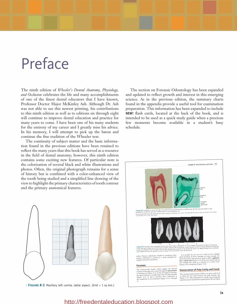
The continuity of subject matter and the basic informa-tion found in the previous editions have been retained to reflect the many years that this book has served as a resource in the field of dental anatomy; however, this ninth edition contains some exciting new features. Of particular note is the colorization of several black and white illustrations and photos. Often, the original photograph remains for a sense of history but is combined with a color-enhanced view of the tooth being studied and a simplified line drawing of the view to highlight the primary characteristics of tooth contour and the primary anatomical features.
ix
Chapter �� Pulp Chambers and Canals ���
cedure. Elective endodontics should be considered when
extreme calcification is present in a tooth scheduled for
complex restorative procedures.
Foramen
The neurovascular bundle, which supplies the internal
contents of the pulp cavity, enters through the apical foramen
or foramina (see Figure 13-1). As the root begins to develop,
the apical foramen is actually larger than the pulp chamber
(Figure 13-4, 1), but it becomes more constricted at
the completion of root formation (Figure 13-4, 2 through
5).
It is possible for any root of a tooth to have multiple
apical foramina. If these openings are large enough, the
space that leads to the main root canal is called a supplemen-
tary or lateral canal (Figure 13-5). If the root canal breaks up
into multiple tiny canals, it is referred to as a delta system2
because of its complexity (Figure 13-6).
Demarcation of Pulp Cavity and Canal
The cementoenamel junction (CEJ) is not quite at the level
at which the root canal becomes the pulp chamber (see
Figure 13-1). This demarcation is mainly macroscopically
based but may be visualized by exploring the CEJ (see Figure
F����� ���� Comparison in size of the pulps of two intact lower first permanent molars at different ages. A, The young pulp chamber is large
(magnification 8). B, The older pulp is greatly reduced in size (magnification 8).
(A From Berkovitz BKB, Holland GR, Moxham BJ: Oral anatomy, histology, and embryology, ed 3, St Louis, 2002, Mosby. B from Avery JK, Chiego DJ: Essentials of oral histology
and embryology, ed 3, St Louis, 2006, Mosby.)
A B
Coronalpulp
Radicularpulp
Cementumaroundapical canal
F����� ���� Maxillary canine. Labiolingual sections show various stages of development. 1, Crown complete; root partially completed with large pulp
cavity, wide open at apical end. 2, Tooth almost complete, except for lack of constriction of apical foramen. 3, Canine of young individual with large pulp
cavity and completed root tip. 4, Typical canine of adult, demonstrating constriction of the foramen. 5, Canine of an elderly individual with a constricted
pulp chamber and canal; this specimen has lost its original crown form because of wear during function.
1 2 3 4 5
http://freedentaleducation.blogspot.com

x Preface
Evolve
We are very excited about the inclusion of the NEW! “helpsite.” Faculty and students should feel free
to use as much of this material as is helpful for their courses. For the instructor, we’ll have all of the figures from the book in electronic form, a collection of 300 test questions, and a PowerPoint lecture intended as an aid to develop new teaching materials or to further enhance course curricula. For the student, we’ll have the same flash cards that appear in the book, only in electronic form, labeling exercises, and animations. Visit the Evolve website at http://evolve.elsevier.com/Nelson/dentalanatomy/.
DVD
Introduced in the eighth edition, the accompanying DVD in this ninth edition has been expanded in several areas to include new content, including 3-D animations, electronic flash cards, labeling exercises, and a virtual reality tooth identi-fication quiz. The additional material includes patterns of mandibular movement and the resulting occlusal contacts that occur when the subject demonstrates maximum inter-cuspation of the teeth when the temporomandibular joints are positioned in centric relation.
The tooth identification quiz is considered a “rite of passage” in many dental school curricula, and with the increasing difficulty encountered in collecting real tooth specimens for study, a virtual reality experience in tooth identification may help refine the skills required to examine a tooth and make a correct identification based on the stu-dent’s knowledge of the type trait criteria. In this section the student will view a Quick Time Virtual Reality movie of the tooth that allows inspection from multiple directions until a positive identification is realized.
Icons
Throughout the book you will notice DVD icons in the text margin. When the DVD icon is shown, the reader can access the appropriate portion of the DVD identified to view the material that the text presents in a simulated 3-D and interactive format.
True to previous editions, specific and general references have been provided for further study. Through these refer-ences the reader may realize the rich history of contribu-
W
The dentition may be considered to be the single best
physiological indicator of chronological age in juveniles.14 A
knowledge of dental age has practical clinical applications;
however, it is recognized that the coverage of these applica-
tions here must be brief. When indicated, references to a
more detailed coverage are provided. Values for predicting
age from stages of the formation of permanent mandibular
teeth are considered in the section Tooth Formation Stand-
ards in this chapter.Dental age has been assessed on the basis of the number
of teeth at each chronological age7 or on stages of the forma-
tion of crowns and roots of the teeth.14 Dental age during
the mixed dentition period (transition from primary to per-
manent dentition) may be assessed on the basis of which
teeth have erupted, the amount of resorption of the roots of
primary teeth, and the amount of development of the per-
manent teeth.28
Dental age can reflect an assessment of physiological age
comparable to age based on skeletal development, weight,
or height.29 When the teeth are forming, the crowns and
roots of the teeth appear to be the tissues least affected by
environmental influences (nutrition, endocrinopathies, etc.);
however, when a substance such as the antibiotic tetracy-
cline (Figure 2-19, A) is ingested by the mother during
certain times of the development of the dentitions, signifi-
cant discoloration from yellow to brown to bluish violet and
from part (cervical) to all of the enamel may occur.30,31
The benefits of fluoride for the control of dental caries
are well established. However, its widespread use has resulted
in an increasing prevalence of fluorosis (see Figure 2-19, B)
in both nonfluoridated and optimally fluoridated popula-
tions.32 Parents should be advised about the best early use
of fluoride to reduce the prevalence of clinically noticeable
fluorosis. Children under age 6 should use only a pea-sized
amount of fluoride toothpaste; parents should consult their
dentists concerning the use of fluoride toothpaste by chil-
dren under age 2 years.33
Dental development may be based also on the emergence
(eruption) of the teeth; however, because caries, tooth loss,
and severe malnutrition may influence the emergence of
teeth through the gingiva,11,12 chronologies of the eruption
of teeth are less satisfactory for dental age assessment than
those based on tooth formation. In addition, tooth forma-
tion may be divided appropriately into a number of stages
that cover continuously the development of teeth34,35 in con-
trast to the single episode of tooth eruption. The stages of
development are considered in the section Tooth Forma-
tion Standards in this chapter.The importance of the emergence of the teeth to the
development of oral motor behavior is often overlooked,
F����� ���� Panoramic radiograph of a child about 7 years of age. This type of examination is of great value in registering an overall record of
development.
(From Pappas GC, Wallace WR: Panoramic sialography, Dent Radiogr Photogr 43:27, 1970.)
F����� ���� A, Tetracycline staining. B, Enamel fluorosis.
(From Neville BW, Damm DD, Allen CM, et al: Oral and maxillofacial pathology, ed 3,
St. Louis, 2009, Saunders.)
A
B
�� Dental Age
http://freedentaleducation.blogspot.com

Preface xi
tions from many disciplines. In this regard, the terminology used in the sections on Occlusion have been updated to reflect both the terms used in the present day and the ter-minology necessary to understand the historical develop-ment and foundational research of the field.
Once again every attempt has been made to consider what is important for the student and practitioner to know and what may be of use to those from other scientific fields.
Stanley J Nelson, DDS, MS
http://freedentaleducation.blogspot.com

This page intentionally left blank
http://freedentaleducation.blogspot.com

AcknowledgmentsI would also like to acknowledge all the hard work pro-
vided by John Dolan, Brian Loehr, Courtney Sprehe, and the rest of the Elsevier Science staff for making the ninth edition a reality.
To the students, faculty, and staff of the University of Nevada—Las Vegas School of Dental Medicine, thanks for putting up with me during this 2-year project and affording me this opportunity.
To my wife Mary Sarah, thank you for all your encour-agement and assistance, and finally to my father Charles S. Nelson, DDS, thank you for your example, direction, and support as I progress in life and this wonderful profession of dentistry.
SJN
A special thank you is offered to Dr. Edward Herschaft of the University of Nevada—Las Vegas School of Dental Medicine for writing the section on forensic odontology found in Chapter 4, as well as our colleague Dr. David Ord for his contribution of the forensic photographs included in this section. Also, thanks to my colleague Dr. Bill Dahlke for his help in writing the test questions. I would like to thank David Baker, MA, of the University of Texas Health Sciences Center at San Antonio along with his colleagues Sam Newman and Lee Bennack. Without their work pro-viding the new animations and the QuickTime VR photog-raphy, the DVD would not have been possible. A special thank you goes to Jodie Bernard for her work in providing the wonderful new illustrations found throughout the text. I would like to thank Dr. George Ash and the rest of the Ash family for helping me with Dr. Ash’s files.
xiii
http://freedentaleducation.blogspot.com

This page intentionally left blank
http://freedentaleducation.blogspot.com

xv
1 IntroductiontoDentalAnatomy,1Formation of the Dentitions (Overview), 1Nomenclature, 2Formulae for Mammalian Teeth, 2Tooth Numbering Systems, 2Division into Thirds, Line Angles, and Point Angles, 9Tooth Drawing and Carving, 10Measurement of Teeth, 11Summary, 11
2 DevelopmentandEruptionoftheTeeth,21
Clinical Considerations, 21Variability, 22Malformations, 22Chronology of Primary Dentition, 23Development and Eruption/Emergence of the Teeth, 23The Dentitions, 26Neuromuscular Development, 27Transitional (Mixed) Dentition Period, 28Loss of Primary Teeth, 29Permanent Dentition, 30Size of Teeth, 31Dental Pulp, 31Cementoenamel Junction, 32Dental Age, 33Tooth Formation Standards, 35Chronologies of Human Dentition, 35Types of Chronologies, 35Stages of Tooth Formation, 35Age of Attainment, 35Age Prediction, 37Maturity Assessment, 37Duration of Root and Crown Formation, 40Summary of Chronologies, 41Sequence of Eruption, 41Estimating Time of Enamel Hypoplasia, 41
3 ThePrimary(Deciduous)Teeth,45Life Cycle, 45Importance of Primary Teeth, 45Nomenclature, 45Major Contrasts between Primary and Permanent
Teeth, 47Pulp Chambers and Pulp Canals, 48Detailed Description of Each Primary Tooth, 50
4 Forensics,ComparativeAnatomy,Geometries,andFormandFunction,67
Forensic Dentistry, 67Comparative Dental Anatomy, 69Facial and Lingual Aspects of All Teeth, 73Summary of Schematic Outlines, 75Form and Function of the Permanent Dentition, 75Alignment, Contacts, and Occlusion, 76
5 OrofacialComplex:FormandFunction,81
Form and Function, 81Form Follows Function, 81Articulation of Teeth, 81Physiological Form of the Teeth and Periodontium, 82Fundamental Curvatures, 82Proximal Contact Areas, 82Interproximal Spaces (Formed by Proximal Surfaces in
Contact), 84Embrasures (Spillways), 86Contact Areas and Incisal and Occlusal Embrasures from
the Labial and Buccal Aspect, 88Contact Areas and Labial, Buccal, and Lingual Embrasures
from the Incisal and Occlusal Aspects, 89Facial and Lingual Contours at the Cervical Thirds
(Cervical Ridges) and Lingual Contours at the Middle Thirds of Crowns, 91
The Height of Epithelial Attachment: Curvatures of the Cervical Lines (Cementoenamel Junction [CEJ]) Mesially and Distally, 94
Contents
http://freedentaleducation.blogspot.com

xvi Contents
6 ThePermanentMaxillaryIncisors,99Maxillary Central Incisor, 99Maxillary Lateral Incisor, 106
7 ThePermanentMandibularIncisors,113Mandibular Central Incisor, 113Mandibular Lateral Incisor, 119
8 ThePermanentCanines:MaxillaryandMandibular,125
Maxillary Canine, 125Mandibular Canine, 132
9 ThePermanentMaxillaryPremolars,141Maxillary First Premolar, 141Maxillary Second Premolar, 151
10 ThePermanentMandibularPremolars,157
Mandibular First Premolar, 157Mandibular Second Premolar, 165
11 ThePermanentMaxillaryMolars,171Maxillary First Molar, 171Maxillary Second Molar, 181Maxillary Third Molar, 184
12 ThePermanentMandibularMolars,189Mandibular First Molar, 189Mandibular Second Molar, 199Mandibular Third Molar, 203
13 PulpChambersandCanals,209Pulp, Chamber, and Canals, 209Radiographs, 209Foramen, 211Demarcation of Pulp Cavity and Canal, 211Pulp Horns, 212
Clinical Applications, 212Pulp Cavities of the Maxillary Teeth, 213Pulp Cavities of the Mandibular Teeth, 222Radiographs: Pulp Chamber and Canals, 233Crown and Root Fractures, 233
14 Dento-osseousStructures,BloodVessels,andNerves,239
The Maxillae, 239The Mandible, 245Arterial Supply to the Teeth, 252Nerve Supply to the Jaws and Teeth, 256
15 TheTemporomandibularJoints,Teeth,andMuscles,andTheirFunctions,259
Temporomandibular Articulation, 259Muscles, 265Mandibular Movements and Muscle Activity, 270
16 Occlusion,275Concepts of Occlusion, 275Development of the Dentitions, 276Primary Dentition, 276Mixed (Transitional) Dentition, 279Permanent Dentition, 282Cusp, Fossa, and Marginal Ridge Relations, 288Lateral Occlusal Relations, 294Biomechanics of Chewing Function, 298Neurobehavioral Aspects of Occlusion, 298Oral Motor Behavior, 303Swallowing, 304Summary, 304
AppendixA ReviewofToothMorphology,309
AppendixB ToothTraitsofthePermanentDentition,327
Index,335
FlashCards
http://freedentaleducation.blogspot.com

Introduction to Dental Anatomy
remains intact (barring loss from dental caries or trauma) until the child is about 6 years of age. At about that time the first succedaneous or permanent teeth begin to emerge into the mouth. The emergence of these teeth begins the transi-tion or mixed dentition period in which there is a mixture of deciduous and succedaneous teeth present. The transition period lasts from about 6 to 12 years of age and ends when all the deciduous teeth have been shed. At that time the permanent dentition period begins. Thus, the transition from the primary dentition to the permanent dentition begins with the emergence of the first permanent molars, shedding of the deciduous incisors, and emergence of the permanent incisors. The mixed dentition period is often a difficult time for the young child because of habits, missing teeth, teeth of different colors and hues, crowding of the teeth, and malposed teeth.
The permanent, or succedaneous, teeth replace the exfo-liated deciduous teeth in a sequence of eruption that exhibits some variance, an important topic that will be considered in Chapter 16.
After the shedding of the deciduous canines and molars, emergence of the permanent canines and premolars, and emergence of the second permanent molars, the permanent dentition is completed (including the roots) at about 14 to 15 years of age, except for the third molars, which are com-pleted at 18 to 25 years of age. In effect, the duration of the permanent dentition period is 12+ years. The completed permanent dentition consists of 32 teeth if none are con-genitally missing, which may be the case. The development of the teeth, dentitions, and the craniofacial complex are considered in Chapter 2. The development of occlusion for both dentitions is discussed in Chapter 16.
Dental anatomy is defined here as, but is not limited to, the study of the development, morphology, function, and iden-tity of each of the teeth in the human dentitions, as well as the way in which the teeth relate in shape, form, structure, color, and function to the other teeth in the same dental arch and to the teeth in the opposing arch. Thus, the study of dental anatomy, physiology, and occlusion provides one of the basic components of the skills needed to practice all phases of dentistry.
The application of dental anatomy to clinical practice can be envisioned in Figure 1-1, A where a disturbance of enamel formation (considered briefly in Chapter 2) has resulted in esthetic, psychological, and periodontal problems that may be corrected by an appropriate restorative dental treatment such as that illustrated in Figure 1-1, B. The practitioner has to have knowledge of the morphology, occlusion, esthet-ics, phonetics, and functions of these teeth to undertake such treatment.
Formation of the Dentitions (Overview)
Humans have two sets of teeth in their lifetime. The first set of teeth to be seen in the mouth is the primary or decidu-ous dentition, which begins to form prenatally at about 14 weeks in utero and is completed postnatally at about 3 years of age. In the absence of congenital disorders, dental disease, or trauma, the first teeth in this dentition begin to appear in the oral cavity at the mean age of 6, and the last emerge at a mean age of 28 ± 4 months. The deciduous dentition
�
11
http://freedentaleducation.blogspot.com

� Tooth Numbering Systems
Nomenclature
The first step in understanding dental anatomy is to learn the nomenclature, or the system of names, used to describe or classify the material included in the subject. When a sig-nificant term is used for the first time here, it is emphasized in bold. Additional terms will be discussed as needed in subsequent chapters.
The term mandibular refers to the lower jaw, or mandible. The term maxillary refers to the upper jaw, or maxilla. When more than one name is used in the literature to describe something, the two most commonly used names will be used initially. After that they may be combined or used separately as consistent with the literature of a particular specialty of dentistry, for example, primary or deciduous dentition, perma-nent or succedaneous dentition. A good case may be made for the use of both terms. By dictionary definition,1 the term primary can mean “constituting or belonging to the first stage in any process.” The term deciduous can mean “not permanent, transitory.” The same unabridged dictionary refers the reader from the definition of deciduous tooth to milk tooth, which is defined as “one of the temporary teeth of a mammal that are replaced by permanent teeth. Also called baby tooth, deciduous tooth.” The term primary can indicate a first dentition and the term deciduous can indicate that the first dentition is not permanent, but not unimportant. The term succedaneous can be used to describe a successor denti-tion and does not suggest permanence, whereas the term permanent suggests a permanent dentition, which may not be the case due to dental caries, periodontal diseases, and
trauma. All four of these descriptive terms appear in the professional literature.
Formulae for Mammalian Teeth
The denomination and number of all mammalian teeth are expressed by formulae that are used to differentiate the human dentitions from those of other species. The denomi-nation of each tooth is often represented by the initial letter in its name (e.g., I for incisor, C for canine, P for premolar, M for molar). Each letter is followed by a horizontal line and the number of each type of tooth is placed above the line for the maxilla (upper jaw) and below the line for the mandible (lower jaw). The formulae include one side only, with the number of teeth in each jaw being the same for humans.
The dental formula for the primary/deciduous teeth in humans is as follows:
I C M22
11
22
10=
This formula should be read as: incisors, two maxillary and two mandibular; canines, one maxillary and one man-dibular; molars, two maxillary and two mandibular—or 10 altogether on one side, right or left (Figure 1-2, A).
A dental formula for the permanent human dentition is as follows:
I C P M22
11
22
33
16=
Premolars have now been added to the formula, two maxillary and two mandibular, and a third molar has been added, one maxillary and one mandibular (Figure 1-2, B).
Systems for scoring key morphological traits of the per-manent dentition that are used for anthropological studies are not described here. However, a few of the morphological traits that are used in anthropological studies2 are consid-ered in the following chapters, (e.g., shoveling, Carabelli’s trait, enamel extensions, and peg-shaped incisors). Some anthropologists use di1, di2, dc, dm1, and dm2 notations for the deciduous dentition and I1, I2, C, P1, P2, M1, M2, and M3 for the permanent teeth. These notations are generally limited to anthropological tables because of keyboard incompatibility.
Tooth Numbering Systems
In clinical practice some “shorthand” system of tooth nota-tion is necessary for recording data. There are several systems in use in the world, but only a few are considered here. In 1947 a committee of the American Dental Associa-tion (ADA) recommended the symbolic (Zsigmondy/Palmer) system as the numbering method of choice.3 However, because of difficulties with keyboard notation of the sym-bolic notation system, the ADA in 1968 officially recom-mended the “universal” numbering system. Because of some
Figure �-� A, Chronological developmental disorder involving all the anterior teeth. B, Illustration of restored teeth just after completion, taking in account esthetics, occlusion, and periodontal health. Note that the gingival response is not yet resolved.
(From Ash MM, Ramfjord S: Occlusion, ed 4, Philadelphia, 1995, Saunders.)
B
A
http://freedentaleducation.blogspot.com

Chapter � Introduction to Dental Anatomy �
limitations and lack of widespread use internationally, rec-ommendations for a change sometimes are made.4
The universal system of notation for the primary denti-tion uses uppercase letters for each of the primary teeth: For the maxillary teeth, beginning with the right second molar, letters A through J, and for the mandibular teeth, letters K through T, beginning with the left mandibular second molar. The universal system notation for the entire primary dentition is as follows:
Midsagittal Plane
Right LeftT S R Q P O N M L KA B C D E F G H I J
The symbolic system for the permanent dentition was introduced by Adolph Zsigmondy of Vienna in 1861 and then modified for the primary dentition in 1874.
Central incisor (first incisor)
Lateral incisor (second incisor)Canine (cuspid)First molar
Second molar
Second molar
First molar
A
CanineLateral incisor (second incisor)
Central incisor (first incisor)
RIG
HT
LEF
T
MANDIBULAR
MAXILLARY
Central incisor (first incisor)
Lateral incisor (second incisor)
Canine (cuspid)
First premolar (first bicuspid)
Second premolar (second bicuspid)
First premolar (bicuspid)
Canine (cuspid)
Lateral incisor (second incisor)
Central incisor (first incisor)B
Second premolar (bicuspid)
First molar
First molar
Second molar
Second molar
Third molar
Third molar
RIG
HT
LEF
T
MANDIBULAR
MAXILLARY
Figure �-� A, Casts of deciduous, or primary, dentition. B, Casts of permanent dentition.
(A from Berkovitz BK, Holland GR, Moxham BJ: Oral anatomy, histology and embryology, ed 3, St. Louis, 2002, Mosby.)
III C, D
http://freedentaleducation.blogspot.com

� Tooth Numbering Systems
Independently, Palmer also published the symbolic system in 1870. The symbolic system is most often referred to as the Palmer notation system in the United States and less fre-quently as the Zsigmondy/Palmer notation system. In this system the arches are divided into quadrants with the entire dentition being notated as follows:
E D C B A A B C D EE D C B A A B C D E
Thus, for a single tooth such as the maxillary right central incisor the designation is A . For the mandibular left central incisor, the notation is given as A . This numbering system presents difficulty when an appropriate font is not available for keyboard recording of Zsigmondy/Palmer symbolic notations. For simplification this symbolic notation is often designated as Palmer’s dental notation rather than Zsig-mondy/Palmer notation.
In the universal notation system for the permanent denti-tion, the maxillary teeth are numbered from 1 through 16, beginning with the right third molar. Beginning with the mandibular left third molar, the teeth are numbered 17 through 32. Thus, the right maxillary first molar is desig-nated as 3, the maxillary left central incisor as 9, and the right mandibular first molar as 30. The following universal notation designates the entire permanent dentition.
1 2 3 4 5 6 7 8 9 10 11 12 13 14 15 1632 31 30 29 28 27 26 25 24 23 22 21 20 19 18 17
The Zsigmondy/Palmer notation for the permanent den-tition is a four-quadrant symbolic system in which, begin-ning with the central incisors, the teeth are numbered 1 through 8 (or more) in each arch. For example, the right maxillary first molar is designated as 6 , and the left man-dibular central incisor as 1 . The Palmer notation for the entire permanent dentition is as follows:
8 7 6 5 4 3 2 2 3 4 5 6 7 88 7 6 5 4 3 2
1 11 1 2 3 4 5 6 7 8
Viktor Haderup of Denmark in 1891 devised a variant of the eight-tooth quadrant system in which plus (+) and minus (−) were used to differentiate between upper and lower quadrants and between right and left quadrants; in other words, +1 indicates the upper left central incisor and 1− indicates the lower right central incisor. Primary teeth were numbered as follows: upper right, 05+ to 01+; lower left, −01 to −05. This system is still taught in Denmark.5
The universal system is acceptable to computer language, whereas the Palmer notation is generally incompatible with computers and word processing systems. Each tooth in the universal system is designated with a unique number, which leads to less confusion than with the Palmer notation.
A two-digit system proposed by Fédération Dentaire Internationale (FDI) for both the primary and permanent dentitions has been adopted by the World Health Organiza-tion and accepted by other organizations such as the Inter-
national Association for Dental Research. The FDI system of tooth notation is as follows.
For the primary teeth:
Upper Right
55 54 53 52 62 63 64 6585 84 83 82
51 6181 72 73 74 75
Lower Right
71
Upper Left
Lower Left
Numeral 5 indicates the maxillary right side, and 6 indicates the maxillary left side. The second number of the two-digit number is the tooth number for each side. The number 8 indicates the mandibular right side, and the number 7 indicates the mandibular left side. The second number of the two-digit system is the tooth number. Thus, for example the number 51 refers to the maxillary right central incisor.
For the permanent teeth:
Upper Right
18 17 16 15 14 13 12 22 23 24 25 26 27 2848 47 46 45 44 43 42
11 2141 31 32 33 34 35 36 37 38
Lower Right
Upper Left
Lower Left
Thus, as in the two-digit FDI system for the primary dentition, the first digit indicates the quadrant: 1 to 4 for the permanent dentition and 5 to 8 for the primary denti-tion. The second digit indicates the tooth within a quadrant: 1 to 8 for the permanent teeth and 1 to 5 for the primary teeth. For example, the permanent upper right central incisor is 11 (pronounced “one one,” not “eleven”).
THE CROWN AND ROOT
Each tooth has a crown and root portion. The crown is covered with enamel, and the root portion is covered with cementum. The crown and root join at the cementoenamel junction (CEJ). This junction, also called the cervical line (Figure 1-3), is plainly visible on a specimen tooth. The main bulk of the tooth is composed of dentin, which is clear in a cross section of the tooth. This cross section displays a pulp chamber and a pulp canal, which normally contain the pulp tissue. The pulp chamber is in the crown portion mainly, and the pulp canal is in the root (Figure 1-4).The spaces are continuous with each other and are spoken of collectively as the pulp cavity.
The four tooth tissues are enamel, cementum, dentin, and pulp. The first three are known as hard tissues, the last as soft tissue. The pulp tissue furnishes the blood and nerve supply to the tooth. The tissues of the teeth must be con-sidered in relation to the other tissues of the orofacial struc-tures (Figures 1-5 and 1-6) if the physiology of the teeth is to be understood.
http://freedentaleducation.blogspot.com

Chapter � Introduction to Dental Anatomy �
some of the premolars; or multiple, with a bifurcation or trifurcation dividing the root portion into two or more extensions or roots with their apices or terminal ends, as found on all molars and in some premolars.
The root portion of the tooth is firmly fixed in the bony process of the jaw, so that each tooth is held in its position relative to the others in the dental arch. That portion of the jaw serving as support for the tooth is called the alveolar process. The bone of the tooth socket is called the alveolus (plural, alveoli) (Figure 1-7).
The crown portion is never covered by bone tissue after it is fully erupted, but it is partly covered at the cervical third in young adults by soft tissue of the mouth known as the gingiva or gingival tissue, or “gums.” In some persons, all of the enamel and frequently some cervical cementum may not be covered by the gingiva.
SURFACES AND RIDGES
The crowns of the incisors and canines have four surfaces and a ridge, and the crowns of the premolars and molars have five surfaces. The surfaces are named according to their positions and uses (Figure 1-8). In the incisors and canines, the surfaces toward the lips are called labial surfaces; in the premolars and molars, those facing the cheek are the buccal surfaces. When labial and buccal surfaces are spoken of collectively, they are called facial surfaces. All surfaces facing toward the tongue are called lingual surfaces. The surfaces of the premolars and molars that come in contact (occlusion) with those in the opposite jaw during the act of closure are called occlusal surfaces. These are
Figure �-� Maxillary central incisor (facial aspect). A, Apex of root; R, root; CL, cervical line; C, crown; IE, incisal edge.
A
R
CL
C
IE
Figure �-� Schematic drawings of longitudinal sections of an anterior and a posterior tooth. A, Anterior tooth. A, Apex; AF, apical foramen; SC, supplementary canal; B, bone; C, cementum; PM, periodontal ligament; PC, pulp canal; G, gingiva; GC, gingival crevice; GM, gingival margin; PCH, pulp chamber; D, dentin; E, enamel; CR, crown. B, Posterior tooth. A, Apices; PC, pulp canal; PCH, pulp chamber; PH, pulp horn; F, fissure; CU, cusp; CEJ, cementoenamel junction; BI, bifurcation of roots.
A
B
CR
A B
A
BI
CEJ
CU
F
PH
PCH
PC
AAFSC
B
C
PM
B
PC
GGCGM
PCH
D
E
A
The crown of an incisor tooth may have an incisal ridge or edge, as in the central and lateral incisors; a single cusp, as in the canines; or two or more cusps, as on premolars and molars. Incisal ridges and cusps form the cutting surfaces on tooth crowns.
The root portion of the tooth may be single, with one apex or terminal end, as usually found in anterior teeth and
IV A(8), B
http://freedentaleducation.blogspot.com

� Tooth Numbering Systems
Figure �-� Sagittal sections through the maxillary and mandibular central incisors.
Vestibularmucosa
Free gingivalmargin
Attachedgingiva
Anterior oralvestibule
Attachedgingiva
Labialmucosa
Figure �-� Section through the second maxillary molar and adjacent tissues.
Palatine vein
Palatine artery
Palatine glands
Palatine nerve
Figure �-7 Left maxillary bone showing the alveolar process with three molars in place and the alveoli of the central incisor, lateral incisor, canine, and first and second premolars. Note the opening at the bottom of the canine alveolus, an opening that accommodates the nutrient blood and nerve supply to the tooth in life. Although they do not show up in the photograph, the other alveoli present the same arrangement.
called incisal surfaces with respect to incisors and canines.
The surfaces of the teeth facing toward adjoining teeth in the same dental arch are called proximal or proximate sur-faces. The proximal surfaces may be called either mesial or distal. These terms have special reference to the position of the surface relative to the median line of the face. This line is drawn vertically through the center of the face, passing between the central incisors at their point of contact with each other in both the maxilla and the mandible. Those proximal surfaces that, following the curve of the arch, are faced toward the median line are called mesial surfaces, and those most distant from the median line are called distal surfaces.
Four teeth have mesial surfaces that contact each other: the maxillary and mandibular central incisors. In all other instances, the mesial surface of one tooth contacts the distal surface of its neighbor, except for the distal surfaces of third
molars of permanent teeth and distal surfaces of second molars in deciduous teeth, which have no teeth distal to them. The area of the mesial or distal surface of a tooth that touches its neighbor in the arch is called the contact area.
Central and lateral incisors and canines as a group are called anterior teeth; premolars and molars as a group, poste-rior teeth.
OTHER LANDMARKS
To study an individual tooth intelligently, one should rec-ognize all landmarks of importance by name. Therefore, at this point it is necessary to become familiar with additional terms, such as the following:
http://freedentaleducation.blogspot.com

Chapter � Introduction to Dental Anatomy 7
Figure �-8 Application of nomenclature. Tooth numbers 1 to 8 indicating left maxillary teeth. Tooth surfaces related to the tongue (lingual), cheek (buccal), lips (labial), and face (facial), apply to four quadrants and the upper left quadrant. The teeth or their parts or surfaces may be described as being away from the midline (distal) or toward the midline (mesial).
1. Central incisor (first incisor)2. Lateral incisor (second incisor)3. Canine (cuspid)4. First premolar (first bicuspid)5. Second premolar (second bicuspid)6. First molar7. Second molar8. Third molar
There are eight tooth namesincluded in each quadrant of thedental arches they are repeated toinclude right, left, maxillary andmandibular, making a total ofthirty-two teeth in all.
Lingual
LabialD
ista
l Facial
Buccal
Med
ian
Line
1
2
3
4
5
6
7
8
Third Molar
Aw
ay fr
om m
edia
n lin
e T
owar
d m
edia
n lin
e
Mes
ial
cusp triangular ridge developmental groovetubercle transverse ridge supplemental groovecingulum oblique ridge pitridge fossa lobemarginal ridge sulcus
A cusp is an elevation or mound on the crown portion of a tooth making up a divisional part of the occlusal surface (Figures 1-4 and 1-9).
A tubercle is a smaller elevation on some portion of the crown produced by an extra formation of enamel (see Figure 4-14, A). These are deviations from the typical form.
A cingulum (Latin word for “girdle”) is the lingual lobe of an anterior tooth. It makes up the bulk of the cervical third of the lingual surface. Its convexity mesiodistally resembles a girdle encircling the lingual surface at the cervical third (see Figures 1-10 and 4-13, A).
A ridge is any linear elevation on the surface of a tooth and is named according to its location (e.g., buccal ridge, incisal ridge, marginal ridge).
Marginal ridges are those rounded borders of the enamel that form the mesial and distal margins of the occlusal
Figure �-9 Some landmarks on the maxillary first molar. BCR, Buccocervical ridge; BG, buccal groove; MBC, mesiobuccal cusp; SG, supplemental groove; TF, triangular fossa; MLC, mesiolingual cusp; DG, developmental groove; DLC, distolingual cusp; OR, oblique ridge; DMR, distal marginal ridge; DBC, distobuccal cusp; CF, central fossa.
CF
DBC
DMR
OR
DLC
DG
BCR
BGMBC
SGTF
MLC
IV A(3), B
http://freedentaleducation.blogspot.com

8 Tooth Numbering Systems
F igure 1-10 A, Maxillary right lateral incisor (lingual aspect). CL, Cervical line; CI, cingulum (also called the linguocervical ridge ); MR, marginal ridge; IR, incisal ridge; LF, lingual fossa. B, Mamelons on erupting, noncontacting central incisors.
( B from Bath-Balogh M, Fehrenbach MJ: Illustrated dental embryology, histology, and anatomy, ed 2, St. Louis, 2006, Saunders.)
BA
MR
CL
CI
MR
IRLF
F igure 1-11 A, Mesial view of a maxillary right fi rst premolar. MR, Marginal ridge; S, sulcus traversing occlusal surface; CR, cusp ridge; BCR, buccocervical ridge. B, Occlusal view of maxillary right fi rst premolar. CR, Cusp ridge; TR, triangular ridges; Trans R, transverse ridge, formed by two triangular ridges that cross the tooth transversely. C, Occlusal view of a maxillary right fi rst molar. Trans R, Transverse ridge; TR, triangular ridge; P, pit formed by junction of developmental grooves; SG, supplemental groove; DG, developmental groove; TR, triangular ridge.
BCR
CR
CR
TRDGSG
P
TRTrans R
TR
TRTrans R
S
A B C
MR
surfaces of premolars and molars and the mesial and distal margins of the lingual surfaces of the incisors and canines ( Figures 1-10, A, and 1-11 ).
Triangular ridges descend from the tips of the cusps of molars and premolars toward the central part of the occlusal surfaces. They are so named because the slopes of each side of the ridge are inclined to resemble two sides of a triangle ( Figures 1-11, B and C, and 1-12 ). They are named after the cusps to which they belong, for example, the triangular ridge of the buccal cusp of the maxillary fi rst premolar.
When a buccal and a lingual triangular ridge join, they form a transverse ridge. A transverse ridge is the union of two triangular ridges crossing transversely the surface of a pos-terior tooth ( Figure 1-11, B and C ).
The oblique ridge is a ridge crossing obliquely the occlusal surfaces of maxillary molars and formed by the union of the triangular ridge of the distobuccal cusp and the distal cusp ridge of the mesiolingual cusp ( Figure 1-9 ).
A fossa is an irregular depression or concavity. Lingual
fossae are on the lingual surface of incisors ( Figure 1-10 ).
Central fossae are on the occlusal surface of molars. They are formed by the convergence of ridges terminating at a central point in the bottom of the depression where there is a junc-tion of grooves ( Figure 1-12 ). Triangular fossae are found on molars and premolars on the occlusal surfaces mesial or distal to marginal ridges ( Figure 1-9 ). They are sometimes found on the lingual surfaces of maxillary incisors at the edge of the lingual fossae where the marginal ridges and the cingulum meet (see Figure 4-14, A ).
A sulcus is a long depression or valley in the surface of a tooth between ridges and cusps, the inclines of which meet at an angle. A sulcus has a developmental groove at the junction of its inclines. (The term sulcus should not be con-fused with the term groove .)
A developmental groove is a shallow groove or line between the primary parts of the crown or root. A supplemental groove, less distinct, is also a shallow linear depression on the surface of a tooth, but it is supplemental to a developmental groove and does not mark the junction of primary parts. Buccal and lingual grooves are developmental grooves found on the
http://freedentaleducation.blogspot.com

Chapter � Introduction to Dental Anatomy 9
buccal and lingual surfaces of posterior teeth (Figures 1-9 and 1-12).
Pits are small pinpoint depressions located at the junction of developmental grooves or at terminals of those grooves. For instance, central pit is a term used to describe a landmark in the central fossa of molars where developmental grooves join (Figure 1-11, C).
A lobe is one of the primary sections of formation in the development of the crown. Cusps and mamelons are repre-sentative of lobes. A mamelon is any one of the three rounded protuberances found on the incisal ridges of newly erupted incisor teeth (Figure 1-10, B). (For further description of lobes, see Figures 4-11 through 4-14).
The roots of the teeth may be single or multiple. Both maxillary and mandibular anterior teeth have only one root each. Mandibular first and second premolars and the maxil-lary second premolar are single rooted, but the maxillary first premolar has two roots in most cases, one buccal and one lingual. Maxillary molars have three roots, one mesiobuc-cal, one distobuccal, and one lingual. Mandibular molars have two roots, one mesial and one distal. It must be under-stood that description in anatomy can never follow a hard-and-fast rule. Variations frequently occur. This is especially true regarding tooth roots, for example, facial and lingual roots of the mandibular canine.
Division into Thirds, Line Angles, and Point Angles
For purposes of description, the crowns and roots of teeth have been divided into thirds, and junctions of the crown surfaces are described as line angles and point angles. Actu-ally, there are no angles or points or plane surfaces on the teeth anywhere except those that appear from wear (e.g., attrition, abrasion) or from accidental fracture. Line angle and
point angle are used only as descriptive terms to indicate a location.
When the surfaces of the crown and root portions are divided into thirds, these thirds are named according to their location. Looking at the tooth from the labial or buccal aspect, we see that the crown and root may be divided into thirds from the incisal or occlusal surface of the crown to the apex of the root (Figure 1-13). The crown is divided into an incisal or occlusal third, a middle third, and a cervical third. The root is divided into a cervical third, a middle third, and an apical third.
The crown may be divided into thirds in three directions: inciso- or occlusocervically, mesiodistally, or labio- or buc-colingually. Mesiodistally, it is divided into the mesial, middle, and distal thirds. Labio- or buccolingually it is divided into labial or buccal, middle, and lingual thirds. Each of the five surfaces of a crown may be so divided. There will be one middle third and two other thirds, which are named according to their location, for example, cervical, occlusal, mesial, lingual.
A line angle is formed by the junction of two surfaces and derives its name from the combination of the two surfaces that join. For instance, on an anterior tooth, the junction of the mesial and labial surfaces is called the mesiolabial line angle.
The line angles of the anterior teeth (Figure 1-14, A) are as follows:
mesiolabial distolingualdistolabial labioincisalmesiolingual linguoincisal
Figure �-�� Mandibular right first molar. MLC, Mesiolingual cusp; MMR, mesial marginal ridge; MBC, mesiobuccal cusp; MBG, mesiobuccal groove; BCR, buccocervical ridge; CF, central fossa; DBG, distobuccal groove; DBC, distobuccal cusp; DC, distal cusp; TR, triangular ridge; DLC, distolingual cusp; TRR, transverse ridge.
MLC
TRR
DLCTR
DCDBCDBG
CF
MMR
MBC
MBG
BCR
Figure �-�� Division into thirds.
Apical
Middle
Cervical
Cervical
Cervical
Middle
Occlusal
Middle
Incisal
Dis
tal
Mid
dle
Mes
ial
Mes
ial
Buc
cal
Mid
dle
Ling
ual
Mid
dle
Dis
tal
Ling
ual
Mid
dle
Labi
al
IV A(30), B
http://freedentaleducation.blogspot.com

Because the mesial and distal incisal angles of anterior teeth are rounded, mesioincisal line angles and distoincisal line angles are usually considered nonexistent. They are spoken of as mesial and distal incisal angles only.
The line angles of the posterior teeth (Figure 1-14, B) are as follows:
mesiobuccal distolingual bucco-occlusaldistobuccal mesio-occlusal linguo-occlusalmesiolingual disto-occlusal
A point angle is formed by the junction of three surfaces. The point angle also derives its name from the combination of the names of the surfaces forming it. For example, the junction of the mesial, buccal, and occlusal surfaces of a molar is called the mesiobucco-occlusal point angle.
The point angles of the anterior teeth are (Figure 1-15, A):
mesiolabioincisal mesiolinguoincisaldistolabioincisal distolinguoincisal
The point angles of the posterior teeth are (Figure 1-15, B):
mesiobucco-occlusal mesiolinguo-occlusaldistobucco-occlusal distolinguo-occlusal
Tooth Drawing and Carving
The subject of drawing and carving of teeth is being intro-duced at this point because it has been found through expe-rience that a laboratory course in tooth morphology (dissection, drawing, and carving) should be carried on simultaneously with lectures and reference work on the subject of dental anatomy. Illustrations and instruction in tooth form drawing and carving, however, are not included here.
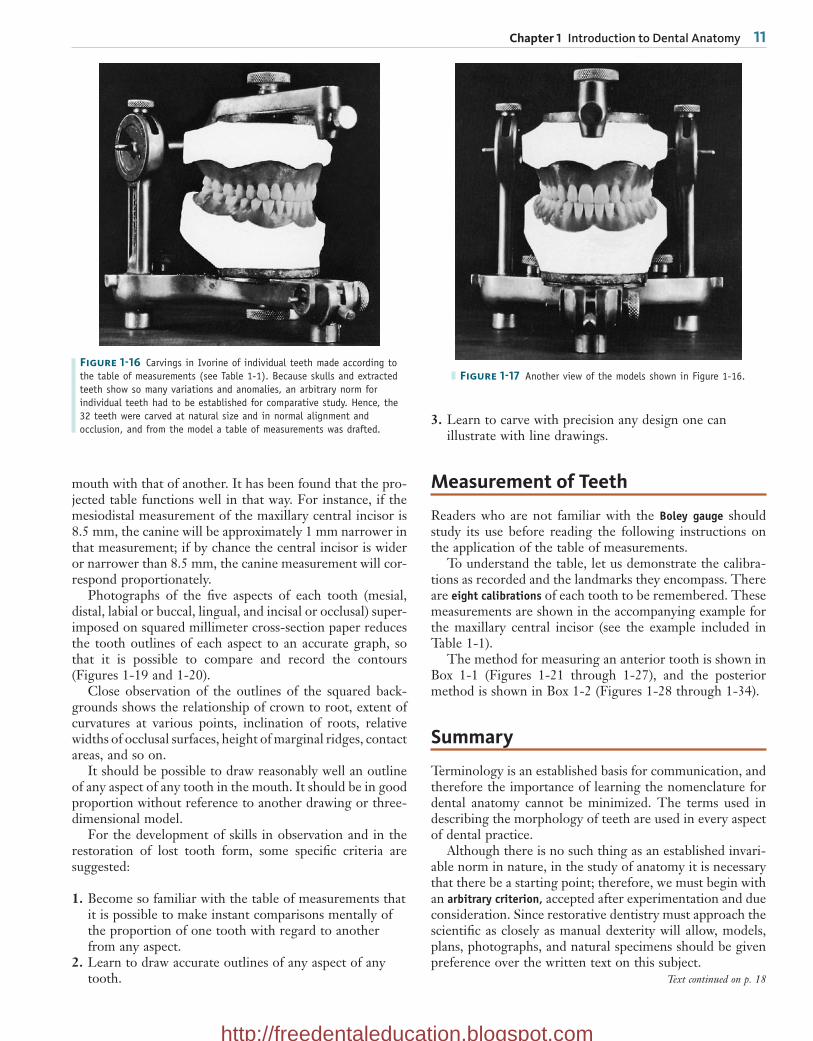
The basis for the specifications to be used for carving individual teeth is a table of average measurements for per-manent teeth given by Dr. G. V. Black.6 However, teeth carved or drawn to these average dimensions cannot be set into place for an ideal occlusion. Therefore, for purposes of producing a complete set of articulated teeth (Figures 1-16, 1-17, and 1-18) carved from Ivorine, minor changes have been made in Dr. Black’s table. Also, carving teeth to natural size, calibrated to tenths of a millimeter, is not practical. The adjusted measurements are shown in Table 1-1. The only fractions listed in the model table are 0.5 mm and 0.3 mm in a few instances. Fractions are avoided whenever possible to facilitate familiarity with the table and to avoid confusion.
A table of measurements must be arbitrarily agreed upon so that a reasonable comparison can be made when apprais-ing the dimensions of any one aspect of one tooth in the
Figure �-�� Line angles. A, Anterior teeth. B, Posterior teeth.
Linguoincisal line angle
Distolabial line angle
Linguo-occlusalline angle
Disto-occlusalline angle
Distolingual line angle
Distobuccal line angle
Mesio-occlusal line angle
Bucco-occlusal line angle
Mesiolingual line angle
Mesiobuccal line angle
Distolingual line angle
Labioincisal line angle
Mesiolabial line angle
Mesiolingual line angle
A
B
MesialLingual
Buccal Distal
MesialLingual
Labial Distal
Figure �-�� A, Point angles on anterior teeth. B, Point angles on posterior teeth.
Distolabioincisal pointangle
Mesiolabioincisal pointangle
Mesiolinguoincisal pointangle
Distolinguo-occlusal pointangle
Distobucco-occlusal pointangle
Distolinguoincisal pointangle
A
B
Mesiolinguo-occlusal pointangle
Mesiobucco-occlusal pointangle
MesialLingual
Buccal Distal
MesialLingual
Labial Distal
�0 Tooth Drawing and Carving
http://freedentaleducation.blogspot.com
Chapter � Introduction to Dental Anatomy ��
Figure �-�� Carvings in Ivorine of individual teeth made according to the table of measurements (see Table 1-1). Because skulls and extracted teeth show so many variations and anomalies, an arbitrary norm for individual teeth had to be established for comparative study. Hence, the 32 teeth were carved at natural size and in normal alignment and occlusion, and from the model a table of measurements was drafted.
Figure �-�7 Another view of the models shown in Figure 1-16.
mouth with that of another. It has been found that the pro-jected table functions well in that way. For instance, if the mesiodistal measurement of the maxillary central incisor is 8.5 mm, the canine will be approximately 1 mm narrower in that measurement; if by chance the central incisor is wider or narrower than 8.5 mm, the canine measurement will cor-respond proportionately.
Photographs of the five aspects of each tooth (mesial, distal, labial or buccal, lingual, and incisal or occlusal) super-imposed on squared millimeter cross-section paper reduces the tooth outlines of each aspect to an accurate graph, so that it is possible to compare and record the contours (Figures 1-19 and 1-20).
Close observation of the outlines of the squared back-grounds shows the relationship of crown to root, extent of curvatures at various points, inclination of roots, relative widths of occlusal surfaces, height of marginal ridges, contact areas, and so on.
It should be possible to draw reasonably well an outline of any aspect of any tooth in the mouth. It should be in good proportion without reference to another drawing or three-dimensional model.
For the development of skills in observation and in the restoration of lost tooth form, some specific criteria are suggested:
1. Become so familiar with the table of measurements that it is possible to make instant comparisons mentally of the proportion of one tooth with regard to another from any aspect.
2. Learn to draw accurate outlines of any aspect of any tooth.
3. Learn to carve with precision any design one can illustrate with line drawings.
Measurement of Teeth
Readers who are not familiar with the Boley gauge should study its use before reading the following instructions on the application of the table of measurements.
To understand the table, let us demonstrate the calibra-tions as recorded and the landmarks they encompass. There are eight calibrations of each tooth to be remembered. These measurements are shown in the accompanying example for the maxillary central incisor (see the example included in Table 1-1).
The method for measuring an anterior tooth is shown in Box 1-1 (Figures 1-21 through 1-27), and the posterior method is shown in Box 1-2 (Figures 1-28 through 1-34).
Summary
Terminology is an established basis for communication, and therefore the importance of learning the nomenclature for dental anatomy cannot be minimized. The terms used in describing the morphology of teeth are used in every aspect of dental practice.
Although there is no such thing as an established invari-able norm in nature, in the study of anatomy it is necessary that there be a starting point; therefore, we must begin with an arbitrary criterion, accepted after experimentation and due consideration. Since restorative dentistry must approach the scientific as closely as manual dexterity will allow, models, plans, photographs, and natural specimens should be given preference over the written text on this subject.
Text continued on p. 18
http://freedentaleducation.blogspot.com

Figure �-�8 Occlusal view of the models shown in Figures 1-16 and 1-17.
�� Summary
http://freedentaleducation.blogspot.com

Chapter � Introduction to Dental Anatomy ��
Table �-� Measurements of the Teeth: Specifications for Drawing and Carving Teeth of Average Size*
Length of Crown
Length of Root
Mesiodistal Diameter
of Crown†
Mesiodistal Diameter of
Crown at Cervix
Labio- or Buccolingual
Diameter of Crown
Labio- or Buccolingual
Diameter of Crown at
Cervix
Curvature of Cervical
Line—Mesial
Curvature of Cervical Line—Distal
Maxillary TeethCentral incisor 10.5 13.0 8.5 7.0 7.0 6.0 3.5 2.5
Lateral incisor 9.0 13.0 6.5 5.0 6.0 5.0 3.0 2.0
Canine 10.0 17.0 7.5 5.5 8.0 7.0 2.5 1.5
First premolar 8.5 14.0 7.0 5.0 9.0 8.0 1.0 0.0
Second premolar 8.5 14.0 7.0 5.0 9.0 8.0 1.0 0.0
First molar 7.5 B L 10.0 8.0 11.0 10.0 1.0 0.0
12 13
Second molar 7.0 B L 9.0 7.0 11.0 10.0 1.0 0.0
11 12
Third molar 6.5 11.0 8.5 6.5 10.0 9.5 1.0 0.0
Mandibular TeethCentral incisor 9.0‡ 12.5 5.0 3.5 6.0 5.3 3.0 2.0
Lateral incisor 9.5‡ 14.0 5.5 4.0 6.5 5.8 3.0 2.0
Canine 11.0 16.0 7.0 5.5 7.5 7.0 2.5 1.0
First premolar 8.5 14.0 7.0 5.0 7.5 6.5 1.0 0.0
Second premolar 8.0 14.5 7.0 5.0 8.0 7.0 1.0 0.0
First molar 7.5 14.0 11.0 9.0 10.5 9.0 1.0 0.0
Second molar 7.0 13.0 10.5 8.0 10.0 9.0 1.0 0.0
Third molar 7.0 11.0 10.0 7.5 9.5 9.0 1.0 0.0
B, Buccal; L, lingual.*In millimeters. This table has been “proved” by carvings shown in Figures 1-16 and 1-17.†The sum of the mesiodistal diameters, both right and left, which gives the arch length, is maxillary, 128 mm; mandibular, 126 mm.‡Lingual measurement is approximately 0.5 mm longer.
Table �-� Measurements of the Teeth: Specifications for Drawing and Carving Teeth of Average Size—cont’d
Measurements of the Teeth: an Example*
Length of Crown
Length of Root
Mesiodistal Diameter
of Crown†
Mesiodistal Diameter of
Crown at Cervix
Labio- or Buccolingual
Diameter of Crown
Labio- or Buccolingual
Diameter of Crown at
Cervix
Curvature of Cervical
Line—Mesial
Curvature of Cervical Line—Distal
Maxillary TeethCentral incisor 10.5 13.0 8.5 7.0 7.0 6.0 3.5 2.5
*In millimeters.†The sum of the mesiodistal diameters, both right and left, which gives the arch length, is maxillary, 128 mm; mandibular, 126 mm.
http://freedentaleducation.blogspot.com

Figure �-�9 Maxillary left canine. When viewing the mesial and distal aspects, note the curvature or bulge on the crown at the cervical third below the cementoenamel junction. This is called the cervical ridge, or the cervicoenamel ridge.
Figure �-�0 Maxillary right first molar. When viewing the mesial and distal aspects, note the curvature or bulge on the crown at the cervical third below the cementoenamel junction.
�� Summary
IV A(3), B
http://freedentaleducation.blogspot.com
Chapter � Introduction to Dental Anatomy ��
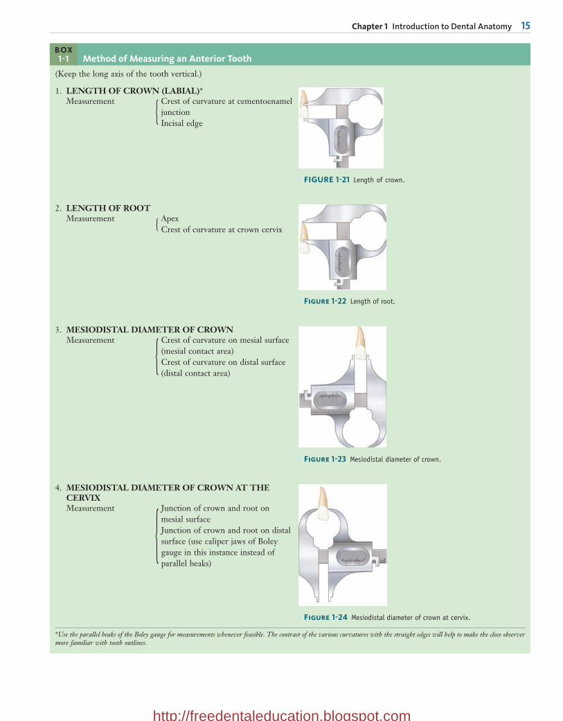
(Keep the long axis of the tooth vertical.)
B OX�-� Method of Measuring an Anterior Tooth
*Use the parallel beaks of the Boley gauge for measurements whenever feasible. The contrast of the various curvatures with the straight edges will help to make the close observer more familiar with tooth outlines.
2. LENGTH OF ROOTMeasurement { Apex
Crest of curvature at crown cervix
FIGURE �-�� Length of crown.
Figure �-�� Length of root.
3. MESIODISTAL DIAMETER OF CROWNMeasurement { Crest of curvature on mesial surface
(mesial contact area)Crest of curvature on distal surface (distal contact area)
Figure �-�� Mesiodistal diameter of crown.
1. LENGTH OF CROWN (LABIAL)*Measurement { Crest of curvature at cementoenamel
junctionIncisal edge
4. MESIODISTAL DIAMETER OF CROWN AT THE CERVIXMeasurement
{Junction of crown and root on mesial surfaceJunction of crown and root on distal surface (use caliper jaws of Boley gauge in this instance instead of parallel beaks)
Figure �-�� Mesiodistal diameter of crown at cervix.
http://freedentaleducation.blogspot.com

B OX�-� Method of Measuring an Anterior Tooth—cont’d
5. LABIOLINGUAL DIAMETER OF CROWNMeasurement { Crest of curvature on labial surface
Crest of curvature on lingual surface
Figure �-�� Labiolingual diameter of crown.
6. LABIOLINGUAL DIAMETER OF CROWN AT THE CERVIXMeasurement
{Junction of crown and root on labial surfaceJunction of crown and root on lingual surface (use caliper jaws in this instance also)
Figure �-�� Labiolingual diameter of cervix.
Figure �-�7 Curvature of cementoenamel junction on mesial.
7. CURVATURE OF CEMENTOENAMEL JUNC-TION ON MESIAL†Measurement
{Crest of curvature of cementoenamel junction on labial and lingual surfacesCrest of curvature of cementoenamel junction on mesial surface
†This measurement is most important because normally it represents the extent of curvature approximately of the periodontal attachment when the tooth is in situ.
8. CURVATURE OF CEMENTOENAMEL JUNC-TION ON DISTAL (Turn the tooth around and cali-brate as in Figure 1-27.)Measurement
{Crest of curvature of cementoenamel junction on labial and lingual surfacesCrest of curvature of cementoenamel junction on distal surface
�� Summary
http://freedentaleducation.blogspot.com

Chapter � Introduction to Dental Anatomy �7
B OX�-� Method of Measuring a Posterior Tooth
(Keep the long axis of the tooth vertical.)
1. LENGTH OF CROWN (BUCCAL)Measurement { Crest of buccal cusp or cusps
Crest of curvature at cementoenamel junction
Figure �-�8 Length of crown.
2. LENGTH OF ROOTMeasurement { Crest of curvature at crown cervix
Apex of root
Figure �-�9 Length of root.
3. MESIODISTAL DIAMETER OF CROWNMeasurement { Crest of curvature on mesial surface
(mesial contact area)Crest of curvature on distal surface (distal contact area)
Figure �-�0 Mesiodistal diameter of crown.
Figure �-�� Mesiodistal diameter of crown at cervix.
4. MESIODISTAL DIAMETER OF CROWN AT THE CERVIXMeasurement
{Junction of crown and root on mesial surfaceJunction of crown and root on distal surface (use caliper jaws of Boley gauge instead of parallel beaks)
http://freedentaleducation.blogspot.com

B OX�-� Method of Measuring a Posterior Tooth—cont’d
5. BUCCOLINGUAL DIAMETER OF CROWNMeasurement { Crest of curvature on buccal surface
Crest of curvature on lingual surface
Figure �-�� Buccolingual diameter of crown.
6. BUCCOLINGUAL DIAMETER OF CROWN AT THE CERVIXMeasurement { Junction of crown and root on
buccal surfaceJunction of crown and root on lingual surface (use caliper jaws)
Figure �-�� Buccolingual diameter of crown at cervix.
7. CURVATURE OF CEMENTOENAMEL JUNCTION ON MESIALMeasurement
{Crest of curvature of cementoenamel junction on mesial surfaceCrest of curvature of cementoenamel junction on buccal and lingual surfaces
8. CURVATURE OF CEMENTOENAMEL JUNCTION ON DISTAL (Turn tooth around and measure as in Figure 1-34.)Measurement
{Crest of curvature of cementoenamel junction on distal surfaceCrest of curvature of cementoenamel junction on buccal and lingual surfaces
Figure �-�� Curvature of cementoenamel junction on mesial.
Every curve and segment of a normal tooth has some functional basis, and it is important to reproduce them accu-rately. The successful clinician in dentistry or, for that matter, any designer of dental restorations should be able to mentally create pictures of the teeth from any aspect and relate those aspects of dental anatomy to function. Com-plete pictures can be formed only when one is familiar with the main details of tooth form.
References1. Webster’s new universal unabridged dictionary, New York, 1996,
Barnes & Noble Books.2. Turner II CG, Nichol CR, Scott GR: Scoring procedures for
key morphological traits of the permanent dentition: The Arizona State University Dental Anthropology System. In Kelley MA, Larsen CS, editors: Advances in dental anthropology, New York, 1991, Wiley-Liss.
�8 Summary
http://freedentaleducation.blogspot.com

Chapter � Introduction to Dental Anatomy �9
3. Lyons H: Committee adopts official method for the symbolic designation of teeth, J Am Dent Assoc 34:647, 1947.
4. Peck S, Peck L: A time for change of tooth numbering systems, J Dent Educ 57:643, 1993.
5. Carlsen O: Dental morphology, Copenhagen, 1987, Munksgaard.
6. Black GV: Descriptive anatomy of the human teeth, ed 4, Philadelphia, 1897, S. S. White Dental Manufacturing.
BibliographyAmerican Dental Association, Committee on Nomenclature:
Committee adopts official method for the symbolic designation of teeth, J Am Dent Assoc 34:647, 1947.
American Dental Association, Committee on Dental Education and Hospitals: Tooth numbering and radiographic mounting, Am Dent Assoc Trans 109:25, 247, 1968.
Fédération Dentaire Intemationale: Two-digit system of designating teeth, Int Dent J 21:104, 1971.
Goodman P: A universal system for identifying permanent and primary teeth, J Dent Child 34:312, 1987.
Haderup V: Dental nomenklatur og stenograft, Dansk Tandl Tidskr 3:3, 1891.
Palmer C: Palmer’s dental notation, Dental Cosmos 33:194, 1981.World Health Organization: Oral health surveys: basic methods, ed
3, Geneva, 1987, The Organization.Zsigmondy A: Grundzüge einer praktischen Methode zur
raschen und genauen Vonnerkung der zahnärztlichen Beobachtungen und Operationen, Dtsch Vjschr Zahnhk 1:209, 1861.
Zsigmondy A: A practical method for rapidly noting dental observations and operations, Br J Dent Sci 17:580, 1874.
Use the CD-ROM to view animations and study ques-tions; and go to the site for additional study resources.
http://freedentaleducation.blogspot.com

This page intentionally left blank
http://freedentaleducation.blogspot.com

Development and Eruption of the Teeth
as far as the epithelial attachment of the gingiva to the enamel. In addition, in pathologically deepened crevices, tooth surfaces can be sensed as far as the attachment of the periodontal ligament to the cementum. Perhaps the simplest example of clinical observation is the assignment of dental age or the assessment of dental development by looking into a child’s mouth to note the teeth that have emerged through the gingiva. In the absence of other data, however, the number of teeth present are simply counted.1
When observations from clinical and radiographic sources of information are coupled with sufficient knowledge of dental morphology and the chronologies of the human den-tition, the clinician has the foundation for the diagnosis and management of most disorders involving the size, shape, number, arrangement, esthetics, and development of the teeth and also problems related to the sequence of tooth eruption and occlusal relationships. For example, in Figure 2-1, A, the gingival tissues are excellent; however, the form of the maxillary incisors and interdental spacing might be considered to be an esthetic problem by a patient. To accept the patient’s concern that a cosmetic problem is present and needs correction requires that the practitioner be able to transform the patient’s idea of esthetics into reality by orthodontics and cosmetic restorative dentistry. The situa-tion in Figure 2-1, B demonstrates a periodontal problem (localized gingivitis of the gingival margin of the right central incisor), which is in part a result of the inadequate proximal contact relations of the incisors, leading to food impaction and accumulation of dental plaque and some cal-culus. For the most part, however, it is the result of inade-quate home care hygiene. Most conservative correction relates to removal of the irritants and daily tooth brushing
Knowledge of the development of the teeth and their emer-gence into the oral cavity is applicable to clinical practice, anthropology, demography, forensics, and paleontology. However, dental applications are considered primarily. This chapter considers the development and eruption of the teeth, specific chronologies of both the primary and perma-nent human dentitions, dental age, tooth formation stand-ards, and applications to dental practice (e.g., an understanding of both the chronology of dental development so that surgi-cal intervention does not harm normal growth and the rela-tionship between dental age and the effects of disease and environmental risks). The use of the terms primary and decid-uous, or often, primary/deciduous, reflects the difference of opinion about the most appropriate term to describe the first dentition in humans. Readers of the literature are able to deal objectively with both terms.
Clinical Considerations
It must be kept in mind that the dental practitioner sees in a “normal” healthy mouth not only the clinical crowns of the teeth surrounded by the gingival tissues, but also the number, shape, size, position, coloration, and angulations of the teeth; the outlines of the roots of the teeth; occlusal con-tacts; evidence of function and parafunction; and phonetics and esthetics. Most of the parts of the teeth that are hidden by the gingiva can be visualized radiographically. This can also be done by using a periodontal probe to locate the depth of normal or pathologically deepened gingival crev-ices or a dental explorer to sense the surfaces of the teeth within the gingival crevice apical to the free gingival margin
21
22
http://freedentaleducation.blogspot.com
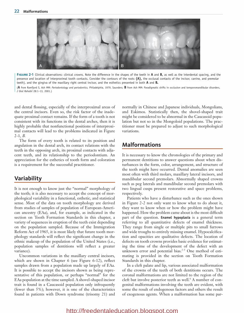
and dental flossing, especially of the interproximal areas of the central incisors. Even so, the risk factor of the inade-quate proximal contact remains. If the form of a tooth is not consistent with its functions in the dental arches, then it is highly probable that nonfunctional positions of interproxi-mal contacts will lead to the problems indicated in Figure 2-1, B.
The form of every tooth is related to its position and angulation in the dental arch, its contact relations with the teeth in the opposing arch, its proximal contacts with adja-cent teeth, and its relationship to the periodontium. An appreciation for the esthetics of tooth form and coloration is a requirement for the successful practitioner.
Variability
It is not enough to know just the “normal” morphology of the teeth; it is also necessary to accept the concept of mor-phological variability in a functional, esthetic, and statistical sense. Most of the data on tooth morphology are derived from studies of samples of population of European-Ameri-can ancestry (EAa), and, for example, as indicated in the section on Tooth Formation Standards in this chapter, a variety of sequences in eruption of the teeth exist depending on the population sampled. Because of the Immigration Reform Act of 1965, it is most likely that future tooth mor-phology standards will reflect the significant change in the ethnic makeup of the population of the United States (i.e., population samples of dentitions will reflect a greater variance).
Uncommon variations in the maxillary central incisors, which are shown in Chapter 6 (see Figure 6-12), reflect samples drawn from a population made up largely of EAa. It is possible to accept the incisors shown as being repre-sentative of this population, or perhaps “normal” for the EAa population at the time sampled. A shovel-shaped incisor trait is found in a Caucasoid population only infrequently (fewer than 5%); however, it is one of the characteristics found in patients with Down syndrome (trisomy 21) and
normally in Chinese and Japanese individuals, Mongolians, and Eskimos. Statistically then, the shovel-shaped trait might be considered to be abnormal in the Caucasoid popu-lation but not so in the Mongoloid populations. The prac-titioner must be prepared to adjust to such morphological variations.
Malformations
It is necessary to know the chronologies of the primary and permanent dentitions to answer questions about when dis-turbances in the form, color, arrangement, and structure of the teeth might have occurred. Dental anomalies are seen most often with third molars, maxillary lateral incisors, and mandibular second premolars. Abnormally shaped crowns such as peg laterals and mandibular second premolars with two lingual cusps present restorative and space problems, respectively.
Patients who have a disturbance such as the ones shown in Figure 2-2 not only want to know what to do about it, they want to know when or how the problem might have happened. How the problem came about is the most difficult part of the question. Enamel hypoplasia is a general term referring to all quantitative defects of enamel thickness. They range from single or multiple pits to small furrows and wide troughs to entirely missing enamel. Hypocalcifica-tion and opacities are qualitative defects. The location of defects on tooth crowns provides basic evidence for estimat-ing the time of the development of the defect with an unknown error and potential bias.2–5 One method of esti-mating is provided in the section on Tooth Formation Standards in this chapter.
In a cleft palate and lip, various associated malformation of the crowns of the teeth of both dentitions occurs. The coronal malformations are not limited to the region of the cleft but involve posterior teeth as well.6 A number of con-genital malformations involving the teeth are evident, with some the result of endogenous factors and others the result of exogenous agents. When a malformation has some par-
Figure 2-1 Clinical observations: clinical crowns. Note the difference in the shapes of the teeth in A and B, as well as the interdental spacing, and the presence and location of interproximal tooth contacts. Consider the contours of the roots (A), the occlusal contacts of the incisor, canine, and premolar teeth), and the gingiva of the maxillary right central incisor, and the esthetics presented in both A and B. (A from Ramfjord S, Ash MM: Periodontology and periodontics, Philadelphia, 1979, Saunders; B from Ash MM: Paradigmatic shifts in occlusion and temporomandibular disorders, J Oral Rehabil 28:1–13, 2001.)
A B
22 Malformations
http://freedentaleducation.blogspot.com

Chapter 2 Development and Eruption of the Teeth 23
ticular characteristics (e.g., screwdriver-shaped central inci-sors) and is consistent with a particular phase of dental development, it may be possible to determine the cause of the disturbance. This aspect is considered further in the section on Dental Age in this chapter.
Chronology of Primary Dentition
The chronology of the primary teeth presented in Table 2-1 is based on data derived from Tables 2-3 and 2-4 in the section on Tooth Formation Standards in this chapter. The universal numbering system is used in Table 2-1. The picto-rial charts (Figures 2-3 and 2-4) are not intended to be used as ideal standards of normal development. Their use is directed toward showing patients the general aspects of development rather than providing precise guidance for clinical procedures.
Development and Eruption/Emergence of the Teeth
Historically, the term eruption was used to denote the tooth’s emergence through the gingiva, but then it became more completely defined to mean continuous tooth movement from the dental bud to occlusal contact.7 Not all tables of dental chronologies reflect the latter definition of eruption, however; the terms eruption and emergence are used here at this time in such a way as to avoid any confusion between the historical use of eruption and its more recent expanded meaning.
Emergence of the primary dentition takes place between the sixth and thirtieth months of postnatal life. It takes from 2 to 3 years for the primary dentition to be completed, beginning with the initial calcification of the primary central incisor to the completion of the roots of the primary second molar (see Figure 2-3).
The emergence of the primary dentition through the alveolar mucous membrane is an important time for the development of oral motor behavior and the acquisition of
masticator skills.8 At this time of development, the presence of “teething” problems suggests how the primary dentition can affect the development of future neurobehavioral mech-anisms, including jaw movements and mastication. Learning of mastication may be highly dependent on the stage and
Figure 2-2 A, Hypoplasia of the enamel. B, Defect in tooth structure caused by trauma to the primary predecessor during development of the permanent central incisor.
(A from Neville BW, Damm DD, Allen CM, et al: Oral and maxillofacial pathology, ed 3, St Louis, 2009, Saunders; B from Ash MM: Oral pathology, ed 6, Philadelphia, 1992, Lea & Febiger.)
A B
Table 2-1 Chronology of Primary Teeth*
Tooth
First Evidence of
Calcification (Weeks in
Utero)
Crown Completed (Months)
Eruption (Mean
Age) (Months)
Root Completed
(Years)
Upper
i1 E, F 14 112
10 112
i2 D, G 16 212
11 2
c C, H 17 9 19 314
m1 B, I 15 6 16 212
m2 A, J 19 11 29 3
Maxillary TeethRight A B C D F G I J Left
O N M KMandibular Teeth
E HT S R Q P L
Lower
i1 P, O 14 212
8 112
i2 Q, N 16 3 13 112
c R, M 17 9 20 314
m1 S, L 1512 51
216 21
4
m2 T, K 18 10 27 3
i1, Central incisor; i2, lateral incisor; c, canine; m1, first molar; m2, second molar.*Universal numbering system for primary/deciduous dentition; see Chapter 1. See Tables 2-3 and 2-4 for detailed presentation of the data.
http://freedentaleducation.blogspot.com
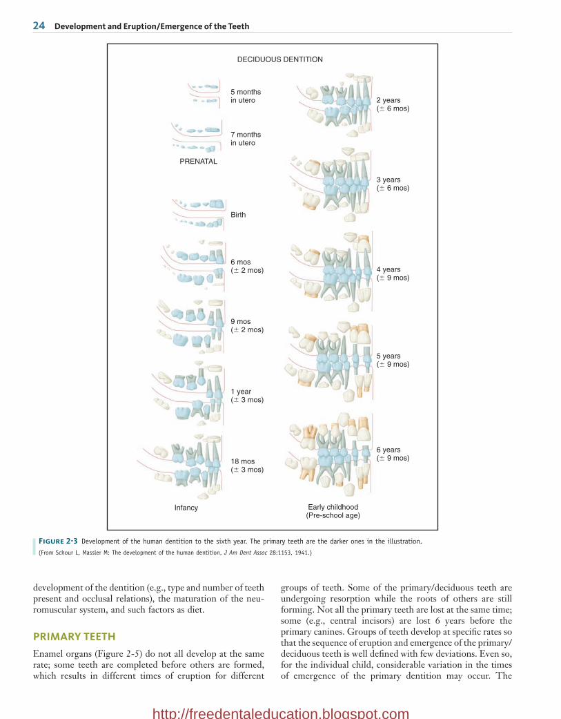
development of the dentition (e.g., type and number of teeth present and occlusal relations), the maturation of the neu-romuscular system, and such factors as diet.
PRIMARY TEETH
Enamel organs (Figure 2-5) do not all develop at the same rate; some teeth are completed before others are formed, which results in different times of eruption for different
groups of teeth. Some of the primary/deciduous teeth are undergoing resorption while the roots of others are still forming. Not all the primary teeth are lost at the same time; some (e.g., central incisors) are lost 6 years before the primary canines. Groups of teeth develop at specific rates so that the sequence of eruption and emergence of the primary/deciduous teeth is well defined with few deviations. Even so, for the individual child, considerable variation in the times of emergence of the primary dentition may occur. The
Figure 2-3 Development of the human dentition to the sixth year. The primary teeth are the darker ones in the illustration.
(From Schour L, Massler M: The development of the human dentition, J Am Dent Assoc 28:1153, 1941.)
2 years(� 6 mos)
3 years(� 6 mos)
4 years(� 9 mos)
5 years(� 9 mos)
6 years(� 9 mos)
5 monthsin utero
7 monthsin utero
PRENATAL
DECIDUOUS DENTITION
Birth
6 mos(� 2 mos)
9 mos(� 2 mos)
1 year(� 3 mos)
18 mos(� 3 mos)
Early childhood(Pre-school age)
Infancy
24 Development and Eruption/Emergence of the Teeth
http://freedentaleducation.blogspot.com

Chapter 2 Development and Eruption of the Teeth 25
Figure 2-4 Development of the human dentition from the seventh year to maturity. Note the displacement of the primary teeth.
(From Schour L, Massler M: The development of the human dentition, J Am Dent Assoc 28:1153, 1941.)
11 years(� 9 mos)
12 years(� 6 mos)
15 years(� 6 mos)
21years
35years
7 years(� 9 mos)
8 years(� 9 mos)
9 years(� 9 mos)
10 years(� 9 mos)
MIXED DENTITION PERMANENT DENTITION
Late childhood(school age)
Adolescenceand adulthood
primary dentition is completely formed by about age 3 and functions for a relatively short period of time before it is lost completely at about age 11. Permanent dentition is com-pleted by about age 25 if the third molars are included (see Figures 2-3 and 2-4).9
Calcification of the primary teeth begins in utero from 13 to 16 weeks postfertilization. By 18 to 20 weeks, all the primary teeth have begun to calcify. Primary tooth crown formation takes only some 2 to 3 years from initial calcifica-
tion to root completion. However, mineralization of the permanent dentition is entirely postnatal, and the formation of each tooth takes about 8 to 12 years. The variability in tooth development is similar to that for eruption, sexual maturity, and other similar growth indicators.10
Crown formation of the primary teeth continues after birth for about 3 months for the central incisor, about 4 months for the lateral incisor, about 7 months for the primary first molar, about 8.5 months for the canine, and
http://freedentaleducation.blogspot.com

about 10.5 months for the second primary molar. During these periods before and after birth, disorders in shape, pigmentation, mineralization, and structure sometimes occur (fluorosis is considered later in this chapter).
Crown and Root DevelopmentDental development can be considered to have two compo-nents: (1) the formation of crowns and roots and (2) the eruption of the teeth. Of these two, the former seems to be much more resistant to environmental influences; the latter can be affected by caries and tooth loss.11,12
After the crown of the tooth is formed, development of the root portion begins. At the cervical border of the enamel (the cervix of the crown), cementum starts to form as a root covering of the dentin. The cementum is similar in some ways to bone tissue and covers the root of the tooth in a thin layer. In the absence of a succeeding permanent tooth, the root of the primary tooth may only partially resorb. When root resorption does not follow the usual pattern, the per-manent tooth cannot emerge or is otherwise kept out of its normal place. In addition, the failure of the root to resorb may bring about prolonged retention of the primary tooth. Although mandibular teeth do not begin to move occlusally until crown formation is complete, their eruption rate does not closely correlate with root elongation. After the crown and part of the root are formed, the tooth penetrates the alveolar gingiva and makes its entry (emergence) into the mouth.
Further formation of the root is considered to be an active factor in moving the crown toward its final position in the mouth. The process of eruption of the tooth is com-pleted when most of the crown is in evidence and when it has made contact with its antagonist or antagonists in the opposing jaw. The root formation is not finished when the tooth emerges; however, the formation of root dentin and cementum continues after the tooth is in use. Ultimately, the root is completed with a complete covering of cemen-tum. Additional formation of cementum may occur in response to tooth movement or further eruption of the teeth. Also, cementum may be added (repaired) and/or
resorbed in response to periodontal trauma from occlusion. The covering of cementum of the permanent teeth is much thicker than that of the primary teeth.
The Dentitions
The human dentitions are usually categorized as being primary, mixed (transitional), and permanent dentitions. The transition from the primary/deciduous dentition to the permanent dentition is of particular interest because of changes that may herald the onset of malocclusion and provide for its interception and correction. Thus of impor-tance for the practitioner are the interactions between the morphogenesis of the teeth, development of the dentition, and growth of the craniofacial complex.
PRENATAL/PERINATAL/POSTNATAL DEVELOPMENT
The first indication of tooth formation occurs as early as the sixth week of prenatal life when the jaws have assumed their initial shape; however, at this time the jaws are rather small compared with the large brain case and orbits. The lower face height is small compared with the neurocranium (Figure 2-6). The mandibular arch is larger than the maxillary arch, and the vertical dimensions of the jaws are but little devel-oped. When the jaws close at this stage in the development of the dentition, they make contact with the tongue, which in turn makes contact with the cheeks. The shape of the prenatal head varies considerably, but the relative difference between the brain case, orbits, and lower face height remains the same. All stages of tooth formation fill both jaws during this stage of development.
DEVELOPMENT OF THE PRIMARY DENTITION
Considerable growth follows birth in the neurocranium and splanchnocranium. Usually at birth, no teeth are visible in the mouth; however, occasionally, infants are born with
Figure 2-5 Enamel organ. A, 1, Beginning of first primary molar; 2, bell stage of second primary molar; 3, dental lamina of first permanent molar. B, Partially developed primary incisor and, lingually, the developing permanent incisor.
(A from Ash MM: Oral pathology, ed 6, Philadelphia, 1992, Lea & Febiger; B from Avery JK, Chiego Jr DJ: Essentials of oral histology and embryology, ed 3, St. Louis, 2006, Mosby.)
Clinicalcrown
Dentinogingivaljunction
Junctionalor epithelialattachment
Developingtooth
A B
26 The Dentitions
http://freedentaleducation.blogspot.com

Chapter 2 Development and Eruption of the Teeth 27
erupted mandibular incisors. Development of both primary and permanent teeth continues in this period, and jaw growth follows the need for additional space posteriorly for additional teeth. In addition the alveolar bone height increases to accommodate the increasing length of the teeth. However, growth of the anterior parts of the jaws is limited after about the first year of postnatal life.
SEQUENCE OF EMERGENCE OF PRIMARY TEETH
The predominant sequence of eruption of the primary teeth in the individual jaw is central incisor (A), lateral incisor (B), first molar (D), canine (C), and second molar (E), as seen in Table 2-1. Variations in that order may be the result of reversals of central and lateral incisors or first molar and lateral incisor, or eruption of two teeth at the same time.13 This subject is considered in more detail in the section on Tooth Formation Standards in this chapter and in Chapter 16, which addresses development of the primary occlusion.
Investigations of the chronology of emergence of primary teeth in different racial and ethnic groups show considerable variation,7 and little information is available on tooth forma-tion in populations of nonwhite/non-European ancestry.14 World population differences in tooth standards suggest that patterned differences may exist that in fact are not large.14 Tooth size, morphology, and formation are highly inheritable characteristics.15 Few definitive correlations exist between primary tooth emergence and other physiological parameters such as skeletal maturation, size, and sex.16
EMERGENCE OF THE PRIMARY TEETH
At about 8 (6 to 10) months of age, the mandibular central incisors emerge through the alveolar gingiva, followed by the other anterior teeth, so that by about 13 to 16 months,
all eight primary incisors have erupted (see Table 2-1). Then the first primary molars emerge by about 16 months of age and make contact with opposing teeth several months later, before the canines have fully erupted. Passage through the alveolar crest (Figure 2-7) occurs when approximately two thirds of the root is formed,17 followed by emergences through the alveolar gingiva into the oral cavity when about three fourths of the root is completed.18 The emergence data are consistent with those of Smith.14
The primary first molars emerge with the maxillary molar tending most often to erupt earlier than the mandibular first molar.19 Some evidence shows a difference by gender for the first primary molars, but no answer is available for why the first molar has a different pattern of sexual dimorphism.7
The primary maxillary canines erupt at about 19 (16 to 22) months (Figure 2-8), and the mandibular canines erupt at 20 (17 to 23) months. The primary second mandibular molar erupts at a mean age of 27 (23 to 31, boys) (24 to 30, girls) months, and the primary maxillary second molar follows at a mean age of 29 (25 to 33 ± 1SD) months. In Figures 2-8, A and B, the first molars are in occlusion.
Neuromuscular Development
A mature neuromuscular controlled movement of the man-dible requires the presence and articulation of the teeth and
Figure 2-6 Neonatal skull showing large brain case and orbits; the neurocranium is larger than the splanchnocranium, which contains the jaws and all the developing teeth.
(From Avery JK, Chiego Jr DJ: Essentials of oral histology and embryology, ed 3, St. Louis, 2006, Mosby.)
Figure 2-7 Section of mandible in a 9-month-old infant cut through an unerupted primary canine and its permanent successor, which lies lingually and apically to it. The enamel of the primary canine crown is completed and lost because of decalcification. Root formation has begun. CEJ, Cementoenamel junction.
(Modified from Schour I, Noyes HJ: Oral histology and embryology, ed 8, Philadelphia, 1960, Lea & Febiger.)
Oral epithelium
Alveolar cryptbone
United epithelium
CEJ
Hertwig’s epithelialsheath
Permanent tooth
http://freedentaleducation.blogspot.com

proprioceptive input from the periodontium. Thus, the contact of opposing first primary molars is the beginning of the development of occlusion and a neuromuscular substrate for more complex mandibular and tongue functions.
PRIMARY DENTITION
The primary/deciduous dentition is considered to be com-pleted by about 30 months or when the second primary molars are in occlusion (Figure 2-9). The dentition period includes the time when no apparent changes occur intraorally (i.e., from about 30 months to about 6 years of age).
The form of the dental arch remains relatively constant without significant changes in depth or width. A slight
increase in the intercanine width occurs about the time the primary incisors are lost, and an increase in size in both jaws in a sagittal direction is consistent with the space needed to accommodate the succedaneous teeth. An increase in the vertical dimension of the facial skeleton occurs as a result of alveolar bone deposition, condyle growth, and deposition of bone at the synchondrosis of the basal part of the occipital bone and sphenoid bones, and at the maxillary suture complex.20 The splanchnocranium remains small in com-parison with the neurocranium. The part of the jaws that contain the primary teeth has almost reached adult width. At the first part of the transition period, which occurs at about age 8, the width of the mandible approximates the width of the neurocranium. The dental arches are complete, and the occlusion of the primary dentition is functional. During this period, attrition is sufficient in many children and is quite observable. The primary occlusion is considered in Chapter 16.
Transitional (Mixed) Dentition Period
The first transition dentition begins with the emergence and eruption of the mandibular first permanent molars and ends with the loss of the last primary tooth, which usually occurs at about age 11 to 12. The initial phase of the transition period lasts about 2 years, during which time the permanent
Figure 2-8 Skull of a child about 20 months of age. A, View showing all incisors present and erupting canines. B, Lateral view. First primary molars are in occlusion; mandibular second molars are just emerging opposite the already erupted maxillary molar.
(Modified from Karl W: Atlas der Zahnheilkunde, Berlin, [no publication date available], Verlag von Julius Springer.)
A
B
Figure 2-9 A, Skull of child 4 years old with completed primary dentition. B, Completed primary dentition. Note the incisal wear.
(A Modified from van der Linden FPGM, Duterloo HS: Development of the human dentition: an atlas, New York, 1976, Harper & Row; B from Bird DL, Robinson DS: Modern dental assisting, ed 9, St. Louis, 2009, Saunders.)
A
B
28 Transitional (Mixed) Dentition Period
http://freedentaleducation.blogspot.com

Chapter 2 Development and Eruption of the Teeth 29
first molars erupt (Figures 2-10 and 2-11), the primary inci-sors are shed, and the permanent incisors emerge and erupt into position (Figure 2-12). The permanent teeth do not begin eruptive movements until after the crown is com-pleted. During eruption, the permanent mandibular first molar is guided by the distal surface of the second primary molar. If a distal step in the terminal plane is evident, maloc-clusion occurs (see Figure 16-5).
Loss of Primary Teeth
The premature loss of primary teeth because of caries has an effect on the development of the permanent dentition21 and not only may reflect an unfortunate lack of knowledge as to the course of the disease but also establishes a negative attitude about preventing dental caries in the adult denti-tion. Loss of primary teeth may lead to lack of space for the permanent dentition. It is sometimes assumed by laypersons that the loss of primary teeth, which are sometimes referred to as baby teeth or milk teeth, is of little consequence because they are only temporary. However, the primary dentition may be in use from age 2 to 7 or older, or about 5 or more years in all. Some of the teeth are in use from 6 months until 12 years of age, or 11.5 years in all. Thus these primary teeth are in use and contributing to the health and well-being of the individual during the first years of greatest development, physically and mentally.
Figure 2-11 Same child as in Figure 2-10. A, Right side. B, Left side showing position of first permanent molars and empty bony crypt of developing second molar lost during preparation of the specimen. C, Front view showing right side with bone covering roots and developing permanent teeth, and left side with developing anterior permanent teeth.
A
B
C
Figure 2-10 Primary dentition with first permanent molars present. A, Maxillary arch. B, Mandibular arch.
B
A
Figure 2-12 Eruption of the permanent central incisors. Note the incisal edges demonstrating mamelons and the width of the emerging incisors.
http://freedentaleducation.blogspot.com

Premature loss of primary teeth, retention of primary teeth, congenital absence of teeth, dental anomalies, and insufficient space are considered important factors in the initiation and development of an abnormal occlusion. Pre-mature loss of primary teeth from dental neglect is likely to cause a loss of arch length with a consequent tendency for crowding of the permanent dentition. Arch length is con-sidered in more detail in Chapter 16.
Permanent Dentition
The permanent dentition consisting of 32 teeth is com-pleted from 18 to 25 years of age if the third molar is included.
Apparently there are four or more centers of formation (developmental lobes) for each tooth. The formation of each center proceeds until a coalescence of all of them takes place. During this period of odontogenesis, injury to the developing tooth can lead to anomalous morphological fea-tures (e.g., peg-shaped lateral incisor). Although no lines of demarcation are found in the dentin to show this develop-ment, signs are found on the surfaces of the crowns and roots; these are called developmental grooves (see Figure 4-12, B). Fractures of the teeth occur most commonly along these grooves (see Figure 13-26 ).
The follicles of the developing incisors and canines are in a position lingual to the deciduous roots (see Figures 2-7 and 2-11; see also Figure 3-4).
The developing premolars, which eventually take the place of deciduous molars, are within the bifurcation of primary molar roots (Figure 2-13, A and B). The permanent incisors, canines, and premolars are called succedaneous teeth because they take the place of their primary predecessors.
The central incisor is the second permanent tooth to emerge into the oral cavity. Eruption time is quite close to that of the first molar (i.e., tooth emergence occurs between 6 and 7 years) (Table 2-2). As with the first molar, at 6 years 50% of individuals have reached the stage considered to be the age of attainment of the stage or, more specifically, the age of emergence for the central incisor. The mandibular permanent teeth tend to erupt before maxillary teeth. The mandibular central incisor usually erupts before the maxil-lary central incisor (see Figure 2-12) and may erupt simul-taneously with or even before the mandibular first molar. The mandibular lateral incisor may erupt along with the central incisor.
Before the permanent central incisor can come into posi-tion, the primary central incisor must be exfoliated. This is brought about by the resorption of the deciduous roots. The permanent tooth in its follicle attempts to move into the position held by its predecessor. Its influence on the primary root evidently causes resorption of the root, which continues until the primary crown has lost its anchorage, becomes loose, and is finally exfoliated. In the meantime, the perma-nent tooth has moved occlusally so that when the primary tooth is lost, the permanent one is at the point of eruption and in proper position to succeed its predecessor.
Mandibular lateral incisors erupt very soon after the central incisors, often simultaneously. The maxillary central incisors erupt next in chronological order, and maxillary lateral inci-sors make their appearance about 1 year later (see Table 2-2 and Figures 2-3 and 2-4). The first premolars follow the maxillary laterals in sequence when the child is about 10 years old; the mandibular canines (cuspids) often appear at the same time. The second premolars follow during the next year, and then the maxillary canines follow. Usually, the second molars come in when the individual is about 12 years of age;
Figure 2-13 A, View of the right side of the skull of a child of 9 to 10 years of age. Note the amount of resorption of the roots of the primary maxillary molars, the relationship of the developing premolars above them, and the open pulp chambers and the pulp canals in the developing mandibular teeth. The roots of the first permanent molars have been completed. B, Left side. Note the placement of the permanent maxillary canine and second premolar, and the position and stage of development of the maxillary second permanent molar. The bony crypt of the lost mandibular second permanent premolar is in full view. Note the large openings in the roots of the mandibular second permanent molar.
A B
30 Permanent Dentition
http://freedentaleducation.blogspot.com
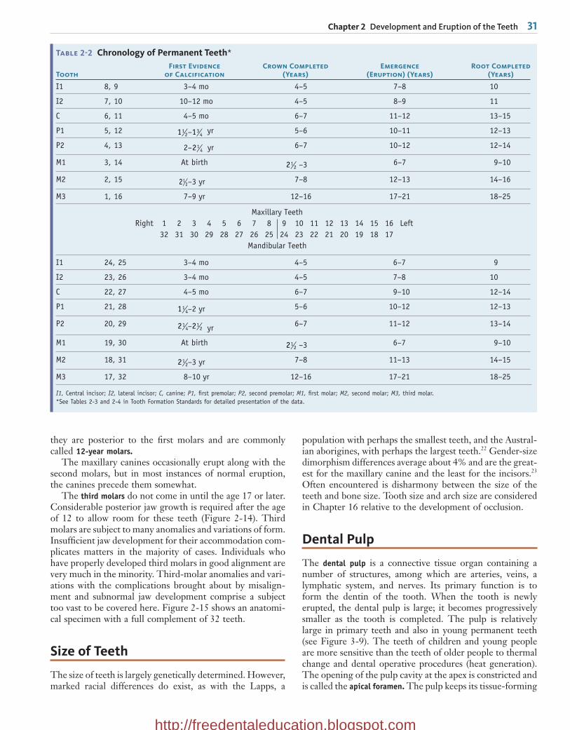
Chapter 2 Development and Eruption of the Teeth 31
they are posterior to the first molars and are commonly called 12-year molars.
The maxillary canines occasionally erupt along with the second molars, but in most instances of normal eruption, the canines precede them somewhat.
The third molars do not come in until the age 17 or later. Considerable posterior jaw growth is required after the age of 12 to allow room for these teeth (Figure 2-14). Third molars are subject to many anomalies and variations of form. Insufficient jaw development for their accommodation com-plicates matters in the majority of cases. Individuals who have properly developed third molars in good alignment are very much in the minority. Third-molar anomalies and vari-ations with the complications brought about by misalign-ment and subnormal jaw development comprise a subject too vast to be covered here. Figure 2-15 shows an anatomi-cal specimen with a full complement of 32 teeth.
Size of Teeth
The size of teeth is largely genetically determined. However, marked racial differences do exist, as with the Lapps, a
Table 2-2 Chronology of Permanent Teeth*
ToothFirst Evidence
of CalcificationCrown Completed
(Years)Emergence
(Eruption) (Years)Root Completed
(Years)
I1 8, 9 3–4 mo 4–5 7–8 10
I2 7, 10 10–12 mo 4–5 8–9 11
C 6, 11 4–5 mo 6–7 11–12 13–15
P1 5, 12 112–13
4 yr 5–6 10–11 12–13
P2 4, 13 2–214 yr 6–7 10–12 12–14
M1 3, 14 At birth 212 –3 6–7 9–10
M2 2, 15 212–3 yr 7–8 12–13 14–16
M3 1, 16 7–9 yr 12–16 17–21 18–25
Maxillary TeethRight 1 2 3 4 5 6 7 8 9 10 11 12 13 14 15 16 Left
32 31 30 29 28 27 26 25 24 23 22 21 20 19 18 17Mandibular Teeth
I1 24, 25 3–4 mo 4–5 6–7 9
I2 23, 26 3–4 mo 4–5 7–8 10
C 22, 27 4–5 mo 6–7 9–10 12–14
P1 21, 28 114–2 yr 5–6 10–12 12–13
P2 20, 29 214–21
2 yr 6–7 11–12 13–14
M1 19, 30 At birth 212 –3 6–7 9–10
M2 18, 31 212–3 yr 7–8 11–13 14–15
M3 17, 32 8–10 yr 12–16 17–21 18–25
I1, Central incisor; I2, lateral incisor; C, canine; P1, first premolar; P2, second premolar; M1, first molar; M2, second molar; M3, third molar.*See Tables 2-3 and 2-4 in Tooth Formation Standards for detailed presentation of the data.
population with perhaps the smallest teeth, and the Austral-ian aborigines, with perhaps the largest teeth.22 Gender-size dimorphism differences average about 4% and are the great-est for the maxillary canine and the least for the incisors.23 Often encountered is disharmony between the size of the teeth and bone size. Tooth size and arch size are considered in Chapter 16 relative to the development of occlusion.
Dental Pulp
The dental pulp is a connective tissue organ containing a number of structures, among which are arteries, veins, a lymphatic system, and nerves. Its primary function is to form the dentin of the tooth. When the tooth is newly erupted, the dental pulp is large; it becomes progressively smaller as the tooth is completed. The pulp is relatively large in primary teeth and also in young permanent teeth (see Figure 3-9). The teeth of children and young people are more sensitive than the teeth of older people to thermal change and dental operative procedures (heat generation). The opening of the pulp cavity at the apex is constricted and is called the apical foramen. The pulp keeps its tissue-forming
http://freedentaleducation.blogspot.com

Figure 2-14 Development of the maxillary and mandibular third molars.
A
B
Figure 2-15 Maxillary (A) and mandibular (B) arches with full complement of 32 teeth.
A
B
function (e.g., to form secondary dentin), especially with the advance of dental caries toward the pulp. The pulp cavity becomes smaller and more constricted with age (see Figure 13-3). The pulp chamber within the crown may become almost obliterated with a secondary deposit (e.g., osteoden-tin). This process is not so extensive in deciduous teeth.
Cementoenamel Junction
At the cementoenamel junction (CEJ) (see Figures 1-3 and 1-4), visualized anatomically as the cervical line, the follow-ing several types of junctions are found: (1) the enamel overlapping the cementum, (2) an end-to-end approximat-ing junction, (3) the absence of connecting enamel and cementum so that the dentin is an external part of the surface of the root, and (4) an overlapping of the enamel by the cementum. These different junctions have clinical sig-nificance in the presence of disease (e.g., gingivitis, recession of the gingiva with exposure of the CEJ, loss of attachment of the supporting periodontal fibers in periodontitis); cervi-cal sensitivity, caries, and erosion; and placement of the margins of dental restorations.
The CEJ is a significant landmark for probing the level of the attachment of fibers to the tooth in the presence of periodontal diseases. Using a periodontal probe (Figure 2-16, A), it is possible to relate the position of the gingival margin and the attachment to the CEJ (see Figure 2-16, B). Probing is done clinically to determine the level of perio-dontal support (i.e., regardless of whether a loss of perio-dontal attachment due to periodontal disease has occurred,
as with pathologically deepened gingival crevices [periodon-tal pockets]). The clinician should be able to envision the CEJ of each tooth and relate it to areas of risk (see Figures 5-25 and 5-26) (e.g., enamel projection into the bifurcation of the mandibular molar [see Figure 2-16, C]). The enamel extension is apical to its normal CEJ level and is a risk factor for periodontal disease because the periodontal fibers, which are imbedded in cementum to support the tooth, are not in their usual position and do not act as a barrier to the advance of periodontal disease. In effect, the epithelial attachment over the surface of the enamel, which does not have this kind of attachment, may become detached in the narrow and difficult-to-clean bifurcation area because of plaque and cal-culus. Thus enamel projections into buccal and lingual bifurcations are considered to increase vulnerability to the advance of periodontal disease.24
Thus the location and nature of the CEJ are more than descriptive terms used simply to describe some aspect of
32 Cementoenamel Junction
http://freedentaleducation.blogspot.com
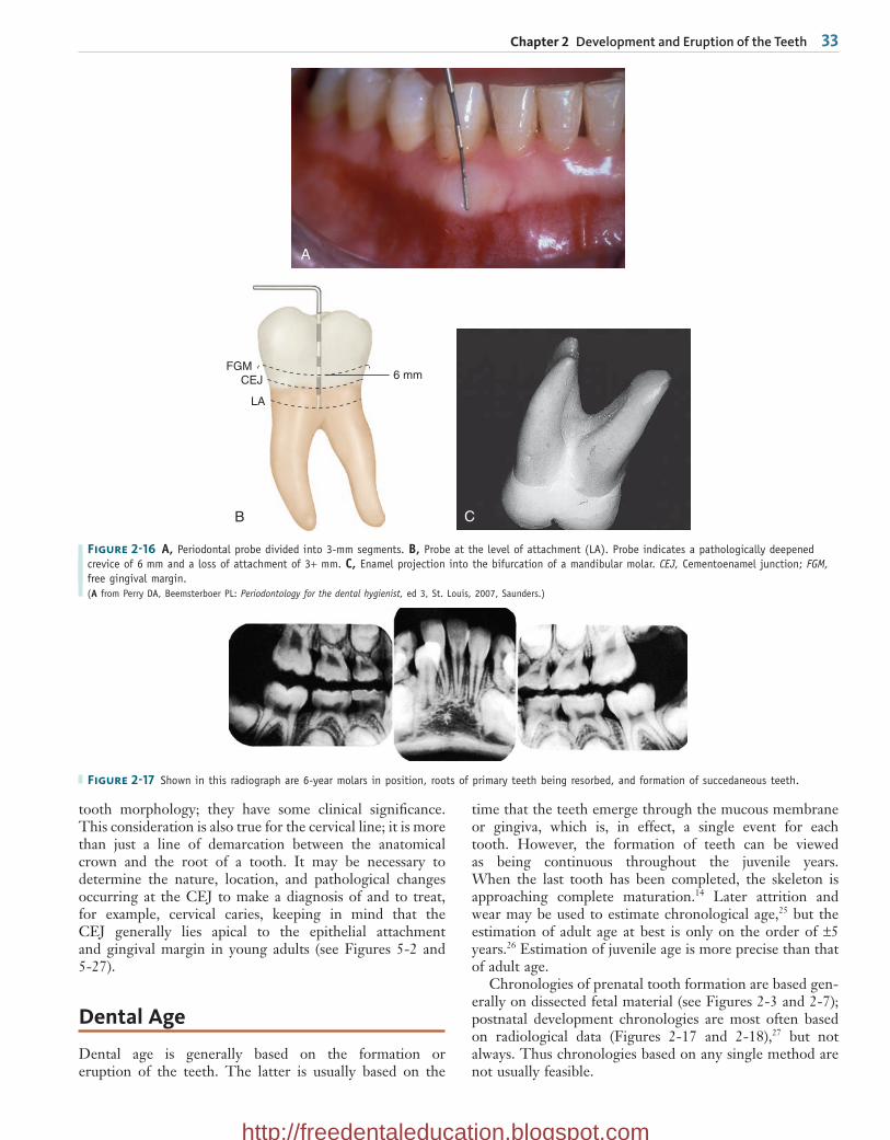
Chapter 2 Development and Eruption of the Teeth 33
tooth morphology; they have some clinical significance. This consideration is also true for the cervical line; it is more than just a line of demarcation between the anatomical crown and the root of a tooth. It may be necessary to determine the nature, location, and pathological changes occurring at the CEJ to make a diagnosis of and to treat, for example, cervical caries, keeping in mind that the CEJ generally lies apical to the epithelial attachment and gingival margin in young adults (see Figures 5-2 and 5-27).
Dental Age
Dental age is generally based on the formation or eruption of the teeth. The latter is usually based on the
time that the teeth emerge through the mucous membrane or gingiva, which is, in effect, a single event for each tooth. However, the formation of teeth can be viewed as being continuous throughout the juvenile years. When the last tooth has been completed, the skeleton is approaching complete maturation.14 Later attrition and wear may be used to estimate chronological age,25 but the estimation of adult age at best is only on the order of ±5 years.26 Estimation of juvenile age is more precise than that of adult age.
Chronologies of prenatal tooth formation are based gen-erally on dissected fetal material (see Figures 2-3 and 2-7); postnatal development chronologies are most often based on radiological data (Figures 2-17 and 2-18),27 but not always. Thus chronologies based on any single method are not usually feasible.
Figure 2-17 Shown in this radiograph are 6-year molars in position, roots of primary teeth being resorbed, and formation of succedaneous teeth.
A
6 mm
B C
LA
CEJFGM
Figure 2-16 A, Periodontal probe divided into 3-mm segments. B, Probe at the level of attachment (LA). Probe indicates a pathologically deepened crevice of 6 mm and a loss of attachment of 3+ mm. C, Enamel projection into the bifurcation of a mandibular molar. CEJ, Cementoenamel junction; FGM, free gingival margin.(A from Perry DA, Beemsterboer PL: Periodontology for the dental hygienist, ed 3, St. Louis, 2007, Saunders.)
http://freedentaleducation.blogspot.com
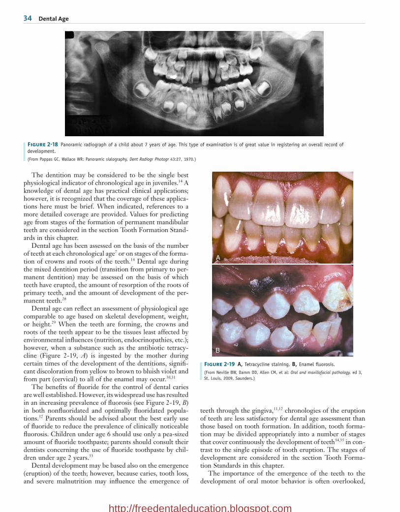
The dentition may be considered to be the single best physiological indicator of chronological age in juveniles.14 A knowledge of dental age has practical clinical applications; however, it is recognized that the coverage of these applica-tions here must be brief. When indicated, references to a more detailed coverage are provided. Values for predicting age from stages of the formation of permanent mandibular teeth are considered in the section Tooth Formation Stand-ards in this chapter.
Dental age has been assessed on the basis of the number of teeth at each chronological age7 or on stages of the forma-tion of crowns and roots of the teeth.14 Dental age during the mixed dentition period (transition from primary to per-manent dentition) may be assessed on the basis of which teeth have erupted, the amount of resorption of the roots of primary teeth, and the amount of development of the per-manent teeth.28
Dental age can reflect an assessment of physiological age comparable to age based on skeletal development, weight, or height.29 When the teeth are forming, the crowns and roots of the teeth appear to be the tissues least affected by environmental influences (nutrition, endocrinopathies, etc.); however, when a substance such as the antibiotic tetracy-cline (Figure 2-19, A) is ingested by the mother during certain times of the development of the dentitions, signifi-cant discoloration from yellow to brown to bluish violet and from part (cervical) to all of the enamel may occur.30,31
The benefits of fluoride for the control of dental caries are well established. However, its widespread use has resulted in an increasing prevalence of fluorosis (see Figure 2-19, B) in both nonfluoridated and optimally fluoridated popula-tions.32 Parents should be advised about the best early use of fluoride to reduce the prevalence of clinically noticeable fluorosis. Children under age 6 should use only a pea-sized amount of fluoride toothpaste; parents should consult their dentists concerning the use of fluoride toothpaste by chil-dren under age 2 years.33
Dental development may be based also on the emergence (eruption) of the teeth; however, because caries, tooth loss, and severe malnutrition may influence the emergence of
teeth through the gingiva,11,12 chronologies of the eruption of teeth are less satisfactory for dental age assessment than those based on tooth formation. In addition, tooth forma-tion may be divided appropriately into a number of stages that cover continuously the development of teeth34,35 in con-trast to the single episode of tooth eruption. The stages of development are considered in the section Tooth Forma-tion Standards in this chapter.
The importance of the emergence of the teeth to the development of oral motor behavior is often overlooked,
Figure 2-18 Panoramic radiograph of a child about 7 years of age. This type of examination is of great value in registering an overall record of development.
(From Pappas GC, Wallace WR: Panoramic sialography, Dent Radiogr Photogr 43:27, 1970.)
Figure 2-19 A, Tetracycline staining. B, Enamel fluorosis.
(From Neville BW, Damm DD, Allen CM, et al: Oral and maxillofacial pathology, ed 3, St. Louis, 2009, Saunders.)
A
B
34 Dental Age
http://freedentaleducation.blogspot.com

Chapter 2 Development and Eruption of the Teeth 35
undoubtedly partly as a result of the paucity of information available. However, the appearance of the teeth in the mouth at a strategic time in the maturation of the infant’s nervous system and its interface with the external environment must have a profound effect on the neurobehavioral mechanisms underlying the infant’s development and learning of feeding behavior, particularly the acquisition of masticatory skills.
Tooth Formation Standards
Events in the formation of human dentition are based primarily on data from studies of dissected prenatal ana-tomic material and from radiographic imaging of the teeth of the same subjects over time (longitudinal data) or of dif-ferent subjects of different ages seen once (cross-sectional data). From these kinds of studies, both descriptive information and chronological data may be obtained. To assemble a complete description or chronology of human tooth formation, it would seem necessary to use data based on more than one source and methodology. However, it is not easy to define ideal tooth formation standards from studies that examine different variables and use many different statistical methods. Subjects surveyed in most studies of dental development are essentially of European derivation, and population differences can only be estab-lished by studies that share methodology and information on tooth formation in populations of nonwhite/non- European ancestry.14
The age of emergence of teeth has been established for a number of population groups, but much less is known about chronologies of tooth formation.
Chronologies of Human Dentition
The history of chronological studies demonstrates the dif-ficulty in obtaining adequate documentation of the source of the data being presented. Many early tables and charts disagreed on the timing of chronological events. More precise information was needed to avoid injury to develop-ing teeth during surgery on young children, especially related to the repair of cleft palate. One of the earliest of the widely used tables was that of Kronfeld.36 Kronfeld’s table was partly reprinted and altered by Schour and Massler37 and has a long history of subsequent development and compilations.10,19 Table 2-3 is an expanded and revised version that reflects the accumulated history of chronologies of tooth formation of Table 2-4. The latter table (Table 2-4) is the most commonly reprinted version of Kronfeld’s chronology.10
Even Table 2-3 and related chronologies36–40 have some deficiencies in sampling and collection methods, sufficient to prevent acceptance of chronologies that are not consid-ered an ideal for standards of normal growth. The sugges-tions for revision made by Lunt and Law19 in their modification of the calcification and eruption schedules of the primary dentition (see Table 2-4) have been incorpo-
rated into the Logan and Kronfeld chronology shown in Table 2-3. The problems associated with revising a table completely or “plugging in” revised data is apparent when it becomes necessary to make critical choices from among available sources, as illustrated in Tables 2-5 and 2-6 by Smith.14
Types of Chronologies
Chronologies of dental development reflect the use of dif-ferent statistical methods to produce three different types of tooth formation data: age of attainment chronologies based on tooth emergence, age prediction chronologies based on being in a stage, and maturity assessment scales used to assess whether a subject of known age is in front of or behind compared with a reference population.
Stages of Tooth Formation
Radiographic studies of tooth formation have used at least three stages: beginning calcification, crown completion, and root completion. Nolla34 expanded the number of stages to 11 and Gleiser and Hunt to 13,44 which has served as the basis for several studies, including that of Moorrees et al,35 who defined 14 stages of permanent tooth formation (Figure 2-20). The 14 stages are not numbered but are designated by abbreviations (C = cusp; Cr = crown; R = root; Cl = cleft; A = apex) and subscripts (i = initiated; co = coalescence; oc = outline complete; and c = complete). Moorrees et al45 studied the development of mandibular canines and pro-vided normative data.
Age of Attainment
The age of attainment of a growth stage is not easily deter-mined, because in a proportion of the cases observed the attainment of the stage has not occurred, and in others the stage is over. Several procedures to answer the question of when a growth stage did occur have been used to construct chronologies of tooth formation, but many of these methods lead to chronologies that are not comparable for various reasons, including the problem of having fundamentally dif-ferent underlying variables. Thus, the major statistical methods used to construct different statistically based chro-nologies of tooth formation relate to fundamentally differ-ent variables and should be used for different purposes.14 Such tables attempt to answer when the event usually happens—that is, at what age does the transition into the stage occur?14
Age of attainment type of chronologies may be produced by cumulative distributive functions or probit analysis14 and by the average of age at first appearance less one half interval between examinations.46 Cumulative distributive functions, which have been used by a number of investigators (includ-ing Garn et al10,47 and Demirjian and Levesque48), appear to
http://freedentaleducation.blogspot.com

36 Age of Attainment
Table 2-3 Chronology of Human Dentition*
Dentition Tooth
First Evidence of Calcification Crown Completed Eruption Root Completed
(Weeks in Utero)c (Months) (Months)a,d (Years)
Primary (upper) i1 14 (13–16) 112 10 (8–12) 11
2
i2 16 ( 1423–161
2)b 212 11 (9–13) 2
c 17 (15–18)b 9 19 (16–22) 314
m1 1512 ( 141
2–17) 6 16 (13–19) (14–18) 212
m2 19 (16–2312) 11 29 (25–33) 3
Primary (lower) i1 14 (13–16) 212 8 (6–10) 11
2
i2 16 ( 1423– )b 3 13 (10–16) 11
2
c 17(16– )b 9 20 (17–23) 314
m1 1512 (141
2–17) 512 16 (14–18) 21
4
m2 18 (17–1912) 10 27 (23–31) (24–30) 3
Permanent (upper) I1 3–4 mo 4–5 yr 7–8 yr 10
I2 10–12 mo 4–5 yr 8–9 yr 11
C 4–5 mo 6–7 yr 11–12 yr 13–15
P1 112–13
4 yr 5–6 yr 10–11 yr 12–13
P2 2–214 yr 6–7 yr 10–12 yr 12–14
M1 At birth 212–3 yr 6–7 yr 9–10
M2 212–3 yr 7–8 yr 12–13 yr 14–16
M3 7–9 yr 12–16 yr 17–21 yr 18–25
Permanent (lower) I1 3–4 mo 4–5 yr 6–7 yr 9
I2 3–4 mo 4–5 yr 7–8 yr 10
C 4–5 mo 6–7 yr 9–10 yr 12–14
P1 134–2 yr 5–6 yr 10–12 yr 12–13
P2 214–21
2 yr 6–7 yr 11–12 yr 13–14
M1 At birth 212–3 yr 6–7 yr 9–10
M2 212–3 yr 7–8 yr 11–13 yr 14–15
M3 8–10 yr 12–16 yr 17–21 yr 18–25
i1, Central incisor; i2, lateral incisor; c, canine; m1, first molar; m2, second molar; I1, central incisor; I2, lateral incisor; C, canine; P1, first premolar; P2, second premolar; M1, first molar; M2, second molar; M3, third molar.*Part of the data from chronology of the growth of human teeth in Schour and Massler,37 modified from Kronfeld36 for permanent teeth and Kronfeld and Schour38 for the deciduous teeth. From Logan and Kronfeld,39 slightly modified by McCall and Schour (in Orban40) and reflecting other chronologies: a, Lysell et al13; b, Nomata41; c, Kraus and Jordan42; Lunt and Law19; d, mean age in months, ±1 standard deviation.
http://freedentaleducation.blogspot.com

Chapter 2 Development and Eruption of the Teeth 37
Table 2-4 Modified Table of Human Dentition*
Deciduous Tooth
Hard Tissue Formation Begins† (Fertilization Age in Utero, Weeks)
Amount of Enamel Formed at Birth
Enamel Completed (months after
Birth)
Eruption (Mean Age‡ in Months,
±1 SD)Root Completed
(Years)
MaxillaryCentral incisor 14 (13–16) • Five sixths 11
2 10 (8–12) 112
Lateral incisor 16 ( 1423 – 161
2 )§ • Two thirds 212 11 (9–13) 2
Canine 17 (15–18)§ • One third 9 19 (16–22) 314
First molar 1512 ( 141
2 –17) • Cusps united; occlusal completely calcified plus one half to three fourths crown height†
6 16 (13–19) boys (14–19) girls
212
Second molar 19 (16– 2312 ) • Cusps united; occlusal
incompletely calcified; calcified tissue covers one fifth to one fourth crown height†
11 29 (25–33) 3
MandibularCentral incisor 14 (13–16) • Three fifths 21
2 8 (6–10) 112
Lateral incisor 16 ( 1423 – )§ • Three fifths 3 13 (10–16) 11
2
Canine 17 (16– )§ • One third 9 20 (17–23) 314
First molar 1512 ( 141
2 –17) • Cusps united; occlusal completely calcified†
512 16 (14–18) 21
4
Second molar 18 (17– 1912 ) • Cusps united; occlusal
incompletely calcified†10 27 (23–31) boys
(24–30) girls3
SD, Standard deviation.*Modification of the table from Chronology of the Human Dentition (Logan and Kronfeld,39 slightly modified by McCall and Schour [in Orban40]), suggested by Lunt and Law,19 for the Calcification and Eruption of the Primary Dentition.†From Kraus and Jordan,42 pp. 107, 109, and 127 (except variation ranges of lateral incisors and canines).‡Modified from Lysell et al.13
§Variation ranges of lateral incisors and canines from Nomata.41 (Fetal length-to-age conversions were made; no values are available for late onset in mandibular lateral incisors and canines, since all values from Nomata’s data are earlier than the mean values from Kraus and Jordan.42) Fetal length-to-age data from Patten.43
be the best method of determining the age of attainment.49 An example of this type of chronology is a schedule of tooth emergence as illustrated in Figure 2-21, where the propor-tion of subjects that have attained a particular stage is plotted against the midpoint of each age group. A chronology of age of attainment of tooth formation for females is shown as an example of this type of chronology (Table 2-7). Age of attainment schedules are useful clinically when it is neces-sary to avoid damage to developing teeth during treatment.
Age Prediction
Chronologies of tooth formation based on the average age of subjects in a stage of development have been suggested by several investigators.44,50–52 Although these kinds of chro-nologies are better suited for age prediction than age of
attainment, no chronology is ideal for this purpose, and an alternate strategy suggested by Goldstein53 has been used by Smith14 to calculate age prediction tables for mandibular tooth formation. Values for predicting age in females are presented in Table 2-8. Such a table is useful to find out the age of the individual. In this schedule, each tooth is assessed independently, and the mean of all available ages is assigned as the dental age. In Table 2-8 the age related to a stage reflects the midpoint between mean age of attainment of the current stage and the next one. Age prediction chronologies are used for assessing unknown ages of patients and for forensic and archaeological applications.
Maturity Assessment
Chronologies of maturity assessment have been based on mean stage for age, where stages, rather than
http://freedentaleducation.blogspot.com

Figure 2-20 Stages of permanent tooth formation. See text for definition of abbreviations.
(From Moorrees CFA et al: Age variation of formation stages for ten permanent teeth, J Dent Res 42:1490, 1963.)
CROWN
ROOT
Ci Cco Coc Cr1/2 Cr3/4 Crc
Ri Cli
A1/2 Ac
R1/4 R1/2 R3/4 Rc
APEX
Table 2-6 Ages for Postnatal Development of Mandibular Deciduous Teeth Expressed in Decimal Years
Mandibular Tooth
Age Crown Completed (Years) Age Root Completed (Years)
Moorrees et al45* Kronfeld and Schour38†
Moorrees et al45* Kronfeld and Schour38†Mean −2 SD to +2 SD Mean −2 SD to +2 SD
di1 — — 0.1–0.2 — — 1.5
di2 — — 0.2 — — 1.5–2.0
dc — — 0.7 — — 3.25
Males 0.7 0.4–1.0 — 3.1 2.4–3.8 —
Females 0.7 0.4–1.0 — 3.0 2.3–3.8 —
dm1 — — 0.5 — — 2.25
Males 0.4 0.2–0.7 — 2.0 1.5–2.5 —
Females 0.3 0.1–0.5 — 1.8 1.3–2.3 —
dm2 — — 0.8–0.9 — — 3.0
Males 0.7 0.4–1.0 — 3.1 2.4–3.9 —
Females 0.7 0.4–1.0 — 2.8 2.2–3.6 —
SD, Standard deviation; di1, central incisor; di2, lateral incisor; dc, canine; dm1, first molar; dm2, second molar.*These data comprise an age-of-attainment schedule.†The basis of these values may be some combination of “tooth ring analysis” and observation of an infant sample; no other values could be located for deciduous incisors in studies with documented methods.14
38 Maturity Assessment
Table 2-5 Available Values for Prenatal Formation of Primary Teeth
Tooth
Age of Attainment Schedule
Beginning Calcification
(Weeks Postfertilization) Stage for Age Schedule
Sunderland et al58Amount of Crown Formed
at Birth
50th Percentile Range*
Kronfeld and Schour38†
Kraus and Jordan42
di1 15 13–17 35
—
di2 17 14–19 35
—
dc 19 17–20 13
—
dm1 16 14–17 Cusps united Occlusal united
dm2 19 18–20 Cusp tips isolated Cusps united
di1, Deciduous central incisor; di2, lateral incisor; dc, canine; dm1, first molar; dm2, second molar.*Earliest age at which mineralization is seen through age at which 100% of the sample shows initial mineralization.†These values are based on “tooth ring analysis”; they remain almost the only nonpictorial data available for deciduous incisors.14
http://freedentaleducation.blogspot.com

Chapter 2 Development and Eruption of the Teeth 39
Figure 2-21 Age of attainment of growth stage using a cumulative distribution function in which data represent tooth emergence. M1, First molar; I1, central incisor; I2, lateral incisor; C, canine; P1, first premolar; P2, second premolar; M2, second molar.
(Modified from Smith HB, Garn SM: Polymorphisms in eruption sequence of permanent teeth in American children, Am J Phys Anthropol 74:289, 1987.)
100
8085th percentile
M1 I1 I2 P1 M2
P2
C
15 percentile
Median
60
40
20
0
5 10
Years
Toot
h er
uptio
n (%
)
15
Table 2-7 Mean Age of Attainment of Developmental Stages for Females (Permanent Mandibular Teeth)*Developmental Stage I1 I2 C P1 P2 M1 M2 M3
Ci — — 0.5 1.8 3.0 0.0 3.5 9.6
Cco — — 0.8 2.2 3.6 0.3 3.7 10.1
Coc — — 1.2 2.9 4.2 0.8 4.2 10.7
Cr12
— — 2.0 3.6 4.8 1.0 4.8 11.3
Cr34
— — 3.0 4.3 5.4 1.5 5.4 11.7
Crc — — 4.0 5.1 6.2 2.2 6.2 12.3
Ri — — 4.7 5.8 6.8 2.7 7.0 12.9
Cli — — — — — 3.5 7.7 13.5
R14
4.5 4.7 5.3 6.5 7.5 4.5 9.2 14.8
R12
5.1 5.2 7.1 8.2 8.8 5.1 9.8 15.7
R23
5.6 5.9 — — — — — —
R34
6.1 6.4 8.3 9.2 10.0 5.7 10.7 16.6
Rc 6.6 7.6 8.9 9.9 10.6 6.0 11.2 17.2
A12
7.4 8.1 9.9 11.1 12.0 7.0 12.5 18.3
Ac 7.7 8.5 11.3 12.2 13.7 8.7 14.6 20.7
I1, Central incisor; I2, lateral incisor; C, canine; P1, first premolar; P2, second premolar; M1, first molar; M2, second molar; M3, third molar; Ci, Cusp initiated; Cco, cusp coalesced; Coc, cusp outline completed; Cr1
2, crown half completed; Cr3
4, crown three quarters completed; Crc, crown completed; Ri, root initiated; Cli, initial cleft formation;
R14, root one fourth completed; R1
2, root half completed; R2
3, root two thirds completed; R3
4, root three fourths completed; Rc, root completed; A1
2, apex half
completed; Ac, apex completed.*Values interpolated from Moorrees et al35; all ages in years.14
http://freedentaleducation.blogspot.com

Table 2-8 Values for Predicting Age from Stages of Permanent Mandibular Tooth Formation—Females*Developmental Stage I1 I2 C P1 P2 M1 M2 M3
Ci — — 0.6 2.0 3.3 0.2 3.6 9.9
Cco — — 1.0 2.5 3.9 0.5 4.0 10.4
Coc — — 1.6 3.2 4.5 0.9 4.5 11.0
Cr12
— — 2.5 4.0 5.1 1.3 5.1 11.5
Cr34
— — 3.5 4.7 5.8 1.8 5.8 12.0
Crc — — 4.3 5.4 6.5 2.4 6.6 12.6
Ri — — 5.0 6.1 7.2 3.1 7.3 13.2
Cli — — — — — 4.0 8.4 14.1
R14
4.8 5.0 6.2 7.4 8.2 4.8 9.5 15.2
R12
5.4 5.6 7.7 8.7 9.4 5.4 10.3 16.2
R23
5.9 6.2 — — — — — —
R34
6.4 7.0 8.6 9.6 10.3 5.8 11.0 16.9
Rc 7.0 7.9 9.4 10.5 11.3 6.5 11.8 17.7
A12
7.5 8.3 10.6 11.6 12.8 7.9 13.5 19.5
Ac — — — — — — — —
See Table 2-7 for definitions of tooth designations and developmental stages.*Values interpolated from Moorrees et al35; all ages in years.14
Table 2-9 Primary Crown and Root Formation: Onset and Duration* Mandibular Deciduous Dentition
Crown Formation Start to Finish Root Length Growth Start to FinishTooth Emergence Root Apex Closure
di1†: 6 mo before birth to 2–3 mo after birth
Start: 3 mo to ? Data sparse or unavailable
8 mo Data sparse or not yet available ( 112 yr?)
di2: 5.6 mo before birth to 3–4 mo after birth
Start: 4 mo to ? Data sparse or unavailable
13 mo Data sparse or not yet available ( 112 yr)
dm1: 5.2 mo before birth to 6–7 mo after birth
Start: 7 moFinish: 10 mo
16 mo 16 to 23 mo
c: 4.2 mo before birth to 812 mo
after birthStart: 81
2 moFinish: 24 mo
20 mo 23 to 36 mo
dm2: 4 mo before birth to 1012 mo
after birthStart: 101
2 moFinish: 251
2 mo
27 mo 2512 to 35 mo
*Data partly derived from Smith.14 Smith’s sources included Moorrees et al,35,45 Sunderland et al,58 Anderson et al,46 Kronfeld,36 Lysell et al,13 and Hume.59
†di1, di2, c, dm1, and dm2 are general deciduous tooth designations used by some anthropologists to indicate the central and lateral incisors, canine, first molar, and second molar, respectively.
subject ages, are averaged.34,54 However, to avoid problems associated with the calculation of mean age or mean stage, maturity scales have been designed by several investigators, including Wolanski,55 Demirjian et al,29 Healy and Goldstein,56 and Nystrom et al.57 Such scales are useful when maturity is assessed for subjects of known age but are not designed for anthropological or forensic applications.14
Duration of Root and Crown Formation
The onset and duration of crown and root formation of the primary dentition are illustrated in Table 2-9, which answers questions about the relationship between onset and comple-tion of tooth formation from start to finish.
40 Duration of Root and Crown Formation
http://freedentaleducation.blogspot.com

Chapter 2 Development and Eruption of the Teeth 41
Summary of Chronologies
Compared with older, descriptive chronologies based on dissection and those based on radiological plus statistical methods to produce developmental data, newer methods tend to avoid attributing discrepancies to population differ-ences because of methodological or sampling effects. The data in Tables 2-7 and 2-8 have been recommended for deciduous tooth development.
Cumulative distribution functions and probit analysis are recommended for generating statistical solutions for sched-ules of age of attainment of growth stages.14,35
Clinicians can use chronologies to avoid treatment that can damage developing teeth (attainment schedules), to assess an unknown age of a patient (e.g., age prediction in forensics, demographics), and to assess growth (maturity).14
Sequence of Eruption
The sequence of eruption of the primary teeth does show some variation. Such timing is a result in large part of hered-ity and only somewhat of environmental factors. Jaw revers-als in eruption of canines and first molars have been found to be important in increasing the variety of sequences.13,60 When differences according to jaws are considered, Lunt and Law19 conclude that the lateral incisor, first molar, and canine tend to erupt earlier in the maxilla than in the man-dible. Sato and Ogiwara60 found the following characteristic order in about one third of their sample of children:
AB D C E
A B D CE
However, this arrangement of mean ages of eruption to yield a mean order of eruption was found to occur only in a small percentage of the subjects in the study by Lysell et al.13 The sequence and age of eruption of primary teeth are illustrated in Table 2-10.
Estimating Time of Enamel Hypoplasia
To estimate the time of enamel hypoplasia, measure in mil-limeters the distance from the CEJ to the midpoint of the enamel defect. As a comparison, note in Table 6-1 that the cervicoincisal length of the crown of the permanent maxil-lary central incisor is 10.5 mm. In Table 2-3 and Table 6-1, the first evidence of calcification is 3 to 4 months. Assuming that rate of development is constant and that a maxillary central incisor develops over a period of 4 to 5 years, the age of development of the defect is related inversely to the distance from the CEJ to the enamel defect and can be computed as follows according to a first-degree polynomial for estimating the chronological age at which the enamel defect occurred5,61:
ADF ACF years of formation crown height
distance of defect= − (
× from CEJ)
Here, ADF = age at which the formation of the enamel hypoplasia occurred; and ACF = age at which crown is completed. For example, assume that the cervical-incisal length of the crown of the permanent maxillary central incisor is 10.5 mm, the crown is completed at 4 to 5 years, and the midpoint of the defect is 6.6 mm from the CEJ. Plugging in the data for both 4 years and 5 years and obtaining an average, the age of formation of the defect is estimated to be at about 2 years, keeping in mind that for estimating time here, dividing the development of enamel into intervals of one or even a few months is an accuracy that is not justified, and 6-month or yearly periods are more realistic. Computed estimated ages of hypoplasia vary somewhat, depending on the chronologi-cal tables used.
References 1. Moorrees CFA, Kent RL: A step function model using tooth
counts to assess the developmental timing of the dentition, Am Hum Biol 5:55, 1978.
2. Massler M et al: Developmental pattern of the child as reflected in the calcification pattern of the teeth, Am J Dis Child 63:33, 1941.
3. Sarnat BG, Schour I: Enamel hypoplasias (chronic enamel aplasia) in relationship to systemic diseases: a chronological, morphological and etiological classification, J Am Dent Assoc 28:1989, 1941.
4. Goodman AH, Armelagos GJ, Rose JC: Enamel hypoplasias as indicators of stress in three prehistoric populations from Illinois, Hum Biol 52:515, 1980.
5. Goodman AH, Rose JC: Dental enamel hypoplasias as indicators of nutritional status. In Kelly MA, Larsen CS, editors: Advances in dental anthropology, New York, 1991, Wiley-Liss.
6. Kraus B, Jordan R, Pruzansky S: Dental abnormalities in the deciduous and permanent dentitions of individuals with cleft lip and palate, J Dent Res 45:1736, 1966.
7. Demirjian A: Dentition. In Falkner F, Tanner JM, editors: Human growth: a comprehensive treatise, vol 2, ed 2, New York, 1986, Plenum.
Table 2-10 Sequence and Age at Eruption of Primary Teeth
Teeth AgeMean Number of
Teeth PresentDistribution in
Number of Teeth
A– 6 mo — 1–3, 33%
A 9 mo 3 1–6, 80%
B 12 mo 6 4–8, 50%
DB–, D
18 mo 12 9–16, 85%
CC
24 mo 16 15–18, 60%
E–, E 30 mo 19 20, 70%
A, Central incisor; B, lateral incisor; D, first molar; C, canine; E, second molar; underscore indicates maxillary teeth; overscore indicates mandibular teeth.
http://freedentaleducation.blogspot.com

8. Bosma JF: Maturation and function of the oral and pharyngeal region, Am J Orthod 49:94, 1963.
9. Schour L, Massler M: The development of the human dentition, J Am Dent Assoc 28:1153, 1941.
10. Garn SM et al: Variability of tooth formation, J Dent Res 38:135, 1959.
11. Alvarez J, Navia JM: Nutritional status, tooth eruption and dental caries: a review, Am J Clin Nutr 49:417, 1989.
12. Rönnerman A: The effect of early loss of primary molars on tooth eruption and space conditions: a longitudinal study, Acta Odontol Scand 35:229, 1977.
13. Lysell L et al: Time and order of eruption of the primary teeth: a longitudinal study, Odontol Revy 13:21, 1962.
14. Smith BH: Standards of human tooth formation and dental age assessment. In Kelley MA, Larsen CS, editors: Advances in dental anthropology, New York, 1991, Wiley-Liss.
15. Garn SM et al: Genetic, nutritional, and maturational correlates of dental development, J Dent Res 44:228, 1965.
16. Falkner F: Deciduous tooth eruption, Arch Dis Child 32:386, 1957.
17. Schour I, Noyes HJ: Oral histology and embryology, ed 8, Philadelphia, 1960, Lea & Febiger.
18. Moyers RE: Handbook of orthodontics, ed 3, Chicago, 1973, Year Book.
19. Lunt RC, Law DB: A review of the chronology of deciduous teeth, J Am Dent Assoc 89:87, 1974.
20. van der Linden FPGM, Duterloo HS: Development of the human dentition: an atlas, Hagerstown, MD, 1976, Harper & Row.
21. Adler P: Studies on the eruption of the permanent teeth. IV. The effect upon the eruption of the permanent teeth of caries in the deciduous dentition, and of urbanization, Acta Genet Stat Med 8:78, 1958.
22. Garn SM, Lewis AB: Tooth-size, body-size and “giant” fossil man, Am J Anthropol 61:874, 1958.
23. Garn SM, Lewis AB, Kerewsky RS: Relationship between sexual dimorphism in tooth size as studied within families, Arch Oral Biol 12:299, 1966.
24. Masters DH, Hoskins SW: Projection of cervical enamel into molar furcations, J Periodont 35:49, 1964.
25. Kay RF, Cant JGH: Age assessment using cementum annulus counts and tooth wear in a free-ranging population of Macaca mulata, Am J Primatol 15:1, 1988.
26. Hojo M: On the pattern of the dental abrasion, Sonderabdruck aus Okajimas 26:11, 1954.
27. Pappas GC, Wallace WR: Panoramic sialography, Dent Radiogr Photogr 43:27, 1970.
28. Proffit WR, Fields HW: Contemporary orthodontics, ed 3, St Louis, 2000, Mosby.
29. Demirjian A et al: A new system of dental age assessment, Hum Biol 45:211, 1970.
30. Stewart DJ: The effects of tetracycline upon the dentition, Br J Dermatol 76:374, 1964.
31. Ash MM: Oral pathology, Philadelphia, 1992, Lea & Febiger.
32. Ismail AI, Bandekar RR: Fluoride supplements and fluorosis: a meta-analysis, Community Dent Oral Epidemiol 27:48, 1999.
33. Centers for Disease Control and Prevention: Recommendations for using fluoride to prevent and control dental caries in the United States, MMWR Recomm Rep 50(RR-14):1, 2001.
34. Nolla CM: The development of permanent teeth, doctoral thesis, Ann Arbor, 1952, University of Michigan.
35. Moorrees CFA et al: Age variation of formation stages for ten permanent teeth, J Dent Res 42:1490, 1963.
36. Kronfeld R: Development and calcification of the human deciduous and permanent dentition, Bur 15:11, 1935.
37. Schour I, Massler M: Studies in tooth development: the growth pattern of human teeth, part II, J Am Dent Assoc 27:1918, 1940.
38. Kronfeld R, Schour I: Neonatal dental hypoplasia, J Am Dent Assoc 26:18, 1939.
39. Logan WHG, Kronfeld R: Development of the human jaws and surrounding structures from birth to age fifteen, J Am Dent Assoc 20:379, 1933.
40. Orban B: Oral histology and embryology, ed 2, St Louis, 1944, Mosby.
41. Nomata N: A chronological study on the crown formation of the human deciduous dentition, Bull Tokyo Med Dent Univ 11(Mach):55, 1964.
42. Kraus BS, Jordan RE: The human dentition before birth, Philadelphia, 1965, Lea & Febiger.
43. Patten BM: Human embryology, Philadelphia, 1946, Blakison’s.
44. Gleiser I, Hunt EE: The permanent mandibular first molar: its calcification, eruption, and decay, Am J Phys Anthropol 13:253, 1955.
45. Moorrees CFA: Formation and resorption of three deciduous teeth in children, Am J Phys Anthropol 21:205, 1963.
46. Anderson DL et al: Age of attainment of mineralization stages of the permanent dentition, J Forensic Sci 21:191, 1976.
47. Garn SM et al: Variability of tooth formation in man, Science 128:1510, 1958.
48. Demirjian A, Levesque GY: Sexual differences in dental development and prediction of emergence, J Dent Res 59:1110, 1980.
49. Tanner JM: Use and abuse of growth standards. In Falkner F, Tanner JM, editors: Human growth: a comprehensive treatise, vol 3, ed 2, New York, 1986, Plenum.
50. Demisch A, Wartman P: Calcification of the mandibular third molar and its relation to skeletal and chronological age in children, Child Dev 27:459, 1956.
51. Haataja J: Development of the mandibular permanent teeth of Helsinki children, Proc Finn Dent Soc 61:43, 1965.
52. Fass EN: A chronology of growth of the human dentition, J Dent Child 36:391, 1969.
53. Goldstein H: The design and analysis of longitudinal studies, London, 1979, Academic Press.
54. Nolla CM: The development of permanent teeth, J Dent Child 27:254, 1960.
55. Wolanski N: A new method for the evaluation of tooth formation, Acta Genet Stat Med 16:186, 1966.
56. Healy MJR, Goldstein H: An approach to scaling of categorized attributes, Biometrika 63:219, 1976.
57. Nystrom M et al: Dental maturity in Finnish children, estimated from the development of seven permanent mandibular teeth, Acta Odontol Scand 44:193, 1986.
58. Sunderland EP et al: A histological study of the chronology of initial mineralization in the human deciduous dentition, Arch Oral Biol 32:167, 1987.
59. Hume VO: Ranges of normalcy in the eruption of permanent teeth, J Dent Child 16:11, 1949.
60. Sato S, Ogiwara Y: Biostatistic study of the eruption order of deciduous teeth, Bull Tokyo Dent Coll 12:45, 1971.
42 References
http://freedentaleducation.blogspot.com
Chapter 2 Development and Eruption of the Teeth 43
61. Murray KA, Murray SA: Computer software for hypoplasia analysis, Am J Phys Anthropol 78:277, 1989.
BibliographyHowe RP: A examination of dental crowding and its relationship
to tooth size and arch dimension, Am J Orthod 83:363, 1983.Johanson G: Age determinations from human teeth, Odontol Revy
22(suppl):1, 1971.Kraus BS: Calcification of the human deciduous teeth, J Am
Dent Assoc 59:1128, 1959.McCall JO, Wald SS: Clinical dental roentgenology, ed 2,
Philadelphia, 1947, Saunders.
Moorrees CFA, Kent RL: Interrelations in the timing of root formation and tooth emergence, Proc Finn Dent Soc 77:113, 1981.
Smith BH, Garn SM: Polymorphisms in eruption sequence of permanent teeth in American children, Am J Phys Anthropol 74:289, 1987.
Use the CD-ROM to view animations and study ques-tions; and go to the site for additional study resources.
http://freedentaleducation.blogspot.com

This page intentionally left blank
http://freedentaleducation.blogspot.com
The Primary (Deciduous) Teeth
are sometimes thought of erroneously as only an annoyance, this belief fails to acknowledge the role of the primary teeth in mastication and their function in maintaining the space for eruption of the permanent teeth.
A lack of space associated with premature loss of decidu-ous teeth is a significant factor in the development of maloc-clusion and is considered in Chapter 16. The development of adequate spacing (Figure 3-2) is an important factor in the development of normal occlusal relations in the perma-nent dentition. Thus there should be no question of the importance of preventing and treating dental decay and providing the child with a comfortable functional occlusion of the deciduous teeth. Therefore in this book the primary teeth are described in advance of the permanent dentition so that they may be given their proper sequence in the study of dental anatomy and physiology. The development of the primary occlusion is considered in Chapter 16.
Nomenclature
Some of the terminology for the primary dentition has already been introduced in Chapter 2; therefore the cover-age here is more in the nature of a review. The process of exfoliation of the primary teeth takes place between the seventh and the twelfth years. This does not, however, indi-cate the period at which the root resorption of the deciduous tooth begins. It is only 1 or 2 years after the root is com-pletely formed and the apical foramen is established that resorption begins at the apical extremity and continues in the direction of the crown until resorption of the entire root has taken place and the crown is lost from lack of support.
Life Cycle
After the roots of the primary dentition are completed at about age 3, several of the primary teeth are in use only for a relatively short period. Some of the primary teeth are found to be missing at age 4, and by age 6, as many as 19% may be missing.1 By age 10, only about 26% may be present. The second molars in both arches and the maxillary incisors appear to be the most unstable of the primary teeth. Even so, the developing and completed primary dentition serves a number of purposes during that time and the period of transition to the permanent dentition.
Importance of Primary Teeth
The general order of eruption of the primary dentition is illustrated diagrammatically in Figure 3-1: central incisor, lateral incisor, first molar, canine, and second molar, with the mandibular pairs preceding the maxillary teeth.2,3 The loss of the deciduous teeth tends to mirror the eruption sequence: incisors, first molars, canines, and second molars, with the mandibular pairs preceding the maxillary teeth.
The high peak for caries attack occurs at age 13, when only 5% of the primary teeth remain. The susceptibility to dental caries is a function of exposure time to the oral envi-ronment and morphological type. The increase in preva-lence of dental caries among tooth types is the reverse of their order of eruption. However, the relative susceptibility of different tooth surfaces is a complex problem. Although dental caries of the primary dentition and loss of these teeth
45
33
http://freedentaleducation.blogspot.com

Figure 3-1 Diagrammatic representation of the chronology of the primary teeth. Eruption is completed at the approximate time indicated by the dotted area on the roots of the teeth. iu, Intrauterine.
(Modified from McBeath EC: New concept of the development and calcification of the teeth, J Am Dent Assoc 23:675, 1936; and Noyes EB, Shour I, Noyes HJ: Dental histology and embryology, ed 5, Philadelphia, 1938, Lea & Febiger.)
4 mo
4 mm
8 mo12 mo16 mo20 mo24 mo28 mo32 mo
3 years
8 mm12 mm16 mm20 mm2 years26 mm28 mm3 years
5 mo iu
5 mo iu
Birth
Birth
1 mo iu
1 mo iu
Pos
tnat
alP
rena
tal
Pre
nata
lP
ostn
atal
Figure 3-2 Primary dentition in a child 5 years of age.
The primary teeth number 20 total—10 in each jaw—and they are classified as follows: four incisors, two canines, and four molars in each jaw. Figure 3-3 shows the primary denti-tion as numbered with the universal system of notation described in Chapter 1. Beginning with the median line, the teeth are named in each jaw on each side of the mouth as follows: central incisor, lateral incisor, canine, first molar, and second molar.
The primary teeth have been called temporary, milk, or baby teeth. These terms are improper because they foster the implication that these teeth are useful for a short period
only. It is emphasized again that they are needed for many years of growth and physical development. Premature loss of primary teeth because of dental caries is preventable and is to be avoided.
The first permanent molar, commonly called the 6-year molar, makes its appearance in the mouth before any of the primary teeth are lost. It comes in immediately distal to the primary second molar (see Figure 2-10).
The primary dentition is complete at about 2.5 years of age, and no obvious intraoral changes in the dentition occur (Figures 3-4 and 3-5) until the eruption of the first perma-nent molar. The position of the incisors is usually relatively upright with spacing often between them. Attrition occurs, and a pattern of wear may be present.
The primary molars are replaced by permanent premolars. No premolars are present in the primary set, and no teeth in the deciduous set resemble the permanent premolar. However, the crowns of the primary maxillary first molars resemble the crowns of the permanent premolars as much as they do any of the permanent molars. Nevertheless, they have three well-defined roots, as do maxillary first perma-nent molars. The deciduous mandibular first molar is unique in that it has a crown form unlike that of any permanent tooth (see Figure 3-24, C). It does, however, have two strong roots, one mesial and one distal—an arrangement similar to that of a mandibular permanent molar. These two primary teeth, the maxillary and mandibular first molars, differ from any teeth in the permanent set when crown forms are com-pared, in particular (see Figures 3-21 and 3-24). The primary
46 Nomenclature
http://freedentaleducation.blogspot.com

Chapter 3 The Primary (Deciduous) Teeth 47
Figure 3-3 Universal numbering system for primary dentition.
(From Ash MM, Ramfjord S: Occlusion, ed 4, Philadelphia, 1995, Saunders.)
A
T
S
R
QP O
N
M
L
K
B
C
D
E F
G
H
I
J
first molars, maxillary and mandibular, are described in detail later in this chapter.
Major Contrasts between Primary and Permanent Teeth
In comparison with their counterparts in the permanent dentition, the primary teeth are smaller in overall size and crown dimensions. They have markedly more prominent cervical ridges, are narrower at their “necks,” are lighter in color, and have roots that are more widely flared; in addi-tion, the buccolingual diameter of primary molar teeth is less than that of permanent teeth.4 More specifically, in comparison with permanent teeth, the following differences are noted:
1. The crowns of primary anterior teeth are wider mesiodistally in comparison with their crown length than are the permanent teeth.
2. The roots of primary anterior teeth are narrower and longer comparatively. Narrow roots with wide crowns
present an arrangement at the cervical third of crown and root that differs markedly from that of the permanent anterior teeth.
3. The roots of the primary molars accordingly are longer and more slender and flare more, extending out beyond projected outlines of the crowns. This flare allows more room between the roots for the development of permanent tooth crowns (see Figures 3-21 and 3-22).
4. The cervical ridges of enamel of the anterior teeth are more prominent. These bulges must be considered seriously when they are involved in any operative procedure (see Figure 3-13).
5. The crowns and roots of primary molars at their cervical portions are more slender mesiodistally.
6. The cervical ridges buccally on the primary molars are much more pronounced, especially on the maxillary and mandibular first molars (see Figures 3-25 through 3-28).
7. The buccal and lingual surfaces of primary molars are flatter above the cervical curvatures than those of permanent molars, which narrows the occlusal surfaces.
8. The primary teeth are usually less pigmented and are whiter in appearance than the permanent teeth.
http://freedentaleducation.blogspot.com

Figure 3-4 A, This is a specimen of a 5- to 6-year-old child prepared to show a full complement of primary teeth, the beginning of resorption of deciduous roots in some, and no apparent resorption in others. This front view shows the relative positions of the developing crowns of the permanent anterior teeth. The maxillary central and lateral incisors and the canine are shown overlapped in a narrow space, waiting for future development of the maxilla that will allow them to develop roots and improve the alignment. B, Right side. Unless they were lost in preparation, no development shows of mandibular permanent premolars. The tiny cusps formed at this age would be lost easily in preparation. C, Left side. Note the crowns of permanent maxillary premolars located between the roots of the first and second primary molars, with their roots still intact. Note the well-developed first permanent maxillary molar entirely erupted with half its roots formed. Ordinarily, the mandibular first permanent molar comes in and takes its place first, the maxillary molar following. The specimen shows the mandibular molar still covered with bone and no roots in evidence. The maxillary second molar crown is well developed and located in a place that is about level with the present root development of the permanent maxillary first molar.
A
B C
Pulp Chambers and Pulp Canals
A comparison of sections of primary and permanent teeth demonstrates the shape and relative size of pulp chambers and canals (Figure 3-6), which is noted here:
1. Crown widths in all directions are large in comparison with root trunks and cervices.
2. The enamel is relatively thin and has a consistent depth.3. The dentin thickness between the pulp chambers and
the enamel is limited, particularly in some areas (lower second primary molar).
4. The pulp horns are high, and the pulp chambers are large (Figure 3-7, A and B).
5. Primary roots are narrow and long when compared with crown width and length.
48 Pulp Chambers and Pulp Canals
http://freedentaleducation.blogspot.com

Chapter 3 The Primary (Deciduous) Teeth 49
Figure 3-5 A sectional close-up of the specimen in Figure 3-4. A, The left side of the maxilla. The developing crowns of the central and lateral incisors, the canine, and two premolars are clearly in view. B, The left side of the maxilla, posteriorly. The molar relationship, both deciduous and permanent, is accented here. C, This is a good view of the mandible anteriorly and to the left. Permanent central and lateral incisors and the canine may be seen. Notice that the permanent canine develops distally to the primary canine root. D, Posteriorly, examination of the specimen mandible fails to find crown development of permanent premolars. However, the hollow spaces showing between the roots of primary molars may indicate a loss of material during the difficult process of dissection. The first permanent molar has progressed in crown formation, but the maturation of the whole tooth with alignment is far behind its opposition in the maxilla above it (see Figure 3-4, C).
A B
DC
Figure 3-6 Comparison of maxillary, primary, and permanent second molars, linguobuccal cross section. A, The enamel cap of primary molars is thinner and has a more consistent depth. B, A comparatively greater thickness of dentin is over the pulpal wall at the occlusal fossa of primary molars. C, The pulpal horns are higher in primary molars, especially the mesial horns, and pulp chambers are proportionately larger. D, The cervical ridges are more pronounced, especially on the buccal aspect of the first primary molars. E, The enamel rods at the cervix slope occlusally instead of gingivally as in the permanent teeth. F, The primary molars have a markedly constricted neck compared with the permanent molars. G, The roots of the primary teeth are longer and more slender in comparison with crown size than those of the permanent teeth. H, The roots of the primary molars flare out nearer the cervix than do those of the permanent teeth.
(From Finn SB: Clinical pedodontics, ed 2, Philadelphia, 1957, Saunders.)
C
D
E
F
G
H
B
A
http://freedentaleducation.blogspot.com

Figure 3-7 A and B, Pulp chambers in the primary molars. Note the contours of the pulp horns within them.
(Modified from Finn SB: Clinical pedodontics, ed 2, Philadelphia, 1957, Saunders.)
Lingualview
Occlusalview
A
B
Lingualview
Occlusalview
UPPER SECONDPRIMARY MOLAR
LOWER FIRSTPRIMARY MOLAR
LOWER SECONDPRIMARY MOLAR
UPPER FIRSTPRIMARY MOLAR
6. Molar roots of primary teeth flare markedly and thin out rapidly as the apices are approached.
Studying the comparisons between the deciduous and the permanent dentitions (Figures 3-8 and 3-9) is of utmost importance. Discussion of further variations between the macroscopic form of the deciduous and the permanent teeth follows, with a detailed description of each deciduous tooth.
Detailed Description of Each Primary Tooth
MAXILLARY CENTRAL INCISOR
Labial AspectIn the crown of the primary central incisor, the mesiodistal diameter is greater than the cervicoincisal length (Figures 3-10 and 3-11, A). (The opposite is true of permanent
50 Detailed Description of Each Primary Tooth
http://freedentaleducation.blogspot.com

Chapter 3 The Primary (Deciduous) Teeth 51
Figure 3-8 Permanent central incisor. A, Pulp canal; B, pulp horns. This figure represents a sectioned central incisor of a young person. Although the pulp canal is rather large, it is smaller than the pulp canal shown in Figure 3-9, and it becomes more constricted apically. Note the dentin space between the pulp horns and the incisal edge of the crown.
B
A
Figure 3-9 Primary central incisor. A, Pulp canal; B, pulp horns. This figure represents a sectioned primary central incisor. The pulp chamber with its horns and the pulp canal are broader than those found in Figure 3-8. The apical portion of the canal is much less constricted than that of the permanent tooth. Note the narrow dentin space incisally.
B
A
Figure 3-10 Primary maxillary central incisors (first incisors). 1, Labial aspect. Note the lack of character in the mold form; also note the mesiodistal width compared with the shorter crown length. A little of the crown length was lost through abrasion before the date of extraction. 2, Mesial aspect. The cervical ridges are quite prominent labially and lingually, with the bulge much greater than that found on permanent incisors. This characteristic is common to each primary tooth to a varied degree. Normally, these curvatures are covered by gingival tissue with epithelial attachment (see Chapter 5). 3, Incisal aspect.
1
2
3
central incisors.) The labial surface is very smooth, and the incisal edge is nearly straight. Developmental lines are usually not seen. The root is cone-shaped with even, tapered sides. The root length is greater in comparison with the crown length than that of the permanent central incisor. It is advisable when studying both the primary and permanent teeth to make direct comparisons between the table of meas-urements of the primary teeth (Table 3-1) and that of per-manent teeth (see Table 1-1).
Lingual AspectThe lingual aspect of the crown shows well-developed mar-ginal ridges and a highly developed cingulum (Figure 3-12, A). The cingulum extends up toward the incisal ridge far enough to make a partial division of the concavity on the
lingual surface below the incisal edge, practically dividing it into a mesial and distal fossa.
The root narrows lingually and presents a ridge for its full length in comparison with a flatter surface labially. A cross section through the root where it joins the crown shows an outline that is somewhat triangular in shape, with the labial surface making one side of the triangle and mesial and distal surfaces making up the other two sides.
Mesial and Distal AspectsThe mesial and distal aspects of the primary maxillary central incisors are similar (Figure 3-13, A; see also Figure 3-10). The measurement of the crown at the cervical third shows the crown from this aspect to be wide in relation to its total length. The average measurement is only about 1 mm less
http://freedentaleducation.blogspot.com

C B A
F E D
Figure 3-11 Primary right anterior teeth, labial aspect. A, Maxillary central incisor. B, Maxillary lateral incisor. C, Maxillary canine. D, Mandibular central incisor. E, Mandibular lateral incisor. F, Mandibular canine.
Table 3-1 Table of Measurements of the Primary Teeth of Man (Averages Only) (in Millimeters)
Length Overall
Length of Crown
Length of Root
Mesiodistal Diameter of Crown
Mesiodistal Diameter of Crown at Cervix
Labiolingual Diameter of Crown
Labiolingual Diameter of Crown at Cervix
Upper TeethCentral incisor 16.0 6.0 10.0 6.5 4.5 5.0 4.0
Lateral incisor 15.8 5.6 11.4 5.1 3.7 4.0 3.7
Canine 19.0 6.5 13.5 7.0 5.1 7.0 5.5
First molar 15.2 5.1 10.0 7.3 5.2 8.5 6.9
Second molar 17.5 5.7 11.7 8.2 6.4 10.0 8.3
Lower TeethCentral incisor 14.0 5.0 9.0 4.2 3.0 4.0 3.5
Lateral incisor 15.0 5.2 10.0 4.1 3.0 4.0 3.5
Canine 17.5 6.0 11.5 5.0 3.7 4.8 4.0
First molar 15.8 6.0 9.8 7.7 6.5 7.0 5.3
Second molar 18.8 5.5 11.3 9.9 7.2 8.7 6.4
From Black GV: Descriptive anatomy of the human teeth, ed 4, Philadelphia, 1897, S.S. White Dental Company.
Figure 3-12 Primary right anterior teeth, lingual aspect. A, Maxillary central incisor. B, Maxillary lateral incisor. C, Maxillary canine. D, Mandibular central incisor. E, Mandibular lateral incisor. F, Mandibular canine.
CBA
FED
than the entire crown length cervicoincisally. Because of the short crown and its labiolingual measurement, the crown appears thick at the middle third and even down toward the incisal third. The curvature of the cervical line, which repre-sents the cementoenamel junction (CEJ), is distinct, curving
toward the incisal ridge. However, the curvature is not as great as that found on its permanent successor. The cervical curvature distally is less than the curvature mesially, a design that compares favorably with the permanent central incisor.
52 Detailed Description of Each Primary Tooth
http://freedentaleducation.blogspot.com

Chapter 3 The Primary (Deciduous) Teeth 53
Figure 3-13 Primary right anterior teeth, mesial aspect. A, Maxillary central incisor. B, Maxillary lateral incisor. C, Maxillary canine. D, Mandibular central incisor. E, Mandibular lateral incisor. F, Mandibular canine.
CBA
FED
Figure 3-14 Primary right anterior teeth, incisal aspect. A, Maxillary central incisor. B, Maxillary lateral incisor. C, Maxillary canine. D, Mandibular central incisor. E, Mandibular lateral incisor. F, Mandibular canine.
ABC
DEF
Although the root appears more blunt from this aspect than it did from the labial and lingual aspects, it is still of an even taper and the shape of a long cone. However, it is blunt at the apex. Usually the mesial surface of the root will have a developmental groove or concavity, whereas distally, the surface is generally convex.
Note the development of the cervical ridges of enamel at the cervical third of the crown labially and lingually.
Incisal AspectAn important feature to note from the incisal aspect is the measurement mesiodistally compared with the measure-ment labiolingually (Figure 3-14, A; see also Figure 3-10, 3). The incisal edge is centered over the main bulk of the crown and is relatively straight. Looking down on the incisal edge, the labial surface is much broader and also smoother than the lingual surface. The lingual surface tapers toward the cingulum.
The mesial and the distal surfaces of this tooth are rela-tively broad. The mesial and distal surfaces toward the incisal ridge or at the incisal third are generous enough to make good contact areas with the adjoining teeth, although this facility is used for a short period only because of rapid changes that take place in the jaws of children.
MAXILLARY LATERAL INCISOR
In general, the maxillary lateral is similar to the central incisor from all aspects, but its dimensions differ (Figure 3-15; see also Figures 3-11, B; 3-12, B; 3-13, B; and 3-14, B). Its crown is smaller in all directions. The cervicoincisal length of the lateral crown is greater than its mesiodistal
width. The distoincisal angles of the crown are more rounded than those of the central incisor. Although the root has a similar shape, it is much longer in proportion to its crown than the central ratio indicates when a comparison is made.
MAXILLARY CANINE
Labial AspectExcept for the root form, the labial aspect of the maxillary canine does not resemble either the central or the lateral incisor (Figure 3-16; see also Figure 3-11, C). The crown is more constricted at the cervix in relation to its mesiodistal width, and the mesial and distal surfaces are more convex. Instead of an incisal edge that is relatively straight, it has a long, well-developed, sharp cusp.
Compared with that of the permanent maxillary canine, the cusp on the primary canine is much longer and sharper, and the crest of contour mesially is not as far down toward the incisal portion. A line drawn through the contact areas of the deciduous canine would bisect a line drawn from the cervix to the tip of the cusp. In the permanent canine, the contact areas are not at the same level. When the cusp is intact, the mesial slope of the cusp is longer than the distal slope. The root of the primary canine is long, slender, and tapering and is more than twice the crown length.
Lingual AspectThe lingual aspect shows pronounced enamel ridges that merge with each other (see Figure 3-12, C). They are the cingulum, mesial and distal marginal ridges, and incisal cusp ridges, besides a tubercle at the cusp tip, which is a continu-ation of the lingual ridge connecting the cingulum and the cusp tip. This lingual ridge divides the lingual surface into shallow mesiolingual and distolingual fossae.
The root of this tooth tapers lingually. It is usually inclined distally also above the middle third (see Figures 3-11, C and 3-13, C).
Mesial AspectFrom the mesial aspect, the outline form is similar to that of the lateral and central incisors (see Figures 3-13, C and 3-16, 2). However, a difference in proportion is evident. The
http://freedentaleducation.blogspot.com

Figure 3-15 Primary maxillary lateral incisors (second incisors). 1, Labial aspect. 2, Mesial aspect. 3, Incisal aspect.
3
2
1
Figure 3-16 Primary maxillary canines. 1, Labial aspect. 2, Mesial aspect. 3, Incisal aspect.
3
2
1
54 Detailed Description of Each Primary Tooth
http://freedentaleducation.blogspot.com
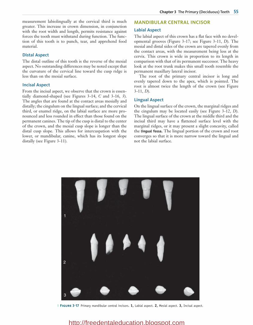
Chapter 3 The Primary (Deciduous) Teeth 55
measurement labiolingually at the cervical third is much greater. This increase in crown dimension, in conjunction with the root width and length, permits resistance against forces the tooth must withstand during function. The func-tion of this tooth is to punch, tear, and apprehend food material.
Distal AspectThe distal outline of this tooth is the reverse of the mesial aspect. No outstanding differences may be noted except that the curvature of the cervical line toward the cusp ridge is less than on the mesial surface.
Incisal AspectFrom the incisal aspect, we observe that the crown is essen-tially diamond-shaped (see Figures 3-14, C and 3-16, 3). The angles that are found at the contact areas mesially and distally; the cingulum on the lingual surface; and the cervical third, or enamel ridge, on the labial surface are more pro-nounced and less rounded in effect than those found on the permanent canines. The tip of the cusp is distal to the center of the crown, and the mesial cusp slope is longer than the distal cusp slope. This allows for intercuspation with the lower, or mandibular, canine, which has its longest slope distally (see Figure 3-11).
Figure 3-17 Primary mandibular central incisors. 1, Labial aspect. 2, Mesial aspect. 3, Incisal aspect.
3
2
1
MANDIBULAR CENTRAL INCISOR
Labial AspectThe labial aspect of this crown has a flat face with no devel-opmental grooves (Figure 3-17; see Figure 3-11, D). The mesial and distal sides of the crown are tapered evenly from the contact areas, with the measurement being less at the cervix. This crown is wide in proportion to its length in comparison with that of its permanent successor. The heavy look at the root trunk makes this small tooth resemble the permanent maxillary lateral incisor.
The root of the primary central incisor is long and evenly tapered down to the apex, which is pointed. The root is almost twice the length of the crown (see Figure 3-11, D).
Lingual AspectOn the lingual surface of the crown, the marginal ridges and the cingulum may be located easily (see Figure 3-12, D). The lingual surface of the crown at the middle third and the incisal third may have a flattened surface level with the marginal ridges, or it may present a slight concavity, called the lingual fossa. The lingual portion of the crown and root converges so that it is more narrow toward the lingual and not the labial surface.
http://freedentaleducation.blogspot.com
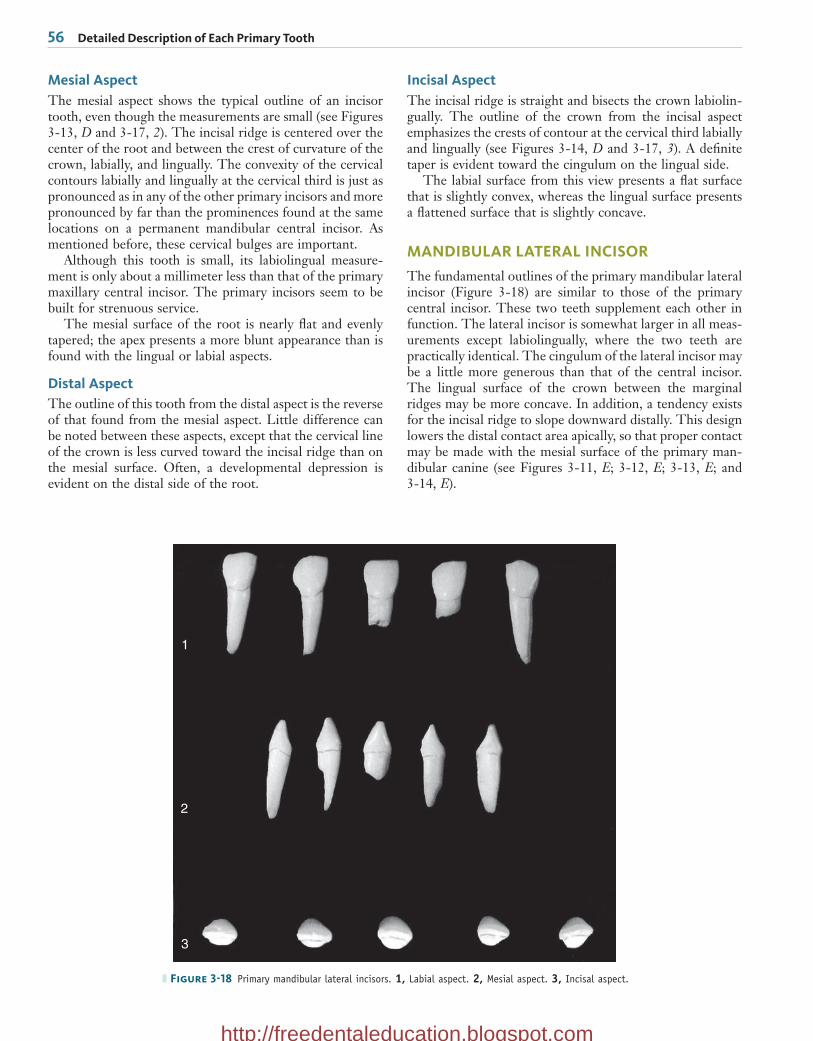
Mesial AspectThe mesial aspect shows the typical outline of an incisor tooth, even though the measurements are small (see Figures 3-13, D and 3-17, 2). The incisal ridge is centered over the center of the root and between the crest of curvature of the crown, labially, and lingually. The convexity of the cervical contours labially and lingually at the cervical third is just as pronounced as in any of the other primary incisors and more pronounced by far than the prominences found at the same locations on a permanent mandibular central incisor. As mentioned before, these cervical bulges are important.
Although this tooth is small, its labiolingual measure-ment is only about a millimeter less than that of the primary maxillary central incisor. The primary incisors seem to be built for strenuous service.
The mesial surface of the root is nearly flat and evenly tapered; the apex presents a more blunt appearance than is found with the lingual or labial aspects.
Distal AspectThe outline of this tooth from the distal aspect is the reverse of that found from the mesial aspect. Little difference can be noted between these aspects, except that the cervical line of the crown is less curved toward the incisal ridge than on the mesial surface. Often, a developmental depression is evident on the distal side of the root.
Figure 3-18 Primary mandibular lateral incisors. 1, Labial aspect. 2, Mesial aspect. 3, Incisal aspect.
3
2
1
Incisal AspectThe incisal ridge is straight and bisects the crown labiolin-gually. The outline of the crown from the incisal aspect emphasizes the crests of contour at the cervical third labially and lingually (see Figures 3-14, D and 3-17, 3). A definite taper is evident toward the cingulum on the lingual side.
The labial surface from this view presents a flat surface that is slightly convex, whereas the lingual surface presents a flattened surface that is slightly concave.
MANDIBULAR LATERAL INCISOR
The fundamental outlines of the primary mandibular lateral incisor (Figure 3-18) are similar to those of the primary central incisor. These two teeth supplement each other in function. The lateral incisor is somewhat larger in all meas-urements except labiolingually, where the two teeth are practically identical. The cingulum of the lateral incisor may be a little more generous than that of the central incisor. The lingual surface of the crown between the marginal ridges may be more concave. In addition, a tendency exists for the incisal ridge to slope downward distally. This design lowers the distal contact area apically, so that proper contact may be made with the mesial surface of the primary man-dibular canine (see Figures 3-11, E; 3-12, E; 3-13, E; and 3-14, E).
56 Detailed Description of Each Primary Tooth
http://freedentaleducation.blogspot.com

Chapter 3 The Primary (Deciduous) Teeth 57
MANDIBULAR CANINE
Little difference in functional form is evident between the mandibular canine and the maxillary canine. The difference is mainly in the dimensions. The crown is perhaps 0.5 mm shorter, and the root is at least 2 mm shorter; the mesiodistal measurement of the mandibular canine at the root trunk is greater when compared with its mesiodistal measurement at the contact areas than is that of the maxillary canine (Figure 3-19). It is “thicker” accordingly at the “neck” of the tooth. The outstanding variation in size between the two decidu-ous canines is shown by the labiolingual calibration. The deciduous maxillary canine is much larger labiolingually (see Figure 3-13).
The cervical ridges labially and lingually are not quite as pronounced as those found on the maxillary canine. The greatest variation in outline form when one compares the two teeth is seen from the labial and lingual aspects; the distal cusp slope is longer than the mesial slope. The opposite arrangement is true of the maxillary canine. This makes for proper intercuspation of these teeth during mastication.
Figure 3-20 illustrates the primary mandibular canines (see also Figures 3-11, F; 3-12, F; 3-13, F; and 3-14, F).
Figure 3-19 A comparison of primary canines, both in the size and shape of the crowns. Two of them have their roots intact and show no dissolution. A, Maxillary canines. B, Mandibular canines. Compare Figures 3-16 and 3-20.
B
A
Figure 3-20 Primary mandibular canines. 1, Labial aspect. 2, Mesial aspect. 3, Incisal aspect.
1
2
3
http://freedentaleducation.blogspot.com

MAXILLARY FIRST MOLAR
Buccal AspectThe widest measurement of the crown of the maxillary first molar is at the contact areas mesially and distally (Figure 3-21, A). From these points, the crown converges toward the cervix, with the measurement at the cervix being fully 2 mm less than the measurement at the contact areas. This dimensional arrangement furnishes a narrower look to the cervical portion of the crown and root of the primary maxil-lary first molar than that of the same portion of the perma-nent maxillary first molar. The occlusal line is slightly scalloped but with no definite cusp form. The buccal surface is smooth, and little evidence of developmental grooves is noted. It is from this aspect that one may judge the relative size of the primary maxillary first molar when it is compared with the second molar. It is much smaller in all measure-ments than the second molar. Its relative shape and size suggest that it was designed to be the “premolar section” of the primary dentition. In function it acts as a compromise between the size and shape of the anterior primary teeth and the molar area, this area being held temporarily by the larger primary second molar. At age 6, the large first permanent molar is expected to take its place distal to the second primary molar, which will complete a more extensive molar area for masticating efficiency.
The roots of the maxillary first molar are slender and long, and they spread widely. All three roots may be seen from this aspect. The distal root is considerably shorter than the mesial one. The bifurcation of the roots begins almost immediately at the site of the cervical line (CEJ). Actually, this arrangement is in effect for the entire root trunk, which includes a trifurcation, and this is a characteristic of all primary molars, whether maxillary or mandibular. Perma-
nent molars do not possess this characteristic. The root trunk on permanent molars is much heavier, with a greater distance between the cervical lines to the points of bifurca-tions (see Figure 11-8).
Lingual AspectThe general outline of the lingual aspect of the crown is similar to that of the buccal aspect (Figure 3-22, A). The crown converges considerably in a lingual direction, which makes the lingual portion calibrate less mesiodistally than the buccal portion.
The mesiolingual cusp is the most prominent cusp on this tooth. It is the longest and sharpest cusp. The distolin-gual cusp is poorly defined; it is small and rounded when it exists at all. From the lingual aspect, the distobuccal cusp may be seen, since it is longer and better developed than the distolingual cusp. A type of primary maxillary first molar that is not uncommon and presents as one large lingual cusp with no developmental groove in evidence lingually is a three-cusped molar (see Figure 3-25, 4, second from left).
All three roots also may be seen from this aspect. The lingual root is larger than the others.
Mesial AspectFrom the mesial aspect, the dimension at the cervical third is greater than the dimension at the occlusal third (Figure 3-23, A). This is true of all molar forms, but it is more pro-nounced on primary than on permanent teeth. The mesio-lingual cusp is longer and sharper than the mesiobuccal cusp. A pronounced convexity is evident on the buccal outline of the cervical third. This convexity is an outstand-ing characteristic of this tooth. It actually gives the impres-sion of overdevelopment in this area when comparisons are made with any other tooth, primary or permanent, with the
Figure 3-21 Primary right molars, buccal aspect. A, Maxillary first molar. B, Maxillary second molar. C, Mandibular first molar. D, Mandibular second molar.
AB
CD
Figure 3-22 Primary right molars, lingual aspect. A, Maxillary first molar. B, Maxillary second molar. C, Mandibular first molar. D, Mandibular second molar.
BA
DC
58 Detailed Description of Each Primary Tooth
http://freedentaleducation.blogspot.com

Chapter 3 The Primary (Deciduous) Teeth 59
Figure 3-23 Primary right molars, mesial aspect. A, Maxillary first molar. B, Maxillary second molar. C, Mandibular first molar. D, Mandibular second molar.
A B
C D
Figure 3-24 A, Maxillary first molar. MBC, Mesiobuccal cusp; MTF, mesial triangular fossa; MP, mesial pit; CP, central pit; MLC, mesiolingual cusp; DLC, distolingual cusp; DTF, distal triangular fossa; DDG, distal developmental groove; BDG, buccal developmental groove; DBC, distobuccal cusp; CDG, central developmental groove. B, Maxillary second molar. BDG, Buccal developmental groove; MBC, mesiobuccal cusp; CDG, central developmental groove; MLC, mesiolingual cusp; FC, fifth cusp; LDG, lingual developmental groove; DLC, distolingual cusp; DDG, distal developmental groove; OR, oblique ridge; DBC, distobuccal cusp; CP, central pit. C, Mandibular first molar. CDG, Central developmental groove; DBC, distobuccal cusp; BDG, buccal developmental groove; CP, central pit; DLC, distolingual cusp; LDC, lingual developmental groove; MLC, mesiolingual cusp; MP, mesial pit; MBC, mesiobuccal cusp. D, Mandibular second molar. DBC, Distobuccal cusp; CP, central pit; DC, distal cusp; DBDG, distobuccal developmental groove; DP, distal pit; CDG, central developmental groove; DLC, distolingual cusp; LDG, lingual developmental groove; MLC, mesiolingual cusp; MP, mesial pit; MBDG, mesiobuccal developmental groove; MBC, mesiobuccal cusp.
CDG
DBC MBC
A
C D
B
CP
DBC
CDG
DP
CP
DC
MTF
MPCP
DDG
DTF
BDG
DLC
MLC
MP
MBC
BDG
CP
DBC
CDG
DLC
MLC
LDC
LDG
DDG
DLCLDG
OR
BDG
MBC
BDG
CDGMLC
FC
DLCMLC
MP
MBDG
MBC
DBC
DBDG
opposite the contact area. The distobuccal root is hidden behind the mesiobuccal root. The lingual root from this aspect looks long and slender and extends lingually to a marked degree. It curves sharply in a buccal direction above the middle third.
Distal AspectFrom the distal aspect, the crown is narrower distally than mesially; it tapers markedly toward the distal end (Figure 3-24, A). The distobuccal cusp is long and sharp, and the distolingual cusp is poorly developed. The prominent bulge seen from the mesial aspect at the cervical third does not continue distally. The cervical line may curve occlusally, or it may extend straight across from the buccal surface to the lingual surface. All three roots may be seen from this angle, but the distobuccal root is superimposed on the mesiobuccal root so that only the buccal surface and the apex of the latter may be seen. The point of bifurcation of the distobuccal root and the lingual root is near the CEJ and, as described earlier, is typical.
Occlusal AspectThe calibration of the distance between the mesiobuccal line angle and the distobuccal line angle is definitely greater than the calibration between the mesiolingual line angle and the distolingual line angle (see Figure 3-24, A). Therefore the crown outline converges lingually. Also, the calibration
mandibular first primary molar being a close contender. The cervical line mesially shows some curvature in the direction of the occlusal surface.
The mesiobuccal and lingual roots are visible only when looking at the mesial side of this tooth from a point directly
http://freedentaleducation.blogspot.com

from the mesiobuccal line angle to the mesiolingual line angle is definitely greater than that found at the distal line angles. Therefore the crown also converges distally. Never-theless, these convergences are not reflected entirely in the working occlusal surface because it is more nearly rectangu-lar, with the shortest sides of the rectangle represented by the marginal ridges. The occlusal surface has a central fossa. A mesial triangular fossa is just inside the mesial marginal ridge, with a mesial pit in this fossa and a sulcus with its central groove connecting the two fossae. A well-defined buccal developmental groove divides the mesiobuccal cusp and the distobuccal cusp occlusally. Supplemental grooves radiate from the pit in the mesial triangular fossa as follows: one buccally, one lingually, and one toward the marginal ridge, with the last sometimes extending over the marginal ridge mesially.
Sometimes the primary maxillary first molar has a well-defined triangular ridge connecting the mesiolingual cusp with the distobuccal cusp. When well developed, it is called the oblique ridge. In some of these teeth, the ridge is indefi-nite, and the central developmental groove extends from the mesial pit to the distal developmental groove. This disto-occlusal groove is always seen and may or may not extend through to the lingual surface, outlining a distolingual cusp. The distal marginal ridge is thin and poorly developed in comparison with the mesial marginal ridge.
Summary of the occlusal aspect of the maxillary first primary molarThe form of the maxillary first primary molar varies from that of any tooth in the permanent dentition. Although no premolars are in the primary set, in some respects the crown of this primary molar resembles a permanent maxillary premolar. Nevertheless, the divisions of the occlu-sal surface and the root form with its efficient anchorage make it a molar, both in type and function. Figure 3-25 presents all the aspects of the primary maxillary first molars.
MAXILLARY SECOND MOLAR
Buccal AspectThe primary maxillary second molar has characteristics resembling those of the permanent maxillary first molar, but it is smaller (see Figure 3-21, B). The buccal view of this tooth shows two well-defined buccal cusps with a buccal developmental groove between them (Figure 3-26, 1). In line with all primary molars, the crown is narrow at the cervix in comparison with its mesiodistal measurement at the contact areas. This crown is much larger than that of the first primary molar. Although from this aspect the roots appear slender, they are much longer and heavier than those that are a part of the maxillary first molar. The point of bifurcation between the buccal roots is close to the cervical line of the crown. The two buccal cusps are more nearly equal in size and development than those of the primary maxillary first molar.
Figure 3-25 Primary maxillary first molars. 1, Buccal aspect. Note the flare of roots. 2, Mesial aspect. The cervical ridge on the buccal surface is curved to the extreme. Also note the flat or concave buccal surface above this bulge as it approaches the occlusal surface. 3, Lingual aspect. 4, Occlusal aspect. This aspect emphasizes the extensive width of the mesial portion of primary first molars. The four specimens show size differentials even in deciduous teeth (see Figure 3-24).
1
2
3
4
Lingual AspectLingually, the crown shows the following three cusps: (1) the mesiolingual cusp, which is large and well developed; (2) the distolingual cusp, which is well developed (more so than that of the primary first molar); and (3) a third supple-mental cusp, which is apical to the mesiolingual cusp and sometimes called the tubercle of Carabelli, or the fifth cusp (see Figure 3-22, B). This cusp is poorly developed and merely acts as a buttress or supplement to the bulk of the mesiolingual cusp. If the tubercle of Carabelli seems to be missing, some traces of developmental lines or “dimples” remain (see Figure 3-26, 3). A well-defined developmental groove separates the mesiolingual cusp from the distolingual cusp and connects with the developmental groove, which outlines the fifth cusp.
All three roots are visible from this aspect; the lingual root is large and thick in comparison with the other two
60 Detailed Description of Each Primary Tooth
http://freedentaleducation.blogspot.com

Chapter 3 The Primary (Deciduous) Teeth 61
Figure 3-26 Primary maxillary second molars. 1, Buccal aspect. 2, Mesial aspect. 3, Occlusal aspect.
1
2
3
roots. It is approximately the same length as the mesiobuccal root. If it should differ, it will be on the short side.
Mesial AspectFrom the mesial aspect, the crown has a typical molar outline that resembles that of the permanent molars very much (see Figures 3-23, B and 3-26, 2). The crown appears short because of its width buccolingually in comparison with its length. The crown of this tooth is usually only about 0.5 mm longer than the crown of the first deciduous molar, but the buccolingual measurement is 1.5 to 2 mm greater. In addi-tion, the roots are 1.5 to 2 mm longer. The mesiolingual
cusp of the crown with its supplementary fifth cusp appears large in comparison with the mesiobuccal cusp. The mesiobuccal cusp from this angle is relatively short and sharp. Little curvature to the cervical line is evident. Usually, it is almost straight across from buccal surface to lingual surface.
The mesiobuccal root from this aspect is broad and flat. The lingual root has somewhat the same curvature as the lingual root of the maxillary first deciduous molar.
The mesiobuccal root extends lingually far out beyond the crown outline. The point of bifurcation between the mesiobuccal root and the lingual root is 2 or 3 mm apical to the cervical line of the crown; this differs in depth on the root trunk from comparisons of this area in the recent dis-cussion of primary molars. The mesiobuccal root presents itself as being quite wide from the mesial aspect. It measures approximately two thirds the width of the root trunk, which leaves one third for the lingual root. The mesiolingual cusp is directly below their bifurcation. Although from this aspect the curvature is strong lingually at the cervical portion, as on most deciduous teeth the crest of curvature buccally at the cervical third is nominal and resembles the curvature found at this point on the permanent maxillary first molar. In this, it differs entirely from the prominent curvature found on the primary maxillary first molars at the cervical third buccally.
Distal AspectFrom the distal aspect, it is apparent that the distal calibra-tion of the crown is less than the mesial measurement, but the variation is found on the crown of the deciduous maxil-lary first molar. From both the distal and the mesial aspects, the outline of the crown lingually creates a smooth, rounded line, whereas a line describing the buccal surface is almost straight from the crest of curvature to the tip of the buccal cusp. The distobuccal cusp and the distolingual cusp are about the same in length. The cervical line is approximately straight, as was found mesially.
All three roots are seen from this aspect, although only a part of the outline of the mesiobuccal root may be seen, since the distobuccal root is superimposed over it. The dis-tobuccal root is shorter and narrower than the other roots. The point of bifurcation between the distobuccal root and the lingual root is more apical in location than any of the other points of bifurcation. The point of bifurcation between these two roots on the distal is more nearly centered above the crown than that on the mesial between the mesiobuccal and lingual roots.
Occlusal AspectFrom the occlusal aspect, this tooth resembles the perma-nent first molar (see Figures 3-24 and 3-26, 3). It is some-what rhomboidal and has four well-developed cusps and one supplemental cusp: mesiobuccal, distobuccal, mesiolingual, distolingual, and fifth cusps. The buccal surface is rather flat with the developmental groove between the cusps less marked than that found on the first permanent molar.
http://freedentaleducation.blogspot.com

Developmental grooves, pits, oblique ridge, and others are almost identical. The character of the “mold” is constant.
The occlusal surface has a central fossa with a central pit, a well-defined mesial triangular fossa, just distal to the mesial marginal ridge, with a mesial pit at its center. A well-defined developmental groove called the central groove is also at the bottom of a sulcus, connecting the mesial triangular fossa with the central fossa. The buccal developmental groove extends buccally from the central pit, separating the triangular ridges, which are occlusal continuations of the mesiobuccal and distobuccal cusps. Supplemental grooves often radiate from these developmental grooves.
The oblique ridge is prominent and connects the mesio-lingual cusp with the distobuccal cusp. Distal to the oblique ridge, the distal fossa is found, which harbors the distal devel-opmental groove. The distal groove has branches of supple-mental grooves within the distal triangular fossa, which is rather indefinitely outlined just mesial to the distal marginal ridge.
Figure 3-27 Primary mandibular first molars. This tooth has characteristics unlike those of any other tooth in the mouth, primary or permanent. 1, Buccal aspect. 2, Lingual aspect. 3, Occlusal aspect.
1
2
3
The distal groove acts as a line of demarcation between the mesiolingual and distolingual cusps and continues on to the lingual surface as the lingual developmental groove. The distal marginal ridge is as well developed as the mesial mar-ginal ridge. It will be remembered that the marginal ridges are not developed equally on the primary maxillary first molar.
MANDIBULAR FIRST MOLAR
The mandibular first molar does not resemble any of the other teeth, deciduous or permanent. Because it varies so much from all others, it appears strange and primitive (Figure 3-27).
Buccal AspectFrom the buccal aspect, the mesial outline of the crown of the primary mandibular first molar is almost straight from the contact area to the cervix, constricting the crown very
62 Detailed Description of Each Primary Tooth
http://freedentaleducation.blogspot.com

Chapter 3 The Primary (Deciduous) Teeth 63
gually. Part of the two buccal cusps may be seen from this angle.
From the lingual aspect, the crown length mesially and distally is more uniform than it is from the buccal aspect. The cervical line is straighter.
Mesial AspectThe most noticeable detail from the mesial aspect is the extreme curvature buccally at the cervical third (see Figure 3-28, B). Except for this detail, the crown outline of this tooth from this aspect resembles the mesial aspect of the primary second molar and that aspect of the permanent mandibular molars. In this comparison, the buccal cusps are placed over the root base, and the lingual outline of the crown extends out lingually beyond the confines of the root base.
Both the mesiobuccal cusp and the mesiolingual cusp are in view from this aspect, as is the well-developed mesial marginal ridge. Because the mesiobuccal crown length is greater than the mesiolingual crown length, the cervical line slants upward buccolingually. Note the flat appearance of the buccal outline of the crown from the crest of curvature of the buccal surface at the cervical third to the tip of the mesiobuccal cusp. All of the primary molars have flattened buccal surfaces above this cervical ridge.
The outline of the mesial root from the mesial aspect does not resemble the outline of any other primary tooth root. The buccal and lingual outlines of the root drop straight down from the crown and are approximately parallel for more than half their length, tapering only slightly at the apical third. The root end is flat and almost square. A devel-opmental depression usually extends almost the full length of the root on the mesial side.
Distal AspectThe distal aspect of the mandibular first molar differs from the mesial aspect in several ways. The cervical line does not drop buccally. The length of crown buccally and lingually is more uniform, and the cervical line extends almost straight across buccolingually. The distobuccal cusp and the disto-lingual cusp are not as long or as sharp as the two mesial cusps. The distal marginal ridge is not as straight and well defined as the mesial marginal ridge. The distal root is rounder and shorter and tapers more apically.
Figure 3-28 Three rare specimens of the primary mandibular first molars. A, Buccal aspect. B, Mesial aspect. These three specimens have their roots intact with little or no resorption showing. They enable the viewer to observe the actual shape and size of the mesial and distal roots. The mesial root is broad, curved, and long, with fluting down the center. This makes for tremendous anchorage. The distal root is much shorter, but it is heavy and also curved. It does its share in bracing the crown, being in partnership with the mesial root during the process.
A B
little at the cervix (Figure 3-28, A). The outline describing the distal portion, however, converges toward the cervix more than usual, so that the contact area extends distally to a marked degree (see Figures 3-21, C and 3-27, 1).
The distal portion of the crown is shorter than the mesial portion, with the cervical line dipping apically where it joins the mesial root.
The two buccal cusps are rather distinct, although no developmental groove is evident between them. The mesial cusp is larger than the distal cusp. A developmental depres-sion dividing them (not a groove) extends over to the buccal surface.
The roots are long and slender, and they spread greatly at the apical third beyond the outline of the crown.
The buccal aspect emphasizes the strange, primitive look of this tooth. The primary first mandibular molar from this angle impresses one with the thought of the possibility that at some time in the dim past, a fusion of two teeth ended in a strange single combination. That thought seems particu-larly apropos when a well-formed specimen of the tooth in question is located—one with its roots intact, showing no evidences of decalcification.
From the buccal aspect, if a line is drawn from the bifur-cation of the roots to the occlusal surface, the tooth will be evenly divided mesiodistally. However, the mesial portion represents a tooth with a crown almost twice as tall as the distal half, and the root is again a third as long as the distal one. Two complete teeth are represented, but their dimensions differ considerably (see Figures 3-21, C and 3-27, 1).
Lingual AspectThe crown and root converge lingually to a marked degree on the mesial surface (see Figures 3-22, C and 3-27, 2). Distally, the opposite arrangement is true of both crown and root. The distolingual cusp is rounded and suggests a developmental groove between this cusp and the mesiolin-gual cusp. The mesiolingual cusp is long and sharp at the tip, more so than any of the other cusps. The sharp and prominent mesiolingual cusp (almost centered lingually but in line with the mesial root) is an outstanding characteristic found occlusally on the primary first mandibular molar. It is noted that the mesial marginal ridge is so well developed that it might almost be considered another small cusp lin-
http://freedentaleducation.blogspot.com

Occlusal AspectThe general outline of this tooth from the occlusal aspect is rhomboidal (see Figure 3-27, 3). The prominence present mesiobuccally is noticeable from this aspect, which accents the mesiobuccal line angle of the crown in comparison with the distobuccal line angle and thereby emphasizes the rhom-boidal form.
The mesiolingual cusp may be seen as the largest and best developed of all the cusps, and it has a broad, flattened surface lingually. The buccal developmental groove of the occlusal surface divides the two buccal cusps evenly. This developmental groove is short, extending from between the buccal cusp ridges to a point approximately in the center of the crown outline at a central pit. The central developmen-tal groove joins it at this point and extends mesially, separat-ing the mesiobuccal cusp and the mesiolingual cusp. The central groove ends in a mesial pit in the mesial triangular fossa, which is immediately distal to the mesial marginal ridge. Two supplemental grooves join the developmental groove in the center of the mesial triangular fossa; one sup-plemental groove extends buccally and the other extends lingually.
The mesiobuccal cusp exhibits a well-defined triangular ridge on the occlusal surface, which terminates in the center of the occlusal surface buccolingually at the central develop-mental groove. The lingual developmental groove extends lin-gually from this point, separating the mesiolingual cusp and the distolingual cusp. Usually, the lingual developmental groove does not extend through to the lingual surface but stops at the junction of lingual cusp ridges. Some supple-mental grooves immediately mesial to the distal marginal ridge in the distal triangular fossa join with the central devel-opmental groove.
MANDIBULAR SECOND MOLAR
The primary mandibular second molar has characteristics that resemble those of the permanent mandibular first molar, although its dimensions differ (Figure 3-29).
Buccal AspectFrom the buccal aspect, the primary mandibular second molar has a narrow mesiodistal calibration at the cervical portion of the crown compared with the calibration mesio-distally on the crown at contact level. The mandibular first permanent molar, accordingly, is wider at the cervical portion (see Figures 3-21, D and 3-29, 1).
From this aspect also, mesiobuccal and distobuccal devel-opmental grooves divide the buccal surface of the crown occlusally into three cuspal portions almost equal in size. This arrangement forms a straight buccal surface presenting a mesiobuccal, a buccal, and a distobuccal cusp. It differs, therefore, from the mandibular first permanent molar, which has an uneven distribution buccally, presenting two buccal cusps and one distal cusp.
The roots of the primary second molar from this angle are slender and long. They have a characteristic flare mesio-
distally at the middle and apical thirds. The roots of this tooth may be twice as long as the crown.
The point of bifurcation of the roots starts immediately below the CEJ of crown and root.
Lingual AspectFrom the lingual aspect, two cusps of almost equal dimen-sions can be observed (see Figures 3-22, D and 3-29, 2). A short, lingual groove is between them. The two lingual cusps are not quite as wide as the three buccal cusps; this arrangement narrows the crown lingually. The cervical line is relatively straight, and the crown extends out over the root more distally than it does mesially. The mesial portion of the crown seems to be a little higher than the distal portion of the crown when viewed from the lingual aspect. It gives the impression of being tipped distally. A portion of each of the three buccal cusps may be seen from this aspect.
The roots from this aspect give somewhat the same appearance as from the buccal aspect. Note the length of the roots.
Mesial AspectFrom the mesial aspect, the outline of the crown resembles that of the permanent mandibular first molar (see Figures 3-23, D and 3-29, 3). The crest of contour buccally is more prominent on the primary molar, and the tooth seems to be more constricted occlusally because of the flattened buccal surface above this cervical ridge.
The crown is poised over the root of this tooth in the same manner as all mandibular posteriors; its buccal cusp is over the root and the lingual outline of the crown extending out beyond the root line. The marginal ridge is high, a characteristic that makes the mesiobuccal cusp and the mesi-olingual cusp appear rather short. The lingual cusp is longer, or higher, than the buccal cusp. The cervical line is regular, although it extends upward buccolingually, making up for the difference in length between the buccal and lingual cusps.
The mesial root is unusually broad and flat with a blunt apex that is sometimes serrated.
Distal AspectThe crown is not as wide distally as it is mesially; therefore it is possible to see the mesiobuccal and distobuccal cusps from the distal aspect. The distolingual cusp appears well developed, and the triangular ridge from the tip of this cusp extending down into the occlusal surface is seen over the distal marginal ridge.
The distal marginal ridge dips down more sharply and is shorter buccolingually than the mesial marginal ridge. The cervical line of the crown is regular, although it has the same upward incline buccolingually on the distal as on the mesial.
The distal root is almost as broad as the mesial root and is flattened on the distal surface. The distal root tapers more at the apical end than does the mesial root.
64 Detailed Description of Each Primary Tooth
http://freedentaleducation.blogspot.com

Chapter 3 The Primary (Deciduous) Teeth 65
Figure 3-29 Primary mandibular second molars. 1, Buccal aspect. 2, Lingual aspect. 3, Mesial aspect. 4, Occlusal aspect.
1
2
3
4
Occlusal AspectThe occlusal aspect of the primary mandibular second molar is somewhat rectangular (see Figures 3-24, D and 3-29, 4). The three buccal cusps are similar in size. The two lingual cusps are also equally matched. However, the total mesio-distal width of the lingual cusps is less than the total mesio-distal width of the three buccal cusps.
Well-defined triangular ridges extend occlusally from each one of these cusp tips. The triangular ridges end in the center of the crown buccolingually in a central developmental groove that follows a staggered course from the mesial trian-gular fossa, just inside the mesial marginal ridge, to the distal triangular fossa, just mesial to the distal marginal ridge. The distal triangular fossa is not as well defined as the mesial triangular fossa. Developmental grooves branch off from the central groove both buccally and lingually, dividing the cusps. The two buccal grooves are confluent with the buccal developmental grooves of the buccal surface, one mesial and one distal, and the single lingual developmental groove is con-fluent with the lingual groove on the lingual surface of the crown.
Scattered over the occlusal surface are supplemental grooves on the slopes of triangular ridges and in the mesial and distal triangular fossae. The mesial marginal ridge is
better developed and more pronounced than the distal mar-ginal ridge. The outline of the crown converges distally. An outline following the tips of the cusps and the marginal ridges conforms to the outline of a rectangle more closely than does the gross outline of the crown in its entirety.
A comparison occlusally between the deciduous mandib-ular second molar and the permanent mandibular first molar brings out the following points of difference. In the decidu-ous molar the mesiobuccal, distobuccal, and distal cusps are almost equal in size and development. The distal cusp of the permanent molar is smaller than the other two. Because of the small buccal cusps, the deciduous tooth crown is nar-rower buccolingually, in comparison with its mesiodistal measurement, than is the permanent tooth.
References1. Fulton JT, Hughes JT, Mercer CV: The life cycle of the
human teeth, Chapel Hill, NC, 1964, Department of Epidemiology, School of Public Health, University of North Carolina.
2. McBeath EC: New concept of the development and calcification of the teeth, J Am Dent Assoc 23:675, 1936.
3. Noyes EB, Shour I, Noyes HJ: Dental histology and embryology, ed 5, Philadelphia, 1938, Lea & Febiger.
http://freedentaleducation.blogspot.com

66 Bibliography
4. Finn SB: Clinical pedodontics, ed 2, Philadelphia, 1957, Saunders.
BibliographyBarker BC: Anatomy of root canals: IV. Deciduous teeth, Aust
Dent J 20:101, 1975.Baume LJ: Physiologic tooth migration and its significance for
the development of occlusion. I. The biogenetic course of the deciduous dentition, J Dent Res 29:123, 1950.
Broadbelt AG: On the growth pattern of the human head, from the third month to the eighth year of life, Am J Anat 68:209, 1941.
Carlsen O: Carabelli’s structure on the human maxillary deciduous first molar, Acta Odontol Scand 26:395, 1968.
Carlsen O, Andersen J: On the anatomy of the pulp chamber and root canals in human deciduous teeth, Tandlaegebladet 70:93, 1966.
de Campos Russo M et al: Observations on the pulpal floor of human deciduous teeth and possible implications in endodontic treatment, Rev Fac Odontol Aracatuba 3:61, 1974.
Fanning EA: Effect of extraction of deciduous molars on the formation and eruption of their successors, Angle Orthod 32:44, 1962.
Friel S: The development of ideal occlusion of the gum pads and the teeth, Am J Orthod 40:196, 1954.
Friel S: Occlusion: observations on its development from infancy to old age, Int J Orthod Oral Surg 13:322, 1927.
Moorrees CFA, Chadha M: Crown diameters of corresponding tooth groups in the deciduous and permanent dentition, J Dent Res 41:466, 1962.
Richardson AS, Castaldi CR: Dental development during the first two years of life, J Can Dent Assoc 33:418, 1967.
Van der Linden FPGM, Duterloo HS: Development of the human dentition: an atlas, Hagerstown, MD, 1976, Harper & Row.
Woo RK et al: Accessory canals in deciduous molars, J Int Assoc Dent Child 12:51, 1981.
Use the CD-ROM to view animations and study ques-tions; and go to the site for additional study resources.
http://freedentaleducation.blogspot.com

Forensics, Comparative Anatomy, Geometries, and Form and Function
a brief summary is given here with general references added to cited references for further reading.
JAWS AND TEETH
Unlike the static, genetically determined friction ridges found on the human hand and foot (commonly referred to as fingerprints and footprints) each individual’s dentition is fluid and changes throughout life as the deciduous teeth are exfoliated and the permanent dentition erupts. In addition, the teeth are subject to dental and periodontal disease, the oral manifestations of systemic diseases that may affect the individual, and alterations of dental structures related to techniques of restoration and replacement that are employed by the dental practitioner.
Historically, despite this potential for an ever-changing dentition, inspection of the teeth and jaws has been used as a legally accepted method of human identification. Signifi-cant cases in which identification has been resolved by means of examination of dental evidence include identification of Revolutionary War hero Dr. Joseph Warren, the prominent Boston murder victim Dr. George Parkman, and Adolph Hitler.3
Paramount to the success of a forensic dental comparison between antemortem and postmortem dental records is the requirement that both sets of records provide the maximum amount of dental information for analysis. Thus, dentists are encouraged to document all dental anomalies, existing res-torations, and missing teeth in their written, photographic, and radiographic records.
This chapter includes brief discussions of forensic dentistry, comparative dental anatomy, and some relationships of form to function in the permanent dentition. The limited space allotted to these subjects should not suggest that they lack importance. The literature on these subjects is extensive, and the reader should review the references cited for addi-tional information.
Forensic Dentistry
Forensic dentistry (odontology) is the field within the greater disciplines of dentistry and forensic science that evaluates, manages, and presents dental evidence in legal proceedings in the interest of justice.1 Forensic dental casework often involves identification of unidentified or missing individuals, human remains, or victims of mass fatality incidents, including natural and accidental disas-ters. This is accomplished by comparison of a victim’s dentition and supporting structures with dental records of known individuals. The latter records may be obtained from private dental offices, prison or military dental data-bases, or records retained by the Federal Bureau of Inves-tigation through its National Crime Information Center’s Missing, Unidentified, and Wanted Persons files in a Web environment.2
Since forensic odontology is one of several forensic spe-cialties, the forensic dentist’s role often interfaces with those of the anthropologist, criminologist, toxicologist, patholo-gist, and law enforcement official involved with a case. Only
67
44
http://freedentaleducation.blogspot.com

CHRONOLOGICAL AGE
The determination of dental maturity and tooth develop-ment has been considered in Chapter 2. As indicated there, the determination of chronological age is possible with some reasonable degree of accuracy. When it is employed in con-junction with evaluation of the stage of development of osseous tissues,4 the two methods can be used to determine an estimated age for an individual less than 20 years old. This is accomplished by entering data from both methods into an electronic encyclopedia on maxillofacial, dental, and skeletal development.5
Chronological age determination is a central issue in population studies, and racial and sex differences in tooth development and eruption patterns are acknowledged.6,7 Dental age assessment is also important in the forensic dental evaluation of human remains or living individuals. Aging of the dentition has been employed in forensic dental casework involving estimation of age of unidentified individuals.8,9
Separation of victims of multiple fatality incidents by age facilitates the narrowing of searches for eventual identifica-tion by comparison of medical and dental records. Dental evaluation of illegal immigrants, who may present authori-ties with misinformation concerning their age, is important in cases in which protection of unaccompanied minors is a concern.2
A variety of dental age estimation procedures have been employed. Some are useful in the analysis of radiographic changes associated with development of the dentition of children.10 Others use dental radiographs to determine the dental chronological age in living or deceased adult individuals.11,12
Estimation of the age of an adult may also be based on six characteristics first identified by Gustafson in 195013 by his observation of ground sections of extracted teeth. These six variables include attrition of the occlusal or incisal sur-faces, degree of deposition of secondary dentin (evaluation of the size of the pulp chamber and canal), deposition of apical cementum, attachment level of the periodontium, root resorption, and radicular translucency.
In addition, biochemical methods that assess the ratios of levorotatory and dextrorotatory isomers of the long-lived, metabolically stable amino acid aspartate have been employed in both living and deceased subjects to age tissues in which this substance is found. Through a racemization process the L form of aspartic acid is slowly transformed into its stereoi-somer, which is the D form of the amino acid. As enamel and dentin age, levels of the D form of aspartic acid increase in these calcified dental structures. These values can be measured and related to known levels for age estimation.14
DENTAL DNA
Each individual’s unique genetic information is contained within the nuclear and mitochondrial deoxyribonucleic acid (DNA) molecules of their cells. Only identical twins share the same DNA. Nuclear DNA is transmitted from either
parent, whereas mitochondrial DNA is derived only through a maternal route. As a unique biological molecule, DNA offers the forensic scientist a means of positively identifying an individual when this material can be obtained from tissues or bodily fluids recovered at a crime scene or from human remains or other forensic evidence.
The most widely used method for forensic analysis of DNA material is the restriction fragment length polymor-phism (RFLP) technique. This laboratory procedure requires that large amounts (>100 ng) of DNA be used in the analysis. The polymerase chain reaction (PCR) tech-nique is employed when this cannot be accomplished because of degradation of the DNA molecule submitted as evidence, when only small amounts (<100 pg) of DNA are available for analysis, or when the DNA sample is fragmented.15 The PCR method amplifies the amount of DNA available for analysis by copying a specific locus of genetic material.16 Since only small amounts of DNA evidence need be evalu-ated using PCR technology, a positive identification may still be effected when human remains have been left unburied for long periods or have been incinerated, or when DNA trace evidence is obtained from saliva, blood, or fluid samples.
The calcified and pulp tissues within a tooth often present the forensic scientist with the most uncontami-nated and protected DNA samples for analysis. Thus, even small amounts of DNA recovered from these tissues often may be analyzed using the PCR method when other means of identification have been lost or degraded as evidence.17,18
In addition, the sex of a decedent can be determined through DNA analysis of the sex-linked amelogenin gene. A DNA sample from the victim can be compared directly with one obtained from the clothing or other personal effects of a putative subject of interest or indirectly with a sample from the parents or siblings of the individual one is attempting to identify.19
BITE MARKS
When the dentition of a human or animal impresses the surface of an object during the act of biting, a pattern mark is often imprinted on that surface (Figure 4-1, A). Like a tool mark left as forensic evidence, the pattern left by the teeth can be evaluated and compared to the dentition that allegedly caused it (Figure 4-1, B). Whether the bite mark pattern involves the skin of a victim or suspect or remains on the surface of an inanimate object, to be probative, it must have class and individual characteristics consistent with a mark caused by teeth.
Class characteristics of a bite mark include the size and shape of the pattern. In most cases this should be consistent with the dental arch size of the suspected biter (human or animal) and retain a circular shape consisting of two half arches (maxilla and mandible) separated by a space (tempo-romandibular joint). One arch should be larger in its great-est dimension, representing the greater arch length of the maxilla.
68 Forensic Dentistry
http://freedentaleducation.blogspot.com

Chapter 4 Forensics, Comparative Anatomy, Geometries, and Form and Function 69
Individual characteristics associated with a bite mark involve the patterns routinely made by specific teeth. In the human dentition these include the following:
Maxillary central incisor—large rectangleMandibular incisor and maxillary lateral incisor—small
rectangleCuspid—point or triangle (when there is incisal wear)Maxillary cuspid—figure-eight pattern directed buccal to
lingualMandibular cuspid—point representing the buccal cuspMolar—not routinely seen in the patterns left by human
biters
It is generally accepted that no two individuals have an identical dentition based on variations in the arrangement, spacing, size, and shape of specific teeth and dental arches. Currently, however, neither quantitative values nor data-bases have been established for the dentition that are similar to those described for fingerprint and DNA analysis and comparison. Thus, although bite mark evidence has been admissible in the federal and state courts of the United States based on the Frye rule and decisions related to the Federal Rules of Evidence,20,21 this evidence is often used in an exculpatory manner.
Guidelines have been established and are continually reviewed and revised to provide the forensic odontologist with evidence gathering and analytical procedures to be fol-lowed to ensure that recovered bite mark evidence is admis-sible in court and supports corroborative evidence in the case.22
Comparative Dental Anatomy
To understand the human dentition, it is helpful to compare the dentitions of other vertebrates. In doing so, it should become clear that the dentition in humans is different in many ways from that of other vertebrates in form and func-tion. However, it should be equally clear that the presence
of related characteristics in a wide range of vertebrates sug-gests a plan common to all.
Only a brief summary of the topic is presented here, starting with a simple form of tooth, the single cone or lobe, and then combinations of lobes forming more complicated teeth that are found in highly developed animals and in humans. Additional material on the subject may be found in the references and bibliography presented at the end of this chapter.
Figure 4-2 graphically illustrates a theory of the follow-ing four phylogenetic classes of tooth forms23–25:
1. Single cone (haplodont)2. Three cusps in line (triconodont)3. Three cusps in a triangle (tritubercular molar)4. Four cusps in a quadrangle (quadritubercular molar)
The haplodont class is represented schematically by the simplest form of tooth, the single cone (Figure 4-2, A). This type of dentition usually includes many teeth in both jaws and is seen where jaw movements are limited to simple open and close (hinge) movements (Figure 4-3). No occlusion of the teeth occurs in this class, since the teeth are used mainly for prehension or combat.26,27 Their main function is the procurement of food. Jaw move-ments are related to and governed by tooth form in all cases.
The triconodont class exhibits three cusps in line in pos-terior teeth as indicated in Figure 4-2, B. Anthropologically, the largest cusp is centered with a smaller cusp located anteriorly and another posteriorly. Purely triconodont den-titions are not seen, although certain breeds of dogs28 and other carnivores have teeth reflecting the triconodont form (Figure 4-4). Nevertheless, dogs and other animals carnivo-rous by nature are considered to be in the third category (Figures 4-5 through 4-8), namely, the tritubercular class.29,30 The three-cusp arrangement of the triconodont class and the more efficient three-cornered tritubercular molar arrangement are both consistent with the teeth’s bypassing each other more or less when the jaw is opened or closed.
Figure 4-1 Forensic bite marks. A, Forehead bite marks. B, Bite marks related to maxillary dental cast. (Courtesy Dr. David Ord.)
A B
http://freedentaleducation.blogspot.com

However, the quadritubercular class reflects an occlusal contact relationship between the teeth of the upper and lower jaws. Articulation of the jaws and teeth is consistent with jaw movements and functions of these classes of teeth.
Animals that have dentitions similar to that of humans are anthropoid apes. This group of animals includes the chimpanzee, gibbon, gorilla, and orangutan (Figures 4-9 and 4-10). The shapes of individual teeth in these animals
are amazingly close to their counterparts in the human mouth. Nevertheless, the development of the canines, the arch form, and the jaw development are quite different.
The multiplication and fusion of lobes during tooth devel-opment are demonstrated graphically when human teeth are viewed from the mesial or distal aspects. Anterior teeth, which are used for incising or apprehending food, reflect the single cone, whereas the posterior or multicusped teeth, which are used for grinding food in addition to having a shearing action, appear to be two or more cones fused (Figure 4-11). Although the schematic form of the teeth from the mesial or distal aspects is that of a single cone in anterior teeth, or what seems to be an indication of a fusion of two or more cones in posterior teeth, close observation causes one to come to the conclusion that each tooth crown, regard-
A
B
C
D
Figure 4-2 Classification of cusp forms. A, Haplodont. B, Triconodont. C, Tritubercular molar. D, Quadritubercular molar. The solid black dots represent upper molar cusps. The circles represent lower molar cusps.
(From Thompson AH: Comparative dental anatomy, ed 2, revised by M Dewey, St Louis, 1915, Mosby.)
Figure 4-3 The Mississippi alligator. An interesting commentary on the anatomy of the alligator: because of the alligator’s physical problems, the upper jaw is the mobile one. The lower jaw, closer to the ground, is static.
(From Kronfeld R: Dental histology and comparative dental anatomy, Philadelphia, 1937, Lea & Febiger.)
Figure 4-4 Permanent dentition of Canis familiaris.
(Modified from Tims HWM: Notes on the dentition of the dog, Anat Anz Bd 11:537, 1896.)
Figure 4-5 Jaws of a dog (collie). The premolars are tritubercular and widely spaced; the upper carnassial (fourth premolar, P4) articulates with the lower carnassial (first molar, M1). I, Incisor; C, canine.
(From Boyd J: Color atlas of clinical anatomy of the dog and cat, ed 2, St Louis, 2001, Mosby.)
I I
C
P4
P4M1
70 Comparative Dental Anatomy
http://freedentaleducation.blogspot.com

Chapter 4 Forensics, Comparative Anatomy, Geometries, and Form and Function 71
less of location, appears to be a combination of four or more lobes. Each lobe represents a primary center of formation.
All anterior teeth show traces of four lobes, three labially and one lingually, the lingual lobe being represented by the cingulum. Each labial lobe of the incisor terminates incisally in rounded eminences known as mamelons. These mamelons are prominent in newly erupted incisors. Soon after erup-tion they are worn down by use unless, through misalign-ment, they escape incisal wear. Maxillary central incisors often show traces of the fusion of three lobes on the labial face by visible markings in the enamel called labial grooves ( Figures 4-12, A ; 4-13; and 4-14 ).
In the anterior teeth, the four lobes are called the mesial,
labial, distal, and lingual lobes. In premolars they are called
mesial, buccal, distal, and lingual lobes, or as in the case of the mandibular second premolar, which often has two lingual cusps, the mesial, buccal, distal, mesiolingual, and distolingual
lobes, making fi ve in all (see Figure 10-17 ). The molar lobes are named the same as the cusps (e.g.,
mesiobuccal lobe ). The tip of each cusp represents the primary center of formation of each lobe. *
It is possible, of course, to fi nd a variation in the number of lobes in molars. Tubercles of enamel may be found in addi-tion to the primary lobes. When present, they are usually smaller than and supplementary to the major lobes.
F igure 4-6 Normal radiographic anatomy of a dog (unspecifi ed). A, Radiograph of a left mandibular fourth premolar tooth along with partial images of the third premolar (on left ) and the fi rst molar (on right ). B, Anatomical features outlined for identifi cation. C, A sectioned specimen showing internal anatomy.
(From DuPont GA, DeBowes LJ: Atlas of dental radiography in dogs and cats, St Louis, 2009, Saunders.)
7
89
6
19
1515
26
1820
1217
16
25
17
10
5
14
3
7
2122
2
8 119
2221
3
10
4
15
1317
26
116
27
25
10
24
1
4 23
A
B
C
F igure 4-7 Occlusal view of the mandible of a dog (collie). The mandible is long and slender. The cutting edges of the four premolars (7) and three molars (6) are arranged in a sagittal plane. 9, Incisor; 8, canine.
(From Boyd J: Color atlas of clinical anatomy of the dog and cat, ed 2, St Louis, 2001, Mosby.)
F igure 4-8 The bear. The extent of development of teeth and jaws and occlusion is often used to rate animal form. Compare with Figure 4-9. C , Canine; P, premolar; M, molar.
(From Kronfeld R: Dental histology and comparative dental anatomy, Philadelphia, 1937, Lea & Febiger.)
* Description and tables of the development of the teeth are given under the complete description of each tooth, and in Chapter 2 .
http://freedentaleducation.blogspot.com

Figure 4-9 The ape. I, Incisor; C, canine; P, premolar; M, molar.
(From Kronfeld R: Dental histology and comparative dental anatomy, Philadelphia, 1937, Lea & Febiger.)
Figure 4-10 Occlusal view of the upper jaw of an orangutan. The arch is square, and the canines, premolars, and molars stand in a straight sagittal line. Note the diastema between lateral incisor and canine.
(From Kronfeld R: Dental histology and comparative dental anatomy, Philadelphia, 1937, Lea & Febiger.)
Figure 4-11 The functional form of the teeth when outlined schematically from the mesial or distal aspects is that of the fusion of two or three cones. A, Maxillary incisor. B, Maxillary premolar. C, Maxillary first molar. Note that the major portion of the incisor in view is made up of one cone or lobe.
A CB
Figure 4-12 General outlines of some of the lobes. A, Labial aspect of maxillary central incisor, showing the labial grooves marking the division of the lobes. 1, Mesial lobe; 2, labial lobe; 3, distal lobe. The lingual lobe, or cingulum, is not in view (see Figure 4-13, A, 4). B and E, Mesial and occlusal aspects of maxillary first premolar. 1, Mesial lobe; 2, buccal lobe; 3, distal lobe; 4, lingual lobe. C, Occlusal aspect of mandibular first molar. 1, Mesiobuccal lobe; 2, distobuccal lobe; 3, distal lobe; 4, mesiolingual lobe; 5, distolingual lobe. Lobes on molars are named the same as cusps. D, Occlusal aspects of maxillary first molar. 1, Mesiobuccal lobe; 2, distobuccal lobe; 3, mesiolingual lobe; 4, distolingual lobe; 5, fifth lobe (fifth cusp).
A B
C D E
3
33
3
11
1
11
22 2
22
4 4 4
4
55
Figure 4-13 A, Lingual aspect of maxillary canine, with primary centers marked. 1, Mesial lobe; 2, central lobe (cusp); 3, distal lobe; 4, lingual lobe (cingulum). B, Incomplete formation demonstrated by a developmental groove distolingually on a maxillary lateral incisor. This groove will sometimes harbor fissures at various points along its length, especially in the coronal portion. A tooth with this handicap is more subject to caries.
A B
4
13
2
72 Comparative Dental Anatomy
http://freedentaleducation.blogspot.com

Chapter 4 Forensics, Comparative Anatomy, Geometries, and Form and Function 73
TOOTH FORM AND JAW MOVEMENTS
In general terms, the primates are bunodont and relatively isognathous and therefore have limited lateral jaw move-ment. Bunodont refers to tooth-bearing conical cusps. Isog-nathous means equally jawed; anisognathus means unequally jawed. Humans are not perfectly isognathous (i.e., the max-illary arch overlaps horizontally the mandibular arch). The shape of the glenoid (mandibular) fossae (see Figure 15-1) is correlated also with tooth form and jaw movements. A number of living and extinct species demonstrate a number of dental forms, from the bunodont to the selenodont (i.e., molars with crescent-shaped cusps) types. Each type is accompanied by an increased mobility of the mandible in a lateral direction and increased anisognathism. When the incisal point is viewed from the front during mastication, the mandible moves up and down without lateral deviation in dogs, cats, pigs, and all other animals with bunodont articulations. Lateral movement increases in a number of animals to the extreme lateral excursions seen in the giraffe, camel, and ox. In relation to the latter type of movement, it has been proposed that where the condyle is greatly elon-gated transversely and very flat, great lateral movement occurs during mastication, associated with selenodont molars, and a great degree of anisognathism. Also of interest for the gnathologist is the putative correlation between the directions of the ridges and grooves and a radius drawn to the center of the glenoid fossa.
It has been suggested that the simplest type of jaw move-ment is opening and closing without lateral excursion and coexistent with the simple bunodont molar. With increasing complexity of movement, an apparent increase in the com-plexity of enamel folding, ridges, and crests occurs, which might be consistent with the hypothesis of the mechanical genesis of tooth forms.31 Correlations between the forms of the teeth, joints, muscles, skull, bones, and jaw movement appear to be consistent with the functions of each species.
GEOMETRIES OF CROWN OUTLINES
In a general way, all aspects of each tooth crown except the incisal or occlusal aspects may be outlined schematically within one of three geometric figures: a triangle, trapezoid, or rhomboid. To one unfamiliar with dental anatomy, it may seem an exaggeration to say that curved outlines of tooth crowns can be included within geometric figures. Nevertheless, it seems very plausible to consider fundamen-tal outlines schematically to assist in visualization (see Figures 4-15 and 4-16).
Facial and Lingual Aspects of All Teeth
The outlines of the facial and lingual aspects of all the teeth may be represented by trapezoids of various dimensions. The shortest of the uneven sides represent the bases of the crowns at the cervices, and the longest of the uneven sides represent the working surfaces, or the incisal and occlusal surfaces, the line made through the points of contact of neighboring teeth in the same arch (Figure 4-15). Disre-garding the overlap of anterior teeth and the cusp forms of the cusped teeth in the schematic drawing, the fundamental plan governing the form and arrangement of the teeth from this aspect seems apparent.
The occlusal line that forms the longest uneven side of each of the trapezoids represents the approximate point at which the opposing teeth come together when the jaw is closed in the intercuspal position or centric occlusion. The viewer must not become confused at this point and think that all of each tooth actually makes contact at the occlusal level. The illustration is made to help in visualizing the fundamental shape of the teeth from the labial and buccal aspects (Figure 4-16).
This arrangement brings out the following fundamentals of form:
Figure 4-14 A, Maxillary lateral incisor, which shows prominences at the centers of calcification. T, Tubercle, prominence equal to a small cusp at the cingulum; LF, lingual fossa; MR, marginal ridges; IR, incisal ridge with a prominent enamel rise. B, Maxillary canine showing evidence of lobe formation. 1, Mesial lobe; 2, labial lobe; 3, distal lobe.
MR
12IR
T
LFMR 3
Figure 4-15 Schematic drawing of facial (labial and buccal) aspects of the teeth only, illustrating the teeth as trapezoids of various dimensions. Note the relations of each tooth to its opposing tooth or teeth in the opposite arch. Each tooth has two antagonists except number 1 below and number 8 above.
Occlusal
Med
ian
Alveolar Bone Gingivae
1 Central Incisor
1 2 3 4 5 6 7 8
1 2 3 4 5 6 7 8
3 Canine 7 2nd Premolar
2 Lateral Incisor 6 1st Molar
5 2nd Molar
4 1st Premolar 8 3rd Molar
http://freedentaleducation.blogspot.com
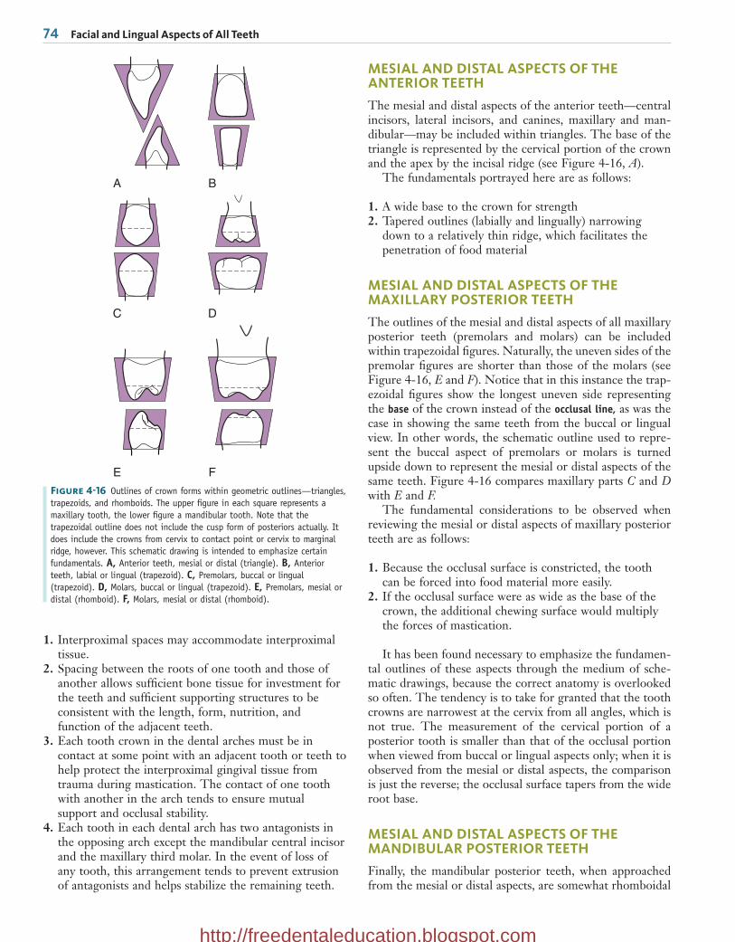
1. Interproximal spaces may accommodate interproximal tissue.
2. Spacing between the roots of one tooth and those of another allows sufficient bone tissue for investment for the teeth and sufficient supporting structures to be consistent with the length, form, nutrition, and function of the adjacent teeth.
3. Each tooth crown in the dental arches must be in contact at some point with an adjacent tooth or teeth to help protect the interproximal gingival tissue from trauma during mastication. The contact of one tooth with another in the arch tends to ensure mutual support and occlusal stability.
4. Each tooth in each dental arch has two antagonists in the opposing arch except the mandibular central incisor and the maxillary third molar. In the event of loss of any tooth, this arrangement tends to prevent extrusion of antagonists and helps stabilize the remaining teeth.
MESIAL AND DISTAL ASPECTS OF THE ANTERIOR TEETH
The mesial and distal aspects of the anterior teeth—central incisors, lateral incisors, and canines, maxillary and man-dibular—may be included within triangles. The base of the triangle is represented by the cervical portion of the crown and the apex by the incisal ridge (see Figure 4-16, A).
The fundamentals portrayed here are as follows:
1. A wide base to the crown for strength2. Tapered outlines (labially and lingually) narrowing
down to a relatively thin ridge, which facilitates the penetration of food material
MESIAL AND DISTAL ASPECTS OF THE MAXILLARY POSTERIOR TEETH
The outlines of the mesial and distal aspects of all maxillary posterior teeth (premolars and molars) can be included within trapezoidal figures. Naturally, the uneven sides of the premolar figures are shorter than those of the molars (see Figure 4-16, E and F). Notice that in this instance the trap-ezoidal figures show the longest uneven side representing the base of the crown instead of the occlusal line, as was the case in showing the same teeth from the buccal or lingual view. In other words, the schematic outline used to repre-sent the buccal aspect of premolars or molars is turned upside down to represent the mesial or distal aspects of the same teeth. Figure 4-16 compares maxillary parts C and D with E and F.
The fundamental considerations to be observed when reviewing the mesial or distal aspects of maxillary posterior teeth are as follows:
1. Because the occlusal surface is constricted, the tooth can be forced into food material more easily.
2. If the occlusal surface were as wide as the base of the crown, the additional chewing surface would multiply the forces of mastication.
It has been found necessary to emphasize the fundamen-tal outlines of these aspects through the medium of sche-matic drawings, because the correct anatomy is overlooked so often. The tendency is to take for granted that the tooth crowns are narrowest at the cervix from all angles, which is not true. The measurement of the cervical portion of a posterior tooth is smaller than that of the occlusal portion when viewed from buccal or lingual aspects only; when it is observed from the mesial or distal aspects, the comparison is just the reverse; the occlusal surface tapers from the wide root base.
MESIAL AND DISTAL ASPECTS OF THE MANDIBULAR POSTERIOR TEETH
Finally, the mandibular posterior teeth, when approached from the mesial or distal aspects, are somewhat rhomboidal
Figure 4-16 Outlines of crown forms within geometric outlines—triangles, trapezoids, and rhomboids. The upper figure in each square represents a maxillary tooth, the lower figure a mandibular tooth. Note that the trapezoidal outline does not include the cusp form of posteriors actually. It does include the crowns from cervix to contact point or cervix to marginal ridge, however. This schematic drawing is intended to emphasize certain fundamentals. A, Anterior teeth, mesial or distal (triangle). B, Anterior teeth, labial or lingual (trapezoid). C, Premolars, buccal or lingual (trapezoid). D, Molars, buccal or lingual (trapezoid). E, Premolars, mesial or distal (rhomboid). F, Molars, mesial or distal (rhomboid).
A
C
E
B
D
F
74 Facial and Lingual Aspects of All Teeth
http://freedentaleducation.blogspot.com
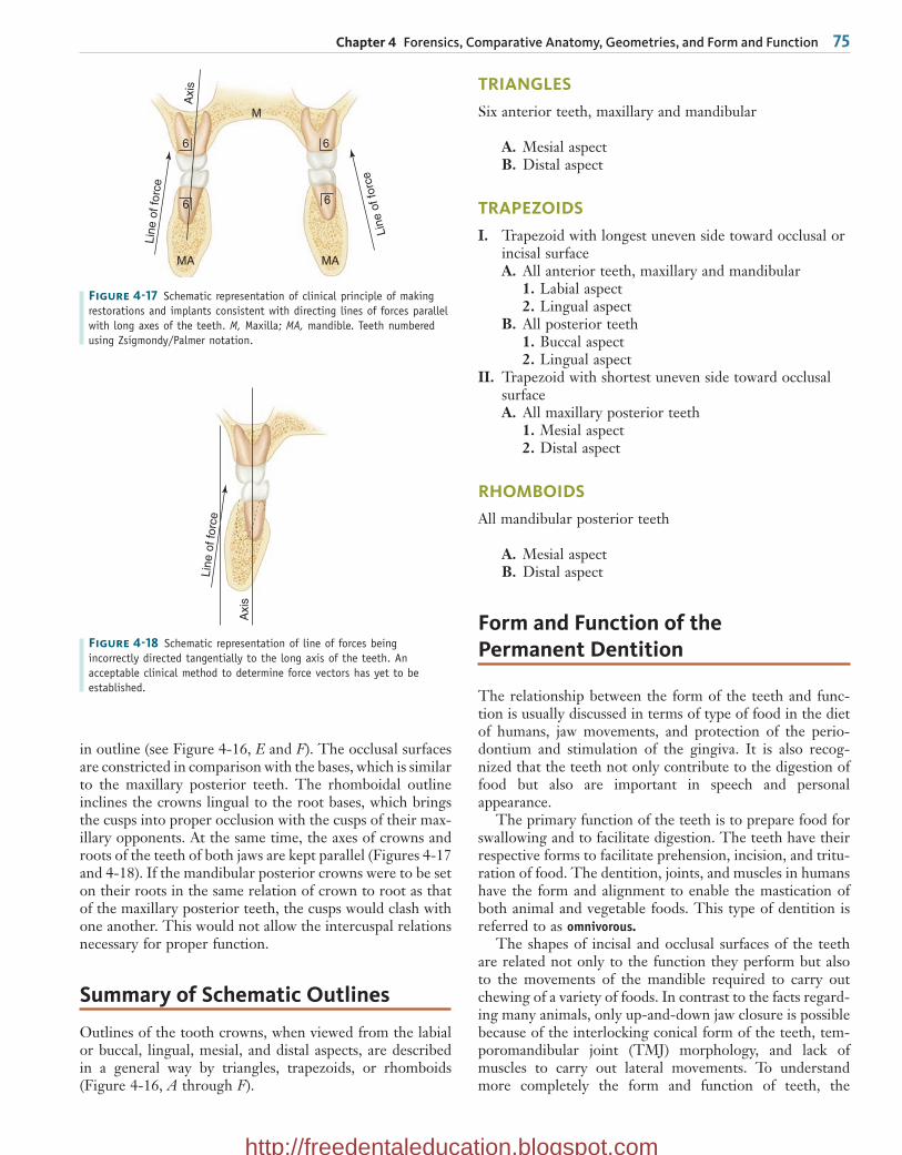
Chapter 4 Forensics, Comparative Anatomy, Geometries, and Form and Function 75
in outline (see Figure 4-16, E and F). The occlusal surfaces are constricted in comparison with the bases, which is similar to the maxillary posterior teeth. The rhomboidal outline inclines the crowns lingual to the root bases, which brings the cusps into proper occlusion with the cusps of their max-illary opponents. At the same time, the axes of crowns and roots of the teeth of both jaws are kept parallel (Figures 4-17 and 4-18). If the mandibular posterior crowns were to be set on their roots in the same relation of crown to root as that of the maxillary posterior teeth, the cusps would clash with one another. This would not allow the intercuspal relations necessary for proper function.
Summary of Schematic Outlines
Outlines of the tooth crowns, when viewed from the labial or buccal, lingual, mesial, and distal aspects, are described in a general way by triangles, trapezoids, or rhomboids (Figure 4-16, A through F).
TRIANGLES
Six anterior teeth, maxillary and mandibular
A. Mesial aspectB. Distal aspect
TRAPEZOIDS
I. Trapezoid with longest uneven side toward occlusal or incisal surfaceA. All anterior teeth, maxillary and mandibular
1. Labial aspect2. Lingual aspect
B. All posterior teeth1. Buccal aspect2. Lingual aspect
II. Trapezoid with shortest uneven side toward occlusal surfaceA. All maxillary posterior teeth
1. Mesial aspect2. Distal aspect
RHOMBOIDS
All mandibular posterior teeth
A. Mesial aspectB. Distal aspect
Form and Function of the Permanent Dentition
The relationship between the form of the teeth and func-tion is usually discussed in terms of type of food in the diet of humans, jaw movements, and protection of the perio-dontium and stimulation of the gingiva. It is also recog-nized that the teeth not only contribute to the digestion of food but also are important in speech and personal appearance.
The primary function of the teeth is to prepare food for swallowing and to facilitate digestion. The teeth have their respective forms to facilitate prehension, incision, and tritu-ration of food. The dentition, joints, and muscles in humans have the form and alignment to enable the mastication of both animal and vegetable foods. This type of dentition is referred to as omnivorous.
The shapes of incisal and occlusal surfaces of the teeth are related not only to the function they perform but also to the movements of the mandible required to carry out chewing of a variety of foods. In contrast to the facts regard-ing many animals, only up-and-down jaw closure is possible because of the interlocking conical form of the teeth, tem-poromandibular joint (TMJ) morphology, and lack of muscles to carry out lateral movements. To understand more completely the form and function of teeth, the
Figure 4-17 Schematic representation of clinical principle of making restorations and implants consistent with directing lines of forces parallel with long axes of the teeth. M, Maxilla; MA, mandible. Teeth numbered using Zsigmondy/Palmer notation.
Line
of f
orce
Line
of f
orce
MA MA
M
Axi
s
66
6 6
Figure 4-18 Schematic representation of line of forces being incorrectly directed tangentially to the long axis of the teeth. An acceptable clinical method to determine force vectors has yet to be established.
Line
of f
orce
Axi
s
http://freedentaleducation.blogspot.com

protective aspects of form and their functional relationships are considered.
Alignment, Contacts, and Occlusion
When the teeth in the mandibular arch come into contact with those in the maxillary arch in any functional relation, they are said to be in occlusion. The term occlusion is also used to designate the anatomical alignment of the teeth and their relationship to the rest of the masticatory system. Malocclusion is a term usually used to describe deviations in intramaxillary and/or intermaxillary relations of the teeth and/or jaws. Occlusion is considered in more detail in Chapter 16.
As demonstrated in Chapter 1, especially relative to Table 1-1, a very low probability exists of drawing at random one tooth of each class and type from a large sample of natural teeth with the expectation of being able to place them in proper alignment and articulation, as in Figures 1-16 through 1-18 in Chapter 1. This fact should indicate the complexity of the process of development and eruption of the teeth into a normal occlusion, which means mini-mally the proper alignment, and arrangement of the teeth within each jaw and the interdigitation of the teeth between the jaws.32 In addition, each tooth (including implants) in the arch should be placed in its most advantageous angle to withstand the forces brought to bear on it. The angles of the teeth are considered in Chapter 16 (see Figure 16-20). A general restorative principle states that occlusal forces in dental restorations should be directed along the long axis of the teeth (see Figure 4-17). Although this concept seems appropriate, it is not evidence based (i.e., no randomized clinical trial results support the concept). Unfortunately, at this time, no acceptable method of testing the hypothesis is available (i.e., no dynamic clinical biomechanical method, including telemetry, has determined the vector forces gen-erated between teeth during function). However, some evi-dence suggests that tangential loading (see Figure 4-18) results in reduced chewing forces and that negative feed-back from receptors in the periodontium mediate chewing forces. Receptor thresholds for axially directed forces appear to be higher than those for tangentially directed forces and suggest a positive feedback control on axially directed tooth forces. To position dental implants correctly, an intraoral appliance is used to guide the placement of the implant into a position and angulation consistent with angulations of opposing teeth or those suggested in Box 16-1 and Figure 16-20.
The buccal and lingual contours of the teeth have an influence on the way in which food is directed to and away from the gingival tissues. When a tooth is normally posi-tioned, the gingival margin and sulcus have a physiological relationship to the tooth in function. Food impaction, impinging trauma from tough foods, and accumulation of dental plaque may be the consequence of malposed teeth or overcontouring or undercontouring of restorations involv-ing buccolingual surfaces. The exact relationship between
the contouring of restorations and gingival health is not clear. Ideas concerning the relationship of faulty buccal con-tours (undercontouring) to the initiation of gingivitis have been challenged. Plaque control by toothbrushing may be more important than simple deviations of the contour. However, overcontoured and undercontoured restorations should be avoided. Overcontouring of facial and lingual surfaces may inhibit natural cleaning by the tongue, cheeks, and passage of food and may require special toothbrushing methods.
INTERPROXIMAL FORM
Proximal contacts of approximating teeth in the arch protect the soft tissues (gingiva) between the teeth and are referred to as the interproximal spaces (Figures 4-19 and 4-20; see also Figure 5-1). The gingiva, which normally fills this pyrami-dal-like space and extends from the alveolar bone to and around the proximal contacts of the teeth (see Figure 5-2, B) may not fill these spaces (Figure 4-21). In the absence of correct proximal tooth contacts and marginal ridges, food impaction may occur.
The gingiva within the interproximal space is called the gingival or interdental papilla (see Figure 5-1). Normally, the gingiva covers part of the cervical third of the tooth crowns and fills the interproximal spaces (see Figures 4-21 and 5-1). The gingival line follows the curvature but not
Figure 4-19 The mandibular centrals and laterals contact each other at the incisal third. The form of each tooth, plus the location of the contact areas, creates narrow pointed spaces between the teeth that differ from other interproximal spaces in other segments of the arches. GL, Variable gingival line representing the gingival level; CL, cervical line as established by the cementoenamel junction.
GLCL
Figure 4-20 Contact design and interproximal (sometimes called interdental) spaces illustrated by the mandibular canine and first and second premolars. Note the variation in contact areas in relation to crown length.
76 Alignment, Contacts, and Occlusion
http://freedentaleducation.blogspot.com

Chapter 4 Forensics, Comparative Anatomy, Geometries, and Form and Function 77
necessarily the level of the cervical line. The cervical line is defined as the cementoenamel junction and has been con-sidered in Chapter 2. The gingival line and the cervical line must not be thought of as being identical; although they normally follow a similar curvature, they are seldom at the same level on the tooth. The cervical line is a stable ana-tomical demarcation, whereas the gingival line merely rep-resents the gingival level on the tooth at any one period in the individual’s life, and this level is variable (e.g., gingival recession). Misalignment of the teeth may change the gin-gival line, which may not be conducive to the health of the tissue (e.g., partial absence of bone over the root of a tooth as in dehiscence [cleft] or fenestration [window]) (see Figure 4-21, B).
Even when the teeth are in good alignment, unless the proper relation is kept between the width of each tooth at the cervix and the width at the point of contact with neigh-boring teeth, the spacing interdentally is changed. This is an important point to observe in clinical examinations and to maintain in approximating restorations (e.g., do not reduce the interproximal space at the expense of the inter-dental gingival papilla).
When considering the form of the teeth from the mesial and distal aspects, it is possible to observe a curvature on the crowns at the cervical third above the cervical line, labially or buccally and lingually. It is called the cervicoenamel ridge (see Figure 1-19), or simply cervical ridge, with the location added (buccocervical ridge, and so forth). As already indi-cated, this ridge should not be overcontoured or undercon-toured in full-crown restorations.
ROOT FORM
The length and shape of the root (or roots) of each tooth must be viewed as important; the canine, for instance,
because of its position and the work required of it, would be torn out of its socket or at least displaced by forces brought to bear on it if the root were not of extra size and length. Fracture would be imminent if the root were not larger than that of other single-rooted teeth. The root form therefore is associated with the overall form of the tooth and the work it has to do.
The angle at which the incisal and occlusal surfaces of the tooth crowns are placed with respect to the root bases is also important. The mesial view of an anterior tooth will show that the incisal ridge or cusp is centered over the root (see Figure 1-4). The mesial view of an upper first molar, which is a multicusped tooth, demonstrates the same principle. The points on the occlusal surface that are contacted by opposing teeth will prove to be well within the confines of the root base of the crown. The measurement from cusp tip to cusp tip buccolingually is much less than the buccolingual diameter of the root base (see Figure 1-4). Note the flare of the roots for stabilization.
The length and shape of the roots, the angles at which the incisal and occlusal surfaces are placed with respect to the roots, sufficient dimensions for strength, and an efficient design for thorough work with resistance against lines of force suggest their importance to occlusal stability.
OCCLUSAL CURVATURE
When occlusion is considered, the concept of what consti-tutes normalcy arises. Although this is considered conceptu-ally in Chapter 16, it is appropriate to give a brief definition here. Normal implies a situation that is found commonly in the absence of disease or disorders and that exhibits an associated range of values that reflects the adaptive physio-logical range of a dynamic biological system, including the masticatory system.31 Therefore one must be careful not to
Figure 4-21 Influence of malalignment of teeth on form of gingival line and bone. A, Change in gingival line (free gingival margin) of central incisor. B, Absence of bone over root of tooth in labioversion (fenestration).
(B from Perry DA, Beemsterboer PL: Periodontology for the dental hygienist, ed 3, St Louis, 2007, Saunders.)
B
http://freedentaleducation.blogspot.com

attach rigid biomechanical significance to observations that are not yet evidence based (i.e., verified by appropriately designed clinical trials). It is also important not to reject clinical procedures based on statistically significant differ-ences when the biological significance of the difference cannot be determined.
It can be observed that the occlusal and incisal surfaces of all the crowns taken together in either arch do not contact a flat plane. For example, when the teeth are viewed from a point opposite the first molars buccally, a line fol-lowing the occlusal and incisal surfaces describes a curve. Graf von Spee described this curvature of natural teeth originally in the German literature in 1890, and for that reason, it is called the curve of Spee (Figure 4-22). No acceptable scientific evidence exists that the occlusion should be spherical (i.e., that each cusp and incisal edge touch or conform to a segment of the surface of a sphere). This kind of curvature suggested by the lingual inclination of the mandibular molars is the basis for the curve of Wilson (i.e., the curvature of the mandibular teeth is concave and that of the maxillary teeth convex). A template for these curvatures cannot be used to set teeth in complete dentures or to perform full-mouth oral rehabilitation. The degree of anterior-posterior curvature is related to the amount of anterior guidance that is needed to obtain pos-terior disclusion of the teeth in lateral and protrusive man-dibular movements.
References 1. Herschaft EE: Forensic dentistry. In Neville BW et al,
editors: Oral and maxillofacial pathology, ed 2, Philadelphia, 2002, Saunders.
2. Herschaft EE et al, editors: Manual of forensic odontology, ed 4, Albany, NY, 2007, American Society of Forensic Odontology.
Figure 4-22 Teeth are in the intercuspal position (ICP) or centric occlusion (CO). The curved line is the curve of Spee.
(From Bath-Balogh M, Fehrenbach MJ: Illustrated dental embryology, histology, and anatomy, ed 2, St Louis, 2006, Saunders.)
3. Bowers CM: Forensic dental evidence: an investigator’s handbook, San Diego, 2004, Elsevier.
4. Lampe H, Roetzcher K: Age determination from the human skeleton, Med Law 13:623, 1994.
5. Reich KJ, Demirjian A: A multimedia tool for the assessment of age remains: the electronic encyclopedia for maxillo-facial, dental and skeletal development. In Reichs KJ, editor: Forensic odontology, ed 2, Springfield, IL, 1998, Charles C Thomas.
6. Kasper K, Senn D: Reliability of third molar development on a north Texas Hispanic population: a comparison study for age estimation, Proceedings of the American Academy of Forensic Sciences, vol 10, Colorado Springs, CO, February 2004, American Academy of Forensic Sciences (abstract F19).
7. Loevy HT: Maturation of permanent teeth in black and Latino children, Acta Odontol Pediatr 4:59, 1983.
8. Willems G: A review of the most commonly used dental age estimation techniques, J Forensic Odontostomatol 19:9, 2001.
9. Pretty IA: The use of dental aging techniques in forensic odontological practice, J Forensic Sci 48:1127, 2003.
10. Moorrees CF, Fanning EA, Hunt EE: Age variation of formation stages for ten permanent teeth, J Dent Res 42:1490, 1963.
11. Mincer HH, Harris EF, Berryman HE: The ABFO study of third molar development and its use as an estimator of chronological age, J Forensic Sci 38:379, 1993.
12. Kvaal SI, Sellevold BJ, Solheim T: A comparison of different non-destructive methods of age estimation in skeletal material, Int J Osteoarchaeol 4:36, 1994.
13. Gustofson G: Age determination on teeth, J Am Dent Assoc 41:45–54, 1950.
14. Ohtani S, Abe I, Yamamoto T: An application of D- and L-aspartic acid mixtures as standard specimens for the chronological age estimation, J Forensic Sci 50:1298, 2005.
15. Wagner GN: Scientific methods of investigation. In Stimson PG, Mertz CA, editors: Forensic dentistry, Boca Raton, FL, 1997, CRC Press.
16. Sweet DJ: Bitemarks as biological evidence. In Dorion RBJ, editor: Bitemark evidence, New York, 2005, Marcel Dekker.
17. Sweet DJ, Sweet CH: DNA analysis of dental pulp to link incinerated remains of homicide victim to crime scene, J Forensic Sci 40(2):310-314, 1995.
18. Sweet DJ, Hildebrand CH: Recovery of DNA from human teeth by cryogenic grinding, J Forensic Sci 43:1199, 1998.
19. Sweet DJ: Why a dentist for identification? Dent Clin North Am 45:241, 2001.
20. Frye v United States, 293 F 10 13 (DC Cir 1923). 21. Daubert v Merrell Dow Pharmaceuticals, 509 US 579, 113 S
Ct 2786, 125 LEd2d 469 (1993). 22. American Board of Forensic Odontology: guidelines for bite
mark analysis, J Am Dent Assoc 112:383, 1986. 23. Osborn HF: Evolution of mammalian molar teeth. In
Gregory WK, editor: Biological studies and addresses, vol 1, New York, 1907, Macmillan.
24. Gregory WK: The origin and evolution of human dentition, Baltimore, 1906, Williams & Wilkins.
25. Thompson AH: Comparative dental anatomy, ed 2, revised by Martin Dewey, St Louis, 1915, Mosby.
26. Kronfeld RL: Dental histology and comparative dental anatomy, Philadelphia, 1937, Lea & Febiger.
27. Ferguson MW: Review of the value of the American alligator (Alligator mississippensis) as a model for research in
78 References
http://freedentaleducation.blogspot.com

Chapter 4 Forensics, Comparative Anatomy, Geometries, and Form and Function 79
craniofacial development, J Craniofac Genet Dev Biol 1:123, 1982.
28. Tims HWM: Notes on the dentition of the dog, Anat Anz Bd 11:537, 1896.
29. Cope EE: On the tritubercular molar in human dentition, J Morphol 2:7, 1888.
30. Wortman JL: Origin of the tritubercular molar, Am J Sci 13:93, 1902.
31. Ryder JA: On the mechanical genesis of tooth-forms, Proc Acad Nat Sci Philadelphia, 1878.
32. Ash MM, Ramjford SP: Occlusion, ed 4, Philadelphia, 1995, Saunders.
BibliographyAsh MM: Physiology of the mouth. In Bunting RW, editor: Oral
hygiene, ed 3, Philadelphia, 1954, Lea & Febiger.Barker BC: Dental anthropology: some variations and anomalies
on human tooth form, Aust Dent J 18:132, 1973.Cooke WP: Value and use of temporary teeth, Br J Dent Sci
66:267, 1923.Farer JW, Isaacson D: Biologic contours, J Prev Dent 1:4, 1974.Fixott RH, editor: Forensic odontology, Dent Clin North Am
45(2, theme issue):217, 2001.Gale GL: Dentistry, bite marks, and investigation of crime, J Cal
Dent Assoc 24:28, 1996.Graf H: Occlusal forces during function. In Graf H, editor:
Occlusion: research in form and function (symposium report), Ann Arbor, 1975, University of Michigan.
Hellman M: The relationship of form to position in teeth and its bearing on occlusion, Dent Items Interest 42:161, 1920.
Humphreys HF: Function in the evolution of man’s dentition, Br Dent J 42:939, 1921.
Lindhe J, Wicen PO: The effects on the gingiva of chewing fibrous foods, J Periodont Res 4:193, 1969.
Perel ML: Periodontal considerations of crown contours, J Prosthet Dent 26:627, 1971.
Ramfjord SP, Ash MM: Periodontology and periodontics, Philadelphia, 1979, Saunders.
Russell ES: Form and function, New York, 1917, Dutton.Shaw DM: Form and function of teeth: a theory of maximum
shear, J Anat Physiol 13:97, 1917.Wheeler RC: Complete crown form and the periodontium,
J Prosthet Dent 11:722, 1961.Youdelis RA, Weaver JD, Sapkos S: Facial and lingual contours
of artificial complete crown restorations and their effect on the periodontium, J Prosthet Dent 19:61, 1973.
Use the CD-ROM to view animations and study ques-tions; and go to the site for additional study resources.
http://freedentaleducation.blogspot.com

This page intentionally left blank
http://freedentaleducation.blogspot.com

Orofacial Complex: Form and Function
that in restorative dentistry, the dependent relationship between the original biomechanical behavior of stress con-centrations in incisor teeth6 and the nature of the materials used to restore these teeth is addressed. Thus the term form not only means shape but also biomechanical attributes that contribute to the maintenance of function. Other examples include the dependent relationship between esthetics and optimum occlusion,7 physical forces and the periodontal ligament,8 and arch form and beta function,2 as well as the controversial interrelationship between the morphology and function of the discomuscular apparatus of the human tem-poromandibular joint. Although the function of implant-supported restorations seems to be generally achieved, the esthetics and biocompatibility of the materials used (i.e., design and fabrication of restorative components with dimensions that closely resemble natural tooth anatomy and esthetics) remain a challenge.9,10
Articulation of Teeth
Anyone who has carved a set of Ivorine teeth, even to the dimensions set forth in Table 1-1, can attest to the difficulty of aligning the carved teeth into what might be thought to be only one proper articulation of the teeth on an articula-tor. Form and function are not a template by which it is possible to draw a straight-line correlation between the shape of a tooth and all of its functions. A restored crown of a maxillary central incisor should meet standards of func-tion and esthetics (see Figure 1-1).
This chapter discusses primarily the physiological form of the teeth and periodontium, with consideration of the cov-erage of form and function in Chapter 4 and the coverage of orofacial structures in other chapters on muscles and joints.
Form and Function
The phrase form and function reflects a concept of interrela-tion of the shape or attributes of something with its func-tion. In dentistry the phrase may indicate the entire masticatory system, acting as a biomechanical engine for the reduction of food. If so, then arguably it may be possible to try to make a case that each of the components of the mas-ticatory system has some kind of physiological relationship to its individual functions and to that of the whole system, including its maintenance. These relationships have a spec-trum of possible associations: speech and jaw motion1; arch form and mathematical function2; integration of form, func-tion, and esthetics in mandibular anterior ceramic veneers3; morphological and functional analysis of the orofacial complex4; and the condylar disk as the controlling factor in the form of the condylar head.5 The search for the meaning of form and function continues.
Form Follows Function
For some clinicians the general principle that becomes operant is form follows function, which means, for example,
81
55
http://freedentaleducation.blogspot.com

Physiological Form of the Teeth and Periodontium
The form of the teeth is consistent with the function they are to perform and with their position and arrangement in the structures involved in oral motor behavior, especially mastica-tion. To attempt to relate form and function in all the struc-tures involved in such functions is beyond the scope of this book, and the reader is referred to several of the publications listed in the references and bibliography of this chapter.
Fundamental Curvatures
It can be assumed that the form of the teeth and their arrangement are related to incising or crushing food without causing damage to the supporting structures; for them to be otherwise might be inconsistent with the survival of the species. Some of the form must also relate to that of the jaws and face and to occlusal forces that dictate that the teeth be at various angles and positions in the dental arches. Beyond assumptions or teleological approaches to the morphology of the teeth, the relationships of tooth form to the form of the supporting structures, including the gingiva, must be considered in terms of clinical significance. Thus food impaction may occur as a result of gingival enlargement and of the driving of food between the teeth because of improper marginal ridges and/or contact areas, irrespective of teleo-logical considerations of tooth morphology.
Although a great deal has been said about the relationship of the health of the gingiva to the contours of the teeth, as indicated in the references included at the end of this chapter, such appraisals are generally descriptive, involve restorations, and are retrospective observations. To conclude that the gin-gival tissues around a tooth (natural or restored) need neither stimulation nor protection suggests that the true significance of buccolingual crown contours has not been evaluated. In no instance has the influence of axial contours, which begin with the height of the contour of buccal and lingual surfaces, been tested in relation to such functions as chewing efficiency or occlusal stability. And even though the significance of buc-colingual crown contours to gingival health relative to gingi-val stimulation, self-cleansing mechanisms, and gingival protection has been seriously questioned, the significance of other contours has also been questioned. Dental plaque accu-mulates on teeth in the absence of adequate oral hygiene, and gingivitis occurs in spite of “self-cleansing” mechanisms.11 The influence of a primitive diet does not appear to prevent gingivitis. However, plaque formation and gingivitis may be related more to bacterial attachment mechanisms, bacterial toxins, and food substrates than to physical form. It should not be concluded from such observations that curvatures have nothing to do with the function and health of the masticatory system. The effect of overcontouring or undercontouring of surfaces of the teeth may be related more to occlusal mecha-nisms than to self-cleansing mechanisms and be of more sig-nificance to food self-cleansing efficiency and/or musculature
in one patient than in another. The role of protective reflexes in relation to the contours of the teeth is not known. Even though the significance of tooth forms has yet to be clarified, ideas about form and function that have been held should be understood, and liberties with form avoided. The following are considered:
1. Proximal contact areas2. Interproximal spaces (formed by proximal surface in
contact)3. Embrasures (spillways)4. Labial and buccal contours at the cervical thirds
(cervical ridges) and lingual contours at the middle thirds of crowns
5. Curvatures of the cervical lines on mesial and distal surfaces (cementoenamel junction [CEJ])
These headings include form, which has been considered to have a direct or primary bearing on the protection of the periodontium. Many other details of tooth form may have an indirect bearing on the stability of the teeth through their contribution to the maintenance of efficiency during func-tion. Some of these details are cusp forms, the proportions of various measurements of the crowns and roots, root form, and anchorage and angles at which teeth are set in the jaws. It has been suggested that when well-formed teeth are in normal alignment with normal gingival contour (Figures 5-1 and 5-2), the efficient use of the toothbrush during home care of the teeth is enhanced.11
Proximal Contact Areas
Soon after the alignment of all of the teeth in their respec-tive positions in the jaws takes place, a positive contact relation of one tooth with another in each arch should be established mesially and distally (Figure 5-3). Except for the
Figure 5-1 Clinical appearance of gingiva. A, Attached gingiva above and interdental papilla below; B, mucogingival line separating attached gingival from mucosa; C, free gingival margin; D, posterior vestibular fornix; E, anterior vestibular fornix or mucobuccal fold; F, frenum area.
(From Bird DL, Robinson DS: Torres and Ehrlich modern dental assisting, ed 9, St Louis, 2009, Saunders.)
Mucogingival junction
Mandibularbuccalfrenum
Mandibularvestibule
Attachedgingiva
Interdentalgingiva
Maxillaryvestibule
Marginalgingiva
Maxillarylabialfrenum
Alveolarmucosa
C
D F
E
A
B
82 Proximal Contact Areas
III A-H
http://freedentaleducation.blogspot.com

Chapter 5 Orofacial Complex: Form and Function 83
Figure 5-2 A, Principal fibers of the supporting structures. B, Schematic representation of periodontal structure at the junction of the enamel, cementum, and dentin showing attachment of gingival tissue to the tooth via the junctional epithelium. E, Enamel.
(From Ramfjord SR, Ash MM: Periodontology and periodontics, Philadelphia, 1979, Saunders.)
Obliquefibers
Dentoalveolarcrest fibers
Horizontalfibers
Interradicalfibers
Transseptalfibers
Apicalfibers
GingivalcreviceFree gingival marginKeratinizedepithelium
Gingivalgroove
JunctionalepitheliumAttachedgingiva
MucogingivaljunctionAlveolarmucosa
Alveolar process
Alveolarbone
Crevicularepithelium
DENTIN
Epithelialattachment
Cementum
Alveolarcrest
Periodontalmembrane
E
A
B
Figure 5-3 Proximal “contacts” vary in size in relation to type of teeth and wear of these areas. Note flat contacts in molar region compared with absence of flattening in relation to space between canine and premolar. The form of the interproximal spaces is altered by wear of contact areas, extrusion of teeth, or tipping of teeth.
last molars (third molars, if present), each tooth has two contacting members adjoining it. The last molar is in contact only with the tooth mesial to it. Although the areas of contact are still very circumscribed, especially on anterior teeth, these are areas and not mere points of contact (Figure 5-4). Actually, the term contact point, which is often used to designate the contact of teeth in the same arch, is a misno-mer. When the individual is quite young and the teeth are newly erupted, some of the teeth come close to having point contacts only when the contacting surfaces are nearly perfect curvatures. Examples of the few contacts made by such rounded surfaces are contacts located distally on canines and mesially on first premolars, maxillary and mandibular.
The proper contact relation between neighboring teeth in each arch is important for the following reasons: it serves to keep food from packing between the teeth, and it helps stabilize the dental arches by the combined anchorage of all the teeth in either arch in positive contact with each other (Figures 5-5 and 5-6; see also Figure 5-3). Except for the third molars, each tooth in the arch is supported in part by its contact with two neighboring teeth, one mesial and one distal. The third molars (and the second molars if no third molar is present) are prevented from drifting distally where there is no contacting tooth by the angulation of their occlu-sal surfaces with their roots and by the angle of the direction of the occlusal forces in their favor. This is explained more fully in later chapters.
If, for any reason, food is forced between the teeth past the contact areas, the result may be pathological. The gin-gival tissue, which normally fills the interdental spaces, may become inflamed (gingivitis) and ultimately involve deeper periodontal structures with loss of bone and attachment (periodontitis).
Excessive occlusal forces on an individual tooth may occur when normal forces are no longer distributed over several teeth, as may happen when teeth are lost or when normal forces become excessive with loss of supporting structures as a result of periodontal diseases.
Contact areas must be observed from two aspects to obtain the proper perspective for locating them: the labial or buccal aspect, and the incisal or occlusal aspect (Figures 5-7 and 5-8).
Figure 5-4 Schematic representation of form of gingiva in relation to contact area.
http://freedentaleducation.blogspot.com

Interproximal Spaces (Formed by Proximal Surfaces in Contact)
The interproximal spaces between the teeth are triangularly shaped spaces normally filled by gingival tissue (gingival papillae). The base of the triangle is the alveolar process, the
Figure 5-5 Contact areas. A, “Contacts” without evidence of dysfunction. B, “Restored” contact areas associated with dysfunction from food impaction. C, Loss of contacts associated with bone loss due to periodontal disease.
A
B
C
Figure 5-6 Relationship of cusps to embrasures in some dentitions. A, Casts in the intercuspal position and teeth in “normal” occlusion. B, Other casts in intercuspal position with alignment of the teeth preventing normal contact relations between cusps and embrasure areas.
A
B
The labial or buccal view will demonstrate the relative positions of the contact areas cervicoincisally or cervico-occlusally. The center of the area from this aspect is gauged by its relation to the length of the crown portion of the tooth (Figure 5-9).
The incisal or occlusal view will show the relative position of the contact areas labiolingually or buccolingually. In this instance, the center of the area may be located in its relation to the labiolingual or buccolingual measurement of the crown (Figure 5-10; see also Figure 5-8). The point at which the contact area is bisected also depends on the outline of the form of the crown from the incisal or occlusal aspect. This outline is governed by the alignment of the tooth in the arch and also by the occlusal relation with its antagonists in the opposing arch. The mandibular first molar is an excel-lent example (Figure 5-11; see also Figure 5-10). The contact and embrasure design for this tooth is explained later when the incisal and occlusal aspects of the teeth are considered.
Figure 5-7 Contact levels found normally on mandibular teeth. Arrows point to embrasure spaces. A, Central and lateral incisors. B, Central and lateral incisors and canine. C, Lateral incisor, canine, and first premolar. D, Canine and first and second premolars. E, First and second premolars and first molar. F, Second premolar and first and second molars. G, First, second, and third molars.
A B C
ED
F G
84 Interproximal Spaces (Formed by Proximal Surfaces in Contact)
http://freedentaleducation.blogspot.com

Chapter 5 Orofacial Complex: Form and Function 85
sides of the triangle are the proximal surfaces of the contact-ing teeth, and the apex of the triangle is in the area of contact. The form of the interproximal space will vary with the form of the teeth in contact and will depend also on the relative position of the contact areas (Figures 5-12 through 5-14). Normally, a separation of 1 to 1.5 mm exists between the enamel and alveolar bone. Thus the distance from the CEJ (cervical line) to the crest of the alveolar bone as seen radiographically (see Figure 5-9) is 1 to 1.5 mm in a normal occlusion in the absence of disease.
Proper contact and alignment of adjoining teeth allows proper spacing between them for the normal bulk of gingi-val tissue attached to the bone and teeth. This gingival tissue is a continuation of the gingiva covering all of the alveolar process. The surface keratinization of the gingiva and the density and elasticity of the gingival tissues help maintain these tissues against trauma during mastication and invasion by bacteria.
Because the teeth are narrower at the cervix mesiodistally than they are toward the occlusal surfaces and the outline of the root continues to taper from that point to the apices of the roots, considerable spacing is created between the roots of one tooth and the roots of adjoining teeth. This arrangement allows sufficient bone tissue between one tooth and another, which anchors the teeth securely in the jaws.
Figure 5-8 Outline drawings of the maxillary teeth from the incisal and occlusal aspects with broken lines bisecting the contact areas. These illustrations show the relative positions of the contact areas labiolingually and buccolingually. Arrows point to embrasure spaces. A, Central incisors and lateral incisor. B, Central and lateral incisors and canine. C, Lateral incisor, canine, and first premolar. D, Canine, first premolar, and second premolar. E, First molar, second premolar, and first molar. F, Second premolar, first molar, and second molar. G, First, second, and third molars.
A
C D
E F
G
B
Figure 5-9 Radiograph demonstrating form of alveolar crest, contact areas, and relation to form of the crown.
http://freedentaleducation.blogspot.com

It also simplifies the problem of space for the blood and nerve supply to the surrounding alveolar process and other investing tissues of the teeth (see Figure 5-12).
The type of tooth also has a bearing on the interproximal space. Some individuals have teeth that are wide at the cer-vices, constricting the space at the base. Others have teeth that are more slender at the cervices than usual; this type of tooth widens the space. Teeth that are oversize or unusually small will likewise affect the interproximal spacing. Never-
theless, this spacing conforms to a plan that is fairly uniform, provided that the anatomical form is normal and the teeth are in good alignment.
Embrasures (Spillways)
When two teeth in the same arch are in contact, their cur-vatures adjacent to the contact areas form spillway spaces called embrasures. The spaces that widen out from the area of contact labially or buccally and lingually are called labial or buccal and lingual interproximal embrasures. These embra-sures are continuous with the interproximal spaces between the teeth (see Figure 5-10). Above the contact areas incisally and occlusally, the spaces, which are bounded by the mar-ginal ridges as they join the cusps and incisal ridges, are called the incisal or occlusal embrasures. These embrasures, and the labial or buccal and lingual embrasures, are continu-ous (Figure 5-15; see also Figure 5-8). The curved proximal surfaces of the contacting teeth roll away from the contact area at all points, occlusally, labially or buccally, and lin-gually and cervically, and the embrasures and interproximal spaces are continuous, as they surround the areas of contact.
The form of embrasures serves two purposes: (1) it pro-vides a spillway for food during mastication, a physiological form that reduces the forces brought to bear on the teeth during the reduction of any material that offers resistance; and (2) it prevents food from being forced through the contact area. When teeth wear down to the contact area so that no embrasure remains, especially in the incisors, food is pushed into the contact area even when teeth are not mobile.
The design of contact areas, interproximal spaces, and embrasures varies with the form and alignment of the
Figure 5-10 Contact relations in a patient with “normal” occlusion. A, Maxillary arch. B, Mandibular arch.
A
B
Figure 5-11 Broad contact areas of the mandibular first and second molars in a young adult, 21 years old.
Figure 5-12 Schematic drawing of distribution of the periodontal blood vessels and interdental papillae. 1, Inferior alveolar artery; 2, dental arteriole; 3, pulpal branches; 4, periodontal ligament arteriole; 5 and 6, interalveolar arterioles.
(From Ramfjord SR, Ash MM: Periodontology and periodontics, Philadelphia, 1979, Saunders.)
1
2
445
6
3
86 Embrasures (Spillways)
http://freedentaleducation.blogspot.com

Chapter 5 Orofacial Complex: Form and Function 87
various teeth; each section of the two arches shows simi-larity of form. In other words, the contact form, the inter-proximal spacing, and the embrasure form seem rather constant in sectional areas of the dental arches. These sections are named as follows: the maxillary anterior section, the mandibular anterior section, the maxillary posterior section, and the mandibular posterior section. All embrasure spaces are reflections of the form of the teeth involved. Maxillary central and lateral incisors exhibit one embrasure form, the mandibular incisors another, and so on.
Maxillary posteriors and mandibular posteriors appar-ently require an embrasure design geared for their sections. In some cases, the constancy has to be attained by a tooth form adaptation (Figure 5-16). The canines, for instance, are shaped so that they act as a catalyst in these matters between anterior and posterior teeth. A line bisecting the labial portion of either canine seems to create an anterior
Figure 5-13 The form of the gingiva is related to the form of the teeth, contact areas, spacing between the teeth, and effects of periodontal disease and dental caries. A, Interdental papillae do not fill the interproximal areas in several places because of spacing between the teeth. B, Clinically normal gingivae; the form is different because of the form of the teeth, including contact areas.
(From Bath-Balogh M, Fehrenbach MJ: Illustrated dental embryology, histology, and anatomy, ed 2, St Louis, 2006, Saunders.)
AlveolarmucosaMucogingivaljunctionAttachedgingivaMaxillary
teeth
Mandibularteeth
A B
Figure 5-14 The form of the teeth, position and wear of contact areas, type of teeth, and level of eruption of the teeth determine the form of the interproximal “spaces.” These factors also determine the interproximal shape of the crest of alveolar bone.
Figure 5-15 Outline drawings of the maxillary teeth in contact, with dotted lines bisecting the contact areas at the various levels as found normally. Arrows point to embrasure spaces. A, Central and lateral incisors. B, Central and lateral incisors and canine. C, Lateral incisor, canine, and first premolar. D, Canine and first and second premolars. E, First and second premolars and first molar. F, Second premolar, first molar, and second molar. G, First, second, and third molars.
A
D E
F G
B C
Figure 5-16 Calibration of maxillary first molar. A, Calibration at prominent buccal line angles that functions by deflecting food material during mastication. B, Calibration of lingual contour from contact area mesially to contact area distally. There are no prominent line angles, but the development of the mesiolingual cusp has widened the rounded lingual form. Thus the two lingual embrasures are kept similar in size, even though the tooth makes contact with two teeth that are dissimilar lingually. This way, the lingual gingival tissue interproximally is properly protected by the equalization of lingual embrasures.
A
B
III A, B
http://freedentaleducation.blogspot.com

half mesially that resembles half of an anterior tooth and a posterior half that resembles a posterior tooth. The mesial contact is at one level for contact with the lateral incisor, but the distal contact form must be at a level consistent with the contact form of the first premolar, whether maxillary or mandibular (see Figure 5-15, C).
Contact Areas and Incisal and Occlusal Embrasures from the Labial and Buccal Aspect
It is advisable to refer continually to the illustrations of contacts and embrasures during the reading of the descrip-tions that follow. Locate the illustration of interest (see Figure 5-15, A) while reading the details concerning contact area levels in the following paragraphs.
MAXILLARY TEETH
Central IncisorsThe contact areas mesially on both central incisors are located at the incisal third of the crowns. Since the mesio-incisal third of these teeth approaches a right angle, the incisal embrasure is very slight.
Central and Lateral IncisorsThe distal outline of the central incisor crown is rounded. The lateral incisor has a shorter crown and has a more rounded mesioincisal angle than the central incisor. The form of these two teeth coming into contact with each other therefore opens up an embrasure space distal to the central incisors larger than the small one mesial to the central incisors. A line bisecting the contact areas distal to the central incisor and mesial to the lateral incisor approaches the junction of the middle and incisal thirds of each crown.
Lateral Incisor and CanineThe distal contact area on the lateral incisor is approxi-mately at the middle third. The mesial contact area on the canine is at the junction of the incisal and middle thirds. The form of these teeth creates an embrasure that is more open than the two previously described.
Canine and First PremolarThe canine has a long distal slope to its cusp, which puts the distal crest of curvature at the center of the middle third of the crown. The contact area is therefore at that point. This is an important observation to be made clini-cally. As mentioned, it is at this point in the dental arch that the canine, situated between the anterior and poste-rior segments, becomes a part of both (see Figure 5-15, B and C).
The first premolar has a long cusp form also, which puts its mesial contact area rather high up on the crown. Usually it is just cervical to the junction of the occlusal and middle
thirds. The embrasure between these teeth has a wide angle.
First and Second PremolarsThe contact areas of the first and second premolars are similar to those just mentioned, although usually a little cervical to the junction of the occlusal and middle thirds of the crowns. The form of these teeth creates a wide occlusal embrasure.
Note that the design of the interproximal spaces changes also with the form and dimensions of the teeth in contact.
Second Premolar and First MolarThe position of the second premolar and first molar contact areas cervico-occlusally is about the same as that found between the premolars.
First and Second and Second and Third MolarsThe two contact and embrasure forms for the first and second and second and third molars may be described together, since they are similar. The distal outline of the first molar is round, a fact that puts the contact area approxi-mately at the center of the middle third of the crown. Here again, it must be emphasized that contact levels on maxillary molars (and even on premolars to some extent) tend to be centered in the middle third of the anatomical crown.
The mesial contact area of the second molar also approaches the middle third of the crown. The occlusal embrasure is generous as a consequence, even though the cusps are not long.
The contact and embrasure design of the second and third molars is similar to that of the first and second molars. The molars become progressively shorter from the first through the third. Again, the dimensions of the tooth crowns affect the contact and embrasure design.
MANDIBULAR TEETH
Central IncisorsThe mesial contact areas on the mandibular central incisors are located at the incisal third of the crowns. At the time of the eruption of these teeth, the mesial and distal incisal angles are slightly rounded, and the mamelons are noticea-ble on the incisal ridges. Soon, however, incisal wear reduces the incisal ridge to a straight surface, and the mesial and distal angles approach right angles in sharpness. This is due partly to wear at the contact areas (see Figures 5-7 and 5-14). In many instances, the contact areas extend to the mesioincisal angle. Therefore a small incisal embrasure occurs mesially between the mandibular central incisors unless wear through usage obliterates it.
Central and Lateral IncisorsThe distal contact areas and the incisal embrasures on the central incisors and the mesial contact areas and incisal embrasures on the lateral incisors are similar to those just described. Because the mandibular central and lateral inci-sors are small mesiodistally and supplement each other in
88 Contact Areas and Incisal and Occlusal Embrasures from the Labial and Buccal Aspect
http://freedentaleducation.blogspot.com

Chapter 5 Orofacial Complex: Form and Function 89
function, the design of their crowns brings about similar contact and embrasure forms.
Note the slender Gothic arch–like spaces that circum-scribe the interproximal spaces between the mandibular anterior teeth.
Lateral Incisor and CanineThe positions of the contact areas distally on the lateral incisor and mesially on the canine are approximately the same, cervicoincisally, as the other two just described. The teeth are in contact at the incisal third close to the incisal ridges. However, the mesioincisal angle of the canine is more rounded than the others, which form opens up a small incisal embrasure at this point.
The interproximal spacing between lateral and canine is very similar in outline to the two interproximal spaces just described.
Canine and First PremolarThe distal slope of the cusp of the mandibular canine is pronounced and long, which places the distal contact area on this tooth somewhat cervical to the junction of its incisal and middle thirds.
The first premolar has a long buccal cusp, and although its crown is shorter than that of the canine, the mesial contact area has about the same relation cervico-occlusally as that found distally on the canine and is just cervical to the junction of the occlusal and middle thirds. Thus the whole arrangement places these contact areas level with each other.
The occlusal embrasure is quite wide and pronounced because of the cusp forms of the two teeth. The interproxi-mal space has been reduced by the lowering of the contact areas cervically, comparing favorably to the design for pos-terior mandibular teeth.
First and Second PremolarsFrom the buccal aspect, the crowns of the first and second premolars are similar. The buccal cusp of the second premo-lar is not quite as long as that of the first premolar. The contact of these teeth is nearly level with that of the canine and first premolar. The slope of the cusps creates a large occlusal embrasure. The interproximal space is a little smaller than that between the canine and first premolar.
Second Premolar and First MolarThe contact and embrasure design for the second premolar and first molar is similar to that just described for the premo-lars. The mesiobuccal cusp of the first molar is shorter and more rounded than the cusp of the second premolar, which varies the embrasure somewhat, and since the crown of the molar is a little shorter, it reduces the interproximal space to that extent.
First and Second and Second and Third MolarsThe two contact and embrasure designs of the first and second and second and third molars may be described together, since they are similar.
The proximal surfaces—that is, the distal surface of the first molar, mesial surface of the second molar, distal surface of the second molar, and mesial surface of the third molar—are quite round. The occlusal embrasures are therefore gen-erous above the points of contact, even though the cusps are short and rounded.
Because the molars become progressively shorter from the first to the third, the centers of the contact areas also drop cervically. A line bisecting the contact areas of the second and third molars is located approximately at the center of the middle thirds of the crowns.
The interproximal spaces have been reduced considera-bly because of their shortened form.
Contact Areas and Labial, Buccal, and Lingual Embrasures from the Incisal and Occlusal Aspects
To study the relative positions of contact areas and the related labial, buccal, and lingual embrasures and also to get proper perspective, the eye must be directed at the incisal surfaces of anterior teeth and directly above the surface of each tooth being examined in series (Figure 5-17; see also Figures 5-8, 5-10, and 5-11). Posterior teeth are examined in the same manner. Look down on each tooth or group of teeth by facing the occlusal surfaces on a line with the long axis.
The problem at this point is to discover the relative positions of contacts in a labiolingual or buccolingual direction and to observe the embrasure form facially and lingually created by the tooth forms and their contact relations.
Figure 5-17 Contact relation of mandibular teeth labiolingually and buccolingually when surveyed from the incisal and occlusal aspects. Arrows point to embrasure spaces. A, Central incisors and lateral incisor. B, Central and lateral incisors and canine. C, Lateral incisor, canine, and first premolar. D, Canine, first premolar, and second premolar. E, First premolar, second premolar, and first molar. F, Second premolar and first, second, and third molars.
A B C
E
F
D
http://freedentaleducation.blogspot.com

A generalization may be established in locating contact areas faciolingually. Anterior teeth will have their contacts centered labiolingually, whereas posterior teeth will have contact areas slightly buccal to the center buccolingually. This buccal inclination must be carefully studied and must not be overemphasized.
Except for the maxillary first molar, all crowns converge more lingually than facially from contact areas. The maxil-lary first molar is the only tooth wider lingually than buc-cally (see Figure 5-16). Its formation makes a necessary adjustment of the mesiolingual embrasure when the tooth form of maxillary posteriors changes from the maxillary premolar form to the purely rhomboidal form of maxillary second and third molars. This situation is discussed more fully later on.
The narrower measurement lingually rather than facially causes wider embrasures lingually compared with facial embrasures. Compare the two types of embrasures displayed by maxillary central and lateral incisors.
MAXILLARY TEETH
See Figures 5-8 and 5-10, A.
Central IncisorsThe contact areas of the central incisors are centered labio-lingually. The labial embrasure is a V-shaped space created by the labial form of these crowns. The lingual embrasure widens out more than the labial embrasure because of the lingual convergence of the crowns (see Figure 5-10, A). Note the centering of the labioincisal edge with respect to the crown outline of these teeth, and the narrowness of the lingual surfaces in comparison with the broad labial faces.
Central and Lateral IncisorsThe contact areas of the central and lateral teeth are cen-tered labiolingually also.
Lateral Incisor and CanineThe contact area of the lateral incisor and canine is centered labiolingually on both canine and lateral incisors. The lingual embrasure is similar to that of the central and lateral incisors, but the labial embrasure is changed somewhat by a definite convexity at the mesiolabial line angle of the canine.
Canine and First PremolarThe contact area of the canine and first premolar is centered on the distal surface of the canine but is a little buccal to the center on the mesial surface of the first premolar. The embrasure design lingually is marked by a concavity in the region of the distolingual line angle of the canine and by a developmental groove crossing the mesial marginal ridge of the first premolar.
First and Second PremolarsThe contact areas of the first and second premolars are nearly centered buccolingually. The embrasures buccally
and lingually are regular in outline, although slightly differ-ent in design.
The prominence of the mesiobuccal and distobuccal line angles of the premolars is in direct contrast to the even taper of these teeth lingually, as viewed from the occlusal aspect. This form demonstrates a slight variation between buccal and lingual embrasures.
Second Premolar and First MolarAs usual, a line bisecting the contact areas of the second premolar and first molar is nearly centered on the distal surface of the second premolar. The area on the mesial surface of the first molar is located farther buccally than other contact areas on the maxillary posterior teeth. The contact areas are wider on molars because of the greater width buccolingually of the molar teeth.
The buccal embrasure between these teeth and the loca-tion of the mesial contact area of the first molar are influ-enced by the prominence of the mesiobuccal line angle of the maxillary first molar and the matching prominence of the distobuccal line angle of the maxillary second premolar. The lingual embrasure is kept standard for the molar area by the enlargement of the mesiolingual cusp of the first molar. Occasionally, this cusp carries a small conformation lingually as part of the change in form (fifth cusp, or cusp of Carabelli). Usually, the mesiolingual cusp of the maxillary first molar is rounded out, with no more than a develop-mental groove showing that an extra cusp formation may have been intended.
The mesiolingual lobe of this tooth is always large, however, causing the tooth to be wider lingually from its mesiolingual line angle to its distolingual line angle than it is from the mesiobuccal line angle to the distobuccal line angle. If it were not for this fact, the rhomboid form of the first molar in contact with the tapered form of the second premolar would open up a lingual embrasure of extremely large proportions. The large mesiolingual lobe makes up for the change in occlusal outline from premolar form to molar form, keeping the conformity of the lingual embrasures (see Figure 5-16).
First and Second and Second and Third MolarsThe contact and embrasure forms of the first and second and second and third molars may be described together, since they are similar. Although the mesiobuccal line angles of the second and third molars are not as sharp as that of the first molar, they are prominent nevertheless.
The distobuccal line angles of all the maxillary molars are indistinct and rounded, so that the buccal embrasure forms are shaped and characterized mainly by the prominent mesiobuccal line angle. The mesiolingual line angles of the second and third molars are rounded and in conjunction with the rounded distolingual line angles; the lingual embra-sures between first, second, and third molars present a regular and open form (see Figure 5-8, F and G).
The contact areas are broad and centered buccolingually. The embrasures are uniform. Note the generous propor-tions of the buccal embrasures.
90 Contact Areas and Labial, Buccal, and Lingual Embrasures from the Incisal and Occlusal Aspects
http://freedentaleducation.blogspot.com

Chapter 5 Orofacial Complex: Form and Function 91
MANDIBULAR TEETH
See Figures 5-10, 5-11, and 5-17.
Central Incisors and Central and Lateral IncisorsThe contact areas and embrasures of the central incisors and the central and lateral incisors may be described together, since they are similar.
Although these teeth are narrow mesiodistally, their labi-olingual measurements are not much less than those of the maxillary central and lateral incisors. The mandibular central incisors come within a millimeter or so of having the same labiolingual diameter as that of the maxillary central inci-sors; the mandibular lateral incisors have a labiolingual diam-eter as great if not greater than that of the maxillary lateral incisors.
The contact areas are centered labiolingually, and the embrasures are uniform. Although the mesiodistal dimen-sions are less, the outline form of the incisal aspects of the mandibular central and lateral incisors is similar to that of the maxillary central and lateral incisors in that the lingual outlines have a rounded taper in comparison with broader, flattened labial faces.
Lateral Incisor and CanineThe contact areas of the lateral incisor and canine are cen-tered, and the lingual embrasure is similar to those just described. The labial embrasure is influenced by the promi-nence of the mesiolabial line angle of the canine. Note that the maxillary canine presents the same characteristic.
Canine and First PremolarThe canine and first premolar contact areas are approxi-mately centered, and the buccal embrasure is smooth and uniform in outline. The lingual embrasure is opened up somewhat by a slight concavity on the canine distolingually and by a characteristic developmental groove across the marginal ridge of the first premolar mesiolingually.
First and Second PremolarsThe contact areas of the first and second premolars are nearly centered buccolingually but are broader than those found mesial to them, because the distal curvature of the first premolar describes a larger arc than the mesial curva-ture and the mesial contacting surface of the second premo-lar is relatively broad and describes a shallower curved surface than that of the distal surface of the first premolar.
Because of the lingual convergence of the first premolar and the narrow lingual cusp form, the lingual embrasure is as wide as the one mesial to it.
Second Premolar and First MolarThe second premolar and first molar contact areas are wide and almost centered. The extent of the contact areas is sometimes increased by a slight concavity in the outline of the mesial surface of the first molar below the marginal ridge. The mesial contact area of the first molar is located farther buccally than any of the other contact areas on man-
dibular posterior teeth. It must be remembered that the same situation holds true in the upper dental arch when the contacts of the maxillary first molars and second premolars are described.
The prominence of the first molar at the mesiobuccal line angle is readily apparent. The mesial outline of the crown tapers to the lingual, forming a generous lingual embrasure in conjunction with the smooth curvature of the second premolar distolingually.
First and Second MolarThe contact areas of the first and second molars are nearly centered buccolingually, although they are not so broad as the contact just described. This variation is brought about by the design of the first molar distally. The distal contact area of the first molar is confined to the distal cusp, which does not present the broad surface for contact with the second molar that was found mesially in contact with the second premolar. This form, along with the rounded outline at the distobuccal line angle, opens up both embrasures wider than those found immediately mesial.
The outline of the first molar crown just lingual to the distal contact area presents a straight line and an occasional concavity.
The second molar outline buccally and lingually on both sides of the mesial contact area is uniformly rounded.
Second and Third MolarsThe contact areas of the second and third molars are broad, and they are nearly centered buccolingually. When the third molar is normally developed, it is similar in outline to the second molar from the occlusal aspect. The buccal and lingual embrasures between these teeth are almost alike in form and extent.
A straight line may be drawn through the contact areas of the second premolar and the three molars, and it will come very near to bisecting all of the contact areas. These four mandibular teeth are set in a line that is almost straight (see Figures 5-10, B, and 5-17, F).
Facial and Lingual Contours at the Cervical Thirds (Cervical Ridges) and Lingual Contours at the Middle Thirds of Crowns
The teeth are unique in that their static outside form is physiological. Even the maxillary teeth, which are firmly set in their alveoli, when moving through food material acti-vated by mandibular movement, change their functional crown form from a static to a dynamic form. All details of tooth form have some effect on the stabilization of the tooth in the arch.
Examinations show that all tooth crowns, when viewed from mesial or distal aspects, have rather uniform curva-tures at the cervical thirds and at the middle thirds labially
III C, D
http://freedentaleducation.blogspot.com
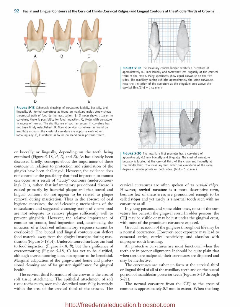
or buccally or lingually, depending on the teeth being examined (Figure 5-18, A, D, and E). As has already been discussed briefly, concepts about the importance of these contours in relation to protection and stimulation of the gingiva have been challenged. However, the evidence does not contradict the possibility that food impaction or trauma can occur as a result of “faulty” contours (undercontour-ing). It is, rather, that inflammatory periodontal disease is caused primarily by bacterial plaque and that buccal and lingual contours do not appear to be related to plaque removal during mastication. Thus in the absence of oral hygiene measures, the self-cleansing mechanisms of the musculature and suggested cleansing action of coarse food are not adequate to remove plaque sufficiently well to prevent gingivitis. However, the relative importance of contour on trauma, food impaction, and, occasionally, the initiation of a localized inflammatory response cannot be overlooked. The buccal and lingual contours can deflect food material away from the gingival margins during mas-tication (Figure 5-18, A). Undercontoured surfaces can lead to food impaction (Figure 5-18, B), but the significance of overcontouring (Figure 5-18, C) has yet to be clarified, although overcontouring does not appear to be beneficial. Marginal adaptation of the gingiva and home and profes-sional cleaning are of far greater significance for gingival health.
The cervical third formation of the crowns is the area of soft tissue attachment. The epithelial attachment of soft tissue to the teeth, soon to be described more fully, is entirely within the area of the cervical third of the crowns. The
cervical curvatures are often spoken of as cervical ridges. However, cervical curvature is a more descriptive term, because few of these areas are pronounced enough to be called ridges and yet rarely is a normal tooth seen with no curvature at all.
In young persons, and some older ones, most of the cur-vature lies beneath the gingival crest. In older persons, the CEJ may be visible or may be just under the gingival crest, with most of the prominent curvature exposed.
Gradual recession of the gingivae throughout life may be a normal occurrence. However, root exposure may lead to cemental caries, cervical sensitivity, and abrasion with improper tooth brushing.
All protective curvatures are most functional when the teeth are in proper alignment. It should be quite plain that when teeth are malposed, their curvatures are displaced and may be ineffective.
The curvatures are rather uniform at the cervical third or lingual third of all of the maxillary teeth and on the buccal portion of mandibular posterior teeth (Figures 5-19 through 5-21).
The normal curvature from the CEJ to the crest of contour is approximately 0.5 mm in extent. When the long
Figure 5-18 Schematic drawings of curvatures labially, buccally, and lingually. A, Normal curvatures as found on maxillary molar. Arrow shows theoretical path of food during mastication. B, If molar shows little or no curvature, there is possibility for food impaction. C, Molar with curvature in excess of normal. The significance of such an excess in curvature has not been firmly established. D, Normal cervical curvatures as found on maxillary incisors. The crests of curvature are opposite each other labiolingually. E, Curvatures as found on mandibular posterior teeth.
A
D E
B C
Figure 5-19 The maxillary central incisor exhibits a curvature of approximately 0.5 mm labially and somewhat less lingually at the cervical third of the crown. Many specimens show equal curvature on the two sides. The maxillary canine exhibits approximately the same curvature. Note the limitation of the curvature at the cingulum area above the cervical line.(Grid = 1 sq mm.)
Figure 5-20 The maxillary first premolar has a curvature of approximately 0.5 mm buccally and lingually. The crest of curvature buccally is located at the cervical third of the crown and lingually at the middle third. The maxillary first molar has curvatures of the same degree at similar points on both sides. (Grid = 1 sq mm.)
92 Facial and Lingual Contours at the Cervical Thirds (Cervical Ridges) and Lingual Contours at the Middle Thirds of Crowns
http://freedentaleducation.blogspot.com

Chapter 5 Orofacial Complex: Form and Function 93
axis of the tooth is placed vertically, it is found that this curvature is fairly constant and may be recognized as average or normal for the maxillary teeth, labially or buccally and lingually, and for the mandibular posterior teeth on the buccal surfaces. The curvature, lingually, of mandibular posterior teeth extends about 1 mm beyond the cervical line. Here, however, the extreme curvature does not contribute to the stasis of food material at the cervix because of the activity of the tongue in keeping the lingual surfaces of these teeth clean.
Figure 5-19 shows that the maxillary central incisor and canine have curvatures, labially and lingually, that are almost identical. Because the canines have a more massive development
Figure 5-21 Mandibular first premolar and first molar. Both teeth have a curvature of approximately 0.5 mm buccally at the cervical third of the crown and a curvature of approximately 1 mm lingually, with the crest of curvature at the middle third. (Grid = 1 sq mm.)
Figure 5-22 Photographs of a natural specimen of a mandibular first molar, taken with a lens capable of two-diameter registrations. Cutouts of these photos were placed on graphs depicting squared millimeters. The result is an accurate graph in millimeters of tooth outlines from five aspects. (Grid = 1 sq mm.)
of the cingulum, clinical observation only gives an impression of greater curvature. This is an optical illusion that is dispelled when the outline of the canine is properly compared with that of other teeth on a graph.
The maxillary premolar and molar show the same limited curvatures. The crest of curvature, lingually, on all posterior teeth is at or near the middle third of the crowns.
When curvatures are found that are greater in extent than 0.5 mm, rarely is the curvature as much as 1 mm, except lingually on mandibular posterior teeth and often lingually on maxillary posterior teeth. In these instances, the crest of contour is found at the middle third of the crowns instead of at the cervical third (see Figure 5-21). These crests are always lingual.
The eye is easily confused at times when viewing certain aspects of the teeth because of the abrupt sweep of curva-tures as they travel from the cervical line toward occlusal and incisal surfaces (i.e., the buccal surfaces of mandibular posterior teeth, the lingual surfaces of posterior maxillary teeth, or the lingual surfaces of canines). When the actual photographs are placed on a background of squares (Figure 5-22), with the long axis of each tooth positioned vertically, we readily see that the extent of curvature from the cervical line to the crest of curvature facially or lingually is slight. Quite often, in restorative procedures, it is greatly overesti-mated and reproduced.
Figure 5-23 shows a mandibular central incisor and canine from the mesial aspect. Here, the curvature at the
http://freedentaleducation.blogspot.com
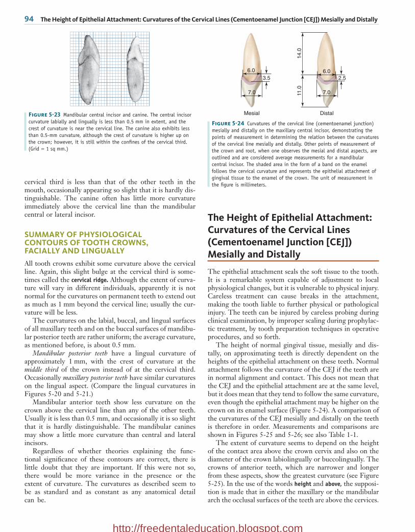
cervical third is less than that of the other teeth in the mouth, occasionally appearing so slight that it is hardly dis-tinguishable. The canine often has little more curvature immediately above the cervical line than the mandibular central or lateral incisor.
SUMMARY OF PHYSIOLOGICAL CONTOURS OF TOOTH CROWNS, FACIALLY AND LINGUALLY
All tooth crowns exhibit some curvature above the cervical line. Again, this slight bulge at the cervical third is some-times called the cervical ridge. Although the extent of curva-ture will vary in different individuals, apparently it is not normal for the curvatures on permanent teeth to extend out as much as 1 mm beyond the cervical line; usually the cur-vature will be less.
The curvatures on the labial, buccal, and lingual surfaces of all maxillary teeth and on the buccal surfaces of mandibu-lar posterior teeth are rather uniform; the average curvature, as mentioned before, is about 0.5 mm.
Mandibular posterior teeth have a lingual curvature of approximately 1 mm, with the crest of curvature at the middle third of the crown instead of at the cervical third. Occasionally maxillary posterior teeth have similar curvatures on the lingual aspect. (Compare the lingual curvatures in Figures 5-20 and 5-21.)
Mandibular anterior teeth show less curvature on the crown above the cervical line than any of the other teeth. Usually it is less than 0.5 mm, and occasionally it is so slight that it is hardly distinguishable. The mandibular canines may show a little more curvature than central and lateral incisors.
Regardless of whether theories explaining the func-tional significance of these contours are correct, there is little doubt that they are important. If this were not so, there would be more variance in the presence or the extent of curvature. The curvatures as described seem to be as standard and as constant as any anatomical detail can be.
The Height of Epithelial Attachment: Curvatures of the Cervical Lines (Cementoenamel Junction [CEJ]) Mesially and Distally
The epithelial attachment seals the soft tissue to the tooth. It is a remarkable system capable of adjustment to local physiological changes, but it is vulnerable to physical injury. Careless treatment can cause breaks in the attachment, making the tooth liable to further physical or pathological injury. The teeth can be injured by careless probing during clinical examination, by improper scaling during prophylac-tic treatment, by tooth preparation techniques in operative procedures, and so forth.
The height of normal gingival tissue, mesially and dis-tally, on approximating teeth is directly dependent on the heights of the epithelial attachment on these teeth. Normal attachment follows the curvature of the CEJ if the teeth are in normal alignment and contact. This does not mean that the CEJ and the epithelial attachment are at the same level, but it does mean that they tend to follow the same curvature, even though the epithelial attachment may be higher on the crown on its enamel surface (Figure 5-24). A comparison of the curvatures of the CEJ mesially and distally on the teeth is therefore in order. Measurements and comparisons are shown in Figures 5-25 and 5-26; see also Table 1-1.
The extent of curvature seems to depend on the height of the contact area above the crown cervix and also on the diameter of the crown labiolingually or buccolingually. The crowns of anterior teeth, which are narrower and longer from these aspects, show the greatest curvature (see Figure 5-25). In the use of the words height and above, the supposi-tion is made that in either the maxillary or the mandibular arch the occlusal surfaces of the teeth are above the cervices.
Figure 5-23 Mandibular central incisor and canine. The central incisor curvature labially and lingually is less than 0.5 mm in extent, and the crest of curvature is near the cervical line. The canine also exhibits less than 0.5-mm curvature, although the crest of curvature is higher up on the crown; however, it is still within the confines of the cervical third. (Grid = 1 sq mm.)
Figure 5-24 Curvatures of the cervical line (cementoenamel junction) mesially and distally on the maxillary central incisor, demonstrating the points of measurement in determining the relation between the curvatures of the cervical line mesially and distally. Other points of measurement of the crown and root, when one observes the mesial and distal aspects, are outlined and are considered average measurements for a mandibular central incisor. The shaded area in the form of a band on the enamel follows the cervical curvature and represents the epithelial attachment of gingival tissue to the enamel of the crown. The unit of measurement in the figure is millimeters.
Mesial Distal
7.0
6.0
7.0
6.0
11.0
14.0
3.5 2.5
94 The Height of Epithelial Attachment: Curvatures of the Cervical Lines (Cementoenamel Junction [CEJ]) Mesially and Distally
http://freedentaleducation.blogspot.com

Chapter 5 Orofacial Complex: Form and Function 95
Any point approaching the incisal edge or occlusal surface of a crown is considered above the cervix, and the height is increased as it approaches occlusal levels.
Periodontal attachment that follows the curvature of the CEJ mesially and distally seems to be about as high
Figure 5-25 Schematic drawing of the crowns of maxillary and mandibular teeth with associated graphs of the average extent of curvatures of the cervical line mesially and distally. A, Maxillary teeth. B, Mandibular teeth. Compare the graph of cervical curvatures with a line drawn through the center of contact areas. Note that the graph tends to run somewhat parallel to this line. (Grid = 1 sq mm.)
A
B
00
0
0
0
0
Figure 5-26 Graphs of typical forms of maxillary teeth accenting the outlines of cervical lines mesially and distally. (Grid = 1 sq mm.)
Mesial Distal Mesial
First premolar First molar
Central incisor
Canine
Distal
on mandibular anterior teeth as on their counterparts that are larger in the maxillary arch. Although the crowns of the mandibular anterior teeth average 1 mm less in labio-lingual diameter (the lateral incisors excepted), the contact areas are higher accordingly, being near the incisal edges on centrals and laterals. Consequently, measurements usually show less than 1-mm variation in mesial and distal curvatures between maxillary and mandibular anterior teeth.
Posterior teeth show little variation in either arch. Figure 5-25 is a diagrammatic drawing of the outlines of the teeth on one side of the arch when viewed from the labial and buccal aspects. These outlines have been placed so that a direct comparison can be made with the graphs below them. The graphs demonstrate the relative height of individual attachments in the average normal case. They are based on cases having upper central incisors with crowns 10.5 to 11 mm in length. Unless the teeth were very large or very small, the graphs would not vary from those illustrated by more than 0.5 mm.
The curvature of the cervical line of most teeth (CEJ) is approximately 1 mm less distally than mesially. The greater curvature is found at the median line on central incisors, maxillary and mandibular. The height of attachment is dependent on the height of the contact areas of the two teeth creating the interproximal space. If the mesial curva-ture of the central incisor is calibrated at 3.5 mm, the distal curvature is about 1 mm less, or 2.5 mm.
http://freedentaleducation.blogspot.com

The six anterior teeth, both maxillary and mandibular, when compared with posteriors, exhibit the greatest curva-ture. Because the canine crowns function distally as poste-rior teeth, their curvatures distally represented by the CEJ are slight, being about 1 to 1.5 mm on the average. Premo-lars and molars have rather uniform but slight curvatures. The contact levels are low in relation to total crown length; consequently, these teeth do not have high periodontal attachments interproximally. The average premolar or molar has a curvature mesially of only 1 mm or less, with no curvature or even a minus curvature distally (Figure 5-27).
From the operator’s point of view, anterior teeth espe-cially must be carefully approached (when attachment is normal) as, for example, in preparing them for restoration. Posterior teeth have curvatures that are less critical.
A summary of the height of periodontal attachment interproxi-mally indicates the attachment to be highest at the median line on central incisors. In distal progression the height of attachment decreases along with the decrease in curvature of CEJs until the mesial surface of the first premolar is reached. From this point distally through third molars, curvatures are slight.
The possibility of injury to mesial and distal periodontal attachment during tooth preparation should be considered. The height of attachment must be ascertained by careful probing and by the continuous observation of landmarks during the operation. Improper usage of impression materi-als must also be avoided.
Figure 5-27 Graphs of typical forms of mandibular teeth accenting the outlines of cervical lines mesially and distally. (Grid = 1 sq mm.)
Mesial
Central incisor Canine
Distal
Mesial
First premolar First molar
Distal
To secure scientific data regarding comparative cur-vatures, it was necessary to examine many tooth speci-mens. It was found that, usually, a graph of the cementoenamel curvatures from the median line distally could be staggered, as in Figure 5-25. It is noticed that in the posterior teeth, the variation in curvature is slight and the amount of curvature is minor, with the variance ranging from 1 mm mesially toward the occlusal surface to a slight curvature in the opposite direction. (Note the distal aspect of the mandibular first molar in Figure 5-27.)
Figures 5-26 and 5-27 are graphs of maxillary and man-dibular teeth demonstrating typical cervical curvatures mesially and distally.
References 1. Ostry DJ, Vatikiotis-Bateson E, Gribble PL: An examination
of the degrees of freedom of human jaw motion in speech and mastication, J Speech Lang Hear Res 40:1341, 1997.
2. Braun S et al: The form of the human dental arch, Angle Orthod 68:29, 1998.
3. Nixon RL: Mandibular ceramic veneers: an examination of diverse cases integrating form, function, and esthetics, Pract Periodontics Aesthet Dent 7:17, 1995.
4. Watanabe M et al: Morphological and functional analysis of dento-orofacial complex in monozygotic twins with Duchenne type muscular dystrophy, J Jpn Orthod Soc 49:522, 1990.
96 References
http://freedentaleducation.blogspot.com

Chapter 5 Orofacial Complex: Form and Function 97
5. McDonald F: The condylar disk as a controlling factor in the form of the condylar head, J Craniomandib Disord 3:83, 1989.
6. Magne P, Versluis A, Douglas WH: Rationalization of incisor shape: experimental-numerical analysis, J Prosthet Dent 81:345, 1999.
7. Sorensen DA: Form follows function-based rationale for esthetic dentistry, J Indiana Dent Assoc 77:25, 1998.
8. McCulloch CA et al: Role of physical forces in regulating the form and function of the periodontal ligament, Periodontology 24:56, 2000.
9. Daftary F: Dentoalveolar morphology: evaluation of natural root form versus cylindrical implant fixtures, Pract Periodontics Aesthet Dent 9: 469, 1997.
10. Daftary F: Prosthetically formulated natural aesthetic implant prostheses, Pract Periodontics Aesthetic Dent 6:75, 1994.
11. Ramfjord SR, Ash MM: Periodontology and periodontics, Philadelphia, 1979, Saunders.
BibliographyAsh MM, Karring T: Periodontal and occlusal considerations in
operative dentistry. In Horsted-Bindslev P, Mjor IA, editors: Modern concepts in operative dentistry, Copenhagen, 1988, Munksgaard.
Beaudreau DE: Tooth form and contour, J Am Soc Psychosom Dent Med 3:36, 1973.
Becker C, Kaldahl WB: Current theories of crown contour, margin placement and pontic design, J Prosthet Dent 45:268, 1981.
Butler PM, Joysey KA, editors: Development, function and evolution of teeth, New York, 1978, Academic Press.
Douglas WH: Form, function and strength in the restored dentition, Ann R Austral Coll Dent Surg 13:35, 1996.
Farer J, Isaacson D: Biologic contours, J Prev Dent 1:4, 1974.Herlands RE et al: Forms, contours and extensions of full
coverage restorations in occlusal reconstruction, Dent Clin North Am 6:147, March 1962.
Lindhe J, Wicen PO: The effects on the gingivae of chewing fibrous foods, J Periodont Res 4:193, 1969.
Maeda Y et al: Biomechanical study of temporomandibular joint on its form and function. Part I: Condyle morphology in frontal section, J Osaka Univ Dent Sch 33:65, 1993.
Morris M: Artificial crown contours and gingival health, J Prosthet Dent 12:1146, 1962.
Nissan HF, Ride RA, Noble WH: Physiologic design criteria for fixed dental restorations, Dent Clin North Am 15:543, 1971.
Perel ML: Periodontal considerations of crown contours, J Prosthet Dent 26:627, 1971.
Ryder JA: On the mechanical genesis of tooth forms, Proc Acad Natl Sci Phila 30:45, 1878.
Sackett BP, Gildenhuys RR: The effect of axial crown over-contour on adolescents, J Periodontol 47:320, 1976.
Volchansky A: The role of clinical crown height and gingival margin position in oral disease, Diastema 7:17, 1979-1980.
Wade AB: Effect on dental plaque of chewing apples, Dent Pract 21:194, 1971.
Wheeler RC: Complete crown form and the periodontium, J Prosthet Dent 11:722, 1961.
Youdelis RA, Weaver JD, Sapkos S: Facial and lingual contours of artificial complete crown restorations and their effects on the periodontium, J Prosthet Dent 29:61, 1973.
Use the CD-ROM to view animations and study ques-tions; and go to the site for additional study resources.
http://freedentaleducation.blogspot.com

This page intentionally left blank
http://freedentaleducation.blogspot.com

The Permanent Maxillary Incisors
Knowing the proportions of the individual tooth helps one learn the proportions of one tooth to another. Outline draw-ings of the five aspects of the teeth are explained more fully elsewhere.1
Maxillary Central Incisor
Figures 6-1 through 6-12 illustrate the maxillary central incisor in various aspects. The maxillary central incisor is the widest mesiodistally of any of the anterior teeth (Table 6-1). The labial face is less convex than that of the maxillary lateral incisor or canine, which gives the central incisor a squared or rectangular appearance (see Figures 6-7 and 6-8). From this aspect, the crown nearly always looks symmetrical and regularly formed, having a nearly straight incisal edge, a cervical line with even curvature toward the root, a mesial side with straight outline, the distal side being more curved. The mesial incisal angle is relatively sharp, the distal incisal angle rounded (see Figure 6-2).
Although the labial surface of the crown is usually convex, especially toward the cervical third, some central incisors are flat at the middle and incisal portions. The enamel surface is relatively smooth. When the tooth is newly erupted or if little wear is evident, mamelons will be seen on the incisal ridge. The middle one is the smallest. The developmental lines on the labial surface that divide the surface into three parts are most noticeable at the middle portion if they can be distinguished at all (see Figure 2-12).
Lingually, the surface form of the maxillary central incisor is more irregular. The largest part of the middle and incisal
The maxillary incisors are four in number. The maxillary central incisors are centered in the maxilla, one on either side of the median line, with the mesial surface of each in contact with the mesial surface of the other. The maxillary and mandibular central incisors are the only neighboring teeth in the dental arches with mesial surfaces in contact. The right and left maxillary lateral or second incisors are distal to the central incisors.
The maxillary central incisor is larger than the lateral incisor. These teeth supplement each other in function, and they are similar anatomically. The incisors are shearing or cutting teeth. Their major function is to punch and cut food material during the process of mastication. These teeth have incisal ridges or edges rather than cusps such as are found on the canines and posterior teeth.
It might be good at this point to differentiate between the two terms incisal ridge and incisal edge. The incisal ridge is that portion of the crown which makes up the complete incisal portion. When an incisor is newly erupted, the incisal portion is rounded and merges with the mesioincisal and distoincisal angles and the labial and lingual surfaces. This ridge portion of the crown is called the incisal ridge. The term edge implies an angle formed by the merging of two flat surfaces. Therefore, an incisal edge does not exist on an incisor until occlusal wear has created a flattened surface linguoincisally, which surface forms an angle with the labial surface. The incisal edge is formed by the junction of the linguoincisal surface, sometimes called the incisal surface, and the labial surface (Figure 6-1).
Preceding the description of each tooth in this and sub-sequent chapters, the chronology of calcification and erup-tion of each tooth will be given as taken from Table 2-2.
99
66
http://freedentaleducation.blogspot.com

Figure 6-1 Maxillary right central incisor, lingual and incisal aspects. The labioincisal edge [LIE (1)] and linguoincisal edge [LIE (2)] border the incisal ridge. CL, Cervical line; C, cingulum; MMR, mesial marginal ridge; LF, lingual fossa; DMR, distal marginal ridge.
CLC
MMR
(1) LIE LIE (2)
LIE (2) LIE (1)
LF
MMR
LF
DMR CDMR
Figure 6-2 Maxillary right central incisor, labial aspect.
Figure 6-3 Maxillary right central incisor, lingual aspect.
Figure 6-4 Maxillary right central incisor, mesial aspect.
Figure 6-5 Maxillary right central incisor, distal aspect. Figure 6-6 Maxillary right central incisor, incisal aspect.
100 Maxillary Central Incisor
http://freedentaleducation.blogspot.com

Chapter 6 The Permanent Maxillary Incisors 101
portions of the lingual area is concave. Mesial and distal marginal ridges border the concavity, the lingual portion of the incisal ridge, and the convexity apically to the cingulum. The lingual topography gives a scooplike form to the crown (see Figure 6-3). An exaggeration of the marginal ridges, known as a shovel-shaped incisor, is a genetically determined variation seen in Mongoloid races, including North and South American Indians.2–4
The maxillary central incisor usually develops normally. One anomaly that sometimes occurs is a short root. Another variation is an unusually long crown (see Figure 6-12, 4 and 5). The maxillary central incisors are the most prominent teeth in the mouth. There are two basic forms: the first is
relatively wide at the cervix, when viewed from the labial aspect, in comparison with the mesiodistal width at the contact areas (see Figure 6-9, 1 and 4); the second form is relatively narrow at the cervix, where the root joins the crown, in comparison with the mesiodistal width at the contact areas (see Figure 6-9, 5, 7, and 9).
In the description of the central incisor, an attempt will be made to strike an average between the extremes of the two forms, keeping in mind that crown sizes are gender dimorphic, with male larger than female. The extent of dimorphism varies among populations.5 However, gender-specific correlations between enamel thickness and crown width of dentin are low.6
Figure 6-7 Maxillary right central incisor. Squared millimeter graph outlines of five aspects are shown. In the incisal view, the labial aspect is at the top of the drawing. (Grid = 1 sq mm.)
Labial Lingual
DistalMesial
Incisal
http://freedentaleducation.blogspot.com
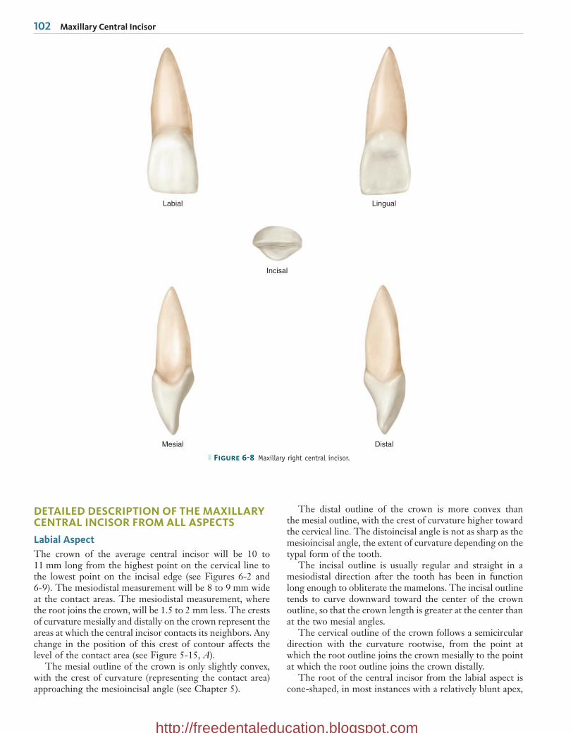
Figure 6-8 Maxillary right central incisor.
Labial Lingual
Incisal
Mesial Distal
DETAILED DESCRIPTION OF THE MAXILLARY CENTRAL INCISOR FROM ALL ASPECTS
Labial AspectThe crown of the average central incisor will be 10 to 11 mm long from the highest point on the cervical line to the lowest point on the incisal edge (see Figures 6-2 and 6-9). The mesiodistal measurement will be 8 to 9 mm wide at the contact areas. The mesiodistal measurement, where the root joins the crown, will be 1.5 to 2 mm less. The crests of curvature mesially and distally on the crown represent the areas at which the central incisor contacts its neighbors. Any change in the position of this crest of contour affects the level of the contact area (see Figure 5-15, A).
The mesial outline of the crown is only slightly convex, with the crest of curvature (representing the contact area) approaching the mesioincisal angle (see Chapter 5).
The distal outline of the crown is more convex than the mesial outline, with the crest of curvature higher toward the cervical line. The distoincisal angle is not as sharp as the mesioincisal angle, the extent of curvature depending on the typal form of the tooth.
The incisal outline is usually regular and straight in a mesiodistal direction after the tooth has been in function long enough to obliterate the mamelons. The incisal outline tends to curve downward toward the center of the crown outline, so that the crown length is greater at the center than at the two mesial angles.
The cervical outline of the crown follows a semicircular direction with the curvature rootwise, from the point at which the root outline joins the crown mesially to the point at which the root outline joins the crown distally.
The root of the central incisor from the labial aspect is cone-shaped, in most instances with a relatively blunt apex,
102 Maxillary Central Incisor
http://freedentaleducation.blogspot.com

Chapter 6 The Permanent Maxillary Incisors 103
Figure 6-9 Maxillary central incisor, labial aspect. Ten typical specimens are shown.
109876
54321
Figure 6-10 Maxillary central incisor, mesial aspect. Ten typical specimens are shown.
1 2 3 4 5
6 7 8 9 10
IV A(8), B
http://freedentaleducation.blogspot.com

Figure 6-11 Maxillary central incisor, incisal aspect. Ten typical specimens are shown.
1 2 3 4 5
6 7 8 9 10
Figure 6-12 Maxillary central incisor. Ten specimens with uncommon variations are shown. 1, Extralingual inclination of incisal portion of crown. Note developmental (palatoradicular) groove traversing root and part of crown. 2, Root extremely long. 3, Specimen small in all dimensions. 4, Crown extremely long, root very short. 5, Specimen malformed; crown unusually long; cervix very wide. 6, Root short and tapering. 7, Same as specimen 6. 8, Crown nearly as wide at the cervix as at contact areas, crown long, root short. 9, Root with unusual curvature. 10, Crown and root narrow labiolingually; root comparable with that of specimen 2.
1 3 42 5
6 7 8 9 10
104 Maxillary Central Incisor
http://freedentaleducation.blogspot.com

Chapter 6 The Permanent Maxillary Incisors 105
and the outline mesially and distally is regular. The root is usually 2 or 3 mm longer than the crown, although it varies considerably. (See illustrations of typical central incisors and those of variations from the labial aspects in Figures 6-9 and 6-12.)
A line drawn through the center of the root and crown of the maxillary central incisor tends to parallel the mesial outline of the crown and root.
Lingual AspectThe lingual outline of the maxillary central incisor is the reverse of that found on the labial aspect (see Figure 6-3). The lingual aspect of the crown is different, however, when we compare the surface of the lingual aspect with that of the labial aspect. From the labial aspect, the surface of the crown is smooth generally. The lingual aspect has convexities and a concavity. The outline of the cervical line is similar, but immediately below the cervical line a smooth convexity is to be found; this is called the cingulum (see Figure 6-1).
Mesially and distally confluent with the cingulum are the marginal ridges. Between the marginal ridges, below the cin-gulum, a shallow concavity is present called the lingual fossa. Outlining the lingual fossa, the linguoincisal edge is raised somewhat, being on a level with the marginal ridges mesially and distally, completing the lingual portion of the incisal ridge of the central incisor.
From the foregoing description, we note that the lingual fossa is bordered mesially by the mesial marginal ridge, incisally by the lingual portion of the incisal ridge, distally by the distal marginal ridge, and cervically by the cingulum. Usually there are developmental grooves extending from the cingulum into the lingual fossa.
The crown and root taper lingually, so that the crown calibration at the two labial line angles is greater than the calibration at the two lingual line angles, and the lingual portion of the root is narrower than the labial portion. A cross section of the root at the cervix shows the root to be
generally triangular with rounded angles. One side of the triangle is labial, with the mesial and distal sides pointing lingually. The mesial side of this triangle is slightly longer than the distal side (see Figure 13-8, C, 3, 4, 5, and 6).
Mesial AspectThe mesial aspect of this tooth has the fundamental form of an incisor. The crown is wedge-shaped, or triangular, with the base of the triangle at the cervix and the apex at the incisal ridge (see Figure 4-16, A and Figures 6-4 and 6-10).
Usually a line drawn through the crown and the root from the mesial aspect through the center of the tooth will bisect the apex of the root and also the incisal ridge of the crown. The incisal ridge of the crown is therefore on a line with the center of the root. This alignment is charac-teristic of maxillary central and lateral incisors. A straight line drawn through the center of the crown and root from the mesial or distal aspects will rarely if ever pass lingual to the incisal edge. Maxillary incisors are occasionally seen with the incisal ridges lingual to the bisecting line (see Figure 6-12, 1).
Labially and lingually, immediately coronal to the cervi-cal line are the crests of curvature of these surfaces. These crests of contour give the crown its greatest labiolingual measurement.
Normally, the curvature labially and lingually is approxi-mately 0.5 mm in extent (see Figure 6-4) before continuing the outlines to the incisal ridge.
The labial outline of the crown from the crest of curva-ture to the incisal ridge is very slightly convex. The lingual outline is convex at the point where it joins the crest of curvature at the cingulum; it then becomes concave at the mesial marginal ridge and slightly convex again at the lin-guoincisal ridge and the incisal edge.
The cervical line outlining the cementoenamel junction (CEJ) mesially on the maxillary central incisor curves
Table 6-1 Maxillary Central Incisor
First evidence of calcification 3–4 mo
Enamel completed 4–5 yr
Eruption 7–8 yr
Root completed 10 yr
Measurement Table
Cervicoincisal Length of
CrownLength
of Root
Mesiodistal Diameter of Crown
Mesiodistal Diameter of
Crown at Cervix
Labio- or Buccolingual
Diameter of Crown
Labio- or Buccolingual
Diameter of Crown at
Cervix
Curvature of Cervical
Line—Mesial
Curvature of Cervical Line—Distal
Dimensions* suggested for carving technique
10.5 13.0 8.5 7.0 7.0 6.0 3.5 2.5
*In millimeters.
http://freedentaleducation.blogspot.com

106 Maxillary Lateral Incisor
incisally to a noticeable degree. This cervical curvature is greater on the mesial surface of this tooth than on any surface of any other tooth in the mouth. The curvature varies in extent, depending on the length of the crown and the measurement of the crown labiolingually. On an average central incisor of 10.5 to 11 mm in crown length, the cur-vature is 3 to 4 mm (see Figure 5-26).
The root of this tooth from the mesial aspect is cone-shaped, and the apex of the root is usually bluntly rounded.
Distal AspectLittle difference is evident between the distal and mesial outlines of this tooth (see Figure 6-5). When looking at the central incisor from the distal aspect, it may be noted that the crown gives the impression of being somewhat thicker toward the incisal third. Because of the slope of the labial surface distolingually, more of that surface is seen from the distal aspect; this creates the illusion of greater thickness. Actually, most teeth are turned a little on their root bases to adapt to the dental arch curvature. The maxillary central incisor is no exception.
The curvature of the cervical line outlining the CEJ is less in extent on the distal than on the mesial surfaces. Most teeth show this characteristic.
Incisal AspectThe specimen of this tooth is posed in the illustrations so that the incisal edge is centered over the root (see Figures 6-6 and 6-11). A view of the crown from this aspect super-imposes it over the root entirely so that the latter is not visible.
From this aspect, the labial face of the crown is relatively broad and flat in comparison with the lingual surface, espe-cially toward the incisal third. Nevertheless, the cervical portion of the crown labially is convex, although the arc described is broad.
The incisal ridge may be seen clearly, and a differentia-tion between the incisal edge and the remainder of the incisal ridge, with its slope toward the lingual, is easily distinguished.
The outline of the lingual portion tapers lingually toward the cingulum. The cingulum of the crown makes up the cervical portion of the lingual surface.
The mesiolabial and distolabial line angles are prominent from the incisal aspect. The relative positions of these line angles should be compared with the mesiolingual and dis-tolingual line angles, which are represented by the borders of the mesial and distal marginal ridges. The mesiodistal calibration of the crown at the labial line angles is greater than the same calibration at the lingual line angles.
The crown of this tooth shows more bulk from the incisal aspect than one would expect from viewing it from the mesial or distal aspect. Relatively broad surfaces are at the site of contact areas mesially and distally. Comparison should also be made between the dimensions of the crown labiolingually and mesiodistally. The labiolingual calibra-tion of the crown is more than two thirds as great as the
mesiodistal calibration. A cursory examination would not reveal this detail.
Bilaterally, the outline of the incisal aspect is rather uniform. The lingual portion shows some variation, however, in that a line drawn from the mesioincisal angle to the center of the cingulum lingually will be longer than one drawn from the same point on the cingulum to the distoincisal angle. The crown conforms to a triangular outline reflected by the outline of the root cross section at the cervix men-tioned earlier.
Maxillary Lateral Incisor
Figures 6-13 through 6-21 illustrate the maxillary lateral incisor in various aspects. Because the maxillary lateral incisor supplements the central incisor in function, the crowns bear a close resemblance. The lateral incisor is smaller in all dimensions except root length (Table 6-2). Because it resembles the maxillary central incisor in form, direct comparisons are made with the central incisor in its description.
This tooth differs from the central incisor in its develop-ment, which may vary considerably. Maxillary lateral inci-sors vary in form more than any other tooth in the mouth except the third molar. If the variation is too great, it is considered a developmental anomaly. A common situation is to find maxillary lateral incisors with a nondescript, pointed form; such teeth are called peg-shaped laterals (see Figure 6-21, 7 and 8). In some individuals, the lateral inci-sors are missing entirely7; in these cases, the maxillary central incisor may be in contact distally with the canine. The pres-ence of a palatogingival groove in maxillary incisors may be a predisposing factor in localized periodontal disease8 (see Figure 6-21, 3). This groove is also referred to as the pala-toradicular groove.9
Figure 6-13 Maxillary right lateral incisor, labial aspect. (Grid = 1 sq mm.)
http://freedentaleducation.blogspot.com

Chapter 6 The Permanent Maxillary Incisors 107
Figure 6-15 Maxillary right lateral incisor, mesial aspect. (Grid = 1 sq mm.) Figure 6-16 Maxillary right lateral incisor, distal aspect. (Grid =
1 sq mm.)
Figure 6-17 Maxillary right lateral incisor, incisal aspect. (Grid = 1 sq mm.)
One type of malformed maxillary lateral incisor has a large, pointed tubercle as part of the cingulum; some have deep developmental grooves that extend down on the root lingually with a deep fold in the cingulum; and some show twisted roots, distorted crowns, and so on (see Figure 6-21).
DETAILED DESCRIPTION OF THE MAXILLARY LATERAL INCISOR FROM ALL ASPECTS
Labial AspectAlthough the labial aspect of the maxillary lateral incisor may appear to favor that of the central incisor, usually it has more curvature, with a rounded incisal ridge and rounded incisal angles mesially and distally (see Figures 6-13 and 6-19). Although the crown is smaller in all dimensions, its proportions usually correspond to those of the central incisor.
Figure 6-14 Maxillary right lateral incisor, lingual aspect. (Grid = 1 sq mm.)
http://freedentaleducation.blogspot.com

108 Maxillary Lateral Incisor
1 32 4 5
6 7 8 9 10
54321
6 7 8 9 10
Figure 6-19 Maxillary lateral incisor, labial aspect. Ten typical specimens are shown.
Figure 6-18 Maxillary lateral incisor, incisal aspect. Ten typical specimens are shown.
IV A(7), B
http://freedentaleducation.blogspot.com
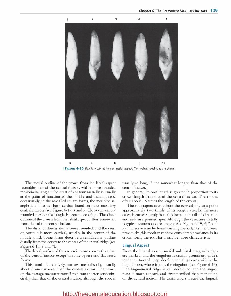
Chapter 6 The Permanent Maxillary Incisors 109
Figure 6-20 Maxillary lateral incisor, mesial aspect. Ten typical specimens are shown.
1 32 4 5
6 7 8 9 10
The mesial outline of the crown from the labial aspect resembles that of the central incisor, with a more rounded mesioincisal angle. The crest of contour mesially is usually at the point of junction of the middle and incisal thirds; occasionally, in the so-called square forms, the mesioincisal angle is almost as sharp as that found on most maxillary central incisors (see Figure 6-19, 4 and 5). However, a more rounded mesioincisal angle is seen more often. The distal outline of the crown from the labial aspect differs somewhat from that of the central incisor.
The distal outline is always more rounded, and the crest of contour is more cervical, usually in the center of the middle third. Some forms describe a semicircular outline distally from the cervix to the center of the incisal ridge (see Figure 6-19, 3 and 7).
The labial surface of the crown is more convex than that of the central incisor except in some square and flat-faced forms.
This tooth is relatively narrow mesiodistally, usually about 2 mm narrower than the central incisor. The crown on the average measures from 2 to 3 mm shorter cervicoin-cisally than that of the central incisor, although the root is
usually as long, if not somewhat longer, than that of the central incisor.
In general, its root length is greater in proportion to its crown length than that of the central incisor. The root is often about 1.5 times the length of the crown.
The root tapers evenly from the cervical line to a point approximately two thirds of its length apically. In most cases, it curves sharply from this location in a distal direction and ends in a pointed apex. Although the curvature distally is typical, some roots are straight (see Figure 6-19, 4, 7, and 9), and some may be found curving mesially. As mentioned previously, this tooth may show considerable variance in its crown form; the root form may be more characteristic.
Lingual AspectFrom the lingual aspect, mesial and distal marginal ridges are marked, and the cingulum is usually prominent, with a tendency toward deep developmental grooves within the lingual fossa, where it joins the cingulum (see Figure 6-14). The linguoincisal ridge is well developed, and the lingual fossa is more concave and circumscribed than that found on the central incisor. The tooth tapers toward the lingual,
http://freedentaleducation.blogspot.com

110 Maxillary Lateral Incisor
1 2 3 4 5
6 7 8 9 10
Figure 6-21 Maxillary lateral incisor. Ten specimens with uncommon variations are shown. 1, Odd twist to crown and root. 2, Malformed generally. 3, Deep developmental (palatoradicular) groove distally; note pit in lingual fossa. 4, Same as specimen 3 with pit and groove connected. 5, Deep concavity above contact area of the crown. 6, Abnormally large but well formed. 7, Single-cusp development and malformed root called peg lateral incisor. 8, Same as specimen 7, except root is straight. 9, Same as specimen 5, with deep lingual pit in addition. 10, Resemblance to a small maxillary central incisor more marked than the average.
Table 6-2 Maxillary Lateral Incisor
First evidence of calcification 10–12 mo
Enamel completed 4–5 yr
Eruption 8–9 yr
Root completed 11 yr
Measurement Table
Cervicoincisal Length of
CrownLength
of Root
Mesiodistal Diameter of Crown
Mesiodistal Diameter of
Crown at Cervix
Labio- or Buccolingual
Diameter of Crown
Labio- or Buccolingual
Diameter of Crown at
Cervix
Curvature of Cervical
Line—Mesial
Curvature of Cervical Line—Distal
Dimensions* suggested for carving technique
9.0 13.0 6.5 5.0 6.0 5.0 3.0 2.0
*In millimeters.
http://freedentaleducation.blogspot.com

Chapter 6 The Permanent Maxillary Incisors 111
resembling a central incisor in this respect. It is not uncom-mon to find a deep developmental groove at the side of the cingulum, usually on the distal side, which may extend up on the root for part or all of its length. Faults in the enamel of the crown are often found in the deep portions of these developmental grooves (see Figure 6-21, 3 and 4).
Mesial AspectThe mesial aspect of the maxillary lateral incisor is similar to that of a small central incisor except that the root appears longer (see Figures 6-15 and 6-20). The crown is shorter, the root is relatively longer, and the labiolingual measure-ment of the crown and root is a millimeter or so less than that of the maxillary central incisor of the same mouth.
The curvature of the cervical line is marked in the direc-tion of the incisal ridge, although because of the small size of the crown the actual extent of curvature is less than that found on the central incisor. The heavy development of the incisal ridge accordingly makes the incisal portion appear somewhat thicker than that of the central incisor.
The root appears as a tapered cone from this aspect, with a bluntly rounded apical end. This varies in individuals, with the apical end sometimes being quite blunt, while at other times, it is pointed. In a good many cases, the labial outline of the root from this aspect is straight. As in the central incisor, a line drawn through the center of the root tends to bisect the incisal ridge of the crown.
Distal AspectBecause of the placement of the crown on the root, the width of the crown distally appears thicker than it does on the mesial aspect from marginal ridge to labial face (see Figure 6-16). The curvature of the cervical line is usually a millimeter or so less in depth than on the mesial side. It is not uncommon to find a developmental groove distally on this crown extending on the root for part or all of its length.
Incisal AspectThe incisal aspect of this tooth sometimes resembles that of the central incisor, or it may resemble that of a small canine (see Figures 6-17 and 6-18). If the tooth conforms in devel-
opment to its central incisor neighbor in other respects, it will, from the incisal aspect, resemble a central incisor except in size (see Figure 6-18, 5 and 9). However, the cingulum may be large, as is the incisal ridge. In addition, the labio-lingual dimension may be greater than usual in comparison with the mesiodistal dimension. If these variations are present, the tooth has a marked resemblance to a small canine (see Figure 6-18, 3 and 10).
All maxillary lateral incisors exhibit more convexity labi-ally and lingually from the incisal aspect than do the maxil-lary central incisors.
References1. Ash MM: Wheeler’s atlas of tooth form, Philadelphia, 1984,
Saunders.2. Carbonell VM: Variations in frequency of shovel-shaped
incisors in different populations. In Brothwell DR, editor: Dental anthropology, vol 5, London, 1963, Pergamon Press.
3. Dahlberg AA, Mikkelson O: The shovel-shaped character in the teeth of the Pima Indians, Am J Phys Anthropol 5:234, 1947.
4. Hrdlicka A: Shovel-shaped teeth, Am J Phys Anthropol 3:429, 1920.
5. Garn SM et al: Sexual dimorphism in the buccolingual tooth diameter, J Dent Res 45:1819, 1966.
6. Harris EF, Hicks JD: A radiographic assessment of enamel thickness in human maxillary lateral incisors, Arch Oral Biol 43:825, 1998.
7. Meskin LH, Gorlin RJ: Agenesis and peg-shaped permanent maxillary lateral incisors, J Dent Res 42:1576, 1963.
8. Lee KW et al: Plato-gingival grooves in maxillary incisors. A possible predisposing factor to localized periodontal disease, Br Dent J 124:14, 1968.
9. Kogon SL: The prevalence, location, and conformation of palato-radicular grooves in maxillary incisors, J Periodontol 57:231, 1986.
Use the CD-ROM to view animations and study ques-tions; and go to the site for additional study resources.
IV A, B
http://freedentaleducation.blogspot.com

This page intentionally left blank
http://freedentaleducation.blogspot.com

The Permanent Mandibular Incisors
are parallel with each other, fitting together during incising action.
Mandibular Central Incisor
Figures 7-1 through 7-12 illustrate the mandibular central incisor in various aspects. Generally, the mandibular central incisor is the smallest tooth in the dental arches (Table 7-1). The crown has little more than half the mesiodistal diameter of the maxillary central incisor; however, the labiolingual diameter is only about 1 mm less. The lines of greatest masticatory stress are brought to bear on the mandibular incisors in a labiolingual direction, which makes this rein-forcement necessary.
The single root is very narrow mesiodistally and corre-sponds to the narrowness of the crown, although the root and crown are wide labiolingually. The length of the root is as great as, if not greater than, that of the maxillary central incisor.
DETAILED DESCRIPTION OF THE MANDIBULAR CENTRAL INCISOR FROM ALL ASPECTS
Labial Aspect
The labial aspect of the mandibular central incisor is regular, tapering evenly from the relatively sharp mesial and distal incisal angles to the apical portion of the root (see Figures 7-7 through 7-9). The incisal ridge of the crown is straight and is at approximately a right angle to the long axis of the
The mandibular incisors are four in number. The mandibu-lar central incisors are centered in the mandible, one on either side of the median line, with the mesial surface of each one in contact with the mesial surface of the other. The right and left mandibular lateral or second incisors are distal to the central incisors. They are in contact with the central incisors mesially and with the canines distally.
The mandibular incisors have smaller mesiodistal dimen-sions than any of the other teeth. The central incisor is somewhat smaller than the lateral incisor, which is the reverse of the situation in the maxilla.
These teeth are similar in form and have smooth crown surfaces that show few traces of developmental lines. Mamel-ons on the incisal ridges are worn off soon after eruption, if the occlusion is normal, which leaves the incisal ridges smooth and straight (compare Figure 7-9, 7 and 8). The contact areas are near the incisal ridges mesially and distally, and lines drawn through the contact areas are near the same level on both central and lateral incisors; here also the situ-ation is unlike that of the maxillary incisors. The mandibular incisors show uniform development, with few instances of malformations or anomalies (see Figure 7-12).1,2
The anatomical form of these teeth differs entirely from that of the maxillary incisors. The inclination of the crowns differs from the mesial and distal aspects; the labial faces are inclined lingually so that the incisal ridges are lingual to a line bisecting the root. After normal wear has taken place, obliterating the mamelons, the incisal surfaces thus created show a labial inclination when the occlusion has been normal. Note that the incisal surfaces of maxillary incisors have a lingual inclination. With this arrangement, the incisal planes of the mandibular and maxillary incisors
113
77
http://freedentaleducation.blogspot.com

114 Mandibular Central Incisor
Figure 7-1 Mandibular right central incisor, lingual and incisal aspects. IR, Incisal ridge; DMR, distal marginal ridge; LF, lingual fossa; CL, cervical line; C, cingulum; MMR, mesial marginal ridge.
IR
MMR
C
CL
DMR
LF
DMR
IR
MMRC
Figure 7-2 Mandibular right central incisor, labial aspect. (Grid = 1 sq mm.)
Figure 7-3 Mandibular right central incisor, lingual aspect. (Grid = 1 sq mm.)
Figure 7-4 Mandibular right central incisor, mesial aspect. (Grid = 1 sq mm.)
Figure 7-5 Mandibular right central incisor, distal aspect. (Grid = 1 sq mm.)
Figure 7-6 Mandibular right central incisor, incisal aspect. (Grid = 1 sq mm.)
http://freedentaleducation.blogspot.com

Chapter 7 The Permanent Mandibular Incisors 115
Figure 7-7 Mandibular right central incisor. Graph outlines of five aspects are shown. (Grid = 1 sq mm.)
Labial Lingual
Distal
Incisal
Mesial
tooth. Usually, the mesial and distal outlines of the crown make a straight drop downward from the incisal angles to the contact areas, which are incisal to the junction of incisal and middle thirds of the crown. The mesial and distal sides of the crown taper evenly from the contact areas to the narrow cervix.
The mesial and distal root outlines are straight with the mesial and distal outlines of the crown down to the apical portion. The apical third of the root terminates in a small, pointed taper, in most cases curving distally. Sometimes the roots are straight (see Figure 7-9, 2 and 10).
The labial face of the mandibular central incisor crown is ordinarily smooth, with a flattened surface at the incisal third; the middle third is more convex, narrowing down to the convexity of the root at the cervical portion.
Except in newly erupted teeth, central incisors show few traces of developmental lines. The labial surface of the root of the mandibular central incisor is regular and convex.
Lingual AspectThe lingual surface of the crown is smooth, with very slight concavity at the incisal third between the inconspicu-ous marginal ridges (see Figures 7-1, 7-3, 7-7, and 7-8). In some instances, the marginal ridges are more prominent near the incisal edges (see Figure 7-11, 2 and 8). In these cases, the concavity between the marginal ridges is more distinct.
The lingual surface becomes flat and then convex as progression is made from the incisal third to the cervical third.
http://freedentaleducation.blogspot.com

116 Mandibular Central Incisor
Figure 7-8 Mandibular right central incisor.
Labial Lingual
Incisal
Mesial Distal
No developmental lines mark the cingulum development on this tooth at the cervical third. No other tooth in the mouth, except the mandibular lateral incisor, shows so few developmental lines and grooves. The outlines and surfaces of the mandibular incisors are regular and symmetrical.
Mesial AspectThe curvature labially and lingually above the cervical line is less than that found on maxillary incisors (see Figures 7-4, 7-7, 7-8, and 7-10).
The outline of the labial face of the crown is straight above the cervical curvature, sloping rapidly from the crest of curvature to the incisal ridge. The lingual outline of the crown is a straight line inclined labially for a short distance above the smooth convexity of the cingulum; the straight
outline joins a concave line at the middle third of the crown, which extends upward to join the rounded outline of a narrow incisal ridge. The incisal ridge is rounded or worn flat, and its center is usually lingual to the center of the root.
The curvature of the cervical line representing the cementoenamel junction (CEJ) on the mesial surface is marked, curving incisally approximately one third the length of the crown.
The root outlines from the mesial aspect are straight with the crown outline from the cervical line, so that the root diameter is uniform through the cervical third and part of the middle third; the outline of the root begins to taper in the middle third area, tapering rapidly in the apical third to either a bluntly rounded or a pointed root end.
http://freedentaleducation.blogspot.com
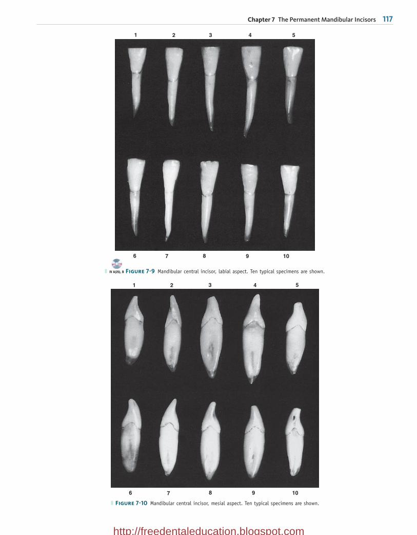
Chapter 7 The Permanent Mandibular Incisors 117
1 2 3 5
6
4
7 8 9 10
Figure 7-10 Mandibular central incisor, mesial aspect. Ten typical specimens are shown.
1 2 43 5
6 7 8 9 10
Figure 7-9 Mandibular central incisor, labial aspect. Ten typical specimens are shown.IV A(25), B
http://freedentaleducation.blogspot.com

Figure 7-11 Mandibular central incisor, incisal aspect. Ten typical specimens are shown.
1 32 4 5
6 7 8 9 10
Figure 7-12 Mandibular central incisor. Ten specimens with uncommon variations are shown. 1, Crown and root very broad mesiodistally; malformed enamel at incisal third of crown. 2, Crown wide at incisal third, with short crown; root length extreme. 3, Unusual contours at middle third of crown; cervix narrow. 4, Well-formed crown; short root. 5, No curvature labially at cervical third; extreme labial curvature at root end. 6, Specimen well formed but undersized. 7, Contact areas pointed at incisal edge; crown and root very long. 8, Crown long and narrow; root short. 9, Crown measurement at cervical third same as root; crown and root of extreme length. 10, Crown and root very wide labiolingually; greater curvature than average above cervical line at the cervical third of the crown.
1 2 3 5
6 7 8 9 10
4
http://freedentaleducation.blogspot.com

Chapter 7 The Permanent Mandibular Incisors 119
Table 7-1 Mandibular Central Incisor
First evidence of calcification 3–4 mo
Enamel completed 4–5 yr
Eruption 6–7 yr
Root completed 9 yr
Measurement Table
Cervicoincisal Length of
CrownLength
of Root
Mesiodistal Diameter of Crown
Mesiodistal Diameter of
Crown at Cervix
Labio- or Buccolingual
Diameter of Crown
Labio- or Buccolingual
Diameter of Crown at
Cervix
Curvature of Cervical
Line—Mesial
Curvature of Cervical Line—Distal
Dimensions* suggested for carving technique
9.5 12.5 5.0 3.5 6.0 5.3 3.0 2.0
*In millimeters.
The mesial surface of the crown is convex and smooth at the incisal third and becomes broader and flatter at the middle third cervical to the contact area; it then becomes quite flat, with a tendency toward concavity immediately below the middle third of the crown and above the cervical line (see Figure 7-10, 5, 8, and 10).
The mesial surface of the root is flat just below the cervi-cal line. Most of these roots have a broad developmental depression for most of the root length. The depressions usually are deeper at the junction of the middle and apical thirds (see Figure 7-10, 3 and 9).
Distal AspectThe cervical line representing the CEJ curves incisally about 1 mm less on the distal aspect than on the mesial aspect (see Figures 7-5, 7-7, and 7-8).
The distal surface of the crown and root of the mandibu-lar central incisor is similar to that of the mesial surface. The developmental depression on the distal surface of the root may be more marked, with a deeper, more well-defined developmental groove at its center.
Incisal AspectThe incisal aspect illustrates the bilateral symmetry of the mandibular central incisor (see Figures 7-1, 7-6, 7-7, 7-8, and 7-11). The mesial half of the crown is almost identical with the distal half.
The incisal edge is almost at right angles to a line bisect-ing the crown labiolingually. This feature is characteristic of the tooth and serves as a mark of identification in differ-entiation between mandibular central and lateral incisors (see Mandibular Lateral Incisor later). Note the comparison between the diameter of these crowns labiolingually and their diameters mesiodistally. The labiolingual diameter is always greater.
The labial surface of the crown is wider mesiodistally than the lingual surface. The crown is usually wider labially
than lingually at the cervical third, which latter area is rep-resented by a smooth cingulum.
The labial surface of the crown at the incisal third, although rather broad and flat in comparison with the cer-vical third, has a tendency toward convexity, whereas the lingual surface of the crown at the incisal third has an incli-nation toward concavity.
When this tooth is posed from the incisal aspect so that the line of vision is on a line with the long axis of the tooth, more of the labial surface may be seen than of the lingual surface.
Mandibular Lateral Incisor
Figures 7-13 through 7-21 illustrate the mandibular lateral incisor in various aspects. The mandibular lateral incisor is
Figure 7-13 Mandibular right lateral incisor, labial aspect. (Grid = 1 sq mm.)
http://freedentaleducation.blogspot.com

Figure 7-14 Mandibular right lateral incisor, lingual aspect. (Grid = 1 sq mm.)
Figure 7-15 Mandibular right lateral incisor, mesial aspect. (Grid = 1 sq mm.)
Figure 7-16 Mandibular right lateral incisor, distal aspect. (Grid = 1 sq mm.)
Figure 7-17 Mandibular right lateral incisor, incisal aspect. (Grid = 1 sq mm.)
Figure 7-18 Mandibular lateral incisor, incisal aspect. Ten typical specimens are shown.
1 32 4 5
6 7 8 9 10
120 Mandibular Lateral Incisor
http://freedentaleducation.blogspot.com
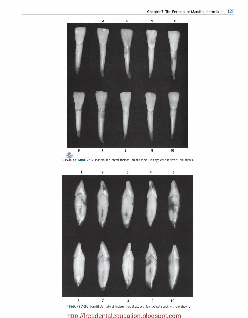
Chapter 7 The Permanent Mandibular Incisors 121
1 2 43 5
6 7 8 9 10
Figure 7-20 Mandibular lateral incisor, mesial aspect. Ten typical specimens are shown.
1 2 43 5
6 7 8 9 10
Figure 7-19 Mandibular lateral incisor, labial aspect. Ten typical specimens are shown.IV A(26), B
http://freedentaleducation.blogspot.com
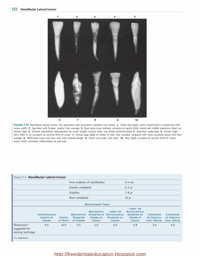
Figure 7-21 Mandibular lateral incisor. Ten specimens with uncommon variations are shown. 1, Tooth very large; cervix constricted in comparison with crown width. 2, Specimen well formed, smaller than average. 3, Root extra long; extreme curvature at apical third; mesial and middle mamelons intact on incisal ridge. 4, Extreme mesiodistal measurement for crown length; contact areas very broad cervicoincisally. 5, Specimen undersized. 6, Incisal ridge thin; little or no curvature at cervical third of crown. 7, Incisal edge labial to center of root; root rounded; cingulum with more curvature above root than average. 8, Malformed crown and root; root with extreme length. 9, Crown very wide; root short. 10, Very slight curvature at cervical third of crown; entire tooth oversized, malformation at root end.
1 32 4 5
6 7 8 9 10
Table 7-2 Mandibular Lateral Incisor
First evidence of calcification 3–4 mo
Enamel completed 4–5 yr
Eruption 7–8 yr
Root completed 10 yr
Measurement Table
Cervicoincisal Length of
CrownLength
of Root
Mesiodistal Diameter of Crown
Mesiodistal Diameter of
Crown at Cervix
Labio- or Buccolingual
Diameter of Crown
Labio- or Buccolingual
Diameter of Crown at
Cervix
Curvature of Cervical
Line—Mesial
Curvature of Cervical Line—Distal
Dimensions* suggested for carving technique
9.5 14.0 5.5 4.0 6.5 5.8 3.0 2.0
*In millimeters.
122 Mandibular Lateral Incisor
http://freedentaleducation.blogspot.com

Chapter 7 The Permanent Mandibular Incisors 123
the second mandibular tooth from the median line, right or left. It resembles the mandibular central incisor so closely that only a brief description of each aspect of the lateral incisor is necessary. Direct comparison is made with the mandibular central incisor, and the variations are men-tioned. The two incisors operate in the dental arch as a team; therefore their functional form is related. As with the mandibular central incisor, the shape of the lateral incisor is uniform compared with that of other teeth. Rarely, it will have a labial and lingual root division in the cervical third. Somewhat more commonly it has two canals in the single root.3
The mandibular lateral incisor is somewhat larger than the mandibular central incisor (compare measurements), but generally speaking, its form closely resembles that of the mandibular central incisor (Table 7-2). Ten specimens with uncommon variations are shown in Figure 7-21.
BRIEF DESCRIPTION OF THE MANDIBULAR LATERAL INCISOR FROM ALL ASPECTS
Labial and Lingual AspectsThe labial and lingual aspects show the added fraction of approximately 1 mm of crown diameter mesiodistally added to the distal half (see Figures 7-13 and 7-14). This, however, is not always true (see Figure 7-19, 3 and 6). The lingual aspect of the mandibular incisors in some Mongoloid groups is marked by a deep but short cervicoincisal groove, which is vulnerable to dental caries.4
Mesial and Distal AspectsThe mesial side of the crown is often longer than the distal side; this causes the incisal ridge, which is straight, to slope downward in a distal direction (see Figure 7-19, 1). The distal contact area is more toward the cervical than the mesial contact area to contact properly the mesial contact area of the mandibular canine.
Except for size, no marked difference is evident between the mesial and distal surfaces of central and lateral incisors (see Figures 7-15, 7-16, and 7-20). Even the curvatures of the cervical line mesially and distally are similar in extent.
A tendency exists toward a deeper concavity immediately above the cervical line on the distal surface of the mandibu-lar lateral incisor.
Although the crown of the mandibular lateral incisor is somewhat longer than that of the central incisor (usually a fraction of a millimeter), the root may be considerably longer. Therefore in most instances the tooth is a little larger in all dimensions. The root form is similar to that of the central incisor, including the presence of developmental depressions mesially and distally.
Incisal AspectThe incisal aspect of the mandibular lateral incisor provides a feature that can usually serve to identify this tooth. The incisal edge is not at approximate right angles to a line bisecting the crown and root labiolingually, as was found when observing the central incisor; the edge follows the curvature of the mandibular dental arch, which gives the crown of the mandibular lateral incisor the appearance of being twisted slightly on its root base (see Figures 7-17 and 7-18). It is interesting to note that the labiolingual root axes of mandibular central and lateral incisors remain almost parallel in the alveolar process, even though the incisal ridges are not directly in line.
References1. Carlsen O: Dental morphology, Copenhagen, 1987,
Munksgaard.2. Pindborg JJ: Pathology of the dental tissues, Philadelphia, 1970,
Saunders.3. Woelfel JB, Scheid RC: Dental anatomy: its relevance to
dentistry, ed 5, Baltimore, 1997, Williams & Wilkins.4. Hanihara K: Racial characteristics in the dentition, J Dent Res
46:293, 1967.
Use the CD-ROM to view animations and study ques-tions; and go to the site for additional study resources.
IV A, B
http://freedentaleducation.blogspot.com

This page intentionally left blank
http://freedentaleducation.blogspot.com

The Permanent Canines: Maxillary and Mandibular
Both maxillary and mandibular canines have another quality that must not be overlooked: the positions and forms of these teeth and their anchorage in the bone, along with the bone ridge over the labial portions of the roots, called the canine eminence, have a cosmetic value. They help form a foundation that ensures normal facial expression at the corners of the mouth. Loss of all of these teeth makes it extremely difficult, if not impossible, to make replacements that restore that natural appearance of the face for any length of time. It would therefore be difficult to place a value on the canines, and their importance is manifested by their efficiency in function, stability, and aid in maintaining natural facial expression.
In function, the canines support the incisors and premo-lars, since they are located between these groups. The canine crowns have some characteristics of functional form, which bears a resemblance to incisor form and also to the premolar form.
Maxillary Canine
Figures 8-1 through 8-12 illustrate the maxillary canine in various aspects. The outline of the labial or lingual aspect of the maxillary canine is a series of curves or arcs except for the angle made by the tip of the cusp. This cusp has a mesial incisal ridge and a distal incisal ridge.
The mesial half of the crown makes contact with the lateral incisor, and the distal half contacts the first premolar. Therefore the contact areas of the maxillary canine are at different levels cervicoincisally.
The maxillary and mandibular canines bear a close resemblance to each other, and their functions are closely related. The four canines are placed at the “corners” of the mouth; each one is the third tooth from the median line, right and left, in the maxilla and mandible. They are commonly referred to as the cornerstone of the dental arches.1 They are the longest teeth in the mouth; the crowns are usually as long as those of the maxillary central incisors, and the single roots are longer than those of any of the other teeth. The middle labial lobes have been highly developed incisally into strong, well-formed cusps. Crowns and roots are markedly convex on most surfaces. The shape and position of the canines contribute to the guidance of the teeth into the intercuspal position by “canine guidance.”2
The shape of the crowns, with their single pointed cusps, their locations in the mouth, and the extra anchorage fur-nished by the long, strongly developed roots, makes these canines resemble those of the carnivore. This resemblance to the prehensile teeth of the carnivore gives rise to the term canine.
Because of the labiolingual thickness of crown and root and the anchorage in the alveolar process of the jaws, these teeth are perhaps the most stable in the mouth. The crown portions of the canines are shaped in a manner that pro-motes cleanliness. This self-cleansing quality, along with the efficient anchorage in the jaws, tends to preserve these teeth throughout life. When teeth are lost, the canines are usually the last ones to go. They are very valuable teeth, when considered either as units of the natural dental arches or as possible assistants in stabilizing replacements of lost teeth in prosthetic procedures.
125
88
http://freedentaleducation.blogspot.com

Figure 8-1 Maxillary right canine, lingual and incisal aspects. CL, Cervical line; C, cingulum; MMR, mesial marginal ridge; MLF, mesiolingual fossa; MCR, mesial cusp ridge; DCR, distal cusp ridge; LR, lingual ridge; DLF, distolingual fossa; DMR, distal marginal ridge.
CLC
MMRMLFMCR DCR
LR
DCRDMR
DLFDLF
MLFC
MCR
Figure 8-2 Maxillary left canine, labial aspect. (Grid = 1 sq mm.)
Figure 8-3 Maxillary left canine, lingual aspect. (Grid = 1 sq mm.)
Figure 8-4 Maxillary left canine, mesial aspect. (Grid = 1 sq mm.)
Figure 8-5 Maxillary left canine, distal aspect. (Grid = 1 sq mm.) Figure 8-6 Maxillary left canine, incisal aspect. (Grid = 1 sq mm.)
126 Maxillary Canine
http://freedentaleducation.blogspot.com

Chapter 8 The Permanent Canines: Maxillary and Mandibular 127
From a labial view, the mesial half of the crown resembles a portion of an incisor, whereas the distal half resembles a portion of a premolar. This tooth seems to be a compromise in the change from anterior to posterior teeth in the dental arch.
It is apparent that the construction of this tooth has reinforcement, labiolingually, to offset directional lines of the force brought against it when in use. The incisional (incising) portion is thicker labiolingually than that of either the maxillary central or the lateral incisor.
The labiolingual measurement of the crown is about 1 mm greater than that of the maxillary central incisor (Table 8-1). The mesiodistal measurement is approximately 1 mm less.
The cingulum shows greater development than that of the central incisor.
The root of the maxillary canine is usually the longest of any root with the possible exception of that of the mandibu-lar canine, which may be as long at times. The root is thick labiolingually, with developmental depressions mesially and distally that help furnish the secure anchorage this tooth has in the maxilla. Uncommon variations are shown in Figure 8-12.
DETAILED DESCRIPTION OF THE MAXILLARY CANINE FROM ALL ASPECTS
Labial Aspect
From the labial aspect, the crown and root are narrower mesiodistally than those of the maxillary central incisor. The difference is about 1 mm in most mouths. The cervical line
Figure 8-7 Maxillary right canine. Graph outlines of five aspects are shown. (Grid = 1 sq mm.)
Labial Lingual
Incisal
Mesial Distal
http://freedentaleducation.blogspot.com

Figure 8-8 Maxillary right canine.
Labial
Incisal
Lingual
Mesial Distal
labially is convex, with the convexity toward the root portion (see Figures 8-2, 8-7, 8-8, and 8-9).
Mesially, the outline of the crown may be convex from the cervix to the center of the mesial contact area, or the crown may exhibit a slight concavity above the contact area from the labial aspect. The center of the contact area mesially is approximately at the junction of middle and incisal thirds of the crown.
Distally, the outline of the crown is usually concave between the cervical line and the distal contact area. The distal contact area is usually at the center of the middle third of the crown. The two levels of contact areas mesially and distally should be noted (see Figure 5-7, B and C).
Unless the crown has been worn unevenly, the cusp tip is on a line with the center of the root. The cusp has a mesial slope and a distal slope, the mesial slope being the shorter
of the two. Both slopes show a tendency toward concavity before wear has taken place (see Figure 8-9, 5 and 6). These depressions are developmental in character.
The labial surface of the crown is smooth, with no devel-opmental lines of note except shallow depressions mesially and distally, dividing the three labial lobes. The middle labial lobe shows much greater development than the other lobes. This produces a ridge on the labial surface of the crown. A line drawn over the crest of this ridge, from the cervical line to the tip of the cusp, is a curved one inclined mesially at its center. All areas mesial to the crest of this ridge exhibit convexity except for insignificant developmen-tal lines in the enamel. Distally to the labial ridge (see incisal aspect), a tendency exists toward concavity at the cervical third of the crown, although convexity is noted elsewhere in all areas approaching the labial ridge (see Figure 8-11, 7, 8, and 9).
128 Maxillary Canine
http://freedentaleducation.blogspot.com

1 3 42 5
6 7 8 9 10
Figure 8-9 Maxillary canine, labial aspect. Ten typical specimens are shown.
Figure 8-10 Maxillary canine, mesial aspect. Ten typical specimens are shown.
1 3 42 5
6 7 8 9 10
IV A(6), B
http://freedentaleducation.blogspot.com

Figure 8-11 Maxillary canine, incisal aspect. Ten typical specimens are shown.
1 2 43 5
6 8 97 10
Figure 8-12 Maxillary canine. Ten specimens with uncommon variations are shown. 1, Crown very long, with extreme curvature at apical third of the root. 2, Entire tooth unusually long. Note hypercementosis at root end. 3, Very short crown; root small and malformed. 4, Mesiodistal dimension of crown at contact are extreme; calibration at cervix narrow in comparison; root short for crown of this size. 5, Extreme labiolingual calibration; root with unusual curvature. 6, Tooth malformed generally. 7, Large crown; short root. 8, Root overdeveloped and very blunt at apex. 9, Odd curvature of root; extra length. 10, Crown poorly formed; root extra long.
1 32 4 5
6 8 97 10
130 Maxillary Canine
http://freedentaleducation.blogspot.com

Chapter 8 The Permanent Canines: Maxillary and Mandibular 131
Table 8-1 Maxillary Canine
First evidence of calcification 4–5 mo
Enamel completed 6–7 yr
Eruption 11–12 yr
Root completed 13–15 yr
Measurement Table
Cervicoincisal Length of
CrownLength
of Root
Mesiodistal Diameter of Crown
Mesiodistal Diameter of
Crown at Cervix
Labio- or Buccolingual
Diameter of Crown
Labio- or Buccolingual
Diameter of Crown at
Cervix
Curvature of Cervical
Line—Mesial
Curvature of Cervical Line—Distal
Dimensions* suggested for carving technique
10.0 17.0 7.5 5.5 8.0 7.0 2.5 1.5
*In millimeters.
The root of the maxillary canine appears slender from the labial aspect when compared with the bulk of the crown; it is conical in form with a bluntly pointed apex. It is not uncommon for this root to have a sharp curve in the vicinity of the apical third. This curvature may be in a mesial or distal direction, but in most instances is the latter (see Figure 8-9, 1 and 6). The labial surface of the root is smooth and convex at all points.
Lingual AspectThe crown and root are narrower lingually than labially.
The cervical line from the lingual aspect differs some-what from the curvature found labially and shows a more even curvature. The line may be straight for a short interval at this point (see Figures 8-3, 8-7, and 8-8).
The cingulum is large and, in some instances, is pointed like a small cusp (see Figure 8-10, 7). In this type, definite ridges are found on the lingual surface of the crown below the cingulum and between strongly developed marginal ridges. Although depressions are to be found between these ridge forms, seldom are any deep developmental grooves present.
Occasionally, a well-developed lingual ridge is seen that is confluent with the cusp tip; this extends to a point near the cingulum. Shallow concavities are evident between this ridge and the marginal ridges. When these concavities are present, they are called mesial and distal lingual fossae (see Figures 8-1 and 8-8).
Sometimes the lingual surface of the canine crown is so smooth that fossae or minor ridges are difficult to distin-guish. However, a tendency exists toward concavities where the fossae are usually found, and heavy marginal ridges with a well-formed cingulum are to be expected. The smooth cingulum, marginal ridges, and lingual portion of the incisal ridges are usually confluent, with little evidence of develop-mental grooves.
The lingual portion of the root of the maxillary canine is narrower than the labial portion. Because of this formation,
much of the mesial and distal surface of the root is visible from the lingual aspect. Developmental depressions mesially and distally may be seen on most of these roots, extending most of the root length. The lingual ridge of the root is rather narrow, but it is smooth and convex at all points from the cervical line to the apical end (see Figure 8-3).
Mesial AspectThe mesial aspect of the maxillary canine presents the outline of the functional form of an anterior tooth. However, it generally shows greater bulk and greater labiolingual measurement than any of the other anterior teeth (see Figures 8-4, 8-7, 8-8, and 8-10).
The outline of the crown is wedge-shaped, the greatest measurement being at the cervical third and the wedge point being represented by the tip of the cusp.
The curvature of the crown below the cervical line labi-ally and lingually corresponds in extent to the curvature of the maxillary central and lateral incisors. Nevertheless, the crest of that curvature is found at a level more incisal, because the middle labial and the lingual lobes are more highly developed (see Figure 8-10, 5 and 10). Many canines show a flattened area labially at the cervical third of the crown, which appears as a straight outline from the mesial aspect. It is questionable just how much wear has to do with this effect (see Figure 8-10, 1 and 2).
Below the cervical third of the crown, the labial face may be presented by a line only slightly convex from the crest of curvature at the cervical third to the tip of the cusp. The line usually becomes straighter as it approaches the cusp.
The entire labial outline from the mesial aspect exhibits more convexity from the cervical line to the cusp tip than the maxillary central incisor does from cervix to incisal edge.
The lingual outline of the crown from the mesial aspect may be represented by a convex line describing the cingu-lum, which convexity straightens out as the middle third is
http://freedentaleducation.blogspot.com
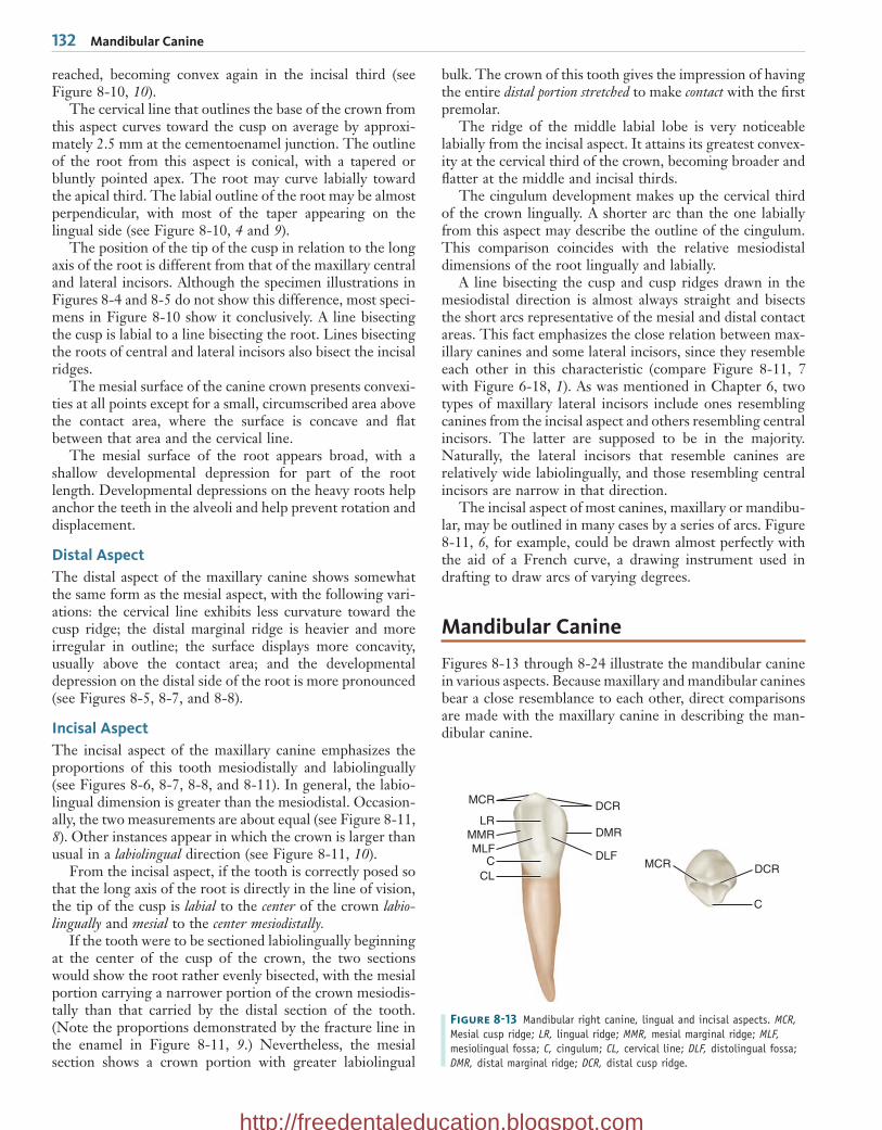
132 Mandibular Canine
reached, becoming convex again in the incisal third (see Figure 8-10, 10).
The cervical line that outlines the base of the crown from this aspect curves toward the cusp on average by approxi-mately 2.5 mm at the cementoenamel junction. The outline of the root from this aspect is conical, with a tapered or bluntly pointed apex. The root may curve labially toward the apical third. The labial outline of the root may be almost perpendicular, with most of the taper appearing on the lingual side (see Figure 8-10, 4 and 9).
The position of the tip of the cusp in relation to the long axis of the root is different from that of the maxillary central and lateral incisors. Although the specimen illustrations in Figures 8-4 and 8-5 do not show this difference, most speci-mens in Figure 8-10 show it conclusively. A line bisecting the cusp is labial to a line bisecting the root. Lines bisecting the roots of central and lateral incisors also bisect the incisal ridges.
The mesial surface of the canine crown presents convexi-ties at all points except for a small, circumscribed area above the contact area, where the surface is concave and flat between that area and the cervical line.
The mesial surface of the root appears broad, with a shallow developmental depression for part of the root length. Developmental depressions on the heavy roots help anchor the teeth in the alveoli and help prevent rotation and displacement.
Distal AspectThe distal aspect of the maxillary canine shows somewhat the same form as the mesial aspect, with the following vari-ations: the cervical line exhibits less curvature toward the cusp ridge; the distal marginal ridge is heavier and more irregular in outline; the surface displays more concavity, usually above the contact area; and the developmental depression on the distal side of the root is more pronounced (see Figures 8-5, 8-7, and 8-8).
Incisal AspectThe incisal aspect of the maxillary canine emphasizes the proportions of this tooth mesiodistally and labiolingually (see Figures 8-6, 8-7, 8-8, and 8-11). In general, the labio-lingual dimension is greater than the mesiodistal. Occasion-ally, the two measurements are about equal (see Figure 8-11, 8). Other instances appear in which the crown is larger than usual in a labiolingual direction (see Figure 8-11, 10).
From the incisal aspect, if the tooth is correctly posed so that the long axis of the root is directly in the line of vision, the tip of the cusp is labial to the center of the crown labio-lingually and mesial to the center mesiodistally.
If the tooth were to be sectioned labiolingually beginning at the center of the cusp of the crown, the two sections would show the root rather evenly bisected, with the mesial portion carrying a narrower portion of the crown mesiodis-tally than that carried by the distal section of the tooth. (Note the proportions demonstrated by the fracture line in the enamel in Figure 8-11, 9.) Nevertheless, the mesial section shows a crown portion with greater labiolingual
bulk. The crown of this tooth gives the impression of having the entire distal portion stretched to make contact with the first premolar.
The ridge of the middle labial lobe is very noticeable labially from the incisal aspect. It attains its greatest convex-ity at the cervical third of the crown, becoming broader and flatter at the middle and incisal thirds.
The cingulum development makes up the cervical third of the crown lingually. A shorter arc than the one labially from this aspect may describe the outline of the cingulum. This comparison coincides with the relative mesiodistal dimensions of the root lingually and labially.
A line bisecting the cusp and cusp ridges drawn in the mesiodistal direction is almost always straight and bisects the short arcs representative of the mesial and distal contact areas. This fact emphasizes the close relation between max-illary canines and some lateral incisors, since they resemble each other in this characteristic (compare Figure 8-11, 7 with Figure 6-18, 1). As was mentioned in Chapter 6, two types of maxillary lateral incisors include ones resembling canines from the incisal aspect and others resembling central incisors. The latter are supposed to be in the majority. Naturally, the lateral incisors that resemble canines are relatively wide labiolingually, and those resembling central incisors are narrow in that direction.
The incisal aspect of most canines, maxillary or mandibu-lar, may be outlined in many cases by a series of arcs. Figure 8-11, 6, for example, could be drawn almost perfectly with the aid of a French curve, a drawing instrument used in drafting to draw arcs of varying degrees.
Mandibular Canine
Figures 8-13 through 8-24 illustrate the mandibular canine in various aspects. Because maxillary and mandibular canines bear a close resemblance to each other, direct comparisons are made with the maxillary canine in describing the man-dibular canine.
Figure 8-13 Mandibular right canine, lingual and incisal aspects. MCR, Mesial cusp ridge; LR, lingual ridge; MMR, mesial marginal ridge; MLF, mesiolingual fossa; C, cingulum; CL, cervical line; DLF, distolingual fossa; DMR, distal marginal ridge; DCR, distal cusp ridge.
MCR
LRMMRMLF
C
CL
DCR
DMR
DLFDCR
C
MCR
http://freedentaleducation.blogspot.com

Chapter 8 The Permanent Canines: Maxillary and Mandibular 133
Figure 8-14 Mandibular left canine, labial aspect. (Grid = 1 sq mm.)
Figure 8-15 Mandibular left canine, lingual aspect. (Grid = 1 sq mm.)
Figure 8-16 Mandibular left canine, mesial aspect. (Grid = 1 sq mm.)
Figure 8-17 Mandibular left canine, distal aspect. (Grid = 1 sq mm.)
Figure 8-18 Mandibular left canine, incisal aspect. (Grid = 1 sq mm.)
The mandibular canine crown is narrower mesiodistally than that of the maxillary canine, although it is just as long in most instances and in many instances is longer by 0.5 to 1 mm (Table 8-2). The root may be as long as that of the maxillary canine, but usually it is somewhat shorter. The labiolingual diameter of crown and root is usually a fraction of a millimeter less.
The lingual surface of the crown is smoother, with less cingulum development and less bulk to the marginal ridges. The lingual portion of this crown resembles the form of the lingual surfaces of the mandibular lateral incisors.
The cusp of the mandibular canine is not as well devel-oped as that of the maxillary canine, and the cusp ridges are thinner labiolingually. Usually the cusp tip is on a line with the center of the root, from the mesial or distal aspect, but sometimes it lies lingual to the line, as with the mandibular incisors.
A variation in the form of the mandibular canine is bifurcated roots. This variation is not rare (see Figure 8-24, 1, 2, 5, 6 and Figure 8-25).
http://freedentaleducation.blogspot.com

134 Mandibular Canine
Figure 8-19 Mandibular right canine. Graph outlines of five aspects are shown. (Grid = 1 sq mm.)
Labial Lingual
Incisal
Mesial Distal
DETAILED DESCRIPTION OF THE MANDIBULAR CANINE FROM ALL ASPECTS
Labial AspectFrom the labial aspect, the mesiodistal dimensions of the mandibular canine are less than those of the maxillary canine. The difference is usually about 1 mm. The man-dibular canine is broader mesiodistally than either of the mandibular incisors, for example, about 1 mm wider than the mandibular lateral incisor (see Figures 8-14, 8-19, 8-20, and 8-21).
The essential differences between mandibular and maxillary canines viewed from the labial aspect may be described as follows:
• The crowns of the mandibular canines appear longer. Sometimes they are longer, but the effect of greater length is emphasized by the narrowness of the crown mesiodistally and the height of the contact areas above the cervix.
• The mesial outline of the crown of the mandibular canine is nearly straight with the mesial outline of the root, with the mesial contact area being near the mesioincisal angle.
• When the cusp ridges have not been affected by wear, the cusp angle is on a line with the center of the root, as on the maxillary canine. The mesial cusp ridge is shorter.
• The distal contact area of the mandibular canine is more toward the incisal aspect than that of the
http://freedentaleducation.blogspot.com

Chapter 8 The Permanent Canines: Maxillary and Mandibular 135
Figure 8-20 Mandibular right canine.
Labial Lingual
Incisal
Mesial Distal
maxillary canine but is not up to the level of the mesial area.
• The cervical line labially has a semicircular curvature apically.
• Many mandibular canines give the impression from this aspect of being bent distally on the root base. The maxillary canine crowns are more likely to be in line with the root.
• The mandibular canine root is shorter by l or 2 mm on average than that of the maxillary canine, and its apical end is more sharply pointed. Root curvatures are infrequent. When curvature of root ends is present, it is often in a mesial direction (see Figure 8-21, 1, 2, 3, and 4).
Lingual AspectIn comparing the lingual aspect of the mandibular canine with that of the maxillary canine, the following differences are noted.
The lingual surface of the crown of the mandibular canine is flatter, simulating the lingual surfaces of mandibular inci-sors (see Figures 8-13, 8-15, 8-19, and 8-20). The cingulum is smooth and poorly developed. The marginal ridges are less distinct. This is true of the lingual ridge except toward the cusp tip, where it is raised. Generally speaking, the lingual surface of the crown is smooth and regular.
The lingual portion of the root is narrower relatively than that of the maxillary canine. It narrows down to little more than half the width of the labial portion.
http://freedentaleducation.blogspot.com

136 Mandibular Canine
1 432 5
6 7 8 9 10
Figure 8-21 Mandibular canine, labial aspect. Ten typical specimens are shown.
Figure 8-22 Mandibular canine, mesial aspect. Ten typical specimens are shown.
1 2 3 4 5
6 87 9 10
IV A(27)
http://freedentaleducation.blogspot.com

Chapter 8 The Permanent Canines: Maxillary and Mandibular 137
Figure 8-23 Mandibular canine, incisal aspect. Ten typical specimens are shown.
6 87 9 10
1 2 3 54
Figure 8-24 Mandibular canine. Ten specimens with uncommon variations are shown. 1, Well-formed crown; two roots, one lingual and one labial. 2, Same as specimen 1, with longer roots. 3, Well-formed crown portion; poorly formed root. 4, Root longer than average, with extreme curvature. 5, Deep developmental groove dividing the root. 6, Same as specimen 5. 7, Crown resembling mandibular lateral incisor; root short. 8, Root extra long, with odd mesial curvature starting at cervical third. 9, Crown extra long and irregular in outline. Root short and poorly formed at apex. 10, Crown with straight mesial and distal sides, wide at cervix, with a root of extreme length.
1 42 3 5
6 87 9 10
http://freedentaleducation.blogspot.com

138 Mandibular Canine
Table 8-2 Mandibular Canine
First evidence of calcification 4–5 mo
Enamel completed 6–7 yr
Eruption 9–10 yr
Root completed 12–14 yr
Measurement Table
Cervicoincisal Length of
CrownLength
of Root
Mesiodistal Diameter of Crown
Mesiodistal Diameter of
Crown at Cervix
Labio- or Buccolingual
Diameter of Crown
Labio- or Buccolingual
Diameter of Crown at
Cervix
Curvature of Cervical
Line—Mesial
Curvature of Cervical Line—Distal
Dimensions* suggested for carving technique
11.0 16.0 7.0 5.5 7.5 7.0 2.5 1.0
*In millimeters.
Figure 8-25 Mesial view of a left mandibular canine demonstrating prominent root bifurcation.
Mesial AspectFrom the mesial aspect, characteristic differences are evident between the two teeth in question. The mandibular canine has less curvature labially on the crown, with very little curvature directly above the cervical line. The curvature at the cervical portion as a rule is less than 0.5 mm. The lingual outline of the crown is curved in the same manner as that of the maxillary canine, but it differs in degree (see Figures 8-16, 8-19, 8-20, and 8-22).
The cingulum is not as pronounced, and the incisal portion of the crown is thinner labiolingually, which allows the cusp to appear more pointed and the cusp ridge to appear more slender. The tip of the cusp is more nearly centered over the root, with a lingual placement in some cases comparable to the placement of incisal ridges on man-dibular incisors.
The cervical line curves more toward the incisal portion than does the cervical line on the maxillary canine.
The roots of the two teeth are quite similar from the mesial aspect with the possible exception of a more pointed root tip on the mandibular canine. The developmental depression mesially on the root of the mandibular canine is more pronounced and sometimes quite deep.
Distal AspectLittle difference from the distal aspect can be seen between mandibular and maxillary canines except for those features mentioned under mesial aspect that are common to both (see Figures 8-17, 8-19, and 8-20).
Incisal AspectThe outlines of the crowns of mandibular and maxillary canines from the incisal aspect are often similar (see Figures 8-13, 8-18, 8-19, 8-20, and 8-23). The main differences to be noted are as follows:
• The mesiodistal dimension of the mandibular canine is less than the labiolingual dimension. A similarity is evident in this, but the outlines of the mesial surface are less curved.
• The cusp tip and mesial cusp ridge are more likely to be inclined in a lingual direction in the mandibular canine, with the distal cusp ridge and the contact area extension distinctly so. Note that the cusp ridges of the maxillary canine with the contact area extensions are more nearly in a straight line mesiodistally from the incisal aspect.
IV A, B
http://freedentaleducation.blogspot.com

Chapter 8 The Permanent Canines: Maxillary and Mandibular 139
References1. Kraus BS: Dental anatomy and occlusion, Baltimore, 1969,
Williams & Wilkins.2. Ash MM, Ramfjord SP: Occlusion, ed 4, Philadelphia, 1995,
Saunders.
Use the CD-ROM to view animations and study ques-tions; and go to the site for additional study resources.
http://freedentaleducation.blogspot.com

This page intentionally left blank
http://freedentaleducation.blogspot.com
The Permanent Maxillary Premolars
articulate with opposing teeth when the jaws are brought together; this makes them more efficient as grinding teeth, and they function much like the molars, but to a lesser degree.
The maxillary premolar crowns are shorter than those of the maxillary canines, and the roots are also shorter. The root lengths equal those of the molars. The crowns are a little longer than those of the molars.
Because of the cusp development buccally and lingually, the marginal ridges are in a more horizontal plane and are considered part of the occlusal surface of the crown rather than part of the lingual surface, as in the case of incisors and canines.
When premolars have two roots, one is placed buccally and one lingually.
Maxillary First Premolar
Figures 9-1 through 9-16 illustrate the maxillary first premo-lar from all aspects. The maxillary first premolar has two cusps, a buccal and a lingual, each being sharply defined. The buccal cusp is usually about 1 mm longer than the lingual cusp. The crown is angular, and the buccal line angles are prominent.
The crown is shorter than that of the canine by 1.5 to 2 mm on the average (Table 9-1). Although this tooth resembles the canine from the buccal aspect, it differs in that the contact areas mesially and distally are at about the same level. The root is shorter. If the buccal cusp form has not been changed by wear, the mesial slope of the cusp is longer than the distal slope. The opposite arrangement is true of
The maxillary premolars number four: two in the right maxilla and two in the left maxilla. They are posterior to the canines and immediately anterior to the molars.
The premolars are so named because they are anterior to the molars in the permanent dentition. In zoology, the premolars are those teeth that succeed the deciduous molars regardless of the number to be succeeded. The term bicus-pid, which is widely used to describe human teeth, presup-poses two cusps, a supposition that makes the term misleading, because mandibular premolars in the human subject may show a variation in the number of cusps from one to three. Among carnivores, in the study of comparative dental anatomy, premolar forms differ so greatly that a more descriptive single term than premolar is out of the question. The term premolar is used widely in dental anatomy, human and comparative; therefore it will be used here. However, its use here does not suggest that the term bicuspid should not be used when appropriate.
The maxillary premolars are developed from the same number of lobes as anterior teeth—four. The primary dif-ference in development is the well-formed lingual cusp, developed from the lingual lobe, which is represented by the cingulum development on incisors and canines. The middle buccal lobe on the premolars, corresponding to the middle labial lobe of the canines, remains highly developed, with the maxillary premolars resembling the canines when viewed from the buccal aspect. The buccal cusp of the maxillary first premolar, especially, is long and sharp, assisting the canine as a prehensile or tearing tooth. The mandibular first premo-lar assists the mandibular canine in the same manner.
The second premolars, both maxillary and mandibular, have cusps less sharp than the others, and their cusps
141
99
http://freedentaleducation.blogspot.com

142 Maxillary First Premolar
Figure 9-1 Maxillary right first premolar, mesial and occlusal aspects. LR, Lingual root; CL, cervical line; MMDG, mesial marginal developmental groove; LC, lingual cusp; BC, buccal cusp; MCA, mesial contact area; BCR, buccal cervical ridge; MDD, mesial developmental depression; BR, buccal root; MBCR, mesiobuccal cusp ridge; MMR, mesial marginal ridge; MTF, mesial triangular fossa (shaded area); CDG, central developmental groove; MLCR, mesiolingual cusp ridge; DLCR, distolingual cusp ridge; DTF, distal triangular fossa; DMR, distal marginal ridge; DBCR, distobuccal cusp ridge.
BR
MDD
BCR
MCA
BC LC
CL
MMDG
LR
DBCR
DMRDTF
DLCR MLCRCDGMTFMMRMBCR
Figure 9-2 Maxillary left first premolar, buccal aspect. (Grid = 1 sq mm.)
Figure 9-3 Maxillary left first premolar, lingual aspect. (Grid = 1 sq mm.)
Figure 9-4 Maxillary left first premolar, mesial aspect. (Grid = 1 sq mm.)
Figure 9-5 Maxillary left first premolar, distal aspect. (Grid = 1 sq mm.)Figure 9-6 Maxillary left first premolar, occlusal aspect. (Grid = 1 sq mm.)
http://freedentaleducation.blogspot.com

Chapter 9 The Permanent Maxillary Premolars 143
Figure 9-7 Maxillary right first premolar. Graph outlines of five aspects are shown. (Grid = 1 sq mm.)
Buccal Lingual
DistalMesial
Occlusal
Table 9-1 Maxillary First Premolar
First evidence of calcification 112–13
4 yr
Enamel completed 5–6 yr
Eruption 10–11 yr
Root completed 12–13 yr
Measurement Table
Cervico-occlusal
Length of Crown
Length of Root
Mesiodistal Diameter of Crown
Mesiodistal Diameter of
Crown at Cervix
Labio- or Buccolingual
Diameter of Crown
Labio- or Buccolingual
Diameter of Crown at
Cervix
Curvature of Cervical
Line—Mesial
Curvature of Cervical Line—Distal
Dimensions* suggested for carving technique
8.5 14.0 7.0 5.0 9.0 8.0 1.0 0.0
*In millimeters.
http://freedentaleducation.blogspot.com

144 Maxillary First Premolar
Figure 9-8 Maxillary right first premolar.
Buccal
Occlusal
Lingual
Mesial Distal
the maxillary canine. Generally, the first premolar is not as wide in a mesiodistal direction as the canine.
Most maxillary first premolars have two roots (see Figure 9-10) and two pulp canals. When only one root is present, two pulp canals are usually found anyway.
The maxillary first premolar has some characteristics common to all posterior teeth. Briefly, those characteristics that differentiate posterior teeth from anterior teeth are as follows:
1. Greater relative faciolingual measurement compared with the mesiodistal measurement
2. Broader contact areas
3. Contact areas more nearly at the same level4. Less curvature of the cervical line mesially and
distally5. Shorter crown, cervico-occlusally than that of anterior
teeth
DETAILED DESCRIPTION OF THE MAXILLARY FIRST PREMOLAR FROM ALL ASPECTS
Buccal AspectFrom the buccal aspect, the crown is roughly trapezoidal (see Figure 4-16, C). The crown exhibits little curvature at
http://freedentaleducation.blogspot.com
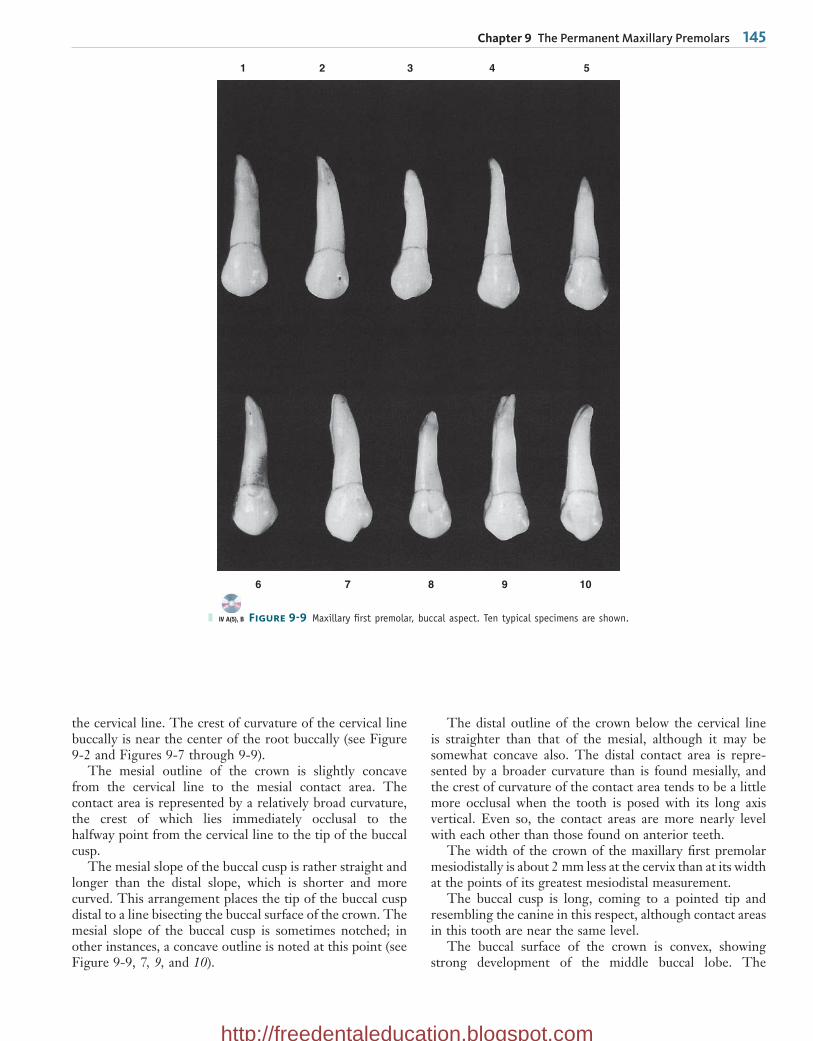
Chapter 9 The Permanent Maxillary Premolars 145
1 2 3 4 5
6 7 8 9 10
Figure 9-9 Maxillary first premolar, buccal aspect. Ten typical specimens are shown.
the cervical line. The crest of curvature of the cervical line buccally is near the center of the root buccally (see Figure 9-2 and Figures 9-7 through 9-9).
The mesial outline of the crown is slightly concave from the cervical line to the mesial contact area. The contact area is represented by a relatively broad curvature, the crest of which lies immediately occlusal to the halfway point from the cervical line to the tip of the buccal cusp.
The mesial slope of the buccal cusp is rather straight and longer than the distal slope, which is shorter and more curved. This arrangement places the tip of the buccal cusp distal to a line bisecting the buccal surface of the crown. The mesial slope of the buccal cusp is sometimes notched; in other instances, a concave outline is noted at this point (see Figure 9-9, 7, 9, and 10).
The distal outline of the crown below the cervical line is straighter than that of the mesial, although it may be somewhat concave also. The distal contact area is repre-sented by a broader curvature than is found mesially, and the crest of curvature of the contact area tends to be a little more occlusal when the tooth is posed with its long axis vertical. Even so, the contact areas are more nearly level with each other than those found on anterior teeth.
The width of the crown of the maxillary first premolar mesiodistally is about 2 mm less at the cervix than at its width at the points of its greatest mesiodistal measurement.
The buccal cusp is long, coming to a pointed tip and resembling the canine in this respect, although contact areas in this tooth are near the same level.
The buccal surface of the crown is convex, showing strong development of the middle buccal lobe. The
IV A(5), B
http://freedentaleducation.blogspot.com

146 Maxillary First Premolar
1 2 3 4 5
6 7 8 9 10
Figure 9-10 Maxillary first premolar, mesial aspect. Ten typical specimens are shown.
Figure 9-11 A, Mesial view of a maxillary right first premolar demonstrating single root and mesial crown concavity. B, Mesial view of a maxillary right second premolar demonstrating single root and no mesial crown concavity.
A B
http://freedentaleducation.blogspot.com

Chapter 9 The Permanent Maxillary Premolars 147
Figure 9-12 Maxillary first premolar, occlusal aspect. Ten typical specimens are shown.
1 2 3 4 5
6 7 8 9 10
continuous ridge from cusp tip to cervical margin on the buccal surface of the crown is called the buccal ridge.
Mesial and distal to the buccal ridge, at or occlusal to the middle third, developmental depressions are usually seen that serve as demarcations between the middle buccal lobe and the mesiobuccal and distobuccal lobes. Although the latter lobes show less development, they are nevertheless prominent and serve to emphasize strong mesiobuccal and distobuccal line angles on the crown.
The roots are 3 or 4 mm shorter than those of the maxil-lary canine, although the outline of the buccal portion of the root form bears a close resemblance.
Lingual AspectFrom the lingual aspect, the gross outline of the maxillary first premolar is the reverse of the gross outline from the buccal aspect (see Figures 9-3, 9-7, and 9-8).
The crown tapers toward the lingual because the lingual cusp is narrower mesiodistally than the buccal cusp. The lingual cusp is smooth and spheroidal from the cervical portion to the area near the cusp tip. The cusp tip is pointed, with mesial and distal slopes meeting at an angle of about 90 degrees.
Naturally, the spheroidal form of the lingual portion of the crown is convex at all points. Sometimes the crest of the smooth lingual portion that terminates at the point of the lingual cusp is called the lingual ridge.
The mesial and distal outlines of the lingual portion of the crown are convex; these outlines are continuous with the mesial and distal slopes of the lingual cusp and straighten out as they join the mesial and distal sides of the lingual root at the cervical line.
The cervical line lingually is regular, with slight curva-ture toward the root and the crest of curvature centered on the root. Because the lingual portion of the crown is nar-rower than the buccal portion, it is possible to see part of the mesial and distal surfaces of crown and root from the lingual aspect, depending on the posing of the tooth and the line of vision.
Because the lingual cusp is not as long as the buccal cusp, the tips of both cusps, with their mesial and distal slopes, may be seen from the lingual aspect.
The lingual portion of the root, or the lingual portion of the lingual root if two roots are present, is smooth and convex at all points. The apex of the lingual root of a two-root specimen tends to be more blunt than the buccal root apex.
Mesial AspectThe mesial aspect of the crown of the maxillary first premo-lar is also roughly trapezoidal (see Figures 9-1, 9-4, 9-7, 9-8, and 9-10). However, the longest of the uneven sides is toward the cervical portion and the shortest is toward the occlusal portion (see Figure 4-16, E).
http://freedentaleducation.blogspot.com
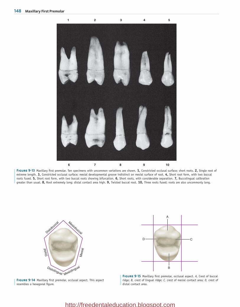
148 Maxillary First Premolar
Figure 9-14 Maxillary first premolar, occlusal aspect. This aspect resembles a hexagonal figure.
Mes
ial
MesiolingualDistolingual
MesiobuccalDistob
ucca
l
Dis
tal
Figure 9-15 Maxillary first premolar, occlusal aspect. A, Crest of buccal ridge; B, crest of lingual ridge; C, crest of mesial contact area; D, crest of distal contact area.
A
B
CD
Figure 9-13 Maxillary first premolar. Ten specimens with uncommon variations are shown. 1, Constricted occlusal surface; short roots. 2, Single root of extreme length. 3, Constricted occlusal surface; mesial developmental groove indistinct on mesial surface of root. 4, Short root form, with two buccal roots fused. 5, Short root form, with two buccal roots showing bifurcation. 6, Short roots, with considerable separation. 7, Buccolingual calibration greater than usual. 8, Root extremely long; distal contact area high. 9, Twisted buccal root. 10, Three roots fused; roots are also uncommonly long.
1 2 3 4 5
6 7 8 9 10
http://freedentaleducation.blogspot.com

Chapter 9 The Permanent Maxillary Premolars 149
Figure 9-16 Maxillary first premolar, occlusal aspect. TBC, Tip of buccal cusp; MBCR, mesiobuccal cusp ridge; MBDG, mesiobuccal developmental groove; MTF, mesial triangular fossa; MMDG, mesial marginal developmental groove; MMR, mesial marginal ridge; MLCR, mesiolingual cusp ridge; LTR, lingual triangular ridge; TLC, tip of lingual cusp; CG, central groove; DLCR, distolingual cusp ridge; DMR, distal marginal ridge; DTF, distal triangular fossa; DBDG, distobuccal developmental groove; BTR, buccal triangular ridge; DBCR, distobuccal cusp ridge. (Compare with Figure 9-1.)
DBCR
BTR
DBDGDTF
DMR
DLCR
CG TLC
LTR
MLCR
MMR
MMDG
MTFMBDGMBCR
TBC
Another characteristic that is representative of all poste-rior maxillary teeth is that the tips of the cusps are well within the confines of the root trunk. (For a definition of root trunk, see Figures 11-3 and 11-8.) In other words, the measurement from the tip of the buccal cusp to the tip of the lingual cusp is less than the buccolingual measurement of the root at its cervical portion.
Most maxillary first premolars have two roots, one buccal and one lingual; these are clearly outlined from the mesial aspect.
The cervical line may be regular in outline (see Figure 9-10, 1) or irregular (see Figure 9-10, 4). In either case, the curvature occlusally is less (about 1 mm on the average) than the cervical curvature on the mesial of any of the anterior teeth. The extent of the curvature of the cervical line mesially on these teeth is constant within a fraction of a millimeter and is similar to the average curvature of the mesial of all posterior teeth.
From the mesial aspect, the buccal outline of the crown curves outward below the cervical line. The crest of curva-ture is often located approximately at the junction of cervical and middle thirds; however, the crest of curvature may be located within the cervical third (see Figure 9-10, 1 and 10). From the crest of curvature, the buccal outline continues as a line of less convexity to the tip of the buccal cusp, which is directly below the center of the buccal root (when two roots are present).
The lingual outline of the crown may be described as a smoothly curved line starting at the cervical line and ending at the tip of the lingual cusp. The crest of this curvature is most often near the center of the middle third. Some speci-mens show a more abrupt curvature at the cervical third (see Figure 9-10, 2 and 9).
The tip of the lingual cusp is on a line, in most cases, with the lingual border of the lingual root. The lingual cusp is always shorter than the buccal cusp, the average difference being about 1 mm. This difference, however, may be greater (see Figure 9-10, 1, 4, and 10). From this aspect, it is noted that the cusps of the maxillary first premolar are long and sharp, with the mesial marginal ridge at about the level of the junction of the middle and occlusal thirds.
A distinguishing feature of this tooth is found on the mesial surface of the crown. Immediately cervical to the mesial contact area, centered on the mesial surface and bordered buccally and lingually by the mesiobuccal and mesiolingual line angles, is a marked depression called the mesial developmental depression (see Figure 9-1). This mesial concavity continues apically beyond the cervical line, joins a deep developmental depression between the roots, and ends at the root bifurcation. On single-root specimens, the concavity on the crown and root is also plainly seen, although it may not be as deeply marked. Maxillary second premolars do not have this feature (see Figure 9-11, A and B).
Another distinguishing feature of the maxillary first premolar is a well-defined developmental groove in the enamel of the mesial marginal ridge. This groove is in alignment with the developmental depression on the mesial surface of the root but is not usually connected with it. This marginal groove is continuous with the central groove of the occlusal surface of the crown, crossing the marginal ridge immediately lingual to the mesial contact area and terminating a short distance cervical to the mesial marginal ridge on the mesial surface (see Figure 9-10, 10).
The buccal outline of the buccal root, above the cervical line, is straight, with a tendency toward a lingual inclination. On those buccal roots having a buccal inclination above the root bifurcation, the outline may be relatively straight up to the apical portion of the buccal root or it may curve buccally at the middle third. Buccal roots may take a buccal or lingual inclination, apical to middle thirds.
The lingual outline of the lingual root is rather straight above the cervical line. It may not exhibit much curvature between the cervix and the apex. Many cases, however, show considerable curvature to lingual roots apical to the middle thirds. The lingual root may take a buccal or lingual inclina-tion (see Figure 9-10, 1, 2, and 9).
The root trunk is long on this tooth, making up about half of the root length. The bifurcation on those teeth with two roots begins at a more occlusal point mesially than dis-tally. Generally speaking, when bifurcated, the root is bifur-cated for half its total length.
Except for the deep developmental groove and depres-sion at or below the bifurcation, the mesial surface of the root portion of this tooth is smoothly convex buccally and lingually. Even when only one root is present, the develop-mental depression is very noticeable for most of the root length. In such cases, roots with buccal and lingual outlines
http://freedentaleducation.blogspot.com

150 Maxillary First Premolar
ending in a blunt apex above the center of the crown are seen (see Figure 9-10, 4 and 5).
Distal AspectFrom the distal aspect (see Figures 9-5, 9-7, and 9-8), the anatomy of the crown and root of the maxillary first premo-lar differs from that from the mesial aspect as follows:
• The crown surface is convex at all points except for a small, flattened arc just cervical to the contact area and buccal to the center of the distal surface.
• The curvature of the cervical line is less on the distal than on the mesial surface, often showing a line straight across from buccal to lingual.
• A deep developmental groove crossing the distal marginal ridge of the crown is not evident. If a developmental groove should be noticeable, it is shallow and insignificant.
• The root trunk is flattened on the distal surface above the cervical line with no outstanding developmental signs.
• The bifurcation of the roots is abrupt near the apical third, with no developmental groove leading to it such as that found mesially.
Occlusal AspectThe occlusal aspect of the maxillary first premolar resembles roughly a six-sided or hexagonal figure (see Figures 9-7, 9-8, 9-12, and 9-14). The six sides are made up of the mesiobuc-cal, which is mesial to the buccal ridge; mesial; mesiolingual, which is mesial to the lingual ridge; distolingual; distal; and distobuccal. This hexagonal figure, however, is not equilat-eral. The two buccal sides are nearly equal, the mesial side is shorter than the distal side, and the mesiolingual side is shorter than the distolingual side (see Figure 9-14).
The relation and position of various anatomical points are to be considered from the occlusal aspect. A drawing of the outline of this occlusal aspect, when placed within a rectangle the dimensions of which represent the mesiodistal and buccolingual width of the crown, demonstrates the rela-tive positions of the mesial and distal contact areas and also those of the buccal and lingual ridges (see Figures 9-6 and 9-15).
The crest of the distal contact area is somewhat buccal to that of the mesial contact area, and the crest of the buccal ridge is somewhat distal to that of the lingual ridge. The crests of curvature represent the highest points on the buccal and lingual ridges and the mesial and distal contact areas.
Close observation of the crown from this aspect reveals the following characteristics (see Figure 9-15):
1. The distance from the buccal crest (A in Figure 9-15) to the mesial crest (C) is slightly longer than the distance from the buccal crest to the distal crest (D).
2. The distance from the mesial crest to the lingual crest is much shorter than the distance from the distal crest to the lingual crest.
3. The crown is wider on the buccal than on the lingual.4. The buccolingual dimension of the crown is much
greater than the mesiodistal dimension.The occlusal surface of the maxillary first premolar is
circumscribed by the cusp ridges and marginal ridges. The mesiobuccal and distobuccal cusp ridges are in line with each other, and their alignment is in a distobuccal direction. In other words, even though they are in the same alignment, the distobuccal cusp ridge is buccal to the mesiobuccal cusp ridge (see Figure 9-16).
The angle formed by the convergence of the mesiobuccal cusp ridge and the mesial marginal ridge approaches a right angle. The angle formed by the convergence of the disto-buccal cusp ridge and the distal marginal ridge is acute. The mesiolingual and distolingual cusp ridges are confluent with the mesial and distal marginal ridges; these cusp ridges are curved, following a semicircular outline from the marginal ridges to their convergence at the tip of the lingual cusp.
When one looks at the occlusal aspect of the maxillary first premolar, posing the tooth so that the line of vision is in line with the long axis, more of the buccal surface of the crown is seen than of the lingual surface. When the tooth is viewed from the mesial aspect, the tip of the buccal cusp is nearer the center of the root trunk than is the lingual cusp.
The occlusal surface of this tooth has no supplemental grooves in most cases, a fact that makes the surface relatively smooth. A well-defined central developmental groove divides the surface evenly buccolingually. It is located at the bottom of the central sulcus of the occlusal surface, extending from a point just mesial to the distal marginal ridge to the mesial marginal ridge, where it joins the mesial marginal development groove. This latter groove crosses the mesial marginal ridge and ends on the mesial surface of the crown (see Figures 9-1 and 9-4).
Two collateral developmental grooves join the central groove just inside the mesial and distal marginal ridges. These grooves are called the mesiobuccal developmental groove and the distobuccal developmental groove. The junctions of the grooves are deeply pointed and are named the mesial and distal developmental pits.
Just distal to the mesial marginal ridge, the triangular depression that harbors the mesiobuccal developmental groove is called the mesial triangular fossa. The depression in the occlusal surface, just mesial to the distal marginal ridge, is called the distal triangular fossa.
Although no supplemental grooves are present in most instances, smooth developmental depressions may be visible radiating from the central groove and giving the occlusal surface an uneven appearance.
The buccal triangular ridge of the buccal cusp is promi-nent, arising near the center of the central groove and con-verging with the tip of the buccal cusp. The lingual triangular ridge is less prominent; it also arises near the center of the central groove and converges with the tip of the lingual cusp.
The lingual cusp is pointed more sharply than the buccal cusp.
http://freedentaleducation.blogspot.com

Chapter 9 The Permanent Maxillary Premolars 151
Maxillary Second Premolar
Figures 9-17 through 9-25 illustrate the maxillary second premolar from all aspects. The maxillary second premolar supplements the maxillary first premolar in function. The two teeth resemble each other so closely that only a brief
description of each aspect of the second premolar is neces-sary. Direct comparison is made between it and the first premolar, and variations are mentioned.
The maxillary second premolar is less angular, giving a more rounded effect to the crown from all aspects. It has a single root.
Considerable variations in the relative sizes of the two teeth may be seen, because the second premolar does not appear true to form as often as does the first premolar (Table 9-2). The maxillary second premolar may have a crown that is noticeably smaller cervico-occlusally and also mesiodistally; however, it may also be larger in those dimen-sions. Usually the root of the second premolar is as long as, if not a millimeter or so longer than, that of the first premo-lar. The two teeth have about the same dimensions on the average, except for the tendency toward greater length of the second premolar root. Ten specimens with uncommon variations are shown in Figure 9-25.
DETAILED DESCRIPTION OF THE MAXILLARY SECOND PREMOLAR FROM ALL ASPECTS
Buccal AspectFrom the buccal aspect, it may be noticed that the buccal cusp of the second premolar is not as long as that of the first premolar and it appears less pointed (see Figure 9-17). Also,
Figure 9-17 Maxillary left second premolar, buccal aspect. (Grid = 1 sq mm.)
Figure 9-18 Maxillary left second premolar, lingual aspect. (Grid = 1 sq mm.)
Figure 9-19 Maxillary left second premolar, mesial aspect. (Grid = 1 sq mm.)
Figure 9-20 Maxillary left second premolar, distal aspect. (Grid = 1 sq mm.)
Figure 9-21 Maxillary left second premolar, occlusal aspect. (Grid = 1 sq mm.)
http://freedentaleducation.blogspot.com

1 2 3 4 5
6 7 8 9 10
Figure 9-22 Maxillary second premolar, buccal aspect. Ten typical specimens are shown.
Table 9-2 Maxillary Second Premolar
First evidence of calcification 2– 214 yr
Enamel completed 6–7 yr
Eruption 10–12 yr
Root completed 12–14 yr
Measurement Table
Cervico-occlusal
Length of Crown
Length of Root
Mesiodistal Diameter of Crown
Mesiodistal Diameter of
Crown at Cervix
Labio- or Buccolingual
Diameter of Crown
Labio- or Buccolingual
Diameter of Crown at
Cervix
Curvature of Cervical
Line—Mesial
Curvature of Cervical Line—Distal
Dimensions* suggested for carving technique
8.5 14.0 7.0 5.0 9.0 8.0 1.0 0.0
*In millimeters.
152 Maxillary Second Premolar
IV A(4), B
http://freedentaleducation.blogspot.com

Chapter 9 The Permanent Maxillary Premolars 153
Figure 9-23 Maxillary second premolar, mesial aspect. Ten typical specimens are shown.
1 2 3 4 5
6 7 8 9 10
the mesial slope of the buccal cusp ridge is usually shorter than the distal slope. The opposite is true of the first premolar.
In many instances, the crown and root of the second premolar are thicker at their cervical portions. This is not the rule, however (see Figure 9-22, 5, 6, 7, and 9). The buccal ridge of the crown may not be as prominent as that of the first premolar.
Lingual AspectFrom the lingual aspect, little variation may be seen except that the lingual cusp is longer, which makes the crown longer on the lingual side (see Figure 9-18).
Mesial AspectThe mesial aspect shows the difference in cusp length between the first and second premolars (see Figures 9-4 and 9-19). The cusps of the second premolar are shorter, with
the buccal and lingual cusps more nearly the same length. Greater distance between cusp tips widens the occlusal surface buccolingually.
No developmental depression is evident on the mesial surface of the crown as on the first premolar; the crown surface is convex instead. A shallow developmental groove appears on the single tapered root.
No deep developmental groove crossing the mesial mar-ginal ridge is evident, and except for the variation in root form, no outstanding variation is noted when viewed from the distal aspect.
Distal AspectThe distal root depression is deeper than the mesial depres-sion on the maxillary second premolar. This characteristic of the distal root surface is the opposite of that of the maxil-lary first premolar, in which the depression is on the mesial surface of the root. A knowledge of where these depressions
http://freedentaleducation.blogspot.com

1 2 3 4 5
6 7 8 9 10
Figure 9-24 Maxillary second premolar, occlusal aspect. Ten typical specimens are shown.
Figure 9-25 Maxillary second premolar. Ten specimens with uncommon variations are shown. 1, Root dwarfed and malformed. 2, Broad occlusal surface; lingual outline of crown straight. 3, Malformed root. 4, Crown very broad mesiodistally; root dwarfed. 5, Root extremely long. 6, Root dwarfed and very pointed at apex. 7, Root extremely long; bifurcation at root end. 8, Crown wider than usual buccolingually, curvature at cervical third extreme. 9, Root malformed, thick at apical third. 10, Root unusually long, bifurcated at apical third.
1 2 3 4 5
6 7 8 9 10
154 Maxillary Second Premolar
http://freedentaleducation.blogspot.com

Chapter 9 The Permanent Maxillary Premolars 155
are facilitates periodontal instrumentation, that is, scaling and root planing.
Occlusal AspectFrom the occlusal aspect, some differences are to be noted between the two premolars. On the second premolar, the outline of the crown is more rounded or oval, rather than angular (see Figures 9-21 and 9-24). There are, of course, exceptions. The central developmental groove is shorter and more irregular, and there is a tendency toward multiple supplementary grooves radiating from the central groove. These supplementary grooves terminate in shallow depres-sions in the enamel that may extend out to the cusp ridges.
This arrangement makes for an irregular occlusal surface and gives the surface a very wrinkled appearance.
BibliographyBath-Balogh M, Fehrenbach MJ: Dental embryology, histology, and
anatomy, Philadelphia, 1997, Saunders.Carlsen O: Dental morphology, Copenhagen, 1987, Munksgaard.Carlsen O, Hansen H: Some radicular structures observed in
human premolars, Tandlægebladet 74:137, 1970.Sowter JB, editor: Dental laboratory technology: dental anatomy,
Chapel Hill, 1972, University of North Carolina.Woefel JB, Scheid RC: Dental anatomy: its relevance to dentistry,
ed 5, Baltimore, 1997, Williams & Wilkins.
Use the CD-ROM to view animations and study ques-tions; and go to the site for additional study resources.
IV A, B
http://freedentaleducation.blogspot.com

This page intentionally left blank
http://freedentaleducation.blogspot.com

The Permanent Mandibular Premolars
Mandibular First Premolar
Figures 10-1 through 10-12 illustrate the mandibular first premolar from all aspects. The mandibular first premolar is the fourth tooth from the median line and the first posterior tooth in the mandible. This tooth is situated between the canine and second premolar and has some characteristics common to each of them.
The characteristics that resemble those of the mandibular canine are as follows:
1. The buccal cusp is long and sharp and is the only occluding cusp.
2. The buccolingual measurement is similar to that of the canine.
3. The occlusal surface slopes sharply lingually in a cervical direction.
4. The mesiobuccal cusp ridge is shorter than the distobuccal cusp ridge.
5. The outline form of the occlusal aspect resembles the outline form of the incisal aspect of the canine (compare Figure 10-6 and Figure 8-18).
The characteristics that resemble those of the second mandibular premolar are as follows:
1. Except for the longer cusp, the outline of crown and root from the buccal aspect resembles that of the second premolar.
2. The contact areas, mesially and distally, are near the same level.
The mandibular premolars number four: two are situated in the right side of the mandible and two in the left side. They are immediately posterior to the mandibular canines and anterior to the molars.
The mandibular first premolars are developed from four lobes, as were the maxillary premolars. The mandibular second premolars are, in most instances, developed from five lobes, three buccal and two lingual lobes.
The first premolar has a large buccal cusp, which is long and well formed, with a small, nonfunctioning lingual cusp that in some specimens is no longer than the cingulum found on some maxillary canines (see Figure 10-10, 3 and 8; and Figure 10-12, 4 and 7). The second premolar has three well-formed cusps in most cases, one large buccal cusp and two smaller lingual cusps. The form of both mandibular premolars fails to conform to the implications of the term bicuspid, a term that implies two functioning cusps.
The mandibular first premolar has many of the charac-teristics of a small canine, because its sharp buccal cusp is the only part of it occluding with maxillary teeth. It func-tions along with the mandibular canine. The mandibular second premolar has more of the characteristics of a small molar, because its lingual cusps are well developed, a fact that places both marginal ridges high and produces a more efficient occlusion with antagonists in the opposite jaw. The mandibular second molar functions by being supplementary to the mandibular first molar.
The first premolar is always the smaller of the two man-dibular premolars, whereas the opposite is true, in many cases, of the maxillary premolars.
157
1010
http://freedentaleducation.blogspot.com

158 Mandibular First Premolar
Figure 10-1 Mandibular right first premolar, mesial and occlusal aspects. BC, Buccal cusp; BTR, buccal triangular ridge; LC, lingual cusp; MLDG, mesiolingual developmental groove; CL, cervical line; BCR, buccal cervical ridge; MCA, mesial contact area; MMR, mesial marginal ridge; DBCR, distobuccal cusp ridge; DMR, distal marginal ridge; CDG, central developmental groove; MBCR, mesiobuccal cusp ridge.
BC
MMR
MCABCR
BTRLC
MBCRMLDG
MMRCL
MLDG
LCCDGBTR
DMR
DBCR
BC
Figure 10-2 Mandibular right first premolar, buccal aspect. The specimen in this photograph shows a mesial inclination of the root. Mandibular premolars and canines have this tendency, although most of the roots of these teeth will curve, if at all, in a distal direction. (Grid = 1 sq mm.)
Figure 10-3 Mandibular right first premolar, lingual aspect. (Grid = 1 sq mm.)
Figure 10-4 Mandibular right first premolar, mesial aspect. (Grid = 1 sq mm.)
Figure 10-5 Mandibular right first premolar, distal aspect. (Grid = 1 sq mm.)
Figure 10-6 Mandibular right first premolar, occlusal aspect. (Grid = 1 sq mm.)
http://freedentaleducation.blogspot.com

Chapter 10 The Permanent Mandibular Premolars 159
Figure 10-7 Mandibular right first premolar. Graph outlines of five aspects are shown. (Grid = 1 sq mm.)
Buccal Lingual
DistalMesial
Occlusal
3. The curvatures of the cervical line mesially and distally are similar.
4. The tooth has more than one cusp.
Although the root of the mandibular first premolar is shorter generally than that of the mandibular second premo-lar, it is closer to the length of the second premolar root than it is to that of the mandibular canine (Table 10-1).
DETAILED DESCRIPTION OF THE MANDIBULAR FIRST PREMOLAR FROM ALL ASPECTS
Buccal AspectFrom the buccal aspect, the form of the mandibular first premolar crown is nearly symmetrical bilaterally (see Figures
10-2, 10-7, 10-8, and 10-9). The middle buccal lobe is well developed, which results in a large, pointed buccal cusp. The mesial cusp ridge is shorter than the distal cusp ridge.
The contact areas are broad from this aspect; they are almost at the same level mesially and distally, this level being a little more than half the distance from cervical line to cusp tip. The measurement mesiodistally at the cervical line is small compared with the measurement at the contact areas.
From the buccal aspect, the crown is roughly trapezoidal (see Figure 4-16, C). The cervical margin is represented by the shortest of the uneven sides.
The crown exhibits little curvature at the cervical line buccally, because of the slight curvature of the cervical line on the mesial and distal surfaces of the tooth. The crest of curvature of the cervical line buccally approaches the center of the root buccally.
http://freedentaleducation.blogspot.com

160 Mandibular First Premolar
Figure 10-8 Mandibular right first premolar.
Buccal Lingual
Mesial
Occlusal
Distal
Table 10-1 Mandibular First Premolar
First evidence of calcification 134–2 yr
Enamel completed 5–6 yr
Eruption 10–12 yr
Root completed 12–13 yr
Measurement Table
Cervico-occlusal
Length of Crown
Length of Root
Mesiodistal Diameter of Crown
Mesiodistal Diameter of
Crown at Cervix
Labio- or Buccolingual
Diameter of Crown
Labio- or Buccolingual
Diameter of Crown at
Cervix
Curvature of Cervical
Line—Mesial
Curvature of Cervical Line—Distal
Dimensions* suggested for carving technique
8.5 14.0 7.0 5.0 7.5 6.5 1.0 0.0
*In millimeters.
http://freedentaleducation.blogspot.com

Chapter 10 The Permanent Mandibular Premolars 161
1 2 3 4 5
6 7 8 9 10
Figure 10-9 Mandibular first premolar, buccal aspect. Ten typical specimens are shown.
Figure 10-10 Mandibular first premolar, mesial aspect. Ten typical specimens are shown.
1 2 3 4 5
6 7 8 9 10
IV A(28), B
http://freedentaleducation.blogspot.com

162 Mandibular First Premolar
Figure 10-11 Mandibular first premolar, occlusal aspect. Ten typical specimens are shown.
1 2 3 4 5
6 7 8 9 10
Figure 10-12 Mandibular first premolar. Ten specimens with uncommon variations are shown. 1, Crown oversized. 2, Crown and root diminutive. 3, Mesial and distal sides of crown straight; cervix wide mesiodistally; root extra long. 4, Unusual formation of lingual portion of crown; root with deep developmental groove mesially. 5, Bifurcated root. 6, Lingual cusp long; little lingual curvature; root of extra length. 7, No lingual cusp; root bifurcated. 8, Dwarfed root. 9, Crown poorly formed; root unusually long. 10, Very long curved root for crown so small.
1 2 3 4 5
6 7 8 9 10
http://freedentaleducation.blogspot.com

Chapter 10 The Permanent Mandibular Premolars 163
The mesial outline of the crown is straight or slightly concave above the cervical line to a point where it joins the curvature of the mesial contact area. The center of the contact area mesially is occlusal to the cervical line, a dis-tance equal to a little more than half the crown length. The outline of the mesial slope of the buccal cusp usually shows some concavity unless wear has obliterated the original form.
The tip of the buccal cusp is pointed and, in most cases, is located a little mesial to the center of the crown buccally (see Figure 10-9, 3, 7, 8, and 9). The mandibular canine has the same characteristic to a greater degree.
The distal outline of the crown is slightly concave above the cervical line to a point where it is confluent with the curvature describing the distal contact area. This curvature is broader than that describing the curvature of the mesial contact area. The distal slope of the buccal cusp usually exhibits some concavity.
The cervix of the mandibular first premolar crown is narrow mesiodistally when compared with the crown width at the contact areas.
The root of this tooth is 3 or 4 mm shorter than that of the mandibular canine, although the outline of the buccal portion of the root bears a close resemblance to that of the canine.
The buccal surface of the crown is more convex than in the maxillary premolars, especially at the cervical and middle thirds.
The development of the middle buccal lobe is outstand-ing, ending in a pointed buccal cusp. Developmental depres-sions are often seen between the three lobes (see Figure 10-9, 2, 3, 8, and 10).
The continuous ridge from the cervical margin to the cusp tip is called the buccal ridge.
In general, the enamel of the buccal surface of the crown is smooth and shows no developmental grooves and few developmental lines. If the latter are present, they are seen as very fine horizontal cross lines at the cervical portion.
Lingual AspectThe crown of the mandibular first premolar tapers toward the lingual, since the lingual measurement mesiodistally is less than that buccally. The lingual cusp is always small (see Figures 10-3, 10-7, and 10-8). The major portion of the crown is made up of the middle buccal lobe (see Figure 10-11). This makes it resemble the canine.
The crown and the root taper markedly toward the lingual so that most of the mesial and distal surfaces of both may be seen from the lingual aspect.
The occlusal surface slopes greatly toward the lingual in a cervical direction down to the short lingual cusp. Most of the occlusal surface of this tooth can therefore be seen from this aspect.
The cervical portion of the crown lingually is narrow and convex, with concavities in evidence between the cervical line and the contact areas on the lingual portion of mesial and distal surfaces. The contact areas and marginal ridges
are pronounced and extend out above the narrow cervical portion of the crown.
Although the lingual cusp is short and less developed (resembling a strongly developed cingulum at times), it usually shows a pointed tip. This cusp tip is in alignment with the buccal triangular ridge of the occlusal surface, which is in plain view. The mesial and distal occlusal fossae are on each side of the triangular ridge (see Figure 10-1).
A characteristic of the lingual surface of the mandibular first premolar is the mesiolingual developmental groove. This groove acts as a line of demarcation between the mesiobuc-cal lobe and the lingual lobe and extends into the mesial fossa of the occlusal surface.
The root of this tooth is much narrower on the lingual side, and there is a narrow ridge, smooth and convex, the full length of the root. This formation allows most of the mesial and distal surfaces of the root to be seen. Often, developmental depressions in the root may be seen with developmental grooves mesially. The root of this tooth tapers evenly from the cervix to a pointed apex.
Mesial AspectFrom the mesial aspect (see Figures 10-4, 10-7, 10-8, and 10-10), the mandibular first premolar shows an outline that is fundamental and characteristic of all mandibular posterior teeth when viewed from the mesial or distal aspect. The crown outline is roughly rhomboidal (see Figure 4-16, E), and the tip of the buccal cusp is nearly centered over the root. The convexity of the outline of the lingual lobe is lingual to the outline of the root. The surface of the crown presents an overhang above the root trunk in a lingual direc-tion. The tip of the cusp is on a line approximately with the lingual border of the root. This differs from the situation found in maxillary posterior teeth, where both buccal and lingual cusp tips are well within the confines of the root trunks.
The mandibular first premolar, when viewed from the mesial aspect, often shows the buccal cusp centered over the root (see Figure 10-4). In other instances, the buccal cusp tip is a little buccal to the center, corresponding to the typical placement of buccal cusps on all mandibular poste-rior teeth.
The buccal outline of the crown from this aspect is prom-inently curved from the cervical line to the tip of the buccal cusp; the crest of the curvature is near the middle third of the crown. This accented convexity and the location of the crest of contour are characteristic of all mandibular poste-rior teeth on the buccal surfaces.
The lingual outline of the crown, representative of the lingual outline of the lingual cusp, is a curved outline of less convexity than that of the buccal surface. The crest of cur-vature lingually approaches the middle third of the crown, with the curvature ending at the tip of the lingual cusp, which is in line with the lingual border of the root.
The distance from the cervical line lingually to the tip of the lingual cusp is about two thirds of that from the cervical line buccally to the tip of the buccal cusp.
http://freedentaleducation.blogspot.com

164 Mandibular First Premolar
The mesiobuccal lobe development is prominent from this aspect; it creates by its form the mesial contact area and the mesial marginal ridge, which in turn has a sharp inclina-tion lingually in a cervical direction. The lingual border of the mesial marginal ridge merges with the developmental depression mesiolingually; this harbors the mesiolingual developmental groove.
Some of the occlusal surface of the crown mesially may be seen with the mesial portion of the buccal triangular ridge. The slope of this ridge parallels that of the mesial marginal ridge, although the crest of the triangular ridge is above it. The sulcus formed by the convergence of buccal and lingual triangular ridges is directly above the mesiolin-gual groove from this aspect.
The cervical line on the mesial surface is rather regular, curving occlusally. The crest of the curvature is centered buccolingually, the average curvature being about 1 mm in extent. It may, however, be a fraction of a millimeter, or the line may be straight across buccolingually.
The surface of the crown mesially is smooth except for the mesiolingual groove. The surface is plainly convex at the mesial contact area, which is centered on a line with the tip of the buccal cusp. Immediately below the convexity of the contact area, the surface is sharply concave between that area and the cervical line. The distance between the contact area and the cervical line is very short.
The root outline from the mesial aspect is a tapered form the cervix, ending in a relatively pointed apex in line with the tip of the buccal cusp. The lingual outline may be straight, the buccal outline more curved.
The mesial surface of the root is smooth and flat from the buccal margin to the center. From this point, it too converges sharply toward the root center lingually, often displaying a deep developmental groove in this area. Shallow grooves are nearly always in evidence, and occasionally a deep developmental groove will end in a bifurcation at the apical third (see Figure 10-12, 5 and 7).
Distal AspectThe distal aspect of the mandibular first premolar differs from the mesial aspect in some respects (see Figures 10-5, 10-7, and 10-8). The distal marginal ridge is higher above the cervix, and it does not have the extreme lingual slope of the mesial marginal ridge, being more nearly at right angles to the axis of crown and root. The marginal ridge is confluent with the lingual cusp ridge; it has no developmental groove on the distal marginal ridge. The major portion of the distal surface of the crown is smoothly convex, the spheroidal form having an unbroken curved surface.
Below this curvature and just above the cervical line, a concavity is to be noted that is linear in form and that extends buccolingually. The distal contact area is broader than the mesial, although it is centered in the same relation to the crown outlines. The center of the distal contact area is at a point midway between buccal and lingual crests of curvature and midway between the cervical line and the tip of the buccal cusp.
The curvature of the cervical line distally may be the same as that found mesially, although less curvature distally is the general rule when one is describing all posterior teeth.
The surface of the root distally exhibits more convexity than was found mesially. A shallow developmental depres-sion is centered on the root, but rarely does it contain a deep developmental groove.
The distal surface slopes from the buccal margin toward the center of the root lingually, but the slope is more gradual than that found mesially.
Occlusal AspectThe occlusal aspects of many specimens show considerable variation in the gross outlines of this tooth. Both mandibular premolars exhibit more variations in form occlusally than do the maxillary premolars (see Figures 10-6, 10-7, 10-8, and 10-11).
The usual outline form of the mandibular first premolar from the occlusal aspect is roughly diamond-shaped and similar to the incisal aspect of mandibular canines (see Figure 10-11, 1, 3, 4, and 7 through 10). Some of these teeth have a circular form similar to that of some mandibular second premolars (see Figure 10-11, 2); others conform to the gross outlines of the more common second premolars (see Figure 10-11, 5 and 6).
The characteristics common to all mandibular first premolars, regardless of type, when viewed from the occlu-sal aspect are as follows:
1. The middle buccal lobe makes up the major bulk of the tooth crown.
2. The buccal ridge is prominent.3. The mesiobuccal and distobuccal line angles are
prominent, although rounded.4. The curvatures representing the contact areas,
immediately lingual to the buccal line angles, are relatively broad, with the distal area being the broader of the two.
5. The crown converges sharply to the center of the lingual surface, starting from points approximating the mesial and distal contact areas. This formation makes that part of the crown represented by buccal cusp ridges, marginal ridges, and lingual lobe triangular in form, with the base of the triangle at the buccal cusp ridges and the point of the triangle at the lingual cusp.
6. The marginal ridges are well developed.7. The lingual cusp is small.8. The occlusal surface shows a heavy buccal triangular
ridge and a small lingual triangular ridge.
The occlusal surface harbors two depressions. These are called the mesial and distal fossae because of their irregular form, even though they correspond in location to the mesial and distal triangular fossae of other posterior teeth.
The most common type of mandibular first premolar shows a mesiolingual developmental depression and groove. These constrict the mesial surface of the crown and create
http://freedentaleducation.blogspot.com

Chapter 10 The Permanent Mandibular Premolars 165
a smaller mesial contact area that is in contact with the mandibular canine. The distal portion of the crown is described by a larger arc that creates a broader contact area in contact with the second mandibular premolar, which has a broader proximal surface than the canine (see Figures 5-7, D, 5-10, B, and 5-17, D).
The mesial fossa is more linear in form, being more sulcate and containing the mesial developmental groove, which extends buccolingually. This groove is confluent with its extension, which becomes the mesiolingual developmental groove as it passes over the mesiolingual surface. The distal fossa is more circular in most cases and is circumscribed by the distobuccal cusp ridge, the distal marginal ridge, the buccal triangular ridge, and the distolingual cusp ridge.
The distal fossa may contain a distal developmental groove that is crescent-shaped (see Figure 10-11, 2). It may harbor a distal developmental pit with accessory supplemental grooves radiating from it (see Figure 10-11, 10), or it may contain a linear groove running mesiodistally with an arrangement resembling the typical triangular fossa (see Figure 10-11, 4, 5, and 6).
Because of the position of this crown over the root, most of the buccal surface may be seen from the occlusal aspect, whereas very little of the lingual surface is in view.
Mandibular Second Premolar
Figures 10-13 through 10-21 illustrate the mandibular second premolar from all aspects. The mandibular second premolar resembles the mandibular first premolar from the buccal aspect only. Although the buccal cusp is not as pro-nounced, the mesiodistal measurement of the crown and its general outline are similar (Table 10-2). The tooth is larger and has better development in other respects. This tooth assumes two common forms. The first form, which probably occurs most often, is the three-cusp type, which appears
Figure 10-13 Mandibular left second premolar, buccal aspect. (Grid = 1 sq mm.)
Figure 10-14 Mandibular left second premolar, lingual aspect. (Grid = 1 sq mm.)
Figure 10-15 Mandibular left second premolar, mesial aspect. (Grid = 1 sq mm.)
Figure 10-16 Mandibular left second premolar, distal aspect. (Grid = 1 sq mm.)
http://freedentaleducation.blogspot.com
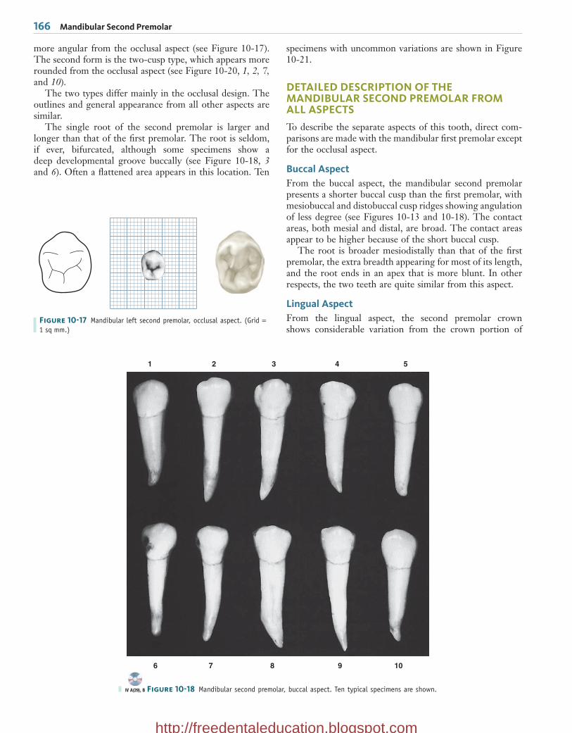
Figure 10-17 Mandibular left second premolar, occlusal aspect. (Grid = 1 sq mm.)
1 2 3 4 5
6 7 8 9 10
Figure 10-18 Mandibular second premolar, buccal aspect. Ten typical specimens are shown.
more angular from the occlusal aspect (see Figure 10-17). The second form is the two-cusp type, which appears more rounded from the occlusal aspect (see Figure 10-20, 1, 2, 7, and 10).
The two types differ mainly in the occlusal design. The outlines and general appearance from all other aspects are similar.
The single root of the second premolar is larger and longer than that of the first premolar. The root is seldom, if ever, bifurcated, although some specimens show a deep developmental groove buccally (see Figure 10-18, 3 and 6). Often a flattened area appears in this location. Ten
specimens with uncommon variations are shown in Figure 10-21.
DETAILED DESCRIPTION OF THE MANDIBULAR SECOND PREMOLAR FROM ALL ASPECTS
To describe the separate aspects of this tooth, direct com-parisons are made with the mandibular first premolar except for the occlusal aspect.
Buccal AspectFrom the buccal aspect, the mandibular second premolar presents a shorter buccal cusp than the first premolar, with mesiobuccal and distobuccal cusp ridges showing angulation of less degree (see Figures 10-13 and 10-18). The contact areas, both mesial and distal, are broad. The contact areas appear to be higher because of the short buccal cusp.
The root is broader mesiodistally than that of the first premolar, the extra breadth appearing for most of its length, and the root ends in an apex that is more blunt. In other respects, the two teeth are quite similar from this aspect.
Lingual AspectFrom the lingual aspect, the second premolar crown shows considerable variation from the crown portion of
166 Mandibular Second Premolar
IV A(29), B
http://freedentaleducation.blogspot.com

Chapter 10 The Permanent Mandibular Premolars 167
Figure 10-19 Mandibular second premolar, mesial aspect. Ten typical specimens are shown.
1 2 3 4 5
6 7 8 9 10
Figure 10-20 Mandibular second premolar, occlusal aspect. Ten typical specimens are shown.
1 2 3 4 5
6 7 8 9 10
http://freedentaleducation.blogspot.com
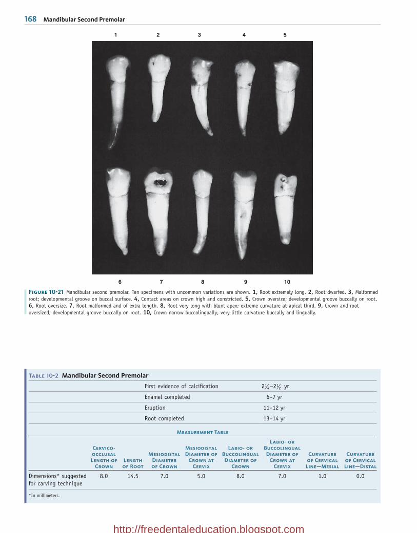
Figure 10-21 Mandibular second premolar. Ten specimens with uncommon variations are shown. 1, Root extremely long. 2, Root dwarfed. 3, Malformed root; developmental groove on buccal surface. 4, Contact areas on crown high and constricted. 5, Crown oversize; developmental groove buccally on root. 6, Root oversize. 7, Root malformed and of extra length. 8, Root very long with blunt apex; extreme curvature at apical third. 9, Crown and root oversized; developmental groove buccally on root. 10, Crown narrow buccolingually; very little curvature buccally and lingually.
1 2 3 4 5
6 7 8 9 10
Table 10-2 Mandibular Second Premolar
First evidence of calcification 2 214
12− yr
Enamel completed 6–7 yr
Eruption 11–12 yr
Root completed 13–14 yr
Measurement Table
Cervico-occlusal
Length of Crown
Length of Root
Mesiodistal Diameter of Crown
Mesiodistal Diameter of
Crown at Cervix
Labio- or Buccolingual
Diameter of Crown
Labio- or Buccolingual
Diameter of Crown at
Cervix
Curvature of Cervical
Line—Mesial
Curvature of Cervical Line—Distal
Dimensions* suggested for carving technique
8.0 14.5 7.0 5.0 8.0 7.0 1.0 0.0
*In millimeters.
168 Mandibular Second Premolar
http://freedentaleducation.blogspot.com

Chapter 10 The Permanent Mandibular Premolars 169
the first premolar (see Figure 10-14). The variations are as follows:
1. The lingual lobes are developed to a greater degree, which makes the cusp or cusps (depending on the type) longer.
2. Less of the occlusal surface may be seen from this aspect. Nevertheless, because the lingual cusps are not as long as the buccal cusp, part of the buccal portion of the occlusal surface may be seen.
3. In the three-cusp type, the lingual development brings about the greatest variation between the two teeth. Mesiolingual and distolingual cusps are present, with the former being the larger and the longer one in most cases. A groove is between them, extending a very short distance on the lingual surface and usually centered over the root (see Figure 10-20, 8).
In the two-cusp type, the single lingual cusp development attains equal height with the three-cusp type. The two-cusp type has no groove, but it shows a developmental depression distolingually where the lingual cusp ridge joins the distal marginal ridge (see Figure 10-20, 2 and 3).
The lingual surface of the crown of all mandibular second premolars is smooth and spheroidal, having a bulbous form above the constricted cervical portion.
The root is wide lingually, although not quite as wide as the buccal portion. Less difference in dimension is evident here than was found on the first premolar, so that much less convergence toward the lingual is seen.
Because in most instances the lingual portion of the crown converges little from the buccal portion, less of the mesial and distal sides of this tooth may be seen from this aspect than are seen from the lingual aspect of the first premolar.
The lingual portion of the root is smoothly convex for most of its length. Considered overall, the second premolar is the larger of the two mandibular premolars.
Mesial AspectFrom the mesial aspect (see Figures 10-15 and 10-19), the second premolar differs from the first premolar as follows:
1. The crown and root are wider buccolingually.2. The buccal cusp is not as nearly centered over the root
trunk, and it is shorter.3. The lingual lobe development is greater.4. The marginal ridge is at right angles to the long axis of
the tooth.5. Less of the occlusal surface may be seen.6. No mesiolingual developmental groove is present on
the crown portion.7. The root is longer and in most cases slightly convex on
the mesial surface; however, this convexity is not always present (see Figure 10-19, 6, 7, and 8).
8. The apex of the root is usually more blunt on the second premolar.
Distal AspectThe distal aspect of the mandibular second premolar is similar to the mesial aspect, except that more of the occlusal surface may be seen (see Figure 10-16). This is possible because the distal marginal ridge is at a lower level than the mesial marginal ridge when the tooth is posed vertically. The crowns of all posterior teeth are tipped distally to the long axes of the roots, so that when the specimen tooth is held vertically, more of the occlusal surface may be seen from the distal aspect than from the mesial aspect. This is a characteristic possessed by all posterior teeth, mandibular and maxillary. The angulation of occlusal surfaces to long axes of all posterior teeth is an important observation to remember, not only in the study of individual tooth forms but also later, in the study of alignment and occlusion.
Occlusal AspectAs mentioned earlier, two common forms of this tooth are evident. The outline form of each type shows some variation from the occlusal aspect (see Figures 10-17 and 10-20). The two types are similar in that portion that is buccal to the mesiobuccal and distobuccal cusp ridges.
The three-cusp type appears square lingual to the buccal cusp ridges when highly developed (see Figure 10-20, 8). The round, or two-cusp, type appears round lingual to the buccal cusp ridges (see Figure 10-20, 3).
The square type (see Figure 10-20, 8) has three cusps that are distinct; the buccal cusp is the largest, the mesiolingual cusp is next, and the distolingual cusp is the smallest.
Each cusp has well-formed triangular ridges separated by deep developmental grooves. These grooves converge in a central pit and form a Y shape on the occlusal surface. The central pit is located midway between the buccal cusp ridge and the lingual margin of the occlusal surface and slightly distal to the central point between mesial and distal marginal ridges.
Starting at the central pit, the mesial developmental groove travels in a mesiobuccal direction and ends in the mesial tri-angular fossa just distal to the mesial marginal ridge. The distal developmental groove travels in a distobuccal direc-tion, is somewhat shorter than the mesial groove, and ends in the distal triangular fossa mesial to the distal marginal side. The lingual developmental groove extends lingually between the two lingual cusps and ends on the lingual surface of the crown just below the convergence of the lingual cusp ridges. The mesiolingual cusp is wider mesio-distally than the distolingual cusp. This arrangement places the lingual developmental groove distal to center on the crown.
Supplemental grooves and depressions are often seen, radiating from the developmental grooves. Occasionally, a groove crosses one or both of the marginal ridges. On a tooth of this type, the point angles are distinct. Develop-mental grooves are often deep.
Figure 10-20, 8 is representative. Variations of this devel-opment may be seen in Figure 10-20, 4, 5, 6, and 9.
http://freedentaleducation.blogspot.com

The round or two-cusp type (see Figure 10-20, 3) differs considerably from the three-cusp type when viewed from the occlusal aspect. It is a true typal form of the two-cusp type. Variations may be seen in Figure 10-20, 1, 2, 7, and 10.
The occlusal characteristics of the two-cusp type are as follows:
1. The outline of the crown is rounded lingual to the buccal cusp ridges.
2. Some lingual convergence of mesial and distal sides occurs, although no more than is found in some variations of the square type.
3. The mesiolingual and distolingual line angles are rounded.
4. One well-developed lingual cusp is directly opposite the buccal cusp in a lingual direction.
A central developmental groove on the occlusal surface travels in a mesiodistal direction. This groove may be straight (see Figure 10-20, 3), but it is most often crescent-shaped (see Figure 10-20, 1, 7, and 10). The central groove has its terminals centered in mesial and distal fossae, which are roughly circular depressions having supplemental grooves and depressions radiating from the central groove and its terminals. The enamel surface inside these fossae and around their peripheries is very irregular, acting as a con-trast to the smoothness of cusp ridges, marginal ridges, and the transverse ridge from buccal cusp to lingual cusp.
Some of these teeth show mesial and distal developmental pits centered in the mesial and distal fossae instead of an unbroken central groove (see Figure 10-20, 2).
Although photographs do not demonstrate it very well, most of these two-cusp specimens show a developmental depression crossing the distolingual cusp ridge.
BibliographyCarlsen O: Human lower premolars: macro-morphologic
observations on the ontogenesis of the root complex, Scand J Dent Res 78:5, 1970.
Kraus B, Purr ML: Lower first premolar: a definition of discrete morphologic traits, J Dent Res 32:554, 1953.
Ludwig PJ: The mandibular second premolars: morphologic variation and inheritance, J Dent Res 36:263, 1957.
Osborn JR, editor: Dental anatomy and embryology, Oxford, 1981, Blackwell Scientific Publications.
Renner RP: An introduction to dental anatomy and esthetics, Chicago, 1985, Quintessence.
Schumacher G-H: Odontographie: Eine Oberflächenenanatomie der Zähne, Leipzig, 1983, JM Barth.
Use the CD-ROM to view animations and study ques-tions; and go to the site for additional study resources.
170 Mandibular Second Premolar
IV A, B
http://freedentaleducation.blogspot.com

The Permanent Maxillary Molars
Because the development of the bones of the face is down-ward and forward, sufficient space has been created nor-mally at the age of 6 for the accommodation of this tooth.
The normal location of the first permanent molar is at the center of the fully developed adult jaw anteroposteri-orly. As a consequence of the significance of their positions and the circumstances surrounding their eruption, the first molars may also be considered cornerstones of the dental arches. A full realization of the significance of these teeth as units in the arches and their function and positions relative to the other teeth will be gained when an opportunity comes to study the arrangement of the teeth with their occlusion and the temporomandibular articulation of the jaws. Subse-quent chapters cover those phases. The mandibular molars are described in Chapter 12.
Maxillary First Molar
Figures 11-1 through 11-18 illustrate the maxillary first molar from all aspects. The crown of this tooth is wider buccolingually than mesiodistally. Usually the extra dimen-sion buccolingually is about 1 mm (Table 11-1). This, however, varies in individuals (see Figure 11-17, 1, 5, 7, and 9). From the occlusal aspect, the inequality of the measure-ments in the two directions appears slight. Although the crown is relatively short, it is broad both mesiodistally and buccolingually, which gives the occlusal surface its generous dimensions.
The maxillary first molar is normally the largest tooth in the maxillary arch. It has four well-developed functioning cusps and one supplemental cusp of little practical use. The
The maxillary molars differ in design from any of the teeth previously described. These teeth assist the mandibular molars in performing the major portion of the work in the mastication and comminution of food. They are the largest and strongest maxillary teeth, by virtue both of their bulk and of their anchorage in the jaws. Although the crowns on the molars may be somewhat shorter than those on the premolars, their dimensions are greater in every respect. The root portion may be no longer than that of the premo-lars, but instead of one root or a bifurcated root, the maxil-lary molar root is broader at the base in all directions and is trifurcated into three well-developed prongs that are actu-ally three full-size roots emanating from a common broad base above the crown.
Generally speaking, the maxillary molars have large crowns with four well-formed cusps. They have three roots, two buccal and one lingual. The lingual root is the largest. The crowns have two buccal cusps and two lingual cusps. The outlines and curvatures of all the maxillary molars are similar. Developmental variations will be set forth under descriptions of the separate molars.
Before a detailed description of the maxillary first molar is begun, some statements will be made that are applicable to all first molars, mandibular and maxillary.
The permanent first molars usually appear in the oral cavity when the child is 6 years old. The mandibular molars precede the maxillary molars. The first permanent molar (maxillary or mandibular) erupts posterior to the second deciduous molar, taking up a position in contact with it. Therefore the first molar is not a succedaneous tooth because it has no predecessor. The deciduous teeth are all still in position and functioning when the first molar takes its place.
171
1111
http://freedentaleducation.blogspot.com

172 Maxillary First Molar
Table 11-1 Maxillary First Molar
First evidence of calcification At birth
Enamel completed 3–4 yr
Eruption 6 yr
Root completed 9–10 yr
Measurement Table
Cervico-occlusal
Length of Crown
Length of Root
Mesiodistal Diameter of Crown
Mesiodistal Diameter of Crown at Cervix
Labio- or Buccolingual
Diameter of Crown
Labio- or Buccolingual
Diameter of Crown at
Cervix
Curvature of Cervical
Line—Mesial
Curvature of Cervical Line—Distal
Dimensions* suggested for carving technique
7.5 Buccal = 12Lingual = 13
10.0 8.0 11.0 10.0 1.0 0.0
*In millimeters.
four large cusps of most physiological significance are the mesiobuccal, the distobuccal, the mesiolingual, and the dis-tolingual. A supplemental cusp is called the cusp or tubercle of Carabelli. This morphological trait can take the form of a well-developed fifth cusp, or it can grade down to a series of grooves, depressions, or pits on the mesial portion of the lingual surface. This trait has been used to distinguish populations.
Figure 11-1 Maxillary right first molar, occlusal aspect. MBCR, Mesiobuccal cusp ridge; CF, central fossa (shaded area); MTF, mesial triangular fossa (shaded area); MMR, mesial marginal ridge; MLCR, mesiolingual cusp ridge; OR, oblique ridge; DLCR, distolingual cusp ridge; DF, distal fossa; DTF, distal triangular fossa (shaded area); DMR, distal marginal ridge; DBCR, distobuccal cusp ridge.
DBCRMBCR
CF
MTFMMR
MLCR
ORDLCR
DF
DTF
DMR
Figure 11-2 Maxillary right first molar, occlusal aspect, developmental grooves. BG, Buccal groove; BGCF, buccal groove of central fossa; CGCF, central groove of central fossa; FCG, fifth cusp groove; LG, lingual groove; DOG, distal oblique groove; TGOR, transverse groove of oblique ridge; CP, central pit.
CP
TGOR
DOG
LG FCG
CGCF
BGCF
BG
Figure 11-3 Maxillary right first molar, buccal aspect. LR, Lingual root; MBR, mesiobuccal root; BDG, buccal developmental groove; MBC, mesiobuccal cusp; MLC, mesiolingual cusp; DBC, distobuccal cusp; CL, cervical line; DBR, distobuccal root.
DBR
CL
DBCMLC
MBC
BDG
Roottrunk
MBR
LR
This supplemental cusp is found lingual to the mesiolin-gual cusp, which is the largest of the well-developed cusps. Usually, a developmental groove is found, leaving a record of cusp development, unless it has been erased by frictional wear. The fifth cusp or a developmental trace at its usual site serves to identify the maxillary first molar. A specimen of this tooth showing no trace of its typical characteristic would be rare.
The three roots of generous proportions are the mesiobuc-cal, distobuccal, and lingual. These roots are well separated and well developed, and their placement gives this tooth maximum anchorage against forces that would tend to unseat it. The roots have their greatest spread parallel to the line of greatest force brought to bear against the crown diagonally in a buccolingual direction. The lingual root is the longest root. It is tapered and smoothly rounded. The mesiobuccal root is not as long, but it is broader buccolin-gually and shaped (in cross section) so that its resistance to
http://freedentaleducation.blogspot.com

Chapter 11 The Permanent Maxillary Molars 173
torsion is greater than that of the lingual root. The distobuc-cal root is the smallest of the three and smoothly rounded.
The development of maxillary first molars rarely deviates from the accepted normal. Ten specimens with uncommon variations are shown in Figure 11-18.
DETAILED DESCRIPTION OF THE MAXILLARY FIRST MOLAR FROM ALL ASPECTS
Buccal AspectThe crown is roughly trapezoidal, with cervical and occlusal outlines representing the uneven sides (see Figures 11-4, 11-13, 11-14, and 11-15). The cervical line is the shorter of the uneven sides (see Figure 4-16, D).
When the buccal aspect of this tooth is viewed with the line of vision at right angles to the buccal developmental groove of the crown, the distal side of the crown can be seen in perspective, which is possible because of the obtuse char-acter of the distobuccal line angle (see Occlusal Aspect later in this chapter). Parts of four cusps are seen, the mesiobuc-cal, distobuccal, mesiolingual, and distolingual.
The mesiobuccal cusp is broader than the distobuccal cusp, and its mesial slope meets its distal slope at an obtuse
Figure 11-4 Maxillary right first molar, buccal aspect. (Grid = 1 sq mm.)
Figure 11-5 Maxillary right first molar, lingual aspect. DBR, Distobuccal root; LR, lingual root; CL, cervical line; LDG, lingual developmental groove; DLC, distolingual cusp; MLC, mesiolingual cusp; FC, fifth cusp; MBR, mesiobuccal root.
MBR
FC
MLC
LDG
DLC
CL
LRDBR
Figure 11-6 Maxillary right first molar, lingual aspect. (Grid = 1 sq mm.)
Figure 11-7 Maxillary right first molar, mesial aspect. (Grid = 1 sq mm.)
Figure 11-8 Maxillary right first molar, mesial aspect. LR, Lingual root; FC, fifth cusp; MLC, mesiolingual cusp; MMR, mesial marginal ridge; MBC, mesiobuccal ridge; MCA, mesial contact area; CR, cervical ridge; MBR, mesiobuccal root.
MBR
MCA
MBC
MMR
MLC
FC
Roottrunk
LR
CR
angle. The mesial slope of the distobuccal cusp meets its distal slope at approximately a right angle. The distobuccal cusp is therefore sharper than the mesiobuccal cusp, and it is at least as long and often longer (see Figure 11-15, 4, 6, 7, 8, and 9).
The buccal developmental groove that divides the two buccal cusps is approximately equidistant between the mesiobuccal and distolingual line angles. The groove slants
http://freedentaleducation.blogspot.com

174 Maxillary First Molar
occlusoapically in a line of direction parallel to the long axis of the distobuccal root. It terminates at a point approxi-mately half the distance from its origin occlusally to the cervical line of the crown. Although the groove is not deep at any point, it becomes more shallow toward its termina-tion, gradually fading out. Lateral to its terminus is a dip in the enamel of the crown that is developmental in character and extends for some distance mesially and distally.
The cervical line of the crown does not have much cur-vature from mesial to distal; however, it is not as smooth and regular as that found on some of the other teeth. The line is generally convex with the convexity toward the roots.
The mesial outline of the crown from this aspect follows a nearly straight path downward and mesially, curving occlusally as it reaches the crest of contour of the mesial surface, which is the contact area. This crest is approxi-mately two thirds the distance from cervical line to tip of mesiobuccal cusp. The mesial outline continues downward and distally and becomes congruent with the outline of the mesial slope of the mesiobuccal cusp.
The distal outline of the crown is convex; the distal surface is spheroidal. The crest of curvature on the distal side of the crown is located at a level approximately half the distance from cervical line to tip of cusp. The distal contact area is in the middle of the middle third.
Often from this aspect, a flattened area or a concave area is seen on the distal surface immediately above the distobuc-cal cusp at the cervical third of the crown.
All three of the roots may be seen from the buccal aspect. The axes of the roots are inclined distally. The roots are not straight, although the buccal roots show an inclination to curvature halfway between the point of bifurcation and the apices. The mesiobuccal root curves distally, starting at the middle third. Its axis usually is at right angles to the cervical line. The distal root is straighter, with its long axis at an acute angle distally with the cervical line. It has a tendency toward curvature mesially at its middle third.
The point of bifurcation of the two buccal roots is located approximately 4 mm above the cervical line. This
measurement varies somewhat, of course. Nevertheless, the point is much farther removed from the cervical line than in the deciduous molars. This relation is typical when all permanent molars are compared with all deciduous molars.
A deep developmental groove buccally on the root trunk of the maxillary first molar, which starts at the bifurcation and progresses downward, becoming more shallow until it terminates in a shallow depression at the cervical line. Some-times this depression extends slightly onto the enamel at the cervix.
The reader must keep in mind the fact that molar roots originate as a single root on the base of the crown. They then are divided into three roots, as in the maxillary molars, or two roots, as in the mandibular molars. The common root base is called the root trunk (see Figures 11-3 and 11-8).
In judging the length of the roots and the direction of their axes, we must indicate the part of the root trunk that is congruent with each root as part of it, because they [i.e., the root trunk and the root] function as an entity. Usually, the lingual root is the longest, and the two buccal roots are approximately equal in length. Considerable variance is evident, although the difference is a matter of a millimeter or so only in the average first molars with normal development.
From the buccal aspect, a measurement of the roots inclusively at their greatest extremities mesiodistally is less than a calibration of the diameter of the crown mesiodistally.
No invariable rule covers the relative length of crown and root when the upper first molar is described. On the average, the roots are about twice as long as the crown.
Lingual AspectFrom the lingual aspect, the gross outline of the maxillary first molar is the reverse of that from the buccal aspect (see Figures 11-5, 11-6, 11-13, and 11-14). Photographs or drawings show this only approximately, because all teeth have breadth and thickness; consequently, the perspective of two dimensions plus the human element, which enters into the technique of posing specimens and making draw-ings and photographs, are bound to result in some error in graphic interpretation.
The variation between the outline of the mesial surface and that of the distal surface is apparent. Because of the roundness of the distolingual cusp, the smooth curvature of the distal outline of the crown becoming confluent with the curvature of the cusp creates an arc that is almost a semicir-cle. The line that describes the lingual developmental groove is also confluent with the outline of the distolingual cusp, progressing mesially and cervically and ending at a point at the approximate center of the lingual surface of the crown. A shallow depression in the surface extends from the termi-nus of the lingual groove to the center of the lingual surface of the lingual root at the cervical line and then continues in an apical direction on the lingual root, fading out at the middle third of the root.
Figure 11-9 Maxillary right first molar, distal aspect. DBR, Distobuccal root; MBR, mesiobuccal root; BDG, buccal developmental groove; DBC, distobuccal cusp; DMR, distal marginal ridge; DLC, distolingual cusp; DCA, distal contact area; CL, cervical line; LR, lingual root.
LR
CL
DCA
DLC
DMR
DBC
BDG
MBR
DBR
http://freedentaleducation.blogspot.com

Chapter 11 The Permanent Maxillary Molars 175
an irregular developmental groove, which may be described as starting in a depression of the mesiolingual line angle of the crown, extending occlusally toward the point of the mesiolingual cusp, then making an obtuse-angle turn toward the terminus of the lingual groove and fading out near the lingual groove terminus. If the fifth cusp is well developed, its cusp angle will be sharper and less obtuse than that of the mesiolingual cusp. The cusp ridge of the fifth cusp is approximately 2 mm cervical to the cusp ridge of the mesio-lingual cusp (see Figure 11-5).
All three of the roots are visible from the lingual aspect, with the large lingual root making up most of the fore-ground. The lingual portion of the root trunk is continuous with the entire cervical portion of the crown lingually. The lingual root is conical, terminating in a bluntly rounded apex.
All of the mesial outline of the mesiobuccal root and part of its apex may be seen from this angle.
The distal outline of the distobuccal root is seen above its middle third, including all of its apical outline.
Mesial AspectFrom the mesial aspect, the increased buccolingual dimen-sions may be observed, as well as the outlines of the cervical curvatures of the crown at the cervical third buccally and lingually, the difference in dimensions between the crown at its greatest measurement, and the distance between the cusp tips in a buccolingual direction (see Figures 11-7, 11-8, 11-13, 11-14, and 11-16).
Starting at the cervical line buccally, the outline of the crown makes a short arc buccally to its crest of curvature within the cervical third of the crown. The extent of this curvature is about 0.5 mm (see Figure 11-13). The line of the buccal surface then describes a shallow concavity imme-diately occlusal to the crest of curvature (see Buccal Aspect earlier in this chapter). The outline then becomes slightly convex as it progresses downward and inward to circum-scribe the mesiobuccal cusp, ending at the tip of the cusp well within projected outlines of the root base.
If the tooth is posed so that the line of vision is at right angles to the mesial contact area, the only cusps in sight are the mesiobuccal, the mesiolingual, and the fifth cusps. The mesiobuccal root hides the distobuccal root.
The lingual outline of the crown curves outward and lingually approximately to the same extent as on the buccal side. The level of the crest of curvature is near the middle third of the crown rather than a point within the cervical third, as it is buccally.
If the fifth cusp is well developed, the lingual outline dips inward to reveal it. If it is undeveloped, the lingual outline continues from the crest of curvature as a smoothly curved arc to the tip of the mesiolingual cusp. The point of the cusp is more clearly centered within projected outlines of the root base than the tip of the mesiobuccal cusp. The mesiolingual cusp is on a line with the long axis of the lingual root.
The mesial marginal ridge, which is confluent with the mesiobuccal and mesiolingual cusp ridges, is irregular, the outline curving cervically about one fifth the crown length
Figure 11-10 Maxillary right first molar, distal aspect. (Grid = 1 sq mm.)
Figure 11-11 Maxillary molar primary cusp triangle. The distolingual lobe, represented by shaded areas, becomes progressively smaller on maxillary molars, starting with the first molar, which presents the greatest development of the lobe. The plain areas, roughly triangular in outline, represent the maxillary molar primary cusp triangles.
The lingual cusps are the only ones to be seen from the lingual aspect. The mesiolingual cusp is much larger, and before occlusal wear, it is always the longest cusp the tooth possesses. Its mesiodistal width is about three fifths of the mesiodistal crown diameter, with the distolingual cusp making up the remaining two fifths. The angle formed by the mesial outline of the crown and the mesial slope of the mesiolingual cusp is almost 90 degrees. An obtuse angle describes the junction of the mesial and distal slopes of this cusp.
The distolingual cusp is so spheroidal and smooth that it is difficult to describe any angulation on the mesial and distal slopes.
The lingual developmental groove starts approximately in the center of the lingual surface mesiodistally, curves sharply to the distal as it crosses between the cusps, and continues on to the occlusal surface.
The fifth cusp appears attached to the mesiolingual surface of the mesiolingual cusp. It is outlined occlusally by
Figure 11-12 Maxillary right first molar, occlusal aspect. (Grid = 1 sq mm.)IV A(3), B
http://freedentaleducation.blogspot.com

176 Maxillary First Molar
Figure 11-13 Maxillary right first molar. Graph outlines of five aspects are shown. (Grid = 1 sq mm.)
Buccal Lingual
DistalMesial
Occlusal
and centering its curvature below the center of the crown buccolingually.
The cervical line of the crown is irregular, curving occlu-sally, but as a rule not more than 1 mm at any one point. If a definite curvature is present, it reaches its maximum imme-diately above the contact area.
The mesial contact area is apical to the marginal ridge but closer to it than the cervical line, approximately at the junction of the middle and occlusal thirds of the crown (see Figure 11-8). It is also somewhat buccal to the center of the crown buccolingually. A shallow concavity is usually found just above the contact area on the mesial surface of the maxillary first molar. This concavity may be continued to the mesial surface of the root trunk at its cervical third.
The mesiobuccal root is broad and flattened on its mesial surface; this flattened surface often exhibits smooth flutings for part of its length. The width of this root near the crown from the buccal surface to the point of bifurcation on the root trunk is approximately two thirds of the crown meas-urement buccolingually at the cervical line. The buccal outline of the root extends upward and outward from the crown, ending at the blunt apex. The greatest projection on this root is usually buccal to the greatest projection of the crown. The lingual outline of the root is relatively straight from the bluntly rounded apex down to the bifurcation with the lingual root.
The level of the bifurcation is a little closer to the cervical line than is found between the roots buccally. A smooth depression congruent with the bifurcation extends
http://freedentaleducation.blogspot.com

Chapter 11 The Permanent Maxillary Molars 177
Figure 11-14 Maxillary right first molar.
Buccal Lingual
Mesial
Occlusal
Distal
1 2 3 4 5
6 7 8 9 10
Figure 11-15 Maxillary first molar, buccal aspect. Ten typical specimens are shown.IV A(3), B
http://freedentaleducation.blogspot.com

178 Maxillary First Molar
Figure 11-16 Maxillary first molar, mesial aspect. Ten typical specimens are shown.
1 2 3 4 5
6 7 8 9 10
occlusally and lingually almost to the cervical line directly above the mesiolingual line angle of the crown.
The lingual root is longer than the mesial root but is narrower from this aspect. It is banana-shaped, extending lingually with its convex outline to the lingual and its concave outline to the buccal. At its middle and apical thirds, it is outside of the confines of the greatest crown projection. Although its apex is rounded, the root appears more pointed toward the end than the mesiobuccal root.
Distal AspectThe gross outline of the distal aspect is similar to that of the mesial aspect (see Figures 11-9, 11-10, 11-13, and 11-14). Certain variations must be noted when the tooth is viewed from the distal aspect.
Because of the tendency of the crown to taper distally on the buccal surface, most of the buccal surface of the crown may be seen in perspective from the distal aspect. This is because the buccolingual measurement of the crown mesially is greater than the same measurement distally. All of the decrease in measurement distally is a result of the slant of the buccal side of the crown.
The distal marginal ridge dips sharply in a cervical direc-tion, exposing triangular ridges on the distal portion of the occlusal surface of the crown.
The cervical line is almost straight across from buccal to lingual. Occasionally it curves apically 0.5 mm or so.
The distal surface of the crown is generally convex, with a smoothly rounded surface except for a small area near the distobuccal root at the cervical third. This concavity contin-ues on to the distal surface of the distobuccal root, from the
cervical line to the area of the root that is on a level with bifurcation separating the distobuccal and lingual roots.
The distobuccal root is narrower at its base than either of the others. An outline of this root, when the tooth is viewed from the distal aspect, starts buccally at a point immediately above the distobuccal cusp, follows a concave path inward for a short distance, then turns outward in a buccal direction, completing a graceful convex arc from the concavity to the rounded apex. This line lies entirely within the confines of the outline of the mesiobuccal root. The lingual outline of the root from the apex to the bifurcation is slightly concave. No concavity is evident between the bifurcation of the roots and the cervical line. However, the surface at this point on the root trunk has a tendency toward convexity.
The bifurcation here is more apical than either of the other two areas on this tooth. The area from cervical line to bifurcation is 5 mm or more in extent.
Occlusal AspectFrom the occlusal aspect, the maxillary first molar is some-what rhomboidal. An outline following the four major cusp ridges and the marginal ridges is especially so (see Figures 11-1, 11-2, 11-12, 11-13, 11-14, and 11-17).
A measurement of the crown buccolingually and mesial to the buccal and lingual grooves is greater than the meas-urement on that portion of the crown which is distal to these developmental grooves. Also, a measurement of the crown immediately lingual to contact areas mesiodistally is greater than the measurement immediately buccal to the contact areas. Thus it is apparent that the maxillary first molar
http://freedentaleducation.blogspot.com

Chapter 11 The Permanent Maxillary Molars 179
Figure 11-17 Maxillary first molar, occlusal aspect. Ten typical specimens are shown.
1 2 3 4 5
6 7 8 9 10
Figure 11-18 Maxillary first molar. Ten specimens with uncommon variations are shown. 1, Unusual curvature of buccal roots. 2, Roots abnormally long with extreme curvature. 3, Lingual and distobuccal roots fused. 4, Mesiodistal measurement of root trunk smaller than usual. 5, Extreme rhomboidal development of crown; fifth cusp with maximum development. 6, Tooth well developed but much smaller than usual. 7, Extreme buccolingual measurement. 8, Extreme length, especially of the distobuccal root; buccal cusps narrow mesiodistally. 9, Well-developed crown; roots poorly developed. 10, Extreme development of lingual portion of the crown compared with buccal development.
1 2 3 4 5
6 7 8 9 10
http://freedentaleducation.blogspot.com
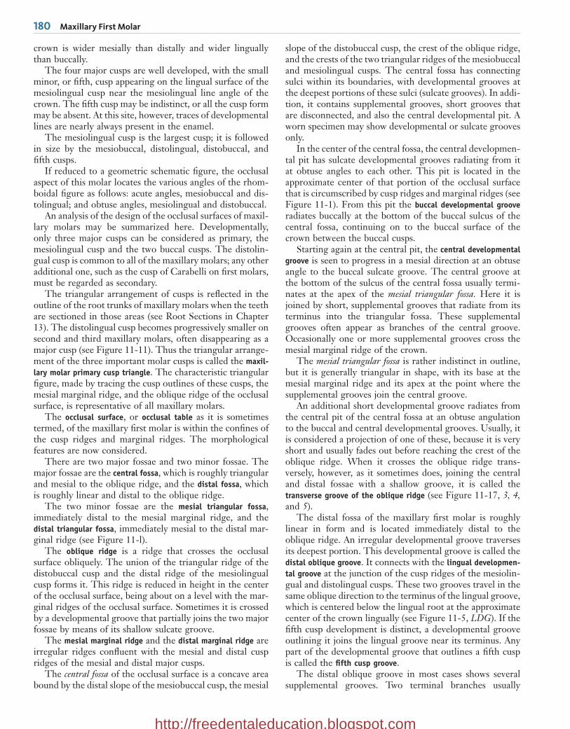
180 Maxillary First Molar
crown is wider mesially than distally and wider lingually than buccally.
The four major cusps are well developed, with the small minor, or fifth, cusp appearing on the lingual surface of the mesiolingual cusp near the mesiolingual line angle of the crown. The fifth cusp may be indistinct, or all the cusp form may be absent. At this site, however, traces of developmental lines are nearly always present in the enamel.
The mesiolingual cusp is the largest cusp; it is followed in size by the mesiobuccal, distolingual, distobuccal, and fifth cusps.
If reduced to a geometric schematic figure, the occlusal aspect of this molar locates the various angles of the rhom-boidal figure as follows: acute angles, mesiobuccal and dis-tolingual; and obtuse angles, mesiolingual and distobuccal.
An analysis of the design of the occlusal surfaces of maxil-lary molars may be summarized here. Developmentally, only three major cusps can be considered as primary, the mesiolingual cusp and the two buccal cusps. The distolin-gual cusp is common to all of the maxillary molars; any other additional one, such as the cusp of Carabelli on first molars, must be regarded as secondary.
The triangular arrangement of cusps is reflected in the outline of the root trunks of maxillary molars when the teeth are sectioned in those areas (see Root Sections in Chapter 13). The distolingual cusp becomes progressively smaller on second and third maxillary molars, often disappearing as a major cusp (see Figure 11-11). Thus the triangular arrange-ment of the three important molar cusps is called the maxil-lary molar primary cusp triangle. The characteristic triangular figure, made by tracing the cusp outlines of these cusps, the mesial marginal ridge, and the oblique ridge of the occlusal surface, is representative of all maxillary molars.
The occlusal surface, or occlusal table as it is sometimes termed, of the maxillary first molar is within the confines of the cusp ridges and marginal ridges. The morphological features are now considered.
There are two major fossae and two minor fossae. The major fossae are the central fossa, which is roughly triangular and mesial to the oblique ridge, and the distal fossa, which is roughly linear and distal to the oblique ridge.
The two minor fossae are the mesial triangular fossa, immediately distal to the mesial marginal ridge, and the distal triangular fossa, immediately mesial to the distal mar-ginal ridge (see Figure 11-l).
The oblique ridge is a ridge that crosses the occlusal surface obliquely. The union of the triangular ridge of the distobuccal cusp and the distal ridge of the mesiolingual cusp forms it. This ridge is reduced in height in the center of the occlusal surface, being about on a level with the mar-ginal ridges of the occlusal surface. Sometimes it is crossed by a developmental groove that partially joins the two major fossae by means of its shallow sulcate groove.
The mesial marginal ridge and the distal marginal ridge are irregular ridges confluent with the mesial and distal cusp ridges of the mesial and distal major cusps.
The central fossa of the occlusal surface is a concave area bound by the distal slope of the mesiobuccal cusp, the mesial
slope of the distobuccal cusp, the crest of the oblique ridge, and the crests of the two triangular ridges of the mesiobuccal and mesiolingual cusps. The central fossa has connecting sulci within its boundaries, with developmental grooves at the deepest portions of these sulci (sulcate grooves). In addi-tion, it contains supplemental grooves, short grooves that are disconnected, and also the central developmental pit. A worn specimen may show developmental or sulcate grooves only.
In the center of the central fossa, the central developmen-tal pit has sulcate developmental grooves radiating from it at obtuse angles to each other. This pit is located in the approximate center of that portion of the occlusal surface that is circumscribed by cusp ridges and marginal ridges (see Figure 11-1). From this pit the buccal developmental groove radiates buccally at the bottom of the buccal sulcus of the central fossa, continuing on to the buccal surface of the crown between the buccal cusps.
Starting again at the central pit, the central developmental groove is seen to progress in a mesial direction at an obtuse angle to the buccal sulcate groove. The central groove at the bottom of the sulcus of the central fossa usually termi-nates at the apex of the mesial triangular fossa. Here it is joined by short, supplemental grooves that radiate from its terminus into the triangular fossa. These supplemental grooves often appear as branches of the central groove. Occasionally one or more supplemental grooves cross the mesial marginal ridge of the crown.
The mesial triangular fossa is rather indistinct in outline, but it is generally triangular in shape, with its base at the mesial marginal ridge and its apex at the point where the supplemental grooves join the central groove.
An additional short developmental groove radiates from the central pit of the central fossa at an obtuse angulation to the buccal and central developmental grooves. Usually, it is considered a projection of one of these, because it is very short and usually fades out before reaching the crest of the oblique ridge. When it crosses the oblique ridge trans-versely, however, as it sometimes does, joining the central and distal fossae with a shallow groove, it is called the transverse groove of the oblique ridge (see Figure 11-17, 3, 4, and 5).
The distal fossa of the maxillary first molar is roughly linear in form and is located immediately distal to the oblique ridge. An irregular developmental groove traverses its deepest portion. This developmental groove is called the distal oblique groove. It connects with the lingual developmen-tal groove at the junction of the cusp ridges of the mesiolin-gual and distolingual cusps. These two grooves travel in the same oblique direction to the terminus of the lingual groove, which is centered below the lingual root at the approximate center of the crown lingually (see Figure 11-5, LDG). If the fifth cusp development is distinct, a developmental groove outlining it joins the lingual groove near its terminus. Any part of the developmental groove that outlines a fifth cusp is called the fifth cusp groove.
The distal oblique groove in most cases shows several supplemental grooves. Two terminal branches usually
http://freedentaleducation.blogspot.com

Chapter 11 The Permanent Maxillary Molars 181
appear, forming two sides of the triangular depression immediately mesial to the distal marginal ridge. These two sides, in combination with the slope mesial to the distal marginal ridge, form the distal triangular fossa. The distal outline of the distal marginal ridge of the crown shows a slight concavity.
The distolingual cusp is smooth and rounded from the occlusal aspect, and an outline of it, from the distal concavity of the distal marginal ridge to the lingual groove of the crown, describes an arch of an ellipse.
The lingual outline of the distolingual cusp is straight with the lingual outline of the fifth cusp, unless the fifth cusp is unusually large. In the latter case the lingual outline of the fifth cusp is more prominent lingually (see Figure 11-17, 9). The cusp ridge of the distolingual cusp usually extends lingually farther than the cusp ridge of the mesiolingual cusp.
Maxillary Second Molar
Figures 11-19 through 11-27 illustrate the maxillary second molar from all aspects. The maxillary second molar supple-
Figure 11-19 Maxillary left second molar, buccal aspect. (Grid = 1 sq mm.)
Figure 11-20 Maxillary left second molar, lingual aspect. (Grid = 1 sq mm.)
Figure 11-21 Maxillary left second molar, mesial aspect. (Grid = 1 sq mm.)
Figure 11-22 Maxillary left second molar, distal aspect. (Grid = 1 sq mm.)
Figure 11-23 Maxillary left second molar, occlusal aspect. (Grid = 1 sq mm.)
ments the first molar in function. In describing this tooth, direct comparisons will be made with the first molar both in form and development.
Generally speaking, the roots of this tooth are as long as, if not somewhat longer than, those of the first molar (Table 11-2). The distobuccal cusp is not as large or as well devel-oped, and the distolingual cusp is smaller. No fifth cusp is evident.
The crown of the maxillary second molar is 0.5 mm or so shorter cervico-occlusally than that of the first molar, but the measurement of the crown buccolingually is about the
http://freedentaleducation.blogspot.com

1 2 3 4 5
6 7 8 9 10
Table 11-2 Maxillary Second Molar
First evidence of calcification 212 yr
Enamel completed 7–8 yr
Eruption 12–13 yr
Root completed 14–16 yr
Measurement Table
Cervico-occlusal
Length of Crown
Length of Root
Mesiodistal Diameter of Crown
Mesiodistal Diameter of Crown at Cervix
Labio- or Buccolingual
Diameter of Crown
Labio- or Buccolingual
Diameter of Crown at
Cervix
Curvature of Cervical
Line—Mesial
Curvature of Cervical Line—Distal
Dimensions* suggested for carving technique
7.0 Buccal = 11Lingual = 12
9.0 7.0 11.0 10.0 1.0 0.0
*In millimeters.
same. Two types of maxillary second molars are found when the occlusal aspect is viewed: (1) The type that is seen most has an occlusal form that resembles that of the first molar, although the rhomboidal outline is more extreme. This is accentuated by the lesser measurement lingually. (2) The second type bears more resemblance to a typical third molar form. The distolingual cusp is poorly developed and makes the development of the other three cusps predominate. This results in a heart-shaped form from the occlusal aspect that is more typical of the maxillary third molar (see Figure 11-26, 1 and 7). Ten specimens with uncommon variations are shown in Figure 11-27.
DETAILED DESCRIPTION OF THE MAXILLARY SECOND MOLAR FROM ALL ASPECTS
Buccal AspectFrom the buccal aspect the crown is a little shorter cervico-occlusally and narrower mesiodistally than is the maxillary first molar (see Figures 11-19 and 11-24). The distobuccal cusp is smaller and allows part of the distal marginal ridge and part of the distolingual cusp to be seen.
The buccal roots are about the same length. These roots are more nearly parallel and inclined distally more than those of the maxillary first molar, so that the end of the
Figure 11-24 Maxillary second molar, buccal aspect. Ten typical specimens are shown.
182 Maxillary Second Molar
IV A(2), B
http://freedentaleducation.blogspot.com

Chapter 11 The Permanent Maxillary Molars 183
Figure 11-25 Maxillary second molar, mesial aspect. Ten typical specimens are shown.
1 2 3 4 5
6 7 8 9 10
Figure 11-26 Maxillary second molar, occlusal aspect. Ten typical specimens are shown.
1 2 3 4 5
6 7 8 9 10
distobuccal root is slightly distal to the distal extremity of the crown. The apex of the mesiobuccal root is on a line with the buccal groove of the crown instead of the tip of the mesiobuccal cusp, as was found on the first molar.
Lingual AspectWays in which the second molar differs from the first molar to be noted here, in addition to those mentioned earlier, are the following:
1. The distolingual cusp of the crown is smaller.2. The distobuccal cusp may be seen through the
sulcus between the mesiolingual and distolingual cusp.
3. No fifth cusp is evident.4. The apex of the lingual root is in line with
the distolingual cusp tip instead of the lingual groove as was found on the first molar (see Figure 11-20).
http://freedentaleducation.blogspot.com
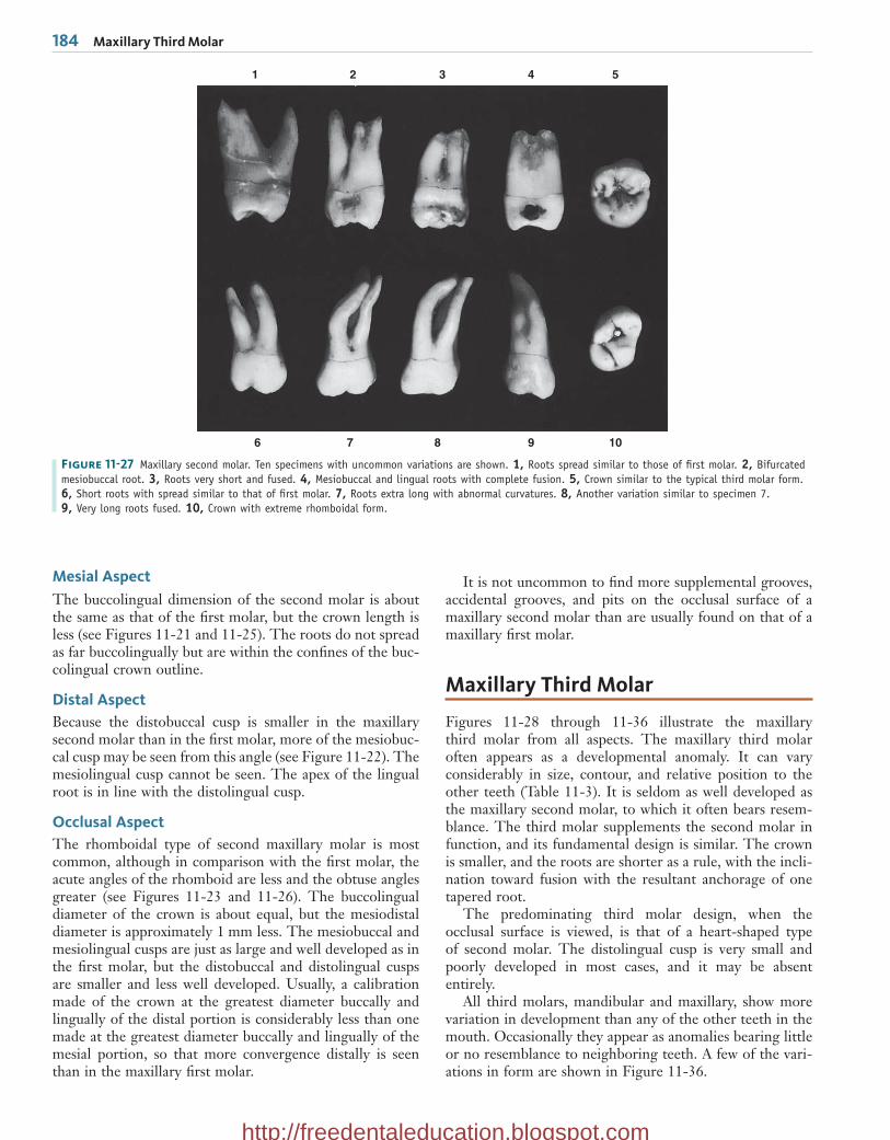
Figure 11-27 Maxillary second molar. Ten specimens with uncommon variations are shown. 1, Roots spread similar to those of first molar. 2, Bifurcated mesiobuccal root. 3, Roots very short and fused. 4, Mesiobuccal and lingual roots with complete fusion. 5, Crown similar to the typical third molar form. 6, Short roots with spread similar to that of first molar. 7, Roots extra long with abnormal curvatures. 8, Another variation similar to specimen 7. 9, Very long roots fused. 10, Crown with extreme rhomboidal form.
1 2 3 4 5
6 7 8 9 10
Mesial AspectThe buccolingual dimension of the second molar is about the same as that of the first molar, but the crown length is less (see Figures 11-21 and 11-25). The roots do not spread as far buccolingually but are within the confines of the buc-colingual crown outline.
Distal AspectBecause the distobuccal cusp is smaller in the maxillary second molar than in the first molar, more of the mesiobuc-cal cusp may be seen from this angle (see Figure 11-22). The mesiolingual cusp cannot be seen. The apex of the lingual root is in line with the distolingual cusp.
Occlusal AspectThe rhomboidal type of second maxillary molar is most common, although in comparison with the first molar, the acute angles of the rhomboid are less and the obtuse angles greater (see Figures 11-23 and 11-26). The buccolingual diameter of the crown is about equal, but the mesiodistal diameter is approximately 1 mm less. The mesiobuccal and mesiolingual cusps are just as large and well developed as in the first molar, but the distobuccal and distolingual cusps are smaller and less well developed. Usually, a calibration made of the crown at the greatest diameter buccally and lingually of the distal portion is considerably less than one made at the greatest diameter buccally and lingually of the mesial portion, so that more convergence distally is seen than in the maxillary first molar.
It is not uncommon to find more supplemental grooves, accidental grooves, and pits on the occlusal surface of a maxillary second molar than are usually found on that of a maxillary first molar.
Maxillary Third Molar
Figures 11-28 through 11-36 illustrate the maxillary third molar from all aspects. The maxillary third molar often appears as a developmental anomaly. It can vary considerably in size, contour, and relative position to the other teeth (Table 11-3). It is seldom as well developed as the maxillary second molar, to which it often bears resem-blance. The third molar supplements the second molar in function, and its fundamental design is similar. The crown is smaller, and the roots are shorter as a rule, with the incli-nation toward fusion with the resultant anchorage of one tapered root.
The predominating third molar design, when the occlusal surface is viewed, is that of a heart-shaped type of second molar. The distolingual cusp is very small and poorly developed in most cases, and it may be absent entirely.
All third molars, mandibular and maxillary, show more variation in development than any of the other teeth in the mouth. Occasionally they appear as anomalies bearing little or no resemblance to neighboring teeth. A few of the vari-ations in form are shown in Figure 11-36.
184 Maxillary Third Molar
http://freedentaleducation.blogspot.com

Chapter 11 The Permanent Maxillary Molars 185
Figure 11-28 Maxillary right third molar, buccal aspect. (Grid = 1 sq mm.)
Figure 11-29 Maxillary right third molar, lingual aspect. (Grid = 1 sq mm.)
Figure 11-30 Maxillary right third molar, mesial aspect. (Grid = 1 sq mm.)
Figure 11-31 Maxillary right third molar, distal aspect. (Grid = 1 sq mm.)
Figure 11-32 Maxillary right third molar, occlusal aspect. (Grid = 1 sq mm.)
It is necessary to give a short description of the third molar that is considered average in its development and one that would be in good proportion to the other maxillary molars and with an occlusal form considered normal. In describing the normal maxillary third molar, direct comparisons will be made with the maxillary second molar.
DETAILED DESCRIPTION OF THE MAXILLARY THIRD MOLAR FROM ALL ASPECTS
Buccal AspectFrom the buccal aspect, the crown of the third molar is shorter cervico-occlusally and narrower mesiodistally than that of the second molar (see Figures 11-28 and 11-33). The roots are usually fused, functioning as one large root, and they are shorter cervicoapically. The fused roots end in a taper at the apex. The roots have a distinct slant to the distal, giving the apices of the fused root a more distal relation to the center of the crown.
Lingual AspectIn addition to the differences just mentioned, in comparison with the maxillary second molar, only one large lingual cusp is present, and therefore no lingual groove is evident (see Figure 11-29). However, in many cases, a third molar with the same essential features has a poorly developed distolin-gual cusp with a developmental groove lingually (see Figure 11-35, 2).
Mesial AspectFrom the mesial aspect, aside from the differences in measurement, the main feature is the taper to the fused roots and a bifurcation, usually in the region of the apical third. Figure 11-30 does not show a bifurcation. See Figure 11-34, 1, 2, and 3. The root portion is considerably shorter in relation to the crown length. Both the crown and the root portions tend to be poorly developed, with irregular outlines.
http://freedentaleducation.blogspot.com

Figure 11-33 Maxillary third molar, buccal aspect. Ten typical specimens are shown.
Figure 11-34 Maxillary third molar, mesial aspect. Ten typical specimens are shown.
1 2 3 4 5
6 7 8 9 10
1 2 3 4 5
6 7 8 9 10
Distal AspectFrom the distal aspect, most of the buccal surface of the crown is in view (see Figure 11-31). More of the occlusal surface may be seen than can be seen on the second molar from this aspect because of the more acute angulation of the occlusal surface in relation to the long axis of the root. The
measurement from the cervical line to the marginal ridge is short.
Occlusal AspectThe occlusal aspect of a typical maxillary third molar presents a heart-shaped outline (see Figures 11-32 and 11-35).
186 Maxillary Third Molar
http://freedentaleducation.blogspot.com

Chapter 11 The Permanent Maxillary Molars 187
Figure 11-35 Maxillary third molar, occlusal aspect. Ten typical specimens are shown.
1 2 3 4 5
6 7 8 9 10
Figure 11-36 Maxillary third molar. Twelve specimens with uncommon variations are shown. 1, Very short fused root form. 2, Extremely long roots with extreme distal angulation. 3, Complete fusion of roots with extreme distal angulation. 4, Three roots well separated; crown very wide at cervix. 5, Extreme rhomboidal outline to crown, with developmental grooves oddly placed. 6, Overdeveloped mesiobuccal cusp. 7, Crown wide at cervix, with roots perpendicular. 8, Very large crown; poorly developed root form. 9, Complete absence of typical design. 10, Specimen abnormally large, with four roots well separated. 11, Five well-developed cusps, atypical in form. 12, Small specimen, atypical cusp form.
1 2 3 4 5 6
7 8 9 10 11 12
The lingual cusp is large and well developed, and little or no distolingual cusp is evident, which gives a semicircular outline to the tooth from one contact area to the other. Three functioning cusps are seen on this type of tooth: two buccal and one lingual.
The occlusal aspect of this tooth usually presents many supplemental grooves and many accidental grooves unless the tooth is very much worn.
The third molar may show four distinct cusps. This type may have a strong oblique ridge, a central fossa and a distal fossa, with a lingual developmental groove similar to that of the rhomboidal type of second molar. In most instances, the crown converges more lingually from the buccal areas than in the second molar, losing its rhomboidal outline. This is not always true, however (compare 1 and 3 in Figure 11-35).
http://freedentaleducation.blogspot.com

Table 11-3 Maxillary Third Molar
First evidence of calcification 7–9 yr
Enamel completed 12–16 yr
Eruption 17–21 yr
Root completed 18–25 yr
Measurement Table
Cervico-occlusal
Length of Crown
Length of Root
Mesiodistal Diameter of Crown
Mesiodistal Diameter of
Crown at Cervix
Labio- or Buccolingual
Diameter of Crown
Labio- or Buccolingual
Diameter of Crown at
Cervix
Curvature of Cervical
Line—Mesial
Curvature of Cervical Line—Distal
Dimensions* suggested for carving technique
6.5 11.0 8.5 6.5 10.0 9.5 1.0 0.0
*In millimeters.
BibliographyAlexandersen V: Mandibular third molar: the root complex. II.
Morphogenetic considerations, Tandlægebladet 66:53, 1985.Ash MM: Wheeler’s atlas of tooth form, ed 5, Philadelphia, 1984,
Saunders.Black GV: Descriptive anatomy of the human teeth, ed 4,
Philadelphia, 1897, S.S. White Dental Manufacturing.Carabelli G: Anatomie des Mundes, Vienna, 1842, Braumuller and
Seidel.Carbonell VM: The tubercle of Carabelli in the Kish dentition,
Mesopotamia, 3000 bc, J Dent Res 39:124, 1960.Carlsen O: Dental morphology, Copenhagen, 1987,
Munksgaard.Diamond M: Dental anatomy, New York, 1929, Macmillan.Hopewell-Smith A: An introduction to dental anatomy and
physiology, Philadelphia, 1913, Lea and Febiger.
Kraus BS: Carabelli’s anomaly of the maxillary molar teeth, Am J Hum Genet 3:348, 1951.
Kraus BS, Jordan RE, Abrams L: Dental anatomy and occlusion, Baltimore, 1969, Williams & Wilkins.
Tomes CS: A manual of dental anatomy, London, 1894, Churchill.
Woelfel JB, Scheid RC: Dental anatomy: its relevance to dentistry, Baltimore, 1997, Williams & Wilkins.
Use the CD-ROM to view animations and study ques-tions; and go to the site for additional study resources.
188 Maxillary Third Molar
http://freedentaleducation.blogspot.com

The Permanent Mandibular Molars
Mandibular First Molar
Figures 12-1 through 12-17 illustrate the mandibular first molar from all aspects. Normally, the mandibular first molar is the largest tooth in the mandibular arch. It has five well-developed cusps: two buccal, two lingual, and one distal (see Figure 12-1). It has two well-developed roots, one mesial and one distal, which are very broad buccolingually. These roots are widely separated at the apices.
The dimension of the crown mesiodistally is greater by about 1 mm than the dimension buccolingually (Table 12-1). Although the crown is relatively short cervico- occlusally, it has mesiodistal and buccolingual measurements that provide a broad occlusal form.
The mesial root is broad and curved distally, with mesial and distal fluting that provides the anchorage of two roots (see Figure 13-22). The distal root is rounder, broad at the cervical portion, and pointed in a distal direction. The formation of these roots and their positions in the mandible serve to brace the crown of the tooth efficiently against the lines of force that might be brought to bear against it.
DETAILED DESCRIPTION OF THE MANDIBULAR FIRST MOLAR FROM ALL ASPECTS
Buccal AspectFrom the buccal aspect, the crown of the mandibular first molar is roughly trapezoidal, with cervical and occlusal out-lines representing the uneven sides of the trapezoid. The
The mandibular molars are larger than any other mandibu-lar teeth. They are three in number on each side of the mandible: the first, second, and third mandibular molars. They resemble each other in functional form, although comparison of one with another shows variations in the number of cusps and some variation in size, occlusal design, and the relative lengths and positions of the roots.
The crown outlines exhibit similarities of outline from all aspects, and each mandibular molar has two roots, one mesial and one distal. Third molars and some second molars may show a fusion of these roots. All mandibular molars have crowns that are roughly quadrilateral, being somewhat longer mesiodistally than buccolingually. Maxillary molar crowns have their widest measurement buccolingually.
The mandibular molars perform the major portion of the work of the lower jaw in mastication and in the comminu-tion of food. They are the largest and strongest mandibular teeth, both because of their bulk and because of their anchorage.
The crowns of the molars are shorter cervico-occlusally than those of the teeth anterior to them, but their dimen-sions are greater in every other respect. The root portions are not as long as those of some of the other mandibular teeth, but the combined measurements of the multiple roots, with their broad bifurcated root trunks, result in superior anchorage and greater efficiency.
Usually the sum of the mesiodistal measurements of mandibular molars is equal to or greater than the combined mesiodistal measurements of all the teeth anterior to the first molar and up to the median line.
The crowns of these molars are wider mesiodistally than buccolingually. The opposite is true of maxillary molars.
189
1212
http://freedentaleducation.blogspot.com

190 Mandibular First Molar
level. Before they are worn, the buccal cusps and the distal cusp have curvatures that are characteristic of each one (see Figures 12-4 and 12-14, 4).
The mesiobuccal cusp is usually the widest mesiodistally of the three cusps. This cusp has some curvature but is
Figure 12-1 Mandibular right first molar, occlusal aspect. DBCR, Distobuccal cusp ridge; DBDG, distobuccal developmental groove; DCR, distal cusp ridge; DTF, distal triangular fossa (shaded area); DLCR, distolingual cusp ridge; LDG, lingual developmental groove; MLCR, mesiolingual cusp ridge; MTF, mesial triangular fossa (shaded area); SG, a supplemental groove; MBCR, mesiobuccal cusp ridge; MBDG, mesiobuccal developmental groove.
MBDG
MBCRSG
MTF
MLCR
LDG
DLCR
DTF
DCRDBDG
DBCR
Table 12-1 Mandibular First Molar
First evidence of calcification At birth
Enamel completed 212–3 yr
Eruption 6–7 yr
Root completed 9–10 yr
Measurement Table
Cervico-occlusal
Length of Crown
Length of Root
Mesiodistal Diameter of Crown
Mesiodistal Diameter of
Crown at Cervix
Labio- or Buccolingual
Diameter of Crown
Labio- or Buccolingual
Diameter of Crown at
Cervix
Curvature of Cervical
Line—Mesial
Curvature of Cervical Line—Distal
Dimensions* suggested for carving technique
7.5 14.0 11.0 9.0 10.5 9.0 1.0 0.0
*In millimeters.
occlusal side is the longer (see Figures 12-3, 12-4, 12-12, 12-13, and 12-14).
If this tooth is posed vertically, all five of its cusps are in view. The two buccal cusps and the buccal portion of the distal cusp are in the foreground, with the tips of the lingual cusps in the background. The lingual cusps may be seen because they are higher than the others.
Two developmental grooves appear on the crown portion. These grooves are called the mesiobuccal developmental groove and the distobuccal developmental groove. The first-named groove acts as a line of demarcation between the mesiobuc-cal lobe and the distobuccal lobe. The latter groove sepa-rates the distobuccal lobe from the distal lobe (see Figures 12-2 and 12-3).
The mesiobuccal, distobuccal, and distal cusps are rela-tively flat. These cusp ridges show less curvature than those of any of the teeth described so far. The distal cusp, which is small, is more pointed than either of the buccal cusps. Flattened buccal cusps are typical of all mandibular molars. In most first molar specimens the buccal cusps are worn considerably, with the buccal cusp ridges almost at the same
Figure 12-2 Mandibular right first molar, occlusal aspect. Shaded area is the central fossa. CP, Central pit; DMR, distal marginal ridge; DP, distal pit; CDG, central developmental groove; MP, mesial pit; MMR, mesial marginal ridge.
MMR
Mes
ial
MP
LingualCDG
DPDMR
Dis
tal
CPBuccal
Figure 12-3 Mandibular right first molar, buccal aspect. MBDG, Mesiobuccal developmental groove; CL, cervical line; MR, mesial root; DR, distal root; DBDG, distobuccal developmental groove; DC, distal cusp; DLC, distolingual cusp; DBC, distobuccal cusp; MLC, mesiolingual cusp; MBC, mesiobuccal cusp.
DC
DLC
DBC MLC
MBC
MBDG
CL
MRDR
DBDG
http://freedentaleducation.blogspot.com

Chapter 12 The Permanent Mandibular Molars 191
Figure 12-4 Mandibular right first molar, buccal aspect. (Grid = 1 sq mm.) Figure 12-5 Mandibular right first molar, lingual aspect. DLC,
Distolingual cusp; LDG, lingual developmental groove; CL, cervical line; MLC, mesiolingual cusp.
Roottrunk
MLC DLC
LDG
CL
ApexApex
Figure 12-6 Mandibular right first molar, lingual aspect. (Grid = 1 sq mm.) Figure 12-7 Mandibular right first molar, mesial aspect. (Grid =
1 sq mm.)
Figure 12-8 Mandibular right first molar, mesial aspect. MMR, Mesial marginal ridge; MLC, mesiolingual cusp; MCA, mesial contact area; CL, cervical line; DD, developmental depression; CR, cervical ridge; MBC, mesiobuccal cusp.
MMR
MLC
MCA
CL
DD
Mesial root
CR
MBC
Figure 12-9 Mandibular right first molar, distal aspect. MMR, Mesial marginal ridge; MBC, mesiobuccal cusp; DBC, distobuccal cusp; DC, distal cusp; DBDG, distobuccal developmental groove; DCA, distal contact area; CL, cervical line; DMR, distal marginal ridge; DLC, distolingual cusp; MLC, mesiolingual cusp.
MMRMBCDBCDC
DBDG
DCA
CL
Mesialroot
DMR
DLC
MLC
Distal root
http://freedentaleducation.blogspot.com

192 Mandibular First Molar
Figure 12-10 Mandibular right first molar, distal aspect. (Grid = 1 sq mm.)
Figure 12-11 Mandibular right first molar, occlusal aspect. (Grid = 1 sq mm.)
Figure 12-12 Mandibular right first molar. Graph outlines of five aspects are shown. (Grid = 1 sq mm.)
Buccal Lingual
Mesial Distal
Occlusal
http://freedentaleducation.blogspot.com

Chapter 12 The Permanent Mandibular Molars 193
Figure 12-13 Mandibular right first molar.
Buccal Lingual
Mesial Distal
Occlusal
1 2 3 4 5
6 7 8 9 10
Figure 12-14 Mandibular first molar, buccal aspect. Ten typical specimens are shown.IV A(30), B
http://freedentaleducation.blogspot.com

194 Mandibular First Molar
relatively flat. The distobuccal cusp is almost as wide, with a cusp ridge of somewhat greater curvature. The two buccal cusps make up the major portion of the buccal surface of the crown. The distal cusp provides a very small part of the buccal surface, because the major portion of the cusp makes up the distal portion of the crown, providing the distal contact area on the center of the distal surface of the distal cusp. The distal cusp ridge is very round occlusally and is sharper than either of the two buccal cusps.
These three cusps have the mesiobuccal and distobuccal grooves as lines of demarcation. The mesiobuccal groove is the shorter of the two, having its terminus centrally located cervico-occlusally. This groove is situated a little mesial to the root bifurcation buccally. The distobuccal groove has its terminus near the distobuccal line angle at the cervical third of the crown. It travels occlusally and somewhat mesially, parallel with the axis of the distal root.
The cervical line of the mandibular first molar is com-monly regular in outline, dipping apically toward the root bifurcation.
The mesial outline of the crown is somewhat concave at the cervical third up to its junction with the convex outline of the broad contact area. The distal outline of the crown is straight above the cervical line to its junction with the convex outline of the distal contact area, which is also the outline of the distal portion of the distal cusp.
The calibration of this tooth at the cervical line is 1.5 to 2 mm less mesiodistally than the mesiodistal measurement at the contact areas, which of course represents the greatest mesiodistal measurement of the crown.
The surface of the buccal portion of the crown is smoothly convex at the cusp portions with developmental grooves between the cusps. Approximately at the level of the ends of the developmental grooves, in the middle third, a develop-mental depression is noticeable. It runs in a mesiodistal direction just above the cervical ridge of the buccal surface (see Figure 12-14, 6 and 8). This cervical ridge may show a smooth depression in it that progresses cervically, joining with the developmental concavity just below the cervical line, which is congruent with the root bifurcation buccally.
The roots of this tooth are, in most instances, well formed and constant in development.
When the tooth is posed so that the mesiobuccal groove is directly in the line of vision, part of the distal surface of the root trunk may be seen, and in addition, part of the distal area of the mesial root is visible because the lingual portion of the root is turned distally. These areas may be seen in addition to the buccal areas of the roots and root trunk.
The mesial root is curved mesially from a point shortly below the cervical line to the middle third portion. From this point, it curves distally to the tapered apex, which is located directly below the mesiobuccal cusp. The crest of curvature of the root mesially is mesial to the crown cervix. The distal outline of the mesial root is concave from the bifurcation of the root trunk to the apex.
The distal root is less curved than the mesial root, and its axis is in a distal direction from cervix to apex. The root
may show some curvature at its apical third in either a mesial or a distal direction (see Figure 12-14, 1 and 8). The apex is usually more pointed than that of the mesial root and is located below or distal to the distal contact area of the crown. Considerable variation is evident in the comparative lengths of mesial and distal roots (see Figure 12-14).
Both roots are wider mesiodistally at the buccal areas than they are lingually. Developmental depressions are present on the mesial and distal sides of both roots, which lessens the mesiodistal measurement at those points. They are somewhat thicker at the lingual borders. This arrange-ment provides a secure anchorage for the mandibular first molar, preventing rotation. This I-beam principle increases the anchorage of each root (see Figure 13-22).
The point of bifurcation of the two roots is located approximately 3 mm below the cervical line. A deep devel-opmental depression is evident buccally on the root trunk, which starts at the bifurcation and progresses cervically, becoming shallower until it terminates at or immediately above the cervical line. This depression is smooth with no developmental groove or fold.
Lingual AspectFrom the lingual aspect, three cusps may be seen: two lingual cusps and the lingual portion of the distal cusp (see Figures 12-5, 12-6, 12-12, and 12-13). The two lingual cusps are pointed, and the cusp ridges are high enough to hide the two buccal cusps from view. The mesiolingual cusp is the widest mesiodistally, with its cusp tip somewhat higher than the distolingual cusp. The distolingual cusp is almost as wide mesiodistally as the mesiolingual cusp. The mesiolingual and distolingual cusp ridges are inclined at angles that are similar on both lingual cusps. These cusp ridges form obtuse angles at the cusp tips of approximately 100 degrees.
The lingual developmental groove serves as a line of demar-cation between the lingual cusps, extending downward on the lingual surface of the crown for a short distance only. Some mandibular first molars show no groove on the lingual surface but show a depression lingual to the cusp ridges. The angle formed by the distolingual cusp ridge of the mesiolin-gual cusp and the mesiolingual cusp ridge of the distolingual cusp is more obtuse than the angulation of the cusp ridges at the tips of the lingual cusps.
The distal cusp is at a lower level than the mesiolingual cusp.
The mesial outline of the crown from this aspect is convex from the cervical line to the marginal ridge. The crest of contour mesially, which represents the contact area, is somewhat higher than the crest of contour distally.
The distal outline of the crown is straight immediately above the cervical line to a point immediately below the distal contact area; this area is represented by a convex cur-vature that also outlines the distal surface of the distal cusp. The junction of the distolingual cusp ridge of the distolin-gual cusp with the distal marginal ridge is abrupt; it gives the impression of a groove at this site from the lingual aspect. Sometimes, a shallow developmental groove occurs at this point (see Figure 12-10). Part of the mesial and distal
http://freedentaleducation.blogspot.com

Chapter 12 The Permanent Mandibular Molars 195
surfaces of the crown and root trunk may be seen from this aspect because the mesial and distal sides converge lingually.
The cervical line lingually is irregular and tends to point sharply toward the root bifurcation and immediately above it.
The surface of the crown lingually is smooth and sphe-roidal on each of the lingual lobes. The surface is concave at the side of the lingual groove above the center of the crown lingually. Below this point, the surface of the crown becomes almost flat as it approaches the cervical line.
The roots of the mandibular first molar appear somewhat different from the lingual aspect. They measure about 1 mm longer lingually than buccally, but the length seems more extreme (see Figures 12-6 and 12-7). This impression is derived from the fact that the cusp ridges and cervical line are at a higher level (about 1 mm). This arrangement adds a millimeter to the distance from root bifurcation to cervical line. In addition, the mesiodistal measurement of the root trunk is less toward the lingual surface than toward the buccal surface. Consequently, this slenderness lingually, in addition to the added length, makes the roots appear longer than they are from the lingual aspect (see Figure 12-9).
As was mentioned, the root bifurcation lingually starts at a point approximately 4 mm below the cervical line. This developmental depression is quite deep at this point, although it is smooth throughout and progresses cervically, becoming more shallow until it fades out entirely immedi-ately below the cervical line. The depression is rarely reflected in the cervical line or the enamel of the lingual surface of the crown, as is found in many cases on the buccal surface of this tooth.
This bifurcation groove of the root trunk is located almost in line with the lingual developmental groove of the crown.
Mesial AspectWhen the mandibular first molar is viewed from the mesial aspect, with the specimen held with its mesial surface at right angles to the line of vision, two cusps and one root only are to be seen: the mesiobuccal and mesiolingual cusps and the mesial root (see Figures 12-7, 12-8, 12-12, 12-13, and 12-15).
The buccolingual measurement of the crown is greater at the mesial portion than it is at the distal portion. The buccolingual measurement of the mesial root is also greater than the same measurement of the distal root. Therefore, because the mesial portions of the tooth are broader and the mesial cusps are higher, the distal portions of the tooth cannot be seen from this angle.
As already indicated, all of the posterior mandibular teeth have crown outlines from the mesial aspect that show a characteristic relation between crown and root. The crown from the mesial or distal aspect is roughly rhomboidal, and the entire crown has a lingual tilt in relation to the root axis. It should be remembered that the crowns of maxillary pos-terior teeth have the center of the occlusal surfaces between the cusps in line with the root axes (see Figure 4-16, E and F).
It is interesting to note the difference between the outline form of the mandibular first molar and the mandibular second premolar from the mesial aspect (see Chapter 10). The first molar compares as follows:
Figure 12-15 Mandibular first molar, mesial aspect. Ten typical specimens are shown.
1 2 3 4 5
6 7 8 9 10
http://freedentaleducation.blogspot.com

196 Mandibular First Molar
1. The crown is a fraction of a millimeter to 1 millimeter shorter in the first molar.
2. The root is usually that much shorter also.3. The buccolingual measurement of crown and root of
the molar is 2 mm or more greater.4. The lingual cusp is longer than the buccal cusp. (The
opposite is true of the second premolar.)
Regardless of these differences, the two teeth have the same functional form except for the added reinforcement given to the molar lingually. Because of the added root width buccolin-gually, the buccal cusps of the first molar do not approach the center axis of the root as does the second premolar, and the lingual cusp tips are within the lingual outline of the roots instead of being on a line with them.
From the mesial aspect, the buccal outline of the crown of the mandibular first molar is convex immediately above the cervical line. Before occlusal wear has shortened the buccal cusps, this curvature is over the cervical third of the crown buccally, outlining the buccal cervical ridge (see Figure 12-8). This ridge is more prominent on some first molars than on others (see Figure 12-15). Just as on mandibular premolars, this ridge curvature does not exceed similar con-tours on other teeth as a rule when the mandibular first molar is posed in the position it assumes in the mandibular arch (see Figure 12-7 and Figure 12-15, 1 and 2).
Above the buccal cervical ridge, the outline of the buccal contour may be slightly concave on some specimens (see Figure 12-15, 1 and 2), or the outline may just be less convex or even rather flat as it continues occlusally outlining the contour of the mesiobuccal cusp. The mesiobuccal cusp is located directly above the buccal third of the mesial root.
The lingual outline of the crown is straight in a lingual direction, starting at the cervical line and joining the lingual curvature at the middle third, the lingual curvature being pronounced between this point and the tip of the mesiolin-gual cusp. The crest of the lingual contour is located at the center of the middle third of the crown. The tip of the mesiolingual cusp is in a position directly above the lingual third of the mesial root.
The mesial marginal ridge is confluent with the mesial ridges of the mesiobuccal and mesiolingual cusps. The mar-ginal ridge is placed about 1 mm below the level of the cusp tips.
The cervical line mesially is rather irregular and tends to curve occlusally about 1 mm toward the center of the mesial surface of the tooth (see Figure 12-15, 1, 4, 9, and 10). The cervical line may assume a relatively straight line buccolin-gually (see Figure 12-15, 3, 6, and 8).
In all instances, the cervical line is at a higher level lingually than buccally, usually about 1 mm higher. The difference in level may be greater. This relation depends on the assumption that the tooth is posed vertically. When the first molar is in its normal position in the lower jaw, leaning to the lingual, the cervical line is nearly level buccolingually.
The surface of the crown is convex and smooth over the mesial contours of the mesiolingual and mesiobuccal lobes.
A flattened or slightly concave area exists at the cervical line immediately above the center of the mesial root. This area is right below the contact area and joins the concavity of the central portion of the root at the cervix. The contact area is almost centered buccolingually in the mesial surface of the crown, and it is placed below the crest of the marginal ridge about one third the distance from marginal ridge to cervical line. (See stained contact area on specimen in Figure 12-7. Before contact wear has occurred, the contact area is not as broad. Refer also to Figure 12-4.)
The buccal outline of the mesial root drops straight down from the cervical line buccally to a point near the junction of cervical and middle thirds of the root. A gentle curve starts lingually from this point to the apex, which is located directly below the mesiobuccal cusp.
The lingual outline of the mesial root is slanted in a buccal direction, although the outline is nearly straight from the cervical line lingually to the point of junction of middle and apical thirds of the root. From this point, the curvature is sharply buccal to the bluntly tapered apex. On those specimens that show a short bifurcation at the mesial root end, the curvature at the apical third lingually is slight (see Figure 12-15, 2 and 10).
The mesial surface of the mesial root is convex at the buccal and lingual borders, with a broad concavity between these convexities the full length of the root from cervical line to apex. If a specimen tooth is held in front of a strong light so that the distal side of the mesial root can be seen from the apical aspect, it is noted that the same contours exist on the root distally as are found mesially, and the root is very thin where the concavities are superimposed. The root form appears to be two narrow roots fused together with thin, hard tissue between.
The mesial surface of the distal root is smooth, with no deep developmental depressions.
Distal AspectBecause the gross outline of the distal aspect of the crown and root of the mandibular first molar is similar to that from the mesial aspect, the description of outline form will not be repeated. When this aspect is considered from the standpoint of a three-dimensional figure, however, more of the tooth is seen from the distal aspect, because the crown is shorter distally than mesially and the buccal and lingual surfaces of the crown converge distally (see Figures 12-10, 12-12, and 12-13). The buccal surface shows more convergence than the lingual surface. The distal root is more narrow buccolingually than the mesial root.
If a specimen of the first molar is held with the distal surface of the crown at right angles to the line of vision, a great part of the occlusal surface may be seen and some part of each of the five cusps also, which compares favorably with the mandibular second premolar. This is caused in part by the placement of the crowns on the roots with a distal incli-nation to the long axes. The slight variation in crown length distally does not provide this view of the occlusal surface (see Figures 12-9 and 12-10).
http://freedentaleducation.blogspot.com

Chapter 12 The Permanent Mandibular Molars 197
From the distal aspect, the distal cusp is in the foreground on the crown portion. The distal cusp is placed a little buccal to the center buccolingually, with the distal contact area appearing on its distal contour.
The distal contact area is placed just below the distal cusp ridge of the distal cusp and at a slightly higher level above the cervical line than was found mesially compared with the location of the mesial contact area.
The distal marginal ridge is short and is made up of the distal cusp ridge of the distal cusp and the distolingual cusp ridge of the distolingual cusp. These cusp ridges dip sharply in a cervical direction, meeting at an obtuse angle. Often a developmental groove or depression is found crossing the marginal ridge at this point. The point of this angle is above the lingual third of the distal root instead of being centered over the root as is true of the center of the mesial marginal ridge.
The distal contact area is centered over the distal root, which arrangement places it buccal to the center point of the distal marginal ridge.
The surface of the distal portion of the crown is convex on the distal cusp and the distolingual cusp. Contact wear may produce a flattened area at the point of contact on the distal surface of the distal cusp. Just above the cervical line, the enamel surface is flat where it joins the flattened surface of the root trunk distally.
The cervical line distally usually extends straight across buccolingually. It may be irregular, dipping rootwise just below the distal contact area (see Figure 12-10).
The end of the distobuccal developmental groove is located on the distal surface and forms a concavity at the cervical portion of the distobuccal line angle of the crown. The distal portion of the crown extends out over the root trunk distally at quite an angle (see Figure 12-4). The smooth, flat surface below the contact area remains fairly constant to the apical third of the distal root. Sometimes a
developmental depression is found here. The apical third portion of the root is more rounded as it tapers to a sharper apex than is found on the mesial root.
The lingual border of the mesial root may be seen from the distal aspect.
Occlusal AspectThe mandibular first molar is somewhat hexagonal from the occlusal aspect (see Figure 12-2). The crown measurement is 1 mm or more greater mesiodistally than buccolingually. It must be remembered that the opposite arrangement is true of the maxillary first molar.
The buccolingual measurement of the crown is greater on the mesial than on the distal side. Also, the measurement of the crown at the contact areas, which includes the two buccal cusps and the distal cusp, is greater than the mesio-distal measurement of the two lingual cusps. In other words, the crown converges lingually from the contact areas. This convergence varies in individual specimens (see Figure 12-16, 1 and 4).
It is interesting to note the degree of development of the individual cusps from the occlusal aspect (see Figures 12-1, 12-2, 12-11, 12-12, 12-13, and 12-16). The mesiobuccal cusp is slightly larger than either of the two lingual cusps, which are almost equal to each other in size; the distobuccal cusp is smaller than any one of the other three mentioned, and the distal cusp is in most cases much the smallest of all.
More variance is evident in the development of the dis-tobuccal and distal lobes than in any of the others (see Figure 12-16, 1, 7, and 10).
When the tooth is posed so that the line of vision is paral-lel with the long axis, a great part of the buccal surface may be seen, whereas only a small portion of the lingual surface may be seen lingual to the lingual cusp ridges. No part of the mesial or distal surfaces is in view below the outline of
Figure 12-16 Mandibular first molar, occlusal aspect. Ten typical specimens are shown.
1 2 3 4 5
6 7 8 9 10
http://freedentaleducation.blogspot.com

198 Mandibular First Molar
the mesial and distal marginal ridges. (Compare tooth out-lines from the other aspects.)
All mandibular molars, including the first molar, are essentially quadrilateral in form. The mandibular first molar, in most instances, has a functioning distal cusp, although it is small in comparison with the other cusps. Occasionally, four-cusp first molars are found, and more often, one dis-covers first molars with distobuccal and distal cusps showing fusion with little or no trace of a distobuccal developmental groove between them (see Figure 12-16, 1 and Figure 12-17, 4 and 5). From a developmental viewpoint, all mandibular molars have four major cusps, whereas maxillary molars have only three major cusps (see Figure 11-11).
The occlusal surfaces of the mandibular first molar may be described as follows: there is a major fossa and two minor fossae. The major fossa is the central fossa (see Figure 12-2). It is roughly circular, and it is centrally placed on the occlu-sal surface between buccal and lingual cusp ridges. The two minor fossae are the mesial triangular fossa, immediately distal to the mesial marginal ridge, and the distal triangular fossa, immediately mesial to the distal marginal ridge (see Figure 12-1).
The developmental grooves on the occlusal surface are the central developmental groove, the mesiobuccal developmental groove, the distobuccal developmental groove, and the lingual developmental groove. Supplemental grooves, accidental short grooves, and developmental pits are also found. Most of the supplemental grooves are tributary to the developmental grooves within the bounds of cusp ridges.
The central fossa of the occlusal surface is a concave area bounded by the distal slope of the mesiobuccal cusp, both
mesial and distal slopes of the distobuccal cusp, the mesial slope of the distal cusp, the distal slope of the mesiolingual cusp, and the mesial slope of the distolingual cusp (see Figure 12-2).
All of the developmental grooves converge in the center of the central fossa at the central pit.
The mesial triangular fossa of the occlusal surface is a smaller concave area than the central fossa, and the mesial slope of the mesiobuccal cusp, the mesial marginal ridge, and the mesial slope of the mesiolingual cusp bound it. The mesial portion of the central developmental groove termi-nates in this fossa. Usually a buccal and a lingual supplemen-tal groove join it at a mesial pit within the boundary of the mesial marginal ridge. Sometimes a supplemental groove crosses the mesial marginal ridge lingual to the contact area (see Figure 12-16, 2, 8, 9, and 10).
The distal triangular fossa is in most instances less distinct than the mesial fossa. The distal slope of the distal cusp, the distal marginal ridge, and the distal slope of the distolingual cusp bound it. The central groove has its other terminal in this fossa. Buccal and lingual supplemental grooves are less common here. An extension of the central groove quite often crosses the distal marginal ridge, however, lingual to the distal contact area.
Starting at the central pit in the central fossa, the central developmental groove travels an irregular course mesially, terminating in the mesial triangular fossa. A short distance mesially from the central pit, it joins the mesiobuccal devel-opmental groove. The latter groove courses in a mesiobuc-cal direction at the bottom of a sulcate groove separating the mesiobuccal and distobuccal cusps. At the junction of
Figure 12-17 Mandibular first molar. Ten specimens with uncommon variations are shown. 1, Root extremely long, crown small. 2, Mesial root longer than average with rounded apex. 3, Crown very wide buccolingually; roots short. 4, Roots short. 5, Crown has no buccal developmental grooves. 6, Crown and roots poorly formed. 7, Roots dwarfed. 8, Roots short; crown wide buccolingually. 9, Crown and root oversize buccolingually. 10, Extra tubercle or cusp attached to mesiolingual lobe.
1 2 3 4 5
6 7 8 9 10
http://freedentaleducation.blogspot.com

Chapter 12 The Permanent Mandibular Molars 199
the cusp ridges of those cusps, the mesiobuccal groove of the occlusal surface is confluent with the mesiobuccal groove of the buccal surface of the crown. The lingual developmen-tal groove of the occlusal surface is an irregular groove coursing in a lingual direction at the bottom of the lingual sulcate groove to the junction of lingual cusp ridges, where it is confluent with the lingual extension of the same groove. Again starting at the central pit, the central groove may be followed in a distobuccal direction to a point where it is joined by the distobuccal developmental groove of the occlusal surface. From this point, the central groove courses in a distolingual direction, terminating in the distal triangu-lar fossa. The distobuccal groove passes from its junction with the central groove in a distobuccal course, joining its buccal extension on the buccal surface of the crown at the junction of the cusp ridges of the distobuccal and distal cusps.
The central developmental groove seems to be centrally located in relation to the buccolingual crown dimension. This arrangement makes the triangular ridges of lingual cusps longer than the triangular ridges of buccal cusps.
Note the relative position and relative size of the distal cusp from the occlusal aspect. The distal portion of it joins the distal contact area of the crown.
Mandibular Second Molar
Figures 12-18 through 12-26 illustrate the mandibular second molar from all aspects. The mandibular second molar supplements the first molar in function. Its anatomy differs in some details.
Normally, the second molar is smaller than the first molar by a fraction of a millimeter in all dimensions (Table 12-2). It does not, however, run true to form. It is not uncommon to find mandibular second molar crowns some-what larger than first molar crowns, and although the roots are not as well formed, they may be longer.
The crown has four well-developed cusps, two buccal and two lingual, of nearly equal development. Neither a distal nor a fifth cusp is evident, but the distobuccal cusp is larger than that found on the first molar.
Figure 12-18 Mandibular left second molar, buccal aspect. (Grid = 1 sq mm.)
Figure 12-19 Mandibular left second molar, lingual aspect. (Grid = 1 sq mm.)
Figure 12-20 Mandibular left second molar, mesial aspect. (Grid = 1 sq mm.)
Figure 12-21 Mandibular left second molar, distal aspect. (Grid = 1 sq mm.)
Figure 12-22 Mandibular left second molar, occlusal aspect. (Grid = 1 sq mm.)
http://freedentaleducation.blogspot.com

Table 12-2 Mandibular Second Molar
First evidence of calcification 212–3 yr
Enamel completed 7–8 yr
Eruption 11–13 yr
Root completed 14–15 yr
Measurement Table
Cervico-occlusal
Length of Crown
Length of Root
Mesiodistal Diameter of Crown
Mesiodistal Diameter of
Crown at Cervix
Labio- or Buccolingual
Diameter of Crown
Labio- or Buccolingual
Diameter of Crown at
Cervix
Curvature of Cervical
Line—Mesial
Curvature of Cervical Line—Distal
Dimensions* suggested for carving technique
7.0 13.0 10.5 8.0 10.0 9.0 1.0 0.0
*In millimeters.
The tooth has two well-developed roots, one mesial and one distal. These roots are broad buccolingually, but they are not as broad as those of the first molar, nor are they as widely separated.
DETAILED DESCRIPTION OF THE MANDIBULAR SECOND MOLAR FROM ALL ASPECTS
In describing this tooth, direct comparisons will be made with the first mandibular molar. Uncommon variations are shown in Figure 12-26.
Buccal AspectFrom the buccal aspect the crown is somewhat shorter cervico-occlusally and narrower mesiodistally than is the first molar (see Figure 12-18). The crown and root show a tendency toward greater overall length but are not always longer (see Figure 12-23, 4, 7, and 9).
Only one developmental groove lies buccally—the buccal developmental groove. This groove acts as a line of demarcation between the mesiobuccal and the disto-buccal cusps, which are about equal in their mesiodistal measurements.
1 2 3 4 5
6 7 8 9 10
Figure 12-23 Mandibular second molar, buccal aspect. Ten typical specimens are shown.
200 Mandibular Second Molar
IV A(31), B
IV A, B
http://freedentaleducation.blogspot.com

Chapter 12 The Permanent Mandibular Molars 201
The cervical line buccally in many instances points sharply toward the root bifurcation (see Figure 12-23, 1, 2, 3, 5, and 9).
The roots may be shorter than those of the first molar, but they vary considerably in this and in their development generally. The roots are usually closer together, and their axes are nearly parallel. They may spread as much as those of the first molar (see Figure 12-23, 5), or they may be fused for all or part of their length (see Figure 12-23, 8 and 9).
The roots are inclined distally in relation to the occlusal plane of the crown, their axes forming more of an acute angle with the occlusal plane than is found on the first molar. When one compares all of the mandibular molars, it may seem that the first molar shows one angulation of roots to occlusal plane, the second molar shows a more acute angle, and the third molar shows an angle that is more acute still (see Figure 16-20).
Lingual AspectDifferences in detail between the mandibular second molar and the mandibular first molar, to be noted from the lingual aspect (see Figure 12-19), are as follows:
1. The crown and root of the mandibular second molar converge lingually but to a slight degree; little of the mesial or distal surfaces may therefore be seen from this aspect.
2. The mesiodistal calibration at the cervix lingually is always greater accordingly than that of the first molar.
3. The curvatures mesially and distally on the crown that describe the contact areas are more noticeable from the
lingual aspect. They prove to be at a slightly lower level, especially in the distal area, than those of the first molar.
Mesial AspectExcept for the differences in measurement from the mesial aspect, the second molar differs little from the first molar (see Figures 12-20 and 12-24).
The cervical ridge buccally on the crown portion is in most instances less pronounced, and the occlusal surface may be more constricted buccolingually (see Figure 12-24, 2, 8, and 10).
The cervical line shows less curvature, being straight and regular in outline buccolingually.
The mesial root is somewhat pointed apically. If part of the distal root is in sight, it is seen buccally. In the first molar, when the distal root is in sight from the mesial aspect, it is in view lingually.
Distal AspectFrom the distal aspect, the second molar is similar in form to the first molar except for the absence of a distal cusp and a distobuccal groove (see Figure 12-21). The contact area is centered on the distal surface buccolingually and is placed equidistant from cervical line and marginal ridge.
Occlusal AspectThe occlusal aspect of the mandibular second molar differs considerably from that of the first molar (see Figures 12-22 and 12-25). These variations serve as marks of identity. The small distal cusp of the first molar is not present, and the
Figure 12-24 Mandibular second molar, mesial aspect. Ten typical specimens are shown.
1 2 3 4 5
6 7 8 9 10
http://freedentaleducation.blogspot.com

Figure 12-25 Mandibular second molar, occlusal aspect. Ten typical specimens are shown.
1 2 3 4 5
6 7 8 9 10
Figure 12-26 Mandibular second molar. Ten specimens with uncommon variations are shown. 1, Mesiodistal measurements at contact areas and cervix almost equal. 2, Roots twisted and of extra length. 3, Very small specimen; roots short. 4, Roots short for such a large crown. 5, Roots thick and malformed generally. 6, Dwarfed crown, roots extra long. 7, Mesial aspect, protective curvature buccally and lingually is absent. 8, Roots of extra size; occlusal surface constricted buccolingually. 9, Roots malformed. 10, Crown wide mesiodistally at the cervix; roots short.
1 2 3 4 5
6 7 8 9 10
distobuccal lobe development is just as pronounced, and sometimes more so, than that of the mesiobuccal lobe. No distobuccal developmental groove is evident occlusally or buccally. The buccal and lingual developmental grooves meet the central developmental groove at right angles at the central pit on the occlusal surface. These grooves form a cross, dividing the occlusal portion of the crown into four nearly equal parts.
In general, the cusp slopes on the occlusal surface are not as smooth as those found on first molars because they are
roughened by many supplemental grooves radiating from the developmental grooves.
The following characteristics of mandibular second molars from the occlusal aspect are noted:
1. Many of them are rectangular from the occlusal aspect (see Figure 12-25, 7 and 9).
2. Many show considerable prominence cervically on the mesiobuccal lobe only (see Figure 12-25, 1, 3, and 6).
202 Mandibular Second Molar
http://freedentaleducation.blogspot.com
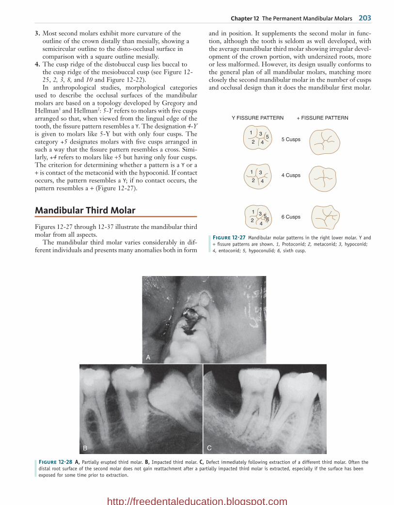
Chapter 12 The Permanent Mandibular Molars 203
3. Most second molars exhibit more curvature of the outline of the crown distally than mesially, showing a semicircular outline to the disto-occlusal surface in comparison with a square outline mesially.
4. The cusp ridge of the distobuccal cusp lies buccal to the cusp ridge of the mesiobuccal cusp (see Figure 12-25, 2, 3, 8, and 10 and Figure 12-22).In anthropological studies, morphological categories
used to describe the occlusal surfaces of the mandibular molars are based on a topology developed by Gregory and Hellman1 and Hellman2: 5-Y refers to molars with five cusps arranged so that, when viewed from the lingual edge of the tooth, the fissure pattern resembles a Y. The designation 4-Y is given to molars like 5-Y but with only four cusps. The category +5 designates molars with five cusps arranged in such a way that the fissure pattern resembles a cross. Simi-larly, +4 refers to molars like +5 but having only four cusps. The criterion for determining whether a pattern is a Y or a + is contact of the metaconid with the hypoconid. If contact occurs, the pattern resembles a Y; if no contact occurs, the pattern resembles a + (Figure 12-27).
Mandibular Third Molar
Figures 12-27 through 12-37 illustrate the mandibular third molar from all aspects.
The mandibular third molar varies considerably in dif-ferent individuals and presents many anomalies both in form
Figure 12-27 Mandibular molar patterns in the right lower molar. Y and + fissure patterns are shown. 1, Protoconid; 2, metaconid; 3, hypoconid; 4, entoconid; 5, hypoconulid; 6, sixth cusp.
5 Cusps
1
1
1
2
2
2
3
3
3
4
4
4
65
5
4 Cusps
6 Cusps
Y FISSURE PATTERN + FISSURE PATTERN
Figure 12-28 A, Partially erupted third molar. B, Impacted third molar. C, Defect immediately following extraction of a different third molar. Often the distal root surface of the second molar does not gain reattachment after a partially impacted third molar is extracted, especially if the surface has been exposed for some time prior to extraction.
A
B C
and in position. It supplements the second molar in func-tion, although the tooth is seldom as well developed, with the average mandibular third molar showing irregular devel-opment of the crown portion, with undersized roots, more or less malformed. However, its design usually conforms to the general plan of all mandibular molars, matching more closely the second mandibular molar in the number of cusps and occlusal design than it does the mandibular first molar.
http://freedentaleducation.blogspot.com

Figure 12-29 Mandibular right third molar, buccal aspect. (Grid = 1 sq mm.)
Figure 12-30 Mandibular right third molar, lingual aspect. (Grid = 1 sq mm.)
Figure 12-31 Mandibular right third molar, mesial aspect. (Grid = 1 sq mm.)
Figure 12-32 Mandibular right third molar, distal aspect. (Grid = 1 sq mm.)
Figure 12-33 Mandibular right third molar, occlusal aspect. (Grid = 1 sq mm.)
Occasionally, mandibular third molars are seen that are well formed and comparable in size and development to the mandibular first molar.
Many instances of mandibular third molars with five or more cusps are found, with the crown portions larger than those of the second molar (Table 12-3). In these cases, the alignment and occlusion with other teeth is not normal, because insufficient room is available in the alveolar process of the mandible for the accommodation of such a large tooth, and the occlusal form is too variable.
Although it is possible to find dwarfed specimens of man-dibular third molars (see Figure 12-37, 2), most of them that are not normal in size are larger than normal, in the crown
portion particularly. Roots of these oversized third molars may be short and poorly formed.
The opposite situation is likely in maxillary third molars. Most of the anomalies are undersized. Mandibular third molars are the most likely to be impacted, wholly or par-tially, in the jaw. The lack of space accommodation is the chief cause.
If the third molar is congenitally absent from one side of the mandible or maxilla, it will most likely be absent from the other. However, no significant association is evident between third molar agenesis in the maxilla and mandible. Partial eruption of mandibular third molar teeth may result in periodontal defects on the distal aspects by the second molars and, in some instances, resorption of distal root sur-faces occurs (see Figure 12-28). When third molars are to be restored, it should be remembered that the depth of the enamel on the occlusal surface is relatively greater than that on first or second molars.
DETAILED DESCRIPTION OF THE MANDIBULAR THIRD MOLAR FROM ALL ASPECTS
Buccal AspectFrom the buccal aspect, mandibular third molars vary considerably in outline. At the same time, they all have
204 Mandibular Third Molar
http://freedentaleducation.blogspot.com

Chapter 12 The Permanent Mandibular Molars 205
Figure 12-34 Mandibular third molar, buccal aspect. Ten typical specimens are shown.
1 2 3 4 5
6 7 8 9 10
Figure 12-35 Mandibular third molar, mesial aspect. Ten typical specimens are shown.
1 2 3 4 5
6 7 8 9 10
certain characteristics in common (see Figures 12-29 and 12-34).
The outline of the crowns from this aspect is in a general way that of all mandibular molars. The crown is wider at contact areas mesiodistally than at the cervix, the buccal cusps are short and rounded, and the crest of contour mesially and distally is located a little more than half the distance from cervical line to tips of cusps. The type of third molar, which is more likely to be in fair alignment and in good occlusion with other teeth, is the four-cusp type; this
is smaller and shows two buccal cusps only from this aspect (see Figure 12-34, 1, 4, 5, 8, 9, and 10).
The average third molar also shows two roots, one mesial and one distal. These roots are usually shorter, with a poorer development generally, than the roots of first or second molars, and their distal inclination in relation to the occlusal plane of the crown is greater. The roots may be separated with a definite point of bifurcation, or they may be fused for all or part of their length (see Figure 12-34).
http://freedentaleducation.blogspot.com

Figure 12-36 Mandibular third molar, occlusal aspect. Ten typical specimens are shown.
1 2 3 4 5
6 7 8 8 10
Figure 12-37 Mandibular third molar. Ten specimens with uncommon variations are shown. 1, Oversize generally, extra root lingually. 2, Dwarfed specimen; odd extra cusp; fused roots. 3, Crown resembling that of first molar; long, slender roots. 4, Formation closely resembling that of second molar. 5, Large crown; malformed roots. 6, Multicusp crown; dwarfed roots. 7, No resemblance to typical functional form. 8, Large crown; dwarfed roots. 9, Odd crown form and root form. 10, Crown long cervico-occlusally; roots fused and malformed.
1 2 3 4 5
6 7 8 9 10
Table 12-3 Mandibular Third Molar
First evidence of calcification 8–10 yr
Enamel completed 12–16 yr
Eruption 17–21 yr
Root completed 18–25 yr
Measurement Table
Cervico-occlusal
Length of Crown
Length of Root
Mesiodistal Diameter of Crown
Mesiodistal Diameter of
Crown at Cervix
Labio- or Buccolingual
Diameter of Crown
Labio- or Buccolingual
Diameter of Crown at
Cervix
Curvature of Cervical
Line—Mesial
Curvature of Cervical Line—Distal
Dimensions* suggested for carving technique
7.0 11.0 10.0 7.5 9.5 9.0 1.0 0.0
*In millimeters.
206 Mandibular Third Molar
http://freedentaleducation.blogspot.com

Chapter 12 The Permanent Mandibular Molars 207
Lingual AspectObservations from the lingual aspect add little to those already made from the buccal aspect. The mandibular third molar, when well developed, corresponds closely in form to the second molar except for size and root development (see Figure 12-30).
Mesial AspectFrom the mesial aspect, this tooth resembles the mandibular second molar except in dimensions (see Figures 12-31 and 12-35). The roots, of course, are shorter, with the mesial root tapering more from cervix to apex. The apex of the mesial root is usually more pointed.
Distal AspectThe anatomical appearance of the distal portion of this tooth is much like that of the second molar except for size (see Figure 12-32).
Those specimens that have oversize crown portions are much more spheroidal above the cervical line. The distal root appears small, both in length and in buccolingual meas-urement, compared with the large crown portion.
Occlusal AspectThe occlusal aspect of the third mandibular molar is quite similar to that of the second mandibular molar when the development is such as to facilitate good alignment and occlusion (see Figure 12-36, 2, 3, 4, and 6 through 9). The tendency is toward a more rounded outline and a smaller buccolingual measurement distally (see Figure 12-33).
References1. Gregory WK, Hellman M: The crown patterns of fossils and
recent human molar teeth and their meaning, Nat Hist 26:300, 1926.
2. Hellman M: Our third molar teeth: their eruption, presence and absence, Dent Cosmos 78:750, 1936.
BibliographyAlexandersen V, Carlsen O: Mandibular third molar: the root
complex. 2. Morphogenetic considerations, Tandlægebladet 66:53, 1962.
Ash MM et al: A study of periodontal hazards of third molars, J Periodont 33:209, 1962.
Banks HV: Incidence of third molar development, Angle Orthod 4:223, 1934.
Comas J: Manual of physical anthropology, Springfield, IL, 1960, Charles C Thomas.
Garn SM, Arthur BL, Vicinus JH: Third molar polymorphism and its significance to dental caries, J Dent Res 42:1344, 1963.
Goblirsch AN: A study of third molar teeth, J Am Dent Assoc 17:1849, 1930.
Hellman M: Racial characters in the human dentition, Proc Am Philos Soc 67:157, 1928.
Nanda RS: Agenesis of the third molar in man, Am J Orthod 40:698, 1954.
Use the CD-ROM to view animations and study ques-tions; and go to the site for additional study resources.
http://freedentaleducation.blogspot.com

This page intentionally left blank
http://freedentaleducation.blogspot.com

Pulp Chambers and Canals
(i.e., the outline form of the pulp chamber corresponds with the shape of the crown, whereas the outline form of the pulp canal corresponds with the shape of the roots of a tooth).
The dental pulp within these cavities originates from the mesenchyme and has been assigned a number of different functions: formative, nutritive, sensory, and defensive. The initial function of the dental pulp is the formation of dentin during the developmental period. The complex sensory system within the dental pulp controls the blood flow and is responsible for at least mediation of the sensation of pain. The formation of reparative dentin or secondary dentition (osteoid-like dentin) represents a defensive response to any form of irritation, whether it is mechanical, thermal, chemi-cal, or bacterial. The reactive dentin is usually limited to the area of pulpal irritation. Separating reactive changes (response to injury) from purely aging-related changes may be difficult or impossible to do at this time.
Radiographs
The use of radiographs or digital radiography for the diag-nosis and treatment of pulpal disease requires that the mor-phological features of the pulp chambers and root canals, which are three-dimensional, be visualized when compressed into a “two-dimensional” radiographic image. Thus, radio-graphic views taken of the teeth from a facial orientation show a monoplane, buccolingual view of the hard tooth structures and radiolucent spaces for the pulp and canals (Figure 13-2). Mesiodistal aspects of longitudinal sections
The terminology and essential features of the pulp chambers and pulp canals are considered before presenting the details of pulp chambers and canals using sectioned tooth speci-mens. Then a brief section on radiographic visualization of pulp chambers and canals is provided. After that a short section is presented on crown and root fractures. A final section considers the relationship of the teeth to the man-dibular canal.
The use of the term pulp chamber, pulp cavity, or coronal pulp to designate that part of the crown normally filled with soft tissue varies with the anatomist; however, as with the terms root pulp and radicular pulp, and pulp canal and root canal, it is probably a matter of professional preference, because a good case can be made for each of the terms. These two sets of terms relating to crown and root are used as if they have the same meaning, with the recognition that arguably there may be contextualized differences.
Pulp, Chamber, and Canals
The crown and root portion of a tooth that contains the pulp tissues has been arbitrarily divided into the pulp chamber and the root or pulp canal (Figure 13-1). The complexities of these cavities cannot be fully appreciated without studying longitudinal and transverse sections of each of the repre-sentative types of teeth.
The dental pulp is the soft tissue component of the tooth. It occupies the internal cavities of the tooth (i.e., the pulp chamber and pulp canal). In general, the outline of the pulp tissue corresponds to the external outline form of the tooth
209
1313
http://freedentaleducation.blogspot.com

SIZE OF THE PULP CAVITY
The size of the pulp chamber depends on the age of the tooth and its history of trauma. Secondary dentin is formed continuously throughout the life of the tooth as a normal process, as long as the vitality of the tooth is maintained. The formation of secondary dentin is not uniform, because the odontoblasts adjacent to the floor and roof of the pulp cavity produce greater quantities of secondary dentin than do the odontoblasts located adjacent to the walls of the pulp cavity.1 Therefore the size of the pulp cavity is much larger in a young individual than in an adult (Figure 13-3, A and B) and should be considered before extensive tooth reduction is accomplished, especially in a young person.
Various traumatic injuries occur that, if severe enough, will initiate a different type of dentin formation. Irritation-induced or reparative dentin may be formed in response to the carious process, abrasion, and attrition, as well as to operative procedures. This response is protective but may ultimately be detrimental in later years, because a finite amount of space is present within the pulp cavity. The size of the pulp cavity in a given tooth should be compared with that in the other teeth. If the calcification demonstrated is a localized phenomenon and is extensive, elective endodon-tic therapy is strongly suggested before any restorative pro-Figure 13-1 Buccolingual section of a maxillary premolar.
Cementum
Enamel
Pulpchamber
Pulp orroot canals
APICAL FORAMEN
Dentin
Pulp horns
Figure 13-2 Dental radiographic examination.
210 Radiographs
usually are seen only incidentally (e.g., on radiographs of malposed, rotated teeth). Thus, the radiographic anatomy of the pulp cavity from a mesial-distal aspect is not well known. Radiographic views of the pulp chambers and canals will be considered in more detail later.
http://freedentaleducation.blogspot.com
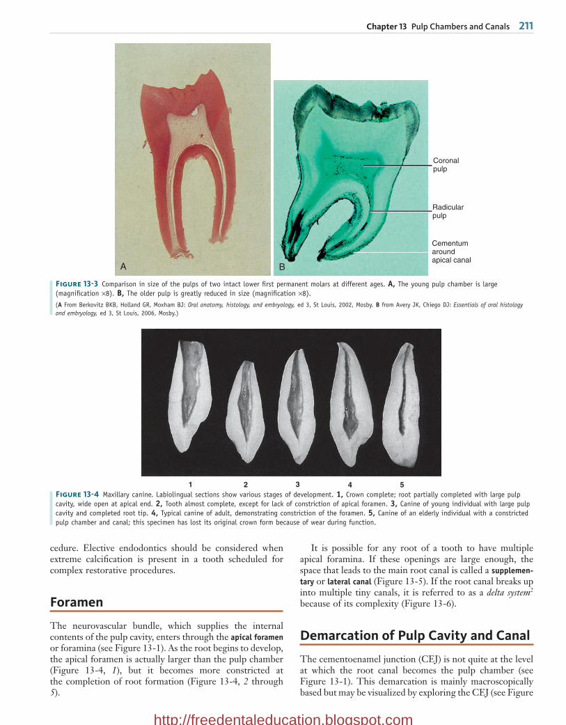
Chapter 13 Pulp Chambers and Canals 211
cedure. Elective endodontics should be considered when extreme calcification is present in a tooth scheduled for complex restorative procedures.
Foramen
The neurovascular bundle, which supplies the internal contents of the pulp cavity, enters through the apical foramen or foramina (see Figure 13-1). As the root begins to develop, the apical foramen is actually larger than the pulp chamber (Figure 13-4, 1), but it becomes more constricted at the completion of root formation (Figure 13-4, 2 through 5).
It is possible for any root of a tooth to have multiple apical foramina. If these openings are large enough, the space that leads to the main root canal is called a supplemen-tary or lateral canal (Figure 13-5). If the root canal breaks up into multiple tiny canals, it is referred to as a delta system2 because of its complexity (Figure 13-6).
Demarcation of Pulp Cavity and Canal
The cementoenamel junction (CEJ) is not quite at the level at which the root canal becomes the pulp chamber (see Figure 13-1). This demarcation is mainly macroscopically based but may be visualized by exploring the CEJ (see Figure
Figure 13-3 Comparison in size of the pulps of two intact lower first permanent molars at different ages. A, The young pulp chamber is large (magnification ×8). B, The older pulp is greatly reduced in size (magnification ×8).
(A From Berkovitz BKB, Holland GR, Moxham BJ: Oral anatomy, histology, and embryology, ed 3, St Louis, 2002, Mosby. B from Avery JK, Chiego DJ: Essentials of oral histology and embryology, ed 3, St Louis, 2006, Mosby.)
A B
Coronalpulp
Radicularpulp
Cementumaroundapical canal
Figure 13-4 Maxillary canine. Labiolingual sections show various stages of development. 1, Crown complete; root partially completed with large pulp cavity, wide open at apical end. 2, Tooth almost complete, except for lack of constriction of apical foramen. 3, Canine of young individual with large pulp cavity and completed root tip. 4, Typical canine of adult, demonstrating constriction of the foramen. 5, Canine of an elderly individual with a constricted pulp chamber and canal; this specimen has lost its original crown form because of wear during function.
1 2 3 4 5
http://freedentaleducation.blogspot.com
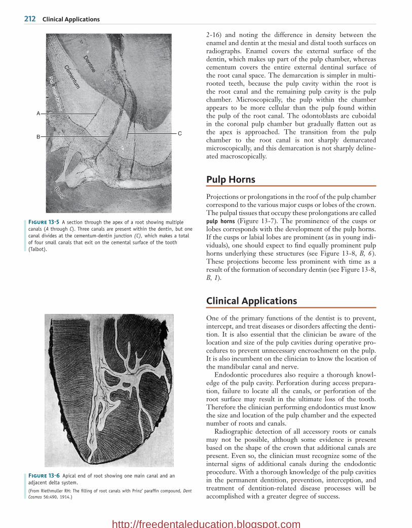
2-16) and noting the difference in density between the enamel and dentin at the mesial and distal tooth surfaces on radiographs. Enamel covers the external surface of the dentin, which makes up part of the pulp chamber, whereas cementum covers the entire external dentinal surface of the root canal space. The demarcation is simpler in multi-rooted teeth, because the pulp cavity within the root is the root canal and the remaining pulp cavity is the pulp chamber. Microscopically, the pulp within the chamber appears to be more cellular than the pulp found within the pulp of the root canal. The odontoblasts are cuboidal in the coronal pulp chamber but gradually flatten out as the apex is approached. The transition from the pulp chamber to the root canal is not sharply demarcated microscopically, and this demarcation is not sharply deline-ated macroscopically.
Pulp Horns
Projections or prolongations in the roof of the pulp chamber correspond to the various major cusps or lobes of the crown. The pulpal tissues that occupy these prolongations are called pulp horns (Figure 13-7). The prominence of the cusps or lobes corresponds with the development of the pulp horns. If the cusps or labial lobes are prominent (as in young indi-viduals), one should expect to find equally prominent pulp horns underlying these structures (see Figure 13-8, B, 6 ). These projections become less prominent with time as a result of the formation of secondary dentin (see Figure 13-8, B, 1).
Clinical Applications
One of the primary functions of the dentist is to prevent, intercept, and treat diseases or disorders affecting the denti-tion. It is also essential that the clinician be aware of the location and size of the pulp cavities during operative pro-cedures to prevent unnecessary encroachment on the pulp. It is also incumbent on the clinician to know the location of the mandibular canal and nerve.
Endodontic procedures also require a thorough knowl-edge of the pulp cavity. Perforation during access prepara-tion, failure to locate all the canals, or perforation of the root surface may result in the ultimate loss of the tooth. Therefore the clinician performing endodontics must know the size and location of the pulp chamber and the expected number of roots and canals.
Radiographic detection of all accessory roots or canals may not be possible, although some evidence is present based on the shape of the crown that additional canals are present. Even so, the clinician must recognize some of the internal signs of additional canals during the endodontic procedure. With a thorough knowledge of the pulp cavities in the permanent dentition, prevention, interception, and treatment of dentition-related disease processes will be accomplished with a greater degree of success.
Figure 13-5 A section through the apex of a root showing multiple canals (A through C). Three canals are present within the dentin, but one canal divides at the cementum-dentin junction (C), which makes a total of four small canals that exit on the cemental surface of the tooth (Talbot).
CB
A
BoneBone
Pol. m
embrane
Cem
entum
Dentine
Figure 13-6 Apical end of root showing one main canal and an adjacent delta system.
(From Riethmuller RH: The filling of root canals with Prinz’ paraffin compound, Dent Cosmos 56:490, 1914.)
212 Clinical Applications
http://freedentaleducation.blogspot.com
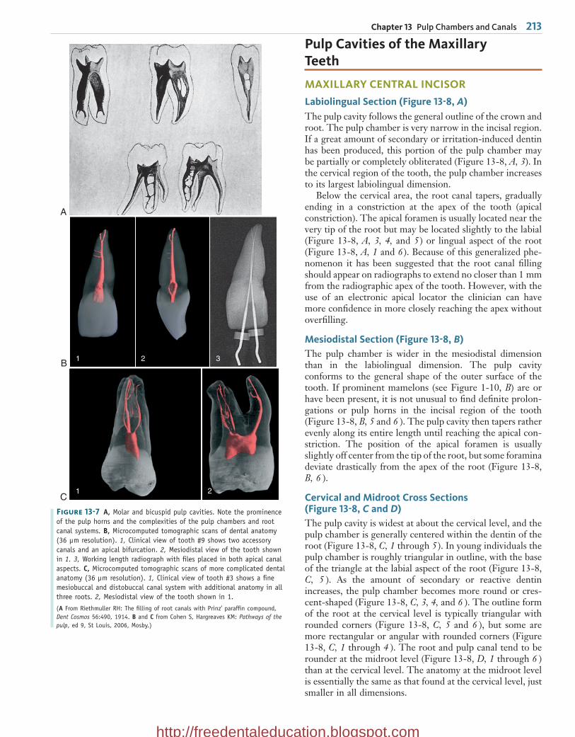
Chapter 13 Pulp Chambers and Canals 213
Pulp Cavities of the Maxillary Teeth
MAXILLARY CENTRAL INCISOR
Labiolingual Section (Figure 13-8, A)The pulp cavity follows the general outline of the crown and root. The pulp chamber is very narrow in the incisal region. If a great amount of secondary or irritation-induced dentin has been produced, this portion of the pulp chamber may be partially or completely obliterated (Figure 13-8, A, 3). In the cervical region of the tooth, the pulp chamber increases to its largest labiolingual dimension.
Below the cervical area, the root canal tapers, gradually ending in a constriction at the apex of the tooth (apical constriction). The apical foramen is usually located near the very tip of the root but may be located slightly to the labial (Figure 13-8, A, 3, 4, and 5 ) or lingual aspect of the root (Figure 13-8, A, 1 and 6 ). Because of this generalized phe-nomenon it has been suggested that the root canal filling should appear on radiographs to extend no closer than 1 mm from the radiographic apex of the tooth. However, with the use of an electronic apical locator the clinician can have more confidence in more closely reaching the apex without overfilling.
Mesiodistal Section (Figure 13-8, B)The pulp chamber is wider in the mesiodistal dimension than in the labiolingual dimension. The pulp cavity conforms to the general shape of the outer surface of the tooth. If prominent mamelons (see Figure 1-10, B) are or have been present, it is not unusual to find definite prolon-gations or pulp horns in the incisal region of the tooth (Figure 13-8, B, 5 and 6 ). The pulp cavity then tapers rather evenly along its entire length until reaching the apical con-striction. The position of the apical foramen is usually slightly off center from the tip of the root, but some foramina deviate drastically from the apex of the root (Figure 13-8, B, 6 ).
Cervical and Midroot Cross Sections (Figure 13-8, C and D)The pulp cavity is widest at about the cervical level, and the pulp chamber is generally centered within the dentin of the root (Figure 13-8, C, 1 through 5 ). In young individuals the pulp chamber is roughly triangular in outline, with the base of the triangle at the labial aspect of the root (Figure 13-8, C, 5 ). As the amount of secondary or reactive dentin increases, the pulp chamber becomes more round or cres-cent-shaped (Figure 13-8, C, 3, 4, and 6 ). The outline form of the root at the cervical level is typically triangular with rounded corners (Figure 13-8, C, 5 and 6 ), but some are more rectangular or angular with rounded corners (Figure 13-8, C, 1 through 4 ). The root and pulp canal tend to be rounder at the midroot level (Figure 13-8, D, 1 through 6 ) than at the cervical level. The anatomy at the midroot level is essentially the same as that found at the cervical level, just smaller in all dimensions.
Figure 13-7 A, Molar and bicuspid pulp cavities. Note the prominence of the pulp horns and the complexities of the pulp chambers and root canal systems. B, Microcomputed tomographic scans of dental anatomy (36 µm resolution). 1, Clinical view of tooth #9 shows two accessory canals and an apical bifurcation. 2, Mesiodistal view of the tooth shown in 1. 3, Working length radiograph with files placed in both apical canal aspects. C, Microcomputed tomographic scans of more complicated dental anatomy (36 µm resolution). 1, Clinical view of tooth #3 shows a fine mesiobuccal and distobuccal canal system with additional anatomy in all three roots. 2, Mesiodistal view of the tooth shown in 1.
(A From Riethmuller RH: The filling of root canals with Prinz’ paraffin compound, Dent Cosmos 56:490, 1914. B and C from Cohen S, Hargreaves KM: Pathways of the pulp, ed 9, St Louis, 2006, Mosby.)
B
A
C
1
1 2
2 3
http://freedentaleducation.blogspot.com

214 Pulp Cavities of the Maxillary Teeth
Figure 13-8 Maxillary central incisor. Sections of natural specimens. A, 1 through 6, Labiolingual sections. This aspect does not appear in radiographs. B, 1 through 6, Mesiodistal sections. C, 1 through 6, Cervical cross sections of root. D, 1 through 6, Midroot cross sections.
1
A
B
C
D
2 3 4 5 6
MAXILLARY LATERAL INCISOR
Labiolingual Section (Figure 13-9, A)The anatomy of the lateral incisor is similar to that of the central incisor. The pulp cavity of the lateral incisor gener-ally follows the outline form of the crown and the root. The pulp horns are usually prominent. The pulp chamber is narrow in the incisal region and may become very wide at the cervical level of the tooth (Figure 13-9, A, 1, 2, 3, and 5). Those teeth lacking this cervical enlargement of the pulp chamber possess a root canal that tapers slightly to the apical constriction (Figure 13-9, A, 4 and 6 ). Many of the apical foramina appear to be located at the tip of the root in the labiolingual aspect (Figure 13-9, A, 1, 4, and 6 ), whereas some exit on the labial (Figure 13-9, A, 2, and 3) or lingual aspect of the root tip (Figure 13-9, A, 5 ).
Mesiodistal Section (Figure 13-9, B)The pulp cavity closely follows the external outline of the tooth. The pulpal projections or pulp horns appear to be blunted when viewed from the labial aspect of the tooth. The pulp chamber and root canal gradually taper toward the apex, which often demonstrates a significant curve toward
the distal in the apical region (Figure 13-9, B, 1 through 4, and 6 ).
Cervical and Midroot Cross Sections (Figure 13-9, C and D)The cervical cross section shows the pulp chamber to be centered within the root. The root form of this tooth shows a large variation in shape (see the discussion of the lateral incisor in Chapter 6). The outline form of this tooth may be triangular, oval, or round (Figure 13-9, C, 1 through 6 ). The pulp chamber generally follows the outline form of the root, but secondary dentin may narrow the canal signifi-cantly (Figure 13-9, D, 4 and 6 ).
MAXILLARY CANINE
Labiolingual Section (Figure 13-10, A and D)The maxillary canine has the largest labiolingual root dimen-sion of any tooth in the mouth. Because the pulp cavity corresponds closely to the outline of the tooth, the size of the pulp chamber of this tooth may also be the largest in the mouth.
The incisal aspect of the canine corresponds to the shape of the crown. If a prominent cusp is present, a long narrow
http://freedentaleducation.blogspot.com

Chapter 13 Pulp Chambers and Canals 215
Figure 13-9 Maxillary lateral incisor. Sections of natural specimens. A, 1 through 6, Labiolingual sections. This aspect does not appear in radiographs. B, 1 through 6, Mesiodistal sections. C, 1 through 6, Cervical cross sections of root. D, 1 through 6, Midroot cross sections.
1
A
B
C
D
2 3 4 5 6
projection from the pulp chamber (the pulp horn) will be present. The pulp chamber and incisal third or half of the root canal may be very wide, showing a very abrupt constric-tion of the root canal in the apical region, which then gently tapers toward the apex (Figure 13-10, A, 4, 5, 6, and 8; D, 16, 17, and 18). In other instances, a root canal may taper evenly from the pulp chamber to the apex of the root (Figure 13-10, A, 9; D, 10, 11, 13, and 14 ).
Some canines have severe curves in the apical aspect of the root (Figure 13-10, A, 3 and 7 ). The apical foramen may appear to exit at the tip of the root (Figure 13-10, A, 2 and 5 through 8; D, 11, 17, and 18 ) or labially to the apex of the root (Figure 13-10, A, 1, 3, and 4; D, 12 through 16 ).
Mesiodistal Section (Figure 13-10, B and E)The pulp cavity is much narrower in the mesiodistal aspect. The dimension and degree of taper of the pulp canal of the maxillary canine are very similar to those of the central and lateral incisors; however, the cuspid has a much longer root. The pulp cavity gently tapers from the incisal aspect to the apical foramen. A mesial or distal curve of the apical root may be present (Figure 13-10, B, 1, 4, 6, and 8; E, 14, 17, and 18). The apical foramen may appear to exit at the tip of the root (Figure 13-10, B, 1, 3 through 5, 7, and 9; E, 10, 11, 13, 14, 17, and 18) or slightly to the mesial or distal aspect of the root (Figure 13-10, B, 2, 6, and 8; E, 12, 15, and 16 ).
Cervical Cross Section (Figure 13-10, C)In cervical cross section, the shape of the root and pulp cavity is oval (Figure 13-10, C, 6, 7, and 9), triangular (Figure
13-10, C, 8), or elliptical (Figure 13-10, C, 1 through 5). The pulp chamber and canal are often centered within the crown and root (Figure 13-10, C, 1, 3, 4, and 9).
MAXILLARY FIRST PREMOLAR
Buccolingual Section (Figure 13-11, A and D)The maxillary first premolar may have two well-developed roots (Figure 13-11, A, 1, 2, and 9; D, 10 and 14), two root projections that are not fully separated (Figure 13-11, A, 3, 5, 7, and 8; D, 11, 12, 13, 15, 16, and 17 ), or one broad root (Figure 13-11, A, 4 and 6; D, 18). The majority of maxillary first premolars have two root canals (Figure 13-11, A and D). A small percentage of maxillary first premolar teeth may have three roots that may be almost undetectable radiographically.
The pulp horn usually extends further incisally under the buccal cusp, because this cusp is usually better developed than the lingual cusp. The pulp horns may be blunted (Figure 13-11, A, 1, 5, and 6; D, 11) in teeth possessing cusps that demonstrate a fair amount of attrition. The pulp chamber floor is below the cervical level of all the variations found in the maxillary first premolar. The pulp chamber of teeth having the least root separation usually shows the largest incisal-apical dimension (Figure 13-11, A, 4; D, 18). Those teeth possessing a partial root separation may also have this large dimension (Figure 13-11, A, 8; D, 10). Teeth having two separate canals usually demonstrate a rather small pulp chamber in the incisal-apical direction (Figure 13-11, A, 1, 2, and 9; D, 11 and 14). The shape of the pulp
http://freedentaleducation.blogspot.com
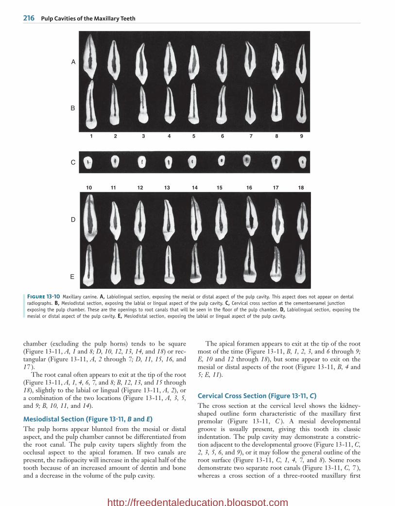
216 Pulp Cavities of the Maxillary Teeth
A
B
C
D
E
1
10 11 12 13 14 15 16 17 18
2 3 4 5 6 7 8 9
Figure 13-10 Maxillary canine. A, Labiolingual section, exposing the mesial or distal aspect of the pulp cavity. This aspect does not appear on dental radiographs. B, Mesiodistal section, exposing the labial or lingual aspect of the pulp cavity. C, Cervical cross section at the cementoenamel junction exposing the pulp chamber. These are the openings to root canals that will be seen in the floor of the pulp chamber. D, Labiolingual section, exposing the mesial or distal aspect of the pulp cavity. E, Mesiodistal section, exposing the labial or lingual aspect of the pulp cavity.
chamber (excluding the pulp horns) tends to be square (Figure 13-11, A, 1 and 8; D, 10, 12, 13, 14, and 18) or rec-tangular (Figure 13-11, A, 2 through 7; D, 11, 15, 16, and 17 ).
The root canal often appears to exit at the tip of the root (Figure 13-11, A, 1, 4, 6, 7, and 8; B, 12, 13, and 15 through 18), slightly to the labial or lingual (Figure 13-11, A, 2), or a combination of the two locations (Figure 13-11, A, 3, 5, and 9; B, 10, 11, and 14).
Mesiodistal Section (Figure 13-11, B and E)The pulp horns appear blunted from the mesial or distal aspect, and the pulp chamber cannot be differentiated from the root canal. The pulp cavity tapers slightly from the occlusal aspect to the apical foramen. If two canals are present, the radiopacity will increase in the apical half of the tooth because of an increased amount of dentin and bone and a decrease in the volume of the pulp cavity.
The apical foramen appears to exit at the tip of the root most of the time (Figure 13-11, B, 1, 2, 3, and 6 through 9; E, 10 and 12 through 18), but some appear to exit on the mesial or distal aspects of the root (Figure 13-11, B, 4 and 5; E, 11).
Cervical Cross Section (Figure 13-11, C)The cross section at the cervical level shows the kidney-shaped outline form characteristic of the maxillary first premolar (Figure 13-11, C ). A mesial developmental groove is usually present, giving this tooth its classic indentation. The pulp cavity may demonstrate a constric-tion adjacent to the developmental groove (Figure 13-11, C, 2, 3, 5, 6, and 9), or it may follow the general outline of the root surface (Figure 13-11, C, 1, 4, 7, and 8). Some roots demonstrate two separate root canals (Figure 13-11, C, 7 ), whereas a cross section of a three-rooted maxillary first
http://freedentaleducation.blogspot.com

Chapter 13 Pulp Chambers and Canals 217
Figure 13-11 Maxillary first premolar. A, Buccolingual section, exposing the mesial or distal aspect of the pulp cavity. This aspect does not appear on dental radiographs. B, Mesiodistal section, exposing the buccal or lingual aspect of the pulp cavity. C, Cervical cross section at the cementoenamel junction exposing the pulp chamber. These are the openings to root canals that will be seen in the floor of the pulp chamber. D, Buccolingual section, exposing the mesial or distal aspect of the pulp cavity. E, Mesiodistal section, exposing the buccal or lingual aspect of the pulp cavity.
A
B
C
D
E
1
10 11 12 13 14 15 16 17 18
2 3 4 5 6 7 8 9
premolar will show three separate canals (Figure 13-11, C, 3).
MAXILLARY SECOND PREMOLAR
Buccolingual Section (Figure 13-12, A and D)Most maxillary second premolars have only one root and canal. Two roots are possible, although two canals within a single root may also be found.
The pulp cavity may demonstrate well-developed pulp horns (Figure 13-12, A, 1, 2, 6, 7, and 8; D, 10, 11, 12, 14, 16, and 17 ); others may have blunted or nonexistent pulp horns (Figure 13-12, A, 3, 4, 5, and 9; D, 13, 15, and 18). The pulp chamber and root canal are very broad in the buc-colingual aspect of teeth with single canals. The pulp cavity does not show a well-defined demarcation between the root cavity and the pulp cavity because of the large buccolingual extent of the pulp cavity in the upper half of the tooth. In
the apical half or third of the tooth, the pulp cavity often narrows abruptly (Figure 13-12, A, 1, 2, 4, 6, and 8; D, 11, 12, 13, and 18) and then tapers gently toward the apex. Some teeth possess dentinal islands in the apical third of the root; this situation essentially forces the clinician to treat these as teeth with two canals (Figure 13-22, A, 5; D, 15 and 17). Other maxillary second premolars have a canal that bifurcates at the apical third of the root (Figure 13-12, D, 10, 15, and 16 ). It should also be noted that buccal and lingual pulpal projections or fins are present at the level of the CEJ (Figure 13-12, A, 2; D, 12 and 18). Some teeth show a constriction at this same level (Figure 13-12, D, 10).
The apical foramen often appears to exit at the tip of the root (Figure 13-12, A, 1, 2, 5, 6, and 8; D, 11, 12, and 14 through 18). Some apical foramina appear to exit on the buccal aspect of the root (Figure 13-12, A, 4; D, 13), on the lingual aspect of the root (Figure 13-12, A, 7 and 9), or on both sides of the root tip (Figure 13-12, A, 3; D, 10 and 15).
http://freedentaleducation.blogspot.com

218 Pulp Cavities of the Maxillary Teeth
Figure 13-12 Maxillary second premolar. A, Buccolingual section, exposing the mesial or distal aspect of the pulp cavity. This aspect does not appear on dental radiographs. B, Mesiodistal section, exposing the buccal or lingual aspect of the pulp cavity. C, Cervical cross section at the cementoenamel junction exposing the pulp chamber. These are the openings to root canals that will be seen in the floor of the pulp chamber. D, Buccolingual section, exposing the mesial or distal aspect of the pulp cavity. E, Mesiodistal section, exposing the buccal or lingual aspect of the pulp cavity.
A
B
C
D
E
1
10 11 12 13 14 15 16 17 18
2 3 4 5 6 7 8 9
Mesiodistal Section (Figure 13-12, B and E)The view of the pulp cavity in the mesiodistal section of the second maxillary premolar does not vary from that found in the maxillary first premolar. The pulp horns are blunted, and the pulp cavity tapers slightly from the occlusal aspect to the apex. The apical foramen may appear to be located off center of the root tip (Figure 13-12, A, 2, 6, and 9; D, 10 and 13) or appear to exit at the root tip (Figure 13-12, B, 1, 3, 4, 5, 7, and 8; E, 11, 12, and 14 through 18).
Cervical Cross Section (Figure 13-12, C)The cervical cross section of the maxillary second premolar is usually oval (Figure 13-12, C, 2 and 4 through 9), with some teeth having a kidney-shaped cross section (Figure 13-12, C, 1 and 3). The pulp cavity is centered in the root and may have a constriction in the middle of the canal space (Figure 13-12, C, 1, 4, 6, and 9), an entire separation (Figure 13-12, C, 2), or an elliptical pulp cavity (Figure 13-12, C, 3, 5, 7, and 8).
MAXILLARY FIRST MOLAR
Buccolingual Section (Figure 13-13, A and D)The buccolingual section of the maxillary first molar in Figure 13-13 shows the pulp cavities of the mesiobuccal and palatal root. These roots were chosen to demonstrate the pulp cavity anatomy because of the complexity of the mesiobuccal root. The distobuccal root is straighter and smaller and presents fewer variations in shape. The entire removal of the pulp is an impossibility in many maxillary first molar teeth because of the complexities of the root canal system (see Figure 13-7).
The maxillary first molar usually has three roots and three canals. The palatal root usually has the largest dimen-sions, followed by the mesiobuccal and distobuccal roots, respectively. The mesiobuccal root is often very wide buc-colingually and usually possesses an accessory canal com-monly called MB2, which usually is the smallest of all the canals in this tooth.
http://freedentaleducation.blogspot.com

Chapter 13 Pulp Chambers and Canals 219
The pulp horns are usually prominent in this tooth (Figure 13-13, A, 1, and 4 through 8; D, 10 through 13, and 15 through 18). The pulp chamber is somewhat rectangular (excluding the pulp horns) when viewed from the mesial aspect of the tooth. The palatal root usually has the largest canal (Figure 13-13, A, 1, 2, 3, 5, and 6; D, 10, 15, 17, and 18). The mesiobuccal canal is often very small (Figure 13-13, A, 2 and 6; D, 10, 14, and 17 ), but some mesiobuccal canals may be very wide within a very wide root (Figure 13-13, A, 9; D, 12 and 13).
The presence of an unmarked MB2 is strongly suggested when the root canal appears to be off to one side (Figure 13-13, A, 2, 6, and 8; D, 10, 11, 13, 14, 15, 17, and 18).
The root canals of the wide mesiobuccal roots are widest at the midroot level and taper to a very fine diameter at the apical foramen. The palatal root canal and the mesiobuccal root canal of most teeth taper gently to the apical region, where they terminate at or near the apex. The apical foramen of the palatal canal may appear to exit at the apex (Figure 13-13, A, 1, and 4 through 9; D, 11 through 17 ), slightly
lingually (Figure 13-13, A, 2; D, 10) or buccally to the root tip (Figure 13-13, A, 3; D, 18).
The apical foramen of the mesiobuccal canal may appear to exit on the tip of the root (Figure 13-13, A, 2 through 5, 7, 8, and 9; D, 10, 12, 13, 17, and 18) or on the buccal (Figure 13-13, A, 1 and 6; D, 14 and 15 ) or lingual (Figure 13-13, D, 16 ) aspect of the root.
Mesiodistal Section (Figure 13-13, B and E)The mesiodistal section of the maxillary first molar includes the distobuccal root, which was not visible on the previously mentioned buccolingual section. The mesiobuccal root has a great tendency to possess a more curved root and canal than the distobuccal root (Figure 13-13, B, 1 through 8; E, 12, 14, and 15). Some of the buccal canals are relatively straight (Figure 13-13, E, 10, 11, and 13).
The pulp horns are very distinct from this view, with the mesiobuccal pulp horn usually appearing a little larger than the distobuccal pulp horn (Figure 13-13, B, 1 and 3 through
Figure 13-13 Maxillary first molar. A, Buccolingual section, exposing the mesial or distal aspect of the pulp cavity. This aspect does not appear on dental radiographs. B, Mesiodistal section, exposing the buccal or lingual aspect of the pulp cavity. C, Five cross sections at cervical line and four cross sections at midroot. D, Buccolingual section, exposing the mesial or distal aspect of the pulp cavity. E, Mesiodistal section, exposing the buccal aspect of the pulp cavity.
A
B
C
D
E
1
10 11 12 13 14 15 16 17 18
2 3 4 5 6 7 8 9
http://freedentaleducation.blogspot.com

220 Pulp Cavities of the Maxillary Teeth
9; E, 10 through 13 and 15 through 18). In some teeth, the pulp horns are of equal size (Figure 13-13, B, 2; E, 14). The pulp chamber is somewhat square (if the pulp horns are excluded) when viewed from the buccal aspect. The demar-cation of the root canal is much more distinct in the mesio-distal section. The root canals appear much smaller when viewed from the buccal or lingual aspect. The canals taper slightly as they approach the apical foramen. The apical foramen often appears to be located at the tip of the root (Figure 13-13, B, 1 through 7 and 9; E, 11, 14, 16, and 18), but the apical foramen may appear to be located on the mesial (Figure 13-13, E,10 and 17, mesial root only) or on the distal aspect of the root (Figure 13-13, B, 8; E, 12 and 13, distal root only).
Cervical Cross Section (Figure 13-13, C)The cervical outline form of the maxillary first molar is rhomboidal with rounded corners (Figure 13-13, C, 1 through 5 ). The mesiobuccal angle has an acute angle, the distobuccal angle is obtuse, and the lingual angles are essen-tially right angles. The orifices of the root canals have the following relation to the floor of the pulp chamber.
The palatal canal is centered lingually; the distobuccal canal is near the obtuse angle of the pulp chamber; the mesiobuccal root canal is buccal and mesial to the distobuc-cal canal, in what seems to be the extreme corner, positioned within the acute angle of the pulp chamber. If an accessory mesiobuccal canal (MB2) is present, it will be located lingual to the mesiobuccal canal. The canals of this tooth form a triangular pattern; a line drawn between the mesiobuccal canal and the palatal canal makes the base of the triangle, and the distobuccal canal, which is slightly closer to the palatal canal, makes the third point of the triangle. If a mesiobuccal accessory canal is present, it will be between the mesiobuccal and palatal canal just off an imaginary line between the two canals (Figure 13-13, C, 7, 8, and 9). The MB2 may even be mesial to a line connecting the mesiobuc-cal and the palatal canals (Figure 13-13, C, 8).
Midroot Cross Section (Figure 13-13, C)The midroot sections were added to the molar descriptions because some molars possess more than one canal within the root (Figure 13-13, C, 6 through 9). The palatal root is usually the largest root having a round outline form. The distobuccal canal is oval to round but much smaller than the palatal root. The mesiobuccal root is an elongated oval to kidney-shaped root with the indentation located toward the furcation. The root canals of the palatal and distobuccal root are oval to round, whereas the mesiobuccal canals are elon-gated (Figure 13-13, C, 6 and 9), elliptical (Figure 13-13, C, 7 ), or round (Figure 13-13, C, 8 and 9). The pulp canals may be extremely difficult to locate and instrument if secondary or irritation-induced dentin is abundant (Figure 13-13, C, 9). A thorough knowledge of the anatomy of the pulp chambers and canals is necessary if endodontic procedures are to be accomplished.
MAXILLARY SECOND MOLAR
Buccolingual Section (Figure 13-14, A and D)The buccal roots of the maxillary second molar are straighter and closer together than those of the maxillary first molar. The tendency for root fusion is greater in the second maxil-lary molar than in the first maxillary molar, but the palatal root is usually separate. Most often maxillary second molars possess three roots and three canals.
The mesiobuccal root of the maxillary second molar is not as complex as that formed in the maxillary first molar. The tendency for a very wide mesiobuccal canal is not present in the maxillary second molar. The presence of two canals in the mesiobuccal root is not as common in the maxillary second molar as in the maxillary first molar; however, it does occur (Figure 13-14, A, 5 and 7 ).
The pulp horns may be well developed (Figure 13-14, A, 1 through 3, 5, 8, and 9; D, 10 through 18) or virtually absent (Figure 13-14, A, 4, 6, and 7 ). The pulp chamber appears somewhat rectangular (excluding the pulp horns). The pulp canals gradually taper toward the apex until reach-ing the apical constriction, which occurs just before the apical foramen.
The mesiobuccal root canal of the maxillary second molar does not have the tendency to be extremely large, as is demonstrated in the mesiobuccal canal of the maxillary first molar. The apical foramen of the palatal root often appears to exit at the tip of the root (Figure 13-14, A, 1, 2, 3, and 5 through 8; D, 11, 13, 14, 16, 17, and 18), but it may exist on the lingual (Figure 13-14, A, 4 and 9; D, 15) or buccal aspect of the root (Figure 13-14, D, 10 and 12).
Mesiodistal Section (Figure 13-14, B and E)The mesiodistal section of the maxillary second molar is similar to that of the maxillary first molar. The buccal roots of the second molar are not as far apart as they are in the maxillary first molar, and their buccal roots have a greater tendency to be fused.
The pulp horns are usually well developed (Figure 13-14, B, 1 through 5, 8, and 9; E, 10 through 15 and 17). Some teeth demonstrate an obvious blunting or absence of the pulp horns (Figure 13-14, B, 6 and 7; E, 18). The mesiobuc-cal pulp horn is often larger than the distobuccal pulp horn (Figure 13-14, B, 1, 4, 5, 8, and 9; E, 10, 12, 13, 15, 17, and 18).
The pulp chamber appears much smaller in the mesio-distal section than in the buccolingual section. The pulp chamber is square (excluding the pulp horns) when viewed from the buccal aspect. The pulp canals gently taper from the pulp chamber to the apical constriction. The mesiobuc-cal pulp canal has a greater tendency to be curved than the distobuccal canal. The majority of the apical foramen appears to exit at the tip of the root (Figure 13-14, B, 1, 2, and 4 through 9; E, 10 through 16 and 18).
Cervical Cross Section (Figure 13-14, C)The cervical cross section of the maxillary second molar demonstrates angulations of the outline form that are more
http://freedentaleducation.blogspot.com
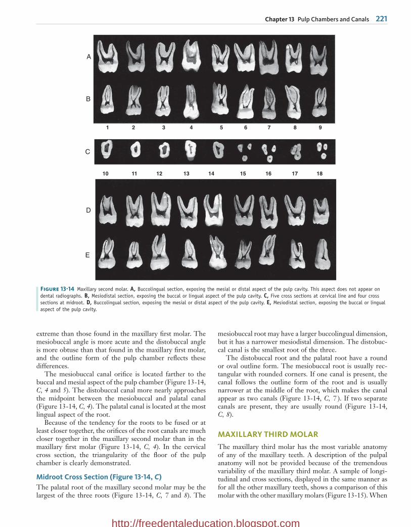
Chapter 13 Pulp Chambers and Canals 221
Figure 13-14 Maxillary second molar. A, Buccolingual section, exposing the mesial or distal aspect of the pulp cavity. This aspect does not appear on dental radiographs. B, Mesiodistal section, exposing the buccal or lingual aspect of the pulp cavity. C, Five cross sections at cervical line and four cross sections at midroot. D, Buccolingual section, exposing the mesial or distal aspect of the pulp cavity. E, Mesiodistal section, exposing the buccal or lingual aspect of the pulp cavity.
A
B
C
D
E
1
10 11 12 13 14 15 16 17 18
2 3 4 5 6 7 8 9
extreme than those found in the maxillary first molar. The mesiobuccal angle is more acute and the distobuccal angle is more obtuse than that found in the maxillary first molar, and the outline form of the pulp chamber reflects these differences.
The mesiobuccal canal orifice is located farther to the buccal and mesial aspect of the pulp chamber (Figure 13-14, C, 4 and 5). The distobuccal canal more nearly approaches the midpoint between the mesiobuccal and palatal canal (Figure 13-14, C, 4). The palatal canal is located at the most lingual aspect of the root.
Because of the tendency for the roots to be fused or at least closer together, the orifices of the root canals are much closer together in the maxillary second molar than in the maxillary first molar (Figure 13-14, C, 4). In the cervical cross section, the triangularity of the floor of the pulp chamber is clearly demonstrated.
Midroot Cross Section (Figure 13-14, C)The palatal root of the maxillary second molar may be the largest of the three roots (Figure 13-14, C, 7 and 8). The
mesiobuccal root may have a larger buccolingual dimension, but it has a narrower mesiodistal dimension. The distobuc-cal canal is the smallest root of the three.
The distobuccal root and the palatal root have a round or oval outline form. The mesiobuccal root is usually rec-tangular with rounded corners. If one canal is present, the canal follows the outline form of the root and is usually narrower at the middle of the root, which makes the canal appear as two canals (Figure 13-14, C, 7 ). If two separate canals are present, they are usually round (Figure 13-14, C, 8).
MAXILLARY THIRD MOLAR
The maxillary third molar has the most variable anatomy of any of the maxillary teeth. A description of the pulpal anatomy will not be provided because of the tremendous variability of the maxillary third molar. A sample of longi-tudinal and cross sections, displayed in the same manner as for all the other maxillary teeth, shows a comparison of this molar with the other maxillary molars (Figure 13-15). When
http://freedentaleducation.blogspot.com
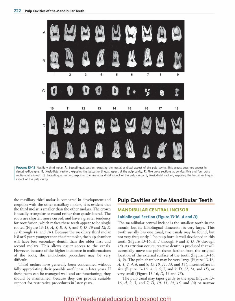
Figure 13-15 Maxillary third molar. A, Buccolingual section, exposing the mesial or distal aspect of the pulp cavity. This aspect does not appear in dental radiographs. B, Mesiodistal section, exposing the buccal or lingual aspect of the pulp cavity. C, Five cross sections at cervical line and four cross sections at midroot. D, Buccolingual section, exposing the mesial or distal aspect of the pulp cavity. E, Mesiodistal section, exposing the buccal or lingual aspect of the pulp cavity.
A
B
C
D
E
1
10 11 12 13 14 15 16 17 18
2 3 4 5 6 7 8 9
222 Pulp Cavities of the Mandibular Teeth
the maxillary third molar is compared in development and eruption with the other maxillary molars, it is evident that the third molar is smaller than the other molars. The crown is usually triangular or round rather than quadrilateral. The roots are shorter, more curved, and have a greater tendency for root fusion, which makes these teeth appear to be single rooted (Figure 13-15, A, 8; B, 3, 5, and 8; D, 10 and 12; E, 11 through 14, and 16 ). Because the maxillary third molar is 8 or 9 years younger than the first molar, the pulp chamber will have less secondary dentin than the older first and second molars. This allows easier access to the canals. However, because of the higher incidence in malformations of the roots, the endodontic procedure may be very difficult.
Third molars have generally been condemned without fully appreciating their possible usefulness in later years. If these teeth can be managed well and are functioning, they should be maintained, because they can provide suitable support for restorative procedures in later years.
Pulp Cavities of the Mandibular Teeth
MANDIBULAR CENTRAL INCISOR
Labiolingual Section (Figure 13-16, A and D)The mandibular central incisor is the smallest tooth in the mouth, but its labiolingual dimension is very large. This tooth usually has one canal; two canals may be found, but not very frequently. The pulp horn is well developed in this tooth (Figure 13-16, A, 1 through 6 and 8; D, 10 through 18). As attrition occurs, reactive dentin is produced that will essentially move the pulp tissue farther from the original location of the external surface of the tooth (Figure 13-16, A, 9). The pulp chamber may be very large (Figure 13-16, A, 1, 2, 4, 6, and 8; D, 10, 11, 13, and 17 ), intermediate in size (Figure 13-16, A, 3, 5, 7, and 9; D, 12, 14, and 15), or very small (Figure 13-16, D, 16 and 18).
The pulp canal may taper gently to the apex (Figure 13-16, A, 2, 3, and 7; D, 10, 11, 14, 16, and 18) or narrow
http://freedentaleducation.blogspot.com

Chapter 13 Pulp Chambers and Canals 223
Figure 13-16 Mandibular central incisor (first incisor). A, Labiolingual section, exposing the mesial or distal aspect of the pulp cavity. This aspect does not appear on dental radiographs. B, Mesiodistal section, exposing the labial or lingual aspect of the pulp cavity. C, Cervical cross section at the cementoenamel junction exposing the pulp chamber. These are the openings to root canals that will be seen in the floor of the pulp chamber. D, Labiolingual section, exposing the mesial or distal aspect of the pulp cavity. E, Mesiodistal section, exposing the labial or lingual aspect of the pulp cavity.
A
B
C
D
E
1
10 11 12 13 14 15 16 17 18
2 3 4 5 6 7 8 9
or mesially or distally to the apex of the root (Figure 13-16, B, 1, 3, 4, 6, 7, and 9; E, 10, 11, 13, 15, and 18).
Cervical Cross Section (Figure 13-16, C)The cervical cross section demonstrates the proportions of the root. The mesiodistal dimension is small, whereas the labiolingual dimension is very large. The external shape is variable; it may be round, oval, or elliptical. The more nearly round the root, the more nearly round the canal (Figure 13-16, C ). Two separate canals may be present, or a dentinal island may make it appear as though two canals are present (Figure 13-17).
MANDIBULAR LATERAL INCISOR
Labiolingual Section (Figure 13-18, A and D)The mandibular lateral incisor tends to be a little larger than the mandibular central incisor in all dimensions, and the pulp chamber is also larger. The form and function of the
abruptly in the apical 3 to 4 mm of the root (Figure 13-16, A, 1, 4, 5, 6, and 8; D, 12, 13, 15, and 17 ).
The apical foramen may appear to exit at the apex (Figure 13-16, A, 1, 4, 6, 7, and 9; D, 11, 12, 15, 16, and 18) or on the buccal aspect of the root (Figure 13-16, A, 2, 3, 5, and 8; D, 10, 13, 14, and 17 ).
Mesiodistal Section (Figure 13-16, B and E)A buccal or facial view of a mesiodistal section of the man-dibular central incisor demonstrates the narrowness of the pulp cavity. A small endodontic file can generally be used to negotiate these canals in spite of this narrowness, because of the wide labiolingual dimension of the pulp chamber. However, secondary or tertiary (irritation-induced) dentin may interfere with endodontic treatment (Figure 13-16, D, 16 and 18; E, 13 and 18).
The pulp horn is usually prominent but single. The canal also appears narrow, having a gentle taper from the pulp chamber to the apical constriction. The canal may exit at the apex (Figure 13-16, B, 2, 5, and 8; E, 12, 14, 16, and 17 )
http://freedentaleducation.blogspot.com

224 Pulp Cavities of the Maxillary Teeth
A
B
C
D
E
1
10 11 12 13 14 15 16 17 18
2 3 4 5 6 7 8 9
Figure 13-18 Mandibular lateral incisor (second incisor). A, Labiolingual section, exposing the mesial or distal aspect of the pulp cavity. This aspect does not appear on dental radiographs. B, Mesiodistal section, exposing the labial or lingual aspect of the pulp cavity. C, Cervical cross section at the cementoenamel junction, exposing the pulp chamber. These are the openings to root canals that will be seen in the floor of the pulp chamber. D, Labiolingual section, exposing the mesial or distal aspect of the pulp cavity. E, Mesiodistal section, exposing the labial or lingual aspect of the pulp cavity.
mandibular lateral incisor are identical to those of the central incisor.
The pulp horn is usually prominent. The pulp chamber may possess a dimension that is very large (Figure 13-18, A, 2, 4, 5, 8, and 9; D, 10, 11, 16, 17, and 18), intermediate in size (Figure 13-18, A, 1, 3, 6, and 7; D, 12), or small (Figure 13-18, D, 14 and 15). The pulp canal may taper gently from the apex (Figure 13-18, A, 1, 2, 4, 6, 7, and 9; D, 12, 14 through 17 ) or narrow abruptly in the last 3 to 4 mm of the canal (Figure 13-18, A, 3, 5, and 8; D, 10 through 13). The apical foramen may appear to exit at the tip of the root (Figure 13-18, A, 1 through 6, 8, and 9; D, 12 through 15, 17, and 18) or on the buccal or lingual aspect of the root tip (Figure 13-18, A, 7; D, 10, 11, and 16 ).
Mesiodistal Section (Figure 13-18, B and E)The pulp chamber and canal, as viewed from this aspect, will demonstrate a slender cavity. The mandibular lateral Figure 13-17 Mandibular central incisor with two canals.
http://freedentaleducation.blogspot.com

Chapter 13 Pulp Chambers and Canals 225
incisor resembles the mandibular incisor, but it may appear a little wider and have a pulpal dimension that is larger. The pulp horns are prominent, and the pulp chamber and canal gently taper to the apex. The apical foramen may appear to exit at the tip (Figure 13-18, A, 1, 2, 3, 6, and 9; D, 12 through 15, 17, and 18) or on the mesial or distal aspect of the root tip (Figure 13-18, A, 5, 7, and 8; D, 10, 11 and 16 ).
Cervical Cross Section (Figure 13-18, C)The cervical cross section of the mandibular lateral incisor shows the pulp canal centered in the root. A comparison of several of the sections demonstrates a root somewhat larger than that of the mandibular central incisor. Considerable variation is evident in the form of the root. The root outline form is oval to elliptical. Some cross sections of the larger teeth resemble the cervical cross sections of small mandibu-lar canines (Figure 13-18, C, 2, 3, and 4). The root canal follows the outline form of the root, and some roots dem-onstrate root grooves (Figure 13-18, C, 6 ).
MANDIBULAR CANINE
Labiolingual Section (Figure 13-19, A and D)
The pulp cavity of the mandibular canine is similar in size and shape to that of the maxillary canine. The mandibular canine tends to be a little shorter, although the opposite can be found. It is not uncommon to find two roots or at least two canals in the mandibular canine. A dentinal island may be found in any tooth that demonstrates an extremely wide labiolingual dimen-sion and a narrow mesiodistal dimension. Because the pres-ence of two canals cannot be easily detected radiographically, their presence must be ruled out clinically as well. The pulp cavity in a tooth of this kind varies according to where the section is examined (see Figure 8-24, 1, 2, 5, and 6 ).
The pulp horn is prominent in the mandibular canine unless an extensive amount of attrition has taken place (Figure 13-19, A, 1 and 3). The pulp chamber usually is very wide (Figure 13-19, A, 1, 3, 4, 5, and 7; D, 10, 12, 14, 15, 17, and 18) but may be average to small (Figure 13-19, A, 2, 6, and 9; D, 11, 13, and 16 ).
Figure 13-19 Mandibular canine. A, Labiolingual section, exposing the mesial or distal aspect of the pulp cavity. This aspect does not appear on dental radiographs. B, Mesiodistal section, exposing the labial or lingual aspect of the pulp cavity. C, Cervical cross section at the cementoenamel junction, exposing the pulp chamber. These are the openings to root canals that will be seen in the floor of the pulp chamber. D, Labiolingual section, exposing the mesial or distal aspect of the pulp cavity. E, Mesiodistal section, exposing the labial or lingual aspect of the pulp cavity.
A
B
C
D
E
1
10 11 12 13 14 15 16 17 18
2 3 4 5 6 7 8 9
http://freedentaleducation.blogspot.com

Some mandibular canines demonstrate an abrupt nar-rowing of the pulp cavity when passing from the region of the pulp chamber to the region of the pulp canal (Figure 13-19, A, 8; D, 13). Other mandibular canines demonstrate an abrupt narrowing of the pulp canal in the apical region (Figure 13-19, A, 1 through 6; D, 11, 12, and 18), after which the canal gently tapers to the apex. If an abrupt nar-rowing of the canal is absent, the tooth will demonstrate a canal that gently tapers to the apical foramen.
The apical foramen often appears to exit at the tip of the apex (Figure 13-19, A, 3, 5, 7, and 9; D, 10, 12, 14 through 16 ), slightly buccally (Figure 13-19, A, 1, 2, 6, and 8; D, 11, 13, and 17 ), or lingually to the root tip (Figure 13-19, D, 18).
Mesiodistal Section (Figure 13-19, B and E)The mesiodistal cross section of the mandibular canine appears very similar to that of the maxillary canine. The mesiodistal section demonstrates how narrow this tooth is in the mesiodistal aspect. This view also shows the degree of curvature of the apical portion of the root. The curvature of the root canal may be in the mesial direction (Figure 13-
19, E, 17 ). The pulp horn is usually prominent but appears blunted in this view. The pulp chamber and canal show a continuous gentle taper to the apex, where the apical foramen appears to exit at the tip of the root (Figure 13-19, B, 1 through 4; E, 10, 11, 13, 15, and 17 ) or slightly mesially or distally to the root tip (Figure 13-19, B, 5 through 7; E, 12, 14, and 18).
Cervical Cross Section (Figure 13-19, C)A cervical cross section of the mandibular canine shows considerable variation in size and shape (Figure 13-19, C, 1 through 9). The outline form of the root may be oval (Figure 13-19, C, 1, 4, 7, and 8), rectangular (Figure 13-19, C, 2, 5, 6, and 9), or triangular (Figure 13-19, C, 3). The size and shape of the canal are also variable. The pulp cavity outline form closely resembles the root form.
MANDIBULAR FIRST PREMOLAR
Buccolingual Section (Figure 13-20, A and D)The mandibular first premolar looks like a small mandibular canine with an extra small cusp. The pulp cavity also looks
226 Pulp Cavities of the Mandibular Teeth
Figure 13-20 Mandibular first premolar. A, Buccolingual section, exposing the mesial or distal aspect of the pulp cavity. This aspect does not appear on dental radiographs. B, Mesiodistal section, exposing the buccal or lingual aspect of the pulp cavity. C, Cervical cross section at the cementoenamel junction, exposing the pulp chamber. These are the openings to root canals that will be seen in the floor of the pulp chamber. D, Buccolingual section, exposing the mesial or distal aspect of the pulp cavity. E, Mesiodistal section, exposing the buccal or lingual aspect of the pulp cavity.
A
B
C
D
E
1
10 11 12 13 14 15 16 17 18
2 3 4 5 6 7 8 9
http://freedentaleducation.blogspot.com
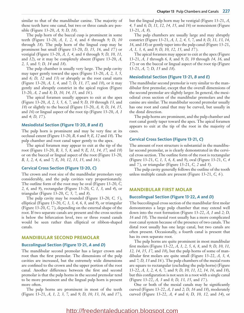
Chapter 13 Pulp Chambers and Canals 227
similar to that of the mandibular canine. The majority of these teeth have one canal, but two or three canals are pos-sible (Figure 13-20, A, 9; D, 18).
The pulp horn of the buccal cusp is prominent in some teeth (Figure 13-20, A, 1, 2, 4, and 6 through 9; D, 10 through 18). The pulp horn of the lingual cusp may be prominent but small (Figure 13-20, D, 15, 16, and 17 ) or vestigial (Figure 13-20, A, 1, 4, and 6 through 9; D, 10, 11, and 12), or it may be completely absent (Figure 13-20, A, 2, 3, and 5; D, 14 and 18).
The pulp chamber is usually very large. The pulp cavity may taper gently toward the apex (Figure 13-20, A, 2, 3, 5, and 6; D, 12 and 13) or abruptly as the root canal starts (Figure 13-20, A, 1, 4, and 7; D, 11, 17, and 18), or it may gently and abruptly constrict in the apical region (Figure 13-20, A, 2 and 8; D, 10, 14, 15, and 16 ).
The apical foramen usually appears to exit at the apex (Figure 13-20, A, 2, 3, 5, 6, 7, and 9; D, 10 through 13, and 18) or slightly to the buccal (Figure 13-20, A, 8; D, 14, 15, and 16) or lingual aspect of the root tip (Figure 13-20, A, 1 and 4; D, 17 ).
Mesiodistal Section (Figure 13-20, B and E)The pulp horn is prominent and may be very fine at its occlusal extent (Figure 13-20, B, 6 and 9; E, 12 and 18). The pulp chamber and root canal taper gently to the apex.
The apical foramen may appear to exit at the tip of the root (Figure 13-20, B, 3, 5, 8, and 9; E, 11, 14, 17, and 18) or on the buccal or lingual aspect of the root (Figure 13-20, B, 1, 2, 4, 6, and 7; E, 10, 12, 13, 15, and 16 ).
Cervical Cross Section (Figure 13-20, C)The crown and root size of the mandibular premolars vary considerably, and the pulp cavities vary proportionately. The outline form of the root may be oval (Figure 13-20, C, 2, 6, and 9), rectangular (Figure 13-20, C, 1, 3, and 4), or triangular (Figure 13-20, C, 5, 7, and 8).
The pulp cavity may be rounded (Figure 13-20, C, 5 ), elliptical (Figure 13-20, C, 1, 3, 4, 6, 8, and 9), or triangular (Figure 13-20, C, 7 ), depending on the external shape of the root. If two separate canals are present and the cross section is below the bifurcation level, two or three round canals would be seen rather than elliptical or ribbon-shaped canals.
MANDIBULAR SECOND PREMOLAR
Buccolingual Section (Figure 13-21, A and D)The mandibular second premolar has a larger crown and root than the first premolar. The dimensions of the pulp cavities are increased, but the extremely wide dimensions are confined to the crown and the upper portion of the root canal. Another difference between the first and second premolar is that the pulp horns in the second premolar tend to be more prominent and the lingual pulp horn is present more often.
The pulp horns are prominent in most of the teeth (Figure 13-21, A, 1, 2, 3, 7, and 9; D, 10, 13, 16, and 17 ),
but the lingual pulp horn may be vestigial (Figure 13-21, A, 4, 5 and 6; D, 11, 12, 14, 15, and 16) or nonexistent (Figure 13-21, A, 8).
The pulp chambers are usually large and may abruptly constrict (Figure 13-21, A, 1, 2, 4, 5, 7, and 8; D, 11, 13, 14, 16, and 18) or gently taper into the pulp canal (Figure 13-21, A, 1, 3, 6, and 9; D, 10, 12, 15, and 17 ).
The apical foramen may appear to exit at the apex (Figure 13-21, A, 1 through 6, 8, and 9; D, 10 through 14, 16, and 17) or on the buccal or lingual aspect of the root tip (Figure 13-21, A, 7; D, 15 and 18).
Mesiodistal Section (Figure 13-21, B and E)The mandibular second premolar is very similar to the man-dibular first premolar, except that the overall dimensions of the second premolar are slightly larger. In general, the mesi-odistal cross section of the mandibular premolars and the canine are similar. The mandibular second premolar usually has one root and canal that may be curved, but usually in the distal direction.
The pulp horns are prominent, and the pulp chamber and root canal gently taper toward the apex. The apical foramen appears to exit at the tip of the root in the majority of cases.
Cervical Cross Section (Figure 13-21, C)The amount of root structure is substantial in the mandibu-lar second premolar, as is clearly demonstrated in the cervi-cal cross section. The outline form of the root is rectangular (Figure 13-21, C, 1, 3, 6, 8, and 9), oval (Figure 13-21, C, 4 and 7 ), or triangular (Figure 13-21, C, 2 and 5).
The pulp cavity generally follows the outline of the tooth unless multiple canals are present (Figure 13-21, C, 6 ).
MANDIBULAR FIRST MOLAR
Buccolingual Section (Figure 13-22, A and D)The buccolingual cross section of the mandibular first molar demonstrates a large pulp chamber that may extend well down into the root formation (Figure 13-22, A, 1 and 2; D, 16 and 18). The mesial root usually has a more complicated root canal system because of the presence of two canals. The distal root usually has one large canal, but two canals are often present. Occasionally, a fourth canal is present that has its own separate root.
The pulp horns are quite prominent in most mandibular first molars (Figure 13-22, A, 1, 2, 5, 6, 8, and 9; D, 10, 11, 12, 14, 15, 17, and 18), but the pulp horns of some of man-dibular first molars are quite small (Figure 13-22, A, 3, 4, and 7; D, 13 and 16 ). The pulp chambers of the mesial roots are square to rectangular (excluding the pulp horns) (Figure 13-22, A, 1, 2, 4, 6, 7, and 9; D, 10, 11, 12, 14, 16, and 18), but this configuration is not seen in a root with a single canal (Figure 13-22, A, 3 and 8; D, 13, 15, and 17 ).
One or both of the mesial canals may be significantly curved (Figure 13-22, A, 1 and 2; D, 16 and 18), moderately curved (Figure 13-22, A, 4 and 6; D, 10, 12, and 14), or
http://freedentaleducation.blogspot.com

228 Pulp Cavities of the Mandibular Teeth
relatively straight (Figure 13-22, A, 7 and 9). The two canals may join each other in the apical region to exit in a common foramen (Figure 13-22, A, 1, 2, and 6; D, 14), or they may have separate apical foramina (Figure 13-22, A, 4, 7, and 9; D, 10, 12, 16, and 18).
The apical foramen usually appears to exit on the tip of the broad mesial root (Figure 13-22, A, 1, 2, 4, 6, 7, and 9; D, 10, 14, and 16), but in some roots one of the two canals exits on the side of the root tip (Figure 13-22, D, 12 and 18). The diameter of the mesial canals is usually very small and demonstrates a slight taper.
The distal root usually has one large pulp chamber, which is very wide in the buccolingual dimension (Figure 13-22, A, 3, 5, and 8; D, 15 and 17 ), whereas other distal roots may possess a pulp chamber that is more constricted (Figure 13-22, D, 11 and 13). The distal root usually has one large pulp canal, which may show a considerable buccolingual dimen-sion until the canal constricts abruptly a few millimeters from the apex of the root (Figure 13-22, A, 3, 5 and 8; D, 17 ). A constriction of the canal in the last few millimeters of the root is not always present (Figure 13-22, D, 15). When two canals are present, they will be partially or
completely separated by a dentinal island (Figure 13-22, D, 11). The apical foramen of a root with a single canal usually appears to be located at the apex of the root (Figure 13-22, A, 3 and 8; D, 13), but it may be slightly buccal or lingual to the apex of the root (Figure 13-22, A, 5; D, 15 and 17 ).
Mesiodistal Section (Figure 13-22, B and E)The mesiodistal section of the mandibular first molar presents few variations in the form of the pulp chamber or canals. The mesial and distal pulp cavities have canals and chambers that are centered within the roots and crowns.
The pulp horns may be prominent (Figure 13-22, B, 8; E, 11, 14, and 15), moderately evident (Figure 13-22, B, 2, 3, 6, and 7; E, 12), or barely detectable (Figure 13-22, B, 4; E, 13 and 16 ), or may demonstrate a combination of these variations (Figure 13-22, B, 1 and 9; E, 17 and 18).
The pulp chambers are usually rectangular (excluding the pulp horns) and may be large (Figure 13-22, B, 1, 4 through 7, and 9; E, 10 through 18), or very small (Figure 13-22, B, 2, 3, and 8).
Figure 13-21 Mandibular second premolar. A, Buccolingual section, exposing the mesial or distal aspect of the pulp cavity. This aspect does not appear on dental radiographs. B, Mesiodistal section, exposing the buccal or lingual aspect of the pulp cavity. C, Cervical cross section at the cementoenamel junction, exposing the pulp chamber. These are the openings to root canals that will be seen in the floor of the pulp chamber. D, Buccolingual section, exposing the mesial or distal aspect of the pulp cavity. E, Mesiodistal section, exposing the buccal or lingual aspect of the pulp cavity.
A
B
C
D
E
1
10 11 12 13 14 15 16 17 18
2 3 4 5 6 7 8 9
http://freedentaleducation.blogspot.com

Chapter 13 Pulp Chambers and Canals 229
Figure 13-22 Mandibular first molar. A, Buccolingual section, exposing the mesial or distal aspect of the pulp cavity. This aspect does not appear on dental radiographs. B, Mesiodistal section, exposing the buccal or lingual aspect of the pulp cavity. C, Five cross sections at cervical line and four cross sections at midroot. D, Buccolingual section, exposing the mesial or distal aspect of the pulp cavity. E, Mesiodistal section, exposing the buccal or lingual aspect of the pulp cavity.
A
B
C
D
E
1
10 11 12 13 14 15 16 17 18
2 3 4 5 6 7 8 9
The mesial root and canal usually show considerable cur-vature (Figure 13-22, B, 1, 3, 4, 5, 7, 8, and 9; E, 12, 13, 15, 16, and 18). Some canals demonstrate less curvature (Figure 13-22, B, 6; E, 10, 11, 14, and 17 ). Extensive secondary or reactive dentin deposition may also be seen (Figure 13-22, B, 2).
The apical foramen usually appears to exit at the tip of the root (Figure 13-22, B, 1, 5, 7, and 8; E, 10, 11, 12, 14, 15, and 17) but may appear to exit on the mesial (Figure 13-22, B, 2, 4, 6, and 9; E, 16 ) or on the distal aspect of the root (Figure 13-22, B, 3).
The distal root is usually straighter and tends to be a little shorter than the curved mesial root (Figure 13-22, B, 1, 3, 6, and 8; E, 12, 14, and 15); however, it may be the same length (Figure 13-22, B, 2, 4, and 7; E, 10, 11, 13, 16, 17, and 18) or even slightly longer (Figure 13-22, B, 5 and 9). The distal canal is usually larger than the mesial canal (Figure 13-22, B, 2, and 4 through 7; E, 11, 14, 15, 16, and 18), but the canals may look very similar in size when viewed from the buccal aspect (Figure 13-22, B, 1, 3, 8, and 9; E, 10, 12, 13, and 17 ).
The distal canal usually tapers gently to the apical con-striction. The apical foramen most often appears to be
located on the distal aspect of the root (Figure 13-22, B, 2, 3, 5, 7, and 8; E, 13, 16, and 17 ). In some teeth, this distal deviation is quite marked (Figure 13-22, B, 5 and 7; E, 13 and 17 ). Mesial deviation of the canal does occur (Figure 13-22, B, 6 ); however, it is usually only a minor deviation. The apical foramen of the distal root will often appear to be located at the tip of the root (Figure 13-22, B, 1; E, 10, 11, 12, 14, 15, and 18).
Cervical Cross Section (Figure 13-22, C, 1 through 5)The cervical cross section of the mandibular first molar is generally quadrilateral in form. Distally, it tapers a little from the wider buccolingual measurement of the mesial aspect of the tooth. The pulp chamber outline generally follows that of the root (Figure 13-22, C, 1, 2, and 5) but may show buccal (Figure 13-22, C, 3) and/or lingual (Figure 13-22, C, 4) projections of dentin if the pulp chamber is excessively narrowed by secondary or reactive osteodentin.
The pulp chamber floor has two small funnel-shaped openings into the mesial root (one buccal and one lingual), whereas the distal aspect of the pulp chamber usually shows a single opening that is less constricted.
http://freedentaleducation.blogspot.com

Midroot Cross Section (Figure 13-22, C, 6 through 9)The midroot view of the mandibular molar usually demon-strates the root canal form, which is consistent with the major form of this tooth.
The mesial root usually is somewhat kidney-shaped, with two separate canals (Figure 13-22, C, 7 and 9), but a figure-eight shape of the root is also very common (Figure 13-22, C, 6 and 8). The two canals may be totally separate (Figure 13-22, C, 6 and 8), or one may be confluent with the other canal (Figure 13-22, C, 7 and 9). Even three canals may be found in this root on occasion.
The distal root is usually rounder than the mesial root (Figure 13-22, C, 7 and 9), but a very wide root is also common (Figure 13-22, C, 6 and 8). Those roots that tend to be round usually demonstrate only one canal, whereas the broader distal roots tend to have two canals (Figure 13-22, C, 6 and 8) or a very thin canal that is single; or they may possess a dentinal island (Figure 13-22, C, 8). Even in a root with a single canal, the distal canal tends to show a devel-opmental depression or concavity on the mesial aspect of the root (Figure 13-22, C, 7 and 9).
MANDIBULAR SECOND MOLAR
Anatomically, the mandibular second molar has many simi-larities with the mandibular first molar (Figure 13-23). The proportions of the crown and root are very similar to those of the mandibular first molar. The roots of the second molar may be straighter with less divergence from the furcation than in the first molar. The roots may be shorter, but there is no assurance that any of these differences will be mani-fested in any one tooth.
Buccolingual Section (Figure 13-23, A and D)The buccolingual section of the mandibular second molar demonstrates a pulp chamber and pulp canals that tend to be more variable and complex than those found in the man-dibular first molar.
The pulp horns of the mandibular second molar are usually rather prominent (Figure 13-23, A, 1, 3, 5, 6, 8, and 9; D, 10, 13, 14, 15, 17, and 18), but some pulp horns may be small to nonexistent (Figure 13-23, A, 2, 4, and 7; D, 11, 12, and 16 ). The pulp chamber of the mesial root (Figure 13-23, A, 1, 3, 5, 6, and 8; D, 12, 13, 14, 16, 17, and 18)
230 Pulp Cavities of the Mandibular Teeth
Figure 13-23 Mandibular second molar. A, Buccolingual section, exposing the mesial or distal aspect of the pulp cavity. This aspect does not appear on dental radiographs. B, Mesiodistal section, exposing the buccal or lingual aspect of the pulp cavity. C, Five cross sections at cervical line and four cross sections at midroot. D, Buccolingual section, exposing the mesial or distal aspect of the pulp cavity. E, Mesiodistal section, exposing the buccal aspect of the pulp cavity.
A
B
C
D
E
1
10 11 12 13 14 15 16 17 18
2 3 4 5 6 7 8 9
http://freedentaleducation.blogspot.com

Chapter 13 Pulp Chambers and Canals 231
is well demarcated because of the presence of two canals. The pulp chamber (excluding the pulp horns) may be somewhat square (Figure 13-23, A, 1, 3, and 8; D, 10, 12, 14, 16, 17, and 18) or rectangular (Figure 13-23, A, 5 and 6; D, 13).
Two root canals are usually present in the mesial root, but only one may be present. The mesial canals may be large (Figure 13-23, A, 1; D, 17 and 18), medium (Figure 13-23, A, 3, 5, 6, and 8; D, 13), or small (Figure 13-23, D, 12, 14, and 16 ). The curvature of these canals may be severe (Figure 13-23, A, 1; D, 13), moderate (Figure 13-23, A, 5 and 8; D, 12 and 16 ), virtually absent (Figure 13-23, D, 14 and 18), or a combination of the aforementioned variations (Figure 13-23, A, 3 and 6; D, 17 ). Most of the canals appear to exit from the mesial root separately (Figure 13-23, A, 3, 5, 6, and 8; D, 12, 13, 14, 16, and 18), but some join just before reaching the apex so that a common canal exits from the apex (Figure 13-23, A, 1; D, 17 ).
The apical foramen usually appears to be located at the tip of the root (Figure 13-23, A, 1, 3, and 6; D, 12, 17, and 18), but some appear to exit slightly to the buccal or lingual aspect of the apex of the root (Figure 13-23, A, 5 and 8; D, 14 and 16 ).
The pulp chamber of the distal root of the mandibular second molar is not as easily identified because of the extremely large pulp canal that is usually present (Figure 13-23, A, 2, 4, 7, and 9; D, 11 and 15). One canal is usually present in the distal root, but two totally or partially separate canals are possible (Figure 13-23, D, 10).
Pulp horns may be present, but they are not nearly as prominent as in the mesial root (Figure 13-23, A, 2, 4, 7, and 9; D, 11 and 15) unless two canals are present (Figure 13-23, D, 10).
The pulp canal is usually very large in the mesiobuccal sections. The pulp canal may taper gently from the pulp chamber until the apical constriction (Figure 13-23, A, 2 and 7; D, 10 and 15), or an abrupt constriction of the canal may occur in the last 2 to 3 mm of the canal (Figure 13-23, A, 4 and 9; D, 11). The apical foramen often appears to be located at the tip of the root (Figure 13-23, A, 1, 4, 7, and 9; D, 10, 11, and 15).
Mesiodistal Section (Figure 13-23, B and E)The mesiodistal sections of the mandibular second molar are very similar to those of the mandibular first molar. However, the roots of the mandibular first molar tend to be straighter and closer together (less furcation deviation).
The pulp horns are usually prominent (Figure 13-23, B, 1, 2, 3, 5, 7, 8, and 9; E, 10, 11, 13, 15, and 18), but some are small or absent (Figure 13-23, B, 4 and 6; E, 12, 14, 16, and 17 ).
The pulp chamber is rectangular (excluding the pulp horns). The size of the chamber varies from very large (Figure 13-23, B, 1, 3, 4, 5, 7, and 9; E, 13 and 16 ) to very small (Figure 13-23, B, 2 and 8; E, 11, 14, 17, and 18).
The curvature of the mesial canal may be severe (Figure 13-23, B, 3, 6, 8, and 9; E, 11, 14, 16, and 17), moderate (Figure 13-23, B, 2, 4, and 7; E, 10, 13, 15, and 18), or essentially
straight (Figure 13-23, B, 1 and 5; E, 12). The canals gently taper from the pulp chamber to the apical constriction.
The apical foramen usually appears to be located at the tip of the root (Figure 13-23, B, 2, 4 through 7, and 8; E, 11, 12, 13, 15, 17, and 18), but the foramen may appear to be located mesially (Figure 13-23, B, 3; E, 10, 13, and 16 ) or distally (Figure 13-23, B, 9; E, 14) on the root tip.
The distal canal may be slightly curved (Figure 13-23, B, 1 through 5, and 7; E, 11, 14, and 16 ) or straight (Figure 13-23, B, 4, 6, 8, and 9; E, 10, 12, 13, 17, and 18). The distal root may be slightly shorter than (Figure 13-23, B, 1, 2, 4, and 7), equal to (Figure 13-23, B, 3, 5, and 6; E, 10, 13, 14, 16, and 17), or longer than (Figure 13-23, B, 8 and 9; E, 11, 12, 15, and 18) the mesial root.
The distal canal is usually larger than the mesial canals (Figure 13-23, B, 3, 4, and 6 through 9; E, 11, 13, 15, and 16 ) but may be equal to the mesial canals (Figure 13-23, B, 1, 2, and 5; E, 12, 14, and 18). The distal canal tapers gently to the apex.
The apical foramen usually appears to be located at the tip of the root (Figure 13-23, B, 1 through 7; E, 10 through 13, 16, and 18), but the foramen may appear to exit mesially (Figure 13-23, B, 9) or distally (Figure 13-23, B, 8; E, 14, 15, and 17) to the apex of the root.
Cervical Cross Section (Figure 13-23, C, 1 through 5)The cervical cross section of the mandibular second molar is similar to that of the mandibular first molar (Figure 13-23, C, 1 through 5). The outline form of the mandibular second molar is more triangular (rather than quadrilateral like that of the mandibular first molar) because of the smaller dimen-sions that are usually seen in the distal aspect of this tooth. The pulp chamber also tends to be triangular. The floor of the pulp chamber may have two openings, one mesially and one distally, which are centered within the dentin. If only one canal is present in the distal root, it will be centered within the dentin.
Midroot Cross Section (Figure 13-23, C, 6 through 9)Midroot cross sections of the mandibular molars demon-strate that the mesial root is very broad buccolingually and narrow mesiodistally (Figure 13-23, C, 6 through 9). The outline form is kidney-shaped (Figure 13-23, C, 6 and 7) or slightly in the form of a figure eight (Figure 13-23, C, 8 and 9).
The canals may be totally separate (Figure 13-23, C, 9) or confluent (Figure 13-23, C, 6, 7 and 8), which makes it difficult to determine the presence of two mesial canals (Figure 13-23, C, 8). The distal root may be rounder than the mesial root, because the outline form of this root is usually oval (Figure 13-23, C, 6 and 9), but broad distal roots are also seen (Figure 13-23, C, 7 and 8). One canal is usually present in the distal root, but two canals are often present.
MANDIBULAR THIRD MOLAR
The mandibular third molar pulp cavities vary greatly (Figure 13-24). The pulp cavity resembles the second man-
http://freedentaleducation.blogspot.com

dibular molar most, but the crown looks too large for the roots, which may be shorter and curved and tend to be fused together.
Buccolingual Section (Figure 13-24, A and D)In the buccolingual section, the pulp cavities of the man-dibular third molar show a great deal of variation. Two roots and three canals are often present (Figure 13-24, B, 6 and 7 ), but two canals or two roots are also possible (Figure 13-24, C, 8 and 9). The presence of one canal and one root can also be found, but usually these teeth are not of much value for restorative purposes, because they have short roots that taper quickly.
Most mandibular third molars have prominent pulp horns (Figure 13-24, A, 1, 2, and 4 through 9; D, 10, 11, 12, 14, 15, 17, and 18), although others demonstrate small to nonexistent pulp horns (Figure 13-24, A, 3; D, 13 and 16 ).
The mesial roots of most mandibular third molars (Figure 13-24, A, 2 and 9; D, 10, 11, 13, and 14) demonstrate a square pulp chamber (excluding the pulp horns). The mesial root usually has two canals (Figure 13-24, A, 2, 5, and 9; D,
10, 11, and 14), but a single mesial root can be found (Figure 13-24, D, 14). The canals may be very curved (Figure 13-24, A, 5 and 9; D, 10) or relatively straight (Figure 13-24, A, 2; D, 11 and 13) as they taper gently toward the apical constriction.
The apical foramen usually appears to be located on the tip of the root (Figure 13-24, A, 1 and 5; D, 10, 11, and 13), but it may be located buccally (Figure 13-24, A, 9; D, 14) or lingually to the tip of the root. If two canals are present, they usually possess separate apical foramina (Figure 13-24, A, 2 and 5; D, 10, 11, and 13), but some canals join in the apical region, exiting through a common foramen (Figure 13-24, A, 9).
The distal root of mandibular third molars possesses a very large pulp chamber and canal that are difficult to delin-eate into separate areas (Figure 13-24, A, 1, 3, 4, 6, 7, and 8; D, 12, and 15 through 18).
The pulp canals, which are very large (Figure 13-24, A, 1, 4, 6, and 7; D, 12, 15, and 18), may taper gently to the root tip (Figure 13-24, A, 4, 6, and 7; D, 16 and 18) or may demonstrate an abrupt constriction of the canal in
232 Pulp Cavities of the Mandibular Teeth
Figure 13-24 Mandibular third molar. A, Buccolingual section, exposing the mesial or distal aspect of the pulp cavity. This aspect does not appear on dental radiographs. B, Mesiodistal section, exposing the buccal or lingual aspect of the pulp cavity. C, Five cross sections at cervical line and four cross sections at midroot. D, Buccolingual section, exposing the mesial or distal aspect of the pulp cavity. E, Mesiodistal section, exposing the buccal or lingual aspect of the pulp cavity.
A
B
C
D
E
1
10 11 12 13 14 15 16 17 18
2 3 4 5 6 7 8 9
http://freedentaleducation.blogspot.com

Chapter 13 Pulp Chambers and Canals 233
the last few millimeters (Figure 13-24, A, 1; D, 12, 15, and 17).
The pulp chambers are square or rectangular (excluding the pulp horns). The pulp chambers, which are very small (Figure 13-24, A, 3 and 8), tend to show a constriction at the junction of the pulp chamber and canal, after which they taper gently to the apical constriction.
The apical foramen usually appears to be located at the tip of the root (Figure 13-24, A, 3, 6, 7, and 8; D, 16 and 17), but it may be located buccally or lingually to the root tip (Figure 13-24, A, 1 and 4; D, 15 and 18).
Mesiodistal Section (Figure 13-24, B and E)The pulp horns of the mandibular third molar may be prominent (Figure 13-24, B, 1 and 5; E, 10, 11, 12, 14, and 15), small (Figure 13-24, A, 4, 6, and 7; D, 17), or nearly absent (Figure 13-24, B, 2, 3, 8, and 9; E, 13, 16, and 18).
The pulp chambers are usually square or rectangular (excluding pulp horns) when viewed from the buccal aspect (Figure 13-24, B, 1 through 4, 7, and 9; E, 10, 12, 13, 15, 17, and 18), but they may be somewhat square (Figure 13-24, B, 5; E, 14).
The degree of curvature of the mesial root may be slight (Figure 13-24, E, 13, single-rooted), moderate (Figure 13-24, B, 1, 2, 5, 8, and 9; E, 11, 15, and 17), or severe (Figure 13-24, B, 3, 4, 6, and 7; E, 10, 12, 14, 16, and 18).
The canal within the mesial root may be large (Figure 13-24, B, 2 and 4; E, 10) or very small (Figure 13-24, B, 1, 3, 5, 8, and 9; E, 11, 12, and 14 through 18). The canals usually taper gently to the apical constriction. The apical foramen may appear to be located at the apex of the root (Figure 13-24, B, 1, 2, 3, 5, 6, 8, and 9; E, 11, 12, 14, 17, 18) or mesially (Figure 13-24, B, 7) or distally to the apex of the root (Figure 13-24, B, 4; E, 10 and 15).
The length of the mesial roots may be equal to (Figure 13-24, B, 2, 3, and 8; E, 10 and 11), shorter than (Figure 13-24, B, 5 and 9; E, 18), or longer than (Figure 13-24, B, 1, 4, 6, and 7; E, 12, 14, 15, and 17) the length of the distal root.
The distal canal may be larger than the mesial canal (Figure 13-24, B, 2 through 5, and 9; E, 14 and 18), but many are equal in size (Figure 13-24, B, 1, 6, 7, and 8; E, 10, 11, 12, 15, and 17). The distal canal gently tapers to the apical constriction (Figure 13-24, B, 1 through 8; E, 10 through 18).
The apical foramen may appear to be located at the apex of the root (Figure 13-24, B, 1 through 5, 7, and 9; E, 10, 11, 12, 15, 17, and 18), or it may be mesial (Figure 13-24, B, 8) or distal (Figure 13-24, B, 6; E, 14) to the apex of the root.
Some teeth show only one root with one or two canals. If only one canal is present (Figure 13-24, E, 13), the canal will be very large. If the third molar is multirooted, the canals will be much smaller (Figure 13-24, E, 16 ).
Cervical Cross Section (Figure 13-24, C, 1 through 5)The cervical cross section demonstrates a variable outline form that may be rectangular (Figure 13-24, C, 1 through 4) or triangular (Figure 13-24, C, 5).
Midroot Cross Section (Figure 13-24, C, 6 through 9)The mesial root, when present, is oval to figure eight in shape (Figure 13-24, C, 6 and 7 ). The distal root is oval (Figure 13-24, C, 6) or kidney-shaped (Figure 13-24, C, 7 ). If the roots are fused (Figure 13-24, C, 8) or only one root is present (Figure 13-24, C, 9), the canals are usually larger. The canals in the roots that are kidney-shaped or are in the form of a figure eight are more ellipsoidal.
Radiographs: Pulp Chamber and Canals
Visualization of the pulp chamber and pulp canal(s) by standard radiography or digital radiography provides the clinician with evidence to augment what has been found clinically. All teeth should be examined periodically radiographically and clinically. Knowing what might be expected anatomically about the pulp chamber and pulp canal(s), which has been considered in a previous section, helps when radiographs are taken. The radiographs in Figure 13-25, A through I illustrate normal pulp chambers and pulp canals. They are what are seen generally in the dental office, not the highly selected radiographs of the various teeth.
Crown and Root Fractures
Fractures of teeth may involve the crown, the crown and root, or the root. Probably the most common are fractures of the crown.3 In some cracked teeth, only the enamel is involved; in others, both the enamel and dentin may be affected; but initially or later, the pulp also may be involved with or without the loss of tooth structure (e.g., cusp). In severe trauma the whole crown may be lost. The clinician should be familiar with the most likely places, morphologi-cally, for cracking or fractures to occur. The most likely places involve developmental grooves (Figure 13-26), often in relation to restorations.
Fractures of cusps of the maxillary first premolar and mandibular first molars often occur along developmen-tal grooves or stress lines as shown in Figure 13-26. Such fractures may occur in connection with bruxism and clenching. It is not uncommon for the distolingual cusp of the mandibular first molar to fracture in association with a large restoration, which leads to pulpitis (Figure 13-27).
Fractures of the root cause symptoms that resemble those of other dental problems; therefore the diagnosis of root fracture may be difficult,3 especially when the fracture does not appear on the radiograph (Figure 13-28).
Fractures may be an extension from the crown and may extend to the pulp or involve only the root adjacent to the periodontal ligament. Horizontal root fractures are most likely the result of external physical trauma or clenching and bruxism. Vertical root fractures may be caused by bruxing
http://freedentaleducation.blogspot.com

Figure 13-25 A, Large pulp chambers and pulp canals in a young adult. B, Mandibular incisors. Normal pulp canals. C, Lateral incisor and canine. Normal chambers and canals. Root resorption on lateral incisor. D, Maxillary canine and premolars with normal chambers and canals. Root resorption on first premolar associated with orthodontics.
A B
C
A B
D
234 Crown and Root Fractures
and clenching or may be the result of restorations with endodontic posts in root canals.
RELATION OF POSTERIOR ROOT APICES TO THE MANDIBULAR CANAL
Of interest to the clinician doing endodontics, periapical surgery, or placement of implants is the position of the mandibular nerve (canal) and the mental foramen.
The mandibular nerve traversing the mandible in the mandibular canal occupies different positions relative to the apices of the molar and premolar teeth (Figure 13-29, A through D). In Figure 13-30, the distance from the outer to the inner surface of the buccal plate (1), from the inner surface of the cortical plate to the apex of a tooth (2), and from the canal to the apex of the tooth (3) reflect the data obtained from sections of the mandible.4
The mean width of the buccal plate (Figure 13-30, B, 1) over the premolar is 1.9 ± 0.49 mm; over the first molar, 2.38 ± 0.57 mm; over the second molar, 5.6 ± 0.93 mm; and over the third molar, 2.34 ± 1.0 mm.
The mean distance from the inner buccal plate to the apex of the tooth (Figure 13-30, B, 2) for the second premo-
lar is 3.78 ± 1.04 mm; for the first molar, 4.1 ± 0.98 mm; for the second molar, 7.1 ± 1.4 mm; and for the third molar, 4.03 ± 1.8 mm.
The mean vertical distance from the canal to the apex (Figure 13-30, B, 3) for the second premolar is 3.07 ± 0.43 mm; for the first molar, 4.03 ± 0.31 mm; for the second molar, 2.5 ± 0.25 mm; and for the third molar, 1.96 ± 0.27 mm.
With regard to the buccolingual alignment of the man-dibular canal to root apices, the canal is in vertical alignment with the second premolars for 65% of the time, slightly lingual to the apices of the first molar apices for 71% of the time, in line with the apices of second molars for 73% of the time, and in line with the third molars for 56% of the time.
Data are based on the vertical axis of each tooth with the recognition that more than one apex is present. Thus, the position of the mandibular canal relative to the apices of the teeth tends to follow the dotted line in Figure 13-29.
The position of the mental foramen relative to the man-dibular premolars and first molar can be described as follows: the foramen is between the first and second premolars 33% of the time, in line with the second premolar 11% of the
http://freedentaleducation.blogspot.com

Chapter 13 Pulp Chambers and Canals 235
E
F G
H I
E, Mandibular lateral incisor, canine, and first premolar with normal chambers and canals. F, Mandibular second premolar and first and second molars with normal chambers and canals. Note curvature of the mesial root of the first molar. G, Mandibular molars and premolars with normal chambers and pulp canals. H, Impacted third molar adjacent to second mandibular molar. The pulp chambers and pulp canals are normal. I, First and second primary molars being resorbed in association with the erupting permanent premolars. Mandibular second molar in the process of erupting.
Figure 13-25 cont’d
Figure 13-26 Fracture lines most commonly seen in first maxillary premolar and first mandibular molar.
http://freedentaleducation.blogspot.com

236 Crown and Root Fractures
Figure 13-28 A, Radiograph showing loss of lamina dura on the mesial root of the mandibular molar but no indication of root fracture. B, Longitudinal fracture on the distal aspect of the crown and coronal root.
(A from Ash MM: Schienentherapie, ed 2, Munich, 1999, Urban & Fischer. B from Cohen S, Hargreaves KM: Pathways of the pulp, ed 9, St Louis, 2006, Mosby.)
A
B
Figure 13-29 Diagrammatic representation of sections through the mandible at the position of the second premolar and first, second, and third molars. Dotted line represents the faciolingual position of the mandibular canal.
D
C
B
A
A
B
1 2 3
Figure 13-30 A, Diagrammatic representation of a cross section of the mandible. B, Enlarged view with three measurements taken as indicated in the text.
Figure 13-27 A, Radiograph showing periapical radiolucency associated with parulis shown in B. B, Fracture of distolingual cusp related to undermined tooth structure and amalgam restoration.
(From Ash MM, Ramjford S: Occlusion, ed 4, Philadelphia, 1995, Saunders.)
A B
http://freedentaleducation.blogspot.com

Chapter 13 Pulp Chambers and Canals 237
Gardner DG et al: Taurodontism, shovel-shaped incisors and the Klinefelter syndrome, Dent J 44:372, 1978.
Harris WE: Unusual root canal anatomy in the maxillary molar, J Endod 6:573, 1980.
Hess W, Zurcher E: The anatomy of root canals, London, 1925, John Bale Sons and Danielsson.
Ibrahim SM et al: Pulp cavities of permanent teeth, Egypt Dent J 23:83, 1977.
Kerekes K et al: Morphometric observations on the root canals of human molars, J Endod 3:114, 1977.
Kirkham DB: The location and incidence of accessory pulp canals in periodontal pockets, J Am Dent Assoc 91:353, 1975.
Mageean JF: The significance of root canal morphology in endodontics, J Br Endod Soc 6:67, 1972.
Middletoti-Shaw JC: The teeth, the bony palate and the mandible in Bantu races of South Africa, London, 1931, John Bale Sons and Danielsson.
Okumura T: Anatomy of the root canals, J Am Dent Assoc 14:632, 1927.
Senyurek MS: Pulp cavities of molars in primates, Am J Phys Anthropol 25:119, 1939.
Stone LH et al: Maxillary molars demonstrating more than one palatal root canal, Oral Surg 51:649, 1981.
Sutalo J et al: Morphologic characteristics of root canals in upper and lower premolars, Acta Stomatol Croat 14:23, 1980.
Tidmarsh BG: Micromorphology of pulp chambers in human molar teeth, Int Endod J 13:69, 1980.
Vertucci FJ, Williams RG: Furcation canals in the human mandibular first molar, Oral Surg 38:308, 1974.
Vertucci FJ et al: Root canal morphology of the human maxillary first premolar, Oral Surg 99:194, 1979.
Vertucci FJ et al: Root canal morphology of the human maxillary second premolar, Oral Surg 88:456, 1974.
Warren EM: The relationship between crown size and the incidence of bifid root canals in mandibular incisor teeth, Oral Surg 52:425, 1981.
time, and distal to the second premolar 56% of the time. In terms of vertical position, the foramen can be found coronal to the apices 22% of the time, at the apices 15% of the time, and below the apices 63% of the time.4
References1. Kronfeld R: Dental histology and comparative dental anatomy,
Philadelphia, 1937, Lea & Febiger.2. Riethmuller RH: The filling of root canals with Prinz’
paraffin compound, Dent Cosmos 56:490, 1914.3. Mullally BH, Ahmed M: Periodontal signs and symptoms
associated with vertical root fracture, Dent Update 27:356, 2000.
4. Ash JL, Ash CM: Mandibular canal and mental foramen: relation to posterior root apices, J Dent Res 1997 (abstract).
BibliographyAcosta Vigouroux AS et al: Anatomy of the pulp chamber floor
of the permanent maxillary first molar, J Endod 4:214, 1978.Barker BC et al: Anatomy of root canals: I. Permanent incisors,
canines and premolars, Aust Dent J 18:320, 1973.Barker BC et al: Anatomy of root canals. II. Permanent maxillary
molars, Aust Dent J 19:46, 1974.Barker BC et al: Anatomy of root canals. III. Permanent
mandibular molars, Aust Dent J 19:408, 1974.Burah JG et al: A study of the presence of accessory foramina
and the topography of molar furcations, Oral Surg 38:451, 1974.
Carlsen O, Andersen J: On the anatomy of the pulp chamber and root canals in human deciduous teeth, Tandlægebladet 70:93, 1966.
Carlsen O, Andersen J: Radix mesiolingualis and radix distolingualis in a collection of permanent maxillary molars, Acta Odont Scand 58:229, 2000.
Carlsen O, Andersen J: Radix paramolaris and distomolaris in Danish permanent maxillary molars, Acta Odont Scand 57:283, 1999.
Carns EJ, Skidmore AE: Configurations and deviations of root canals of maxillary first premolars, Oral Surg 36:880, 1973.
Use the CD-ROM to view animations and study ques-tions; and go to the site for additional study resources.
http://freedentaleducation.blogspot.com

This page intentionally left blank
http://freedentaleducation.blogspot.com
Dento-osseous Structures, Blood Vessels, and Nerves
also called the antrum of Highmore. From the dental view-point, in addition to its general shape and the processes mentioned, several landmarks on this bone are among the most important, including the incisive fossa, canine fossa, canine eminence, infraorbital foramen, posterior alveolar foramina, maxillary tuberosity, pterygopalatine fossa, and incisive canal.
The body of the maxilla has the following four surfaces: anterior or facial, infratemporal, orbital, and nasal.
ANTERIOR SURFACE
The anterior or facial surface (Figures 14-2 and 14-3) is separated above from the orbital aspect by the infraorbital ridge. Medially it is limited by the margin of the nasal notch, and posteriorly, it is separated from the posterior surface by the anterior border of the zygomatic process, which has a confluent ridge directly over the roots of the first molar. The ridge corresponding to the root of the canine tooth is usually the most pronounced and is called the canine eminence.
Anterior to the canine eminence, overlying the roots of the incisor teeth, is a shallow concavity known as the incisive fossa. Posterior to the canine eminence on a higher level is a deeper concavity called the canine fossa. The floor of this canine fossa is formed in part by the projecting zygomatic process. Above this fossa and below the infraorbital ridge is the infraorbital foramen, the external opening of the infraor-bital canal. The major portion of the canine fossa is directly above the roots of the premolars.
The development of the dentitions has been discussed and a brief review of the development of the neurocranium and splanchnocranium has been presented in Chapter 2. There-fore, in this chapter the focus is on the dentoalveolar and dento-osseous structures of the permanent dentition. The forms of the roots of the teeth and their sizes and angula-tions govern the shape of the alveoli in the jawbones, and this in turn shapes the contour of the dento-osseous por-tions facially.
The osseous structures that support the teeth are the maxilla and the mandible. The maxilla, or upper jaw, con-sists of two bones: a right maxilla and a left maxilla sutured together at the median line. Both maxillae in turn are joined to other bones of the head (Figure 14-1). The mandible, or lower jaw, has no osseous union with the skull and is a movable (ginglymoarthrodial) joint.
The Maxillae
The maxillae make up a large part of the bony framework of the facial portion of the skull. They form the major portion of the roof of the mouth, or hard palate, and assist in the formation of the floor of the orbit and the sides and base of the nasal cavity. They support the 16 permanent maxillary teeth.
Each maxilla is an irregular bone, somewhat cuboidal in shape, which consists of a body and four processes: the zygomatic, frontal, palatine, and alveolar processes. The maxilla is hollow and contains the maxillary sinus air space,
239
1414
http://freedentaleducation.blogspot.com

240 The Maxillae
POSTERIOR SURFACE
The posterior or infratemporal surface (Figures 14-3 and 14-4) is bounded above by the posterior edge of the orbital surface. Inferiorly and anteriorly, it is separated from the anterior surface by the zygomatic process and the zygomatic ridge, which runs from the inferior border of the zygomatic process to the alveolus of the maxillary first molar. This surface is more or less convex and is pierced in a downward direction by two or more posterior alveolar foramina. These two canals are on a level with the lower border of the zygo-matic process and are somewhat distal to the roots of the third molar.
The inferior portion of this surface is more prominent where it overhangs the root of the third molar and is called the maxillary tuberosity. Medially, this tuberosity is limited by a sharp, irregular margin that articulates with the pyrami-dal process of the palatine bone and, in some cases, the lateral pterygoid plate of the sphenoid bone. The maxillary tuberosity is the origin for some fibers of the medial ptery-goid muscle.
A portion of the infratemporal surface superior to the maxillary tuberosity is the anterior boundary to the ptery-gomaxillary fissure.
ORBITAL SURFACE
The orbital surface is smooth and together with the orbital surface of the zygomatic bone forms the floor of the orbit. The junction of this surface and the anterior surface forms the infraorbital margin or ridge, which runs superiorly to form part of the nasal process. Its posterior border or edge coincides with the inferior boundary of the inferior orbital fissure.
The thin medial edge of the orbital surface is notched anteriorly, forming the lacrimal groove. Behind this groove, it articulates for a short distance with the lacrimal bone, then for a greater length with a thin portion of the ethmoid bone, and terminates posteriorly in a surface that articulates with the orbital process of the palatal bone. Its lateral area is continuous with the base of the zygomatic process (see Figure 14-3).
Traversing the posterior portion of the orbital surface is the infraorbital groove. This groove begins at the center of the posterior surface and runs anteriorly. The anterior portion of this groove is covered, becoming the infraorbital canal, the anterior opening of which is located directly below the infraorbital ridge on the anterior surface.
If the covered portion of this canal were to be laid open, the orifices of the middle and anterior superior alveolar
Figure 14-1 Representation of an adult skull with permanent dentition. The maxilla consists of a body and four processes (malar, nasal, alveolar, and palatine) that articulate by synarthrosis with cranial and other facial bones, (e.g., frontal, nasal, ethmoid, malar bones). The mandible articulates with the temporal bone by the temporomandibular joint (see Figure 15-6).
Figure 14-2 Frontal view of left maxilla.
Nasal process
Infra-orbitalridgeInfra-orbitalforamen
Alveolarridge
Nasal spine
Incisive fossa Canineeminence
Interdentalsepta
Alveoli
Figure 14-3 Lateral view of left maxilla.
Infra-orbitalmargin
Infra-orbitalforamen
Nasal spine
Canine fossa
Frontal process
Orbital surface
Infra-orbitalgroove
Zygomatic(malar) process
Posterior alveolarforamina
Zygomatic ridge
Alveolar ridge
Maxillary tuberosity
Infratemporal surface
Canineeminence
http://freedentaleducation.blogspot.com

Chapter 14 Dento-osseous Structures, Blood Vessels, and Nerves 241
canal would be seen transmitting the corresponding vessels and nerves to the premolars, canines, and incisor teeth.
NASAL SURFACE
The nasal surface (Figures 14-5 and 14-6) is directed medially toward the nasal cavity. It is bordered below by the superior surface of the palatine process. Anteriorly, it is limited by the sharp edge of the nasal notch. Above and anteriorly, it is continuous with the medial surface of the frontal process. Behind this, it is deeply channeled by the lacrimal groove, which is converted into a canal by articulation with the lacrimal and inferior turbinate bones.
Behind this groove the upper edge of the nasal surface corresponds to the medial margin of the orbital surface, and the maxilla articulates in this region with the lacrimal bone, a thin portion of the ethmoid bone, and the orbital process of the palatine bone.
The posterior border of the maxilla, which articulates with the palatine bone, is traversed obliquely from above downward and slightly medially by a groove, which, by
articulation with the palate bone, is converted into the greater palatine canal. Toward the posterior and upper part of this nasal surface, a large, irregular opening into the maxillary sinus (antrum of Highmore) may be seen. In an articulated skull, this opening is partially covered by the uncinate process of the ethmoid bone and the inferior nasal concha.
Anterior to the lacrimal groove, the nasal surface is ridged for the attachment of the inferior nasal concha. Below this the bone forms a lateral wall of the inferior nasal meatus. Above the ridge for a small distance on the medial side of the nasal process, the smooth lateral wall of the middle meatus appears.
ZYGOMATIC PROCESS
The zygomatic process may be seen in the lateral views of the maxillary bone as a roughly triangular eminence whose apex is placed inferiorly directly over the first molar roots. The lateral border is rough and spongelike in appearance, where it has been disarticulated from the zygomatic or cheek bone (see Figures 14-1 and 14-3).
Figure 14-4 Posterior view of left maxilla.
Frontal process
Orbital surface
Infra-orbital canal
Zygomatic process
Posterior alveolarforamina
Nasal foramen ofincisive canal
Palatine process
Floor of nose
Part of greaterpalatine
Roughened areafor attachment ofpalatine bone
Figure 14-5 Medial view of left maxilla.
Portion ofsphenoidal sinus
Frontal process
Middle meatusLacrimal groove
Conchal crest
Inferior meatus
Palatal process atintermaxillarysuture
Incisive groove
Maxillary sinus
Greater palatinegroove
Hard palate ofpalatal process
Figure 14-6 Medial view of right maxilla. This specimen has not been disarticulated completely and has the maxillary teeth in situ.
Maxillary sinus
Sphenoidal sinus
Inferior nasalconcha
Medial lamina ofpterygoid process
Hard palate
Inferior meatus
Incisive canals
http://freedentaleducation.blogspot.com

242 The Maxillae
FRONTAL PROCESS
The frontal process (see Figures 14-2 through 14-5) arises from the upper and anterior body of the maxilla.
Part of this process is formed by the upward continuation of the infraorbital margin medially. Its edge articulates with the nasal bone. Superiorly, the process articulates with the frontal bone. The medial surface of the frontal process forms part of the lateral wall of the nasal cavity. Anteriorly, the frontal process articulates with the nasal bone.
PALATINE PROCESS
The palatine process (Figures 14-2 through 14-8) is a hori-zontal ledge extending medially from the nasal surface of the maxilla. Its superior surface forms a major portion of the nasal floor. The inferior surfaces of the combined left and right palatine processes form the hard palate as far posteri-orly as the second molar, where they articulate with the horizontal parts of the palatine bone (Figures 14-7 and 14-8) at the transverse palatine suture.
The inferior surface of the palatine process is rough and pitted for the palatine mucous glands in the roof of the
mouth and is pierced by numerous small foramina for the passage of blood vessels and nerve fibers. At the posterior border of the process is a groove or canal that passes the greater palatine nerve and vessels to the palatal soft tissues. The posterior edge of the palatine process becomes rela-tively thin where it joins the palatine bone at the point of the greater palatine foramen. The palatine process becomes progressively thicker anteriorly from the posterior border. Anteriorly, the palatal process is confluent with the alveolar process surrounding the roots of the anterior teeth.
Immediately posterior to the central incisor alveolus, when looking at the medial aspect of the maxilla, one sees a smooth groove that is half of the incisive canal, when the two maxillae are joined together. The incisive fossa into which the canals open may be seen immediately lingual to the central incisors at the median line, or intermaxillary suture where the maxillae are joined. Two canals open later-ally into the incisive foramen, the foramina of Stenson, carrying the nasopalatine nerves and vessels. Occasionally, two midline foramina are present, the foramina of Scarpi.
Extending laterally from the incisive foramen to the space between the lateral incisor and canine alveoli are the rem-nants of the suture between the maxilla and premaxilla. In
Figure 14-7 Palatine view of maxilla. Note the dental foramina in the deepest portion of the canine alveolus.
Zygomatic process
Tuberosity
Posterior border of palatalprocess (point of suturewith horizontal portionof palatal bone)
Hard palate ofpalatal process
Point of intermaxillarysuture
Alveolar process
Figure 14-8 View of inferior surface of the maxilla showing alveolar process and alveoli.
Alveolus ofmaxillary central
incisor Interdental septumbetween first andsecond premolars
Infra-orbital foramen
Zygomatic process
Zygomatic ridge
Interradicular septabetween alveoli ofthird molar
Palatine process
Greater palatineforamen
http://freedentaleducation.blogspot.com

Chapter 14 Dento-osseous Structures, Blood Vessels, and Nerves 243
most mammals the premaxilla remains an independent bone.
ALVEOLAR PROCESS
The alveolar process makes up the inferior portion of the maxilla; it is that portion of the bone which surrounds the roots of the maxillary teeth and which gives them their osseous support. The process extends from the base of the tuberosity posterior to the last molar to the median line anteriorly, where it articulates with the same process of the opposite maxilla (see Figures 14-7 and 14-8). It merges with the palatine process medially and with the zygomatic process laterally (see Figure 14-8).
When one looks directly at the inferior aspect of the maxilla toward the alveoli with the teeth removed, it is apparent that the alveolar process is curved to conform with the dental arch. It completes, with its fellow of the opposite side, the alveolar arch supporting the roots of the teeth of the maxilla.
The process has a facial (labial and buccal) surface and a lingual surface with ridges corresponding to the surfaces of the roots of the teeth supported by it. It is made up of labi-obuccal and lingual plates of very dense but thin cortical bone separated by interdental septa of cancellous bone.
The facial plate is thin, and the positions of the alveoli are well marked on it by visible ridges as far posteriorly as the distobuccal root of the first molar (see Figure 14-2). The margins of these alveoli are frail, and their edges are sharp and thin. The buccal plate over the second and third molars, including the alveolar margins, is thicker. Generally, the lingual plate of the alveolar process is heavier than the facial plate. In addition, the alveolar process is longer where it surrounds the anterior teeth, sometimes extending posteri-orly to include the premolars. In short, it extends farther down in covering the lingual portion of the roots.
The bone is very thick lingually over the deeper portions of the alveoli of the anterior teeth and premolars. The merging of the alveolar process with the palatal process brings about this formation. The lingual plate is paper thin over the lingual alveolus of the first molar, however, and rather thin over the lingual alveoli of the second and third molars. This thin lingual plate over the molar roots is part of the formation of the greater palatine canal (see Figure 14-8).
The alveolar process is maintained by the presence of the teeth. Should any tooth be lost, that portion of the alveolar process that supported the missing tooth will be subject to atrophic reduction. Should all of the teeth be lost, the alveo-lar process will eventually be virtually lost.
ALVEOLI (TOOTH SOCKETS)
The alveolar cavities are formed by the facial and lingual plates of the alveolar process and by connecting septa of bone placed between the two plates. The form and depth of each alveolus are determined by the form and length of the root it supports (see Table 1-1).
The alveolus nearest the median line is that of the central incisor (Figure 14-9; see also Figure 14-8). The periphery is regular and round, and the interior of the alveolus is evenly tapered and triangular in cross section, with the apex toward the lingual.
The second alveolus in line is that of the lateral incisor. It is generally conical and egg-shaped, or ovoid, with the widest portion to the labial. It is narrower mesiodistally than labiolingually and is smaller on cross section, although it is often deeper than the central alveolus. Sometimes, it is curved at the upper extremity (Figure 14-10; see also Figure 14-8).
The canine alveolus is the third from the median line. It is much larger and deeper than those just described. The periphery is oval and regular in outline, with the labial width greater than the lingual. The socket extends distally. It is flattened mesially and somewhat concave distally. The bone is so frail at the canine eminence on the facial surface of the alveolus that the root of the canine is often exposed on the labial surface near the middle third (see Figure 14-2).
The first premolar alveolus (see Figures 14-8 and 14-10) is kidney-shaped in cross section, with the cavity partially divided by a spine of bone that fits into the mesial develop-mental groove of the root of this tooth. This spine divides
Figure 14-9 Alveoli of the central incisor, lateral incisor, and canine.
Figure 14-10 Alveoli of the premolar area.
http://freedentaleducation.blogspot.com

244 The Maxillae
the cavity into a buccal and a lingual portion. If the tooth root is bifurcated for part of its length, as is often the case, the terminal portion of the cavity is separated into buccal and lingual alveoli. The socket is flattened distally and much wider buccolingually than mesiodistally (see Table 1-1).
The second premolar alveolus is also kidney-shaped, but the curvatures are in reverse to those of the first premolar alveolus. The proportions and depth are almost the same. The septal spine is located on the distal side instead of the medial side, because the second premolar root is inclined to have a well-defined developmental groove distally. This tooth usually has one broad root with a blunt end, but it is occasionally bifurcated at the apical third.
The first molar alveolus (Figures 14-8 and 14-11) is made up of three distinct alveoli widely separated. The lingual alveolus is the largest; it is round, regular, and deep. The cavity extends in the direction of the hard palate, having a lingual plate over it that is very thin. The lingual periphery of this alveolus is extremely sharp and frail. This condition may contribute to the tissue recession often seen at this site.
The mesiobuccal and distobuccal alveoli of the first molar have no outstanding characteristics except that the buccal plates are thin. The bone is somewhat thicker at the periph-eries than that found on the lingual alveolus. Nevertheless, it is thinner farther up on the buccal plate. It is not uncom-mon for one to find the roots uncovered by bone in spots when examining dry specimens.
The forms of the buccal alveoli resemble the forms of the roots they support. The mesiobuccal alveolus is broad buc-colingually, with the mesial and distal walls flattened. The distobuccal alveolus is rounder and more conical.
The septa that separate the three alveoli (interradicular septa) are broad at the area that corresponds to the root bifurcation, and they become progressively thicker as the peripheries of the alveoli are approached. The bone septa are very cancellous, which denotes a rich blood supply, as is true of all the septa, including those separating the various teeth as well.
A general description of the alveoli of the second molar would coincide with that of the first molar; these alveoli are closer together, since the roots of this tooth do not spread as much. As a consequence, the septa separating the alveoli are not as heavy.
The third molar alveolus is similar to that of the second molar, except that it is somewhat smaller in all dimensions. Figure 14-11 shows a third molar socket to accommodate a tooth with three well-defined roots, a rare occurrence. Usually, the two buccal (and often all three) roots will be fused. The interradicular septum changes accordingly. If the roots of the tooth are fused, a septal spine will appear in the alveolus at the points of fusion on the roots marked by deep developmental grooves.
MAXILLARY SINUS
The maxillary sinus lies within the body of the bone and is of corresponding pyramidal form; the base is directed toward the nasal cavity. Its summit extends laterally into the root of the zygomatic process. It is closed in laterally and above by the thin walls that form the anterolateral, posterolateral, and orbital surfaces of the body. The sinus overlies the alveolar process in which the molar teeth are implanted, more particularly, the first and second molars, the alveoli of which are separated from the sinus by a thin layer of bone. Occasionally, the maxillary sinus will extend forward far enough to overlie the premolars also. It is not uncommon to find the bone covering the alveoli of some of the posterior teeth extending above the floor of the cavity of the maxillary sinus, forming small hillocks.
Regardless of the irregularity and the extension of the alveoli into the maxillary sinus, a layer of bone always separates the roots of the teeth and the floor of the sinus in the absence of pathological conditions. A layer of sinus mucosa is also always between the root tips and the sinus cavity.
MAXILLARY ARTICULATION
The maxilla articulates with the nasal, frontal, lacrimal, and ethmoid bones, above and laterally with the zygomatic bone, and occasionally with the sphenoid bone. Posteriorly and medially, it articulates with the palatal bone. Medially, it supports the inferior turbinate and the vomer and articulates with the opposite maxilla.
Figure 14-11 Alveoli of the molar area. Note the thinness of the buccal plates over the first molar roots compared with those of the second and third maxillary molars. The third molar alveoli are rarely separated as distinctly as in this specimen. Figures 14-9, 14-10, and 14-11 demonstrate a number of significant points concerning the maxillary alveoli. In Figure 14-9 the facial cortical plate of bone is thin over the anterior teeth and is considerably thicker over the posterior teeth, especially the molars. Cancellous bone seems to exist buccal to some of the posterior roots. In Figure 14-10 interradicular septa are thick but with numerous nutrient canals. In Figure 14-11 cancellous bone, furnishing numerous opportunities for blood supply, is evident in the apical portions of the alveoli. The anterior alveoli are lined laterally with a layer of smooth cortical bone. This lining is less prominent in the posterior alveoli.
http://freedentaleducation.blogspot.com
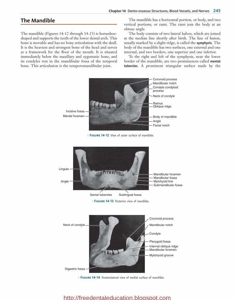
Chapter 14 Dento-osseous Structures, Blood Vessels, and Nerves 245
The Mandible
The mandible (Figures 14-12 through 14-23) is horseshoe-shaped and supports the teeth of the lower dental arch. This bone is movable and has no bony articulation with the skull. It is the heaviest and strongest bone of the head and serves as a framework for the floor of the mouth. It is situated immediately below the maxillary and zygomatic bone, and its condyles rest in the mandibular fossa of the temporal bone. This articulation is the temporomandibular joint.
The mandible has a horizontal portion, or body, and two vertical portions, or rami. The rami join the body at an obtuse angle.
The body consists of two lateral halves, which are joined at the median line shortly after birth. The line of fusion, usually marked by a slight ridge, is called the symphysis. The body of the mandible has two surfaces, one external and one internal, and two borders, one superior and one inferior.
To the right and left of the symphysis, near the lower border of the mandible, are two prominences called mental tubercles. A prominent triangular surface made by the
Figure 14-12 View of outer surface of mandible.
Incisive fossa
Mental foramen
Coronoid processMandibular notchCondyle condyloidprocess
Neck of condyle
RamusOblique ridge
Body of mandibleAngleFacial notch
Figure 14-13 Posterior view of mandible.
Lingula
Angle
Mandibular foramenMandibular fossaMylohyoid lineSubmandibular fossa
Sublingual fossaGenial tubercles
Figure 14-14 Posterolateral view of medial surface of mandible.
Neck of condyle
Digastric fossa
Coronoid process
Mandibular notch
Condyle
Pterygoid fossa
Internal oblique ridgeMandibular foramen
Mylohyoid groove
http://freedentaleducation.blogspot.com
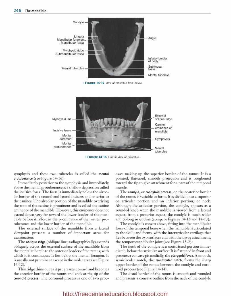
symphysis and these two tubercles is called the mental protuberance (see Figure 14-16).
Immediately posterior to the symphysis and immediately above the mental protuberance is a shallow depression called the incisive fossa. The fossa is immediately below the alveo-lar border of the central and lateral incisors and anterior to the canines. The alveolar portion of the mandible overlying the root of the canine is prominent and is called the canine eminence of the mandible. However, this eminence does not extend down very far toward the lower border of the man-dible before it is lost in the prominence of the mental pro-tuberance and the lower border of the mandible.
The external surface of the mandible from a lateral viewpoint presents a number of important areas for examination.
The oblique ridge (oblique line, radiographically) extends obliquely across the external surface of the mandible from the mental tubercle to the anterior border of the ramus, with which it is continuous. It lies below the mental foramen. It is usually not prominent except in the molar area (see Figure 14-12).
This ridge thins out as it progresses upward and becomes the anterior border of the ramus and ends at the tip of the coronoid process. The coronoid process is one of two proc-
esses making up the superior border of the ramus. It is a pointed, flattened, smooth projection and is roughened toward the tip to give attachment for a part of the temporal muscle.
The condyle, or condyloid process, on the posterior border of the ramus is variable in form. It is divided into a superior or articular portion and an inferior portion, or neck. Although the articular portion, the condyle, appears as a rounded knob when the mandible is viewed from a lateral aspect, from a posterior aspect, the condyle is much wider and oblong in outline (compare Figures 14-12 and 14-13).
The condyle is convex above, fitting into the mandibular fossa of the temporal bone when the mandible is articulated to the skull, and forms, with the interarticular cartilage that lies between the two surfaces and with the tissue attachment, the temporomandibular joint (see Figure 15-2).
The neck of the condyle is a constricted portion imme-diately below the articular surface. It is flattened in front and presents a concave pit medially, the pterygoid fovea. A smooth, semicircular notch, the mandibular notch, forms the sharp upper border of the ramus between the condyle and coro-noid process (see Figure 14-14).
The distal border of the ramus is smooth and rounded and presents a concave outline from the neck of the condyle
Figure 14-15 View of mandible from below.
Condyle
LingulaMandibular foramen
Mandibular fossa
Mylohyoid ridgeSubmandibular fossa
Genial tubercles
Angle
Inferior borderof body
Sublingualfossa
Mental tubercle
Figure 14-16 Frontal view of mandible.
Mylohyoid line
Incisive fossa
Mentalforamen
Mentalprotuberance
Externaloblique ridge
Canineeminence ofmandible
Symphysis
Mentaltubercles
246 The Mandible
http://freedentaleducation.blogspot.com

Chapter 14 Dento-osseous Structures, Blood Vessels, and Nerves 247
to the angle of the jaw, where the posterior border of the ramus and the inferior border of the body of the mandible join. The border of this angle is rough, being the attachment of the masseter muscle (see Figure 15-18) and the styloman-dibular ligament (see Figure 15-7).
An important landmark on the lateral aspect of the man-dible is the mental foramen. It should be noted that this opening of the anterior end of the mandibular canal is directed upward, backward, and laterally. The foramen is usually located midway between the superior and inferior border of the body of the mandible when the teeth are in position, and most often, it is below the second premolar tooth, a little below the apex of the root. The position of this foramen is not constant, and it may be between the first premolar and the second premolar tooth. After the teeth are lost and resorption of alveolar bone has taken place, the mental foramen may appear near the crest of the alveolar border. In childhood, before the first permanent molar has come into position, this foramen is usually immediately below the first primary molar and nearer the lower border.
It is interesting to note that when the mandible is observed from a point directly opposite the first molar, most of the distal half of the third molar is hidden by the anterior border of the ramus. When the mandible is viewed from in front, directly opposite the median line, the second and third molars are located 5 to 7 mm lingually to the anterior border of the ramus (compare Figures 14-12 and 14-16).
INTERNAL SURFACE OF THE MANDIBLE
Observation of the mandible from the rear shows that the median line is marked by a slight vertical depression, repre-senting the line of union of the right and left halves of the mandible, and that immediately below this, at the lower third, the bone is roughened by eminences called the supe-rior and inferior mental spines, or genial tubercles (see Figures 14-15 and 14-34, C).
The internal surface of the body of the mandible is divided into two portions by a well-defined ridge, the mylo-hyoid line. It occupies a position closely corresponding to the lateral oblique ridge on the surface. It starts at or near the lowest part of the mental spines and passes backward and upward, increasing in prominence until the anterior portion of the ramus is reached; there, it smoothes out and gradually disappears (see Figures 14-14 and 14-34, C).
This ridge is the point of origin of the mylohyoid muscle, which forms the central portion of the floor of the mouth. Immediately posterior to the median line and above the anterior part of the mylohyoid ridge a smooth depression, the sublingual fossa, may be seen. The sublingual gland lies in this area.
A small, roughened oval depression, the digastric fossa, is found on each side of the symphysis immediately below the mylohyoid line and extending onto the lower border. Toward the center of the body of the mandible, between the mylohyoid line and the lower border of the bone, a smooth oblong depression is located, called the submandibu-lar fossa. It continues back on the medial surface of the
ramus to the attachment of the lateral pterygoid muscle. The submandibular gland lies within this fossa.
The mandibular foramen is located on the medial surface of the ramus midway between the mandibular notch and the angle of the jaw and also midway between the internal oblique line and the posterior border of the ramus. The mandibular canal begins at this point, passing downward and forward horizontally.
The anterior margin of the foramen is formed by the lingula, or mandibular spine, which gives attachment to the sphenomandibular ligament. Coming obliquely downward from the base of the foramen beneath the lingula is a decided groove, the mylohyoid groove. Behind this groove toward the angle of the mandible, a roughened surface for the attach-ment of the medial pterygoid muscle may be seen.
ALVEOLAR PROCESS
The border of the alveolar process outlines the alveoli of the teeth and is very thin at its anterior portion around the roots of the incisor teeth but thicker posteriorly where it encom-passes the roots of the molars. The alveolar process, which comprises the superior border of the body of the mandible, differs from the same process in the maxillae in one very important particular: it is not as cancellous, and instead of the facial plate’s being relatively thin, it is equally as heavy as the lingual plate. Although the bone over the anterior teeth, including the canine, is very thin and may be entirely missing over the cervical portion of the root, the bone that does cover the root is the compact type.
The inferior border of the mandible is strong and rounded and gives to the bone the greatest portion of its strength (see Figure 14-15).
When one looks down on the mandible from a point above the alveoli of the first molars (see Figure 14-18), it can be noticed that although the alveolar border may be thinner anteriorly than posteriorly, the body of the bone is uniform throughout. The lines of direction of the posterior alveoli are inclined lingually to conform to the lingual incli-nation of the teeth when they are in position. The anterior teeth, of course, have their alveoli tipped labially; therefore when one looks down on the mandible from above the alveolar process, more of the bone may be seen lingual to the anterior teeth than lingual to the posterior teeth. In contrast, posteriorly, more of the bone may be seen buccal to the teeth than lingual. Therefore the outline of the arch of the teeth does not correspond to the outline of the arch of the bone. The dental arch is narrower posteriorly than the mandibular arch.
The lingual walls of the alveoli of the second and third molars are relatively thin near the bottoms of the sockets, although the bone near the periphery is somewhat thicker and very compact. If a specimen of the mandible from which the third molar has been removed is held up to the light, the bone at the bottom of the socket is so thin that light will penetrate it. This thinness of bone is consistent with the submandibular fossa below the mylohyoid ridge (see Figure 14-15; Figure 14-22, A and B; and Figure 14-24).
http://freedentaleducation.blogspot.com

The bone buccal to the last two molars is very heavy and thick, being reinforced by the external oblique ridge. Pos-terior to the third molar a triangular shallow fossa is out-lined; it is called the retromolar triangle (Figure 14-17). The cortical plate over this fossa is not as heavy as the bone sur-rounding it, and it is more cancellous under the thin cortical plate covering it.
ALVEOLI
The first alveolus right or left of the median line is that of the first, or central, incisor. The periphery of the alveolus often dips down lingually and labially and exposes the root for part of its length. This arrangement makes an interden-tal spine out of the interdental septum separating the alveo-lus of the mandibular central incisors. The central incisor alveolus is flattened on its mesial surface and is usually some-what concave distally to accommodate the developmental groove on the root (see Figures 14-18 and 14-19).
The alveolus of the mandibular second, or lateral, incisor is similar to that of the central incisor. It usually has the
following variations: the socket is larger and deeper to accommodate a larger and longer root; the periphery does not dip down as far on the lingual surface but may dip more on the labial aspect of the tooth, exposing more of the root of the lateral incisor. The interdental septum extends up just as high between the teeth as that between the central incisors.
The canine alveolus is quite large and oval and, of course, deep to accommodate the root of the mandibular canine. The lingual plate is stronger and much heavier than that over the alveoli just described, although the thin labial plate may thin out at its edges and expose just as much of the canine root on the labial side. The labial outline of the alveolus is wider than the lingual outline, and the mesial and distal walls of the sockets are irregular to accommodate developmental grooves, both mesially and distally, on the canine root (see Figures 14-18 and 14-20).
The alveoli of the first and second premolars are similar in outline. The outline is smooth and rounded, although the dimensions are greater buccolingually than mesiodistally. The alveolus of the second premolar is usually somewhat
Figure 14-17 View of mandible from above.
Retromolar triangle
Coronoid process
Neck of condyle
Condyle
Lingula
Figure 14-18 Alveolar process of the mandible showing alveoli.
Retromolartriangle
Alveolus of thirdmolar
Interradicularseptum betweenmesial anddistal roots offirst molar
Interdental septumbetween the rootsof the secondpremolarand first molar
Alveolus ofcentral incisor
Alveolusof canine
248 The Mandible
http://freedentaleducation.blogspot.com

Chapter 14 Dento-osseous Structures, Blood Vessels, and Nerves 249
Figure 14-19 Closeup view of one of the three separate divisions of the mandibular alveoli (as shown in Figures 14-19, 14-20, and 14-21). This picture indicates the relative sizes and shapes of the incisors for comparison with other mandibular teeth.
Figure 14-20 This view of the mandibular alveoli includes the canine, the first and second premolars, and a clear view of the mandibular first molar alveoli. Note the excellent design for the anchorage of first molar roots. Apparently, the blood supply for the interseptal bone lessens as anterior teeth are approached. The apical portion of the canine alveolus displays the single opening in the bone for the blood and nerve supply to the tooth pulp.
larger than that of the first premolar. The buccal plate of the alveoli is relatively thin, but the lingual plate is heavy; the interdental septum has become heavier at this point compared with the interdental septa found between the anterior teeth. The interdental septum between the canine and first premolar is relatively thin, although uniform in outline. The septum between the first premolar and second premolar is nearly twice as thick.
Progressing posteriorly (see Figures 14-18 and 14-20), the interdental septum between the second premolar socket and the alveolus of the mesial root of the first molar is twice as thick as that found between the first and second premo-lars. The socket of the first molar is divided by an interroot septum, which is strong and regular. The alveolus of the mesial root is kidney-shaped, much wider buccolingually than mesiodistally, and constricted in the center to accom-modate developmental grooves found mesial and distal to the mesial root of the first molar. The alveolus of the distal root of the first molar is evenly oval with no constriction,
conforming to the rounded shape of this root. The inter-dental septum between the alveoli of the mandibular first molar and the socket of the second molar is thick mesiodis-tally, although cancellous in character.
The mandibular second molar alveolus may be divided into two alveoli, as was the case in the first molar. However, often it is found to be one compartment near the periphery of the alveolus but divides into two compartments in the deeper portions. A septal spine occurs where the develop-mental grooves on the root are deep enough, or an inter-radicular septum will appear when the roots are entirely divided. The interdental septum between the second molar sockets and the third molar sockets is not as thick mesiodis-tally as the two interdental septa immediately anterior.
The mandibular third molar alveolus is usually irregular in outline (see Figure 14-21). Usually it is much narrower toward the distal than toward the mesial aspect of the alveo-lus. It may have interradicular septa or septal spines to accommodate itself to the irregularity of the root.
http://freedentaleducation.blogspot.com

Mesiodistal views of the teeth and their supporting struc-tures can be made clinically using computed tomography to demonstrate the way that individual teeth relate to each other or to cortical and cancellous bone; however, vertical sections of a skull were made to illustrate these relation-ships1 shown in Figure 14-24. (See also Figures 13-29 and 13-30.)
Some classical illustrations of sections of the axial rela-tions of the maxillary and mandibular teeth are shown in Figures 14-25 through 14-32. These figures demonstrate the directional lines of the axes of the teeth and their alveoli (see also Box 16-1 and Figure 16-20). In addition, the radio-graphs of the sections graphically illustrate the relative den-sities of the teeth and supporting structures and show the outline and relative thickness of the bone over the various teeth at the site of each section.2
In Figures 14-25 through 14-32, note the axial relations of the superior and inferior teeth, the relative thickness of labial and lingual alveolar plates, the characteristics of the cancellous tissue, the relative densities, and the relation of the teeth to important structures. Compare the changes in the external contour and internal architecture of the adja-cent sections. The sections in this series, with the exception of those in Figure 14-28, were taken from the same cadaver
Figure 14-21 Alveoli of the first, second, and third molars. Features for special attention include the thin and perforated surface of the retromolar triangular space distal to the third molar alveolus and the cancellous formation in the alveoli proper and also in the interdental septa, which would allow a rich blood supply.
Figure 14-22 Illustration of the relative thickness of bone covering lingual mandibular second and third molar roots. A, Measurement of the thickness of bony cover lingual to the apex of the third mandibular molar immediately below the mylohyoid ridge. It measures only 0.5 mm. B, Repetition of measurement in the deepest portion lingually of the second molar alveolus. It measures fully 2 mm.
A
B
Figure 14-23 Comparison of the size and shape of mandibles at various ages. Top, Mandible of a 5-year-old. Notice the rounded bowlike form. Notice also the amount of space between the second deciduous molar and the ramus. Middle, Mandible of a 9-year-old. Notice the angular outline with constriction at the point of second permanent molar development. Bottom, Well-developed mandible of an individual approximately 50 years of age. The bone is regular in outline. The lingual constriction has lessened and has retreated to the third molar area.
250 The Mandible
http://freedentaleducation.blogspot.com

Chapter 14 Dento-osseous Structures, Blood Vessels, and Nerves 251
Figure 14-24 Some fine vertical sections of a skull made with radiographic problems in view. These radiographs of faciolingual sections show the extent of tooth attachment, the way in which individual teeth compare with each other, and the variances between cortical and cancellous bone in anchorage (see Figures 14-25 through 14-32). The maxillary canine was impacted in this specimen. Maxillary third molars were missing.
(From Updegrave WJ: Normal radiodontic anatomy, Dent Radiogr Photogr 31:57, 1958.)
Figure 14-25 Central incisor regions, showing relation of superior central incisor to inferior lateral incisor.
(From MacMillan HW: The structure and function of the alveolar process, J Am Dent Assoc 11:1059, 1924.)
Figure 14-26 Lateral incisor regions. Note position of apex of superior lateral incisor.
(From MacMillan HW: The structure and function of the alveolar process, J Am Dent Assoc 11:1059, 1924.)
http://freedentaleducation.blogspot.com

Figure 14-27 Canine regions. Note anterior extremity of maxillary antrum.
(From MacMillan HW: The structure and function of the alveolar process, J Am Dent Assoc 11:1059, 1924.)
Figure 14-28 First premolar regions.
(From MacMillan HW: The structure and function of the alveolar process, J Am Dent Assoc 11:1059, 1924.)
Figure 14-29 Second premolar regions.
(From MacMillan HW: The structure and function of the alveolar process, J Am Dent Assoc 11:1059, 1924.)
Figure 14-30 First molar regions, showing relations of distobuccal root (A), distal half (B), and mesiobuccal (C) and lingual roots with mesial half of lower molar (D).
(From MacMillan HW: The structure and function of the alveolar process, J Am Dent Assoc 11:1059, 1924.)
A
B
C
D
and are from the left side. A plaster cast was made before sectioning. The sections were reassembled in the cast and held in exact relation while being radiographed.
Figures 14-33, I and II illustrate the appearance of some of the bony landmarks of the maxilla and mandible as visualized in intraoral periapical radiography.3 Surfaces and dento-osseous structures are often important to recognize in relation to diagnostic and treatment aspects (Figure 14-34).
Arterial Supply to the Teeth
The arteries and nerve branches to the teeth are mere terminals of the central systems. This book must confine itself to dental anatomy and the parts imme-diately associated, and references will therefore be made only to those terminals that supply the teeth and the sup-porting structures.
252 Arterial Supply to the Teeth
http://freedentaleducation.blogspot.com
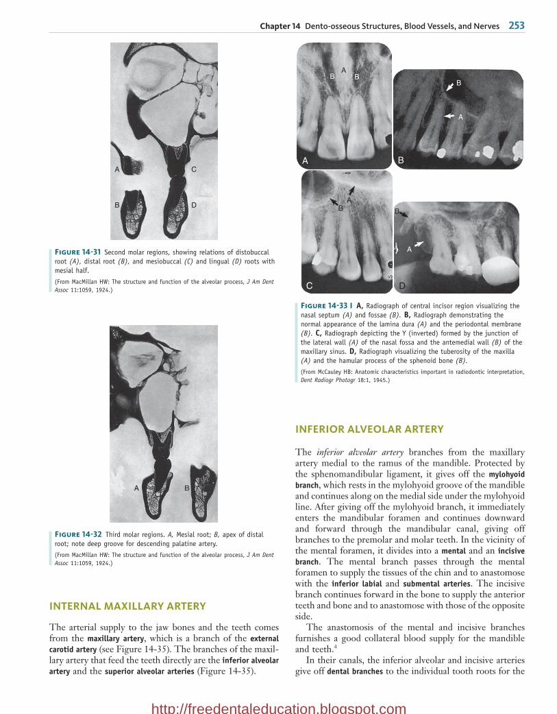
Chapter 14 Dento-osseous Structures, Blood Vessels, and Nerves 253
Figure 14-31 Second molar regions, showing relations of distobuccal root (A), distal root (B), and mesiobuccal (C) and lingual (D) roots with mesial half.
(From MacMillan HW: The structure and function of the alveolar process, J Am Dent Assoc 11:1059, 1924.)
B
A
D
C
Figure 14-32 Third molar regions. A, Mesial root; B, apex of distal root; note deep groove for descending palatine artery.
(From MacMillan HW: The structure and function of the alveolar process, J Am Dent Assoc 11:1059, 1924.)
A B
Figure 14-33 I A, Radiograph of central incisor region visualizing the nasal septum (A) and fossae (B). B, Radiograph demonstrating the normal appearance of the lamina dura (A) and the periodontal membrane (B). C, Radiograph depicting the Y (inverted) formed by the junction of the lateral wall (A) of the nasal fossa and the antemedial wall (B) of the maxillary sinus. D, Radiograph visualizing the tuberosity of the maxilla (A) and the hamular process of the sphenoid bone (B).
(From McCauley HB: Anatomic characteristics important in radiodontic interpretation, Dent Radiogr Photogr 18:1, 1945.)
BBB
B B
A
A
A
A
A B
DC
INTERNAL MAXILLARY ARTERY
The arterial supply to the jaw bones and the teeth comes from the maxillary artery, which is a branch of the external carotid artery (see Figure 14-35). The branches of the maxil-lary artery that feed the teeth directly are the inferior alveolar artery and the superior alveolar arteries (Figure 14-35).
INFERIOR ALVEOLAR ARTERY
The inferior alveolar artery branches from the maxillary artery medial to the ramus of the mandible. Protected by the sphenomandibular ligament, it gives off the mylohyoid branch, which rests in the mylohyoid groove of the mandible and continues along on the medial side under the mylohyoid line. After giving off the mylohyoid branch, it immediately enters the mandibular foramen and continues downward and forward through the mandibular canal, giving off branches to the premolar and molar teeth. In the vicinity of the mental foramen, it divides into a mental and an incisive branch. The mental branch passes through the mental foramen to supply the tissues of the chin and to anastomose with the inferior labial and submental arteries. The incisive branch continues forward in the bone to supply the anterior teeth and bone and to anastomose with those of the opposite side.
The anastomosis of the mental and incisive branches furnishes a good collateral blood supply for the mandible and teeth.4
In their canals, the inferior alveolar and incisive arteries give off dental branches to the individual tooth roots for the
http://freedentaleducation.blogspot.com

Figure 14-33 II A, Radiograph of the medial palatine suture, the appearance of which might be interpreted as a fracture. Radiographs visualize various extensions of the maxillary sinus. B, Alveolar extension. C, Tuberosity extension. D, Radiograph in which the canal for a superior alveolar artery is seen. E, Radiograph showing typical superimposition of the coronoid process: A, of the mandible on the tuberosity; B, of the maxilla.
(From McCauley HB: Anatomic characteristics important in radiodontic interpretation, Dent Radiogr Photogr 18:1, 1945.)
A
A
B
A
B
D
C
E
Figure 14-34 Surface landmarks. Various dental structures in the patient’s face can be quickly located by means of surface landmarks. Surface landmarks are identified in A. The photograph of the bony skull (B) was made from the same angle of view. Features of both are numbered and identified in the legend. The medial aspect of the mandible (C) shows anatomical details not clearly seen in the other illustrations. The maxilla and zygoma are shown in D. The bony anatomy of the hard palate and its adjoining structures is shown in E.1, Frontal bone (forehead)2, Glabella3, Supraorbital ridge (superciliary ridge)4, Frontonasal suture (bridge of nose)5, Maxillofrontal suture6, Maxillonasal suture7, Nasal bone8, Medial canthus9, Lateral canthus10, Infraorbital ridge11, Frontal process of maxilla12, Zygomatic process of maxilla13, Zygomaticomaxillary suture14, Zygomatic bone (cheekbone)15, Zygomatic arch16, Infraorbital foramen17, Canine fossa18, Canine eminence19, Incisive fossa20, Nasal ala21, Nares22, Philtrum23, Anterior nasal spine24, Inferior nasal concha
25, Alveolar process26, Temporomandibular articulation27, Tragus of ear28, Auricula29, Labial commissure30, Vermilion border of lip31, Coronoid process of mandible32, Ramus of mandible33, Body of mandible34, Gonial angle of mandible35, Infratemporal fossa36, Condyle37, External acoustic meatus38, Mental protuberance39, Mastoid process of temporal bone40, Maxillary tuberosity41, Posterior nasal spine42, Articular eminence43, Styloid process of temporal bone44, Mandibular fossa45, Vomer46, Greater palatine foramen47, Lesser palatine foramen48, Palatine bone
49, Palatine process of maxilla50, Midpalatal suture51, Incisive foramen52, Lateral pterygoid plate of sphenoid bone53, Inferior orbital fissure54, Mental foramen55, Oblique line56, Mandibular foramen57, Mandibular notch58, Internal oblique ridge59, Submandibular fossa60, Sublingual fossa61, Genial tubercle62, Inferior border of mandible63, Symphysis64, Mylohyoid line65, Hamular process of sphenoid bone66, Zygomaticotemporal suture67, Greater wing of sphenoid bone68, Lacrimal bone69, Maxillolacrimal suture70, Lacrimal fossa
254 Arterial Supply to the Teeth
http://freedentaleducation.blogspot.com

Chapter 14 Dento-osseous Structures, Blood Vessels, and Nerves 255
62
3429
3022
2120
42
1
3
8 9
1014
3627
28
1
2 34
576
11
16 3510 1424
2318 19 18
2517
52
12 1315
263137
57
3932
55
3325
6254
19 18
38
6334
B
A
E
D
C
36
31
57
32
5658
643459
25
60
6261
63
6769
68
70
67
1066
15
52
4065
25
1217 24
1918
51
5049
12
48 4640 41
53
35 24 47 1545 52
44
42
43
http://freedentaleducation.blogspot.com

Figure 14-35 Projection of maxillary artery and its branches in relation to brain, skull, and mandible, including the teeth.
Bregma
Superior frontal gyrus
Anterior branchCentral sulcus
Lateral cerebral fissure
Posterior branch
Superficialtemporal artery
Inion
Middle temporal gyrus
Nasion
Infraorbitala
Posterior superiorisalveolarartery
Middle meningealartery
Internal maxillaryartery
Inferior alveolarartery
External carotidartery
Internal carotidartery
Common carotidartery
Lambda
Superior-parietallobule
supply of the pulp and periodontal membrane at the root apex (see Figure 5-12). Other branches enter the interdental septa, supply bone and adjacent periodontal membrane, and terminate in the gingiva. Numerous small anastomoses connect these vessels with those supplying the neighboring alveolar mucosa.
SUPERIOR ALVEOLAR ARTERIES
The posterior superior alveolar artery branches from the max-illary artery superior to the maxillary tuberosity to enter the alveolar canals along with the posterior superior alveolar nerves and supplies the maxillary teeth, alveolar bone, and membrane of the sinus. A branch of variable size runs forward on the periosteum at the junction of the alveolar process and maxillary body supplying the gingiva, alveolar mucosa, and cheek. When it is large, it may supplant in part the buccal artery.
A middle superior alveolar branch is usually given off by the infraorbital continuation of the maxillary artery somewhere along the infraorbital groove or canal. It runs downward between the sinus mucosa and bone or in canals in the bone and joins the posterior and anterior alveolar vessels. Its main distribution is to the maxillary premolar teeth.
Anterior superior alveolar branches arise from the infraor-bital artery just before this vessel leaves its foramen. They course down the anterior aspect of the maxilla in bony canals to supply the maxillary anterior teeth and their supporting tissues and to join the middle and posterior superior alveolar branches in completing an anastomotic plexus.
Branches to the teeth, periodontal ligament, and bone are derived from the superior alveolar artery in the same manner as described for the inferior alveolar artery.
DESCENDING PALATINE AND SPHENOPALATINE ARTERIES
The palatal blood supply comes from two sources but chiefly from the descending palatine artery, which descends from its origin from the maxillary through the greater canal. Its greater palatine branch enters the palate through the greater palatine foramen and runs forward with its accompanying vein and nerve in a groove at the junction of the palatine and alveolar processes. It is distributed to the bone, glands, and mucosa of the hard palate and to the bone and mucosa of the alveolar process, in which it forms anastomoses with fine branches of the superior alveolaris. Minor branches of the descending palatine artery pass to the soft palate through lesser palatine foramina in the palatine bone.
The nasopalatine branch of the sphenopalatine artery courses obliquely forward and downward on the septum and enters the palate through the incisive canal. It has a limited distri-bution to the incisive papilla and adjacent palate and forms an anastomosis with the greater palatine.
Nerve Supply to the Jaws and Teeth
The sensory nerve supply to the jaws and teeth is derived from the maxillary and mandibular branches of the fifth
256 Nerve Supply to the Jaws and Teeth
http://freedentaleducation.blogspot.com

Chapter 14 Dento-osseous Structures, Blood Vessels, and Nerves 257
Figure 14-36 Distribution of the trigeminal nerve. Inf., Inferior; Sup., superior.
Lacrimalbranch
Frontalbranch
Nasociliarybranch
Mucousmembraneof maxillary
sinus
Lingualbranch
Mental branch
Inferior alveolarbranches
Mandibulardivision
Infraorbital branch
Ophthalmic division
Maxillary division
Trigeminalnerve
Semilunarganglion
Superior alveolar
branches
Figure 14-37 Mandibular nerve. The inferior alveolar nerve branch (in the cut section of the mandible and the mandibular canal) provides innervation for the mandibular teeth. The injection of a local anesthetic into the vicinity of the lingula (see Figure 14-17) blocks the sensory innervation of the inferior alveolar nerve and the lingual nerve (“mandibular block”), which results in a feeling of numbness of the mandibular teeth, lower lip, and side of the tongue on the side of the injection.
cranial, or trigeminal nerve (Figure 14-36), whose ganglion, the trigeminal, is located at the apex of the petrous portion of the temporal bone. The innervation of the orofacial region includes, in addition to the trigeminal nerve (includ-ing V2 and V3), other cranial nerves, (e.g., VII, XI, XII) (Figure 14-37).
MAXILLARY NERVE
The maxillary nerve (see Figure 14-36) courses forward through the wall of the cavernous sinus and leaves the skull through the foramen rotundum.5 It crosses the pterygopala-tine fossa, where it gives branches to the pterygopalatine ganglion, a parasympathetic ganglion. This ganglion gives off several branches, now containing visceral motor and sensory fibers, to the mucous membrane of the mouth, nose, and pharynx.
The branches of clinical significance include a greater palatine branch that enters the hard palate through the greater palatine foramen and is distributed to the hard palate and palatal gingiva as far forward as the canine tooth; a lesser palatine branch from the ganglion that enters the soft palate through the lesser palatine foramina; and a nasopalatine branch of the posterior or superior lateral nasal branch of the ganglion that runs downward and forward on the nasal septum. Entering the palate through the incisive canal, it is distributed to the incisive papilla and to the palate anterior to the anterior palatine nerve.
The maxillary nerve also has a posterior superior alveolar branch from its pterygopalatine portion. This nerve enters the alveolar canals on the infratemporal surface of the
maxilla and, forming a plexus, is distributed to the molar teeth and the supporting tissues.
The maxillary nerve enters the orbit and, as the infraor-bital nerve, runs forward in its floor, first in the infraorbital groove and then in the infraorbital canal. It terminates at
http://freedentaleducation.blogspot.com

the infraorbital foramen in branches distributed to the upper face. At a variable distance after it enters the orbit, a middle superior alveolar branch arises from the infraorbital nerve and runs through the lateral wall of the maxillary sinus. It is distributed to the premolar teeth and surrounding tissues and joins the alveolar plexus. The middle superior alveolar nerve may be associated closely with the posterior superior alveolar nerve as its origin but often branches near the infraorbital foramen.
An anterior superior alveolar branch leaves the infraor-bital nerve just inside the infraorbital foramen and is distrib-uted through bony canals to the incisor and canine teeth. All three superior alveolar nerves join in a plexus above the process. From the plexus, dental branches are given off to each tooth root and interdental branches to the bone, perio-dontal membrane, and gingiva, the distribution being similar to that described for the arteries.
MANDIBULAR NERVE
The mandibular nerve (see Figure 14-37) leaves the skull through the foramen ovale and almost immediately breaks up into its several branches. The chief branch to the lower jaw is the inferior alveolar nerve, which at first runs directly downward across the medial surface of the lateral pterygoid, at the lower border of which it is directed laterally and downward across the outer surface of the medial pterygoid muscle to reach the mandibular foramen. Just before enter-ing the foramen, it releases the mylohyoid branch, which is a motor branch to the mylohyoid muscle and anterior belly of the digastric muscle.
The inferior alveolar nerve continues forward through the mandibular canal beneath the roots of the molar teeth to the level of the mental foramen. During this part of its course, it gives off branches to the molar and premolar teeth and their supporting bone and soft tissues. The nerves to the teeth do not arise as individual branches but as two or three larger branches that form a plexus from which inferior dental branches enter individual tooth roots and interdental branches supply alveolar bone, periodontal membrane, and gingiva.
At the mental foramen, the nerve divides and a smaller incisive branch continues forward to supply the anterior teeth and bone, and a larger mental branch emerges through the foramen to supply the skin of the lower lip and chin.
Other branches of the mandibular nerve contribute in some degree to the innervation of the mandible and its investing membranes. The buccal nerve, although chiefly
distributed to the mucosa of the cheek, has a branch that is usually distributed to a small area of the buccal gingiva in the first molar area, but in some cases, its distribution may extend from the canine to the third molar. The lingual nerve, as it enters the floor of the mouth, lies against the body of the mandible and has mucosal branches to a variable area of lingual mucosa and gingiva. The mylohyoid nerve may some-times continue its course forward on the lower surface of the mylohyoid muscle and enter the mandible through small foramina on either side of the midline. In some individuals, it is thought to contribute to the innervation of central inci-sors and periodontal ligament.
References1. Updegrave WJ: Normal radiodontic anatomy, Dent Radiogr
Photogr 31:57, 1958.2. MacMillan HW: The structure and function of the alveolar
process, J Am Dent Assoc 11:1059, 1924.3. McCauley HB: Anatomic characteristics important in
radiodontic interpretation, Dent Radiogr Photogr 18:1, 1945.4. Jones TS, Shepard WC: A manual of surgical anatomy,
Philadelphia, 1945, Saunders.5. King BG, Showers MJ: Human anatomy and physiology, ed 6,
Philadelphia, 1969, Saunders.
BibliographyCallander CL: Surgical anatomy, ed 2, Philadelphia, 1939,
Saunders.Deaver JB: Surgical anatomy of the human body, ed 2, Philadelphia,
1926, Blakiston.Head and neck: brain. In Brash JC, Jamieson EB, editors:
Cunningham’s manual of practical anatomy, vol 3, ed 10, New York, 1940, Oxford University Press.
Morris H: Human anatomy, ed 10, Philadelphia, 1942, Blakiston.Pernkopf E: Vestibulum and cavum oris, pharynx. In Atlas of
topographical and applied human anatomy: head and neck, vol 1, Philadelphia, 1963, Saunders (Translated by H Monsen; edited by H Femer).
Rohen JW, Yokochi C: Color atlas of anatomy: a photographic study of the human body, ed 2, New York, 1988, Igaku-Shoin Medical.
Use the CD-ROM to view animations and study ques-tions; and go to the site for additional study resources.
258 Bibliography
http://freedentaleducation.blogspot.com

The Temporomandibular Joints, Teeth, and Muscles, and Their Functions
nation of gliding movements and a loose hinge movement. The osseous portions of the joint are the anterior portion of the mandibular (glenoid) fossa and articular eminence of the temporal bone, and the condyloid process of the mandible (Figure 15-1). The functional surfaces of both the condyle and the eminence, along with the anterior aspects of the condyle, are the functional articular surfaces, not the man-dibular fossa. Interposed between the condyle and temporal bone is the articular disk. It consists of dense collagenous connective tissue that, in the central area, is relatively avas-cular, hyalinized, and devoid of nerves (Figure 15-2). The disk is not seen on radiographs, but the bony structures in one plane can be viewed by a transcranial projection (Figure 15-3).
MANDIBULAR FOSSA
The mandibular fossa is an oval or oblong depression in the temporal bone just anterior to the auditory canal (Figure 15-4). It is bounded anteriorly by the eminentia articularis (articular eminence), externally by the middle root of the zygoma and the auditory process, and posteriorly by the tympanic plate of the petrous portion of this bone (see Figure 14-1). The shape of the mandibular fossa conforms to some extent, although not exactly, to the posterior and superior surfaces of the condyloid process of the mandible.
The masticatory system is comprised of joints, muscles, teeth, and nerves that are integrated in the act of mastica-tion. However, that is not to say that other functions and parafunctions are not performed by the components of this system (e.g., speech, yawning, singing, bruxism, clenching, and so on). In addition, when disorders occur in any com-ponent of the system, symptoms may be reflected to adja-cent and associated structures (e.g., temporomandibular joint and muscle disorders) with pain (myalgia). Dysfunction of one or more of the masticatory muscles (e.g., lateral pterygoid muscle) can lead to pain and dysfunction of muscles not usually considered to be masticatory muscles (e.g., neck muscles), and such conditions are sometimes referred to as craniomandibular disorders. Also to be consid-ered in the diagnosis of orofacial pain are possible associa-tions between the symptoms of toothache, headache, and joint and muscle disorders. In addition, the role of mandibu-lar movements and movements related to yawning and opening of the eustachian tubes in subjective hearing prob-lems (stuffiness of the ears) is considered.1 This chapter provides the necessary anatomical and functional basis for further study into disorders of the masticatory system.
Temporomandibular Articulation
The temporomandibular joint (TMJ) is an example of ging-lymoarthrodial articulation, and its movements are a combi-
259
1515
http://freedentaleducation.blogspot.com
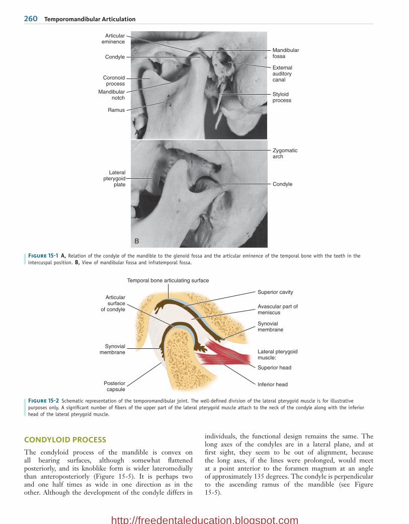
260 Temporomandibular Articulation
Figure 15-1 A, Relation of the condyle of the mandible to the glenoid fossa and the articular eminence of the temporal bone with the teeth in the intercuspal position. B, View of mandibular fossa and infratemporal fossa.
Articulareminence
Condyle
Coronoidprocess
Mandibularnotch
Ramus
Lateralpterygoid
plate
Mandibularfossa
Externalauditorycanal
Styloidprocess
Zygomaticarch
Condyle
A
B
Figure 15-2 Schematic representation of the temporomandibular joint. The well-defined division of the lateral pterygoid muscle is for illustrative purposes only. A significant number of fibers of the upper part of the lateral pterygoid muscle attach to the neck of the condyle along with the inferior head of the lateral pterygoid muscle.
Temporal bone articulating surface
Synovialmembrane
Posteriorcapsule
Articularsurface
of condyle
Superior cavity
Avascular part ofmeniscus
Synovialmembrane
Lateral pterygoidmuscle:
Inferior head
Superior head
CONDYLOID PROCESS
The condyloid process of the mandible is convex on all bearing surfaces, although somewhat flattened posteriorly, and its knoblike form is wider lateromedially than anteroposteriorly (Figure 15-5). It is perhaps two and one half times as wide in one direction as in the other. Although the development of the condyle differs in
individuals, the functional design remains the same. The long axes of the condyles are in a lateral plane, and at first sight, they seem to be out of alignment, because the long axes, if the lines were prolonged, would meet at a point anterior to the foramen magnum at an angle of approximately 135 degrees. The condyle is perpendicular to the ascending ramus of the mandible (see Figure 15-5).
http://freedentaleducation.blogspot.com

Chapter 15 The Temporomandibular Joints, Teeth, and Muscles, and Their Functions 261
JOINT CAPSULE
The TMJ is enclosed in a capsule (Figure 15-6) that is attached at the borders of the articulating surfaces of the mandibular fossa and eminence of the temporal bone and to the neck of the mandible. The anterolateral side of the capsule may be thickened to form a band referred to as the temporomandibular ligament. It is not always so thickened, but when clearly distinguishable as a ligament, it appears to originate on the zygomatic arch and to pass backward to attach on the lateral and/or distal surfaces of the neck of the mandible.
The capsule consists of an internal synovial layer and an outer fibrous layer containing veins, nerves, and collagen fibers. The innervation for the capsule arises from the
trigeminal nerve, and several kinds of receptors have been described, including free nerve endings.2 The vascular supply arises from the maxillary, temporal, and masseteric arteries.
MANDIBULAR LIGAMENTS
Accessory ligaments, including the stylomandibular and sphe-nomandibular ligaments, are considered a part of the mastica-tory apparatus (Figure 15-7). These ligaments do not have a direct relationship with mandibular articulation, although they may stabilize the articular system during jaw movements.
The sphenomandibular ligament arises from the angular spine of the sphenoid bone and from the petrotympanic
Figure 15-3 Radiograph showing the temporomandibular joint in open (left) and closed (right) position of mandible.
Figure 15-4 Exterior of the base of the skull showing the mandibular fossa in the inferior surface of the squamous part of the temporal bone at the base of the zygomatic process. It is divided into two halves by the petrotympanic fissure. The anterior half is included in the temporomandibular articulation.
Mastoidprocess
Auditorymeatus
Post-glenoidprocess
Styloidprocess
Angular spine of sphenoid
Tegmentympani
(edge)
Huguiercanal
Foramenovale
Medialglenoidlip
Zygoma
http://freedentaleducation.blogspot.com
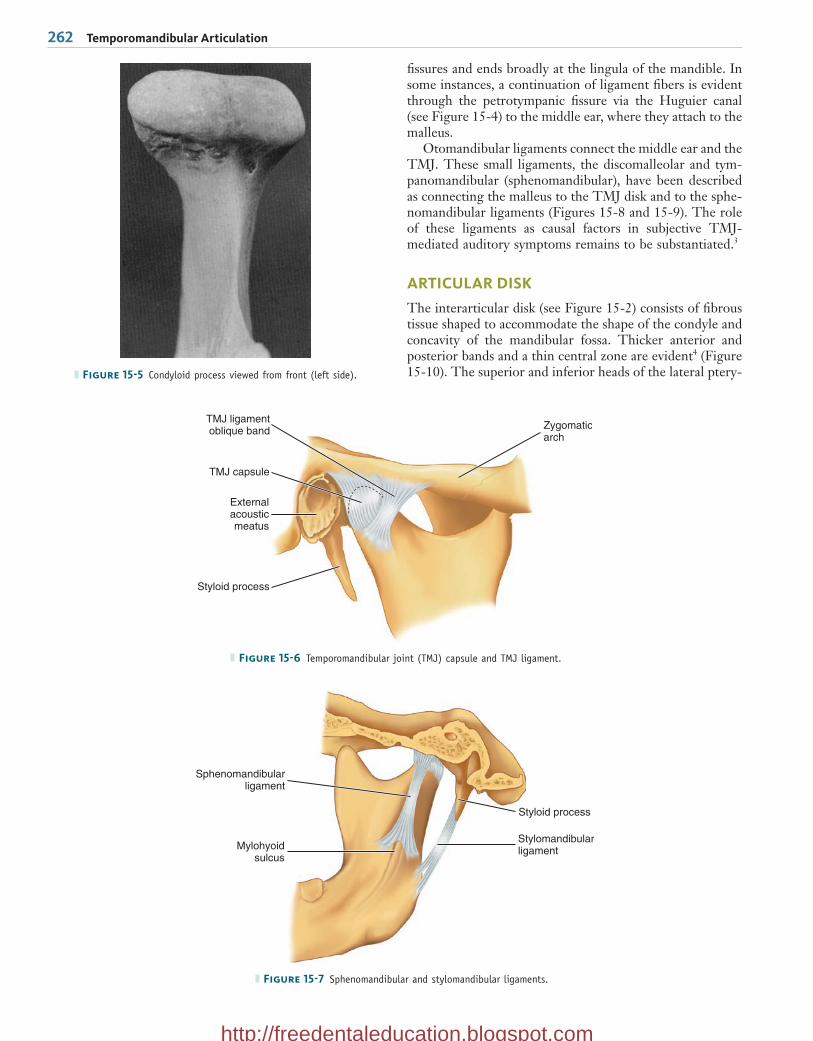
262 Temporomandibular Articulation
Figure 15-5 Condyloid process viewed from front (left side).
Figure 15-6 Temporomandibular joint (TMJ) capsule and TMJ ligament.
Zygomaticarch
TMJ capsule
TMJ ligamentoblique band
Externalacousticmeatus
Styloid process
Figure 15-7 Sphenomandibular and stylomandibular ligaments.
Styloid process
Sphenomandibularligament
Mylohyoidsulcus
Stylomandibularligament
fissures and ends broadly at the lingula of the mandible. In some instances, a continuation of ligament fibers is evident through the petrotympanic fissure via the Huguier canal (see Figure 15-4) to the middle ear, where they attach to the malleus.
Otomandibular ligaments connect the middle ear and the TMJ. These small ligaments, the discomalleolar and tym-panomandibular (sphenomandibular), have been described as connecting the malleus to the TMJ disk and to the sphe-nomandibular ligaments (Figures 15-8 and 15-9). The role of these ligaments as causal factors in subjective TMJ- mediated auditory symptoms remains to be substantiated.3
ARTICULAR DISK
The interarticular disk (see Figure 15-2) consists of fibrous tissue shaped to accommodate the shape of the condyle and concavity of the mandibular fossa. Thicker anterior and posterior bands and a thin central zone are evident4 (Figure 15-10). The superior and inferior heads of the lateral ptery-
http://freedentaleducation.blogspot.com

Chapter 15 The Temporomandibular Joints, Teeth, and Muscles, and Their Functions 263
goid muscle both insert into the pterygoid fovea of the mandible with a part of the superior head inserting into the disk and capsule. The disk divides the articulating surfaces into upper and lower compartments that provide for smooth gliding function. As the jaw opens and moves forward, the intermediate zone of the disk is interposed between the anterior slope of the articular eminence and the condyle, and the bilaminar region of the disk fills in the mandibular fossa (Figure 15-11). The upper head of the lateral ptery-goid muscle, which does not appear to be active during mandibular opening movement, stabilizes the relationship
of the disk to the eminence. Continued anterior displace-ment of the disk (Figure 15-12) with the posterior band in an anterior position with the jaw closed can prevent the jaw from opening normally (i.e., locking). The cause of disk derangement is multifactorial but may include acute and chronic trauma.
MANDIBULAR POSITIONS
Basic jaw positions are usually described as centric occlusion, intercuspal position, centric relation, retruded contact posi-tion, and rest position of the mandible.5 Centric occlusion or intercuspal position is defined as maximum intercuspation of the teeth. Centric relation is a position of the mandible (or path of opening and closing without translation of the con-dyles) in which the condyles are in their uppermost position in the mandibular fossae and related anteriorly to the distal slope of the articular eminence (see Figure 16-44). Because the mandible appears to rotate around a transverse axis through the condyle in centric relation movement, guidance of the jaw by the clinician (see Figure 16-42) in opening and closing movements that do not have translation is referred to as hinge axis movement (Figure 15-13). In this position, the condyles are considered to be in the terminal hinge position. Under physiological conditions of the masticatory system, centric relation is used to transfer the position of the mandible (in relation to the maxilla) to an articulator.
In the natural dentition, centric occlusion is, in the major-ity of people, anterior to centric relation contact on the average by approximately 1 mm.6 Centric occlusion (or acquired or habitual centric as it is sometimes called) is a tooth-determined position, whereas centric relation is a jaw-to-jaw relation determined by the condyles in the fossae. Closure into occlusion occurs usually anterior to centric relation; however, a coincidence of centric relation contact
Figure 15-8 Ligaments attached to the malleus.
(From Ash MM et al: Current concepts of the relationship and management of temporomandibular disorders and auditory symptoms, J Mich Dent Assoc 72:550, 1990.)
Chorda tympani nerve(behind membrane)
Discomalleolarligament
Sphenomandibularligament
Chorda tympani nerve
Figure 15-9 Ligaments attached to the malleus: DML, Discomalleolar ligament fibers; PML, fibers from sphenomandibular ligament; n., nerve; PMAL, fibers from the discomalleolar and sphenomandibular ligament.
MalleusDML
Disk
PML
Chordatympani nerve
PMAL
http://freedentaleducation.blogspot.com

264 Temporomandibular Articulation
Figure 15-10 Articular disk and associated structures.
Intermediatezone
Lower jointspace
Posterior diskband
Posteriorattachment
Anteriorband
Eminence
Mandibularramus
Auditorycanal
Upper jointspace
Lateral pterygoidmuscle
Figure 15-11 Articular disk: jaw in open position.
Posterior disk band
Anteriorband
Figure 15-12 Articular disk, anterior displacement: jaw in closed position.
Posterior disk band
and the intercuspal position is evident in about 10% of the population.
Rest position is a postural position of the mandible deter-mined largely by neuromuscular activity and to a lesser degree by the viscoelastic properties of the muscles. Thus, because tonicity of muscles may be influenced by the central nervous system as a result of factors such as emotional stress and by local peripheral factors such as a sore tooth, the rest position of the mandible is not consistent. The interocclusal space with the mandible in rest position and head in upright position is about 1 to 3 mm at the incisors but has consider-able normal variance even up to 8 to 10 mm without evi-dence of dysfunction.
MANDIBULAR MOVEMENTS
In lateral movements (Figure 15-14), the condyle appears to rotate with a slight lateral shift in the direction of the move-
Figure 15-13 Schematic representation of mandibular movement envelope in the sagittal plane. CR, Centric relation; CO, centric occlusion; F, maximum protrusion; R, rest position; E, maximum opening; B to CR, opening and closing on hinge axis with no change in radius (r).
Hinge axisHinge axis closure
CRCO F
R
B
E
r
r
I A, B
http://freedentaleducation.blogspot.com

Chapter 15 The Temporomandibular Joints, Teeth, and Muscles, and Their Functions 265
ment. This movement is called the Bennett movement and may have both immediate and progressive components. By the use of recording equipment such as a pantograph or kinesiograph, it is possible to record mandibular movements in relation to a particular plane of reference (e.g., sagittal, horizontal, or frontal planes). If a point (the incisive point) located between the incisal edges of the two mandibular central incisors is tracked during maximal lateral, protrusive, retrusive, and wide opening movements, such movements are seen to take place within a border or envelope of move-ments.6 Functional and parafunctional movements occur within these borders. However, most functional movements such as those associated with mastication occur chiefly around centric. Border movements in the horizontal plane are shown in Figure 15-14.
The maximum opening movement is 50 to 60 mm, depending on the age and size of the individual. An arbitrary lower limit for normal of 40 mm may be in error, inasmuch as some individuals may have no difficulty incising a large apple and have no history of TMJ muscle dysfunction. The maximum lateral movement in the absence of TMJ muscle dysfunction, including pain, is about 10 to 12 mm. The maximum protrusive movement is approximately 8 to 11 mm, again depending on the size of the subject and skull morphology. The retrusive range for adults and children is
about 1 mm, although 2 to 3 mm may be observed infre-quently.7 The retrusive range, as measured from centric occlusion to centric relation, is considered a discrepancy between centric occlusion and centric relation. Border movements in the sagittal plane are shown in Figure 15-13.
All values for border movements must be related to function—that is, a maximum lateral movement of 7 to 8 mm to the right (10 to 12 mm to the left) must be related to the occlusion and to whether translation of the left condyle occurs, because the latter may be “fixed” because of dysfunction or pain. Such values should also be related to other functions such as incising, chewing, swallowing, and speaking. However, if such values are made a part of every patient’s dental record, any change can be evaluated in terms of dysfunction.
Muscles
Masticatory functions, speaking, yawning, and swallowing, involve reflex contraction and relaxation of the muscles of mastication whose activity is initiated voluntarily. It is impossible to determine clinically if a particular muscle is participating in a particular movement solely from its origin
Figure 15-14 Right mandibular movement with schematic representation of movement at the incisal point in the horizontal plane (CR, LL, P, RL) and at the condyle (W, C, B, P) made by a pantograph. Teeth are not in occlusion. CR, Centric relation; LL, left lateral; P, protrusive; RL, right lateral; CO, centric occlusion; IEC, incisal edge contact. On the right side, the condyle moves from C (centric) to right working (W). On the balancing side, the left condyle moves from C along line B and makes an angle BG, called the Bennett angle. C to P, Straight protrusive movement.
P
IEC LL
CR
RL
CO
WC
P BG
C
CW
P
B
B
I C, D
http://freedentaleducation.blogspot.com

and insertion. Patterns of muscle contraction are complex and even in the same areas may have different functions.
The complex movements of the TMJ suggest that muscles of mastication exhibit differential regional action and regional differences in their histochemical profiles. Thus, to consider a “muscle” as a contracting entity is an oversimplification. In reality, each muscle is a collection of motor units with different properties located in different parts of a single muscle and exhibiting different activities. However, for obvious reasons, the action of the various muscles will be given as a contracting entity.
The masticatory muscles concerned with mandibular movements include the lateral pterygoid, digastric, mas-seter, medial pterygoid, and temporalis muscles. Also, the mylohyoid and geniohyoid muscles are involved in mastica-tory functions.
Several muscles associated with the ear, throat, and neck are of interest to the dentist, including tensors tympani and palatini, because the latter two may relate to subjective hearing disorders such as stuffiness, some forms of tinnitus, and noise.1,8–10
LATERAL PTERYGOID MUSCLE
The lateral pterygoid muscle has two origins: one head origi-nates on the outer surface of the lateral pterygoid plate, and an upper or superior head originates on the greater sphenoid wing (Figures 15-15, 15-16, and 15-17, C). The insertion is on the anterior surface of the neck of the condyle. In addi-tion, an insertion is evident of some fibers to the capsule of the joint and anterior aspect of the articular disk (Figure 15-17, C).
The superior head is active during various jaw-closing movements only, whereas the inferior head is active during jaw-opening movements and protrusion only.11 The lateral pterygoid is anatomically suited for protraction, depression, and contralateral abduction. It may also be active during other movements for joint stabilization. The superior head is active during such closing movements as chewing and clenching of the teeth and during swallowing. Presumably,
Figure 15-15 Positions of the lateral and medial pterygoid muscles shown with cutaway sections of bone.
Disk
Medial pterygoidmuscle
Lateralpterygoid
muscle
Buccinatormuscle
Figure 15-16 View of the medial and lateral pterygoid muscles. Note the insertion of the temporalis muscle also in Figure 15-17, B.
Styloidprocess
Occipitalcondyles
Medialpterygoidmuscle
Zygomaticarch
Lateralpterygoidmuscle
Anteriortemporalismuscle
Externalauditorymeatus
Temporalismuscle
the superior head positions or stabilizes the condylar head and disk against the articular eminence during mandibular closing. The inferior head assists in the translation of the condyle downward, anteriorly, and contralaterally during jaw opening. The lateral pterygoid muscle is innervated by the trigeminal nerve (V) (see Figure 14-37).
MASSETER MUSCLE
The masseter muscle extends from the zygomatic arch to the ramus and body of the mandible. The insertion of this muscle is broad, extending from the region of the second molar on the lateral surface of the mandible to the posterior lateral surface of the ramus (Figure 15-18; see also Figure 15-19). The masseter muscle is covered partly by the platysma muscle (Figure 15-19) and by the risorius muscle. The platysma is activated during firm clenching in some individuals and, having some insertion in the orbicular muscle (orbicularis oris), is sometimes active in facial expres-
266 Muscles
http://freedentaleducation.blogspot.com

Chapter 15 The Temporomandibular Joints, Teeth, and Muscles, and Their Functions 267
Figure 15-17 A, Coronal section showing the mandibular nerve exiting the foramen ovale (FO), the superior (SH) and inferior (IH) heads of the lateral pterygoid muscle, the ascending ramus (R), and the masseter (M) and medial pterygoid (MP) muscle. B, Optic nerve (ON), temporalis muscle (TM), deep masseter (DM) or zygomaticomandibular muscle, and coronoid process (CP). ZA, Zygomatic arch. C, Horizontal section at the level of the temporomandibular joint, showing the condyle (T), superior head of lateral pterygoid muscle (SH), inferior head (IH), ascending ramus (R), masseter muscle (M), and temporalis muscle (TM). D, Disk; S, styloid. (Redrawn from Widmalm SE, Lillie JH, Ash MM: Anatomical and electromyographic studies of the lateral pterygoid muscle, J Oral Rehabil 14:429, 1987.)
A B
C
sion. The risorius is affected by emotion and is active in facial expression.
The superficial part of the masseter muscle is separated distinctly only from the deeper layer of the muscle at the posterior upper part of the muscle. The masseter muscle is covered partly and to a variable degree with the parotid gland tissue. The center of the lower third of the masseter muscle is about 2 to 3 cm from the anterior border of
the sternocleidomastoid muscle, which contracts during clenching in some individuals. The masseter muscle is active during forceful jaw closing and may assist in protru-sion of the mandible. The masseter muscle is innervated by the fifth nerve (masseter nerve). The zygomaticomandibular muscle (deep masseter muscle) inserts at the coronoid process and originates on the inner surface of the zygomatic arch (see Figure 15-17, B). It may be an antagonist to the
http://freedentaleducation.blogspot.com

Figure 15-18 Masticatory muscles shown include the temporalis and masseter muscles. The deep masseter is attached to the zygoma (see Figure 15-17, C). TMJ, Temporomandibular joint.
TMJcapsule
Massetermuscle
Temporalismuscle
Buccinatormuscle (cut)
Figure 15-19 Muscles of facial expression and accessory muscles of mastication. Sup., Superior; proc., procerus.
Orbicularisoculi muscle
Zygomaticmajor muscle
Levator labiisup. muscle
Orbicularisoris muscle
Depressor angularisoris muscleRisorius muscle
Buccinator muscle
Nasalis proc.alaris muscle
Parotid duct
Temporalismuscle
Styloglossusmuscle
Pterygomandibularraphe
Masseter muscle(cut)
Platysma muscle
15-20). On passing to the zygomatic arch, it forms a tendon that inserts into the anterior border and mesial surface of the coronoid process of the mandible and along the anterior border of the ascending ramus of the mandible (see Figure 15-16). The anterior fibers extend along the anterior border of the ramus almost to the third molar. The muscle has three component parts and appears to behave as if it con-sisted of three distinct parts. The temporal muscle is the principal positioner of the mandible during elevation. The posterior part is active in retruding the mandible, and the anterior part is active in clenching. The anterior part may act as a synergist with the masseter in clenching, whereas the posterior part acts as an antagonist to the masseter in retruding the jaw. The temporalis muscle is innervated by temporal branches of the mandibular division of the fifth nerve.
DIGASTRIC MUSCLE
The attachment of the anterior digastric muscle is at or near the lower border of the mandible and near the midline (see Figure 15-20). A tendon is between the anterior and poste-rior digastric muscles that is attached by a looplike strip of fascia to the hyoid bone. The anterior digastric muscle is covered by the platysma muscle, and beneath lie the mylo-hyoid and the geniohyoid muscles. All of these muscles are considered to be active during various phases of jaw opening. A mylohyoid branch of the mandibular division of the fifth nerve innervates the anterior digastric muscle (see Figure 14-37); the digastric branch of the facial nerve innervates the posterior digastric muscle.
GENIOHYOID MUSCLE
The geniohyoid muscle lies superior to the mylohyoid muscle and adjacent to the midline. It arises from the mental spine on the posterior aspect of the symphysis menti of the man-
posterior temporalis and a synergist for the lateral pterygoid muscle.
MEDIAL PTERYGOID MUSCLE
The medial pterygoid muscle arises from the medial surface of the lateral pterygoid plate and from the palatine bone (see Figures 15-15 and 15-16). It inserts on the medial surface of the angle of the mandible and on the ramus up to the mandibular foramen. The principal functions of the medial pterygoid muscle are elevation and lateral positioning of the mandible. It is active during protrusion. The innervation is a branch of the mandibular division of the fifth nerve.
TEMPORALIS MUSCLE
The temporalis muscle is fan-shaped and originates in the temporal fossa (see Figures 15-16; 15-17, B; 15-18; and
268 Muscles
http://freedentaleducation.blogspot.com

Chapter 15 The Temporomandibular Joints, Teeth, and Muscles, and Their Functions 269
Figure 15-20 Muscles of the neck. TMJ, Temporomandibular joint.
Superficialmassetermuscle
Digastricanterior bellymuscle
Digastricposterior belly
muscle
Hyoid bone
Omohyoidsuperior bellymuscle
Sternohyoidmuscle
Sternocleidomastoidmuscle
Omohyoidinferior bellymuscle
Splenius capitismuscle
Levator scapulaemuscle
Trapeziusmuscle
Deep massetermuscle
TMJcapsule
Anterior, middle, andposterior scalene muscles
Anterior temporalismuscle
dible. It inserts on the anterior surface of the hyoid bone. When the mandible is fixed, the hyoid bone is drawn forward and upward; when the hyoid bone is fixed, the lower jaw is depressed. Innervation is from C1, C2, and possibly the hypoglossal nerve (see Figure 14-37).
TENSOR TYMPANI AND PALATINI MUSCLES
The tensor tympani, tensor veli palatini, and levator veli palatini muscles (Figures 15-21 and 15-22) may have clinical
Figure 15-21 Section through the ear showing inner ear structures and the tensor tympani muscle, which is active in stretching the tympanic membrane.
Temporalis muscle
Tympanic membrane(cut)
Tensortympani muscle
Auditorytube (cut)
Levator palatini muscle
Styloidprocess
significance for subjective auditory symptoms associated with temporomandibular and muscle disorders (TMDs).1,8,9 These muscles are innervated by the trigeminal nerve and therefore may respond to similar kinds of ascending infor-mation from joints, skin, and muscles and from descending inputs from higher centers that converge on interneurons and the trigeminal motor nucleus (Figure 15-23). Distur-bances of auditory tube opening during swallowing that may occur with TMJ and TMDs appear to be consistent with restricted function and anatomy of the tensor palatini and
http://freedentaleducation.blogspot.com

Figure 15-22 Muscles of the throat. Eustachian tube and tensor palatinus muscle, which is active in opening the auditory tube.
Figure 15-23 Information from joints, skin, muscles, and higher centers converge on the interneurons and trigeminal motor nucleus.
Higher centers
Interneurons
Trigeminalmotorneurons
Tensors tympani, palatini,masseter, temporalis, pterygoid
Jointskin
Musclepain
contraction headache. The possible role of the epicranius muscles in tension headache has yet to be clarified. The sternocleidomastoid muscle (see Figure 15-20) is often affected in patients with TMJ muscle dysfunction and often co-contracts with jaw clenching. The functions of the orbic-ularis oris and buccinator muscles (see Figure 15-19) appear to have a significant role for optimal function of complete dentures. The seventh (VII) nerve innervates all the muscles of facial expression.
Other muscles that are of interest to the dentist because of TMJ and muscle disorders are the scalenus, splenius, ili-ocostalis cervicis, and omohyoideus. However, the associa-tion between TMDs and myalgia of the muscles has not been established. A number of problems in oral motor func-tion, such as lip posture in the aged, may be related to a generalized deterioration in performance or to a specific motor function rather than the dental state or prescription medication. Most reports on motor disorders and aging have focused on disturbances in control originating in the nervous system.
Mandibular Movements and Muscle Activity
Mandibular movement during normal function and during parafunction (e.g., bruxism) involve complex neuromuscular patterns originating in part in a pattern generator in the brainstem and modified by influences from higher centers (see Figure 15-23), namely, the cerebral cortex and basal ganglia, and from peripheral influences (e.g., the periodon-tium, muscles, and so on). However, a detailed discussion of such movements is beyond the scope of this text. Rather, the discussion relates to muscle activity as seen in electro-myography for jaw opening and closing, protrusion, retru-sion, and lateral movements.
MANDIBULAR OPENING
The digastric, mylohyoid, and geniohyoid muscles are active during jaw opening, either slowly or maximally against resistance. No activity occurs in the temporalis and masseter muscles when the mouth is opened slowly and the jaw is opened maximally, although some activity may occur in the medial pterygoid muscle. When the jaw is opened against resistance, the temporalis muscle remains silent. During opening movements, the lateral pterygoid muscles show initial and sustained activity. In forced depression, the digas-tric muscle is activated almost as soon as the lateral ptery-goid muscle is. Generally, the activity of the anterior digastric muscle follows that of the lateral pterygoid muscle.
MANDIBULAR CLOSING
While the mandible is being elevated slowly, without contact of the teeth, no activity is evident in any portion of the temporalis muscle. Elevation without contact or resistance
levator veli palatini muscles.10 If a causal connection between subjective hearing symptoms and TMDs is to be established, additional evidence-based research is needed.
Restricted jaw opening and prevention of yawning seen in TMJ and TMDs can lead to otological symptoms such as subjective hearing loss associated with sensations of stuffi-ness in the ear.1 Thus, pain in and around the joints and muscles may influence the otomandibular muscles via interneurons in the intertrigeminal area and might be responsible for a few cases of tinnitus associated with restricted jaw opening due to TMJ and muscle disorders.
HEAD AND NECK MUSCLES
The muscles of the head and neck have been of interest to the dentist because of a potential relationship among the occlusion, TMJs, and pain in these muscles and/or muscle
270 Mandibular Movements and Muscle Activity
http://freedentaleducation.blogspot.com

Chapter 15 The Temporomandibular Joints, Teeth, and Muscles, and Their Functions 271
is brought about by contraction of the masseter and medial pterygoid muscle. The temporalis, masseter, and medial pterygoid muscles affect elevation against resistance. The suprahyoid muscles act as an antagonist of the elevator muscles. Closure into maximal intercuspation (centric occlu-sion) may involve contraction of facial and neck muscles.
RETRUSION
Voluntary mandibular retrusion with the mouth closed is brought about by contraction of the posterior fibers of the temporalis muscle and by the suprahyoid and infrahyoid muscles. Retraction of the mandible from protrusion and without occlusal contact is effected by the contraction of the posterior and middle fibers of the temporalis muscles. Slight activity of the suprahyoid may be the result of slight jaw opening to allow the teeth to glide over each other from centric occlusion to centric relation.
PROTRUSION
Protrusion of the mandible without occlusal contact results from contraction of the lateral and medial pterygoid muscles and also masseter muscles. Protraction against resistance is brought about by contraction of the lateral and medial pterygoid muscles and of the masseter and suprahyoid muscle group. Protrusion with the teeth in occlusion is achieved by contraction of the pterygoid and masseter muscles. Only slight activity occurs in the suprahyoid muscles. In combined protraction and opening, activity is evident in the medial and lateral pterygoid muscles, the masseter muscles, and sometimes the anterior fibers of the temporalis muscles.
LATERAL MOVEMENTS
Lateral movement of the mandible to the right side (without occlusal contact) is achieved by ipsilateral contraction of primarily the posterior fibers of the temporalis muscle. The suprahyoid muscles are active in maintaining the jaw slightly depressed and protruded. Movement to the left side without occlusal contact is brought about by the contralateral con-traction of the medial pterygoid and masseter muscles. Lateral movement to the right side against resistance is achieved by the ipsilateral contraction of the temporalis muscle and by some activity in the ipsilateral masseter and medial pterygoid muscles. Movement to the left side against resistance is achieved by the contralateral contraction of the medial pterygoid and masseter muscles. Lateral movement to the right with occlusal contact is achieved by ipsilateral contraction of the temporalis muscle. Movement to the left with occlusal contact is brought about by contralateral con-traction of the medial pterygoid and masseter muscles. Both lateral pterygoid muscles initiate depression of the mandi-ble, and the contralateral muscle initiates a lateral transver-sion. Lateral movements of the jaw are achieved by ipsilateral contraction of the posterior and middle fibers of the tem-poralis muscle and by contralateral contraction of the lateral
and medial pterygoid muscles and the anterior fibers of the temporalis muscle. Parts of the temporal and masseter muscles may act as antagonists or synergists during horizon-tal movements and minimum separation of the teeth.
CHEWING
Chewing is highly complex oral motor behavior usually seen in the frontal plane in simple form (Figure 15-24). No archetypal chewing cycle exists. The means of the dimen-sions of the chewing cycle are between 16 and 20 mm for vertical movements and between 3 and 5 mm for lateral movements. The duration of the cycle varies from 0.6 to 1 second depending on the type of food. The speed of masti-catory movement varies within each cycle, both according to the type of food and among individuals. Speed, duration, and form of the chewing cycle vary with the type of occlu-sion, kind of food, and presence of dysfunction.
Occlusal contacts occur in centric occlusion in at least 80% to 90% of all chewing cycles, especially near complete trituration of the bolus. With closing and opening move-ments, contact gliding is seen. In the closing phase, the contact glide depends on the type of occlusion and type of food. Where tough food is the normal diet (e.g., as with Australian aborigines) with corresponding occlusal wear, the chewing movement shows a long contact glide (2.8 ± 0.35 mm)12 compared with the short contact glide (0.90 ± 0.36 mm) of Europeans living on a modern diet of easily triturated food.13 The chewing force reaches a maximum in centric occlusion and lasts for 40 to 170 milliseconds, and the peak electromyographic activity of the temporal and masseter muscles lasts for a mean of 41 ± 26 milliseconds. Some chewing force is maintained for gliding tooth contact on the opening phase. In the intercuspal position, the jaw is stationary, or it pauses for approximately 100 milliseconds before the next cycle begins.
Figure 15-24 Mandibular movements during the process of chewing naturally. Incisor point movement is seen in the frontal plane. L, Left; C.O., centric occlusion; R, right.
RL
C.O.
4 mm
I C
http://freedentaleducation.blogspot.com

SWALLOWING
Swallowing involves most of the tongue muscles and buccal musculature.14 In the initial stage of swallowing, the bolus moves from the mouth to the fauces (Figure 15-25). The bolus is then moved from the fauces to the esophagus and finally through the esophagus to the stomach. When saliva is swallowed, total participation of the suprahyoid muscles occurs, with marked activity of the digastric and mylohyoid muscles, followed by moderate activity in the geniohyoid muscles. The medial pterygoid muscle is often active; less often, the temporalis and masseter muscles are active with occlusal contact.
ORAL MOTOR BEHAVIOR
Oral motor behavior refers to function and parafunction of the mouth and associated structures. More generally, behavior includes observable actions ranging from simple movements such as retrusion or protrusion to more complex movements such as chewing. To accomplish complex behavior, sensori-motor systems consisting of muscles and neural processes are required for the initiation, programming, and execution of motor functions.
Chewing movements depend on complex integrative neural processes of the central nervous system that may be initiated by either internal or external influences, including innate drives, emotional states, and instructions to patients.
During chewing, a large amount of proprioceptive (i.e., muscle sense) and exteroceptive (i.e., tactile sense) informa-tion is fed to the central nervous system (e.g., cerebral cortex, brainstem, basal ganglia, spinal cord). Rhythmic movements such as chewing are largely programmed or preprogrammed and involve learning, which reduces the need for peripheral sensory input. However, inputs from muscle, joint, tendon, and periodontal receptors still have important functions, especially in relation to learning, new experiences, and protective reflexes. Neuronal mechanisms must be present to provide for modification of reflexes and continued updating of masticatory movements by informa-tion about such factors as occlusal forces and the state and location of the bolus.
OVERVIEW OF MASTICATION
The act of mastication begins with “setting the system” by sight, tactile sense, and smell to receive the food. Involve-ment of the tactile sense may range from picking up the food to grasping the food with the incisor teeth. When the food is taken into the mouth, the lips, tongue, and periodontium function to estimate size, hardness, and other characteristics that must be correlated with previous behavior required for chewing. This information sets the chewing program in the pattern generator, including subsequences that relate to central and peripheral influences already in progress. Oro-facial receptors such as periodontal mechanoreceptors may
Figure 15-25 Oral pharynx, with special reference to the palatine tonsil and the musculature of the palate. M., Musculus; A., arteria; v., vena.
Arcus dentalissuperior
Veli palatina major artery
Expansion of thelevator veli palatini
Hamuluspterygoideus
Pharyngopalatinus muscle
Raphe pterygomandibularis
Buccopharyngeus muscle
Buccinator muscle
Tonsilla palatina
Glossopalatinus muscle
Palatinadescendens artery
Maxillarisinterna artery
Tonsillaris artery
Palatina ascendens arteryPharyngea
ascendens artery
Maxillarisexterna artery
Carotis externa arteryLingualis artery
Ramus dorsalislinguae
Palatum durum
Palatum molle
Arcusglossopalatinus
Arcuspharyngopalatinus
Uvula (palatina)
Tonsillapalatina
Constrictorpharyngis superiormuscle
Lateral band oflymphoid tissue
Buccopharyngeusmuscle
Pharyngopalatinusmuscle
GlossopalatinusmuscleTonsilla
palatina
B
A
Pharyngea
Tongue
272 Mandibular Movements and Muscle Activity
http://freedentaleducation.blogspot.com

Chapter 15 The Temporomandibular Joints, Teeth, and Muscles, and Their Functions 273
monitor occlusal forces and control the jaw-closing muscles. The chewing program may be altered according to stages of chewing or in response to information from receptors in specific areas such as the palatal mucosa and tongue. The chewing rhythm may be stopped because of noxious stimuli. It is probable that rhythmic and repetitive chewing behavior is disturbed by dysfunctional states of the occlusion and/or the TMJs through which the bolus is shifted and the jaw moved to another position for the next forceful tooth-food contact. Thus chewing cycles may occur without the development of an actual power stroke in dysfunction states.
References1. Ash MM et al: Current concepts of the relationship and
management of temporo-mandibular disorders and auditory symptoms, J Mich Dent Assoc 72:550, 1990.
2. Thilander B: Innervation of the temporomandibular joint capsule in man, Trans R Sci Dent 7:9, 1961.
3. Ash CM, Pinto OF: The TMJ and the middle ear: structural and functional correlates for aural symptoms associated with temporomandibular joint dysfunction, Int J Prosthodont 4:51, 1991.
4. Dolwick MF, Sanders B: TMJ internal derangement and arthrosis, St Louis, 1985, Mosby.
5. Ash MM: Philosophy of occlusion, Dent Clin North Am 39:233, 1995.
6. Posselt U: Studies in the mobility of the human mandible, Acta Odont Scand 10(suppl):3, 1952.
7. Ash MM, Ramfjord SP: Occlusion, ed 4, Philadelphia, 1995, Saunders.
8. Rubinstein B et al: Prevalence of signs and symptoms of craniomandibular disorders in tinnitus patients, Cranio 4:186, 1990.
9. Myrhaug H: The theory of otosclerosis and morbus meniere (labyrinthine vertigo) being caused by the same mechanism: physical irritants, an otognathic syndrome, Bergen, 1981, Bergmanns Boklrykkeri A/S.
10. Misurya VK: Functional anatomy of tensor palatini and levator palatini muscles, Arch Otolaryngol 102:265, 1976.
11. Widmalm SE, Lillie JH, Ash MM Jr: Anatomical and electromyographic studies of the lateral pterygoid muscle, J Oral Rehabil 14:429, 1987.
12. Beyron H: Occlusal relations and mastication in Australian aborigines, Acta Odont Scand 22:597, 1964.
13. Ahlgren J: Masticatory movements in man. In Anderson DJ, Matthews B, editors: Mastication, Bristol, England, 1976, John Wright and Sons.
14. Anson JB: An atlas of human anatomy, ed 2, Philadelphia, 1962, Saunders.
BibliographyAarstad T: The capsular ligament of the temporomandibular joint
and retrusion facets of the dentition in relation to mandibular movements, Oslo, 1954, Acad Forlag (Translated by H Christie).
Ahlgren J: Mechanisms of mastication, Acta Odont Scand 24(suppl)44:1, 1966.
Ash MM: Paradigmatic shifts in TMD and occlusion, J Oral Rehabil 28:1, 2001.
Buxbaum JD et al: A comparison of centric relation with maximum intercuspation based upon quantitative electromyography, J Oral Rehabil 9:45, 1982.
Callander CL: Surgical anatomy, ed 2, Philadelphia, 1939, Saunders.
Carlsoo S: Nervous coordination and mechanical function of the mandibular elevators, Acta Odont Scand 10(suppl)11:1, 1952.
Deaver JB: Surgical anatomy of the human body, ed 2, Philadelphia, 1926, Blakiston.
Duthie N, Yemm R: Muscles involved in voluntary mandibular retrusion in man, J Oral Rehabil 9:155, 1982.
Eagle WW: Asymptomatic styloid process, Arch Otolaryngol 49:490, 1949.
Eriksson P-O et al: Special histochemical muscle-fibre characteristics of the human lateral pterygoid muscle, Arch Oral Biol 26:495, 1981.
Gandevia SC, Mahutte CK: Joint mechanics as a determinant of motor unit organization in man, Med Hypotheses 6:527, 1980.
Gibbs CH et al: Functional movements of the mandible, J Prosthet Dent 26:604, 1971.
Gosen AJ: Mandibular leverage and occlusion, J Prosthet Dent 31:369, 1974.
Grant PG: Lateral pterygoid: two muscles? Am J Anat 138:1, 1973.
Hansson T et al: Thickness of the soft tissue layers and the articular disk in the temporomandibular joint, Acta Odont Scand 35:77, 1977.
Hickey JC: Mandibular movements in three dimensions, J Prosthet Dent 13:72, 1963.
Hjortsjo CH: The mechanism in the temporomandibular joint, Acta Odont Scand 11:5, 1953.
Hylander WL: The human mandible: lever or link? Am J Phys Anthropol 43:227, 1975.
Kawumura Y, Nobuhara M: Studies on masticatory function. II. The swallowing threshold of persons with normal occlusion and malocclusion, Med J Osaka Univ 8:241, 1957.
Lehr RP, Owens SE: An electromyographic study of the human lateral pterygoid muscles, Anat Rec 196:441, 1980.
McNamara JA: The independent functions of the two heads of the lateral pterygoid muscle, Am J Anat 138:197, 1973.
Moiler E: The chewing apparatus, Acta Physiol Scand 69(suppl)280:1, 1966.
Owall B, Moller E: Tactile sensibility during chewing and biting, Odontol Revy 25:327, 1974.
Smith RJ: Mandibular biomechanics and temporomandibular joint function in primates, Am J Phys Anthropol 49:341, 1978.
Stohler CS, Ash MM: Mandibular displacement in complete chewing sequence, J Dent Res 61:273, 1982 (abstract).
Storey AT: Joint and tooth articulation in disorders of jaw movement. In Kawamura Y, Dubner R, editors: Oral-facial sensory and motor function, Tokyo, 1981, Quintessence.
Takahashi T et al: The role of oral kinesthesia in the determination of the swallowing threshold, J Dent Res 62:327, 1983.
Toller PA: The synovial apparatus and temporomandibular joint function, Br Dent J 111:355, 1961.
Vitti M, Basmajian JV: Integrated actions of masticatory muscles: simultaneous EMG from eight intramuscular electrodes, Anat Rec 187:173, 1977.
http://freedentaleducation.blogspot.com

Wompler HW et al: Scanning electron microscopic and radiographic correlation of articular surfaces and supporting bone in the mandibular condyle, J Dent Res 59:754, 1980.
Wyke BD: Neuromuscular mechanisms influencing mandibular posture: a neurologist’s view of current concepts, J Dent 2:111, 1974.
Yurkstas AA: The masticatory act, J Prosthet Dent 15:248, 1965.
Use the CD-ROM to view animations and study ques-tions; and go to the site for additional study resources.
274 Bibliography
http://freedentaleducation.blogspot.com

Occlusion
misunderstood and seldom mentioned as a part of a concept of occlusion.
In the past, ideas regarding occlusion were often based on complete dentures. Because of the problems of instability of denture bases, the concept of “balanced occlusion” was developed to consider bilateral contacts in all functional excursions to prevent tipping of the denture bases. Although some clinicians have advocated such concepts in the past for use with regard to the natural dentition, acceptance was limited and unsupported by research evidence. Even so, some of the concepts related to condylar guidance, cusp height, incisal guidance, curve of Spee, and plane of occlu-sion have been useful in the restoration of natural teeth. Several concepts of an “ideal” or optimal occlusion of the natural dentition have been suggested by Angle,3 Schuyler,4 Beyron,5 D’Amico,6 Friel,7 Hellman,8 Lucia,9 Stallard and Stuart,10 and Ramfjord and Ash.11 These concepts stress to varying degrees static and/or functional characteristics of an occlusion as being theoretical practical goals for diagnosis and treatment of the occlusion. Some of the ideas have been developed principally in relation to orthodontics or com-plete dentures, and others for full mouth rehabilitation. None are completely applicable to the natural dentition; some provide for specific occlusal contact relations and joint positions, and few concepts of occlusion consider in princi-ple, or in practical ways, related muscle and oromotor func-tions. The idea of a functional rather than simply a static relationship of occlusal surfaces has become increasingly important because of the recognition that functional distur-bances of the masticatory system can be related to maloc-clusion, occlusal dysfunction, and disturbances of oral motor behavior, including bruxism. Thus, for example, the occlusal contact relationships shown in Figure 16-1 reflect oromotor
The definition of the term occlusion in many dictionaries only states that it is the act of closure or being closed, but some dental dictionaries (e.g., Zwemer1) go well beyond this simple definition to include a static, morphological tooth contact relationship. It may be defined also as the contact relationship of the teeth in function or parafunction.2 However, the term refers not only to contact at an occlusal interface, but also to all those factors concerned with the development and stability of the masticatory system and with the use of the teeth in oral motor behavior. Because brief definitions of occlusion are too limited to be useful as a basis for dental practice, more complete explanations evolve into concepts or paradigms of occlusion that reflect prevailing interests and clinical convenience. Thus a modern concept of occlusion must include the idea of an integrated system of functional units involving teeth, joints, and muscles of the head and neck. The solutions to problems such as bruxism, orthodontic relapse, denture instability, and peri-odontal trauma require concepts of occlusion that extend well beyond static arrangements of teeth, occlusal contacts, and jaw position.
Concepts of Occlusion
Concepts of occlusion vary with almost every specialty of dentistry. Common to some are definitions based on a static view of the dentition in which descriptions of the occlusion emphasize the fit of particular parts of individual maxillary teeth with specified parts of mandibular teeth. Until recently, only a few concepts of occlusion have included functional criteria, and because the dentofacial complex is highly mobile, ideas of occlusal stability and homeostasis are often
275
1616
http://freedentaleducation.blogspot.com

behavior consistent with bruxism (grinding of the teeth) that requires preventive occlusal therapy to control its adverse effects (e.g., an occlusal bite plane stabilization–type splint) (Figure 16-2). The restoration of lost tooth structure due to aggressive bruxism, as for other losses from other causes, requires knowledge of dental morphology, physiology, restorative materials, occlusion, esthetics, and personal habits (e.g., pipe smoking).
Development of the Dentitions
Functional disturbances of the masticatory system may have their beginning during the development of occlusion, a time when the substrate for tongue and swallowing habits, chewing patterns, teeth clenching, and bruxism may be established. It is also the time when occlusal discontinuity, occlusal interferences to function, and occlusal instability often develop. The development of malocclusion is a reflec-tion of disturbances in the normal processes of occlusal development. Therefore some knowledge of the process of
occlusal development is necessary for the practice of dentistry.
Primary Dentition
Any consideration of the development of the occlusions should begin with the primary dentition. It is during this period in the development of the oral-facial complex that oral motor behavior reflects learning related to the advent of the teeth. Human oral functions that are acquired or modified during the natural progression from birth through infancy to adulthood are in part related to the development of occlusion, both of the deciduous and permanent teeth (i.e., occlusion defined in its broadest sense).
Perhaps many of the reflex mechanisms of the oral-facial area and sensory and higher-center influences are important for the acquisition of masticatory skills, just one of the many motor behaviors that come under the phrase oral motor func-tion. Thus of particular importance is the novel sensory apparatus of the teeth that makes their appearance with the primary/deciduous teeth at an important time in the matu-ration of the nervous system and its interface with the environment.
The development of the muscle matrix and the active growth of the facial skeleton occur at a very strategic time for the maturation of the nervous system and the develop-ment of oral motor functions involving the teeth and chewing. It is also at this time that jaw positions and postur-ing of the mandible in relationship to the teeth takes place.
OVERVIEW OF THE PRIMARY OCCLUSION
The primary teeth are arranged in the jaws in the form of two arches: a maxillary and a mandibular. An outline follow-ing the labial and buccal surfaces of the maxillary teeth describes the segment of an ellipse and is larger than the segment following the same surfaces on the mandibular teeth (see Figure 1-2, A).
The relation between the maxillary and mandibular primary teeth when in occlusion is such that each tooth, with the exception of the mandibular central incisor and the maxillary second molar, occludes with two teeth of the opposing jaw. The primary teeth should be in normal align-ment and occlusion shortly after the age of 2, with all the roots fully formed by the time the child is 3 years old. A year or so after the teeth have fully erupted and have assumed their respective positions in the arches, the rapid develop-ment of the jaws is sufficient to create an interdental space, or diastema, between some of them.
The anterior teeth separate and usually show greater separation as time goes on by a process that is caused by the growth of the jaws and the approach of the permanent teeth from the lingual side. This separation usually begins between the ages of 4 and 5 years. The canines and molars are sup-posed to keep their positive contact relation during all the jaw growth. However, some shifting and separation are seen
Figure 16-1 Effects of bruxism in a young patient.
(From Ash MM: Paradigmatic shifts in occlusion and temporomandibular disorders, J Oral Rehabil 28:1-13, 2001.)
Figure 16-2 Stabilization occlusal bite plane splint to prevent the effects of bruxism.
(From Ash MM, Ramfjord S: Occlusion, ed 4, Philadelphia, 1995, Saunders.)
276 Primary Dentition
http://freedentaleducation.blogspot.com

Chapter 16 Occlusion 277
quite often. Because the teeth do not hold their relative positions for long, they are worn off rapidly on incisal ridges and occlusal surfaces. As an example, when a primary canine is lost 8 years or more after its eruption, its long, sharp cusp has in most instances been worn down. If the primary teeth are in good alignment, the occlusion is most efficient during the time that these teeth are in their original positions. This situation exists for only a relatively short time. After normal jaw growth has resulted in considerable separation, the occlusion is supported and made more efficient by the erup-tion and occlusion of the first permanent molars immediately distal to the primary second molars. The child is now approxi-mately 6 years of age and will use some of the primary teeth for 6 more years.
ERUPTION CHRONOLOGY OF THE PRIMARY TEETH
The chronology of the emergence/eruption of the primary teeth, which is shown in Table 16-1, is derived from data provided in Chapter 2.
The timing of the eruption or emergence of the teeth is due in large part to heredity and only somewhat to environ-mental factors. According to Falkner12 the development and eruption of the primary dentition are quite independent of the development and maturation of the child as a whole. The significance of local environmental factors for the development of occlusion considered in its broadest sense is relatively unknown. Thus learning of mastication may be
highly dependent on the stage of development of the occlu-sion (type and number of teeth present), maturation of the neuromuscular system, and factors such as diet.
MEAN AGE OF ERUPTION
The mean age of eruption of the primary teeth as shown in Table 16-1 is demonstrated schematically in Figure 16-3, although one should keep in mind the range of values seen in Table 16-1 and the idea that a 6-month acceleration or delay is considered normal. Even so, on average the sequence generally follows that shown in Figure 16-3.
CONTACT RELATIONS
The contact relations of the teeth tend to vary with the degree of bruxism present in the child. A number of factors appear to be related to the development of contact relations at the time of eruption of the teeth, including the position of the tooth germ, presence of permanent teeth, develop-ment of the condyles, cuspal inclines, and neuromuscular influences. Generally, little attention has been paid to the cusp-fossa and temporomandibular joint (TMJ) relation-ships, and usually only tooth-to-tooth observations have been made, namely, the mesiolingual cusp of the maxillary molar occludes in the central fossa of the deciduous man-dibular molar. Jaw-to-jaw relations with regard to the posi-tion of the condyle have received little attention. Perhaps discrepancies between condylar position and tooth position have little meaning in a relatively plastic, rapidly growing and changing system, including the nervous system.
PRIMARY ARCH FORM
The arch form and width for both the primary and perma-nent dentitions has been largely established by the age of 9 months.13 This observation may seem untrue in view of the apparent difference in facial appearance between the child of that age and a young adult with a permanent dentition. What does change substantially, of course, is the anteropos-terior dimension of the jaws, an increase in which is neces-sary for the incorporation of the permanent molars into the occlusion. It must be kept in mind that the alveolar bone and basal bone determine the shape of the dental arches. Arch form will be considered in more detail in the section on the permanent dentition.
Figure 16-3 Mean age (in months) of emergence of the primary/deciduous dentition.
(From Ash MM, Ramfjord S: Occlusion, ed 4, Philadelphia, 1995, Saunders.)
29 16 19 11 10
27 16 20 13 8
Table 16-1 Eruption of Primary Teeth*
Emergence of Teeth
Tooth (Months)
Upp
er
i1 E, F 10 (8–12)
i2 D, G 11 (9–13)
c C, H 19 (16–22)
m1 B, I 16 (13–19)� (14–18)�
m2 A, T 29 (25–33)
Maxillary Teeth
Right A B C D E | F G H I J Left
T S R Q P | O N M L K
Mandibular Teeth
Low
er
i1 P, O 8 (6–10)
i2 Q, N 13 (10–16)
c R, M 20 (17–23)
m1 S, L 16 (14–18)
m2 T, K 27 (23–31)� (24–30)�
i1, Central incisor; i2, lateral incisor; C, canine; m1, first molar; m2, second molar.*Universal numbering system for primary/deciduous dentition (see Chapter 1). See Table 2-3 for detailed presentation of the data.
http://freedentaleducation.blogspot.com

INTERDENTAL SPACING
The position of the deciduous teeth in the arches generally shows some degree of interdental spacing, which tends to decrease slightly with age. The size of the primary teeth and the spacing between them has a relationship to the position of the permanent teeth and the size of the dental arches (e.g., sufficient interdental space is needed for the perma-nent teeth to erupt into an uncrowded position). One of the indicators of future sufficiency or insufficiency of space in the dental arches for the permanent teeth is the presence or absence of spacing between the teeth of the primary denti-tion14,15 (i.e., spacing between the primary teeth [Figure 16-4] is necessary for the proper alignment of the permanent dentition).
The probability of crowding of the permanent dentition based on the amount of interdental spacing of the primary teeth is given in Table 16-2.
PRIMARY MOLAR RELATIONSHIPS
Primary molar relationships have been described by Moyers as the flush terminal plane, mesial step, and distal step
(Figure 16-5, A, B, and C).14 Of particular interest to the orthodontist is the fact that the mandibular second primary molar has a greater mesiodistal diameter than the maxillary second molar. As a result of this difference in dimensions of the two teeth, the distal surfaces of these two molars are in the same plane; a flush terminal plane is located at the end of the deciduous dentition. It has been reported that if a “step” (deviation of the flush terminal plane) occurs because of carious lesions or other disturbances, a tendency to inter-fere with the development of normal occlusal relations of the permanent first molars is evident. Also, it has been shown that the natural wearing away of the cusps in the deciduous dentition allows the mandible to assume a more forward position during a period when the mandible is growing more rapidly than the maxilla. In the absence of cuspal interferences, there is some evidence that the perma-nent incisors erupt with less vertical overlap and the perma-nent molars erupt into a more favorable occlusion. Several orthodontic techniques have been directed toward the func-tional protraction of the mandible during growth in patients with anteroposterior jaw discrepancies.
EFFECTS OF TERMINAL PLANE RELATIONSHIPS
The effect of these deciduous molar relationships (see Figure 16-5) on the development of relationships of the permanent molars (Figure 16-6) is influenced to some degree by the presence or absence of several factors: differential growth of the jaws, forward growth of the mandible, and sufficient leeway space to accommodate a mesial shift of the perma-
Table 16-2 Probability of Crowding of Permanent Teeth Based on Available Spaces Between Primary Teeth
Primary Teeth Chances of Crowding
>6 mm None
3–5 mm 1 in 5
>3 mm 1 in 2
No spacing 2 in 3
Crowded 1 in 1
Data from Leighton BC: The early signs of malocclusion, Trans Eur Orthod Soc 45:353, 1969; and Leighton BC: Early recognition of normal occlusion. In McNamara JA Jr, editor: Craniofacial growth series: the biology of occlusal development, Monograph 7, Ann Arbor, 1977, Center for Human Growth and Development, University of Michigan.
Figure 16-4 Primary dentition in a child 5 years of age with adequate (greater than 6 mm) interdental spacing in both arches.
Figure 16-5 Terminal plane relationship in the primary dentition. A, Flush. B, Distal step. C, Mesial step.
A B C
Figure 16-6 A, Angle Class I molar relationship. B, Class II molar relationship. C, Class III molar relationship.
A B C
278 Primary Dentition
http://freedentaleducation.blogspot.com

Chapter 16 Occlusion 279
nent molars.16 Morphological growth studies on the growth process of a primary dentition having a distal step have been reported by Inuzuka17 and Bishara et al.18 In the latter study it was found that more than 60% of the subjects in the study developed a Class I molar relationship (see Figure 16-6, A; see also Figure 16-31), almost 35% developed a Class II relationship (see Figure 16-6, B), and slightly more than 4% developed a Class III molar relationship (see Figure 16-6, C). The sides of the arch that started with a distal step in the deciduous dentition proceeded to develop into a Class II molar relationship in the permanent dentition. Of the sides with a flush terminal plane, 56% progressed into a Class I molar relationship and 44% into a Class II molar relationship. The mesial step in the deciduous dentition indicates a greater probability for a Class I molar relation-ship and a lesser probability for a Class III molar relation-ship. The Class I molar relationship is considered to be a “normal” relationship (i.e., the mesial buccal cusp of the maxillary molar occludes in the mesiobuccal developmental groove of the first permanent molar) (see Figure 16-6, A). The completion of the eruption of the primary teeth into occlusion results in the contact relations shown in the section on the permanent dentition.
DETAILS OF THE PRIMARY OCCLUSION
The occlusion of the primary teeth in a 3-year-old child is described. After separation has begun, the migration of the teeth changes the occlusion. Nevertheless, if development is normal, the spacing of the teeth is rather uniform (Figure 16-7). This biological change opens up contacts in the arch between teeth and increases occlusal wear. These changes anticipate the child’s needs, however, because if normal healthy reactions are in effect, the child seldom suffers from mechanical irritations during this severe adjustment period.
The normal occlusion of the primary teeth at the age of 3 years is as follows.
1. Mesial surfaces of maxillary and mandibular central incisors are in line with each other at the median line.
2. The maxillary central incisor occludes with the mandibular central incisor and the mesial third of the
mandibular lateral incisor. The mandibular anterior teeth strike the maxillary anterior teeth lingually above the level of the incisal ridges.
3. The maxillary lateral incisor occludes with the distal two thirds of the mandibular lateral incisor and that portion of the mandibular canine which is mesial to the point of its cusp.
4. The maxillary canine occludes with that portion of the mandibular canine distal to its cusp tip and the mesial third of the mandibular first molar (that portion mesial to the tip of the mesiobuccal cusp).
5. The maxillary first molar occludes with the distal two thirds of the mandibular first molar and the mesial portion of the mandibular second molar, which portion is represented by the mesial marginal ridge and the mesial triangular fossa.
6. The maxillary second molar occludes with the remainder of the mandibular second molar, with the distal surface of the maxillary molar projecting slightly over the distal portion of the mandibular second molar.
The interrelation of cusps and incisal ridges of the oppos-ing arches of primary teeth may be studied in Figure 16-8. The size relation of deciduous and permanent arches is illustrated in Figure 16-9 (see also Figure 1-2).
Mixed (Transitional) Dentition
The transition from primary to permanent dentition begins at about 6 years of age with the emergence of the mandibu-lar permanent central incisors (Figures 16-10 and 16-11) or the first permanent molars at 6 to 7 years of age. The timing of the shedding of the primary teeth has an effect on the emergence of the permanent teeth (i.e., early shedding of primary teeth advances the emergence of the permanent teeth).19
ARCH DIMENSIONS AND TOOTH SIZE
The growth of the jaw provides space for the teeth to erupt and emerge into the mouth. Forward rotation of the maxil-lomandibular complex, which is the more usual pattern, influences the magnitude of tooth eruption as well as the ultimate anteroposterior position of the incisors. Lingual positioning of the mandibular incisors relative to the man-dible often accompanies forward rotation during growth.20,21 Changes in the anteroposterior position of the incisors with rotation reflect a major influence on changes in arch length. It has been suggested that changes in arch length, which occur in both arches with rotation during growth associated with movement of the incisors, may be of relatively greater importance than forward movement of the molars.15
An important portion of the dental arch in the develop-ment of the occlusion of the permanent dentition is the premolar segment. In this section, the erupting premolars are significantly smaller in mesiodistal dimension than the primary molars, which they replace. The dynamics of this
Figure 16-7 Primary/deciduous dentition.
(From Ash MM, Ramfjord S: Occlusion, ed 4, Philadelphia, 1995, Saunders.)
http://freedentaleducation.blogspot.com

280 Mixed (Transitional) Dentition
Figure 16-9 Drawings of a sagittal section through the permanent and primary incisors. The labial surface at the cervical margin is oriented in the same plane. Note that the midalveolar point of the permanent incisors (B) is more lingual than the same point of the primary incisor (A) but that the incisal edge of the permanent incisor is more labial than that of the primary incisor.
A
B
Figure 16-10 Observe available space and size of emerging permanent incisors and incisal edge mamelons. Child is 6.5 years of age.
(From Ash MM, Ramfjord S: Occlusion, ed 4, Philadelphia, 1995, Saunders.)
Figure 16-8 Occlusal contact relations of primary dentition. A, Contacts on maxillary teeth (top) and contacts on mandibular teeth (bottom). B, Casts of normally developed teeth of a child 6 years of age. Top, Frontal view. Bottom, Sagittal view.
(Modified from Ash MM, Ramfjord S: Occlusion, ed 4, Philadelphia, 1995, Saunders.)
A
B
change in arch dimensions, particularly in the mandibular arch, is important to a proper understanding of the develop-ment of occlusion and malocclusion. An often-confusing point in the analysis of the mixed dentition is the actual decrease in arch perimeter during the growth of the mandi-ble. The arch perimeter of the permanent dentition, as measured from the mesials of the mandibular first molars, decreases an average of about 4 mm.22 This occurs at the same time that the mandible and basal bone are experienc-ing significant growth posteriorly. Because this change is found to a greater extent in the mandible than in the maxilla and because of the pronounced tendency for the lower molars to drift mesially, occlusal relationships are in a flux during the later stages of the mixed dentition.
http://freedentaleducation.blogspot.com

Chapter 16 Occlusion 281
When a discrepancy is evident between the aggregate mesiodistal dimension of the teeth and the size of the bony supporting arches, crowding or protrusion can occur. The discrepancy may relate to small or large teeth, small or large bony bases, or some combination of these dimensions. It was shown by Howe et al23 that arch width and perimeter dimensions can relate to differences between crowded and uncrowded dentitions.
ARCH WIDTH
Arch width is measured between corresponding lingual points (e.g., intersection of the lingual groove and gingival margin) on the teeth in the same arch (e.g., transpalatal
width between second molars). Maxillary intermolar width, for example, can be as much as 6 mm less in crowded denti-tions than in uncrowded dentitions. A transpalatal width increase of 2.5 to 3.0 mm occurs in untreated persons from 7 to 15 years of age. According to a study by Spillane and McNamara,24 if an inadequate transpalatal width is evident in a narrow dental arch (less than 31 mm) in the early mixed dentition, it is unlikely that the child will reach adequate arch dimensions through normal growth mechanisms. The distance (perimeter) around the dental arch as measured from the mesial surfaces of right and left maxillary second molars is significantly greater in uncrowded dentitions than in crowded dentitions. Other researchers have obtained similar findings. Orthodontists undertake the prevention or treatment of tooth and arch size discrepancies. However, it is in the best interests of their patients for practitioners to know something of the significance of the differences in size between the primary/deciduous teeth and the permanent teeth and between tooth size and arch size.
DIFFERENCES IN TOOTH SIZE
The relationship of the size of the dental arches to differ-ences between the size of the primary/deciduous teeth and the size of the permanent teeth is important relative to the development of the permanent dentition. For example, the aggregate size of the posterior permanent teeth is generally smaller in the mesiodistal dimension than that of the primary teeth, and the posterior succedaneous teeth (permanent teeth) generally have a smaller total mesiodistal dimension than the primary teeth. The difference is related to the leeway space, or the amount of space gained by the differ-ence in the mesiodistal dimensions of the premolars and the primary molars.14,15 The average mesiodistal size of the primary dentition in that area is 47 mm, which, when com-pared with the succeeding aggregate tooth dimension of 42.2 mm, indicates an average gain of 4.8 mm in available space. In effect, the difference in size of the posterior suc-cedaneous/permanent teeth and the primary teeth provides for the mesial movement of the permanent molars (Figure 16-12). Thus a simple comparison of tooth size of the two dentitions can indicate a need for additional space in both dental arches as soon as the primary incisors are lost, except when sufficient interdental spacing is evident in the primary dentition.25
Figure 16-11 Early mixed dentition in a child with a full complement of primary teeth and first permanent molars. A, Maxilla. B, Mandible.
(From Ash MM, Ramfjord S: Occlusion, ed 4, Philadelphia, 1995, Saunders.)
B
A
Figure 16-12 Leeway space. Difference in mesiodistal dimensions between primary teeth (A) and permanent teeth (B). Arrows indicate the mesial movement of the permanent molars after loss of primary molars and eruption of the second permanent premolar.
BA
http://freedentaleducation.blogspot.com

Some of the space made available by the leeway space (the difference in sizes between the premolars and primary molars) must be used for alignment of the lower incisors, because these teeth erupt with an average of 1.6 mm of crowding.26 The mandibular molar will use the remainder of the space. This movement of the mandibular molar may correct an end-to-end molar relationship (normal for the mixed dentition) into a normal molar relationship in the permanent dentition (i.e., the mesial lingual cusp of the max-illary first molar occludes in the central fossa of the man-dibular first molar and the mesial buccal cusp of the maxillary first molar occludes between the mesial and distal buccal cusps of the mandibular first molar) (see Figure 16-27).
INCISAL LIABILITY
If the second molars erupt before the premolars erupt fully, a significant shortening of the arch perimeter occurs, and malocclusion may be more likely to occur.27 Because of the discrepancy in mesiodistal crown dimensions between the primary and permanent incisors, some degree of transient crowding may occur (incisal liability) at about the age of 8 to 9 and persist until the emergence of the canines, when the space for the teeth may again be adequate.
Permanent Dentition
The sequence of eruption of the permanent dentition is more variable than that of the primary dentition and does not follow the same anteroposterior pattern. In addition, significant differences in the eruption sequences between the maxillary arch and the mandibular arch do not appear in the eruption of the primary dentition (Table 16-3).
The most common sequences of eruption in the maxilla are 6-1-2-4-3-5-7-8 and 6-1-2-4-5-3-7-8. The most common sequences for the mandibular arch are (6-1)-2-3-4-5-7-8 and (6-1)-2-4-3-5-7-8.28 These are also the most favorable sequences for the prevention of malocclusion (Figure 16-13). As noted earlier, should the second molars erupt before the premolars are fully erupted, significant shortening of the arch perimeter resulting in malocclusion is likely to occur, even if the alveolar bone arch dimensions are adequate for the size of the permanent dentition.27
The eruption of permanent teeth also follows the ten-dency for the mandibular tooth of one type to erupt before the maxillary tooth erupts. This tendency is reversed in the premolar eruption sequence. This is the result of the differ-ence in eruption timing of the canines in the two arches. In the mandibular arch, the canine erupts before the premolar, whereas in the maxillary arch the canine generally erupts after the premolar.
The timing of eruption of the permanent dentition is not critical as long as the eruption times are not too far from the normal values. The sequence of eruption varies some-what, with the dentition in girls erupting an average of 5 months earlier than that in boys. However, sexual differ-ences are less significant than the tendency exhibited by the individual in the eruption times of previously erupted teeth.
Figure 16-13 Favorable emergence sequence (numerical) of permanent teeth.
1
1
7
7
5
5
4
46
63
3
2
2
282 Permanent Dentition
If any tooth has erupted early or late, succeeding teeth will also be early or late in their eruption.
DENTAL ARCH FORM
The teeth are positioned on the maxilla and mandible in such a way as to produce a curved arch when viewed from
Table 16-3 Chronology of Permanent Teeth*
Eruption
Tooth (Years)
Upp
er
CI 8, 9 7–8
LI 7, 10 8–9
C 6, 11 11–12
P1 5, 12 10–11
P2 4, 13 10–12
m1 3, 14 9–10
m2 2, 15 12–13
m3 1, 16 17–21
Maxillary Teeth
Right 1 2 3 4 5 6 7 8 | 9 10 11 12 13 14 15 16 Left
32 31 30 29 28 27 26 25 | 24 23 22 21 20 19 18 17
Mandibular Teeth
Low
er
CI 24, 25 6–7
LI 23, 26 7–8
C 22, 27 9–10
P1 21, 28 10–12
P2 20, 29 11–12
m1 19, 30 6–7
m2 18, 31 11–13
m3 17, 32 17–21
CI, Central incisor; LI, lateral incisor; C, canine; m1, first molar; m2, second molar; m3, third molar.*Universal 1 numbering system for the permanent dentition (see Chapter 1). See Tables 2-3 and 2-4 for detailed presentation of the data.
http://freedentaleducation.blogspot.com

Chapter 16 Occlusion 283
the occlusal surface (Figure 16-14). This arch form is in large part determined by the shape of the underlying basal bone.
Malpositioning of individual teeth does not alter the arch form. When multiple teeth are misplaced, however, then irregularities or asymmetries may develop in arch form. A tapered arch form generally occurs in the maxillary arch and is quite often the result of a pathological narrowing of the anterior maxilla. Less commonly, a severe thumb-sucking habit may result in arch narrowing of the anterior maxilla (Figure 16-15).
The basic pattern of tooth position is the arch. On the basis of qualitative observations, anthropologists have described the general shape of the palatal arch as being paraboloid, U-shaped, ellipsoid, rotund, and horseshoe-shaped.29,30 An arch has long been known architecturally (as the word architecture itself implies) as a strong, stable arrange-ment with forces being transmitted normal to the apex of a catenary curve.31,32 The shape of the arch form of the facial surfaces of the teeth was thought by Currier33 to be a segment of an ellipse. In the past, interest in arch form was directed toward finding an “ideal” or basic mean arch form pattern that functionally interrelates alveolar bone and teeth and could have clinical application. However, any ideal arch pattern tends to ignore variance, a clinical reality which
suggests that adaptation mechanisms are more important for occlusal stability than any ideal template.
Changes in arch form, within anatomical limits, do not have any significant effect on occlusion unless the change is in only one of the two dental arches. Discrepancies in arch form between the maxillary and mandibular arches generally result in poor occlusal relationships. Arch form distortion in only one arch can be advantageous when the basal bone structure is incorrectly positioned, as in severe mandibular retrognathism or prognathism. In such cases, the arch form distortion in one arch allows a better occlusion on the pos-terior aspect than is otherwise possible.
OVERLAP OF THE TEETH
The arch form of the maxilla tends to be larger than that of the mandible. As a result, the maxillary teeth “overhang” the mandibular teeth when the teeth are in centric occlusion (the position of maximal intercuspation). The lateral or anteroposterior aspect of this overhang is called overjet, a term that can be made more specific as indicated in Figure 16-16. This relationship of the arches and teeth has func-tional significance, including the possibility of increased
Figure 16-14 The curvature of the maxillary (A) and mandibular (B) arches as seen from the occlusal (horizontal) plane tends to be maintained even though the tipped third molars alter the curvature (curve of Spee) of the arches as seen from the sagittal plane.
B
A
Figure 16-15 Anterior open bite.
(From Dawson PE: Functional occlusion: from TMJ to smile design, ed 1, St Louis, 2007, Mosby.)
Figure 16-16 The terms overjet and overbite are commonly used to describe horizontal and vertical overlap of teeth.
Verticaloverlap
Overbite
Horizontaloverlap
Overjet
http://freedentaleducation.blogspot.com

duration of occlusal contacts in protrusive and lateral move-ments in incising and mastication.
The significance of vertical and horizontal overlap has to be related to mastication, jaw movements, speech, type of diet, and esthetics. Excessive vertical overlap of the anterior teeth may result in tissue impingement and is referred to as an impinging overbite (Figure 16-17). Correction is not simply a matter of trying to increase vertical dimension by restorations on posterior teeth. Orthodontics is generally required, and sometimes orthognathic surgery is recommended.
Gingivitis and periodontitis may occur from continued impinging overbite. The degree of vertical and horizontal overlap should be sufficient to allow jaw movement in function without interference. There should be sufficient vertical overlap (with the cuspid providing the primary guidance) to enable the disocclusion of the posterior teeth. Such movement in masticatory function is controlled by neuromuscular mechanisms developed out of past learning in relation to physical contact of the teeth. When protective reflexes are bypassed in parafunction, trauma from occlusion involving the teeth, supporting structures, and TMJs may occur. However, aside from cheek biting as a result of insufficient horizontal overlap of the molars and trauma to the gingiva from an impinging overbite, no convincing evidence shows that a certain degree of overbite or overjet is optimal for effective mastication or stability of the occlu-sion. Providing the correct vertical and/or horizontal overlap requires appropriate knowledge of dental morphology, esthetics, phonetics, restorative dentistry, function, and orthodontics.
The overlapping of the maxillary teeth over the mandibu-lar teeth has a protective feature: during opening and closing movements of the jaws, the cheeks, lips, and tongue are less likely to be caught. Because the facial occlusal margins of the maxillary teeth extend beyond the facial occlusal margins of the mandibular teeth and the linguo-occlusal margins of the mandibular teeth extend lingually in relation to the linguo-occlusal margins of the maxillary teeth, the soft tissues are displaced during the act of closure until the teeth have had an opportunity to come together in occlusal
contact. Cheek biting is commonly associated with dental restorations of second permanent molars that have been made with an end-to-end occlusal relationship (i.e., without overjet).
CURVATURES OF OCCLUSAL PLANES
The occlusal surfaces of the dental arches do not generally conform to a flat plane (e.g., the mandibular arch has one or more curved planes conforming to the arrangement of the teeth in the dental arches). Perhaps the most well known is the curve of von Spee, who noted that the cusps and incisal ridges of the teeth tended to display a curved alignment when the arches were observed from a point opposite the first molars. This alignment (Figure 16-18) is spoken of as the compensating curve or curve of Spee.34 This curvature is within the sagittal plane only. Monson35 visualized a three-dimensional spherical curvature involving both the right and left bicuspid and molar cusps and the right and left condyles. It was supposed that the center of a sphere with an 8-inch diameter was the vector for converging lines of masticatory forces passing through the center of the teeth and that the occlusal surfaces of the molar teeth were congruent with the surface of a sphere of some dimension. This hypothesis was not supported by research by Dempster et al36 (i.e., the longitudinal axes of the roots of the teeth do not converge toward a common center) (Figure 16-19). Although the idea was incorporated into complete dentures and the design of some early articulators,37,38 such curvature has not been accepted as a goal of treatment even for dentures. However, the curvature of the occlusal plane such as the curve of Spee does have clinical significance in relation to tooth guid-ance—that is, canine and/or incisal guidance as applied in orthodontics and restorative dentistry. In these, disocclusion of the posterior teeth is desired during anterior (protrusive) movements.
Orthodontists tend to use no more than a slight arc of the curve of Spee when finalizing the occlusion, possibly to decrease the amount of vertical overlap of the maxillary canines and incisors needed to cause posterior disocclusion
Figure 16-17 Impinging overbite.
(From Dawson PE: Functional occlusion: from TMJ to smile design, ed 1, St Louis, 2007, Mosby.)
Figure 16-18 Curvature of the arch in the sagittal plane can be seen extending through the cusp tips from the third molar to the incisors.
284 Permanent Dentition
http://freedentaleducation.blogspot.com

Chapter 16 Occlusion 285
in protrusive movements and to decrease the frequency of relapse. Increasing the curve of Spee can compensate for smaller maxillary anterior teeth, especially lateral incisors. Reducing the curve of Spee can reduce the vertical overlap of the teeth. Any minimal standard, such as that the deepest curve should be less than 1.5 mm, cannot be considered a template. Generally, the deeper the curve of Spee, the more difficult it is to make and adjust interocclusal appliances that are used in the treatment of bruxism.
INCLINATION AND ANGULATION OF THE ROOTS OF THE TEETH
The relationship of the axes of the maxillary and mandibular teeth to each other varies with each tooth group (i.e., inci-sors, canines, premolars, and molars), as illustrated in Figure 16-19. Knowing about the relative root angles has several uses: (1) it aids in visualizing how the x-ray beam must be directed to obtain true normal projections of the roots of the teeth; (2) it helps relate the direction of occlusal forces in restorations along the long axis of the teeth; (3) it guides control of orthodontic forces for proper angulations of the teeth; (4) and it aids in using templates to place dental implants with the right angulations.
No absolute rules may be assumed when describing the axial relations of maxillary and mandibular teeth in centric occlusion (Figure 16-20). Each tooth must be placed at the angle that best withstands the lines of force brought against it during function. The angle at which it is placed depends on the function the tooth has to perform. If the tooth is placed at a disadvantage, its longevity may be at risk. Although it is one of the principles of clinical practice to direct occlusal forces along the long axis of the teeth, practi-cal methods for application of that principle clinically by determining such a force vector have yet to be established. The anterior teeth, as shown in Figures 14-24 through 14-26, seem to be placed at a disadvantage when viewed from mesial or distal aspects.
The lines of force during mastication or during the mere opening or closing of the jaws are tangent generally to the long axes of these teeth (see Figure 14-25). It has been sug-gested that they are designed for momentary biting and shearing only and not for the assumption of the full force of the jaws. Neuromuscular control mechanisms are highly developed for the control of such transient forces.
The mesiodistal and faciolingual axial inclinations of the teeth are usually described in terms of angles between the long axis of a tooth and a line drawn perpendicular to a
Figure 16-19 Orientation of the crowns and roots. Top, Maxillary teeth in frontal (left) and lateral (right) view; bottom, mandibular teeth in lateral (left) and frontal (right) view.
(From Dempster WT, Adams WJ, Duddles RA: Arrangement in the jaws of the roots of the teeth, J Am Dent Assoc 67:779, 1963.)
LOWER MOLARS Mesial roots Distal roots
SINGLE ROOTED TEETH Incisors Cuspids Bicuspids
BICUSPID I Buccal roots Palatal roots
UPPER MOLARS Mesiobuccal roots Distobuccal roots Palatal roots
SINGLE ROOTED TEETH Incisors Cuspids Upper II bicuspids
http://freedentaleducation.blogspot.com

Figure 16-20 A, Anterior and lateral view of average arrangement of the teeth showing mean inclination. Longitudinal axes of roots have been extended beyond the crowns and appear in orthographic projection. B, Orthographic projection of mandibular teeth seen from an anterior and lateral perspective. Root axes are extended beyond the crowns. The degree of obliquity of roots represents an average of the skulls examined.
A B
horizontal plane or to the median plane. The axial inclina-tions of the teeth and roots vary, but in general, they appear to correspond favorably to the data summarized in Box 16-1, adapted from Andrews39 for crowns and consistent with the data of Dempster et al36 for the roots of teeth illustrated in Figure 16-19.
FUNCTIONAL FORM OF THE TEETH AT INCISAL AND OCCLUSAL THIRDS
The incisal and occlusal thirds of the tooth crowns present convex or concave surfaces at all contacting occlusal areas. When the teeth of one jaw come into occlusal contact with their antagonists in the opposite jaw during various man-dibular movements, curved surfaces come into contact with curved surfaces. These curved surfaces may be convex or concave. A convex surface representing a segment of the occlusal third of one tooth may come into contact with a convex or a concave segment of another tooth; always, however, curved segments contact curved segments, large or small (Figure 16-21). Lingual surfaces of maxillary inci-sors present some concave surfaces where convex portions of the incisal ridges of mandibular incisors come into occlu-sal contact.
The posterior teeth show depressions in the depth of sulci and developmental grooves; nevertheless, the enamel sides of the sulci are formed by convexities that point into
Figure 16-21 Incisal or occlusal thirds of the crowns of teeth showing curves and concave surfaces of all contacting occlusal areas.
the developmental grooves. Cusps that are rather pointed contact the rolls of hard enamel that make up marginal ridges on posterior teeth. Until the cusps are worn flat, the deeper portions of the sulci and grooves act as escapements for food, because the convex surfaces of opposing teeth are
286 Permanent Dentition
Mesiodistal Faciolingual
7 6 5 4 3 2 1 1 2 3 4 5 6 7
8° 10° 5° 9° 17° 7° 2° 28° 26° 16° 5° 6° 8° 10°
14° 10° 9° 6° 6° 0° 2° 22° 23° 12° 9° 9° 20° 20°
7 6 5 4 3 2 1 1 2 3 4 5 6 7
B OX16-1 Angulation/Inclination of the Teeth*
*Zsigmondy/Palmer notation system for dentition.
http://freedentaleducation.blogspot.com

Chapter 16 Occlusion 287
prevented from fitting into them perfectly by the curved sides of the sulci (Figure 16-22).
Although the teeth when in centric occlusion seem to intercuspate rather closely, on examination, it is found that escapements have been provided. These escapement spaces are needed for efficient occlusion during mastication. When occluding surfaces come together, some escapement spaces are so slight that light is scarcely admitted through them; they vary in degree of opening from such small ones to generous ones of a millimeter or more at the widest points of embrasure. It should not be assumed that the teeth fit snugly together like gears, at least not unless they are worn down severely from abrasive food or bruxism. As can be seen in Figure 16-23, in unworn teeth the rounded surfaces do not mesh closely together. Escapement space is provided in the teeth by the form of the cusps and ridges, the sulci and developmental grooves, and the interdental spaces or embra-sures when teeth come together in occlusion.
The significance of the incisal and occlusal thirds of the teeth has been an area of controversy because so little objec-tive information is available on the influence of flat or convex surfaces on function. The use of anatomical and nonana-tomical occlusal surfaces of denture teeth has only height-ened interest in the role of cuspal form, escapements for food, and cutting surfaces in mastication. An examination of contemporary Eskimo or aboriginal dentitions shows that by the age of 20 most of the teeth are flattened, and it has been suggested that the only advantage of cusps on the teeth in humans is the establishment of the teeth in their correct position during the development of occlusion.40 Although such ideas have little acceptance, some clinicians errone-ously grind the posterior teeth flat under the impression that dental caries, as well as some forms of TMJ muscle dysfunc-tion and perhaps periodontal disease, may be reduced. Although the evaluation of mastication is an interesting subject and the persistence of cusps into early middle age
has occurred only in Western man for the last 200 to 300 years,41,42 it must be accepted that cusp form and function at this time have to be consistent with the dentition of modern man. Although a number of concepts of occlusal morphology have been suggested for restorations, none has been demonstrated to increase the health and comfort of patients more than another.
FACIAL AND LINGUAL RELATIONS OF EACH TOOTH IN ONE ARCH TO ITS ANTAGONISTS IN THE OPPOSING ARCH IN CENTRIC OCCLUSION
In the intercuspal position, facial views of the normal denti-tion show each tooth of one arch in occlusal with portions of two others in the opposing arch (Figure 16-24; see also Figure 16-20) with the exception of the mandibular central incisors and the maxillary third molars. Each of the excep-tions named has one antagonist only in the opposing jaw.
Because each tooth has two antagonists, the loss of one still leaves one antagonist remaining, which will keep the tooth in occlusal contact with the opposing arch and keep it in its own arch relation at the same time by preventing elongation and displacement through the lack of antago-nism. The permanency of the arch forms depends on the mutual support of the teeth in contact with each other. When a tooth is lost, the adjoining teeth in the same arch may, depending on occlusal forces, migrate in an effort to fill the void (Figure 16-25). The migration of adjoining
Figure 16-22 Maxillary second molar. Note rounded cusp tips and ridges and the rounded, turned “irregular” ridge folding into the central fossa and into the mesial and distal fossae.
Figure 16-23 First molars positioned in simulated contact relations in the intercuspal position. In the left illustration the teeth are viewed from a right angle, which suggests that the mesiobuccal cusp of the maxillary molar fits snugly into the mesiobuccal groove of the mandibular molar. However, in the right illustration the view of the tipped molars shows that the triangular ridge of the mesiobuccal cusp does not fit tightly into the mesiobuccal groove and that there are many escapement spaces present.
http://freedentaleducation.blogspot.com

that make contact with opposing teeth in the intercuspal position (centric occlusion) and contribute to occlusal stabil-ity. Thus, for example, the mesiolingual cusp of the maxil-lary first molar (a supporting cusp) makes contact with the central fossa (central stop) of the mandibular first molar (Figures 16-27 and 16-28, first molar views6–8). The clinical application of the occlusal scheme of contacts shown in Figure 16-27 is based on the concept of obtaining occlusal stability (e.g., in the intercuspal position, as in clenching, occlusal forces should be directed along the long axes of the teeth). An idealized schematic representation of all centric stops is shown in Figure 16-29.
Lingual views of the occlusal relations with the teeth in centric occlusion (Figure 16-30) show the intercuspation of lingual cusps and how far the maxillary teeth occlude later-ally to the lingual cusps of the mandibular arch (Figure 16-31).
Figures and legends featuring landmarks on teeth are repeated here for use as a ready reference for identifying occlusal contacts (Figures 16-32 through 16-35).
Cusp, Fossa, and Marginal Ridge Relations
The contact relationship of the supporting cusps of the molars and premolars and fossae of the opposing teeth is
Figure 16-24 Schematic drawing showing the facial (buccal) relations of teeth in opposing arches in an idealized occlusion.
Figure 16-25 Illustration demonstrating possible migration, extrusion, and improper contacts and occlusal relations following loss of a mandibular molar.
Figure 16-26 A, Hypererupted third molar due to loss of mandibular third molar. B, Casts mounted in centric relation on articulator showing undesirable centric relation contact of extruded molar with the lower molar.
(From Ash MM, Ramfjord S: Occlusion, ed 4, Philadelphia, 1995, Saunders.)
B
A
teeth disturbs the contact relationships in that vicinity. In the meantime, tooth movement changes occlusal relations with antagonists in opposing dental arches. The common result is hypereruption of the tooth opposing the space left by the lost tooth (Figure 16-26).
OCCLUSAL CONTACTS AND INTERCUSPAL RELATIONS BETWEEN ARCHES
A working knowledge of occlusal contact and intercusp rela-tions of both dental arches in the intercuspal position or centric occlusion is necessary for any discussion of occlusal relations, whether for the natural dentition or a proposed restoration of the dentition. Thus the dentist should know for discussion purposes where a particular supporting cusp makes contact with a centric stop on the opposing tooth. For example, the lingual cusps of the maxillary posterior teeth and the buccal cusps of the posterior mandibular teeth are referred to as supporting cusps.
Areas of occlusal contact that a supporting cusp makes with opposing teeth in centric occlusion are centric stops. The area of contact on the supporting cusp that makes contact with the opposing tooth in centric occlusion is also a centric stop. Therefore centric stops are areas of a tooth
288 Cusp, Fossa, and Marginal Ridge Relations
http://freedentaleducation.blogspot.com

Chapter 16 Occlusion 289
Figure 16-27 Example of idealized cusp-fossa relationship. A, Mesiolingual cusp of maxillary first molar occludes in the central fossa of the mandibular first molar. Distal buccal cusp of mandibular first molar occludes in the central fossa of the maxillary first molar. B, Concept of occlusion in which all supporting cusps occlude in fossae.
A B
Figure 16-28 Normal intercuspation of maxillary and mandibular teeth. 1, Central incisors (labial aspect). 2, Central incisors (mesial aspect). 3, Maxillary canine in contact with mandibular canine and first premolar (facial aspect). 4, Maxillary first premolar and mandibular first premolar (buccal aspect). 5, Maxillary first premolar and mandibular first premolar (mesial aspect). 6, First molars (buccal aspect). 7, First molars (mesial aspect). 8, First molars (distal aspect).
1 2 3 4
5 6 7 8
http://freedentaleducation.blogspot.com
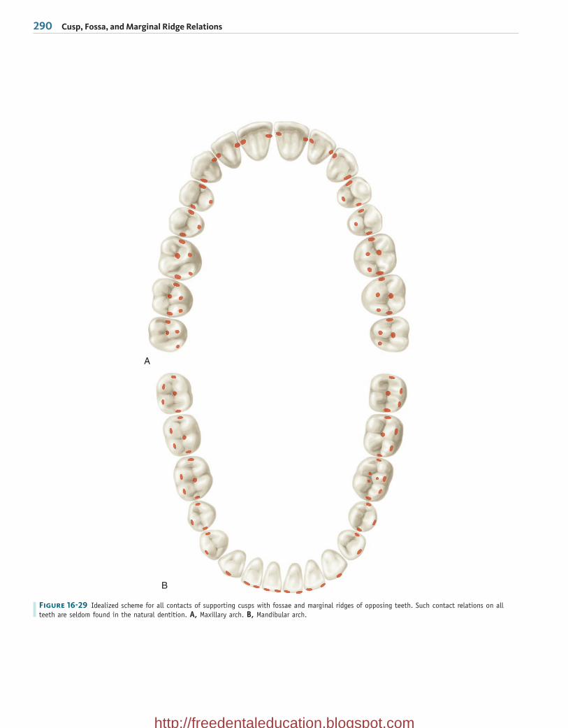
Figure 16-29 Idealized scheme for all contacts of supporting cusps with fossae and marginal ridges of opposing teeth. Such contact relations on all teeth are seldom found in the natural dentition. A, Maxillary arch. B, Mandibular arch.
A
B
290 Cusp, Fossa, and Marginal Ridge Relations
http://freedentaleducation.blogspot.com

Chapter 16 Occlusion 291
Figure 16-30 Lingual view of the teeth in centric occlusion.
Figure 16-31 Occlusal contact relations in the intercuspal position consistent with Angle Class I molar relationship. A, Viewed from the facial aspect. B, Viewed from the lingual aspect.
(From Ash MM, Ramfjord S: Occlusion, ed 4, Philadelphia, 1995, Saunders.)
A B
Figure 16-32 Maxillary right first molar, occlusal landmarks. MBCR, Mesiobuccal cusp ridge; CF, central fossa (shaded area); MTF, mesial triangular fossa (shaded area); MMR, mesial marginal ridge; MLCR, mesiolingual cusp ridge; OR, oblique ridge; DLCR, distolingual cusp ridge; DF, distal fossa; DTF, distal triangular fossa (shaded area); DMR, distal marginal ridge; DBCR, distobuccal cusp ridge.
DBCR
MBCR
CF
MTF
MMR
MLCR
OR
DLCR
DF
DTF
DMR
Figure 16-33 Mandibular right first molar, occlusal aspect. DBCR, Distobuccal cusp ridge; DBDG, distobuccal developmental groove; DCR, distal cusp ridge; DTF, distal triangular fossa (shaded area); DLCR, distolingual cusp ridge; LDG, lingual developmental groove; MLCR, mesiolingual cusp ridge; MTF, mesial triangular fossa (shaded area); SG, supplemental groove; MBCR, mesiobuccal cusp ridge; MBDG, mesiobuccal developmental groove.
MBCR
MBDG DBCR
DBDG
DCR
DTF
DLCR
LDG
SG
MTF
MLCR
shown in Figure 16-36. The simulated relationship does not reflect all the variance that may occur in these relationships. The lingual cusps of the maxillary premolars do not neces-sarily make contact in the fossa of the mandibular but occlude with the marginal ridges of the premolars or premo-lars and first molars as indicated in Figures 16-36, A, and 16-37.
CONCEPT OF 138 POINTS OF OCCLUSAL CONTACT
One scheme of occlusal contacts presented by Hellman included 138 points of possible occlusal contacts for 32 teeth.8 Later, with some modifications for application to complete occlusal restoration, most of the same contacts were made a part of the concept of occlusion in which sup-porting cusps and opposing stops (in centric relation) are tripoded and, with lateral/protrusive movements, immediate disocclusion of the posterior teeth takes place with canine (cuspid) guidance.
The list of occlusal contacts (total, 138) follows:
1. Lingual surfaces of maxillary incisors and canines, 62. Labial surface of mandibular incisors and canines, 63. Triangular ridges of maxillary buccal cusps of
premolars and molars, 164. Triangular ridges of lingual cusps of mandibular
premolars and molars, 165. Buccal embrasure of mandibular premolars and
molars, 8
http://freedentaleducation.blogspot.com

Figure 16-34 Mandibular right first molar, occlusal aspect. Shaded area is the central fossa. CP, Central pit; DMR, distal marginal ridge; DP, distal pit; CDG, central developmental groove; MP, mesial pit; MMR, mesial marginal ridge.
Buccal
Lingual
CP
Dis
tal
DMR
DPCDG
MPM
esia
l
MMR
Figure 16-35 Maxillary central incisor (lingual aspect). CL, Cervical line; CI, cingulum; MR, marginal ridge; IR, incisal ridge; LF, lingual fossa.
MR
CL
CI
MR
IRLF
Figure 16-36 A, Relationship of supporting cusps to marginal ridges. B, relationship of supporting cusps to fossae.
A B
6. Lingual embrasures of maxillary premolars and molars (including the canine and first premolar embrasure accommodating the mandibular premolar), 10
7. Lingual cusp points of maxillary premolars and molars, 16
8. Buccal cusp points of mandibular premolars and molars, 16
9. Distal fossae of premolars, 810. Central fossae of the molars, 1211. Mesial fossae of the mandibular molars, 612. Distal fossae of the maxillary molars, 613. Lingual grooves of the maxillary molars, 614. Buccal grooves of the mandibular molars, 6*
*Hellman did not list the distobuccal groove of the mandibular first molar, possibly because it is normally out of occlusal contact.
Therefore, if a complete description without the omis-sion of any detail of ideal occlusion is desired, close scrutiny of a good skull or casts showing 32 teeth would make it possible to compile a list of all ridge-sulcus combinations, all cusp embrasure combinations, and so on. Usually, if the combinations of points that have been mentioned in the last few pages can be established, some details such as the approximate location of hard contact in occlusion are auto-matic. Friel’s concept of occlusal contacts in an “ideal” occlusion is illustrated in Figure 16-37.43 Compare with the contacts in Figure 16-29.
Concepts of ideal occlusion are used primarily in ortho-dontics and restorative dentistry. Application of the goal of a total of 138 points for orthodontics and complete oral restorative rehabilitation has not been shown to be practical or necessary for occlusal stability or function. That is not to say that such concepts for ideal occlusal contacts have no
292 Cusp, Fossa, and Marginal Ridge Relations
http://freedentaleducation.blogspot.com
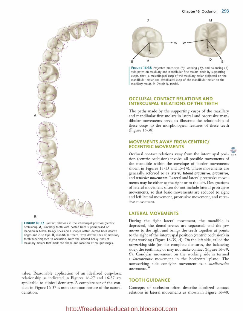
Chapter 16 Occlusion 293
Figure 16-37 Contact relations in the intercuspal position (centric occlusion). A, Maxillary teeth with dotted lines superimposed on mandibular teeth. Heavy lines and T shapes within dotted lines denote ridges and cusp tips. B, Mandibular teeth, with dotted lines of maxillary teeth superimposed in occlusion. Note the slanted heavy lines of maxillary molars that mark the shape and location of oblique ridges.
A
B
Figure 16-38 Projected protrusive (P), working (W), and balancing (B) side paths on maxillary and mandibular first molars made by supporting cusps, that is, mesiolingual cusp of the maxillary molar projected on the mandibular molar and distobuccal cusp of the mandibular molar on the maxillary molar. D, Distal; M, mesial.
D M
B
P
W
M
P
B
W
D
value. Reasonable application of an idealized cusp-fossa relationship as indicated in Figures 16-27 and 16-37 are applicable to clinical dentistry. A complete set of the con-tacts in Figure 16-37 is not a common feature of the natural dentition.
OCCLUSAL CONTACT RELATIONS AND INTERCUSPAL RELATIONS OF THE TEETH
The paths made by the supporting cusps of the maxillary and mandibular first molars in lateral and protrusive man-dibular movements serve to illustrate the relationship of these cusps to the morphological features of these teeth (Figure 16-38).
MOVEMENTS AWAY FROM CENTRIC/ECCENTRIC MOVEMENTS
Occlusal contact relations away from the intercuspal posi-tion (centric occlusion) involve all possible movements of the mandible within the envelope of border movements shown in Figures 15-13 and 15-14). These movements are generally referred to as lateral, lateral protrusive, protrusive, and retrusive movements. Lateral and lateral protrusive move-ments may be either to the right or to the left. Designations of lateral movement often do not include lateral protrusive movements, so that basic movements are reduced to right and left lateral movement, protrusive movement, and retru-sive movement.
LATERAL MOVEMENTS
During the right lateral movement, the mandible is depressed, the dental arches are separated, and the jaw moves to the right and brings the teeth together at points to the right of the intercuspal position (centric occlusion) in right working (Figure 16-39, A). On the left side, called the nonworking side (or, for complete dentures, the balancing side), the teeth may or may not make contact (Figure 16-39, C). Condylar movement on the working side is termed a laterotrusive movement in the horizontal plane. The nonworking side condylar movement is a mediotrusive movement.44
TOOTH GUIDANCE
Concepts of occlusion often describe idealized contact relations in lateral movements as shown in Figure 16-40.
II A, B
http://freedentaleducation.blogspot.com

However, in the natural dentition, a variety of contact rela-tions may be found, including group function, cuspid disoc-clusion only, or some combination of canine, premolar, and molar contacts in lateral movements. Group function refers to multiple contacts in lateral or eccentric mandibular move-ments (Figure 16-41, A) rather than simply canine (cuspid) guidance (Figure 16-41, B). Incisal guidance refers to contact of the anterior teeth during protrusive movements of the mandible. Condylar guidance and neuromuscular guidance are considered later.
PROTRUSIVE MOVEMENTS
During protrusive movements, the mandible is depressed and then moves forward, bringing the anterior teeth together at points most favorable for the incision of food. A retrusive movement follows protrusive movement to the intercuspal position.
OBTAINING CENTRIC RELATION
Guidance by the clinician of the jaw into centric relation (Figure 16-42, A and B) and then into initial contact with the teeth (centric relation contact), occurs usually with one or two (premature) contacts. Often the contacts are either on the oblique ridge of the maxillary molar and/or the mesial cusp ridge of the maxillary first premolar (Figure 16-43). In the centric relation position the condyle-disk assemblies are positioned on the anterior slope of the man-dibular fossae (Figure 16-44). After initial contact of the teeth the patient can squeeze the teeth together and the mandible will slide forward or eccentrically, depending on the position of the interference (Figure 16-45). This move-ment from the premature contact in centric relation to centric occlusion (intercuspal position) is called a slide in centric. Centric occlusion and intercuspal position are both maximal intercuspation. If the occlusal interferences in
centric relation contact are removed by selective grinding, the ability to close into maximum intercuspation without interference any place between centric relation and centric occlusion is called freedom in centric (see Figures 15-13 and 15-14).
RETRUSIVE MOVEMENTS
Retrusive movement from centric occlusion to retruded contact position in which the condyles are in the rearmost, uppermost position seems to occur in bruxism but infre-quently in mastication and swallowing, except when centric occlusion and centric relation are coincident or when maximum intercuspation occurs at centric relation contact. The adverse neuromuscular responses that may occur to retrusion and closure of the jaw in the presence of occlusal interferences to maximum intercuspation in centric relation can be detected electromyographically and by the clinician. However, the jaw does not go to the retrusive position nec-essarily even after elimination of such occlusal interferences by occlusal adjustment. Although the occlusion (anterior guidance) may be reconstructed to guide the mandible into the retruded contact position and maximum intercuspation in centric relation, a movement from centric relation to the intercuspal position (slide in centric) may develop again. The significance of the return of a small slide from centric relation occlusion to the intercuspal position (centric rela-tion to occlusion) has not been determined.45 The return of a slide occurs with orthodontics as well.46
Lateral Occlusal Relations
When the mandibular teeth make their initial contact with the maxillary teeth in right or left lateral occlusal relation, they bear a right or left lateral relation to intercuspal posi-tion or centric occlusion. The canines, premolars, and molars of one side of the mandible make their occlusal contact facial (labial or buccal) to their facial cusp ridges at some portion of their occlusal thirds (see Figure 16-40). Those points on the mandibular teeth make contact with maxillary teeth at points just lingual to their facial cusp ridges. The central and lateral incisors of the working side are not usually in contact at the same time; if they are, the labioincisal portions of the mandibular teeth of that side are in contact with the linguoincisal portions of the maxillary teeth.
During the sliding contact action, from the most facial contact points to centric occlusion, the teeth intercuspate and slide over each other in a directional line approximately parallel with the oblique ridge of the upper first molar. The oblique ridge of the maxillary first molar relates occlusally to the combined sulci of the distobuccal and developmental grooves of the occlusal surface of the mandibular first molar.
As the teeth of one side move from lateral relation to centric occlusion, the cusps and ridges bear a certain rela-tionship to each other; the cusps and ridges (including mar-
Figure 16-39 Right side contact relations of first maxillary and mandibular molars: A, Right working side. B, Centric occlusion (intercuspal position). C, Nonworking side.
A B C
294 Lateral Occlusal Relations
http://freedentaleducation.blogspot.com

Chapter 16 Occlusion 295
A
1
2
3
4
5
5
4
3
2
1 B
Figure 16-40 A, Patient’s left side showing left working side contacts (group function) and schematic of working side occlusal contacts and guiding inclines in left lateral movement. B, Patient’s right side showing nonworking side occlusal contacts and guiding inclines. Nonworking contacts are not necessary except in complete dentures.
(From Ash MM, Ramfjord S: Occlusion, ed 4, Philadelphia, 1995, Saunders.)
Figure 16-41 A, Right lateral movement: nonworking side. Multiple working side contacts. B, Right lateral movement: canine (cuspid) guidance on working side.
BA
http://freedentaleducation.blogspot.com

Figure 16-42 Guidance into centric relation by clinician. A, One-handed guidance. B, Two-handed guidance.
(From Ash MM, Ramfjord S: Occlusion, ed 4, Philadelphia, 1995, Saunders.)
BA
Figure 16-43 Mark of premature contact in centric relation on oblique ridge. RCP, Retruded contact position.
(From Ash MM, Ramfjord S: Occlusion, ed 4, Philadelphia, 1995, Saunders.)
RCP
Figure 16-44 A, Incorrect assumption about the normal position of the disk-condyle assembly. B, Correct position of the assembly in centric relation.
Disk
A B
Figure 16-45 Differences in jaw position between centric relation and the intercuspal position/centric occlusion position. A, Centric relation. B, Intercuspal position/centric occlusion.
(From Ash MM, Ramfjord S: Occlusion, ed 4, Philadelphia, 1995, Saunders.)
B
A
ginal ridges) of the canines and posterior teeth of the mandibular arch have an intercusping relationship to the cusps and ridges of the teeth of the maxillary arch (see Figure 16-38). The crowns of the teeth are formed in such a way that cusps and ridges may slide over each other without
mutual interference. In addition, the crowns of the teeth are turned on the root bases to accommodate the angled move-ment across their opponents (Figure 16-46).
The cusp tip of the mandibular canine moves through the linguoincisal embrasure of the maxillary lateral incisor and canine. The cusp tip is often in contact with one of the marginal ridges making up the lingual embrasure above it. Its mesial cusp ridge is usually out of contact during the
296 Lateral Occlusal Relations
http://freedentaleducation.blogspot.com

Chapter 16 Occlusion 297
lateral movement. Its distal ridge contacts the mesial cusp ridge of the maxillary canine.
The cusp tip of the mandibular first premolar moves through the occlusal embrasure of the maxillary canine and first premolar (Figures 16-47 and 16-48). Its mesiobuccal
ridge contacts the distal cusp ridge of the maxillary canine, and its distobuccal cusp ridge contacts the mesio-occlusal slope of the buccal cusp of the maxillary first premolar.
The mandibular second premolar buccal cusp moves through the occlusal embrasure and then over the linguo-occlusal embrasure of the maxillary first and second premo-lars. Its mesiobuccal cusp ridge contacts the disto-occlusal slope of the buccal cusp of the maxillary first premolar, and its distobuccal cusp ridge contacts the mesio-occlusal slope of the buccal cusp of the upper second premolar.
The lingual cusps of all premolars are out of contact until centric relation is attained. Then the only lingual cusps in contact are those of the maxillary premolars, with the pos-sible addition of the distolingual cusp of a mandibular second premolar of the three-cusp type. The molars have a more involved lateral occlusal relation because of their more complex design.
It has been noted previously, while describing the lateral occlusal relations of canines and premolars, that cusps, cusp ridges, sulci, and embrasures bear an interrelationship to each other. Cusps and elevations on the teeth of one arch pass between or over cusps and through embrasures or sulci. The tooth form and the alignment of the opposing teeth of both jaws make this possible. The cusps of the teeth of one jaw simply do not ride up and down the cusp slopes of the teeth in the opposing jaw. This explanation of the occlusal process has created wide misunderstanding. The cusp, ridge, fossa, and embrasure form of occlusion allow interdigitation without a “locked-in” effect. There is no clashing of cusp against cusp or any interference between parts of the occlu-sal surfaces if the development is proper.
OCCLUSAL CYCLE IN THE MOLAR AREAS DURING RIGHT OR LEFT LATERAL OCCLUSAL RELATIONS
In lateral movements during mastication, the mandible drops downward and to the right or left of centric occlusion. As it continues the cycle of movement and returns toward centric occlusion, the bucco-occlusal portions of mandibular molars come into contact with the occlusal portions of the maxillary molars lingual to the summits of buccal cusps and
Figure 16-46 Occlusion of right maxillary first premolar with its distal (D) and lingual surfaces surveyed by a right angle distolingually (see Figure 9-15). A, Line following the angulation of the mesial surface (M), which is somewhat parallel with the vertical line of the right angle distally. This formation allows a proper contact relationship with the distal proximal surface of the maxillary canine; simultaneously, it cooperates with the canine in keeping the lingual embrasure design within normal limitations. B, Line demonstrating a more extreme angulation of the distal portion of the first premolar. This form allows cusp and ridge to bypass mandibular teeth over the distal marginal ridge surface of the maxillary tooth with normal jaw movements during lateral occlusal relations. C, Line aligned with mesiobuccal and distobuccal line angles, demonstrating the adaptation of the form of the buccal surface of the crown to the dental arch form without changing the functional position of the crown and root.
A B
C
M D
Figure 16-47 Lingual aspect. Interrelationship of cusp, ridges, and embrasures. A, Mandibular first premolar relation to maxillary canine and the first premolar approaching occlusal contact. B, Mandibular first molar relation to the maxillary first molar on the verge of occlusal contact.
A B
Figure 16-48 Arrows indicate path of left lateral movement of mandibular teeth over the maxillary teeth on nonworking side. Note the relationship of paths to morphological features of the teeth and embrasures.
http://freedentaleducation.blogspot.com
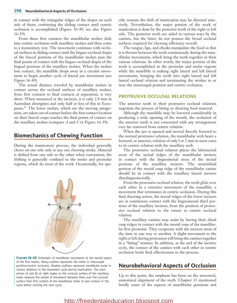
in contact with the triangular ridges of the slopes on each side of them, continuing the sliding contact until centric occlusion is accomplished (Figure 16-49; see also Figure 16-39).
From these first contacts the mandibular molars slide into centric occlusion with maxillary molars and then come to a momentary rest. The movement continues with occlu-sal surfaces in sliding contact until the linguo-occlusal slopes of the buccal portions of the mandibular molars pass the final points of contact with the linguo-occlusal slopes of the lingual portions of the maxillary molars. When the molars lose contact, the mandible drops away in a circular move-ment to begin another cycle of lateral jaw movement (see Figure 16-49).
The actual distance traveled by mandibular molars in contact across the occlusal surfaces of maxillary molars, from first contacts to final contacts at separation, is very short. When measured at the incisors, it is only 2.8 mm in Australian aborigines and only half or less of this in Euro-peans.47 The lower molars, which are the moving antago-nists, are taken out of contact before the first contact location on their buccal cusps reaches the final points of contact on the maxillary molars (compare A and C in Figure 16-39).
Biomechanics of Chewing Function
During the masticatory process, the individual generally chews on one side only at any one chewing stroke. Material is shifted from one side to the other when convenient; the shifting is generally confined to the molar and premolar regions, which do most of the work. Occasionally, for spe-
cific reasons the shift of mastication may be directed ante-riorly. Nevertheless, the major portion of the work of mastication is done by the posterior teeth of the right or left side. The posterior teeth are aided in various ways by the canines, but the latter do not possess the broad occlusal surfaces required for chewing efficiency overall.
The tongue, lips, and cheeks manipulate the food so that it is thrown between the teeth continuously during the man-dibular movements, which bring the teeth together in their various relations. In other words, the major portion of the work is accomplished in the premolar and molar regions while the mandible is making right lateral and left lateral movements, bringing the teeth into right lateral and left lateral occlusal relation and terminating the strokes in or near the intercuspal position and centric occlusion.
PROTRUSIVE OCCLUSAL RELATIONS
The anterior teeth in their protrusive occlusal relations negotiate the process of biting or shearing food material.
Although the mandible may be lowered considerably in producing a wide opening of the mouth, the occlusion of the anterior teeth is not concerned with any arrangement very far removed from centric relation.
When the jaw is opened and moved directly forward to the normal protrusive relation, the mandibular arch bears a forward, or anterior, relation of only l or 2 mm in most cases to its centric relation with the maxillary arch.
The protrusive occlusal relation places the labioincisal areas of the incisal ridges of the mandibular incisors in contact with the linguoincisal areas of the incisal portions of the maxillary incisors. The mesiolabial portion of the mesial cusp ridge of the mandibular canine should be in contact with the maxillary lateral incisors distolinguoincisally.
From the protrusive occlusal relation, the teeth glide over each other in a retrusive movement of the mandible, a movement that terminates in centric occlusion. During this final shearing action, the incisal ridges of the lower incisors are in continuous contact with the linguoincisal third por-tions of the maxillary incisors, from the position of protru-sive occlusal relation to the return to centric occlusal relation.
The maxillary canines may assist by having their distal cusp ridges in contact with the mesial cusp of the mandibu-lar first premolar. They cooperate with the incisors most of the time in one way or another. A slight movement to the right or left during protrusion will bring the canines together in a “biting” manner. In addition, at the end of the incisive cycle, the contact of the canines with each other in centric occlusion lends final effectiveness to the process.
Neurobehavioral Aspects of Occlusion
Up to this point, the emphasis has been on the structural, anatomical alignment of the teeth. Chapter 15 mentioned briefly some of the aspects of mandibular positions and
Figure 16-49 Schematic of mandibular movements at the mesial aspect of the first molars. Heavy outline represents the molar in intercuspal position/centric occlusion. Shadow outlines represent mandibular molar in various relations in the movement cycle during mastication. The short arrows (A and B) at right angles to the occlusal surface of the maxillary molar measure the extent of movement between them over the occlusal surface from first contact of the mandibular molar to last contact in the cycle before starting the next cycle.
A B
298 Neurobehavioral Aspects of Occlusion
http://freedentaleducation.blogspot.com

Chapter 16 Occlusion 299
movements and muscle function. In addition, although it would be impossible to do justice to the topic of the neuro-science of occlusion in a brief review, it is imperative to call attention to the meaning of occlusion in its broadest sense.
Recent ideas concerning the diagnosis and treatment of disturbances such as chronic orofacial pain, temporoman-dibular disorders, craniomandibular disorders, and bruxism, and the diagnosis and treatment of malocclusion involving orthognathic surgery require a greater knowledge of the neurobehavioral aspects of oral motor behavior than ever before in the practice of dentistry.
The neurobehavioral aspects of occlusion relate to func-tion and parafunction of the stomatognathic system. Func-tion includes a variety of actions or human behavior such as chewing, sucking, swallowing, speech, and respiration. Parafunction refers to action such as bruxism (e.g., clenching and grinding of the teeth). All these functions require highly developed sensorimotor mechanisms. The coordination of occlusal contacts, jaw motion, and tongue movement during mastication requires a very intricate control system involv-ing a number of guiding influences from the teeth and their supporting structures, TMJs, masticatory muscles, and higher centers in the central nervous system. Frequent contact of the teeth during mastication without biting the tongue; closure of the jaw to facilitate swallowing (occurring about 600 times a day); remarkable tactile sensitivity in which threshold values for detecting foreign bodies between the teeth may be as little as 8 µm; and the presence of pro-tective reflexes suggest the need for intricate mechanisms of control of jaw position and occlusal forces.
The presence of several classes of teeth, powerful mus-culature, and a most delicate positional control system indicates that it is important to understand the strategy underlying such sensitive control mechanisms. Although the ease with which these mechanisms may be disturbed at the periphery (i.e., the teeth, joints, periodontium, peripheral neural system) and centrally (brainstem and higher centers) is not well understood, the adaptive capacity of the stoma-tognathic system appears to be considerable. On an indi-vidual clinical basis, however, the responses of a patient to occlusal therapy may be reflected in oral behavior outside the range of normal. Inasmuch as function and parafunction share similar anatomical, physiological, and psychological substrates, it is necessary to review briefly the neurobehav-ioral correlates of the activities of the stomatognathic system.
OCCLUSAL STABILITY
The stability of the occlusion and the maintenance of tooth position are dependent on all of the forces that act on the teeth. Occlusal forces, eruptive forces, lip and cheek pres-sure, periodontal support, and tongue pressure are all involved in maintaining the position of the teeth. As long as all of these forces are balanced, the teeth and the occlusion will remain stable. Should one or more of the influences change in magnitude, duration, or frequency, stability is lost and the teeth will shift, disrupting a previously stable occlu-
sion. The loss of teeth, tooth structure, or occlusal support-ing cusps, or a decrease in their support from periodontal disease or trauma is a factor in maintaining occlusal stability.
Occlusal stability refers to the tendency of the teeth, jaws, joints, and muscles to remain in an optimal functional state. A few of the mechanisms involved include mesial migration of teeth, eruption of teeth to compensate for occlusal wear or intrusion by occlusal forces, remodeling of bone, protec-tive reflexes and control of occlusal forces, reparative proc-esses, and a number of others even less understood than those just listed. Although the strategy for stability related to the functional level required for survival appears obvious, orchestration of such diverse mechanisms can be couched only in terms such as homeostasis. The influence on occlusal stability of such factors as disease, aging, and dysfunction has yet to be clarified.
From a clinical standpoint, several concepts of occlusal stability are used as goals for occlusal therapy, including maintenance of a stable jaw relation in centric occlusion and centric relation; direction of occlusal forces along the long axis of a tooth; maintenance of centric stops, supporting cusps, and contact vertical dimension; replacement of lost teeth; and control of tooth mobility. Discussion of these aspects of occlusal stability is more appropriately found in books on occlusion.
Mesial migration is a term used to describe the migration of teeth in a mesial direction. The cause of this phenomenon has not been fully clarified, although a number of ideas have been advanced. There seems to be little doubt that mesial drift does occur, but no general agreement is evident on how much movement occurs, which teeth move, and how the movement is achieved. Suggested causes include traction of the transseptal fiber system,48 forces of mastication,49,50 and tongue pressure.51 The strategy behind the mesial migration appears to be related to closure of proximal tooth contacts. Although occlusal forces may be considered as a passive mechanism and traction of transseptal fibers as an active one, it is difficult to determine in what way occlusal stability is influenced. The contact relations of the teeth may promote occlusal stability, but if incorrect relations are present, opening of proximal contacts can occur (Figure 16-50).
A tendency exists to assume that a particular arrangement of teeth is unstable (Figure 16-51); however, such an occlu-sal relationship may have become stabilized, at least at a particular point in time. Whether an occlusion is fully stable can be determined only by periodic evaluation of that occlu-sion. Many factors (caries, periodontal disease, occlusal trauma, bruxism) may upset the delicate balance of an already marginally stable occlusion.
An ideal occlusion may be defined as one that has no structural, functional, or neurobehavioral characteristics that tend to interfere with occlusal stability. A response to bruxism may be loss of tooth structure, increased tooth mobility, root resorption (Figure 16-52), or decreased tooth mobility and increased density and thickness of the support-ing tissues.
http://freedentaleducation.blogspot.com

GUIDANCE OF OCCLUSION
Guidance of occlusion is usually discussed only in terms of tooth contact or anatomical or physical guidance, and more specifically in relation to canine and incisal guidance. Less specific is the term anterior guidance, which may refer to tooth guidance for all or any of the anterior teeth or to guidance involving the neuromuscular system. Yet another type of guidance is condylar (disk-condylar complex) guid-ance, which, like incisal guidance, may refer to so-called mechanical equivalents of guidance on an articulator. Again, as with anterior guidance, the neuromuscular aspects of condylar guidance are not well understood. The paths of the condyles in the mandibular fossae are not well represented
Figure 16-50 A, Opening of contact between mandibular molars due to a protrusive interference on the second molar (mirror view of left side). B, Outline of excessive restored mesial buccal cusp ridge, which involved protrusive bruxing by the patient.
B
A
Figure 16-51 Occlusal instability cannot be determined simply on the basis of occlusal contact relations and spacing of the teeth. A changing occlusion that restabilizes suggests adaptation.
Figure 16-52 Root resorption associated with high restoration and clenching habit. Top, Prior to placement of restoration. Bottom left, “Soreness” of the tooth began soon after the restoration and then rapidly some root resorption. High restoration was removed by selective grinding. Bottom right, Several months later, no evidence of soreness or additional root resorption.
300 Neurobehavioral Aspects of Occlusion
http://freedentaleducation.blogspot.com

Chapter 16 Occlusion 301
in the mechanical equivalents of an articulator, especially in less than a “fully adjustable” articulator; and neuromuscular mechanisms are not represented at all.
It is of particular importance to the clinician to make certain that physical guidance of a restoration (or of the natural teeth in the treatment of dysfunction or malocclu-sion) is in harmony with the neuromuscular system and neurobehavioral attributes of the patient. Although some degree of compatibility may be assured through evaluation of occlusal relations and determination that smooth gliding movements are present in various excursions, the acceptance and adaptability of the neuromuscular system may not be apparent until an unfavorable response occurs (Figure 16-53).
ADAPTATION
In an ideal occlusion, there should be no need for adapta-tion, but the criteria for it can be guidelines only, because their implementation may reflect clinical skill beyond the ordinary. Even minor occlusal discrepancies in a few indi-viduals may result in acute orofacial pain and/or TMJ and muscle symptoms. It is not uncommon to have some kind of response (structural function and/or psychological) after the restoration of a maxillary central incisor if an occlusal
interference to complete closure in centric occlusion has been placed by mistake in the restoration (Figure 16-54). If the interference cannot be avoided comfortably by man-dibular displacement in chewing and swallowing by neu-romuscular mechanisms (functional adaptation), if the tooth becomes mobile and is moved out of position (structural adaptation), and/or if the patient cannot ignore the discom-fort or the presence of change for even a short period (behavioral adaptation), overt symptoms of dysfunction of the muscles, joints, periodontium, or teeth (pulp) may occur. However, such adaptive response and failure of adaptation are observed only rarely, and such observations do not qualify as scientific evidence. Models of research to test the validity of these clinical observations have suffered from serious flaws in design. Still, no clinician would knowingly put an occlusal interference in a restoration.
In addition to physical guidance from the teeth when the teeth are in contact during mastication or empty move-ments, mandibular guidance may occur before and during contact of the teeth from receptors in the periodontium and TMJs, and from other peripheral sensory receptors, as well as from higher centers in the central nervous system. It has been suggested that the anatomical relationships of the teeth and joints provide passive guidances and that active guid-ances involve reflexes originating in receptors in or around
Figure 16-53 Anterior crowding. A, Absence of crowding. B, Crowding following restorative dentistry by only a few months. Clinical findings of very tight proximal contacts and an occlusal interference in centric relation are only presumptive evidence of a cause-and-effect relationship between restorative treatment and anterior crowding.
B
A
Figure 16-54 Occlusal interference to closure in intercuspal position/centric occlusion due to restoration of the left maxillary central incisor. A, Centric relation position. B, Articulating paper mark on mandibular left lateral incisor. Temporomandibular disorder–like symptoms were relieved with adjustment of the centric occlusion interference.
B
A
http://freedentaleducation.blogspot.com
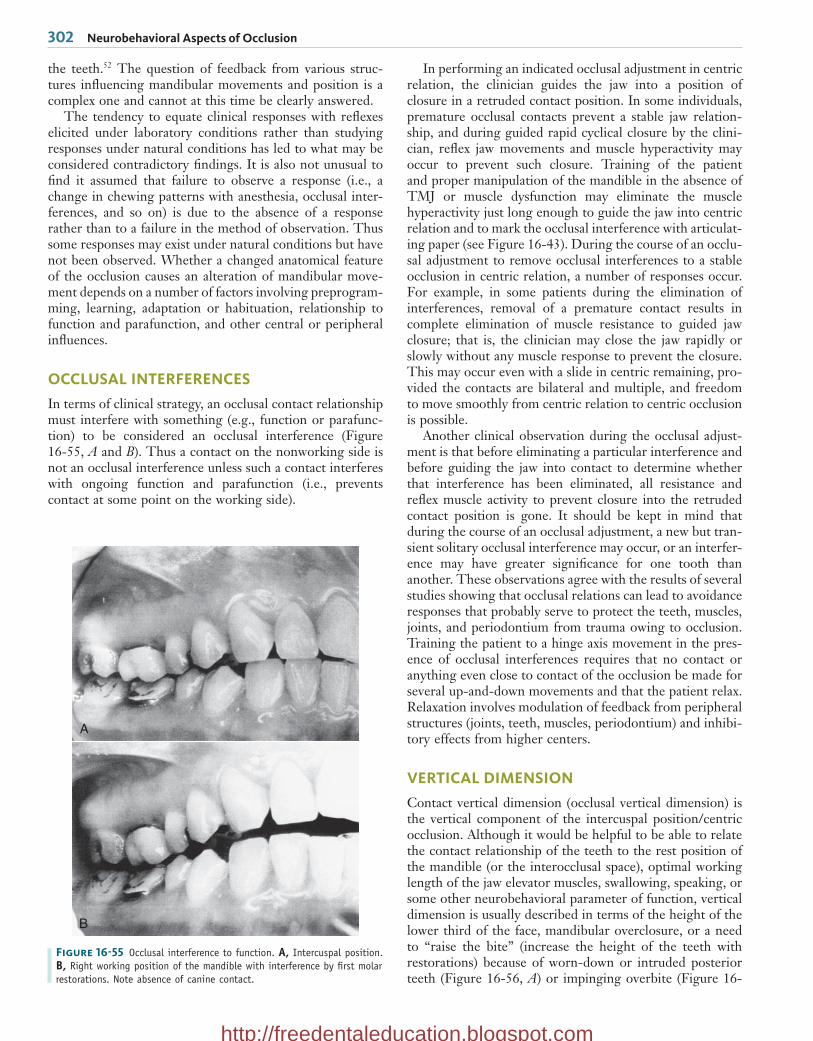
the teeth.52 The question of feedback from various struc-tures influencing mandibular movements and position is a complex one and cannot at this time be clearly answered.
The tendency to equate clinical responses with reflexes elicited under laboratory conditions rather than studying responses under natural conditions has led to what may be considered contradictory findings. It is also not unusual to find it assumed that failure to observe a response (i.e., a change in chewing patterns with anesthesia, occlusal inter-ferences, and so on) is due to the absence of a response rather than to a failure in the method of observation. Thus some responses may exist under natural conditions but have not been observed. Whether a changed anatomical feature of the occlusion causes an alteration of mandibular move-ment depends on a number of factors involving preprogram-ming, learning, adaptation or habituation, relationship to function and parafunction, and other central or peripheral influences.
OCCLUSAL INTERFERENCES
In terms of clinical strategy, an occlusal contact relationship must interfere with something (e.g., function or parafunc-tion) to be considered an occlusal interference (Figure 16-55, A and B). Thus a contact on the nonworking side is not an occlusal interference unless such a contact interferes with ongoing function and parafunction (i.e., prevents contact at some point on the working side).
Figure 16-55 Occlusal interference to function. A, Intercuspal position. B, Right working position of the mandible with interference by first molar restorations. Note absence of canine contact.
B
A
In performing an indicated occlusal adjustment in centric relation, the clinician guides the jaw into a position of closure in a retruded contact position. In some individuals, premature occlusal contacts prevent a stable jaw relation-ship, and during guided rapid cyclical closure by the clini-cian, reflex jaw movements and muscle hyperactivity may occur to prevent such closure. Training of the patient and proper manipulation of the mandible in the absence of TMJ or muscle dysfunction may eliminate the muscle hyperactivity just long enough to guide the jaw into centric relation and to mark the occlusal interference with articulat-ing paper (see Figure 16-43). During the course of an occlu-sal adjustment to remove occlusal interferences to a stable occlusion in centric relation, a number of responses occur. For example, in some patients during the elimination of interferences, removal of a premature contact results in complete elimination of muscle resistance to guided jaw closure; that is, the clinician may close the jaw rapidly or slowly without any muscle response to prevent the closure. This may occur even with a slide in centric remaining, pro-vided the contacts are bilateral and multiple, and freedom to move smoothly from centric relation to centric occlusion is possible.
Another clinical observation during the occlusal adjust-ment is that before eliminating a particular interference and before guiding the jaw into contact to determine whether that interference has been eliminated, all resistance and reflex muscle activity to prevent closure into the retruded contact position is gone. It should be kept in mind that during the course of an occlusal adjustment, a new but tran-sient solitary occlusal interference may occur, or an interfer-ence may have greater significance for one tooth than another. These observations agree with the results of several studies showing that occlusal relations can lead to avoidance responses that probably serve to protect the teeth, muscles, joints, and periodontium from trauma owing to occlusion. Training the patient to a hinge axis movement in the pres-ence of occlusal interferences requires that no contact or anything even close to contact of the occlusion be made for several up-and-down movements and that the patient relax. Relaxation involves modulation of feedback from peripheral structures (joints, teeth, muscles, periodontium) and inhibi-tory effects from higher centers.
VERTICAL DIMENSION
Contact vertical dimension (occlusal vertical dimension) is the vertical component of the intercuspal position/centric occlusion. Although it would be helpful to be able to relate the contact relationship of the teeth to the rest position of the mandible (or the interocclusal space), optimal working length of the jaw elevator muscles, swallowing, speaking, or some other neurobehavioral parameter of function, vertical dimension is usually described in terms of the height of the lower third of the face, mandibular overclosure, or a need to “raise the bite” (increase the height of the teeth with restorations) because of worn-down or intruded posterior teeth (Figure 16-56, A) or impinging overbite (Figure 16-
302 Neurobehavioral Aspects of Occlusion
http://freedentaleducation.blogspot.com

Chapter 16 Occlusion 303
56, B). At the present time, no acceptable test of a presumed loss of vertical dimension appears available. The neuro-behavioral aspects of the interocclusal space and occlusal vertical dimension are complex and require much further study.
It is not possible to determine with scientific assurance that aggressive bruxing and clenching in centric have caused intrusion or wear of the teeth or has been compensated for by eruption of the posterior teeth. Even short-term intru-sion or loss of stops on posterior teeth may result in reflex-produced responses from anterior teeth because of premature contacts on anterior teeth. Again, no acceptable tests are available to determine with scientific assurance, even in the presence of dysfunction (e.g., temporomandibular disorder), that correction of a presumed loss of contact vertical dimen-sion will correct the dysfunction.
Oral Motor Behavior
The term oral motor behavior is a convenience of speech by which it is possible to refer with a brief phrase to observable actions involving orofacial structures, including “simple” actions like assumption of mandibular rest position and much more complex movements such as mastication. Human behavior reflects the translation of past, present, and ongoing ideas and learning (including sensations and emotions) into movements and actions. Although many of the responses or actions are common among all persons, the subjective
response of a particular individual to a stimulus (including a change in the occlusion) may involve an inner experiential aspect of emotion in which the sensory experience may not fall into the usual acceptable range of pleasant or unpleasant. This aspect of sensation, referred to as affect, is the basis of much suffering and pleasure, including that related to the occlusion and occlusal therapy. In mentalistic terms, feelings of pleasantness and unpleasantness are correlated with “motivation or intention to respond” and with emotion. Even a simple reflex may be considered as a unit of behavior. The advantage of viewing occlusion (in terms of function and parafunction) as human behavior is that the clinician has a better understanding of functional disturbances (e.g., TMJ and muscle disorders) and the recognition that how patients feel and respond to their occlusion is an important aspect of diagnosis and dental treatment.
MOTIVATION
Emotion is a motivational phenomenon that plays a signifi-cant role in the determination of behavior. Motivation or drive and emotional states may be the basis for oral motor behavior, which is a fundamental component of ingestive responses, and for other behaviors essential for adaptation and survival. In effect, oral motor behavior can be initiated not only by situations involving cognitive processes but also by emotive processes including homeostatic drives (e.g., hunger) concerned with the internal environment and non-homeostatic drives (e.g., fear) related to adaptation to the external environment. The external environment may begin at the interface between oral sensory receptors and external stimuli involved in oral function and parafunction. However, ideas suggesting a “hard” interface between the internal and external environment are rapidly being reevaluated in terms of functional criteria. It is no longer possible to restrict thinking to a concept of the external environment as being “out there.” From a psychophysiological standpoint, an interface between the external and internal worlds may not exist.
HOMEOSTASIS
As already indicated, oral motor behavior involves the trans-lation of thought, sensation, and emotion into actions. Implied is the idea that actions or behavior may change because of learning and that some drive or motivation alters existing responses to environmental variables. Although the neural substrate for the translation of innate drives appears to exist and regulatory behavior such as eating has clear value to the immediate survival of an individual, other oral motor behavior may not have clear antecedents in individual or group survival. However, during the development of the nervous system and motor functions, oral motor behavior may be highly dependent on homeostatic drives concerned with ingestive processes. Later oral motor behavior is an expression of the plasticity of the organism over a long period and consists of a whole complex of emotive and cognitive determinants that cannot be related easily to a
Figure 16-56 Vertical dimension. A, Use of a posterior bilateral onlay splint in an attempt to “raise the bite” and eliminate temporomandibular disorder symptoms. B, An impinging overbite may require comprehensive orthodontics for correction, not an appliance as in A.
B
A
http://freedentaleducation.blogspot.com

hypothetical construct of homeostatic needs in the adult organism even in a teleological sense.
EXECUTION OF MOTOR BEHAVIOR
Although oral motor behavior is judged on the basis of observable actions, the strategy and tactics of the occlusal aspects of human behavior are based on past dental experi-ences and the present state of the joints, muscles, periodon-tium, contact relation of the teeth, and central nervous system. The neural mechanisms that underlie the initiation, programming, and execution of motor behavior can be described only briefly here.
Complex behavior may involve neuronal circuits called pattern generators, which, when activated, elicit stereotyped, rhythmic, and/or coordinated movements. The pattern generators for locomotion appear to be in the spinal cord and to be activated by discrete nuclei or regions in the brainstem, and they can be accessed by higher-level struc-tures in the central nervous system. The pattern generators for chewing and swallowing are located in the brainstem medullary-pontine reticular formation. As already men-tioned, limbic structures appear to have access to pattern generators.
Chewing is a type of oral motor behavior that demon-strates centrally programmed movement and in part periph-erally driven movement. Controlled interaction is more often the case than either purely centrally programmed movement or purely peripherally controlled movement. Pattern generators may be part of the particular programs accessed by higher brain centers to generate complex behavior.
Swallowing
Swallowing involves the coordination of nearly 20 different muscles with motor neurons distributed from mesencephalic to posterior medullary levels. The patterning of muscular contractions is independent of the stimulus necessary to evoke swallowing. The neurons responsible for coordina-tion include a swallowing center, in which neuronal groups fire automatically in a particular sequence when stimulated to achieve the necessary pattern of muscle activity to produce swallowing. “Triggering,” or initiation of a central program, involves neurons in the motor cortex that act as command elements and control patterns of neuronal activity as well as receive feedback from the systems controlled. Thus move-ment may be centrally programmed (drive) and at the same time modulated by peripheral influences.
The cyclical movements of the jaw in mastication reflect past experiences, adaptive behavior, neuronal activity of the mesencephalic rhythm generator and the trigeminal motor nucleus, and the influence of oral reflexes, either condi-tioned or unconditioned. The pattern generator may be influenced by sensory input from the orofacial area and influences from higher centers in the central nervous system.
Summary
To understand occlusion in its broadest sense, it is necessary to consider, in addition to TMJ articulation, muscles, and teeth, some of the neurobehavioral mechanisms that give meaning to the presence and function of the masticatory system. Although many of the neural mechanisms mediating interaction between occlusion and thoughts, sensations, and emotions are complex and often indeterminate, it is possible to suggest strategies that could account for the variety of responses (physiological and psychological) that occur in function and parafunction.
The “obvious” strategy to compensate for wear of proxi-mal contact areas is mesial migration of the teeth, and the strategy to compensate for wear of the occlusal surfaces is eruption of the teeth. The strategy for regulating the con-traction of the jaw elevators to achieve a normal resting position of the mandible with a small interocclusal space is a postural reflex (stretch reflex). The overall strategy for motivation to have access to the muscles of mastication might be to provide a drive for ingestive processes, especially during the early stages of development of the masticatory system and maturation of the nervous system—swallowing in fetal life, suckling in the newborn, and chewing in the young infant.
It appears plausible, at least, that emotion may be impor-tant not only as a motivational phenomenon but also as a reflection of what is agreeable or disagreeable about some-thing that is placed in the mouth, including items not con-sidered to be food and perhaps even restorations that interfere with function or parafunction. Demonstration of evidence for and against strategies to explain completely neurobehavioral mechanisms of “occlusion” requires much more space than provided here.
The extensive “education” of persons through the televi-sion media, newspapers, and magazines and through profes-sional dental care and instructions to patients not only has produced an awareness of the teeth and mouth but also has coupled these structures to a sense of health and comfort. The affect involved in such a sense of well-being about oral health involves part of the same neurobehavioral substrate underlying innate drives, motivation, and emotional states necessary for biological adaptation and survival of the species. Ingestive processes, which include oral motor responses involved in mastication, are essential for survival. Functional disturbances of the masticatory system may involve psychophysiological mechanisms that are related to the teeth and their functions. Therefore occlusal interfer-ences to function or parafunction may then involve more than simply contact relations of the teeth; they may involve psychophysiological mechanisms of human behavior as well.
No scientific evidence is available for making a specific structure or psychophysiological mechanism the sole cause of TMJ/muscle dysfunction. However, any attempt to negate the role of the teeth in human behavior, including dysfunction, perhaps is made by those who have not had the
304 Summary
http://freedentaleducation.blogspot.com

Chapter 16 Occlusion 305
opportunity to observe the effect on affect, favorable neu-romuscular response, and elimination of discomfort when appropriate occlusal therapy is rendered.
Unfortunately, neuroscientific evidence to separate the subjective from objective clinical observations has not lived up to its full potential. Scientific clinical studies in which true cause-and-effect relationships can be determined are difficult to design, especially when such relationships may be indirect, “on-again, off-again,” and significantly influ-enced by the observer and other factors under natural condi-tions.52 Considerably more appropriate research is needed to establish the role of occlusion and related factors in TMJ/muscle dysfunction.
References1. Zwemer TJ: Mosby’s dental dictonary, St Louis, 1998,
Mosby.2. Ash MM, Ramfjord SP: Occlusion, ed 4, Philadelphia, 1995,
Saunders.3. Angle EH: The angle system of regulation and retention of the
teeth, ed 1, Philadelphia, 1887, S.S. White Dental Manufacturing.
4. Schuyler CH: Principles employed in full denture prostheses which may be applied to other fields of dentistry, J Am Dent Assoc 16:2045, 1929.
5. Beyron HL: Characteristics of functionally optimal occlusion and principles of occlusal rehabilitation, J Am Dent Assoc 48:648, 1954.
6. D’Amico A: The canine teeth—normal functional relation of the natural teeth in man, J South Calif Dent Assoc 1:6–23; 2:49–60; 4:127–142; 5:175–182; 6:194–208; 7:239–241, 1958.
7. Friel S: The development of ideal occlusion of the gum pads and teeth, Am J Orthod 40:1963, 1954.
8. Hellman M: Factors influencing occlusion. In Gregory WK, Broadbent BH, Hellman M, editors: Development of occlusion, Philadelphia, 1941, University of Pennsylvania Press.
9. Lucia VO: The gnathological concept of articulation, Dent Clin North Am 6:183, 1962.
10. Stallard H, Stuart C: Concepts of occlusion, Dent Clin North Am 7:591, 1963.
11. Ramfjord SP, Ash MM: Occlusion, Philadelphia, 1966, Saunders.
12. Falkner F: Deciduous tooth eruption, Arch Dis Child 32:386, 1957.
13. Richardson AS, Castaldi CR: Dental development during the first two years of life, J Can Dent Assoc 33:418, 1967.
14. Moyers RE: Handbook of orthodontics, ed 3, Chicago, 1973, Year Book.
15. Proffit WR et al: Contemporary orthodontics, St Louis, 1986, Mosby, p 67.
16. Murray JJ: Dynamics of occlusal adjustment: a cephalometric analysis, Alumni Bull Univ Mich Sch Dent 69:32, 1959.
17. Inuzuka K: Changes in molar relationships between the deciduous and permanent dentitions: a longitudinal study, Am J Orthod Dentofacial Orthop 93:19, 1990.
18. Bishara SE, Hoppens BJ, Jakobsen JR, et al: Changes in molar relationship between deciduous and permanent dentitions: a longitudinal study, Am J Orthod Dentofacial Orthop 93:19, 1988.
19. Clements EMB et al: Age at which deciduous teeth are shed, Br Med J 1:1508, 1957.
20. Bjork A: The use of metallic implants in the study of facial growth in children: method and application, Am J Phys Anthropol 29:243, 1968.
21. Bjork A, Skieller V: Normal and abnormal growth of the mandible: a system of longitudinal cephalometric implant studies over a period of 25 years, Eur J Orthod 51:1, 1983.
22. Moorrees C: The dentition of the growing child: a longitudinal study of dental development between 3 and 18 years of age, Cambridge, MA, 1959, Harvard University Press.
23. Howe RP, McNamara JA Jr, O’Conner KA: An examination of dental crowding and its relationship to tooth size and arch dimension, Am J Orthod 83:363, 1983.
24. Spillane LM, McNamara JA Jr: Arch width development relative to initial transpalatal width, J Dent Res IADR 1538:374, 1989 (abstract).
25. McNamara JA Jr, Brudon WL: Orthodontics and dentofacial orthopedics, Ann Arbor, 2001, Needham Press, p 33.
26. Moorrees C, Chadha JM: Available space for the incisors during dental development—a growth study based on physiologic age, Angle Orthod 35:12, 1965.
27. Lo RT, Moyers RE: Studies in the etiology and prevention of malocclusion. I. The sequences of eruption of the permanent dentition, Am J Orthod 39:460, 1953.
28. Knott J, Meredith HV: Statistics on the eruption of the permanent dentition from serial data from North American white children, Angle Orthod 36:68, 1966.
29. Martin R: Lehrbuck der anthropologie, Jena, Germany, 1914, G Fischer.
30. Hrdlizka A: Anthropometry, Philadelphia, 1920, Wistar Institute of Anatomy.
31. MacConaill MA, Scher EA: Ideal form of the human dental arcade, with some prosthetic applications, Dent Rec 69:285, 1949.
32. Scott JH: What determines the form of the dental arches, Orthod Rec 1:15, 1958.
33. Currier JH: A computerized geometric analysis of human dental arch form, Am J Orthod 56:164, 1969.
34. Spee FG: Die Verschiebungsbahn des Unterkiefers am Schädel, Arch Anat Physiol Anat Abt 285–294, 1890.
35. Monson GS: Occlusion as applied to crown and bridgework, J Nat Dent Assoc 7:399, 1920.
36. Dempster WT, Adams WJ, Duddles RA: Arrangement in the jaws of the roots of the teeth, J Am Dent Assoc 67:779, 1963.
37. Bonwill WGA: The geometrical and mechanical laws of the articulation of human teeth—the anatomical articulator. In Litch WF, editor: The American system of dentistry in treatises by various authors: operative and prosthetic dentistry, vol 2, Philadelphia, 1886–87, Lea & Febiger, pp 486–498.
38. Bonwill WGA: Scientific articulation of human teeth as founded on geometrical mathematical laws, Dent Items Interest 21:817, 1899.
39. Andrews LF: The diagnostic system: occlusal analysis, Dent Clin North Am 20:671, 1976.
40. Poole DFG: Evolution of mastication. In Anderson DJ, Matthews B, editors: Mastication, Bristol, England, 1976, John Wright and Sons.
41. Butler PM: A zoologist looks at occlusion, Br J Orthod 1:205, 1974.
http://freedentaleducation.blogspot.com

42. Mills JRE: Attrition in animals. In Poole DFG, Stack MV, editors: The eruption and occlusion of teeth, Cokston symposium no 27, London, 1976, Butterworth.
43. Friel S: Occlusion: observations on its development from infancy to old age, Int J Orthod Surg 13:322, 1927.
44. The glossary of prosthodontic terms, J Prosthet Dent 94(1):10, 2005.
45. Celenza FB: The centric position: replacement and character, J Prosthet Dent 30:591, 1973.
46. Johnston LE: Gnathologic assessment of centric slides in postretention orthodontic patients, J Prosthet Dent 60:712, 1988.
47. Beyron HL: Occlusal relations and mastication in Australian aborigines, Acta Odontol Scand 22:597, 1964.
48. Picton DCA, Moss JP: The effect of approximal drift of altering the horizontal component of biting force in adult monkeys (Macacus), Arch Oral Biol 25:45, 1980.
49. Dewel BF: Clinical observations on the axial inclination of teeth, Am J Orthod 35:98, 1949.
50. Van Beek H, Fidler VJ: An experimental study of the effect of functional occlusion on mesial tooth migration in macaque monkeys, Arch Oral Biol 22:269, 1977.
51. Yilmaz RS et al: Mesial drift of human teeth assessed from ankylosed deciduous molars, Arch Oral Biol 25:127, 1980.
52. Dubner R et al: The neural basis of oral and facial function, New York, 1978, Plenum Press.
BibliographyAckerman RJ: Tooth migration during the transitional dentition,
Dent Clin North Am 20:661, 1976.Anderson DJ, Matthews B, editors: Mastication, Bristol, England,
1976, John Wright and Sons.Arnold NR, Frumker SC: Occlusal treatment, Philadelphia, 1976,
Lea & Febiger.Ash MM: Paradigmatic shifts in occlusion and
temporomandibular disorders, J Oral Rehabil 28:1, 2001.Ash MM, Ramfjord SP: Introduction to functional occlusion,
Philadelphia, 1982, Saunders.Bates JF, Stafford GD, Harrison A: Masticatory function: a
review of the literature. I. The form of the masticatory cycle, J Oral Rehabil 2:281, 1975.
Baume LR: Physiologic tooth migration and its significance for the development of occlusion, J Dent Res 29:123, 1950.
Beyron HL: Optimal occlusion, Dent Clin North Am 13:537, 1969.
Brodie AG: Temporomandibular joint, Ill Dent J 8:2, 1939.Burch JG: Patterns of change in human mandibular arch width
during jaw excursions, Arch Oral Biol 17:623, 1972.Dawson P: Evaluation, diagnosis and treatment of occlusal problems,
St Louis, 1974, Mosby.Dolwick MF, Sanders B: TMJ internal derangement and arthrosis,
St Louis, 1985, Mosby.Finn SB: Clinical pedodontics, ed 4, Philadelphia, 1973,
Saunders.Gibbs CH et al: Functional movements of the mandible,
J Prosthet Dent 26:604, 1971.Graf H: Occlusal forces during function. In Rowe NH, editor:
Occlusal research in form and function, Ann Arbor, 1975, University of Michigan Press.
Gysi A: The problem of articulation, Dent Cosmos 52:1, 1910.Hannam AG et al: The relationship between dental occlusion,
muscle activity, and associated jaw movement in man, Arch Oral Biol 22:25, 1977.
Hatton M: Measure of the effects of heredity and environment in eruption of the deciduous teeth, J Dent Res 34:397, 1955.
Hemley S: Fundamentals of occlusion, Philadelphia, 1944, Saunders.
Higley LB: Some controversies over the temporomandibular joint, J Am Dent Assoc 27:594, 1940.
Klatsky M: A cinefluorographic study of the human masticatory apparatus in function, Am J Orthod 26:664, 1940.
Kornfeld M: Mouth rehabilitation, 2 vols, St Louis, 1967, Mosby.
Kraus JA: Dental anatomy and occlusion, Baltimore, 1969, Williams & Wilkins.
Kurth LE: Mandibular movements in mastication, J Am Dent Assoc 29:1769, 1942.
Leighton BC: Early recognition of normal occlusion. In McNamara JA Jr, editor: Craniofacial growth series: the biology of occlusal development, Monograph 7, Ann Arbor, 1977, Center for Human Growth and Development, University of Michigan.
Leighton BC: The early signs of malocclusion, Trans Eur Orthod Soc 45:353, 1969.
Logan WHG, Kronfeld R: Development of the human jaws and surrounding structures from birth to age of fifteen years, J Am Dent Assoc 20:379, 1933.
Lord FP: Movements of the jaw and how they are effected, Int J Orthod 23:557, 1937.
MacLean PD: Some psychiatric implications of physiological studies of front temporal portions of the limbic system (visceral brain), Electroencephalogr Clin Neurophysiol 4:407, 1952.
MacMillan HW: Foundations of mandibular movement, J Am Dent Assoc 231:429, 1934.
Mann AW, Pankey LD: Oral rehabilitation, J Prosthet Dent 10:135, 1960.
Matthews B: Mastication. In Lavelle CLB, editor: Applied physiology of the mouth, Bristol, England, 1975, John Wright and Sons.
Meredith HV: Order and age of eruption for the deciduous dentition, J Dent Res 25:43, 1946.
Mogenson GJ et al: From motivation to action: functional interface between the limbic system and the motor system, Prog Neurobiol 14:69, 1980.
Moss JP, Picton DC: The effect on approximal drift of cheek teeth of dividing mandibular molars of adult monkeys (Macacus), Arch Oral Biol 19:1211, 1974.
Moss JP, Picton DCA: Mesial drift in teeth in adult monkeys (Macacus) when forces from the cheeks and tongue have been eliminated, Arch Oral Biol 15:979, 1970.
O’Leary TJ: Tooth mobility, Dent Clin North Am 13:567, 1969.
Prentiss HL: Regional anatomy emphasizing mandibular movements with specific reference to full denture construction, J Am Dent Assoc 15:1085, 1923.
Rees LA: The structure and function of the mandibular joint, J Br Dent A 96:125, 1954.
Robinson M: The temporomandibular joint: theory of reflex controlled nonlever action of the mandible, J Am Dent Assoc 33:1260, 1946.
306 Bibliography
http://freedentaleducation.blogspot.com

Chapter 16 Occlusion 307
Savalle WPM: Some aspects of the morphology of the human temporomandibular joint capsule, Acta Anat (Basel) 131:292, 1988.
Sessle BJ, Hannam AG, editors: Mastication and swallowing: biology and clinical correlates, Toronto, 1976, University of Toronto Press.
Taylor A: Proprioception in the strategy of jaw movement control. In Kawamura Y, Dubner R, editors: Oral-facial sensory and motor functions, Tokyo, 1981, Quintessence.
Yamada Y, Ash MM: An electromyographic study of jaw opening and closing reflexes in man, Arch Oral Biol 27:13, 1982.
Use the CD-ROM to view animations and study ques-tions; and go to the site for additional study resources.
http://freedentaleducation.blogspot.com

This page intentionally left blank
http://freedentaleducation.blogspot.com

Appendix A
Review of Tooth Morphology
309
Appendix A includes color renditions of Figures 2-3 and 2-4 considered in Chapter 2 on the eruption and development of the teeth. They can be used to demonstrate to patients the development of the dentitions from 5 months in utero to adolescence and adulthood.
Also included are representative views of the facial/labial/buccal and occlusal/incisal aspects of all of the teeth consid-
ered in Chapters 6 through 12. Because of the proximity of these illustrations to the traits and characteristics of the teeth provided in Appendix B, page turns to earlier chapters can be minimized. It is also possible to view these same illustrations on the CD-ROM while examining the traits and characteristics in Appendix B.
http://freedentaleducation.blogspot.com
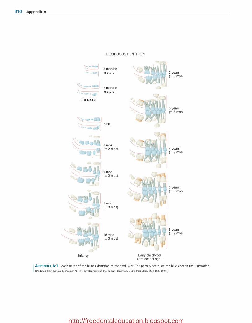
310 Appendix A
Appendix A-1 Development of the human dentition to the sixth year. The primary teeth are the blue ones in the illustration.
(Modified from Schour L, Massler M: The development of the human dentition, J Am Dent Assoc 28:1153, 1941.)
2 years(� 6 mos)
3 years(� 6 mos)
4 years(� 9 mos)
5 years(� 9 mos)
6 years(� 9 mos)
5 monthsin utero
7 monthsin utero
PRENATAL
DECIDUOUS DENTITION
Birth
6 mos(� 2 mos)
9 mos(� 2 mos)
1 year(� 3 mos)
18 mos(� 3 mos)
Early childhood(Pre-school age)
Infancy
http://freedentaleducation.blogspot.com

Review of Tooth Morphology 311
Appendix A-2 Development of the human dentition from the seventh year to maturity. Note the displacement of the primary teeth.
(Modified from Schour L, Massler M: The development of the human dentition, J Am Dent Assoc 28:1153, 1941.)
11 years(� 9 mos)
12 years(� 6 mos)
15 years(� 6 mos)
21years
35years
7 years(� 9 mos)
8 years(� 9 mos)
9 years(� 9 mos)
10 years(� 9 mos)
MIXED DENTITION PERMANENT DENTITION
Late childhood(school age)
Adolescenceand adulthood
http://freedentaleducation.blogspot.com

312 Appendix A
Appendix A-3 Maxillary central incisor (right).
Mesial
Incisal
Distal
Facial/Labial
D M
Lingual
http://freedentaleducation.blogspot.com

Review of Tooth Morphology 313
Appendix A-4 Maxillary lateral incisor (right).
Incisal
D M
Mesial Distal
LingualFacial/Labial
http://freedentaleducation.blogspot.com

314 Appendix A
Appendix A-5 Mandibular central incisor (right).
Incisal
D M
DistalMesial
Facial/Labial Lingual
http://freedentaleducation.blogspot.com

Review of Tooth Morphology 315
Appendix A-6 Mandibular lateral incisor (right).
D M
Incisal
Mesial Distal
LingualFacial/Labial
http://freedentaleducation.blogspot.com

316 Appendix A
Appendix A-7 Maxillary canine (right).
Incisal
D M
Mesial Distal
LingualFacial/Labial
http://freedentaleducation.blogspot.com

Review of Tooth Morphology 317
Appendix A-8 Mandibular canine (right).
Incisal
D M
Mesial Distal
LingualFacial/Labial
http://freedentaleducation.blogspot.com

318 Appendix A
Appendix A-9 Maxillary first premolar.
Occlusal
D M
Mesial Distal
LingualFacial/Buccal
http://freedentaleducation.blogspot.com

Review of Tooth Morphology 319
Appendix A-10 Maxillary second premolar (right).
Occlusal
D M
Mesial Distal
LingualFacial/Buccal
http://freedentaleducation.blogspot.com

320 Appendix A
D M
Occlusal
DistalMesial
Facial/Buccal Lingual
Appendix A-11 Mandibular first premolar (right).
http://freedentaleducation.blogspot.com

Review of Tooth Morphology 321
Appendix A-12 Mandibular second premolar (right).
Occlusal
D M
Mesial Distal
Facial/Buccal Lingual
http://freedentaleducation.blogspot.com

322 Appendix A
Appendix A-13 Maxillary first molar (right).
D M
Occlusal
DistalMesial
LingualFacial/Buccal
http://freedentaleducation.blogspot.com

Review of Tooth Morphology 323
Appendix A-14 Maxillary second molar (right).
D M
Occlusal
Mesial Distal
LingualFacial/Buccal
http://freedentaleducation.blogspot.com

324 Appendix A
Appendix A-15 Mandibular first molar (right).
D M
Occlusal
Mesial Distal
Facial/Buccal Lingual
http://freedentaleducation.blogspot.com

Review of Tooth Morphology 325
Appendix A-16 Mandibular second molar (right).
D M
Occlusal
Mesial Distal
Facial/Buccal Lingual
http://freedentaleducation.blogspot.com

This page intentionally left blank
http://freedentaleducation.blogspot.com

Appendix B
Tooth Traits of the Permanent Dentition
of proximal contacts, heights of contour, curvature of the cementoenamel junction, and features of various profile, incisal, and occlusal views are summarized to facilitate the study of dental anatomy.
Appendix B includes tables listing important traits and char-acteristics of the teeth in the permanent dentition. The tables can be used in conjunction with the illustrations in Appendix A to note differences and similarities in tooth morphology. Tooth notation systems, dimensions, position
327
http://freedentaleducation.blogspot.com

328 Appendix B
Table 1 Maxillary Incisors: Type Traits and Other Characteristics
Central Incisor Lateral Incisor
Facial/Labial Aspect Fig. 6-9 Fig. 6-19
Proximal contacts Cervico-incisal location —
Mesial Incisal third Junction incisal/middle thirds
Distal Junction incisal/middle thirds Middle third
Mesioincisal angle Sharp right angle Slightly rounded
Distoincisal angle Slightly rounded Distinctly rounded
Mesial profile Straight Slightly rounded
Distal profile Nearly round Distinctly rounded
Mesiodistal width Comparatively wide Comparatively narrow
Pulp horn(s) 3 (facial view) Usually 2 (facial view)
Lobes 4 (Fig. 4-12, A) 4
Lingual Aspect Fig. 6-3 Fig. 6-14
Marginal ridges Moderate More prominent
Cingulum Moderately pronounced More prominent
Fossa Moderately deep Deep
Incisal Aspect Fig. 6-11 Fig. 6-18
Outline Triangular Ovoid
Labial Slightly convex More convex
Dimensions Table 6-1 Table 6-2
Crown length (cervico-incisal) 10.5 mm 9 mm
Crown diameter
Mesiodistal 8.5 mm 6.5 mm
Cervical 7.0 mm 5.0 mm
Labiolingual 7.0 mm 6.0 mm
Contour height 0.5 mm; Figs. 6-4, 6-5 0.5 mm; Fig. 6-13
Facial/lingual Both cervical third Both cervical third
Curvature at CEJ Table 6-1 Table 6-2
Mesial 3.5 mm 3.0 mm
Distal 2.5 mm 2.0 mm
Root Figs. 6-3, 6-5, 6-9, 6-10 Figs. 6-13, 6-19, 6-20
Length 13.0 mm 13.0 mm
Pulp canal(s) 1 Less frequent apical accessory canals
Chronology Tables 2-3, 6-1 Tables 2-3, 6-2
Eruption 7–8 yr 8–9 yr
Root completed 10 yr 11 yr
Tooth Notations Chapter 1 Chapter 1
Universal Right: 8; left: 9 Right: 7; left: 10
International (FDI) Right: 11; left: 21 Right: 12; left: 22
Palmer Right/left: 1 1 Right/left: 2 2
http://freedentaleducation.blogspot.com

Tooth Traits of the Permanent Dentition 329
Table 2 Mandibular Incisors: Type Traits and Other Characteristics
Central Incisor Lateral Incisor
Facial/Labial Aspect Figs. 7-2, 7-9 Figs. 7-13, 7-19
Symmetry Symmetrical bilaterally Asymmetrical
Proximal contacts Fig. 5-8, A Fig. 5-8, B
Mesial Incisal third Incisal third
Distal Incisal third Incisal third
Mesioincisal angles Sharp right angles Some rounding
Distoincisal angles Sharp right angles More rounded than mesioincisal angle
Curvature at CEJ Fig. 5-27, Table 7-1 Table 7-2
Mesial 3.0 mm 3.0 mm
Distal 2.0 mm 2.0 mm
Incisal Aspect Fig. 7-11 Fig. 7-18
Incisal edge (ridge) Right angle to line bisecting cingulum Distolingual twist to line bisecting cingulum
Pulp horn(s) 1 or 0 Variable; more prominent
Lobes 4 4
Dimensions Table 7-1 Table 7-2
Crown length (cervico-incisal) 9.0 mm 9.5 mm
Crown diameter
Mesiodistal 5 mm 5.5 mm
Cervical 3.5 mm 4.0 mm
Labiolingual 6.0 mm 6.5 mm
Contour height Less than 0.5 mm; Fig. 7-7 Less than 0.5 mm
Facial/lingual Both cervical third Both cervical third
Root
Dimensions Table 7-1 Table 7-2
Length 12.5 mm 14.0 mm
Pulp (root) canal(s) Usually 1; 2 possible 1
Chronology Table 7-1 Table 7-2
Eruption 6–7 yr 7–8 yr
Root completed 9 yr 10 yr
Tooth Notations Chapter 1 Chapter 1
Universal Right: 25; left: 24 Right: 26; left: 23
International (FDI) Right: 41; left: 31 Right: 42; left: 32
Palmer Right/left: 1 1 Right/left: 2 2
http://freedentaleducation.blogspot.com

330 Appendix B
Table 3 Maxillary and Mandibular Incisors: Arch Traits and Other Characteristics
Maxillary Incisors Mandibular Incisors
Central incisor wider than lateral incisor Lateral incisor wider than central incisor
Wider than mandibular central incisor Narrowest of incisor class
Marginal ridges and cingulum more prominent Marginal ridges and cingulum not prominent
Lingual fossa pronounced, often with a lingual pit Lingual fossa shallow without grooves or pits
Crown width greater mesiodistally than labiolingually Crown width greater labiolingually than mesiodistally
Roots rounded in cross section Roots thin mesiodistally
Incisal edge labial to root axis Incisal edge lingual to root axis
Table 4 Canines: Type and Arch Traits and Other Characteristics
Maxillary Canine Mandibular Canine
Facial/Labial Aspect
Proximal contacts Fig. 5-8, C Fig. 5-7, C
Mesial Junction incisal/middle thirds Incisal third
Distal Middle third Middle third
Mesial Aspect Wider faciolingually Narrower, longer
Lingual Aspect Deeper lingual fossae Flat lingual surface
Marginal ridges Pronounced; 2 fossae Parallel or slightly converging
Cingulum Large, centered mesiodistally Smaller, may be off center distally
Lingual pits, grooves Common None
Incisal Aspect Marked asymmetry of mesial/distal halves Less symmetry; distal cusp ridge rotated
Incisal/Proximal Aspects Cusp tip may be at or labial to root axis line Cusp tip lingual to root axis line
Dimensions
Mesiodistal 7.5 mm 7.0 mm
Labiolingual 8.0 mm 7.5 mm
Curvature at CEJ 2.5 mm (mesial) 1.0 mm (distal)
Incisal-cervical 10.0 mm 11.0 mm
Contour height 0.5 mm Less than 0.5 mm
Facial/lingual Both cervical third Both cervical third
Pulp horn(s) 1 1
Lobes 4 4
Root
Terminal (number) 1 Maybe 2 (Fig. 8-24)
Length 17 mm 16 mm
Chronology Table 8-1 Table 8-2
Eruption 11–12 yr 9–10 yr
Root completed 13–15 yr 12–14 yr
Tooth Notations Chapter 1 Chapter 1
Universal Right: 6; left: 11 Right: 27; left: 22
International (FDI) Right: 13; left: 23 Right: 43; left: 33
Palmer Right/left: 3 3 Right/left: 3 3
http://freedentaleducation.blogspot.com

Tooth Traits of the Permanent Dentition 331
Table 5 Maxillary Premolars: Type Traits and Other CharacteristicsFirst Premolar Second Premolar
Facial/Buccal Aspect
Proximal contacts Mesial/distal: middle third Mesial/distal: middle third
Shoulders Prominent Narrow
Buccal cusp Tipped more to distal Not tipped
Cusp ridges Longer mesial ridge Similar
Cusp size, height Slightly wider, longer Shorter
Lingual Aspect Buccal profile visible Profile not visible
Mesial Aspect
Mesiomarginal groove Crosses marginal ridge Does not cross ridge
Mesial concavity Present Not present
Mesial root depression Present Present
Occlusal Aspect Fig. 9-6 Fig. 9-21
Profile Hexagonal Ovoid
Central groove Long Short
Supplemental groves Usually not present Many; often present
Lobes 4 4
Pulp horn(s) 2 2
Dimensions Table 9-1 Table 9-2
Cervico-occlusal 8.5 mm 8.5 mm
Crown diameter
Mesiodistal 7.0 mm 7.0 mm
Cervical 5.0 mm 5.0 mm
Buccolingual 9.0 mm 9.0 mm
Contour height Fig. 9-5 Fig. 9-19
Facial/buccal crest Cervical third Cervical third
Lingual Middle third Middle third
Curvature at CEJ Figs. 9-4, 9-5
Mesial 1.0 mm 1.0 mm
Distal 0.0 mm 0.0 mm
Root
Length of root 14.0 mm 14.0 mm
Grooves Distinct, longitudinal No distinct grooves
Number of roots Usually 2 1 root
Pulp canal(s) Often 2 Usually 1
Chronology Table 9-1 Table 9-2
Eruption 10–11 yr 10–12 yr
Root completed 12–13 yr 12–14 yr
Tooth Notations Chapter 1 Chapter 1
Universal Right, 5; left, 12 Right, 4; left, 13
International (FDI) Right, 14; left, 24 Right, 15: left, 25
Palmer Right/left: 4 4 Right/left: 5 5
http://freedentaleducation.blogspot.com

332 Appendix B
Table 6 Mandibular Premolars: Type Traits and Other Characteristics
First Premolar Second Premolar
Facial/Buccal Aspect Figs. 10-2, 10-9 Figs. 10-13, 10-18
Proximal contacts Fig. 5-7 Fig. 5-7
Cervico-occlusal Mesial/distal: middle third Mesial/distal: middle third
Form Asymmetrical Bilaterally symmetrical
Lingual Aspect Fig. 10-3 Fig. 10-14
Buccal profile All buccal profile seen None seen
Cusp height Lingual less than buccal Buccal/lingual cusps nearly equal
Mesial Aspect Fig. 10-1 Fig. 10-15
Occlusal plane Tilted lingually Essentially horizontal
Transverse ridge or buccal triangular ridge Transverse ridge present (Fig. 10-1) No transverse ridge present (Fig. 10-17)
Occlusal Aspect Figs. 10-6, 10-11 Fig. 10-17
Outline form Diamond-shaped Square
Cusps 2 (Fig. 10-1) 2 or 3 (Fig. 10-20)
Lobes 4 4 or 5
Dimensions Table 10-1 Table 10-2
Crown length (cervico-occlusal) 8.5 mm 8.0 mm
Crown diameter
Mesiodistal 7.0 mm 7.0 mm
Cervical 5.0 mm 5.0 mm
Buccolingual 7.5 mm 8.0 mm
Contour height Figs. 5-27, 10-4 Fig. 10-16
Facial Cervical third Middle third
Lingual Middle third Middle third
Curvature at CEJ Table 10-1 (cervical line) Table 10-2
Mesial 1.0 mm 1.0 mm
Distal 0.0 mm 0.0 mm
Root Table 10-1 Table 10-2
Length 14.0 mm 14.5 mm
Pulp canal(s) 1 1
Pulp horn(s) 1 2
Chronology Table 2-3; Table 10-1 Table 2-3; Table 10-2
Eruption 10–12 yr 11–12 yr
Root completed 12–13 yr 13–14 yr
Tooth Notations Chapter 1 Chapter 1
Universal Right: 28; left: 21 Right: 29; left: 20
International (FDI) Right: 44; left: 34 Right: 45; left: 35
Palmer Right/left: 4 4 Right/left: 5 5
http://freedentaleducation.blogspot.com
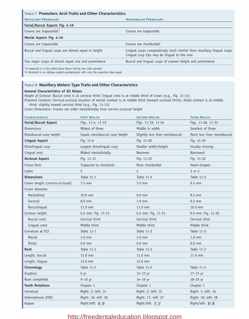
Table 7 Premolars: Arch Traits and Other CharacteristicsMaxillary Premolars Mandibular Premolars
Facial/Buccal Aspect: Fig. 4-16
Crowns are trapezoidal* Crowns are trapezoidal
Mesial Aspect: Fig. 4-16
Crowns are trapezoidal Crowns are rhomboidal†
Buccal and lingual cusps are almost equal in height Lingual cusps comparatively much shorter than maxillary lingual cuspsLingual cusp tips may be lingual to the root
Two major cusps of almost equal size and prominence Buccal and lingual cusps of uneven height and prominence
*A trapezoid is a four-sided plane figure having two sides parallel.†A rhomboid is an oblique-angled parallelogram with only the opposite sides equal.
Table 8 Maxillary Molars: Type Traits and Other Characteristics
General Characteristics of All MolarsHeight of Contour: Buccal crest is at cervical third; lingual crest is at middle third of crown (e.g., Fig. 11-13)Proximal Contacts: Cervical-occlusal location of mesial contact is at middle third (toward occlusal third); distal contact is at middle
third, slightly toward cervical third (e.g., Fig. 11-13)Crown Dimensions: Crowns are wider mesiodistally than cervico-occlusal height
Characteristic First Molar Second Molar Third Molar
Facial/Buccal Aspect Figs. 11-4, 11-15 Figs. 11-19, 11-24 Figs. 11-28, 11-33
Dimensions Widest of three Middle in width Smallest of three
Distobuccal cusp height Equals mesiobuccal cusp height Slightly less than mesiobuccal Much less than mesiobuccal
Lingual Aspect Fig. 11-6 Fig. 11-20 Fig. 11-29
Distolingual cusp Largest distolingual cusp Smaller width/height Usually missing
Lingual root Widest mesiodistally Narrower Narrowest
Occlusal Aspect Fig. 11-12 Fig. 11-23 Fig. 11-32
Crown form Trapezoid to rhomboid More rhomboidal Heart-shaped
Lobes 5 4 3 or 4
Dimensions Table 11-1 Table 11-2 Table 11-3
Crown length (cervico-occlusal) 7.5 mm 7.0 mm 6.5 mm
Crown diameter
Mesiodistal 10.0 mm 9.0 mm 8.5 mm
Cervical 8.0 mm 7.0 mm 6.5 mm
Buccolingual 11.0 mm 11.0 mm 10.0 mm
Contour height 0.5 mm; Fig. 11-13 0.5 mm; Fig. 11-21 0.5 mm; Fig. 11-30
Buccal crest Cervical third Cervical third Cervical third
Lingual crest Middle third Middle third Middle third
Curvature at CEJ Table 11-1 Table 11-2 Table 11-3
Mesial 1.0 mm 1.0 mm 1.0 mm
Distal 0.0 mm 0.0 mm 0.0 mm
Root Table 11-1 Table 11-2 Table 11-3
Length, buccal 12.0 mm 11.0 mm 11.0 mm
Length, lingual 13.0 mm 12.0 mm
Chronology Table 11-1 Table 11-2 Table 11-3
Eruption 6 yr 12–13 yr 17–21 yr
Root completed 9–10 yr 14–16 yr 18–25 yr
Tooth Notations Chapter 1 Chapter 1 Chapter 1
Universal Right: 3; left: 14 Right: 2; left: 15 Right: 1; left: 16
International (FDI) Right: 16; left: 26 Right: 17; left: 27 Right: 18; left: 28
Palmer Right/left: 6 6 Right/left: 7 7 Right/left: 8 8
http://freedentaleducation.blogspot.com

334 Appendix B
Table 9 Mandibular Molars: Type Traits and Other Characteristics
Characteristic First Molar Second Molar Third Molar
Facial/Buccal Aspect Figs. 12-4, 12-14 Figs. 12-18, 12-23 Figs. 12-29, 12-34
Crown size Largest; widest mesiodistally Smaller Smallest
Cusps 5 Cusps: mesiobuccal, distobuccal, mesiolingual, distolingual, distal
4 Cusps: mesiobuccal, distobuccal, mesiolingual, distolingual
4 Cusps: mesiobuccal, distobuccal, mesiolingual, distolingual
Groove(s)/pit(s) 1 Mesiobuccal ± pit, 1 distobuccal 1 Buccal ± pit 1 Buccal ± pit
Root(s) Figs. 12-4, 12-14 Fig. 12-18 Fig. 12-29
Widely separated; nearly vertical Close together, parallel; distally inclined
Most short, fused, less inclination
Lingual Aspect Fig. 12-6 Fig. 12-19 Fig. 12-30
Cervix Crown narrower Less narrow Less narrow
Occlusal Aspect Fig. 12-2 Fig. 12-25 Fig. 12-33
Crown form Quadrilateral or hexagonal, pentagonal Quadrilateral or rectangular Quadrilateral or ovoid
Lobes 5 (Fig. 4-12, C) 4 4
Pulp horn(s) 5 4 4
Dimensions Table 12-1 Table 12-2 Table 12-3
Crown length (cervico-occlusal) 7.5 mm 7.0 mm 7.0 mm
Crown diameter
Mesiodistal 11.0 mm 10.5 mm 10.0 mm
Cervical 9.0 mm 8.0 mm 7.5 mm
Buccolingual 10.5 mm 10.0 mm 9.5 mm
Contour height Figs. 12-7, 12-12 Figs. 12-20, 12-21 Figs. 12-31, 12-32
Buccal crest Cervical third, 0.5 mm Cervical third, 0.5 mm Cervical third, 0.5 mm
Lingual crest Middle third, 1.0 mm Middle third, 1.0 mm Middle third, 1.0 mm
Curvature at CEJ Figs. 5-27, 12-12 Fig. 12-12 Fig. 12-32
Mesial 1.0 mm 1.0 mm 1.0 mm
Distal 0.0 mm 0.0 mm 0.0 mm
Root Fig. 12-7 Fig. 12-21 Fig. 12-31
Length 14.0 mm; Table 1-1 13.0 mm; Table 1-1 11.0 mm; Table 1-1
Chronology Table 12-1 Table 12-2 Table 12-3
Eruption 6–7 yr 11–13 yr 17–21 yr
Root complete 9–10 yr 14–15 yr 18–25 yr
Tooth Notations Chapter 1 Chapter 1 Chapter 1
Universal Right: 30; left: 19 Right: 31; left: 18 Right: 32; left : 17
International (FDI) Right: 46; left: 36 Right: 47; left: 37 Right: 48; left: 38
Palmer Right/left: 6 6 Right/left: 7 7 Right/left: 8 8
http://freedentaleducation.blogspot.com
IndexArch. See also Dental arch; Mandibular
arch; Maxillary arch width of, occlusion and, 281
Artery, 252–256alveolar, 253carotid, 253inferior alveolar, 253, 253–256inferior labial, 253internal maxillary, 253maxillary, 253
projection of, 256fpalatine, 256sphenopalatine, 256submental, 253superior alveolar, 253, 256–258
Articular disk, 262–263jaw position and, 264fstructures associated with, 264ftemporomandibular joint movement
and, 259Articular eminence, temporomandibular
joint movement and, 259Articulation, of teeth, 81Aspartate, age determination with, 68
BBaby teeth, 46
loss of, 29Bennett movement, 264–265Bicuspid, definition of, 141, 157Bite marks, 68–69Bite plane splint, 276fBlack, G. V., 10Blood vessel, 239–258
periodontal, 86fBoley gauge, 11Bone
dental, 239–258teeth malalignment influence on, 77f
Bruxismcontact relations and, 277effects of, 276focclusion affected by, 275–276response to, 299stomatognathic system and, 299
Buccal cervical ridge, mandibular first molar, 196
AADA. See American Dental AssociationAdaptation, occlusion and, 301–302Affect, oral motor behavior and, 303Age
dental, 33–35prediction of, 37
dental maturity for identification of, 68dentition emergence and, 1estimation of, dental characteristics
and, 68Age of attainment, 35–37, 39fAlveolar artery, 253–256, 256–258Alveolar crest
radiograph of, 85ftooth emergence through, 27
Alveolar gingiva, tooth emergence through, 27
Alveolar process, 243description of, 5mandibular, 247–248
Alveolusdescription of, 5mandibular, 248–252
example of, 248fview of, 249f
maxillary, 243–244American Dental Association, tooth
number systems and, 2–3Animal, dentition of, 70Anisognathus, definition of, 73Anterior crowding, example of, 301fAnterior superior alveolar artery, 256Antrum of Highmore, 239, 241Ape, dentition of, 72fApical foramen, 211
description of, 31–32mandibular canine, 226mandibular first molar, 228, 229mandibular first premolar, 227mandibular second molar, 231mandibular third molar, 232maxillary canine, 215maxillary second premolar, 217
335
Buccal developmental groove, maxillary first molar, 180
Buccal groove, description of, 8–9Buccal nerve, 258Buccal ridge
mandibular first premolar, 163maxillary first premolar, 145–147
Buccal surface, definition of, 5–6Buccal triangular ridge, maxillary first
premolar, 150Bunodont, definition of, 73
CCalcification, tooth formation stages and,
35Canine
age of attainment of, 39farch traits and characteristics of, 330tcontact areas of, 88, 89, 90, 91crown formation in, 25–26deciduous (See Canine, primary)embrasures of, 88, 89, 90, 91follicle of, 30interproximal spacing and, 89mandibular, 125–140, 57
alveolus of, 248, 249faspects of, 52f, 53f, 134–138, 135f,
317fcervical cross section of, 226chronology of development of, 37tcurvature of, 94fdistal aspect of, 133f, 138emergence of, 27eruption of, 30–31graph of forms of, 96fgraph outline of aspects of, 134fincisal aspect of, 57f, 132f, 133f,
137f, 138labial aspect of, 57f, 133f, 134–135,
136flabiolingual section of, 225–226lingual aspect of, 132f, 133f, 135measurements of, 138tmesial aspect of, 57f, 133f, 136f, 138,
138fmesiodistal section of, 226radiograph of, 235f
Page numbers followed by f indicate figures; t, tables; b, boxes.
http://freedentaleducation.blogspot.com

336 Index
sections of, 225–226, 225ftraits and characteristics of, 330tvariations of, 137f
maxillary, 125–140, 53–55alveolus of, 243, 243farch traits and characteristics of,
330taspects of, 14f, 52f, 53f, 128f,
316fcervical cross section of, 215chronology of development of, 37tcurvature of, 93distal aspect of, 55, 131–132, 132eruption of, 30–31graph of forms of, 95fgraph outlines of aspects of, 127fincisal aspect of, 54f, 55, 132, 135labial aspect of, 53, 54f, 127–131,
127–132, 134–138labiolingual section of, 211f,
214–215lingual aspect of, 53, 72f, 125–132,
127–131, 131measurements of, 131tmesial aspect of, 53–55, 54f, 131–
132, 131, 134–135mesiodistal section of, 215permanent, 53primary occlusion and, 288fprotrusive occlusal relations and,
298sections of, 216ftraits and characteristics of,
330tvariations of, 138
measurement of, 13tmesial and distal aspects of, 74occlusal contacts of, 291permanent, 125–140, 1, 53primary, 1, 46
comparison of, 57femergence of, 27
region of, radiography of, 252fsuccedaneous (See Canine,
permanent)surfaces of, 5–6traits and characteristics of, 330t
Canine eminence, 125, 239Canine fossa, 239Caries. See Dental cariesCarotid artery, 253Carving, teeth, 10–11CEJ. See Cementoenamel junctionCementoenamel junction, 32–33
curvature of, 15b, 17b, 94–96, 94fdescription of, 4enamel hypoplasia and, 41pulp cavity and, 211–212
Cementumdescription of, 4root development and, 26
Centers of formation, 30
Central developmental groovemandibular first molar, 198mandibular second premolar, 170maxillary first molar, 180maxillary first premolar, 150
Central fossadescription of, 8mandibular first molar, 198, 198–199maxillary first molar, 59–60, 180maxillary second molar, 62
Central groove, maxillary second molar and, 62
Central pitmandibular first molar, 198maxillary second molar, 62
Centric relation positionexample of, 301fmandibular position and, 263
Centric stop, 288Cervical line
curvature of, 92, 94–96, 94fdefinition of, 4maxillary central incisor, 51–52,
105–106Cervical ridge, 14f
description of, 77, 92, 94facial contour at, 91–94lingual contour at, 91–94mandibular first molar, 196
Cervicoenamel ridge, 14f, 77Cheek biting, 284Chewing, 271
biomechanics of, 298mandibular movement during, 271fmastication and, 272–273neural processes and, 272occlusion and, 271as oral motor behavior, 304
Childdentition development in, 24f, 25fmixed dentition in, 281fprimary teeth in
example of, 46f, 48fspecimen of, 48f, 49f
skull of, example of, 28f, 30fChronological age, dental maturity for
identification of, 68Chronological development disorder,
2fCingulum
definition of, 7lobes and, 71mandibular canine, 138maxillary canine, 127, 131, 132maxillary central incisor and, 105
Cleft lip and palate, teeth malformations associated with, 22–23
Clinical considerations, teeth and, 21–22Clinical crown, 21, 22fComparative anatomy, 67–80Comparative dental anatomy, 69–73Condylar guidance, occlusal guidance
and, 300–301
Condyledescription of, 260mandible and, 246, 260fmandibular movement and, 264–265
Condyloid process, 260front view of, 262fmandible and, 246temporomandibular joint movement
and, 259Contact area, 88–89
alignment of teeth and, 82–84definition of, 6embrasures and, 89–91example of, 84fform of gingiva and, 87foutline drawings of, 85fradiograph of, 85f
Contact pointbuccal view of, 84definition of, 82–83example of, 83fincisal view of, 84labial view of, 84occlusal view of, 84
Contact relationsdevelopment of, 277intercuspal relations and, 293, 293focclusal
in intercuspal position, 291fof primary dentition, 280f
Contourcrown, 90facial and lingual, of crowns, 90
Coronal pulp, definition of, 209Coronoid process
mandible and, 246radiograph of, 254f
Cranial nerve, 256–257Craniomandibular disorder, 259Crow, molar, maxillary, 171Crown, 4–5
buccolingual diameter of, 17bcanine, 125
mandibular, 133, 134, 135maxillary, 125, 127–128, 127, 128,
131completion of, tooth formation stages
and, 35contours of, 94curvature of, 91–92, 94development of, 26division into thirds of, 9formation of, 25–26, 40, 40tforms of, 74ffracture of, 233–237geometries of, 73incisal thirds of, 286–287incisor
mandibular, 113mandibular central, 113, 115, 116,
119mandibular lateral, 123maxillary central, 99, 101, 106
Canine (Continued)
http://freedentaleducation.blogspot.com

Index 337
maxillary lateral, 109labiolingual diameter of, 15blength of, 15b, 17bmesiodistal diameter of, 15b, 17bmiddle third of, lingual contours at,
91–94molar
mandibular, 189mandibular first, 189–190, 194, 195,
196, 197mandibular second, 199mandibular third, 205maxillary first, 171, 174, 176,
178occlusal thirds of, 286–287orientation of, root and, 285fpremolar, 141
mandibular first, 159, 163, 164maxillary first, 141–144, 144–145,
147, 149, 151f, 153fmaxillary second, 153
of primary teeth versus permanent teeth, 47
schematic drawing of, 95fschematic outlines of, 75
Curvatureof cervical lines, 94–96of crowns, 90description of, 82of mandibular teeth, 94maxillary central incisor, 105,
105–106schematic drawings of, 92f
Curve of Spee, 78, 78f, 284, 284–285
Curve of Wilson, 78Cusp
caninemandibular, 133, 134maxillary, 128, 132
contact relationship of, occlusion affected by, 288–294
definition of, 7forms of, 70finterrelationship of, with ridges and
embrasures, 297fmolar
mandibular, 189mandibular first, 190, 194, 197,
198–199mandibular second, 203maxillary first, 175, 180maxillary third, 186–187, 187
premolar, 141mandibular, 157mandibular second, 169maxillary first, 141, 147, 149, 151f,
152fsupporting, 288
Cusp-fossa relationship, 289f, 292–293
Cusp of Carabelli, 90
Cuspidbite mark identification and, 69mandibular, bite mark identification
and, 69maxillary, bite mark identification and, 69
DDeciduous, definition of, 2Deciduous dentition
casts of, 3fformation of, 1
Deciduous teeth. See Teeth, primaryDehiscence, description of, 76–77Delta system, 211, 212fDental age, 33–35
assessment of, 68Dental anatomy, 1–20
comparative, 69–73definition of, 1nomenclature of, 2
Dental archalveolar process and, 243contact form and, 86–87curvature of, 284fdimensions of, occlusion and, 279–281embrasure and, 86–87form of, 282–283interproximal spacing and, 86–87maxillary molar and, 171occlusal contacts and, 288primary form of, 277size of, 292fstabilization of, 83width of, 281
Dental cariesloss of primary teeth and, 29prevalence of, primary teeth and, 45
Dental formula, teeth described with, 2Dental maturity, chronological age
determined with, 68Dental pulp. See PulpDentin
crown and root fractures and, 233description of, 4formation of, 209reparative, 210–211secondary, formation of, 210
Dentistry, forensic, 67–69Dentition. See also Teeth
animal, 70ape, 72fcasts of, 3fchronology of, 35, 36tcurvatures of, 82deciduous, development of, 310fdental formula for, 2development of, 26–27, 276
in older child, 25fto sixth year, 310fin young child, 24f
form and function in, 67–80, 75–76formation of, 1mixed
in child, 281fdevelopment of, 311focclusion and, 279–282
notation systems for, 2–9omnivorous, 75–76orangutan, 72fperinatal, 26permanent
casts of, 3fchronology of, 36tdevelopment of, 30–31, 311fformation of, 1mineralization of, 25occlusion and, 282–288representation of, 240ftooth traits of, 327–334transition of primary to, 26
postnatal, 26prenatal, 26primary
in child, 278fchronology of, 23, 36tcompletion of, 28development of, 26–27emergence of, 23–24, 23, 24–25,
277fformation of, 1occlusal contact relations of, 280focclusion and, 276–279terminal plane relationship in, 278–
279, 278fradiograph of, 210ftransition of primary to permanent, 26transitional (mixed), 28–29
Dento-osseous structures, 239–258Deoxyribonucleic acid
bite mark identification and, 69dental, 68forensic analysis of, 68
Descending palatine artery, 256Developmental groove
description of, 8–9molar
mandibular first, 190, 194, 198maxillary first, 180maxillary second, 62
premolarmandibular first, 165maxillary first, 150maxillary first molar and, 59–60permanent dentition and, 30
Developmental lobe, permanent dentition formation and, 30
Diastema, occlusion and, 276Digastric fossa, mandible and, 247Digastric muscle, 268Disk-condyle assembly, 296fDistal aspect
of anterior teeth, 74of maxillary posterior teeth, 74
Distal development pit, maxillary first premolar, 150
Crown (Continued) Dentition (Continued)
http://freedentaleducation.blogspot.com

338 Index
Distal fossamandibular first premolar, 164mandibular second premolar, 170maxillary second molar and, 62
Distal incisal angle, 10Distal lobe, in anterior teeth, 71Distal marginal ridge, maxillary second
molar and, 62Distal surface, 6Distal triangular fossa
molarmandibular first, 198maxillary first, 180maxillary second, 62
premolar, maxillary first, 150Distobuccal developmental groove
mandibular first molar, 190maxillary first premolar, 150
Distolingual lobe, 71Division into thirds, 9–10DNA. See Deoxyribonucleic acidDog
dentition of, 70f, 71fmandible of, 71f
Drawing, teeth, 10–11
EEA. See European-AmericanEmbrasure, 86–88
buccal, 89–91cusp relationship to, 84fincisal, 88–89incisal and occlusal, 88–89interrelationship of, with ridges and
cusps, 297flabial, 89–91lingual, 89–91occlusal, 88–89
Emergence, definition of, 23Eminentia articularis, 259Emotion, oral motor behavior and,
303Enamel
absence of, 32crown and root fractures and, 233description of, 4hypoplasia of, 22, 23f, 41
Enamel organ, 26fEpithelial attachment, 32, 94–96Eruption
chronology of, 277definition of, 23mean age of, 277of permanent teeth, 282sequence of, 41of teeth, 21–44
Escapement space, 287Ethnicity
size of teeth and, 31tooth morphology and, 22European-American, tooth morphology
and, 22External carotid artery, 253
FFace
development of, 28surface landmarks of, 254f
Facial expression, muscles involved with, 268f
Facial surface, 5–6FDI. See Fédération Dentaire
InternationaleFederal Bureau of Investigation, 67Fédération Dentaire Internationale, 4Female, age of attainment of permanent
teeth in, 39tFenestration, description of, 76–77Fifth cranial nerve, 256–257Fifth cusp groove, maxillary first molar,
180Fluoride, benefits of, 34Fluoride toothpaste, 34Fluorosis, prevalence of, 34Follicle, permanent dentition
development and, 30Foramen, 211Foramina of Scarpi, 251fForamina of Stenson, 251fForensic dentistry, 67–69Forensics, 67–80Form and function
definition of, 81dental anatomy and, 67–80jaw movement and, 73maxillary incisor, 99of orofacial complex, 81–98of permanent dentition, 75–76
Form follows function, 81Fossa
contact relationship of, occlusion affected by, 288–294
description of, 8Fracture, crown and root, 233–237Freedom in centric, 294Frontal process, maxilla and, 242
GGeniohyoid muscle, 268–269Geometry, dental anatomy and, 67–80Gingiva
clinical appearance of, 82fdescription of, 5form of, 87fhealth of, contours of teeth and, 82height of, 94interproximal space and, 76–77keratinization of, 85recession of, 92schematic representation of, 83fteeth contours and, 82teeth malalignment influence on, 77ftooth emergence through, 21, 27visualization of, 21
Gingival papilla, 84–85interproximal space and, 76–77
Gingival tissue, description of, 5
Gingivitiscontours of teeth and, 82description of, 83effect of curvature on, 91–92overbite associated with, 284
Ginglymoarthrodial articulation, 259Glenoid fossa
condyle relationship to, 260fjaw movement and, 73
Group function, 295fdefinition of, 293–294
Gums. See Gingiva
HHaderup, Viktor, 4Haplodont class, tooth forms and, 69, 70fHard palate, 239Hard tissue, description of, 4Head, muscles of, 270Hinge axis movement, 263Homeostasis
occlusal, 299oral motor behavior and, 303–304
Huguier canal, 261–262
IICP. See Intercuspal positionIdentification, human
bite marks for, 68–69forensic dentistry and, 67
Impinging overbite, 284, 284fIncisal edge, definition of, 99Incisal liability, 282Incisal ridge
definition of, 99maxillary central incisor and, 106
Incisal surface, 5–6Incisive foramen, 242Incisive fossa, 239
mandible and, 246Incisor
centralage of attainment of, 39fcontact areas of, 88–89, 88, 90, 91crown formation in, 25–26curvature of, 92f, 93, 94fdevelopment of, 30embrasures of, 88–89, 88, 90, 91graph of forms of, 95f, 96flateral incisor relation to, 251fmandibular (See Incisor, mandibular
central)maxillary (See Incisor, maxillary
central)measurement of, 13tmesial and distal aspects of, 74permanent, 51fprimary, 46, 51fprimary occlusion and, 287fradiograph of region of, 253f
crown of, 5follicle of, 30lateral
http://freedentaleducation.blogspot.com
Index 339
age of attainment of, 39fcentral incisor relation to, 251fcontact areas of, 88–89, 88, 89, 90,
91crown formation in, 25–26embrasures of, 88–89, 88, 89, 90, 91mandibular (See Incisor, mandibular
lateral)maxillary (See Incisor, maxillary
lateral)measurement of, 13tmesial and distal aspects of, 74primary, 46
mandibular, 113–124arch traits and characteristics of,
330tbite mark identification and, 69protrusive occlusal relations and,
298surfaces of, 6traits and characteristics of, 329t
mandibular central, 55–56, 113–119aspects of, 52f, 53f, 314fcanals of, 224fcervical cross of, 223chronology of development of, 37tdistal aspect of, 56, 114f, 116f, 119emergence of, 27eruption of, 30graph outlines of aspects of, 115fincisal aspect of, 56, 114f, 116f, 118f,
119labial aspect of, 55, 113–115, 114f,
116f, 117flabiolingual section of, 222–223lingual aspect of, 55, 114f, 115–116,
116fmeasurements of, 119tmesial aspect of, 56, 114f, 116–119,
116f, 117fmesiodistal section of, 223primary occlusion and, 287fsections of, 222–223, 223fvariations of, 118f
mandibular lateral, 56, 56f, 119–123alveolus of, 248aspects of, 52f, 53f, 315fcervical cross section of, 225chronology of development of,
37tdistal aspect of, 120f, 123eruption of, 30–31incisal aspect of, 120f, 123labial aspect of, 121f, 123labiolingual section of, 223–224lingual aspect of, 120f, 123measurements of, 122tmesial aspect of, 120f, 121f, 123mesiodistal section of, 224–225radiograph of, 235fsections of, 223–225, 224fvariations of, 122f
maxillary, 99–112arch traits and characteristics of,
330tfunction of, 99graph of forms of, 95fsurfaces of, 6traits and characteristics of, 328t
maxillary central, 50–53, 99, 99–106alveolus of, 243, 243faspects of, 52f, 53f, 312fbite mark identification and, 69cervical section of, 213chronology of development of, 37tdistal aspect of, 51–53, 100f, 102f,
106eruption of, 30–31graph outlines of, 101fincisal aspect of, 53, 100f, 102f, 104f,
106labial aspect of, 50–51, 100f, 102–
105, 102f, 103flabiolingual section of, 213landmarks of, 8flingual aspect of, 51, 100f, 102f, 105,
292fmeasurements of, 105tmesial aspect of, 51–53, 100f, 102f,
103f, 105–106mesiodistal section of, 213midroot cross section of, 213primary, 51fprimary occlusion and, 287fpulp cavity of, 213sections of, 213–222variations of, 104f
maxillary lateral, 53, 73f, 99, 106–111alveolus of, 243, 243faspects of, 52f, 53f, 313fbite mark identification and, 69cervical section of, 214chronology of development of, 37tdistal aspect of, 107f, 111eruption of, 30–31incisal aspect of, 54f, 107f, 108f, 111labial aspect of, 54f, 106f, 107–109,
108flabiolingual section of, 214lingual aspect of, 107f, 109–111measurements of, 110tmesial aspect of, 54f, 107f, 109f,
111mesiodistal section of, 214midroot cross section of, 214peg-shaped, 106primary, 54fsections of, 215fvariations of, 110f
occlusal contacts of, 291permanent
development of, 28–29emergence of, 28–29sagittal section of, 280f
permanent central, eruption of, 29fpermanent mandibular, 113–124permanent maxillary, 99–112primary, 46
sagittal section of, 280fsagittal sections through, 6fshovel-shaped, 99–101surfaces of, 5–6
Infantdentition development in, 26–27mandible section in, 27f
Inferior alveolar artery, 253, 253–256Inferior alveolar nerve, 258Inferior mental spine, mandible and, 247Infraorbital groove, maxillary orbital
surface and, 240Infraorbital nerve, 257–258, 258Interarticular disk, 262–263Intercuspal position
centric occlusion and, 78fcontact relations in, 293fjaw position in, 296fmandibular position and, 263
Intercuspal relations, 293Interdental papilla, 76–77, 86fInterdental septum, 248–249Interdental space, 278
occlusion and, 276International Association for Dental
Research, 4Interproximal form, 76–77Interproximal space, 76, 84–86
dental arch and, 86–87of mandibular anterior teeth, 89type of teeth and, 86
Interradicular septa, 244Isognathous, definition of, 73
JJaw
of dog, 70fforensic dentistry and, 67movement of, form and function of
teeth and, 73nerve supply to, 256–258position of, in intercuspal position,
296fpositions of, 263stability of, 302
Joint capsule, 261Juvenile, indicators of age of teeth in, 34
KKronfeld’s chronology, 35
LLabial groove, lobes and, 71Labial lobe, in anterior teeth, 71Labial surface, 5–6Lacrimal groove, 240, 241Lamina dura, loss of, 236fLandmarks, 6–9
Incisor (Continued) Incisor (Continued) Incisor (Continued)
http://freedentaleducation.blogspot.com

340 Index
Lateral movement, mandibular, 293, 295fLateral protrusive movement,
mandibular, 293Lateral pterygoid muscle, 266, 266fLeeway space, 281, 281f, 282Ligament
mandibular, 261–262sphenomandibular, 247, 261, 262fstylomandibular, 261, 262ftemporomandibular, 261
Line angle, 9–10Lingual developmental groove
mandibular first molar, 194maxillary first molar, 180maxillary second molar and, 62
Lingual fossadescription of, 8mandibular central incisor and, 55maxillary canine, 131maxillary central incisor and, 105
Lingual groove, description of, 8–9Lingual lobe, in anterior teeth, 71Lingual nerve, 258Lingual ridge, maxillary first premolar,
147Lingual surface, 5–6Lingual triangular ridge, maxillary first
premolar, 150Lobe
description of, 9, 71general outline of, 72f
MMalleus, ligaments attached to, 263fMalocclusion
definition of, 76development of, primary teeth and,
45incisal liability and, 282
Mamelondescription of, 9lobes and, 71mandibular incisor, 113
Mandible, 245–252alveolar process of, 247–248, 248falveoli of, 248–252bony landmarks of, 252, 254fclosing of, muscle involvement in,
270–271comparison of, at various ages, 250fcondyle relationship to, 260fcondyloid process of, 260frontal view of, 246fin infant, 27finterior surface of, 247lateral movement of, 271, 293lateral protrusive movement of, 293ligaments of, 261–262movement of, 264–265, 293
during chewing, 271, 271fdental forms and, 73at first molar, 298fmuscle involvement in, 270–273
schematic representation of, 264f, 265f
opening of, muscle involvement in, 270outer surface of, 245fpositions of, 263–264posterior view of, 245fposterolateral view of, 245fprotrusion of, 271, 293rest position of, 264retrusion of, 271, 293sections of, 236fsurface landmarks of, 254fthickness of bone through, 250fview of
from above, 248ffrom below, 246f
Mandibular, definition of, 2Mandibular arch
bite mark identification and, 68contact relations of, 290fcurvature of, 283fexample of, 32fnormal occlusion of, 86focclusal contacts and, 288occlusion and, 279–280primary dentition in, 29fwidth of, occlusion and, 281
Mandibular canal, root apices and, 234–237
Mandibular foramen, 247Mandibular fossa, 259
radiograph of, 261ftemporomandibular joint movement
and, 259Mandibular ligament, 261–262Mandibular nerve, 257f, 258
coronal section of, 267fMandibular notch, 246Mandibular spine, 247Marginal ridge
contact relationship of, occlusion affected by, 288–294
description of, 7–8maxillary central incisor and, 105maxillary first molar, 180maxillary second molar and, 62
Masseter muscle, 266–268Mastication
chewing and, 298lateral movement during, 297–298muscle and, 265–266, 266, 268fneurobehavioral mechanisms and, 304overview of, 272–273
Masticatory muscle, 268fMasticatory system, 259Maturity assessment, 37–40Maxilla, 239–244
alveolar process of, 243alveoli of, 243–244anterior surface of, 239articulation of, 244bones of, 239–244
bony landmarks of, 252, 254ffrontal process of, 242frontal view of, 240finferior surface of, 242flateral view of, 240fmedial view of, 241fnasal surface of, 241orbital surface of, 240–241palatine process of, 242–243palatine view of, 242fposterior surface of, 240posterior view of, 241frepresentation of, 240fsurface landmarks of, 254fzygomatic process and, 240, 241
Maxillary, definition of, 2Maxillary arch
bite mark identification and, 68contact relations of, 290fcurvature of, 283fexample of, 32fnormal occlusion of, 86fprimary dentition in, 29fwidth of, occlusion and, 281
Maxillary artery, projection of, 256fMaxillary nerve, 257–258Maxillary sinus, 239, 244
radiograph of, 254fMaxillary tuberosity, 240Measurement, teeth, 11, 13t, 15b,
17bMedial pterygoid muscle, 268Mental foramen, 247Mental protuberance, 245–246Mental spine, mandible and, 247Mental tubercle, 245–246Mesenchyme, dental pulp and, 209Mesial aspect
of anterior teeth, 74of maxillary posterior teeth, 74
Mesial development groove, mandibular second premolar, 169
Mesial development pit, maxillary first premolar, 150
Mesial developmental depression, maxillary first premolar, 149
Mesial developmental groove, mandibular first premolar, 165
Mesial fossamandibular first premolar, 164mandibular second premolar, 170
Mesial incisal angle, 10Mesial lobe, in anterior teeth, 71Mesial marginal developmental groove,
maxillary first premolar, 150Mesial marginal ridge
maxillary first molar, 180maxillary second molar and, 62
Mesial migration, description of, 299Mesial pit, mandibular first molar,
198Mesial surface, 6
Mandible (Continued) Maxilla (Continued)
http://freedentaleducation.blogspot.com

Index 341
Mesial triangular fossamolar
mandibular first, 198maxillary first, 59–60, 180
premolarmandibular second, 169maxillary first, 150
Mesiobuccal developmental groovemandibular first molar, 190maxillary first premolar, 150
Mesiolabial line angle, 9Mesiolingual developmental groove,
mandibular first premolar, 163Mesiolingual lobe, 71Milk teeth, 46
definition of, 2loss of, 29
Mineralization, tooth, 25Mixed dentition, formation of, 1Molar
class I relationship of, 278–279, 278fclass II relationship of, 278–279, 278fclass III relationship of, 278–279, 278fdeciduous, 1first
age of attainment of, 39fcontact areas of, 88, 89, 90, 91contact relations of, 287fcurvature of, 93fembrasures of, 88, 89, 90, 91graph of forms of, 95f, 96fmandibular (See Molar, mandibular
first)maxillary (See Molar, maxillary first)measurement of, 13tprimary, 46radiography of region of, 252f
mandibular, 189–208centric occlusion and, 298fracture of, 235floss of, 288floss of lamina dura on, 236fopening of contact between, 300fradiograph of, 235ftraits and characteristics of, 334t
mandibular first, 28–29, 59f, 62–64, 189–199
alveolus of, 249, 249f, 250faspects of, 193f, 324fbuccal aspect of, 62–63, 189–194,
190f, 191f, 193fbuccolingual section of, 227–228cervical cross section of, 229chronology of development of, 37tdistal aspect of, 63, 191f, 192f,
196–197fracture of, 233graph outlines of aspects of, 192flingual aspect of, 63, 191f, 194–195measurements of, 190tmesial aspect of, 63, 191f, 195–196,
195fmesiodistal section of, 228–229
midroot cross section of, 230occlusal aspect of, 64, 190f, 192f,
197–199, 197f, 292fpermanent, 64primary, 46–47, 62f, 63fsections of, 227–230, 229ftraits and characteristics of, 334tvariations of, 198f
mandibular second, 59f, 64–65, 199–203
alveolus of, 249, 250faspects of, 200–203, 325fbuccal aspect of, 64, 65f, 199f, 200–
201, 200fbuccolingual section of, 230–231cervical cross section of, 231chronology of development of, 37tdistal aspect of, 64, 199f, 201emergence of, 27lingual aspect of, 64, 65f, 199f, 201measurements of, 200tmesial aspect of, 64, 65f, 199f, 201,
201fmesiodistal section of, 231midroot cross section of, 231occlusal aspect of, 65, 65f, 199f, 201–
203, 202fprimary, 65fsections of, 230–231, 230ftraits and characteristics of, 334tvariations of, 202f
mandibular third, 203–207alveolus of, 249, 250faspects of, 204–207buccal aspect of, 204–205, 204f, 205fbuccolingual section of, 232–233cervical cross section of, 233development of, 32fdistal aspect of, 204f, 207fissure patterns of, 203fimpacted, 203flingual aspect of, 204f, 207measurements of, 206tmesial aspect of, 204f, 205f, 207mesiodistal section of, 233midroot cross section of, 233occlusal aspect of, 204f, 206f, 207partially erupted, 203fsections of, 231–233, 232ftraits and characteristics of, 334tvariations of, 206f
maxillary, 171–188alveoli of, 244fcentric occlusion and, 298curvature of, 93drawing of, 14fprimary cusp triangle of, 175fsection through, 6ftraits and characteristics of, 333t
maxillary first, 58–60, 59f, 171–181alveolus of, 244aspects of, 177f, 322f
buccal aspect of, 58, 172f, 173f, 177fbuccolingual section of, 218–219calibration of, 87fcervical cross section of, 220chronology of development of, 37tdistal aspect of, 59, 174f, 175f, 178graph outlines of aspects of, 176flandmarks of, 7flingual aspect of, 58, 174–175, 175–
178, 178measurements of, 172tmesial aspect of, 58–59, 173f, 175–
178, 178fmesiodistal section of, 219–220midroot cross section of, 220occlusal aspect of, 59–60, 172f, 175f,
178–181, 179focclusal landmarks of, 291fprimary, 46–47, 60fprimary occlusion and, 289froots of, 58sections of, 218–220, 219ftraits and characteristics of, 333tvariations of, 179f
maxillary second, 59f, 60–62, 181–184aspects of, 323fbuccal aspect of, 60, 181f, 182–183,
182fcervical cross section of, 220–221chronology of development of, 37tdistal aspect of, 61, 181f, 184lingual aspect of, 60–61, 181f, 183measurements of, 182tmesial aspect of, 61, 181f, 183f, 184mesiodistal section of, 220midroot cross section of, 221occlusal aspect of, 61–62, 181f, 183f,
184primary, 61fprimary occlusion and, 289frounded cusp of, 287fsections of, 220–221, 221ftraits and characteristics of, 333tvariations of, 184f
maxillary third, 184–187alveolus of, 244buccal aspect of, 185, 185f, 186fdevelopment of, 32fdistal aspect of, 185f, 186lingual aspect of, 185, 185fmeasurements of, 188tmesial aspect of, 185, 185f, 186focclusal aspect of, 185f, 186–187,
187fsections of, 221–222, 222ftraits and characteristics of, 333tvariations of, 187f
mesial and distal aspects of, 74occlusal contacts of, 291, 292occlusal cycle and, 297–298permanent mandibular, 189–208permanent maxillary, 171–188
Molar (Continued) Molar (Continued)
http://freedentaleducation.blogspot.com

342 Index
primary, 46, 58f, 59fleeway space and, 281pulp chamber in, 50frelationships of, 278
primary firstcrown formation in, 25–26emergence of, 27
secondage of attainment of, 39fcomparison of primary and
permanent, 49fcontact areas of, 88, 89, 90, 91crown formation in, 25–26embrasures of, 88, 89, 90, 91mandibular (See Molar, mandibular
second)maxillary (See Molar, maxillary second)measurement of, 13tprimary, 46radiograph of region of, 253f
six-yearappearance of, 46radiograph of, 33f
size of pulp in, 211fsurfaces of, 5–6third
contact areas of, 88, 89, 90, 91embrasures of, 88, 89, 90, 91eruption of, 31formation of, 1hypererupted, 288fmandibular (See Molar, mandibular
third)maxillary (See Molar, maxillary third)measurement of, 13tradiograph of region of, 253f
12-year, 30–31Molar lobe, 71Motivation, oral motor behavior and, 303Motor behavior, oral, 303–304
execution of, 304homeostasis and, 303–304
Mouth, roof of, 239Muscle, 259–274, 265–270
digastric, 268geniohyoid, 268–269head, 270lateral pterygoid, 266, 266fmandibular movement and, 270–273masseter, 266–268masticatory, 265–266, 266, 268fmedial pterygoid, 268neck, 269f, 270palatini, 269–270platysma, 266–267pterygoid, 266frisorius, 266–267sternocleidomastoid, 267–268temporalis, 268tensor tympani, 269–270, 269fthroat, 270fzygomaticomandibular, 267–268
Mylohyoid groove, 247Mylohyoid line, 247Mylohyoid nerve, 258
NNasal cavity, 241National Crime Information Center, 67Neck, muscles of, 269f, 270Nerve
buccal, 258dental, 239–258fifth cranial, 256–257inferior alveolar, 258infraorbital, 257–258, 258to jaw, 256–258lingual, 258mandibular, 234, 257f, 258
coronal section of, 267fmaxillary, 257–258mylohyoid, 258to teeth, 256–258trigeminal, 256–257
distribution of, 257fNeurocranium, 26–27, 27f, 28Neuromuscular development, dentition
development and, 27–28Nomenclature
application of, 7fof dental anatomy, 2for primary teeth, 45–47
OOblique groove, maxillary first molar, 180Oblique ridge
description of, 8mandible and, 246maxillary first molar, 180maxillary first molar and, 59–60
Occlusal bite plane splint, 276fOcclusal contact, 295fOcclusal curvature, 77–78Occlusal line, facial and lingual aspects
and, 73Occlusal plane, curvature of, 284–285Occlusal relations, lateral, 294–298Occlusal surface, 5–6
maxillary first molar, 180Occlusion, 275–308, 76–78
abnormal, premature loss of primary teeth associated with, 30
adaptation in, 301–302adjustment of, 302balanced, 275–276centric, 78f, 263, 263–264
guidance into, 296flingual view of, 291frelation of maxillary and mandibular
teeth in, 301ftooth relationships in, 287–288
chewing and, 271concepts of, 275–276cusp-fossa relationship and, 289fdefinition of, 76, 275
development of, 276discrepancy in, 301escapement space and, 287guidance of, 300–301ideal, 292–293, 299interference in, 301, 301f, 302neurobehavioral aspects of, 298–303normal, 77–78, 86f138 points of contact of, 291–293optimal, 275–276permanent dentition and, 282–288points of contact of, 291–293primary, 276–277, 279primary dentition and development of,
276–279protrusive movement and, 298stability of, 299vertical dimension of, 302–303
Odontoblast, dentin formation and, 210Odontology, definition of, 67Omnivorous dentition, 75–76Open bite, example of, 283fOptic nerve, 267fOral motor behavior, 272, 303–304
execution of, 304homeostasis and, 303–304teeth and, 34–35
Oral motor function, occlusion and, 276Oral pharynx, representation of, 272fOrangutan, dentition of, 72fOrofacial complex, form and function of,
81–98Orofacial pain, occlusal discrepancy
associated with, 301Orthodontics, ideal occlusion concept in,
292–293Overbite, 283f
impinging, 284, 284fOverjet, 283–284, 283f
PPalatal arch, shape of, 283Palate, representation of, 272fPalatine artery, 256Palatine process, maxilla and, 242–243Palatine suture, radiograph of, 254fPalatini muscles, 269–270Palatoradicular groove, 106Palmer notation system, 3–4Parafunction, stomatognathic system, 299Pattern generator, oral motor behavior
and, 304PCR. See Polymerase chain reactionPeriodontal disease, causes of, 91–92Periodontal probe
segments of, 33ftooth visualization with, 21
Periodontitisdescription of, 83overbite associated with, 284
Periodontium, physiological form of, 82Permanent, definition of, 2
Molar (Continued) Occlusion (Continued)
http://freedentaleducation.blogspot.com
Index 343
Permanent dentition. See Dentition, permanent
Permanent teeth. See Teeth, permanentPharynx, representation of, 272fPit, description of, 9Plaque
contours of teeth and, 82effect of curvature on, 91–92
Platysma muscle, 266–267Point angle, 9–10Polymerase chain reaction, 68Posterior superior alveolar artery, 256Premolar
definition of, 141development of, 30emergence of, 1first
age of attainment of, 39fcontact areas of, 88, 89, 90, 91curvature of, 92f, 93fembrasures of, 88, 89, 90, 91eruption of, 30–31graph of forms of, 95f, 96fmandibular (See Premolar,
mandibular first)maxillary (See Premolar, maxillary
first)measurement of, 13tradiography of region of, 252f
leeway space and, 281lobes in, 71mandibular, 157–170
arch traits and characteristics of, 333t
traits and characteristics of, 332tmandibular first, 157–165
alveolus of, 248–249, 249faspects of, 160f, 320fbuccal aspect of, 158f, 159–165, 161fbuccolingual section of, 226–227cervical cross section of, 227distal aspect of, 158f, 164graph outlines of aspects of, 159flingual aspect of, 158f, 163measurements of, 160tmesial aspect of, 158f, 161f, 163–164mesiodistal section of, 227occlusal aspect of, 158f, 162f,
164–165occlusal movement and, 297radiograph of, 235fsections of, 226–227traits and characteristics of, 332tvariations of, 162f
mandibular second, 165–170alveolus of, 248–249, 249faspects of, 321fbuccal aspect of, 165f, 166, 166fbuccolingual section of, 227cervical cross section of, 227distal aspect of, 165f, 169lingual aspect of, 165f, 166–169measurements of, 168t
mesial aspect of, 165f, 167f, 169mesiodistal section of, 227occlusal aspect of, 166f, 167f,
169–170occlusal movement and, 297radiograph of, 235fsections of, 227, 228ftraits and characteristics of, 332tvariations of, 168f
maxillary, 141–156alveolus of, 243farch traits and characteristics of,
333tbuccolingual section of, 210fcurvature of, 93traits and characteristics of, 331t
maxillary first, 141–150alveolus of, 243–244aspects of, 144f, 318fbuccal aspect of, 142f, 144–147, 145fbuccolingual section of, 215–216cervical cross section of, 216–217distal aspect of, 142f, 150fracture of, 233, 235fgraph outlines of aspects of, 143flingual aspect of, 142f, 147measurements of, 143tmesial aspect of, 142f, 146f, 147–150mesial view of, 8fmesiodistal section of, 216occlusal aspect of, 142f, 147f, 148f,
149f, 150occlusion of, 297fsections of, 217f, 218ftraits and characteristics of, 331tvariations of, 148f
maxillary second, 151–155alveolus of, 244aspects of, 319fbuccal aspect of, 151–153, 151f,
152fbuccolingual section of, 217–218cervical cross section of, 218distal aspect of, 151f, 153–155lingual aspect of, 151f, 153measurements of, 152tmesial aspect of, 151f, 153, 153fmesiodistal section of, 218occlusal aspect of, 151f, 154f, 155sections of, 218ftraits and characteristics of, 331tvariations of, 154f
mesial and distal aspects of, 74occlusal contacts of, 291, 292permanent, 46–47permanent mandibular, 157–170permanent maxillary, 141–156second
age of attainment of, 39fcontact areas of, 88, 89, 90, 91embrasures of, 88, 89, 90, 91eruption of, 30–31
mandibular (See Premolar, mandibular second)
maxillary (See Premolar, maxillary second)
measurement of, 13tradiography of region of, 252f
surfaces of, 5–6Primary, definition of, 2Primary center of formation, 70–71Primary cusp triangle, maxillary first
molar, 180Primary dentition. See Dentition, primaryPrimary teeth. See Teeth, primaryProbe
periodontal, segments of, 33ftooth visualization with, 21
Protrusion, mandibular, 271Protrusive movement
mandibular, 294occlusal relations and, 298
Proximal contact area, alignment of teeth and, 82–84
Proximal surface, 6Pterygoid fovea, mandible and, 246Pulp, 31–32
dental, 209description of, 4function of, 209size of, in molars, 211f
Pulp canal, 209–238definition of, 209demarcation of, 211–212incisor
central, 51fmandibular central, 222, 222–223mandibular lateral, 224–225
molarmandibular second, 231mandibular third, 232–233
multiple, 212fpremolar, maxillary first, 144in primary and permanent teeth, 48–50radiography of, 233, 234fvisualization of, 233
Pulp cavitybicuspid, 213fcanine, mandibular, 225, 226definition of, 209demarcation of, 211–212incisor
maxillary central, 213maxillary lateral, 214
of mandibular teeth, 222–233of maxillary teeth, 213–222molar, 213f
maxillary first, 218premolar, maxillary second, 217size of, 210–211
Pulp chamber, 209–238canine, maxillary, 214–215definition of, 209incisor
Premolar (Continued) Premolar (Continued)
http://freedentaleducation.blogspot.com

344 Index
mandibular lateral, 223–224, 224–225
maxillary central, 213molar
mandibular first, 227, 228mandibular second, 230–231, 231mandibular third, 233maxillary second, 220primary, 50f
premolarmandibular first, 227mandibular second, 227maxillary first, 236f
in primary and permanent teeth, 48–50radiography of, 209–211, 233, 234fvisualization of, 233
Pulp horn, 212canine, mandibular, 225, 226incisor
central, 51fmandibular central, 222, 223mandibular lateral, 224maxillary lateral, 214
molarmandibular first, 227, 228mandibular second, 230–231, 231mandibular third, 232, 233maxillary first, 219, 219–220maxillary second, 220primary, 50f
premolarmandibular first, 227mandibular second, 227maxillary first, 236f
QQuadritubercular class, tooth forms and,
69, 69–70, 70f
RRadicular pulp, definition of, 209Radiography
pulp chamber and canal, 233of pulp chamber and root canal,
209–211tooth visualization with, 21
Raise the bite, 302–303, 303fRamus, mandibular, 245, 246, 246–247Relative root angle, 285Restriction fragment length
polymorphism, 68Retromolar triangle, 248Retrusion, mandibular, 271Retrusive movement, mandibular, 293,
294RFLP. See Restriction fragment length
polymorphismRhomboid, crown outlines and, 75Ridge
definition of, 7tooth, 2
Risorius muscle, 266–267
Root, 4–5apices of, mandibular canal and,
234–237bifurcated, of mandibular canine, 133,
138fcanine, 125
mandibular, 133, 135, 138maxillary, 127–128, 127, 131, 132
completion of, tooth formation stages and, 35
description of, 5, 9development of, 26division into thirds of, 9form of, 77formation of, 40, 40tfracture of, 233–237incisor
mandibular, 113mandibular central, 113, 115, 116,
119mandibular lateral, 123maxillary central, 101maxillary lateral, 109, 111
inclination and angulation of, 285–286
length of, 15b, 17bmolar
mandibular, 189mandibular first, 194, 195, 196, 228mandibular second, 200, 201mandibular third, 205maxillary, 171maxillary first, 172–173, 174, 175,
176, 176–178, 178, 218, 219maxillary second, 220, 221
orientation of, crown and, 285fpremolar, 141
mandibular first, 159, 163, 164maxillary first, 144, 147, 149, 149–
150, 236fmaxillary second, 153
of primary teeth versus permanent teeth, 47
relative angle of, 285resorption of, 26, 30
clenching habit and, 300fof primary teeth, 45
shape of, 77spacing between, 85–86
Root canal, 209–238canine
mandibular, 226maxillary, 214–215
definition of, 209molar
mandibular first, 229, 230mandibular second, 231maxillary first, 218, 219, 219–220, 220maxillary second, 220
premolarmandibular second, 227maxillary first, 216
radiography of, 209–211
Root pulp, definition of, 209Root trunk, maxillary first molar, 174,
180
SSepta, interradicular, 244Septum, interdental, 248–249Sinus, maxillary, 244Skeleton, facial, development of, 28Skull
child, example of, 30frepresentation of, 240fsurface landmarks of, 254fvertical sections of, 251f
Slide in centric, 294Soft tissue, description of, 4Sphenomandibular ligament, 247, 261,
262fSphenopalatine artery, 256Spillways, 86–88Splanchnocranium, 26–27, 27f, 28Sternocleidomastoid muscle, 267–268Stomatognathic system, 299Stylomandibular ligament, 261, 262fSublingual fossa, mandible and, 247Submandibular fossa, mandible and, 247Succedaneous, definition of, 2Sulcus, description of, 8Superior alveolar artery, 253, 256–258Superior mental spine, mandible and, 247Supplemental groove, description of, 8–9Surfaces, tooth, 2Swallowing, 272, 304Swallowing center, 304Symbolic system of notation, 3–4Symphysis, mandibular, 245
TTeeth. See also Dentition
alignment of, 76–78anterior
description of, 6line angles of, 9, 10fpoint angles of, 10schematic drawings of, 5f
arterial supply to, 252–256articulation of, 81baby, loss of, 29buccal aspects of, 73fcarving of, 10–11chronology of formation of, 33clinical considerations for, 21–22contact areas of, 82–84contact relations of, 277contacts of, 76–78curvatures of, 82deciduous (See Teeth, primary)defect in structure of, 23fdevelopment of, 21–44distal aspects of, 74, 74–75drawing of, 10–11emergence of, 1eruption of, 21–44
Pulp chamber (Continued)
http://freedentaleducation.blogspot.com

Index 345
facial aspects of, 73–75, 73ffaciolingual views of, 251fforensic dentistry and, 67form and function of, 75–76, 81form of, 22, 73, 87f
classes of, 69formation of
age of attainment and, 35–37early indications of, 26stages of, 35, 38fstandards for, 35
fracture of, 233–237function of, 259–274functional form of, 72finclination and angulation of, 285–286,
286b, 286fintercuspal relations of, 293interproximal spaces between, 84–86labial aspects of, 73flandmarks of, 6–9lingual aspects of, 73–75malformations of, 22–23mammalian, formulae for, 2mandibular
age of attainment of, 39tage prediction from, 40tcontact areas of, 88–89, 91contact relations of, 89f, 290fcurvature of, 94embrasures of, 88–89, 91eruption chronology of, 282teruption of, 30, 277t, 282graph of forms of, 96fintercuspation of, 289fmeasurement of, 13tmesial and distal aspects of, 74–75occlusion and, 276postnatal development of, 38tpulp cavities of, 222–233schematic drawing of crown of, 95fsections of axial relations of, 250
maxillarycontact areas of, 88, 90contact relations of, 290fembrasures of, 88, 90eruption chronology of, 282teruption of, 277t, 282graph of forms of, 95fintercuspation of, 289fmeasurement of, 13tmesial and distal aspects of, 74occlusion and, 276outline drawings of, 85f, 87fpulp cavities of, 213–222schematic drawing of crown of, 95fsections of axial relations of, 250
measurement of, 11, 13t, 15b, 17bmesial aspects of, 74, 74–75mesial migration of, 299milk, 2
loss of, 29misalignment of, 76–77
morphology of, 22review of, 309–326
nerve supply to, 256–258numbering systems for, 2–9occlusion of, 76–78oral motor behavior and, 34–35overlap of, 283–284permanent
age of attainment of, 39tage prediction from, 40tchronology of, 31tcrowding of, 278tdevelopment of, 30, 311femergence of, 1eruption chronology of, 282teruption of, 282primary teeth contrasted with, 47tooth traits of, 327–334
physiological form of, 82physiological indicators of age of, 34position of, maintenance of, 299posterior
description of, 6line angles of, 10, 10fpoint angles of, 10schematic drawings of, 5f
primary, 45–66calcification of, 25chronology of, 23, 23t, 46fcrowding of, 278tcrown formation in, 25–26detailed description of, 50–65development and eruption of, 24–26development of, 310femergence of, 1, 27eruption chronology of, 277eruption of, 41, 45, 277timportance of, 45interdental spacing and, 278life cycle of, 45loss of, 29–30, 45mandibular, postnatal development
of, 38tnomenclature for, 45–47occlusion and, 276permanent teeth contrasted with, 47premature loss of, 30prenatal formation of, 38tsize of, 52tspecimen of, 48f, 49ftable of measurements of, 52tuniversal numbering system for, 47f
radiograph of, 210frelationships of
in centric occlusion, 287–288schematic drawing of, 288f
root portion of, 5schematic drawings of longitudinal
sections of, 5fsize of, 31
differences in, 281–282occlusion and, 279–281
temporary, 46temporomandibular joint and, 259–274type of, interproximal space and, 86variability of, 22
Teething, problems associated with, 23–24
Temporal bonecondyle relationship to, 260fradiograph of, 261f
Temporalis muscle, 268Temporomandibular articulation,
259–265Temporomandibular disorders
muscle disorders and, 269–270, 270occlusal discrepancy associated with,
301Temporomandibular joint, 259–274
description of, 261horizontal section of, 267fmandibular articulation and, 245movements of, 259muscle disorders and, 269–270, 270muscle function and, 266pain in, occlusal discrepancy associated
with, 301radiograph of, 261fschematic representation of, 260ftemporomandibular ligament and, 262f
Temporomandibular ligament, 261Tensor tympani muscle, 269–270, 269fTerminal plane relationship, 278–279Tetracycline, 34Throat, muscles of, 270fThumb-sucking, 283Tooth. See TeethTooth formation standards, 35Tooth guidance, 293–294Tooth numbering systems, 2–9Tooth socket, maxillary, 243–244Toothpaste, fluoride, 34Transverse ridge, description of, 8Trapezoid, crown outlines and, 75Triangle, crown outlines and, 75Triangular fossa
description of, 8mandibular first molar, 198mandibular second premolar, 169maxillary first molar, 180maxillary first premolar, 150
Triangular ridgedescription of, 8maxillary first premolar, 150
Triconodont class, tooth forms and, 69, 69–70, 70f
Trifurcation, maxillary first molar and, 58Trigeminal nerve, 256–257
distribution of, 257fTritubercular class, tooth forms and, 69,
69–70, 70fTubercle
definition of, 7lobes and, 71
Teeth (Continued) Teeth (Continued) Teeth (Continued)
http://freedentaleducation.blogspot.com

346 Index
Tubercle of Carabelli, 60, 171–172
UUniversal notation system, 3, 4Universal numbering system, primary
teeth described with, 47f
VVertical dimension, occlusal,
302–303Von Spee, Graf, 78
WWorld Health Organization, 4
ZZsigmondy, Adolph, 3–4Zsigmondy/Palmer notation system,
3–4Zygomatic process, 240, 248–252
radiograph of, 261fZygomaticomandibular muscle, 267–268
http://freedentaleducation.blogspot.com

This page intentionally left blank
http://freedentaleducation.blogspot.com

This page intentionally left blank
http://freedentaleducation.blogspot.com

This page intentionally left blank
http://freedentaleducation.blogspot.com

This page intentionally left blank
http://freedentaleducation.blogspot.com

This page intentionally left blank
http://freedentaleducation.blogspot.com

Reinforce your understanding with the companion DVD!
Strengthen your understanding of dental anatomy, physiology, and occlusion with interactive practice and assessment tools. The companion DVD included in this textbook gives you access to:
Exam-style study questionsmodeled on the National Board Dental Exam
24 3-D animations that depict dental structures and movements in vibrant detail
26 QuickTime Virtual Reality animations with 360-degree views of dental anatomy
See for yourself the benefits this multimedia tool has to offer!
Start using your DVD now!
http://freedentaleducation.blogspot.com
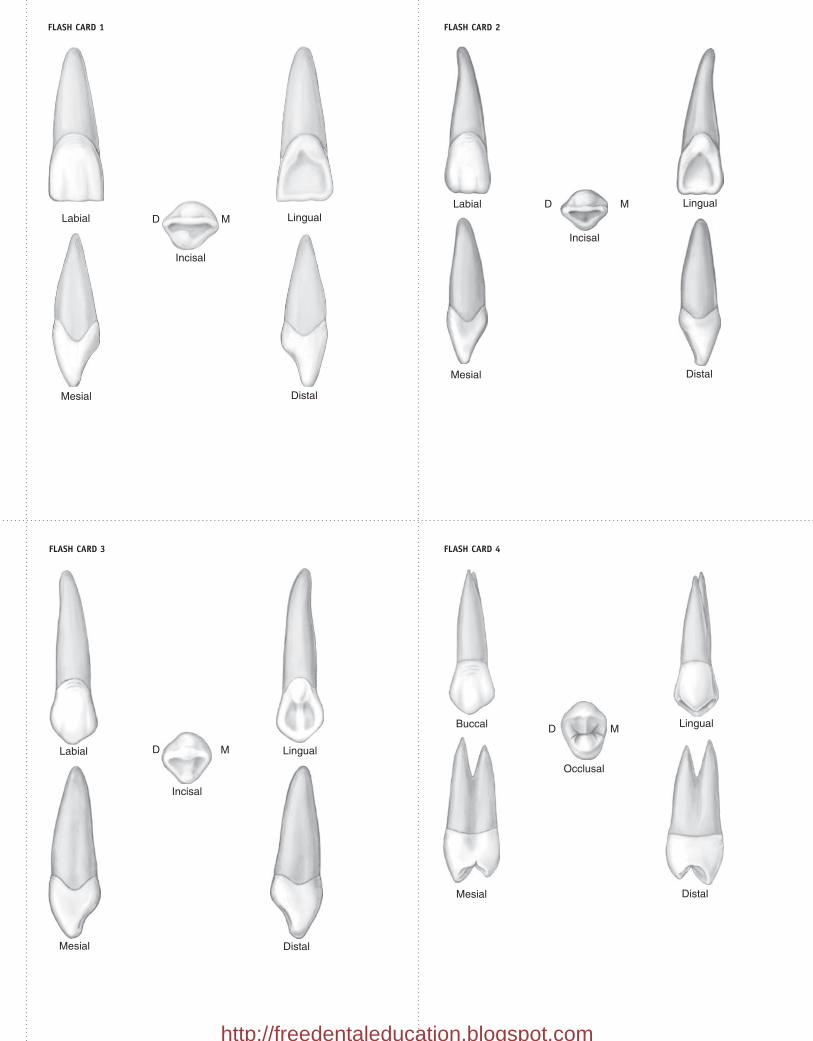
FLASH CARD 1
Labial
Incisal
MD Lingual
DistalMesial
FLASH CARD 2
Labial
Incisal
MD Lingual
DistalMesial
FLASH CARD 3
Labial
Incisal
MD Lingual
DistalMesial
FLASH CARD 4
Buccal Lingual
Mesial Distal
Occlusal
D M
http://freedentaleducation.blogspot.com

Maxillary Right Central IncisorUniversal: 8 International (FDI): 11 Palmer: 1CHARACTERISTICS CENTRAL INCISOR
Facial/Labial AspectProximal contacts Cervico-incisal location Mesial Incisal third Distal Junction incisal/middle thirdMesioincisal angle Sharp right angleDistoincisal angle Slight roundingMesiodistal width Comparatively widePulp horn(s) 3 (facial view)Lobes 4Lingual AspectMarginal ridges ModerateCingulum Moderately pronouncedFossa Moderately deepIncisal AspectOutline TriangularLabial Slightly convexDimensionsCrown length 10.5 mm (cervico-incisal)Crown diameter Mesiodistal 8.5 mm Labiolingual 7.0 mmContour height 0.5 mm Facial/lingual Both cervical thirdRootLength 13.0 mmPulp canal(s) 1ChronologyEruption 7–8 yrRoot completed 10 yr
Copy
right
© 2
010,
200
3, 1
993,
198
4, 1
974,
196
5, 1
958,
195
0, 1
940
by S
aund
ers,
an
impr
int o
f Els
evie
r, In
c.
Copy
right
© 2
010,
200
3, 1
993,
198
4, 1
974,
196
5, 1
958,
195
0, 1
940
by S
aund
ers,
an
impr
int o
f Els
evie
r, In
c.
Maxillary Right Lateral IncisorUniversal: 7 International (FDI): 12 Palmer: 2CHARACTERISTICS LATERAL INCISOR
Facial/Labial AspectProximal contacts Mesial Junction incisal/middle thirds Distal Middle thirdMesioincisal angle Slightly roundedDistoincisal angle Distinctly roundedMesiodistal width Comparatively narrowPulp horn(s) Usually 2 (facial view)Lobes 4Lingual AspectMarginal ridges More prominentCingulum More prominentFossa DeepIncisal AspectOutline OvoidLabial More convexDimensionsCrown length 9 mm (cervico-incisal)Crown diameter Mesiodistal 6.5 mm Labiolingual 6.0 mmContour height 0.5 mm Facial/lingual Both cervical thirdRootLength 13.0 mmPulp canal(s) Less frequent apical accessory canalsChronologyEruption 8–9 yrRoot completed 11 yr
Copy
right
© 2
010,
200
3, 1
993,
198
4, 1
974,
196
5, 1
958,
195
0, 1
940
by S
aund
ers,
an
impr
int o
f Els
evie
r, In
c.
Maxillary Right CanineUniversal: 6 International (FDI): 13 Palmer: 3CHARACTERISTICS CANINE
Facial/Labial AspectProximal contacts Mesial Junction incisal/middle third Distal Middle thirdMesial Aspect Wider faciolinguallyLingual Aspect Deeper lingual fossaeMarginal ridges Pronounced; 2 fossaeCingulum Large, centered mesiodistally Lingual pits, grooves CommonIncisal Aspect Marked asymmetry of mesial/distal halvesIncisal/Proximal Aspects Cusp tip may be at or labial to root axis
lineDimensionsMesiodistal 7.5 mmLabiolingual 8.0 mmIncisal-cervical 10.0 mmContour height 0.5 mm Facial/lingual Both cervical thirdPulp horn(s) 1Lobes 4RootTerminal (number) 1Length 17 mmChronologyEruption 11–12 yrRoot completed 13–15 yr
Copy
right
© 2
010,
200
3, 1
993,
198
4, 1
974,
196
5, 1
958,
195
0, 1
940
by S
aund
ers,
an
impr
int o
f Els
evie
r, In
c.
Maxillary Right First PremolarUniversal: 5 International (FDI): 14 Palmer: 4CHARACTERISTICS FIRST PREMOLAR
Facial/Buccal AspectProximal contacts Mesial/distal: middle thirdShoulders ProminentBuccal cusp Tipped more to distalCusp ridges Longer mesial ridgeCusp size, height Slightly wider, longerLingual Aspect Buccal profile visibleMesial AspectMesiomarginal groove Crosses marginal ridgeMesial concavity PresentMesial root depression PresentOcclusal AspectProfile HexagonalCentral groove LongSupplemental groves Usually not presentLobes 4Pulp horn(s) 2DimensionsCrown length 8.5 mm (cervico-occlusal)Crown diameter Mesiodistal 7.0 mm Cervical 5.0 mm Buccolingual 9.0 mmContour height Facial/buccal crest Cervical third Lingual Middle thirdRootLength 14.0 mmGrooves Distinct, longitudinalNumber of roots Usually 2Pulp canal(s) Often 2ChronologyEruption 10–11 yrRoot completed 12–13 yr
http://freedentaleducation.blogspot.com
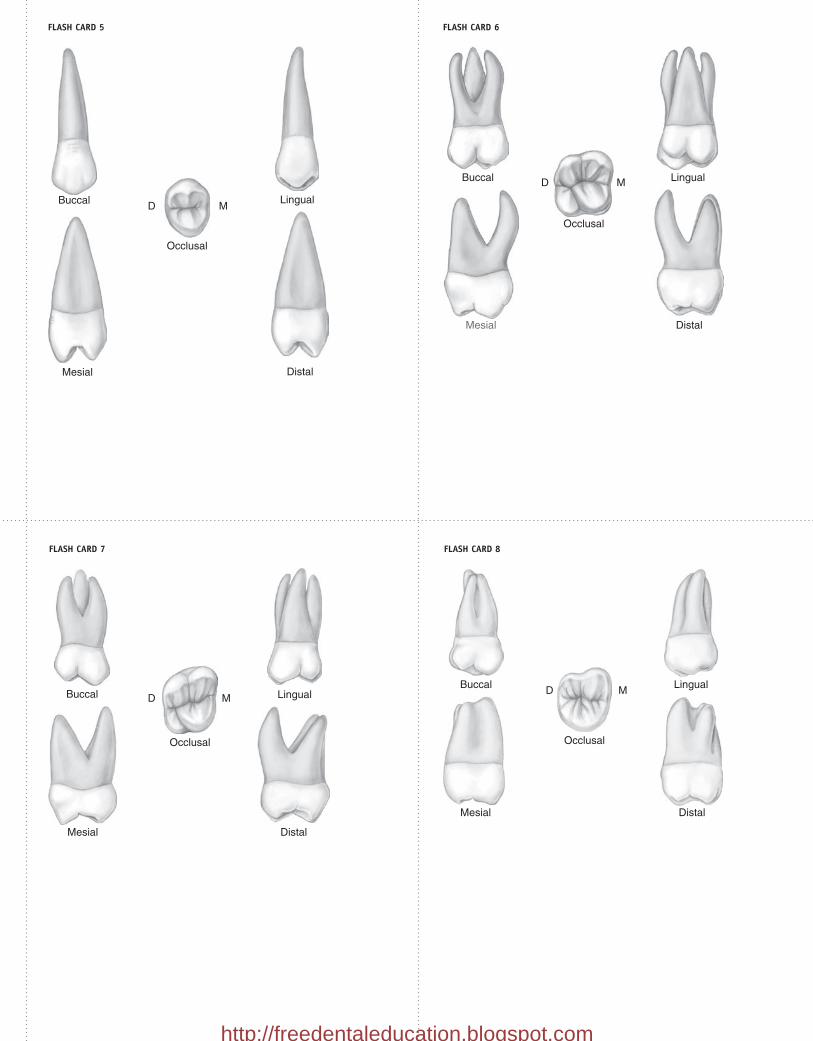
FLASH CARD 5
Buccal Lingual
Distal
Occlusal
D M
Mesial
FLASH CARD 6
Buccal Lingual
Mesial Distal
D M
Occlusal
FLASH CARD 7
Buccal Lingual
Mesial Distal
D M
Occlusal
FLASH CARD 8
Buccal
Occlusal
D M
Mesial
Lingual
Distal
http://freedentaleducation.blogspot.com

Copy
right
© 2
010,
200
3, 1
993,
198
4, 1
974,
196
5, 1
958,
195
0, 1
940
by S
aund
ers,
an
impr
int o
f Els
evie
r, In
c.
Maxillary Right Second PremolarUniversal: 4 International (FDI): 15 Palmer: 5CHARACTERISTICS SECOND PREMOLAR
Facial/Buccal AspectProximal contacts Mesial/distal: middle thirdShoulders NarrowBuccal cusp Not tippedCusp ridges SimilarCusp size, height ShorterLingual Aspect Profile not visibleMesial AspectMesiomarginal groove Does not cross ridgeMesial concavity Not presentMesial root depression PresentOcclusal AspectProfile OvoidCentral groove ShortSupplemental groves Many; often presentLobes 4Pulp horn(s) 2DimensionsCrown length 8.5 mm (cervico-occlusal)Crown diameter Mesiodistal 7.0 mm Cervical 5.0 mm Buccolingual 9.0 mmContour height Facial/buccal crest Cervical third Lingual Middle thirdRootLength 14.0 mmGrooves No distinct groovesNumber of roots 1 rootPulp canal(s) Usually 1ChronologyEruption 10–12 yrRoot completed 12–14 yr
Copy
right
© 2
010,
200
3, 1
993,
198
4, 1
974,
196
5, 1
958,
195
0, 1
940
by S
aund
ers,
an
impr
int o
f Els
evie
r, In
c.
Maxillary Right First MolarUniversal: 3 International (FDI): 16 Palmer: 6CHARACTERISTICS FIRST MOLAR
Facial/Buccal AspectDimensions Widest of three maxillary molarsDistobuccal cusp height Equals mesiobuccal cusp heightProximal contacts Cervical-occlusal location of mesial contact
is at middle, distal contact is at middle third
Lingual AspectDistolingual cusp Largest distolingual cuspLingual root Widest mesiodistallyOcclusal AspectCrown form Square to rhomboidLobes 5DimensionsCrown length 7.5 mm (cervico-occlusal)Crown diameter Mesiodistal 10.0 mm Cervical 8.0 mm Buccolingual 11.0 mmContour height 0.5 mm Buccal crest Cervical third Lingual crest Middle thirdRootLength, buccal 12.0 mmLength, lingual 13.0 mmChronologyEruption 6 yrRoot completed 9–10 yr
Copy
right
© 2
010,
200
3, 1
993,
198
4, 1
974,
196
5, 1
958,
195
0, 1
940
by S
aund
ers,
an
impr
int o
f Els
evie
r, In
c.
Maxillary Right Second MolarUniversal: 2 International (FDI): 17 Palmer: 7CHARACTERISTICS SECOND MOLAR
Facial/Buccal AspectDimensions Width between that of first and third
maxillary molarsDistobuccal cusp height Slightly less than mesiobuccal cuspProximal contacts Cervical-occlusal location of mesial contact
is at middle, the distal contact is at middle third
Lingual AspectDistolingual cusp Smaller width/heightLingual root NarrowerOcclusal AspectCrown form More rhomboidalLobes 4DimensionsCrown length 7.0 mm (cervico-occlusal)Crown diameter Mesiodistal 9.0 mm Cervical 7.0 mm Buccolingual 11.0 mmContour height 0.5 mm Buccal crest Cervical third Lingual crest Middle thirdRootLength, buccal 11.0 mmLength, lingual 12.0 mmChronologyEruption 12–13 yrRoot completed 14–16 yr
Copy
right
© 2
010,
200
3, 1
993,
198
4, 1
974,
196
5, 1
958,
195
0, 1
940
by S
aund
ers,
an
impr
int o
f Els
evie
r, In
c.
Maxillary Right Third MolarUniversal: 1 International (FDI): 18 Palmer: 8CHARACTERISTICS THIRD MOLAR
Facial/Buccal AspectDimensions Smallest of three maxillary molarsDistobuccal cusp height Much less than mesiobuccalProximal contacts Cervical-occlusal location of mesial contact
is at middle, the distal contact is at middle third
Lingual AspectDistolingual cusp Usually missingLingual root NarrowestOcclusal AspectCrown form Heart-shapedLobes 3 or 4DimensionsCrown length 6.5 mm (cervico-occlusal)Crown diameter Mesiodistal 8.5 mm Cervical 6.5 mm Buccolingual 10.0 mmContour height 0.5 mm Buccal crest Cervical third Lingual crest Middle thirdRootLength, buccal 11.0 mmLength, lingual Roots fused usuallyChronologyEruption 17–21 yrRoot completed 18–25 yr
http://freedentaleducation.blogspot.com

FLASH CARD 9
Facial/Labial
Incisal
D M
DistalMesial
Lingual
FLASH CARD 10
D M
Incisal
Mesial Distal
LingualFacial/Labial
FLASH CARD 11
D M
Incisal
DistalMesial
Facial/Labial Lingual
FLASH CARD 12
Buccal Lingual
Mesial Distal
Occlusal
DM
http://freedentaleducation.blogspot.com

Mandibular Right Central IncisorUniversal: 25 International (FDI): 41 Palmer: 1CHARACTERISTICS CENTRAL INCISOR
Facial/Labial AspectSymmetry Symmetrical bilaterallyProximal contacts Mesial Incisal third Distal Incisal thirdMesioincisal angles Sharp right anglesDistoincisal angles Sharp right anglesIncisal Aspect Fig. 7-11Incisal edge (ridge) Right angle to line bisecting cingulumPulp horn(s) 1 or 0Lobes 4DimensionsCrown length 9.0 mm (cervico-incisal)Crown diameter Mesiodistal 5 mm Cervical 3.5 mm Labiolingual 6.0 mmContour height Less than 0.5 mm; Fig. 7-7 Facial/lingual Both cervical thirdRootLength 12.5 mmPulp (root) canal(s) Usually 1; 2 possibleChronologyEruption 6–7 yrRoot completed 9 yr
Copy
right
© 2
010,
200
3, 1
993,
198
4, 1
974,
196
5, 1
958,
195
0, 1
940
by S
aund
ers,
an
impr
int o
f Els
evie
r, In
c.
Mandibular Right Lateral IncisorUniversal: 26 International (FDI): 42 Palmer: 2CHARACTERISTICS LATERAL INCISOR
Facial/Labial AspectSymmetry AsymmetricalProximal contacts Mesial Incisal third Distal Incisal thirdMesioincisal angles Some roundingDistoincisal angles More rounded than mesioincisal angleIncisal AspectIncisal edge (ridge) Distolingual twist to a line bisecting
cingulumPulp horn(s) Variable; more prominentLobes 4DimensionsCrown length 9.5 mm (cervico-incisal)Crown diameter Mesiodistal 5.5 mm Cervical 4.0 mm Labiolingual 6.5 mmContour height Less than 0.5 mm Facial/lingual Both cervical thirdRootLength 14.0 mmPulp (root) canal(s) 1ChronologyEruption 7–8 yrRoot completed 10 yr
Copy
right
© 2
010,
200
3, 1
993,
198
4, 1
974,
196
5, 1
958,
195
0, 1
940
by S
aund
ers,
an
impr
int o
f Els
evie
r, In
c.
Copy
right
© 2
010,
200
3, 1
993,
198
4, 1
974,
196
5, 1
958,
195
0, 1
940
by S
aund
ers,
an
impr
int o
f Els
evie
r, In
c.
Mandibular Right CanineUniversal: 27 International (FDI): 43 Palmer: 3CHARACTERISTICS CANINE
Facial/Labial AspectProximal contacts Mesial Incisal third Distal Middle thirdMesial Aspect Narrower, longer than maxillary canineLingual Aspect Flat lingual surfaceMarginal ridges Parallel or slightly convergingCingulum Smaller, may be off center distally Lingual pits, grooves NoneIncisal Aspect Greater symmetry than max. canine; distal
cusp ridge rotatedIncisal/Proximal Aspects Cusp tip lingual to root axis lineDimensionsMesiodistal 7.0 mmLabiolingual 7.5 mmIncisal-cervical 11.0 mmContour height Less than 0.5 mm Facial/lingual Both cervical thirdPulp horn(s) 1Lobes 4RootTerminal (number) Maybe 2Length 16 mmChronologyEruption 9–10 yrRoot completed 12–14 yr
Copy
right
© 2
010,
200
3, 1
993,
198
4, 1
974,
196
5, 1
958,
195
0, 1
940
by S
aund
ers,
an
impr
int o
f Els
evie
r, In
c.
Mandibular Right First PremolarUniversal: 28 International (FDI): 44 Palmer: 4CHARACTERISTICS FIRST PREMOLAR
Facial/Buccal AspectProximal contacts Cervico-occlusal Mesial/distal, middle thirdForm AsymmetricalLingual AspectBuccal profile All buccal profile seenCusp height Lingual less than buccalMesial AspectOcclusal plane Tilted linguallyTransverse ridge or buccal triangular
ridgeTransverse ridge present
Occlusal AspectOutline form Diamond-shapedCusps 2Lobes 4DimensionsCrown length 8.5 mm (cervico-occlusal)Crown diameter Mesiodistal 7.0 mm Cervical 5.0 mm Buccolingual 7.5 mmContour height Facial Cervical third Lingual Middle thirdRootLength 14.0 mmPulp canal(s) 1Pulp horn(s) 1ChronologyEruption 10–12 yrRoot completed 12–13 yr
http://freedentaleducation.blogspot.com

FLASH CARD 13
Buccal
Mesial Distal
Lingual
Occlusal
M D
FLASH CARD 14
Buccal
Mesial
Lingual
Distal
Occlusal
DM
FLASH CARD 15
Buccal
Mesial
Lingual
Distal
Occlusal
DM
FLASH CARD 16
Buccal
Mesial
Lingual
Distal
Occlusal
DM
http://freedentaleducation.blogspot.com

Copy
right
© 2
010,
200
3, 1
993,
198
4, 1
974,
196
5, 1
958,
195
0, 1
940
by S
aund
ers,
an
impr
int o
f Els
evie
r, In
c.
Mandibular Right Second PremolarUniversal: 29 International (FDI): 45 Palmer: 5CHARACTERISTICS SECOND PREMOLAR
Facial/Buccal AspectProximal contacts Cervico-occlusal Mesial/distal, middle thirdForm Bilaterally symmetricalLingual AspectBuccal profile None seenCusp height Buccal/lingual cusps nearly equalMesial AspectOcclusal plane Essentially horizontalTransverse ridge or buccal triangular ridge No transverse ridge presentOcclusal AspectOutline form SquareCusps 2 or 3Lobes 4 or 5DimensionsCrown length 8.0 mm (cervico-occlusal)Crown diameter Mesiodistal 7.0 mm Cervical 5.0 mm Buccolingual 8.0 mmContour height Facial Middle third Lingual Middle thirdRootLength 14.5 mmPulp canal(s) 1Pulp horn(s) 2ChronologyEruption 11–12 yrRoot completed 13–14 yr
Mandibular Right First MolarUniversal: 30 International (FDI): 46 Palmer: 6CHARACTERISTICS FIRST MOLAR
Facial/Buccal AspectCrown size Largest; widest mesiodistallyCusps 5 Cusps: mesiobuccal, distobuccal,
mesiolingual, distolingual, distalProximal contacts Cervical-occlusal location of mesial contact
is at middle, distal contact is at middle third
Groove(s)/pit(s) 1 Mesiobuccal ± pit, 1 distobuccalRoot(s) Widely separated; nearly verticalLingual AspectCervix Crown narrowerOcclusal AspectCrown form Quadrilateral or hexagonal, pentagonalLobes 5Pulp horn(s) 5DimensionsCrown length 7.5 mm (cervical-occlusal)Crown diameter Mesiodistal 11.0 mm Cervical 9.0 mm Buccolingual 10.5 mmContour height Buccal crest Cervical third, 0.5 mm Lingual crest Middle third, 1.0 mmRootLength 14.0 mmChronologyEruption 6–7 yrRoot complete 9–10 yr
Copy
right
© 2
010,
200
3, 1
993,
198
4, 1
974,
196
5, 1
958,
195
0, 1
940
by S
aund
ers,
an
impr
int o
f Els
evie
r, In
c.
Copy
right
© 2
010,
200
3, 1
993,
198
4, 1
974,
196
5, 1
958,
195
0, 1
940
by S
aund
ers,
an
impr
int o
f Els
evie
r, In
c.
Mandibular Right Second MolarUniversal: 31 International (FDI): 47 Palmer: 7CHARACTERISTICS SECOND MOLAR
Facial/Buccal AspectCrown size Smaller than first mandibular molarCusps 4 Cusps: mesiobuccal, distobuccal,
mesiolingual, distolingualProximal contacts Cervical-occlusal location of mesial contact
is at middle, distal contact is at middle third
Groove(s)/pit(s) 1 Buccal ± pitRoot(s) Close together, parallel; distally inclinedLingual AspectCervix Less narrowOcclusal AspectCrown form Quadrilateral or rectangularLobes 4Pulp horn(s) 4DimensionsCrown length 7.0 mm (cervico-occlusal)Crown diameter Mesiodistal 10.5 mm Cervical 8.0 mm Buccolingual 10.0 mmContour height Buccal crest Cervical third, 0.5 mm Lingual crest Middle third, 1.0 mmRootLength 13.0 mmChronologyEruption 11–13 yrRoot complete 14–15 yr
Copy
right
© 2
010,
200
3, 1
993,
198
4, 1
974,
196
5, 1
958,
195
0, 1
940
by S
aund
ers,
an
impr
int o
f Els
evie
r, In
c.
Mandibular Right Third MolarUniversal: 32 International (FDI): 48 Palmer: 8CHARACTERISTICS THIRD MOLAR
Facial/Buccal AspectCrown size Smallest of three mandibular molarsCusps 4 Cusps: mesiobuccal, distobuccal,
mesiolingual, distolingualProximal contacts Cervical-occlusal location of mesial contact
is at middle, distal contact is at middle third
Groove(s)/pit(s) 1 Buccal ± pitRoot(s) Most short, fused, less inclinationLingual AspectCervix Less narrowOcclusal AspectCrown form Quadrilateral or ovoidLobes 4Pulp horn(s) 4DimensionsCrown length 7.0 mm (cervico-occlusal)Crown diameter Mesiodistal 10.0 mm Cervical 7.5 mm Buccolingual 9.5 mmContour height Buccal crest Cervical third, 0.5 mm Lingual crest Middle third, 1.0 mmRootLength 11.0 mmChronologyEruption 17–21 yrRoot complete 18–25 yr
http://freedentaleducation.blogspot.com

FLASH CARD 17
Labial
Incisal
M D Lingual
DistalMesial
FLASH CARD 18
Labial
Incisal
M D Lingual
DistalMesial
FLASH CARD 19
Labial
Incisal
M D Lingual
DistalMesial
FLASH CARD 20
Buccal Lingual
Mesial Distal
Occlusal
DM
http://freedentaleducation.blogspot.com

Copy
right
© 2
010,
200
3, 1
993,
198
4, 1
974,
196
5, 1
958,
195
0, 1
940
by S
aund
ers,
an
impr
int o
f Els
evie
r, In
c.
Maxillary Left Central IncisorUniversal: 9 International (FDI): 21 Palmer: 1CHARACTERISTICS CENTRAL INCISOR
Facial/Labial AspectProximal contacts Cervico-incisal location Mesial Incisal third Distal Junction incisal/middle thirdMesioincisal angle Sharp right angleDistoincisal angle Slightly roundedMesiodistal width Comparatively widePulp horn(s) 3 (facial view)Lobes 4Lingual AspectMarginal ridges ModerateCingulum Moderately pronouncedFossa Moderately deepIncisal AspectOutline TriangularLabial Slightly convexDimensionsCrown length 10.5 mm (cervico-incisal)Crown diameter Mesiodistal 8.5 mm Labiolingual 7.0 mmContour height 0.5 mm Facial/lingual Both cervical thirdRootLength 13.0 mmPulp canal(s) 1ChronologyEruption 7–8 yrRoot completed 10 yr
Copy
right
© 2
010,
200
3, 1
993,
198
4, 1
974,
196
5, 1
958,
195
0, 1
940
by S
aund
ers,
an
impr
int o
f Els
evie
r, In
c.
Maxillary Left Lateral IncisorUniversal: 10 International (FDI): 22 Palmer: 2CHARACTERISTICS LATERAL INCISOR
Facial/Labial AspectProximal contacts Mesial Junction incisal/middle thirds Distal Middle thirdMesioincisal angle Slightly roundedDistoincisal angle Distinct roundingMesiodistal width Comparatively narrowPulp horn(s) Usually 2 (facial view)Lobes 4Lingual AspectMarginal ridges More prominentCingulum More prominentFossa DeepIncisal AspectOutline OvoidLabial More convexDimensionsCrown length 9 mm (cervico-incisal)Crown diameter Mesiodistal 6.5 mm Labiolingual 6.0 mmContour height 0.5 mm Facial/lingual Both cervical thirdRootLength 13.0 mmPulp canal(s) Less frequent apical accessory canalsChronologyEruption 8–9 yrRoot completed 11 yr
Copy
right
© 2
010,
200
3, 1
993,
198
4, 1
974,
196
5, 1
958,
195
0, 1
940
by S
aund
ers,
an
impr
int o
f Els
evie
r, In
c.
Maxillary Left CanineUniversal: 11 International (FDI): 23 Palmer: 3CHARACTERISTICS LEFT CANINE
Facial/Labial AspectProximal contacts Mesial Junction incisal/middle third Distal Middle thirdMesial Aspect Wider mesiodistallyLingual Aspect Deeper lingual fossaeMarginal ridges Pronounced; 2 fossaeCingulum Large, centered mesiodistally Lingual pits, grooves CommonIncisal Aspect Marked asymmetry of mesial/distal halvesIncisal/Proximal Aspects Cusp tip may be at or labial to root axis
lineDimensionsMesiodistal 7.5 mmLabiolingual 8.0 mmIncisal-cervical 10.0 mmContour height 0.5 mm Facial/lingual Both cervical thirdPulp horn(s) 1Lobes 4RootTerminal (number) 1Length 17 mmChronologyEruption 11–12 yrRoot completed 13–15 yr
Copy
right
© 2
010,
200
3, 1
993,
198
4, 1
974,
196
5, 1
958,
195
0, 1
940
by S
aund
ers,
an
impr
int o
f Els
evie
r, In
c.
Maxillary Left First PremolarUniversal: 12 International (FDI): 24 Palmer: 4CHARACTERISTICS FIRST PREMOLAR
Facial/Buccal AspectProximal contacts Mesial/distal: middle thirdShoulders ProminentBuccal cusp Tipped more to distalCusp ridges Longer mesial ridgeCusp size, height Slightly wider, longerLingual Aspect Buccal profile visibleMesiomarginal groove Crosses marginal ridgeMesial concavity PresentMesial root depression PresentOcclusal AspectProfile HexagonalCentral groove LongSupplemental groves Usually not presentLobes 4Pulp horn(s) 2Dimensions Crown length 8.5 mm (cervico-occlusal)Crown diameter Mesiodistal 7.0 mm Cervical 5.0 mm Buccolingual 9.0 mmContour height Facial/buccal crest Cervical third Lingual Middle thirdRootLength 14.0 mmGrooves Distinct, longitudinalNumber of roots Usually 2Pulp canal(s) Often 2ChronologyEruption 10–11 yrRoot completed 12–13 yr
http://freedentaleducation.blogspot.com

FLASH CARD 21
Buccal Lingual
Mesial Distal
Occlusal
DM
FLASH CARD 22
Buccal Lingual
Mesial Distal
DM
Occlusal
FLASH CARD 23
Buccal
Occlusal
DM
Mesial
Lingual
Distal
FLASH CARD 24
Buccal Lingual
Mesial Distal
DM
Occlusal
http://freedentaleducation.blogspot.com

Copy
right
© 2
010,
200
3, 1
993,
198
4, 1
974,
196
5, 1
958,
195
0, 1
940
by S
aund
ers,
an
impr
int o
f Els
evie
r, In
c.
Maxillary Left Second PremolarUniversal: 13 International (FDI): 25 Palmer: 5CHARACTERISTICS SECOND PREMOLAR
Facial/Buccal AspectProximal contacts Mesial/distal: middle thirdShoulders NarrowBuccal cusp Not tippedCusp ridges SimilarCusp size, height ShorterLingual Aspect Profile not visibleMesiomarginal groove Does not cross ridgeMesial concavity Not presentMesial root depression PresentOcclusal AspectProfile OvoidCentral groove ShortSupplemental groves Many; often presentLobes 4Pulp horn(s) 2DimensionsCrown length 8.5 mm (cervico-occlusal)Crown diameter Mesiodistal 7.0 mm Cervical 5.0 mm Buccolingual 9.0 mmContour height Facial/buccal crest Cervical third Lingual Middle thirdRootLength 14.0 mmGrooves No distinct groovesNumber of roots 1Pulp canal(s) Usually 1ChronologyEruption 10–12 yrRoot completed 12–14 yr
Copy
right
© 2
010,
200
3, 1
993,
198
4, 1
974,
196
5, 1
958,
195
0, 1
940
by S
aund
ers,
an
impr
int o
f Els
evie
r, In
c.
Maxillary Left First MolarUniversal: 14 International (FDI): 26 Palmer: 6CHARACTERISTICS FIRST MOLAR
Facial/Buccal AspectDimensions Widest of three maxillary molarsDistobuccal cusp height Equals mesiobuccal cusp heightProximal contacts Cervical-occlusal location of mesial contact
is at middle third, distal contact is at middle third
Lingual AspectDistolingual cusp Largest distolingual cuspLingual root Widest mesiodistallyOcclusal AspectCrown form Square to rhomboidLobes 5DimensionsCrown length 7.5 mm (cervico-occlusal)Crown diameter Mesiodistal 10.0 mm Cervical 8.0 mm Buccolingual 11.0 mmContour height 0.5 mm Buccal crest Cervical third Lingual crest Middle thirdRootLength, buccal 12.0 mmLength, lingual 13.0 mmChronologyEruption 6 yrRoot completed 9–10 yr
Copy
right
© 2
010,
200
3, 1
993,
198
4, 1
974,
196
5, 1
958,
195
0, 1
940
by S
aund
ers,
an
impr
int o
f Els
evie
r, In
c.
Maxillary Left Second MolarUniversal: 15 International (FDI): 27 Palmer: 7CHARACTERISTICS SECOND MOLAR
Facial/Buccal AspectDimensions Width between that of first and third
maxillary molarsDistobuccal cusp height Slightly less than mesiobuccal cuspProximal contacts Cervical-occlusal location of mesial contact
is at middle third, distal contact is at middle third
Lingual AspectDistolingual cusp Smaller width/heightLingual root NarrowerOcclusal AspectCrown form More rhomboidalLobes 4DimensionsCrown length 7.0 mm (cervico-occlusal)Crown diameter Mesiodistal 9.0 mm Cervical 7.0 mm Buccolingual 11.0 mmContour height 0.5 mm Buccal crest Cervical third Lingual crest Middle thirdRootLength, buccal 11.0 mmLength, lingual 12.0 mmChronologyEruption 12–13 yrRoot completed 14–16 yr
Copy
right
© 2
010,
200
3, 1
993,
198
4, 1
974,
196
5, 1
958,
195
0, 1
940
by S
aund
ers,
an
impr
int o
f Els
evie
r, In
c.
Maxillary Left Third MolarUniversal: 16 International (FDI): 28 Palmer: 8CHARACTERISTICS THIRD MOLAR
Facial/Buccal AspectDimensions Smallest of three maxillary molarsDistobuccal cusp height Much less than mesiobuccal cuspProximal contacts Cervical-occlusal location of mesial contact
is at middle third, distal contact is at middle third
Lingual AspectDistolingual cusp Usually missingLingual root NarrowestOcclusal AspectCrown form Heart-shapedLobes 3 or 4DimensionsCrown length 6.5 mm (cervico-occlusal)Crown diameter Mesiodistal 8.5 mm Cervical 6.5 mm Buccolingual 10.0 mmContour height 0.5 mm Buccal crest Cervical third Lingual crest Middle thirdRootLength, buccal 11.0 mmLength, lingual Roots fused usuallyChronologyEruption 17–21 yrRoot completed 18–25 yr
http://freedentaleducation.blogspot.com

FLASH CARD 25
Incisal
DM
DistalMesial
Facial/Labial Lingual
FLASH CARD 26
DM
Incisal
Mesial Distal
LingualFacial/Labial
FLASH CARD 27
DM
Incisal
DistalMesial
Facial/Labial Lingual
FLASH CARD 28
Lingual
Distal
Occlusal
D MBuccal
Mesial
http://freedentaleducation.blogspot.com

Copy
right
© 2
010,
200
3, 1
993,
198
4, 1
974,
196
5, 1
958,
195
0, 1
940
by S
aund
ers,
an
impr
int o
f Els
evie
r, In
c.
Mandibular Left Central IncisorUniversal: 24 International (FDI): 31 Palmer: 1CHARACTERISTICS CENTRAL INCISOR
Facial/Labial AspectSymmetry Symmetrical bilaterallyProximal contacts Mesial Incisal third Distal Incisal thirdMesioincisal angles Sharp right anglesDistoincisal angles Sharp right anglesIncisal AspectIncisal edge (ridge) Right angle to line bisecting cingulumPulp horn(s) 1 or 0Lobes 4DimensionsCrown length 9.0 mmCrown diameter Mesiodistal 5 mm Cervical 3.5 mm Labiolingual 6.0 mmContour height Less than 0.5 mm Facial/lingual Both cervical thirdRootLength 12.5 mmPulp (root) canal(s) Usually 1; 2 possibleChronologyEruption 6–7 yrRoot completed 9 yr
Copy
right
© 2
010,
200
3, 1
993,
198
4, 1
974,
196
5, 1
958,
195
0, 1
940
by S
aund
ers,
an
impr
int o
f Els
evie
r, In
c.
Mandibular Left Lateral IncisorUniversal: 23 International (FDI): 32 Palmer: 2CHARACTERISTICS LATERAL INCISOR
Facial/Labial AspectSymmetry AsymmetricalProximal contacts Mesial Incisal third Distal Incisal thirdMesioincisal angles Some roundingDistoincisal angles More rounded than mesioincisal angleIncisal AspectIncisal edge (ridge) Distolingual twist to a line bisecting
cingulumPulp horn(s) Variable; more prominenceLobes 4DimensionsCrown length 9.5 mm (cervico-incisal)Crown diameter Mesiodistal 5.5 mm Cervical 4.0 mm Labiolingual 6.5 mmContour height Less than 0.5 mm Facial/lingual Both cervical thirdRootLength 14.0 mmPulp (root) canal(s) 1ChronologyEruption 7–8 yrRoot completed 10 yr
Copy
right
© 2
010,
200
3, 1
993,
198
4, 1
974,
196
5, 1
958,
195
0, 1
940
by S
aund
ers,
an
impr
int o
f Els
evie
r, In
c.
Mandibular Left CanineUniversal: 22 International (FDI): 33 Palmer: 3CHARACTERISTICS LEFT CANINE
Facial/Labial AspectProximal contacts Mesial Incisal third Distal Middle thirdMesial Aspect Narrower, longerLingual Aspect Flat lingual surfaceMarginal ridges Parallel or slightly convergingCingulum Smaller, may be off center distally Lingual pits, grooves NoneIncisal Aspect Less symmetry; distal cusp ridge rotatedIncisal/Proximal Aspects Cusp tip lingual to root axis lineDimensionsMesiodistal 7.0 mmLabiolingual 7.5 mmIncisal-cervical 11.0 mmContour height Less than 0.5 mm Facial/lingual Both cervical thirdPulp horn(s) 1Lobes 4RootTerminal (number) Maybe 2Length 16 mmChronologyEruption 9–10 yrRoot completed 12–14 yr
Copy
right
© 2
010,
200
3, 1
993,
198
4, 1
974,
196
5, 1
958,
195
0, 1
940
by S
aund
ers,
an
impr
int o
f Els
evie
r, In
c.
Mandibular Left First PremolarUniversal: 21 International (FDI): 34 Palmer: 4CHARACTERISTICS FIRST PREMOLAR
Facial/Buccal AspectProximal contacts Cervico-occlusal Mesial/distal, middle thirdForm AsymmetricalLingual AspectBuccal profile All buccal profile seenCusp height Lingual less than buccalMesial AspectOcclusal plane Tilted linguallyTransverse ridge or buccal triangular ridge Transverse ridge presentOcclusal AspectOutline form Diamond-shapedCusps 2Lobes 4DimensionsCrown length 8.5 mm (cervico-occlusal)Crown diameter Mesiodistal 7.0 mm Cervical 5.0 mm Buccolingual 7.5 mmContour height Facial Cervical third Lingual Middle thirdRootLength 14.0 mmPulp canal(s) 1Pulp horn(s) 1ChronologyEruption 10–12 yrRoot completed 12–13 yr
http://freedentaleducation.blogspot.com
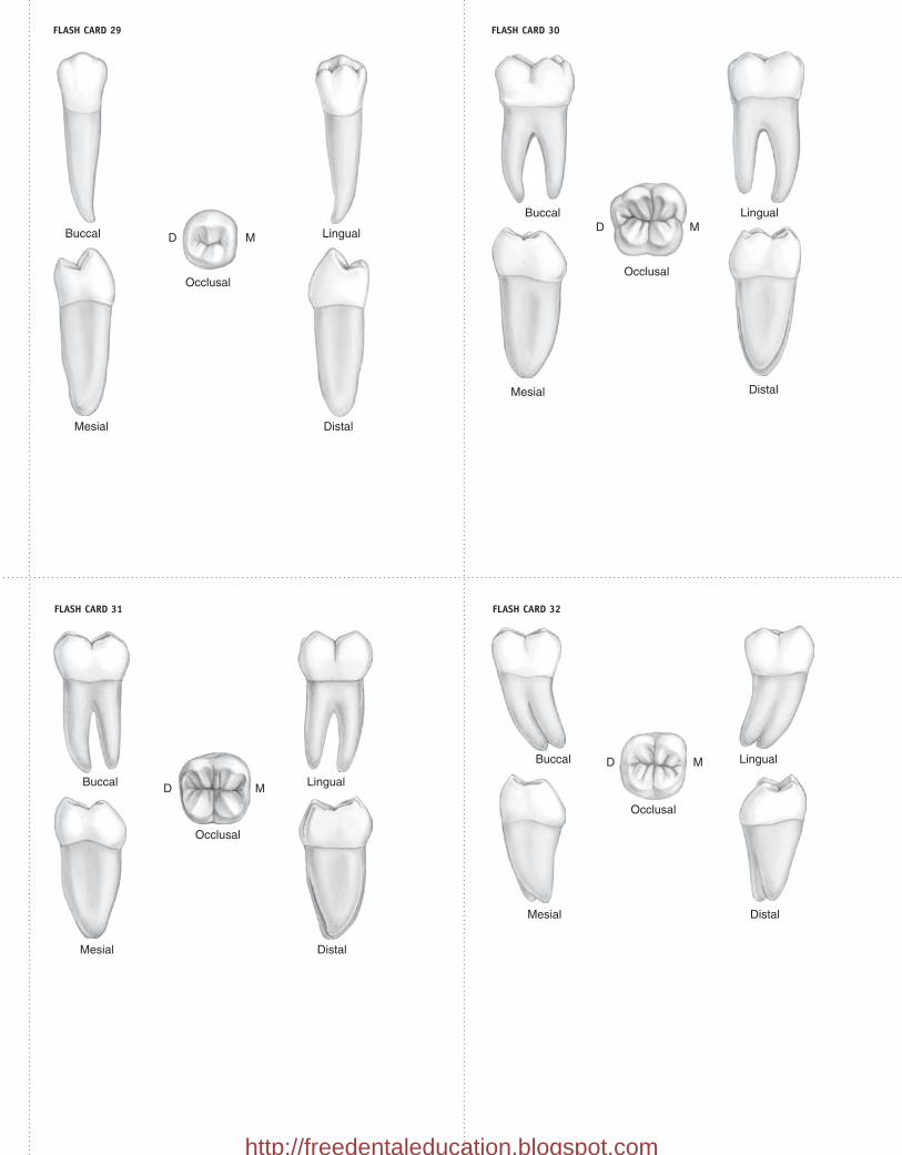
FLASH CARD 29
Buccal
Mesial Distal
Lingual
Occlusal
MD
FLASH CARD 30
Buccal
Mesial
Lingual
Distal
Occlusal
D M
FLASH CARD 31
Buccal
Mesial
Lingual
Distal
Occlusal
D M
FLASH CARD 32
Buccal
Mesial
Lingual
Distal
Occlusal
D M
http://freedentaleducation.blogspot.com

Copy
right
© 2
010,
200
3, 1
993,
198
4, 1
974,
196
5, 1
958,
195
0, 1
940
by S
aund
ers,
an
impr
int o
f Els
evie
r, In
c.
Mandibular Left Second PremolarUniversal: 20 International (FDI): 35 Palmer: 5CHARACTERISTICS SECOND PREMOLAR
Facial/Buccal AspectProximal contacts Cervico-occlusal Mesial/distal, middle thirdForm Bilaterally symmetricalLingual AspectBuccal profile None seenCusp height Buccal/lingual cusps nearly equalMesial AspectOcclusal plane Essentially horizontalTransverse ridge or buccal triangular ridge No transverse ridge presentOcclusal AspectOutline form SquareCusps 2 or 3Lobes 4 or 5DimensionsCrown length 8.0 mm (cervico-occlusal)Crown diameter Mesiodistal 7.0 mm Cervical 5.0 mm Buccolingual 8.0 mmContour height Facial Middle third Lingual Middle thirdRootLength 14.5 mmPulp canal(s) 1Pulp horn(s) 2ChronologyEruption 11–12 yrRoot completed 13–14 yr
Copy
right
© 2
010,
200
3, 1
993,
198
4, 1
974,
196
5, 1
958,
195
0, 1
940
by S
aund
ers,
an
impr
int o
f Els
evie
r, In
c.
Mandibular Left First MolarUniversal: 19 International (FDI): 36 Palmer: 6CHARACTERISTICS FIRST MOLAR
Facial/Buccal AspectCrown size Largest; widest mesiodistallyCusps 5 Cusps: mesiobuccal, distobuccal,
mesiolingual, distolingual, distalProximal contacts Cervical-occlusal location of mesial contact
is at middle third, distal contact is at middle third
Groove(s)/pit(s) 1 Mesiobuccal ± pit, 1 distobuccalRoot(s) Widely separated; nearly verticalLingual AspectCervix Crown narrowerOcclusal AspectCrown form Quadrilateral or hexagonal, pentagonalLobes 5Pulp horn(s) 5DimensionsCrown length 7.5 mm (cervical-occlusal)Crown diameter Mesiodistal 11.0 mm Cervical 9.0 mm Buccolingual 10.5 mmContour height Buccal crest Cervical third, 0.5 mm Lingual crest Middle third, 1.0 mmRootLength 14.0 mmChronologyEruption 6–7 yrRoot complete 9–10 yr
Copy
right
© 2
010,
200
3, 1
993,
198
4, 1
974,
196
5, 1
958,
195
0, 1
940
by S
aund
ers,
an
impr
int o
f Els
evie
r, In
c.
Mandibular Left Second MolarUniversal: 18 International (FDI): 37 Palmer: 7CHARACTERISTICS SECOND MOLAR
Facial/Buccal AspectCrown size Smaller than first mandibular molarCusps 4 Cusps: mesiobuccal, distobuccal,
mesiolingual, distolingualProximal contacts Cervical-occlusal location of mesial contact
is at middle third, distal contact is at middle third
Groove(s)/pit(s) 1 Buccal ± pitRoot(s) Close together, parallel; distally inclinedLingual AspectCervix Less narrowOcclusal AspectCrown form Quadrilateral or rectangularLobes 4Pulp horn(s) 4DimensionsCrown length 7.0 mm (cervico-occlusal)Crown diameter Mesiodistal 10.5 mm Cervical 8.0 mm Buccolingual 10.0 mmContour height Buccal crest Cervical third, 0.5 mm Lingual crest Middle third, 1.0 mmRootLength 13.0 mmChronologyEruption 11–13 yrRoot complete 14–15 yr
Copy
right
© 2
010,
200
3, 1
993,
198
4, 1
974,
196
5, 1
958,
195
0, 1
940
by S
aund
ers,
an
impr
int o
f Els
evie
r, In
c.
Mandibular Left Third MolarUNIvERSAL: 17 INTERNATIONAL (FDI): 38 PALMER: 8CHARACTERISTICS THIRD MOLAR
Facial/Buccal AspectCrown size Smallest of three mandibular molarsCusps 4 Cusps: mesiobuccal, distobuccal,
mesiolingual, distolingualProximal contacts Cervical-occlusal location of mesial contact
is at middle third, distal contact is at middle third
Groove(s)/pit(s) 1 Buccal ± pitRoot(s) Most short, fused, less inclinationLingual AspectCervix Less narrowOcclusal AspectCrown form Quadrilateral or ovoidLobes 4Pulp horn(s) 4DimensionsCrown length 7.0 mm (cervico-occlusal)Crown diameter Mesiodistal 10.0 mm Cervical 7.5 mm Buccolingual 9.5 mmContour height Buccal crest Cervical third, 0.5 mm Lingual crest Middle third, 1.0 mmRootLength 11.0 mmChronologyEruption 17–21 yrRoot complete 18–25 yr
http://freedentaleducation.blogspot.com

FLASH CARD 33 FLASH CARD 34
FLASH CARD 35 FLASH CARD 36
Copy
right
© 2
010,
200
3, 1
993,
198
4, 1
974,
196
5, 1
958,
195
0, 1
940
by S
aund
ers,
an
impr
int o
f Els
evie
r, In
c.
Copy
right
© 2
010,
200
3, 1
993,
198
4, 1
974,
196
5, 1
958,
195
0, 1
940
by S
aund
ers,
an
impr
int o
f Els
evie
r, In
c.
Copy
right
© 2
010,
200
3, 1
993,
198
4, 1
974,
196
5, 1
958,
195
0, 1
940
by S
aund
ers,
an
impr
int o
f Els
evie
r, In
c.
Copy
right
© 2
010,
200
3, 1
993,
198
4, 1
974,
196
5, 1
958,
195
0, 1
940
by S
aund
ers,
an
impr
int o
f Els
evie
r, In
c.
Figure 1-2, A Figure 1-2, B
Figure 5-2, B Figure 6-1
http://freedentaleducation.blogspot.com

CLC
MMR
(1) LIE LIE (2)
LIE (2) LIE (1)
LF
MMR
LF
DMR CDMR
Central incisor (first incisor)Lateral incisor (second incisor)Canine (cuspid)First premolar (first bicuspid)Second premolar (second bicuspid)
First premolar (bicuspid)Canine (cuspid)Lateral incisor (second incisor)Central incisor (first incisor)
Second premolar (bicuspid)
First molar
First molar
Second molar
Second molar
Third molar
Third molar
RIG
HT
LEF
T
MANDIBULAR
MAXILLARY
GingivalcreviceFree gingival marginKeratinizedepithelium
Gingivalgroove
JunctionalepitheliumAttachedgingiva
MucogingivaljunctionAlveolarmucosa
Alveolar process
Alveolarbone
Crevicularepithelium
DENTIN
Epithelialattachment
Cementum
Alveolarcrest
Periodontalmembrane
E
Central incisor (first incisor)
Lateral incisor (second incisor)Canine (cuspid)First molar
Second molar
Second molar
First molar
CanineLateral incisor (second incisor)
Central incisor (first incisor)
RIG
HT
LEF
T
MANDIBULAR
MAXILLARY
http://freedentaleducation.blogspot.com

FLASH CARD 37 FLASH CARD 38
FLASH CARD 39 FLASH CARD 40
Copy
right
© 2
010,
200
3, 1
993,
198
4, 1
974,
196
5, 1
958,
195
0, 1
940
by S
aund
ers,
an
impr
int o
f Els
evie
r, In
c.
Copy
right
© 2
010,
200
3, 1
993,
198
4, 1
974,
196
5, 1
958,
195
0, 1
940
by S
aund
ers,
an
impr
int o
f Els
evie
r, In
c.
Copy
right
© 2
010,
200
3, 1
993,
198
4, 1
974,
196
5, 1
958,
195
0, 1
940
by S
aund
ers,
an
impr
int o
f Els
evie
r, In
c.
Copy
right
© 2
010,
200
3, 1
993,
198
4, 1
974,
196
5, 1
958,
195
0, 1
940
by S
aund
ers,
an
impr
int o
f Els
evie
r, In
c.
Figure 7-1 Figure 8-1
Figure 8-13 Figure 9-1
http://freedentaleducation.blogspot.com

BR
MDD
BCR
MCA
BC LC
CL
MMDG
LR
DBCR
DMRDTF
DLCR MLCRCDGMTFMMRMBCR
CLC
MMRMLFMCR DCR
LR
DCRDMR
DLFDLF
MLFC
MCR
MCR
LRMMRMLF
C
CL
DCR
DMR
DLFDCR
C
MCR
IR
MMR
C
CL
DMR
LF
DMR
IR
MMRC
http://freedentaleducation.blogspot.com

FLASH CARD 41
FLASH CARD 43 FLASH CARD 44
FLASH CARD 42
Copy
right
© 2
010,
200
3, 1
993,
198
4, 1
974,
196
5, 1
958,
195
0, 1
940
by S
aund
ers,
an
impr
int o
f Els
evie
r, In
c.
Copy
right
© 2
010,
200
3, 1
993,
198
4, 1
974,
196
5, 1
958,
195
0, 1
940
by S
aund
ers,
an
impr
int o
f Els
evie
r, In
c.
Copy
right
© 2
010,
200
3, 1
993,
198
4, 1
974,
196
5, 1
958,
195
0, 1
940
by S
aund
ers,
an
impr
int o
f Els
evie
r, In
c.
Copy
right
© 2
010,
200
3, 1
993,
198
4, 1
974,
196
5, 1
958,
195
0, 1
940
by S
aund
ers,
an
impr
int o
f Els
evie
r, In
c.
Figure 9-15 Figure 10-1
Figure 11-1 Figure 11-2
http://freedentaleducation.blogspot.com

A
B
CD
DBCRMBCR
CF
MTFMMR
MLCR
ORDLCR
DF
DTF
DMRCP
TGOR
DOG
LG FCG
CGCF
BGCF
BG
BC
MMR
MCABCR
BTRLC
MBCRMLDG
MMRCL
MLDG
LCCDGBTR
DMR
DBCR
BC
http://freedentaleducation.blogspot.com

FLASH CARD 45 FLASH CARD 46
FLASH CARD 47 FLASH CARD 48
Copy
right
© 2
010,
200
3, 1
993,
198
4, 1
974,
196
5, 1
958,
195
0, 1
940
by S
aund
ers,
an
impr
int o
f Els
evie
r, In
c.
Copy
right
© 2
010,
200
3, 1
993,
198
4, 1
974,
196
5, 1
958,
195
0, 1
940
by S
aund
ers,
an
impr
int o
f Els
evie
r, In
c.
Copy
right
© 2
010,
200
3, 1
993,
198
4, 1
974,
196
5, 1
958,
195
0, 1
940
by S
aund
ers,
an
impr
int o
f Els
evie
r, In
c.
Copy
right
© 2
010,
200
3, 1
993,
198
4, 1
974,
196
5, 1
958,
195
0, 1
940
by S
aund
ers,
an
impr
int o
f Els
evie
r, In
c.
Figure 11-3 Figure 11-5
Figure 11-8 Figure 11-9
http://freedentaleducation.blogspot.com

LR
CL
DCA
DLC
DMR
DBC
BDG
MBR
DBR
MBR
FC
MLC
LDG
DLC
CL
LRDBR
MBR
MCA
MBC
MMR
MLC
FC
Roottrunk
LR
CR
DBR
CL
DBCMLC
MBC
BDG
Roottrunk
MBR
LR
http://freedentaleducation.blogspot.com

FLASH CARD 49 FLASH CARD 50
FLASH CARD 51 FLASH CARD 52
Copy
right
© 2
010,
200
3, 1
993,
198
4, 1
974,
196
5, 1
958,
195
0, 1
940
by S
aund
ers,
an
impr
int o
f Els
evie
r, In
c.
Copy
right
© 2
010,
200
3, 1
993,
198
4, 1
974,
196
5, 1
958,
195
0, 1
940
by S
aund
ers,
an
impr
int o
f Els
evie
r, In
c.
Copy
right
© 2
010,
200
3, 1
993,
198
4, 1
974,
196
5, 1
958,
195
0, 1
940
by S
aund
ers,
an
impr
int o
f Els
evie
r, In
c.
Copy
right
© 2
010,
200
3, 1
993,
198
4, 1
974,
196
5, 1
958,
195
0, 1
940
by S
aund
ers,
an
impr
int o
f Els
evie
r, In
c.
Figure 12-1 Figure 12-2
Figure 12-3 Figure 12-5
http://freedentaleducation.blogspot.com

Roottrunk
MLC DLC
LDG
CL
ApexApex
DC
DLC
DBC MLC
MBC
MBDG
CL
MRDR
DBDG
MBDG
MBCRSG
MTF
MLCR
LDG
DLCR
DTF
DCRDBDG
DBCRMMR
Mes
ial
MP
LingualCDG
DPDMR
Dis
tal
CPBuccal
http://freedentaleducation.blogspot.com

FLASH CARD 53
FLASH CARD 55
FLASH CARD 54
FLASH CARD 56
Copy
right
© 2
010,
200
3, 1
993,
198
4, 1
974,
196
5, 1
958,
195
0, 1
940
by S
aund
ers,
an
impr
int o
f Els
evie
r, In
c.
Copy
right
© 2
010,
200
3, 1
993,
198
4, 1
974,
196
5, 1
958,
195
0, 1
940
by S
aund
ers,
an
impr
int o
f Els
evie
r, In
c.
Copy
right
© 2
010,
200
3, 1
993,
198
4, 1
974,
196
5, 1
958,
195
0, 1
940
by S
aund
ers,
an
impr
int o
f Els
evie
r, In
c.
Copy
right
© 2
010,
200
3, 1
993,
198
4, 1
974,
196
5, 1
958,
195
0, 1
940
by S
aund
ers,
an
impr
int o
f Els
evie
r, In
c.
Figure 12-8 Figure 12-9
Figure 13-1 Figure 14-12
http://freedentaleducation.blogspot.com

MMR
MLC
MCA
CL
DD
Mesial root
CR
MBC
Cementum
Enamel
Pulpchamber
Pulp orroot canals
APICAL FORAMEN
Dentin
Pulp horns
MMRMBCDBCDC
DBDG
DCA
CL
Mesialroot
DMR
DLC
MLC
Distal root
Inci
sive
foss
a
Men
tal f
oram
en
Cor
onoi
d pr
oces
sM
andi
bula
r no
tch
Con
dyle
con
dylo
idpr
oces
s
Nec
k of
con
dyle
Ram
usO
bliq
ue r
idge
Bod
y of
man
dibl
eA
ngle
Fac
ial n
otch
http://freedentaleducation.blogspot.com

FLASH CARD 57 FLASH CARD 58
FLASH CARD 59 FLASH CARD 60
Copy
right
© 2
010,
200
3, 1
993,
198
4, 1
974,
196
5, 1
958,
195
0, 1
940
by S
aund
ers,
an
impr
int o
f Els
evie
r, In
c.
Copy
right
© 2
010,
200
3, 1
993,
198
4, 1
974,
196
5, 1
958,
195
0, 1
940
by S
aund
ers,
an
impr
int o
f Els
evie
r, In
c.
Copy
right
© 2
010,
200
3, 1
993,
198
4, 1
974,
196
5, 1
958,
195
0, 1
940
by S
aund
ers,
an
impr
int o
f Els
evie
r, In
c.
Copy
right
© 2
010,
200
3, 1
993,
198
4, 1
974,
196
5, 1
958,
195
0, 1
940
by S
aund
ers,
an
impr
int o
f Els
evie
r, In
c.
Figure 14-16 Figure 14-35
Figure 15-2 Figure 15-16
http://freedentaleducation.blogspot.com

Sty
loid
proc
ess
Occ
ipita
lco
ndyl
es
Med
ial
pter
ygoi
dm
uscl
e
Zyg
omat
icar
ch
Late
ral
pter
ygoi
dm
uscl
e
Ant
erio
rte
mpo
ralis
mus
cle
Ext
erna
lau
dito
rym
eatu
s
Tem
pora
lism
uscl
e Tem
pora
l bon
e ar
ticul
atin
g su
rfac
e
Syn
ovia
lm
embr
ane
Pos
terio
rca
psul
e
Art
icul
arsu
rfac
eof
con
dyle
Sup
erio
r ca
vity
Ava
scul
ar p
art o
fm
enis
cus
Syn
ovia
lm
embr
ane
Late
ral p
tery
goid
mus
cle:
Infe
rior
head
Sup
erio
r he
ad
Bre
gma
Sup
erio
r fr
onta
l gyr
us
Ant
erio
r br
anch
Cen
tral
sul
cus
Late
ral c
ereb
ral f
issu
re
Pos
terio
r br
anch
Sup
erfic
ial
tem
pora
l art
ery
Inio
n
Mid
dle
tem
pora
l gyr
us
Nas
ion
Infr
aorb
itala
Pos
terio
r su
perio
risal
veol
arar
tery
Mid
dle
men
inge
alar
tery
Inte
rnal
max
illar
yar
tery
Infe
rior
alve
olar
arte
ry
Ext
erna
l car
otid
arte
ry
Inte
rnal
car
otid
arte
ry
Com
mon
car
otid
arte
ry
Lam
bda
Sup
erio
r-pa
rieta
llo
bule
Myl
ohyo
id li
ne
Inci
sive
foss
a
Men
tal
fora
men
Men
tal
prot
uber
ance
Ext
erna
lob
lique
rid
ge
Can
ine
emin
ence
of
man
dibl
e
Sym
phys
is
Men
tal
tube
rcle
s
http://freedentaleducation.blogspot.com

FLASH CARD 61
FLASH CARD 63 FLASH CARD 64
Copy
right
© 2
010,
200
3, 1
993,
198
4, 1
974,
196
5, 1
958,
195
0, 1
940
by S
aund
ers,
an
impr
int o
f Els
evie
r, In
c.
Copy
right
© 2
010,
200
3, 1
993,
198
4, 1
974,
196
5, 1
958,
195
0, 1
940
by S
aund
ers,
an
impr
int o
f Els
evie
r, In
c.
Copy
right
© 2
010,
200
3, 1
993,
198
4, 1
974,
196
5, 1
958,
195
0, 1
940
by S
aund
ers,
an
impr
int o
f Els
evie
r, In
c.
Copy
right
© 2
010,
200
3, 1
993,
198
4, 1
974,
196
5, 1
958,
195
0, 1
940
by S
aund
ers,
an
impr
int o
f Els
evie
r, In
c.
FLASH CARD 62
Figure 15-19 Figure 15-25
Figure 16-32 Figure 16-34
http://freedentaleducation.blogspot.com

Orb
icul
aris
ocul
i mus
cle
Zyg
omat
icm
ajor
mus
cle
Leva
tor
labi
isu
p. m
uscl
e
Orb
icul
aris
oris
mus
cle
Dep
ress
or a
ngul
aris
oris
mus
cle
Ris
oriu
s m
uscl
e
Buc
cina
tor
mus
cle
Nas
alis
pro
c.al
aris
mus
cle
Par
otid
duc
t
Tem
pora
lism
uscl
e
Sty
logl
ossu
sm
uscl
e
Pte
rygo
man
dibu
lar
raph
e
Mas
sete
r m
uscl
e(c
ut)
Pla
tysm
a m
uscl
e
Arc
us d
enta
lissu
perio
r
Vel
i pal
atin
a m
ajor
art
ery
Exp
ansi
on o
f the
leva
tor
veli
pala
tini
Ham
ulus
pter
ygoi
deus
Pha
ryng
opal
atin
us m
uscl
e
Rap
he p
tery
gom
andi
bula
ris
Buc
coph
aryn
geus
mus
cle
Buc
cina
tor
mus
cle
Ton
silla
pal
atin
a
Glo
ssop
alat
inus
mus
cle
Pal
atin
ade
scen
dens
art
ery
Max
illar
isin
tern
a ar
tery
Ton
silla
ris a
rter
y
Pal
atin
a as
cend
ens
arte
ryP
hary
ngea
asce
nden
s ar
tery
Max
illar
isex
tern
a ar
tery
Car
otis
ext
erna
art
ery
Ling
ualis
art
ery
Ram
us d
orsa
lislin
guae
Pal
atum
dur
um
Pal
atum
mol
le
Arc
usgl
osso
pala
tinus
Arc
usph
aryn
gopa
latin
us
Uvu
la (
pala
tina)
Ton
silla
pala
tina
Con
stric
tor
phar
yngi
s su
perio
rm
uscl
eLa
tera
l ban
d of
lym
phoi
d tis
sue
Buc
coph
aryn
geus
mus
cle
Pha
ryng
opal
atin
usm
uscl
e
Glo
ssop
alat
inus
mus
cle
Ton
silla
pala
tina
B
A
Pha
ryng
ea
Tong
ue
DBCR
MBCR
CF
MTF
MMR
MLCR
OR
DLCR
DF
DTF
DMR
Buccal
Lingual
CP
Dis
tal
DMR
DPCDG
MP
Mes
ial
MMR
http://freedentaleducation.blogspot.com