Ways to Improve Insights into Clindamycin Pharmacology and ...
24
Citation: Armengol Álvarez, L.; Van de Sijpe, G.; Desmet, S.; Metsemakers, W.-J.; Spriet, I.; Allegaert, K.; Rozenski, J. Ways to Improve Insights into Clindamycin Pharmacology and Pharmacokinetics Tailored to Practice. Antibiotics 2022, 11, 701. https:// doi.org/10.3390/antibiotics11050701 Academic Editor: Alain Bosquet Mélou Received: 15 April 2022 Accepted: 18 May 2022 Published: 21 May 2022 Publisher’s Note: MDPI stays neutral with regard to jurisdictional claims in published maps and institutional affil- iations. Copyright: © 2022 by the authors. Licensee MDPI, Basel, Switzerland. This article is an open access article distributed under the terms and conditions of the Creative Commons Attribution (CC BY) license (https:// creativecommons.org/licenses/by/ 4.0/). antibiotics Review Ways to Improve Insights into Clindamycin Pharmacology and Pharmacokinetics Tailored to Practice Laura Armengol Álvarez 1, *, Greet Van de Sijpe 2,3 , Stefanie Desmet 4,5 , Willem-Jan Metsemakers 6,7 , Isabel Spriet 2,3 , Karel Allegaert 2,6,8 and Jef Rozenski 1 1 Rega Institute for Medical Research, KU Leuven, B-3000 Leuven, Belgium; [email protected] 2 Department of Pharmaceutical and Pharmacological Sciences, KU Leuven, B-3000 Leuven, Belgium; [email protected] (G.V.d.S.); [email protected] (I.S.); [email protected] (K.A.) 3 Pharmacy Department, University Hospitals Leuven, B-3000 Leuven, Belgium 4 Department of Laboratory Medicine, University Hospitals Leuven, B-3000 Leuven, Belgium; [email protected] 5 Department of Microbiology, Immunology and Transplantation, KU Leuven, B-3000 Leuven, Belgium 6 Department of Development and Regeneration, KU Leuven, B-3000 Leuven, Belgium; [email protected] 7 Department of Trauma Surgery, University Hospitals Leuven, B-3000 Leuven, Belgium 8 Department of Clinical Pharmacy, Erasmus MC, NL-3015 CN Rotterdam, The Netherlands * Correspondence: [email protected] Abstract: Given the increase in bacterial resistance and the decrease in the development of new antibiotics, the appropriate use of old antimicrobials has become even more compulsory. Clindamycin is a lincosamide antibiotic approved for adults and children as a drug of choice for systemic treatment of staphylococcal, streptococcal, and gram-positive anaerobic bacterial infections. Because of its profile and high bioavailability, it is commonly used as part of an oral multimodal alternative for prolonged parenteral antibiotic regimens, e.g., to treat bone and joint or prosthesis-related infections. Clindamycin is also frequently used for (surgical) prophylaxis in the event of beta-lactam allergy. Special populations (pediatrics, pregnant women) have altered cytochrome P450 (CYP)3A4 activity. As clindamycin is metabolized by the CYP3A4/5 enzymes to bioactive N-demethyl and sulfoxide metabolites, knowledge of the potential relevance of the drug’s metabolites and disposition in special populations is of interest. Furthermore, drug–drug interactions derived from CYP3A4 inducers and inhibitors, and the data on the impact of the disease state on the CYP system, are still limited. This narrative review provides a detailed survey of the currently available literature on pharmacology and pharmacokinetics and identifies knowledge gaps (special patient population, drug–drug, and drug–disease interactions) to describe a research strategy for precision medicine. Keywords: antibiotic; clindamycin; bacterial infections; pharmacokinetics; special patient populations; CYP450 enzymes; drug–drug interactions 1. Introduction The widespread use of antibiotics has contributed to the ongoing increase in antimi- crobial drug resistance over the last years, an emerging health issue highlighted as a key research area of the Health Research and Innovation by the European Commission [1]. This worldwide health problem [2], followed by the decrease in the development of new antimi- crobials, resulted in a worrying situation, as alternatives to treat infectious diseases become scarce. Given this concern, new approaches are needed, including the appropriate use of old ‘wonder drugs’ such as clindamycin (CLI), a potent and widely used antimicrobial. CLI is currently being exploited for its use in combination with other agents for combating re- sistant pathogens. In addition, CLI’s systemic exposure depends on drug hepatic clearance and drug–drug interactions that may occur and are of interest for reference and special patient populations. In addition, drug–disease interactions may also occur in the presence Antibiotics 2022, 11, 701. https://doi.org/10.3390/antibiotics11050701 https://www.mdpi.com/journal/antibiotics
-
Upload
khangminh22 -
Category
Documents
-
view
1 -
download
0
Transcript of Ways to Improve Insights into Clindamycin Pharmacology and ...
Citation: Armengol Álvarez, L.; Van
de Sijpe, G.; Desmet, S.; Metsemakers,
W.-J.; Spriet, I.; Allegaert, K.;
Rozenski, J. Ways to Improve Insights
into Clindamycin Pharmacology and
Pharmacokinetics Tailored to Practice.
Antibiotics 2022, 11, 701. https://
doi.org/10.3390/antibiotics11050701
Academic Editor: Alain
Bosquet Mélou
Received: 15 April 2022
Accepted: 18 May 2022
Published: 21 May 2022
Publisher’s Note: MDPI stays neutral
with regard to jurisdictional claims in
published maps and institutional affil-
iations.
Copyright: © 2022 by the authors.
Licensee MDPI, Basel, Switzerland.
This article is an open access article
distributed under the terms and
conditions of the Creative Commons
Attribution (CC BY) license (https://
creativecommons.org/licenses/by/
4.0/).
antibiotics
Review
Ways to Improve Insights into Clindamycin Pharmacology andPharmacokinetics Tailored to PracticeLaura Armengol Álvarez 1,*, Greet Van de Sijpe 2,3, Stefanie Desmet 4,5, Willem-Jan Metsemakers 6,7 ,Isabel Spriet 2,3, Karel Allegaert 2,6,8 and Jef Rozenski 1
1 Rega Institute for Medical Research, KU Leuven, B-3000 Leuven, Belgium; [email protected] Department of Pharmaceutical and Pharmacological Sciences, KU Leuven, B-3000 Leuven, Belgium;
[email protected] (G.V.d.S.); [email protected] (I.S.); [email protected] (K.A.)3 Pharmacy Department, University Hospitals Leuven, B-3000 Leuven, Belgium4 Department of Laboratory Medicine, University Hospitals Leuven, B-3000 Leuven, Belgium;
[email protected] Department of Microbiology, Immunology and Transplantation, KU Leuven, B-3000 Leuven, Belgium6 Department of Development and Regeneration, KU Leuven, B-3000 Leuven, Belgium;
[email protected] Department of Trauma Surgery, University Hospitals Leuven, B-3000 Leuven, Belgium8 Department of Clinical Pharmacy, Erasmus MC, NL-3015 CN Rotterdam, The Netherlands* Correspondence: [email protected]
Abstract: Given the increase in bacterial resistance and the decrease in the development of newantibiotics, the appropriate use of old antimicrobials has become even more compulsory. Clindamycinis a lincosamide antibiotic approved for adults and children as a drug of choice for systemic treatmentof staphylococcal, streptococcal, and gram-positive anaerobic bacterial infections. Because of itsprofile and high bioavailability, it is commonly used as part of an oral multimodal alternative forprolonged parenteral antibiotic regimens, e.g., to treat bone and joint or prosthesis-related infections.Clindamycin is also frequently used for (surgical) prophylaxis in the event of beta-lactam allergy.Special populations (pediatrics, pregnant women) have altered cytochrome P450 (CYP)3A4 activity.As clindamycin is metabolized by the CYP3A4/5 enzymes to bioactive N-demethyl and sulfoxidemetabolites, knowledge of the potential relevance of the drug’s metabolites and disposition in specialpopulations is of interest. Furthermore, drug–drug interactions derived from CYP3A4 inducers andinhibitors, and the data on the impact of the disease state on the CYP system, are still limited. Thisnarrative review provides a detailed survey of the currently available literature on pharmacologyand pharmacokinetics and identifies knowledge gaps (special patient population, drug–drug, anddrug–disease interactions) to describe a research strategy for precision medicine.
Keywords: antibiotic; clindamycin; bacterial infections; pharmacokinetics; special patient populations;CYP450 enzymes; drug–drug interactions
1. Introduction
The widespread use of antibiotics has contributed to the ongoing increase in antimi-crobial drug resistance over the last years, an emerging health issue highlighted as a keyresearch area of the Health Research and Innovation by the European Commission [1]. Thisworldwide health problem [2], followed by the decrease in the development of new antimi-crobials, resulted in a worrying situation, as alternatives to treat infectious diseases becomescarce. Given this concern, new approaches are needed, including the appropriate use ofold ‘wonder drugs’ such as clindamycin (CLI), a potent and widely used antimicrobial. CLIis currently being exploited for its use in combination with other agents for combating re-sistant pathogens. In addition, CLI’s systemic exposure depends on drug hepatic clearanceand drug–drug interactions that may occur and are of interest for reference and specialpatient populations. In addition, drug–disease interactions may also occur in the presence
Antibiotics 2022, 11, 701. https://doi.org/10.3390/antibiotics11050701 https://www.mdpi.com/journal/antibiotics
Antibiotics 2022, 11, 701 2 of 24
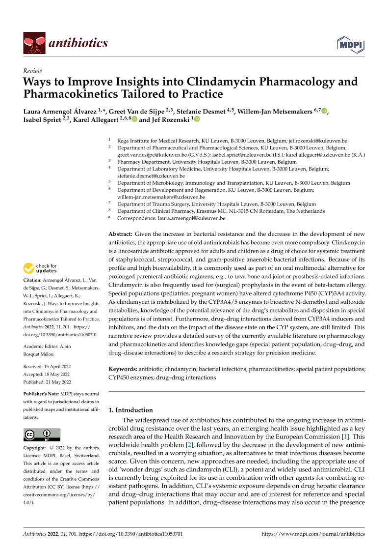
of inflammation or disease state. Subsequently, in this narrative review, we will provide adetailed survey of the currently available literature on the pharmacology and pharmacoki-netics of CLI, and we will identify relevant knowledge gaps (special patient population,drug–drug, and drug–disease interactions) to describe a future research approach towardspersonalized medicine (Figure 1).
Antibiotics 2022, 11, x FOR PEER REVIEW 2 of 28
occur in the presence of inflammation or disease state. Subsequently, in this narrative re-view, we will provide a detailed survey of the currently available literature on the phar-macology and pharmacokinetics of CLI, and we will identify relevant knowledge gaps (special patient population, drug–drug, and drug–disease interactions) to describe a fu-ture research approach towards personalized medicine (Figure 1).
Figure 1. Schematic overview of the main topics addressed in this review.
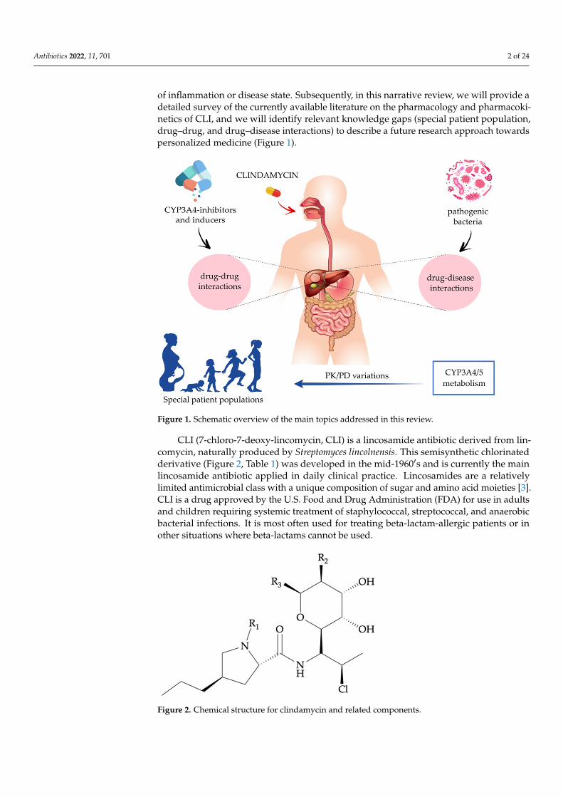
CLI (7-chloro-7-deoxy-lincomycin, CLI) is a lincosamide antibiotic derived from lin-comycin, naturally produced by Streptomyces lincolnensis. This semisynthetic chlorinated derivative (Figure 2, Table 1) was developed in the mid-1960′s and is currently the main lincosamide antibiotic applied in daily clinical practice. Lincosamides are a relatively lim-ited antimicrobial class with a unique composition of sugar and amino acid moieties [3]. CLI is a drug approved by the U.S. Food and Drug Administration (FDA) for use in adults and children requiring systemic treatment of staphylococcal, streptococcal, and anaerobic bacterial infections. It is most often used for treating beta-lactam-allergic patients or in other situations where beta-lactams cannot be used.
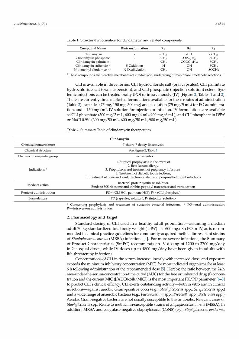
Table 1. Structural information for clindamycin and related components.
Compound Name Biotransformation R1 R2 R3 Clindamycin - -CH3 -OH -SCH3
Clindamycin phosphate - -CH3 -OPO3H2 -SCH3 Clindamycin palmitate - -CH3 -OCOC15H31 -SCH3 Clindamycin sulfoxide 1 S-Oxidation -H -OH -SCH3
N-demethyl clindamycin 1 N-Dealkylation -CH3 -OH -SOCH3 1 These compounds are bioactive metabolites of clindamycin, undergoing human phase I metabolic reactions.
Figure 1. Schematic overview of the main topics addressed in this review.
CLI (7-chloro-7-deoxy-lincomycin, CLI) is a lincosamide antibiotic derived from lin-comycin, naturally produced by Streptomyces lincolnensis. This semisynthetic chlorinatedderivative (Figure 2, Table 1) was developed in the mid-1960′s and is currently the mainlincosamide antibiotic applied in daily clinical practice. Lincosamides are a relativelylimited antimicrobial class with a unique composition of sugar and amino acid moieties [3].CLI is a drug approved by the U.S. Food and Drug Administration (FDA) for use in adultsand children requiring systemic treatment of staphylococcal, streptococcal, and anaerobicbacterial infections. It is most often used for treating beta-lactam-allergic patients or inother situations where beta-lactams cannot be used.
Antibiotics 2022, 11, x FOR PEER REVIEW 3 of 28
Figure 2. Chemical structure for clindamycin and related components.
CLI is available in three forms: CLI hydrochloride salt (oral capsules), CLI palmitate hydrochloride salt (oral suspension), and CLI phosphate (injection solution) esters. Sys-temic infections can be treated orally (PO) or intravenously (IV) (Figure 2, Tables 1 and 2). There are currently three marketed formulations available for these routes of administra-tion (Table 2): capsules (75 mg, 150 mg, 300 mg) and a solution (75 mg/5 mL) for PO ad-ministration, and a 150 mg/mL IV solution for injection or infusion. IV formulations are available as CLI phosphate (300 mg/2 mL, 600 mg/4 mL, 900 mg/6 mL), and CLI phosphate in D5W or NaCl 0.9% (300 mg/50 mL, 600 mg/50 mL, 900 mg/50 mL).
Table 2. Summary Table of clindamycin therapeutics.
Clindamycin Chemical nomenclature 7-chloro-7-deoxy-lincomycin
Chemical structure See Figure 2, Table 1 Pharmacotherapeutic
group Lincosamides
Indications 1
1. Surgical prophylaxis in the event of 2. beta-lactam allergy;
3. Prophylaxis and treatment of pregnancy infections; 4. Treatment of diabetic foot infections;
Treatment of bone and joint, fracture-related, and peripros-thetic joint infections
Mode of action Bacterial protein synthesis inhibitor.
Binds to 50S ribosome and inhibits peptidyl transferase and translocation
Route of administration PO 2 (CLI HCl, palmitate HCl); IV 2 (CLI phosphate) Formulations PO (capsules, solution); IV (injection solution)
1 Concerning prophylaxis and treatment of systemic bacterial infections; 2 PO—oral administration; IV—intravenous administration.
2. Pharmacology and Target Standard dosing of CLI used in a healthy adult population—assuming a median
adult 70 kg standardized total body weight (TBW)—is 600 mg.q8h PO or IV, as is recom-mended in clinical practice guidelines for community-acquired methicillin-resistant strains of Staphylococcus aureus (MRSA) infections [4]. For more severe infections, the Sum-mary of Product Characteristics (SmPC) recommends an IV dosing of 1200 to 2700 mg/day in 2–4 equal doses, while IV doses up to 4800 mg/day have been given in adults with life-threatening infections.
Concentrations of CLI in the serum increase linearly with increased dose, and expo-sure exceeds the minimum inhibitory concentration (MIC) for most indicated organisms for at least 6 h following administration of the recommended dose [5]. Hereby, the ratio
Figure 2. Chemical structure for clindamycin and related components.
Antibiotics 2022, 11, 701 3 of 24
Table 1. Structural information for clindamycin and related components.
Compound Name Biotransformation R1 R2 R3
Clindamycin - -CH3 -OH -SCH3Clindamycin phosphate - -CH3 -OPO3H2 -SCH3Clindamycin palmitate - -CH3 -OCOC15H31 -SCH3
Clindamycin sulfoxide 1 S-Oxidation -H -OH -SCH3N-demethyl clindamycin 1 N-Dealkylation -CH3 -OH -SOCH3
1 These compounds are bioactive metabolites of clindamycin, undergoing human phase I metabolic reactions.
CLI is available in three forms: CLI hydrochloride salt (oral capsules), CLI palmitatehydrochloride salt (oral suspension), and CLI phosphate (injection solution) esters. Sys-temic infections can be treated orally (PO) or intravenously (IV) (Figure 2, Tables 1 and 2).There are currently three marketed formulations available for these routes of administration(Table 2): capsules (75 mg, 150 mg, 300 mg) and a solution (75 mg/5 mL) for PO administra-tion, and a 150 mg/mL IV solution for injection or infusion. IV formulations are availableas CLI phosphate (300 mg/2 mL, 600 mg/4 mL, 900 mg/6 mL), and CLI phosphate in D5Wor NaCl 0.9% (300 mg/50 mL, 600 mg/50 mL, 900 mg/50 mL).
Table 2. Summary Table of clindamycin therapeutics.
Clindamycin
Chemical nomenclature 7-chloro-7-deoxy-lincomycin
Chemical structure See Figure 2, Table 1
Pharmacotherapeutic group Lincosamides
Indications 1
1. Surgical prophylaxis in the event of2. Beta-lactam allergy;
3. Prophylaxis and treatment of pregnancy infections;4. Treatment of diabetic foot infections;
5. Treatment of bone and joint, fracture-related, and periprosthetic joint infections
Mode of action Bacterial protein synthesis inhibitor.Binds to 50S ribosome and inhibits peptidyl transferase and translocation
Route of administration PO 2 (CLI HCl, palmitate HCl); IV 2 (CLI phosphate)
Formulations PO (capsules, solution); IV (injection solution)
1 Concerning prophylaxis and treatment of systemic bacterial infections; 2 PO—oral administration;IV—intravenous administration.
2. Pharmacology and Target
Standard dosing of CLI used in a healthy adult population—assuming a medianadult 70 kg standardized total body weight (TBW)—is 600 mg.q8h PO or IV, as is recom-mended in clinical practice guidelines for community-acquired methicillin-resistant strainsof Staphylococcus aureus (MRSA) infections [4]. For more severe infections, the Summaryof Product Characteristics (SmPC) recommends an IV dosing of 1200 to 2700 mg/dayin 2–4 equal doses, while IV doses up to 4800 mg/day have been given in adults withlife-threatening infections.
Concentrations of CLI in the serum increase linearly with increased dose, and exposureexceeds the minimum inhibitory concentration (MIC) for most indicated organisms for at least6 h following administration of the recommended dose [5]. Hereby, the ratio between the 24 harea-under-the-serum-concentration-time curve (AUC) for the free or unbound drug (f) concen-tration and the current MIC ([fAUC0-24h/MIC]) is the most important PK/PD parameter [6–8]to predict CLI’s clinical efficacy. CLI exerts outstanding activity—both in vitro and in clinicalinfections—against aerobic Gram-positive cocci (e.g., Staphylococcus spp., Streptococcus spp.)and a wide range of anaerobic bacteria (e.g., Fusobacterium spp., Prevotella spp., Bacteroides spp.).Aerobic Gram-negative bacteria are not usually susceptible to this antibiotic. Relevant cases ofStaphylococcus spp. Relate to methicillin-susceptible strains of Staphylococcus aureus (MSSA). Inaddition, MRSA and coagulase-negative staphylococci (CoNS) (e.g., Staphylococcus epidermis,
Antibiotics 2022, 11, 701 4 of 24
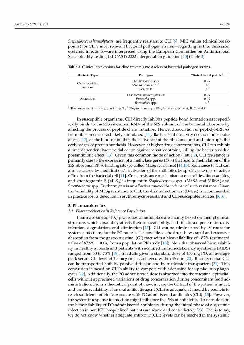
Staphylococcus haemolyticus) are frequently resistant to CLI [9]. MIC values (clinical break-points) for CLI’s most relevant bacterial pathogen strains—regarding further discussedsystemic infections—are interpreted using the European Committee on AntimicrobialSusceptibility Testing (EUCAST) 2022 interpretation guideline [10] (Table 3).
Table 3. Clinical breakpoints for clindamycin’s most relevant bacterial pathogen strains.
Bacteria Type Pathogen Clinical Breakpoints 1
Gram-positiveaerobes
Staphylococcus spp. 0.25Streptococcus spp. 2 0.5
Scheme 0. 0.5
AnaerobesFusobacterium necrophorum 0.25
Prevotella spp. 0.25Bacteroides spp. 4 3
1 The concentrations are given in mg/L; 2 Streptococcus spp.: Streptococcus groups A, B, C, and G.
In susceptible organisms, CLI directly inhibits peptide bond formation as it specif-ically binds to the 23S ribosomal RNA of the 50S subunit of the bacterial ribosome byaffecting the process of peptide chain initiation. Hence, dissociation of peptidyl-tRNAsfrom ribosomes is most likely stimulated [11]. Bacteriostatic activity occurs in most situ-ations [12], as the binding inhibits the active site of the ribosome unit and interrupts theearly stages of protein synthesis. However, at higher drug concentrations, CLI can exhibita time-dependent bactericidal action against sensitive strains, killing the bacteria with apostantibiotic effect [13]. Given this common mode of action (Table 2), CLI resistance isprimarily due to the expression of a methylase genes (Erm) that lead to methylation of the23S ribosomal RNA-binding site (so-called MLSB resistance) [14,15]. Resistance to CLI canalso be caused by modification/inactivation of the antibiotics by specific enzymes or activeefflux from the bacterial cell [11]. Cross-resistance mechanism to macrolides, lincosamides,and streptogramin B (MLSB) is frequent in Staphylococcus spp. (MSSA and MRSA) andStreptococcus spp. Erythromycin is an effective macrolide inducer of such resistance. Giventhe variability of MLSB resistance to CLI, the disk induction test (D-test) is recommendedin practice for its detection in erythromycin-resistant and CLI-susceptible isolates [9,16].
3. Pharmacokinetics3.1. Pharmacokinetics in Reference Population
Pharmacokinetic (PK) properties of antibiotics are mainly based on their chemicalstructure, which absolutely affects their bioavailability, half-life, tissue penetration, dis-tribution, degradation, and elimination [17]. CLI can be administered by IV route forsystemic infections, but the PO route is also possible, as the drug shows rapid and extensiveabsorption from the gastrointestinal (GI) tract with a bioavailability of ~87% (estimatedvalue of 87.6% ± 0.09, from a population PK study [18]). Note that observed bioavailabil-ity in healthy subjects and patients with acquired immunodeficiency syndrome (AIDS)ranged from 53 to 75% [19]. In adults given a standard dose of 150 mg PO, an averagepeak serum CLI level of 2.5 mcg/mL is achieved within 45 min [20]. It appears that CLIcan be transported both by passive diffusion and by nucleoside transporters [21]. Thisconclusion is based on CLI’s ability to compete with adenosine for uptake into phago-cytes [22]. Additionally, the PO administered dose is absorbed into the intestinal epithelialcells without appreciated variations of drug concentration during concomitant food ad-ministration. From a theoretical point of view, in case the GI tract of the patient is intact,and the bioavailability of an oral antibiotic agent (CLI) is adequate, it should be possible toreach sufficient antibiotic exposure with PO administered antibiotics (CLI) [23]. However,the systemic response to infection might influence the PKs of antibiotics. To date, data onthe bioavailability of PO-administered antibiotics during the initial phase of a systemicinfection in non-ICU hospitalized patients are scarce and contradictory [23]. That is to say,we do not know whether adequate antibiotic (CLI) levels can be reached in the systemic
Antibiotics 2022, 11, 701 5 of 24
circulation when such drugs are administered PO during the initial stage of an infectiousillness [23].
Given its lipophilic properties, CLI has a high volume of distribution (Vd) (in healthyadults, CLI has a Vd at a steady state of 0.79 L/kg [19]). The degree of protein bindingin healthy adults is concentration-dependent and ranges from 62% to 94%. Binding re-lates primarily to alpha-1-acid glycoprotein (AAG) concentration [8], followed by plasmaprotein albumin. As it travels through the bloodstream, this lincosamide is widely dis-tributed in body fluids, organs, and tissues, including bones and abscesses. CLI crossesthe human placenta readily, but it does not efficiently cross the blood–brain barrier, andno significant levels (~20%) [13] are attained in the cerebrospinal fluid, even in the settingof meningitis [20,24,25]. As described in the SmPC, high CLI serum concentrations areachieved in bone tissue, synovial fluid, peritoneal fluid, pleural fluid, expectorations, andpus. The following concurrent concentrations of the drug are reported compared with theblood compartment: in bone tissue 40% (20–75%), in synovial fluid 50%, in peritoneal fluid50%, in pleural fluid 50–90%, in expectorations 30–75%, and in pus 30%. In a referencepopulation, CLI exhibits good bioavailability after PO administration [18].
CLI undergoes hepatic metabolism to the major bioactive sulfoxide (primary metabo-lite) and N-demethyl metabolites (Figure 2, Table 1), and also some inactive metabolites.Recent in vitro studies in human liver and intestinal microsomes indicated that CLI ispredominantly oxidized by phase I cytochrome P450 (CYP)3A4 enzyme, with a minorcontribution from phase I CYP3A5, to form the aforementioned metabolites [5]. In healthyadults, hepatic drug clearance (CL) is about 0.3–0.4 L/h/kg [5]. Because CLI has a lowhepatic extraction ratio, CL decreases when hepatic intrinsic clearance and the unboundconcentration decrease [19].
After hepatic metabolism and within 24 h after intake, only about 10% of an oral doseof CLI is excreted in the urine as active drug and metabolites, ~3.6% in the feces, with theremainder excreted as inactive metabolites [26]. Biological elimination half-life (T1/2) inhealthy adults with a normal renal function is about 2–3 h [26]. The T1/2 may be prolongedin patients with moderate to severe hepatic or renal dysfunction, but no specific dosingadjustment is recommended. Dosage adjustment and monitoring are only recommendedfor patients with severe hepatic impairment or failure, but there are no specific guidelinesfor CLI. Since CLI is hepatically cleared, adjustment for renal dysfunction is generallynot required.
3.2. Pharmacokinetics in Special Patient Populations
CLI’s metabolites are not just of scientific interest but can also be relevant whenconsidering bacterial killing. Better understanding and quantitative data of their potentialpharmacodynamic (PD) activity are still missing. Given the biotransformation pathwayinvolved, it is important to elucidate relevant covariates of drug metabolism, such as age,gestation, or drug–drug interaction, as these hold the potential for individual exposure andresponse to therapy. Subsequently, extrapolating the conventional adult dose of CLI basedon covariates such as TBW is not appropriate and should be further explored.
Special patient populations (SPPs) undergo several alterations that can potentiallyimpact both the metabolism and disposition of CLI. We focused on two SPPs (pediatrics andpregnancy, breastfeeding, and postpartum) to illustrate the needed insights and research toimprove precision medicine in these populations (Table 4). Anatomic and physiological age-related changes tend to be related to TBW, body composition, and function (e.g., variationsin fat mass, body water, plasma volume and proteins, and glomerular filtration). Thesechanges can be further affected by pathophysiological and nonmaturational events such asinflammation. One of the relevant mechanisms of altered PK in these special populations isdue to variations in the CYP3A activity. Consequently, knowledge of the potential relevanceof CLI’s metabolites and disposition in SPPs is fundamental. We selected these two SPPs,but the same strategy could be considered for other specific settings, such as for patientswith cancer or coronavirus disease 2019 (COVID-19).
Antibiotics 2022, 11, 701 6 of 24
Pediatrics is a diverse SPP as it covers pediatric age groups, including neonates (birthto <28 days), infants (28 days to <2 years), young children (2 to <6 years), old children(6 to <12 years), and adolescents (12 to 18 years), as defined by the International Conferenceon Harmonization E11 guidance (International Council for Harmonisation, 2000) [27]. Thesecond SPP group includes pregnant, breastfeeding, and postpartum women. Pregnancychanges are not uniform, and their extent depends on the stage of gestation; hereby, thisSPP is divided into five classes of women: prepregnant, first trimester, second trimester,third trimester, and breastfeeding women or postpartum women. In order to guaranteesafe and efficient use of CLI in each SPP, its PK data should be considered, as well as dosageguidelines concerning the systemic treatment of relevant bacterial infections. Many of theanatomic and physiologic changes observed in these populations will result in markedchanges in absorption, distribution, metabolism, elimination (ADME) [8] (Table 4), anddosing regimens.
3.2.1. Pediatrics
CLI has been used for many years in the treatment of infections in children. It hastraditionally been a component of empiric antibiotic regimens for bone and joint infectionswhere anaerobes are likely causative pathogens, as well as in the treatment of seriousskin and soft tissue infections. The exact ontogeny of oral drug absorption GI processesin pediatrics is still to be elucidated, especially for the most vulnerable groups (neonatesand infants of <6 months). Consequently, drug labeling changes frequently and does notinclude neonatal dosing data (<1 month) [28]. In neonates, the dose should be based onboth TBW and age to allow for a slower elimination [20]. According to the SmPC, for thetreatment of serious infections, CLI 15 to 25 mg/kg/day can be administered (in threeor four equal doses) for children and adolescents. For more severe infections, doses of25 to 40 mg/kg/day for children and adolescents can also be administered in three or fourdivided doses. Despite the existing guidelines, available PK data from pediatric PK studiesare scarce, and age-based optimal dosing is still unknown [29]. It is recommended thatchildren be given no less than 300 mg/day regardless of the TBW.
Apart from a brief peak postnatally (pH ~7), where mean gastric pH is high directlyafter birth, gastric pH rapidly decreases (pH ~2.0–2.7 values in neonates [30]) and remainsaround a value of 2–3 in children of all ages [31] (typically < 3 in children and adoles-cents [30]). As CLI shows high lipophilicity, changes in gastric pH do not translate intoabsorption rate variations [32]. However, data concerning the extent to which absorptionin the GI tract could be affected for neonates are deficient: gastric emptying reported in theliterature is highly variable in children younger than 6 months [33], and GI transit time isalso slower in neonates and infants yet reaches adult values at the age of 2 years [33,34](Table 4). Nevertheless, CLI is a highly bioavailable PO, but, given the clear knowledge gapregarding the bioavailability of PO administered antibiotics in non-ICU patients duringthe initial phase of systemic infection [23], CLI can also be administered by IV route forsystemic infections.
Distribution in pediatrics [29] can be notably affected by changes in protein binding(Table 4). Neonates display a continually changing plasma profile, as the presence of fetalproteins and endogenous substrates, which are known to interfere with drug binding, canlead to unexpected complications because of a higher than expected ‘free’ drug fraction.Moreover, serum protein level and drug-binding rate of newborn infants are very low, andadult values (77 mg/mL) are reached at ~10 months of age [35]. Besides maturationalchanges, AAG concentrations are also affected by nonmaturational factors such as post-surgery or inflammation, as both result in an increase in AAG synthesis as part of theacute-phase response. Booker et al. determined how concentrations of AAG changed in in-fants requiring major surgery [35]. Despite a high interpatient variation (0.07–0.78 mg/mL),the overall mean perioperative AAG was 0.38 mg/mL, although concentrations doubledto 0.76 mg/mL on day 4 after surgery. Next to AAG’s response to surgical stress, inflam-mation in disease states can impact AAG levels in pediatrics. Ipek et al. confirmed results
Antibiotics 2022, 11, 701 7 of 24
from previous studies by reporting a statistically significant increase in AAG levels duringneonatal bacterial sepsis, showing positive values (>0.7 g/L) of 1.1 ± 0.4 and 0.8 ± 0.4 g/Lfor confirmed and clinical sepsis, respectively [36].
Regarding developmental changes in CLI’s hepatic metabolism during childhood,CYP3A4 is not detectable before birth. However, CYP3A4 generally increases postnatally tobecome the dominant CYP enzyme in the adult liver and intestine [37] and becomes activeduring the first weeks [38]. A recently published review from van Groen et al. [39] statedan increase in CYP3A4 microsomal levels, although activity data show a decrease duringfetal life (from 6% to 3%). CYP3A4 enzyme shows low catalytic activity in fetuses with50% of adult levels in infants up to 1 year, while adult levels are slowly reached in infantsand young children (between 1 to 5 years). Subsequently, CYP3A4′s enzyme activity isfound to be age-dependent (Table 4). In terms of CYP3A5, van Groen et al. stated thatprotein expression shows no clear developmental pattern and is age-independent [32,39].In contrast, CYP3A5 is polymorphic, and its expression levels vary between individuals andpopulations. CYP3A5 can be found in significant levels in 10–40% of Caucasians, 40–50%of Chinese subjects, and in approximately 90% of individuals with African origins [40].
Besides maturation, inflammation and organ failure are proven to be relevant nonmat-urational factors for CYP3A4-mediated drug metabolism. Brussee et al. [41] recently carriedout the first population PK model that quantified the influence of maturation, inflammation,and organ failure on midazolam CL (and potentially other selective CYP3A substrates)in term neonates, infants, children, adolescents, and adults with varying levels of criticalillness. Predictions based on this model indicated a 30% decrease in midazolam CL whenC-reactive protein concentrations that reflect the presence of inflammation threefold in-crease from 32 to 100 mg/L [41]. Furthermore, CL decreased by 26% when disease severity,expressed as the number of failing organs, increased from one to two [41].
Antibiotics 2022, 11, 701 8 of 24
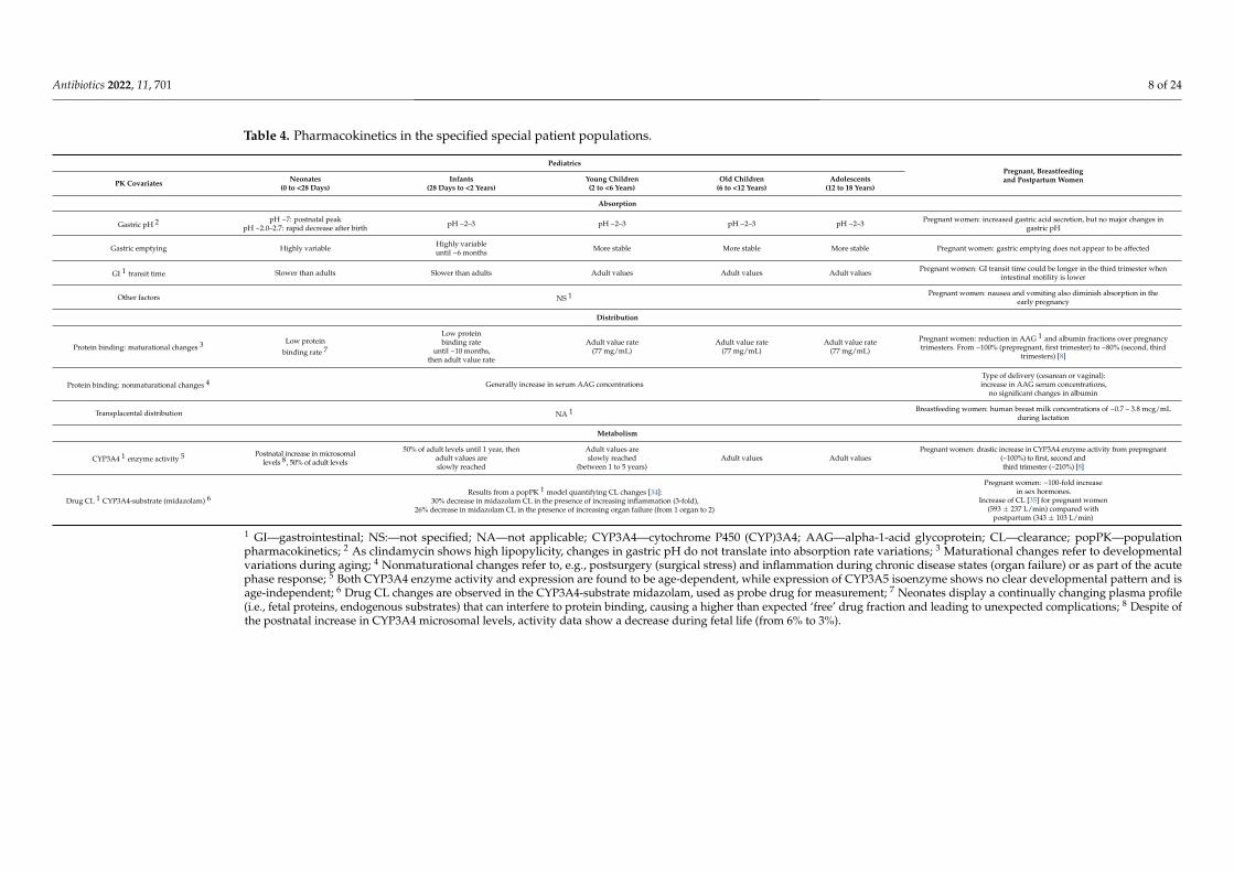
Table 4. Pharmacokinetics in the specified special patient populations.
PediatricsPregnant, Breastfeedingand Postpartum WomenPK Covariates Neonates
(0 to <28 Days)Infants
(28 Days to <2 Years)Young Children(2 to <6 Years)
Old Children(6 to <12 Years)
Adolescents(12 to 18 Years)
Absorption
Gastric pH 2 pH ~7: postnatal peakpH ~2.0–2.7: rapid decrease after birth pH ~2–3 pH ~2–3 pH ~2–3 pH ~2–3 Pregnant women: increased gastric acid secretion, but no major changes in
gastric pH
Gastric emptying Highly variable Highly variableuntil ~6 months More stable More stable More stable Pregnant women: gastric emptying does not appear to be affected
GI 1 transit time Slower than adults Slower than adults Adult values Adult values Adult values Pregnant women: GI transit time could be longer in the third trimester whenintestinal motility is lower
Other factors NS 1 Pregnant women: nausea and vomiting also diminish absorption in theearly pregnancy
Distribution
Protein binding: maturational changes 3 Low protein
binding rate 7
Low proteinbinding rate
until ~10 months,then adult value rate
Adult value rate(77 mg/mL)
Adult value rate(77 mg/mL)
Adult value rate(77 mg/mL)
Pregnant women: reduction in AAG 1 and albumin fractions over pregnancytrimesters. From ~100% (prepregnant, first trimester) to ~80% (second, third
trimesters) [8]
Protein binding: nonmaturational changes 4 Generally increase in serum AAG concentrationsType of delivery (cesarean or vaginal):increase in AAG serum concentrations,
no significant changes in albumin
Transplacental distribution NA 1 Breastfeeding women: human breast milk concentrations of ~0.7 – 3.8 mcg/mLduring lactation
Metabolism
CYP3A4 1 enzyme activity 5 Postnatal increase in microsomallevels 8, 50% of adult levels
50% of adult levels until 1 year, thenadult values areslowly reached
Adult values areslowly reached
(between 1 to 5 years)Adult values Adult values
Pregnant women: drastic increase in CYP3A4 enzyme activity from prepregnant(~100%) to first, second andthird trimester (~210%) [8]
Drug CL 1 CYP3A4-substrate (midazolam) 6Results from a popPK 1 model quantifying CL changes [34]:
30% decrease in midazolam CL in the presence of increasing inflammation (3-fold),26% decrease in midazolam CL in the presence of increasing organ failure (from 1 organ to 2)
Pregnant women: ~100-fold increasein sex hormones.
Increase of CL [35] for pregnant women(593 ± 237 L/min) compared with
postpartum (343 ± 103 L/min)
1 GI—gastrointestinal; NS:—not specified; NA—not applicable; CYP3A4—cytochrome P450 (CYP)3A4; AAG—alpha-1-acid glycoprotein; CL—clearance; popPK—populationpharmacokinetics; 2 As clindamycin shows high lipopylicity, changes in gastric pH do not translate into absorption rate variations; 3 Maturational changes refer to developmentalvariations during aging; 4 Nonmaturational changes refer to, e.g., postsurgery (surgical stress) and inflammation during chronic disease states (organ failure) or as part of the acutephase response; 5 Both CYP3A4 enzyme activity and expression are found to be age-dependent, while expression of CYP3A5 isoenzyme shows no clear developmental pattern and isage-independent; 6 Drug CL changes are observed in the CYP3A4-substrate midazolam, used as probe drug for measurement; 7 Neonates display a continually changing plasma profile(i.e., fetal proteins, endogenous substrates) that can interfere to protein binding, causing a higher than expected ‘free’ drug fraction and leading to unexpected complications; 8 Despite ofthe postnatal increase in CYP3A4 microsomal levels, activity data show a decrease during fetal life (from 6% to 3%).
Antibiotics 2022, 11, 701 9 of 24
3.2.2. Pregnant, Breastfeeding, and Postpartum Women
CLI for systemic use is FDA-classified as a Pregnancy Category B drug and is generallyconsidered safe and effective in pregnancy [26], given that, based on experimental animalstudies, CLI is not expected to increase the risk of congenital anomalies [42]. Because riskcannot be excluded, the SmPC states that the drug should be used during pregnancy only ifclearly needed. According to the lactation section on the SmPC, PO-, and IV-administeredCLI diffuses across the placenta barrier into the fetal circulation, appearing in humanbreast milk in ranges from 0.7 to 3.8 mcg/mL (i.e., breast milk/maternal plasma ratio0.08–3.1 [8,43]). Hence, nursing mothers should stop breastfeeding during CLI therapy (ifpossible) [42] because CLI has the potential to cause undesired effects on breastfed infants’GI flora. Moreover, CLI should not be taken by nursing mothers because of the potentialfor serious adverse reactions in nursing infants [42].
The effect of pregnancy on drug absorption over trimesters is mainly determinedby factors such as gastric pH, gastric emptying, and GI transit time (Table 4). Previousreviews [44,45] reported increased gastric acid secretion during pregnancy, but severaloriginal studies addressing heartburn during pregnancy show that there are no majorchanges in gastric pH over trimesters compared with nonpregnant women [33]. Gastricemptying does not appear to be affected by pregnancy [33,46], but overall, GI transit timecould be longer in the third trimester when intestinal motility is lower [33]. Drug absorptionis also diminished by nausea and vomiting early in pregnancy, which can result in lowerplasma drug concentrations [44].
As mentioned earlier, significant levels of CLI are achieved in human breast milkas a result of its distribution across the blood–placental barrier (Table 4). This distribu-tion during pregnancy can be influenced by changes in TBW, regional blood flows, tissuecomposition (such as body water and body fat), plasma composition, and volume and alter-ations in the unbound fraction of a given antimicrobial [8]. Over the pregnancy trimesters,most covariates (e.g., TBW, fat mass, body water, plasma volume, cardiac output) in-crease until the third trimester. However, both AAG and albumin fractions are reducedfrom ~100% (prepregnant and first trimester) to ~80% (second and third trimesters), asdescribed by Allegaert et al. [8] (Table 4). Consequently, the binding of CLI may be af-fected, which may lead to difficulties in maintaining adequate plasma concentrations ofCLI (highly protein-bound) as the measurement of total drug concentration in plasma mayno longer be a valid indicator for dose adjustment. As studied by Larijani et al. [47], thetype of delivery (cesarean or vaginal) further affects serum AAG and albumin concentra-tions. Regarding AAG levels, study findings demonstrate that in both the cesarian sectionand tuboplasty patients, exposure increased similarly on different postoperative days(146.3 ± 19.2; 134.8 ± 8.2 mg/dL). Moreover, even if the increase in AAG was delayed inpregnant women, both vaginal and cesarean delivery resulted in an increase in serum AAGconcentration. Concerning albumin levels, data indicated that nonpregnant women hadhigher serum albumin concentrations than pregnant women. However, albumin concentra-tions did not significantly change throughout the study except for a significant immediatedecrease postoperative after the cesarian section.
Pregnancy is characterized by about a 100-fold increase in circulating progesteroneand estrogens, with an increase over advancing gestational age [48]. Subsequently, drugmetabolism is also altered over pregnancy, secondary to the characteristic elevated levels ofsex hormones and changes in drug-metabolizing enzymes [44]. Hebert et al. carried outa study to evaluate the effects of pregnancy on in vivo CYP3A activities in humans usingCYP3A4-substrate midazolam as a probe drug for measurement [49] (Table 4). They re-ported a higher (apparent) oral unbound drug CL for pregnant women (593 ± 237 L/min)compared with postpartum (343 ± 103 L/min), representing approximately twofoldCYP3A4 in vivo induction during the third trimester [49]. Later findings confirmed thatplacental growth hormone estrogens (17-beta estradiol), cortisol, and progesterone poten-tially induce this in vivo CYP3A4 increase [50]. Moreover, Allegaert et al. reported a drasticincrease in CYP3A4 enzyme activity from the prepregnant stage (100%) to the first, second
Antibiotics 2022, 11, 701 10 of 24
and third trimesters (210%), next to increasing RNA expression protein levels and hepaticCL [8].
4. Clinical Practice and Efficacy
CLI is FDA-approved for serious bacterial infections, mainly caused by S. aureus, suchas septicemia, intra-abdominal infections, lower respiratory tract infections, bone and jointinfections, and skin and soft tissue infections [51]. Anesthesiologists and surgeons willoften administer CLI per The American Society of Health-System Pharmacists (ASHP) andInfectious Diseases Society of America (IDSA) guidelines as prophylaxis in the operatingroom [51]. Evidence for the use of this drug in systemic conditions is reviewed to showCLI’s potential applicability and indications (Tables 2 and 5). Moreover, recommended MICvalues (clinical breakpoints) for CLI’s most relevant pathogen strains for the mentionedindications are interpreted using the EUCAST 2022 guideline (Table 3).
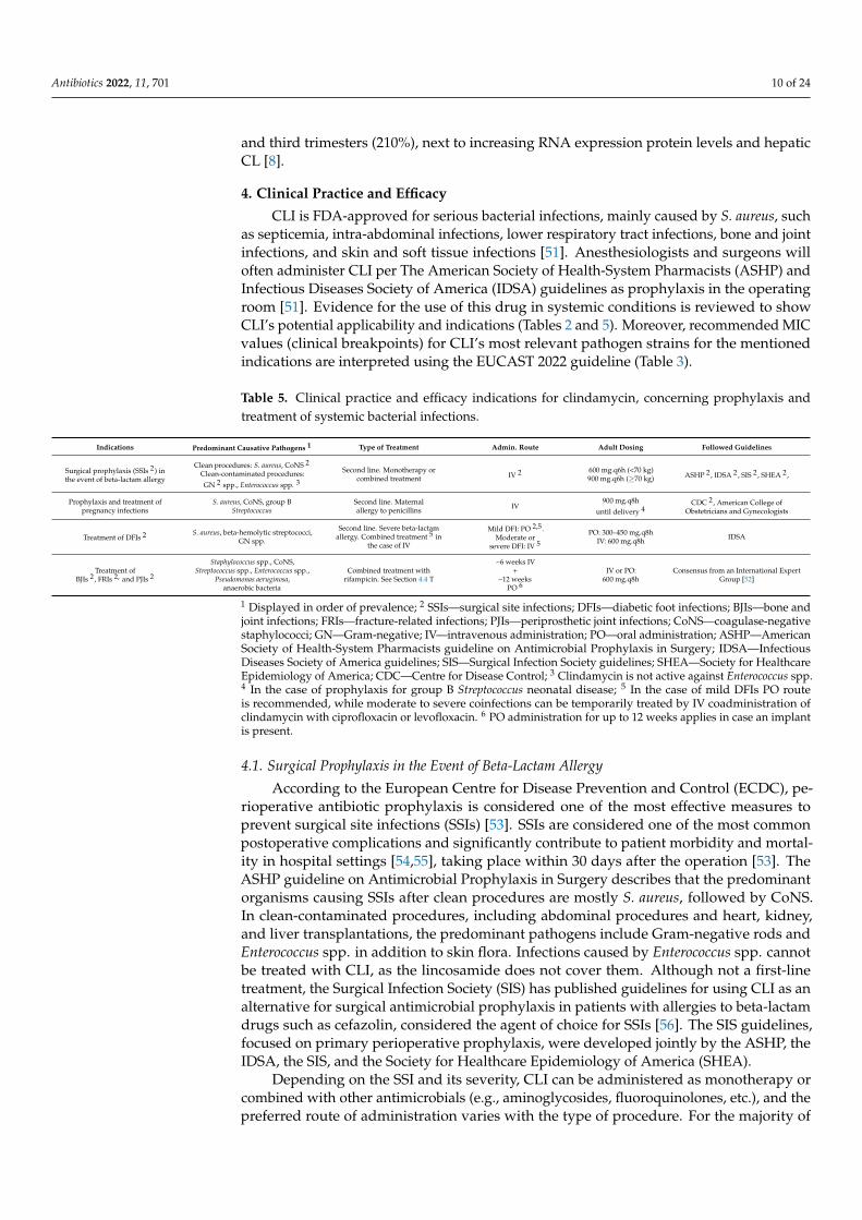
Table 5. Clinical practice and efficacy indications for clindamycin, concerning prophylaxis andtreatment of systemic bacterial infections.
Indications Predominant Causative Pathogens 1 Type of Treatment Admin. Route Adult Dosing Followed Guidelines
Surgical prophylaxis (SSIs 2) inthe event of beta-lactam allergy
Clean procedures: S. aureus, CoNS 2
Clean-contaminated procedures:
GN 2 spp., Enterococcus spp. 3
Second line. Monotherapy orcombined treatment IV 2 600 mg.q6h (<70 kg)
900 mg.q6h (≥70 kg) ASHP 2, IDSA 2, SIS 2, SHEA 2,
Prophylaxis and treatment ofpregnancy infections
S. aureus, CoNS, group BStreptococcus
Second line. Maternalallergy to penicillins IV
900 mg.q8h
until delivery 4CDC 2, American College of
Obstetricians and Gynecologists
Treatment of DFIs 2 S. aureus, beta-hemolytic streptococci,GN spp.
Second line. Severe beta-lactamallergy. Combined treatment 5 in
the case of IV
Mild DFI: PO 2,5.Moderate or
severe DFI: IV 5
PO: 300–450 mg.q8hIV: 600 mg.q8h IDSA
Treatment ofBJIs 2, FRIs 2, and PJIs 2
Staphylococcus spp., CoNS,Streptococcus spp., Enterococcus spp.,
Pseudomonas aeruginosa,anaerobic bacteria
Combined treatment withrifampicin. See Section 4.4 T
~6 weeks IV+
~12 weeksPO 6
IV or PO:600 mg.q8h
Consensus from an International ExpertGroup [52]
1 Displayed in order of prevalence; 2 SSIs—surgical site infections; DFIs—diabetic foot infections; BJIs—bone andjoint infections; FRIs—fracture-related infections; PJIs—periprosthetic joint infections; CoNS—coagulase-negativestaphylococci; GN—Gram-negative; IV—intravenous administration; PO—oral administration; ASHP—AmericanSociety of Health-System Pharmacists guideline on Antimicrobial Prophylaxis in Surgery; IDSA—InfectiousDiseases Society of America guidelines; SIS—Surgical Infection Society guidelines; SHEA—Society for HealthcareEpidemiology of America; CDC—Centre for Disease Control; 3 Clindamycin is not active against Enterococcus spp.4 In the case of prophylaxis for group B Streptococcus neonatal disease; 5 In the case of mild DFIs PO routeis recommended, while moderate to severe coinfections can be temporarily treated by IV coadministration ofclindamycin with ciprofloxacin or levofloxacin. 6 PO administration for up to 12 weeks applies in case an implantis present.
4.1. Surgical Prophylaxis in the Event of Beta-Lactam Allergy
According to the European Centre for Disease Prevention and Control (ECDC), pe-rioperative antibiotic prophylaxis is considered one of the most effective measures toprevent surgical site infections (SSIs) [53]. SSIs are considered one of the most commonpostoperative complications and significantly contribute to patient morbidity and mortal-ity in hospital settings [54,55], taking place within 30 days after the operation [53]. TheASHP guideline on Antimicrobial Prophylaxis in Surgery describes that the predominantorganisms causing SSIs after clean procedures are mostly S. aureus, followed by CoNS.In clean-contaminated procedures, including abdominal procedures and heart, kidney,and liver transplantations, the predominant pathogens include Gram-negative rods andEnterococcus spp. in addition to skin flora. Infections caused by Enterococcus spp. cannotbe treated with CLI, as the lincosamide does not cover them. Although not a first-linetreatment, the Surgical Infection Society (SIS) has published guidelines for using CLI as analternative for surgical antimicrobial prophylaxis in patients with allergies to beta-lactamdrugs such as cefazolin, considered the agent of choice for SSIs [56]. The SIS guidelines,focused on primary perioperative prophylaxis, were developed jointly by the ASHP, theIDSA, the SIS, and the Society for Healthcare Epidemiology of America (SHEA).
Depending on the SSI and its severity, CLI can be administered as monotherapy orcombined with other antimicrobials (e.g., aminoglycosides, fluoroquinolones, etc.), and thepreferred route of administration varies with the type of procedure. For the majority of
Antibiotics 2022, 11, 701 11 of 24
procedures, IV administration is ideal as it produces rapid, reliable, and predictable serumand tissue concentrations [56]. Regarding the SIS guidelines [56], standard preoperativeIV dosing in adults and pediatrics is 900 mg and 10 mg/kg, respectively, and continuedevery 6 h. This means that from the initiation of the preoperative dose, redosing every6 h is recommended, as long as the indication of prophylaxis holds. However, practi-cal guidelines provided by the MDA Anderson Cancer Center and Sarasota MemorialHealth Care System [57,58] recommend an IV weight-based dosing of 600 mg (<70 kg) and900 mg (≥70 kg) every 6–8 h, depending on the center. Standard IV dosing recommenda-tion of 600 mg.q8h (<70 kg) is also followed by the University Hospitals Leuven, amongother European facilities.
4.2. Prophylaxis and Treatment of Pregnancy Infections
CLI has been extensively prescribed for several decades to prevent or treat infectionsduring pregnancy and peripartum [8,26]. The most commonly performed surgical proce-dure during pregnancy is cesarean delivery, which may lead to postcesarean infections(e.g., maternal febrile morbidity, wound infection, endometritis, and other serious com-plications) [8,59]. Other pregnancy infections, such as endometritis or chorioamnionitis,can occur after fetal surgery [8]. In the presence of maternal allergy to penicillins, CLIis a potent alternative for the antimicrobial prophylaxis and treatment of pregnancy andperipartum infections caused by S. aureus, followed by CoNS and group B Streptococcus(GBS) in the event of, e.g., allergy to beta-lactams [60].
Recommended guidelines produced by the Center for Disease Control (CDC) andthe American College of Obstetricians and Gynecologists recommended a CLI dosage of900 mg.q8h IV until delivery in the case of prophylaxis for GBS neonatal disease [61,62],resulting in a rapid decline of colony counts (<5%) within the first 2 h of administration [8].In women during pregnancy or postpartum period, genital tract colonization with GBSis usually asymptomatic, but GBS clinical manifestations include the abovementionedinfections [63]. Furthermore, data from a prospective study on the transplacental passageof CLI confirmed that the CDC-recommended dosage produces therapeutic maternal andcord blood levels [43].
4.3. Treatment of Diabetic Foot Infections
A diabetic foot infection (DFI) is defined as any inframalleolar infection in a personwith diabetes mellitus and mostly arises from diabetic foot ulcers [64]. DFIs are consideredspecific skin and soft tissue infections that, provided by the severity of the lesion or afoot ulcer, can cause complications (e.g., diabetic foot osteomyelitis), leading to morbidity,hospitalization, and amputations. According to infection severity (wounds), the 2012IDSA Guideline for the Diagnosis and Treatment of DFIs [65] classified the infections intomild (superficial and limited size and depth), moderate (deeper or more extensive), orsevere (accompanied by systemic signs or metabolic perturbations). Commonly isolatedmicroorganisms are S. aureus, beta-hemolytic streptococci, and Gram-negative species [66].Acute infections are usually monomicrobial, while serious infections in hospitalized patientsare often caused by 3–5 bacterial species [64,67].
According to the 2012 IDSA guideline, treatment with CLI is an effective choice fortreating mild, moderate, and severe DFIs. Although not directly stated in the IDSA 2012guidelines, CLI is not usually administered as a first-line treatment but as an alternative inthe event of severe beta-lactam allergy [68,69]. In the case of mild DFIs caused by MSSAand Streptococcus spp., PO administration of CLI (300–450 mg.q8h) is recommended [65,70].When systemic signs emerge, moderate to severe coinfections may require hospitalizationand can be temporarily treated by IV coadministration of CLI (600 mg.q8h is the dosingregimen used by University Hospitals Leuven) with ciprofloxacin or levofloxacin untilpatient stabilization, switching then to the oral equivalent [65,69]. CLI is a good choicegiven its high oral bioavailability and bone penetration profile. This lincosamide has alsoshown to be effective in clinical trials, including in patients with DFIs and in the treatment of
Antibiotics 2022, 11, 701 12 of 24
diabetic foot osteomyelitis [65,69,71], an infectious disease that is one of the most commonexpressions of DFIs. However, CLI is not currently an antimicrobial specifically approvedby the FDA for the treatment of DFIs [65].
4.4. Treatment of Bone and Joint, Fracture-Related, and Periprosthetic Joint Infections
Bone and joint infections (BJIs) can be subdivided into multiple subgroups, includingfracture-related infections (FRIs), periprosthetic joint infections (PJIs), spinal infections,septic arthritis, and diabetic foot osteomyelitis [52,72]. PJI is a serious complication aftertotal joint arthroplasty that remains a core problem in orthopedic surgery [73] and is con-sidered a device-associated infection. FRI, on the other hand, also remains a challengingcomplication that primarily creates a heavy burden for orthopedic trauma patients. Com-pared with PJIs, FRIs have unique features (i.e., fracture, bone healing, soft tissue injury)that need to be considered [74]. The most common (biofilm-forming) bacteria causing BJIsin adults are Staphylococcus spp. (MSSA, MRSA), followed by CoNS, Streptococcus spp.,Enterococcus spp., Pseudomonas aeruginosa, and anaerobic bacteria [52,69]. Few studies havereported cases with well-identified Streptococcus spp. After conducting a 5-year study ofinterregional reference centers in the south of France, Seng et al. [75] reported the fivemost represented Streptococcus spp. to be GBS or S. agalactiae (37%), S. dysgalactieae (12%),S. anginosus (11%), S. constellatus (10%), and S. pneumoniae (9%). In contrast, CLI is notactive against Enterococcus spp.
BJIs treatment generally requires surgery combined with antibiotic therapy. Even ifthe duration of antimicrobial therapy in BJIs is controversial and not well investigated,antibiotic therapy is normally IV administered for at least 6 weeks and continued withPO administration for up to 12 weeks in case an implant is present [52,74]. Moreover, arecent randomized controlled trial showed that patients treated with up to 7 days of IVantibiotics followed by oral therapy had the same outcome as those with prolonged IVtherapy (usually 6–12 weeks) [74,76]. The usual CLI dosage used in adults for the treatmentof BJIs is 600 mg.q8h taken PO or IV [18]. CLI is widely used for the treatment of BJIs inadults and children for its potential activity against biofilm-forming bacteria and high levelsof bone penetration of 30% [18,77,78]. In addition, CLI has good oral bioavailability andis well tolerated [19,79]. A Consensus from an International Expert Group [52] publishedrecommendations for systemic antimicrobial therapy in FRI, with variations in CLI’s dosingregimen depending on the causative infectious agent.
To prevent the emergence of S. aureus resistance, synergistic bactericidal activity athigher concentrations has been achieved through the concomitant administration of CLIand rifampicin (RIF-CLI). Rifampicin-based combination therapy regimens have beenshown effective in eradicating staphylococcal biofilms [80,81]. Several guidelines havebeen published regarding the use of antibiotic therapy in conjunction with surgery whennecessary [82,83], but no specified regulations have been established for this RIF-CLI combi-nation, as due to the involvement of the CYP3A4 metabolism, a common fear of drug–druginteractions have been an ongoing research question. Thus, most aspects of antibiotictreatment for BJIs are still mostly based on expert opinions. Recommendations by the Con-sensus from an International Expert Group suggest coadministration of CLI (600 mg.q8h)with rifampicin (300–450 mg.q12h) to treat systemic FRIs caused by Staphylococcus spp. [52].Overall, this specific combination (RIF-CLI) shows potential clinical applicability for BIJsand is discussed later in the review.
Bone composition is different from that of the other tissues, such as cardiac muscle orlung tissue, because bone and joint tissues are less vascularized. It is, therefore, difficult topredict whether agents showing good penetration into other tissues will also achieve highconcentrations in bone [84]. Consequently, knowledge of the rate and extent of antibioticpenetration into bone tissue is fundamental for the successful treatment of BJIs.
CLI’s high levels of bone and joint penetration (~30%) are due to its physicochemicaland PK properties. A bone concentration/MIC ratio of 5 is required for time-dependent bac-tericidal action [85]. CLI is suitable for this clinical purpose because of its good penetration
Antibiotics 2022, 11, 701 13 of 24
profile and adequate activity (AUC/MIC ratio) against biofilm-forming bacteria. Most bonepenetration studies of CLI were conducted in the 1970s [84,86]. CLI concentrations weredetermined by bioassay, and the findings of these studies display substantial variability inthe reported mean bone/plasma concentration ratio of CLI (0.21–0.45). This could be dueto the presence of an infection, differences in the analytical techniques applied, or activemetabolites of CLI measured in the bioassay [84]. Quantification of drugs by bioassay wascarried out in these older studies, whereas nowadays, LC-MS methods are consideredmuch more sensitive and accurate. A review published by Thabit et al. [87] on antibi-otic bone penetration indicates that CLI could reach the susceptibility MIC breakpoint ofGram-positive cocci in ischemic tissues (≤0.25 mg/L for Streptococcus spp. and ≤0.5 mg/Lfor Staphylococcus spp.), but less likely that of anaerobes [(4)3 mg/L for Bacteroides spp.].MIC clinical breakpoint values have been updated using the EUCAST 2022 interpretationguideline [88].
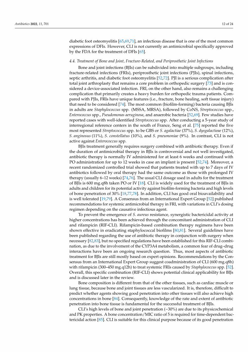
Rifampicin also has an optimal penetration into biofilms compared with other cur-rently available antimicrobial agents but cannot be administered in monotherapy becauseof the rapid emergence of resistant mutants [77]. Subsequently, RIF-CLI is a well-toleratedcombination [77] that has been tested for the treatment of BJIs. Both antimicrobials areinexpensive, possess a good oral bioavailability, and can successfully infiltrate the rigidbone structure into the synovial space to inhibit biofilm formation and bacterial adher-ence [18,77,78]. In 2012, Bouazza et al. [18] performed a retrospective population PK studyto predict optimal administered PO and IV dosing of CLI for patients with osteomyeli-tis. Results indicated that CLI’s CL increases with TBW, so presumably, standard doses(600 mg.q8h) should be incremented (900 mg.q8h) for patients over the ~70 kg standard-ized TBW. Moreover, this study indicated a potential effect of RIF-CLI on the CL, as CLI’sCL was increased by 43%, and subtherapeutic CLI concentrations were observed undercombined therapy [18]. These assumptions should be prospectively confirmed. Thus, afew prospective PK studies have been conducted over the past decade to improve thetreatment of BJIs (Table 6) [79,85,89]. All these studies reported plasma concentrations inorder to estimate CLI’s penetration into the bone during monotherapy and in combination(RIF-CLI), generally administered PO and IV.
Table 6. Summary of relevant prospective pharmacokinetic studies for cotreatment of clindamycinand rifampicin in bone and joint infections.
PharmacokineticStudies * Posology and Route of Administration
TheoreticalTarget Plasma
Concentration 1
Measured PlasmaConcentration 1
MeasurementTechnique 1
[Reference] CLI 4 RIF 4 Monotherapyvs. Combined
Curis et al.[79]
600 mg.q8h,PO/IV 4 bolus NS, PO/IV bolus Cmin
4 = 1.7 Cmin2,4 = 1.36 vs. 0.29
Cmax2,4 = 7.48 vs. 4.46 HPLC-UV 4
Bernard et al.[89] 600 mg.q8h, PO 4 600 mg.q12h, PO Cmin = [2–4]
Cmax4 = [5–8]
Cmin3,4 = 4.7 vs. 0.79
Cmax3,4 = 10.2 vs. 3.48 NS 4
Zeller et al.[85]
2400 mg/day, IV 4 infusion;750 mg.q8h, PO
600 mg.q12h, PO Css4 = [5–8] Cmin
2,4 = 2.09 vs. 0.18Cmax
2,4 = 7.95 vs. 1.53 LC-MS/MS 4
1 Regarding clindamycin in monotherapy vs. combined with rifampicin. The concentrations are given in mg/L;2 Median values; 3 Mean values; 4 RIF—rifampicin; CLI—clindamycin; NS—not specified; Css—target steady-stateconcentration; Cmin—trough concentration; Cmax—peak concentration; LC-MS/MS—liquid chromatographycoupled with tandem mass spectrometry; HPLC-UV—high-performance liquid chromatography with UV detector;IV—intravenous administration; PO—oral administration; PO/IV—administration of both routes; * Dosagevariations for high total body weight (TBW).
In 2015, Curis et al. [79] prospectively analyzed the influence of rifampicin on CLI’splasma concentrations in patients with BJIs, confirming the previous hypothesis that indicateda slight increase in treatment failure associated with increased TBW. Findings also confirmedthat CLI-RIF administration lowers the trough concentrations (Cmin) and peak concentrations(Cmax) of CLI. CLI measured plasma concentrations (monotherapy vs. combined treatment)

Antibiotics 2022, 11, 701 14 of 24
resulted in median Cmin (1.36 vs. 0.29 mg/L) and median Cmax (7.48 vs. 4.46 mg/L) valuesthat were also lower for patients under CLI-RIF treatment. In addition, Bernard et al. [89](2015) reported mean Cmin (4.7 vs. 0.79 mg/L) and mean Cmax (10.2 vs. 3.48 mg/L) valuessystemically below the recommended therapeutic ranges for combined therapy, confirmingsubtherapeutic CLI concentrations in plasma during CLI-RIF using the oral route. Insteadof CLI monotherapy, Bernard et al. administered CLI concomitantly with levofloxacin(500 mg once daily) as control. Lastly, a recent study from Zeller et al. [85] (2021) quantifiedthat during RIF-CLI, median Cmin was also markedly lower (2.09 vs. 0.18 mg/L), as wasmedian Cmax (7.95 vs. 1.53 mg/L).
A theoretical target plasma value with regard to Cmin was defined for the two firstdiscussed studies [79,89], and both studies obtained a Cmin value in combined treatmentbelow the previously set threshold value. By following the same criteria, target CLIconcentrations were similarly set across the studies, and dosage variations for high TBWpatients were performed in all studies. The impact of the administration route on themagnitude of the CLI-RIF interaction will be discussed in the following sections.
5. CYP3A4-Mediated Drug–Drug Interactions
Among the CYP3A enzyme subfamily, the CYP3A4 isoenzyme is the most abundantin the human liver (~40%) and is implicated in phase I metabolism of more than 50% ofall prescribed medications [90,91]. CYP3A4 can recognize and metabolize a wide array ofxenobiotic substances, known as CYP3A4 substrates. In addition, induction or inhibition ofCYP3A4-metabolized pathways can result in enhanced or suppressed metabolic capacityand drug plasma concentrations, respectively. Because of the key role of CYP3A4 in drugmetabolism, such enzyme changes can lead to a PK drug–drug interaction (DDI) whilecoadministering these CYP3A4 substrates together with CYP3A4-inhibitors or inducers.
CYP3A4 inhibition can result in serious adverse events since the intrinsic CL of thevictim drug(s) is reduced, leading to undesirable elevations of plasma drug concentra-tions [92]. CYP3A4 induction may cause a reduction in the therapeutic efficacy of CYP3A4substrates, as the victim drug(s) elimination is increased, lowering drug concentrationsand provoking a decrease in the victim drug(s) pharmacological effect [93]. Inhibition is analmost immediate response, while induction is a relatively slow regulatory process [92].Moreover, in the presence of active metabolites, three factors should be considered dur-ing DDI interpretation: the metabolic pathway, the ratio of the parent drug (CLI) to itsmetabolites, and the potency of the metabolites [94]. However, current insight into thepotential relevance of CLI’s active metabolites on this DDI is still very limited [92], asquantitative data on their exposure are not yet available. Quantitatively predicting theclinical magnitude of CYP3A-mediated DDIs is complicated, considering that the clinicaloutcomes depend on contributing factors such as interindividual variability associatedwith patients and drugs [92,93]. Insufficiency of proper experimental tools also contributesto the difficult prediction of these kinds of DDIs, as the potential of both human interactionsis commonly assessed by in vitro models [90,93].
In the following subsections, we will reflect on the clinical relevance of the DDIsthat can occur between our victim drugs (CLI and its active metabolites) and specificperpetrators (CYP3A4-inhibitors and inducers) (Table 7), based on the PK and the PD ofCLI and its metabolites. Although not covered in this review, the topic of potential PKDDIs could be further extended to food–drug or health products (e.g., Chinese medicine).
Antibiotics 2022, 11, 701 15 of 24
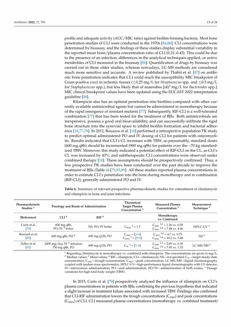
Table 7. CYP3A4-mediated drug–drug interactions arising from combined therapies with clindamycin.
Type of CYP3A4-Mediated DDI 1 Drug Drug Class DDI 2
MechanismDDI 2
PotencyIndication
Type ofCombined
Treatment 3
Admin.Route
Adult Dosingof Drug
CYP3A4-inhibition
Erythromycin Macrolideantibiotic
Mechanism-basedinhibition
Moderateinhibition
Gastroprokinetic:control acid reflux
Combined in lowdoses PO 1 125–250 mq.q12h
RitonavirAntiretroviral:
proteaseinhibitor HIV-1
Competitive andnoncompetitive,
irreversibleinhibition
Potentinhibition
Mild to moderateCOVID-19 1 caused by
the severeSARS-CoV-2 1 virus
Paxolavid ®
(nirmaltrevir/ritonavir)
POPaxolavid ®
(300 mg/100 mg).q12hfor 5 days
CYP3A4-induction
Rifampicin Rifamycinantibiotic
TranscriptionalPXR 1 agonism
Potentinhibition Treatment of BJIs 1 See Table 6 PO or IV 1 See Table 6
1 DDI—drug–drug interaction; PO—oral administration; IV—intravenous administration; COVID-19—coronavirus disease 2019; SARS-CoV-2—severe acute respiratory syndromecoronavirus 2; PXR—pregnane xenobiotic receptor; BJIs—bone and joint infections; 2 Regarding CYP3A4 inhibition or induction; 3 Combined treatment with clindamycin.
Antibiotics 2022, 11, 701 16 of 24
5.1. CYP3A4-Inhibition: Macrolides and Antiretroviral Drugs—Clindamycin
Some macrolide antibiotics (erythromycin) and antiretroviral drugs (ritonavir) stronglyinhibit CYP3A4, potentially leading to an increase in CLI concentration and risk of CLItoxicity or side effects. For macrolides, data suggest that CYP3A4 is subjected to mechanism-based inhibition [90], while the protease inhibitor HIV-1 ritonavir is a competitive andnoncompetitive, irreversible CYP3A4-inhibitor [95] (Table 7).
Macrolides are not often combined with CLI in daily clinical practice when used asantibiotics. However, despite being an effective inducer of MLSB resistance, erythromycinis a moderate CYP3A4-inhibitor [96] that might be combined with CLI when administeredPO in a low dose (125–250 mg.q12h) as a gastroprokinetic to help control acid reflux.Furthermore, when Staphylococcus spp. are tested for CLI susceptibility, inducible MLSBresistance due to erythromycin is tested. Isolates with such inducible resistance are resistantto erythromycin but appear susceptible to CLI in routine in vitro testing. However, clinicalfailures of CLI therapy for the treatment of MRSA infections have been documented forstrains that are CLI sensitive but erythromycin resistant. Therefore, in such a case, CLIwould be reported as ‘resistant’.
Regarding antiviral drugs, nirmatrelvir and ritonavir are two coadministered antiviralmedications marketed as Paxolavid ® for the treatment of mild to moderate COVID-19caused by the severe acute respiratory syndrome coronavirus 2 (SARS-CoV-2) virus. Pax-olavid ® is administered PO to adults and children (≥12 years old weighing at least 40 kg)in a dose of 300 mg/100 mg (nirmatrelvir/ritonavir) twice a day for 5 days [97]. Paxolavid ®
is not an FDA-approved drug, as it is still considered an investigational medicine. TheFDA has issued an Emergency Use Authorization (EUA) to make this antiviral availableduring the COVID-19 pandemic [97]. Nirmatrelvir inhibits proteolysis by binding the 3CLprotease, ultimately leading to the cessation of viral replication. Ritonavir is not activeagainst SARS-CoV-2 and acts as a booster agent by potently inhibiting CYP3A4, therebymaximizing the nirmatrelvir concentration in plasma [98]. A patient infected with this virusundergoing CLI treatment may experience a CYP3A4-inhibiting effect due to ritonavir’srole as a CYP3A4-inhibitor. All in all, caution should be given when extrapolating the effectof these CYP3A4-inhibitors (macrolides and antivirals) alone to the effect of combinationregimens on drugs with CYP3A4 activities such as CLI.
5.2. CYP3A4-Induction: Rifampicin–Clindamycin
Despite many CYP enzymes known to be inducible, CYP3A4 induction is probablyone of the most important causes of documented induction-based interactions [99] and amajor concern in clinical practice. In contrast to the extensive list of CYP3A4 inhibitors, onlya small number of drugs are identified as CYP3A4 inducers. This is the case for rifampicin,a potent CYP3A4-inducing agent that undergoes a transcriptional mode of action since thedrug is a specific pregnane xenobiotic receptor (PXR) agonist (Table 7). Moreover, currentfindings suggest that flucloxacillin might be a CYP3A4 inducer, as flucloxacillin has beenfound to decrease the levels of the following CYP3A4-metabolized drugs: voriconazole,quinidine, and tacrolimus [100–103]. For this, recent studies describing the DDIs betweenflucloxacillin and azole antifungals, e.g., voriconazole and isavuconazole (both metabolizedby CYP3A4) [104,105], have been conducted, and it is unclear if this should be extrapolatedto other antibiotics such as CLI.
For RIF-CLI interaction, full CYP induction is reached after approximately 1 week, andrecovery to baseline activity after rifampicin withdrawal is about 2 weeks [106]. Becausedirect assessment of CYP induction in vivo through the measurement of enzyme activity isnot possible, indirect evaluation can be performed through the comparison of CLI plasmaAUC before and after cotreatment with rifampicin. Both administration route and drug’scharacteristics need to be considered for this comparison. The route of administration isan important factor since CLI shows a high hepatic CL of ~1470 mL/min considering anaverage CL value of 0.35 L/h/kg (70 kg standardized TBW). During cotreatment withrifampicin, oral AUC is significantly downregulated, resulting in a greater magnitude
Antibiotics 2022, 11, 701 17 of 24
of changes in oral AUC compared with increased CL changes. This could primarily bedue to an increase in prehepatic first-pass effects but also because hepatic blood flow ismuch bigger than systemic CL. IV AUC is not sensitive to changes in enzyme activity sincesystemic CL is not affected by the first-pass effect and is limited by hepatic blood flow. Thisgeneralized decrease in systemic exposure is translated into therapeutic and pharmacologi-cal efficacy consequences that may lead to dose adjustments. In addition, because of theformation of CLI-active metabolites [5], interpretation of clinical consequences would besomewhat complicated in this case.
Previously mentioned findings from Zeller et al. [85] regarding Cmin and Cmax values(Table 6) confirm that the magnitude of RIF-CLI was markedly increased by oral intake,describing a pronounced decrease in concentrations of PO administered CLI. Assumingthat CLI plasma AUC before and after cotreatment is likely the most relevant indicator toassess CYP-inductive effect, a ~12-fold decrease comparing monotherapy vs. combined(37.7 vs. 3.1 mg.h/L) of oral AUC0–8 h was reported. In addition, a ~19-fold increase inoral CLI’s CL (7-fold higher compared with the IV route) was observed. According toZeller et al., this might suggest that rifampicin more specifically increases the prehepaticfirst-pass effects. These conclusions are not only supported by the variations in the CL butalso by the significantly lower bioavailability of oral CLI in monotherapy vs. combined(59% vs. 10%). There was no significant change in T1/2 (~1.2 h less in RIF-CLI), independentof the route of administration, supporting the argument that CYP induction has little effecton systemic CL of high hepatic CL drugs such as CLI [93].
6. Drug–Disease Interactions: Impact of Inflammation on CYP3A4/5 Activity
Acute and chronic inflammatory responses can be caused by infectious or nonin-fectious stimuli, leading to the alteration of hepatic functions that are critical for drugmetabolism, such as drug-metabolizing CYP isoenzyme activity. Good evidence showsthat under disease conditions, hepatic metabolism plays a crucial role in the secretion ofinflammatory mediators, which can modulate drug metabolism by reducing CYP expres-sion [107]. One of the key stimulators involved in both acute- and chronic-phase responsesis interleukin-6 (IL-6), together with C-reactive protein (CRP), an IL-6-regulated acute-phaseprotein that is synthesized in the liver when IL-6 levels increase during inflammation. Nextto IL-6 and CRP, plasma protein AAG is an essential biomarker to assess inflammation’sclinical impact on CYP-induced drug metabolism. Although long-term investigations onthis effect are still scarce, current published data in adults suggest that inflammation hasan isoform-specific and intensity-specific impact [108] as a result of pretranscriptional andpost-transcriptional mechanisms [107]. CLI being primarily a CYP3A4-substrate meansthat CYP3A4 drug metabolic rate is reduced, and levels of protein and mRNA subsequentlydecrease at the level of gene transcription. As a result, there is a decrease in CYP3A4-dependent drug CL and impaired CLI biotransformation during the operation of hostdefense mechanisms. If an anti-inflammatory drug is administered to treat inflammationdisease, mediator levels are lowered, and metabolic capacity appears to return to baselinelevel when the disease is resolved [108].
A recently published review from C.M. White [109] assessed the concentration of aCYP3A4 substrate with an acute infectious or noninfectious cause of inflammation froma total of 23 studies. Evaluated CYP3A4 substrates in descending order of predominanceincluded quinine (8 studies), midazolam (4 studies), lopinavir (2 studies), erythromycin(2 studies), and other miscellaneous drugs (1 study). A correlation between increasingbiomarker concentration and reduced CL of CYP3A4 substrates was found in 10 of 12,2 of 2, and 2 of 3 studies assessing CRP, AAG, and IL-6, respectively. This associationis especially strong for benzodiazepines. Several studies showed large changes in drugconcentration/dose ratio, AUC, or CL, in patients with inflammation. However, que-tiapine had a small increase in the concentration of only 11% among people with CRPconcentrations > 5 mg/L, which is qualitatively less robust than what was seen with benzo-diazepines (midazolam, alprazolam), perampanel, and antimicrobial agents (erythromycin,
Antibiotics 2022, 11, 701 18 of 24
cyclosporin). In assessed studies concerning populations with malaria [110–115], whenpatients developed an acute malarial infection, CYP3A4 substrate drug concentrations wereelevated or CL was reduced, even though no markers of inflammation were concomitantlyassessed. Moreover, patients with acute malarial infection had a much higher AUC0–12hcompared with convalescent patients (37.9 ± 14.7 vs. 17.9 ± 8.5 mcg/mL) [115].
Given that inflammation is an important determinant contributing to variation in CYPactivities between and within individuals [107], an effort must be made to better understandthe impact of clinically useful inflammatory biomarkers released during inflammation, suchas IL-6, CRP, and AAG. These mediators can measure the severity of inflammation, thatbeing proportional to the potential suppression of CYP3A4 activity, and evaluate the clinicaleffectiveness of CYP3A4 substrates in reference and special populations. Mechanistically,understanding enzyme specificity and mechanisms of regulation will allow us to improvedrug efficacy or safety, improve knowledge of acute and chronic diseases, and personalizepatients’ drug regimens. Moreover, transcriptional effects have a great impact on CYP3A4substrates with a relatively narrow therapeutic index such as CLI since an increase intoxicity can more easily occur and complicate therapeutics-causing adverse events (AEs).For this, clinicians with patients on narrow therapeutic index CYP3A4 substrates mustmonitor their patients more judiciously when new infections or other inflammatory stimulioccur to prevent AEs or loss of drug efficacy [109].
7. Safety and Adverse Event Profile
Even if CLI is generally a well-tolerated drug, the SmPC describes some AEs thatcan be resolved with dose adjustment or discontinuation of the antibiotic [51]. CLI’s mostfrequent AEs with systemic administration are mainly GI, resulting from CLI substantialchanges in the GI tract’s healthy flora [51]. The destruction of much of this microflora mayresult in the development of Clostridium difficile-associated diarrhea (CDAD). Diarrhea isthe most common AE and occurs in up to 20% of patients, and CDAD may occur morefrequently compared with other oral agents [116]. Other common GI AEs include nausea,vomiting, or stomach pains. A more severe and also uncommon GI that may develop(during or after treatment) is C. difficile-associated pseudomembranous enterocolitis (PMC).This potentially life-threatening infection is often associated with CLI and is caused bya complication of CDAD, which can lead to fulminant colitis, sepsis, and death. Grow-ing PMC incidence corresponds with the increased use of wide-spectrum antibiotics inhospitalized patients [117]. Although PMC may affect all age groups, the incidence islow in the pediatric SPP. Mortality is rare in pediatrics and involves patients with seriouscoexistent illness, infection, or congenital disabilities [118]. Regarding pregnant women, anassociation between C. difficile infection and pregnancy has not been stressed [119]. Theoccurrence of PMC for both discussed SPPs (pediatrics and pregnant, breastfeeding, andpostpartum women) is of particular concern, and further research into the scope and riskfactors for children and peripartum PMC is warranted.
A recently published systematic review [120] updating the evidence for associationsbetween antibiotic classes and C. difficile infection stated the modest association observedbetween clindamycin and such disease. This up-to-date synthesis of evidence in relation tothis potential risk suggests that CLI is relatively safe or not the most unsafe drug comparedwith other antibiotics (e.g., carbapenems, third- and fourth-generation cephalosporins).Nonetheless, this relevant complication should be considered during CLI’s use. Besides, be-cause predicting risks for DDIs involving CYP3A4-induction and inhibition is difficult, cau-tion and TDM are needed when administering CLI together with CYP3A4-substrates, anddose adjustment of these substrates might be necessary. Moreover, CYP3A4 metabolism-mediated PD (side) effects should also be considered and assessed.
Antibiotics 2022, 11, 701 19 of 24
8. Discussion
In this review, we explored the pharmacology and PKs of CLI in a reference populationand selected SPPs (pediatrics, pregnant, breastfeeding, and postpartum women). Ourconclusion is that there is a major knowledge gap regarding the potential PD and PKrelevance of CLI’s active metabolites in daily clinical practice.
Important observations on the PK/PD relevance of CLI’s activity metabolites were al-ready reported by Wynalda et al. [5] in 2003, as this group examined the in vitro metabolismof CLI in human liver and intestinal microsomes. Following incubation with human liver,ileum, and jejunum microsomes and recombinant CYP3A4 and CYP3A5 microsomes,CLI sulfoxide was found to be the major metabolite formed, accounting for more than90% of total consumed CLI. A minor metabolite, N-demethyl CLI, was also identified.Only CYP3A4 and, to a certain extent, CYP3A5 were able to catalyze the formation ofCLI sulfoxide, and only inhibitors of CYP3A could attenuate the rate of CLI’s sulfoxideformation. In addition, in vitro findings showed that CLI did not inhibit the metabolicactivity of other CYPs (e.g., CYP1A2, CYP2C9, CYP2C19, CYP2E1, or CYP2D6) and onlymoderately inhibited CYP3A4. These data provide evidence of the mechanism of the DDIbetween CLI and the aforementioned CYP3A4 inducers. Because of the limited data onthe involved biotransformation routes (Figure 2, Table 1), determining to what extent thealtered CLI metabolite exposure results in altered CLI’s drug efficacy or safety is not yetpossible. Furthermore, metabolites quantification in plasma and other human matrices(e.g., urine, bile, bone, amniotic fluid, etc.) is still to be determined and must be studied inthe future to evaluate their potential relevance to CLI’s disposition.
Regarding CLI’s clinical pharmacology and therapeutics for the selected SPPs, futureresearch is urgently required, especially regarding topics such as dose optimization andadapted drug therapy. Based on limited data on PK and efficacy and safety in the SPPbackground, we claim that further studies should be designed and carried out to facilitatethe efficient evaluation of CLI’s PKs. Moreover, experimental CLI-RIF administration forthe treatment of BJIs is based on performed PK studies (Table 6). A key limiting factor ofthe discussed studies is the low number of participants for most of the studies, leading toa low power statistical analysis. Furthermore, a detailed methodological description waseven absent in the report by Bernard et al. [89]. Overall, the bioavailability of CLI decreasessignificantly in the setting of induction by rifampicin during the CLI-RIF cotreatment.Given the marked increase, through oral intake, in the magnitude of RIF-CLI, optimalCLI dose in combination with rifampicin remains to be determined, and close-targetdrug monitoring (TDM) of drug and bioactive metabolites is advised to guide dosageadjustments. Moreover, all previous investigations only considered the CLI, and, to ourknowledge, the potential relevance of CLI’s metabolites in the PK/PD target remainsunexplored. In the future, the performance of PK studies, including CLI’s metabolites in alarger number of both healthy and ill patients, will reflect best clinical practice regardingthe CLI-RIF CYP3A4-inducing DDI.
Additionally, because SPPs possess CYP3A4 altered activity, clinical implications ofCYP3A4 induction and inhibition must be evaluated for these special populations andcompared to healthy adult populations in the future. Overall, awareness of these DDIs willcontribute to safer and more controlled management of patients receiving treatment for thementioned systemic infections. Given the DDI metabolic variation involved, caution andclose TDM are needed when CLI and CYP3A inducers and inhibitors are simultaneouslyadministered. Dose optimization might be necessary, certainly for antimicrobials suchas CLI, likely to undergo therapeutic failure because of underdosing and low exposure.Regarding the impact of inflammation on CYP3A4/5 activity, many drugs were assessedin C.M. White’s review [109]. Nevertheless, the extent to which the results from CYP3A4-metabolized drugs can be applied to all CYP3A4 substrates remains unclear. To ourknowledge, no study has directly evaluated inflammation-induced CYP expression instandard or other populations for CLI.
Antibiotics 2022, 11, 701 20 of 24
Additional studies will be needed to obtain more data on CLI’s PK and PD, includingthe metabolites, in order to further improve the assessment of CLI’s efficacy and safety.Moreover, efforts should be made to discover novel strategies in the approach to thetreatment of bacterial infections in special patient populations.
9. Conclusions
Based on the paucity of data found on CLI’s PKs, efficacy and safety in the field ofsystemic infections, we claim that efforts should be made to discover new strategies inthe approach to antimicrobial infection with respect to the role of CLI, to facilitate theevaluation of antimicrobial prophylaxis and treatment in this specific setting. To date,knowledge of CLI’s active metabolite contribution to the efficacy and safety of CLI is scarceand inconclusive. In conclusion, identification of the current knowledge gaps (SPPs, DDIs)with respect to the pharmacology and PKs of CLI is crucial to developing a research strategytoward precision medicine.
Author Contributions: Conceptualization, L.A.Á., I.S., K.A. and J.R.; writing—original draft prepa-ration, L.A.Á., G.V.d.S., S.D., W.-J.M., I.S., K.A. and J.R.; writing—review and editing: all coauthorsrevised the manuscript and approved the final manuscript. All authors have read and agreed to thepublished version of the manuscript.
Funding: This research received no external funding.
Conflicts of Interest: The authors declare no conflict of interest.
References1. European Comission Health Research and Innovation. Antimicrobial Drug Resistance (AMR). Available online: https://ec.europa.
eu/info/research-and-innovation/research-area/health-research-and-innovation/antimicrobial-drug-resistance-amr_en (ac-cessed on 10 April 2022).
2. Prestinaci, F.; Pezzotti, P.; Pantosti, A. Antimicrobial resistance: A global multifaceted phenomenon. Pathog. Glob. Health 2015,109, 309–318. [CrossRef] [PubMed]
3. Guay, D. Update on clindamycin in the management of bacterial, fungal and protozoal infections. Expert Opin. Pharmacother.2007, 8, 2401–2444. [CrossRef] [PubMed]
4. Liu, C.; Bayer, A.; Cosgrove, S.E.; Daum, R.S.; Fridkin, S.K.; Gorwitz, R.J.; Kaplan, S.L.; Karchmer, A.W.; Levine, D.P.; Murray,B.E.; et al. Clinical Practice Guidelines by the Infectious Diseases Society of America for the Treatment of Methicillin-ResistantStaphylococcus aureus Infections in Adults and Children. Clin. Infect. Dis. 2011, 52, e18–e55. [CrossRef] [PubMed]
5. Wynalda, M.A.; Hutzler, J.M.; Koets, M.D.; Podoll, T.; Wienkers, L.C. In vitro metabolism of clindamycin in human liver andintestinal microsomes. Drug Metab. Dispos. 2003, 31, 878–887. [CrossRef] [PubMed]
6. Shah, S.; Barton, G.; Fischer, A. Pharmacokinetic considerations and dosing strategies of antibiotics in the critically ill patient.J. Intensive Care Soc. 2015, 16, 147–153. [CrossRef]
7. Muller, A.E.; Mouton, J.W.; Oostvogel, P.M.; Dörr, P.J.; Voskuyl, R.A.; DeJongh, J.; Steegers, E.A.P.; Danhof, M. Pharmacokineticsof clindamycin in pregnant women in the peripartum period. Antimicrob. Agents Chemother. 2010, 54, 2175–2181. [CrossRef]
8. Allegaert, K.; Muller, A.E.; Russo, F.; Schoenmakers, S.; Deprest, J.; Koch, B.C.P. Pregnancy-related pharmacokinetics andantimicrobial prophylaxis during fetal surgery, cefazolin and clindamycin as examples. Prenat. Diagn. 2020, 40, 1178–1184.[CrossRef] [PubMed]
9. Schreckenberger, P.C.; Ilendo, E.; Ristow, K.L. Incidence of Constitutive and Inducible Clindamycin Resistance in Staphylococcusaureus and Coagulase-Negative Staphylococci in a Community and a Tertiary Care Hospital. J. Clin. Microbiol. 2004, 42, 2777–2779.[CrossRef]
10. European Committee on Antimicrobial Susceptibility Testing. MIC Distributions and Epidemiological Cut-Off Value (ECOFF)Setting, EUCAST SOP 10. 2022. Available online: https://www.eucast.org/organization/subcommittees/mic_distributions_and_ecoffs/ (accessed on 10 April 2022).
11. Spížek, J.; Rezanka, T. Lincosamides: Chemical structure, biosynthesis, mechanism of action, resistance, and applications. Biochem.Pharmacol. 2017, 133, 20–28. [CrossRef]
12. Levison, M.E.; Levison, J.H. Pharmacokinetics and Pharmacodynamics of Antibacterial Agents. Infect. Dis. Clin. N. Am. 2009, 23,791–815. [CrossRef]
13. Wade, K.C.; Benjamin, D.K. Clinical Pharmacology of Anti-Infective Drugs. In Infectious Diseases of the Fetus and Newborn; Elsevier:Amsterdam, The Netherlands, 2011; pp. 1160–1211.
14. Chavez-Bueno, S.; Bozdogan, B.; Katz, K.; Bowlware, K.L.; Cushion, N.; Cavuoti, D.; Ahmad, N.; McCracken, G.H.; Appelbaum,P.C. Inducible Clindamycin Resistance and Molecular Epidemiologic Trends of Pediatric Community-Acquired Methicillin-Resistant Staphylococcus aureus in Dallas, Texas. Antimicrob. Agents Chemother. 2005, 49, 2283–2288. [CrossRef] [PubMed]
Antibiotics 2022, 11, 701 21 of 24
15. Leclercq, R. Mechanisms of Resistance to Macrolides and Lincosamides: Nature of the Resistance Elements and Their ClinicalImplications. Clin. Infect. Dis. 2002, 34, 482–492. [CrossRef] [PubMed]
16. Lewis, J.S.; Jorgensen, J.H. Inducible Clindamycin Resistance in Staphylococci: Should Clinicians and Microbiologists beConcerned? Clin. Infect. Dis. 2005, 40, 280–285. [CrossRef] [PubMed]
17. McKinnon, P.S.; Davis, S.L. Pharmacokinetic and Pharmacodynamic Issues in the Treatment of Bacterial Infectious Diseases. Eur.J. Clin. Microbiol. Infect. Dis. 2004, 23, 271–288. [CrossRef] [PubMed]
18. Bouazza, N.; Pestre, V.; Jullien, V.; Curis, E.; Urien, S.; Salmon, D.; Tréluyer, J.M. Population pharmacokinetics of clindamycinorally and intravenously administered in patients with osteomyelitis. Br. J. Clin. Pharmacol. 2012, 74, 971–977. [CrossRef][PubMed]
19. Gatti, G.; Flaherty, J.; Bubp, J.; White, J.; Borin, M.; Gambertoglio, J. Comparative study of bioavailabilities and pharmacokineticsof clindamycin in healthy volunteers and patients with AIDS. Antimicrob. Agents Chemother. 1993, 37, 1137–1143. [CrossRef]
20. Buck, M.L.; Pharm, D. A Monthly Newsletter for Health Care Professionals. Pediatr. Pharmacother. 2010, 16, 2–5.21. Hand, W.L.; King-Thompson, N.L. Membrane transport of clindamycin in alveolar macrophages. Antimicrob. Agents Chemother.
1982, 21, 241–247. [CrossRef]22. Steinberg, T.H. Cellular Transport of Drugs. Clin. Infect. Dis. 1994, 19, 916–921. [CrossRef]23. van den Broek, A.K.; Prins, J.M.; Visser, C.E.; van Hest, R.M. Systematic review: The bioavailability of orally administered
antibiotics during the initial phase of a systemic infection in non-ICU patients. BMC Infect. Dis. 2021, 21, 285. [CrossRef]24. Picardi, J.L.; Lewis, H.P.; Tan, J.S.; Phair, J.P. Clindamycin concentrations in the central nervous system of primates before and
after head trauma. J. Neurosurg. 1975, 43, 717–720. [CrossRef] [PubMed]25. Castle, S.S. Clindamycin. In xPharm: The Comprehensive Pharmacology Reference; Elsevier: Amsterdam, The Netherlands, 2007;
pp. 1–4.26. Brandon Bookstaver, P.; Bland, C.M.; Griffin, B.; Stover, K.R.; Eiland, L.S.; McLaughlin, M. A review of antibiotic use in pregnancy.
Pharmacotherapy 2015, 35, 1052–1062. [CrossRef] [PubMed]27. Food and Drug Administration, HHS. International Conference on Harmonisation; guidance on E11 clinical investigation of
medicinal products in the pediatric population; availability. Notice. Fed. Regist. 2000, 65, 78493–78494.28. Laughon, M.M.; Avant, D.; Tripathi, N.; Hornik, C.P.; Cohen-Wolkowiez, M.; Clark, R.H.; Smith, P.B.; Rodriguez, W. Drug labeling
and exposure in neonates. JAMA Pediatr. 2014, 168, 130–136. [CrossRef]29. Gonzalez, D.; Delmore, P.; Bloom, B.T.; Cotten, C.M.; Poindexter, B.B.; McGowan, E.; Shattuck, K.; Bradford, K.K.; Smith,
P.B.; Cohen-Wolkowiez, M.; et al. Clindamycin Pharmacokinetics and Safety in Preterm and Term Infants. Antimicrob. AgentsChemother. 2016, 60, 2888–2894. [CrossRef]
30. Van Den Abeele, J.; Rayyan, M.; Hoffman, I.; Van de Vijver, E.; Zhu, W.; Augustijns, P. Gastric fluid composition in a paediatricpopulation: Age-dependent changes relevant for gastrointestinal drug disposition. Eur. J. Pharm. Sci. 2018, 123, 301–311.[CrossRef]
31. Mooij, M.G.; de Koning, B.A.; Huijsman, M.L.; de Wildt, S.N. Ontogeny of oral drug absorption processes in children. ExpertOpin. Drug Metab. Toxicol. 2012, 8, 1293–1303. [CrossRef]
32. Yokoi, T. Essentials for starting a pediatric clinical study (1): Pharmacokinetics in children. J. Toxicol. Sci. 2009, 34, SP307–SP312.[CrossRef]
33. Stillhart, C.; Vucicevic, K.; Augustijns, P.; Basit, A.W.; Batchelor, H.; Flanagan, T.R.; Gesquiere, I.; Greupink, R.; Keszthelyi, D.;Koskinen, M.; et al. Impact of gastrointestinal physiology on drug absorption in special populations—An UNGAP review. Eur. J.Pharm. Sci. 2020, 147, 105280. [CrossRef]
34. Bonner, J.J.; Vajjah, P.; Abduljalil, K.; Jamei, M.; Rostami-Hodjegan, A.; Tucker, G.T.; Johnson, T.N. Does age affect gastric emptyingtime? A model-based meta-analysis of data from premature neonates through to adults. Biopharm. Drug Dispos. 2015, 36, 245–257.[CrossRef]
35. Booker, P.D.; Taylor, C.; Saba, G. Perioperative changes in alpha 1-acid glycoprotein concentrations in infants undergoing majorsurgery. Br. J. Anaesth. 1996, 76, 365–368. [CrossRef]
36. Ipek, I.Ö.; Saracoglu, M.; Bozaykut, A. α 1-Acid glycoprotein for the early diagnosis of neonatal sepsis. J. Matern. Neonatal Med.2010, 23, 617–621. [CrossRef]
37. Tracy, T.S.; Chaudhry, A.S.; Prasad, B.; Thummel, K.E.; Schuetz, E.G.; Zhong, X.-B.; Tien, Y.-C.; Jeong, H.; Pan, X.; Shireman,L.M.; et al. Interindividual Variability in Cytochrome P450-Mediated Drug Metabolism. Drug Metab. Dispos. 2016, 44, 343–351.[CrossRef] [PubMed]
38. O’Hara, K.; Wright, I.M.R.; Schneider, J.J.; Jones, A.L.; Martin, J.H. Pharmacokinetics in neonatal prescribing: Evidence base,paradigms and the future. Br. J. Clin. Pharmacol. 2015, 80, 1281–1288. [CrossRef] [PubMed]
39. van Groen, B.D.; Nicolaï, J.; Kuik, A.C.; Van Cruchten, S.; van Peer, E.; Smits, A.; Schmidt, S.; de Wildt, S.N.; Allegaert, K.; DeSchaepdrijver, L.; et al. Ontogeny of Hepatic Transporters and Drug-Metabolizing Enzymes in Humans and in NonclinicalSpecies. Pharmacol. Rev. 2021, 73, 597–678. [CrossRef]
40. Elens, L.; van Gelder, T.; Hesselink, D.A.; Haufroid, V.; van Schaik, R.H. CYP3A4*22: Promising newly identified CYP3A4 variantallele for personalizing pharmacotherapy. Pharmacogenomics 2013, 14, 47–62. [CrossRef] [PubMed]
Antibiotics 2022, 11, 701 22 of 24
41. Brussee, J.M.; Vet, N.J.; Krekels, E.H.J.; Valkenburg, A.J.; Jacqz-Aigrain, E.; van Gerven, J.M.A.; Swart, E.L.; van den Anker, J.N.;Tibboel, D.; de Hoog, M.; et al. Predicting CYP3A-mediated midazolam metabolism in critically ill neonates, infants, children andadults with inflammation and organ failure. Br. J. Clin. Pharmacol. 2018, 84, 358–368. [CrossRef]
42. Barrett, S.; Blake, J.; Bradley, C.; Cleary, B.; Cronin, C.; Delaney, A.; Drew, R.; Duddy, P.; Fitzgerald, D.; Flaherty, E.; et al.Medication Guidelines for Obstetrics and Gynaecology: Antimicrobial safety in pregnancy and lactation. Inst. Obstet. Gynaecol.Health Serv. Exec. 2017, 2, 80.
43. Wear, C.D.; Towers, C.V.; Brown, M.S.; Weitz, B.; Porter, S.; Wolfe, L. Transplacental passage of clindamycin from mother toneonate. J. Perinatol. 2016, 36, 960–961. [CrossRef]
44. Costantine, M.M. Physiologic and pharmacokinetic changes in pregnancy. Front. Pharmacol. 2014, 5. [CrossRef]45. Pariente, G.; Leibson, T.; Carls, A.; Adams-Webber, T.; Ito, S.; Koren, G. Pregnancy-Associated Changes in Pharmacokinetics: A
Systematic Review. PLoS Med. 2016, 13, e1002160. [CrossRef] [PubMed]46. Chiloiro, M.; Darconza, G.; Piccioli, E.; De Carne, M.; Clemente, C.; Riezzo, G. Gastric emptying and orocecal transit time in
pregnancy. J. Gastroenterol. 2001, 36, 538–543. [CrossRef] [PubMed]47. Larijani, G.E.; Norris, M.C.; Ala-Kokko, T.I.; Leighton, B.A.; Desimone, C. Serum Concentration of Alpha,-Acid Glycoprotein and
Albumin following Cesarean Section and Vaginal Delivery. DICP 1990, 24, 328–329. [CrossRef] [PubMed]48. Abduljalil, K.; Furness, P.; Johnson, T.N.; Rostami-Hodjegan, A.; Soltani, H. Anatomical, Physiological and Metabolic Changes
with Gestational Age during Normal Pregnancy. Clin. Pharmacokinet. 2012, 51, 365–396. [CrossRef]49. Hebert, M.; Easterling, T.; Kirby, B.; Carr, D.; Buchanan, M.; Rutherford, T.; Thummel, K.; Fishbein, D.; Unadkat, J. Effects of
Pregnancy on CYP3A and P-glycoprotein Activities as Measured by Disposition of Midazolam and Digoxin: A University ofWashington Specialized Center of Research Study. Clin. Pharmacol. Ther. 2008, 84, 248–253. [CrossRef]
50. Papageorgiou, I.; Grepper, S.; Unadkat, J.D. Induction of Hepatic CYP3A Enzymes by Pregnancy-Related Hormones: Studies inHuman Hepatocytes and Hepatic Cell Lines. Drug Metab. Dispos. 2013, 41, 281–290. [CrossRef]
51. Murphy, P.B.; Bistas, K.G.; Le, J.K. Clindamycin. [Updated 10 April 2022]. In StatPearls [Internet]; StatPearls Publishing: TreasureIsland, FL, USA, 2022. Available online: https://www.ncbi.nlm.nih.gov/books/NBK519574/ (accessed on 10 April 2022).
52. Depypere, M.; Kuehl, R.; Metsemakers, W.-J.; Senneville, E.; McNally, M.A.; Obremskey, W.T.; Zimmerli, W.; Atkins, B.L.; Trampuz,A. Fracture-Related Infection (FRI) Consensus Group Recommendations for Systemic Antimicrobial Therapy in Fracture-RelatedInfection: A Consensus From an International Expert Group. J. Orthop. Trauma 2020, 34, 30–41. [CrossRef]
53. European Centre for Disease Prevention and Control. Surveillance of Surgical Site Infections and Prevention Indicators in EuropeanHospitals: HAI-Net SSI protocol, version 2.2; European Centre for Disease Prevention and Control: Solna, Sweden, 2017.
54. Young, P.Y.; Khadaroo, R.G.; FRCSC. Surgical Site Infections. Surg. Clin. N. Am. 2014, 94, 1245–1264. [CrossRef]55. Mundhada, A.; Tenpe, S. A study of organisms causing surgical site infections and their antimicrobial susceptibility in a tertiary
care Government Hospital. Indian J. Pathol. Microbiol. 2015, 58, 195. [CrossRef]56. Bratzler, D.W.; Dellinger, E.P.; Olsen, K.M.; Perl, T.M.; Auwaerter, P.G.; Bolon, M.K.; Fish, D.N.; Napolitano, L.M.; Sawyer, R.G.;
Slain, D.; et al. Clinical Practice Guidelines for Antimicrobial Prophylaxis in Surgery. Surg. Infect. (Larchmt). 2013, 14, 73–156.[CrossRef]
57. Antibiotic Prophylaxis for Inpatient and Outpatient Surgery. Available online: https://www.smh.com/Portals/0/Documents/Services/Surgery%20Forms/Antibiotic%20Surgical%20Prophylaxis%20Protocol%20SMH%20October%202018%20Update.pdf(accessed on 10 April 2022).
58. Lepetic, A.; Vujacich, C.; Calmaggi, A.; Guerrini, G.M.; Arzac, M.d.R.G. Surgical antibiotic prophylaxis. Morb. Obes. 2009, 173–194.[CrossRef]
59. Heesen, M.; Klöhr, S.; Rossaint, R.; Allegeaert, K.; Deprest, J.; Van de Velde, M.; Straube, S. Concerning the timing of antibioticadministration in women undergoing caesarean section: A systematic review and meta-analysis. BMJ Open 2013, 3, e002028.[CrossRef] [PubMed]
60. Kawakita, T.; Landy, H.J. Surgical site infections after cesarean delivery: Epidemiology, prevention and treatment. Matern. HealthNeonatol. Perinatol. 2017, 3, 12. [CrossRef]
61. Verani, J.R.; McGee, L.; Schrag, S.J. Division of Bacterial Diseases, National Center for Immunization and Respiratory Diseases,Centers for Disease Control and Prevention (CDC) Prevention of perinatal group B streptococcal disease–revised guidelines fromCDC, 2010. MMWR Recomm. Rep. 2010, 59, 1–36. [PubMed]
62. ACOG Committee Opinion No. 485: Prevention of early-onset group B streptococcal disease in newborns. Obstet. Gynecol. 2011,117, 1019–1027. [CrossRef]
63. Melin, P. Neonatal group B streptococcal disease: From pathogenesis to preventive strategies. Clin. Microbiol. Infect. 2011, 17,1294–1303. [CrossRef] [PubMed]
64. Sharkey, M.; Hu, S.S. Diabetic Foot Infections. In Emergency Management of Infectious Diseases; Chin, R.L., Ed.; CambridgeUniversity Press: Cambridge, UK, 2007; pp. 143–146.
65. Lipsky, B.A.; Berendt, A.R.; Cornia, P.B.; Pile, J.C.; Peters, E.J.G.; Armstrong, D.G.; Deery, H.G.; Embil, J.M.; Joseph, W.S.; Karchmer,A.W.; et al. 2012 Infectious Diseases Society of America Clinical Practice Guideline for the Diagnosis and Treatment of DiabeticFoot Infections. Clin. Infect. Dis. 2012, 54, e132–e173. [CrossRef] [PubMed]
66. De Vries, M.G.; Ekkelenkamp, M.B.; Peters, E.J.G. Are clindamycin and ciprofloxacin appropriate for the empirical treatment ofdiabetic foot infections? Eur. J. Clin. Microbiol. Infect. Dis. 2014, 33, 453–456. [CrossRef]
Antibiotics 2022, 11, 701 23 of 24
67. Lipsky, B.A. Medical Treatment of Diabetic Foot Infections. Clin. Infect. Dis. 2004, 39, S104–S114. [CrossRef]68. Government of South Australia. Diabetic Foot Infections: Antibiotic Management Clinical Guideline. Gov. South Aust. 2019.69. Colston, J.; Atkins, B. Bone and joint infection. Clin. Med. 2018, 18, 150–154. [CrossRef] [PubMed]70. Matheson, E.M.; Bragg, S.W.; Blackwelder, R.S. Diabetes-Related Foot Infections: Diagnosis and Treatment. Am. Fam. Physician
2021, 104, 386–394. [PubMed]71. Mader, J.T.; Adams, K.; Morrison, L. Comparative evaluation of cefazolin and clindamycin in the treatment of experimental
Staphylococcus aureus osteomyelitis in rabbits. Antimicrob. Agents Chemother. 1989, 33, 1760–1764. [CrossRef] [PubMed]72. Depypere, M.; Morgenstern, M.; Kuehl, R.; Senneville, E.; Moriarty, T.F.; Obremskey, W.T.; Zimmerli, W.; Trampuz, A.; Lagrou,
K.; Metsemakers, W.-J. Pathogenesis and management of fracture-related infection. Clin. Microbiol. Infect. 2020, 26, 572–578.[CrossRef] [PubMed]
73. Wimmer, M.D.; Hischebeth, G.T.R.; Randau, T.M.; Gathen, M.; Schildberg, F.A.; Fröschen, F.S.; Kohlhof, H.; Gravius, S. Difficult-to-treat pathogens significantly reduce infection resolution in periprosthetic joint infections. Diagn. Microbiol. Infect. Dis. 2020,98, 115114. [CrossRef]
74. Metsemakers, W.-J.; Morgenstern, M.; Senneville, E.; Borens, O.; Govaert, G.A.M.; Onsea, J.; Depypere, M.; Richards, R.G.;Trampuz, A.; Verhofstad, M.H.J.; et al. General treatment principles for fracture-related infection: Recommendations from aninternational expert group. Arch. Orthop. Trauma Surg. 2020, 140, 1013–1027. [CrossRef]
75. Seng, P.; Vernier, M.; Gay, A.; Pinelli, P.-O.; Legré, R.; Stein, A. Clinical features and outcome of bone and joint infections withstreptococcal involvement: 5-year experience of interregional reference centres in the south of France. New Microbes New Infect.2016, 12, 8–17. [CrossRef]
76. Li, H.-K.; Rombach, I.; Zambellas, R.; Walker, A.S.; McNally, M.A.; Atkins, B.L.; Lipsky, B.A.; Hughes, H.C.; Bose, D.; Kümin, M.;et al. Oral versus Intravenous Antibiotics for Bone and Joint Infection. N. Engl. J. Med. 2019, 380, 425–436. [CrossRef]
77. Czekaj, J.; Dinh, A.; Moldovan, A.; Vaudaux, P.; Gras, G.; Hoffmeyer, P.; Lew, D.; Bernard, L.; Uçkay, I. Efficacy of a combined oralclindamycin-rifampicin regimen for therapy of staphylococcal osteoarticular infections. Scand. J. Infect. Dis. 2011, 43, 962–967.[CrossRef]
78. Zeller, V.; Dzeing-Ella, A.; Kitzis, M.-D.; Ziza, J.-M.; Mamoudy, P.; Desplaces, N. Continuous Clindamycin Infusion, an InnovativeApproach to Treating Bone and Joint Infections. Antimicrob. Agents Chemother. 2010, 54, 88–92. [CrossRef]
79. Curis, E.; Pestre, V.; Jullien, V.; Eyrolle, L.; Archambeau, D.; Morand, P.; Gatin, L.; Karoubi, M.; Pinar, N.; Dumaine, V.; et al.Pharmacokinetic variability of clindamycin and influence of rifampicin on clindamycin concentration in patients with bone andjoint infections. Infection 2015, 43, 473–481. [CrossRef] [PubMed]
80. Trampuz, A.; Zimmerli, W. Antimicrobial Agents in Orthopaedic Surgery. Drugs 2006, 66, 1089–1105. [CrossRef] [PubMed]81. Leijtens, B.; Elbers, J.B.W.; Sturm, P.D.; Kullberg, B.J.; Schreurs, B.W. Clindamycin-rifampin combination therapy for staphylococcal
periprosthetic joint infections: A retrospective observational study. BMC Infect. Dis. 2017, 17, 321. [CrossRef] [PubMed]82. Berbari, E.F.; Kanj, S.S.; Kowalski, T.J.; Darouiche, R.O.; Widmer, A.F.; Schmitt, S.K.; Hendershot, E.F.; Holtom, P.D.; Huddleston,
P.M.; Petermann, G.W.; et al. 2015 Infectious Diseases Society of America (IDSA) Clinical Practice Guidelines for the Diagnosisand Treatment of Native Vertebral Osteomyelitis in Adultsa. Clin. Infect. Dis. 2015, 61, e26–e46. [CrossRef]
83. Spilf, O. Recommendations for bone and joint prosthetic device infections in clinical practice (prosthesis, implants, osteosynthesis).Méd. Mal. Infect. 2010, 40, 185–211. [CrossRef]
84. Pea, F. Penetration of Antibacterials into Bone. Clin. Pharmacokinet. 2009, 48, 125–127. [CrossRef]85. Zeller, V.; Magreault, S.; Heym, B.; Salmon, D.; Kitzis, M.D.; Billaud, E.; Marmor, S.; Jannot, A.S.; Salomon, L.; Jullien, V. Influence
of the clindamycin administration route on the magnitude of clindamycin–rifampicin interaction: A prospective pharmacokineticstudy. Clin. Microbiol. Infect. 2021, 27, 1857.e1–1857.e7. [CrossRef]
86. Boselli, E.; Allaouchiche, B. Bone tissue diffusion of antibiotics [Diffusion osseuse des antibiotiques]. Press. Med. 1999, 28,2265–2276.
87. Thabit, A.K.; Fatani, D.F.; Bamakhrama, M.S.; Barnawi, O.A.; Basudan, L.O.; Alhejaili, S.F. Antibiotic penetration into bone andjoints: An updated review. Int. J. Infect. Dis. 2019, 81, 128–136. [CrossRef]
88. The European Committee on Antimicrobial Susceptibility Testing. Breakpoint Tables for Interpretation of MICs and ZoneDiameters. Version 12.0. 2022. Available online: http://www.eucast.org (accessed on 10 April 2022).
89. Bernard, A.; Kermarrec, G.; Parize, P.; Caruba, T.; Bouvet, A.; Mainardi, J.L.; Sabatier, B.; Nich, C. Dramatic reduction ofclindamycin serum concentration in staphylococcal osteoarticular infection patients treated with the oral clindamycin-rifampicincombination. J. Infect. 2015, 71, 200–206. [CrossRef]
90. Zhou, S.; Chan, E.; Li, X.; Huang, M. Clinical outcomes and management of mechanism-based inhibition of cytochrome P450 3A4.Ther. Clin. Risk Manag. 2005, 1, 3–13. [CrossRef]
91. Rahmioglu, N.; Heaton, J.; Clement, G.; Gill, R.; Surdulescu, G.; Zlobecka, K.; Hodgkiss, D.; Ma, Y.; Hider, R.C.; Smith, N.W.; et al.Genetic epidemiology of induced CYP3A4 activity. Pharmacogenet. Genom. 2011, 21, 642–651. [CrossRef] [PubMed]
92. Lin, J.H.; Lu, A.Y. Interindividual Variability in Inhibition and Induction of Cytochrome P450 Enzymes. Annu. Rev. Pharmacol.Toxicol. 2001, 41, 535–567. [CrossRef] [PubMed]
93. Lin, J.H. CYP induction-mediated drug interactions: In vitro assessment and clinical implications. Pharm. Res. 2006, 23, 1089–1116.[CrossRef] [PubMed]
Antibiotics 2022, 11, 701 24 of 24
94. Molenaar-Kuijsten, L.; Van Balen, D.E.M.; Beijnen, J.H.; Steeghs, N.; Huitema, A.D.R. A Review of CYP3A Drug-Drug InteractionStudies: Practical Guidelines for Patients Using Targeted Oral Anticancer Drugs. Front. Pharmacol. 2021, 12. [CrossRef]
95. Agarwal, S.; Agarwal, S.K. Lopinavir-Ritonavir in SARS-CoV-2 Infection and Drug-Drug Interactions with Cardioactive Medica-tions. Cardiovasc. Drugs Ther. 2021, 35, 427–440. [CrossRef]
96. European Medicines Agency. Guideline on the investigation of drug interactions. Eur. Med. Agency 2012, 44, 59.97. Pfizer. Fact Sheet for Patients, Parents, and Caregivers Emergency Use Authorization (EUA) of Paxolavid for Coronavirus Disease 2019
(COVID-19); Pfizer: New York, NY, USA, 2022; pp. 1–6.98. Komorowski, A.S.; Tseng, A.; Vandersluis, S.; Leung, E.; Ciccotelli, W.; Langford, B.J.; Andany, N.; Razak, F.; Wadhwa, W.; Jüni, P.;
et al. Evidence-based recommendations on the use of nirmatrelvir/ritonavir (Paxlovid) for adults in Ontario; Ontario COVID-19 ScienceAdvisory Table: Toronto, ON, Canada, 2022; Volume 3, p. 57. [CrossRef]
99. Tompkins, L.M.; Wallace, A.D. Mechanisms of cytochrome P450 induction. J. Biochem. Mol. Toxicol. 2007, 21, 176–181. [CrossRef]100. Huwyler, J.; Wright, M.; Gutmann, H.; Drewe, J. Induction of Cytochrome P450 3A4 and P-Glycoprotein by the Isoxazolyl-
Penicillin Antibiotic Flucloxacillin. Curr. Drug Metab. 2006, 7, 119–126. [CrossRef]101. Comuth, W.J.; Comuth, J.A.; Hauer, R.N.W.; Malingré, M.M. Interaction of flucloxacillin and quinidine. Eur. J. Clin. Pharmacol.
2012, 68, 891–893. [CrossRef]102. Muilwijk, E.W.; Dekkers, B.G.J.; Henriet, S.S.V.; Verweij, P.E.; Witjes, B.; Lashof, A.M.L.O.; Groeneveld, G.H.; van der Hoeven,
J.; Alffenaar, J.W.C.; Russel, F.G.M.; et al. Flucloxacillin results in suboptimal plasma voriconazole concentrations. Antimicrob.Agents Chemother. 2017, 61, e00915-17. [CrossRef] [PubMed]
103. Veenhof, H.; Schouw, H.M.; Besouw, M.T.P.; Touw, D.J.; Gracchi, V. Flucloxacillin decreases tacrolimus blood trough levels: Asingle-center retrospective cohort study. Eur. J. Clin. Pharmacol. 2020, 76, 1667–1673. [CrossRef] [PubMed]
104. Van Daele, R.; Wauters, J.; De Cock, P.; Buyle, F.; Leys, J.; Van Brantegem, P.; Gijsen, M.; Annaert, P.; Debaveye, Y.; Lagrou, K.; et al.Concomitant Treatment with Voriconazole and Flucloxacillin: A Combination to Avoid. Antibiotics 2021, 10, 1112. [CrossRef][PubMed]
105. Van Daele, R.; Wauters, J.; Vandenbriele, C.; Lagrou, K.; Vos, R.; Debaveye, Y.; Spriet, I. Interaction between flucloxacillin andazoles: Is isavuconazole next? Mycoses 2021, 64, 1508–1511. [CrossRef]
106. Niemi, M.; Backman, J.T.; Fromm, M.F.; Neuvonen, P.J.; Kivistö, K.T. Pharmacokinetic Interactions with Rifampicin. Clin.Pharmacokinet. 2003, 42, 819–850. [CrossRef]
107. de Jong, L.M.; Jiskoot, W.; Swen, J.J.; Manson, M.L. Distinct Effects of Inflammation on Cytochrome P450 Regulation and DrugMetabolism: Lessons from Experimental Models and a Potential Role for Pharmacogenetics. Genes 2020, 11, 1509. [CrossRef]
108. Lenoir, C.; Rodieux, F.; Desmeules, J.A.; Rollason, V.; Samer, C.F. Impact of Inflammation on Cytochromes P450 Activity inPediatrics: A Systematic Review. Clin. Pharmacokinet. 2021, 60, 1537–1555. [CrossRef]
109. White, C.M. Inflammation Suppresses Patients’ Ability to Metabolize Cytochrome P450 Substrate Drugs. Ann. Pharmacother. 2021,56, 809–819. [CrossRef]
110. Trenholme, G.M.; Williams, R.L.; Rieckmann, K.H.; Frischer, H.; Carson, P.E. Quinine disposition during malaria and duringinduced fever. Clin. Pharmacol. Ther. 1976, 19, 459–467. [CrossRef]
111. Sabchareon, A.; Chongsuphajaisiddhi, T.; Attanath, P. Serum quinine concentrations following the initial dose in children withfalciparum malaria. Southeast Asian J. Trop. Med. Public Health 1982, 13, 556–562.
112. White, N.J.; Looareesuwan, S.; Warrell, D.A.; Warrell, M.J.; Bunnag, D.; Harinasuta, T. Quinine pharmacokinetics and toxicity incerebral and uncomplicated falciparum malaria. Am. J. Med. 1982, 73, 564–572. [CrossRef]
113. Supanaranond, W.; Davis, T.M.E.; Pukrittayakamee, S.; Silamut, K.; Karbwang, J.; Molunto, P.; Chanond, L.; White, N.J.Disposition of oral quinine in acute falciparum malaria. Eur. J. Clin. Pharmacol. 1991, 40, 49–52. [CrossRef] [PubMed]
114. Pukrittayakamee, S.; Looareesuwan, S.; Keeratithakul, D.; Davis, T.M.E.; Teja-Isavadharm, P.; Nagachinta, B.; Weber, A.; Smith,A.L.; Kyle, D.; White, N.J. A study of the factors affecting the metabolic clearance of quinine in malaria. Eur. J. Clin. Pharmacol.1997, 52, 487–493. [CrossRef] [PubMed]
115. Babalola, C.P.; Bolaji, O.O.; Ogunbona, F.A.; Sowunmi, A.; Walker, O. Pharmacokinetics of quinine in African patients with acutefalciparum malaria. Pharm. World Sci. 1998, 20, 118–122. [CrossRef] [PubMed]
116. Raveh, D.; Rabinowitz, B.; Breuer, G.S.; Rudensky, B.; Yinnon, A.M. Risk factors for Clostridium difficile toxin-positive nosocomialdiarrhoea. Int. J. Antimicrob. Agents 2006, 28, 231–237. [CrossRef]
117. Osborne, N.G.; Ahluwalia, B. Pseudomembranous Colitis. J. Gynecol. Surg. 2009, 25, 129–131. [CrossRef]118. Brook, I. Pseudomembranous colitis in children. J. Gastroenterol. Hepatol. 2005, 20, 182–186. [CrossRef]119. Garey, K.W.; Jiang, Z.-D.; Yadav, Y.; Mullins, B.; Wong, K.; Dupont, H.L. Peripartum Clostridium difficile infection: Case series
and review of the literature. Am. J. Obstet. Gynecol. 2008, 199, 332–337. [CrossRef]120. Slimings, C.; Riley, T.V. Antibiotics and healthcare facility-associated Clostridioides difficile infection: Systematic review and
meta-analysis 2020 update. J. Antimicrob. Chemother. 2021, 76, 1676–1688. [CrossRef]